WO2023026791A1 - c-KIT陽性腫瘍特異的抗体断片 - Google Patents
c-KIT陽性腫瘍特異的抗体断片 Download PDFInfo
- Publication number
- WO2023026791A1 WO2023026791A1 PCT/JP2022/029636 JP2022029636W WO2023026791A1 WO 2023026791 A1 WO2023026791 A1 WO 2023026791A1 JP 2022029636 W JP2022029636 W JP 2022029636W WO 2023026791 A1 WO2023026791 A1 WO 2023026791A1
- Authority
- WO
- WIPO (PCT)
- Prior art keywords
- antibody
- seq
- amino acid
- acid sequence
- kit
- Prior art date
- Legal status (The legal status is an assumption and is not a legal conclusion. Google has not performed a legal analysis and makes no representation as to the accuracy of the status listed.)
- Ceased
Links
Images










Classifications
-
- C—CHEMISTRY; METALLURGY
- C07—ORGANIC CHEMISTRY
- C07K—PEPTIDES
- C07K16/00—Immunoglobulins [IG], e.g. monoclonal or polyclonal antibodies
- C07K16/18—Immunoglobulins [IG], e.g. monoclonal or polyclonal antibodies against material from animals or humans
- C07K16/24—Immunoglobulins [IG], e.g. monoclonal or polyclonal antibodies against material from animals or humans against cytokines, lymphokines or interferons
-
- A—HUMAN NECESSITIES
- A61—MEDICAL OR VETERINARY SCIENCE; HYGIENE
- A61K—PREPARATIONS FOR MEDICAL, DENTAL OR TOILETRY PURPOSES
- A61K41/00—Medicinal preparations obtained by treating materials with wave energy or particle radiation ; Therapies using these preparations
- A61K41/0057—Photodynamic therapy with a photosensitizer, i.e. agent able to produce reactive oxygen species upon exposure to light or radiation, e.g. UV or visible light; photocleavage of nucleic acids with an agent
-
- A—HUMAN NECESSITIES
- A61—MEDICAL OR VETERINARY SCIENCE; HYGIENE
- A61K—PREPARATIONS FOR MEDICAL, DENTAL OR TOILETRY PURPOSES
- A61K47/00—Medicinal preparations characterised by the non-active ingredients used, e.g. carriers or inert additives; Targeting or modifying agents chemically bound to the active ingredient
- A61K47/50—Medicinal preparations characterised by the non-active ingredients used, e.g. carriers or inert additives; Targeting or modifying agents chemically bound to the active ingredient the non-active ingredient being chemically bound to the active ingredient, e.g. polymer-drug conjugates
- A61K47/51—Medicinal preparations characterised by the non-active ingredients used, e.g. carriers or inert additives; Targeting or modifying agents chemically bound to the active ingredient the non-active ingredient being chemically bound to the active ingredient, e.g. polymer-drug conjugates the non-active ingredient being a modifying agent
- A61K47/68—Medicinal preparations characterised by the non-active ingredients used, e.g. carriers or inert additives; Targeting or modifying agents chemically bound to the active ingredient the non-active ingredient being chemically bound to the active ingredient, e.g. polymer-drug conjugates the non-active ingredient being a modifying agent the modifying agent being an antibody, an immunoglobulin or a fragment thereof, e.g. an Fc-fragment
- A61K47/6835—Medicinal preparations characterised by the non-active ingredients used, e.g. carriers or inert additives; Targeting or modifying agents chemically bound to the active ingredient the non-active ingredient being chemically bound to the active ingredient, e.g. polymer-drug conjugates the non-active ingredient being a modifying agent the modifying agent being an antibody, an immunoglobulin or a fragment thereof, e.g. an Fc-fragment the modifying agent being an antibody or an immunoglobulin bearing at least one antigen-binding site
- A61K47/6849—Medicinal preparations characterised by the non-active ingredients used, e.g. carriers or inert additives; Targeting or modifying agents chemically bound to the active ingredient the non-active ingredient being chemically bound to the active ingredient, e.g. polymer-drug conjugates the non-active ingredient being a modifying agent the modifying agent being an antibody, an immunoglobulin or a fragment thereof, e.g. an Fc-fragment the modifying agent being an antibody or an immunoglobulin bearing at least one antigen-binding site the antibody targeting a receptor, a cell surface antigen or a cell surface determinant
-
- A—HUMAN NECESSITIES
- A61—MEDICAL OR VETERINARY SCIENCE; HYGIENE
- A61K—PREPARATIONS FOR MEDICAL, DENTAL OR TOILETRY PURPOSES
- A61K47/00—Medicinal preparations characterised by the non-active ingredients used, e.g. carriers or inert additives; Targeting or modifying agents chemically bound to the active ingredient
- A61K47/50—Medicinal preparations characterised by the non-active ingredients used, e.g. carriers or inert additives; Targeting or modifying agents chemically bound to the active ingredient the non-active ingredient being chemically bound to the active ingredient, e.g. polymer-drug conjugates
- A61K47/51—Medicinal preparations characterised by the non-active ingredients used, e.g. carriers or inert additives; Targeting or modifying agents chemically bound to the active ingredient the non-active ingredient being chemically bound to the active ingredient, e.g. polymer-drug conjugates the non-active ingredient being a modifying agent
- A61K47/68—Medicinal preparations characterised by the non-active ingredients used, e.g. carriers or inert additives; Targeting or modifying agents chemically bound to the active ingredient the non-active ingredient being chemically bound to the active ingredient, e.g. polymer-drug conjugates the non-active ingredient being a modifying agent the modifying agent being an antibody, an immunoglobulin or a fragment thereof, e.g. an Fc-fragment
- A61K47/6835—Medicinal preparations characterised by the non-active ingredients used, e.g. carriers or inert additives; Targeting or modifying agents chemically bound to the active ingredient the non-active ingredient being chemically bound to the active ingredient, e.g. polymer-drug conjugates the non-active ingredient being a modifying agent the modifying agent being an antibody, an immunoglobulin or a fragment thereof, e.g. an Fc-fragment the modifying agent being an antibody or an immunoglobulin bearing at least one antigen-binding site
- A61K47/6875—Medicinal preparations characterised by the non-active ingredients used, e.g. carriers or inert additives; Targeting or modifying agents chemically bound to the active ingredient the non-active ingredient being chemically bound to the active ingredient, e.g. polymer-drug conjugates the non-active ingredient being a modifying agent the modifying agent being an antibody, an immunoglobulin or a fragment thereof, e.g. an Fc-fragment the modifying agent being an antibody or an immunoglobulin bearing at least one antigen-binding site the antibody being a hybrid immunoglobulin
- A61K47/6877—Medicinal preparations characterised by the non-active ingredients used, e.g. carriers or inert additives; Targeting or modifying agents chemically bound to the active ingredient the non-active ingredient being chemically bound to the active ingredient, e.g. polymer-drug conjugates the non-active ingredient being a modifying agent the modifying agent being an antibody, an immunoglobulin or a fragment thereof, e.g. an Fc-fragment the modifying agent being an antibody or an immunoglobulin bearing at least one antigen-binding site the antibody being a hybrid immunoglobulin the antibody being an immunoglobulin containing regions, domains or residues from different species
-
- A—HUMAN NECESSITIES
- A61—MEDICAL OR VETERINARY SCIENCE; HYGIENE
- A61K—PREPARATIONS FOR MEDICAL, DENTAL OR TOILETRY PURPOSES
- A61K49/00—Preparations for testing in vivo
- A61K49/001—Preparation for luminescence or biological staining
- A61K49/0013—Luminescence
- A61K49/0017—Fluorescence in vivo
- A61K49/0019—Fluorescence in vivo characterised by the fluorescent group, e.g. oligomeric, polymeric or dendritic molecules
- A61K49/0021—Fluorescence in vivo characterised by the fluorescent group, e.g. oligomeric, polymeric or dendritic molecules the fluorescent group being a small organic molecule
- A61K49/0032—Methine dyes, e.g. cyanine dyes
-
- A—HUMAN NECESSITIES
- A61—MEDICAL OR VETERINARY SCIENCE; HYGIENE
- A61K—PREPARATIONS FOR MEDICAL, DENTAL OR TOILETRY PURPOSES
- A61K49/00—Preparations for testing in vivo
- A61K49/001—Preparation for luminescence or biological staining
- A61K49/0013—Luminescence
- A61K49/0017—Fluorescence in vivo
- A61K49/005—Fluorescence in vivo characterised by the carrier molecule carrying the fluorescent agent
- A61K49/0058—Antibodies
-
- A—HUMAN NECESSITIES
- A61—MEDICAL OR VETERINARY SCIENCE; HYGIENE
- A61P—SPECIFIC THERAPEUTIC ACTIVITY OF CHEMICAL COMPOUNDS OR MEDICINAL PREPARATIONS
- A61P35/00—Antineoplastic agents
-
- G—PHYSICS
- G01—MEASURING; TESTING
- G01N—INVESTIGATING OR ANALYSING MATERIALS BY DETERMINING THEIR CHEMICAL OR PHYSICAL PROPERTIES
- G01N33/00—Investigating or analysing materials by specific methods not covered by groups G01N1/00 - G01N31/00
- G01N33/48—Biological material, e.g. blood, urine; Haemocytometers
- G01N33/50—Chemical analysis of biological material, e.g. blood, urine; Testing involving biospecific ligand binding methods; Immunological testing
- G01N33/58—Chemical analysis of biological material, e.g. blood, urine; Testing involving biospecific ligand binding methods; Immunological testing involving labelled substances
- G01N33/582—Chemical analysis of biological material, e.g. blood, urine; Testing involving biospecific ligand binding methods; Immunological testing involving labelled substances with fluorescent label
-
- A—HUMAN NECESSITIES
- A61—MEDICAL OR VETERINARY SCIENCE; HYGIENE
- A61K—PREPARATIONS FOR MEDICAL, DENTAL OR TOILETRY PURPOSES
- A61K39/00—Medicinal preparations containing antigens or antibodies
- A61K2039/505—Medicinal preparations containing antigens or antibodies comprising antibodies
-
- C—CHEMISTRY; METALLURGY
- C07—ORGANIC CHEMISTRY
- C07K—PEPTIDES
- C07K2317/00—Immunoglobulins specific features
- C07K2317/20—Immunoglobulins specific features characterized by taxonomic origin
- C07K2317/24—Immunoglobulins specific features characterized by taxonomic origin containing regions, domains or residues from different species, e.g. chimeric, humanized or veneered
-
- C—CHEMISTRY; METALLURGY
- C07—ORGANIC CHEMISTRY
- C07K—PEPTIDES
- C07K2317/00—Immunoglobulins specific features
- C07K2317/30—Immunoglobulins specific features characterized by aspects of specificity or valency
-
- C—CHEMISTRY; METALLURGY
- C07—ORGANIC CHEMISTRY
- C07K—PEPTIDES
- C07K2317/00—Immunoglobulins specific features
- C07K2317/50—Immunoglobulins specific features characterized by immunoglobulin fragments
- C07K2317/55—Fab or Fab'
-
- C—CHEMISTRY; METALLURGY
- C07—ORGANIC CHEMISTRY
- C07K—PEPTIDES
- C07K2317/00—Immunoglobulins specific features
- C07K2317/50—Immunoglobulins specific features characterized by immunoglobulin fragments
- C07K2317/56—Immunoglobulins specific features characterized by immunoglobulin fragments variable (Fv) region, i.e. VH and/or VL
- C07K2317/565—Complementarity determining region [CDR]
Definitions
- the present invention relates to the field of diagnosis and treatment of gastrointestinal stromal tumors, which are c-KIT positive tumors.
- Gastrointestinal stromal tumor is a tumor derived from mesenchymal cells in the submucosa of the gastrointestinal tract, and is usually covered with normal mucosa. It is difficult to distinguish from submucosal tumors of the type.
- EUS-FNA Endoscopic ultrasound-guided fine needle aspiration
- PET Pulsitron Emission Tomography
- PDT photodynamic therapy
- NIR Near-infrared
- NIR Near-infrared
- PIT Photoimmunotherapy
- This method is a cancer treatment method that minimizes side effects because it damages only cells (tumor cells) that react only with a specific antibody-dye conjugate.
- the key to this therapy is a specific antibody against a cancer cell target molecule.
- Non-Patent Document 1 It is known that GIST tumor cells express c-KIT on their surface, and more than 90% of patients are positive. Therefore, the present inventors used c-KIT as a target molecule, bound an IR700 dye to an antibody against this molecule, and attempted diagnosis and treatment of GIST by near-infrared irradiation (Non-Patent Document 1).
- this method requires 24 hours for fluorescence to reach its maximum value, i.e. antibody accumulation, and 120 hours for S/N ratio to reach its maximum. The problem was that it took time to In addition, a study using a 90Y-labeled c-KIT antibody has been reported, and in this case also several days are required until a therapeutic effect is achieved (Non-Patent Document 2).
- 64Cu labeling was attempted using a Fab fragment of an anti-c-KIT antibody before that, it took 6 to 12 hours for uptake in experiments using nude mice (non-patent document 3, Table 3).
- an object of the present invention is to provide a labeling agent and a therapeutic agent that target c-KIT, which is taken up early in tumors in vivo.
- the present inventors have investigated a molecular species that specifically binds to c-KIT and a labeling agent and therapeutic agent that binds to the molecular species. It was found that by combining with IR700, it can be accumulated in tumors and detected in a short period of several hours after administration.
- PIT treatment may be performed to perform treatment using the same complex used for detection.
- the present invention relates to a conjugate between an antigen-binding fragment of an anti-c-KIT antibody and IR700, and to detection and therapeutic agents containing the conjugate as an active ingredient.
- CDR complementarity determining region
- CDRH heavy chain CDR
- CDRH2 consists of the amino acid sequence set forth in SEQ ID NO: 8
- CDRH3 consists of the amino acid sequence set forth in SEQ ID NO: 9
- CDRH1 consists of the amino acid sequence set forth in SEQ ID NO: 13
- CDRH2 consists of the amino acid sequence set forth in SEQ ID NO: 14
- CDRH3 consists of the amino acid sequence set forth in SEQ ID NO:15.
- a composition comprising a plurality of the complexes according to any one of (1) to (6), wherein IR700 binds per molecule of the antibody antigen-binding fragment of the complexes in the composition.
- a cancer-detecting composition comprising the complex according to any one of (1) to (6) as an active ingredient.
- composition for cancer detection according to (8) which is used for detecting cancer within 6 hours from administration of the composition.
- a composition for treating cancer comprising the complex according to any one of (1) to (6) as an active ingredient.
- the diagnostic composition according to (10) which is used for diagnosing cancer.
- the therapeutic composition according to (12) or (13), wherein the site of accumulation of the composition is irradiated with near-infrared light after administration of the composition.
- the therapeutic composition according to (14) which is irradiated with near-infrared light within 1 to 12 hours after administration of the composition.
- a composition for both diagnosis and treatment containing the conjugate according to any one of (1) to (6) as an active ingredient.
- a method for detecting cancer comprising administering the complex according to any one of (1) to (6).
- a method of treating cancer comprising administering the conjugate according to any one of (1) to (6).
- the complex of the present invention accumulates in tumors within a short time of several hours after administration, so it is possible to shorten the time until tumor detection. Furthermore, the complex of the present invention can be used for cancer therapy because it has a tumor-killing effect by subjecting accumulated tumors to PIT treatment. In particular, since the same complex can be used for both detection and treatment, it is possible to carry out from detection to treatment by administering one drug.
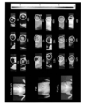
- FIG. 2 is a photograph showing the results of cell imaging with a confocal microscope for each antibody of Example 2.
- FIG. Upper left IgG bound to IR700 (IR700-IgG, negative control), upper right full-length 12A8 antibody bound to IR700 (IR700-12A8 Whole IgG), lower left Fab fragment of 12A8 antibody bound to IR700 (IR700-12A8 Fab), lower right shows the result of F(ab') 2 fragment of 12A8 antibody (IR700-12A8 F(ab') 2 ) bound to IR700.
- FIG. 2 is a graph of the fluorescence intensity measured in FIG. 1.
- FIG. The vertical axis indicates the average fluorescence intensity (cps/cell).
- IgG1 (NC) is the negative control IgG
- whole body is the full-length 12A8 antibody
- F(ab') 2 is the F(ab') 2 fragment of the 12A8 antibody
- Fab is the Fab fragment of the 12A8 antibody.
- the results of fluorescence imaging of each antibody in a tumor-bearing nude mouse model are shown.
- Whole IgG represents the results of the full-length 12A8 antibody, F(ab') 2 the F(ab') 2 fragment of the 12A8 antibody, and Fab the Fab fragment of the 12A8 antibody.
- elapsed time (h) from administration of the labeled antibody or labeled antigen-binding fragment.
- FIG. 4 is a graph showing changes over time in fluorescence intensity measured in FIG. 3.
- the vertical axis represents fluorescence intensity (counts/s/cm 2 /str), and the horizontal axis represents elapsed time (h) after administration of the labeled antibody or labeled antigen-binding fragment.
- 1 is a graph showing the S/N ratio of each antibody in a cancer-bearing nude mouse model. The vertical axis represents the signal to noise ratio, and the horizontal axis represents the elapsed time (h) after administration of the labeled antibody or labeled antigen-binding fragment.
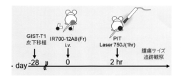
- 1 is a schematic diagram of an in vivo PIT (Photoimmunotherapy) therapeutic trial.
- FIG. 1 It is a photograph comparing before and after PIT treatment using cancer-bearing nude mice.
- mice administered with the Fab fragment of the 12A8 antibody Fab Vehicle
- mice administered with the F(ab') 2 fragment of the 12A8 antibody F(ab' ) 2 Vehicle
- FIG. 1 On the left are photographs of negative control mice that were not subjected to PIT treatment, mice administered with the Fab fragment of the 12A8 antibody (Fab Vehicle) (upper), mice administered with the F(ab') 2 fragment of the 12A8 antibody (F(ab' ) 2 Vehicle) (bottom).
- Fab PIT Fab fragment administration of 12A8 antibody + PIT treatment
- F (ab') 2 fragment administration of 12A8 antibody + PIT treatment F (ab') 2 PIT After
- FIG. 10 is a photograph comparing the fluorescence intensity of fluorescence-labeled anti-c-KIT antibody before and after PIT treatment using cancer-bearing nude mice.
- On the left are photographs of negative control mice that were not subjected to PIT treatment, mice administered with the Fab fragment of the 12A8 antibody (Fab Control (Vehicle)) (upper), mice administered with the F (ab') 2 fragment of the 12A8 antibody (F (ab′) 2 Control (Vehicle) (bottom).
- FIG. 3 is a graph showing changes in tumor size over time in a PIT treatment test using cancer-bearing nude mice. The vertical axis indicates the tumor size (mm 3 ), and the horizontal axis indicates the number of days elapsed after tumor implantation. PIT treatment was performed 28 days after tumor implantation.
- FIG. 1 shows the DNA and amino acid sequences of the heavy chain of humanized anti-c-KIT (12A8) antibody.
- FIG. 1 shows the DNA and amino acid sequences of the heavy chain of humanized anti-c-KIT (12A8) antibody.
- FIG. 1 shows the DNA and amino acid sequences of the light chain of humanized anti-c-KIT (12A8) antibody.
- Fig. 2 shows the DNA sequence and amino acid sequence of the Fab heavy chain of humanized anti-c-KIT (12A8) antibody.
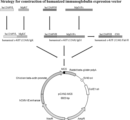
- FIG. 2 shows the structure of the humanized anti-c-KIT (12A8) antibody expression vector.
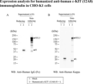
- Fig. 3 is a Western blot photograph showing the results of expression analysis of humanized anti-c-KIT (12A8) IgG1 antibody in CHO-K1 cells. Mock (lane 1) and humanized anti-c-KIT (12A8) IgG1 antibody (lane 2) were used as primary antibodies.
- FIG. 3 is a Western blot photograph showing the results of expression analysis of humanized anti-c-KIT (12A8) Fab antibody in CHO-K1 cells. Mock (lane 1), humanized anti-c-KIT(12A8) Fab antibody (lane 2) and humanized anti-c-KIT(12A8) IgG1 antibody (lane 3) were used as primary antibodies.
- HRP-labeled anti-human Fd antibody was used in A, and HRP-labeled anti-human Kappa antibody was used in B.
- FIG. 10 is a diagram confirming the reactivity of humanized anti-c-KIT(12A8) IgG1 antibody and humanized anti-c-KIT(12A8) Fab antibody by EIA.
- FIG. 10 is a diagram confirming the reactivity of IR700-labeled humanized anti-c-KIT (12A8) antibodies (whole IgG and Fab) by flow cytometry.
- Fig. 10 is a photograph showing the results of fluorescence imaging of each antibody in a tumor-bearing nude mouse model.
- FIG. 10 shows time-course changes in fluorescence intensity of xenograft tumors in mice by IR700-labeled humanized anti-c-KIT (12A8) Fab antibody.
- FIG. 10 shows the time course of S/N ratio in mouse tumor regions using IR700-labeled humanized anti-c-KIT (12A8) Fab antibody.
- the vertical axis indicates the S/N ratio
- the horizontal axis indicates the elapsed time (h) after administration of the labeled antigen-binding fragment.
- FIG. 11 is a photograph showing fluorescence imaging of tumors using IR700-labeled humanized anti-c-KIT (12A8) Fab antibody.
- FIG. 11 is a photograph showing fluorescence imaging showing the effect of photoimmunotherapy when using an IR700-labeled humanized anti-c-KIT(12A8) Fab antibody.
- the antigen-binding fragment of the present specification has a complementarity determining region (CDR) in the antibody heavy chain having the amino acid sequence set forth in SEQ ID NO: 2 or SEQ ID NO: 19, and the amino acid sequence set forth in SEQ ID NO: 4 or SEQ ID NO: 21.
- CDR complementarity determining region
- the amino acid sequence of SEQ ID NO: 2 may be encoded by, for example, the base sequence of SEQ ID NO: 1, and the amino acid sequence of SEQ ID NO: 4 may be, for example, the base sequence of SEQ ID NO: 3.
- the amino acid sequence set forth in SEQ ID NO: 19 may be encoded, for example, by the nucleotide sequence set forth in SEQ ID NO: 18, and the amino acid sequence set forth in SEQ ID NO: 21 may be, for example, It may be encoded by the nucleotide sequence set forth in SEQ ID NO:20.
- the antigen-binding fragment may have a complementarity determining region in the Fab H chain having the amino acid sequence set forth in SEQ ID NO: 6.
- the amino acid sequence set forth in SEQ ID NO: 6 is, for example, It may be encoded by the described base sequence.
- the antigen-binding fragment may have a complementarity determining region in the Fab H chain having the amino acid sequence set forth in SEQ ID NO: 23.
- the amino acid sequence set forth in SEQ ID NO: 23 is, for example, It may be encoded by the described base sequence.
- CDR determination method Kabat et al., Chothia et al., Martin et al., Geldand et al., IMGT (registered trademark) (http://www.imgt.org/IMGTindex/CDR.php), Honnerger et al. ) have been reported (Lars Nieba et al., Protein Engineering, 10(4): 435-444 (1997)). may be determined by any definition.
- the heavy chain CDRs possessed by the antigen-binding fragment herein include CDRH1 consisting of the amino acid sequence set forth in SEQ ID NO:7, CDRH2 consisting of the amino acid sequence set forth in SEQ ID NO:8, and the amino acid set forth in SEQ ID NO:9.
- CDRH3 consisting of the sequence, CDRH1 consisting of the amino acid sequence set forth in SEQ ID NO: 13, CDRH2 consisting of the amino acid sequence set forth in SEQ ID NO: 14, and CDRH3 consisting of the amino acid sequence set forth in SEQ ID NO: 15 can be mentioned.
- the light chain CDRs possessed by the antigen-binding fragment herein include CDRL1 consisting of the amino acid sequence set forth in SEQ ID NO: 10, CDRL2 consisting of the amino acid sequence set forth in SEQ ID NO: 11, and the amino acid set forth in SEQ ID NO: 12. or CDRL1 consisting of the amino acid sequence set forth in SEQ ID NO: 16, CDRL2 consisting of the amino acid sequence GIS (i.e., Gly, Ile, Ser), and CDRL3 consisting of the amino acid sequence set forth in SEQ ID NO: 17. can be done.
- the antigen-binding fragment herein includes (i) CDRH1 consisting of the amino acid sequence set forth in SEQ ID NO: 7, CDRH2 consisting of the amino acid sequence set forth in SEQ ID NO: 8, and CDRH3 consisting of the amino acid sequence set forth in SEQ ID NO: 9.
- CDRL1 consisting of the amino acid sequence set forth in SEQ ID NO: 10
- CDRL2 consisting of the amino acid sequence set forth in SEQ ID NO: 11
- DRL3 consisting of the amino acid sequence set forth in SEQ ID NO: 12
- CDRH1 consisting of the amino acid sequence of
- CDRH2 consisting of the amino acid sequence set forth in SEQ ID NO: 14
- CDRH3 consisting of the amino acid sequence set forth in SEQ ID NO: 15
- CDRL1 consisting of the amino acid sequence set forth in SEQ ID NO: 16
- CDRL2 consisting of the amino acid sequence GIS
- CDRL3 consisting of the amino acid sequence set forth in SEQ ID NO:17.
- antigen-binding fragment refers to a fragment of an antibody that retains the antigen-binding ability of the antibody.
- antigen-binding fragments include F(ab′) 2 , Fab′, Fab, Fab 3 , single-chain Fv (hereinafter referred to as “scFv”), (tandem) bispecific single-chain Fv (sc(Fv ) 2 ), single-chain triple-bodies, nanobodies, divalent VHHs, pentavalent VHHs, minibodies, (double-stranded) diabodies, tandem diabodies, bispecific tribodies, bispecific bibodies, dual affinity retargeting molecules ( DART), triabodies (or tribodies), tetrabodies (or [sc(Fv) 2 ] 2 ), or (scFv-SA) 4 ) disulfide-linked Fv (hereinafter referred to as “dsFv”), compact IgG, heavy chain
- the antigen-binding fragment that constitutes part of the complex of the present invention specifically binds to c-KIT.
- the phrase “specifically” recognizes (binds to) an antigen-binding fragment means that the antigen-binding fragment has substantially greater affinity for c-KIT than affinity for other proteins or peptides. It means binding with high affinity.
- binding with substantially high affinity means that the specific protein or peptide of interest can be distinguished and detected from other proteins or peptides by a desired measurement device or method. means high affinity.
- a substantially higher affinity is 3-fold or more, 4-fold or more, 5-fold or more, 6-fold or more, 7-fold or more, 8-fold or more, as the intensity (e.g., fluorescence intensity) detected by ELISA or EIA. It may mean 9 times or more, 10 times or more, 20 times or more, 30 times or more, 40 times or more, 50 times or more, or 100 times or more.
- the binding rate constant (Ka1) in the binding between the antigen-binding fragment and c-KIT is, for example, 1 ⁇ 10 4 Ms ⁇ 1 or more, 1 ⁇ 10 5 Ms ⁇ 1 or more, 5 ⁇ 10 5 Ms ⁇ 1 or more can be mentioned.
- examples of the dissociation rate constant (Kd1) in binding between the antigen-binding fragment and c-KIT include 1 ⁇ 10 ⁇ 3 or less and 1 ⁇ 10 ⁇ 4 or less.
- the binding constant (KD) in binding between the antigen-binding fragment and c-KIT is, for example, 1 ⁇ 10 ⁇ 8 (M) or less, 5 ⁇ 10 ⁇ 8 (M) or less, 1 ⁇ 10 ⁇ 9 (M) or less.
- the binding rate constant (Ka1), dissociation rate constant (Kd1), and binding constant (KD) of the antigen-binding fragment herein are determined using BIACORE (GE Healthcare Biosciences Co., Ltd., BIACORE-X100). According to the provided manual, after immobilizing biotinylated c-KIT on the SA chip, a test antibody is flowed, the binding rate constant Ka1 and the dissociation rate constant Kd1 are measured, and the binding constant KD value can be determined using bivalent fitting. can.
- Antigen-binding fragments include non-human animal antigen-binding fragments, antigen-binding fragments having non-human animal amino acid sequences and human-derived amino acid sequences.
- the antigen-binding fragment may be a humanized antigen-binding fragment.
- the humanized antigen-binding fragment is based on the genes encoding the H and L chains of a human antibody, and the primary structure portions of the six CDRs are replaced with the H chain (3 It is a fragment of an antibody that is genetically engineered to replace the primary structure of the CDRs of the L chain (3 sites) and L chain (3 sites).
- the antibody of the present invention is preferably a fragment of a humanized antibody.
- the conjugates of the invention may further comprise one or more other antigen-binding fragments of different antibodies, thereby being monospecific, bispecific (bispecific), trispecific (trispecific). ), or multispecific.
- amino acids are represented by single-letter codes. Specifically, A is alanine, L is leucine, R is arginine, K is lysine, N is asparagine, M is methionine, D is aspartic acid, F is phenylalanine, C is cysteine, P is proline, Q is glutamine, S is serine, E is glutamic acid, T is threonine, G is glycine, W is tryptophan, H is histidine, Y is tyrosine, I isoleucine and V is valine.
- IR700 is also called phthalocyanine and means a compound having the following structure.
- IR700 contained in the complex herein may be a derivative thereof as long as it has the above structure.
- IR700 has a substituent that reacts with an amino group or a carboxyl group, and the reaction between the substituent and the amino group or carboxyl group of the antigen-binding fragment allows binding of the antigen-binding fragment and IR700.
- IRDye (registered trademark) 700DX (LI-COR) having the following structure can be used.
- the antigen-binding fragment and IR700 can be bound according to the protocol provided by the manufacturer.
- the binding ratio between the antigen-binding fragment and IR700 is, for example, the number of IR700 molecules bound per antigen-binding fragment (F/P ratio) of 1 to 10, 1 to 5, or 1 to 3. good.
- F/P ratio the number of IR700 molecules bound per antigen-binding fragment
- the average number of IR700 molecules (F/P ratio) bound to one antibody antigen-binding fragment molecule of the complexes in the composition The value may be 1-10, 1-5, 1-3, or 1.5-3.0.
- the present invention relates to a composition for cancer detection/diagnosis, containing the complex as an active ingredient.
- the present invention also provides a method for detecting/diagnosing all cancer cells expressing c-KIT, comprising administering the complex of the present invention to a subject with cancer, and to a method comprising detecting an accumulation of
- cancer refers to cells that have undergone malignant transformation such that they become pathogenic to the host organism.
- cancer includes not only primary cancers, but also metastatic cancers, and in vitro cultures and cell lines derived from cancer cells.
- the cancer may be, for example, a hematopoietic tumor, which is a tumor of blood cells, such as leukemia, such as chronic myelogenous leukemia or acute myelogenous leukemia; myeloma, such as multiple myeloma; lymphoma; be done.
- leukemia such as chronic myelogenous leukemia or acute myelogenous leukemia
- myeloma such as multiple myeloma
- lymphoma lymphoma
- Cancers include, but are not limited to, lung cancer, non-small cell lung cancer, small cell lung cancer, non-Hodgkin's lymphoma, adrenocortical carcinoma, AIDS-related cancer, AIDS-related lymphoma, childhood cerebellar astrocytoma, childhood cerebrum Astrocytoma, basal cell carcinoma, skin cancer (non-melanoma), biliary tract cancer, extrahepatic cholangiocarcinoma, intrahepatic cholangiocarcinoma, bladder cancer, bone and joint cancer, osteosarcoma and malignant fibrous histiocytoma, brain cancer , brain tumor, brain stem glioma, cerebellar astrocytoma, glioma, cerebral astrocytoma/malignant glioma, ependymoma, medulloblastoma, supratentorial primitive neuroectodermal tumor, visual pathway and hypothalamic glioma head and neck cancer, meta
- the complex of the present invention is rapidly accumulated in cancer cells after contact with cancer cells or after administration to a cancer-bearing organism. Therefore, the composition for cancer detection of the present invention can be administered within 24 hours, within 12 hours, within 6 hours, within 5 hours, within 4 hours, within 3 hours, within 2 hours, or within 1 hour after administration of the composition. can be used to detect cancer within From the viewpoint of detection in a short time, it is preferably used to detect cancer within 4 hours, 3 hours, 2 hours, or 1 hour.
- the complex of the present invention since the complex of the present invention has an IR700 molecule, it can kill cancer cells by photoimmunotherapy (PIT) (Mitsunaga M, et al., Nat Med. (2011) 17(12): 1685- 91.). Therefore, the composition for cancer treatment of the present invention is administered to the subject, and after the elapse of time for the composition to accumulate in the cancer, the cancer locality, that is, the accumulation locality of the composition, is irradiated with near-infrared light. By doing so, the cancer therapeutic effect can be exhibited. Accordingly, the present invention relates to a composition for cancer treatment containing the above complex as an active ingredient.
- PIT photoimmunotherapy
- compositions and complexes mainly used for cancer treatment are intended for use in combination with near-infrared light.
- the present invention also relates to a method for treating cancer, comprising administering the complex of the present invention to a subject with cancer, and irradiating near-infrared light to the site of accumulation of the complex in the subject. .
- the composition to accumulate in the cancer is 1 hour to 24 hours, 1 hour to 12 hours, 1 hour to 6 hours, 2 hours to 6 hours after administration of the composition, It can be after 2-4 hours, after 2-3 hours, or after 3-4 hours.
- the antibody fragment of the present invention can be further bound with a labeling substance.
- a labeling substance for example, it can be combined with a fluorescent substance having a wavelength in the visible light region such as 480 to 650 nm or a photosensitive substance to form a complex, which can be applied to the diagnosis and treatment of cancer.
- a radioactive label, an enzyme, a fluorescent label, a bioluminescent label, a chemiluminescent label, or a detectable label such as a metal can be used.
- labels include, but are not limited to, 32P, 3H, 125I, 131I, 14C, radioactive labels such as tritium; ⁇ -galactoxidase, peroxidase, alkaline phosphatase, glucoamylase, enzymes such as glucose oxidase, lactate oxidase, alcohol oxidase, monoamine oxidase, horseradish peroxidase; coenzymes or prosthetic groups such as FAD, FMN, ATP, biotin, heme; Mil, etc.), rhodamine derivatives (tetramethylrhodamine, trimethylrhodamine (RITC), Texas red, rhodamine 110, etc.), Cy dyes (Cy3, Cy5, Cy5.5, Cy7), Cy-chromium, spectrum green, spectrum orange, propidium Fluorescent labels such as iodide, allophycocyanin (APC), R-phycoerythrin (R-PE); biolabel
- the composition of the present invention can be used for cancer detection (diagnosis), and can also be used for treatment as it is. That is, the present invention includes administering the composition of the present invention to a subject, detecting/diagnosing the location and size of cancer in the subject from the fluorescence emitted by IR700 in the administered composition, and The present invention includes methods for detecting/diagnosing and treating cancer, including killing the detected/diagnosed cancer by irradiating the cancer with near-infrared light. Moreover, the present invention may be a composition used for both detection and treatment of cancer (composition for detection and treatment) containing the complex as an active ingredient.
- the present invention relates to the use of the complex of the present invention for producing the composition for detecting cancer, the composition for treating cancer, or the composition for detecting and treating cancer.
- the invention also relates to conjugates of the invention for cancer detection, cancer therapy, or cancer detection and therapy.
- compositions of the present invention include, in addition to intravenous administration, a method of spraying and administering to the gastrointestinal mucosa through forceps holes under endoscopic observation, for example.
- the compositions of the invention are preferably for medical use, and therefore formulations suitable for medical use are preferred.
- Compositions for parenteral administration include, for example, injections, nasal drops, suppositories, and the like. Preferred are injections (solutions such as intravenous injections, subcutaneous injections, intradermal injections, intramuscular injections, intravitreal injections, and drip infusions, and freeze-dried preparations).
- Aqueous liquids for injection include buffers such as phosphate; tonicity agents such as physiological saline, glucose, sucrose, mannitol, sodium chloride and glycerin; Alcohol (e.g., propylene glycol, polyethylene glycol), nonionic surfactant [e.g., polysorbate 80, polysorbate 20, HCO-50 (polyoxyethylene (50 mol) adduct of hydrogenated castor oil)], solubilizer such as ethylenediamine, sulfite Additives such as stabilizers such as phenol, preservatives such as phenol, and soothing agents such as lidocaine can be added.
- buffers such as phosphate
- tonicity agents such as physiological saline, glucose, sucrose, mannitol, sodium chloride and glycerin
- Alcohol e.g., propylene glycol, polyethylene glycol
- nonionic surfactant e.g., polysorbate 80, polysorbate 20, HCO-50 (
- injections can be prepared according to known methods, for example, by dissolving, suspending or emulsifying the complex in a sterile aqueous or oily liquid commonly used for injections. In the case of a freeze-dried preparation, it can be dissolved in water for injection, physiological saline, etc. at the time of use to prepare an injection solution.
- examples of storage containers include ampoules, vials, prefilled syringes, pen-type syringe cartridges, and drip bags.
- Oral administration of proteins is generally considered difficult because they are degraded in the digestive system, but oral administration is also possible when targeting cancers of the digestive system.
- examples of formulations for oral administration include capsules, tablets, syrups, granules and the like.
- composition of the present invention is preferably prepared in dosage unit form to suit the dosage of the active ingredient.
- dosage unit dosage forms include injections (ampoules, vials, prefilled syringes), which usually contain 1 to 1000 mg, 5 to 250 mg, and 10 to 100 mg of the complex of the present invention per dosage unit dosage form. It's okay to be
- the administration of the composition of the present invention may be local or systemic.
- the administration method is not particularly limited, and is administered parenterally or orally as described above.
- Parenteral routes of administration include intraocular, subcutaneous, intraperitoneal, blood (intravenous or intraarterial) or spinal fluid injection or infusion, etc., preferably blood administration.
- Administration of the compositions of the present invention may be single or multiple, and may be temporary, continuous or intermittent.
- one to several doses are administered over a short period of time (within hours, days, 1-2 weeks, etc.).
- the PIT treatment of the cancer can be performed multiple times at intervals.
- the composition is administered one to several times before near-infrared irradiation for each treatment.
- the conjugate of the present invention may be administered alone or in combination with other drugs.
- the dosage of the composition of the present invention is not particularly limited as long as the desired therapeutic effect or detection/diagnostic effect can be obtained, and can be appropriately determined according to symptoms, sex, age, and the like.
- the dose of the composition of the present invention can be determined, for example, using the therapeutic effect on cancer as an index.
- the single dose of the active ingredient of the composition of the present invention is 0.01-20 mg/kg body weight, 0.1-10 mg/kg body weight, 0.1-20 mg/kg body weight, 5 mg/kg body weight is administered once to several times by intravenous injection. In the case of other parenteral administration and oral administration, the amount according to this can be administered. If the symptoms are particularly severe, the dose or number of administrations (number of treatments) may be increased according to the symptoms.
- preparation of its F(ab') 2 fragment and Fab fragment (1)
- Preparation of antigen Human c-KIT (Accession No. NM-000222.3) was used as a GIST target molecule.
- the antigen is an expression vector (pcDNA3.1) cDNA encoding a fusion protein (c-KIT-Fc) between the extracellular region (1-504 amino acids) of the c-KIT gene and the constant region (Fc portion) of mouse IgG2a.
- the recombinant vector was transfected into Chinese hamster ovary (CHO) cells. After culturing c-KIT-Fc-expressing CHO cells in a serum-free TIL medium (Immunobiological Laboratories, Inc.), the culture supernatant was purified using a Protein A column (GE Healthcare). .
- Antigenic determinant (epitope) of mouse anti-c-KIT antibody 12A8 The antigen-determining region recognized by the 12A8 antibody was determined by epitope mapping. A gene encoding a fusion protein between the extracellular region (1-504 amino acids) of human c-KIT and various deletion mutants deleted from the C-terminus of the region and the constant region of mouse IgG2a is prepared, and an expression vector (pcDNA3.1). The generated deletion mutants are human c-KIT1-106 amino acids (c-KIT106-Fc), 1-205 amino acids (c-KIT205-Fc), 1-305 amino acids (c-KIT305-Fc), 1-406 It encodes an amino acid (c-KIT406-Fc).
- Each expression vector carrying these genes was transfected into COS1 cells (ATCC, CRL-1650), cultured in serum-free Dulbecco's MEM (Immuno-Biological Laboratories, Inc.) for 2 days, and the culture supernatant was collected. bottom. The collected culture supernatant was concentrated by ultrafiltration to a 10-fold concentration and dot-blotted onto a PVDF membrane (Millipore, IPVH00010). After blocking treatment of the blotted PVDF membrane, it was reacted with a mouse anti-c-KIT (12A8) antibody, and further reacted with an anti-mouse Ig ⁇ chain HRP-labeled antibody (Southern Biotech, 1050-05). Chemiluminescence detection was performed using Amersham ECL (Cytiva, RPN2106) for detection.
- mice IgG2aFc Since all of the various dotted fusion proteins contained mouse IgG2aFc, they were all positive to anti-mouse IgG antibody (a-Mouse HRP), but not to anti-mouse Ig ⁇ chain HRP (a-mouse kappa HRP). didn't react.
- Mouse anti-c-KIT (12A8) antibody (ac-KIT 12A8) reacted only with c-KIT504-Fc without C-terminal deletion used as an antigen, and reacted with other C-terminal deletion mutant fusion proteins. Since no reaction was observed with , it was clarified that an epitope exists in the 407-504 amino acid region, which is the extracellular domain of c-KIT.
- 12A8 antibody was adjusted with 0.1M/L sodium acetate buffer (Ph5.5), digested with papain at room temperature for 3 hours, purified by gel filtration chromatography (Ultrogel AcA44 column), and further purified by Protein A column. obtained by removing the Fc fragment through
- the F/P for each IR700-12A8 conjugate was 2.4 for Whole IgG, 2.6 for F(ab′) 2 , 1.9 for Fab, and 1.9 for Fab, negative control IgG1 (in response to c-KIT). not) was 3.8.
- Each binding substance was adjusted with buffer for dilution (PBS) so that the molar concentration at the time of use was the same (0.05 nM (Table 1) for in vitro tests and 0.5 nM (Table 2) for in vivo tests).
- PBS buffer for dilution
- Example 2 In vitro binding of IR700-labeled mouse anti-c-KIT antibody and fragments thereof to GIST and its fluorescence intensity IR700-labeled mouse anti-c-KIT antibody prepared in Example 1 ( Using whole body IgG antibody (hereinafter referred to as whole IgG), its F(ab') 2 fragment and Fab fragment, GIST-T1 cell line was subjected to live cell imaging to confirm whether there was a difference in fluorescence density.
- whole IgG antibody hereinafter referred to as whole IgG
- F(ab') 2 fragment and Fab fragment F(ab') 2 fragment and Fab fragment
- Each of the obtained IR700-labeled binding antibodies was dispensed in small amounts and stored at -20°C until use.
- the GIST cultured cell line GIST-T1 (Dr. Tguchi T., Kouchi University, Kouchi, Japan) was cultured and proliferated using a 10 cm dish (Greiner, CELLSTTAR664160-013) with the addition of the RPMI1640 medium. .
- GIST-T1 cells 8 ⁇ 10 4 cells/dish were seeded on 5 glass bottom dishes (Greiner, CELLview 627965), cultured for 24 hours, the supernatant was removed, and a blocking buffer (DaKo, Protein Block serum-free , X0909) and incubated at room temperature for 15 minutes.
- a blocking buffer DaKo, Protein Block serum-free , X0909
- Each IR700-labeled sample listed in Table 1 was adjusted to working concentration with antibody dilution buffer (PBS containing 0.1% BSA), 15 minutes later blocking buffer was discarded, 12A8 conjugate and control conjugate (IR700- IgG1) was added at 500 ⁇ L each.
- IR700-12A8-labeled samples whole IgG and F(ab′) 2 were adjusted to a final concentration of 0.05 nM, but Fabs with monovalent antigen-binding sites were doubled at a final concentration of 0 to match the number of binding sites. .1 nM. Further, 5 ⁇ L of Hoechst 33342 solution (1 mg/ml, DOJINCO H342) was added and incubated on ice for 1 hour.
- FIG. 1 shows a graph of the fluorescence intensity measured in FIG. 12A8 fragment F(ab′) 2 showed fluorescence intensity similar to whole IgG in cell imaging, and 12A8 fragment Fab showed fluorescence intensity superior to whole IgG in cell imaging.
- Example 3 Fluorescence imaging of mouse xenograft tumor using 12A8 fragment (in vivo test)
- Ten athymic BALB/c nude mice (CLEA Japan Inc., Tokyo, Japan) were administered the immunosuppressant cyclophosphamide (0.2 mg/g body weight).
- GIST-T1 cells were adjusted to 1 ⁇ 10 7 cells/50 ⁇ L with PBS, and suspended in thawed Matrigel (Corning Incorporated, USA) on ice at a ratio of 1:1.
- Two days after administration of the immunosuppressive agent 100 ⁇ L of the suspended GIST-T1 cells were subcutaneously transplanted to the right thigh of nude mice treated with the immunosuppressive agent. When the tumor reached 8 mm, the antibody was administered and observed.
- the S/N ratio (signal to noise ratio) in the in vivo model is shown in FIG.
- the S/N ratio of 12A8 fragment (Fab, F(ab') 2 ) was comparable to Whole IgG. From this result, it is possible to diagnose GIST in a short time (within 3 hours) after administering the antibody to the living body by using a fragment of the GIST-specific antibody (12A8) bound to a fluorescent dye. shown.
- Example 4 In vivo PIT (Photoimmunotherapy) treatment test 28 days after transplanting GIST-T1 into nude mice administered immunosuppressants, 4 mice with similar tumor sizes were selected and sedated with isoflavones. 79 ⁇ g and 66 ⁇ g of IR700-12A8Fab and IR700-12A8F(ab′) 2 were administered to the tail vein, respectively, and about 2 hours later, the tumor was irradiated with a laser (735J) for 1 hour. A schematic of the experimental scheme is shown in FIG.
- Tumor size was observed for 4 weeks, and Alexa Fruor 647-labeled mouse anti-c-KIT antibody (BioLegend Inc., San Diego, USA) was administered to the tail vein 55 days after transplantation, and observed by IVIS 48 hours later. bottom.
- Example 5 Generation of IR700-Humanized Anti-c-KIT (12A8) Antibodies (Whole IgG and Fab) Safely as Diagnostic and/or Therapeutic Compositions for Human Gastrointestinal Stromal Tumors (GIST)
- GIST Gastrointestinal Stromal Tumors
- variable region genes VH and VL of the synthesized humanized anti-c-KIT (12A8) antibody and the natural human IgG1 gene, humanized antibody by overlap PCR (polymerase chain reaction)
- the heavy chain (HC) Figure 10, SEQ ID NO: 18
- LC Figure 11, SEQ ID NO: 20
- HC fuses humanized anti-c-KIT (12A8) antibody VH and human IgG constant region (Fc)
- LC fuses humanized anti-c-KIT (12A8) antibody VL with human Kappa chain constant region (KC). fused.
- the HC gene of the humanized anti-c-KIT (12A8) antibody was used as a template, and a stop codon was inserted immediately before the hinge region (Fab HC) (Fig. 12, SEQ ID NO: 22). ) was amplified by PCR.
- the HC gene, LC gene and Fab HC gene of the humanized anti-c-KIT (12A8) antibody were each inserted into a mammalian cell expression vector (pCXN2-MCS, Immuno-Biological Laboratories, Inc.) (Fig.
- Antibody gene expression vector (i) humanized c-KIT (12A8) IgG1 (humanized c-KIT (12A8) IgG1) vector, (ii) humanized c-KIT (12A8) Ig ⁇ (humanized c-KIT (12A8) Ig ⁇ ) vector, (iii) humanized c-KIT(12A8) Fab H (humanized c-KIT(12A8) Fab H) vector was generated.
- This band has approximately the same size as the Mw (weight average molecular weight) of whole IgG and corresponds to two heavy chain (H) molecules and two light chain (L) molecules.
- the band reacting with the HRP-labeled anti-IgG (Fc) antibody corresponds to the Mw of the heavy chain (HC) of humanized c-KIT (12A8) IgG1
- the HRP-labeled anti-Kappa antibody corresponded to the Mw of the light chain (LC) of humanized c-KIT(12A8)Ig ⁇ .
- Fab 15 is a whole IgG antibody produced with (i) a humanized c-KIT (12A8) IgG1 vector and (ii) a humanized c-KIT (12A8) Ig ⁇ vector, and is an HRP-labeled anti-Fd antibody.
- HRP-labeled anti-Kappa antibody molecules corresponding to the Mw of Fab could not be confirmed in both reduced and non-reduced conditions.
- 12A8) antibody Fab fragment hereinafter also referred to as humanized anti-c-KIT (12A8) Fab antibody was confirmed.
- the humanized anti-c-KIT (12A8) IgG1 antibody-producing clone culture supernatant was serially diluted from 2-fold to 64-fold and added to the antigen solid-phase plate and the negative control plate for primary reaction.
- Humanized anti-c-KIT (12A8) Fab antibody was similarly performed. The primary reaction was carried out at room temperature for 1 hour, after which each well was washed with phosphate buffered saline (PBS). HRP-labeled anti-human Kappa chain antibody was added as a secondary antibody and reacted at room temperature for 30 minutes.
- the humanized anti-c-KIT(12A8) IgG1 antibody and the humanized anti-c-KIT(12A8) Fab antibody have antigen specificity. Since each culture supernatant is used for in-vitro and in-vivo tests, the humanized anti-c-KIT (12A8) IgG1 antibody is a protein A column, and the humanized anti-c-KIT (12A8) Fab antibody is Each was purified with Kan Cap G (KANEKA).
- Example 6 Reactivity study of IR700-humanized anti-c-KIT (12A8) antibody (whole IgG and Fab) by flow cytometry (in vitro) Flow cytometry was performed using IR700-labeled humanized anti-c-KIT (12A8) antibody (whole IgG and Fab) and GIST-T1 cell line to confirm the reactivity of the humanized antibody.
- GIST-T1 cells were collected with trypsin (on ice), and 1 ⁇ 10 6 cells were dispensed into 1.5 mL tubes. Washed 3 times with 1 mL of PBS-BSA-Azide. 1 mL of 4% paraformaldehyde cooled to 4° C. was added and gently mixed by pipetting on ice. It was returned to room temperature and allowed to stand for 15 minutes (the work was carried out at room temperature thereafter). Washed 3 times with 1 mL of PBS-BSA-Azide.
- IR700-labeled mouse anti-c-KIT (12A8) antibody is also referred to as 12A8 (whole IgG)
- IR700-labeled humanized anti-c-KIT (12A8) IgG1 antibody is also referred to as humanized (whole IgG)
- IR700 The labeled humanized anti-c-KIT(12A8) Fab antibody is also referred to as humanized (Fab).
- the MFI of IR700-labeled humanized anti-c-KIT(12A8) IgG1 antibody was similar to that obtained with IR700-labeled mouse anti-c-KIT(12A8) antibody. Considering that the IR700-labeled mouse anti-c-KIT (12A8) antibody has a high F/P ratio of 4.4, the IR700-labeled humanized anti-c-KIT (12A8) IgG1 antibody has good reactivity with c-KIT. was shown.
- Example 7 Fluorescent imaging of xenograft tumors in mice using IR700-labeled humanized anti-c-KIT (12A8) antibodies (whole IgG and Fab)
- fluorescence imaging after administration of IR700-labeled humanized anti-c-KIT (12A8) Fab antibody to a mouse model in vivo showed that the fluorescence intensity in the tumor region (ROI) was 3 It reached a peak after ⁇ 4 hours and then tapered off.
- the fluorescence intensity of the control ROI on the contralateral side (left thigh) to which no tumor cells were administered peaked after 1 to 6 hours and then gradually decreased.
- fluorescence imaging after administration of IR700-labeled humanized anti-c-KIT (12A8) IgG1 antibody almost no fluorescence was observed 1 to 4 hours after administration, and even if confirmed, the fluorescence intensity was weak.
- the S/N ratio in the tumor region (ROI) after administration of the IR700-labeled humanized anti-c-KIT (12A8) Fab antibody peaked after 3 to 4 hours and then gradually decreased. bottom.
- the above results indicate that the IR700-labeled humanized anti-c-KIT (12A8) Fab antibody can accumulate in tumors in a short period of time, and is suitable for diagnosis of GIST in a short period of time. Subsequently, therapeutic experiments using small animals were conducted.
- 80 ⁇ g of IR700-labeled humanized anti-c-KIT (12A8) Fab antibody was administered (n 2), and approximately 3 hours after each, the tumors were irradiated with a laser for 1 hour (735 J). After that, the tumor size was observed for 3 weeks together with a control group (saline-administered group) in which the tumor was not treated.
- the treatment schedule is shown in FIG.
- IVIS IVIS
- the fluorescence intensity increased with the passage of 1 to 3 hours after administration of the IR700-labeled humanized anti-c-KIT (12A8) Fab antibody.
- FIG. 23 as a result of irradiation with a 685 nm laser 3 hours after administration of the antibody, the tumor disappeared.
Landscapes
- Health & Medical Sciences (AREA)
- Life Sciences & Earth Sciences (AREA)
- Chemical & Material Sciences (AREA)
- General Health & Medical Sciences (AREA)
- Medicinal Chemistry (AREA)
- Veterinary Medicine (AREA)
- Animal Behavior & Ethology (AREA)
- Public Health (AREA)
- Engineering & Computer Science (AREA)
- Immunology (AREA)
- Epidemiology (AREA)
- Molecular Biology (AREA)
- Organic Chemistry (AREA)
- Pharmacology & Pharmacy (AREA)
- Biochemistry (AREA)
- Biomedical Technology (AREA)
- Urology & Nephrology (AREA)
- Hematology (AREA)
- Nuclear Medicine, Radiotherapy & Molecular Imaging (AREA)
- Proteomics, Peptides & Aminoacids (AREA)
- Genetics & Genomics (AREA)
- Biophysics (AREA)
- Cell Biology (AREA)
- General Chemical & Material Sciences (AREA)
- Chemical Kinetics & Catalysis (AREA)
- Bioinformatics & Cheminformatics (AREA)
- Pathology (AREA)
- Physics & Mathematics (AREA)
- Biotechnology (AREA)
- Analytical Chemistry (AREA)
- General Physics & Mathematics (AREA)
- Microbiology (AREA)
- Food Science & Technology (AREA)
- Peptides Or Proteins (AREA)
- Medicines Containing Antibodies Or Antigens For Use As Internal Diagnostic Agents (AREA)
- Pharmaceuticals Containing Other Organic And Inorganic Compounds (AREA)
- Medicinal Preparation (AREA)
- Medicines That Contain Protein Lipid Enzymes And Other Medicines (AREA)
Priority Applications (4)
| Application Number | Priority Date | Filing Date | Title |
|---|---|---|---|
| JP2023543774A JP7460063B2 (ja) | 2021-08-27 | 2022-08-02 | c-KIT陽性腫瘍特異的抗体断片 |
| US18/686,639 US20250215073A1 (en) | 2021-08-27 | 2022-08-02 | Antibody fragment specific to c-kit positive tumors |
| CN202280062211.0A CN117980007A (zh) | 2021-08-27 | 2022-08-02 | c-KIT阳性肿瘤特异性抗体片段 |
| EP22861082.0A EP4393516A1 (en) | 2021-08-27 | 2022-08-02 | Antibody fragment specific to c-kit positive tumors |
Applications Claiming Priority (2)
| Application Number | Priority Date | Filing Date | Title |
|---|---|---|---|
| JP2021-139380 | 2021-08-27 | ||
| JP2021139380 | 2021-08-27 |
Publications (1)
| Publication Number | Publication Date |
|---|---|
| WO2023026791A1 true WO2023026791A1 (ja) | 2023-03-02 |
Family
ID=85323099
Family Applications (1)
| Application Number | Title | Priority Date | Filing Date |
|---|---|---|---|
| PCT/JP2022/029636 Ceased WO2023026791A1 (ja) | 2021-08-27 | 2022-08-02 | c-KIT陽性腫瘍特異的抗体断片 |
Country Status (5)
| Country | Link |
|---|---|
| US (1) | US20250215073A1 (https=) |
| EP (1) | EP4393516A1 (https=) |
| JP (1) | JP7460063B2 (https=) |
| CN (1) | CN117980007A (https=) |
| WO (1) | WO2023026791A1 (https=) |
Cited By (6)
| Publication number | Priority date | Publication date | Assignee | Title |
|---|---|---|---|---|
| WO2024040194A1 (en) | 2022-08-17 | 2024-02-22 | Capstan Therapeutics, Inc. | Conditioning for in vivo immune cell engineering |
| WO2024249954A1 (en) | 2023-05-31 | 2024-12-05 | Capstan Therapeutics, Inc. | Lipid nanoparticle formulations and compositions |
| WO2025076127A1 (en) | 2023-10-05 | 2025-04-10 | Capstan Therapeutics, Inc. | Constrained ionizable cationic lipids and lipid nanoparticles |
| WO2025076113A1 (en) | 2023-10-05 | 2025-04-10 | Capstan Therapeutics, Inc. | Ionizable cationic lipids with conserved spacing and lipid nanoparticles |
| WO2025217454A2 (en) | 2024-04-11 | 2025-10-16 | Capstan Therapeutics, Inc. | Ionizable cationic lipids and lipid nanoparticles |
| WO2025217452A1 (en) | 2024-04-11 | 2025-10-16 | Capstan Therapeutics, Inc. | Constrained ionizable cationic lipids and lipid nanoparticles |
Families Citing this family (1)
| Publication number | Priority date | Publication date | Assignee | Title |
|---|---|---|---|---|
| CN120289629B (zh) * | 2025-04-14 | 2025-09-30 | 军事科学院军事医学研究院军事兽医研究所 | 一种犬瘟热病毒h蛋白单克隆抗体2d1b1及其应用 |
Family Cites Families (1)
| Publication number | Priority date | Publication date | Assignee | Title |
|---|---|---|---|---|
| WO2020179749A1 (ja) | 2019-03-05 | 2020-09-10 | 国立大学法人東海国立大学機構 | 標的特異的複合体及びその用途 |
-
2022
- 2022-08-02 WO PCT/JP2022/029636 patent/WO2023026791A1/ja not_active Ceased
- 2022-08-02 JP JP2023543774A patent/JP7460063B2/ja active Active
- 2022-08-02 US US18/686,639 patent/US20250215073A1/en active Pending
- 2022-08-02 EP EP22861082.0A patent/EP4393516A1/en active Pending
- 2022-08-02 CN CN202280062211.0A patent/CN117980007A/zh active Pending
Non-Patent Citations (9)
| Title |
|---|
| CHISATO YOSHIDA ET AL., NUCLEAR MEDICINE AND BIOLOGY, vol. 38, 2011, pages 331 - 337 |
| CHISATO YOSHIDA ET AL., PLOS ONE, vol. 8, no. 3, 2013, pages e59248 |
| FUJIMOTO SHOTA, MUGURUMA NAOKI, OKAMOTO KOICHI, KURIHARA TAKESHI, SATO YASUSHI, MIYAMOTO YOSHIHIKO, KITAMURA SHINJI, MIYAMOTO HIRO: "A Novel Theranostic Combination of Near-infrared Fluorescence Imaging and Laser Irradiation Targeting c-KIT for Gastrointestinal Stromal Tumors", THERANOSTICS, IVYSPRING INTERNATIONAL PUBLISHER, AU, vol. 8, no. 9, 1 January 2018 (2018-01-01), AU , pages 2313 - 2328, XP093039147, ISSN: 1838-7640, DOI: 10.7150/thno.22027 * |
| J. IMMUNOL., vol. 146, pages 3721 - 3728 |
| LARS NIEBA ET AL., PROTEIN ENGINEERING, vol. 10, no. 4, 1997, pages 435 - 444 |
| MAO CHENGQIONG, QU PING, MILEY MICHAEL J., ZHAO YAN, LI ZIBO, MING XIN: "P-glycoprotein targeted photodynamic therapy of chemoresistant tumors using recombinant Fab fragment conjugates", BIOMATERIALS SCIENCE, R S C PUBLICATIONS, GB, vol. 6, no. 11, 24 October 2018 (2018-10-24), GB , pages 3063 - 3074, XP093039151, ISSN: 2047-4830, DOI: 10.1039/C8BM00844B * |
| MITSUNAGA M ET AL., NAT MED., vol. 17, no. 12, 2011, pages 1685 - 91 |
| SHOTA FUJIMOTO ET AL., THERANOSTICS, vol. 8, no. 9, 2018, pages 2313 - 2328 |
| WEI DANFENG, TAO ZE, SHI QIUXIAO, WANG LIJUN, LIU LEI, SHE TIANSHAN, YI QIN, WEN XIANG, LIU LIAN, LI SHENGFU, YANG HAO, JIANG XIAN: "Selective Photokilling of Colorectal Tumors by Near-Infrared Photoimmunotherapy with a GPA33-Targeted Single-Chain Antibody Variable Fragment Conjugate", MOLECULAR PHARMACEUTICS, AMERICAN CHEMICAL SOCIETY, US, vol. 17, no. 7, 6 July 2020 (2020-07-06), US , pages 2508 - 2517, XP093039153, ISSN: 1543-8384, DOI: 10.1021/acs.molpharmaceut.0c00210 * |
Cited By (7)
| Publication number | Priority date | Publication date | Assignee | Title |
|---|---|---|---|---|
| WO2024040194A1 (en) | 2022-08-17 | 2024-02-22 | Capstan Therapeutics, Inc. | Conditioning for in vivo immune cell engineering |
| WO2024040195A1 (en) | 2022-08-17 | 2024-02-22 | Capstan Therapeutics, Inc. | Conditioning for in vivo immune cell engineering |
| WO2024249954A1 (en) | 2023-05-31 | 2024-12-05 | Capstan Therapeutics, Inc. | Lipid nanoparticle formulations and compositions |
| WO2025076127A1 (en) | 2023-10-05 | 2025-04-10 | Capstan Therapeutics, Inc. | Constrained ionizable cationic lipids and lipid nanoparticles |
| WO2025076113A1 (en) | 2023-10-05 | 2025-04-10 | Capstan Therapeutics, Inc. | Ionizable cationic lipids with conserved spacing and lipid nanoparticles |
| WO2025217454A2 (en) | 2024-04-11 | 2025-10-16 | Capstan Therapeutics, Inc. | Ionizable cationic lipids and lipid nanoparticles |
| WO2025217452A1 (en) | 2024-04-11 | 2025-10-16 | Capstan Therapeutics, Inc. | Constrained ionizable cationic lipids and lipid nanoparticles |
Also Published As
| Publication number | Publication date |
|---|---|
| US20250215073A1 (en) | 2025-07-03 |
| JP7460063B2 (ja) | 2024-04-02 |
| JPWO2023026791A1 (https=) | 2023-03-02 |
| EP4393516A1 (en) | 2024-07-03 |
| CN117980007A (zh) | 2024-05-03 |
Similar Documents
| Publication | Publication Date | Title |
|---|---|---|
| JP7460063B2 (ja) | c-KIT陽性腫瘍特異的抗体断片 | |
| JP7450594B2 (ja) | 治療分子 | |
| US11033634B2 (en) | Light chain variable regions | |
| TWI428347B (zh) | 抗體與免疫接合物及彼等之用途 | |
| US9790274B2 (en) | Monoclonal antibodies targeting EpCAM for detection of prostate cancer lymph node metastases | |
| US11401342B2 (en) | Therapeutic molecules binding PSMA | |
| JP7711208B2 (ja) | Dll3に対する結合分子及びその使用 | |
| ES2881575T3 (es) | Moléculas de unión, especialmente anticuerpos, que se unen a L1CAM (CD171) | |
| JP6650432B2 (ja) | 腎臓関連抗原1に対する抗体およびその抗原結合性フラグメント | |
| AU2016308365A1 (en) | Anti-DLL3 antibody drug conjugates and methods of use | |
| MX2010011955A (es) | Inmunoglobulinas de dominio variable doble y usos de las mismas. | |
| AU2017324983A1 (en) | Synthetic antibodies against VEGF and their uses | |
| NL2038589B1 (en) | An antibody molecule | |
| JP2022543378A (ja) | Il-38特異的抗体 | |
| JP7691668B2 (ja) | ヒトlifr抗原結合タンパク質、その製造方法及び応用 | |
| Cabanillas-Bernal et al. | variable single domains (VNARs): broadly neutralizing tools for | |
| KR20260005222A (ko) | Pdgfr알파를 발현하는 암을 이미징하기 위한 시약 및 방법 | |
| WO2026067777A1 (zh) | 一种抗人去整合素和金属蛋白酶9的抗体及其应用 | |
| JP2026513849A (ja) | Pdgfrアルファを発現するがんを画像診断するための試薬及び方法 |
Legal Events
| Date | Code | Title | Description |
|---|---|---|---|
| 121 | Ep: the epo has been informed by wipo that ep was designated in this application |
Ref document number: 22861082 Country of ref document: EP Kind code of ref document: A1 |
|
| WWE | Wipo information: entry into national phase |
Ref document number: 2023543774 Country of ref document: JP |
|
| WWE | Wipo information: entry into national phase |
Ref document number: 202280062211.0 Country of ref document: CN |
|
| WWE | Wipo information: entry into national phase |
Ref document number: 2022861082 Country of ref document: EP |
|
| NENP | Non-entry into the national phase |
Ref country code: DE |
|
| ENP | Entry into the national phase |
Ref document number: 2022861082 Country of ref document: EP Effective date: 20240327 |
|
| WWP | Wipo information: published in national office |
Ref document number: 18686639 Country of ref document: US |