WO2023026791A1 - Antibody fragment specific to c-kit positive tumors - Google Patents
Antibody fragment specific to c-kit positive tumors Download PDFInfo
- Publication number
- WO2023026791A1 WO2023026791A1 PCT/JP2022/029636 JP2022029636W WO2023026791A1 WO 2023026791 A1 WO2023026791 A1 WO 2023026791A1 JP 2022029636 W JP2022029636 W JP 2022029636W WO 2023026791 A1 WO2023026791 A1 WO 2023026791A1
- Authority
- WO
- WIPO (PCT)
- Prior art keywords
- antibody
- seq
- amino acid
- acid sequence
- kit
- Prior art date
Links
- 206010028980 Neoplasm Diseases 0.000 title claims abstract description 113
- 102000008394 Immunoglobulin Fragments Human genes 0.000 title claims abstract description 5
- 108010021625 Immunoglobulin Fragments Proteins 0.000 title claims abstract description 5
- 102000016971 Proto-Oncogene Proteins c-kit Human genes 0.000 title 1
- 108010014608 Proto-Oncogene Proteins c-kit Proteins 0.000 title 1
- 239000012634 fragment Substances 0.000 claims abstract description 69
- 239000000427 antigen Substances 0.000 claims abstract description 57
- 108091007433 antigens Proteins 0.000 claims abstract description 57
- 102000036639 antigens Human genes 0.000 claims abstract description 57
- 238000011282 treatment Methods 0.000 claims abstract description 50
- 238000001514 detection method Methods 0.000 claims abstract description 20
- 239000004480 active ingredient Substances 0.000 claims abstract description 15
- 238000002372 labelling Methods 0.000 claims abstract description 9
- 238000009825 accumulation Methods 0.000 claims abstract description 7
- FWMNVWWHGCHHJJ-SKKKGAJSSA-N 4-amino-1-[(2r)-6-amino-2-[[(2r)-2-[[(2r)-2-[[(2r)-2-amino-3-phenylpropanoyl]amino]-3-phenylpropanoyl]amino]-4-methylpentanoyl]amino]hexanoyl]piperidine-4-carboxylic acid Chemical compound C([C@H](C(=O)N[C@H](CC(C)C)C(=O)N[C@H](CCCCN)C(=O)N1CCC(N)(CC1)C(O)=O)NC(=O)[C@H](N)CC=1C=CC=CC=1)C1=CC=CC=C1 FWMNVWWHGCHHJJ-SKKKGAJSSA-N 0.000 claims abstract description 6
- 125000003275 alpha amino acid group Chemical group 0.000 claims abstract 18
- 201000011510 cancer Diseases 0.000 claims description 69
- 239000000203 mixture Substances 0.000 claims description 68
- 108010047041 Complementarity Determining Regions Proteins 0.000 claims description 22
- 230000001225 therapeutic effect Effects 0.000 claims description 15
- 238000003745 diagnosis Methods 0.000 claims description 14
- 239000000126 substance Substances 0.000 claims description 6
- 238000002560 therapeutic procedure Methods 0.000 claims description 5
- 238000001727 in vivo Methods 0.000 abstract description 14
- 239000003795 chemical substances by application Substances 0.000 abstract description 10
- 239000002131 composite material Substances 0.000 abstract 3
- 230000000295 complement effect Effects 0.000 abstract 1
- 229940039227 diagnostic agent Drugs 0.000 abstract 1
- 239000000032 diagnostic agent Substances 0.000 abstract 1
- 150000001413 amino acids Chemical group 0.000 description 66
- 210000004027 cell Anatomy 0.000 description 48
- 201000011243 gastrointestinal stromal tumor Diseases 0.000 description 36
- 108090000623 proteins and genes Proteins 0.000 description 29
- 241000699666 Mus <mouse, genus> Species 0.000 description 25
- 108010001336 Horseradish Peroxidase Proteins 0.000 description 22
- 206010051066 Gastrointestinal stromal tumour Diseases 0.000 description 21
- 241000699670 Mus sp. Species 0.000 description 21
- 238000000034 method Methods 0.000 description 18
- 108010054477 Immunoglobulin Fab Fragments Proteins 0.000 description 16
- 102000001706 Immunoglobulin Fab Fragments Human genes 0.000 description 16
- 239000013598 vector Substances 0.000 description 15
- 238000002347 injection Methods 0.000 description 11
- 239000007924 injection Substances 0.000 description 11
- 239000013642 negative control Substances 0.000 description 11
- 235000018102 proteins Nutrition 0.000 description 11
- 102000004169 proteins and genes Human genes 0.000 description 11
- 235000001014 amino acid Nutrition 0.000 description 10
- 229940024606 amino acid Drugs 0.000 description 10
- 238000006243 chemical reaction Methods 0.000 description 10
- 239000012228 culture supernatant Substances 0.000 description 10
- 238000011580 nude mouse model Methods 0.000 description 10
- LOKCTEFSRHRXRJ-UHFFFAOYSA-I dipotassium trisodium dihydrogen phosphate hydrogen phosphate dichloride Chemical compound P(=O)(O)(O)[O-].[K+].P(=O)(O)([O-])[O-].[Na+].[Na+].[Cl-].[K+].[Cl-].[Na+] LOKCTEFSRHRXRJ-UHFFFAOYSA-I 0.000 description 8
- 239000002953 phosphate buffered saline Substances 0.000 description 8
- 241000699660 Mus musculus Species 0.000 description 7
- 239000013604 expression vector Substances 0.000 description 7
- 238000000799 fluorescence microscopy Methods 0.000 description 7
- 238000012360 testing method Methods 0.000 description 7
- XLYOFNOQVPJJNP-UHFFFAOYSA-N water Chemical compound O XLYOFNOQVPJJNP-UHFFFAOYSA-N 0.000 description 7
- 230000037396 body weight Effects 0.000 description 6
- 239000003814 drug Substances 0.000 description 6
- 238000002360 preparation method Methods 0.000 description 6
- 230000009257 reactivity Effects 0.000 description 6
- 239000000243 solution Substances 0.000 description 6
- 239000003981 vehicle Substances 0.000 description 6
- 230000035508 accumulation Effects 0.000 description 5
- 239000000975 dye Substances 0.000 description 5
- 230000000694 effects Effects 0.000 description 5
- 108020001507 fusion proteins Proteins 0.000 description 5
- 102000037865 fusion proteins Human genes 0.000 description 5
- 238000000338 in vitro Methods 0.000 description 5
- 108010082117 matrigel Proteins 0.000 description 5
- 210000003462 vein Anatomy 0.000 description 5
- ISWSIDIOOBJBQZ-UHFFFAOYSA-N Phenol Chemical compound OC1=CC=CC=C1 ISWSIDIOOBJBQZ-UHFFFAOYSA-N 0.000 description 4
- 206010039491 Sarcoma Diseases 0.000 description 4
- 208000000453 Skin Neoplasms Diseases 0.000 description 4
- 239000000872 buffer Substances 0.000 description 4
- 238000012217 deletion Methods 0.000 description 4
- 230000037430 deletion Effects 0.000 description 4
- 238000010586 diagram Methods 0.000 description 4
- 238000003384 imaging method Methods 0.000 description 4
- 230000001900 immune effect Effects 0.000 description 4
- 239000003018 immunosuppressive agent Substances 0.000 description 4
- 239000002609 medium Substances 0.000 description 4
- 201000000849 skin cancer Diseases 0.000 description 4
- 108020004414 DNA Proteins 0.000 description 3
- 102000004190 Enzymes Human genes 0.000 description 3
- 108090000790 Enzymes Proteins 0.000 description 3
- 206010018338 Glioma Diseases 0.000 description 3
- DHMQDGOQFOQNFH-UHFFFAOYSA-N Glycine Chemical compound NCC(O)=O DHMQDGOQFOQNFH-UHFFFAOYSA-N 0.000 description 3
- 206010025323 Lymphomas Diseases 0.000 description 3
- 206010035226 Plasma cell myeloma Diseases 0.000 description 3
- DNIAPMSPPWPWGF-UHFFFAOYSA-N Propylene glycol Chemical compound CC(O)CO DNIAPMSPPWPWGF-UHFFFAOYSA-N 0.000 description 3
- 206010041067 Small cell lung cancer Diseases 0.000 description 3
- 238000002835 absorbance Methods 0.000 description 3
- 230000000903 blocking effect Effects 0.000 description 3
- 208000006990 cholangiocarcinoma Diseases 0.000 description 3
- 239000013024 dilution buffer Substances 0.000 description 3
- 238000010494 dissociation reaction Methods 0.000 description 3
- 230000005593 dissociations Effects 0.000 description 3
- 229940088598 enzyme Drugs 0.000 description 3
- 230000005284 excitation Effects 0.000 description 3
- 238000002474 experimental method Methods 0.000 description 3
- 238000000684 flow cytometry Methods 0.000 description 3
- 238000009472 formulation Methods 0.000 description 3
- 230000014509 gene expression Effects 0.000 description 3
- 210000004408 hybridoma Anatomy 0.000 description 3
- 239000007788 liquid Substances 0.000 description 3
- 201000001441 melanoma Diseases 0.000 description 3
- 201000005443 oral cavity cancer Diseases 0.000 description 3
- 238000007911 parenteral administration Methods 0.000 description 3
- 238000002428 photodynamic therapy Methods 0.000 description 3
- 238000003752 polymerase chain reaction Methods 0.000 description 3
- -1 polyoxyethylene Polymers 0.000 description 3
- 108090000765 processed proteins & peptides Proteins 0.000 description 3
- 208000000587 small cell lung carcinoma Diseases 0.000 description 3
- 238000001228 spectrum Methods 0.000 description 3
- 208000024891 symptom Diseases 0.000 description 3
- 229940124597 therapeutic agent Drugs 0.000 description 3
- 208000008732 thymoma Diseases 0.000 description 3
- 238000002054 transplantation Methods 0.000 description 3
- 210000004881 tumor cell Anatomy 0.000 description 3
- 210000000689 upper leg Anatomy 0.000 description 3
- YBJHBAHKTGYVGT-ZKWXMUAHSA-N (+)-Biotin Chemical compound N1C(=O)N[C@@H]2[C@H](CCCCC(=O)O)SC[C@@H]21 YBJHBAHKTGYVGT-ZKWXMUAHSA-N 0.000 description 2
- MTCFGRXMJLQNBG-REOHCLBHSA-N (2S)-2-Amino-3-hydroxypropansäure Chemical compound OC[C@H](N)C(O)=O MTCFGRXMJLQNBG-REOHCLBHSA-N 0.000 description 2
- GXYLXFITCXXCQV-UHFFFAOYSA-N 10-methylacridin-10-ium Chemical class C1=CC=C2[N+](C)=C(C=CC=C3)C3=CC2=C1 GXYLXFITCXXCQV-UHFFFAOYSA-N 0.000 description 2
- PRDFBSVERLRRMY-UHFFFAOYSA-N 2'-(4-ethoxyphenyl)-5-(4-methylpiperazin-1-yl)-2,5'-bibenzimidazole Chemical compound C1=CC(OCC)=CC=C1C1=NC2=CC=C(C=3NC4=CC(=CC=C4N=3)N3CCN(C)CC3)C=C2N1 PRDFBSVERLRRMY-UHFFFAOYSA-N 0.000 description 2
- 208000031261 Acute myeloid leukaemia Diseases 0.000 description 2
- 239000012103 Alexa Fluor 488 Substances 0.000 description 2
- 206010003571 Astrocytoma Diseases 0.000 description 2
- 208000032791 BCR-ABL1 positive chronic myelogenous leukemia Diseases 0.000 description 2
- 208000003174 Brain Neoplasms Diseases 0.000 description 2
- 206010007953 Central nervous system lymphoma Diseases 0.000 description 2
- 208000010833 Chronic myeloid leukaemia Diseases 0.000 description 2
- 241000725101 Clea Species 0.000 description 2
- 206010009944 Colon cancer Diseases 0.000 description 2
- CMSMOCZEIVJLDB-UHFFFAOYSA-N Cyclophosphamide Chemical compound ClCCN(CCCl)P1(=O)NCCCO1 CMSMOCZEIVJLDB-UHFFFAOYSA-N 0.000 description 2
- 206010014733 Endometrial cancer Diseases 0.000 description 2
- 206010014759 Endometrial neoplasm Diseases 0.000 description 2
- 208000021309 Germ cell tumor Diseases 0.000 description 2
- 208000032612 Glial tumor Diseases 0.000 description 2
- PEDCQBHIVMGVHV-UHFFFAOYSA-N Glycerine Chemical compound OCC(O)CO PEDCQBHIVMGVHV-UHFFFAOYSA-N 0.000 description 2
- AGPKZVBTJJNPAG-WHFBIAKZSA-N L-isoleucine Chemical compound CC[C@H](C)[C@H](N)C(O)=O AGPKZVBTJJNPAG-WHFBIAKZSA-N 0.000 description 2
- 239000012097 Lipofectamine 2000 Substances 0.000 description 2
- 208000000172 Medulloblastoma Diseases 0.000 description 2
- 241001465754 Metazoa Species 0.000 description 2
- 208000034578 Multiple myelomas Diseases 0.000 description 2
- 208000033761 Myelogenous Chronic BCR-ABL Positive Leukemia Diseases 0.000 description 2
- 208000033776 Myeloid Acute Leukemia Diseases 0.000 description 2
- 208000034176 Neoplasms, Germ Cell and Embryonal Diseases 0.000 description 2
- 239000002033 PVDF binder Substances 0.000 description 2
- 239000002202 Polyethylene glycol Substances 0.000 description 2
- 239000012980 RPMI-1640 medium Substances 0.000 description 2
- 201000000582 Retinoblastoma Diseases 0.000 description 2
- 244000000231 Sesamum indicum Species 0.000 description 2
- FAPWRFPIFSIZLT-UHFFFAOYSA-M Sodium chloride Chemical compound [Na+].[Cl-] FAPWRFPIFSIZLT-UHFFFAOYSA-M 0.000 description 2
- 208000021712 Soft tissue sarcoma Diseases 0.000 description 2
- QAOWNCQODCNURD-UHFFFAOYSA-N Sulfuric acid Chemical compound OS(O)(=O)=O QAOWNCQODCNURD-UHFFFAOYSA-N 0.000 description 2
- 108010004469 allophycocyanin Proteins 0.000 description 2
- 125000003277 amino group Chemical group 0.000 description 2
- SESFRYSPDFLNCH-UHFFFAOYSA-N benzyl benzoate Chemical compound C=1C=CC=CC=1C(=O)OCC1=CC=CC=C1 SESFRYSPDFLNCH-UHFFFAOYSA-N 0.000 description 2
- 230000015572 biosynthetic process Effects 0.000 description 2
- 210000004369 blood Anatomy 0.000 description 2
- 239000008280 blood Substances 0.000 description 2
- 210000004899 c-terminal region Anatomy 0.000 description 2
- 125000003178 carboxy group Chemical group [H]OC(*)=O 0.000 description 2
- 208000002458 carcinoid tumor Diseases 0.000 description 2
- 229960004397 cyclophosphamide Drugs 0.000 description 2
- 230000003247 decreasing effect Effects 0.000 description 2
- 210000002249 digestive system Anatomy 0.000 description 2
- 201000010099 disease Diseases 0.000 description 2
- 208000037265 diseases, disorders, signs and symptoms Diseases 0.000 description 2
- 239000002552 dosage form Substances 0.000 description 2
- 229940079593 drug Drugs 0.000 description 2
- 150000002148 esters Chemical class 0.000 description 2
- 238000010195 expression analysis Methods 0.000 description 2
- 239000007850 fluorescent dye Substances 0.000 description 2
- 230000004927 fusion Effects 0.000 description 2
- 238000001641 gel filtration chromatography Methods 0.000 description 2
- 230000001506 immunosuppresive effect Effects 0.000 description 2
- 229960003444 immunosuppressant agent Drugs 0.000 description 2
- 229940125721 immunosuppressive agent Drugs 0.000 description 2
- 238000002513 implantation Methods 0.000 description 2
- 238000001802 infusion Methods 0.000 description 2
- 238000001990 intravenous administration Methods 0.000 description 2
- 238000010253 intravenous injection Methods 0.000 description 2
- 230000001678 irradiating effect Effects 0.000 description 2
- 230000003902 lesion Effects 0.000 description 2
- 210000004185 liver Anatomy 0.000 description 2
- 238000004519 manufacturing process Methods 0.000 description 2
- 239000000463 material Substances 0.000 description 2
- 238000005259 measurement Methods 0.000 description 2
- 239000012528 membrane Substances 0.000 description 2
- 229910052751 metal Inorganic materials 0.000 description 2
- 239000002184 metal Substances 0.000 description 2
- 210000004877 mucosa Anatomy 0.000 description 2
- 201000005962 mycosis fungoides Diseases 0.000 description 2
- 239000002773 nucleotide Substances 0.000 description 2
- 125000003729 nucleotide group Chemical group 0.000 description 2
- 238000011275 oncology therapy Methods 0.000 description 2
- 239000000546 pharmaceutical excipient Substances 0.000 description 2
- 229940124531 pharmaceutical excipient Drugs 0.000 description 2
- 239000003504 photosensitizing agent Substances 0.000 description 2
- 239000002504 physiological saline solution Substances 0.000 description 2
- 229920001223 polyethylene glycol Polymers 0.000 description 2
- 229920002981 polyvinylidene fluoride Polymers 0.000 description 2
- 238000002600 positron emission tomography Methods 0.000 description 2
- 208000016800 primary central nervous system lymphoma Diseases 0.000 description 2
- 102000004196 processed proteins & peptides Human genes 0.000 description 2
- 230000002285 radioactive effect Effects 0.000 description 2
- 230000002829 reductive effect Effects 0.000 description 2
- 239000007974 sodium acetate buffer Substances 0.000 description 2
- 238000002415 sodium dodecyl sulfate polyacrylamide gel electrophoresis Methods 0.000 description 2
- 239000002904 solvent Substances 0.000 description 2
- 241000894007 species Species 0.000 description 2
- 125000001424 substituent group Chemical group 0.000 description 2
- 238000003786 synthesis reaction Methods 0.000 description 2
- 210000001519 tissue Anatomy 0.000 description 2
- 238000001262 western blot Methods 0.000 description 2
- BRZYSWJRSDMWLG-DJWUNRQOSA-N (2r,3r,4r,5r)-2-[(1s,2s,3r,4s,6r)-4,6-diamino-3-[(2s,3r,4r,5s,6r)-3-amino-4,5-dihydroxy-6-[(1r)-1-hydroxyethyl]oxan-2-yl]oxy-2-hydroxycyclohexyl]oxy-5-methyl-4-(methylamino)oxane-3,5-diol Chemical compound O1C[C@@](O)(C)[C@H](NC)[C@@H](O)[C@H]1O[C@@H]1[C@@H](O)[C@H](O[C@@H]2[C@@H]([C@@H](O)[C@H](O)[C@@H]([C@@H](C)O)O2)N)[C@@H](N)C[C@H]1N BRZYSWJRSDMWLG-DJWUNRQOSA-N 0.000 description 1
- NHBKXEKEPDILRR-UHFFFAOYSA-N 2,3-bis(butanoylsulfanyl)propyl butanoate Chemical compound CCCC(=O)OCC(SC(=O)CCC)CSC(=O)CCC NHBKXEKEPDILRR-UHFFFAOYSA-N 0.000 description 1
- JKMHFZQWWAIEOD-UHFFFAOYSA-N 2-[4-(2-hydroxyethyl)piperazin-1-yl]ethanesulfonic acid Chemical compound OCC[NH+]1CCN(CCS([O-])(=O)=O)CC1 JKMHFZQWWAIEOD-UHFFFAOYSA-N 0.000 description 1
- LJCNDNBULVLKSG-UHFFFAOYSA-N 2-aminoacetic acid;butane Chemical compound CCCC.CCCC.NCC(O)=O LJCNDNBULVLKSG-UHFFFAOYSA-N 0.000 description 1
- RMXVNDUWESWVKI-UHFFFAOYSA-N 3-(1-adamantyl)dioxetane Chemical compound C1OOC1C1(C2)CC(C3)CC2CC3C1 RMXVNDUWESWVKI-UHFFFAOYSA-N 0.000 description 1
- HUDPLKWXRLNSPC-UHFFFAOYSA-N 4-aminophthalhydrazide Chemical compound O=C1NNC(=O)C=2C1=CC(N)=CC=2 HUDPLKWXRLNSPC-UHFFFAOYSA-N 0.000 description 1
- 208000030507 AIDS Diseases 0.000 description 1
- 208000002008 AIDS-Related Lymphoma Diseases 0.000 description 1
- 208000024893 Acute lymphoblastic leukemia Diseases 0.000 description 1
- 208000014697 Acute lymphocytic leukaemia Diseases 0.000 description 1
- ZKHQWZAMYRWXGA-KQYNXXCUSA-N Adenosine triphosphate Chemical compound C1=NC=2C(N)=NC=NC=2N1[C@@H]1O[C@H](COP(O)(=O)OP(O)(=O)OP(O)(O)=O)[C@@H](O)[C@H]1O ZKHQWZAMYRWXGA-KQYNXXCUSA-N 0.000 description 1
- 108010025188 Alcohol oxidase Proteins 0.000 description 1
- 239000012114 Alexa Fluor 647 Substances 0.000 description 1
- 102000002260 Alkaline Phosphatase Human genes 0.000 description 1
- 108020004774 Alkaline Phosphatase Proteins 0.000 description 1
- 206010061424 Anal cancer Diseases 0.000 description 1
- 208000007860 Anus Neoplasms Diseases 0.000 description 1
- 241001550224 Apha Species 0.000 description 1
- 206010073360 Appendix cancer Diseases 0.000 description 1
- 239000004475 Arginine Substances 0.000 description 1
- DCXYFEDJOCDNAF-UHFFFAOYSA-N Asparagine Natural products OC(=O)C(N)CC(N)=O DCXYFEDJOCDNAF-UHFFFAOYSA-N 0.000 description 1
- 206010060971 Astrocytoma malignant Diseases 0.000 description 1
- 208000010839 B-cell chronic lymphocytic leukemia Diseases 0.000 description 1
- 238000011725 BALB/c mouse Methods 0.000 description 1
- 238000011729 BALB/c nude mouse Methods 0.000 description 1
- 206010004146 Basal cell carcinoma Diseases 0.000 description 1
- 206010005003 Bladder cancer Diseases 0.000 description 1
- 206010006143 Brain stem glioma Diseases 0.000 description 1
- 206010006187 Breast cancer Diseases 0.000 description 1
- 208000026310 Breast neoplasm Diseases 0.000 description 1
- 101100348617 Candida albicans (strain SC5314 / ATCC MYA-2876) NIK1 gene Proteins 0.000 description 1
- 206010007275 Carcinoid tumour Diseases 0.000 description 1
- 206010007279 Carcinoid tumour of the gastrointestinal tract Diseases 0.000 description 1
- 206010008342 Cervix carcinoma Diseases 0.000 description 1
- 108020004705 Codon Proteins 0.000 description 1
- 208000001333 Colorectal Neoplasms Diseases 0.000 description 1
- 241000699802 Cricetulus griseus Species 0.000 description 1
- FBPFZTCFMRRESA-KVTDHHQDSA-N D-Mannitol Chemical compound OC[C@@H](O)[C@@H](O)[C@H](O)[C@H](O)CO FBPFZTCFMRRESA-KVTDHHQDSA-N 0.000 description 1
- BWGNESOTFCXPMA-UHFFFAOYSA-N Dihydrogen disulfide Chemical compound SS BWGNESOTFCXPMA-UHFFFAOYSA-N 0.000 description 1
- 238000002965 ELISA Methods 0.000 description 1
- 206010014967 Ependymoma Diseases 0.000 description 1
- 208000000461 Esophageal Neoplasms Diseases 0.000 description 1
- LFQSCWFLJHTTHZ-UHFFFAOYSA-N Ethanol Chemical compound CCO LFQSCWFLJHTTHZ-UHFFFAOYSA-N 0.000 description 1
- PIICEJLVQHRZGT-UHFFFAOYSA-N Ethylenediamine Chemical compound NCCN PIICEJLVQHRZGT-UHFFFAOYSA-N 0.000 description 1
- 208000006168 Ewing Sarcoma Diseases 0.000 description 1
- 208000017259 Extragonadal germ cell tumor Diseases 0.000 description 1
- 208000022072 Gallbladder Neoplasms Diseases 0.000 description 1
- 201000003741 Gastrointestinal carcinoma Diseases 0.000 description 1
- 108010073178 Glucan 1,4-alpha-Glucosidase Proteins 0.000 description 1
- 102100022624 Glucoamylase Human genes 0.000 description 1
- WQZGKKKJIJFFOK-GASJEMHNSA-N Glucose Natural products OC[C@H]1OC(O)[C@H](O)[C@@H](O)[C@@H]1O WQZGKKKJIJFFOK-GASJEMHNSA-N 0.000 description 1
- 239000004366 Glucose oxidase Substances 0.000 description 1
- 108010015776 Glucose oxidase Proteins 0.000 description 1
- WHUUTDBJXJRKMK-UHFFFAOYSA-N Glutamic acid Natural products OC(=O)C(N)CCC(O)=O WHUUTDBJXJRKMK-UHFFFAOYSA-N 0.000 description 1
- 239000004471 Glycine Substances 0.000 description 1
- 239000007995 HEPES buffer Substances 0.000 description 1
- 208000017604 Hodgkin disease Diseases 0.000 description 1
- 208000021519 Hodgkin lymphoma Diseases 0.000 description 1
- 208000010747 Hodgkins lymphoma Diseases 0.000 description 1
- 101000998952 Homo sapiens Immunoglobulin heavy variable 1-3 Proteins 0.000 description 1
- 101001047628 Homo sapiens Immunoglobulin kappa variable 2-29 Proteins 0.000 description 1
- 101001008874 Homo sapiens Mast/stem cell growth factor receptor Kit Proteins 0.000 description 1
- 101000851030 Homo sapiens Vascular endothelial growth factor receptor 3 Proteins 0.000 description 1
- 206010021042 Hypopharyngeal cancer Diseases 0.000 description 1
- 206010056305 Hypopharyngeal neoplasm Diseases 0.000 description 1
- 102000018071 Immunoglobulin Fc Fragments Human genes 0.000 description 1
- 108010091135 Immunoglobulin Fc Fragments Proteins 0.000 description 1
- 102100036886 Immunoglobulin heavy variable 1-3 Human genes 0.000 description 1
- 102100022949 Immunoglobulin kappa variable 2-29 Human genes 0.000 description 1
- 206010061252 Intraocular melanoma Diseases 0.000 description 1
- 208000009164 Islet Cell Adenoma Diseases 0.000 description 1
- PIWKPBJCKXDKJR-UHFFFAOYSA-N Isoflurane Chemical compound FC(F)OC(Cl)C(F)(F)F PIWKPBJCKXDKJR-UHFFFAOYSA-N 0.000 description 1
- 208000007766 Kaposi sarcoma Diseases 0.000 description 1
- 208000008839 Kidney Neoplasms Diseases 0.000 description 1
- QNAYBMKLOCPYGJ-REOHCLBHSA-N L-alanine Chemical compound C[C@H](N)C(O)=O QNAYBMKLOCPYGJ-REOHCLBHSA-N 0.000 description 1
- DCXYFEDJOCDNAF-REOHCLBHSA-N L-asparagine Chemical compound OC(=O)[C@@H](N)CC(N)=O DCXYFEDJOCDNAF-REOHCLBHSA-N 0.000 description 1
- CKLJMWTZIZZHCS-REOHCLBHSA-N L-aspartic acid Chemical compound OC(=O)[C@@H](N)CC(O)=O CKLJMWTZIZZHCS-REOHCLBHSA-N 0.000 description 1
- ROHFNLRQFUQHCH-YFKPBYRVSA-N L-leucine Chemical compound CC(C)C[C@H](N)C(O)=O ROHFNLRQFUQHCH-YFKPBYRVSA-N 0.000 description 1
- COLNVLDHVKWLRT-QMMMGPOBSA-N L-phenylalanine Chemical compound OC(=O)[C@@H](N)CC1=CC=CC=C1 COLNVLDHVKWLRT-QMMMGPOBSA-N 0.000 description 1
- 101150027568 LC gene Proteins 0.000 description 1
- 108010073450 Lactate 2-monooxygenase Proteins 0.000 description 1
- 206010023825 Laryngeal cancer Diseases 0.000 description 1
- 208000018142 Leiomyosarcoma Diseases 0.000 description 1
- ROHFNLRQFUQHCH-UHFFFAOYSA-N Leucine Natural products CC(C)CC(N)C(O)=O ROHFNLRQFUQHCH-UHFFFAOYSA-N 0.000 description 1
- NNJVILVZKWQKPM-UHFFFAOYSA-N Lidocaine Chemical compound CCN(CC)CC(=O)NC1=C(C)C=CC=C1C NNJVILVZKWQKPM-UHFFFAOYSA-N 0.000 description 1
- 108060001084 Luciferase Proteins 0.000 description 1
- 239000005089 Luciferase Substances 0.000 description 1
- 206010058467 Lung neoplasm malignant Diseases 0.000 description 1
- 208000031422 Lymphocytic Chronic B-Cell Leukemia Diseases 0.000 description 1
- 206010025312 Lymphoma AIDS related Diseases 0.000 description 1
- KDXKERNSBIXSRK-UHFFFAOYSA-N Lysine Natural products NCCCCC(N)C(O)=O KDXKERNSBIXSRK-UHFFFAOYSA-N 0.000 description 1
- 239000004472 Lysine Substances 0.000 description 1
- 208000006644 Malignant Fibrous Histiocytoma Diseases 0.000 description 1
- 206010025537 Malignant anorectal neoplasms Diseases 0.000 description 1
- 208000030070 Malignant epithelial tumor of ovary Diseases 0.000 description 1
- 206010064912 Malignant transformation Diseases 0.000 description 1
- 208000032271 Malignant tumor of penis Diseases 0.000 description 1
- 229930195725 Mannitol Natural products 0.000 description 1
- 206010027476 Metastases Diseases 0.000 description 1
- 102000010909 Monoamine Oxidase Human genes 0.000 description 1
- 108010062431 Monoamine oxidase Proteins 0.000 description 1
- 201000003793 Myelodysplastic syndrome Diseases 0.000 description 1
- 208000014767 Myeloproliferative disease Diseases 0.000 description 1
- 201000007224 Myeloproliferative neoplasm Diseases 0.000 description 1
- 206010028729 Nasal cavity cancer Diseases 0.000 description 1
- 208000001894 Nasopharyngeal Neoplasms Diseases 0.000 description 1
- 206010061306 Nasopharyngeal cancer Diseases 0.000 description 1
- 206010029260 Neuroblastoma Diseases 0.000 description 1
- 208000015914 Non-Hodgkin lymphomas Diseases 0.000 description 1
- 108091028043 Nucleic acid sequence Proteins 0.000 description 1
- 239000004677 Nylon Substances 0.000 description 1
- 206010030155 Oesophageal carcinoma Diseases 0.000 description 1
- 206010031096 Oropharyngeal cancer Diseases 0.000 description 1
- 206010057444 Oropharyngeal neoplasm Diseases 0.000 description 1
- 240000007594 Oryza sativa Species 0.000 description 1
- 235000007164 Oryza sativa Nutrition 0.000 description 1
- 208000007571 Ovarian Epithelial Carcinoma Diseases 0.000 description 1
- 206010033128 Ovarian cancer Diseases 0.000 description 1
- 206010061328 Ovarian epithelial cancer Diseases 0.000 description 1
- 206010033268 Ovarian low malignant potential tumour Diseases 0.000 description 1
- 206010061535 Ovarian neoplasm Diseases 0.000 description 1
- 229910019142 PO4 Inorganic materials 0.000 description 1
- 206010061902 Pancreatic neoplasm Diseases 0.000 description 1
- 108090000526 Papain Proteins 0.000 description 1
- 229930040373 Paraformaldehyde Natural products 0.000 description 1
- 208000000821 Parathyroid Neoplasms Diseases 0.000 description 1
- 208000002471 Penile Neoplasms Diseases 0.000 description 1
- 206010034299 Penile cancer Diseases 0.000 description 1
- 102000057297 Pepsin A Human genes 0.000 description 1
- 108090000284 Pepsin A Proteins 0.000 description 1
- 102000003992 Peroxidases Human genes 0.000 description 1
- 208000009565 Pharyngeal Neoplasms Diseases 0.000 description 1
- 206010034811 Pharyngeal cancer Diseases 0.000 description 1
- BELBBZDIHDAJOR-UHFFFAOYSA-N Phenolsulfonephthalein Chemical compound C1=CC(O)=CC=C1C1(C=2C=CC(O)=CC=2)C2=CC=CC=C2S(=O)(=O)O1 BELBBZDIHDAJOR-UHFFFAOYSA-N 0.000 description 1
- 206010050487 Pinealoblastoma Diseases 0.000 description 1
- 208000007641 Pinealoma Diseases 0.000 description 1
- 208000007913 Pituitary Neoplasms Diseases 0.000 description 1
- 208000007452 Plasmacytoma Diseases 0.000 description 1
- 201000008199 Pleuropulmonary blastoma Diseases 0.000 description 1
- 229920003171 Poly (ethylene oxide) Polymers 0.000 description 1
- 229920001213 Polysorbate 20 Polymers 0.000 description 1
- 208000006664 Precursor Cell Lymphoblastic Leukemia-Lymphoma Diseases 0.000 description 1
- ONIBWKKTOPOVIA-UHFFFAOYSA-N Proline Natural products OC(=O)C1CCCN1 ONIBWKKTOPOVIA-UHFFFAOYSA-N 0.000 description 1
- 206010060862 Prostate cancer Diseases 0.000 description 1
- 208000000236 Prostatic Neoplasms Diseases 0.000 description 1
- 239000004365 Protease Substances 0.000 description 1
- 208000015634 Rectal Neoplasms Diseases 0.000 description 1
- 206010038389 Renal cancer Diseases 0.000 description 1
- 239000006146 Roswell Park Memorial Institute medium Substances 0.000 description 1
- 101100007329 Saccharomyces cerevisiae (strain ATCC 204508 / S288c) COS1 gene Proteins 0.000 description 1
- 208000004337 Salivary Gland Neoplasms Diseases 0.000 description 1
- 206010061934 Salivary gland cancer Diseases 0.000 description 1
- MTCFGRXMJLQNBG-UHFFFAOYSA-N Serine Natural products OCC(N)C(O)=O MTCFGRXMJLQNBG-UHFFFAOYSA-N 0.000 description 1
- 108010003723 Single-Domain Antibodies Proteins 0.000 description 1
- 208000005718 Stomach Neoplasms Diseases 0.000 description 1
- 229930006000 Sucrose Natural products 0.000 description 1
- CZMRCDWAGMRECN-UGDNZRGBSA-N Sucrose Chemical compound O[C@H]1[C@H](O)[C@@H](CO)O[C@@]1(CO)O[C@@H]1[C@H](O)[C@@H](O)[C@H](O)[C@@H](CO)O1 CZMRCDWAGMRECN-UGDNZRGBSA-N 0.000 description 1
- LSNNMFCWUKXFEE-UHFFFAOYSA-N Sulfurous acid Chemical compound OS(O)=O LSNNMFCWUKXFEE-UHFFFAOYSA-N 0.000 description 1
- 108700005078 Synthetic Genes Proteins 0.000 description 1
- 208000031673 T-Cell Cutaneous Lymphoma Diseases 0.000 description 1
- 208000024313 Testicular Neoplasms Diseases 0.000 description 1
- 206010057644 Testis cancer Diseases 0.000 description 1
- AYFVYJQAPQTCCC-UHFFFAOYSA-N Threonine Natural products CC(O)C(N)C(O)=O AYFVYJQAPQTCCC-UHFFFAOYSA-N 0.000 description 1
- 239000004473 Threonine Substances 0.000 description 1
- 206010043515 Throat cancer Diseases 0.000 description 1
- 201000009365 Thymic carcinoma Diseases 0.000 description 1
- 208000024770 Thyroid neoplasm Diseases 0.000 description 1
- 206010062129 Tongue neoplasm Diseases 0.000 description 1
- YZCKVEUIGOORGS-NJFSPNSNSA-N Tritium Chemical compound [3H] YZCKVEUIGOORGS-NJFSPNSNSA-N 0.000 description 1
- 102000004142 Trypsin Human genes 0.000 description 1
- 108090000631 Trypsin Proteins 0.000 description 1
- QIVBCDIJIAJPQS-UHFFFAOYSA-N Tryptophan Natural products C1=CC=C2C(CC(N)C(O)=O)=CNC2=C1 QIVBCDIJIAJPQS-UHFFFAOYSA-N 0.000 description 1
- 208000015778 Undifferentiated pleomorphic sarcoma Diseases 0.000 description 1
- 206010046431 Urethral cancer Diseases 0.000 description 1
- 206010046458 Urethral neoplasms Diseases 0.000 description 1
- 208000007097 Urinary Bladder Neoplasms Diseases 0.000 description 1
- 208000006105 Uterine Cervical Neoplasms Diseases 0.000 description 1
- 208000002495 Uterine Neoplasms Diseases 0.000 description 1
- 201000005969 Uveal melanoma Diseases 0.000 description 1
- KZSNJWFQEVHDMF-UHFFFAOYSA-N Valine Natural products CC(C)C(N)C(O)=O KZSNJWFQEVHDMF-UHFFFAOYSA-N 0.000 description 1
- 206010047741 Vulval cancer Diseases 0.000 description 1
- 208000004354 Vulvar Neoplasms Diseases 0.000 description 1
- 208000033559 Waldenström macroglobulinemia Diseases 0.000 description 1
- 208000008383 Wilms tumor Diseases 0.000 description 1
- DZBUGLKDJFMEHC-UHFFFAOYSA-N acridine Chemical class C1=CC=CC2=CC3=CC=CC=C3N=C21 DZBUGLKDJFMEHC-UHFFFAOYSA-N 0.000 description 1
- 239000000654 additive Substances 0.000 description 1
- 208000020990 adrenal cortex carcinoma Diseases 0.000 description 1
- 208000007128 adrenocortical carcinoma Diseases 0.000 description 1
- 235000004279 alanine Nutrition 0.000 description 1
- 229910052782 aluminium Inorganic materials 0.000 description 1
- XAGFODPZIPBFFR-UHFFFAOYSA-N aluminium Chemical compound [Al] XAGFODPZIPBFFR-UHFFFAOYSA-N 0.000 description 1
- 230000000890 antigenic effect Effects 0.000 description 1
- 201000011165 anus cancer Diseases 0.000 description 1
- 208000021780 appendiceal neoplasm Diseases 0.000 description 1
- ODKSFYDXXFIFQN-UHFFFAOYSA-N arginine Natural products OC(=O)C(N)CCCNC(N)=N ODKSFYDXXFIFQN-UHFFFAOYSA-N 0.000 description 1
- 125000000637 arginyl group Chemical group N[C@@H](CCCNC(N)=N)C(=O)* 0.000 description 1
- 235000009582 asparagine Nutrition 0.000 description 1
- 229960001230 asparagine Drugs 0.000 description 1
- 235000003704 aspartic acid Nutrition 0.000 description 1
- 229960002903 benzyl benzoate Drugs 0.000 description 1
- WQZGKKKJIJFFOK-VFUOTHLCSA-N beta-D-glucose Chemical compound OC[C@H]1O[C@@H](O)[C@H](O)[C@@H](O)[C@@H]1O WQZGKKKJIJFFOK-VFUOTHLCSA-N 0.000 description 1
- OQFSQFPPLPISGP-UHFFFAOYSA-N beta-carboxyaspartic acid Natural products OC(=O)C(N)C(C(O)=O)C(O)=O OQFSQFPPLPISGP-UHFFFAOYSA-N 0.000 description 1
- 201000009036 biliary tract cancer Diseases 0.000 description 1
- 208000020790 biliary tract neoplasm Diseases 0.000 description 1
- 229960002685 biotin Drugs 0.000 description 1
- 235000020958 biotin Nutrition 0.000 description 1
- 239000011616 biotin Substances 0.000 description 1
- 210000000601 blood cell Anatomy 0.000 description 1
- 210000000988 bone and bone Anatomy 0.000 description 1
- 210000001185 bone marrow Anatomy 0.000 description 1
- 208000012172 borderline epithelial tumor of ovary Diseases 0.000 description 1
- 201000002143 bronchus adenoma Diseases 0.000 description 1
- 239000012830 cancer therapeutic Substances 0.000 description 1
- 239000002775 capsule Substances 0.000 description 1
- 239000004359 castor oil Substances 0.000 description 1
- 235000019438 castor oil Nutrition 0.000 description 1
- 230000015556 catabolic process Effects 0.000 description 1
- 201000007455 central nervous system cancer Diseases 0.000 description 1
- 201000007335 cerebellar astrocytoma Diseases 0.000 description 1
- 208000030239 cerebral astrocytoma Diseases 0.000 description 1
- 210000004720 cerebrum Anatomy 0.000 description 1
- 201000010881 cervical cancer Diseases 0.000 description 1
- 238000002038 chemiluminescence detection Methods 0.000 description 1
- 201000004677 childhood cerebellar astrocytic neoplasm Diseases 0.000 description 1
- 208000011654 childhood malignant neoplasm Diseases 0.000 description 1
- 210000004978 chinese hamster ovary cell Anatomy 0.000 description 1
- 229910052804 chromium Inorganic materials 0.000 description 1
- 239000011651 chromium Substances 0.000 description 1
- 230000001684 chronic effect Effects 0.000 description 1
- 208000032852 chronic lymphocytic leukemia Diseases 0.000 description 1
- 238000010367 cloning Methods 0.000 description 1
- 239000005515 coenzyme Substances 0.000 description 1
- 239000000084 colloidal system Substances 0.000 description 1
- 208000029742 colonic neoplasm Diseases 0.000 description 1
- 239000002299 complementary DNA Substances 0.000 description 1
- 150000001875 compounds Chemical class 0.000 description 1
- 238000012790 confirmation Methods 0.000 description 1
- 238000010276 construction Methods 0.000 description 1
- 210000004748 cultured cell Anatomy 0.000 description 1
- 238000012258 culturing Methods 0.000 description 1
- 201000007241 cutaneous T cell lymphoma Diseases 0.000 description 1
- 235000018417 cysteine Nutrition 0.000 description 1
- XUJNEKJLAYXESH-UHFFFAOYSA-N cysteine Natural products SCC(N)C(O)=O XUJNEKJLAYXESH-UHFFFAOYSA-N 0.000 description 1
- 238000006731 degradation reaction Methods 0.000 description 1
- 238000012631 diagnostic technique Methods 0.000 description 1
- 238000010790 dilution Methods 0.000 description 1
- 239000012895 dilution Substances 0.000 description 1
- 238000003113 dilution method Methods 0.000 description 1
- 238000000375 direct analysis in real time Methods 0.000 description 1
- 235000021186 dishes Nutrition 0.000 description 1
- 230000009977 dual effect Effects 0.000 description 1
- 238000012063 dual-affinity re-targeting Methods 0.000 description 1
- 230000001804 emulsifying effect Effects 0.000 description 1
- 201000004101 esophageal cancer Diseases 0.000 description 1
- 208000024519 eye neoplasm Diseases 0.000 description 1
- FVTCRASFADXXNN-SCRDCRAPSA-N flavin mononucleotide Chemical compound OP(=O)(O)OC[C@@H](O)[C@@H](O)[C@@H](O)CN1C=2C=C(C)C(C)=CC=2N=C2C1=NC(=O)NC2=O FVTCRASFADXXNN-SCRDCRAPSA-N 0.000 description 1
- 239000012530 fluid Substances 0.000 description 1
- 238000001943 fluorescence-activated cell sorting Methods 0.000 description 1
- 238000012632 fluorescent imaging Methods 0.000 description 1
- 239000011888 foil Substances 0.000 description 1
- 201000010175 gallbladder cancer Diseases 0.000 description 1
- 206010017758 gastric cancer Diseases 0.000 description 1
- 230000002496 gastric effect Effects 0.000 description 1
- 210000001035 gastrointestinal tract Anatomy 0.000 description 1
- 201000007116 gestational trophoblastic neoplasm Diseases 0.000 description 1
- 239000011521 glass Substances 0.000 description 1
- 239000008103 glucose Substances 0.000 description 1
- 229940116332 glucose oxidase Drugs 0.000 description 1
- 235000019420 glucose oxidase Nutrition 0.000 description 1
- 235000013922 glutamic acid Nutrition 0.000 description 1
- 239000004220 glutamic acid Substances 0.000 description 1
- 125000000291 glutamic acid group Chemical group N[C@@H](CCC(O)=O)C(=O)* 0.000 description 1
- ZDXPYRJPNDTMRX-UHFFFAOYSA-N glutamine Natural products OC(=O)C(N)CCC(N)=O ZDXPYRJPNDTMRX-UHFFFAOYSA-N 0.000 description 1
- 125000000404 glutamine group Chemical group N[C@@H](CCC(N)=O)C(=O)* 0.000 description 1
- 235000011187 glycerol Nutrition 0.000 description 1
- ZEMPKEQAKRGZGQ-XOQCFJPHSA-N glycerol triricinoleate Natural products CCCCCC[C@@H](O)CC=CCCCCCCCC(=O)OC[C@@H](COC(=O)CCCCCCCC=CC[C@@H](O)CCCCCC)OC(=O)CCCCCCCC=CC[C@H](O)CCCCCC ZEMPKEQAKRGZGQ-XOQCFJPHSA-N 0.000 description 1
- PCHJSUWPFVWCPO-UHFFFAOYSA-N gold Chemical compound [Au] PCHJSUWPFVWCPO-UHFFFAOYSA-N 0.000 description 1
- 239000010931 gold Substances 0.000 description 1
- 229910052737 gold Inorganic materials 0.000 description 1
- 239000008187 granular material Substances 0.000 description 1
- 150000003278 haem Chemical class 0.000 description 1
- 201000009277 hairy cell leukemia Diseases 0.000 description 1
- 201000010536 head and neck cancer Diseases 0.000 description 1
- 208000014829 head and neck neoplasm Diseases 0.000 description 1
- 208000019691 hematopoietic and lymphoid cell neoplasm Diseases 0.000 description 1
- 208000024200 hematopoietic and lymphoid system neoplasm Diseases 0.000 description 1
- 208000029824 high grade glioma Diseases 0.000 description 1
- HNDVDQJCIGZPNO-UHFFFAOYSA-N histidine Natural products OC(=O)C(N)CC1=CN=CN1 HNDVDQJCIGZPNO-UHFFFAOYSA-N 0.000 description 1
- 102000057840 human FLT4 Human genes 0.000 description 1
- 102000055152 human KIT Human genes 0.000 description 1
- XMBWDFGMSWQBCA-UHFFFAOYSA-N hydrogen iodide Chemical compound I XMBWDFGMSWQBCA-UHFFFAOYSA-N 0.000 description 1
- 201000006866 hypopharynx cancer Diseases 0.000 description 1
- 230000002267 hypothalamic effect Effects 0.000 description 1
- 230000003100 immobilizing effect Effects 0.000 description 1
- 230000003053 immunization Effects 0.000 description 1
- 238000003018 immunoassay Methods 0.000 description 1
- 230000001861 immunosuppressant effect Effects 0.000 description 1
- PCKPVGOLPKLUHR-UHFFFAOYSA-N indoxyl Chemical group C1=CC=C2C(O)=CNC2=C1 PCKPVGOLPKLUHR-UHFFFAOYSA-N 0.000 description 1
- 230000002757 inflammatory effect Effects 0.000 description 1
- 201000002313 intestinal cancer Diseases 0.000 description 1
- 238000001361 intraarterial administration Methods 0.000 description 1
- 201000007450 intrahepatic cholangiocarcinoma Diseases 0.000 description 1
- 238000010255 intramuscular injection Methods 0.000 description 1
- 239000007927 intramuscular injection Substances 0.000 description 1
- 238000007912 intraperitoneal administration Methods 0.000 description 1
- 210000004153 islets of langerhan Anatomy 0.000 description 1
- CJWQYWQDLBZGPD-UHFFFAOYSA-N isoflavone Natural products C1=C(OC)C(OC)=CC(OC)=C1C1=COC2=C(C=CC(C)(C)O3)C3=C(OC)C=C2C1=O CJWQYWQDLBZGPD-UHFFFAOYSA-N 0.000 description 1
- 150000002515 isoflavone derivatives Chemical class 0.000 description 1
- 235000008696 isoflavones Nutrition 0.000 description 1
- 229960002725 isoflurane Drugs 0.000 description 1
- AGPKZVBTJJNPAG-UHFFFAOYSA-N isoleucine Natural products CCC(C)C(N)C(O)=O AGPKZVBTJJNPAG-UHFFFAOYSA-N 0.000 description 1
- 229960000310 isoleucine Drugs 0.000 description 1
- 201000010982 kidney cancer Diseases 0.000 description 1
- 230000002147 killing effect Effects 0.000 description 1
- 238000002357 laparoscopic surgery Methods 0.000 description 1
- 206010023841 laryngeal neoplasm Diseases 0.000 description 1
- 208000032839 leukemia Diseases 0.000 description 1
- 229960004194 lidocaine Drugs 0.000 description 1
- 230000000670 limiting effect Effects 0.000 description 1
- 238000010859 live-cell imaging Methods 0.000 description 1
- 201000007270 liver cancer Diseases 0.000 description 1
- 208000014018 liver neoplasm Diseases 0.000 description 1
- KNJDBYZZKAZQNG-UHFFFAOYSA-N lucigenin Chemical compound [O-][N+]([O-])=O.[O-][N+]([O-])=O.C12=CC=CC=C2[N+](C)=C(C=CC=C2)C2=C1C1=C(C=CC=C2)C2=[N+](C)C2=CC=CC=C12 KNJDBYZZKAZQNG-UHFFFAOYSA-N 0.000 description 1
- HWYHZTIRURJOHG-UHFFFAOYSA-N luminol Chemical compound O=C1NNC(=O)C2=C1C(N)=CC=C2 HWYHZTIRURJOHG-UHFFFAOYSA-N 0.000 description 1
- 201000005202 lung cancer Diseases 0.000 description 1
- 208000020816 lung neoplasm Diseases 0.000 description 1
- 208000019420 lymphoid neoplasm Diseases 0.000 description 1
- 125000003588 lysine group Chemical group [H]N([H])C([H])([H])C([H])([H])C([H])([H])C([H])([H])C([H])(N([H])[H])C(*)=O 0.000 description 1
- 201000000564 macroglobulinemia Diseases 0.000 description 1
- 230000036212 malign transformation Effects 0.000 description 1
- 208000030883 malignant astrocytoma Diseases 0.000 description 1
- 201000011614 malignant glioma Diseases 0.000 description 1
- 208000006178 malignant mesothelioma Diseases 0.000 description 1
- 208000015486 malignant pancreatic neoplasm Diseases 0.000 description 1
- 208000026045 malignant tumor of parathyroid gland Diseases 0.000 description 1
- 210000004962 mammalian cell Anatomy 0.000 description 1
- 239000000594 mannitol Substances 0.000 description 1
- 235000010355 mannitol Nutrition 0.000 description 1
- 238000013507 mapping Methods 0.000 description 1
- 210000000716 merkel cell Anatomy 0.000 description 1
- 230000004060 metabolic process Effects 0.000 description 1
- 150000002739 metals Chemical class 0.000 description 1
- 230000009401 metastasis Effects 0.000 description 1
- 208000037819 metastatic cancer Diseases 0.000 description 1
- 208000011575 metastatic malignant neoplasm Diseases 0.000 description 1
- 208000037970 metastatic squamous neck cancer Diseases 0.000 description 1
- 229930182817 methionine Natural products 0.000 description 1
- 125000001360 methionine group Chemical group N[C@@H](CCSC)C(=O)* 0.000 description 1
- 210000005087 mononuclear cell Anatomy 0.000 description 1
- 238000010172 mouse model Methods 0.000 description 1
- 206010051747 multiple endocrine neoplasia Diseases 0.000 description 1
- 201000000050 myeloid neoplasm Diseases 0.000 description 1
- 239000007923 nasal drop Substances 0.000 description 1
- 229940100662 nasal drops Drugs 0.000 description 1
- 239000006218 nasal suppository Substances 0.000 description 1
- 201000008026 nephroblastoma Diseases 0.000 description 1
- 210000000653 nervous system Anatomy 0.000 description 1
- 208000002154 non-small cell lung carcinoma Diseases 0.000 description 1
- 239000002736 nonionic surfactant Substances 0.000 description 1
- 229920001778 nylon Polymers 0.000 description 1
- 201000008106 ocular cancer Diseases 0.000 description 1
- 201000002575 ocular melanoma Diseases 0.000 description 1
- 201000006958 oropharynx cancer Diseases 0.000 description 1
- 201000008968 osteosarcoma Diseases 0.000 description 1
- 208000021284 ovarian germ cell tumor Diseases 0.000 description 1
- 210000001672 ovary Anatomy 0.000 description 1
- 201000002528 pancreatic cancer Diseases 0.000 description 1
- 208000008443 pancreatic carcinoma Diseases 0.000 description 1
- 229940055729 papain Drugs 0.000 description 1
- 235000019834 papain Nutrition 0.000 description 1
- 229920002866 paraformaldehyde Polymers 0.000 description 1
- 230000001717 pathogenic effect Effects 0.000 description 1
- 239000008188 pellet Substances 0.000 description 1
- 229940111202 pepsin Drugs 0.000 description 1
- 108040007629 peroxidase activity proteins Proteins 0.000 description 1
- 229960003531 phenolsulfonphthalein Drugs 0.000 description 1
- WVDDGKGOMKODPV-ZQBYOMGUSA-N phenyl(114C)methanol Chemical compound O[14CH2]C1=CC=CC=C1 WVDDGKGOMKODPV-ZQBYOMGUSA-N 0.000 description 1
- COLNVLDHVKWLRT-UHFFFAOYSA-N phenylalanine Natural products OC(=O)C(N)CC1=CC=CC=C1 COLNVLDHVKWLRT-UHFFFAOYSA-N 0.000 description 1
- 208000028591 pheochromocytoma Diseases 0.000 description 1
- NBIIXXVUZAFLBC-UHFFFAOYSA-K phosphate Chemical compound [O-]P([O-])([O-])=O NBIIXXVUZAFLBC-UHFFFAOYSA-K 0.000 description 1
- 239000010452 phosphate Substances 0.000 description 1
- IEQIEDJGQAUEQZ-UHFFFAOYSA-N phthalocyanine Chemical compound N1C(N=C2C3=CC=CC=C3C(N=C3C4=CC=CC=C4C(=N4)N3)=N2)=C(C=CC=C2)C2=C1N=C1C2=CC=CC=C2C4=N1 IEQIEDJGQAUEQZ-UHFFFAOYSA-N 0.000 description 1
- 201000003113 pineoblastoma Diseases 0.000 description 1
- 229920000642 polymer Polymers 0.000 description 1
- 239000000256 polyoxyethylene sorbitan monolaurate Substances 0.000 description 1
- 235000010486 polyoxyethylene sorbitan monolaurate Nutrition 0.000 description 1
- 239000000244 polyoxyethylene sorbitan monooleate Substances 0.000 description 1
- 235000010482 polyoxyethylene sorbitan monooleate Nutrition 0.000 description 1
- 229940068977 polysorbate 20 Drugs 0.000 description 1
- 229920000053 polysorbate 80 Polymers 0.000 description 1
- 229940068968 polysorbate 80 Drugs 0.000 description 1
- 239000003755 preservative agent Substances 0.000 description 1
- 230000002265 prevention Effects 0.000 description 1
- 208000025638 primary cutaneous T-cell non-Hodgkin lymphoma Diseases 0.000 description 1
- 230000002062 proliferating effect Effects 0.000 description 1
- ZDWVWKDAWBGPDN-UHFFFAOYSA-O propidium Chemical compound C12=CC(N)=CC=C2C2=CC=C(N)C=C2[N+](CCC[N+](C)(CC)CC)=C1C1=CC=CC=C1 ZDWVWKDAWBGPDN-UHFFFAOYSA-O 0.000 description 1
- 230000005855 radiation Effects 0.000 description 1
- 206010038038 rectal cancer Diseases 0.000 description 1
- 201000001275 rectum cancer Diseases 0.000 description 1
- 230000009467 reduction Effects 0.000 description 1
- 208000010639 renal pelvis urothelial carcinoma Diseases 0.000 description 1
- 230000004044 response Effects 0.000 description 1
- 201000009410 rhabdomyosarcoma Diseases 0.000 description 1
- MYIOYATURDILJN-UHFFFAOYSA-N rhodamine 110 Chemical compound [Cl-].C=12C=CC(N)=CC2=[O+]C2=CC(N)=CC=C2C=1C1=CC=CC=C1C(O)=O MYIOYATURDILJN-UHFFFAOYSA-N 0.000 description 1
- PYWVYCXTNDRMGF-UHFFFAOYSA-N rhodamine B Chemical class [Cl-].C=12C=CC(=[N+](CC)CC)C=C2OC2=CC(N(CC)CC)=CC=C2C=1C1=CC=CC=C1C(O)=O PYWVYCXTNDRMGF-UHFFFAOYSA-N 0.000 description 1
- YVSWPCCVTYEEHG-UHFFFAOYSA-N rhodamine B 5-isothiocyanate Chemical compound [Cl-].C=12C=CC(=[N+](CC)CC)C=C2OC2=CC(N(CC)CC)=CC=C2C=1C1=CC=C(N=C=S)C=C1C(O)=O YVSWPCCVTYEEHG-UHFFFAOYSA-N 0.000 description 1
- 235000009566 rice Nutrition 0.000 description 1
- 150000003303 ruthenium Chemical class 0.000 description 1
- 239000008159 sesame oil Substances 0.000 description 1
- 235000011803 sesame oil Nutrition 0.000 description 1
- 208000037968 sinus cancer Diseases 0.000 description 1
- 239000011780 sodium chloride Substances 0.000 description 1
- 235000002639 sodium chloride Nutrition 0.000 description 1
- 239000007790 solid phase Substances 0.000 description 1
- 239000003549 soybean oil Substances 0.000 description 1
- 235000012424 soybean oil Nutrition 0.000 description 1
- 230000003393 splenic effect Effects 0.000 description 1
- 238000005507 spraying Methods 0.000 description 1
- 206010041823 squamous cell carcinoma Diseases 0.000 description 1
- 239000003381 stabilizer Substances 0.000 description 1
- 201000011549 stomach cancer Diseases 0.000 description 1
- 238000003860 storage Methods 0.000 description 1
- 238000007920 subcutaneous administration Methods 0.000 description 1
- 239000007929 subcutaneous injection Substances 0.000 description 1
- 238000010254 subcutaneous injection Methods 0.000 description 1
- 239000000758 substrate Substances 0.000 description 1
- 239000005720 sucrose Substances 0.000 description 1
- 239000006228 supernatant Substances 0.000 description 1
- 201000008205 supratentorial primitive neuroectodermal tumor Diseases 0.000 description 1
- 208000011580 syndromic disease Diseases 0.000 description 1
- 239000006188 syrup Substances 0.000 description 1
- 235000020357 syrup Nutrition 0.000 description 1
- 230000009885 systemic effect Effects 0.000 description 1
- 239000003826 tablet Substances 0.000 description 1
- 230000008685 targeting Effects 0.000 description 1
- 210000004876 tela submucosa Anatomy 0.000 description 1
- 201000003120 testicular cancer Diseases 0.000 description 1
- WGTODYJZXSJIAG-UHFFFAOYSA-N tetramethylrhodamine chloride Chemical compound [Cl-].C=12C=CC(N(C)C)=CC2=[O+]C2=CC(N(C)C)=CC=C2C=1C1=CC=CC=C1C(O)=O WGTODYJZXSJIAG-UHFFFAOYSA-N 0.000 description 1
- MPLHNVLQVRSVEE-UHFFFAOYSA-N texas red Chemical compound [O-]S(=O)(=O)C1=CC(S(Cl)(=O)=O)=CC=C1C(C1=CC=2CCCN3CCCC(C=23)=C1O1)=C2C1=C(CCC1)C3=[N+]1CCCC3=C2 MPLHNVLQVRSVEE-UHFFFAOYSA-N 0.000 description 1
- 125000000341 threoninyl group Chemical group [H]OC([H])(C([H])([H])[H])C([H])(N([H])[H])C(*)=O 0.000 description 1
- 201000002510 thyroid cancer Diseases 0.000 description 1
- 201000006134 tongue cancer Diseases 0.000 description 1
- 229910052722 tritium Inorganic materials 0.000 description 1
- 239000012588 trypsin Substances 0.000 description 1
- 125000000430 tryptophan group Chemical group [H]N([H])C(C(=O)O*)C([H])([H])C1=C([H])N([H])C2=C([H])C([H])=C([H])C([H])=C12 0.000 description 1
- 230000036326 tumor accumulation Effects 0.000 description 1
- 230000004614 tumor growth Effects 0.000 description 1
- 230000005909 tumor killing Effects 0.000 description 1
- 208000029729 tumor suppressor gene on chromosome 11 Diseases 0.000 description 1
- OUYCCCASQSFEME-UHFFFAOYSA-N tyrosine Natural products OC(=O)C(N)CC1=CC=C(O)C=C1 OUYCCCASQSFEME-UHFFFAOYSA-N 0.000 description 1
- 125000001493 tyrosinyl group Chemical group [H]OC1=C([H])C([H])=C(C([H])=C1[H])C([H])([H])C([H])(N([H])[H])C(*)=O 0.000 description 1
- 238000000108 ultra-filtration Methods 0.000 description 1
- 238000002604 ultrasonography Methods 0.000 description 1
- 210000000626 ureter Anatomy 0.000 description 1
- 201000000334 ureter transitional cell carcinoma Diseases 0.000 description 1
- 201000005112 urinary bladder cancer Diseases 0.000 description 1
- 210000001635 urinary tract Anatomy 0.000 description 1
- 206010046766 uterine cancer Diseases 0.000 description 1
- 208000037965 uterine sarcoma Diseases 0.000 description 1
- 206010046885 vaginal cancer Diseases 0.000 description 1
- 208000013139 vaginal neoplasm Diseases 0.000 description 1
- 239000004474 valine Substances 0.000 description 1
- 125000002987 valine group Chemical group [H]N([H])C([H])(C(*)=O)C([H])(C([H])([H])[H])C([H])([H])[H] 0.000 description 1
- 210000000239 visual pathway Anatomy 0.000 description 1
- 230000004400 visual pathway Effects 0.000 description 1
- 201000005102 vulva cancer Diseases 0.000 description 1
- 238000005406 washing Methods 0.000 description 1
- 239000008215 water for injection Substances 0.000 description 1
Images


Classifications
-
- A—HUMAN NECESSITIES
- A61—MEDICAL OR VETERINARY SCIENCE; HYGIENE
- A61K—PREPARATIONS FOR MEDICAL, DENTAL OR TOILETRY PURPOSES
- A61K39/00—Medicinal preparations containing antigens or antibodies
- A61K39/395—Antibodies; Immunoglobulins; Immune serum, e.g. antilymphocytic serum
-
- A—HUMAN NECESSITIES
- A61—MEDICAL OR VETERINARY SCIENCE; HYGIENE
- A61K—PREPARATIONS FOR MEDICAL, DENTAL OR TOILETRY PURPOSES
- A61K41/00—Medicinal preparations obtained by treating materials with wave energy or particle radiation ; Therapies using these preparations
-
- A—HUMAN NECESSITIES
- A61—MEDICAL OR VETERINARY SCIENCE; HYGIENE
- A61K—PREPARATIONS FOR MEDICAL, DENTAL OR TOILETRY PURPOSES
- A61K47/00—Medicinal preparations characterised by the non-active ingredients used, e.g. carriers or inert additives; Targeting or modifying agents chemically bound to the active ingredient
- A61K47/50—Medicinal preparations characterised by the non-active ingredients used, e.g. carriers or inert additives; Targeting or modifying agents chemically bound to the active ingredient the non-active ingredient being chemically bound to the active ingredient, e.g. polymer-drug conjugates
- A61K47/51—Medicinal preparations characterised by the non-active ingredients used, e.g. carriers or inert additives; Targeting or modifying agents chemically bound to the active ingredient the non-active ingredient being chemically bound to the active ingredient, e.g. polymer-drug conjugates the non-active ingredient being a modifying agent
- A61K47/68—Medicinal preparations characterised by the non-active ingredients used, e.g. carriers or inert additives; Targeting or modifying agents chemically bound to the active ingredient the non-active ingredient being chemically bound to the active ingredient, e.g. polymer-drug conjugates the non-active ingredient being a modifying agent the modifying agent being an antibody, an immunoglobulin or a fragment thereof, e.g. an Fc-fragment
-
- A—HUMAN NECESSITIES
- A61—MEDICAL OR VETERINARY SCIENCE; HYGIENE
- A61K—PREPARATIONS FOR MEDICAL, DENTAL OR TOILETRY PURPOSES
- A61K49/00—Preparations for testing in vivo
-
- A—HUMAN NECESSITIES
- A61—MEDICAL OR VETERINARY SCIENCE; HYGIENE
- A61P—SPECIFIC THERAPEUTIC ACTIVITY OF CHEMICAL COMPOUNDS OR MEDICINAL PREPARATIONS
- A61P35/00—Antineoplastic agents
-
- A—HUMAN NECESSITIES
- A61—MEDICAL OR VETERINARY SCIENCE; HYGIENE
- A61K—PREPARATIONS FOR MEDICAL, DENTAL OR TOILETRY PURPOSES
- A61K31/00—Medicinal preparations containing organic active ingredients
- A61K31/33—Heterocyclic compounds
- A61K31/395—Heterocyclic compounds having nitrogen as a ring hetero atom, e.g. guanethidine or rifamycins
- A61K31/40—Heterocyclic compounds having nitrogen as a ring hetero atom, e.g. guanethidine or rifamycins having five-membered rings with one nitrogen as the only ring hetero atom, e.g. sulpiride, succinimide, tolmetin, buflomedil
- A61K31/409—Heterocyclic compounds having nitrogen as a ring hetero atom, e.g. guanethidine or rifamycins having five-membered rings with one nitrogen as the only ring hetero atom, e.g. sulpiride, succinimide, tolmetin, buflomedil having four such rings, e.g. porphine derivatives, bilirubin, biliverdine
Definitions
- the present invention relates to the field of diagnosis and treatment of gastrointestinal stromal tumors, which are c-KIT positive tumors.
- Gastrointestinal stromal tumor is a tumor derived from mesenchymal cells in the submucosa of the gastrointestinal tract, and is usually covered with normal mucosa. It is difficult to distinguish from submucosal tumors of the type.
- EUS-FNA Endoscopic ultrasound-guided fine needle aspiration
- PET Pulsitron Emission Tomography
- PDT photodynamic therapy
- NIR Near-infrared
- NIR Near-infrared
- PIT Photoimmunotherapy
- This method is a cancer treatment method that minimizes side effects because it damages only cells (tumor cells) that react only with a specific antibody-dye conjugate.
- the key to this therapy is a specific antibody against a cancer cell target molecule.
- Non-Patent Document 1 It is known that GIST tumor cells express c-KIT on their surface, and more than 90% of patients are positive. Therefore, the present inventors used c-KIT as a target molecule, bound an IR700 dye to an antibody against this molecule, and attempted diagnosis and treatment of GIST by near-infrared irradiation (Non-Patent Document 1).
- this method requires 24 hours for fluorescence to reach its maximum value, i.e. antibody accumulation, and 120 hours for S/N ratio to reach its maximum. The problem was that it took time to In addition, a study using a 90Y-labeled c-KIT antibody has been reported, and in this case also several days are required until a therapeutic effect is achieved (Non-Patent Document 2).
- 64Cu labeling was attempted using a Fab fragment of an anti-c-KIT antibody before that, it took 6 to 12 hours for uptake in experiments using nude mice (non-patent document 3, Table 3).
- an object of the present invention is to provide a labeling agent and a therapeutic agent that target c-KIT, which is taken up early in tumors in vivo.
- the present inventors have investigated a molecular species that specifically binds to c-KIT and a labeling agent and therapeutic agent that binds to the molecular species. It was found that by combining with IR700, it can be accumulated in tumors and detected in a short period of several hours after administration.
- PIT treatment may be performed to perform treatment using the same complex used for detection.
- the present invention relates to a conjugate between an antigen-binding fragment of an anti-c-KIT antibody and IR700, and to detection and therapeutic agents containing the conjugate as an active ingredient.
- CDR complementarity determining region
- CDRH heavy chain CDR
- CDRH2 consists of the amino acid sequence set forth in SEQ ID NO: 8
- CDRH3 consists of the amino acid sequence set forth in SEQ ID NO: 9
- CDRH1 consists of the amino acid sequence set forth in SEQ ID NO: 13
- CDRH2 consists of the amino acid sequence set forth in SEQ ID NO: 14
- CDRH3 consists of the amino acid sequence set forth in SEQ ID NO:15.
- a composition comprising a plurality of the complexes according to any one of (1) to (6), wherein IR700 binds per molecule of the antibody antigen-binding fragment of the complexes in the composition.
- a cancer-detecting composition comprising the complex according to any one of (1) to (6) as an active ingredient.
- composition for cancer detection according to (8) which is used for detecting cancer within 6 hours from administration of the composition.
- a composition for treating cancer comprising the complex according to any one of (1) to (6) as an active ingredient.
- the diagnostic composition according to (10) which is used for diagnosing cancer.
- the therapeutic composition according to (12) or (13), wherein the site of accumulation of the composition is irradiated with near-infrared light after administration of the composition.
- the therapeutic composition according to (14) which is irradiated with near-infrared light within 1 to 12 hours after administration of the composition.
- a composition for both diagnosis and treatment containing the conjugate according to any one of (1) to (6) as an active ingredient.
- a method for detecting cancer comprising administering the complex according to any one of (1) to (6).
- a method of treating cancer comprising administering the conjugate according to any one of (1) to (6).
- the complex of the present invention accumulates in tumors within a short time of several hours after administration, so it is possible to shorten the time until tumor detection. Furthermore, the complex of the present invention can be used for cancer therapy because it has a tumor-killing effect by subjecting accumulated tumors to PIT treatment. In particular, since the same complex can be used for both detection and treatment, it is possible to carry out from detection to treatment by administering one drug.
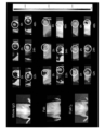
- FIG. 2 is a photograph showing the results of cell imaging with a confocal microscope for each antibody of Example 2.
- FIG. Upper left IgG bound to IR700 (IR700-IgG, negative control), upper right full-length 12A8 antibody bound to IR700 (IR700-12A8 Whole IgG), lower left Fab fragment of 12A8 antibody bound to IR700 (IR700-12A8 Fab), lower right shows the result of F(ab') 2 fragment of 12A8 antibody (IR700-12A8 F(ab') 2 ) bound to IR700.
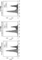
- FIG. 2 is a graph of the fluorescence intensity measured in FIG. 1.
- FIG. The vertical axis indicates the average fluorescence intensity (cps/cell).
- IgG1 (NC) is the negative control IgG
- whole body is the full-length 12A8 antibody
- F(ab') 2 is the F(ab') 2 fragment of the 12A8 antibody
- Fab is the Fab fragment of the 12A8 antibody.
- the results of fluorescence imaging of each antibody in a tumor-bearing nude mouse model are shown.
- Whole IgG represents the results of the full-length 12A8 antibody, F(ab') 2 the F(ab') 2 fragment of the 12A8 antibody, and Fab the Fab fragment of the 12A8 antibody.
- elapsed time (h) from administration of the labeled antibody or labeled antigen-binding fragment.
- FIG. 4 is a graph showing changes over time in fluorescence intensity measured in FIG. 3.
- the vertical axis represents fluorescence intensity (counts/s/cm 2 /str), and the horizontal axis represents elapsed time (h) after administration of the labeled antibody or labeled antigen-binding fragment.
- 1 is a graph showing the S/N ratio of each antibody in a cancer-bearing nude mouse model. The vertical axis represents the signal to noise ratio, and the horizontal axis represents the elapsed time (h) after administration of the labeled antibody or labeled antigen-binding fragment.
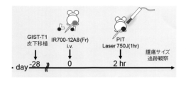
- 1 is a schematic diagram of an in vivo PIT (Photoimmunotherapy) therapeutic trial.
- FIG. 1 It is a photograph comparing before and after PIT treatment using cancer-bearing nude mice.
- mice administered with the Fab fragment of the 12A8 antibody Fab Vehicle
- mice administered with the F(ab') 2 fragment of the 12A8 antibody F(ab' ) 2 Vehicle
- FIG. 1 On the left are photographs of negative control mice that were not subjected to PIT treatment, mice administered with the Fab fragment of the 12A8 antibody (Fab Vehicle) (upper), mice administered with the F(ab') 2 fragment of the 12A8 antibody (F(ab' ) 2 Vehicle) (bottom).
- Fab PIT Fab fragment administration of 12A8 antibody + PIT treatment
- F (ab') 2 fragment administration of 12A8 antibody + PIT treatment F (ab') 2 PIT After
- FIG. 10 is a photograph comparing the fluorescence intensity of fluorescence-labeled anti-c-KIT antibody before and after PIT treatment using cancer-bearing nude mice.
- On the left are photographs of negative control mice that were not subjected to PIT treatment, mice administered with the Fab fragment of the 12A8 antibody (Fab Control (Vehicle)) (upper), mice administered with the F (ab') 2 fragment of the 12A8 antibody (F (ab′) 2 Control (Vehicle) (bottom).
- FIG. 3 is a graph showing changes in tumor size over time in a PIT treatment test using cancer-bearing nude mice. The vertical axis indicates the tumor size (mm 3 ), and the horizontal axis indicates the number of days elapsed after tumor implantation. PIT treatment was performed 28 days after tumor implantation.
- FIG. 1 shows the DNA and amino acid sequences of the heavy chain of humanized anti-c-KIT (12A8) antibody.
- FIG. 1 shows the DNA and amino acid sequences of the heavy chain of humanized anti-c-KIT (12A8) antibody.
- FIG. 1 shows the DNA and amino acid sequences of the light chain of humanized anti-c-KIT (12A8) antibody.
- Fig. 2 shows the DNA sequence and amino acid sequence of the Fab heavy chain of humanized anti-c-KIT (12A8) antibody.
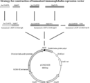
- FIG. 2 shows the structure of the humanized anti-c-KIT (12A8) antibody expression vector.
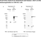
- Fig. 3 is a Western blot photograph showing the results of expression analysis of humanized anti-c-KIT (12A8) IgG1 antibody in CHO-K1 cells. Mock (lane 1) and humanized anti-c-KIT (12A8) IgG1 antibody (lane 2) were used as primary antibodies.
- FIG. 3 is a Western blot photograph showing the results of expression analysis of humanized anti-c-KIT (12A8) Fab antibody in CHO-K1 cells. Mock (lane 1), humanized anti-c-KIT(12A8) Fab antibody (lane 2) and humanized anti-c-KIT(12A8) IgG1 antibody (lane 3) were used as primary antibodies.
- HRP-labeled anti-human Fd antibody was used in A, and HRP-labeled anti-human Kappa antibody was used in B.
- FIG. 10 is a diagram confirming the reactivity of humanized anti-c-KIT(12A8) IgG1 antibody and humanized anti-c-KIT(12A8) Fab antibody by EIA.
- FIG. 10 is a diagram confirming the reactivity of IR700-labeled humanized anti-c-KIT (12A8) antibodies (whole IgG and Fab) by flow cytometry.
- Fig. 10 is a photograph showing the results of fluorescence imaging of each antibody in a tumor-bearing nude mouse model.
- FIG. 10 shows time-course changes in fluorescence intensity of xenograft tumors in mice by IR700-labeled humanized anti-c-KIT (12A8) Fab antibody.
- FIG. 10 shows the time course of S/N ratio in mouse tumor regions using IR700-labeled humanized anti-c-KIT (12A8) Fab antibody.
- the vertical axis indicates the S/N ratio
- the horizontal axis indicates the elapsed time (h) after administration of the labeled antigen-binding fragment.
- FIG. 11 is a photograph showing fluorescence imaging of tumors using IR700-labeled humanized anti-c-KIT (12A8) Fab antibody.
- FIG. 11 is a photograph showing fluorescence imaging showing the effect of photoimmunotherapy when using an IR700-labeled humanized anti-c-KIT(12A8) Fab antibody.
- the antigen-binding fragment of the present specification has a complementarity determining region (CDR) in the antibody heavy chain having the amino acid sequence set forth in SEQ ID NO: 2 or SEQ ID NO: 19, and the amino acid sequence set forth in SEQ ID NO: 4 or SEQ ID NO: 21.
- CDR complementarity determining region
- the amino acid sequence of SEQ ID NO: 2 may be encoded by, for example, the base sequence of SEQ ID NO: 1, and the amino acid sequence of SEQ ID NO: 4 may be, for example, the base sequence of SEQ ID NO: 3.
- the amino acid sequence set forth in SEQ ID NO: 19 may be encoded, for example, by the nucleotide sequence set forth in SEQ ID NO: 18, and the amino acid sequence set forth in SEQ ID NO: 21 may be, for example, It may be encoded by the nucleotide sequence set forth in SEQ ID NO:20.
- the antigen-binding fragment may have a complementarity determining region in the Fab H chain having the amino acid sequence set forth in SEQ ID NO: 6.
- the amino acid sequence set forth in SEQ ID NO: 6 is, for example, It may be encoded by the described base sequence.
- the antigen-binding fragment may have a complementarity determining region in the Fab H chain having the amino acid sequence set forth in SEQ ID NO: 23.
- the amino acid sequence set forth in SEQ ID NO: 23 is, for example, It may be encoded by the described base sequence.
- CDR determination method Kabat et al., Chothia et al., Martin et al., Geldand et al., IMGT (registered trademark) (http://www.imgt.org/IMGTindex/CDR.php), Honnerger et al. ) have been reported (Lars Nieba et al., Protein Engineering, 10(4): 435-444 (1997)). may be determined by any definition.
- the heavy chain CDRs possessed by the antigen-binding fragment herein include CDRH1 consisting of the amino acid sequence set forth in SEQ ID NO:7, CDRH2 consisting of the amino acid sequence set forth in SEQ ID NO:8, and the amino acid set forth in SEQ ID NO:9.
- CDRH3 consisting of the sequence, CDRH1 consisting of the amino acid sequence set forth in SEQ ID NO: 13, CDRH2 consisting of the amino acid sequence set forth in SEQ ID NO: 14, and CDRH3 consisting of the amino acid sequence set forth in SEQ ID NO: 15 can be mentioned.
- the light chain CDRs possessed by the antigen-binding fragment herein include CDRL1 consisting of the amino acid sequence set forth in SEQ ID NO: 10, CDRL2 consisting of the amino acid sequence set forth in SEQ ID NO: 11, and the amino acid set forth in SEQ ID NO: 12. or CDRL1 consisting of the amino acid sequence set forth in SEQ ID NO: 16, CDRL2 consisting of the amino acid sequence GIS (i.e., Gly, Ile, Ser), and CDRL3 consisting of the amino acid sequence set forth in SEQ ID NO: 17. can be done.
- the antigen-binding fragment herein includes (i) CDRH1 consisting of the amino acid sequence set forth in SEQ ID NO: 7, CDRH2 consisting of the amino acid sequence set forth in SEQ ID NO: 8, and CDRH3 consisting of the amino acid sequence set forth in SEQ ID NO: 9.
- CDRL1 consisting of the amino acid sequence set forth in SEQ ID NO: 10
- CDRL2 consisting of the amino acid sequence set forth in SEQ ID NO: 11
- DRL3 consisting of the amino acid sequence set forth in SEQ ID NO: 12
- CDRH1 consisting of the amino acid sequence of
- CDRH2 consisting of the amino acid sequence set forth in SEQ ID NO: 14
- CDRH3 consisting of the amino acid sequence set forth in SEQ ID NO: 15
- CDRL1 consisting of the amino acid sequence set forth in SEQ ID NO: 16
- CDRL2 consisting of the amino acid sequence GIS
- CDRL3 consisting of the amino acid sequence set forth in SEQ ID NO:17.
- antigen-binding fragment refers to a fragment of an antibody that retains the antigen-binding ability of the antibody.
- antigen-binding fragments include F(ab′) 2 , Fab′, Fab, Fab 3 , single-chain Fv (hereinafter referred to as “scFv”), (tandem) bispecific single-chain Fv (sc(Fv ) 2 ), single-chain triple-bodies, nanobodies, divalent VHHs, pentavalent VHHs, minibodies, (double-stranded) diabodies, tandem diabodies, bispecific tribodies, bispecific bibodies, dual affinity retargeting molecules ( DART), triabodies (or tribodies), tetrabodies (or [sc(Fv) 2 ] 2 ), or (scFv-SA) 4 ) disulfide-linked Fv (hereinafter referred to as “dsFv”), compact IgG, heavy chain
- the antigen-binding fragment that constitutes part of the complex of the present invention specifically binds to c-KIT.
- the phrase “specifically” recognizes (binds to) an antigen-binding fragment means that the antigen-binding fragment has substantially greater affinity for c-KIT than affinity for other proteins or peptides. It means binding with high affinity.
- binding with substantially high affinity means that the specific protein or peptide of interest can be distinguished and detected from other proteins or peptides by a desired measurement device or method. means high affinity.
- a substantially higher affinity is 3-fold or more, 4-fold or more, 5-fold or more, 6-fold or more, 7-fold or more, 8-fold or more, as the intensity (e.g., fluorescence intensity) detected by ELISA or EIA. It may mean 9 times or more, 10 times or more, 20 times or more, 30 times or more, 40 times or more, 50 times or more, or 100 times or more.
- the binding rate constant (Ka1) in the binding between the antigen-binding fragment and c-KIT is, for example, 1 ⁇ 10 4 Ms ⁇ 1 or more, 1 ⁇ 10 5 Ms ⁇ 1 or more, 5 ⁇ 10 5 Ms ⁇ 1 or more can be mentioned.
- examples of the dissociation rate constant (Kd1) in binding between the antigen-binding fragment and c-KIT include 1 ⁇ 10 ⁇ 3 or less and 1 ⁇ 10 ⁇ 4 or less.
- the binding constant (KD) in binding between the antigen-binding fragment and c-KIT is, for example, 1 ⁇ 10 ⁇ 8 (M) or less, 5 ⁇ 10 ⁇ 8 (M) or less, 1 ⁇ 10 ⁇ 9 (M) or less.
- the binding rate constant (Ka1), dissociation rate constant (Kd1), and binding constant (KD) of the antigen-binding fragment herein are determined using BIACORE (GE Healthcare Biosciences Co., Ltd., BIACORE-X100). According to the provided manual, after immobilizing biotinylated c-KIT on the SA chip, a test antibody is flowed, the binding rate constant Ka1 and the dissociation rate constant Kd1 are measured, and the binding constant KD value can be determined using bivalent fitting. can.
- Antigen-binding fragments include non-human animal antigen-binding fragments, antigen-binding fragments having non-human animal amino acid sequences and human-derived amino acid sequences.
- the antigen-binding fragment may be a humanized antigen-binding fragment.
- the humanized antigen-binding fragment is based on the genes encoding the H and L chains of a human antibody, and the primary structure portions of the six CDRs are replaced with the H chain (3 It is a fragment of an antibody that is genetically engineered to replace the primary structure of the CDRs of the L chain (3 sites) and L chain (3 sites).
- the antibody of the present invention is preferably a fragment of a humanized antibody.
- the conjugates of the invention may further comprise one or more other antigen-binding fragments of different antibodies, thereby being monospecific, bispecific (bispecific), trispecific (trispecific). ), or multispecific.
- amino acids are represented by single-letter codes. Specifically, A is alanine, L is leucine, R is arginine, K is lysine, N is asparagine, M is methionine, D is aspartic acid, F is phenylalanine, C is cysteine, P is proline, Q is glutamine, S is serine, E is glutamic acid, T is threonine, G is glycine, W is tryptophan, H is histidine, Y is tyrosine, I isoleucine and V is valine.
- IR700 is also called phthalocyanine and means a compound having the following structure.
- IR700 contained in the complex herein may be a derivative thereof as long as it has the above structure.
- IR700 has a substituent that reacts with an amino group or a carboxyl group, and the reaction between the substituent and the amino group or carboxyl group of the antigen-binding fragment allows binding of the antigen-binding fragment and IR700.
- IRDye (registered trademark) 700DX (LI-COR) having the following structure can be used.
- the antigen-binding fragment and IR700 can be bound according to the protocol provided by the manufacturer.
- the binding ratio between the antigen-binding fragment and IR700 is, for example, the number of IR700 molecules bound per antigen-binding fragment (F/P ratio) of 1 to 10, 1 to 5, or 1 to 3. good.
- F/P ratio the number of IR700 molecules bound per antigen-binding fragment
- the average number of IR700 molecules (F/P ratio) bound to one antibody antigen-binding fragment molecule of the complexes in the composition The value may be 1-10, 1-5, 1-3, or 1.5-3.0.
- the present invention relates to a composition for cancer detection/diagnosis, containing the complex as an active ingredient.
- the present invention also provides a method for detecting/diagnosing all cancer cells expressing c-KIT, comprising administering the complex of the present invention to a subject with cancer, and to a method comprising detecting an accumulation of
- cancer refers to cells that have undergone malignant transformation such that they become pathogenic to the host organism.
- cancer includes not only primary cancers, but also metastatic cancers, and in vitro cultures and cell lines derived from cancer cells.
- the cancer may be, for example, a hematopoietic tumor, which is a tumor of blood cells, such as leukemia, such as chronic myelogenous leukemia or acute myelogenous leukemia; myeloma, such as multiple myeloma; lymphoma; be done.
- leukemia such as chronic myelogenous leukemia or acute myelogenous leukemia
- myeloma such as multiple myeloma
- lymphoma lymphoma
- Cancers include, but are not limited to, lung cancer, non-small cell lung cancer, small cell lung cancer, non-Hodgkin's lymphoma, adrenocortical carcinoma, AIDS-related cancer, AIDS-related lymphoma, childhood cerebellar astrocytoma, childhood cerebrum Astrocytoma, basal cell carcinoma, skin cancer (non-melanoma), biliary tract cancer, extrahepatic cholangiocarcinoma, intrahepatic cholangiocarcinoma, bladder cancer, bone and joint cancer, osteosarcoma and malignant fibrous histiocytoma, brain cancer , brain tumor, brain stem glioma, cerebellar astrocytoma, glioma, cerebral astrocytoma/malignant glioma, ependymoma, medulloblastoma, supratentorial primitive neuroectodermal tumor, visual pathway and hypothalamic glioma head and neck cancer, meta
- the complex of the present invention is rapidly accumulated in cancer cells after contact with cancer cells or after administration to a cancer-bearing organism. Therefore, the composition for cancer detection of the present invention can be administered within 24 hours, within 12 hours, within 6 hours, within 5 hours, within 4 hours, within 3 hours, within 2 hours, or within 1 hour after administration of the composition. can be used to detect cancer within From the viewpoint of detection in a short time, it is preferably used to detect cancer within 4 hours, 3 hours, 2 hours, or 1 hour.
- the complex of the present invention since the complex of the present invention has an IR700 molecule, it can kill cancer cells by photoimmunotherapy (PIT) (Mitsunaga M, et al., Nat Med. (2011) 17(12): 1685- 91.). Therefore, the composition for cancer treatment of the present invention is administered to the subject, and after the elapse of time for the composition to accumulate in the cancer, the cancer locality, that is, the accumulation locality of the composition, is irradiated with near-infrared light. By doing so, the cancer therapeutic effect can be exhibited. Accordingly, the present invention relates to a composition for cancer treatment containing the above complex as an active ingredient.
- PIT photoimmunotherapy
- compositions and complexes mainly used for cancer treatment are intended for use in combination with near-infrared light.
- the present invention also relates to a method for treating cancer, comprising administering the complex of the present invention to a subject with cancer, and irradiating near-infrared light to the site of accumulation of the complex in the subject. .
- the composition to accumulate in the cancer is 1 hour to 24 hours, 1 hour to 12 hours, 1 hour to 6 hours, 2 hours to 6 hours after administration of the composition, It can be after 2-4 hours, after 2-3 hours, or after 3-4 hours.
- the antibody fragment of the present invention can be further bound with a labeling substance.
- a labeling substance for example, it can be combined with a fluorescent substance having a wavelength in the visible light region such as 480 to 650 nm or a photosensitive substance to form a complex, which can be applied to the diagnosis and treatment of cancer.
- a radioactive label, an enzyme, a fluorescent label, a bioluminescent label, a chemiluminescent label, or a detectable label such as a metal can be used.
- labels include, but are not limited to, 32P, 3H, 125I, 131I, 14C, radioactive labels such as tritium; ⁇ -galactoxidase, peroxidase, alkaline phosphatase, glucoamylase, enzymes such as glucose oxidase, lactate oxidase, alcohol oxidase, monoamine oxidase, horseradish peroxidase; coenzymes or prosthetic groups such as FAD, FMN, ATP, biotin, heme; Mil, etc.), rhodamine derivatives (tetramethylrhodamine, trimethylrhodamine (RITC), Texas red, rhodamine 110, etc.), Cy dyes (Cy3, Cy5, Cy5.5, Cy7), Cy-chromium, spectrum green, spectrum orange, propidium Fluorescent labels such as iodide, allophycocyanin (APC), R-phycoerythrin (R-PE); biolabel
- the composition of the present invention can be used for cancer detection (diagnosis), and can also be used for treatment as it is. That is, the present invention includes administering the composition of the present invention to a subject, detecting/diagnosing the location and size of cancer in the subject from the fluorescence emitted by IR700 in the administered composition, and The present invention includes methods for detecting/diagnosing and treating cancer, including killing the detected/diagnosed cancer by irradiating the cancer with near-infrared light. Moreover, the present invention may be a composition used for both detection and treatment of cancer (composition for detection and treatment) containing the complex as an active ingredient.
- the present invention relates to the use of the complex of the present invention for producing the composition for detecting cancer, the composition for treating cancer, or the composition for detecting and treating cancer.
- the invention also relates to conjugates of the invention for cancer detection, cancer therapy, or cancer detection and therapy.
- compositions of the present invention include, in addition to intravenous administration, a method of spraying and administering to the gastrointestinal mucosa through forceps holes under endoscopic observation, for example.
- the compositions of the invention are preferably for medical use, and therefore formulations suitable for medical use are preferred.
- Compositions for parenteral administration include, for example, injections, nasal drops, suppositories, and the like. Preferred are injections (solutions such as intravenous injections, subcutaneous injections, intradermal injections, intramuscular injections, intravitreal injections, and drip infusions, and freeze-dried preparations).
- Aqueous liquids for injection include buffers such as phosphate; tonicity agents such as physiological saline, glucose, sucrose, mannitol, sodium chloride and glycerin; Alcohol (e.g., propylene glycol, polyethylene glycol), nonionic surfactant [e.g., polysorbate 80, polysorbate 20, HCO-50 (polyoxyethylene (50 mol) adduct of hydrogenated castor oil)], solubilizer such as ethylenediamine, sulfite Additives such as stabilizers such as phenol, preservatives such as phenol, and soothing agents such as lidocaine can be added.
- buffers such as phosphate
- tonicity agents such as physiological saline, glucose, sucrose, mannitol, sodium chloride and glycerin
- Alcohol e.g., propylene glycol, polyethylene glycol
- nonionic surfactant e.g., polysorbate 80, polysorbate 20, HCO-50 (
- injections can be prepared according to known methods, for example, by dissolving, suspending or emulsifying the complex in a sterile aqueous or oily liquid commonly used for injections. In the case of a freeze-dried preparation, it can be dissolved in water for injection, physiological saline, etc. at the time of use to prepare an injection solution.
- examples of storage containers include ampoules, vials, prefilled syringes, pen-type syringe cartridges, and drip bags.
- Oral administration of proteins is generally considered difficult because they are degraded in the digestive system, but oral administration is also possible when targeting cancers of the digestive system.
- examples of formulations for oral administration include capsules, tablets, syrups, granules and the like.
- composition of the present invention is preferably prepared in dosage unit form to suit the dosage of the active ingredient.
- dosage unit dosage forms include injections (ampoules, vials, prefilled syringes), which usually contain 1 to 1000 mg, 5 to 250 mg, and 10 to 100 mg of the complex of the present invention per dosage unit dosage form. It's okay to be
- the administration of the composition of the present invention may be local or systemic.
- the administration method is not particularly limited, and is administered parenterally or orally as described above.
- Parenteral routes of administration include intraocular, subcutaneous, intraperitoneal, blood (intravenous or intraarterial) or spinal fluid injection or infusion, etc., preferably blood administration.
- Administration of the compositions of the present invention may be single or multiple, and may be temporary, continuous or intermittent.
- one to several doses are administered over a short period of time (within hours, days, 1-2 weeks, etc.).
- the PIT treatment of the cancer can be performed multiple times at intervals.
- the composition is administered one to several times before near-infrared irradiation for each treatment.
- the conjugate of the present invention may be administered alone or in combination with other drugs.
- the dosage of the composition of the present invention is not particularly limited as long as the desired therapeutic effect or detection/diagnostic effect can be obtained, and can be appropriately determined according to symptoms, sex, age, and the like.
- the dose of the composition of the present invention can be determined, for example, using the therapeutic effect on cancer as an index.
- the single dose of the active ingredient of the composition of the present invention is 0.01-20 mg/kg body weight, 0.1-10 mg/kg body weight, 0.1-20 mg/kg body weight, 5 mg/kg body weight is administered once to several times by intravenous injection. In the case of other parenteral administration and oral administration, the amount according to this can be administered. If the symptoms are particularly severe, the dose or number of administrations (number of treatments) may be increased according to the symptoms.
- preparation of its F(ab') 2 fragment and Fab fragment (1)
- Preparation of antigen Human c-KIT (Accession No. NM-000222.3) was used as a GIST target molecule.
- the antigen is an expression vector (pcDNA3.1) cDNA encoding a fusion protein (c-KIT-Fc) between the extracellular region (1-504 amino acids) of the c-KIT gene and the constant region (Fc portion) of mouse IgG2a.
- the recombinant vector was transfected into Chinese hamster ovary (CHO) cells. After culturing c-KIT-Fc-expressing CHO cells in a serum-free TIL medium (Immunobiological Laboratories, Inc.), the culture supernatant was purified using a Protein A column (GE Healthcare). .
- Antigenic determinant (epitope) of mouse anti-c-KIT antibody 12A8 The antigen-determining region recognized by the 12A8 antibody was determined by epitope mapping. A gene encoding a fusion protein between the extracellular region (1-504 amino acids) of human c-KIT and various deletion mutants deleted from the C-terminus of the region and the constant region of mouse IgG2a is prepared, and an expression vector (pcDNA3.1). The generated deletion mutants are human c-KIT1-106 amino acids (c-KIT106-Fc), 1-205 amino acids (c-KIT205-Fc), 1-305 amino acids (c-KIT305-Fc), 1-406 It encodes an amino acid (c-KIT406-Fc).
- Each expression vector carrying these genes was transfected into COS1 cells (ATCC, CRL-1650), cultured in serum-free Dulbecco's MEM (Immuno-Biological Laboratories, Inc.) for 2 days, and the culture supernatant was collected. bottom. The collected culture supernatant was concentrated by ultrafiltration to a 10-fold concentration and dot-blotted onto a PVDF membrane (Millipore, IPVH00010). After blocking treatment of the blotted PVDF membrane, it was reacted with a mouse anti-c-KIT (12A8) antibody, and further reacted with an anti-mouse Ig ⁇ chain HRP-labeled antibody (Southern Biotech, 1050-05). Chemiluminescence detection was performed using Amersham ECL (Cytiva, RPN2106) for detection.
- mice IgG2aFc Since all of the various dotted fusion proteins contained mouse IgG2aFc, they were all positive to anti-mouse IgG antibody (a-Mouse HRP), but not to anti-mouse Ig ⁇ chain HRP (a-mouse kappa HRP). didn't react.
- Mouse anti-c-KIT (12A8) antibody (ac-KIT 12A8) reacted only with c-KIT504-Fc without C-terminal deletion used as an antigen, and reacted with other C-terminal deletion mutant fusion proteins. Since no reaction was observed with , it was clarified that an epitope exists in the 407-504 amino acid region, which is the extracellular domain of c-KIT.
- 12A8 antibody was adjusted with 0.1M/L sodium acetate buffer (Ph5.5), digested with papain at room temperature for 3 hours, purified by gel filtration chromatography (Ultrogel AcA44 column), and further purified by Protein A column. obtained by removing the Fc fragment through
- the F/P for each IR700-12A8 conjugate was 2.4 for Whole IgG, 2.6 for F(ab′) 2 , 1.9 for Fab, and 1.9 for Fab, negative control IgG1 (in response to c-KIT). not) was 3.8.
- Each binding substance was adjusted with buffer for dilution (PBS) so that the molar concentration at the time of use was the same (0.05 nM (Table 1) for in vitro tests and 0.5 nM (Table 2) for in vivo tests).
- PBS buffer for dilution
- Example 2 In vitro binding of IR700-labeled mouse anti-c-KIT antibody and fragments thereof to GIST and its fluorescence intensity IR700-labeled mouse anti-c-KIT antibody prepared in Example 1 ( Using whole body IgG antibody (hereinafter referred to as whole IgG), its F(ab') 2 fragment and Fab fragment, GIST-T1 cell line was subjected to live cell imaging to confirm whether there was a difference in fluorescence density.
- whole IgG antibody hereinafter referred to as whole IgG
- F(ab') 2 fragment and Fab fragment F(ab') 2 fragment and Fab fragment
- Each of the obtained IR700-labeled binding antibodies was dispensed in small amounts and stored at -20°C until use.
- the GIST cultured cell line GIST-T1 (Dr. Tguchi T., Kouchi University, Kouchi, Japan) was cultured and proliferated using a 10 cm dish (Greiner, CELLSTTAR664160-013) with the addition of the RPMI1640 medium. .
- GIST-T1 cells 8 ⁇ 10 4 cells/dish were seeded on 5 glass bottom dishes (Greiner, CELLview 627965), cultured for 24 hours, the supernatant was removed, and a blocking buffer (DaKo, Protein Block serum-free , X0909) and incubated at room temperature for 15 minutes.
- a blocking buffer DaKo, Protein Block serum-free , X0909
- Each IR700-labeled sample listed in Table 1 was adjusted to working concentration with antibody dilution buffer (PBS containing 0.1% BSA), 15 minutes later blocking buffer was discarded, 12A8 conjugate and control conjugate (IR700- IgG1) was added at 500 ⁇ L each.
- IR700-12A8-labeled samples whole IgG and F(ab′) 2 were adjusted to a final concentration of 0.05 nM, but Fabs with monovalent antigen-binding sites were doubled at a final concentration of 0 to match the number of binding sites. .1 nM. Further, 5 ⁇ L of Hoechst 33342 solution (1 mg/ml, DOJINCO H342) was added and incubated on ice for 1 hour.
- FIG. 1 shows a graph of the fluorescence intensity measured in FIG. 12A8 fragment F(ab′) 2 showed fluorescence intensity similar to whole IgG in cell imaging, and 12A8 fragment Fab showed fluorescence intensity superior to whole IgG in cell imaging.
- Example 3 Fluorescence imaging of mouse xenograft tumor using 12A8 fragment (in vivo test)
- Ten athymic BALB/c nude mice (CLEA Japan Inc., Tokyo, Japan) were administered the immunosuppressant cyclophosphamide (0.2 mg/g body weight).
- GIST-T1 cells were adjusted to 1 ⁇ 10 7 cells/50 ⁇ L with PBS, and suspended in thawed Matrigel (Corning Incorporated, USA) on ice at a ratio of 1:1.
- Two days after administration of the immunosuppressive agent 100 ⁇ L of the suspended GIST-T1 cells were subcutaneously transplanted to the right thigh of nude mice treated with the immunosuppressive agent. When the tumor reached 8 mm, the antibody was administered and observed.
- the S/N ratio (signal to noise ratio) in the in vivo model is shown in FIG.
- the S/N ratio of 12A8 fragment (Fab, F(ab') 2 ) was comparable to Whole IgG. From this result, it is possible to diagnose GIST in a short time (within 3 hours) after administering the antibody to the living body by using a fragment of the GIST-specific antibody (12A8) bound to a fluorescent dye. shown.
- Example 4 In vivo PIT (Photoimmunotherapy) treatment test 28 days after transplanting GIST-T1 into nude mice administered immunosuppressants, 4 mice with similar tumor sizes were selected and sedated with isoflavones. 79 ⁇ g and 66 ⁇ g of IR700-12A8Fab and IR700-12A8F(ab′) 2 were administered to the tail vein, respectively, and about 2 hours later, the tumor was irradiated with a laser (735J) for 1 hour. A schematic of the experimental scheme is shown in FIG.
- Tumor size was observed for 4 weeks, and Alexa Fruor 647-labeled mouse anti-c-KIT antibody (BioLegend Inc., San Diego, USA) was administered to the tail vein 55 days after transplantation, and observed by IVIS 48 hours later. bottom.
- Example 5 Generation of IR700-Humanized Anti-c-KIT (12A8) Antibodies (Whole IgG and Fab) Safely as Diagnostic and/or Therapeutic Compositions for Human Gastrointestinal Stromal Tumors (GIST)
- GIST Gastrointestinal Stromal Tumors
- variable region genes VH and VL of the synthesized humanized anti-c-KIT (12A8) antibody and the natural human IgG1 gene, humanized antibody by overlap PCR (polymerase chain reaction)
- the heavy chain (HC) Figure 10, SEQ ID NO: 18
- LC Figure 11, SEQ ID NO: 20
- HC fuses humanized anti-c-KIT (12A8) antibody VH and human IgG constant region (Fc)
- LC fuses humanized anti-c-KIT (12A8) antibody VL with human Kappa chain constant region (KC). fused.
- the HC gene of the humanized anti-c-KIT (12A8) antibody was used as a template, and a stop codon was inserted immediately before the hinge region (Fab HC) (Fig. 12, SEQ ID NO: 22). ) was amplified by PCR.
- the HC gene, LC gene and Fab HC gene of the humanized anti-c-KIT (12A8) antibody were each inserted into a mammalian cell expression vector (pCXN2-MCS, Immuno-Biological Laboratories, Inc.) (Fig.
- Antibody gene expression vector (i) humanized c-KIT (12A8) IgG1 (humanized c-KIT (12A8) IgG1) vector, (ii) humanized c-KIT (12A8) Ig ⁇ (humanized c-KIT (12A8) Ig ⁇ ) vector, (iii) humanized c-KIT(12A8) Fab H (humanized c-KIT(12A8) Fab H) vector was generated.
- This band has approximately the same size as the Mw (weight average molecular weight) of whole IgG and corresponds to two heavy chain (H) molecules and two light chain (L) molecules.
- the band reacting with the HRP-labeled anti-IgG (Fc) antibody corresponds to the Mw of the heavy chain (HC) of humanized c-KIT (12A8) IgG1
- the HRP-labeled anti-Kappa antibody corresponded to the Mw of the light chain (LC) of humanized c-KIT(12A8)Ig ⁇ .
- Fab 15 is a whole IgG antibody produced with (i) a humanized c-KIT (12A8) IgG1 vector and (ii) a humanized c-KIT (12A8) Ig ⁇ vector, and is an HRP-labeled anti-Fd antibody.
- HRP-labeled anti-Kappa antibody molecules corresponding to the Mw of Fab could not be confirmed in both reduced and non-reduced conditions.
- 12A8) antibody Fab fragment hereinafter also referred to as humanized anti-c-KIT (12A8) Fab antibody was confirmed.
- the humanized anti-c-KIT (12A8) IgG1 antibody-producing clone culture supernatant was serially diluted from 2-fold to 64-fold and added to the antigen solid-phase plate and the negative control plate for primary reaction.
- Humanized anti-c-KIT (12A8) Fab antibody was similarly performed. The primary reaction was carried out at room temperature for 1 hour, after which each well was washed with phosphate buffered saline (PBS). HRP-labeled anti-human Kappa chain antibody was added as a secondary antibody and reacted at room temperature for 30 minutes.
- the humanized anti-c-KIT(12A8) IgG1 antibody and the humanized anti-c-KIT(12A8) Fab antibody have antigen specificity. Since each culture supernatant is used for in-vitro and in-vivo tests, the humanized anti-c-KIT (12A8) IgG1 antibody is a protein A column, and the humanized anti-c-KIT (12A8) Fab antibody is Each was purified with Kan Cap G (KANEKA).
- Example 6 Reactivity study of IR700-humanized anti-c-KIT (12A8) antibody (whole IgG and Fab) by flow cytometry (in vitro) Flow cytometry was performed using IR700-labeled humanized anti-c-KIT (12A8) antibody (whole IgG and Fab) and GIST-T1 cell line to confirm the reactivity of the humanized antibody.
- GIST-T1 cells were collected with trypsin (on ice), and 1 ⁇ 10 6 cells were dispensed into 1.5 mL tubes. Washed 3 times with 1 mL of PBS-BSA-Azide. 1 mL of 4% paraformaldehyde cooled to 4° C. was added and gently mixed by pipetting on ice. It was returned to room temperature and allowed to stand for 15 minutes (the work was carried out at room temperature thereafter). Washed 3 times with 1 mL of PBS-BSA-Azide.
- IR700-labeled mouse anti-c-KIT (12A8) antibody is also referred to as 12A8 (whole IgG)
- IR700-labeled humanized anti-c-KIT (12A8) IgG1 antibody is also referred to as humanized (whole IgG)
- IR700 The labeled humanized anti-c-KIT(12A8) Fab antibody is also referred to as humanized (Fab).
- the MFI of IR700-labeled humanized anti-c-KIT(12A8) IgG1 antibody was similar to that obtained with IR700-labeled mouse anti-c-KIT(12A8) antibody. Considering that the IR700-labeled mouse anti-c-KIT (12A8) antibody has a high F/P ratio of 4.4, the IR700-labeled humanized anti-c-KIT (12A8) IgG1 antibody has good reactivity with c-KIT. was shown.
- Example 7 Fluorescent imaging of xenograft tumors in mice using IR700-labeled humanized anti-c-KIT (12A8) antibodies (whole IgG and Fab)
- fluorescence imaging after administration of IR700-labeled humanized anti-c-KIT (12A8) Fab antibody to a mouse model in vivo showed that the fluorescence intensity in the tumor region (ROI) was 3 It reached a peak after ⁇ 4 hours and then tapered off.
- the fluorescence intensity of the control ROI on the contralateral side (left thigh) to which no tumor cells were administered peaked after 1 to 6 hours and then gradually decreased.
- fluorescence imaging after administration of IR700-labeled humanized anti-c-KIT (12A8) IgG1 antibody almost no fluorescence was observed 1 to 4 hours after administration, and even if confirmed, the fluorescence intensity was weak.
- the S/N ratio in the tumor region (ROI) after administration of the IR700-labeled humanized anti-c-KIT (12A8) Fab antibody peaked after 3 to 4 hours and then gradually decreased. bottom.
- the above results indicate that the IR700-labeled humanized anti-c-KIT (12A8) Fab antibody can accumulate in tumors in a short period of time, and is suitable for diagnosis of GIST in a short period of time. Subsequently, therapeutic experiments using small animals were conducted.
- 80 ⁇ g of IR700-labeled humanized anti-c-KIT (12A8) Fab antibody was administered (n 2), and approximately 3 hours after each, the tumors were irradiated with a laser for 1 hour (735 J). After that, the tumor size was observed for 3 weeks together with a control group (saline-administered group) in which the tumor was not treated.
- the treatment schedule is shown in FIG.
- IVIS IVIS
- the fluorescence intensity increased with the passage of 1 to 3 hours after administration of the IR700-labeled humanized anti-c-KIT (12A8) Fab antibody.
- FIG. 23 as a result of irradiation with a 685 nm laser 3 hours after administration of the antibody, the tumor disappeared.
Abstract
The purpose of the present invention is to provide a detecting agent (diagnostic agent) and a treatment agent that are taken into tumors in vivo promptly and that bind to c-KIT target molecules. In the quest for a molecular species that specifically binds to c-KIT, and a labelling agent and a treatment agent that bind to said molecular species, the inventors found that by combining an anti-c-KIT antibody fragment and IR700, rapid accumulation into tumors takes place in just several hours after administration, and detection and treatment becomes possible. Accordingly, the present invention pertains to an antigen-binding fragment of anti-c-KIT antibody, IR700, and a composite body, and also pertains to a detecting agent and a treatment agent that contain said composite body as an active ingredient. Specifically, the present invention pertains to: a complementary determining region (CDR) in the antibody heavy chain which has the amino acid sequence described in SEQ ID NO: 2; and a composite body having IR700 and an antigen-binding fragment of an antibody having a CDR in the antibody light chain which has the amino acid sequence described in SEQ ID NO: 4.
Description
本発明は、c-KIT陽性腫瘍である消化管間質腫瘍の診断および治療の分野に関する。
The present invention relates to the field of diagnosis and treatment of gastrointestinal stromal tumors, which are c-KIT positive tumors.
消化管間質腫瘍(Gastrointestinal stromal tumor:GIST)は消化管の粘膜下の間葉系細胞から由来する腫瘍で、通常は正常な粘膜に覆われているため、一般的な内視鏡では他のタイプの粘膜下腫瘍と区別することが困難である。
Gastrointestinal stromal tumor (GIST) is a tumor derived from mesenchymal cells in the submucosa of the gastrointestinal tract, and is usually covered with normal mucosa. It is difficult to distinguish from submucosal tumors of the type.
最近、EUS-FNA(Endoscopic ultrasound-guided fine needle aspiration)超音波内視鏡穿刺吸引細胞診が開発されGISTの診断が可能になった。しかし、これは非常に高度な技術を要し、特に2cm以下の場合EUS-FNSでも実施が困難で、組織学的診断が出来ないまま、経時的にフォローアップしている状態である。また、2cm以下のGISTでも肝臓への転移や早い腫瘍増殖が見られる等が報告され、もっとシンプルで効果的な診断技術と治療法が待望されている。
Recently, EUS-FNA (Endoscopic ultrasound-guided fine needle aspiration) ultrasound endoscopic fine needle aspiration cytology was developed, making it possible to diagnose GIST. However, this requires a very advanced technique, and it is difficult to perform even EUS-FNS especially when the size is 2 cm or less. In addition, it has been reported that even a GIST of 2 cm or less causes metastasis to the liver and early tumor growth, etc., and a simpler and more effective diagnostic technique and therapeutic method are eagerly awaited.
PET(Positron emission tomography)ポジトロン放射断層撮影がGIST等でも診断に利用されているが、リンパ腫や平滑筋肉腫等を含む他の悪性腫瘍や、炎症性の病巣との区別が困難であるうえに、患者が高用量の放射線を被爆するという問題もある。
PET (Positron Emission Tomography) is also used for diagnosis in GIST, etc., but it is difficult to distinguish from other malignant tumors, including lymphoma and leiomyosarcoma, and inflammatory lesions. There is also the problem of patient exposure to high doses of radiation.
また、PDT(Photodynamic therapy)光力学療法では、光線の特異的な波長によって活性化される光感受性物質を利用して、様々なタイプのがんで治療が試みられたが、腫瘍細胞を死滅させる一方で、非がん細胞をも傷めるために重大な副作用が見られる。
In addition, PDT (photodynamic therapy) uses photosensitizers that are activated by specific wavelengths of light to treat various types of cancer. However, it also damages non-cancer cells and has serious side effects.
一方、GISTの標的分子c-KITを利用し、抗c-KIT抗体にAlexa Fluor 488(AF488)を結合してGISTのモデルマウスを用いて、腹腔鏡検査の開発が行われた。しかし、その目論見は、正常組織の自家蛍光が強すぎ、擬陽性の比率が高すぎるため実用化されなかった。
On the other hand, using the GIST target molecule c-KIT, laparoscopy was developed using a GIST model mouse by binding Alexa Fluor 488 (AF488) to the anti-c-KIT antibody. However, the idea was not put to practical use because the autofluorescence of normal tissue was too strong and the rate of false positives was too high.
そこで考えられたのが、NIR(Near-infrared)近赤外線である。NIRは、生体において自家蛍光が非常に低く、可視光線よりもっと深く組織へ透過できるため、GISTの内視鏡分子イメージングとして最も的確な戦略である。近年、腫瘍特異的な分子に対する抗体に光感受性物質を結合して、診断や治療が出来る、PIT(Photoimmunotherapy)光免疫療法が発見された。これは、PDTの新しいタイプの利用法として高い選択性が報告されている。この方法は、特異抗体と色素の結合物とのみに反応する細胞(腫瘍細胞)だけに障害をもたらすため、副作用が最小化されるというがん治療法である。この療法は、がん細胞の標的分子に対する特異的な抗体が鍵である。
What came to mind was NIR (Near-infrared) near-infrared rays. NIR is the most appropriate strategy for endoscopic molecular imaging in GIST because it has very low autofluorescence in vivo and can penetrate deeper into tissues than visible light. In recent years, PIT (Photoimmunotherapy) photoimmunotherapy has been discovered, which enables diagnosis and treatment by binding a photosensitizer to an antibody against a tumor-specific molecule. This is reported to be a new type of use of PDT with high selectivity. This method is a cancer treatment method that minimizes side effects because it damages only cells (tumor cells) that react only with a specific antibody-dye conjugate. The key to this therapy is a specific antibody against a cancer cell target molecule.
GISTの腫瘍細胞は表面にc-KITを発現していて、患者の90%以上が陽性であることが知られている。そこで、本発明者らは、c-KITを標的分子として、この分子に対する抗体にIR700色素を結合させ、近赤外線照射によるGISTの診断および治療を試みた(非特許文献1)。しかし、この方法では、蛍光が最大値に達する、すなわち抗体が集積するまでに24時間が必要であり、S/N比が最大値に達するまでには120時間必要なことなど、投与から検出までに時間を要することが問題であった。また、90Y標識したc-KIT抗体を用いた研究が報告されており、この場合も治療効果を奏するまでに数日要している(非特許文献2)。更に、それ以前に抗c-KIT抗体のFab断片を用いて64Cu標識させることが試みられているが、ヌードマウスを用いた実験では取り込みまでに6~12時間を要している(非特許文献3、Table3)。
It is known that GIST tumor cells express c-KIT on their surface, and more than 90% of patients are positive. Therefore, the present inventors used c-KIT as a target molecule, bound an IR700 dye to an antibody against this molecule, and attempted diagnosis and treatment of GIST by near-infrared irradiation (Non-Patent Document 1). However, this method requires 24 hours for fluorescence to reach its maximum value, i.e. antibody accumulation, and 120 hours for S/N ratio to reach its maximum. The problem was that it took time to In addition, a study using a 90Y-labeled c-KIT antibody has been reported, and in this case also several days are required until a therapeutic effect is achieved (Non-Patent Document 2). Furthermore, although 64Cu labeling was attempted using a Fab fragment of an anti-c-KIT antibody before that, it took 6 to 12 hours for uptake in experiments using nude mice (non-patent document 3, Table 3).
そこで、本発明は、in vivoで早期に腫瘍内に取り込まれるc-KITを標的分子とする標識剤および治療薬を提供することを課題とする。
Therefore, an object of the present invention is to provide a labeling agent and a therapeutic agent that target c-KIT, which is taken up early in tumors in vivo.
本発明者らは、c-KIT特異的に結合する分子種および当該分子種と結合させる標識剤および治療剤について検討した結果、抗c-KIT抗体のFab断片またはF(ab’)2断片とIR700とを組み合わせることにより、投与から数時間という短時間で腫瘍に集積させ、検出できることを見出した。
The present inventors have investigated a molecular species that specifically binds to c-KIT and a labeling agent and therapeutic agent that binds to the molecular species. It was found that by combining with IR700, it can be accumulated in tumors and detected in a short period of several hours after administration.
また、抗c-KIT抗体のFab断片またはF(ab’)2断片とIR700と複合体を腫瘍に集積させた後に、PIT処理を施すことにより、検出と同じ複合体を用いて治療も行うことができることも見出した。
In addition, after accumulating the Fab fragment or F(ab′) 2 fragment of the anti-c-KIT antibody and the complex with IR700 in the tumor, PIT treatment may be performed to perform treatment using the same complex used for detection. I also found that
よって、本発明は、抗c-KIT抗体の抗原結合性断片とIR700との複合体に関し、また当該複合体を有効成分として含有する検出薬および治療薬に関する。
(1) 配列番号2又は配列番号19に記載のアミノ酸配列を有する抗体重鎖における相補性決定領域(CDR)、および配列番号4又は配列番号21に記載のアミノ酸配列を有する抗体軽鎖におけるCDRを有する抗体の抗原結合性断片と、IR700とを有する複合体。
(2) 抗体の抗原結合性断片が、FabまたはF(ab’)2である、(1)に記載の複合体。
(3) 配列番号2に記載のアミノ酸配列を有する抗体重鎖におけるCDRが、以下の(a)または(b)である、(1)または(2)に記載の複合体。
(a)重鎖CDR(CDRH)1が、配列番号7に記載のアミノ酸配列からなり、
CDRH2が、配列番号8に記載のアミノ酸配列からなり、かつ、
CDRH3が、配列番号9に記載のアミノ酸配列からなる
(b)CDRH1が、配列番号13に記載のアミノ酸配列からなり、
CDRH2が、配列番号14に記載のアミノ酸配列からなり、かつ、
CDRH3が、配列番号15に記載のアミノ酸配列からなる。
(4) 配列番号4に記載のアミノ酸配列を有する抗体軽鎖におけるCDRが、以下の(a’)または(b’)である、(3)に記載の複合体。
(a’)軽鎖CDR(CDRL)1が、配列番号10に記載のアミノ酸配列からなり、
CDRL2が、配列番号11に記載のアミノ酸配列からなり、かつ、
CDRL3が、配列番号12に記載のアミノ酸配列からなる
(b’)CDRL1が、配列番号16に記載のアミノ酸配列からなり、
CDRL2が、アミノ酸配列GISからなり、かつ、
CDRL3が、配列番号17に記載のアミノ酸配列からなる。
(5) 抗体の抗原結合性断片1分子当たりに結合するIR700分子の数(F/P比)が1~10である、(1)~(4)のいずれかに記載の複合体。
(6) 前記抗体断片が更に別の標識物質と結合することにより標識化されている、(1)~(5)のいずれかに記載の複合体。
(7) 複数の(1)~(6)のいずれかに記載の複合体を有する組成物であって、当該組成物中の該複合体の抗体の抗原結合性断片1分子当たりに結合するIR700分子の数(F/P比)の平均値が1~10である、組成物。
(8) (1)~(6)のいずれかに記載の複合体を有効成分として含有する、がん検出用組成物。
(9) (8)に記載のがん検出用組成物であって、当該組成物の投与から6時間以内にがんを検出するために用いられる、組成物。
(10) (1)~(6)のいずれかに記載の複合体を有効成分として含有する、がん治療用組成物。
(11) がんの診断用である、(10)に記載の診断用組成物。
(12) (1)~(6)のいずれかに記載の複合体を有効成分として含有する、治療用組成物。
(13) がん治療用である、(12)に記載の治療用組成物。
(14) (12)又は(13)に記載の治療用組成物であって、当該組成物の投与後に、当該組成物の集積部位に近赤外光が照射される組成物。
(15) (14)に記載の治療用組成物であって、当該組成物の投与から1時間後以降12時間後以内に近赤外光が照射される、組成物。
(16) (1)~(6)のいずれかに記載の複合体を有効成分として含有する、診断および治療の両方に用いられるための組成物。
(17) がんの診断および治療用である、(16)に記載の組成物。
(18) (1)~(6)のいずれかに記載の複合体を投与することを含む、がんを検出する方法。
(19) (1)~(6)のいずれかに記載の複合体を投与することを含む、がんを治療する方法。
(20) がんの治療において使用するための、(1)~(6)のいずれかに記載の複合体。
(21) がんを治療する医薬の製造における、(1)~(6)のいずれかに記載の複合体の使用。 Accordingly, the present invention relates to a conjugate between an antigen-binding fragment of an anti-c-KIT antibody and IR700, and to detection and therapeutic agents containing the conjugate as an active ingredient.
(1) a complementarity determining region (CDR) in an antibody heavy chain having the amino acid sequence set forth in SEQ ID NO: 2 or SEQ ID NO: 19, and a CDR in an antibody light chain having the amino acid sequence set forth in SEQ ID NO: 4 or SEQ ID NO: 21 and IR700.
(2) The conjugate according to (1), wherein the antigen-binding fragment of the antibody is Fab or F(ab') 2 .
(3) The conjugate according to (1) or (2), wherein the CDR in the antibody heavy chain having the amino acid sequence set forth in SEQ ID NO:2 is (a) or (b) below.
(a) heavy chain CDR (CDRH) 1 consists of the amino acid sequence set forth in SEQ ID NO: 7;
CDRH2 consists of the amino acid sequence set forth in SEQ ID NO: 8, and
CDRH3 consists of the amino acid sequence set forth in SEQ ID NO: 9 (b) CDRH1 consists of the amino acid sequence set forth in SEQ ID NO: 13,
CDRH2 consists of the amino acid sequence set forth in SEQ ID NO: 14, and
CDRH3 consists of the amino acid sequence set forth in SEQ ID NO:15.
(4) The conjugate according to (3), wherein the CDR in the antibody light chain having the amino acid sequence set forth in SEQ ID NO: 4 is (a') or (b') below.
(a') light chain CDR (CDRL) 1 consists of the amino acid sequence set forth in SEQ ID NO: 10;
CDRL2 consists of the amino acid sequence set forth in SEQ ID NO: 11, and
CDRL3 consists of the amino acid sequence set forth in SEQ ID NO: 12 (b') CDRL1 consists of the amino acid sequence set forth in SEQ ID NO: 16,
CDRL2 consists of the amino acid sequence GIS, and
CDRL3 consists of the amino acid sequence set forth in SEQ ID NO:17.
(5) The conjugate according to any one of (1) to (4), wherein the number of IR700 molecules bound per antigen-binding fragment molecule of the antibody (F/P ratio) is 1-10.
(6) The conjugate according to any one of (1) to (5), wherein the antibody fragment is labeled by further binding to another labeling substance.
(7) A composition comprising a plurality of the complexes according to any one of (1) to (6), wherein IR700 binds per molecule of the antibody antigen-binding fragment of the complexes in the composition. A composition having an average number of molecules (F/P ratio) of 1-10.
(8) A cancer-detecting composition comprising the complex according to any one of (1) to (6) as an active ingredient.
(9) The composition for cancer detection according to (8), which is used for detecting cancer within 6 hours from administration of the composition.
(10) A composition for treating cancer, comprising the complex according to any one of (1) to (6) as an active ingredient.
(11) The diagnostic composition according to (10), which is used for diagnosing cancer.
(12) A therapeutic composition containing the complex according to any one of (1) to (6) as an active ingredient.
(13) The therapeutic composition according to (12), which is used for cancer treatment.
(14) The therapeutic composition according to (12) or (13), wherein the site of accumulation of the composition is irradiated with near-infrared light after administration of the composition.
(15) The therapeutic composition according to (14), which is irradiated with near-infrared light within 1 to 12 hours after administration of the composition.
(16) A composition for both diagnosis and treatment, containing the conjugate according to any one of (1) to (6) as an active ingredient.
(17) The composition according to (16), which is for diagnosis and treatment of cancer.
(18) A method for detecting cancer, comprising administering the complex according to any one of (1) to (6).
(19) A method of treating cancer, comprising administering the conjugate according to any one of (1) to (6).
(20) A conjugate according to any one of (1) to (6) for use in treating cancer.
(21) Use of the conjugate according to any one of (1) to (6) in the manufacture of a medicament for treating cancer.
本発明の複合体は、投与後数時間という短時間で腫瘍に集積することから、腫瘍検出までの時間を短縮化することができる。更に、本発明の複合体は、集積した腫瘍に対してPIT処理を行うことにより、腫瘍を殺傷する効果を有することからがん治療に用いることができる。特に、同じ複合体を検出と治療に用いることもできることから、1種類の薬剤投与で検出から治療までを行うことも可能である。
The complex of the present invention accumulates in tumors within a short time of several hours after administration, so it is possible to shorten the time until tumor detection. Furthermore, the complex of the present invention can be used for cancer therapy because it has a tumor-killing effect by subjecting accumulated tumors to PIT treatment. In particular, since the same complex can be used for both detection and treatment, it is possible to carry out from detection to treatment by administering one drug.
(抗原結合性断片)
本明細書の抗原結合性断片は、配列番号2又は配列番号19に記載のアミノ酸配列を有する抗体重鎖における相補性決定領域(CDR)、および配列番号4又は配列番号21に記載のアミノ酸配列を有する抗体軽鎖におけるCDRを有する抗体の断片である。配列番号2及び4はマウス由来の抗原結合断片におけるアミノ酸配列を示し、配列番号19及び配列番号21はヒト化した抗原結合断片におけるアミノ酸配列を示す。
なお、配列番号2に記載のアミノ酸配列は、例えば配列番号1に記載の塩基配列によってコードされるものであってよく、配列番号4に記載のアミノ酸配列は、例えば配列番号3に記載の塩基配列によってコードされるものであってよく、配列番号19に記載のアミノ酸配列は、例えば配列番号18に記載の塩基配列によってコードされるものであってよく、配列番号21に記載のアミノ酸配列は、例えば配列番号20に記載の塩基配列によってコードされるものであってよい。また、当該抗原結合性断片は、配列番号6に記載のアミノ酸配列を有するFab H鎖における相補性決定領域を有するものであってよく、配列番号6に記載のアミノ酸配列は、例えば配列番号5に記載の塩基配列によってコードされるものであってよい。また、当該抗原結合性断片は、配列番号23に記載のアミノ酸配列を有するFab H鎖における相補性決定領域を有するものであってよく、配列番号23に記載のアミノ酸配列は、例えば配列番号22に記載の塩基配列によってコードされるものであってよい。ここで、CDRの決定方法として、Kabatら、Chothiaら、Martinら、Geldandら、IMGT(登録商標)(http://www.imgt.org/IMGTindex/CDR.php)、Honnergerら(AHo’s)などの方法が報告されている(Lars Niebaら、Protein Engineering, 10(4):435-444(1997))が、当該CDRを有する抗体断片がc-KITと特異的に結合する限り、CDRはいずれの定義によって決定されたものであってもよい。 (Antigen-binding fragment)
The antigen-binding fragment of the present specification has a complementarity determining region (CDR) in the antibody heavy chain having the amino acid sequence set forth in SEQ ID NO: 2 or SEQ ID NO: 19, and the amino acid sequence set forth in SEQ ID NO: 4 or SEQ ID NO: 21. is a fragment of an antibody having the CDRs in the antibody light chain with SEQ ID NOs: 2 and 4 show the amino acid sequences of mouse-derived antigen-binding fragments, and SEQ ID NOs: 19 and 21 show the amino acid sequences of humanized antigen-binding fragments.
The amino acid sequence of SEQ ID NO: 2 may be encoded by, for example, the base sequence of SEQ ID NO: 1, and the amino acid sequence of SEQ ID NO: 4 may be, for example, the base sequence of SEQ ID NO: 3. The amino acid sequence set forth in SEQ ID NO: 19 may be encoded, for example, by the nucleotide sequence set forth in SEQ ID NO: 18, and the amino acid sequence set forth in SEQ ID NO: 21 may be, for example, It may be encoded by the nucleotide sequence set forth in SEQ ID NO:20. In addition, the antigen-binding fragment may have a complementarity determining region in the Fab H chain having the amino acid sequence set forth in SEQ ID NO: 6. The amino acid sequence set forth in SEQ ID NO: 6 is, for example, It may be encoded by the described base sequence. In addition, the antigen-binding fragment may have a complementarity determining region in the Fab H chain having the amino acid sequence set forth in SEQ ID NO: 23. The amino acid sequence set forth in SEQ ID NO: 23 is, for example, It may be encoded by the described base sequence. Here, as the CDR determination method, Kabat et al., Chothia et al., Martin et al., Geldand et al., IMGT (registered trademark) (http://www.imgt.org/IMGTindex/CDR.php), Honnerger et al. ) have been reported (Lars Nieba et al., Protein Engineering, 10(4): 435-444 (1997)). may be determined by any definition.
例えば、本明細書における抗原結合性断片が有する重鎖CDRとしては、配列番号7に記載のアミノ酸配列からなるCDRH1、配列番号8に記載のアミノ酸配列からなるCDRH2、および配列番号9に記載のアミノ酸配列からなるCDRH3、あるいは、配列番号13に記載のアミノ酸配列からなるCDRH1、配列番号14に記載のアミノ酸配列からなるCDRH2、および、配列番号15に記載のアミノ酸配列からなるCDRH3を挙げることができる。また、本明細書における抗原結合性断片が有する軽鎖CDRとしては、配列番号10に記載のアミノ酸配列からなるCDRL1、配列番号11に記載のアミノ酸配列からなるCDRL2、および配列番号12に記載のアミノ酸配列からなるDRL3、あるいは、配列番号16に記載のアミノ酸配列からなるCDRL1、アミノ酸配列GIS(すなわち、Gly、Ile、Ser)からなるCDRL2、および配列番号17に記載のアミノ酸配列からなるCDRL3を挙げることができる。例えば、本明細書における抗原結合性断片は、(i)配列番号7に記載のアミノ酸配列からなるCDRH1、配列番号8に記載のアミノ酸配列からなるCDRH2、配列番号9に記載のアミノ酸配列からなるCDRH3、配列番号10に記載のアミノ酸配列からなるCDRL1、配列番号11に記載のアミノ酸配列からなるCDRL2、および配列番号12に記載のアミノ酸配列からなるDRL3を有するか、あるいは(ii)配列番号13に記載のアミノ酸配列からなるCDRH1、配列番号14に記載のアミノ酸配列からなるCDRH2、配列番号15に記載のアミノ酸配列からなるCDRH3、配列番号16に記載のアミノ酸配列からなるCDRL1、アミノ酸配列GISからなるCDRL2、および配列番号17に記載のアミノ酸配列からなるCDRL3を有していてもよい。
For example, the heavy chain CDRs possessed by the antigen-binding fragment herein include CDRH1 consisting of the amino acid sequence set forth in SEQ ID NO:7, CDRH2 consisting of the amino acid sequence set forth in SEQ ID NO:8, and the amino acid set forth in SEQ ID NO:9. CDRH3 consisting of the sequence, CDRH1 consisting of the amino acid sequence set forth in SEQ ID NO: 13, CDRH2 consisting of the amino acid sequence set forth in SEQ ID NO: 14, and CDRH3 consisting of the amino acid sequence set forth in SEQ ID NO: 15 can be mentioned. Further, the light chain CDRs possessed by the antigen-binding fragment herein include CDRL1 consisting of the amino acid sequence set forth in SEQ ID NO: 10, CDRL2 consisting of the amino acid sequence set forth in SEQ ID NO: 11, and the amino acid set forth in SEQ ID NO: 12. or CDRL1 consisting of the amino acid sequence set forth in SEQ ID NO: 16, CDRL2 consisting of the amino acid sequence GIS (i.e., Gly, Ile, Ser), and CDRL3 consisting of the amino acid sequence set forth in SEQ ID NO: 17. can be done. For example, the antigen-binding fragment herein includes (i) CDRH1 consisting of the amino acid sequence set forth in SEQ ID NO: 7, CDRH2 consisting of the amino acid sequence set forth in SEQ ID NO: 8, and CDRH3 consisting of the amino acid sequence set forth in SEQ ID NO: 9. , CDRL1 consisting of the amino acid sequence set forth in SEQ ID NO: 10, CDRL2 consisting of the amino acid sequence set forth in SEQ ID NO: 11, and DRL3 consisting of the amino acid sequence set forth in SEQ ID NO: 12, or (ii) set forth in SEQ ID NO: 13 CDRH1 consisting of the amino acid sequence of , CDRH2 consisting of the amino acid sequence set forth in SEQ ID NO: 14, CDRH3 consisting of the amino acid sequence set forth in SEQ ID NO: 15, CDRL1 consisting of the amino acid sequence set forth in SEQ ID NO: 16, CDRL2 consisting of the amino acid sequence GIS, and CDRL3 consisting of the amino acid sequence set forth in SEQ ID NO:17.
本明細書において、「抗原結合性断片」とは、抗体の断片であって、当該抗体の抗原への結合性を保持する断片を意味する。例えば、抗原結合性断片としては、F(ab’)2、Fab’、Fab、Fab3、一本鎖Fv(以下、「scFv」という)、(タンデム)バイスペシフィック一本鎖Fv(sc(Fv)2)、一本鎖トリプルボディ、ナノボディ、ダイバレントVHH、ペンタバレントVHH、ミニボディ、(二本鎖)ダイアボディ、タンデムダイアボディ、バイスペシフィックトリボディ、バイスペシフィックバイボディ、デュアルアフィニティリターゲティング分子(DART)、トリアボディ(又はトリボディ)、テトラボディ(又は[sc(Fv)2]2)、若しくは(scFv-SA)4)ジスルフィド結合Fv(以下、「dsFv」という)、コンパクトIgG、重鎖抗体、又はそれらの重合体)を挙げることができる。
As used herein, the term "antigen-binding fragment" refers to a fragment of an antibody that retains the antigen-binding ability of the antibody. For example, antigen-binding fragments include F(ab′) 2 , Fab′, Fab, Fab 3 , single-chain Fv (hereinafter referred to as “scFv”), (tandem) bispecific single-chain Fv (sc(Fv ) 2 ), single-chain triple-bodies, nanobodies, divalent VHHs, pentavalent VHHs, minibodies, (double-stranded) diabodies, tandem diabodies, bispecific tribodies, bispecific bibodies, dual affinity retargeting molecules ( DART), triabodies (or tribodies), tetrabodies (or [sc(Fv) 2 ] 2 ), or (scFv-SA) 4 ) disulfide-linked Fv (hereinafter referred to as “dsFv”), compact IgG, heavy chain antibodies , or polymers thereof).
本発明の複合体の一部を構成する抗原結合性断片は、c-KITと特異的に結合する。本明細書において、抗原結合性断片が「特異的に」認識する(結合する)とは、その抗原結合性断片が他のタンパク質やペプチドに対する親和性よりも、c-KITに対して実質的に高い親和性で結合することを意味する。ここで、「実質的に高い親和性で結合する」とは、所望の測定装置または方法によって、目的とする特定のタンパク質やペプチドを他のタンパク質やペプチドから区別して検出することが可能な程度に高い親和性を意味する。例えば、実質的に高い親和性は、ELISA又はEIAにより検出された強度(例えば、蛍光強度)として、3倍以上、4倍以上、5倍以上、6倍以上、7倍以上、8倍以上、9倍以上、10倍以上、20倍以上、30倍以上、40倍以上、50倍以上、100倍以上であることを意味していてもよい。
The antigen-binding fragment that constitutes part of the complex of the present invention specifically binds to c-KIT. As used herein, the phrase “specifically” recognizes (binds to) an antigen-binding fragment means that the antigen-binding fragment has substantially greater affinity for c-KIT than affinity for other proteins or peptides. It means binding with high affinity. Here, "bind with substantially high affinity" means that the specific protein or peptide of interest can be distinguished and detected from other proteins or peptides by a desired measurement device or method. means high affinity. For example, a substantially higher affinity is 3-fold or more, 4-fold or more, 5-fold or more, 6-fold or more, 7-fold or more, 8-fold or more, as the intensity (e.g., fluorescence intensity) detected by ELISA or EIA. It may mean 9 times or more, 10 times or more, 20 times or more, 30 times or more, 40 times or more, 50 times or more, or 100 times or more.
本明細書において、抗原結合性断片とc-KITとの結合における結合速度定数(Ka1)としては、例えば、1×10⁴Ms-1以上、1×10⁵Ms-1以上、5×10⁵Ms-1以上を挙げることができる。また、抗原結合性断片とc-KITとの結合における解離速度定数(Kd1)としては、例えば、1×10⁻³以下、1×10⁻⁴以下が挙げられる。抗原結合性断片とc-KITとの結合における結合定数(KD)としては、例えば、1×10⁻⁸(M)以下、5×10⁻⁸(M)以下、1×10⁻⁹(M)以下、5×10⁻⁹(M)以下であり得る。本明細書における抗原結合性断片の結合速度定数(Ka1)、解離速度定数(Kd1)、及び結合定数(KD)はBIACORE(GEヘルスケアバイオサイエンス株式会社,BIACORE-X100)を用いて、製造者提供のマニュアルに従い、SAチップにビオチン化c-KITを固定後、被検抗体を流し、結合速度定数Ka1、解離速度定数Kd1を測定し、bivalentフィッティングを用いて結合定数KD値を決定することができる。
As used herein, the binding rate constant (Ka1) in the binding between the antigen-binding fragment and c-KIT is, for example, 1×10 4 Ms −1 or more, 1×10 5 Ms −1 or more, 5×10 5 Ms −1 or more can be mentioned. Further, examples of the dissociation rate constant (Kd1) in binding between the antigen-binding fragment and c-KIT include 1×10 −3 or less and 1×10 −4 or less. The binding constant (KD) in binding between the antigen-binding fragment and c-KIT is, for example, 1×10 −8 (M) or less, 5×10 −8 (M) or less, 1×10 −9 (M) or less. 5×10 −9 (M) or less. The binding rate constant (Ka1), dissociation rate constant (Kd1), and binding constant (KD) of the antigen-binding fragment herein are determined using BIACORE (GE Healthcare Biosciences Co., Ltd., BIACORE-X100). According to the provided manual, after immobilizing biotinylated c-KIT on the SA chip, a test antibody is flowed, the binding rate constant Ka1 and the dissociation rate constant Kd1 are measured, and the binding constant KD value can be determined using bivalent fitting. can.
抗原結合性断片は、非ヒト動物の抗原結合性断片、非ヒト動物のアミノ酸配列とヒト由来のアミノ酸配列を有する抗原結合性断片を包含する。例えば、抗原結合性断片は、ヒト化抗原結合性断片であってもよい。ヒト化抗原結合性断片とは、ヒト抗体のH鎖とL鎖をコードする遺伝子をベースとして、その6つのCDRの一次構造部分を、非ヒト動物由来の抗c-KIT抗体のH鎖(3カ所)とL鎖(3か所)のCDRの一次構造と遺伝子工学的に置換した抗体の断片のことである。例えば、本発明の抗体を治療、予防、又は体内に投与することにより用いる診断に使用する場合には、本発明の抗体として好ましくは、ヒト化抗体の断片である。
Antigen-binding fragments include non-human animal antigen-binding fragments, antigen-binding fragments having non-human animal amino acid sequences and human-derived amino acid sequences. For example, the antigen-binding fragment may be a humanized antigen-binding fragment. The humanized antigen-binding fragment is based on the genes encoding the H and L chains of a human antibody, and the primary structure portions of the six CDRs are replaced with the H chain (3 It is a fragment of an antibody that is genetically engineered to replace the primary structure of the CDRs of the L chain (3 sites) and L chain (3 sites). For example, when the antibody of the present invention is used for therapy, prevention, or diagnosis by administering it into the body, the antibody of the present invention is preferably a fragment of a humanized antibody.
本発明の複合体は、更に1種類以上の他の異なる抗体の抗原結合性断片を有していてもよく、それにより、モノスペシフィック、バイスペシフィック(二重特異性)、トリスペシフィック(三重特異性)、又はマルチスペシフィック(多重特異性)であってもよい。
The conjugates of the invention may further comprise one or more other antigen-binding fragments of different antibodies, thereby being monospecific, bispecific (bispecific), trispecific (trispecific). ), or multispecific.
本明細書において、アミノ酸は一文字表記で表される。具体的には、Aはアラニン、Lはロイシン、Rはアルギニン、Kはリシン、Nはアスパラギン、Mはメチオニン、Dはアスパラギン酸、Fはフェニルアラニン、Cはシステイン、Pはプロリン、Qはグルタミン、Sはセリン、Eはグルタミン酸、Tはトレオニン、Gはグリシン、Wはトリプトファン、Hはヒスチジン、Yはチロシン、Iはイソロイシン、Vはバリンを表す。
In this specification, amino acids are represented by single-letter codes. Specifically, A is alanine, L is leucine, R is arginine, K is lysine, N is asparagine, M is methionine, D is aspartic acid, F is phenylalanine, C is cysteine, P is proline, Q is glutamine, S is serine, E is glutamic acid, T is threonine, G is glycine, W is tryptophan, H is histidine, Y is tyrosine, I isoleucine and V is valine.
IR700は、フタロシアニンとも呼ばれ、以下の構造を有する化合物を意味する。
IR700 is also called phthalocyanine and means a compound having the following structure.

本明細書において複合体に含まれるIR700は上記構造を有する限り、その誘導体であってもよい。例えば、IR700がアミノ基またはカルボキシル基と反応する置換基を有し、該置換基と抗原結合性断片のアミノ基またはカルボキシル基との反応により、抗原結合性断片とIR700とを結合させることができる。例えば、本明細書におけるIR700として、以下の構造を有するIRDye(登録商標)700DX(LI-COR社)を用いることができる。この場合、製造者提供のプロトコルに従い、抗原結合性断片とIR700とを結合させることができる。
IR700 contained in the complex herein may be a derivative thereof as long as it has the above structure. For example, IR700 has a substituent that reacts with an amino group or a carboxyl group, and the reaction between the substituent and the amino group or carboxyl group of the antigen-binding fragment allows binding of the antigen-binding fragment and IR700. . For example, as IR700 in this specification, IRDye (registered trademark) 700DX (LI-COR) having the following structure can be used. In this case, the antigen-binding fragment and IR700 can be bound according to the protocol provided by the manufacturer.

抗原結合性断片とIR700との結合比は、例えば、抗原結合性断片1分子当たりに結合するIR700分子の数(F/P比)が1~10、1~5、1~3であってもよい。また、複数の本発明の複合体を有する組成物である場合、当該組成物中の該複合体の抗体の抗原結合性断片1分子当たりに結合するIR700分子の数(F/P比)の平均値は1~10、1~5、1~3、または1.5~3.0であってもよい。
The binding ratio between the antigen-binding fragment and IR700 is, for example, the number of IR700 molecules bound per antigen-binding fragment (F/P ratio) of 1 to 10, 1 to 5, or 1 to 3. good. In the case of a composition comprising a plurality of the complexes of the present invention, the average number of IR700 molecules (F/P ratio) bound to one antibody antigen-binding fragment molecule of the complexes in the composition The value may be 1-10, 1-5, 1-3, or 1.5-3.0.
本発明の複合体はIR700を有することから、その蛍光を測定することにより検出および診断に用いることができる。よって、別の態様において、本発明は、上記複合体を有効成分として含有する、がん検出/診断用組成物に関する。また、本発明は、c-KITを発現する全てのがん細胞を検出/診断する方法であって、がんを有する対象に本発明の複合体を投与すること、および該対象における該複合体の集積箇所を検出することを含む方法に関する。
本明細書において、「がん」は、宿主生物にとって病原体となるような悪性形質転換を受けた細胞を意味する。本明細書中において、がんは、原発性がんのみならず、転移がん、およびがん細胞に由来するインビトロ培養物および細胞株も含む。がんは、例えば、血液細胞などの腫瘍である造血器腫瘍であってもよく、例えば、慢性骨髄性白血病または急性骨髄性白血病などの白血病;多発性骨髄腫などの骨髄腫;リンパ腫などが挙げられる。
がんとしては、これらに限定されるものではないが、肺癌、非小細胞肺癌、小細胞肺癌、非ホジキンリンパ腫、副腎皮質癌、AIDS関連癌、エイズ関連リンパ腫、小児小脳星細胞腫、小児大脳星細胞腫、基底細胞癌、皮膚癌(非黒色腫)、胆道癌、肝外胆管がん、肝内胆管癌、膀胱癌、骨や関節の癌、骨肉腫と悪性線維組織球腫、脳癌、脳腫瘍、脳幹神経膠腫、小脳星細胞腫、神経膠腫、大脳星細胞腫/悪性神経膠腫、上衣腫、髄芽腫、テント上原始神経外胚葉性腫瘍、視経路および視床下部神経膠腫、頭頸部癌、転移性扁平上皮頸部癌、気管支腺腫/カルチノイド、カルチノイド腫瘍、神経系リンパ腫、中枢神経系の癌、中枢神経系リンパ腫、神経芽細胞腫、小児癌、Seziary症候群、頭蓋外胚細胞腫瘍、性腺外胚細胞腫瘍、肝外胆管癌、眼の癌、眼内黒色腫、網膜芽細胞腫、唾液腺癌、口腔癌、口腔空洞癌、口腔咽頭癌、口腔癌、舌の癌、食道がん、胃癌、消化管カルチノイド腫瘍、消化管間質腫瘍(GIST)、小腸癌、結腸癌、結腸直腸癌、直腸癌、肛門直腸癌、肛門癌、胚細胞腫瘍、肝細胞(肝臓)癌、ホジキンリンパ腫、喉頭癌、咽頭癌、下咽頭癌、鼻咽頭癌、副鼻腔および鼻腔癌、咽喉癌、膵島細胞腫瘍(内分泌膵臓)、膵臓癌、副甲状腺癌、肝臓癌、胆嚢癌、虫垂癌、腎臓癌、尿道癌、腎盂と尿管の移行上皮癌および他の泌尿器の癌、扁平上皮癌、陰茎癌、精巣癌、胸腺腫、胸腺腫および胸腺癌、甲状腺癌、前立腺癌、卵巣癌、卵巣上皮癌、卵巣低悪性度腫瘍、卵巣胚細胞腫瘍、妊娠性絨毛腫瘍、乳癌、子宮癌、子宮肉腫、子宮頸癌、子宮内膜がん、子宮体がん、膣がん、外陰癌、およびウィルムス腫瘍、急性リンパ芽球性白血病、急性骨髄性白血病、慢性リンパ性白血病、慢性骨髄性白血病、多発性骨髄腫、慢性骨髄増殖性疾患、ヘアリー細胞白血病、原発性中枢神経系リンパ腫、慢性骨髄増殖性疾患、皮膚T細胞リンパ腫、リンパ系新生物、Waldenstramのマクログロブリン血症、髄芽腫、皮膚癌(非メラノーマ)、皮膚癌(黒色腫)、褐色細胞腫、メルケル細胞皮膚癌、中皮腫、悪性中皮腫、多発性内分泌腫瘍症候群、骨髄異形成症候群、骨髄異形成/骨髄増殖性疾患、松果体芽腫および下垂体腫瘍、形質細胞腫、胸膜肺芽細胞腫、網膜芽細胞腫、横紋筋肉腫、肉腫、ユーイング腫瘍のファミリー、軟組織肉腫、軟組織肉腫、菌状息肉腫、カポジ肉腫が含まれる。さらに、本発明はc-KITを発現する、前がん病変を含めた良性腫瘍の検出(診断)と治療にも有効である。 Since the complex of the present invention has IR700, it can be used for detection and diagnosis by measuring its fluorescence. Therefore, in another aspect, the present invention relates to a composition for cancer detection/diagnosis, containing the complex as an active ingredient. The present invention also provides a method for detecting/diagnosing all cancer cells expressing c-KIT, comprising administering the complex of the present invention to a subject with cancer, and to a method comprising detecting an accumulation of
As used herein, "cancer" refers to cells that have undergone malignant transformation such that they become pathogenic to the host organism. As used herein, cancer includes not only primary cancers, but also metastatic cancers, and in vitro cultures and cell lines derived from cancer cells. The cancer may be, for example, a hematopoietic tumor, which is a tumor of blood cells, such as leukemia, such as chronic myelogenous leukemia or acute myelogenous leukemia; myeloma, such as multiple myeloma; lymphoma; be done.
Cancers include, but are not limited to, lung cancer, non-small cell lung cancer, small cell lung cancer, non-Hodgkin's lymphoma, adrenocortical carcinoma, AIDS-related cancer, AIDS-related lymphoma, childhood cerebellar astrocytoma, childhood cerebrum Astrocytoma, basal cell carcinoma, skin cancer (non-melanoma), biliary tract cancer, extrahepatic cholangiocarcinoma, intrahepatic cholangiocarcinoma, bladder cancer, bone and joint cancer, osteosarcoma and malignant fibrous histiocytoma, brain cancer , brain tumor, brain stem glioma, cerebellar astrocytoma, glioma, cerebral astrocytoma/malignant glioma, ependymoma, medulloblastoma, supratentorial primitive neuroectodermal tumor, visual pathway and hypothalamic glioma head and neck cancer, metastatic squamous neck cancer, bronchial adenoma/carcinoid, carcinoid tumor, nervous system lymphoma, cancer of the central nervous system, central nervous system lymphoma, neuroblastoma, childhood cancer, Seziary syndrome, extracranial germ cell tumor, extragonadal germ cell tumor, extrahepatic cholangiocarcinoma, eye cancer, intraocular melanoma, retinoblastoma, salivary gland cancer, oral cavity cancer, oral cavity cancer, oropharyngeal cancer, oral cavity cancer, tongue cancer, Esophageal cancer, gastric cancer, gastrointestinal carcinoid tumor, gastrointestinal stromal tumor (GIST), small bowel cancer, colon cancer, colorectal cancer, rectal cancer, anorectal cancer, anal cancer, germ cell tumor, hepatocellular (liver) cancer , Hodgkin lymphoma, laryngeal cancer, pharyngeal cancer, hypopharyngeal cancer, nasopharyngeal cancer, sinus and nasal cavity cancer, throat cancer, pancreatic islet cell tumor (endocrine pancreas), pancreatic cancer, parathyroid cancer, liver cancer, gallbladder cancer, appendix cancer , renal cancer, urethral cancer, transitional cell carcinoma of the renal pelvis and ureter and other cancers of the urinary tract, squamous cell carcinoma, penile cancer, testicular cancer, thymoma, thymoma and thymic carcinoma, thyroid cancer, prostate cancer, ovarian cancer, Ovarian epithelial cancer, ovarian low malignant potential tumor, ovarian germ cell tumor, gestational trophoblastic tumor, breast cancer, uterine cancer, uterine sarcoma, cervical cancer, endometrial cancer, endometrial cancer, vaginal cancer, vulvar cancer, and Wilms tumor, acute lymphoblastic leukemia, acute myelogenous leukemia, chronic lymphocytic leukemia, chronic myelogenous leukemia, multiple myeloma, chronic myeloproliferative disorders, hairy cell leukemia, primary central nervous system lymphoma, chronic bone marrow Proliferative disease, cutaneous T-cell lymphoma, lymphoid neoplasm, Waldenstram's macroglobulinemia, medulloblastoma, skin cancer (non-melanoma), skin cancer (melanoma), pheochromocytoma, Merkel cell skin cancer, mesothelium malignant mesothelioma, multiple endocrine neoplasia syndrome, myelodysplastic syndrome, myelodysplastic/myeloproliferative disorders, pineoblastoma and pituitary tumors, plasmacytoma, pleuropulmonary blastoma, retinoblastoma sarcoma, rhabdomyosarcoma, sarcoma, family of Ewing tumors, soft tissue sarcoma, soft tissue sarcoma, mycosis fungoides, Kaposi's sarcoma. Furthermore, the present invention is also effective in detecting (diagnosing) and treating benign tumors, including precancerous lesions, that express c-KIT.
本発明の複合体は、がん細胞と接触後、あるいは、がんを有する生体内に投与後、すみやかにがん細胞に蓄積される。よって、本発明のがん検出用組成物は、当該組成物の投与から24時間以内、12時間以内、6時間以内、5時間以内、4時間以内、3時間以内、2時間以内、または1時間以内にがんを検出するために用いることができる。短時間で検出するという観点からは、4時間以内、3時間以内、2時間以内、または1時間以内にがんを検出するために用いることが好ましい。
The complex of the present invention is rapidly accumulated in cancer cells after contact with cancer cells or after administration to a cancer-bearing organism. Therefore, the composition for cancer detection of the present invention can be administered within 24 hours, within 12 hours, within 6 hours, within 5 hours, within 4 hours, within 3 hours, within 2 hours, or within 1 hour after administration of the composition. can be used to detect cancer within From the viewpoint of detection in a short time, it is preferably used to detect cancer within 4 hours, 3 hours, 2 hours, or 1 hour.
また、本発明の複合体はIR700分子を有することから、PhotoImmunoTherapy(PIT)によりがん細胞を殺傷することができる(Mitsunaga M,et al.,Nat Med.(2011)17(12):1685-91.)。よって、本発明のがん治療用組成物を対象に投与し、がんに当該組成物を集積させる時間の経過後に、がん局所、すなわち、当該組成物の集積局所に近赤外光を照射することにより、がん治療効果を発揮させることができる。よって、本発明は上記複合体を有効成分として含有する、がん治療用組成物に関する。本明細書では、主にがんの治療用に用いられる組成物や複合体が、近赤外光と組み合わせて使用されるためのものである。また、本発明は、がんを有する対象に本発明の複合体を投与すること、および該対象における該複合体の集積箇所に近赤外光を照射することを含む、がんの治療方法に関する。
In addition, since the complex of the present invention has an IR700 molecule, it can kill cancer cells by photoimmunotherapy (PIT) (Mitsunaga M, et al., Nat Med. (2011) 17(12): 1685- 91.). Therefore, the composition for cancer treatment of the present invention is administered to the subject, and after the elapse of time for the composition to accumulate in the cancer, the cancer locality, that is, the accumulation locality of the composition, is irradiated with near-infrared light. By doing so, the cancer therapeutic effect can be exhibited. Accordingly, the present invention relates to a composition for cancer treatment containing the above complex as an active ingredient. In the present specification, compositions and complexes mainly used for cancer treatment are intended for use in combination with near-infrared light. The present invention also relates to a method for treating cancer, comprising administering the complex of the present invention to a subject with cancer, and irradiating near-infrared light to the site of accumulation of the complex in the subject. .
近赤外光としては、650~900nmの波長の光を用いることができ、好ましくは、650~750nm、より好ましくは約700nmである。がんに当該組成物を集積させる時間は、当該組成物の投与から1時間後~24時間後、1時間後~12時間後、1時間後~6時間後、2時間後~6時間後、2時間後~4時間後、2時間後~3時間後、または3時間後~4時間後とすることができる。
As near-infrared light, light with a wavelength of 650 to 900 nm can be used, preferably 650 to 750 nm, more preferably about 700 nm. The time for the composition to accumulate in the cancer is 1 hour to 24 hours, 1 hour to 12 hours, 1 hour to 6 hours, 2 hours to 6 hours after administration of the composition, It can be after 2-4 hours, after 2-3 hours, or after 3-4 hours.
本発明の抗体断片を更に標識物質と結合させることもできる。例えば480~650nmなどの可視光領域の波長の蛍光物質や光感受性物質と結合させて複合体を作り、がんの診断や治療に応用することも可能である。当該標識としては、放射能標識、酵素、蛍光標識、生物発光標識、化学発光標識、金属等の検出可能な標識を用いることができる。このような標識としては、これに限定されるものではないが例として、32P、3H、125I、131I、14C、トリチウム等の放射能標識;β-ガラクトオキシダーゼ、パーオキシダーゼ、アルカリフォスファターゼ、グルコアミラーゼ、グルコースオキシダーゼ、乳酸オキシダーゼ、アルコールオキシダーゼ、モノアミンオキシダーゼ、ホースラディッシュペルオキシダーゼ等の酵素;FAD、FMN、ATP、ビオチン、ヘム等の補酵素又は補欠分子族;フルオレセイン誘導体(フルオレセインイソチオシアネート(FITC)、フルオレセインチオフルバミル等)、ローダミン誘導体(テトラメチルローダミン、トリメチルローダミン(RITC)、テキサスレッド、ローダミン110等)、Cy色素(Cy3、Cy5、Cy5.5、Cy7)、Cy-クロム、スペクトラムグリーン、スペクトラムオレンジ、プロピジウムイオダイド、アロフィコシアニン(APC)、R-フィコエリスリン(R-PE)等の蛍光標識;ルシフェラーゼ等の生物発光標識;あるいは、ルミノール、イソルミノール、N-(4-アミノブチル)-N-エチルイソルミノースエステル等のルミノール誘導体、N-メチルアクリジニウムエステル、N-メチルアクリジニウムアシルスルホンアミドエステル等のアクリジニウム誘導体、ルシゲニン、アダマンチルジオキセタン、インドキシル誘導体、ルテニウム錯体等の化学発光標識;金コロイド等の金属等の検出可能な標識を挙げることができる。
The antibody fragment of the present invention can be further bound with a labeling substance. For example, it can be combined with a fluorescent substance having a wavelength in the visible light region such as 480 to 650 nm or a photosensitive substance to form a complex, which can be applied to the diagnosis and treatment of cancer. As the label, a radioactive label, an enzyme, a fluorescent label, a bioluminescent label, a chemiluminescent label, or a detectable label such as a metal can be used. Examples of such labels include, but are not limited to, 32P, 3H, 125I, 131I, 14C, radioactive labels such as tritium; β-galactoxidase, peroxidase, alkaline phosphatase, glucoamylase, enzymes such as glucose oxidase, lactate oxidase, alcohol oxidase, monoamine oxidase, horseradish peroxidase; coenzymes or prosthetic groups such as FAD, FMN, ATP, biotin, heme; Mil, etc.), rhodamine derivatives (tetramethylrhodamine, trimethylrhodamine (RITC), Texas red, rhodamine 110, etc.), Cy dyes (Cy3, Cy5, Cy5.5, Cy7), Cy-chromium, spectrum green, spectrum orange, propidium Fluorescent labels such as iodide, allophycocyanin (APC), R-phycoerythrin (R-PE); bioluminescent labels such as luciferase; alternatively luminol, isoluminol, N-(4-aminobutyl)-N-ethyl Luminol derivatives such as isoluminose esters, acridinium derivatives such as N-methylacridinium esters and N-methylacridinium acylsulfonamide esters, chemiluminescent labels such as lucigenin, adamantyldioxetane, indoxyl derivatives, ruthenium complexes; gold Detectable labels such as metals such as colloids can be mentioned.
以上のように、本発明の組成物は、がんの検出用(診断用)として使用することができると共に、そのまま治療用としても用いることができる。すなわち、本発明は、本発明の組成物を対象に投与すること、投与された該組成物中のIR700が発する蛍光により、対象におけるがんの位置や大きさを検出/診断すること、および当該検出/診断されたがんに対して近赤外光を照射することにより、当該がんを殺傷することを含む、がんの検出/診断及び治療方法を含む。また、本発明は、前記複合体を有効成分として含有する、がんの検出と治療の両方に用いられる組成物(検出および治療用組成物)であってもよい。
As described above, the composition of the present invention can be used for cancer detection (diagnosis), and can also be used for treatment as it is. That is, the present invention includes administering the composition of the present invention to a subject, detecting/diagnosing the location and size of cancer in the subject from the fluorescence emitted by IR700 in the administered composition, and The present invention includes methods for detecting/diagnosing and treating cancer, including killing the detected/diagnosed cancer by irradiating the cancer with near-infrared light. Moreover, the present invention may be a composition used for both detection and treatment of cancer (composition for detection and treatment) containing the complex as an active ingredient.
その他、本発明は、前記がんの検出用組成物、がんの治療用組成物、または、がんの検出および治療用組成物を製造するための、本発明の複合体の使用に関する。また、本発明は、がんの検出、がんの治療、または、がんの検出および治療のための、本発明の複合体に関する。
In addition, the present invention relates to the use of the complex of the present invention for producing the composition for detecting cancer, the composition for treating cancer, or the composition for detecting and treating cancer. The invention also relates to conjugates of the invention for cancer detection, cancer therapy, or cancer detection and therapy.
本発明の組成物は、目的のがんに到達させることができる限り、経口および非経口のいかなる製剤を採用してもよい。非経口投与には、静脈内投与以外にも、例えば内視鏡観察下に鉗子孔から消化管粘膜などに散布投与する方法も含まれる。本発明の組成物は、好ましくは医療用であり、よって、医療用に適した製剤が好ましい。非経口投与のための組成物としては、例えば、注射剤、点鼻剤、坐剤等を挙げることができる。好ましくは、注射剤(静脈注射剤、皮下注射剤、皮内注射剤、筋肉注射剤、硝子体内注射剤、点滴注射剤などの液剤又は凍結乾燥製剤など)である。注射用の水性液としては、例えば、リン酸塩等の緩衝材;生理食塩水、ブドウ糖、ショ糖、マンニトール、塩化ナトリウム、グリセリン等の等張化剤;例えば、アルコール(例、エタノール)、ポリアルコール(例、プロピレングリコール、ポリエチレングリコール)、非イオン界面活性剤〔例、ポリソルベート80、ポリソルベート20、HCO-50(polyoxyethylene(50mol)adduct of hydrogenated castor oil)〕、エチレンジアミン等の溶解補助剤、亜硫酸塩等の安定剤、フェノール等の保存剤、リドカイン等の無痛化剤等の添加物を添加することができる。油性液としては、例えば、ゴマ油、大豆油などを用いることができ、溶解補助剤として安息香酸ベンジル、ベンジルアルコール等を添加することができる(必要に応じて、「医薬品添加物事典」薬事日報社、「Handbook of Pharmaceutical Excipients Fifth Edition」APhA Publications社参照)。注射剤は、公知の方法に従って、例えば、複合体を通常注射剤に用いられる無菌の水性もしくは油性液に溶解、懸濁または乳化することによって調製することができる。凍結乾燥製剤の場合、用時、注射用水、生理食塩水などで溶解して注射液とすることもできる。また、本発明の組成物を注射剤として使用する場合、保存容器としては、アンプル、バイアル、プレフィルドシリンジ、ペン型注射器用カートリッジ、及び、点滴用バッグ等を挙げることができる。なお、一般的にタンパク質の経口投与は消化器により分解されるため困難とされるが、消化器系のがんを対象とする場合など、経口投与の可能性もある。経口投与の製剤としては、例えば、カプセル剤、錠剤、シロップ剤、顆粒剤等を挙げることができる。
Any oral or parenteral formulation may be adopted for the composition of the present invention as long as it can reach the target cancer. Parenteral administration includes, in addition to intravenous administration, a method of spraying and administering to the gastrointestinal mucosa through forceps holes under endoscopic observation, for example. The compositions of the invention are preferably for medical use, and therefore formulations suitable for medical use are preferred. Compositions for parenteral administration include, for example, injections, nasal drops, suppositories, and the like. Preferred are injections (solutions such as intravenous injections, subcutaneous injections, intradermal injections, intramuscular injections, intravitreal injections, and drip infusions, and freeze-dried preparations). Aqueous liquids for injection include buffers such as phosphate; tonicity agents such as physiological saline, glucose, sucrose, mannitol, sodium chloride and glycerin; Alcohol (e.g., propylene glycol, polyethylene glycol), nonionic surfactant [e.g., polysorbate 80, polysorbate 20, HCO-50 (polyoxyethylene (50 mol) adduct of hydrogenated castor oil)], solubilizer such as ethylenediamine, sulfite Additives such as stabilizers such as phenol, preservatives such as phenol, and soothing agents such as lidocaine can be added. As the oily liquid, for example, sesame oil, soybean oil, etc. can be used, and benzyl benzoate, benzyl alcohol, etc. can be added as solubilizers (see "Pharmaceutical Excipient Dictionary" by Yakuji Nippo Co., Ltd. if necessary). , "Handbook of Pharmaceutical Excipients Fifth Edition" APhA Publications). Injections can be prepared according to known methods, for example, by dissolving, suspending or emulsifying the complex in a sterile aqueous or oily liquid commonly used for injections. In the case of a freeze-dried preparation, it can be dissolved in water for injection, physiological saline, etc. at the time of use to prepare an injection solution. When the composition of the present invention is used as an injection, examples of storage containers include ampoules, vials, prefilled syringes, pen-type syringe cartridges, and drip bags. Oral administration of proteins is generally considered difficult because they are degraded in the digestive system, but oral administration is also possible when targeting cancers of the digestive system. Examples of formulations for oral administration include capsules, tablets, syrups, granules and the like.
本発明の組成物は、活性成分の投与量に適合するような投薬単位の剤形に調製されることが好適である。このような投薬単位の剤形としては、注射剤(アンプル、バイアル、プレフィルドシリンジ)が例示され、投薬単位剤形当たり通常1~1000mg、5~250mg、10~100mgの本発明の複合体を含有していても良い。
The composition of the present invention is preferably prepared in dosage unit form to suit the dosage of the active ingredient. Examples of such dosage unit dosage forms include injections (ampoules, vials, prefilled syringes), which usually contain 1 to 1000 mg, 5 to 250 mg, and 10 to 100 mg of the complex of the present invention per dosage unit dosage form. It's okay to be
本発明の組成物の投与は、局所的であってもよく、全身的であってもよい。投与方法には特に制限はなく、上述のとおり非経口的又は経口的に投与される。非経口的投与経路としては、眼内、皮下、腹腔内、血中(静脈内、若しくは動脈内)又は脊髄液への注射又は点滴等が挙げられ、好ましくは、血中への投与である。本発明の組成物の投与は、1回のみであってもよいし、複数回であってもよく、また、一時的であってもよいし、持続的又は断続的であってもよい。好ましくは、1~数回を短期間(数時間以内、数日以内、1~2週間以内など)に投与する。例えば、1回のPIT治療でがんが完全に治療できない場合には、がんのPIT治療を期間を開けて複数回行うこともできる。この場合には、治療毎に、近赤外照射前に1~数回前記組成物を投与する。また、本発明の複合体は、単独で投与されてもよいし、他の薬剤と共に併用により投与されてもよい。
The administration of the composition of the present invention may be local or systemic. The administration method is not particularly limited, and is administered parenterally or orally as described above. Parenteral routes of administration include intraocular, subcutaneous, intraperitoneal, blood (intravenous or intraarterial) or spinal fluid injection or infusion, etc., preferably blood administration. Administration of the compositions of the present invention may be single or multiple, and may be temporary, continuous or intermittent. Preferably, one to several doses are administered over a short period of time (within hours, days, 1-2 weeks, etc.). For example, if the cancer cannot be completely treated with one PIT treatment, the PIT treatment of the cancer can be performed multiple times at intervals. In this case, the composition is administered one to several times before near-infrared irradiation for each treatment. Also, the conjugate of the present invention may be administered alone or in combination with other drugs.
本発明の組成物の投与量は、所望の治療効果又は検出/診断効果が得られる投与量であれば特に限定は無く、症状、性別、年齢等により適宜決定することができる。本発明の組成物の投与量は、例えば、がんの治療効果を指標として決定することができる。例えば、がん治療のために使用する場合には、本発明の組成物の有効成分の1回量として、0.01~20mg/kg体重、0.1~10mg/kg体重、0.1~5mg/kg体重を、1~数回程度、静脈注射により投与する。他の非経口投与および経口投与の場合もこれに準ずる量を投与することができる。症状が特に重い場合には、その症状に応じて増量または投与回数(治療回数)を増加させてもよい。
The dosage of the composition of the present invention is not particularly limited as long as the desired therapeutic effect or detection/diagnostic effect can be obtained, and can be appropriately determined according to symptoms, sex, age, and the like. The dose of the composition of the present invention can be determined, for example, using the therapeutic effect on cancer as an index. For example, when used for cancer treatment, the single dose of the active ingredient of the composition of the present invention is 0.01-20 mg/kg body weight, 0.1-10 mg/kg body weight, 0.1-20 mg/kg body weight, 5 mg/kg body weight is administered once to several times by intravenous injection. In the case of other parenteral administration and oral administration, the amount according to this can be administered. If the symptoms are particularly severe, the dose or number of administrations (number of treatments) may be increased according to the symptoms.
以下、実施例に基づいて本発明をより具体的に説明する。ただし、本発明はこれらの実施例に限定されるものではない。なお、本願全体を通して引用される全文献は参照によりそのまま本願に組み込まれる。
The present invention will be described more specifically below based on examples. However, the present invention is not limited to these examples. All documents cited throughout this application are hereby incorporated by reference in their entirety.
(実施例1)IR700で標識化されたマウス抗c-KIT抗体(whole bodyのIgG抗体=以下whole IgG)、ならびに、そのF(ab’)2断片およびFab断片の調製
(1)抗原の調整
GISTの標的分子として、ヒトc-KIT(Accession No.NM―000222.3)を用いた。抗原は、c-KIT遺伝子の細胞外領域(1-504アミノ酸)とマウスIgG2aの定常領域(Fc部分)との融合タンパク質(c-KIT-Fc)をコードするcDNAを発現ベクター(pcDNA3.1)へ常法(例えば、「分子生物学基礎実験法」、南江堂)に従い組み込み、さらにその組み換えベクターをチャイニーズハムスターの卵巣(CHO)細胞にトランスフェクションして作製した。c-KIT-Fc発現CHO細胞を無血清培地であるTIL培地(株式会社免疫生物研究所)中で培養後、その培養上清からProtein Aカラム(GEヘルスケア)にて精製することにより得た。 (Example 1) IR700-labeled mouse anti-c-KIT antibody (whole body IgG antibody = hereinafter referred to as whole IgG), and preparation of its F(ab') 2 fragment and Fab fragment (1) Preparation of antigen Human c-KIT (Accession No. NM-000222.3) was used as a GIST target molecule. The antigen is an expression vector (pcDNA3.1) cDNA encoding a fusion protein (c-KIT-Fc) between the extracellular region (1-504 amino acids) of the c-KIT gene and the constant region (Fc portion) of mouse IgG2a. The recombinant vector was transfected into Chinese hamster ovary (CHO) cells. After culturing c-KIT-Fc-expressing CHO cells in a serum-free TIL medium (Immunobiological Laboratories, Inc.), the culture supernatant was purified using a Protein A column (GE Healthcare). .
(2)モノクローナル抗体の作製
BALB/cマウスに(1)で作製した抗原を4回免疫後、脾臓の単核球細胞と融合パートナー細胞のX63-Ag8-653をポリエチレングリコールにて融合し、J.Immunol.146:3721-3728に記載の方法によりハイブリドーマを選択した。選択は、固相化したc-KIT-Fcに反応する抗体を産生する細胞をクローニングすることにより行い、ハイブリドーマ12A8を分離した。クローン12A8は培養上清中にIgG1を産生し、その抗体は、c-KITを発現するSCLC(小細胞肺がん)の細胞株SYやGISTの細胞株GIST-T1に強い反応を示した。 (2) Preparation of monoclonal antibody After immunizing BALB/c mice four times with the antigen prepared in (1), splenic mononuclear cells and fusion partner cells X63-Ag8-653 were fused with polyethylene glycol, and J . Immunol. 146:3721-3728, hybridomas were selected. Selection was performed by cloning cells producing an antibody that reacts with the immobilized c-KIT-Fc, and hybridoma 12A8 was isolated. Clone 12A8 produced IgG1 in the culture supernatant, and the antibody showed a strong reaction with SCLC (small cell lung cancer) cell line SY and GIST cell line GIST-T1 expressing c-KIT.
BALB/cマウスに(1)で作製した抗原を4回免疫後、脾臓の単核球細胞と融合パートナー細胞のX63-Ag8-653をポリエチレングリコールにて融合し、J.Immunol.146:3721-3728に記載の方法によりハイブリドーマを選択した。選択は、固相化したc-KIT-Fcに反応する抗体を産生する細胞をクローニングすることにより行い、ハイブリドーマ12A8を分離した。クローン12A8は培養上清中にIgG1を産生し、その抗体は、c-KITを発現するSCLC(小細胞肺がん)の細胞株SYやGISTの細胞株GIST-T1に強い反応を示した。 (2) Preparation of monoclonal antibody After immunizing BALB/c mice four times with the antigen prepared in (1), splenic mononuclear cells and fusion partner cells X63-Ag8-653 were fused with polyethylene glycol, and J . Immunol. 146:3721-3728, hybridomas were selected. Selection was performed by cloning cells producing an antibody that reacts with the immobilized c-KIT-Fc, and hybridoma 12A8 was isolated. Clone 12A8 produced IgG1 in the culture supernatant, and the antibody showed a strong reaction with SCLC (small cell lung cancer) cell line SY and GIST cell line GIST-T1 expressing c-KIT.
(3)マウス抗c-KIT抗体12A8の抗原決定基(エピトープ)
12A8抗体が認識する抗原決定領域をエピトープマッピングで決定した。ヒトc-KITの細胞外領域(1-504アミノ酸)及び当該領域のC末端から欠失させた各種欠失変異体とマウスIgG2aの定常領域との融合タンパク質をコードする遺伝子を作製し、発現ベクター(pcDNA3.1)に組み込んだ。作製した欠失変異体は、ヒトc-KIT1-106アミノ酸(c-KIT106-Fc)、1-205アミノ酸(c-KIT205-Fc)、1-305アミノ酸(c-KIT305-Fc)、1-406アミノ酸(c-KIT406-Fc)をコードしている。 (3) Antigenic determinant (epitope) of mouse anti-c-KIT antibody 12A8
The antigen-determining region recognized by the 12A8 antibody was determined by epitope mapping. A gene encoding a fusion protein between the extracellular region (1-504 amino acids) of human c-KIT and various deletion mutants deleted from the C-terminus of the region and the constant region of mouse IgG2a is prepared, and an expression vector (pcDNA3.1). The generated deletion mutants are human c-KIT1-106 amino acids (c-KIT106-Fc), 1-205 amino acids (c-KIT205-Fc), 1-305 amino acids (c-KIT305-Fc), 1-406 It encodes an amino acid (c-KIT406-Fc).
これらの遺伝子を保持する各発現ベクターをCOS1細胞(ATCC,CRL-1650)にトランスフェクションし、無血清のDulbecco’s MEM(株式会社免疫生物研究所)で2日間培養後、培養上清を回収した。回収した培養上清を10倍濃度まで限外ろ過にて濃縮し、PVDF膜(株式会社ミリポア、IPVH00010)にドットブロットした。ブロットしたPVDF膜をブロッキング処理後、マウス抗c-KIT(12A8)抗体と反応させ、さらに抗マウスIgκ 鎖HRP標識抗体(Southern Biotech,1050-05)を反応させた。検出はAmersham ECL(Cytiva,RPN2106)を用いて、化学発光検出を実施した。
Each expression vector carrying these genes was transfected into COS1 cells (ATCC, CRL-1650), cultured in serum-free Dulbecco's MEM (Immuno-Biological Laboratories, Inc.) for 2 days, and the culture supernatant was collected. bottom. The collected culture supernatant was concentrated by ultrafiltration to a 10-fold concentration and dot-blotted onto a PVDF membrane (Millipore, IPVH00010). After blocking treatment of the blotted PVDF membrane, it was reacted with a mouse anti-c-KIT (12A8) antibody, and further reacted with an anti-mouse Igκ chain HRP-labeled antibody (Southern Biotech, 1050-05). Chemiluminescence detection was performed using Amersham ECL (Cytiva, RPN2106) for detection.
各種ドットした融合タンパク質には全てにマウスIgG2aFcが含まれるため、抗マウスIgG抗体(a-Mouse HRP)に対しすべて陽性を示したが、抗マウスIgκ鎖HRP(a-mouse kappa HRP)には全く反応しなかった。マウス抗c-KIT(12A8)抗体(a-c-KIT 12A8)は、抗原に用いたC末欠失のないc-KIT504-Fcにのみ反応し、他のC末欠失変異体融合タンパク質とは反応が見られないことから、c-KITの細胞外領域である407-504アミノ酸領域にエピトープが存在することが明らかとなった。
Since all of the various dotted fusion proteins contained mouse IgG2aFc, they were all positive to anti-mouse IgG antibody (a-Mouse HRP), but not to anti-mouse Igκ chain HRP (a-mouse kappa HRP). didn't react. Mouse anti-c-KIT (12A8) antibody (ac-KIT 12A8) reacted only with c-KIT504-Fc without C-terminal deletion used as an antigen, and reacted with other C-terminal deletion mutant fusion proteins. Since no reaction was observed with , it was clarified that an epitope exists in the 407-504 amino acid region, which is the extracellular domain of c-KIT.
(4)抗体の生産
12A8ハイブリドーマは高密度培養フラスコCELLINE CL-1000(Argos BMA-90005)を無血清TIL培地(株式会社免疫生物研究所)で、37℃のCO2インキュベーターにて培養した。培養上清をProtein Aカラムで精製し、12A8(IgG1)抗体を得た。 (4) Production of Antibody 12A8 hybridoma was cultured in a high-density culture flask CELLLINE CL-1000 (Argos BMA-90005) in a serum-free TIL medium (Immuno-Biological Laboratories, Inc.) in a CO 2 incubator at 37°C. The culture supernatant was purified with a Protein A column to obtain 12A8 (IgG1) antibody.
12A8ハイブリドーマは高密度培養フラスコCELLINE CL-1000(Argos BMA-90005)を無血清TIL培地(株式会社免疫生物研究所)で、37℃のCO2インキュベーターにて培養した。培養上清をProtein Aカラムで精製し、12A8(IgG1)抗体を得た。 (4) Production of Antibody 12A8 hybridoma was cultured in a high-density culture flask CELLLINE CL-1000 (Argos BMA-90005) in a serum-free TIL medium (Immuno-Biological Laboratories, Inc.) in a CO 2 incubator at 37°C. The culture supernatant was purified with a Protein A column to obtain 12A8 (IgG1) antibody.
(5)抗体の分解
得られた12A8抗体を0.1M/L酢酸ソーダ緩衝液(Ph3.8)で調整し、ペプシンにて室温2時間消化処理してその後、ゲル濾過クロマトグラフィー(UltrogelAcA44 カラム)にて精製して、12A8 F(ab’)2を得た。 (5) Antibody Degradation The obtained 12A8 antibody was adjusted with 0.1 M/L sodium acetate buffer (Ph3.8), digested with pepsin at room temperature for 2 hours, and then subjected to gel filtration chromatography (UltrogelAcA44 column). to give 12A8 F(ab') 2 .
得られた12A8抗体を0.1M/L酢酸ソーダ緩衝液(Ph3.8)で調整し、ペプシンにて室温2時間消化処理してその後、ゲル濾過クロマトグラフィー(UltrogelAcA44 カラム)にて精製して、12A8 F(ab’)2を得た。 (5) Antibody Degradation The obtained 12A8 antibody was adjusted with 0.1 M/L sodium acetate buffer (Ph3.8), digested with pepsin at room temperature for 2 hours, and then subjected to gel filtration chromatography (UltrogelAcA44 column). to give 12A8 F(ab') 2 .
Fabは12A8抗体を0.1M/L酢酸ソーダ緩衝液(Ph5.5)で調整し、パパインにて室温3時間消化処理後、ゲル濾過クロマトグラフィー(UltrogelAcA44 カラム)にて精製し、さらにProtein Aカラムを通してFc断片を除去して得た。
For Fab, 12A8 antibody was adjusted with 0.1M/L sodium acetate buffer (Ph5.5), digested with papain at room temperature for 3 hours, purified by gel filtration chromatography (Ultrogel AcA44 column), and further purified by Protein A column. obtained by removing the Fc fragment through
(6)IR-700の標識
抗体は、IRDye700DX Protein Labeling KIT(LI-COR Biosciences, Lincoln, NE)を使用し、メーカーの使用説明書に従ってIRDye700で標識化した。12A8抗体のwhole IgG、F(ab’)2、およびFabの分子量を、それぞれ、150,000、110,000、および50,000と見なした。抗体とIRDye700は、モル比で1:5の割合で混合し、室温2時間インキュベーションして結合させた。抗体量は280nm、色素は689nmの吸光度で求め、F/Pを算出した。 (6) Labeling of IR-700 Antibodies were labeled with IRDye700 using IRDye700DX Protein Labeling KIT (LI-COR Biosciences, Lincoln, NE) according to the manufacturer's instructions. The molecular weights of whole IgG, F(ab') 2 and Fab of 12A8 antibody were assumed to be 150,000, 110,000 and 50,000, respectively. The antibody and IRDye700 were mixed at a molar ratio of 1:5 and incubated at room temperature for 2 hours to bind. The antibody amount was determined by the absorbance at 280 nm and the dye was determined by the absorbance at 689 nm, and F/P was calculated.
抗体は、IRDye700DX Protein Labeling KIT(LI-COR Biosciences, Lincoln, NE)を使用し、メーカーの使用説明書に従ってIRDye700で標識化した。12A8抗体のwhole IgG、F(ab’)2、およびFabの分子量を、それぞれ、150,000、110,000、および50,000と見なした。抗体とIRDye700は、モル比で1:5の割合で混合し、室温2時間インキュベーションして結合させた。抗体量は280nm、色素は689nmの吸光度で求め、F/Pを算出した。 (6) Labeling of IR-700 Antibodies were labeled with IRDye700 using IRDye700DX Protein Labeling KIT (LI-COR Biosciences, Lincoln, NE) according to the manufacturer's instructions. The molecular weights of whole IgG, F(ab') 2 and Fab of 12A8 antibody were assumed to be 150,000, 110,000 and 50,000, respectively. The antibody and IRDye700 were mixed at a molar ratio of 1:5 and incubated at room temperature for 2 hours to bind. The antibody amount was determined by the absorbance at 280 nm and the dye was determined by the absorbance at 689 nm, and F/P was calculated.
その結果、それぞれのIR700-12A8の結合物のF/Pは、Whole IgGが2.4、F(ab’)2が2.6、Fabが1.9、陰性対照IgG1(c-KITに反応しない)が3.8であった。
As a result, the F/P for each IR700-12A8 conjugate was 2.4 for Whole IgG, 2.6 for F(ab′) 2 , 1.9 for Fab, and 1.9 for Fab, negative control IgG1 (in response to c-KIT). not) was 3.8.
各結合物は、使用時モル濃度が同一(In vitro試験では0.05nM(表1)、In vivo試験では0.5nM(表2))となるように希釈用Buffer(PBS)で調整した。
Each binding substance was adjusted with buffer for dilution (PBS) so that the molar concentration at the time of use was the same (0.05 nM (Table 1) for in vitro tests and 0.5 nM (Table 2) for in vivo tests).
(実施例2)IR700で標識化されたマウス抗c-KIT抗体及びその断片のGISTへのin vitro結合とその蛍光強度
実施例1で作製したIR700で標識化されたマウス抗c-KIT抗体(whole bodyのIgG抗体=以下whole IgG)、ならびに、そのF(ab’)2断片およびFab断片を用いてGIST-T1細胞株のLive cell imagingを行い、蛍光濃度に差があるかを確認した。 (Example 2) In vitro binding of IR700-labeled mouse anti-c-KIT antibody and fragments thereof to GIST and its fluorescence intensity IR700-labeled mouse anti-c-KIT antibody prepared in Example 1 ( Using whole body IgG antibody (hereinafter referred to as whole IgG), its F(ab') 2 fragment and Fab fragment, GIST-T1 cell line was subjected to live cell imaging to confirm whether there was a difference in fluorescence density.
実施例1で作製したIR700で標識化されたマウス抗c-KIT抗体(whole bodyのIgG抗体=以下whole IgG)、ならびに、そのF(ab’)2断片およびFab断片を用いてGIST-T1細胞株のLive cell imagingを行い、蛍光濃度に差があるかを確認した。 (Example 2) In vitro binding of IR700-labeled mouse anti-c-KIT antibody and fragments thereof to GIST and its fluorescence intensity IR700-labeled mouse anti-c-KIT antibody prepared in Example 1 ( Using whole body IgG antibody (hereinafter referred to as whole IgG), its F(ab') 2 fragment and Fab fragment, GIST-T1 cell line was subjected to live cell imaging to confirm whether there was a difference in fluorescence density.

得られた各IR700標識結合抗体は、少量ずつ分注して-20度℃で使用時まで保管した。
Each of the obtained IR700-labeled binding antibodies was dispensed in small amounts and stored at -20°C until use.
RPMI RPMI1640培地(phenol red非含有)(43mL)に1M
HEPES 1.25mL及びFBS 5mLを添加したものを培地として使用した。 1M in RPMI RPMI1640 medium (without phenol red) (43 mL)
Medium supplemented with 1.25 mL HEPES and 5 mL FBS was used.
HEPES 1.25mL及びFBS 5mLを添加したものを培地として使用した。 1M in RPMI RPMI1640 medium (without phenol red) (43 mL)
Medium supplemented with 1.25 mL HEPES and 5 mL FBS was used.
GISTの培養細胞株GIST-T1(Dr.Tguchi T.,Kouchi University,Kouchi,Japan)は、上記RPMI1640培地を加えて、10cm dish(グライナー社、CELLSTTAR 664160-013)を用い、培養して殖やした。
The GIST cultured cell line GIST-T1 (Dr. Tguchi T., Kouchi University, Kouchi, Japan) was cultured and proliferated using a 10 cm dish (Greiner, CELLSTTAR664160-013) with the addition of the RPMI1640 medium. .
GIST-T1細胞8×10⁴細胞/dishをglass bottomdish(グライナー社、CELLview 627965)5枚に播種し、24時間培養後、上清を除去して、ブロッキングバッファー(DaKo社、Protein Block seruma-free、X0909)1mL加え室温で15分間インキュベーションした。表1に記載の各IR700標識サンプルは、抗体希釈バッファー(0.1%BSA含有PBS)にて使用濃度に調整し、15分後ブロッキングバッファーを廃棄し、12A8結合物および対照結合物(IR700-IgG1)をそれぞれ500μL加えた。IR700-12A8標識サンプルのうち、whole IgGとF(ab’)2は終濃度0.05nMとしたが、抗原結合部位が1価のFabは結合部位数を合わせるために、2倍の終濃度0.1nMとした。さらに、Hoechst 33342溶液(1mg/ml,DOJINCO H342)を5μLずつ添加して、氷上で1時間インキュベーションした。
GIST-T1 cells 8 × 10 4 cells/dish were seeded on 5 glass bottom dishes (Greiner, CELLview 627965), cultured for 24 hours, the supernatant was removed, and a blocking buffer (DaKo, Protein Block serum-free , X0909) and incubated at room temperature for 15 minutes. Each IR700-labeled sample listed in Table 1 was adjusted to working concentration with antibody dilution buffer (PBS containing 0.1% BSA), 15 minutes later blocking buffer was discarded, 12A8 conjugate and control conjugate (IR700- IgG1) was added at 500 μL each. Of the IR700-12A8-labeled samples, whole IgG and F(ab′) 2 were adjusted to a final concentration of 0.05 nM, but Fabs with monovalent antigen-binding sites were doubled at a final concentration of 0 to match the number of binding sites. .1 nM. Further, 5 μL of Hoechst 33342 solution (1 mg/ml, DOJINCO H342) was added and incubated on ice for 1 hour.
その後、IR700標識サンプル含有抗体希釈バッファーを除去して、抗体希釈バッファーで3回洗浄後、PBSを1mlずつ加え、共焦点顕微鏡(Keyence)にて以下の波長で蛍光観察を行った。
IR700 λex:620nm,λem:700nm
Hoechst 33342 λex:360nm,λem:460nm After that, the antibody dilution buffer containing the IR700-labeled sample was removed, and after washing three times with the antibody dilution buffer, 1 ml of PBS was added to each, and fluorescence observation was performed at the following wavelengths with a confocal microscope (Keyence).
IR700 λex: 620nm, λem: 700nm
Hoechst 33342 λex: 360nm, λem: 460nm
図1に示すようにIR700-12A8の各抗体は強い蛍光強度を示したが、陰性対象物IR700-IgG1では反応が見られなかった。また、図1で測定された蛍光強度をグラフにしたものを図2に示す。12A8フラグメントF(ab’)2は、細胞イメージングにおいてwhole IgGと同様の蛍光強度を示し、12A8フラグメントFabは、細胞イメージングにおいてwhole IgGより優れた蛍光強度を示した。
As shown in FIG. 1, each antibody of IR700-12A8 showed strong fluorescence intensity, but no reaction was observed with the negative control IR700-IgG1. FIG. 2 shows a graph of the fluorescence intensity measured in FIG. 12A8 fragment F(ab′) 2 showed fluorescence intensity similar to whole IgG in cell imaging, and 12A8 fragment Fab showed fluorescence intensity superior to whole IgG in cell imaging.
(実施例3)12A8 fragment を用いたMouse xenograft tumorの蛍光イメージング(In vivoによる試験)
無胸腺BALB/cヌードマウス(CLEA Japan Inc.,東京,日本)10匹に免疫抑制剤cyclophosphamide(0.2mg/g体重)を投与した。GIST-T1細胞をPBSにて1×107細胞/50μLに調整し、解凍したMatrigel(Corning Incorporated,USA)と氷上にて1:1の割合で懸濁した。免疫抑制剤投与の2日後、懸濁させたGIST-T1細胞を100μLずつ免疫抑制剤処理したヌードマウスの右大腿部皮下に移植した。腫瘍が8mmになった際に抗体を投与して観察を行った。 (Example 3) Fluorescence imaging of mouse xenograft tumor using 12A8 fragment (in vivo test)
Ten athymic BALB/c nude mice (CLEA Japan Inc., Tokyo, Japan) were administered the immunosuppressant cyclophosphamide (0.2 mg/g body weight). GIST-T1 cells were adjusted to 1×10 7 cells/50 μL with PBS, and suspended in thawed Matrigel (Corning Incorporated, USA) on ice at a ratio of 1:1. Two days after administration of the immunosuppressive agent, 100 μL of the suspended GIST-T1 cells were subcutaneously transplanted to the right thigh of nude mice treated with the immunosuppressive agent. When the tumor reached 8 mm, the antibody was administered and observed.
移植28日目、抗体投与前に励起675nm、発光720nmでIVIS Spectrum(Perkin Elmer Inc. ,Waltham MA)にて自家蛍光を観察した。
On the 28th day after transplantation, autofluorescence was observed with an IVIS Spectrum (Perkin Elmer Inc., Waltham MA) at an excitation of 675 nm and an emission of 720 nm before antibody administration.
次に、IR700-12A8(whole IgG)、IR700-12A8(Fab)、およびIR700-12A8(F(ab’)2)の3種類の標識化抗体又は標識化抗体断片を、それぞれ60μg、40μg、および44μgずつ尾静脈より投与し、1時間、2時間、3時間、6時間、9時間、12時間、24時間、および48時間後に、励起675nm、発光720nmでIVISにて蛍光観察を行った。
Next, 60 μg, 40 μg , and 44 μg of each was administered through the tail vein, and after 1 hour, 2 hours, 3 hours, 6 hours, 9 hours, 12 hours, 24 hours and 48 hours, fluorescence was observed with IVIS at excitation 675 nm and emission 720 nm.
その結果は、図3、4に示した。12A8のフラグメント(Fab、F(ab’)2)抗体はin vivoモデルにおいてwhole IgGと比較して、投与後1時間、2時間、3時間と早期の腫瘍集積と代謝を示した。また、in vivoモデルにおける蛍光強度のピーク値はwhole IgGより高い傾向が得られた。
The results are shown in FIGS. Fragment (Fab, F(ab') 2 ) antibodies of 12A8 showed earlier tumor accumulation and metabolism at 1, 2 and 3 hours after administration compared to whole IgG in in vivo models. In addition, the peak value of fluorescence intensity in the in vivo model tended to be higher than that of whole IgG.
また、In vivoモデルにおけるS/N比(signal to noise ratio)を図5に示す。12A8フラグメント(Fab、F(ab’)2)のS/N比はWhole IgGと同程度であった。この結果から、蛍光色素に結合したGIST特異的抗体(12A8)のフラグメントを利用することにより、抗体を生体に投与後、短時間(3時間以内)にGISTを診断することが可能であることが示された。
Moreover, the S/N ratio (signal to noise ratio) in the in vivo model is shown in FIG. The S/N ratio of 12A8 fragment (Fab, F(ab') 2 ) was comparable to Whole IgG. From this result, it is possible to diagnose GIST in a short time (within 3 hours) after administering the antibody to the living body by using a fragment of the GIST-specific antibody (12A8) bound to a fluorescent dye. shown.

(実施例4)In vivoにおけるPIT(Photoimmunotherapy)治療試験
免疫抑制剤を投与したヌードマウスにGIST-T1を移植して28日後、腫瘍の大きさが近似した4匹を選択し、イソフラボンで鎮静化してIR700-12A8Fab、IR700-12A8F(ab’)2をそれぞれ79μg、66μgずつ尾静脈投与し、約2時間後に腫瘍にレーザーを1時間照射(735J)した。実験スキームの概略図を図6に示す。腫瘍サイズを4週間観察し、移植後55日目に、Alexa Fruor 647を標識したマウス抗c-KIT抗体(BioLegend Inc., San Diego, USA)を尾静脈投与し、48時間後にIVISにて観察した。 (Example 4) In vivo PIT (Photoimmunotherapy)treatment test 28 days after transplanting GIST-T1 into nude mice administered immunosuppressants, 4 mice with similar tumor sizes were selected and sedated with isoflavones. 79 μg and 66 μg of IR700-12A8Fab and IR700-12A8F(ab′) 2 were administered to the tail vein, respectively, and about 2 hours later, the tumor was irradiated with a laser (735J) for 1 hour. A schematic of the experimental scheme is shown in FIG. Tumor size was observed for 4 weeks, and Alexa Fruor 647-labeled mouse anti-c-KIT antibody (BioLegend Inc., San Diego, USA) was administered to the tail vein 55 days after transplantation, and observed by IVIS 48 hours later. bottom.
免疫抑制剤を投与したヌードマウスにGIST-T1を移植して28日後、腫瘍の大きさが近似した4匹を選択し、イソフラボンで鎮静化してIR700-12A8Fab、IR700-12A8F(ab’)2をそれぞれ79μg、66μgずつ尾静脈投与し、約2時間後に腫瘍にレーザーを1時間照射(735J)した。実験スキームの概略図を図6に示す。腫瘍サイズを4週間観察し、移植後55日目に、Alexa Fruor 647を標識したマウス抗c-KIT抗体(BioLegend Inc., San Diego, USA)を尾静脈投与し、48時間後にIVISにて観察した。 (Example 4) In vivo PIT (Photoimmunotherapy)
その結果、図7に示すように陰性対照であるFab(Vehicle)、F(ab’)2(Vehicle)マウスは共に腫瘍の大きさが示指頭大であるが、735Jのレーザー照射を施したマウスではIR700-12A8のFab、F(ab’)2共に、腫瘍が消失していた。
As a result, as shown in FIG. 7, the negative control Fab (Vehicle) and F (ab') 2 (Vehicle) mice both had tumors as large as the tip of the finger, but the mice irradiated with 735J laser In both IR700-12A8 Fab and F(ab') 2 , the tumor disappeared.
IVISにて観察した結果、図8及び図9に示すように、対照マウスは激しい蛍光を発しているが、PITを施されたマウスには蛍光が確認されなかった。GISTの特異的抗体12A8のフラグメント(Fab、F(ab’)2)と色素IR700を結合させることにより、難病GISTを早期に診断できること、そして、抗体投与後早期に治療開始が可能であり、短期間で腫瘍を消失させることも可能であることが判明した(図7~9)。
As a result of observation by IVIS, as shown in FIGS. 8 and 9, the control mice emitted intense fluorescence, but no fluorescence was confirmed in the PIT-treated mice. By binding the fragment (Fab, F(ab') 2 ) of the GIST-specific antibody 12A8 to the dye IR700, it is possible to diagnose the intractable disease GIST at an early stage, and it is possible to start treatment at an early stage after administration of the antibody. It turned out that it was also possible to make the tumor disappear in between (Figs. 7-9).
(実施例5)IR700-ヒト化抗c-KIT(12A8)抗体(whole IgGおよびFab)の作製
ヒトの消化管間質腫瘍(GIST)の診断用組成物及び/又は治療用組成物として安全に利用するため、マウス由来の抗c-KIT モノクローナル抗体12A8クローンの遺伝子配列をヒト化抗体遺伝子に置換した。具体的な方法は以下の通りである。 Example 5 Generation of IR700-Humanized Anti-c-KIT (12A8) Antibodies (Whole IgG and Fab) Safely as Diagnostic and/or Therapeutic Compositions for Human Gastrointestinal Stromal Tumors (GIST) For use, the gene sequence of the mouse-derived anti-c-KIT monoclonal antibody 12A8 clone was replaced with humanized antibody genes. A specific method is as follows.
ヒトの消化管間質腫瘍(GIST)の診断用組成物及び/又は治療用組成物として安全に利用するため、マウス由来の抗c-KIT モノクローナル抗体12A8クローンの遺伝子配列をヒト化抗体遺伝子に置換した。具体的な方法は以下の通りである。 Example 5 Generation of IR700-Humanized Anti-c-KIT (12A8) Antibodies (Whole IgG and Fab) Safely as Diagnostic and/or Therapeutic Compositions for Human Gastrointestinal Stromal Tumors (GIST) For use, the gene sequence of the mouse-derived anti-c-KIT monoclonal antibody 12A8 clone was replaced with humanized antibody genes. A specific method is as follows.
(1)ヒト化遺伝子の合成
マウス抗c-KIT(12A8)抗体の遺伝子配列をヒト抗体遺伝子のデータベース(NBCT)上で、可変領域のホモロジー検索を行った。その結果、重鎖(Heavy chain:HC)はヒトのIGHV1-3と高い相同性があり、また軽鎖(Light chain:LC)はヒト配列のIGKV2-29と高い相同性があった。そこで、マウス抗c-KIT(12A8)抗体の遺伝子配列の相補性決定領域(Complementarity determining region:CDR)を保持したまま、それ以外のフレームワークを相同性が高かったヒト配列に置換して、ヒト化抗c-KIT(12A8)抗体の可変領域重鎖(VH)および軽鎖(VL)配列を構築した。この遺伝子配列情報から人工遺伝子合成を行い、VHおよびVLの遺伝子を作製した。 (1) Synthesis of Humanized Gene The gene sequence of the mouse anti-c-KIT (12A8) antibody was searched for homology of the variable region on the human antibody gene database (NBCT). As a result, the heavy chain (HC) had high homology with human IGHV1-3, and the light chain (Light chain: LC) had high homology with human sequence IGKV2-29. Therefore, while retaining the complementarity determining region (Complementarity determining region: CDR) of the mouse anti-c-KIT (12A8) antibody gene sequence, the other framework was replaced with a highly homologous human sequence, human The variable region heavy chain (VH) and light chain (VL) sequences of the modified anti-c-KIT (12A8) antibody were constructed. Artificial gene synthesis was performed from this gene sequence information to prepare VH and VL genes.
マウス抗c-KIT(12A8)抗体の遺伝子配列をヒト抗体遺伝子のデータベース(NBCT)上で、可変領域のホモロジー検索を行った。その結果、重鎖(Heavy chain:HC)はヒトのIGHV1-3と高い相同性があり、また軽鎖(Light chain:LC)はヒト配列のIGKV2-29と高い相同性があった。そこで、マウス抗c-KIT(12A8)抗体の遺伝子配列の相補性決定領域(Complementarity determining region:CDR)を保持したまま、それ以外のフレームワークを相同性が高かったヒト配列に置換して、ヒト化抗c-KIT(12A8)抗体の可変領域重鎖(VH)および軽鎖(VL)配列を構築した。この遺伝子配列情報から人工遺伝子合成を行い、VHおよびVLの遺伝子を作製した。 (1) Synthesis of Humanized Gene The gene sequence of the mouse anti-c-KIT (12A8) antibody was searched for homology of the variable region on the human antibody gene database (NBCT). As a result, the heavy chain (HC) had high homology with human IGHV1-3, and the light chain (Light chain: LC) had high homology with human sequence IGKV2-29. Therefore, while retaining the complementarity determining region (Complementarity determining region: CDR) of the mouse anti-c-KIT (12A8) antibody gene sequence, the other framework was replaced with a highly homologous human sequence, human The variable region heavy chain (VH) and light chain (VL) sequences of the modified anti-c-KIT (12A8) antibody were constructed. Artificial gene synthesis was performed from this gene sequence information to prepare VH and VL genes.
(2)発現ベクターの構築
合成したヒト化抗c-KIT(12A8)抗体の可変領域遺伝子(VHおよびVL)と天然のヒトIgG1遺伝子を用いて、オーバーラップPCR(polymerase chain reaction)によりヒト化抗体遺伝子の重鎖(HC)(図10、配列番号18)および軽鎖(LC)(図11、配列番号20)を作製した。
HCはヒト化抗c-KIT(12A8)抗体のVHとヒトIgG定常領域(Fc)を融合し、LCはヒト化抗c-KIT(12A8)抗体のVLとヒトKappa鎖定常領域(KC)を融合した。
また、Fabフラグメントの発現に用いるために、ヒト化抗c-KIT(12A8)抗体のHC遺伝子を鋳型とし、ヒンジ領域の直前に停止コドンを挿入した遺伝子(Fab HC)(図12、配列番号22)をPCRで増幅して作製した。
ヒト化抗c-KIT(12A8)抗体のHC遺伝子、LC遺伝子およびFab HC遺伝子をそれぞれ哺乳動物細胞発現ベクター(pCXN2-MCS、株式会社免疫生物研究所)(図13)に挿入し、3種類の抗体遺伝子発現ベクター、(i)ヒト化c-KIT(12A8)IgG1(humanaized c-KIT(12A8)IgG1)ベクター、(ii)ヒト化c-KIT(12A8)Igκ(humanaized c-KIT(12A8)Igκ)ベクター、(iii)ヒト化c-KIT(12A8)Fab H(humanaized c-KIT(12A8)Fab H)ベクターを作製した。 (2) Construction of expression vector Using the variable region genes (VH and VL) of the synthesized humanized anti-c-KIT (12A8) antibody and the natural human IgG1 gene, humanized antibody by overlap PCR (polymerase chain reaction) The heavy chain (HC) (Figure 10, SEQ ID NO: 18) and light chain (LC) (Figure 11, SEQ ID NO: 20) of the gene were generated.
HC fuses humanized anti-c-KIT (12A8) antibody VH and human IgG constant region (Fc), and LC fuses humanized anti-c-KIT (12A8) antibody VL with human Kappa chain constant region (KC). fused.
In addition, for expression of the Fab fragment, the HC gene of the humanized anti-c-KIT (12A8) antibody was used as a template, and a stop codon was inserted immediately before the hinge region (Fab HC) (Fig. 12, SEQ ID NO: 22). ) was amplified by PCR.
The HC gene, LC gene and Fab HC gene of the humanized anti-c-KIT (12A8) antibody were each inserted into a mammalian cell expression vector (pCXN2-MCS, Immuno-Biological Laboratories, Inc.) (Fig. 13), and three types of Antibody gene expression vector, (i) humanized c-KIT (12A8) IgG1 (humanized c-KIT (12A8) IgG1) vector, (ii) humanized c-KIT (12A8) Igκ (humanized c-KIT (12A8) Igκ ) vector, (iii) humanized c-KIT(12A8) Fab H (humanized c-KIT(12A8) Fab H) vector was generated.
(3)CHO-K1細胞による発現
ヒト化抗c-KIT(12A8)抗体の完全なIgG1(whole IgG)の抗体を発現させるために、(i)ヒト化c-KIT(12A8)IgG1ベクターと(ii)ヒト化c-KIT(12A8)Igκベクターを混合し、リポフェクタミン2000(サーモフィッシャー社)をマニュアルに従って用いてCHO-K1細胞にトランスフィクションした。また、ヒト化抗c-KIT(12A8)抗体のFabフラグメントを発現させるために、(ii)ヒト化c-KIT(12A8)Igκベクターと(iii)ヒト化c-KIT(12A8)Fab Hベクターを混合、リポフェクタミン2000にてCHO-K1細胞へトランスフィクションした。
培養は抗生物質G-418(メルク社)を添加した培地を用いた。得られた培養上清に対して酵素免疫定量法(EIA:enzyme immunoassay)やSDS-PAGE(SDSポリアクリルアミドゲル電気泳動)を行い、陽性細胞を選択した。その後陽性コロニーを限界希釈法にて培養し、陽性クローンを選択した。その結果を図14および図15に示す。 (3) Expression by CHO-K1 cells In order to express the humanized anti-c-KIT (12A8) antibody whole IgG1 (whole IgG) antibody, (i) the humanized c-KIT (12A8) IgG1 vector and ( ii) The humanized c-KIT(12A8)Igκ vector was mixed and transfected into CHO-K1 cells using Lipofectamine 2000 (Thermo Fisher) according to the manual. Also, in order to express the Fab fragment of the humanized anti-c-KIT (12A8) antibody, (ii) the humanized c-KIT (12A8) Igκ vector and (iii) the humanized c-KIT (12A8) Fab H vector were used. Mixed and transfected into CHO-K1 cells withLipofectamine 2000.
A medium supplemented with antibiotic G-418 (Merck) was used for culture. The resulting culture supernatant was subjected to enzyme immunoassay (EIA) or SDS-PAGE (SDS polyacrylamide gel electrophoresis) to select positive cells. After that, positive colonies were cultured by the limiting dilution method to select positive clones. The results are shown in FIGS. 14 and 15. FIG.
ヒト化抗c-KIT(12A8)抗体の完全なIgG1(whole IgG)の抗体を発現させるために、(i)ヒト化c-KIT(12A8)IgG1ベクターと(ii)ヒト化c-KIT(12A8)Igκベクターを混合し、リポフェクタミン2000(サーモフィッシャー社)をマニュアルに従って用いてCHO-K1細胞にトランスフィクションした。また、ヒト化抗c-KIT(12A8)抗体のFabフラグメントを発現させるために、(ii)ヒト化c-KIT(12A8)Igκベクターと(iii)ヒト化c-KIT(12A8)Fab Hベクターを混合、リポフェクタミン2000にてCHO-K1細胞へトランスフィクションした。
培養は抗生物質G-418(メルク社)を添加した培地を用いた。得られた培養上清に対して酵素免疫定量法(EIA:enzyme immunoassay)やSDS-PAGE(SDSポリアクリルアミドゲル電気泳動)を行い、陽性細胞を選択した。その後陽性コロニーを限界希釈法にて培養し、陽性クローンを選択した。その結果を図14および図15に示す。 (3) Expression by CHO-K1 cells In order to express the humanized anti-c-KIT (12A8) antibody whole IgG1 (whole IgG) antibody, (i) the humanized c-KIT (12A8) IgG1 vector and ( ii) The humanized c-KIT(12A8)Igκ vector was mixed and transfected into CHO-K1 cells using Lipofectamine 2000 (Thermo Fisher) according to the manual. Also, in order to express the Fab fragment of the humanized anti-c-KIT (12A8) antibody, (ii) the humanized c-KIT (12A8) Igκ vector and (iii) the humanized c-KIT (12A8) Fab H vector were used. Mixed and transfected into CHO-K1 cells with
A medium supplemented with antibiotic G-418 (Merck) was used for culture. The resulting culture supernatant was subjected to enzyme immunoassay (EIA) or SDS-PAGE (SDS polyacrylamide gel electrophoresis) to select positive cells. After that, positive colonies were cultured by the limiting dilution method to select positive clones. The results are shown in FIGS. 14 and 15. FIG.
完全なIgG1(whole IgG)の抗体を発現させるために(i)ヒト化c-KIT(12A8)IgG1ベクターと(ii)ヒト化c-KIT(12A8)Igκベクターを混合してトランスフェクションして得られたCHO-K1細胞の培養上清を、HRP(horseradish peroxidase)標識抗ヒトIgG(Fc)抗体(IBL社)とHRP標識抗ヒトKappa抗体(サザンバイオテック社)により確認した。
その結果、図14に示すように、非還元状態で両方の標識抗体に反応する同一バンドが確認できた。このバンドは、whole IgGのMw(重量平均分子量)とほぼ同じ大きさで重鎖(H)2分子と軽鎖(L)2分子に相当する。また、還元処理した場合、HRP標識抗IgG(Fc)抗体で反応しているバンドは、ヒト化c-KIT(12A8)IgG1の重鎖(HC)のMwに相当し、HRP標識抗Kappa抗体では、ヒト化c-KIT(12A8)Igκの軽鎖(LC)のMwに相当した。 In order to express a complete IgG1 (whole IgG) antibody, (i) a humanized c-KIT (12A8) IgG1 vector and (ii) a humanized c-KIT (12A8) Igκ vector were mixed and transfected. The culture supernatant of the obtained CHO-K1 cells was confirmed with HRP (horseradish peroxidase)-labeled anti-human IgG (Fc) antibody (IBL) and HRP-labeled anti-human Kappa antibody (Southern Biotech).
As a result, as shown in FIG. 14, the same bands that reacted with both labeled antibodies were confirmed under non-reducing conditions. This band has approximately the same size as the Mw (weight average molecular weight) of whole IgG and corresponds to two heavy chain (H) molecules and two light chain (L) molecules. In the case of reduction treatment, the band reacting with the HRP-labeled anti-IgG (Fc) antibody corresponds to the Mw of the heavy chain (HC) of humanized c-KIT (12A8) IgG1, and the HRP-labeled anti-Kappa antibody , corresponded to the Mw of the light chain (LC) of humanized c-KIT(12A8)Igκ.
また、Fabフラグメントを発現させるために(ii)ヒト化c-KIT(12A8)Igκベクターと(iii)ヒト化c-KIT(12A8)Fab Hベクターを混合してトランスフェクションして得られたCHO-K1細胞の培養上清を、HRP標識抗ヒトFd抗体(サザンバイオテック社)とHRP標識抗ヒトKappa抗体により確認した。
その結果、図15に示すように、H鎖を認識する抗FdおよびL鎖を認識する抗Kappa抗体ともに、非還元状態でFabフラグメントのMwに相当するバンドが確認できた。当然ながらwhole IgG相当の高分子量のバンドは両HRP標識抗体で確認されなかった。また、還元処理した場合には、HRP標識抗Fd抗体でヒト化c-KIT(12A8)Fab HのMwに相当するバンドが確認でき、HRP標識抗Kappa抗体ではヒト化c-KIT(12A8)Igκの軽鎖(LC)のMwに相当するバンドが確認できた(図15)。
尚、図15のレーン3は(i)ヒト化c-KIT(12A8)IgG1ベクターと(ii)ヒト化c-KIT(12A8)Igκベクターで作製したwhole IgGの抗体であり、HRP標識抗Fd抗体、HRP標識抗Kappa抗体では還元、非還元状態のいずれにもFabのMwに相当する分子は確認できなかった
以上より、ヒト化抗c-KIT(12A8)IgG1抗体およびヒト化抗c-KIT(12A8)抗体のFabフラグメント(以下、ヒト化抗c-KIT(12A8)Fab抗体とも称する)の作製が確認された。 Also, in order to express the Fab fragment, (ii) the humanized c-KIT (12A8) Igκ vector and (iii) the humanized c-KIT (12A8) Fab H vector were mixed and transfected to obtain CHO- The culture supernatant of K1 cells was confirmed with HRP-labeled anti-human Fd antibody (Southern Biotech) and HRP-labeled anti-human Kappa antibody.
As a result, as shown in FIG. 15, a band corresponding to the Mw of the Fab fragment was confirmed under non-reducing conditions for both the anti-Fd antibody that recognizes the H chain and the anti-Kappa antibody that recognizes the L chain. As a matter of course, no high-molecular-weight bands corresponding to whole IgG were confirmed for both HRP-labeled antibodies. In addition, when reducing treatment, a band corresponding to the Mw of humanized c-KIT (12A8) Fab H can be confirmed with HRP-labeled anti-Fd antibody, and humanized c-KIT (12A8) Igκ with HRP-labeled anti-Kappa antibody. A band corresponding to the Mw of the light chain (LC) of was confirmed (Fig. 15).
Lane 3 in FIG. 15 is a whole IgG antibody produced with (i) a humanized c-KIT (12A8) IgG1 vector and (ii) a humanized c-KIT (12A8) Igκ vector, and is an HRP-labeled anti-Fd antibody. , With HRP-labeled anti-Kappa antibody, molecules corresponding to the Mw of Fab could not be confirmed in both reduced and non-reduced conditions. 12A8) antibody Fab fragment (hereinafter also referred to as humanized anti-c-KIT (12A8) Fab antibody) was confirmed.
その結果、図15に示すように、H鎖を認識する抗FdおよびL鎖を認識する抗Kappa抗体ともに、非還元状態でFabフラグメントのMwに相当するバンドが確認できた。当然ながらwhole IgG相当の高分子量のバンドは両HRP標識抗体で確認されなかった。また、還元処理した場合には、HRP標識抗Fd抗体でヒト化c-KIT(12A8)Fab HのMwに相当するバンドが確認でき、HRP標識抗Kappa抗体ではヒト化c-KIT(12A8)Igκの軽鎖(LC)のMwに相当するバンドが確認できた(図15)。
尚、図15のレーン3は(i)ヒト化c-KIT(12A8)IgG1ベクターと(ii)ヒト化c-KIT(12A8)Igκベクターで作製したwhole IgGの抗体であり、HRP標識抗Fd抗体、HRP標識抗Kappa抗体では還元、非還元状態のいずれにもFabのMwに相当する分子は確認できなかった
以上より、ヒト化抗c-KIT(12A8)IgG1抗体およびヒト化抗c-KIT(12A8)抗体のFabフラグメント(以下、ヒト化抗c-KIT(12A8)Fab抗体とも称する)の作製が確認された。 Also, in order to express the Fab fragment, (ii) the humanized c-KIT (12A8) Igκ vector and (iii) the humanized c-KIT (12A8) Fab H vector were mixed and transfected to obtain CHO- The culture supernatant of K1 cells was confirmed with HRP-labeled anti-human Fd antibody (Southern Biotech) and HRP-labeled anti-human Kappa antibody.
As a result, as shown in FIG. 15, a band corresponding to the Mw of the Fab fragment was confirmed under non-reducing conditions for both the anti-Fd antibody that recognizes the H chain and the anti-Kappa antibody that recognizes the L chain. As a matter of course, no high-molecular-weight bands corresponding to whole IgG were confirmed for both HRP-labeled antibodies. In addition, when reducing treatment, a band corresponding to the Mw of humanized c-KIT (12A8) Fab H can be confirmed with HRP-labeled anti-Fd antibody, and humanized c-KIT (12A8) Igκ with HRP-labeled anti-Kappa antibody. A band corresponding to the Mw of the light chain (LC) of was confirmed (Fig. 15).
(4)EIAによる確認
抗c-KIT(12A8)抗体の抗原認識部位はヒトc-KITタンパク質の細胞外領域なので、EIA用抗原としてc-KIT細胞外領域とマウス抗体のFc領域の融合タンパク質(c-KIT-Fc)を用いた。陰性コントロールとしてヒトFLT4タンパク質の細胞外領域とマウス抗体Fcを融合した(FLT4-Fc)を利用した。
それぞれを96ウェルプレートに固相後、BSA溶液でブロッキングして洗浄し、十分乾燥させてから使用時まで冷蔵保存した。ヒト化抗c-KIT(12A8)IgG1抗体産生クローン培養上清を2倍から64倍まで倍々希釈し、抗原固相プレートおよび陰性コントロールプレートに添加して一次反応を行った。同様にヒト化抗c-KIT(12A8)Fab抗体も同様に行った。一次反応は室温で一時間行い、その後リン酸緩衝液(PBS)で各ウェルを洗浄した。二次抗体としてHRP標識抗ヒトKappa鎖抗体を加え、室温で30分反応した。その後PBSで各ウェルを洗浄し、基質OPD(シグマ・アルドリッチ社)を添加して呈色後、1Nの硫酸を加え反応を停止して490nmの吸光度を測定した。
その結果、図16に示すように、ヒト化抗c-KIT(12A8)IgG1抗体、ヒト化抗c-KIT(12A8)Fab抗体とも抗原c-KIT-FCに反応し、陰性コントロールFLT4-Fcには反応しなかった。また、MOCK(空ベクター)をCHO-K1に発現して用いた陰性コントロールでも反応は認めなかった。
以上のことから、ヒト化抗c-KIT(12A8)IgG1抗体およびヒト化抗c-KIT(12A8)Fab抗体は抗原特異性を有していることが確認できた。それぞれの培養上清は、in-vitro、in-vivoの試験に使用するため、ヒト化抗c-KIT(12A8)IgG1抗体はプロティンAカラムで、ヒト化抗c-KIT(12A8)Fab抗体はKan Cap G(KANEKA社)でそれぞれ精製した。 (4) Confirmation by EIA Since the antigen recognition site of the anti-c-KIT (12A8) antibody is the extracellular region of the human c-KIT protein, a fusion protein of the c-KIT extracellular region and the mouse antibody Fc region ( c-KIT-Fc) was used. As a negative control, a fusion of the extracellular domain of human FLT4 protein and mouse antibody Fc (FLT4-Fc) was used.
Each was solidified on a 96-well plate, blocked with a BSA solution, washed, dried sufficiently, and stored in a refrigerator until use. The humanized anti-c-KIT (12A8) IgG1 antibody-producing clone culture supernatant was serially diluted from 2-fold to 64-fold and added to the antigen solid-phase plate and the negative control plate for primary reaction. Humanized anti-c-KIT (12A8) Fab antibody was similarly performed. The primary reaction was carried out at room temperature for 1 hour, after which each well was washed with phosphate buffered saline (PBS). HRP-labeled anti-human Kappa chain antibody was added as a secondary antibody and reacted at room temperature for 30 minutes. After that, each well was washed with PBS, a substrate OPD (Sigma-Aldrich) was added to develop color, 1N sulfuric acid was added to stop the reaction, and the absorbance at 490 nm was measured.
As a result, as shown in FIG. 16, both the humanized anti-c-KIT (12A8) IgG1 antibody and the humanized anti-c-KIT (12A8) Fab antibody reacted with the antigen c-KIT-FC, and the negative control FLT4-Fc did not react. Also, no reaction was observed in the negative control using MOCK (empty vector) expressed in CHO-K1.
From the above, it was confirmed that the humanized anti-c-KIT(12A8) IgG1 antibody and the humanized anti-c-KIT(12A8) Fab antibody have antigen specificity. Since each culture supernatant is used for in-vitro and in-vivo tests, the humanized anti-c-KIT (12A8) IgG1 antibody is a protein A column, and the humanized anti-c-KIT (12A8) Fab antibody is Each was purified with Kan Cap G (KANEKA).
(実施例6)IR700-ヒト化抗c-KIT(12A8)抗体(whole IgGおよびFab)のフローサイトメトリーによる反応性検討(in vitro)
IR700標識ヒト化抗c-KIT(12A8)抗体(whole IgGおよびFab)とGIST-T1細胞株を用いてフローサイトメトリーを行い、ヒト化抗体の反応性を確認した。 (Example 6) Reactivity study of IR700-humanized anti-c-KIT (12A8) antibody (whole IgG and Fab) by flow cytometry (in vitro)
Flow cytometry was performed using IR700-labeled humanized anti-c-KIT (12A8) antibody (whole IgG and Fab) and GIST-T1 cell line to confirm the reactivity of the humanized antibody.
(1)GIST細胞の調整
GIST-T1細胞をトリプシンにて回収し(on ice)、細胞を1×10⁶ずつ1.5mLチューブに分注した。PBS-BSA-Azide 1mLで3回洗浄した。4℃に冷やした4%パラホルムアルデヒド1mLを添加し、on iceで穏やかにピペッティングにて混和した。室温に戻し15分間静置した(以降は室温にて作業した)。PBS-BSA-Azide 1mLで3回洗浄した。 (1) Preparation of GIST Cells GIST-T1 cells were collected with trypsin (on ice), and 1×10 6 cells were dispensed into 1.5 mL tubes. Washed 3 times with 1 mL of PBS-BSA-Azide. 1 mL of 4% paraformaldehyde cooled to 4° C. was added and gently mixed by pipetting on ice. It was returned to room temperature and allowed to stand for 15 minutes (the work was carried out at room temperature thereafter). Washed 3 times with 1 mL of PBS-BSA-Azide.
GIST-T1細胞をトリプシンにて回収し(on ice)、細胞を1×10⁶ずつ1.5mLチューブに分注した。PBS-BSA-Azide 1mLで3回洗浄した。4℃に冷やした4%パラホルムアルデヒド1mLを添加し、on iceで穏やかにピペッティングにて混和した。室温に戻し15分間静置した(以降は室温にて作業した)。PBS-BSA-Azide 1mLで3回洗浄した。 (1) Preparation of GIST Cells GIST-T1 cells were collected with trypsin (on ice), and 1×10 6 cells were dispensed into 1.5 mL tubes. Washed 3 times with 1 mL of PBS-BSA-Azide. 1 mL of 4% paraformaldehyde cooled to 4° C. was added and gently mixed by pipetting on ice. It was returned to room temperature and allowed to stand for 15 minutes (the work was carried out at room temperature thereafter). Washed 3 times with 1 mL of PBS-BSA-Azide.
(2)抗体の反応
IR700で各種抗体を標識し、得られた以下の抗体を用いた。なお、図18中、IR700標識マウス抗c-KIT(12A8)抗体を12A8(whole IgG)とも称し、IR700標識ヒト化抗c-KIT(12A8)IgG1抗体をヒト化(whole IgG)とも称し、IR700標識ヒト化抗c-KIT(12A8)Fab抗体をヒト化(Fab)とも称する。 (2) Antibody Reaction Various antibodies were labeled with IR700, and the obtained antibodies below were used. In FIG. 18, IR700-labeled mouse anti-c-KIT (12A8) antibody is also referred to as 12A8 (whole IgG), IR700-labeled humanized anti-c-KIT (12A8) IgG1 antibody is also referred to as humanized (whole IgG), IR700 The labeled humanized anti-c-KIT(12A8) Fab antibody is also referred to as humanized (Fab).
IR700で各種抗体を標識し、得られた以下の抗体を用いた。なお、図18中、IR700標識マウス抗c-KIT(12A8)抗体を12A8(whole IgG)とも称し、IR700標識ヒト化抗c-KIT(12A8)IgG1抗体をヒト化(whole IgG)とも称し、IR700標識ヒト化抗c-KIT(12A8)Fab抗体をヒト化(Fab)とも称する。 (2) Antibody Reaction Various antibodies were labeled with IR700, and the obtained antibodies below were used. In FIG. 18, IR700-labeled mouse anti-c-KIT (12A8) antibody is also referred to as 12A8 (whole IgG), IR700-labeled humanized anti-c-KIT (12A8) IgG1 antibody is also referred to as humanized (whole IgG), IR700 The labeled humanized anti-c-KIT(12A8) Fab antibody is also referred to as humanized (Fab).

PBS-BSA-Azideで調整した抗体溶液100μLをGIST-T1細胞のペレットに添加し、混和した。それぞれIR700標識マウス抗c-KIT(12A8)抗体10μg、IR700標識ヒト化抗c-KIT(12A8)IgG1抗体10μg、IR700標識ヒト化抗c-KIT(12A8)Fab抗体10μgを含むように調整した。アルミホイルで暗所にし、室温(20~25℃)で60分間反応させた(以降はなるべく暗所にて作業した)。PBS-BSA-Azideを1mL添加することによる洗浄を3回行った。PBS-BSA-Azideを500μL添加して混和し、フィルター(ナイロンメッシュ;アズワン,42μm)を通した。FACS(CytoFLEX(Beckman coulter))により、638nmで励起させ、712/25BPで測定した。その結果を、図17に示す。なお、IR700標識ヒト化抗c-KIT(12A8)Fab抗体は、10μg添加したが、添加するIR700量を他の抗体と揃えるためには、6.67μgを添加する必要があった。6.67μgを添加した場合、添加したIR700量が約2/3倍になるため、MFIも約2/3倍になると考えられる。
IR700標識ヒト化抗c-KIT(12A8)IgG1抗体のMFIは、IR700標識マウス抗c-KIT(12A8)抗体を用いたときと同様の値であった。IR700標識マウス抗c-KIT(12A8)抗体のF/P比が4.4と高いことを考えると、IR700標識ヒト化抗c-KIT(12A8)IgG1抗体のc-KITとの反応性が良いことが示された。IR700標識ヒト化抗c-KIT(12A8)Fab抗体に関しても、当モル(6.67μg)を投与すれば、IR700標識ヒト化抗c-KIT(12A8)IgG1抗体と同程度のMFIになると考えられるので、反応性が良いことが示唆される。以上の結果をふまえ、小動物を用いたin vivoでの実験を行った。 100 μL of antibody solution prepared with PBS-BSA-Azide was added to the GIST-T1 cell pellet and mixed. Each was adjusted to contain 10 μg of IR700-labeled mouse anti-c-KIT (12A8) antibody, 10 μg of IR700-labeled humanized anti-c-KIT (12A8) IgG1 antibody, and 10 μg of IR700-labeled humanized anti-c-KIT (12A8) Fab antibody. The reaction was carried out at room temperature (20 to 25° C.) for 60 minutes in a dark place covered with aluminum foil (after that, the work was carried out in the dark place as much as possible). Three washes were performed by adding 1 mL of PBS-BSA-Azide. 500 μL of PBS-BSA-Azide was added, mixed, and passed through a filter (nylon mesh; As One, 42 μm). By FACS (CytoFLEX (Beckman coulter)), excitation at 638 nm and measurement at 712/25BP. The results are shown in FIG. Although 10 μg of IR700-labeled humanized anti-c-KIT (12A8) Fab antibody was added, it was necessary to add 6.67 μg in order to match the amount of IR700 to be added with that of other antibodies. When 6.67 μg is added, the amount of added IR700 is about 2/3 times, so it is considered that the MFI is also about 2/3 times.
The MFI of IR700-labeled humanized anti-c-KIT(12A8) IgG1 antibody was similar to that obtained with IR700-labeled mouse anti-c-KIT(12A8) antibody. Considering that the IR700-labeled mouse anti-c-KIT (12A8) antibody has a high F/P ratio of 4.4, the IR700-labeled humanized anti-c-KIT (12A8) IgG1 antibody has good reactivity with c-KIT. was shown. Regarding the IR700-labeled humanized anti-c-KIT (12A8) Fab antibody, if equimolar (6.67 μg) is administered, it is considered that the MFI will be comparable to that of the IR700-labeled humanized anti-c-KIT (12A8) IgG1 antibody. Therefore, it is suggested that the reactivity is good. Based on the above results, in vivo experiments were conducted using small animals.
(実施例7)IR700標識ヒト化抗c-KIT(12A8)抗体(whole IgGおよびFab)を用いたマウスにおける異種移植腫瘍の蛍光イメージング
(Example 7) Fluorescent imaging of xenograft tumors in mice using IR700-labeled humanized anti-c-KIT (12A8) antibodies (whole IgG and Fab)
(1)材料
使用したIR700標識ヒト化抗c-KIT(12A8)抗体(whole IgGおよびFab)は、実施例6で使用した抗体と同じである。 (1) Materials The IR700-labeled humanized anti-c-KIT (12A8) antibodies (whole IgG and Fab) used are the same as those used in Example 6.
使用したIR700標識ヒト化抗c-KIT(12A8)抗体(whole IgGおよびFab)は、実施例6で使用した抗体と同じである。 (1) Materials The IR700-labeled humanized anti-c-KIT (12A8) antibodies (whole IgG and Fab) used are the same as those used in Example 6.
(2)方法
ヌードマウス9匹(日本クレア,BALB/cAJc1-nu/nu,5週齢,メス)にcyclophosphamide(0.2mg/g体重)を投与して免疫抑制処理を行った。2日後、マウス9匹のそれぞれに1×107cellsずつ、100μL(Cell:Matrigel=50:50)の体積となるよう調製したGIST-T1を移植した。具体的には、Matrigelをon iceにて解凍し、細胞を剥がして細胞数をカウントし、GIST-T1細胞:1×10⁷cells/50μLとなるようPBSに懸濁した。On iceにて、細胞液:Matrigel=1:1の割合で、細胞をMatrigelに懸濁して右大腿部に皮下移植した。
腫瘍が8mmになったときに、尾静脈から各種抗体を投与し、その後IVIS(λex:675、λem:720)で投与直後、1時間後、2時間後、3時間後、4時間後、5時間後、6時間後、9時間後、12時間後、24時間後に蛍光観察を行った(各種抗体:n=3)。 (2) Method Immunosuppressive treatment was performed by administering cyclophosphamide (0.2 mg/g body weight) to 9 nude mice (CLEA Japan, BALB/cAJc1-nu/nu, 5 weeks old, female). Two days later, GIST-T1 prepared to a volume of 100 μL (Cell:Matrigel=50:50) was transplanted to each of 9 mice at 1×10 7 cells. Specifically, Matrigel was thawed on ice, the cells were detached, the number of cells was counted, and GIST-T1 cells were suspended in PBS at 1×10 7 cells/50 μL. Cells were suspended in Matrigel at a cell solution:Matrigel ratio of 1:1 in On ice, and subcutaneously transplanted to the right thigh.
When the tumor reached 8 mm, various antibodies were administered from the tail vein, followed by IVIS (λ ex : 675, λ em : 720) immediately, 1 hour, 2 hours, 3 hours and 4 hours after administration. , 5 hours later, 6 hours later, 9 hours later, 12 hours later, and 24 hours later, fluorescence observation was performed (various antibodies: n=3).
その結果、図18~19に示すように、In vivoでマウスモデルにIR700標識ヒト化抗c-KIT(12A8)Fab抗体を投与した後の蛍光イメージングでは、腫瘍領域(ROI)における蛍光強度は3~4時間後にピークに達し、以後漸減した。一方、腫瘍細胞を投与していない反対側(左大腿部)における対照ROIの蛍光強度は1~6時間後にピークとなり、以後漸減した。また、IR700標識ヒト化抗c-KIT(12A8)IgG1抗体を投与した後の蛍光イメージングでは、投与後1~4時間では蛍光はほとんど確認できず、確認できたとしても蛍光強度は弱いものであった。
また、図20に示すように、IR700標識ヒト化抗c-KIT(12A8)Fab抗体を投与した後の腫瘍領域(ROI)におけるS/N比は、3~4時間後にピークに達し、以後漸減した。
以上の結果より、IR700標識ヒト化抗c-KIT(12A8)Fab抗体は、短時間で腫瘍に集積させることができるので、GISTの短時間での診断に適していることが示された。続いて小動物を用いた治療実験を行った。 As a result, as shown in Figures 18-19, fluorescence imaging after administration of IR700-labeled humanized anti-c-KIT (12A8) Fab antibody to a mouse model in vivo showed that the fluorescence intensity in the tumor region (ROI) was 3 It reached a peak after ~4 hours and then tapered off. On the other hand, the fluorescence intensity of the control ROI on the contralateral side (left thigh) to which no tumor cells were administered peaked after 1 to 6 hours and then gradually decreased. Moreover, in fluorescence imaging after administration of IR700-labeled humanized anti-c-KIT (12A8) IgG1 antibody, almost no fluorescence was observed 1 to 4 hours after administration, and even if confirmed, the fluorescence intensity was weak. rice field.
Further, as shown in FIG. 20, the S/N ratio in the tumor region (ROI) after administration of the IR700-labeled humanized anti-c-KIT (12A8) Fab antibody peaked after 3 to 4 hours and then gradually decreased. bottom.
The above results indicate that the IR700-labeled humanized anti-c-KIT (12A8) Fab antibody can accumulate in tumors in a short period of time, and is suitable for diagnosis of GIST in a short period of time. Subsequently, therapeutic experiments using small animals were conducted.
(実施例8)IR700標識ヒト化抗c-KIT(12A8)Fab抗体を用いたマウスにおける異種移植腫瘍の光免疫治療(in vivo)
Example 8 Photoimmunotherapy of xenograft tumors in mice with IR700-labeled humanized anti-c-KIT (12A8) Fab antibody (in vivo)
(1)材料と方法
実施例7で用いたマウスと同様に免疫抑制処理およびGIST-T1移植を行ったマウスのうち、腫瘍サイズが近似したマウス4匹を選択しイソフルランで鎮静した。IR700標識ヒト化抗c-KIT(12A8)Fab抗体を80μg投与し(n=2)、それぞれ約3時間後に腫瘍に対してレーザーを1時間(735J)照射した。その後腫瘍を治療しないコントロール群(生食投与群)とともに腫瘍サイズを3週間観察した。治療スケジュールを図21に示す。Alexa Fluor 647標識抗c-KIT抗体を尾静脈投与し、48時間後にIVISを用いて観察した(n=2)。
その結果、図22に示すように、IR700標識ヒト化抗c-KIT(12A8)Fab抗体を投与してから1~3時間の時間経過に伴い、蛍光強度が増加した。そして、図23に示すように、当該抗体を投与してから3時間後に685nmのレーザーを照射した結果、腫瘍が消失することが示された。 (1) Materials and Methods Of the mice subjected to immunosuppressive treatment and GIST-T1 transplantation in the same manner as the mice used in Example 7, 4 mice with similar tumor sizes were selected and sedated with isoflurane. 80 μg of IR700-labeled humanized anti-c-KIT (12A8) Fab antibody was administered (n=2), and approximately 3 hours after each, the tumors were irradiated with a laser for 1 hour (735 J). After that, the tumor size was observed for 3 weeks together with a control group (saline-administered group) in which the tumor was not treated. The treatment schedule is shown in FIG. Alexa Fluor 647-labeled anti-c-KIT antibody was administered to the tail vein and observed 48 hours later using IVIS (n=2).
As a result, as shown in FIG. 22, the fluorescence intensity increased with the passage of 1 to 3 hours after administration of the IR700-labeled humanized anti-c-KIT (12A8) Fab antibody. Then, as shown in FIG. 23, as a result of irradiation with a 685nm laser 3 hours after administration of the antibody, the tumor disappeared.
実施例7で用いたマウスと同様に免疫抑制処理およびGIST-T1移植を行ったマウスのうち、腫瘍サイズが近似したマウス4匹を選択しイソフルランで鎮静した。IR700標識ヒト化抗c-KIT(12A8)Fab抗体を80μg投与し(n=2)、それぞれ約3時間後に腫瘍に対してレーザーを1時間(735J)照射した。その後腫瘍を治療しないコントロール群(生食投与群)とともに腫瘍サイズを3週間観察した。治療スケジュールを図21に示す。Alexa Fluor 647標識抗c-KIT抗体を尾静脈投与し、48時間後にIVISを用いて観察した(n=2)。
その結果、図22に示すように、IR700標識ヒト化抗c-KIT(12A8)Fab抗体を投与してから1~3時間の時間経過に伴い、蛍光強度が増加した。そして、図23に示すように、当該抗体を投与してから3時間後に685nmのレーザーを照射した結果、腫瘍が消失することが示された。 (1) Materials and Methods Of the mice subjected to immunosuppressive treatment and GIST-T1 transplantation in the same manner as the mice used in Example 7, 4 mice with similar tumor sizes were selected and sedated with isoflurane. 80 μg of IR700-labeled humanized anti-c-KIT (12A8) Fab antibody was administered (n=2), and approximately 3 hours after each, the tumors were irradiated with a laser for 1 hour (735 J). After that, the tumor size was observed for 3 weeks together with a control group (saline-administered group) in which the tumor was not treated. The treatment schedule is shown in FIG. Alexa Fluor 647-labeled anti-c-KIT antibody was administered to the tail vein and observed 48 hours later using IVIS (n=2).
As a result, as shown in FIG. 22, the fluorescence intensity increased with the passage of 1 to 3 hours after administration of the IR700-labeled humanized anti-c-KIT (12A8) Fab antibody. Then, as shown in FIG. 23, as a result of irradiation with a 685
Claims (17)
- 配列番号2又は配列番号19に記載のアミノ酸配列からなる抗体重鎖における相補性決定領域(CDR)、および配列番号4又は配列番号21に記載のアミノ酸配列からなる抗体軽鎖におけるCDRを有する抗体の抗原結合性断片と、IR700とを有する複合体。 An antibody having a complementarity determining region (CDR) in the antibody heavy chain consisting of the amino acid sequence set forth in SEQ ID NO: 2 or SEQ ID NO: 19 and a CDR in the antibody light chain consisting of the amino acid sequence set forth in SEQ ID NO: 4 or SEQ ID NO: 21 A complex comprising an antigen-binding fragment and IR700.
- 抗体の抗原結合性断片が、FabまたはF(ab’)2である、請求項1に記載の複合体。 2. The conjugate of claim 1, wherein the antigen-binding fragment of an antibody is Fab or F(ab') 2 .
- 配列番号2に記載のアミノ酸配列を有する抗体重鎖におけるCDRが、以下の(a)または(b)である、請求項1または請求項2に記載の複合体。
(a)重鎖CDR(CDRH)1が、配列番号7に記載のアミノ酸配列からなり、
CDRH2が、配列番号8に記載のアミノ酸配列からなり、かつ、
CDRH3が、配列番号9に記載のアミノ酸配列からなる
(b)CDRH1が、配列番号13に記載のアミノ酸配列からなり、
CDRH2が、配列番号14に記載のアミノ酸配列からなり、かつ、
CDRH3が、配列番号15に記載のアミノ酸配列からなる。 3. The conjugate according to claim 1 or 2, wherein the CDR in the antibody heavy chain having the amino acid sequence set forth in SEQ ID NO: 2 is (a) or (b) below.
(a) heavy chain CDR (CDRH) 1 consists of the amino acid sequence set forth in SEQ ID NO: 7;
CDRH2 consists of the amino acid sequence set forth in SEQ ID NO: 8, and
CDRH3 consists of the amino acid sequence set forth in SEQ ID NO: 9 (b) CDRH1 consists of the amino acid sequence set forth in SEQ ID NO: 13,
CDRH2 consists of the amino acid sequence set forth in SEQ ID NO: 14, and
CDRH3 consists of the amino acid sequence set forth in SEQ ID NO:15. - 配列番号4に記載のアミノ酸配列を有する抗体軽鎖におけるCDRが、以下の(a’)または(b’)である、請求項3に記載の複合体。
(a’)軽鎖CDR(CDRL)1が、配列番号10に記載のアミノ酸配列からなり、
CDRL2が、配列番号11に記載のアミノ酸配列からなり、かつ、
CDRL3が、配列番号12に記載のアミノ酸配列からなる
(b’)CDRL1が、配列番号16に記載のアミノ酸配列からなり、
CDRL2が、アミノ酸配列GISからなり、かつ、
CDRL3が、配列番号17に記載のアミノ酸配列からなる。 The complex according to claim 3, wherein the CDRs in the antibody light chain having the amino acid sequence set forth in SEQ ID NO: 4 are (a') or (b') below.
(a') light chain CDR (CDRL) 1 consists of the amino acid sequence set forth in SEQ ID NO: 10;
CDRL2 consists of the amino acid sequence set forth in SEQ ID NO: 11, and
CDRL3 consists of the amino acid sequence set forth in SEQ ID NO: 12 (b') CDRL1 consists of the amino acid sequence set forth in SEQ ID NO: 16,
CDRL2 consists of the amino acid sequence GIS, and
CDRL3 consists of the amino acid sequence set forth in SEQ ID NO:17. - 抗体の抗原結合性断片1分子当たりに結合するIR700分子の数(F/P比)が1~10である、請求項1~請求項4のいずれか1項に記載の複合体。 The complex according to any one of claims 1 to 4, wherein the number of IR700 molecules that bind to one molecule of the antigen-binding fragment of the antibody (F/P ratio) is 1-10.
- 前記抗体断片が更に別の標識物質と結合することにより標識化されている、請求項1~請求項5のいずれか1項に記載の複合体。 The complex according to any one of claims 1 to 5, wherein the antibody fragment is labeled by further binding with another labeling substance.
- 複数の請求項1~請求項6のいずれか1項に記載の複合体を有する組成物であって、当該組成物中の該複合体の抗体の抗原結合性断片1分子当たりに結合するIR700分子の数(F/P比)の平均値が1~10である、組成物。 A composition comprising a plurality of the conjugates according to any one of claims 1 to 6, wherein an IR700 molecule binds per molecule of the antibody antigen-binding fragment of the conjugates in the composition. number (F/P ratio) of 1-10.
- 請求項1~請求項6のいずれか1項に記載の複合体を有効成分として含有する、がん検出用組成物。 A cancer detection composition containing the complex according to any one of claims 1 to 6 as an active ingredient.
- 請求項8に記載のがん検出用組成物であって、当該組成物の投与から6時間以内にがんを検出するために用いられる、組成物。 The composition for cancer detection according to claim 8, which is used for detecting cancer within 6 hours from administration of the composition.
- 請求項1~請求項6のいずれか1項に記載の複合体を有効成分として含有する、診断用組成物。 A diagnostic composition containing the complex according to any one of claims 1 to 6 as an active ingredient.
- がんの診断用である、請求項10に記載の診断用組成物。 The diagnostic composition according to claim 10, which is used for diagnosing cancer.
- 請求項1~請求項6のいずれか1項に記載の複合体を有効成分として含有する、治療用組成物。 A therapeutic composition containing the complex according to any one of claims 1 to 6 as an active ingredient.
- がん治療用である、請求項12に記載の治療用組成物。 The therapeutic composition according to claim 12, which is for cancer treatment.
- 請求項12又は請求項13に記載の治療用組成物であって、当該組成物の投与後に、当該組成物の集積部位に近赤外光が照射される組成物。 The therapeutic composition according to claim 12 or 13, wherein near-infrared light is irradiated to the accumulation site of the composition after administration of the composition.
- 請求項14に記載の治療用組成物であって、当該組成物の投与から1時間後以降12時間後以内に近赤外光が照射される、組成物。 The therapeutic composition according to claim 14, which is irradiated with near-infrared light within 1 hour to 12 hours after administration of the composition.
- 請求項1~請求項6のいずれか1項に記載の複合体を有効成分として含有する、診断および治療の両方に用いられるための組成物。 A composition for both diagnosis and treatment, containing the complex according to any one of claims 1 to 6 as an active ingredient.
- がんの診断および治療用である、請求項16に記載の組成物。
17. The composition of claim 16, which is for cancer diagnosis and therapy.
Priority Applications (1)
| Application Number | Priority Date | Filing Date | Title |
|---|---|---|---|
| JP2023543774A JP7460063B2 (en) | 2021-08-27 | 2022-08-02 | c-KIT positive tumor-specific antibody fragment |
Applications Claiming Priority (2)
| Application Number | Priority Date | Filing Date | Title |
|---|---|---|---|
| JP2021-139380 | 2021-08-27 | ||
| JP2021139380 | 2021-08-27 |
Publications (1)
| Publication Number | Publication Date |
|---|---|
| WO2023026791A1 true WO2023026791A1 (en) | 2023-03-02 |
Family
ID=85323099
Family Applications (1)
| Application Number | Title | Priority Date | Filing Date |
|---|---|---|---|
| PCT/JP2022/029636 WO2023026791A1 (en) | 2021-08-27 | 2022-08-02 | Antibody fragment specific to c-kit positive tumors |
Country Status (2)
| Country | Link |
|---|---|
| JP (1) | JP7460063B2 (en) |
| WO (1) | WO2023026791A1 (en) |
Cited By (1)
| Publication number | Priority date | Publication date | Assignee | Title |
|---|---|---|---|---|
| WO2024040194A1 (en) | 2022-08-17 | 2024-02-22 | Capstan Therapeutics, Inc. | Conditioning for in vivo immune cell engineering |
Family Cites Families (1)
| Publication number | Priority date | Publication date | Assignee | Title |
|---|---|---|---|---|
| WO2020179749A1 (en) | 2019-03-05 | 2020-09-10 | 国立大学法人東海国立大学機構 | Target-specific conjugate and use therefor |
-
2022
- 2022-08-02 WO PCT/JP2022/029636 patent/WO2023026791A1/en active Application Filing
- 2022-08-02 JP JP2023543774A patent/JP7460063B2/en active Active
Non-Patent Citations (9)
| Title |
|---|
| CHISATO YOSHIDA ET AL., NUCLEAR MEDICINE AND BIOLOGY, vol. 38, 2011, pages 331 - 337 |
| CHISATO YOSHIDA ET AL., PLOS ONE, vol. 8, no. 3, 2013, pages e59248 |
| FUJIMOTO SHOTA, MUGURUMA NAOKI, OKAMOTO KOICHI, KURIHARA TAKESHI, SATO YASUSHI, MIYAMOTO YOSHIHIKO, KITAMURA SHINJI, MIYAMOTO HIRO: "A Novel Theranostic Combination of Near-infrared Fluorescence Imaging and Laser Irradiation Targeting c-KIT for Gastrointestinal Stromal Tumors", THERANOSTICS, IVYSPRING INTERNATIONAL PUBLISHER, AU, vol. 8, no. 9, 1 January 2018 (2018-01-01), AU , pages 2313 - 2328, XP093039147, ISSN: 1838-7640, DOI: 10.7150/thno.22027 * |
| J. IMMUNOL., vol. 146, pages 3721 - 3728 |
| LARS NIEBA ET AL., PROTEIN ENGINEERING, vol. 10, no. 4, 1997, pages 435 - 444 |
| MAO CHENGQIONG, QU PING, MILEY MICHAEL J., ZHAO YAN, LI ZIBO, MING XIN: "P-glycoprotein targeted photodynamic therapy of chemoresistant tumors using recombinant Fab fragment conjugates", BIOMATERIALS SCIENCE, R S C PUBLICATIONS, GB, vol. 6, no. 11, 24 October 2018 (2018-10-24), GB , pages 3063 - 3074, XP093039151, ISSN: 2047-4830, DOI: 10.1039/C8BM00844B * |
| MITSUNAGA M ET AL., NAT MED., vol. 17, no. 12, 2011, pages 1685 - 91 |
| SHOTA FUJIMOTO ET AL., THERANOSTICS, vol. 8, no. 9, 2018, pages 2313 - 2328 |
| WEI DANFENG, TAO ZE, SHI QIUXIAO, WANG LIJUN, LIU LEI, SHE TIANSHAN, YI QIN, WEN XIANG, LIU LIAN, LI SHENGFU, YANG HAO, JIANG XIAN: "Selective Photokilling of Colorectal Tumors by Near-Infrared Photoimmunotherapy with a GPA33-Targeted Single-Chain Antibody Variable Fragment Conjugate", MOLECULAR PHARMACEUTICS, AMERICAN CHEMICAL SOCIETY, US, vol. 17, no. 7, 6 July 2020 (2020-07-06), US , pages 2508 - 2517, XP093039153, ISSN: 1543-8384, DOI: 10.1021/acs.molpharmaceut.0c00210 * |
Cited By (2)
| Publication number | Priority date | Publication date | Assignee | Title |
|---|---|---|---|---|
| WO2024040194A1 (en) | 2022-08-17 | 2024-02-22 | Capstan Therapeutics, Inc. | Conditioning for in vivo immune cell engineering |
| WO2024040195A1 (en) | 2022-08-17 | 2024-02-22 | Capstan Therapeutics, Inc. | Conditioning for in vivo immune cell engineering |
Also Published As
| Publication number | Publication date |
|---|---|
| JP7460063B2 (en) | 2024-04-02 |
| JPWO2023026791A1 (en) | 2023-03-02 |
Similar Documents
| Publication | Publication Date | Title |
|---|---|---|
| US11033634B2 (en) | Light chain variable regions | |
| JP7450594B2 (en) | therapeutic molecules | |
| TWI428347B (en) | Antibodies and immunoconjugates and uses therefor | |
| US11401342B2 (en) | Therapeutic molecules binding PSMA | |
| US9790274B2 (en) | Monoclonal antibodies targeting EpCAM for detection of prostate cancer lymph node metastases | |
| RU2613886C2 (en) | Antibodies and immunoconjugates rendered by immuno-positron emission tomography, methods of application | |
| AU2016308365A1 (en) | Anti-DLL3 antibody drug conjugates and methods of use | |
| ES2881575T3 (en) | Binding molecules, especially antibodies, that bind to L1CAM (CD171) | |
| MX2010011955A (en) | Dual variable domain immunoglobulins and uses thereof. | |
| JP6650432B2 (en) | Antibody against kidney-associated antigen 1 and antigen-binding fragment thereof | |
| AU2017324983A1 (en) | Synthetic antibodies against VEGF and their uses | |
| WO2023026791A1 (en) | Antibody fragment specific to c-kit positive tumors | |
| WO2022237647A1 (en) | Binding molecule against dll3 and use thereof | |
| CN117980007A (en) | C-KIT positive tumor-specific antibody fragments | |
| JP2022543378A (en) | IL-38 specific antibody | |
| NZ615694B2 (en) | Antibodies against kidney associated antigen 1 and antigen binding fragments thereof |
Legal Events
| Date | Code | Title | Description |
|---|---|---|---|
| 121 | Ep: the epo has been informed by wipo that ep was designated in this application |
Ref document number: 22861082 Country of ref document: EP Kind code of ref document: A1 |
|
| WWE | Wipo information: entry into national phase |
Ref document number: 2023543774 Country of ref document: JP |
|
| WWE | Wipo information: entry into national phase |
Ref document number: 2022861082 Country of ref document: EP |
|
| ENP | Entry into the national phase |
Ref document number: 2022861082 Country of ref document: EP Effective date: 20240327 |