WO2023013765A1 - キメラ抗原受容体を検出するためのプライマーセット - Google Patents
キメラ抗原受容体を検出するためのプライマーセット Download PDFInfo
- Publication number
- WO2023013765A1 WO2023013765A1 PCT/JP2022/030098 JP2022030098W WO2023013765A1 WO 2023013765 A1 WO2023013765 A1 WO 2023013765A1 JP 2022030098 W JP2022030098 W JP 2022030098W WO 2023013765 A1 WO2023013765 A1 WO 2023013765A1
- Authority
- WO
- WIPO (PCT)
- Prior art keywords
- seq
- primer
- nucleotide sequence
- chimeric antigen
- antigen receptor
- Prior art date
- Legal status (The legal status is an assumption and is not a legal conclusion. Google has not performed a legal analysis and makes no representation as to the accuracy of the status listed.)
- Ceased
Links
Images

Classifications
-
- C—CHEMISTRY; METALLURGY
- C07—ORGANIC CHEMISTRY
- C07K—PEPTIDES
- C07K14/00—Peptides having more than 20 amino acids; Gastrins; Somatostatins; Melanotropins; Derivatives thereof
- C07K14/435—Peptides having more than 20 amino acids; Gastrins; Somatostatins; Melanotropins; Derivatives thereof from animals; from humans
-
- C—CHEMISTRY; METALLURGY
- C12—BIOCHEMISTRY; BEER; SPIRITS; WINE; VINEGAR; MICROBIOLOGY; ENZYMOLOGY; MUTATION OR GENETIC ENGINEERING
- C12N—MICROORGANISMS OR ENZYMES; COMPOSITIONS THEREOF; PROPAGATING, PRESERVING, OR MAINTAINING MICROORGANISMS; MUTATION OR GENETIC ENGINEERING; CULTURE MEDIA
- C12N15/00—Mutation or genetic engineering; DNA or RNA concerning genetic engineering, vectors, e.g. plasmids, or their isolation, preparation or purification; Use of hosts therefor
- C12N15/09—Recombinant DNA-technology
-
- C—CHEMISTRY; METALLURGY
- C12—BIOCHEMISTRY; BEER; SPIRITS; WINE; VINEGAR; MICROBIOLOGY; ENZYMOLOGY; MUTATION OR GENETIC ENGINEERING
- C12Q—MEASURING OR TESTING PROCESSES INVOLVING ENZYMES, NUCLEIC ACIDS OR MICROORGANISMS; COMPOSITIONS OR TEST PAPERS THEREFOR; PROCESSES OF PREPARING SUCH COMPOSITIONS; CONDITION-RESPONSIVE CONTROL IN MICROBIOLOGICAL OR ENZYMOLOGICAL PROCESSES
- C12Q1/00—Measuring or testing processes involving enzymes, nucleic acids or microorganisms; Compositions therefor; Processes of preparing such compositions
- C12Q1/68—Measuring or testing processes involving enzymes, nucleic acids or microorganisms; Compositions therefor; Processes of preparing such compositions involving nucleic acids
- C12Q1/6876—Nucleic acid products used in the analysis of nucleic acids, e.g. primers or probes
Definitions
- First-generation chimeric antigen receptors consist of the T-cell receptor subunit TCR ⁇ (CD3 ⁇ ) as the antigen-recognition site and the T-cell activation site.
- Second-generation CARs are composed of antigen recognition sites, co-stimulatory sites, CD3zeta, and the like.
- scFv single chain variable region fragment
- Costimulatory sites include the TNF receptor family, including 4-1BB, OX40, and CD27, and the CD28 family, including CD28 and inducible T cell costimulator (ICOS), and further CAR development is underway. Costimulatory sites have been introduced in hopes of enhancing CAR-T cell cytotoxicity, proliferation, and persistence.
- CAR-T cells do not require the intervention of human leukocyte antigen (HLA) to recognize and activate antigens via scFv, and are one of the cancer immune escape mechanisms. It is expected that even if there is down-modulation of HLA in cancer cells, the effect will be exhibited. CAR-T cells also exert antitumor effects by releasing cytotoxic substances such as TNF, perforin, and granzyme, and by inducing Fas-induced apoptosis of tumor cells.
- HLA human leukocyte antigen
- Item 1 A primer set for detecting a nucleic acid encoding a chimeric antigen receptor, comprising a primer having the nucleotide sequence of SEQ ID NO:1 and a primer having the nucleotide sequence of SEQ ID NO:2.
- Item 2 2.
- the primer set according to claim 1, wherein the nucleic acid comprises a nucleotide sequence complementary to the nucleotide sequence of SEQ ID NO:1 and a nucleotide sequence complementary to the nucleotide sequence of SEQ ID NO:2.
- Item 3 Item 3.
- the primer set according to Item 1 or 2 wherein the chimeric antigen receptor comprises CD28 and CD3 ⁇ .
- Item 4 Item 4.
- the primer set according to Item 3, wherein the nucleotide sequence encoding CD28 comprises the nucleotide sequence of SEQ ID NO:5.
- Item 5 Item 5.
- the primer set according to Item 3 or 4, wherein the nucleotide sequence encoding CD3 ⁇ comprises the nucleotide sequence of SEQ ID NO:6.
- Item 6 Item 6.
- Item 7 Item 7.
- Item 8 Item 8.
- Item 9 A primer set for detecting a nucleic acid encoding a chimeric antigen receptor, comprising a primer consisting of the nucleotide sequence of SEQ ID NO:1 and a primer consisting of the nucleotide sequence of SEQ ID NO:2.
- Item 10 A kit for detecting a nucleic acid encoding a chimeric antigen receptor, comprising the primer set of any one of Items 1-9.
- Item 11 Item 11.
- the kit according to Item 10 further comprising a probe having the base sequence of SEQ ID NO:9.
- Item 12 Item 12.
- Item 13 The primer set according to any one of Items 1 to 9 or the kit according to any one of Items 10 to 12, for detecting a nucleic acid encoding the chimeric antigen receptor present in a biological sample of a subject.
- Item A1 A method for detecting a nucleic acid encoding a chimeric antigen receptor in a biological sample, comprising: extracting DNA from a biological sample, and using the extracted DNA as a template to perform PCR using a primer having the nucleotide sequence of SEQ ID NO: 1 and a primer having the nucleotide sequence of SEQ ID NO: 2; A method, including Item A2 The method according to item A1, wherein the nucleic acid comprises a base sequence complementary to the base sequence of SEQ ID NO:1 and a base sequence complementary to the base sequence of SEQ ID NO:2.
- Item A3 The method of paragraph A1 or A2, wherein said chimeric antigen receptor comprises CD28 and CD3 ⁇ .
- Item A4 The method according to item A3, wherein the base sequence encoding CD28 comprises the base sequence of SEQ ID NO:4.
- Item A5 The method according to item A3 or A4, wherein the base sequence encoding CD3 ⁇ comprises the base sequence of SEQ ID NO:5.
- Item A6 The method according to any one of paragraphs A1 to A5, wherein said PCR is quantitative PCR.
- Item A7 The method according to any one of items A1 to A6, further comprising contacting a probe having the base sequence of SEQ ID NO: 9 with the amplified product of the PCR.
- Item A8 The method according to any one of paragraphs A1 to A7, wherein the biological sample is blood.
- Item A9 The method according to Section A8, wherein the blood is blood collected from a patient to whom the chimeric antigen receptor T cells have been administered.
- Item A10 The method of Section A9, wherein said patient is in need of administration of T cells expressing chimeric antigen receptors comprising CD28 and CD3 ⁇ .
- Item 1B A primer set comprising a primer having the base sequence of SEQ ID NO: 1 and a primer having the base sequence of SEQ ID NO: 2.
- Item 2B The primer set of Section 1B, wherein said primer set is used to detect a nucleic acid encoding a chimeric antigen receptor.
- Item 3B Item 2B, wherein the nucleic acid comprises the nucleotide sequence of SEQ ID NO: 1 or its complementary nucleotide sequence and the nucleotide sequence of SEQ ID NO: 2 or its complementary nucleotide sequence.
- Item 4B The primer set of paragraph 2B or 3B, wherein said chimeric antigen receptor comprises CD28 and CD3 ⁇ .
- Item 5B Item 4B, wherein the CD28-encoding nucleotide sequence comprises the nucleotide sequence of SEQ ID NO:5.
- Item 6B Item 4B or 5B, wherein the nucleotide sequence encoding CD3 ⁇ comprises the nucleotide sequence of SEQ ID NO:6.
- a primer set according to any one of Item 8B The primer set of any of claims 2B-7B, wherein said chimeric antigen receptor is for an antigen against multiple myeloma.
- Item 9B The primer set according to any one of claims 2B to 8B, wherein said nucleic acid is contained in one or more cells selected from PBMCs, T cells, NK cells, and NK-T cells.
- Item 10B A kit for detecting a nucleic acid encoding a chimeric antigen receptor, comprising a primer set according to any one of claims 1B-9B.
- Item 11B The kit according to claim 10B, further comprising a probe having the base sequence of SEQ ID NO: 9 and used for detecting or quantifying a nucleic acid encoding a chimeric antigen receptor.
- Item 12B The primer set according to any of claims 2B-9B or the kit according to claims 10B or 11B, wherein said chimeric antigen receptor is present in a biological sample.
- Item 13B A method for the quantitative detection of chimeric antigen receptor cells expressing (simultaneously/together) CD28 and CD3 ⁇ in a biological sample, comprising: (1) collecting a biological sample from a patient to whom chimeric antigen receptor cells expressing CD28 and CD3 ⁇ have been administered; (2) a step of extracting DNA from a biological sample; and (3) using the extracted DNA as a template, a primer having the nucleotide sequence of SEQ ID NO: 1, a primer having the nucleotide sequence of SEQ ID NO: 2, and a nucleotide sequence of SEQ ID NO: 9.
- a step of performing quantitative PCR by combining probes having A method for detecting chimeric antigen receptor cells comprising: Item 14B 13B.
- Item 15B A method for evaluating efficacy of chimeric antigen receptor cell therapy in a patient in need of chimeric antigen receptor cell therapy, comprising: (1) collecting a biological sample from a patient to whom chimeric antigen receptor cells expressing CD28 and CD3 ⁇ have been administered; (2) a step of extracting DNA from a biological sample; and (3) using the extracted DNA as a template, a primer having the nucleotide sequence of SEQ ID NO: 1, a primer having the nucleotide sequence of SEQ ID NO: 2, and a nucleotide sequence of SEQ ID NO: 9. and performing quantitative PCR to measure the amount of chimeric antigen receptor cells in the biological sample;
- a method, including Item 16B A method of determining a treatment strategy for a patient based on the results of the evaluation method of Section 15B.
- the nucleic acid of interest is a nucleic acid encoding a chimeric antigen receptor. In one embodiment, the nucleic acid of interest is a nucleic acid encoding a polypeptide in which CD28 and CD3 ⁇ are directly or indirectly linked.
- FIG. NGMC are non-transgenic T cells
- CAR-T are CAR-transgenic T cells. Quantitative PCR results measured at each blood sampling time point are shown.
- the nucleotide sequence of SEQ ID NO: 1 is CCACCCGCAAGCATTACC, which matches part of the nucleotide sequence encoding the co-stimulatory molecule CD28. Therefore, the primer having the base sequence of SEQ ID NO: 1 can bind (hybridize) to the complementary strand of the DNA strand encoding CD28.
- the base sequence of SEQ ID NO: 2 is CGCTCCTGCTGAACTTCACTCT, which is complementary to part of the base sequence encoding CD3 ⁇ . Therefore, the primer having the base sequence of SEQ ID NO: 2 can bind to the DNA strand encoding CD3 ⁇ .
- a DNA chain encoding a polypeptide in which CD28 and CD3 ⁇ are linked can be detected.
- a DNA chain encoding a polypeptide in which CD28 and CD3 ⁇ are linked by PCR using a combination (primer set) of a primer having the nucleotide sequence of SEQ ID NO: 1 and a primer having the nucleotide sequence of SEQ ID NO: 2. can be amplified, and the DNA strand can be detected using the presence of the amplification product as an index.
- the number of nucleotides constituting the primer having the base sequence of SEQ ID NO: 1 is arbitrary. In one embodiment, the number of nucleotides constituting the primer having the base sequence of SEQ ID NO: 1 is 15 or more and 25 or less, preferably 16 or more and 25 or less, and preferably 18 or more and 20 or less.
- the primer having the base sequence of SEQ ID NO: 1 is preferably complementary to the complementary strand of the DNA strand encoding CD28 as a whole.
- the number of nucleotides constituting the primer having the base sequence of SEQ ID NO:2 is arbitrary.
- the number of nucleotides constituting the primer having the base sequence of SEQ ID NO: 2 is 15 or more and 25 or less, preferably 18 or more and 25 or less.
- the primer having the base sequence of SEQ ID NO: 2 is preferably complementary to the DNA strand encoding CD3zeta as a whole.
- the number of constituent nucleotides is preferably set in the range of 18 or more and 25 or less for easy preparation for PCR. Nucleotide numbers below the above range do not exhibit sufficient specificity and result in decreased precision, including increased background. On the other hand, when the number of nucleotides exceeds, it is not desirable from a practical point of view because it increases the difficulty of utilization including an increase in design cost.
- the length of the constituent nucleotides is set in the range of 1:1 to 1.5, preferably 1, when comparing the length of the primer encoding CD28 and the length of the primer encoding CD3 ⁇ . : 1.1 to 1.3, more preferably 1:1.15 to 1.25.
- This nucleotide length is set according to the DNA strand to be recognized even when CD28 and CD3 ⁇ are arranged in reverse. Specifically, when the number of nucleotides constituting the primer having the nucleotide sequence of SEQ ID NO: 1 is 15, the number of nucleotides constituting the primer having the nucleotide sequence of SEQ ID NO: 2 is selected from 15 to 23.
- a primer having the nucleotide sequence of SEQ ID NO: 1 and a primer having the nucleotide sequence of SEQ ID NO: 2 can be created by artificial chemical synthesis or isolation.
- the nucleic acids (DNA strands) that can be detected by the primer set are arbitrary as long as each primer has a binding region.
- the DNA strand that can be detected by the primer set preferably has the nucleotide sequence of SEQ ID NO: 1 or its complementary nucleotide sequence and the nucleotide sequence of SEQ ID NO: 2 or its complementary nucleotide sequence.
- such a DNA strand may be a DNA strand encoding a chimeric antigen receptor comprising CD28 and CD3 ⁇ .
- the chimeric antigen receptor preferably comprises scFv, CD28, and CD3 ⁇ , which are preferably linked in this order, optionally via a linker, and constructed in the order scFv-CD28-CD3 ⁇ .
- a CAR construct it is preferable to use a set of primers having the base sequence of SEQ ID NO: 1 and the base sequence of SEQ ID NO: 2 for its detection.
- the scFv amino acid sequence and the nucleotide sequence encoding it are arbitrary.
- the chimeric antigen receptor is preferably specific for multiple myeloma.
- Such chimeric antigen receptors preferably have an scFv that specifically binds to multiple myeloma.
- Such chimeric antigen receptors are disclosed, for example, in WO2017/026331.
- the polynucleotide encoding the light chain variable region (VL) that constitutes the scFv that specifically binds to multiple myeloma preferably has the following base sequence.
- a polynucleotide encoding a heavy chain variable region (VH) that constitutes scFv that specifically binds to multiple myeloma preferably has the following base sequence.
- the nucleotide sequence encoding the amino acid sequence of CD28 that constitutes the chimeric antigen receptor preferably includes the nucleotide sequence of SEQ ID NO:1.
- the nucleotide sequence encoding the CD28 amino acid sequence preferably includes the following nucleotide sequence.
- CD3 ⁇ (TCR ⁇ , CD247) that constitutes the chimeric antigen receptor preferably has a region encoded by a nucleotide sequence complementary to the nucleotide sequence of SEQ ID NO:2.
- the nucleotide sequence encoding the CD3 ⁇ amino acid sequence preferably includes the following nucleotide sequence.
- VL and VH of scFv can be linked via an arbitrary linker.
- the nucleotide sequence encoding the linker connecting VL and VH preferably includes GGCTCCACTAGCGGTTCCGGCAAACCTGGCAGCGGAGAAGGCAGC (SEQ ID NO: 7).
- CD28 and CD3 ⁇ can be linked via any linker.
- the nucleotide sequence encoding the linker that connects CD28 and CD3 ⁇ preferably contains GCGGGCCGCA.
- the polynucleotide encoding the chimeric antigen receptor preferably has a region encoding a signal peptide on the N-terminal side of the region encoding the scFv.
- the nucleotide sequence encoding the signal peptide is arbitrary, but preferably includes ATGGATTTTCAAGTGCAGATTTTCAGCTTCCTGCTAATCAGTGCTTCAGTCATAATGTCCAGAGGA (SEQ ID NO: 8).
- a polynucleotide encoding a chimeric antigen receptor comprising a signal peptide, scFv, CD28 and CD3 ⁇ can be those described in WO2017/026331 or Nature Medicine 23 12 2007 1436.
- Chimeric antigen receptors, nucleic acids encoding them, and T cells containing (or expressing) the nucleic acids can be prepared by known techniques.
- the nucleic acids detected by the primer set may be either single-stranded or double-stranded. In one embodiment, it is preferred that the nucleic acid is double-stranded. When the nucleic acid is double-stranded, one strand constituting the double strand has a nucleotide sequence complementary to the nucleotide sequence of SEQ ID NO: 1, and the other strand has a complementary nucleotide sequence to the nucleotide sequence of SEQ ID NO: 2. It preferably has a base sequence.
- the nucleic acid detected by the primer set may be DNA or RNA, and in one embodiment, the nucleic acid is preferably DNA.
- the detection of the target nucleic acid using the primer set is preferably performed using PCR.
- the primer having the nucleotide sequence of SEQ ID NO: 1 is preferably used as a forward primer
- the primer having the nucleotide sequence of SEQ ID NO: 2 is preferably used as a reverse primer.
- quantitative PCR is preferred, although PCR can be used qualitatively.
- Quantitative PCR also called real-time PCR, is generally a method of temporally monitoring the process of generating amplified products in PCR. In the detection using quantitative PCR, the amplification product by PCR reaction is usually detected by optical means.
- the detection of the amplified product may be performed at or after the completion of the PCR reaction, or may be performed in parallel with the PCR reaction process.
- the detection of amplification products can be performed over time, for example. Detection (monitoring) over time can be, for example, continuous or discontinuous (intermittent). Also, by counting the number of PCR cycles at which the amplification product reaches a predetermined amount, the target nucleic acid contained in the sample can be quantified. Furthermore, it is also possible to quantify the copy number in a sample from a calibration curve calculated from standards.
- the nucleic acid detected or quantified using the primer set is preferably present in cells.
- the form of the nucleic acid is arbitrary, and for example, it may be in a state of being incorporated into a plasmid or into a state of being incorporated into genomic DNA.
- Cells containing the nucleic acid may be of any type, but are preferably one or more selected from, for example, PBMCs, T cells, NK cells, and NK-T cells.
- the nucleic acid or cells having the nucleic acid are preferably present in a biological sample.
- the biological sample may be of any type, but is preferably blood (for example, whole blood).
- the target nucleic acid is preferably detected using any probe that specifically recognizes the product amplified by PCR using the primer set.
- a probe is not particularly limited as long as it is a sequence present between the primer having the nucleotide sequence of SEQ ID NO: 1 and the primer having the nucleotide sequence of SEQ ID NO: 2.
- CACCACGCGACTTCGCAGCCTAT SEQ ID NO: 9
- an oligonucleotide having a base sequence complementary thereto can be made by any technique (eg, chemical synthesis).
- the fluorescent probe it is possible to select and use an appropriate commercially available one.
- fluorescein (FAM) can be used as a fluorescent probe, but it is not limited to this.
- NFQ non-fluorescent quenchers
- MGB minor groove binding
- the number of constituent nucleotides is arbitrary. In one embodiment, the number of nucleotides constituting the probe having the base sequence of SEQ ID NO:9 is preferably 20 to 25 or less.
- the base sequence distance between each primer and the probe is 5 to 25. It is set in the range of 1 nucleotide, preferably 5 to 15 nucleotides or less. More than 25 distances may result in increased reaction times, etc., while less than 5 may affect the degree of fluorescence of the probe.
- a kit for detecting a nucleic acid encoding a chimeric antigen receptor preferably contains the primer set.
- the kit may contain optional components, reagents (eg, PCR reagents), containers, equipment, etc., in addition to the primer set.
- the kit preferably further comprises the probes described above.
- PCR reagents include polymerases, dNTPs (deoxynucleoside triphosphates), intercalators or fluorescence-labeled probes, and buffers.
- a chimeric antigen receptor in a human biological sample can be detected and its amount can be quantified by quantitative PCR using the primer set. Specifically, (1) a step of collecting a biological sample from a patient to whom chimeric antigen receptor cells expressing CD28 and CD3 ⁇ were administered, (2) a step of extracting DNA from the biological sample, and (3) extraction. Using the obtained DNA as a template, a primer having the nucleotide sequence of SEQ ID NO: 1, a primer having the nucleotide sequence of SEQ ID NO: 2, and a probe having the nucleotide sequence of SEQ ID NO: 9 are combined to perform quantitative PCR.
- the nucleic acid encoding the chimeric antigen receptor in the biological sample can be quantitatively detected, and based thereon, the amount of chimeric antigen receptor cells in the biological sample can be determined. Therefore, in CAR-T cell therapy, a biological sample is collected from a subject after administration of CAR-T cells, and is used as a sample to measure the amount of nucleic acid encoding CAR. The kinetics (increase or decrease) can be assessed and also the efficacy of CAR-T cell therapy can be assessed. In addition, the evaluation results can be used to determine subsequent treatment strategies for patients, such as prediction of drug efficacy and appropriateness of drug administration.
- a patient in need of CAR-T cell therapy is, for example, a patient with cancer, and the cancer can be, for example, blood cancer or solid cancer.
- a hematologic cancer can be, for example, lymphoma, leukemia, or myeloma.
- the patient in need of CAR-T cell therapy is a human with multiple myeloma.
- Example 1 Extraction of CAR-T Cell-Derived gDNA Genomic DNA derived from CAR-T cells for preparing a standard curve is extracted from CAR-T cells according to the following procedure. This operation can be performed using a commercially available genomic DNA extraction kit (eg, DNeasy Blood & Tissue Kit), and the following procedure is exemplified.
- the CAR-T cells used in the examples were aITGB7 CAR-T cells, and the control substance was non-transgenic T cells (Takara Bio Inc.) (hereinafter also referred to as NGMC).
- the aITGB7 CAR to be introduced was obtained from Osaka University.
- the aITGB7 CAR employs the anti-integrin ⁇ 7 scFV-CD28-CD3 ⁇ .
- the aITGB7 CAR-T cells contain the nucleotide sequences of SEQ ID NOS:4-8. (1) Thaw CAR-T cells in a constant temperature water bath at 37°C. (2) Collect 1 ⁇ 10 7 cells. (3) Remove the supernatant after centrifugation (2000 ⁇ g, 5 minutes, room temperature). Then, perform tapping, add and mix 400 ⁇ L of PBS, and add 40 ⁇ L of Proteinase K. (4) Add 8 ⁇ L of RNase A (100 mg/mL), mix, and incubate at room temperature for about 2 minutes.
- Genomic DNA is extracted from blood samples according to the following procedure. This operation can be performed using a commercially available genomic DNA extraction kit (eg, DNeasy Blood & Tissue Kit), and the following procedure is exemplified, for example.
- a commercially available genomic DNA extraction kit eg, DNeasy Blood & Tissue Kit
- (1) Take 100 ⁇ L of thawed anticoagulant-treated blood. Add 20 ⁇ L of Proteinase K to this, and then add 100 ⁇ L of PBS. Prepare 3 bottles of this liquid for each individual sample (No. 1 to No. 3).
- qPCR Quantitative PCR is performed using commercially available quantitative PCR reagents (for example, LightCycler 480 Probe Master (manufactured by Roche Diagnostics Co., Ltd.). Hereinafter, the case of using LightCycler 480 Probe Master will be described as an example.
- H2O PCR-grade
- Table 3 shows the sequences of the primers and probes used for quantitative PCR.
- a StepOnePlus (trademark) real-time PCR system (Thermo Fisher Scientific Co., Ltd.) was used for PCR.
- Example 2 After administration of aITGB7 CAR-T cells to severely immunodeficient mice with tail vein transplantation of human multiple myeloma cells, aITGB7 CAR-T in mouse blood was detected using primers that specifically recognize aITGB7 CAR-T cells. Changes in cell amount were confirmed.
- AITGB7 CAR-T cells were used as the test substance, and NGMC was used as the control substance. All T cells were stored in the dark at -80°C.
- the multiple myeloma cell line used was MM.1S derived from human multiple myeloma. Cultivation was performed in RPMI 1640 medium supplemented with 10% fetal bovine serum, 100 Units/mL penicillin and 100 ⁇ g/mL streptomycin under conditions of 37° C., 5% CO 2 . Fetal bovine serum was inactivated by heating at 56°C for 30 minutes.
- NOG mice were used as severely immunodeficient mice. Twenty 5- to 6-week-old females were obtained, and food and water (autoclave-sterilized tap water) were freely available.
- aITGB7 CAR-T was administered at 1.33 ⁇ 10 5 cells/body. Serum-free RPMI 1640 medium was used at the time of transplantation. Since the PK test in NOG mice was performed at 6 time points, 3 mice were assigned to each time point, for a total of 18 mice. Blood sampling time points were 1 hour, 24 hours, 3 days, 1 week, 2 weeks, and 3 weeks after administration. The remaining two animals were bled without administration of aITGB7 CAR-T for preparation of whole blood samples for standard curve preparation.
- test substance 10 days after MM.1S cell tail vein transplantation, the test substance was administered to the tail vein, blood was collected from the mice at the designated time points, and quantitative PCR was performed on the DNA isolated from the whole blood. The amount of T was quantified. Specific conditions and procedures are as follows.
- Blood was collected from the retro-abdominal vena cava under isoflurane (Abbott Japan Co., Ltd.) inhalation anesthesia 1 hour, 24 hours, 3 days, 1 week, 2 weeks, and 3 weeks after administration. Immediately after the blood was collected, it was transferred to a heparin-filled tube, mixed by inversion, and allowed to stand on ice.
- isoflurane Abbott Japan Co., Ltd.
- Genomic DNA was extracted with NucleoSpinTM Blood (MACHEREY-NAGEL) from freeze-thawed aITGB7 CAR-T samples, NGMC samples, and blood samples derived from tumor-bearing mice administered with aITGB7 CAR-T. DNA content was measured with NanoDropTM and the primer set for aITGB7 CAR was used to assess the DNA content of aITGB7 CAR in each sample relative to the total genome content.
- NucleoSpinTM Blood MACHEREY-NAGEL
- Genomic DNA Extraction for aITGB7 CAR Specificity Analysis of Detection System Genomic DNA was extracted from freeze-thawed aITGB7 CAR-T and NGMC in order to analyze the amount of aITGB7 CAR DNA in each sample.
- Genomic DNA Extraction from Whole Blood Samples In order to analyze the amount of aITGB7 CAR DNA in each sample, genomic DNA was extracted from blood samples derived from tumor-bearing mice to which aITGB7 CAR-T was administered.
- the concentration of the extracted genomic DNA solution was measured.
- the extracted genomic DNA solution was stored on ice water until it was subjected to real-time PCR reaction.
- the sample extracted in the "extraction of genomic DNA for aITGB7 CAR specificity analysis of detection system” after measuring the DNA concentration, it was diluted to 1000 times with distilled water at a common ratio of 10 times.
- the primers and probes used for quantitative PCR were those listed in Table 3.
- the CAR gene PCR mixture was prepared by mixing 10 ⁇ L of PCR Master Mix (2X), 0.2 ⁇ L of Forward primer, 0.2 ⁇ L of Reverse primer, 0.5 ⁇ L of Probe and 1.1 ⁇ L of distilled water per reaction. . A necessary amount was prepared according to the number of samples to be measured. 12 ⁇ L of the PCR mixture was dispensed into each well of the PCR plate, and 8 ⁇ L of the corresponding DNA sample was added. Table 7 shows the conditions for real-time PCR.
- Calculation of the amount of aITGB7 CAR DNA Calculate the Ct value using the analysis software built into the real-time PCR device, calculate the ⁇ Ct value according to formula 1, and calculate the relative value of aITGB7 CAR DNA in the sample solution used for quantitative PCR (RQ : Relative Quantification) was calculated according to Equation 2.
- the RQ values of the calibration curve samples and the plasmid concentrations of the calibration curve samples were plotted to obtain a linear equation of an approximate straight line, and the aITGB7 CAR DNA concentration of each sample was calculated.
- the amount of aITGB7 CAR DNA present in a given volume of blood was calculated by further dividing by the total genome concentration isolated.
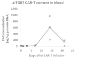
- FIG. 2 shows the results of quantitative PCR measured at each blood sampling time point. After administration of aITGB7 CAR-T at 1.33 ⁇ 10 5 cells/body, DNA of aITGB7 CAR was detected in whole blood from 1 hour later, and showed a maximum level 2 weeks after administration.
- Example 3 Extraction of Genomic DNA From PBMCs obtained from human donors to whom aITGB7 CAR-T cells were administered, gDNA was extracted according to the protocol attached to QIAamp DNA Mini Kit (manufactured by QIAGEN). gDNA samples were stored at -20°C as needed. (1) 10 milliliters of PBS was transferred to a 15 mL tube. (2) PBMC were thawed in a 37° C. water bath and transferred to a 15 mL tube. (3) After centrifuging the tube (300 ⁇ g, 10 minutes, room temperature), the supernatant was removed. (4) After tapping, 1 mL of PBS was transferred to a 15 mL tube to suspend PBMCs.
- the column was set in a new collection tube (2 mL), and 500 ⁇ L of Buffer AW1 (manufactured by QIAGEN) was added.
- the tube was centrifuged at 6000 xg for 1 minute at room temperature.
- the column was set in a new collection tube (2 mL), and 500 ⁇ L of Buffer AW2 (manufactured by QIAGEN) was added.
- the tube was centrifuged at 20000 xg for 3 minutes at room temperature.
- the column was set in a new collection tube (2 mL).
- (21) The tube was centrifuged at 20000 xg for 1 minute at room temperature.
- Standard Solution A standard plasmid stock solution (1.00 ⁇ 10 8 copies/ ⁇ L, SS) was thawed at room temperature, and standard solutions C1 to C10 were prepared according to Table 9 below. Buffer AE was used as the NTC (no template control) sample.
- the standard plasmid is a linear plasmid having a partial DNA base sequence of aITGB7 CAR-T cells.
- test samples H, M, L, LL and BL were prepared according to Tables 10 and 11 below. prepared. Each gDNA extracted in 1 above was melted at room temperature and diluted with Buffer AE to prepare 50 ng/ ⁇ L of gDNA.
- H-1 to Hn, M-1 to Mn, L-1 to Ln, LL-1 to LL-n, and BL-1 to BL-n were prepared according to the number of donors n.
- PCR Master Mix A sufficient amount of PCR Master Mix was prepared on the day of use. The contents of one reaction are described below.
- the H2O (PCR grade) supplied with the LightCycler 480 Probe Master was used.
- LightCycler 480 Probe Master 10.0 ⁇ L 20 ⁇ Pre Mixed Primer and Probe 1.0 ⁇ L H2O ( PCR grade) 1.0 ⁇ L Total 12.0 ⁇ L
- the above "Pre Mixed Primer and Probe” includes a forward primer consisting of the nucleotide sequence of SEQ ID NO: 1, a reverse primer consisting of the nucleotide sequence of SEQ ID NO: 2, and a probe having the nucleotide sequence of SEQ ID NO: 9 (5'-FAM-CACCACGCGACTTCGCAGCCTATC- NFQ-MGB-3') (FAM: Fluorescein, NFQ: Non-Fluorescent Quencher, MGB: Minor Groove Binder).
- Cp values, MSE, inverse regression values and sample concentrations were calculated using analysis software for real-time PCR systems (LightCyclerTM 480 software, Roche Diagnostics).
- C1-C10 were used to generate a standard curve. Acceptance criteria for calibration standards were as follows: (1) MSE less than 0.2. (2) The CV of the inverse regression value is less than 30% on at least 9 points out of 10 (C1 and C10 shall be less than 30%). (3) RE of the inverse regression value is within ⁇ 30% at least 9 points out of 10 (C1 and C10 shall be within ⁇ 30%). (4) The Cp value of NTC is 45.00 or clearly greater than the Cp value of C10.
- CV and RE were evaluated by analyzing H, M, L and LL with three replicates per level. Acceptance criteria for CV and RE were as follows: CV of QC sample is less than 30%. RE of QC sample value within ⁇ 30%.
- Freeze-Thaw Stability CV and RE were assessed by analyzing H and L with one replicate per level. QC samples were freeze-thawed 0, 1 and 3 times at ⁇ 20° C. before performing qPCR to confirm freeze-thaw stability. The melting operation was performed by standing the QC samples at RT. Acceptance criteria for freeze-thaw stability were as follows: QC samples have a CV less than 30%. The RE of the QC sample values is within ⁇ 30%.
- the present invention is not limited to the contents of the above examples.
Landscapes
- Chemical & Material Sciences (AREA)
- Life Sciences & Earth Sciences (AREA)
- Health & Medical Sciences (AREA)
- Organic Chemistry (AREA)
- Genetics & Genomics (AREA)
- Zoology (AREA)
- Engineering & Computer Science (AREA)
- Wood Science & Technology (AREA)
- Proteomics, Peptides & Aminoacids (AREA)
- Biochemistry (AREA)
- Biotechnology (AREA)
- Molecular Biology (AREA)
- General Health & Medical Sciences (AREA)
- Biophysics (AREA)
- Bioinformatics & Cheminformatics (AREA)
- General Engineering & Computer Science (AREA)
- Physics & Mathematics (AREA)
- Biomedical Technology (AREA)
- Microbiology (AREA)
- Analytical Chemistry (AREA)
- Gastroenterology & Hepatology (AREA)
- Plant Pathology (AREA)
- Medicinal Chemistry (AREA)
- Toxicology (AREA)
- Immunology (AREA)
- Measuring Or Testing Involving Enzymes Or Micro-Organisms (AREA)
Priority Applications (1)
| Application Number | Priority Date | Filing Date | Title |
|---|---|---|---|
| JP2023540426A JPWO2023013765A1 (https=) | 2021-08-06 | 2022-08-05 |
Applications Claiming Priority (2)
| Application Number | Priority Date | Filing Date | Title |
|---|---|---|---|
| JP2021130335 | 2021-08-06 | ||
| JP2021-130335 | 2021-08-06 |
Publications (1)
| Publication Number | Publication Date |
|---|---|
| WO2023013765A1 true WO2023013765A1 (ja) | 2023-02-09 |
Family
ID=85154595
Family Applications (1)
| Application Number | Title | Priority Date | Filing Date |
|---|---|---|---|
| PCT/JP2022/030098 Ceased WO2023013765A1 (ja) | 2021-08-06 | 2022-08-05 | キメラ抗原受容体を検出するためのプライマーセット |
Country Status (2)
| Country | Link |
|---|---|
| JP (1) | JPWO2023013765A1 (https=) |
| WO (1) | WO2023013765A1 (https=) |
Citations (5)
| Publication number | Priority date | Publication date | Assignee | Title |
|---|---|---|---|---|
| CN103113470A (zh) * | 2013-02-27 | 2013-05-22 | 四川大学 | 靶向人egfr的基因工程化淋巴细胞及其制备方法和用途 |
| WO2017026331A1 (ja) * | 2015-08-11 | 2017-02-16 | 国立大学法人大阪大学 | 抗体 |
| CN109971835A (zh) * | 2017-12-28 | 2019-07-05 | 上海细胞治疗研究院 | 一种锁核酸修饰的探针以及一种测定car拷贝数的方法 |
| CN109971836A (zh) * | 2017-12-28 | 2019-07-05 | 上海细胞治疗研究院 | 双重荧光定量pcr测定car拷贝数的方法和试剂盒 |
| US20200085871A1 (en) * | 2017-03-17 | 2020-03-19 | University Of Tennessee Research Foundation | Methods of using cytotoxic t cells for treatment of autoimmune diseases |
-
2022
- 2022-08-05 JP JP2023540426A patent/JPWO2023013765A1/ja active Pending
- 2022-08-05 WO PCT/JP2022/030098 patent/WO2023013765A1/ja not_active Ceased
Patent Citations (5)
| Publication number | Priority date | Publication date | Assignee | Title |
|---|---|---|---|---|
| CN103113470A (zh) * | 2013-02-27 | 2013-05-22 | 四川大学 | 靶向人egfr的基因工程化淋巴细胞及其制备方法和用途 |
| WO2017026331A1 (ja) * | 2015-08-11 | 2017-02-16 | 国立大学法人大阪大学 | 抗体 |
| US20200085871A1 (en) * | 2017-03-17 | 2020-03-19 | University Of Tennessee Research Foundation | Methods of using cytotoxic t cells for treatment of autoimmune diseases |
| CN109971835A (zh) * | 2017-12-28 | 2019-07-05 | 上海细胞治疗研究院 | 一种锁核酸修饰的探针以及一种测定car拷贝数的方法 |
| CN109971836A (zh) * | 2017-12-28 | 2019-07-05 | 上海细胞治疗研究院 | 双重荧光定量pcr测定car拷贝数的方法和试剂盒 |
Also Published As
| Publication number | Publication date |
|---|---|
| JPWO2023013765A1 (https=) | 2023-02-09 |
Similar Documents
| Publication | Publication Date | Title |
|---|---|---|
| CN115768899B (zh) | 用于定量重组载体核酸整合的组合物和方法 | |
| JP6636247B2 (ja) | WT1mRNAの発現量定量方法 | |
| KR20080082628A (ko) | 수용체 및 리간드 이소형태의 제조 방법 | |
| JP6447829B2 (ja) | Rna修飾の簡易検出法、及び該検出法を用いた2型糖尿病の検査方法 | |
| CN104894118A (zh) | 一种检测牛病毒性腹泻病毒的引物、探针及试剂盒 | |
| Yamazaki et al. | Quantitative assessment of minimal residual disease (MRD) in canine lymphoma by using real-time polymerase chain reaction | |
| WO2016167317A1 (ja) | 遺伝子変異の検出方法 | |
| CN112210568A (zh) | 一种Ph-like急性淋巴细胞白血病融合基因及其应用和检测试剂盒 | |
| CN103667514A (zh) | 一种人白介素28b基因多态性荧光pcr检测试剂盒 | |
| WO2019088852A1 (en) | Test kits and assays | |
| JP2022189882A (ja) | minor BCR-ABL1遺伝子を検出する方法 | |
| CN117701556A (zh) | Car基因组病毒载体拷贝数检测试剂盒及其应用 | |
| Baras et al. | A real-time quantitative PCR targeting the viral vector for the monitoring of patients treated with axicabtagene ciloleucel | |
| CN108642156B (zh) | 一种t790m基因突变的数字pcr检测试剂盒及其检测方法 | |
| WO2013111800A1 (ja) | Hiv検出用オリゴヌクレオチド、hiv検出キット、及びhiv検出方法 | |
| WO2023013765A1 (ja) | キメラ抗原受容体を検出するためのプライマーセット | |
| US8642265B2 (en) | Compositions and methods for the detection of topoisomerase II complexes with DNA | |
| Jin et al. | CCR5Δ32 59537-G/A promoter polymorphism is associated with low translational efficiency and the loss of CCR5Δ32 protective effects | |
| JPWO2016104794A1 (ja) | Braf変異検出によるegfr阻害剤の効果予測 | |
| CN101781677A (zh) | 检测白血病广谱标记物WT1基因mRNA表达的试剂盒 | |
| JP2023545126A (ja) | Car-t細胞の細胞動態及び体内分布の決定 | |
| WO2022109359A1 (en) | Compositions for the multiplexed detection of viruses | |
| CN107385065B (zh) | 扩增核酸的方法及其应用 | |
| JP7007796B2 (ja) | Abl遺伝子増幅用プライマー、核酸増幅方法及び核酸増幅用キット | |
| RU2620076C1 (ru) | Способ количественного определения мутации G250E киназного домена BCR-ABL у больных хроническим миелоидным лейкозом, резистентных к терапии ингибиторами тирозинкиназ |
Legal Events
| Date | Code | Title | Description |
|---|---|---|---|
| 121 | Ep: the epo has been informed by wipo that ep was designated in this application |
Ref document number: 22853183 Country of ref document: EP Kind code of ref document: A1 |
|
| WWE | Wipo information: entry into national phase |
Ref document number: 2023540426 Country of ref document: JP |
|
| NENP | Non-entry into the national phase |
Ref country code: DE |
|
| 122 | Ep: pct application non-entry in european phase |
Ref document number: 22853183 Country of ref document: EP Kind code of ref document: A1 |