WO2022211014A1 - 凝集性タンパク質の測定方法及び凝集可視化方法、並びにこれらの方法に使用するためのキット - Google Patents
凝集性タンパク質の測定方法及び凝集可視化方法、並びにこれらの方法に使用するためのキット Download PDFInfo
- Publication number
- WO2022211014A1 WO2022211014A1 PCT/JP2022/016478 JP2022016478W WO2022211014A1 WO 2022211014 A1 WO2022211014 A1 WO 2022211014A1 JP 2022016478 W JP2022016478 W JP 2022016478W WO 2022211014 A1 WO2022211014 A1 WO 2022211014A1
- Authority
- WO
- WIPO (PCT)
- Prior art keywords
- protein
- aggregation
- aggregating
- solution
- kda
- Prior art date
- Legal status (The legal status is an assumption and is not a legal conclusion. Google has not performed a legal analysis and makes no representation as to the accuracy of the status listed.)
- Ceased
Links
Images





Classifications
-
- C—CHEMISTRY; METALLURGY
- C07—ORGANIC CHEMISTRY
- C07K—PEPTIDES
- C07K1/00—General methods for the preparation of peptides, i.e. processes for the organic chemical preparation of peptides or proteins of any length
- C07K1/14—Extraction; Separation; Purification
- C07K1/34—Extraction; Separation; Purification by filtration, ultrafiltration or reverse osmosis
-
- C—CHEMISTRY; METALLURGY
- C07—ORGANIC CHEMISTRY
- C07K—PEPTIDES
- C07K1/00—General methods for the preparation of peptides, i.e. processes for the organic chemical preparation of peptides or proteins of any length
- C07K1/13—Labelling of peptides
-
- C—CHEMISTRY; METALLURGY
- C07—ORGANIC CHEMISTRY
- C07K—PEPTIDES
- C07K1/00—General methods for the preparation of peptides, i.e. processes for the organic chemical preparation of peptides or proteins of any length
- C07K1/14—Extraction; Separation; Purification
-
- C—CHEMISTRY; METALLURGY
- C07—ORGANIC CHEMISTRY
- C07K—PEPTIDES
- C07K1/00—General methods for the preparation of peptides, i.e. processes for the organic chemical preparation of peptides or proteins of any length
- C07K1/14—Extraction; Separation; Purification
- C07K1/30—Extraction; Separation; Purification by precipitation
-
- C—CHEMISTRY; METALLURGY
- C07—ORGANIC CHEMISTRY
- C07K—PEPTIDES
- C07K14/00—Peptides having more than 20 amino acids; Gastrins; Somatostatins; Melanotropins; Derivatives thereof
- C07K14/435—Peptides having more than 20 amino acids; Gastrins; Somatostatins; Melanotropins; Derivatives thereof from animals; from humans
- C07K14/46—Peptides having more than 20 amino acids; Gastrins; Somatostatins; Melanotropins; Derivatives thereof from animals; from humans from vertebrates
- C07K14/47—Peptides having more than 20 amino acids; Gastrins; Somatostatins; Melanotropins; Derivatives thereof from animals; from humans from vertebrates from mammals
-
- C—CHEMISTRY; METALLURGY
- C07—ORGANIC CHEMISTRY
- C07K—PEPTIDES
- C07K14/00—Peptides having more than 20 amino acids; Gastrins; Somatostatins; Melanotropins; Derivatives thereof
- C07K14/435—Peptides having more than 20 amino acids; Gastrins; Somatostatins; Melanotropins; Derivatives thereof from animals; from humans
- C07K14/76—Albumins
-
- G—PHYSICS
- G01—MEASURING; TESTING
- G01N—INVESTIGATING OR ANALYSING MATERIALS BY DETERMINING THEIR CHEMICAL OR PHYSICAL PROPERTIES
- G01N33/00—Investigating or analysing materials by specific methods not covered by groups G01N1/00 - G01N31/00
- G01N33/48—Biological material, e.g. blood, urine; Haemocytometers
- G01N33/50—Chemical analysis of biological material, e.g. blood, urine; Testing involving biospecific ligand binding methods; Immunological testing
- G01N33/52—Use of compounds or compositions for colorimetric, spectrophotometric or fluorometric investigation, e.g. use of reagent paper and including single- and multilayer analytical elements
-
- G—PHYSICS
- G01—MEASURING; TESTING
- G01N—INVESTIGATING OR ANALYSING MATERIALS BY DETERMINING THEIR CHEMICAL OR PHYSICAL PROPERTIES
- G01N33/00—Investigating or analysing materials by specific methods not covered by groups G01N1/00 - G01N31/00
- G01N33/48—Biological material, e.g. blood, urine; Haemocytometers
- G01N33/50—Chemical analysis of biological material, e.g. blood, urine; Testing involving biospecific ligand binding methods; Immunological testing
- G01N33/53—Immunoassay; Biospecific binding assay; Materials therefor
-
- G—PHYSICS
- G01—MEASURING; TESTING
- G01N—INVESTIGATING OR ANALYSING MATERIALS BY DETERMINING THEIR CHEMICAL OR PHYSICAL PROPERTIES
- G01N33/00—Investigating or analysing materials by specific methods not covered by groups G01N1/00 - G01N31/00
- G01N33/48—Biological material, e.g. blood, urine; Haemocytometers
- G01N33/50—Chemical analysis of biological material, e.g. blood, urine; Testing involving biospecific ligand binding methods; Immunological testing
- G01N33/58—Chemical analysis of biological material, e.g. blood, urine; Testing involving biospecific ligand binding methods; Immunological testing involving labelled substances
-
- G—PHYSICS
- G01—MEASURING; TESTING
- G01N—INVESTIGATING OR ANALYSING MATERIALS BY DETERMINING THEIR CHEMICAL OR PHYSICAL PROPERTIES
- G01N33/00—Investigating or analysing materials by specific methods not covered by groups G01N1/00 - G01N31/00
- G01N33/48—Biological material, e.g. blood, urine; Haemocytometers
- G01N33/50—Chemical analysis of biological material, e.g. blood, urine; Testing involving biospecific ligand binding methods; Immunological testing
- G01N33/68—Chemical analysis of biological material, e.g. blood, urine; Testing involving biospecific ligand binding methods; Immunological testing involving proteins, peptides or amino acids
-
- G—PHYSICS
- G01—MEASURING; TESTING
- G01N—INVESTIGATING OR ANALYSING MATERIALS BY DETERMINING THEIR CHEMICAL OR PHYSICAL PROPERTIES
- G01N33/00—Investigating or analysing materials by specific methods not covered by groups G01N1/00 - G01N31/00
- G01N33/48—Biological material, e.g. blood, urine; Haemocytometers
- G01N33/50—Chemical analysis of biological material, e.g. blood, urine; Testing involving biospecific ligand binding methods; Immunological testing
- G01N33/68—Chemical analysis of biological material, e.g. blood, urine; Testing involving biospecific ligand binding methods; Immunological testing involving proteins, peptides or amino acids
- G01N33/6893—Chemical analysis of biological material, e.g. blood, urine; Testing involving biospecific ligand binding methods; Immunological testing involving proteins, peptides or amino acids related to diseases not provided for elsewhere
- G01N33/6896—Neurological disorders, e.g. Alzheimer's disease
-
- G—PHYSICS
- G01—MEASURING; TESTING
- G01N—INVESTIGATING OR ANALYSING MATERIALS BY DETERMINING THEIR CHEMICAL OR PHYSICAL PROPERTIES
- G01N2333/00—Assays involving biological materials from specific organisms or of a specific nature
- G01N2333/435—Assays involving biological materials from specific organisms or of a specific nature from animals; from humans
- G01N2333/46—Assays involving biological materials from specific organisms or of a specific nature from animals; from humans from vertebrates
- G01N2333/47—Assays involving proteins of known structure or function as defined in the subgroups
- G01N2333/4701—Details
- G01N2333/4712—Muscle proteins, e.g. myosin, actin, protein
-
- G—PHYSICS
- G01—MEASURING; TESTING
- G01N—INVESTIGATING OR ANALYSING MATERIALS BY DETERMINING THEIR CHEMICAL OR PHYSICAL PROPERTIES
- G01N2333/00—Assays involving biological materials from specific organisms or of a specific nature
- G01N2333/435—Assays involving biological materials from specific organisms or of a specific nature from animals; from humans
- G01N2333/46—Assays involving biological materials from specific organisms or of a specific nature from animals; from humans from vertebrates
- G01N2333/47—Assays involving proteins of known structure or function as defined in the subgroups
- G01N2333/4701—Details
- G01N2333/4713—Plasma globulins, lactoglobulin
-
- G—PHYSICS
- G01—MEASURING; TESTING
- G01N—INVESTIGATING OR ANALYSING MATERIALS BY DETERMINING THEIR CHEMICAL OR PHYSICAL PROPERTIES
- G01N2333/00—Assays involving biological materials from specific organisms or of a specific nature
- G01N2333/435—Assays involving biological materials from specific organisms or of a specific nature from animals; from humans
- G01N2333/76—Assays involving albumins other than in routine use for blocking surfaces or for anchoring haptens during immunisation
Definitions
- the present invention relates to, for example, a method for collecting an agglutinating protein-containing solution, a method for measuring the amount of aggregation of an aggregating protein, a method for visualizing an aggregate of an aggregating protein, and a kit for use in these methods.
- AD Alzheimer's disease
- cognitive dysfunction dementia
- behavioral disorders or personality changes. with symptoms.
- the number of dementia patients worldwide is estimated to be more than 50 million as of 2019, but about 70% are believed to be AD patients, and the incidence is increasing.
- the increase in medical expenses and nursing care problems associated with the increase in AD patients have become major social problems, such as the economic and mental burden on the government and patients.
- AD begins with the aggregation and accumulation of the hydrophobic peptide amyloid- ⁇ protein (often referred to herein as "A ⁇ ") in the patient's brain, followed by hyperphosphorylation of the microtubule-associated protein tau protein. After oxidative fibrosis, nerve cells are destroyed and brain atrophy occurs (Non-Patent Documents 1 to 3).
- a ⁇ hydrophobic peptide amyloid- ⁇ protein
- MSHTS microliter-scale high-throughput screening
- Patent Document 1 discloses a method, apparatus and program for evaluating amyloid formation.
- Fluorescent probes including quantum dots as fluorescent dyes, or quantum dots etc.
- Patent Document 2 discloses a versatile quantum dot nanoprobe for evaluating the amyloid aggregation of proteins and peptides, and a method for evaluating an amyloid formation inhibitor using the quantum dot nanoprobe.
- quantum dot nanoprobes in which a quantum dot is attached via a cysteine to the N-terminus or C-terminus of an amyloidogenic peptide.
- candidate compounds obtained in the Cell-free Assays system often do not show their inhibitory effects in the Cell-Based Assays (test using cells) system. This is probably because the Cell-free Assay system does not necessarily reflect the biological environment. If MSHTS can be performed in a medium that is closer to the biological environment, it will be possible to screen candidate compounds and candidate materials that are expected to have an inhibitory effect in vivo with high accuracy and efficiency. However, when quantum dots were added to a medium or the like, there was a new problem that A ⁇ did not aggregate. Therefore, until now, it could not be used as a screening system for searching for candidate compounds in an environment closer to the biological environment.
- the present invention aims to provide a method for visualizing aggregates of aggregating proteins such as A ⁇ even in an environment close to biological conditions.
- Aggregating proteins e.g., amyloid ⁇ protein
- aggregates under conditions close to biological environments e.g., medium or culture supernatant
- biological environments e.g., medium or culture supernatant
- the medium or culture supernatant contains some contaminants that inhibit the aggregation of amyloid ⁇ protein and other aggregating proteins, and conducted a search for such contaminants.
- the present invention includes the following.
- Aggregation including a first step of passing a protein aggregation-promoting visualization solution through a filtration filter, and a second step of passing an aggregating protein-containing solution through the filtration filter passed through with the protein aggregation-promoting visualization solution.
- method for recovering a sexual protein-containing solution (2) The method according to (1), further comprising a pretreatment step of passing a washing liquid through the filter prior to the first step.
- the filtration filter is an ultrafiltration filter having a nominal molecular weight cutoff (NMWL) of 50 kDa or less.
- NMWL nominal molecular weight cutoff
- the protein aggregation-promoting visualization solution contains a substance having surface activity of 50 kDa or less.
- the substance having surfactant action is a surfactant.
- the surfactant is a nonionic surfactant.
- the nonionic surfactant is selected from at least one of an ester nonionic surfactant, an ether nonionic surfactant, an ester ether nonionic surfactant, and an alkyl glycoside nonionic surfactant. (8). (10) The method according to (9), wherein the nonionic surfactant is an ester ether type nonionic surfactant.
- the aggregating protein-containing solution contains a disease-associated protein.
- the disease is Alzheimer's disease and the protein associated therewith is amyloid ⁇ protein or tau protein.
- Aggregating protein-containing solution contains a substance selected from at least one of tau protein, ⁇ -synuclein, amyloid ⁇ protein, prion protein, TDP-43, polyglutamic acid, Atg-8, and Atg-15 , (1) to (15).
- Recovery rate (%) of agglutinating protein quantitative value of aggregating protein after the second step/quantitative value of aggregating protein before the second step ⁇ 100 ----- Formula 1) (20) The method according to any one of (1) to (19), wherein the aggregation protein is amyloid ⁇ protein.
- Quantum dot-modified amyloid ⁇ protein is added to the solution containing amyloid ⁇ protein recovered by the method described in (20), and the quantum dot is used as an index to determine the amyloid ⁇ protein and the quantum dot-modified amyloid ⁇ protein.
- a method for visualizing agglutinating protein aggregates comprising visualizing aggregating protein aggregates in an aggregating protein-containing solution recovered by the method according to any one of (1) to (21). .
- Visualization of aggregates of aggregating proteins involves incubating a wholly or partially labeled aggregating protein and/or an aggregating fragment thereof in the collected aggregating protein-containing solution, / or an aggregating step of aggregating the aggregating fragment thereof; and a detecting step of detecting the aggregate of the aggregating protein and/or the aggregating fragment thereof, wherein the collected agglutinating protein-containing solution contains the aggregating protein and/or an aggregation-inhibiting substance of 40 kDa or more, which inhibits aggregation of the aggregating fragment thereof, is removed.
- the optical label is a quantum dot.
- Aggregating protein is selected from at least one of tau protein, ⁇ -synuclein, amyloid ⁇ protein, prion protein, TDP-43, polyglutamic acid, Atg-8, and Atg-15, (26)- (31) The method according to any one of the items.
- (33) Add quantum dot-modified amyloid ⁇ protein to the solution containing amyloid ⁇ protein recovered by the method described in (20), and coagulate the amyloid ⁇ protein and the quantum dot-modified amyloid ⁇ protein by the quantum dots. The method according to any one of (26) to (32), wherein the aggregates are visualized.
- amyloid ⁇ protein is amyloid ⁇ 42 protein.
- the protein aggregation-promoting visualization solution contains a surface-active substance of 50 kDa or less.
- the kit according to (42), wherein the substance with surfactant action is a surfactant.
- the nonionic surfactant is selected from at least one of an ester nonionic surfactant, an ether nonionic surfactant, an ester ether nonionic surfactant, and an alkyl glycoside nonionic surfactant.
- the ester ether type nonionic surfactant is Tween-20.
- the protein aggregation promoting visualization solution is an aqueous solution containing Tween-20.
- Aggregating protein-containing solution contains a substance selected from at least one of tau protein, ⁇ -synuclein, amyloid ⁇ protein, prion protein, TDP-43, polyglutamic acid, Atg-8, and Atg-15 , (35) to (51).
- the present invention includes the following.
- the aggregation visualization kit according to any one of (16') to (20'), wherein the aggregation protein is a disease-associated protein.
- the aggregation visualization of (21'), wherein the disease is selected from the group consisting of Alzheimer's disease, Parkinson's disease, amyotrophic lateral sclerosis, Huntington's disease, prion disease, diabetes, and arteriosclerosis. kit.
- a production method characterized by forming an aggregate by an aggregating protein and/or an aggregating fragment thereof, wherein all or aggregating step of incubating a partially labeled aggregating protein to agglutinate said aggregating protein and/or aggregating fragment thereof;
- the production method comprising a confirmation step of confirming the production of (25')
- the production method according to (24') wherein the aggregation inhibitor of 40 kDa or more has a molecular weight of 250 kDa or less.
- contaminants can be removed from a solution containing aggregating proteins, while the aggregating proteins can be efficiently recovered, and aggregates of the aggregating proteins can be visualized in the recovered solution.
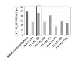
- FIG. 2 shows artificial amyloid ⁇ 42 protein (A ⁇ 42 ) residual rate after ultrafiltration in Examples.
- the artificial A ⁇ 42 residual rate of the "simulated liquid” was calculated as 100%.
- Fig. 2 shows artificial A ⁇ 42 residual rate after ultrafiltration in Examples.
- the artificial A ⁇ 42 residual rate of the "simulated liquid” was calculated as 100%. It shows that A ⁇ 42 in solution could be recovered most efficiently by using the composition solution of Blocking 2.
- Blocking 1 shows a 5% BSA-containing PBS solution left at room temperature (15-25°C) for 2 hours.
- Blocking 2 shows a final concentration of 0.1% tween-20 in a 5% BSA-containing PBS solution.
- Blocking 3 shows a 5% BSA-containing PBS solution left at 4 ⁇ 2°C for 18 hours
- Blocking 4 shows , and 5% BSA-containing PBS solution with tween-20 at a final concentration of 0.1% were left standing at 4 ⁇ 2°C for 18 hours.
- Blocking 6 shows the result of standing treatment at 4 ⁇ 2°C for 18 hours with a solution of 0.4% BSA-containing PBS containing tween-20 at a final concentration of 0.1%.
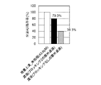
- Fig. 2 shows secretory amyloid ⁇ protein (A ⁇ ) residual rate in Examples. The secretory A ⁇ residual rate of the “culture supernatant” was calculated as 100%. It shows that removal of A ⁇ in the culture supernatant could be reduced by using Blocking 2.
- FIG. 1 secretory amyloid ⁇ protein
- FIG. 1 shows the results of evaluating whether visualization of aggregated A ⁇ and quantitative analysis of A ⁇ aggregation-inhibiting materials by MSHTS using ultrafiltration filtrate using Blocking 2 in Examples are possible.
- control indicates dimethylsulfoxide (DMSO) and "RA” indicates rosmarinic acid.
- Figure 3 shows that aggregated A ⁇ could not be visualized on MSHTS in the filtrate after ultrafiltration using Blocking 2. Since it was considered that the composition of Blocking 2 had an adverse effect, we selected a solution that allows MSHTS to be performed again and that can reduce the removal of A ⁇ from the culture supernatant.
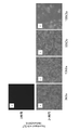
- FIG. 3 shows A ⁇ aggregate formation by solvent species.
- a Fluorescence image at the aggregation start time (0 hours) when PBS is used as the solvent.
- b Fluorescence image after 24 hours of incubation with the solution in a.
- c Fluorescence image after incubation for 24 hours with a solution of BSA added to PBS.
- d Fluorescence image after 24 hours of incubation in a neurobasal medium containing 0.5% v/v B-27 (NBM medium).
- e Fluorescence image after incubation for 24 hours when DFBM with B-27 added to DMEM/F12 medium is used as a solvent.
- FIG. 2 shows A ⁇ aggregate formation when the medium after ultrafiltration with various filter sizes is used as a solvent.
- a Fluorescence image at the start of aggregation (0 hour) when NBM medium after ultrafiltration with a 3 kDa filter is used as the solvent.
- b Fluorescence image after 24 hours of incubation with the solution in a.
- c Fluorescence image after 24 hours of incubation in NBM medium after ultrafiltration with a 10 kDa filter.
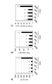
- FIG. 2 shows SDS-PAGE for evaluating A ⁇ aggregation inhibitors.
- a migration sample indicated by a corresponding number in the figure is obtained and migrated.
- the band around 42 kDa indicated by an arrow represents actin
- the band around 50 kDa represents albumin
- the band around 160 kDa represents globulin.
- a first aspect of the present invention is a method for recovering an agglutinative protein-containing solution.
- the method of this embodiment includes a first step of passing a protein aggregation-promoting visualization solution through a filtration filter, and a second step of passing an aggregating protein-containing solution through the filtration filter through which the protein aggregation-promoting visualization solution has been passed. characterized by comprising According to the method of this embodiment, contaminants (aggregation inhibitors) can be removed from a solution containing agglutinating proteins, while the aggregating proteins can be efficiently recovered.
- filtration filter refers to a filter that can fractionate substances with a predetermined molecular weight by filtration.
- examples include ultrafiltration filters.
- the type of ultrafiltration filter is not limited, in the present invention, an ultrafiltration filter having a nominal molecular weight cut off (NMWL) capable of removing aggregation-inhibiting substances of 40 kDa or more that inhibit aggregation of aggregating proteins is preferred.
- NMWL nominal molecular weight cut off
- many ultrafiltration filters are known to be able to filter slightly lower molecular weights than NMWL.
- a 50 kDa (UFC505096, Merck) filtration filter can separate about 90% of a BSA solution (1 mg/mL) with a molecular weight of 67 kDa, but even an ovalbumin solution (1 mg/mL) with a molecular weight of 45 kDa , about half ( ⁇ 65% with a swing basket rotor, ⁇ 55% with a fixed angle rotor) can be fractionated (Amicon (registered trademark) Ultra-15 Centrifugal Filter Devices User Guide). In other words, even if the NMWL is 50 kDa, it is possible to substantially remove proteins of 40 kDa or more.
- ultrafiltration filters with a NMWL of 50 kDa or less are suitable as filtration filters for use in the present invention.
- Ultrafiltration filters with NMWL of 50 kDa or less include, for example, Amicon Ultra-0.5 Ultracel-50 membrane, 50 kDa (UFC505096, Millipore).
- aggregation inhibitor refers to a substance that inhibits aggregation of agglutinative proteins.
- aggregation inhibitors of 40 kDa or more are applicable. Therefore, in the present specification, unless otherwise specified, the term “aggregation inhibitor” simply means an aggregation inhibitor of 40 kDa or more.
- Aggregation inhibitors of 40 kDa or more are not particularly limited as long as they are substances of 40 kDa or more, 41 kDa or more, or 42 kDa or more.
- Aggregation inhibitors of 40 kDa or more are, for example, substances having a molecular weight of 250 kDa or less, 240 kDa or less, 230 kDa or less, 220 kDa or less, or 210 kDa or less.
- Specific examples of aggregation-inhibiting substances of 40 kDa or more include aggregation-inhibiting substances comprising any one or more polypeptides or fragments thereof selected from the group consisting of actin protein, albumin and globulin.
- the aggregating protein is aggregated using an aggregating protein-containing solution from which aggregation inhibitors have been removed.
- the phrase "the aggregation inhibitor has been removed” means that the aggregation inhibitor is removed from the agglutinating protein-containing solution (or medium or culture supernatant) using the method described in the removal step described later.
- aggregation inhibitor When undergoing the removal step, it is preferable that 100% of the aggregation inhibitor is removed in the aggregating protein-containing solution after removal. You may have For example, 0.5% v / v or less, 0.4% v / v or less, 0.3% v / v or less, 0.2% v / v or less, or 0.1% v / v or less in the aggregation protein-containing solution, aggregation inhibitor remains in the acceptable range.
- Protein aggregation-promoting visualization liquid passes through the filtration filter in the process of removing aggregation inhibitors using the filtration filter, reduces protein adsorption, It is a solution that promotes the aggregation of and can visualize the aggregation.
- protein aggregation-promoting visualization liquids include those containing substances with surface activity of 50 kDa or less.
- Substances with surface-active properties include, for example, surfactants, preferably non-ionic surfactants.
- nonionic surfactants include ester nonionic surfactants, ether nonionic surfactants, ester ether nonionic surfactants, and alkylglycoside nonionic surfactants.
- ester-type nonionic surfactants include alkyl sulfate salts, polyoxyethylene alkyl sulfate salts, glycerin fatty acid esters, sorbitan fatty acid esters, sugar fatty acid esters, and fatty acid methyl ester ethoxylates.
- ether-type nonionic surfactants include polyoxyethylene alkyl ethers, fatty acid methyl ester ethoxylates, polyoxyethylene alkylphenyl ethers, and polyoxyethylene polyoxypropylene glycols.
- ester ether type nonionic surfactants examples include Tween-20 (also called Polysorbate 20 or polyethylene glycol sorbitan monolaurate), Tween-40, Tween-60, Tween-80 and Tween-85.
- alkyl glycoside nonionic surfactants include n-octyl- ⁇ -D-glucoside, n-octyl- ⁇ -D-maltoside, n-decyl- ⁇ -D-glucoside (decylglucoside), n-decyl- ⁇ -D-maltoside, n-dodecyl- ⁇ -D-glucoside (lauryl glucoside), n-heptyl- ⁇ -D-thioglucoside, n-octyl- ⁇ -D-thioglucoside, n-nonyl- ⁇ -D- and thiomaltoside.
- the nonionic surfactant is an ester ether type nonionic surfactant, and particularly preferably, the ester ether type nonionic surfactant is Tween-20.
- an aqueous solution containing Tween-20 can be used as a protein aggregation promoting visualization liquid.
- concentration of Tween-20 in the aqueous solution is, for example, 0.1-10%, preferably 0.01-1%, particularly preferably 0.05-0.5%.
- Aggregating protein refers to a protein that aggregates to form an aggregate.
- the kind of aggregating protein is not limited. Examples include polyglutamic acid, disease-associated proteins, or autophagy-associated proteins.
- Disease-associated aggregate proteins include, for example, amyloid ⁇ protein and tau protein (including phosphorylated tau protein), which are causative proteins of Alzheimer's disease, ⁇ -synuclein protein, which is a causative protein of Parkinson's disease, and transmissible spongiencephalopathy.
- amyloid ⁇ proteins such as amyloid ⁇ 42 protein, amyloid ⁇ 43 protein and amyloid ⁇ 38 protein are suitable.
- Autophagy-related proteins include, for example, ubiquitin-like proteins Atg-8 and Atg-12.
- aggregation proteins include, for example, tau protein (e.g., phosphorylated tau protein), ⁇ -synuclein, amyloid ⁇ protein, prion protein, TDP-43, polyglutamic acid, Atg-8, and Atg-15, preferably is amyloid ⁇ protein such as amyloid ⁇ 42 protein, amyloid ⁇ 43 protein, amyloid ⁇ 38 protein.
- the aggregation protein may be a naturally occurring protein, a modified protein obtained by artificially introducing mutations or modifications into a natural protein, or an artificially designed amino acid sequence based on It may be an artificial protein.
- All or part of the aggregating protein and the aggregating fragment described herein are labeled.
- all refers to all aggregating proteins or aggregating fragments
- a portion refers to one or more less than all aggregating proteins or aggregating fragments in the aggregating protein group or aggregating fragment group. It means that the sex fragment is labeled.
- aggregating fragment refers to a peptide that has a region or domain that can contribute to aggregation in the aggregating protein, and that alone has the same aggregating activity as the aggregating protein.
- aggregate refers to an aggregate of two or more aggregating proteins and/or aggregating fragments thereof. As used herein, so-called protein complexes are also included in aggregates.
- Aggregates may be homoaggregates consisting of the same type of protein or aggregating fragments thereof, or heteroaggregates consisting of different types of proteins or aggregating fragments thereof.
- An aggregate may be a complex that itself has a biological function, or it may be a simple aggregate.
- the diameter (size) of the aggregates is not limited, but usually it should be in the range of 10 nm or more and 10 mm or less. It is preferably 100 nm or more and 5 mm or less, more preferably 500 nm or more and 1 mm or less.
- the aggregating protein-containing solution may be any solution as long as it contains the aggregating protein. Qing is mentioned.
- a medium or culture supernatant that is close to the in vivo environment is preferred. For example, after culturing cells that secrete an aggregation protein, the culture supernatant can be recovered from the resulting culture, and the recovered culture supernatant can be used as an aggregation protein-containing solution.
- the aggregating protein contained in the aggregating protein-containing solution may be originally contained in the solvent, added, or both.
- the aggregation protein-containing solution may contain cell metabolites, cell secretion factors, inorganic substances (metallic inorganic salts), and organic acids in addition to the aggregation protein.
- cell metabolites include amino acids such as glutamic acid and aspartic acid, nucleic acids such as 5'guanylic acid, antioxidants such as isoascorbic acid, organic acids such as acetic acid and butyric acid, polyols such as glycerol, and vitamins. Vitamins such as B 2 and vitamin A can be mentioned.
- cell-secreted factors include, for example, Neuronal growth regulator 1 (NEGR1), Neurogenin-3, Kallikrein-8/Neuropsin (Cleaved-Val33), Neurofibromin (NF1), Neuroplastin (NPTN), Neurotrimin (NT) .
- Inorganic substances include metal salts (zinc, copper, iron, selenium, etc.), inorganic salts (Na, K, Ca, etc.), inorganic ions (cations: Li + , Na + , Mg 2+ , K + , Ca 2+ ; anions: Cl ⁇ , Br ⁇ , NO 2 ⁇ , SO 4 2 ⁇ , etc.).
- organic acids examples include succinic acid, pyruvic acid and lower amines. Note that the concentrations of the inorganic substance and the organic acid are not limited. For example, it may range from 0.001 ng/mL to 1.00 ng/mL, or from 0.01 ng/mL to 0.10 ng/mL.
- Cells "Cells" of interest in the present invention are cells that produce and/or secrete aggregating proteins.
- the cells may be cells derived from multicellular organisms.
- Animal-derived cells are preferred, and mammal-derived cells are more preferred. Examples include rodents such as mice, rats, hamsters and guinea pigs, livestock or pet animals such as dogs, cats, rabbits, cows, horses, sheep and goats, and primates such as humans, rhesus monkeys, gorillas and chimpanzees. . Human-derived cells are particularly preferred.
- the type of cells is not limited.” Examples thereof include cells derived from biological tissues, cells derived from cells derived from biological tissues, stem cells, cells differentiated from stem cells, and progenitor cells thereof.
- Bio tissue refers to various tissues that make up the living body of an organism. Examples include epithelial tissue, connective tissue, muscle tissue, nerve tissue, and the like.
- Stetem cells refer to cells that have the ability to differentiate into various cells and the ability to self-renew. Examples include adult stem cells and pluripotent stem cells.
- Adult stem cells are stem cells that exist in each tissue of an adult, have not completed terminal differentiation, and have a certain degree of pluripotency, and are also called somatic stem cells or organizational stem cells. Examples include mesenchymal stem cells, neural stem cells, intestinal epithelial stem cells, hematopoietic stem cells, hair follicle stem cells, melanocyte stem cells and the like.
- Pluripotent stem cells have pluripotency (pluripotency) that can differentiate into all types of cells that make up the living body, and when cultured in vitro under appropriate conditions A cell that can proliferate indefinitely while maintaining pluripotency. Examples thereof include embryonic stem cells (ES cells), embryonic germ stem cells, germline stem cells, and induced pluripotent stem cells (iPS cells).
- ES cells embryonic stem cells
- iPS cells induced pluripotent stem cells
- neural cells induced to differentiate from Alzheimer's disease (AD) patient-derived iPS cells via neural progenitor cells can be used as cells that produce and secrete amyloid ⁇ proteins such as amyloid ⁇ 42 protein.
- Buffer refers to a solution adjusted to maintain the pH within a predetermined range in accordance with the properties of the target substance to be mixed, such as cells and proteins. Examples include phosphate buffer, HEPES buffer, NaHCO 3 /CO 2 buffer, Tris-HCl buffer, glycine buffer and the like.
- the term “medium” refers to a liquid or solid substance prepared for culturing cells (eg, cells that produce and/or secrete aggregating proteins). In principle, it contains more than the necessary minimum amount of components essential for cell growth and/or maintenance. However, in the present invention, the medium can function as a site for aggregating proteins in addition to culturing cells.
- the medium may be either a basal medium or a specialized cell culture medium.
- Basic medium refers to the medium that forms the basis for various animal cell mediums. It can be cultured alone, and various culture additives can be added to prepare a medium specific to various cells (special cell culture medium) according to the purpose.
- Basic media include Neurobasal (registered trademark) medium, BME medium, BGJb medium, CMRL1066 medium, Glasgow MEM medium, Improved MEM Zinc Option medium, IMDM medium (Iscove's Modified Dulbecco's Medium), Medium 199 medium, Eagle MEM medium, ⁇ MEM medium , DMEM medium (Dulbecco's Modified Eagle's Medium), Ham's F10 medium, Ham's F12 medium, RPMI 1640 medium, Fischer's medium, and mixed media thereof (e.g., DMEM/F12 medium). Not limited. In addition, media used for culturing human iPS cells and human ES cells can also be suitably used.
- “Special cell culture medium” refers to a medium that is optimized for culturing specific cells by adding various supplements to the basal medium as described above, or that can induce differentiation into specific cells.
- “M medium” which is Sumitomo bakelite's nerve cell culture medium supplemented with NGF2.5S and BDNF. Culture media for pluripotent stem cells such as human iPS cells and human ES cells also fall under this category.
- the medium may be serum-containing medium or serum-free medium (ie, serum-free medium).
- culture supernatant refers to a culture solution obtained by removing cells by centrifugation, filtration, or the like after culturing cells in the medium for a predetermined period.
- predetermined period means, for example, 6 hours to 4 days, 12 hours to 3 days, 18 hours to 2 days, or One day is fine. Usually, the period is enough until the cells reach confluence.
- Culture supernatants generally contain various substances (nucleic acids, peptides, low-molecular-weight compounds, etc.) metabolized, produced, and/or secreted by cells.
- Labeling means modifying a target substance in order to identify the target substance. The label facilitates and ensures the detection or selection of the target substance.
- Labeling is performed according to the type of target substance.
- the target substance to be detected is an aggregate of an aggregating protein or an aggregating fragment thereof, so the type is a peptide.
- Labeling therefore covers any labeling means capable of directly or indirectly labeling a peptide.
- Direct labeling includes a labeling method in which a labeling substance is bound to an aggregating protein or an aggregating fragment thereof, or a labeling method in which an aggregating protein or an aggregating fragment thereof is expressed as a fusion protein with a labeled peptide.
- Indirect labeling includes a method of labeling an antibody that specifically recognizes and binds to an agglutinating protein or an aggregating fragment thereof, or an active fragment thereof, directly or indirectly via a secondary antibody. .
- a method of binding a labeling substance is preferable.
- Labeling substances used for peptide labeling include, but are not limited to, optical labels, enzymes, radioisotopes, or binding low-molecular-weight compounds.
- optical labeling refers to labeling with a substance that emits visible light, near-infrared light, or near-ultraviolet light, such as a fluorescent substance or a luminescent substance.
- fluorescent substance is a substance that has the property of becoming excited by absorbing excitation light of a specific wavelength and emitting fluorescence when returning to the original ground state. Fluorescent substances include both fluorescent dyes and fluorescent proteins.
- Fluorescent dyes include, for example, Quantum Dots, FITC, Texas, Texas Red®, Alexa Flor 405, Alexa Flor 488, Alexa Flor 647, Alexa Flor 700, Pacific Blue, DyLight 405, DyLight 550, DyLight 650 , PE-Cy5 (phycoerythrin-cyanin5), PE-Cy7 (phycoerythrin -cyanin 7), PE (phycoerythrin), PerCP (peridinin chlorophyll protein), PerCP-Cy5.5 (peridinin chlorophyll protein-cyanin5.5), cy3, cy5 , cy7, FAM, HEX, VIC®, JOE, ROX, TET, Bodipy493, NBD, TAMRA, Quasar® 670, Quasar® 705, APC (Allophycocyanin), Congo Red, Thioflavin T, Thioflavin S, fluorescamine or its derivatives, fluorescein or its derivatives, fluorescein
- Quantum Dot is a nanoscale semiconductor crystal that has optical properties according to quantum mechanics and emits fluorescence in the visible and near-infrared regions. Say. It is usually composed of 10 to 50 atoms with a diameter of 2 nm to 10 nm, and in addition to obtaining a large number of fluorescent colors depending on the particle size, it has excellent characteristics such as fluorescence fading resistance. Applications are progressing as biosensing materials and imaging materials for cells or animals.
- Fluorescent proteins include, for example, GFP.
- Enzymes include, for example, horseradish peroxidase (HRP), alkaline phosphatase (ALP), and glucose oxidase (GOx).
- HRP horseradish peroxidase
- ALP alkaline phosphatase
- GOx glucose oxidase
- Radioisotope refers to an element that emits radiation among isotopes with different mass numbers. Examples include 35 S and the like.
- binding low-molecular-weight compound refers to a low-molecular-weight compound that binds to a specific binding protein based on affinity.
- a specific example is biotin, also known as vitamin B7 .
- Biotin has extremely high affinity with egg white-derived protein avidin, its derivatives streptavidin or neutravidin, and strongly binds to each other. By utilizing this property and labeling the antibody with biotin, other labeling substances (such as fluorescent dyes) labeled with avidin bind to the antibody, enabling labeling of the antibody.
- Visualization refers to making aggregation of agglutinating proteins and/or aggregating fragments thereof recognizable with the naked eye or an optical microscope (including fluorescence microscopes and differential interference contrast microscopes). . Visualization of aggregation by an aggregating protein and/or an aggregating fragment thereof and the formation of these aggregates are synonymous, as they can usually be visualized by the formation of aggregates. The recognition is based on the visual confirmation of the formed aggregates, and when the aggregating protein is labeled, the intensity of the label (fluorescence or luminescence, etc.) and the state (aggregate, spotted, etc.). mentioned.
- the method of this aspect includes a first step of passing a protein aggregation-promoting visualization solution through a filtration filter, and a second step of passing an agglutinating protein-containing solution through the filtration filter that has been passed through with the protein aggregation-promoting visualization solution.
- Pretreatment step of passing a washing liquid through the filtration filter prior to the first step The filtration filter is washed by passing a washing liquid through the filtration filter and subjecting it to centrifugal separation. This operation is repeated once or more, preferably twice or more. If sodium hydroxide is used as the washing liquid, there is a risk that the hybrids will gel during the filtration process, and there is a risk that the aggregating proteins will not form aggregates in the filtrate obtained by the filtration treatment. Therefore, examples of cleaning liquids include pure water, PBS, and physiological saline. Centrifugation is performed, for example, at 3,000 to 25,000 g (eg, 14000 g) for 3 to 15 minutes (eg, 10 minutes) under temperature conditions of 15 to 25° C. (eg, room temperature).
- Methods for quantifying agglutinating proteins include, for example, immunoassays using antibodies against aggregating proteins. Examples of such immunological measurement methods include enzyme-linked immunosorbent assay (ELISA) and immunochromatography.
- Second step of passing the agglutinating protein-containing solution through the filtration filter through which the protein aggregation-promoting visualization liquid was passed After the first step, drop the agglutinating protein-containing solution into the filtration filter through which the protein aggregation-promoting visualization liquid was passed. and filter. After dropping, centrifugation is performed at 3,000 to 25,000 g (eg, 14,000 g) for 3 to 60 minutes (eg, 30 minutes) at a temperature of 10 to 25° C. (eg, room temperature) to collect the filtrate.
- the collected filtrate contains agglutinating proteins, and the use of a protein aggregation-promoting visualization solution allows visualization of aggregates of aggregating proteins in the filtrate.
- the step of quantifying the aggregated protein in the filtrate The aggregated protein in the obtained filtrate is quantified in the same manner as in Section 1-3-3. By this quantification, the recovery rate of the aggregating protein can be obtained by comparing with the amount of the aggregating protein in the aggregating protein-containing solution, which was previously quantified before the second step.
- the recovery rate of the aggregating protein obtained by the formula is, for example, more than 60%, preferably 65% or more, 70% or more, and particularly preferably 75% or more, the aggregating protein is recovered by the method of this embodiment. can be judged to have been significantly recovered.
- the aggregated protein can be efficiently recovered.
- the protein aggregation-promoting visualization liquid aggregates of aggregating proteins can be visualized in the recovered liquid.
- the second aspect of the present invention is a method for measuring the amount of aggregation of agglutinating proteins (herein, often abbreviated as "method for measuring aggregation amount”) or a method for visualizing aggregation of aggregating proteins (herein, , often abbreviated as "aggregation visualization method”).
- the method for measuring the amount of aggregation or the method for visualizing aggregation according to this aspect forms visible aggregates by using an aggregation-inhibiting substance that inhibits aggregation of the aggregation protein and/or aggregation fragments thereof. and detecting it.
- the method for measuring the amount of aggregation or the method for visualizing aggregation of this aspect it is possible to provide a screening technique for searching for an aggregation-inhibiting substance under an environment that is closer to the biological environment such as a medium or cell supernatant.
- the second aspect of the present invention comprises measuring the amount of aggregation of the aggregating protein in a solution (filtrate) containing the aggregating protein recovered by the method of the first aspect.
- Visualization of aggregates of aggregating proteins including visualizing aggregates of aggregating proteins in a solution (filtrate) containing the aggregating proteins recovered by the method for measuring the amount of aggregation or the method of the first aspect. The method.
- the use of the protein aggregation-promoting visualization liquid enables the visualization of the aggregates of the aggregating protein, and as a result, the recovered agglutination
- the amount of aggregation of the coagulable protein can be measured.
- the aggregation amount measurement method or aggregation visualization method of this embodiment includes an aggregation step and a detection step as essential steps. Each step will be described below.
- Aggregation step In the “aggregation step”, the aggregating protein and/or the aggregating fragment thereof are incubated in an agglutinating protein-containing solution to aggregate the aggregating protein and/or the aggregating fragment thereof. It is a process to let This step is characterized by aggregating an aggregating protein and/or an aggregating fragment thereof in an aggregating protein-containing solution from which aggregation inhibitors have been removed.
- an aggregating protein and/or an aggregating fragment thereof can be added to the aggregating protein-containing solution.
- the aggregating protein-containing solution may contain them beforehand.
- the incubation conditions are not particularly limited as long as the aggregating protein and/or its aggregating fragment can aggregate.
- the temperature may be above the melting point of the aggregating protein-containing solution and below the denaturation temperature of the aggregating protein and/or aggregating fragments thereof. It is preferably 5-50°C, 10-45°C, 15-42°C, 20-40°C, 25-39°C, 30-38°C, or 35-37°C.
- the time is not limited, but may be in the range of 30 minutes to 240 hours (10 days).
- the process can optionally include a removal step.
- a removal step any one selected from the group consisting of aggregation inhibitors, i.e., aggregation inhibitors of 40 kDa or more, or actin, albumin, and globulin from the agglutinative protein-containing solution used in this step.
- the step of removing the above polypeptides or fragments thereof is an optional step in the aggregation process.
- the removing step may be the method of the first aspect.
- This step cannot be achieved if the aggregation-inhibiting substance is contained in the solution containing the aggregation protein. Therefore, at least in the aggregation initiation stage, the premise of this step is that the aggregation-inhibiting substance is not contained in the aggregating protein-containing solution used in this step. However, when the solvent for the agglutinative protein-containing solution is a medium, culture supernatant, or the like, the solvent may contain an aggregation-inhibiting substance. This step is characterized by removing such aggregation-inhibiting substances in advance.
- the method of removing the aggregation inhibitor is not limited. Methods known in the art can be used. Examples include filtration methods, adsorption methods, centrifugation methods, or combinations of two or more thereof.
- Frtration method is a method of removal through a filter.
- a solution containing the aggregating protein or its solvent is passed through a filter, and the aggregation-inhibiting substance is captured and removed by the filter based on its molecular weight.
- the aggregation inhibitor to be removed is an aggregation inhibitor of 40 kDa or more
- this step can be achieved by using an ultrafiltration filter capable of molecular weight fractionation at less than 40 kDa.
- this step can be achieved by using an ultrafiltration filter capable of fractionating molecular weights of 40 kDa or more.
- This method is suitable when the molecular weight of the aggregation-inhibiting protein is significantly different from that of the aggregation-inhibiting substance.
- Filtration may be performed by gravity, but centrifugal filtration combined with the centrifugal method is convenient because it can be processed in a short period of time.
- Ultrafiltration filter kits and the like are commercially available from various life science manufacturers as centrifugal filtration filters for centrifugally removing substances with specific molecular weights from samples, and these may be used. Examples thereof include Amicon (registered trademark) Ultra 50K Kit (Merck).
- the "adsorption method” is a method in which a carrier that has a high affinity for aggregation inhibitors is used to adsorb and remove aggregation inhibitors on the surface and/or inside of the carrier. This step can be achieved by mixing the aggregating protein-containing solution or its solvent with the carrier, allowing the aggregation inhibitor to adsorb to the carrier, and then separating the carrier from the aggregating protein-containing solution or its solvent.
- the type of carrier may be appropriately selected according to the type of aggregation inhibitor to be adsorbed. For example, when the aggregation-inhibiting substance is albumin, silica having albumin-adsorptive properties can be used as the carrier.
- Albumin can also be adsorbed onto the carrier surface using the principle of ion exchange.
- a surface-modified carrier can be used to adsorb albumin onto the carrier surface.
- a method of modifying the carrier surface with a myristoylation reagent, a method of modifying the carrier surface with a palmitoylation reagent, a method of modifying the carrier surface with a diphenylcyclohexane compound, or an aggregation inhibitor and a method of modifying the carrier surface using an antibody or active fragment thereof or a nucleic acid aptamer that specifically binds to a method of modifying the carrier surface with a myristoylation reagent, a method of modifying the carrier surface with a palmitoylation reagent, a method of modifying the carrier surface with a diphenylcyclohexane compound, or an aggregation inhibitor and a method of modifying the carrier surface using an antibody or active fragment thereof or a nucleic acid aptamer that
- a myristoylation reagent is, for example, after removing the Boc group of N-Boc-N-tetradecanoyl-L-lysine [B5366], modifying [B5366] on a support such as silica, alumina, polystyrene, or quantum dots.
- a support such as silica, alumina, polystyrene, or quantum dots.
- Palmitoylating reagents can be achieved by, for example, modifying 1-tert-Butyl 5-(N-Succinimidyl)-N-Palmitoyl-L-Glutamate onto supports such as silica, alumina, polystyrene, and quantum dots. can.
- Surface modification using a diphenylcyclohexane compound can be achieved, for example, by modifying a carrier such as silica, alumina, polystyrene, or quantum dots with a diphenylcyclohexane compound.
- a carrier such as silica, alumina, polystyrene, or quantum dots with a diphenylcyclohexane compound.
- an antibody, an active fragment thereof, or a nucleic acid aptamer is, for example, an anti-actin antibody, an anti-albumin antibody, an anti-globulin antibody, an active fragment thereof, an actin-binding nucleic acid aptamer, an albumin-binding nucleic acid aptamer, or a globulin-binding It can be achieved by binding a nucleic acid aptamer or the like to various carriers (resin, glass, magnetic material, etc.).
- Antibodies include monoclonal antibodies, polyclonal antibodies, recombinant antibodies, synthetic antibodies, and antibody fragments. If the antibody is a polyclonal or monoclonal antibody, the immunoglobulin molecule can be of any class (eg IgG, IgE, IgM, IgA, IgD and IgY). Polyclonal antibodies or monoclonal antibodies are not limited, but usually antibodies derived from animals such as mammals or birds may be used. Examples include antibodies derived from mice, rats, guinea pigs, rabbits, goats, donkeys, sheep, camels, horses, chickens, humans, and the like.
- Recombinant antibody refers to a chimeric antibody, humanized antibody, or multispecific antibody.
- a “chimeric antibody” is an antibody produced by combining the amino acid sequences of antibodies derived from different animals, in which the variable region (V region) of one antibody is replaced with the V region of another antibody. For example, an antibody in which the V region of a mouse monoclonal antibody is replaced with that of a human antibody so that the variable region (V region) is mouse-derived and the C region is human-derived.
- Humanized antibody refers to the replacement of the complementarity determining regions (CDRs; CDR1, CDR2 and CDR3) in the variable region (V region) of a non-human mammal, e.g., a suitable mouse antibody, with the CDRs of a human monoclonal antibody. It is a grafted antibody.
- Chimeric antibodies and humanized antibodies have heavy and light chain V regions, or heavy and light chain V regions, although their complementarity-determining regions are derived from non-human animal antibodies such as mice, but the C region or V region Since the framework regions (FR; FR; FR1, FR2, FR3 and FR4) and the C regions of the heavy and light chains are derived from a human antibody, the immune response to the antibody in the human body can be reduced.
- the term "multispecific antibody” refers to a multivalent antibody, that is, an antibody having multiple antigen-binding sites in one molecule, wherein each antigen-binding site binds to a different epitope. Examples thereof include bispecific antibodies, such as IgG, which have two antigen-binding sites and each antigen-binding site binds to a different epitope.
- Synthetic antibody refers to an antibody synthesized chemically or by using recombinant DNA methods. For example, antibodies synthesized de novo using recombinant DNA methods. Specific examples include single chain fragments of variable regions (scFv), diabodies, triabodies, tetrabodies, and the like.
- Antibody fragment includes, for example, Fab, F(ab' 2 ), Fv and the like.
- Antibodies may be modified. Modifications of antibodies include functional modifications or labeling modifications. Functional modifications include, for example, glycosylation, acetylation, formylation, amidation, phosphorylation, or pegylation. Modifications on labels include fluorescent dyes (fluorescein, FITC, rhodamine, Texas Red, Cy3, Cy5), fluorescent proteins (e.g. PE, APC, GFP), enzymes (e.g. horseradish peroxidase, alkaline phosphatase, glucose oxidase) , radioisotopes (eg 3 H, 14 C, 35 S) or labeling with biotin or (strept)avidin.
- fluorescent dyes fluorescein, FITC, rhodamine, Texas Red, Cy3, Cy5
- fluorescent proteins e.g. PE, APC, GFP
- enzymes e.g. horseradish peroxidase, alkaline phosphatase, glucose oxidase
- nucleic acid aptamer is a ligand molecule that has the ability to strongly and specifically bind to a target substance due to its three-dimensional structure and specifically inhibit the function of the target substance.
- nucleic acid aptamer refers to an aptamer composed of nucleic acids.
- a nucleic acid constituting a nucleic acid aptamer may be DNA, RNA, or a combination thereof. If necessary, chemically modified nucleic acids such as PNA, LNA/BNA, methylphosphonate-type DNA, phosphorothioate-type DNA, 2'-O-methyl-type RNA can also be included.
- Nucleic acid aptamers may be labeled, if desired. Any nucleic acid labeling substance known in the art can be used for labeling. For example, radioisotopes (e.g. 32 P, 3 H, 14 C), DIG, biotin, fluorescent dyes (e.g. FITC, Texas, cy3, cy5, cy7, FAM, HEX, VIC, JOE, Rox, TET, Bodipy493 , NBD, TAMRA), or luminescent materials (eg, acridinium esters).
- radioisotopes e.g. 32 P, 3 H, 14 C
- DIG diotin
- biotin e.g. FITC, Texas, cy3, cy5, cy7, FAM, HEX, VIC, JOE, Rox, TET, Bodipy493 , NBD, TAMRA
- luminescent materials eg, acridinium esters
- removal kits are commercially available from various life science manufacturers and may be used to remove albumin and globulin from samples. Examples thereof include AlbuVoid TM (Biotech Support Group), Albumin & IgG Depletion Spintrap TM (Cytiva), actin-binding protein spindown Biochem Kit TM (Cytoskeleton) and ProMax Albumin Removal Kit (Polysciences).
- the "centrifugation method” is a method of molecular weight fractionation by centrifugation. This step can be achieved by removing a fraction of 40 kDa or more containing an aggregation-inhibiting substance after centrifuging the solution containing the aggregation protein or its solvent. If necessary, fractionation by centrifugation using a density gradient using sucrose or the like may be performed. Similar to the filtration method, this method is suitable when the molecular weight of the agglutinating protein is significantly different from that of the aggregation inhibitor. As mentioned above, the centrifugation method can also be used in combination with the filtration method and the adsorption method as an auxiliary method.
- the removal step may be performed multiple times.
- the aggregation-inhibiting substances cannot be sufficiently removed from the aggregating protein-containing solution or its solvent in one step, it is particularly preferable to carry out the steps a plurality of times.
- the removal method for each removal step may be the same or different.
- the “detection step” is a step of detecting aggregates after the aggregation step. In this step, aggregates formed by aggregation of aggregating proteins and/or aggregating fragments thereof are visually and/or quantitatively detected.
- a method for detecting peptide aggregates known in the art can be used. Since aggregates are generally detectable, the presence or absence of aggregates can be confirmed and detected from their shapes by visual observation or observation using an optical microscope. Moreover, when the aggregating protein and/or the aggregating fragment thereof is labeled, the aggregate can be detected by a detection method based on the label. For example, when an aggregating protein and/or an aggregating fragment thereof are labeled with an optical labeling substance such as quantum dots, aggregates may be detected by fluorescence observation using a fluorescence microscope. When spot-like fluorescent clusters are observed, it can be recognized that aggregates are formed.
- the presence or absence of aggregate formation may be detected by quantifying the fluorescence intensity and comparing the fluorescence intensity value with a negative control.
- aggregates can be detected by molecular sieves based on molecular weight such as gel electrophoresis.
- a method for measuring the amount of aggregation of aggregating proteins a method for visualizing aggregates of aggregating proteins, and a method for calculating the aggregation inhibitory activity of aggregating proteins, for example, a trace amount of amyloid ⁇ protein aggregation inhibitor using a quantum dot nanoprobe
- MSHTS throughput screening
- Aggregation sizes of aggregating proteins that can be visualized in this method include, for example, 0.01 to 200 ⁇ m 2 , preferably 1 to 200 ⁇ m 2 , particularly preferably 10 to 150 ⁇ m 2 .
- the aggregation protein is an amyloid ⁇ protein (for example, an amyloid ⁇ 42 protein)
- an amyloid ⁇ protein for example, an amyloid ⁇ 42 protein
- quantum dot-modified amyloid ⁇ protein is added to the solution containing the amyloid ⁇ protein recovered by the method of the first aspect.
- a protein for example, amyloid ⁇ 40 protein
- aggregates of the amyloid ⁇ protein and the quantum dot-modified amyloid ⁇ protein are measured using the quantum dots as an indicator, or the amyloid ⁇ protein and the amyloid ⁇ protein are measured using the quantum dots. Visualize aggregates with QD-modified amyloid- ⁇ protein.
- Quantum dots are nanomaterials with a three-dimensional quantum confinement structure, and semiconductor quantum dots, carbon-based quantum dots, etc. are known.
- Quantum dots include, for example, semiconductor quantum dots, particularly core-shell CdSe/ZnS quantum dots such as Qdot (registered trademark) 525, Qdot545, Qdot565, Qdot585, Qdot605, Qdot655, Qdot705 and Qdot800 (all from Thermo Fisher Scientific). is mentioned.
- the aggregation reaction between the amyloid ⁇ protein in the solution containing the amyloid ⁇ protein recovered by the method of the first aspect and the quantum dot-modified amyloid ⁇ protein is such that the amyloid ⁇ protein polymerizes to form aggregates.
- the reaction conditions are as low as possible.
- the concentration of the amyloid ⁇ protein at the start of the agglutination reaction is about 1 to 100 ⁇ M, preferably 10 to 50 ⁇ M, as the final concentration in the reaction solution.
- the concentration of quantum dot-modified amyloid ⁇ protein is about 0.005 to 0.5%, preferably 0.01 to 0.1% of the amyloid ⁇ protein concentration.
- the agglutination reaction can be performed in the wells of microplates commonly used for fluorescence observation.
- the use of a 1536-well flat-bottom microplate or the like is preferable because the amount of amyloid ⁇ protein aggregation can be measured with a reaction solution having a liquid volume on the order of microliters.
- the temperature of the agglutination reaction is about room temperature to 37°C, preferably 37°C, and the reaction time is about 4-36 hours, preferably 12-24 hours. Since shaking and stirring may excessively promote the formation of aggregates and adversely affect the determination of the amount of amyloid ⁇ protein aggregation, it is preferable to perform the agglutination reaction in a stationary state.
- the fluorescence of the agglutination reaction product obtained in the agglutination reaction is imaged under exposure conditions where the Sum Intensity calculated from the brightness value of each pixel included in the region of interest in the fluorescence image to be imaged is 15 to 85%. .
- the fluorescence of agglutination reaction products (aggregates) based on quantum dots is imaged using an imaging device connected to a computer that controls operations such as setting and controlling imaging conditions and imaging and displaying imaging data. can do.
- the imaging device is typically an epifluorescence microscope equipped with a digital camera (CCD or CMOS). Fluorescence is emitted from the reaction product and imaged with a CCD camera.
- the excitation light can be appropriately determined according to the properties of the quantum dots used. For example, when Qdot605 is used as a quantum dot, the wavelength of excitation light may be shorter than 580 nm, for example 532 to 552 nm.
- a band-pass filter for selectively transmitting the generated fluorescence and imaging the fluorescence may be selected so as to selectively transmit only light in a wavelength band including the fluorescence wavelength of the quantum dots used.
- a bandpass filter capable of selectively transmitting only light in the wavelength band of 594 to 646 nm can be used.
- Sum Intensity (sum of intensity values of all 186,624 pixels included in the region of interest / (186,624 x 255)) x 100
- the number of bits of the fluorescence image is preferably 8 bits. Although it is possible to use an image with a bit number greater than 8 bits, it is desirable to calculate the Sum Intensity after converting to an 8-bit image because the image processing load is large.
- Imaging is performed by referring to the Sum Intensity from the preliminary fluorescence image calculated in this way and controlling the exposure so that the Sum Intensity becomes a predetermined set value.
- Exposure can be controlled by adjusting the exposure time and camera gain (ISO sensitivity) and using a neutral density filter. It is preferable to use the function. For example, if the Sum Intensity calculated from the preliminary fluorescence image is below a predetermined set value, increasing the exposure time and/or increasing the camera gain can increase the Sum Intensity. Also, if the Sum Intensity calculated from the preliminary fluorescence image exceeds a predetermined set value, the Sum Intensity can be lowered by shortening the exposure time and/or lowering the camera gain.
- the set value of Sum Intensity can take any value within the range of 15% to 85%, preferably within the range of 45% to 65%.
- the above preliminary fluorescence image can be captured immediately before capturing the fluorescence of the agglutination reaction product used for determination. Exposure can be feedback controlled. Alternatively, a preliminary fluorescence image may be taken before starting the determination method, and an exposure condition in which the Sum Intensity is a predetermined set value may be determined before the determination method is started.
- the exposure conditions for a Sum Intensity of 15 to 85% are 150 ms to 1.8 s, preferably 300 ms to 1.6 s, at an ISO sensitivity of 200 (1x gain for a camera with an ISO sensitivity of 200). This is the exposure condition for In a further preferred embodiment, the exposure condition where the Sum Intensity is 45 to 65% is the exposure condition corresponding to ISO sensitivity 200 and exposure for 500 to 900 ms.
- using the automatic exposure function of the CCD camera set the target maximum brightness to 50% and expose with an exposure time of up to 160ms at an ISO sensitivity of 200, and the target maximum brightness reaches 50% within the exposure time.
- the target maximum light intensity is the percentage of the brightness value of the brightest pixel among the pixels included in the imaging range with respect to the camera gradation. For example, if the target maximum brightness is set to 50% for a 256-gradation camera capable of capturing 8-bit images, the brightness value of the brightest pixel within the imaging range will be 128. . At this time, the permissible amount of over-illumination may be appropriately set.
- the SD is calculated from the luminance value of each pixel included in the region of interest in the captured fluorescence image.
- the fluorescence image used here may be either color or monochrome.
- grayscale conversion is performed from the RGB values of each pixel, and the image must be an 8-bit image. is preferred.
- the target Sum Intensity can be appropriately set within the range of 45-65%.
- This correction may be performed for all fluorescence images, but from the viewpoint of measuring the amount of amyloid ⁇ protein aggregation with higher accuracy by increasing the value of the brightness value SD, it is calculated by the above formula 2 Fluorescence images whose Sum Intensity is less than the target Sum Intensity are corrected and then SD is calculated. Fluorescence images whose Sum Intensity is greater than or equal to the target Sum Intensity are left unchanged without correction. It is preferable to calculate the SD using the values.
- the amount of aggregation of amyloid ⁇ protein is measured using the fact that the brightness value SD calculated from the fluorescence image of the amyloid ⁇ protein aggregate is positively correlated with the amount of aggregation of the amyloid ⁇ protein. done as an indicator.
- the method of this aspect it is possible to visualize aggregates of aggregating proteins in the recovered liquid (filtrate) obtained by the method of the first aspect, and to measure the amount of aggregation of aggregating proteins. can be measured.
- the aggregating protein in the recovery liquid (filtrate) obtained by the method of the first aspect is an aggregating protein in an environment close to biological conditions, and according to the method of this aspect, Aggregates of aggregating proteins in the environment can be visualized.
- the protein aggregation-promoting visualization solution is used, and the diseases shown in Table 1 caused by various agglutinating proteins are detected in a solution that is closer to the in vivo state (cell secretion factors, etc.). It will be possible to search for preventive medicines and functional processed products against
- a third aspect of the present invention is an aggregation visualization agent.
- the agent for visualizing aggregation of this embodiment can provide a site for aggregation of aggregating proteins in an environment similar to a living body, and consists of a medium or culture supernatant from which aggregation inhibitors have been removed.
- aggregated proteins and/or aggregated fragments thereof in the solution can be aggregated and visualized as aggregates. Therefore, it can be used as a solvent or solvent for agglutinating protein-containing solution in a screening system for searching for candidate compounds having an aggregation-inhibiting effect on aggregating protein.
- the agent for visualizing aggregation of this aspect is composed of a medium or culture supernatant from which aggregation inhibitors have been removed.
- the aggregation visualization agent of this embodiment may be in either a liquid state or a solid state (including a powder state, a granule state, and a grain state). In the case of solid state, it is dissolved in a suitable solution (water, buffer, etc.) at the time of use.
- the components of the aggregation visualization agent of this embodiment include various substances that can be included in the medium or culture supernatant. However, since aggregation inhibitors are removed in advance, they are not included.
- the aggregation visualization agent of this embodiment may or may not contain an aggregation protein and/or an aggregation fragment thereof. Visualizing their aggregation by mixing any aggregation proteins and/or aggregation fragments thereof with the agent for visualizing aggregation of the present invention and allowing them to form aggregates, if necessary can be done.
- the agent for visualizing aggregation of this aspect is prepared by a method of preparing a medium or culture supernatant so that the aggregation inhibitor is not mixed, a method of removing an aggregation inhibitor from the prepared medium or culture supernatant, or a combination thereof. can be prepared.
- Methods for preparing media or culture supernatants that do not contain aggregation-inhibiting substances include methods for preparing components that do not contain aggregation-inhibiting substances, such as albumin-free and/or globulin-free reagents.
- Examples of methods for removing aggregation-inhibiting substances from the prepared medium or culture supernatant include a method according to the method described in "(1) Removal step" in the aggregation step of the second aspect.
- Aggregated visualization agents prepared as liquids may be stored in a liquid state until use, or may be stored after being converted to a solid state.
- storing in a liquid state in order to prevent deactivation and decomposition of various components such as proteins contained in the aggregation visualization agent, store below freezing, preferably below -20°C, below -80°C, or in liquid nitrogen. is preferred.
- the method for converting the aggregation visualization agent prepared as a liquid into a solid state is not limited, but includes, for example, a freeze-drying method. Freeze-drying is a known technique and may be performed according to conventional methods. Aggregated visualization agents prepared in a solid state are preferably stored at 10°C or lower, 4°C or lower, or 0°C or lower.
- the agent for visualizing aggregation of the present invention can be used, for example, in a screening system in which MSHTS or the like is performed in an environment closer to the biological environment to search for candidate compounds that have an aggregation-inhibiting effect on any aggregation-causing protein that causes disease. It can be provided as a solvent or a solvent for an aggregating protein-containing solution that does not inhibit protein aggregation and allows visualization of aggregation.
- a fourth aspect of the present invention includes a kit for recovering agglutinating proteins (herein, often abbreviated as “recovery kit”), a kit for measuring the amount of aggregation of aggregating proteins (herein In the literature, it is often abbreviated as “aggregation amount measurement kit”) or a kit for visualizing aggregates of agglutinating proteins (herein, it is often abbreviated as “aggregation visualization kit”).
- the kit for measuring the amount of aggregation or the kit for visualizing aggregation of this embodiment includes components for aggregating and visualizing agglutinating proteins and/or aggregating fragments thereof in an environment closer to the biological environment such as a medium or culture supernatant. .
- contaminants can be removed from a solution containing agglutinating proteins while efficiently recovering agglutinating proteins.
- Aggregates of aggregating proteins and/or aggregating fragments thereof can be easily detected in media or culture supernatants from which aggregation inhibitors have been removed.
- the kit of this embodiment includes a filtration filter and a protein aggregation-promoting visualization solution as essential components, and a washing solution and means for quantifying agglutinating proteins (e.g., an antibody used in an immunoassay method) as optional components.
- agglutinating proteins e.g., an antibody used in an immunoassay method
- aggregating protein antibodies, etc. means for labeling the aggregating protein (in kits for use in the method of the second aspect), labeled aggregating protein (e.g., quantum dot-modified amyloid ⁇ protein), protocol do.
- the protocol describes how to use the kit of this embodiment.
- Means for removing aggregation-inhibiting substances are used from media or culture supernatants, etc., which are solvents for agglutinating protein-containing solutions, from aggregation-inhibiting substances of 40 kDa or more, or from actin, albumin, and globulin.
- the “aggregating protein labeling means” detects and visualizes aggregation of the target aggregating protein and/or its aggregating fragment.
- a means of labeling the fragments include the labeling substance described in “(11) Labeling” in the definition of terms in the method for collecting an agglutinating protein-containing solution of the first aspect, and reagents and devices capable of modifying the labeling substance.
- the labeling substance is an optical label such as quantum dots
- quantum dots and reagents for adding them to peptides to modify the peptides may be used.
- various appropriate modification reagents and devices may be provided according to the type of labeling substance used.
- labeled aggregating protein refers to an already labeled aggregating protein and/or an aggregating fragment thereof.
- an agglutinating protein-containing solution is prepared using a medium or culture supernatant from which an aggregation-inhibiting substance has been removed, if the target aggregating protein and/or its aggregating fragment has been determined, the protein or the like is labeled. It can be included in the kit of this embodiment in such a state.
- the label conforms to the definition of terms in "(11) Label" in the method for collecting an agglutinating protein-containing solution of the first aspect.
- the aggregation visualization kit of this embodiment can selectively contain other components in addition to the components described above.
- Other components may include various necessary or useful components for visualizing aggregation of aggregating proteins, and are not particularly limited. Specific constituent elements may be appropriately determined according to, for example, the type of aggregating protein. As an example, it can contain a medium or culture supernatant from which aggregation inhibitors have not been removed or have been removed, a labeled secondary antibody, an enzyme substrate necessary for label detection, a washing buffer, a sample diluent, instructions for use, and the like.
- a fifth aspect of the present invention is a production method (herein often abbreviated as "aggregate production method") characterized by forming an aggregate by an aggregating protein.
- the aggregate-producing method of this embodiment comprises incubating the target agglutinating protein and/or an aggregating fragment thereof, which is wholly or partially labeled, in an agglutinating protein-containing solution from which an aggregation-inhibiting substance has been previously removed. , and forming an aggregate thereof.
- an aggregate comprising an aggregating protein and/or an aggregating fragment thereof can be produced and obtained.
- the obtained aggregates can be used in a screening system for searching for candidate compounds having an aggregate dissociation effect capable of dissociating them.
- the aggregate production method of this embodiment includes an aggregation step and a confirmation step as essential steps, and a recovery step as a selection step. Each step will be described below.
- the “aggregation step” in this aspect basically conforms to the aggregation step in the aggregation visualization method of the second aspect.
- the aggregating protein and/or aggregating fragment thereof constituting the desired aggregate are added under predetermined conditions. It is characterized by incubating to form aggregates. At this time, all or part of the aggregating protein and/or its aggregating fragment used for aggregate formation is labeled.
- the labeling method is also according to the definition of the term “(11) labeling” in the method for collecting an agglutinative protein-containing solution of the first aspect, and is not particularly limited, but modification with a labeling substance is preferable.
- the labeling substance although not limited, optical labeling, particularly labeling with quantum dots, is suitable.
- the type of aggregation protein is not limited, but proteins related to diseases caused by protein aggregation, such as A ⁇ and tau protein in AD, are preferable.
- the “confirmation step” is a step of detecting the aggregated protein aggregated in the solution after the aggregation step and confirming the formation of aggregates. This is a step of confirming the formation of aggregates, which are products, and basically conforms to the detection step in the aggregation visualization method of the second embodiment because the aggregates are detected for the confirmation.
- Recovery Step is a step of recovering aggregates when aggregates are confirmed in the confirmation step, and is a selection step in the method for producing aggregates of this aspect.
- the collected aggregates can be resuspended in a solvent as needed.
- Solvents to be used include, for example, physiological saline, buffers, media, culture supernatants, and the like.
- a medium or culture supernatant that is close to the in vivo environment is preferable.
- the medium or culture supernatant used in this case is preferably a medium or culture supernatant from which aggregation inhibitors have been removed.
- Example 1 A mixed solution of B27 (Life Technologies) diluted 50-fold with Neurobasal (phenol red-free) medium (Life Technologies) in Nb medium (NbM) to a final concentration of 50 pM amyloid ⁇ 42 protein (A ⁇ 42 ) solution (DMSO) (hereinafter referred to as simulated liquid) was prepared.
- a filtration filter device (Amicon Ultra-0.5 Ultracel-50 membrane) was attached to a 1.5 mL tube, and 400 ⁇ L of MilliQ water was dropped onto the filtration filter using MilliQ water at room temperature (15 to 25° C.) and a simulant.
- the 1.5 mL tube was centrifuged at 14,000 ⁇ g for 10 minutes at room temperature (15 to 25° C.), the filtrate was discarded, and 400 ⁇ l of MilliQ water was added dropwise to the filter again. Further, centrifuge at 14,000 ⁇ g for 10 minutes at room temperature (15-25°C), discard the filtrate, perform empty centrifugation at 14,000 ⁇ g for 2 minutes at room temperature (15-25°C), and attach the filter device to a new 1.5 mL tube. After dropping 500 ⁇ L of the simulated solution onto the filtration filter, it was centrifuged at 14,000 ⁇ g for 30 minutes at room temperature (15 to 25° C.), and the filtrate was recovered.
- the concentrations of A ⁇ 42 contained in the simulant and filtrate were measured using a human ⁇ -amyloid ELISA kit (Fujifilm Wako Pure Chemical Industries, Ltd.). Assuming that the A ⁇ 42 concentration in the simulant solution was 100%, the A ⁇ 42 concentration in the filtrate was divided by the A ⁇ 42 concentration in the A ⁇ 42 -containing simulant solution and converted into percent to calculate the A ⁇ 42 residual rate, which was confirmed to be 53.8%. (Fig. 1).
- Blocks 1-6 were prepared below.
- Blocking 1 indicates a PBS solution containing 5% BSA
- Blocking 2 indicates a PBS solution containing 5% BSA with tween-20 adjusted to a final concentration of 0.1%
- Blocking 3 indicates a PBS solution containing 5% BSA.
- Blocking 4 indicates a solution in which tween-20 was adjusted to a final concentration of 0.1% in a 5% BSA-containing PBS solution
- Blocking 5 indicates a 0.4% BSA-containing PBS solution
- Blocking 6 indicates a 0.4% BSA-containing PBS solution.
- a solution prepared with tween-20 at a final concentration of 0.1% is shown.
- Example 2 As in Example 1, a filtration filter device (Amicon Ultra-0.5 Ultracel-50 membrane) was attached to a 1.5 mL tube, 400 ⁇ l of MilliQ water was added dropwise to the filtration filter device, and 14,000 ⁇ g, 10 Centrifuge for 1 minute.
- a filtration filter device (Amicon Ultra-0.5 Ultracel-50 membrane) was attached to a 1.5 mL tube, 400 ⁇ l of MilliQ water was added dropwise to the filtration filter device, and 14,000 ⁇ g, 10 Centrifuge for 1 minute.
- the filtrate was discarded, and 500 ⁇ L of blocking 1 to 6 was dropped on the filtration filter and incubated.
- the incubation conditions are Blocking 1, Blocking 2, and Blocking 3 at room temperature (15-25°C) for 2 hours, and Blocking 4, Blocking 5, and Blocking 6 at refrigeration (4 ⁇ 2°C) for 18 hours. did.
- the 1.5 mL tube was centrifuged at room temperature (15-25°C) and 14,000 xg for 2 minutes, the filtrate was discarded, and the tube was centrifuged at room temperature (15-25°C) and 14,000 xg for 10 minutes.
- Example 3 In Example 2 described above, since the A ⁇ 42 residual rate was the highest under the blocking 2 conditions, the amyloid ⁇ protein (A ⁇ ) actually secreted by the cells (including A ⁇ such as A ⁇ 42 and amyloid ⁇ 38 protein (A ⁇ 38 ) ), it was confirmed whether the A ⁇ recovery rate could be similarly maintained at a high level.
- a ⁇ amyloid ⁇ protein
- a ⁇ secreted by cells was prepared by the following procedure.
- iPS cell-derived neural progenitor cells with a survival rate of 70% or more thawed in a constant temperature bath are added to NbM with 5 ⁇ M Y-27632 (Nacalai Tesque) in a culture medium, or Neural Maintenance Medium (Axol) is added to Neural Maintenance Medium Supplement (Axol 5 ⁇ M Y-27632 was added to the NMMs medium supplemented with Y-27632, and 8-16 ⁇ 10 ⁇ 5 cells/well (0.84-1.7 ⁇ 10 ⁇ 5/cm 2 ) were placed on the culture plate from which the coating solution was removed. Cells were seeded at a density of 10 m and cultured for 2 days at 37 °C, 5% CO2 conditions.
- iPS cell-derived neural progenitor cells were treated with Accutage (Stem Cell technologies) for 3-5 minutes, dissociated into single cells by pipetting, added 5 mL of DMEM/F-12, and placed in a centrifuge tube. Cells were decanted and the supernatant removed by centrifugation.
- a culture solution containing 5 ⁇ M Y-27632 added to NbM or a culture solution containing 10 ⁇ M Y-27632 added to NMMs medium was added to the precipitated cells, and the cells were dispersed by pipetting to prepare a cell suspension. Seed cells at a density of 8.0 to 16 ⁇ 10 ⁇ 5 cells/well (0.84 to 1.7 ⁇ 10 ⁇ 5/cm 2 ) on the culture plate after removing the coating solution, and cultured for 2 days at 37°C and 5% CO 2 conditions. did. After that, the culture supernatant was removed by aspiration, 2 mL/well of fresh NbM that had been returned to room temperature was added, and the cells were cultured at 37° C. and 5% CO 2 for 2 days.
- iPS cell-derived neural progenitor cells were treated with Accutage (Stem Cell technologies) for 3-5 minutes, dissociated into single cells by pipetting, added 5 mL of DMEM/F-12, and transferred to a centrifuge tube. Transfer and remove the supernatant by centrifugation.
- the medium was replaced with 2 mL/well of NbM+Shh medium supplemented with Sonic Hedgehog (Shh) protein at a final concentration of 100 ng/mL, and cultured at 37°C, 5% CO 2 for 3.5 days. did. Further, half the culture supernatant was collected from the culture plate, 2 mL/well of NbM+Shh medium was added, and the cells were cultured at 37° C. and 5% CO 2 for 3.5 days.
- Sonic Hedgehog Sonic Hedgehog
- the culture supernatant collected from the culture plate was collected in a centrifugation tube, centrifuged at 300 xg for 3 minutes, then transferred to a new centrifugation tube with 1 mL remaining from the bottom, and frozen at -20°C. (Culture supernatant Day7). Thereafter, half of the medium was collected once every 3.5 days, 2 mL/well of fresh NbM was added, and the cells were cultured at 37°C and 5% CO 2 .
- the culture supernatant of cells 14 days or more after addition of Shh was collected.
- the concentration of A ⁇ contained in these recovered culture supernatants was measured with a human ⁇ -amyloid ELISA kit (Fuji Film Wako Pure Chemical Industries) and found to be 43.6 pM (Fig. 3).
- a human ⁇ -amyloid ELISA kit Fluji Film Wako Pure Chemical Industries
- Fig. 3 The culture supernatant, it was confirmed whether even A ⁇ -containing culture supernatant secreted by actual cells could reduce A ⁇ removal under the conditions of Blocking 2 in Example 2 where the A ⁇ 42 residual rate was the highest.
- the A ⁇ residual rate without blocking was calculated in the same manner as in Example 1. As a result, it was found that under the conditions of Blocking 2, a high A ⁇ residual rate (79.3%) could be maintained even in the A ⁇ -containing culture supernatant secreted by cells (Fig. 3).
- Example 4 From Example 3 described above, it was confirmed that under the conditions of Blocking 2, even when the filtrate was collected from the A ⁇ -containing culture supernatant secreted by actual cells, a high A ⁇ residual rate was exhibited. Therefore, it was confirmed whether or not aggregation of amyloid ⁇ protein could be visualized in this filtrate.
- Quantum dot-modified amyloid ⁇ 40 protein QD-A ⁇ 40
- was dissolved in the filtrate collected in Example 3 this solution was added to 1 mM A ⁇ 42 solution and mixed, 50 ⁇ M A ⁇ 42 and 50 nM QD-A ⁇ 40 were mixed, The filtrate recovered in Example 3 was prepared.
- a 150 mM rosmarinic acid (RA) solution was diluted with the filtrate collected in Example 3 to five concentrations (3000 ⁇ M, 600 ⁇ M, 120 ⁇ M, 24 ⁇ M, 4.8 ⁇ M).
- a ⁇ 42 and QD-A ⁇ 40 were diluted with the filtrate collected in Example 3 to prepare a mixed solution of 50 ⁇ M A ⁇ 42 and 50 nM QD-A ⁇ 40.
- a mixed solution of A ⁇ 42 and QD-A ⁇ 40 and a rosmarinic acid (RA) solution were mixed at a ratio of 1:1 to prepare reaction solutions with final RA concentrations of 1500 ⁇ M, 300 ⁇ M, 60 ⁇ M, 12 ⁇ M and 2.4 ⁇ M.
- a solution in which DMSO was substituted for RA was prepared as a negative control. These reaction solutions were incubated at 37°C for 24 hours, and aggregated A ⁇ was observed under a fluorescence microscope. Figure 4).
- Blocking 8 indicates a solution in which Tween-20 was adjusted to a final concentration of 0.1% in a PBS solution containing 0.4% BSA
- Blocking 9 indicates a PBS solution containing 0.4% BSA
- Blocking 10 indicates a PBS solution containing 0.4% B27.
- Blocking 11 indicates a 0.1% final concentration of tween-20
- Blocking 11 indicates a 0.4% B27-containing PBS solution
- Blocking 12 indicates a 0.1% final concentration of tween-20 in PBS.
- a filtration filter device (Amicon Ultra-0.5 Ultracel-50 membrane) was attached to a 1.5 mL tube, 400 ⁇ l MilliQ water was added dropwise to the filtration filter device, and 14,000 ⁇ g, 10 Centrifuge for 1 minute.
- the filtrate was discarded, 400 ⁇ l of MilliQ water was added dropwise to the filter again, and the mixture was centrifuged at 14,000 ⁇ g at room temperature (15 to 25° C.) for 10 minutes. Discard the filtrate, drop 500 ⁇ L of each blocking (PBS, 8-12) solution onto the filter, and incubate. The incubation conditions were blocking PBS, 8-12, and standing at room temperature (15-25°C) for 2 hours. After blocking, the mixture was centrifuged, the filtrate was discarded, and the mixture was centrifuged at room temperature (15-25°C) at 14,000 xg for 10 minutes.
- the concentrations of A ⁇ 42 contained in the sham and filtrate were measured using a human ⁇ -amyloid ELISA kit (Fujifilm Wako Pure Chemical).
- a ⁇ 42 concentration in each filtrate was calculated with the A ⁇ 42 concentration in the simulated solution as 100%, it was found that the composition and conditions of Blocking 12 had the highest A ⁇ 42 residual rate (83.9%: Fig. 5 ).
- a 50 ⁇ M A ⁇ 42 solution was prepared from the filtrate recovered from each blocking (PBS, 8 to 12) operation, and a 50 nM QD-A ⁇ 40 solution was prepared from the filtrate from which 11.1 ⁇ M QD-A ⁇ 40 was recovered.
- the 0.1% Tween-20 solution of Blocking 12 was confirmed to have the highest sensitivity (SD value) as a protein aggregation-promoting visualization solution.
- a 50 ⁇ M A ⁇ 42 solution was prepared from the filtrate collected by the same operation as in Example 2, and QD-A ⁇ 40 was diluted in the collected filtrate to prepare 50 nM QD-A ⁇ 40 .
- Solution B was prepared. Using this solution B, reaction solutions having final RA concentrations of 1500 ⁇ M, 300 ⁇ M, 60 ⁇ M, 12 ⁇ M and 2.4 ⁇ M were prepared in the same manner as in Example 4. A reaction solution in which DMSO was substituted for RA was also prepared as a negative control. These reaction solutions were incubated at 37°C for 24 hours, aggregated A ⁇ was observed under a fluorescence microscope, and aggregated A ⁇ was quantified. It was confirmed that A ⁇ no longer aggregates.
- Example 6 Confirmation of aggregate formation by solvent species (purpose) In micro-volume high-throughput screening (MSHTS), confirm the formation of amyloid ⁇ protein aggregates when using various solvents.
- MSHTS micro-volume high-throughput screening
- Amyloid ⁇ protein (A ⁇ ) was used as the aggregating protein.
- a 1 mM A ⁇ solution was prepared by the following procedure. Add 5 mL of HFIP (1,1,1,3,3,3-Hexafluoro-2-propanol) (Fujifilm Wako Pure Chemical Industries, Ltd.) to 5 mg of A ⁇ (Human, 1-42) (Peptide Institute) After being suspended, the suspension was allowed to stand at room temperature for 1 hour. After that, it was subjected to ultrasonic treatment at 25° C. and 43 kHz for 10 minutes to be monomerized, and then allowed to stand in a clean bench for 24 hours to volatilize HFIP.
- a ⁇ 42 The A ⁇ solution prepared here is hereinafter referred to as "A ⁇ 42".
- QDA ⁇ Quantum Dot-Modified Amyloid ⁇ Protein
- the sample solution was transferred to two VIVASPIN6 tubes at 145 ⁇ L each, 4500 ⁇ L of water was added, and then centrifuged at 3800 xg at 4°C for 17 minutes. The filtrate was discarded, and the total volume of the obtained solution was combined into one tube so that the total volume was about 140 ⁇ L.
- ⁇ BSA/PBS solution A solution prepared by dissolving BSA (Wako) in PBS to a concentration of 50 mg/mL.
- ⁇ NBM solution This is a solution prepared by adding B-27 (registered trademark) (Gibco) to Neurobasal medium to make it 0.5% v/v. - DFBM solution A solution prepared to 0.5% v/v by adding B-27 (registered trademark) (Gibco) to DMEM/F12 medium (Thermo Fisher Scientific).
- a ⁇ solution containing 50 nM QDA ⁇ and 50 ⁇ M A ⁇ 42 corresponding to double the final concentration was prepared.
- a sample solution was prepared by adding 3 ⁇ L of A ⁇ solution to 3 ⁇ L of each evaluation solution. After centrifuging each sample solution at 10000 ⁇ g for 2 minutes, 5 ⁇ L of each sample solution was dispensed into a well plate. Subsequently, it was centrifuged at 1530 xg for 5 minutes in a plate centrifuge. The time before aggregation was defined as O hours, and after confirming the presence or absence of aggregation, incubation was carried out at 37° C. for 24 hours. After 24 hours, an image was taken using an inverted microscope (TE2000 manufactured by Nikon Corporation/DP72 manufactured by Olympus Corporation) to check the presence or absence of aggregates in each sample solution.
- Example 7 Confirmation of A ⁇ aggregate formation by removal of aggregation inhibitors (1) (Purpose) From Example 6, it was presumed that a substance inhibiting A ⁇ aggregation was present in the medium. Therefore, it was confirmed whether aggregated A ⁇ could be visualized by removing the A ⁇ aggregation inhibitor from the sample solution.
- Example 8 Evaluation of aggregation inhibitors (purpose)
- Example 7 revealed that A ⁇ aggregation inhibitors were removed by a 50 kDa ultrafiltration filter. Even though the nominal molecular weight cutoff (NMWL) is 50 kDa as mentioned above, in practice substances with molecular weights below 50 kDa can also be removed. Therefore, molecular weight fractionation by SDS-PAGE and staining with CBB were performed in order to confirm the components removed in the NBM solution and DFBM solution.
- NMWL nominal molecular weight cutoff
- the gel was recovered, soaked in CBB solution (nacalai tesque) for 20 minutes, washed with 7.5% acetic acid for 30 minutes, heated in a microwave oven about 3 times, and the destained gel was photographed.
- CBB solution nacalai tesque
- the NBM solution and DFBM solution contain a lot of globulin and actin, and the molecular weight of globulin is about 150 kDa, and the molecular weight of actin is 42 kDa.
- the band observed around 150 kDa in SDS-PAGE in FIG. 8 is considered to be globulin, and the band observed around 42 kDa is considered to be actin. Therefore, it was speculated that globulin and actin are also components of A ⁇ aggregation inhibitors.
- aggregation inhibitors can be removed by removing substances having a molecular weight of 40 kDa to 250 kDa, 40 kDa to 240 kDa, 40 kDa to 230 kDa, 40 kDa to 220 kDa, or 40 kDa to 210 kDa. rice field.
- Example 9 Confirmation of A ⁇ aggregate formation by removal of aggregation inhibitors (2) (Method) From Example 7, the image of aggregated A ⁇ formed in the filtrate collected after passage through each filter was automatically detected using the image analysis software Image J to automatically detect the shape of the aggregate (including the use of the threshold function). . Thereafter, 10 aggregates that could be determined to be individual particles were randomly selected for each condition, and the area, perimeter, and Feret diameter of the aggregates were calculated. As for the analysis method of Image J, after importing the photographed image into Image J, the shape of aggregates was detected in Adjust and Color Threshold modes in the Image tab. The shape of this aggregate was determined using the Wand tool, 10 individual particles were randomly selected, and added to Tools and ROI manager in the Analyze tab.
- the Measure tab in the ROI manager to calculate the "Area”, “Perimeter” and “Feret's Diameter” of each particle, average these values and graph them. Note that the Feret diameter is the longest distance among straight lines that connect any two points on the outer circumference of the selection range.
- FIG. 10 shows the concept of screening technology using these results.
- a culture supernatant obtained by culturing disease model cells is collected, and aggregated proteins in the culture supernatant are aggregated and visualized by the method of the present invention. Since the EC 50 can be calculated by adding candidate substances exhibiting aggregation-inhibiting activity such as various compounds and extracts at a predetermined concentration during the visualization, it is possible to select candidate substances that actually exhibit aggregation-inhibiting activity. .
- This screening technique can be automated and thus can be performed at high throughput.
Landscapes
- Health & Medical Sciences (AREA)
- Life Sciences & Earth Sciences (AREA)
- Chemical & Material Sciences (AREA)
- Engineering & Computer Science (AREA)
- Molecular Biology (AREA)
- Biomedical Technology (AREA)
- Immunology (AREA)
- Hematology (AREA)
- Urology & Nephrology (AREA)
- Medicinal Chemistry (AREA)
- General Health & Medical Sciences (AREA)
- Biochemistry (AREA)
- Organic Chemistry (AREA)
- Analytical Chemistry (AREA)
- Proteomics, Peptides & Aminoacids (AREA)
- Cell Biology (AREA)
- Physics & Mathematics (AREA)
- Microbiology (AREA)
- Biotechnology (AREA)
- Food Science & Technology (AREA)
- General Physics & Mathematics (AREA)
- Pathology (AREA)
- Biophysics (AREA)
- Genetics & Genomics (AREA)
- Neurosurgery (AREA)
- Water Supply & Treatment (AREA)
- Neurology (AREA)
- Toxicology (AREA)
- Zoology (AREA)
- Gastroenterology & Hepatology (AREA)
- Investigating Or Analysing Biological Materials (AREA)
- Peptides Or Proteins (AREA)
Abstract
Description
(1)濾過フィルターをタンパク凝集促進可視化液で通液する第1工程と、タンパク凝集促進可視化液で通液した濾過フィルターに、凝集性タンパク質含有溶液を通液する第2工程とを含む、凝集性タンパク質含有溶液の回収方法。
(2)第1工程前に、濾過フィルターを洗浄液で通液する前処理工程をさらに含む、(1)記載の方法。
(3)第2工程前に、凝集性タンパク質含有溶液中の凝集性タンパク質を定量する工程をさらに含む、(1)又は(2)記載の方法。
(4)第2工程後、濾液中の凝集性タンパク質を定量する工程をさらに含む、(1)~(3)のいずれか1記載の方法。
(5)濾過フィルターが、50kDa以下の公称分画分子量(NMWL)を有する限外濾過フィルターである、(1)~(4)のいずれか1記載の方法。
(6)タンパク凝集促進可視化液が、50kDa以下の界面活性作用のある物質を含む、(1)~(5)のいずれか1記載の方法。
(7)界面活性作用のある物質が界面活性剤である、(6)記載の方法。
(8)界面活性剤が非イオン界面活性剤である、(7)記載の方法。
(9)非イオン界面活性剤が、エステル型非イオン界面活性剤、エーテル型非イオン界面活性剤、エステルエーテル型非イオン界面活性剤、アルキルグリコシド型非イオン界面活性剤の少なくとも1種類以上から選択される、(8)記載の方法。
(10)非イオン界面活性剤が、エステルエーテル型非イオン界面活性剤である、(9)記載の方法。
(12)タンパク凝集促進可視化液が、Tween-20を含む水溶液である、(11)記載の方法。
(13)水溶液中におけるTween-20の濃度が0.1~10%である、(12)記載の方法。
(14)凝集性タンパク質含有溶液が緩衝液、培地、又は培養上清を含む、(1)~(13)のいずれか1記載の方法。
(15)凝集性タンパク質含有溶液が、細胞代謝物質、細胞分泌因子、無機物、又は有機酸を含む、(1)~(14)のいずれか1記載の方法。
(16)前記凝集性タンパク質含有溶液が、疾患に関連するタンパク質を含む、(1)~(15)のいずれか1記載の方法。
(17)前記疾患がアルツハイマー病であり、それに関連するタンパク質がアミロイドβタンパク質又はタウタンパク質である、(16)記載の方法。
(18)凝集性タンパク質含有溶液が、タウタンパク質、α-シヌクレイン、アミロイドβタンパク質、プリオンタンパク質、TDP-43、ポリグルタミン酸、Atg-8、及びAtg-15の少なくとも1種類から選択される物質を含む、(1)~(15)のいずれか1記載の方法。
(19)下記1)式で求められる凝集性タンパク質の回収率が、60%を超える、(1)~(18)のいずれか1記載の方法。
凝集性タンパク質の回収率(%)=第2工程後の凝集性タンパク質の定量値/第2工程前の凝集性タンパク質の定量値×100 ‐‐‐‐‐式1)
(20)凝集性タンパク質がアミロイドβタンパク質である、(1)~(19)のいずれか1記載の方法。
(22)(1)~(21)のいずれか1記載の方法により回収した凝集性タンパク質含有溶液において、該凝集性タンパク質の凝集量を測定することを含む、凝集性タンパク質の凝集量の測定方法。
(23)凝集性タンパク質が、タウタンパク質、α-シヌクレイン、アミロイドβタンパク質、プリオンタンパク質、TDP-43、ポリグルタミン酸、Atg-8、及びAtg-15の少なくとも1種類から選択される、(22)記載の方法。
(24)(20)記載の方法により回収したアミロイドβタンパク質を含有する溶液に、量子ドット修飾アミロイドβタンパク質を添加し、該量子ドットを指標に該アミロイドβタンパク質と該量子ドット修飾アミロイドβタンパク質との凝集体を測定する、(22)記載の方法。
(25)アミロイドβタンパク質が、アミロイドβ42タンパク質である、(24)記載の方法。
(26)(1)~(21)のいずれか1記載の方法により回収した凝集性タンパク質含有溶液において、該凝集性タンパク質の凝集体を可視化することを含む、凝集性タンパク質の凝集体の可視化方法。
(27)凝集性タンパク質の凝集体の可視化は、全部又は一部が標識された凝集性タンパク質及び/又はその凝集性断片を前記回収した凝集性タンパク質含有溶液中でインキュベートし、前記凝集性タンパク質及び/又はその凝集性断片を凝集させる凝集工程;及び、前記凝集性タンパク質及び/又はその凝集性断片の凝集体を検出する検出工程を含み、前記回収した凝集性タンパク質含有溶液は、前記凝集性タンパク質及び/又はその凝集性断片の凝集を阻害する、40kDa以上の凝集阻害物質が除去されていることを特徴とする、(26)記載の方法。
(28)前記40kDa以上の凝集阻害物質の分子量が250kDa以下である、(27)記載の方法。
(29)前記40kDa以上の凝集阻害物質がアクチン、アルブミン及びグロブリンから成る群から選択されるいずれか一以上のポリペプチド又はその断片である、(27)又は(28)記載の方法。
(30)前記標識が光学的標識である、(27)~(29)のいずれか1記載の方法。
(32)凝集性タンパク質が、タウタンパク質、α-シヌクレイン、アミロイドβタンパク質、プリオンタンパク質、TDP-43、ポリグルタミン酸、Atg-8、及びAtg-15の少なくとも1種類から選択される、(26)~(31)のいずれか1記載の方法。
(33)(20)記載の方法により回収したアミロイドβタンパク質を含有する溶液に、量子ドット修飾アミロイドβタンパク質を添加し、該量子ドットにより該アミロイドβタンパク質と該量子ドット修飾アミロイドβタンパク質との凝集体を可視化する、(26)~(32)のいずれか1記載の方法。
(34)アミロイドβタンパク質が、アミロイドβ42タンパク質である、(33)記載の方法。
(35)濾過フィルターとタンパク凝集促進可視化液とを含む、(1)~(21)のいずれか1記載の方法により凝集性タンパク質含有溶液から凝集性タンパク質を回収するためのキット。
(36)濾過フィルターとタンパク凝集促進可視化液とを含む、(22)~(25)のいずれか1記載の方法により凝集性タンパク質含有溶液において該凝集性タンパク質の凝集量を測定するためのキット。
(37)濾過フィルターとタンパク凝集促進可視化液とを含む、(26)~(34)のいずれか1記載の方法により凝集性タンパク質含有溶液において該凝集性タンパク質の凝集体を可視化するためのキット。
(38)凝集性タンパク質の標識手段若しくは標識された凝集性タンパク質をさらに含む、(37)記載のキット。
(39)洗浄液をさらに含む、(35)~(38)のいずれか1記載のキット。
(40)凝集性タンパク質の定量化手段をさらに含む、(35)~(39)のいずれか1記載のキット。
(42)タンパク凝集促進可視化液が、50kDa以下の界面活性作用のある物質を含む、(35)~(41)のいずれか1記載のキット。
(43)界面活性作用のある物質が界面活性剤である、(42)記載のキット。
(44)界面活性剤が非イオン界面活性剤である、(43)記載のキット。
(45)非イオン界面活性剤が、エステル型非イオン界面活性剤、エーテル型非イオン界面活性剤、エステルエーテル型非イオン界面活性剤、アルキルグリコシド型非イオン界面活性剤の少なくとも1種類以上から選択される、(44)記載のキット。
(46)非イオン界面活性剤が、エステルエーテル型非イオン界面活性剤である、(45)記載のキット。
(47)エステルエーテル型非イオン界面活性剤が、Tween-20である、(46)記載のキット。
(48)タンパク凝集促進可視化液が、Tween-20を含む水溶液である、(47)記載のキット。
(49)水溶液中におけるTween-20の濃度が0.1~10%である、(48)記載のキット。
(50)凝集性タンパク質含有溶液が緩衝液、培地、又は培養上清を含む、(35)~(49)のいずれか1記載のキット。
(52)凝集性タンパク質含有溶液が、タウタンパク質、α-シヌクレイン、アミロイドβタンパク質、プリオンタンパク質、TDP-43、ポリグルタミン酸、Atg-8、及びAtg-15の少なくとも1種類から選択される物質を含む、(35)~(51)のいずれか1記載のキット。
(53)凝集性タンパク質がアミロイドβタンパク質である、(35)~(52)のいずれか1記載のキット。
(54)アミロイドβタンパク質がアミロイドβ42タンパク質である、(53)記載のキット。
(55)量子ドット修飾アミロイドβタンパク質をさらに含む、(36)~(54)のいずれか1記載のキット。
(1’)凝集性タンパク質の凝集可視化方法であって、全部又は一部が標識された凝集性タンパク質及び/又はその凝集性断片を凝集性タンパク質含有溶液中でインキュベートし、前記凝集性タンパク質及び/又はその凝集性断片を凝集させる凝集工程;及び、前記凝集性タンパク質及び/又はその凝集性断片の凝集体を検出する検出工程を含み、前記凝集性タンパク質含有溶液は前記凝集性タンパク質及び/又はその凝集性断片の凝集を阻害する、40kDa以上の凝集阻害物質が除去されていることを特徴とする、前記方法。
(2’)前記40kDa以上の凝集阻害物質の分子量が250kDa以下である、(1’)に記載の凝集可視化方法。
(3’)前記40kDa以上の凝集阻害物質がアクチン、アルブミン及びグロブリンからなる群から選択されるいずれか一以上のポリペプチド又はその断片である、(1’)又は(2’)に記載の凝集可視化方法。
(4’)前記凝集工程が前記凝集阻害物質を凝集性タンパク質含有溶液から除去する除去ステップを含む、(1’)又は(2’)に記載の凝集可視化方法。
(5’)前記除去ステップが濾過法、吸着法、遠心法、又はその組み合わせを用いる、(4’)に記載の凝集可視化方法。
(6’)前記除去ステップが、50kDa以下の公称分画分子量(NMWL)を有する限外濾過フィルターを用いた濾過法である、(4’)又は(5’)に記載の凝集可視化方法。
(7’)前記凝集性タンパク質含有溶液の溶媒が培地又は培養上清である、(1’)~(6’)のいずれか一に記載の凝集可視化方法。
(8’)前記標識が光学的標識である、(1’)~(7’)のいずれか一に記載の凝集可視化方法。
(9’)前記光学的標識が量子ドットである、(8’)に記載の凝集可視化方法。
(10’)前記凝集性タンパク質が疾患に関連するタンパク質である、(1’)~(9’)のいずれか一に記載の凝集可視化方法。
(12’)前記疾患がアルツハイマー病であり、それに関連するタンパク質がアミロイドβタンパク質又はタウタンパク質である、(10’)に記載の凝集可視化方法。
(13’)40kDa以上の凝集阻害物質、又はアクチン、アルブミン及びグロブリンからなる群から選択されるいずれか一以上のポリペプチド又はその断片からなる凝集阻害物質が除去された培地又は培養上清からなり、凝集性タンパク質の凝集を可視化できる凝集可視化剤。
(14’)前記40kDa以上の凝集阻害物質の分子量が250kDa以下である、(13’)に記載の凝集可視化剤。
(15’)前記40kDa以上の凝集阻害物質がアクチン、アルブミン及びグロブリンからなる群から選択されるいずれか一以上のポリペプチド又はその断片である、(13’)又は(14’)に記載の凝集可視化剤。
(16’)40kDa以上の凝集阻害物質の除去手段、及び/又は凝集性タンパク質の標識手段若しくは標識された凝集性タンパク質を含む、凝集性タンパク質の凝集可視化キット。
(17’)前記40kDa以上の凝集阻害物質の分子量が250kDa以下である、(16’)に記載の凝集可視化キット。
(18’)前記40kDa以上の凝集阻害物質がアクチン、アルブミン及びグロブリンからなる群から選択されるいずれか一以上のポリペプチド又はその断片である、(16’)又は(17’)に記載の凝集可視化キット。
(19’)前記標識が光学的標識である、(16’)~(18’)のいずれか一に記載の凝集可視化キット。
(20’)前記光学的標識が量子ドットである、(19’)に記載の凝集可視化キット。
(22’)前記疾患がアルツハイマー病、パーキンソン病、筋萎縮性側索硬化症、ハンチントン病、プリオン病、糖尿病、及び動脈硬化症からなる群から選択される、(21’)に記載の凝集可視化キット。
(23’)前記疾患がアルツハイマー病であり、それに関連するタンパク質がアミロイドβタンパク質又はタウタンパク質である、(21’)に記載の凝集可視化キット。
(24’)凝集性タンパク質及び/又はその凝集性断片による凝集体を形成することを特徴とする製造方法であって、40kDa以上の凝集阻害物質が除去された凝集性タンパク質含有溶液中で全部又は一部が標識された凝集性タンパク質をインキュベートし、前記凝集性タンパク質及び/又はその凝集性断片を凝集させる凝集工程;及び凝集した前記凝集性タンパク質及び/又はその凝集性断片を検出し、凝集体の生成を確認する確認工程を含む、前記製造方法。
(25’)前記40kDa以上の凝集阻害物質の分子量が250kDa以下である、(24’)に記載の製造方法。
(26’)前記40kDa以上の凝集阻害物質がアクチン、アルブミン及びグロブリンからなる群から選択されるいずれか一以上のポリペプチド又はその断片である、(24’)又は(25’)に記載の製造方法。
1-1.概要
本発明の第1の態様は、凝集性タンパク質含有溶液の回収方法である。本態様の方法は、濾過フィルターをタンパク凝集促進可視化液で通液する第1工程と、タンパク凝集促進可視化液で通液した濾過フィルターに、凝集性タンパク質含有溶液を通液する第2工程とを含むことを特徴とする。本態様の方法によれば、凝集性タンパク質を含む溶液から夾雑物(凝集阻害物質)を除去しつつ、凝集性タンパク質を効率的に回収することができる。
本明細書で使用する以下の用語について定義する。なお、特に断りのない限り、本項に記載の以下の定義は、本発明の他の態様においても共通する。
「濾過フィルター」とは、濾過によって所定の分子量の物質を分画できるフィルターをいう。例えば、限外濾過フィルターが挙げられる。限外濾過フィルターの種類は限定しないが、本発明では、凝集性タンパク質の凝集を阻害する40kDa以上の凝集阻害物質を除去可能な公称分画分子量(NMWL)を有する限外濾過フィルターが好ましい。一般に、多くの限外濾過フィルターでは、NMWLよりも若干低い分子量も濾過できることが知られている。例えば、50 kDa(UFC505096, Merck)の濾過フィルターであれば、分子量67kDaのBSA溶液(1mg/mL)を約90%分画できるが、分子量45kDaのオボアルブミン溶液(1mg/mL)であっても、約半量程度(スイングバスケットローターで~65%、固定角ローターで~55%)は分画できることが知られている(Amicon(登録商標)Ultra-15 Centrifugal Filter Devices User Guide)。つまり、NMWLが50kDaであっても、実質的には40kDa以上のタンパク質を除去することが可能である。従って、限定はしないが、NMWLが50kDa以下の限外濾過フィルターは本発明で使用する濾過フィルターとして好適である。50kDa以下のNMWLを有する限外濾過フィルターとしては、例えばアミコンウルトラ(Amicon Ultra)-0.5 ウルトラセル(Ultracel)-50メンブレン、50 kDa(UFC505096, Millipore)が挙げられる。
「凝集阻害物質」とは、凝集性タンパク質の凝集を阻害する物質をいう。本発明では、40kDa以上の凝集阻害物質が該当する。したがって、本明細書では特段の断りのない限り、単に「凝集阻害物質」と表記した場合、40kDa以上の凝集阻害物質を意味するものとする。40kDa以上の凝集阻害物質は、40kDa以上、41kDa以上、又は42kDa以上の物質であれば特に限定はしない。また、40kDa以上の凝集阻害物質は、例えば250kDa以下、240kDa以下、230kDa以下、220kDa以下、又は210kDa以下の分子量を有する物質である。40kDa以上の凝集阻害物質の具体的な例として、アクチンタンパク質、アルブミン及びグロブリンから成る群から選択されるいずれか一以上のポリペプチド又はその断片から成る凝集阻害物質が挙げられる。
タンパク凝集促進可視化液は、濾過フィルターを用いた凝集阻害物質除去の過程において、濾過フィルターに通液し、タンパク質の吸着を低減し、回収液中にて凝集性タンパク質の凝集を促進し、該凝集を可視化することができる溶液である。
凝集性タンパク質とは、集合して、凝集体を形成するタンパク質をいう。凝集性タンパク質の種類は限定しない。例えば、ポリグルタミン酸、疾患に関連するタンパク質、又はオートファジー関連タンパク質が挙げられる。
本明細書において「凝集体」とは、二以上の凝集性タンパク質及び/又はその凝集性断片の集合体をいう。本明細書では、いわゆるタンパク質複合体も凝集体に包含される。
凝集性タンパク質含有溶液は、凝集性タンパク質を含有する限り、いずれの溶液であってよく、例えば、凝集性タンパク質を含有する生理食塩水、緩衝液、培地又は培養上清が挙げられる。好ましくは生体内の環境に近い培地又は培養上清である。例えば、凝集性タンパク質を分泌する細胞を培養後、得られた培養物から培養上清を回収し、回収した培養上清を、凝集性タンパク質含有溶液として使用することができる。
本発明において対象となる「細胞」は、凝集性タンパク質を産生及び/又は分泌する細胞である。
本明細書において「緩衝液」とは、細胞やタンパク質等の混合する目的の物質の性質に合わせてpHが所定の範囲に維持されるように調整された溶液をいう。例えば、リン酸緩衝液、HEPESバッファ、NaHCO3/CO2バッファ、トリス塩酸バッファ、グリシンバッファ等が挙げられる。
本発明において「培地」とは、細胞(例えば、凝集性タンパク質を産生及び/又は分泌する細胞)を培養するために調製された液状又は固形状の物質をいう。原則として、細胞の増殖及び/又は維持に不可欠の成分を必要最小限以上含有する。ただし、本発明では、培地は細胞の培養以外にも凝集性タンパク質の凝集の場として機能し得る。培地は、基礎培地又は特殊細胞培養用培地のいずれであってもよい。
本明細書において「培養上清」とは、前記培地で細胞を所定の期間培養した後、遠心又は濾過等により細胞を除去して得られる培養液をいう。ここでいう「所定の期間」とは、例えば、一般的な培養条件(例えば5%CO2、37℃)下で、6時間~4日、12時間~3日、18時間~2日、又は1日であればよい。通常は細胞がコンフルエントに達するまでの期間でよい。
培養上清には、一般的に細胞が代謝、産生、及び/又は分泌した様々な物質(核酸、ペプチド、低分子化合物等)を含む。
本明細書において「標識する」とは、目的物質を識別するため、その目的物質を修飾することをいう。標識によって目的物質の検出又は選別等が容易、かつ確実となる。
本明細書において「可視化」とは、凝集性タンパク質及び/又はその凝集性断片の凝集を肉眼又は光学顕微鏡(蛍光顕微鏡、微分干渉顕微鏡を含む)で認識できる状態にすることをいう。通常は凝集体の形成によって可視化できることから、凝集性タンパク質及び/又はその凝集性断片による凝集の可視化と、それらの凝集体の形成は同義である。前記認識は、形成された凝集体の視覚による確認の他、凝集性タンパク質が標識されている場合、その標識(蛍光又は発光等)の強度や状態(塊状又は斑点状等)等に基づく認識が挙げられる。
本態様の方法は、濾過フィルターをタンパク凝集促進可視化液で通液する第1工程と、タンパク凝集促進可視化液で通液した濾過フィルターに、凝集性タンパク質含有溶液を通液する第2工程とを必須の工程として、第1工程前に、濾過フィルターを洗浄液で通液する前処理工程;第2工程前に、凝集性タンパク質含有溶液中の凝集性タンパク質を定量する工程;第2工程後、濾液中の凝集性タンパク質を定量する工程、を選択工程として含む。
濾過フィルターを洗浄液で通液し、遠心分離に供することで洗浄する。この操作を1回以上、好ましくは2回以上繰り返す。洗浄液として水酸化ナトリウムを使用すると、濾過操作時に交雑物がゲル化する虞があり、また、濾過処理で得られた濾液中で凝集性タンパク質が凝集体を形成しなくなる虞がある。従って、洗浄液としては、例えば純水、PBS、生理食塩水などが挙げられる。遠心分離は、例えば15~25℃(例えば室温)の温度条件下、3,000~25,000g(例えば14000g)で3~15分間(例えば10分間)行う。
濾過フィルターをタンパク凝集促進可視化液で通液することで、その後、通液する凝集性タンパク質含有溶液中のタンパク質の吸着を低減する。濾過フィルターにタンパク凝集促進可視化液を滴下し、例えば15~25℃(例えば室温)の温度条件下、0.5~4時間(例えば2時間)インキュベーションを行う。インキュベーション後、遠心分離を、例えば15~25℃(例えば室温)の温度条件下、3,000~25,000g(例えば14000g)で3~15分間(例えば10分間)行う。さらに、第1-3-1節に記載の前処理工程同様、濾過フィルターを洗浄液で通液し、遠心分離に供することで洗浄してもよい。
第2工程前に、事前に凝集性タンパク質含有溶液中の凝集性タンパク質を定量する。凝集性タンパク質の定量方法としては、例えば凝集性タンパク質に対する抗体を用いた免疫学的測定法が挙げられる。このような免疫学的測定方法としては、例えば酵素免疫測定法(ELISA)、イムノクロマトグラフィー法が挙げられる。
第1工程後のタンパク凝集促進可視化液で通液した濾過フィルターに、凝集性タンパク質含有溶液を滴下し、濾過処理を行う。滴下後、遠心分離を、例えば10~25℃(例えば室温)の温度条件下、3,000~25,000g(例えば14000g)で3~60分間(例えば30分間)行い、濾液を回収する。回収した濾液には、凝集性タンパク質が含まれ、タンパク凝集促進可視化液の使用によって、濾液において凝集性タンパク質の凝集体を可視化することができる。
第1-3-3節同様に、得られた濾液中の凝集性タンパク質を定量する。この定量により、第2工程前に、事前に定量した凝集性タンパク質含有溶液中の凝集性タンパク質量と比較し、凝集性タンパク質の回収率を得ることができる。
凝集性タンパク質の回収率(%)=第2工程後の凝集性タンパク質の定量値/第2工程前の凝集性タンパク質の定量値×100 ‐‐‐‐‐式1)
1)式で求められた凝集性タンパク質の回収率が、例えば60%を超える、好ましくは65%以上、70%以上、特に好ましくは75%以上である場合に、本態様の方法により凝集性タンパク質を有意に回収できたと判断することができる。
本態様の方法によれば、凝集性タンパク質を効率的に回収することができる。また、タンパク凝集促進可視化液の使用により、回収液中にて凝集性タンパク質の凝集体を可視化することができる。
2-1.概要
本発明の第2の態様は、凝集性タンパク質の凝集量の測定方法(本明細書では、しばしば「凝集量測定方法」と略記する)、又は凝集性タンパク質の凝集可視化方法(本明細書では、しばしば「凝集可視化方法」と略記する)である。本態様の凝集量測定方法又は凝集可視化方法は、凝集性タンパク質及び/又はその凝集性断片の凝集を阻害する凝集阻害物質を除いた凝集性タンパク質含有溶液を用いることによって可視可能な凝集体を形成させ、それを検出することを特徴とする。本態様の凝集量測定方法又は凝集可視化方法によれば、培地や細胞上清のような生体環境により近い環境下での、凝集阻害物質を探索するスクリーニング技術を提供することができる。
本態様の凝集量測定方法、又は凝集可視化方法は、凝集工程、及び検出工程を必須の工程として含む。以下、それぞれの工程について説明をする。
「凝集工程」は、全部又は一部が標識された凝集性タンパク質及び/又はその凝集性断片を凝集性タンパク質含有溶液中でインキュベートし、前記凝集性タンパク質及び/又はその凝集性断片を凝集させる工程である。本工程は、凝集阻害物質が除去された凝集性タンパク質含有溶液中で、凝集性タンパク質及び/又はその凝集性断片を凝集させることを特徴とする。
(1)除去ステップ
「除去ステップ」は、本工程で使用する凝集性タンパク質含有溶液から凝集阻害物質、すなわち40kDa以上の凝集阻害物質、又はアクチン、アルブミン及びグロブリンからなる群から選択されるいずれか一以上のポリペプチド又はその断片を除去するステップで、凝集工程における選択的ステップである。
「検出工程」は、凝集工程後の凝集体を検出する工程である。本工程では、凝集性タンパク質及び/又はその凝集性断片の凝集によって形成される凝集体を視覚的、及び/又は定量的に検出する。
Sum Intensity(%)=(関心領域内の各画素の輝度値の合計/関心領域内の各画素の理論上の最大輝度値の合計)×100
によって算出されるSum Intensityが15~85%となるように露出を制御することにより行われる。
輝度値Y=0.299×R + 0.587×G + 0.114×B
Sum Intensity(%)=(関心領域内に含まれる全186,624個の画素の輝度値の合計/(186,624×255))×100
補正後輝度値=輝度値×(目標Sum Intensity/前記式2によって算出されるSum Intensity)
によって算出される各画素の補正後輝度値を用いてSDを算出してもよい。この補正により、異なる露出条件で撮像された蛍光画像間でSum Intensityの値を揃えることができる。目標Sum Intensityは、45~65%の範囲から適宜設定することができる。
本発明の方法によれば、生体環境により近い培地や培養上清等において、従来不可能であった凝集性タンパク質を凝集させ、その凝集を可視化及び凝集量を測定できるようになる。これによって、培地や培養上清等でもMSHTS等の高い精度が期待できるスクリーニング技術の構築が可能となる。また、そのスクリーニング技術は、ADやパーキンソン病等のアミロイドーシスをはじめとした凝集性タンパク質に起因する様々な疾病の治療や予防に有用な薬の探索ツールとしての有効性が期待できる。

3-1.概念
本発明の第3の態様は、凝集可視化剤である。本態様の凝集可視化剤は、生体に類似する環境下で凝集性タンパク質の凝集の場を提供し得るものであり、凝集阻害物質が除去された培地又は培養上清からなる。本発明の凝集可視化剤によれば、その溶液中の凝集性タンパク質及び/又はその凝集性断片を凝集させ、凝集体として可視化することができる。それ故に、凝集性タンパク質の凝集阻害効果を有する候補化合物を探索するスクリーニング系において、凝集性タンパク質含有溶液の溶媒又は溶剤として利用することができる。
本態様の凝集可視化剤は、凝集阻害物質が除去された培地又は培養上清からなる。本態様の凝集可視化剤は、液体状態又は固体状態(粉末状態、顆粒状態、粒状態を含む)のいずれであってもよい。固体状態の場合、使用時は、適当な溶液(水、又はバッファ等)に溶解する。
本態様の凝集可視化剤は、前記凝集阻害物質が混在しないように培地又は培養上清を調製する方法、調製した培地又は培養上清から凝集阻害物質を除去する方法、又はそれらの組み合わせによって調製することができる。
本発明の凝集可視化剤は、例えば、生体環境により近い環境下でMSHTS等を実施し、疾患原因となる任意の凝集性タンパク質の凝集阻害効果を有する候補化合物を探索するスクリーニング系において、凝集性タンパク質の凝集を阻害せず、凝集を可視化できる凝集性タンパク質含有溶液の溶媒又は溶剤として提供することができる。
4-1.概要
本発明の第4の態様は、第1又は第2の態様の方法に使用するためのキットである。
本発明の第4の態様は、凝集性タンパク質を回収するためのキット(本明細書では、しばしば「回収キット」と略記する)、凝集性タンパク質の凝集量を測定するためのキット(本明細書では、しばしば「凝集量測定キット」と略記する)又は凝集性タンパク質の凝集体を可視化するためのキット(本明細書では、しばしば「凝集可視化キット」と略記する)である。本態様の凝集量測定キット又は凝集可視化キットは、凝集性タンパク質及び/又はその凝集性断片を培地又は培養上清等の生体環境により近い環境下で凝集させ、可視化するための構成要素を包含する。本発明の回収キットによれば、凝集性タンパク質を含む溶液から夾雑物を除去しつつ、凝集性タンパク質を効率的に回収することができ、本発明の凝集量測定キット又は凝集可視化キットによれば、凝集阻害物質が除去された培地又は培養上清で、凝集性タンパク質及び/又はその凝集性断片による凝集体を容易に検出することができる。
「凝集阻害物質の除去手段」は、凝集性タンパク質含有溶液の溶媒である培地又は培養上清等から、40kDa以上の凝集阻害物質、又はアクチン、アルブミン及びグロブリンからなる群から選択されるいずれか一以上のポリペプチド又はその断片を除去する手段である。本手段は、第2態様の凝集可視化方法における凝集工程の「(1)除去ステップ」に記載の方法を具現化する構成を有していればよい。例えば、40kDaでの分子量分画が可能な限外濾過フィルターを備えたスピンカラム、及び/又は凝集阻害物質を吸着する担体等が挙げられる。
「凝集性タンパク質の標識手段」は、目的の凝集性タンパク質及び/又はその凝集性断片の凝集を検出し、可視化するために凝集性タンパク質及び/又はその凝集性断片を標識する手段である。本手段は、第1態様の凝集性タンパク質含有溶液の回収方法における用語の定義の「(11)標識」に記載の標識物質、及びその標識物質を修飾可能な試薬やデバイス等が挙げられる。例えば、標識物質が量子ドットのような光学的標識であれば、量子ドット及びそれをペプチドに付加し、ペプチドを修飾するための試薬等が挙げられる。具体的な構成については、使用する標識物質の種類に応じて、各種適切な修飾試薬やデバイスを備えればよい。
ここでいう「標識された凝集性タンパク質」は、既に標識された凝集性タンパク質及び/又はその凝集性断片をいう。凝集阻害物質を除去した培地や培養上清を用いて凝集性タンパク質含有溶液を調製する際に、目的の凝集性タンパク質及び/又はその凝集性断片が定まっている場合、そのタンパク質等が標識された状態で本態様のキットに包含され得る。標識については、第1態様の凝集性タンパク質含有溶液の回収方法における用語の定義の「(11)標識」に記載に準ずる。
本態様の凝集可視化キットは、前記構成要素に加えて、その他の構成要素を選択的に含むことができる。その他の構成要素には、凝集性タンパク質の凝集を可視化する上で必要又は有用な様々なものを包含し得、特に限定はしない。具体的な構成要素については、例えば、凝集性タンパク質の種類等に応じて適宜定めればよい。一例として、凝集阻害物質を未除去若しくは除去済みの培地又は培養上清、標識二次抗体、標識の検出に必要な酵素基質、洗浄バッファ、試料希釈液、使用説明書等を含むことができる。
5-1.概念
本発明の第5の態様は、凝集性タンパク質による凝集体を形成することを特徴とする製造方法(本明細書では、しばしば「凝集体製造方法」と略記する)である。本態様の凝集体製造方法は、凝集阻害性物質が予め除かれた凝集性タンパク質含有溶液中で全部又は一部が標識された目的とする凝集性タンパク質及び/又はその凝集性断片をインキュベートして、その凝集体を形成することを特徴とする製造方法である。本態様の凝集体製造方法によれば、凝集性タンパク質及び/又はその凝集性断片からなる凝集体を製造し、入手することができる。得られた凝集体は、それを解離することのできる凝集解離効果を有する候補化合物を探索するスクリーニング系において利用することができる。
本態様の凝集体製造方法は、凝集工程及び確認工程を必須工程として、また回収工程を選択工程として含む。以下、それぞれの工程について説明をする。
本態様における「凝集工程」は、基本的には第2態様の凝集可視化方法における凝集工程に準ずる。第2態様の凝集工程と同様に、凝集阻害性物質が除去された凝集性タンパク質含有溶液中で、目的とする凝集体を構成する凝集性タンパク質及び/又はその凝集性断片を所定の条件下でインキュベートして凝集体を形成させることを特徴とする。この時、凝集体形成に使用する凝集性タンパク質及び/又はその凝集性断片は、全部又は一部が標識されている。標識方法についても、第1態様の凝集性タンパク質含有溶液の回収方法における用語の定義の「(11)標識」に準じ、特に限定はしないが、標識物質による修飾が好ましい。標識物質としては、限定はしないが、光学的標識、特に量子ドットによる標識が好適である。
「確認工程」は、凝集工程後に溶液中で凝集した前記凝集性タンパク質を検出し、凝集体の生成を確認する工程である。生産物である凝集体の形成を確認する工程であって、その確認のため凝集体を検出することから基本的には第2態様の凝集可視化方法における検出工程に準ずる。
「回収工程」は、前記確認工程で凝集体が確認された場合、その凝集体を回収する工程で、本態様の凝集体製造方法における選択工程である。
〔実施例1〕
B27(Life Technologies社)をNeurobasal(フェノールレッド不含)培地(Life Technologies社)で50倍希釈したNb培地(NbM)に終濃度50pMアミロイドβ42タンパク質(Aβ42)溶液(DMSO)に調製した混合溶液(以下、模擬液)を準備した。濾過フィルターデバイス(Amicon Ultra - 0.5 Ultracel - 50メンブレン)を1.5mLチューブに取り付け、常温(15~25℃)のMilliQ水および模擬液を用いて、濾過フィルターにMilliQ水を400μL滴下した。1.5mLチューブを室温(15~25℃)で14,000×g 10分間遠心し、濾液を捨て、再度フィルターにMilliQ水400μlを滴下した。さらに、室温(15~25℃)で14,000×g 10分間遠心し、濾液を棄て室温(15~25℃)で14,000×gで2分間空遠心し、濾過フィルターデバイスを新たな1.5mLチューブに取り付け、模擬液500μLを濾過フィルターに滴下後、室温(15~25℃),14,000×g 30分間遠心し、濾液を回収した。前記、模擬液と濾液に含まれるAβ42濃度をヒトβアミロイドELISAキット(富士フィルム和光純薬)で測定した。模擬液中のAβ42濃度を100%として、濾液中のAβ42濃度をAβ42含有模擬液のAβ42濃度で割りパーセント換算し、Aβ42残存率として算出し53.8%であることが確認された(図1)。なお、精度評価の為、50pM、25pM、10pM、5pM、2.5pM、1pMの人工Aβ42含有NbMで標準曲線を作成し、ヒトAβELISAキット内の標準液とのリカバリー率を比較したところ、92.3%であることが確認できた。
以下、ブロッキング1~6を調製した。ブロッキング1は、5% BSA含有PBS溶液を示し、ブロッキング2は、5% BSA含有PBS溶液にtween-20を最終濃度0.1%に調製した溶液を示し、ブロッキング3は、5% BSA含有PBS溶液を示し、ブロッキング4は、5% BSA含有PBS溶液にtween-20を最終濃度0.1%に調製した溶液を示し、ブロッキング5は、0.4% BSA含有PBS溶液を示し、ブロッキング6は、0.4% BSA含有PBS溶液にtween-20を最終濃度0.1%に調製した溶液を示す。
上述した実施例2でブロッキング2の条件が最もAβ42残存率が高かったことから、実際に細胞が分泌したアミロイドβタンパク質(Aβ)(Aβ42やアミロイドβ38タンパク質(Aβ38)等のAβを含む)含有培養上清を用いた場合も、同様にAβ回収率は、高く維持できるかを確認した。まず、分泌Aβ(細胞が分泌したAβ)含有培養上清を、以下の手順で調製した。
上述した実施例3から、ブロッキング2の条件であれば、実際の細胞が分泌したAβ含有培養上清から濾液を回収しても高いAβ残存率を示すことが確認できた。そこで、本濾液中でアミロイドβタンパク質の凝集が可視化できるか否かを確認した。量子ドット修飾アミロイドβ40タンパク質(QD-Aβ40)を実施例3で回収した濾液で溶解し、この溶液を1mM Aβ42溶液に加えて混合し、50μM Aβ42及び50nM QD-Aβ40を混合した、実施例3で回収した濾液を調製した。150mMロスマリン酸(RA)溶液を実施例3で回収した濾液で5段階の濃度(3000μM、600μM、120μM、24μM、4.8μM)に希釈した。Aβ42及びQD-Aβ40を実施例3で回収した濾液で希釈し、50μM Aβ42と50nM QD-Aβ40混合溶液を調整した。Aβ42及びQD-Aβ40混合溶液とロスマリン酸(RA)溶液を1:1比で混合し、RA終濃度が1500μM、300μM、60μM、12μM、2.4μMとなる反応溶液を準備した。なお、RAをDMSOに代替した溶液は陰性対照として準備した。これらの反応溶液を24時間、37℃でインキュベートし、蛍光顕微鏡で凝集Aβを観察したところ、いずれの溶液でも凝集Aβを定量解析できるほど、十分なSD値の感度を満たすには至らなかった(図4)。
上述した実施例4から、再度ブロッキング液の組成及び処理条件について検討し、新たに以下ブロッキング(Buffer)8~12を調製した。ブロッキング8は、0.4% BSA含有PBS溶液にTween-20を最終濃度0.1%に調製した溶液を示し、ブロッキング9は、0.4% BSA含有PBS溶液を示し、ブロッキング10は、0.4% B27含有PBS溶液にtween-20を最終濃度0.1%に調製した溶液を示し、ブロッキング11は、0.4% B27含有PBS溶液を示し、ブロッキング12は、tween-20をPBSで最終濃度0.1%に調製した溶液を示す。実施例2と同様、濾過フィルターデバイス(Amicon Ultra - 0.5 Ultracel - 50メンブレン)を1.5mLチューブに取り付け、400μl MilliQ水を濾過フィルターデバイスに滴下し、室温(15~25℃)で14,000×g、10分間遠心した。
凝集性タンパク質含有培地(人為的な添加、細胞/組織からの分泌を問わない)から夾雑物を除去すると、培地中の凝集性タンパク質も同時に除去されてしまう問題があった(図1)。
(目的)
微量ハイスループットスクリーニング(MSHTS)において、各種溶媒使用時におけるアミロイドβタンパク質の凝集体の形成を確認する。
(1)アミロイドβタンパク質溶液の調製
凝集性タンパク質にはアミロイドβタンパク質(Aβ)を用いた。以下の手順により、1mM Aβ溶液を調製した。5 mgのAβ(Human,1-42)(ペプチド研究所社)に5mLのHFIP(1,1,1,3,3,3-Hexafluoro-2-propanol)(富士フィルム和光純薬社)を添加し、懸濁した後、懸濁液を1時間室温静置した。その後、25℃、43 kHzで10分間、超音波処理してモノマー化した後、クリーンベンチ内に24時間放置してHFIPを揮発させた。揮発後、1071μLのDMSOで溶解し、得られた溶液を1mM Aβ溶液とした。1.5 mLチューブに256μLずつ分注し、使用まで-80℃にて保存した。
なお、ここで調製したAβ溶液を、以下「Aβ42」と表記する。
以下の手順によりAβを量子ドット(QD)で修飾し、量子ドット修飾化アミロイドβタンパク質(QDAβ)を調製した。修飾方法は以下の手順で行った。
凝集体の形成を確認する以下の評価溶液を、溶媒毎に調製した。
・BSA/PBS溶液
BSA(Wako)をPBSに溶解し、50mg/mLに調製した溶液である。
・NBM溶液
Neurobasal培地にB-27(登録商標)(Gibco社)を添加し、0.5% v/vに調製した溶液である。
・DFBM溶液
DMEM/F12培地(Thermo Fisher Scientific社)にB-27(登録商標)(Gibco社)を添加し、0.5% v/vに調製した溶液である。
最終濃度の2倍量に相当する50nM QDAβ及び50μM Aβ42を含むAβ溶液を調製した。各評価溶液3μLにAβ溶液を3μLずつ添加してサンプル溶液を調製した。各サンプル溶液を10000×gで2分間遠心した後、ウェルプレートに5μLずつ分注した。続いて、プレート遠心機で1530×gで5分間遠心した。凝集前をO時間として、凝集の有無を確認した後、37℃で24時間インキュベートした。24時間後、各サンプル溶液における凝集体の有無を倒立顕微鏡(ニコン社製TE2000/オリンパス社製DP72)を用いて画像を撮影した。
図6に結果を示す。従来法で用いるPBSを溶媒としたサンプル溶液では、bに示すようにインキュベート24時間後にAβの凝集を確認することができた。一方、PBSにBSAを添加したサンプル溶液(c)や、NBM溶液(d)又はDFBM溶液(e)を溶媒としたサンプル培地では24時間後も凝集は確認できなかった。この結果から、培地中には、Aβの凝集を阻害する凝集阻害物質の存在が推定された。
(目的)
実施例6より、培地中にはAβの凝集を阻害する物質の存在が推定された為、前記サンプル液中のAβ凝集阻害物質を除去することで、凝集Aβが可視化できるかを確認した。
分画サイズが異なる4種の限外濾過フィルター(アミコンウルトラ;3kDa:UFC500324、10kDa:UFC501024、50kDa:UFC505096、及び100kDa:UFC510024;Merck社)をキット付属の1.5mLチューブに取り付けた。500μLのNBM溶液を上記各限外濾過フィルターに滴下し、室温(20~25℃) 14,000×gで10分間、遠心した。濾液を回収し、50nM QD-Aβ溶液(量子ドット修飾アミロイドβ)と50μM Aβとの混合溶液を1対1で混合させ、24時間、37℃でインキュベート後、倒立顕微鏡を用いて凝集Aβの様子を観察した。
図7に結果を示す。観察の結果、100kDaの限外濾過で回収した濾液中では、Aβの凝集が不均一に起こり、Aβ凝集の十分な定量が認められずに、EC50算出ができないことが明らかとなった。その一方で、3kDa、10kDa、及び50kDaの限外濾過フィルターで回収した濾液中では、Aβの凝集が均一に起こることが分かった。この結果から、Aβの凝集阻害物質は、50kDaの限外濾過フィルターを通過できない分子量の物質、すなわち50kDaの限外濾過フィルターで除去される物質が含まれることが明らかとなった。
(目的)
実施例7より、Aβの凝集阻害物質は50kDaの限外濾過フィルターで除去されることが明らかとなった。前述のように公称分画分子量(NMWL)が50kDaであっても、実際には50kDa以下の分子量の物質も除去され得る。そこで、NBM溶液及びDFBM溶液内で除去された成分を確認するために、SDS-PAGEによる分子量分画とCBBによる染色を行った。
(1)Aβ凝集阻害物質除去溶液の調製
限外濾過フィルター(アミコンウルトラ;50kDa:UFC505096;Merck社)をキット付属の1.5mLチューブに取り付けた。500μLのNBM溶液、及びDFBM溶液を上記外濾過フィルターに滴下し、室温(20~25℃) 14,000×gで10分間遠心した。濾液を回収し、それぞれをAβの凝集阻害物質が除去されたNBM溶液(除去)、及びDFBM溶液(除去)として調製した。
30% アクリルアミドストック溶液(29% アクリルアミド、1% N,N’-メチレンビスアクリルアミド)、1.5 M Tris-HCl(pH 8.8)溶液、10% SDS(ドデシル硫酸ナトリウム)溶液、10% APS(過硫酸アンモニウム)溶液、TEMED(N,N,N′,N′-テトラメチルエチレンジアミン)を用いて調製した。10%アクリルアミドランニングゲル溶液(最終濃度:9.7% アクリルアミド、0.3% N,N’-メチレンビスアクリルアミド、0.38 M Tris-HCl (pH 8.8)、0.1% SDS、0.033% APS、0.05% TEMED)を2枚のガラス板の間に注入し、続けて水を注入し、1時間静置して10%アクリルアミドランニングゲルを作製した。上部の水を除去後、30% アクリルアミドストック溶液、0.5 M Tris-HCl(pH6.8)溶液、10% SDS溶液、10% APS溶液、TEMEDを用いて調製したスタッキングゲル溶液(最終濃度:4.6% アクリルアミド、0.15% N,N’-メチレンビスアクリルアミド、0.125 M Tris-HCl (pH 6.8)、0.1% SDS、0.033% APS、0.05% TEMED)を注入し、コームを差込み、1時間静置し、10% アクリルアミドゲルを調製した。
25μLのNBM溶液、NBM溶液(除去)、DFBM溶媒、DFBM溶液(除去)、及び50mg/ml BSA/PBSを、それぞれ25μLの2×サンプルバッファー(100 mM Tris-HCl(pH 6.8)、4% SDS、12% 2メルカプトエタノール、20% グリセロール、0.01% ブロモフェノールブルー)と混合し、95℃に熱したヒートブロック(FG-01N,Fast Gene)上で、3分間加熱し、泳動用サンプル溶液を調製した。
NBM溶液、NBM溶液(除去)、DFBM溶媒、及びDFBM溶液(除去)の各泳動用サンプル溶液は10μLずつ、50mg/ml BSA/PBSの泳動用サンプル溶液は1μLを、それぞれ(2)で調整した10%アクリルアミドゲルのレーンに注入し、電源装置(BP-9, BIO CRAFT)を用いて、20 mA, 75 分間の条件で電気泳動を行った。分子量マーカーには10μLのCLEARLY Stained Protein Ladder(Code No. 3454A, Takara Bio)を用いた。
図8に結果を示す。観察の結果、NBM溶液(除去)(レーン3)及びDFBM溶液(除去)(レーン5)には、NBM溶液(レーン2)及びDFBM溶液(レーン4)に見られる矢印で示した42kDa付近、50kD付近と150kDa付近のタンパク質が除去されていることが明らかとなった。ウシ血清アルブミン(BSA)(レーン6)で50kDa付近に主要バンドが存在することや、図6cの結果を鑑み、Aβ凝集阻害物質の成分は分子量が約50kDaのアルブミンであることが推察された。
(方法)
実施例7より、各フィルターで通液後に回収した濾液中で、形成した凝集Aβの画像を画像解析ソフトImage Jを使用し、自動で凝集体の形状を検出(Threshold機能の利用を含む)した。その後、単独の粒子と判断できた凝集体を各条件に無作為に10個ずつ選び、凝集体の面積、周囲長、フェレー径を算出した。Image Jの解析方法は、撮影画像をImage Jに取り込んだ後、Imageタブ内のAdjust、Color Thresholdモードで凝集体の形状を検出した。この凝集体の形状をWand toolを使用し、単独の粒子を無作為に10個ずつ選び、Analyzeタブ内のTools、ROI managerへ加えた。ROI manager内のMeasureタブを押し、各粒子の「Area(面積)」、「Perimeter(周囲長)」、「Feret's Diameter(フェレー径)」を算出し、この値を平均化してグラフ化した。なお、フェレー径とは、選択範囲の外周上にある任意の2点を結ぶ直線のうち、最も長い距離をいう。
結果を図9に示す。A(面積)、B(周囲長)、及びC(フェレー径)のいずれも100kDaの限外濾過フィルターで通液し回収した濾液のみが、他の限外濾過フィルターの濾液と比べてAβ凝集体が、巨大、かつ不均一であることが明らかとなった。したがって、100kDaの限外濾過フィルターで通液し、回収した濾液は、EC50算出には使用できないことが判明した。
Claims (24)
- 濾過フィルターをタンパク凝集促進可視化液で通液する第1工程と、
タンパク凝集促進可視化液で通液した濾過フィルターに、凝集性タンパク質含有溶液を通液する第2工程と、
を含む、凝集性タンパク質含有溶液の回収方法。 - 第1工程前に、濾過フィルターを洗浄液で通液する前処理工程をさらに含む、請求項1記載の方法。
- 第2工程前に、凝集性タンパク質含有溶液中の凝集性タンパク質を定量する工程をさらに含む、請求項1又は2記載の方法。
- 第2工程後、濾液中の凝集性タンパク質を定量する工程をさらに含む、請求項1~3のいずれか1項記載の方法。
- 濾過フィルターが、50kDa以下の公称分画分子量(NMWL)を有する限外濾過フィルターである、請求項1~4のいずれか1項記載の方法。
- タンパク凝集促進可視化液が、50kDa以下の界面活性作用のある物質を含む、請求項1~5のいずれか1項記載の方法。
- タンパク凝集促進可視化液が、Tween-20を含む水溶液である、請求項1~6のいずれか1項記載の方法。
- 水溶液中におけるTween-20の濃度が0.1~10%である、請求項7記載の方法。
- 凝集性タンパク質含有溶液が緩衝液、培地、又は培養上清を含む、請求項1~8のいずれか1項記載の方法。
- 凝集性タンパク質含有溶液が、細胞代謝物質、細胞分泌因子、無機物、又は有機酸を含む、請求項1~9のいずれか1項記載の方法。
- 前記凝集性タンパク質含有溶液が、疾患に関連するタンパク質を含む、請求項1~10のいずれか1項記載の方法。
- 前記疾患がアルツハイマー病であり、それに関連するタンパク質がアミロイドβタンパク質又はタウタンパク質である、請求項11記載の方法。
- 下記1)式で求められる凝集性タンパク質の回収率が、60%を超える、請求項1~12のいずれか1項記載の方法。
凝集性タンパク質の回収率(%)=第2工程後の凝集性タンパク質の定量値/第2工程前の凝集性タンパク質の定量値×100 ‐‐‐‐‐式1) - 請求項1~13のいずれか1項記載の方法により回収した凝集性タンパク質含有溶液において、該凝集性タンパク質の凝集量を測定することを含む、凝集性タンパク質の凝集量の測定方法。
- 請求項1~13のいずれか1項記載の方法により回収した凝集性タンパク質含有溶液において、該凝集性タンパク質の凝集体を可視化することを含む、凝集性タンパク質の凝集体の可視化方法。
- 凝集性タンパク質の凝集体の可視化は、
全部又は一部が標識された凝集性タンパク質及び/又はその凝集性断片を前記回収した凝集性タンパク質含有溶液中でインキュベートし、前記凝集性タンパク質及び/又はその凝集性断片を凝集させる凝集工程;及び、
前記凝集性タンパク質及び/又はその凝集性断片の凝集体を検出する検出工程を含み、
前記回収した凝集性タンパク質含有溶液は、前記凝集性タンパク質及び/又はその凝集性断片の凝集を阻害する、40kDa以上の凝集阻害物質が除去されていることを特徴とする、
請求項15記載の方法。 - 前記40kDa以上の凝集阻害物質の分子量が250kDa以下である、請求項16記載の方法。
- 前記40kDa以上の凝集阻害物質がアクチン、アルブミン及びグロブリンから成る群から選択されるいずれか一以上のポリペプチド又はその断片である、請求項16又は17記載の方法。
- 前記標識が光学的標識である、請求項16~18のいずれか1項記載の方法。
- 前記光学的標識が量子ドットである、請求項19記載の方法。
- 濾過フィルターとタンパク凝集促進可視化液とを含む、請求項1~13のいずれか1項記載の方法により凝集性タンパク質含有溶液から凝集性タンパク質を回収するためのキット。
- 濾過フィルターとタンパク凝集促進可視化液とを含む、請求項14記載の方法により凝集性タンパク質含有溶液において該凝集性タンパク質の凝集量を測定するためのキット。
- 濾過フィルターとタンパク凝集促進可視化液とを含む、請求項15~20のいずれか1項記載の方法により凝集性タンパク質含有溶液において該凝集性タンパク質の凝集体を可視化するためのキット。
- 凝集性タンパク質の標識手段若しくは標識された凝集性タンパク質をさらに含む、請求項23記載のキット。
Priority Applications (2)
| Application Number | Priority Date | Filing Date | Title |
|---|---|---|---|
| US18/285,090 US20240183865A1 (en) | 2021-03-31 | 2022-03-31 | Method for measuring aggregating proteins, method for visualizing aggregation, and kit used therefor |
| JP2023511699A JPWO2022211014A1 (ja) | 2021-03-31 | 2022-03-31 |
Applications Claiming Priority (4)
| Application Number | Priority Date | Filing Date | Title |
|---|---|---|---|
| JP2021061500 | 2021-03-31 | ||
| JP2021-061500 | 2021-03-31 | ||
| JP2021062006 | 2021-03-31 | ||
| JP2021-062006 | 2021-03-31 |
Publications (1)
| Publication Number | Publication Date |
|---|---|
| WO2022211014A1 true WO2022211014A1 (ja) | 2022-10-06 |
Family
ID=83456606
Family Applications (1)
| Application Number | Title | Priority Date | Filing Date |
|---|---|---|---|
| PCT/JP2022/016478 Ceased WO2022211014A1 (ja) | 2021-03-31 | 2022-03-31 | 凝集性タンパク質の測定方法及び凝集可視化方法、並びにこれらの方法に使用するためのキット |
Country Status (3)
| Country | Link |
|---|---|
| US (1) | US20240183865A1 (ja) |
| JP (1) | JPWO2022211014A1 (ja) |
| WO (1) | WO2022211014A1 (ja) |
Cited By (4)
| Publication number | Priority date | Publication date | Assignee | Title |
|---|---|---|---|---|
| CN116272708A (zh) * | 2023-03-16 | 2023-06-23 | 海南医学院 | 一种量子点-抗体复合物微球及其制备方法、应用 |
| WO2024070962A1 (ja) * | 2022-09-27 | 2024-04-04 | 株式会社カネカ | 凝集抑制剤 |
| WO2024204873A1 (ja) * | 2023-03-30 | 2024-10-03 | 株式会社カネカ | オルガノイドを用いた凝集性タンパク質に対する凝集抑制活性又は凝集促進活性を評価する方法、並びにオルガノイドの製造方法 |
| CN119390809A (zh) * | 2024-10-21 | 2025-02-07 | 西北工业大学 | 多尺寸α-突触核蛋白聚集体的快速制备方法 |
Citations (6)
| Publication number | Priority date | Publication date | Assignee | Title |
|---|---|---|---|---|
| JP2005139104A (ja) * | 2003-11-05 | 2005-06-02 | Nihon Pharmaceutical Co Ltd | プリオン蛋白質の分離・採取方法 |
| JP2010043865A (ja) * | 2006-12-12 | 2010-02-25 | Olympus Corp | 異常型プリオンの検出方法 |
| US20130259801A1 (en) * | 2010-12-17 | 2013-10-03 | Laboratoire Francais Du Fractionnement Et Des Biotechnologies | Serum amyloid p protein |
| JP2014504730A (ja) * | 2011-01-18 | 2014-02-24 | アメリカ合衆国 | プリオンの増幅および検出のための方法 |
| WO2020138265A1 (ja) * | 2018-12-26 | 2020-07-02 | 国立大学法人室蘭工業大学 | アミロイド形成を評価する方法、装置及びプログラム |
| WO2020162388A1 (ja) * | 2019-02-05 | 2020-08-13 | 国立大学法人大阪大学 | パーキンソン病と多系統萎縮症との判別方法および判別キット |
Family Cites Families (2)
| Publication number | Priority date | Publication date | Assignee | Title |
|---|---|---|---|---|
| ATE366259T1 (de) * | 1999-03-04 | 2007-07-15 | Praecis Pharm Inc | Beta-amyloid-peptidaggregation regulierende peptide mit d-aminosäuren |
| ATE355374T1 (de) * | 2000-11-27 | 2006-03-15 | Praecis Pharm Inc | Therapeutische agentien und methoden ihrer verwendung zur behandlung von amyloidogenen erkrankungen |
-
2022
- 2022-03-31 JP JP2023511699A patent/JPWO2022211014A1/ja active Pending
- 2022-03-31 WO PCT/JP2022/016478 patent/WO2022211014A1/ja not_active Ceased
- 2022-03-31 US US18/285,090 patent/US20240183865A1/en active Pending
Patent Citations (6)
| Publication number | Priority date | Publication date | Assignee | Title |
|---|---|---|---|---|
| JP2005139104A (ja) * | 2003-11-05 | 2005-06-02 | Nihon Pharmaceutical Co Ltd | プリオン蛋白質の分離・採取方法 |
| JP2010043865A (ja) * | 2006-12-12 | 2010-02-25 | Olympus Corp | 異常型プリオンの検出方法 |
| US20130259801A1 (en) * | 2010-12-17 | 2013-10-03 | Laboratoire Francais Du Fractionnement Et Des Biotechnologies | Serum amyloid p protein |
| JP2014504730A (ja) * | 2011-01-18 | 2014-02-24 | アメリカ合衆国 | プリオンの増幅および検出のための方法 |
| WO2020138265A1 (ja) * | 2018-12-26 | 2020-07-02 | 国立大学法人室蘭工業大学 | アミロイド形成を評価する方法、装置及びプログラム |
| WO2020162388A1 (ja) * | 2019-02-05 | 2020-08-13 | 国立大学法人大阪大学 | パーキンソン病と多系統萎縮症との判別方法および判別キット |
Cited By (5)
| Publication number | Priority date | Publication date | Assignee | Title |
|---|---|---|---|---|
| WO2024070962A1 (ja) * | 2022-09-27 | 2024-04-04 | 株式会社カネカ | 凝集抑制剤 |
| CN116272708A (zh) * | 2023-03-16 | 2023-06-23 | 海南医学院 | 一种量子点-抗体复合物微球及其制备方法、应用 |
| CN116272708B (zh) * | 2023-03-16 | 2023-11-14 | 海南医学院 | 一种量子点-抗体复合物微球及其制备方法、应用 |
| WO2024204873A1 (ja) * | 2023-03-30 | 2024-10-03 | 株式会社カネカ | オルガノイドを用いた凝集性タンパク質に対する凝集抑制活性又は凝集促進活性を評価する方法、並びにオルガノイドの製造方法 |
| CN119390809A (zh) * | 2024-10-21 | 2025-02-07 | 西北工业大学 | 多尺寸α-突触核蛋白聚集体的快速制备方法 |
Also Published As
| Publication number | Publication date |
|---|---|
| JPWO2022211014A1 (ja) | 2022-10-06 |
| US20240183865A1 (en) | 2024-06-06 |
Similar Documents
| Publication | Publication Date | Title |
|---|---|---|
| WO2022211014A1 (ja) | 凝集性タンパク質の測定方法及び凝集可視化方法、並びにこれらの方法に使用するためのキット | |
| CN110168375B (zh) | 检测个体罹患神经退行性疾病风险的方法 | |
| CN101861521A (zh) | 用于确定生物学上感兴趣的肽和蛋白质的高灵敏度免疫分析和试剂盒 | |
| US12209254B2 (en) | Human otic progenitor identification and isolation | |
| Zhao et al. | Analyzing protein–protein interactions in rare cells using microbead-based single-molecule pulldown assay | |
| KR101658620B1 (ko) | 응집형-형성 폴리펩타이드의 응집형을 검출하는 방법 | |
| G. Bruno et al. | Aptamer-quantum dot lateral flow test strip development for rapid and sensitive detection of pathogenic Escherichia coli via intimin, O157-specific LPS and shiga toxin 1 aptamers | |
| WO2006005593A1 (de) | Verfahren zum einfachen und schnellen nachweis von zellen und biomolekülen mit hilfe paramagnetischer partikel | |
| US20260028584A1 (en) | Method for evaluating aggregation-suppressing activity or aggregation-promoting activity on aggregating protein using organoid, and method for producing organoid | |
| US20210278403A1 (en) | Lateral flow assay for assessing recombinant protein expression or reporter gene expression | |
| US20160349270A1 (en) | Flip (fluorescence immunoprecipitation) for high-throughput immunoprecipitation | |
| US20090253217A1 (en) | Method of Quickly Detecting Antigen Using Fluorescence Correlation Spectroscopy or Fluorescence Cross-Correlation Spectroscopy | |
| WO2025076222A1 (en) | Immunoassay for detecting tau phosphorylated at serine 413 | |
| Rajbanshi et al. | Localization, induction, and cellular effects of tau-phospho-threonine 217 | |
| WO2024071361A1 (ja) | 凝集性タンパク質に対する凝集抑制活性又は凝集促進活性を評価する方法 | |
| HK40010247A (en) | Human otic progenitor identification and isolation | |
| HK40010247B (en) | Human otic progenitor identification and isolation |
Legal Events
| Date | Code | Title | Description |
|---|---|---|---|
| 121 | Ep: the epo has been informed by wipo that ep was designated in this application |
Ref document number: 22781224 Country of ref document: EP Kind code of ref document: A1 |
|
| WWE | Wipo information: entry into national phase |
Ref document number: 2023511699 Country of ref document: JP |
|
| WWE | Wipo information: entry into national phase |
Ref document number: 18285090 Country of ref document: US |
|
| NENP | Non-entry into the national phase |
Ref country code: DE |
|
| 122 | Ep: pct application non-entry in european phase |
Ref document number: 22781224 Country of ref document: EP Kind code of ref document: A1 |