WO2022209552A1 - 細胞凝集体を製造する方法 - Google Patents
細胞凝集体を製造する方法 Download PDFInfo
- Publication number
- WO2022209552A1 WO2022209552A1 PCT/JP2022/008909 JP2022008909W WO2022209552A1 WO 2022209552 A1 WO2022209552 A1 WO 2022209552A1 JP 2022008909 W JP2022008909 W JP 2022008909W WO 2022209552 A1 WO2022209552 A1 WO 2022209552A1
- Authority
- WO
- WIPO (PCT)
- Prior art keywords
- cells
- cell
- heparin
- hepatocytes
- less
- Prior art date
- Legal status (The legal status is an assumption and is not a legal conclusion. Google has not performed a legal analysis and makes no representation as to the accuracy of the status listed.)
- Ceased
Links
Images




Classifications
-
- C—CHEMISTRY; METALLURGY
- C12—BIOCHEMISTRY; BEER; SPIRITS; WINE; VINEGAR; MICROBIOLOGY; ENZYMOLOGY; MUTATION OR GENETIC ENGINEERING
- C12N—MICROORGANISMS OR ENZYMES; COMPOSITIONS THEREOF; PROPAGATING, PRESERVING, OR MAINTAINING MICROORGANISMS; MUTATION OR GENETIC ENGINEERING; CULTURE MEDIA
- C12N5/00—Undifferentiated human, animal or plant cells, e.g. cell lines; Tissues; Cultivation or maintenance thereof; Culture media therefor
- C12N5/06—Animal cells or tissues; Human cells or tissues
- C12N5/0602—Vertebrate cells
- C12N5/067—Hepatocytes
- C12N5/0671—Three-dimensional culture, tissue culture or organ culture; Encapsulated cells
-
- C—CHEMISTRY; METALLURGY
- C12—BIOCHEMISTRY; BEER; SPIRITS; WINE; VINEGAR; MICROBIOLOGY; ENZYMOLOGY; MUTATION OR GENETIC ENGINEERING
- C12N—MICROORGANISMS OR ENZYMES; COMPOSITIONS THEREOF; PROPAGATING, PRESERVING, OR MAINTAINING MICROORGANISMS; MUTATION OR GENETIC ENGINEERING; CULTURE MEDIA
- C12N5/00—Undifferentiated human, animal or plant cells, e.g. cell lines; Tissues; Cultivation or maintenance thereof; Culture media therefor
- C12N5/06—Animal cells or tissues; Human cells or tissues
-
- C—CHEMISTRY; METALLURGY
- C12—BIOCHEMISTRY; BEER; SPIRITS; WINE; VINEGAR; MICROBIOLOGY; ENZYMOLOGY; MUTATION OR GENETIC ENGINEERING
- C12N—MICROORGANISMS OR ENZYMES; COMPOSITIONS THEREOF; PROPAGATING, PRESERVING, OR MAINTAINING MICROORGANISMS; MUTATION OR GENETIC ENGINEERING; CULTURE MEDIA
- C12N5/00—Undifferentiated human, animal or plant cells, e.g. cell lines; Tissues; Cultivation or maintenance thereof; Culture media therefor
- C12N5/06—Animal cells or tissues; Human cells or tissues
- C12N5/0602—Vertebrate cells
- C12N5/067—Hepatocytes
-
- C—CHEMISTRY; METALLURGY
- C12—BIOCHEMISTRY; BEER; SPIRITS; WINE; VINEGAR; MICROBIOLOGY; ENZYMOLOGY; MUTATION OR GENETIC ENGINEERING
- C12N—MICROORGANISMS OR ENZYMES; COMPOSITIONS THEREOF; PROPAGATING, PRESERVING, OR MAINTAINING MICROORGANISMS; MUTATION OR GENETIC ENGINEERING; CULTURE MEDIA
- C12N2501/00—Active agents used in cell culture processes, e.g. differentation
- C12N2501/90—Polysaccharides
- C12N2501/91—Heparin
-
- C—CHEMISTRY; METALLURGY
- C12—BIOCHEMISTRY; BEER; SPIRITS; WINE; VINEGAR; MICROBIOLOGY; ENZYMOLOGY; MUTATION OR GENETIC ENGINEERING
- C12N—MICROORGANISMS OR ENZYMES; COMPOSITIONS THEREOF; PROPAGATING, PRESERVING, OR MAINTAINING MICROORGANISMS; MUTATION OR GENETIC ENGINEERING; CULTURE MEDIA
- C12N2513/00—3D culture
-
- C—CHEMISTRY; METALLURGY
- C12—BIOCHEMISTRY; BEER; SPIRITS; WINE; VINEGAR; MICROBIOLOGY; ENZYMOLOGY; MUTATION OR GENETIC ENGINEERING
- C12N—MICROORGANISMS OR ENZYMES; COMPOSITIONS THEREOF; PROPAGATING, PRESERVING, OR MAINTAINING MICROORGANISMS; MUTATION OR GENETIC ENGINEERING; CULTURE MEDIA
- C12N2533/00—Supports or coatings for cell culture, characterised by material
- C12N2533/50—Proteins
- C12N2533/54—Collagen; Gelatin
-
- C—CHEMISTRY; METALLURGY
- C12—BIOCHEMISTRY; BEER; SPIRITS; WINE; VINEGAR; MICROBIOLOGY; ENZYMOLOGY; MUTATION OR GENETIC ENGINEERING
- C12N—MICROORGANISMS OR ENZYMES; COMPOSITIONS THEREOF; PROPAGATING, PRESERVING, OR MAINTAINING MICROORGANISMS; MUTATION OR GENETIC ENGINEERING; CULTURE MEDIA
- C12N2533/00—Supports or coatings for cell culture, characterised by material
- C12N2533/50—Proteins
- C12N2533/56—Fibrin; Thrombin
Definitions
- the present invention relates to a method for producing cell aggregates.
- DILI compounds Compounds that can cause DILI (hereinafter referred to as DILI compounds) are not always easy to identify in animal experiments, and if their toxicity is discovered after clinical trials or drug sales, they will suffer a great loss. Therefore, an evaluation system capable of predicting DILI compounds in advance is desired.
- a structure that artificially mimics a living tissue is useful for an evaluation system capable of predicting a DILI compound in advance.
- a method for artificially producing a structure that mimics a biological tissue for example, a method of producing a three-dimensional tissue by culturing coated cells in which the entire surface of the cultured cells is coated with an adhesive film (Patent Document 1 ), mixing cells with cationic substances and extracellular matrix components to obtain a mixture, collecting cells from the resulting mixture, and forming cell aggregates on a substrate.
- a manufacturing method Patent Document 2 and the like are known.
- the present inventors have proposed a method of producing a three-dimensional tissue with a high collagen concentration by contacting cells with endogenous collagen, preferably further with fibrous exogenous collagen (Patent Document 3). is doing.
- the purpose of the present invention is to provide a method for producing cell aggregates that can be used as a liver model that is closer to the in vivo state.
- the present invention relates to, for example, the following inventions.
- a method for producing a cell aggregate having bile canaliculi a contacting step of contacting cells, including hepatocytes, with heparin in an aqueous medium; culturing said cells contacted with said heparin to form said cell aggregates.
- the method according to [1] or [2], wherein the heparin concentration in contacting the cells in the aqueous medium is 0.5 mg/mL or higher.
- Expression intensity ratio of MRP2 X/Y ⁇ 100 (1)
- X indicates the expression intensity of MRP2 in the cell aggregates formed in the culture step
- Y indicates the expression intensity of control cell aggregates formed by culturing cells containing hepatocytes without contact with heparin and extracellular matrix components.
- FIG. 2 is a micrograph showing observation results of collagen and heparin deposited on cells.
- 1 is a micrograph showing observation results of aggregation tendency of cells treated with collagen (col) and heparin (hep).
- Fig. 3 is a micrograph showing the state of MRP2 expression in cell aggregates after 3 days of culture.
- Fig. 3 is a micrograph showing the observation results of MRP2 expression in cell aggregates after 3 days of culture.
- FIG. 10 is a graph showing the image analysis results of the expression intensity ratio of MRP2.
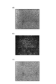
- FIG. Fig. 3 is a micrograph showing the results of a bile acid assay, (a) is bright field, (b) is fluorescence, and (c) is a superimposed photograph of (a) and (b).
- FIG. 3 is a micrograph showing the results of a bile acid assay, (a) is bright field, (b) is fluorescence, and (c) is a superimposed photograph of (a) and (b).
- Fig. 3 is a micrograph showing the results of a bile acid assay, (a) is bright field, (b) is fluorescence, and (c) is a superimposed photograph of (a) and (b).
- Fig. 3 is a micrograph showing the results of a bile acid assay, (a) is bright field, (b) is fluorescence, and (c) is a superimposed photograph of (a) and (b).
- the method according to the present embodiment is a method for producing a cell aggregate having bile canaliculi, comprising a contacting step of contacting cells containing hepatocytes with heparin in an aqueous medium; and culturing to form cell aggregates.
- the cell aggregate obtained by the method according to the present embodiment is a liver model ( It can be used as hepatoid tissue).
- cell aggregate means an aggregate of cells (aggregated cell aggregate) that is artificially produced by cell culture.
- the cell aggregate may be a structure in which cells are arranged three-dimensionally (three-dimensional tissue), or a structure in which cells are arranged two-dimensionally.
- the shape of the cell aggregate is not particularly limited, and examples thereof include sheet-like, spherical, substantially spherical, ellipsoidal, substantially ellipsoidal, hemispherical, substantially hemispherical, semicircular, substantially semicircular, and cuboidal shapes. , a substantially rectangular parallelepiped shape, and the like.
- biological tissue includes sweat glands, lymphatic vessels, sebaceous glands, etc., and has a more complicated structure than cell aggregates. Therefore, cell aggregates and living tissue can be easily distinguished.
- the cell aggregates may be aggregated in a state of adhering to the support, or may be aggregated in a state of not adhering to the support.
- the cells may be somatic or germ cells. Furthermore, the cells may be stem cells, or cultured cells such as primary cultured cells, subcultured cells and cell line cells.
- stem cell means a cell having self-renewal ability and pluripotency.
- Stem cells include pluripotent stem cells that have the ability to differentiate into any cell tumor and tissue stem cells (also called somatic stem cells) that have the ability to differentiate into specific cell tumors.
- Pluripotent stem cells include, for example, embryonic stem cells (ES cells), somatic cell-derived ES cells (ntES cells) and induced pluripotent stem cells (iPS cells).
- Tissue stem cells include, for example, mesenchymal stem cells (eg, bone marrow-derived stem cells), hematopoietic stem cells, and neural stem cells.
- the cells in the cell aggregate according to this embodiment include hepatocytes.
- Hepatocytes also called hepatocytes, are cells having functions such as bile secretion and plasma protein secretion.
- the hepatocytes may be mature hepatocytes or other hepatocytes.
- Mature hepatocytes may be, for example, primary hepatocytes collected from the liver of an animal, may be cells obtained by culturing primary hepatocytes, or may be a cultured cell line derived from primary hepatocytes. good too.
- Other hepatocytes may be, for example, hepatoblasts artificially differentiated from stem cells.
- Primary hepatocytes include primary human hepatocytes such as PXB cells.
- Cultured cell lines include cell lines derived from inactivated hepatoma cells such as HepG2.
- Stem cells to be differentiated into hepatoblasts include embryonic stem cells (ES cells), induced pluripotent stem cells (iPS cells), mesenchymal stem cells and the like.
- Hepatocytes contained in the cell aggregate according to the present embodiment are preferably non-cancerous cells such as primary hepatocytes and hepatoblasts. The hepatocytes may be mature hepatocytes, and PXB Cells are more preferable for ease of handling.
- the hepatocytes contained in the cell aggregate may be of one type, or may be of two or more types.
- a cell aggregate may contain a plurality of hepatocytes with different genotypes of proteins involved in liver function.
- all hepatocytes contained in the cell aggregate may have the same genotype of proteins involved in liver function.
- Proteins involved in liver function include, for example, drug-metabolizing enzymes.
- the total number of cells constituting the cell aggregate according to the present embodiment is not particularly limited, and the thickness and shape of the cell aggregate to be constructed, the size of the cell culture vessel used for construction, etc. It is determined.
- the ratio of the number of hepatocytes to the total number of cells in the cell aggregate is 5% or more, 10% or more, 15% or more, 20% or more, 25% or more, 30% 35% or more, 40% or more, 45% or more, 50% or more, 55% or more, 60% or more, or 65% or more, and 95% or less, 90% or less, 80% or less, or 75% or less can be
- the ratio of the number of hepatocytes to the total number of cells in the cell aggregate is 60% or more and 80% or less, or 60% or more and 70% or less, from the viewpoint of enabling the formation of a liver model that is even closer to the in vivo state.
- the cells may further contain vascular endothelial cells from the viewpoint of enabling the formation of a liver model that is even closer to the in vivo state.
- Vascular endothelial cells mean flat cells that form the surface of the lumen of blood vessels.
- Vascular endothelial cells can be, for example, sinusoidal endothelial cells, human umbilical vein-derived vascular endothelial cells (HUVEC).
- Sinusoidal endothelial cells are hepatic nonparenchymal cells (cells other than hepatocytes among the cells that make up the liver). These cells have a characteristic morphology different from that of vascular endothelial cells.
- the vascular endothelial cells constituting the cell aggregates may be primary cells (primary vascular endothelial cells) collected from the liver of an animal (e.g., human), or cells obtained by culturing primary cells. It may be a cultured cell line obtained by establishing a cell line, or a cell artificially differentiated from a stem cell.
- Primary vascular endothelial cells include, for example, primary sinusoidal endothelial cells such as product model number 5000 manufactured by Sciencecell.
- Cultured cell lines include, for example, a cultured cell line of product model number T0056 manufactured by Applied Biological Materials.
- Stem cells to be differentiated include embryonic stem cells (ES cells), induced pluripotent stem cells (iPS cells), and the like.
- the vascular endothelial cells contained in the cell aggregate according to this embodiment may be non-cancerous cells.
- the ratio of the number of vascular endothelial cells to the total number of cells in the cell aggregate is 5% or more, 10% or more, 15% or more, 20% or more, or 25% or more. and may be 50% or less, 45% or less, 40% or less, 35% or less, 30% or less, or 25% or less.
- the ratio of the number of vascular endothelial cells to the total number of cells in the cell aggregate is 5% or more and 40% or less, 5% or more and 35% or less, from the viewpoint of enabling the formation of a liver model that is even closer to the in vivo state. , 10% to 35%, or 10% to 30%.
- the ratio of sinusoidal endothelial cells to the total number of cells in the cell aggregate may be within the above range.
- the cells in the cell aggregate according to this embodiment may further contain hepatic stellate cells from the viewpoint of enabling the formation of a liver model that is even closer to the in vivo state.
- Hepatic stellate cells are hepatic nonparenchymal cells (cells other than hepatocytes among the cells that make up the liver), have functions such as storing vitamin A, and are located in the region between hepatocytes and sinusoids in the liver. Present in certain Disse spaces.
- the hepatic stellate cells may be, for example, primary cells (primary hepatic stellate cells) collected from the liver of an animal (e.g., human), cells obtained by culturing the primary cells, and establishment of a primary cell line.
- Primary hepatic stellate cells include, for example, Sciencecell model number 5300 primary hepatic stellate cells.
- Cultured cell lines include, for example, cultured cell lines such as LX-2.
- Stem cells to be differentiated include embryonic stem cells (ES cells), induced pluripotent stem cells (iPS cells), mesenchymal stem cells and the like.
- the hepatic stellate cells contained in the cell aggregate according to this embodiment may be non-cancerous cells.
- the ratio of the number of hepatic stellate cells to the total number of cells in the cell aggregate is 1% or more, 2% or more, 3% or more, 4% or more, or 5% or more and may be 20% or less, 15% or less, 14% or less, 13% or less, 12% or less, or 11% or less.
- the ratio of the number of hepatic stellate cells to the total number of cells in the cell aggregate is 1% or more and 15% or less, or 3% or more, from the viewpoint of enabling the formation of a liver model that is even closer to the in vivo state. % or less.
- the cells may contain cells other than hepatocytes, vascular endothelial cells, and hepatic stellate cells.
- Other cells may be, for example, mature somatic cells or undifferentiated cells such as stem cells.
- somatic cells include nerve cells, dendritic cells, immune cells, lymphatic endothelial cells, fibroblasts, epithelial cells (excluding hepatocytes), cardiomyocytes, pancreatic islet cells, and smooth muscle cells. , osteocytes, alveolar epithelial cells, splenocytes, and the like.
- Stem cells include ES cells, iPS cells, mesenchymal stem cells, and the like.
- Other cells may be normal cells, or cells such as cancer cells in which any cell function is enhanced or suppressed.
- a “cancer cell” is a cell that is derived from a somatic cell and has acquired unlimited proliferative potential.
- hepatocytes, vascular endothelial cells, hepatic stellate cells, or other cells contained in the cells are not particularly limited in origin, but are mammals such as humans, monkeys, dogs, cats, rabbits, pigs, cows, mice, and rats. It may be a cell derived from an animal.
- the cells in the cell aggregate according to this embodiment may not contain cells that have been induced to differentiate from induced pluripotent stem cells (iPS cells). That is, the cells used in the contacting step may be mature cells.
- iPS cells induced pluripotent stem cells
- the cells in the cell aggregate according to the present embodiment do not contain cells induced to differentiate from iPS cells, it becomes easier to grasp the degree of differentiation from iPS cells, the ratio of differentiated cells to the whole cells, and the like. As a result, it becomes easier to grasp the content of each cell that constitutes the cell aggregate.
- cells including hepatocytes are brought into contact with heparin in an aqueous medium.
- Methods for contacting heparin with cells include, for example, a method of suspending cells in a solution containing heparin, a method of adding heparin or a solution containing heparin to a cell-containing solution containing cells, and a method of adding heparin to a previously prepared aqueous medium.
- a method of adding a solution containing heparin and a cell or a cell-containing liquid containing cells respectively.
- the added heparin may be in the form of a salt.
- the heparin concentration at the time of contact with the cells in the aqueous medium is 0.1 mg/mL or higher, 0.2 mg/mL or higher, 0.3 mg/mL or higher, 0.4 mg/mL or higher, based on the total amount of the aqueous medium. Or it may be 0.5 mg/mL or more.
- the heparin concentration when contacting cells in an aqueous medium is 12.0 mg/mL or less, 10.0 mg/mL or less, 8.0 mg/mL or less, 6.0 mg/mL or less, based on the total amount of the aqueous medium.
- the heparin concentration when contacting cells in an aqueous medium is 0.1 mg/mL or more and 12.0 mg/mL or less, 0.1 mg/mL or more and 10.0 mg/mL or less, 0.1 mg/mL or more and 10.0 mg/mL or less, based on the total amount of the aqueous medium.
- 1 mg/mL or more and 8.0 mg/mL or less 0.1 mg/mL or more and 6.0 mg/mL or less, 0.2 mg/mL or more and 12.0 mg/mL or less, 0.2 mg/mL or more and 10.0 mg/mL or less, 0.2 mg/mL to 8.0 mg/mL, 0.2 mg/mL to 6.0 mg/mL, 0.3 mg/mL to 12.0 mg/mL, 0.3 mg/mL to 10.0 mg/mL 0.3 mg/mL or more and 8.0 mg/mL or less, 0.3 mg/mL or more and 6.0 mg/mL or less, 0.4 mg/mL or more and 12.0 mg/mL or less, 0.4 mg/mL or more and 10.0 mg /mL or less, 0.4 mg/mL or more and 8.0 mg/mL or less, 0.4 mg/mL or more and 6.0 mg/mL or less, 0.5 mg/mL or more and 12.0 mg/mL or less, 0.4
- the amount of heparin in the contacting step may be, for example, 0.001 mg or more, or 0.01 mg or more, or 1 mg or less, or 0.01 mg or more for 300,000 cells (the number of cells when contacted with heparin). It may be 1 mg or less.
- heparin is not particularly limited, but it may be derived from mammals such as humans, monkeys, dogs, cats, rabbits, pigs, cows, mice and rats.
- Aqueous medium means a liquid medium containing water as an essential component.
- the aqueous medium may be an aqueous medium containing cationic substances.
- Aqueous media containing cationic substances are, for example, Tris-HCl buffer, Tris-Maleate buffer, Bis-Tris-buffer, or HEPES (4-(2-hydroxyethyl)-1-piperazineethanesulfonic acid) It may be a cationic buffer such as
- the aqueous medium containing a cationic substance may be a medium containing water and a cationic compound such as ethanolamine, diethanolamine, triethanolamine, polyvinylamine, polyallylamine, polylysine, polyhistidine, and polyarginine as cationic substances.
- a culture medium can also be used as the aqueous medium.
- the medium include liquid media such as Dulbecco's Modified Eagle medium (DMEM) and vascular endothelial cell medium (EGM2).
- DMEM Dulbecco's Modified Eagle medium
- ECM2 vascular endothelial cell medium
- the liquid medium may be a mixed medium in which two types of medium are mixed.
- the concentration and pH of the cationic substance in the aqueous medium containing the cationic substance are not particularly limited as long as they do not adversely affect cell growth and cell aggregate construction.
- the concentration of the cationic substance may be 10-200 mM, or 30-150 mM, or 50-120 mM based on the total amount of the aqueous medium containing the cationic substance.
- the pH of the aqueous medium (eg, cationic buffer) may be 6.0-8.0, 6.8-7.8, 6.8-7.6, or 7.2-7.6 .
- the extracellular matrix component may be further brought into contact with the cells.
- the cells are more likely to aggregate.
- the extracellular matrix component is further brought into contact with the cells, it becomes easier for the cells to form cell aggregates in which the cells are arranged three-dimensionally.
- the expression intensity ratio of MRP2 which will be described later, tends to be higher, and the liver is even closer to the in vivo state. models are easier to obtain.
- a thicker structure three-dimensional structure
- Methods for further contacting extracellular matrix components with cells include, for example, a method of suspending cells in a liquid containing extracellular matrix components and heparin, and a method of suspending cells in a liquid containing cells containing extracellular matrix components and heparin. and a method of adding heparin or a solution containing heparin, cells or a cell-containing liquid, and an extracellular matrix component or a liquid containing an extracellular matrix component to a previously prepared aqueous medium.
- extracellular matrix component refers to an aggregate of extracellular matrix molecules formed by multiple extracellular matrix molecules.
- Extracellular matrix means a substance that exists outside cells in an organism. Any substance can be used as the extracellular matrix as long as it does not adversely affect the growth of cells and the formation of cell aggregates. Specific examples include, but are not limited to, collagen, elastin, proteoglycan, fibronectin, hyaluronic acid, laminin, vitronectin, tenascin, entactin, fibrillin and cadherin. These extracellular matrix components may be used singly or in combination.
- the extracellular matrix component may, for example, contain or be a collagen component.
- the extracellular matrix component When the extracellular matrix component is a collagen component, it becomes easier to aggregate cells. When the extracellular matrix component is a collagen component, the collagen component functions as a scaffold for cell adhesion, further promoting the formation of three-dimensional cell aggregates.
- the extracellular matrix component in the present embodiment is preferably a substance existing outside animal cells, that is, an animal extracellular matrix component.
- the extracellular matrix molecule may be a modification or variant of the above extracellular matrix molecule, or a polypeptide such as a chemically synthesized peptide, as long as it does not adversely affect cell growth and formation of cell aggregates. good.
- the extracellular matrix component may have repeat sequences represented by Gly-XY characteristic of collagen.
- Gly represents a glycine residue
- X and Y each independently represent any amino acid residue.
- a plurality of Gly-XY may be the same or different.
- the proportion of the sequence represented by Gly-XY in the total amino acid sequence may be 80% or more, preferably 95%. That's it.
- the extracellular matrix component may have an RGD sequence.
- the RGD sequence refers to a sequence represented by Arg-Gly-Asp (arginine residue-glycine residue-aspartic acid residue).
- Arg-Gly-Asp arginine residue-glycine residue-aspartic acid residue.
- Extracellular matrix components containing sequences represented by Gly-XY and RGD sequences include collagen, fibronectin, vitronectin, laminin, cadherin, and the like.
- the concentration of extracellular matrix components when contacted with cells in an aqueous medium is 0.001 mg/mL or higher, 0.01 mg/mL or higher, 0.025 mg/mL or higher, or 0.05 mg, based on the total amount of the aqueous medium. /mL or greater, 0.1 mg/mL or greater, 0.2 mg/mL or greater, 0.3 mg/mL or greater, 0.4 mg/mL or greater, or 0.5 mg/mL or greater.
- the concentration of extracellular matrix components when contacted with cells in an aqueous medium is, for example, 10.0 mg/mL or less, 8.0 mg/mL or less, 6.0 mg/mL or less, based on the total amount of the aqueous medium.
- the concentration of the extracellular matrix component when contacted with the cells in the aqueous medium is 0.001 mg/mL or more and 10.0 mg/mL or less, 0.01 mg/mL or more and 6.0 mg/mL, based on the total amount of the aqueous medium.
- 0.025 mg/mL or more and 5.0 mg/mL or less 0.05 mg/mL or more and 1.0 mg/mL or less, 0.1 mg/mL or more and 0.8 mg/mL or less, or 0.2 mg/mL or more and 0.2 mg/mL or less. It may be 6 mg/mL or less.
- the amount of the extracellular matrix component in the contacting step may be 0.001 mg or more, or 0.01 mg or more, and 1 mg or less with respect to 300,000 cells (the number of cells when contacted with the extracellular matrix component). , or 0.1 mg or less.
- the order in which the components are brought into contact with the cells is not particularly limited. For example, some cells may be brought into contact with the components, and then the remaining cells may be brought into contact with the components. , all cells may be contacted with each component at the same time, or at about the same time.
- the aqueous medium may be mixed by stirring or the like after the addition of each of the above components, or may not be mixed.
- the contacting step may include incubating for a certain period of time after contacting the cells with each of the above components. Examples of the fixed period of time include 1 minute, 3 minutes, 5 minutes, 10 minutes, 15 minutes, 30 minutes, 1 hour, 2 hours, or 3 hours.
- the incubation time may be, for example, 1 minute or more and 3 hours or less, or 1 minute or more and 15 minutes or less.
- Fibrinogen and thrombin may be added, for example, at the same time, or one of them may be added first and then the other.
- a solution containing fibrinogen and a solution containing thrombin may be mixed simultaneously or separately with a cell content containing cells and heparin.
- fibrinogen and thrombin may be included by using equal or approximately equal amounts of a thrombin solution with a concentration of 20 U/mL and a fibrinogen solution with a concentration of 10 mg/mL.
- fibrinogen and thrombin makes it easier to suppress shrinkage that can occur in the culture process described later, and makes it easier to control the shape and size of cell aggregates.
- the cell suspension can be gelled, the cells and the above components can be maintained in a uniformly mixed state, and the cells and the above components can be easily maintained in close proximity.
- fibrinogen and thrombin are contained in the contacting step, or after the contacting step and before the culturing step, cell dispersion in the medium during cell seeding is further suppressed, and cell aggregates can be formed with a smaller amount of cells. It becomes possible.
- a method of accumulating is exemplified by a method of centrifuging the entire aqueous medium. Aggregates of cells and the above components may be, for example, viscous bodies.
- a culture medium can be exemplified as an aqueous medium for resuspension.
- the cells By forming an aggregate of the cells and the above components, the cells can be in a state surrounded by the above components (that is, in a state of being buried), and the cells and the aggregate of the above components are resuspended in the medium.
- turbidity it is possible to proceed to the culturing step in a state of being buried to some extent.
- the viscosity is sufficiently low that it is easier to handle with pipettes and the like.
- cells in contact with heparin are cultured to form cell aggregates with bile canaliculi. Cultivation of cells is performed under conditions in which cells can grow.
- cell aggregates can be formed in which the extracellular matrix components are arranged between at least some cells in the cell aggregates.
- the medium used in the culture process may not contain insulin and transferrin, which are proteins secreted from the liver.
- media used in the culture step include vascular endothelial media (e.g., EGM2 (manufactured by Lonza), EGM2-MV (manufactured by Lonza), Endothelial Cell Growth Medium 2 (manufactured by Promocell), Endothelial Cell Growth Medium MV 2 (manufactured by Promocell) and ECM (manufactured by Sciencecell).
- EGM2 manufactured by Lonza
- EGM2-MV manufactured by Lonza
- Endothelial Cell Growth Medium 2 manufactured by Promocell
- Endothelial Cell Growth Medium MV 2 manufactured by Promocell
- ECM manufactured by Sciencecell
- the medium may be a serum-added medium or a serum-free medium.
- the medium is a mixture of two types of medium. It may be a mixed medium, for example, a mixed medium in which a vascular endothelial medium
- the culture temperature in the culture step may be, for example, 20°C to 40°C or 30°C to 37°C.
- the pH of the medium may be 6-8, or 7.2-7.4.
- the culture time may be 1 day or more, 1 day to 2 weeks, or 1 week to 2 weeks.
- the incubator (support) is not particularly limited, and may be, for example, a dish, a well insert, a low-adhesion plate, or a plate having a bottom shape such as a U-shape or a V-shape.
- the cells may be cultured while adhered to the support, the cells may be cultured without being adhered to the support, or the cells may be separated from the support during culture and cultured.
- a U-shaped or V-shaped bottom surface that inhibits adhesion of the cells to the support is used. It is preferable to use a plate with a low adsorption or a low adsorption plate.
- the cell density in the medium in the culture step can be appropriately determined according to the shape and thickness of the desired cell aggregate, the size of the incubator, and the like.
- the cell density in the medium in the culture step may be 1-10 8 cells/mL, or 10 3 -10 7 cells/mL.
- the cell density in the medium in the culturing step may be the same as the cell density in the aqueous medium in the contacting step.
- first culturing step After the culturing step (hereinafter also referred to as “first culturing step”, and the first contacting step is also referred to as “first contacting step”), the step of further contacting cells (second contacting step) , a step of culturing the cells (second culturing step).
- the cells in the second contacting step and the second culturing step may be of the same type as the cells used in the first contacting step and the first culturing step, or may be of a different type.
- the second contacting step and the second culturing step a two-layered cell aggregate can be produced. Further, by repeatedly including the contacting step and the culturing step, it is possible to produce a multi-layered cell aggregate, and to produce a more complex tissue similar to that of a living body.
- the cell aggregate according to this embodiment has bile canaliculi.
- Bile canaliculi are interstices located between cells, including hepatocytes, and in particular interstices located between contacting hepatocytes.
- bile acids taken up by hepatocytes are known to be excreted into bile canaliculi. That the cell aggregate has bile canaliculi can be confirmed by, for example, the expression of MRP2 (multidrug resistance-associated protein 2), which is a transporter on bile canaliculi.
- MRP2 multidrug resistance-associated protein 2
- the fact that the cell aggregates have bile canaliculi means that, for example, bile acids that are optically detectably labeled are brought into contact with the cell aggregates, and the bile acids that remain in the cells or in the bile canaliculi are removed. It can be confirmed by detecting. Retention of bile acids in cells or bile canaliculi can be confirmed by the method described in Examples below.
- the cell aggregate according to the present embodiment may have an expression intensity ratio of MRP2 represented by the following formula (1) exceeding 100%.
- Expression intensity ratio of MRP2 X/Y ⁇ 100 (1)
- X represents the expression intensity of MRP2 in cell aggregates formed in the culturing step
- Y is formed by culturing cells containing hepatocytes without contact with heparin and extracellular matrix components.
- the expression intensity of control cell aggregates is shown.
- the control cell aggregates were the cell aggregates formed in the above culture step (test cells Aggregates) are cell aggregates produced under the same conditions.
- the control cell aggregates are the test cell aggregates except that the cells are not contacted with heparin and the cells are not contacted with the extracellular matrix components. It is a cell aggregate produced under the same conditions as the aggregate. Specific conditions may be as described in Examples described later.
- the culture period for the test cell aggregate and the control cell aggregate may be 1 day or more, or 3 days or more, and may be 3 days.
- the expression intensity ratio of MRP2 in the cell aggregates according to this embodiment is 105% or more, 110% or more, 120% or more, 130% or more, 140% or more, 150% or more, 160% or more, 170% or more, or 180%. % or more.
- the expression intensity ratio of MRP2 in the cell aggregates according to this embodiment may be, for example, 300% or less, or 250% or less.
- the cell aggregates according to the present embodiment can be suitably used as hepatotoxicity evaluation models for drugs, tissues for transplantation into non-human animals, and the like.
- Step 1-1 Recovery of Cells Other than PXB Cells
- Lx2 and SEC frozen stocks of each cell were awakened, cultured without passage according to the protocol recommended by the manufacturer, and then treated with trypsin according to a normal procedure from the culture flask and Petri dish. After harvesting, the amount of each cell was measured.
- Step 1-2 Collection of PXB cells
- PXB cells were collected by the following procedure, and the amount of cells was measured.
- PXB cells were washed with PBS.
- 0.25% trypsin-EDTA was added to the washed PXB cells, incubated in an incubator, and then the PXB cells were collected.
- the amount of collected PXB cells was measured with a cell counter.
- Step 1-3 The collected cells were mixed at the following ratio and cell amount to obtain a cell mixture.
- Step 1-4 100 ⁇ L of each of the following solutions was added to the cell mixture at room temperature, suspended until the cells were no longer visible, and then centrifuged (400 g ⁇ 2 min) to obtain a viscous body. It was previously confirmed that the pH of each solution was approximately 7.4 regardless of the presence or absence of heparin and collagen.
- Step 1-5 After removing the supernatant from the viscous body-containing solution, HCM was added so that the final volume of the solution was 100 ⁇ L to obtain a cell suspension.
- Step 1-6 The cell suspension was seeded at 10 ⁇ L/well in a 96-well microplate in which 190 ⁇ L of HCM was added in advance to each well, and suspended in the wells.
- Step 2-1 By the same procedure as steps 1-1 to 1-3 of Test Example 1, a cell mixture having the following ratio and cell amount was obtained. ⁇ Total amount of cells: 300,000 cells ⁇ Tissue cell ratio: ⁇ PXB cell: 65% ⁇ SEC: 25% ⁇ LX2: 10%
- Step 2-2 100 ⁇ L of each of the following solutions was added to the cell mixture at room temperature, suspended until the cells were no longer visible, and then centrifuged (400 g ⁇ 2 min) to obtain a viscous body. It was previously confirmed that the pH of each solution was approximately 7.4 regardless of the presence or absence of heparin/collagen.
- Step 2-3 After removing the supernatant from the solution containing the viscous body obtained in step 2-2, the final volume of the solution is "the number of wells to be seeded" ⁇ 20 U / mL thrombin solution (solvent: HCM) was added to obtain a cell suspension.
- Step 2-4 After seeding 2 ⁇ L of 10 mg / mL fibrinogen solution on a 48-well microplate to form a droplet, add each cell suspension obtained in step 2-3 to the inside of the droplet, and then incubator for 1 hour. to form a fibrin gel.
- Step 2-5 0.5 mL of HCM (containing Endothelial Cell Growth Supplement) was added to each well that formed a fibrin gel.
- Step 2-6 Each well that formed a fibrin gel continued to be cultured for 3 days after HCM was added.
- BSA solution (transparency processing/blocking) 100 ⁇ L of a 0.2 (v/v) % TRITON/1 (w/v) % BSA PBS solution (hereinafter referred to as BSA solution) was added to each well and allowed to stand at room temperature for 2 hours.
- An anti-MRP2 antibody was diluted 100-fold with a BSA solution to obtain a primary antibody solution. 100 ⁇ L of the primary antibody solution was added to each well and allowed to stand at 4° C. for 24 hours. After that, the primary antibody solution was thoroughly washed away.
- a secondary antibody solution was obtained by diluting the secondary antibody 200 times with the BSA solution. 100 ⁇ L of the secondary antibody solution was added to each well, shielded from light, and allowed to stand at room temperature for 1 hour. After that, the secondary antibody solution was thoroughly washed away, and 100 ⁇ L of PBS was added to each well.
- the intensity of the captured images was added up to obtain a fluorescence observation image of each hepatotoxicity model (3D model).
- ⁇ Test Example 3 Confirmation of formation of bile canaliculi by bile acid assay> A hepatotoxicity model was prepared in the same manner as described in Test Examples 1 and 2. However, the collagen concentration was 0.3 mg/mL, and the heparin concentration was 0.05 mg/mL, 0.15 mg/mL, 0.5 mg/mL, or 5.0 mg/mL (all final concentrations).
- the medium was replaced with 300 ⁇ L of medium containing 5 ⁇ M CLF (FITC-labeled bile acid CORNING #451041) for which the following work was performed (this assay was performed while the cells were alive). After being left in the incubator for 20 minutes, the tissue was washed 5 times with PBS. The cells were replaced with medium and observed under a fluorescence microscope.
- CLF FITC-labeled bile acid CORNING #451041
- FIG. 1 shows fluorescence images obtained by treating cells with FITC-modified collagen or heparin in advance and confirming the state of deposition of these molecules on the cells.
- the scale bar in FIG. 1 indicates 100 ⁇ m.
- Collagen is relatively deposited on the surface of the cells, while heparin is deposited on the inside of the cells. .
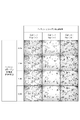
- Test Example 1 cells treated with each solution were seeded in a 96-well microplate, and the results of live cell observation up to 6 hours after seeding are shown in FIG.
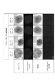
- FIG. 3 shows the results of staining with MRP2 for the hepatotoxicity models prepared under each condition.
- the scale bar in the bright field and fluorescence images in FIG. 3 indicates 500 ⁇ m, and the scale bar in the enlarged view indicates 300 ⁇ m.
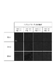
- FIG. 4 is a photograph showing the results of fluorescence observation of MRP2 expression under various treatment conditions.
- the scale bar in FIG. 4 indicates 300 ⁇ m.
- FIG. 5 is a graph showing the expression intensity ratio (expression area ratio) per area calculated based on the image analysis of the hepatotoxicity model shown in FIG.
- Fig. 5 shows results that quantitatively support that the hepatotoxicity model obtained through heparin treatment and heparin/collagen treatment has a higher MRP2 expression intensity ratio than the control.
- ⁇ Test Example 3 Confirmation of formation of bile canaliculi by bile acid assay> 6, 7, 8 and 9 show the results of fluorescence observation after incubation in a medium containing CLF for each hepatotoxicity model prepared with different heparin concentrations.
- FIG. 6 shows the results of fluorescence observation of the control hepatotoxicity model. The scale bar in each figure indicates 500 ⁇ m.
- hepatocytes excrete bile acids taken up from the outside of the liver tissue into the bile canaliculi. It is considered to be a thing.
Landscapes
- Health & Medical Sciences (AREA)
- Engineering & Computer Science (AREA)
- Biomedical Technology (AREA)
- Life Sciences & Earth Sciences (AREA)
- Biotechnology (AREA)
- Genetics & Genomics (AREA)
- Bioinformatics & Cheminformatics (AREA)
- Wood Science & Technology (AREA)
- Zoology (AREA)
- Chemical & Material Sciences (AREA)
- Organic Chemistry (AREA)
- Cell Biology (AREA)
- Microbiology (AREA)
- Biochemistry (AREA)
- General Engineering & Computer Science (AREA)
- General Health & Medical Sciences (AREA)
- Gastroenterology & Hepatology (AREA)
- Micro-Organisms Or Cultivation Processes Thereof (AREA)
Abstract
毛細胆管を有する細胞凝集体を製造する方法であって、肝細胞を含む細胞と、ヘパリンとを、水性媒体中で接触させる接触工程と、ヘパリンと接触させた細胞を培養して、細胞凝集体を形成する培養工程とを含む、方法に関する。
Description
本発明は、細胞凝集体を製造する方法に関する。
肝臓は医薬品開発の過程において毒性が顕現する可能性が高い器官であり、医薬品の摂取によって引き起こされる肝障害(DILI:Drug-Induced-Liver-Injury)は、医薬品の開発及び販売が中止となる主要因となっている。DILIを引き起こす可能性のある化合物(以下、DILI化合物)は、動物実験で特定することが必ずしも容易ではなく、臨床試験又は医薬品販売後にその障害性が発覚した場合には大きな損失を被ることになるため、DILI化合物を事前に予測可能な評価系が求められている。DILI化合物を事前に予測可能な評価系には、人工的に生体組織を模した構造体が有用である。
人工的に生体組織を模した構造体を作製する手法として、例えば、培養細胞の表面全体が接着膜で被覆された被覆細胞を培養することによって、三次元組織体を製造する方法(特許文献1)、細胞をカチオン性物質及び細胞外マトリックス成分と混合して混合物を得て、得られた混合物から細胞を集めて、基材上に細胞集合体を形成することを含む、立体的細胞組織の製造方法(特許文献2)等が知られている。また、本発明者らは、細胞と内因性コラーゲンを接触させ、好ましくはさらに繊維性の外因性コラーゲンを接触させて、コラーゲン濃度が高い三次元組織体を製造する方法(特許文献3)を提案している。
本発明の目的は、より生体内での状態に近い肝臓モデルとして利用可能な細胞凝集体の製造方法を提供することにある。
すなわち、本発明は、例えば以下の各発明に関する。
[1]
毛細胆管を有する細胞凝集体を製造する方法であって、
肝細胞を含む細胞と、ヘパリンとを、水性媒体中で接触させる接触工程と、
前記ヘパリンと接触させた前記細胞を培養して、前記細胞凝集体を形成する培養工程とを含む、方法。
[2]
前記肝細胞が成熟した肝細胞である、[1]に記載の方法。
[3]
前記水性媒体中で前記細胞と接触させる際のヘパリン濃度が、0.5mg/mL以上である、[1]又は[2]に記載の方法。
[4]
前記細胞の総数に対する、前記肝細胞の数の割合が65%以上である、[1]~[3]のいずれかに記載の方法。
[5]
前記接触工程において、前記細胞に細胞外マトリックス成分を更に接触させる、[1]~[4]のいずれかに記載の方法。
[6]
前記細胞外マトリックス成分がコラーゲン成分である、[5]に記載の方法。
[7]
下記式(1)で表されるMRP2の発現強度比が、120%以上である、[1]~[6]のいずれかに記載の方法。
MRP2の発現強度比=X/Y×100 (1)
[式(1)中、
Xは、前記培養工程において形成された前記細胞凝集体のMRP2の発現強度を示し、
Yは、ヘパリン及び細胞外マトリックス成分を接触させないで肝細胞を含む細胞を培養して形成された対照の細胞凝集体の発現強度を示す。]
[1]
毛細胆管を有する細胞凝集体を製造する方法であって、
肝細胞を含む細胞と、ヘパリンとを、水性媒体中で接触させる接触工程と、
前記ヘパリンと接触させた前記細胞を培養して、前記細胞凝集体を形成する培養工程とを含む、方法。
[2]
前記肝細胞が成熟した肝細胞である、[1]に記載の方法。
[3]
前記水性媒体中で前記細胞と接触させる際のヘパリン濃度が、0.5mg/mL以上である、[1]又は[2]に記載の方法。
[4]
前記細胞の総数に対する、前記肝細胞の数の割合が65%以上である、[1]~[3]のいずれかに記載の方法。
[5]
前記接触工程において、前記細胞に細胞外マトリックス成分を更に接触させる、[1]~[4]のいずれかに記載の方法。
[6]
前記細胞外マトリックス成分がコラーゲン成分である、[5]に記載の方法。
[7]
下記式(1)で表されるMRP2の発現強度比が、120%以上である、[1]~[6]のいずれかに記載の方法。
MRP2の発現強度比=X/Y×100 (1)
[式(1)中、
Xは、前記培養工程において形成された前記細胞凝集体のMRP2の発現強度を示し、
Yは、ヘパリン及び細胞外マトリックス成分を接触させないで肝細胞を含む細胞を培養して形成された対照の細胞凝集体の発現強度を示す。]
本発明によれば、より生体内での状態に近い肝臓モデルとして利用可能な細胞凝集体の製造方法を提供することができる。
以下、本発明を実施するための形態について詳細に説明する。ただし、本発明は以下の実施形態に限定されるものではない。
[細胞凝集体]
本実施形態に係る方法は、毛細胆管を有する細胞凝集体を製造する方法であって、肝細胞を含む細胞と、ヘパリンとを、水性媒体中で接触させる接触工程と、ヘパリンと接触させた細胞を培養して、細胞凝集体を形成する培養工程とを含む。
本実施形態に係る方法は、毛細胆管を有する細胞凝集体を製造する方法であって、肝細胞を含む細胞と、ヘパリンとを、水性媒体中で接触させる接触工程と、ヘパリンと接触させた細胞を培養して、細胞凝集体を形成する培養工程とを含む。
本実施形態に係る方法により得られる細胞凝集体は、肝臓の少なくとも一部の機能に類似した機能及び/又は肝臓の少なくとも一部の構造に類似した構造を有する生体組織モデルである、肝臓モデル(類肝組織)として利用可能である。
本明細書において「細胞凝集体」とは、細胞の集合体(塊状の細胞集団)であって、細胞培養によって人工的に作られる集合体を意味する。細胞凝集体は、細胞が三次元的に配置された構造体(三次元組織体)であってよく、細胞が二次元的に配置された構造体であってもよい。
細胞凝集体の形状は特に制限はなく、例えば、シート状、球体状、略球体状、楕円体状、略楕円体状、半球状、略半球状、半円状、略半円状、直方体状、略直方体状等が挙げられる。ここで、生体組織は、汗腺、リンパ管、脂腺等を含み、構成が細胞凝集体より複雑である。そのため、細胞凝集体と生体組織とは容易に区別可能である。また、細胞凝集体は、支持体と接着した状態で塊状に集合したものであってもよく、支持体と接着していない状態で塊状に集合したものであってもよい。
(細胞)
細胞は体細胞又は生殖細胞であってもよい。さらに、細胞は、幹細胞であってもよく、また、初代培養細胞、継代培養細胞及び細胞株細胞等の培養細胞であってもよい。本明細書において「幹細胞」とは、自己複製能及び多分化能を有する細胞を意味する。幹細胞には、任意の細胞腫に分化する能力を持つ多能性幹細胞と、特定の細胞腫に分化する能力を持つ組織幹細胞(体性幹細胞とも呼ばれる)が含まれる。多能性幹細胞としては、例えば、胚性幹細胞(ES細胞)、体細胞由来ES細胞(ntES細胞)及び人工多能性幹細胞(iPS細胞)が挙げられる。組織幹細胞としては、例えば、間葉系幹細胞(例えば、骨髄由来幹細胞)、造血幹細胞及び神経幹細胞が挙げられる。
細胞は体細胞又は生殖細胞であってもよい。さらに、細胞は、幹細胞であってもよく、また、初代培養細胞、継代培養細胞及び細胞株細胞等の培養細胞であってもよい。本明細書において「幹細胞」とは、自己複製能及び多分化能を有する細胞を意味する。幹細胞には、任意の細胞腫に分化する能力を持つ多能性幹細胞と、特定の細胞腫に分化する能力を持つ組織幹細胞(体性幹細胞とも呼ばれる)が含まれる。多能性幹細胞としては、例えば、胚性幹細胞(ES細胞)、体細胞由来ES細胞(ntES細胞)及び人工多能性幹細胞(iPS細胞)が挙げられる。組織幹細胞としては、例えば、間葉系幹細胞(例えば、骨髄由来幹細胞)、造血幹細胞及び神経幹細胞が挙げられる。
本実施形態に係る細胞凝集体において細胞は、肝細胞を含む。肝細胞は、肝実質細胞とも称され、例えば、胆汁の分泌,血漿タンパク質の分泌等の機能を有する細胞である。肝細胞は、成熟した肝細胞又はその他の肝細胞であってよい。成熟した肝細胞は、例えば、動物の肝臓から採取された初代肝細胞であってもよく、初代肝細胞を培養した細胞であってもよく、初代肝細胞を株化した培養細胞株であってもよい。その他の肝細胞は、例えば、幹細胞から人工的に分化させた肝芽細胞であってもよい。初代肝細胞としては、PXB Cellの様な初代ヒト肝細胞が挙げられる。培養細胞株としては、HepG2の様な不活化された肝がん細胞由来の細胞株が挙げられる。肝芽細胞へ分化させる幹細胞としては、胚性幹細胞(ES細胞)、誘導多能性幹細胞(iPS細胞)、間葉系幹細胞等が挙げられる。本実施形態に係る細胞凝集体に含まれる肝細胞としては、初代肝細胞及び肝芽細胞のように、非がん性細胞であることが好ましい。肝細胞は、成熟した肝細胞であってよく、扱いの簡便性からPXB Cellがより好ましい。
細胞凝集体に含まれる肝細胞は、1種類であってもよく、2種類以上であってもよい。例えば、細胞凝集体は、肝機能に関与するタンパク質の遺伝子型が異なる複数の肝細胞を含有していてもよい。また、逆に、細胞凝集体に含まれている全ての肝細胞が、肝機能に関与するタンパク質の遺伝子型が同一であってもよい。肝機能に関与するタンパク質としては、例えば、薬物代謝酵素が挙げられる。
本実施形態に係る細胞凝集体を構成する細胞の総数は、特に限定されるものではなく、構築する細胞凝集体の厚み、形状、構築に使用する細胞培養容器の大きさ等を考慮して適宜決定される。
細胞凝集体における細胞の総数に対する肝細胞の数の割合(肝細胞の数/細胞の総数×100)は、5%以上、10%以上、15%以上、20%以上、25%以上、30%以上、35%以上、40%以上、45%以上、50%以上、55%以上、60%以上、又は65%以上であってよく、95%以下、90%以下、80%以下又は75%以下であってよい。細胞凝集体における細胞の総数に対する肝細胞の数の割合は、より一層生体内での状態に近い肝臓モデルの形成が可能になる観点から、60%以上80%以下、又は60%以上70%以下であってよい。
細胞は、より一層生体内での状態に近い肝臓モデルの形成が可能になる観点から、血管内皮細胞を更に含んでいてもよい。血管内皮細胞は、血管内腔の表面を構成する扁平状の細胞を意味する。血管内皮細胞は、例えば、類洞内皮細胞、ヒト臍帯静脈由来血管内皮細胞(HUVEC)であってよい。類洞内皮細胞は、肝非実質細胞(肝臓を構成する細胞のうちの肝細胞以外の細胞)であり、細胞質に多数の小孔集合(篩板構造)を有し、基底膜を欠くなど他の血管内皮細胞と異なる特徴的な形態を有する細胞である。細胞凝集体を構成する血管内皮細胞としては、動物(例えばヒト)の肝臓から採取された初代細胞(初代血管内皮細胞)であってもよく、初代細胞を培養した細胞であってもよく、初代細胞を株化した培養細胞株であってもよく、幹細胞から人工的に分化させた細胞であってもよい。初代血管内皮細胞としては、例えば、Sciencell社製の製品型番5000等の初代類洞内皮細胞が挙げられる。培養細胞株としては、例えば、Applied Biological Materials社製の製品型番T0056の培養細胞株が挙げられる。分化させる幹細胞としては、胚性幹細胞(ES細胞)、誘導多能性幹細胞(iPS細胞)等が挙げられる。本実施形態に係る細胞凝集体に含まれる血管内皮細胞は、非がん性細胞であってよい。
細胞凝集体における細胞の総数に対する血管内皮細胞の数の割合(血管内皮細胞の数/細胞の総数×100)は、5%以上、10%以上、15%以上、20%以上、又は25%以上であってよく、50%以下、45%以下、40%以下、35%以下、30%以下、又は25%以下であってよい。細胞凝集体における細胞の総数に対する血管内皮細胞の数の割合は、より一層生体内での状態に近い肝臓モデルの形成が可能になる観点から、5%以上40%以下、5%以上35%以下、10%以上35%以下、又は10%以上30%以下であってよい。細胞凝集体における細胞の総数に占める類洞内皮細胞の割合が上記範囲内であってよい。
本実施形態に係る細胞凝集体において細胞は、より一層生体内での状態に近い肝臓モデルの形成が可能になる観点から、肝星細胞を更に含んでいてよい。肝星細胞は、肝非実質細胞(肝臓を構成する細胞のうちの肝細胞以外の細胞)であり、ビタミンAを貯蔵する機能等を有し、肝臓における肝細胞と類洞の間の領域であるディッセ腔に存在する。肝星細胞としては、例えば、動物(例えばヒト)の肝臓から採取された初代細胞(初代肝星細胞)であってもよく、初代細胞を培養した細胞であってもよく、初代細胞を株化した培養細胞株であってもよく、幹細胞から人工的に分化させた細胞であってもよい。初代肝星細胞としては、例えば、Sciencell社製型番5300の初代肝星細胞が挙げられる。培養細胞株としては、例えば、LX-2のような培養細胞株が挙げられる。分化させる幹細胞としては、胚性幹細胞(ES細胞)、誘導多能性幹細胞(iPS細胞)、間葉系幹細胞等が挙げられる。本実施形態に係る細胞凝集体に含まれる肝星細胞は、非がん性細胞であってよい。
細胞凝集体における細胞の総数に対する肝星細胞の数の割合(肝星細胞の数/細胞の総数×100)は、1%以上、2%以上、3%以上、4%以上、又は5%以上であってよく、20%以下、15%以下、14%以下、13%以下、12%以下又は11%以下であってよい。細胞凝集体における細胞の総数に対する、肝星細胞の数の割合は、より一層生体内での状態に近い肝臓モデルの形成が可能になる観点から、1%以上15%以下、又は3%以上12%以下であってよい。
本実施形態において、細胞は、肝細胞、血管内皮細胞及び肝星細胞以外の他の細胞を含んでいてもよい。他の細胞は、例えば、成熟した体細胞であってよく、幹細胞のような未分化な細胞であってもよい。体細胞の具体例としては、例えば、神経細胞、樹状細胞、免疫細胞、リンパ管内皮細胞、線維芽細胞、上皮細胞(但し、肝細胞を除く。)、心筋細胞、膵島細胞、平滑筋細胞、骨細胞、肺胞上皮細胞、脾臓細胞等が挙げられる。幹細胞としては、ES細胞、iPS細胞、間葉系幹細胞等が挙げられる。他の細胞は、正常細胞であってもよく、がん細胞のようにいずれかの細胞機能が亢進又は抑制されている細胞であってもよい。「がん細胞」とは、体細胞から派生して無限の増殖能を獲得した細胞である。
細胞に含まれる肝細胞、血管内皮細胞、肝星細胞又は上記他の細胞の由来は、特に限定されないが、例えば、ヒト、サル、イヌ、ネコ、ウサギ、ブタ、ウシ、マウス、ラット等の哺乳類動物に由来する細胞であってよい。
本実施形態に係る細胞凝集体における細胞は、人工多能性幹細胞(iPS細胞)から分化誘導された細胞を含まなくてよい。すなわち、接触工程で用いる細胞は、成熟した細胞であってよい。本実施形態に係る細胞凝集体における細胞がiPS細胞から分化誘導された細胞を含まない場合には、iPS細胞からの分化の程度、細胞全体に対する分化した細胞の割合等の把握がより容易になり、結果として、細胞凝集体を構成する細胞それぞれの含有率の把握がより一層容易になる。
接触工程では、肝細胞を含む細胞と、ヘパリンとを、水性媒体中で接触させる。ヘパリンと細胞とを接触させる方法は、例えば、ヘパリンを含む溶液に細胞を懸濁する方法、細胞を含む細胞含有液に、ヘパリン又はヘパリンを含む溶液を加える方法、予め用意した水性媒体に、ヘパリン又はヘパリンを含む溶液と、細胞又は細胞を含む細胞含有液とをそれぞれ加える方法等が挙げられる。加えられるヘパリンは塩の形態であってもよい。
水性媒体中で細胞と接触させる際のヘパリン濃度は、水性媒体の全量を基準として、0.1mg/mL以上、0.2mg/mL以上、0.3mg/mL以上、0.4mg/mL以上、又は0.5mg/mL以上であってよい。水性媒体中で細胞と接触させる際のヘパリン濃度は、水性媒体の全量を基準として、12.0mg/mL以下、10.0mg/mL以下、8.0mg/mL以下、6.0mg/mL以下、5.0mg/mL以下、3.0mg/mL以下、1.0mg/mL以下、0.8mg/mL以下、又は0.6mg/mL以下であってよい。水性媒体中で細胞と接触させる際のヘパリン濃度は、水性媒体の全量を基準として、0.1mg/mL以上12.0mg/mL以下、0.1mg/mL以上10.0mg/mL以下、0.1mg/mL以上8.0mg/mL以下、0.1mg/mL以上6.0mg/mL以下、0.2mg/mL以上12.0mg/mL以下、0.2mg/mL以上10.0mg/mL以下、0.2mg/mL以上8.0mg/mL以下、0.2mg/mL以上6.0mg/mL以下、0.3mg/mL以上12.0mg/mL以下、0.3mg/mL以上10.0mg/mL以下、0.3mg/mL以上8.0mg/mL以下、0.3mg/mL以上6.0mg/mL以下、0.4mg/mL以上12.0mg/mL以下、0.4mg/mL以上10.0mg/mL以下、0.4mg/mL以上8.0mg/mL以下、0.4mg/mL以上6.0mg/mL以下、0.5mg/mL以上12.0mg/mL以下、0.5mg/mL以上10.0mg/mL以下、0.5mg/mL以上8.0mg/mL以下、又は0.5mg/mL以上6.0mg/mL以下であってよい。水性媒体中で細胞と接触させる際のヘパリン濃度が上述した範囲内にある場合、より生体に近い肝臓モデルの形成が可能になる。
接触工程におけるヘパリンの量は、例えば、300,000cellsの細胞(ヘパリンと接触させる際の細胞数)に対して、0.001mg以上、又は0.01mg以上であってよく、1mg以下、又は0.1mg以下であってよい。
ヘパリンの由来は、特に限定されないが、例えば、ヒト、サル、イヌ、ネコ、ウサギ、ブタ、ウシ、マウス、ラット等の哺乳類動物に由来してよい。
「水性媒体」とは、水を必須構成成分とする液状媒体を意味する。水性媒体は、カチオン性物質を含む水性媒体であってよい。カチオン性物質を含む水性媒体は、例えば、トリス-塩酸緩衝液、トリス-マレイン酸緩衝液、ビス-トリス-緩衝液、又はHEPES(4-(2-ヒドロキシエチル)-1-ピペラジンエタンスルホン酸)等のカチオン性緩衝液であってよい。カチオン性物質を含む水性媒体は、カチオン性物質としてエタノールアミン、ジエタノールアミン、トリエタノールアミン、ポリビニルアミン、ポリアリルアミン、ポリリシン、ポリヒスチジン、ポリアルギニン等のカチオン性化合物と水とを含む媒体であってもよい。上記水性媒体としては、培地を用いることもできる。培地としては、例えばDulbecco’s Modified Eagle培地(DMEM)、血管内皮細胞専用培地(EGM2)等の液体培地が挙げられる。液体培地は、二種類の培地を混合した混合培地であってもよい。
カチオン性物質を含む水性媒体におけるカチオン性物質(例えば、トリス-塩酸緩衝液におけるトリス)の濃度及びpHは、細胞の生育及び細胞凝集体の構築に悪影響を及ぼさない限り、特に限定されない。例えば、カチオン性物質の濃度は、カチオン性物質を含む水性媒体の全量を基準として、10~200mM、又は30~150mMであってよく、50~120mMであってよい。水性媒体(例えば、カチオン性緩衝液)のpHは、6.0~8.0、6.8~7.8、6.8~7.6、又は7.2~7.6であってよい。
接触工程では、細胞に細胞外マトリックス成分を更に接触させてよい。細胞に細胞外マトリックス成分を更に接触させる場合、細胞をより凝集させやすくなる。また、細胞に細胞外マトリックス成分を更に接触させる場合、細胞が三次元に配置された細胞凝集体をより一層形成しやすくなる。加えて、細胞に細胞外マトリックス成分を更に接触させる場合(特に、コラーゲン成分を接触させる場合)、後述するMRP2の発現強度比がより高くなる傾向があり、より一層生体内での状態に近い肝臓モデルが得られやすくなる。更に、接触工程で細胞に細胞外マトリックス成分を更に接触させた場合、より厚い構造体(三次元構造体)が得られやすくなる。
細胞に細胞外マトリックス成分を更に接触させる方法は、例えば、細胞外マトリックス成分とヘパリンとを含む液に細胞を懸濁する方法、細胞を含む細胞含有液に細胞外マトリックス成分及びヘパリンを含有する液を加える方法、予め用意した水性媒体に、ヘパリン又はヘパリンを含む溶液と、細胞又は細胞含有液と、細胞外マトリックス成分又は細胞外マトリックス成分を含む液とをそれぞれ加える方法等が挙げられる。
本明細書において「細胞外マトリックス成分」とは、複数の細胞外マトリックス分子によって形成されている細胞外マトリックス分子の集合体である。細胞外マトリックスとは、生物において細胞の外に存在する物質を意味する。細胞外マトリックスとしては、細胞の生育及び細胞集合体の形成に悪影響を及ぼさない限り、任意の物質を用いることができる。具体例としては、コラーゲン、エラスチン、プロテオグリカン、フィブロネクチン、ヒアルロン酸、ラミニン、ビトロネクチン、テネイシン、エンタクチン、フィブリリン及びカドヘリン等が挙げられるが、これらに限定されない。細胞外マトリックス成分は、これらの1種単独で用いてもよく、組み合わせて用いてもよい。細胞外マトリックス成分は、例えば、コラーゲン成分を含んでいてよく、コラーゲン成分であってもよい。細胞外マトリックス成分がコラーゲン成分である場合、細胞をより一層凝集させやすくなる。細胞外マトリックス成分がコラーゲン成分である場合、コラーゲン成分が細胞接着の足場として機能し、三次元的な細胞凝集体の形成がより一層促進される。本実施形態における細胞外マトリックス成分は、動物細胞の外に存在する物質、すなわち動物の細胞外マトリックス成分であることが好ましい。細胞外マトリックス分子は、細胞の生育及び細胞集合体の形成に悪影響を及ぼさない限り、上述の細胞外マトリックス分子の改変体及びバリアントであってもよく、化学合成ペプチド等のポリペプチドであってもよい。
細胞外マトリックス成分は、コラーゲンに特徴的なGly-X-Yで表される配列の繰り返しを有していてよい。ここで、Glyはグリシン残基を表し、X及びYはそれぞれ独立に任意のアミノ酸残基を表す。複数のGly-X-Yは、それぞれ同一であっても異なっていてもよい。Gly-X-Yで示される配列の繰り返しを有することによって、分子鎖の配置への束縛が少なく、足場材としての機能がより一層優れたものとなる。Gly-X-Yで示される配列の繰り返しを有する細胞外マトリックス成分において、Gly-X-Yで示される配列の割合は、全アミノ酸配列のうち、80%以上であってよく、好ましくは95%以上である。また、細胞外マトリックス成分は、RGD配列を有していてよい。RGD配列とは、Arg-Gly-Asp(アルギニン残基-グリシン残基-アスパラギン酸残基)で表される配列をいう。細胞外マトリックス成分が、RGD配列を有する場合、細胞接着がより一層促進され、足場材としてより一層好適なものとなる。Gly-X-Yで表される配列と、RGD配列とを含む細胞外マトリックス成分には、コラーゲン、フィブロネクチン、ビトロネクチン、ラミニン、カドヘリン等が含まれる。
水性媒体中で細胞と接触させる際の細胞外マトリックス成分の濃度は、水性媒体の全量を基準として、0.001mg/mL以上、0.01mg/mL以上、0.025mg/mL以上、0.05mg/mL以上、0.1mg/mL以上、0.2mg/mL以上、0.3mg/mL以上、0.4mg/mL以上、又は0.5mg/mL以上であってよい。水性媒体中で細胞と接触させる際の細胞外マトリックス成分濃度は、水性媒体の全量を基準として、例えば、10.0mg/mL以下、8.0mg/mL以下、6.0mg/mL以下、5.0mg/mL以下、3.0mg/mL以下、1.0mg/mL以下、0.8mg/mL以下、又は0.6mg/mL以下であってよい。水性媒体中で細胞と接触させる際の細胞外マトリックス成分の濃度は、水性媒体の全量を基準として、0.001mg/mL以上10.0mg/mL以下、0.01mg/mL以上6.0mg/mL以下、0.025mg/mL以上5.0mg/mL以下、0.05mg/mL以上1.0mg/mL以下、0.1mg/mL以上0.8mg/mL以下、又は0.2mg/mL以上0.6mg/mL以下であってよい。
接触工程における細胞外マトリックス成分の量は、300,000cellsの細胞(細胞外マトリックス成分と接触させる際の細胞数)に対して、0.001mg以上、又は0.01mg以上であってよく、1mg以下、又は0.1mg以下であってよい。
上記細胞に、上記各成分を接触させる順番は、特に制限されず、例えば、一部の細胞と、上記各成分とを接触させた後、残りの細胞を上記各成分と接触させてもよいし、すべての細胞を同時に、又は略同時に上記各成分と接触させてもよい。
水性媒体は、上記各成分の添加後に撹拌等により混合してもよく、混合しなくてもよい。接触工程は、細胞に上記各成分を接触させた後に一定時間インキュベートすることを含んでいてよい。上記の一定時間の例として、1分間、3分間、5分間、10分間、15分間、30分間、1時間、2時間、又は3時間が挙げられる。インキュベートする時間は、例えば、1分間以上3時間以内であってよく、1分間以上15分間以内であってもよい。
接触工程、又は接触工程後かつ培養工程前に、フィブリノゲン及びトロンビンを含有させることを含んでもよい。フィブリノゲン及びトロンビンは、例えば、同時に添加してもよく、いずれか一方を先に添加し、その後他方を添加してもよい。接触工程では、例えば、細胞及びヘパリンを含む細胞含有物に、フィブリノゲンを含む溶液と、トロンビンを含む溶液と、を同時に、又は別々に、混合してよい。例えば、濃度20U/mLのトロンビン溶液と、濃度10mg/mLのフィブリノゲン溶液とを等量又は略等量用いることによって、フィブリノゲン及びトロンビンを含有させてよい。
フィブリノゲン及びトロンビンの添加により、後述する培養工程において生じ得るシュリンクがより抑制されやすくなり、細胞凝集体の形状及び大きさを制御しやすくすることができる。また、細胞懸濁液をゲル化することができるため、各細胞及び上記各成分が均一に混合した状態を維持し、細胞と上記各成分とを近接させた状態を維持しやすくなる。接触工程、又は接触工程後かつ培養工程前に、フィブリノゲン及びトロンビンを含有させる場合、細胞播種時の培地内への細胞分散がより抑制され、より少ない細胞量で細胞の凝集体を形成することが可能となる。
細胞と上記各成分との接触後に、細胞と、上記の成分(ヘパリン及び/又は細胞外マトリックス成分)とを集積させ、水性成分を除去することと、集積して得られた細胞及び上記の成分の集合体に水性媒体を加えて再度細胞懸濁液を得ることを含んでいてもよい。これによって得られる細胞懸濁液は後述する培養工程に供されてよい。集積させる方法は、水性媒体全体を遠心する方法が例示される。細胞及び上記の成分の集合体は、例えば粘稠体であってよい。再度懸濁するための水性媒体としては培養培地を例示することができる。細胞及び上記の成分の集合体を形成することによって細胞が上記の成分に取り囲まれた状態(つまり、埋もれている状態)とすることができ、細胞及び上記の成分の集合体を培地で再懸濁することにより、ある程度埋もれている状態のまま培養工程に移行することができる。細胞及び上記の成分の集合体を再懸濁することによって、粘度が充分に低くなり、ピペット等による取扱がより一層容易になる。
培養工程では、ヘパリンと接触させた細胞を培養して、毛細胆管を有する細胞凝集体を形成する。細胞の培養は、細胞が生育可能な条件で行われる。ヘパリンに加えて細胞外マトリックス成分を接触させた細胞を培養する場合、細胞凝集体における少なくとも一部の細胞間に細胞外マトリックス成分が配置された細胞凝集体を形成することができる。
培養工程で用いる培地は、肝臓から分泌されるタンパク質である、インスリン及びトランスフェリンを含んでいなくてよい。培養工程において用いる培地としては、例えば、血管内皮用培地(例えば、EGM2(ロンザ社製)、EGM2-MV(Lonza社製)、Endothelial Cell Growth Medium 2(Promocell社製)、Endothelial Cell Growth Medium MV 2(Promocell社製)、ECM(Sciencell社製)が挙げられる。培地は、血清を添加した培地であってもよいし、無血清培地であってもよい。培地は、二種類の培地を混合した混合培地であってもよい。例えば、血管内皮用培地と、間葉系幹細胞増殖用培地とを混合した混合培地であってもよい。
培養工程における培養温度は、例えば、20℃~40℃であってもよく、30℃~37℃であってもよい。培地のpHは、6~8であってもよく、7.2~7.4であってもよい。培養時間は、1日以上であってよく、1日~2週間であってもよく、1週間~2週間であってもよい。
培養器(支持体)は、特に制限されず、例えば、ディッシュ、ウェルインサート、低接着プレート、U字、V字等の底面形状を有するプレートであってよい。上記細胞を支持体と接着させたまま培養してもよく、上記細胞を支持体と接着させずに培養してもよく、培養の途中で支持体から引き離して培養してもよい。上記細胞を支持体と接着させずに培養する場合、又は培養の途中で支持体から引き離して培養する場合には、細胞の支持体への接着を阻害するU字、V字等の底面形状を有するプレート、又は低吸着プレートを用いることが好ましい。
培養工程における培地中の細胞密度は、目的とする細胞凝集体の形状、厚さ、培養器のサイズ等に応じて適宜決定できる。例えば、培養工程における培地中の細胞密度は、1~108cells/mLであってよく、103~107cells/mLであってよい。また、培養工程における培地中の細胞密度は、接触工程における水性媒体中の細胞密度と同じであってもよい。
上記培養工程(以下、「第1の培養工程」とも表す。また、最初の接触工程を「第1の接触工程」とも表す。)の後、さらに細胞を接触させる工程(第2の接触工程)、細胞を培養する工程(第2の培養工程)を含んでもよい。第2の接触工程及び第2の培養工程における上記細胞は、第1の接触工程及び第1の培養工程で用いた細胞と同種であってよく、異種であってもよい。第2の接触工程及び第2の培養工程により、二層構造の細胞凝集体を作製することができる。また、さらに、接触工程及び培養工程を繰り返し含むことで複数層の細胞凝集体を作製することができ、より複雑な生体に近い組織を作製することもできる。
本実施形態に係る細胞凝集体は毛細胆管を有している。毛細胆管は、肝細胞を含む細胞の間に位置する間隙であり、特には、接触している肝細胞同士の間に位置する間隙である。生体内において、肝細胞に取り込まれた胆汁酸は毛細胆管に排出されることが知られている。細胞凝集体が、毛細胆管を有していることは、例えば、毛細胆管上のトランスポーターであるMRP2(multidrug resistance-associated protein 2)の発現によって確認することができる。MRP2の発現は、後述する実施例に記載の方法によって確認することができる。また、細胞凝集体が、毛細胆管を有していることは、例えば、光学的に検出可能な標識をした胆汁酸を細胞凝集体に接触させ、細胞内あるいは毛細胆管内に滞留する胆汁酸を検出することにより確認することができる。細胞内あるいは毛細胆管内への胆汁酸の滞留は、後述する実施例に記載の方法によって確認することができる。
本実施形態に係る細胞凝集体は、下記式(1)で表されるMRP2の発現強度比が、100%超であってよい。
MRP2の発現強度比=X/Y×100 (1)
MRP2の発現強度比=X/Y×100 (1)
式(1)中、Xは、培養工程において形成された細胞凝集体のMRP2の発現強度を示し、Yは、ヘパリン及び細胞外マトリックス成分を接触させないで肝細胞を含む細胞を培養して形成された対照の細胞凝集体の発現強度を示す。接触工程において、細胞に細胞外マトリックス成分を接触させない場合には、対照の細胞凝集体は、細胞にヘパリンを接触させないこと以外は、上記の培養工程において形成された細胞凝集体(試験用の細胞凝集体)と同条件で製造される細胞凝集体である。接触工程において、細胞に細胞外マトリックス成分を接触させる場合には、対照の細胞凝集体は、細胞にヘパリンを接触させないこと及び細胞に細胞外マトリックス成分を接触させないこと以外は、試験用の細胞凝集体と同条件で製造される細胞凝集体である。具体的な条件は、後述する実施例に記載のとおりであってよい。試験用の細胞凝集体及び対照の細胞凝集体の培養期間は、1日間以上又は3日間以上であってよく、3日間であってよい。
本実施形態に係る細胞凝集体のMRP2の発現強度比は、105%以上、110%以上、120%以上、130%以上、140%以上、150%以上、160%以上、170%以上、又は180%以上であってよい。本実施形態に係る細胞凝集体のMRP2の発現強度比は、例えば、300%以下、又は250%以下であってよい。
本実施形態に係る細胞凝集体は、薬物の肝毒性評価モデル、非ヒト動物への移植用の組織等として好適に用いることができる。
以下、実施例を挙げて本発明についてさらに具体的に説明する。ただし、本発明はこれら実施例に限定されるものではない。
1.実験手順
[材料、資材及び使用機器]
以下の培養細胞、材料、資材及び機器を準備した。
(培養細胞)
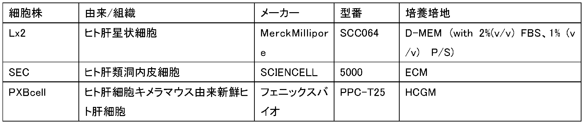
[材料、資材及び使用機器]
以下の培養細胞、材料、資材及び機器を準備した。
(培養細胞)
(試薬及び消耗品)
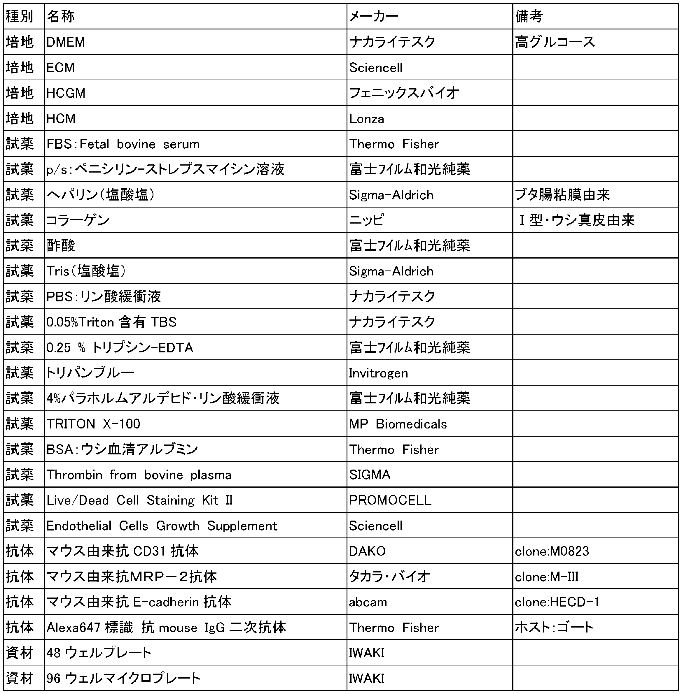
(1)セルカウント装置
・Invitrogen社 Countess(登録商標) II FL
(2)顕微鏡
・横河電機社 共焦点顕微鏡 CQ1
(3)その他
・画像解析ソフト:Image J (Fiji) 個別導入plugin:Mexican hat filter
・Invitrogen社 Countess(登録商標) II FL
(2)顕微鏡
・横河電機社 共焦点顕微鏡 CQ1
(3)その他
・画像解析ソフト:Image J (Fiji) 個別導入plugin:Mexican hat filter
<試験例1:ヘパリン・コラーゲンにより処理した細胞の凝集挙動の確認>
ヘパリン及びコラーゲンをそれぞれ単独、又は同時に用いて細胞を処理した場合に、細胞がどのような凝集挙動を示すかを検証した。
ヘパリン及びコラーゲンをそれぞれ単独、又は同時に用いて細胞を処理した場合に、細胞がどのような凝集挙動を示すかを検証した。
[ヘパリン・コラーゲンによる細胞の処理]
細胞としては、PXB cell、SEC及びLX2を用いた。
細胞としては、PXB cell、SEC及びLX2を用いた。
工程1-1
(PXB cell以外の細胞の回収) Lx2、SECの各細胞の凍結ストックを起眠し、メーカー推奨のプロトコルに基づき継代を挟まずに培養し、通常の手法に基づきトリプシンによって培養フラスコおよびシャーレから回収した後、各細胞量を測定した。
(PXB cell以外の細胞の回収) Lx2、SECの各細胞の凍結ストックを起眠し、メーカー推奨のプロトコルに基づき継代を挟まずに培養し、通常の手法に基づきトリプシンによって培養フラスコおよびシャーレから回収した後、各細胞量を測定した。
工程1-2
(PXB cellの回収) 以下の手順で回収し、細胞量を測定した。
PXB cellをPBSで洗浄した。洗浄後のPXB cellに0.25% トリプシン-EDTAを加え、インキュベータ内でインキュベート後、PXB cellを回収した。回収したPXB cellの細胞量をセルカウンターで計測した。
(PXB cellの回収) 以下の手順で回収し、細胞量を測定した。
PXB cellをPBSで洗浄した。洗浄後のPXB cellに0.25% トリプシン-EDTAを加え、インキュベータ内でインキュベート後、PXB cellを回収した。回収したPXB cellの細胞量をセルカウンターで計測した。
工程1-3
回収した各細胞を、下記の比率及び細胞量で混合し、細胞混合物を得た。
・総細胞量:300,000 cells
・組織内細胞比率:
・PXB cell:65% ・SEC:25% ・LX2:10%
回収した各細胞を、下記の比率及び細胞量で混合し、細胞混合物を得た。
・総細胞量:300,000 cells
・組織内細胞比率:
・PXB cell:65% ・SEC:25% ・LX2:10%
工程1-4
細胞混合物に対して、以下の各溶液を室温で100μL加え、細胞が視認できなくなるまで懸濁した後、遠心(400g×2min)し、粘稠体を得た。尚、ヘパリン及びコラーゲンの有無に関わらず、各溶液のpHは概ね7.4前後であることを事前に確認している。
(1)100mM Tris-HCl 緩衝液
(2)コラーゲン含有液(0.2mg/mL コラーゲン in 100mM Tris-HCl緩衝液*)
(3)コラーゲン及びヘパリン含有液(0.2mg/mL コラーゲン and 0.5mg/mL ヘパリン in 100mM Tris-HCl緩衝液)
*コラーゲンは常温の中性溶液下で速やかにゲル化するため、予め調整した、0.4mg/mLコラーゲンin 5mM 酢酸緩衝液と、200mM Tris-HCL緩衝液とを、上記の各溶液に等量混合し使用した。
細胞混合物に対して、以下の各溶液を室温で100μL加え、細胞が視認できなくなるまで懸濁した後、遠心(400g×2min)し、粘稠体を得た。尚、ヘパリン及びコラーゲンの有無に関わらず、各溶液のpHは概ね7.4前後であることを事前に確認している。
(1)100mM Tris-HCl 緩衝液
(2)コラーゲン含有液(0.2mg/mL コラーゲン in 100mM Tris-HCl緩衝液*)
(3)コラーゲン及びヘパリン含有液(0.2mg/mL コラーゲン and 0.5mg/mL ヘパリン in 100mM Tris-HCl緩衝液)
*コラーゲンは常温の中性溶液下で速やかにゲル化するため、予め調整した、0.4mg/mLコラーゲンin 5mM 酢酸緩衝液と、200mM Tris-HCL緩衝液とを、上記の各溶液に等量混合し使用した。
工程1-5
上記粘稠体を含む溶液から上清を除去した後、溶液の最終容積が100μLの溶液量となるようにHCMを加え、細胞懸濁液を得た。
上記粘稠体を含む溶液から上清を除去した後、溶液の最終容積が100μLの溶液量となるようにHCMを加え、細胞懸濁液を得た。
工程1-6
各ウェルに予めHCMを190μL加えた96ウェルマイクロプレートに、細胞懸濁液を10μL/wellで播種し、ウェル内で懸濁した。
各ウェルに予めHCMを190μL加えた96ウェルマイクロプレートに、細胞懸濁液を10μL/wellで播種し、ウェル内で懸濁した。
[共焦点顕微鏡によるライブセル観察]
予め装置内の温度を37℃に設定し5%CO2を供給した共焦点顕微鏡内に細胞懸濁液を播種及び懸濁した96ウェルマイクロプレートを設置し、播種後30分から6時間後までの凝集状態を経時的に観察した。
予め装置内の温度を37℃に設定し5%CO2を供給した共焦点顕微鏡内に細胞懸濁液を播種及び懸濁した96ウェルマイクロプレートを設置し、播種後30分から6時間後までの凝集状態を経時的に観察した。
<試験例2:ヘパリン・コラーゲンにより処理した細胞のフィブリンゲル内での組織化>
各溶液で処理した肝毒性モデルについて、肝細胞同士の密着状態の観点から差があるかを評価した。
各溶液で処理した肝毒性モデルについて、肝細胞同士の密着状態の観点から差があるかを評価した。
[肝毒性モデルの構築及び培養]
工程2-1
試験例1の工程1-1~1-3までと同様の手順で、下記の比率・細胞量の細胞混合物を得た。
・総細胞量:300,000cells
・組織内細胞比率:
・PXB cell:65% ・SEC:25% ・LX2:10%
工程2-1
試験例1の工程1-1~1-3までと同様の手順で、下記の比率・細胞量の細胞混合物を得た。
・総細胞量:300,000cells
・組織内細胞比率:
・PXB cell:65% ・SEC:25% ・LX2:10%
工程2-2
細胞混合物に対して、以下の各溶液を室温で100μL加え、細胞が視認できなくなるまで懸濁した後、遠心(400g×2min)し、粘稠体を得た。尚、ヘパリン・コラーゲンの有無に関わらず、各溶液のpHは概ね7.4前後であることを事前に確認している。
(1)100mM Tris-HCl 緩衝液
(2)コラーゲン含有液(0.2mg/mL コラーゲン in 100mM Tris-HCl緩衝液*)
(3)ヘパリン含有液(0.5mg/mL ヘパリン in 100mM Tris-HCl緩衝液)
(4)コラーゲン及びヘパリン含有液(0.2mg/mL コラーゲン and 0.5 mg/mL ヘパリン in 100mM Tris-HCl緩衝液)
*コラーゲンは常温の中性溶液下で速やかにゲル化するため、予め調整した、0.4mg/mLコラーゲンin 5mM 酢酸緩衝液と、200mM Tris-HCL緩衝液とを、上記の各溶液に等量混合し使用した。
細胞混合物に対して、以下の各溶液を室温で100μL加え、細胞が視認できなくなるまで懸濁した後、遠心(400g×2min)し、粘稠体を得た。尚、ヘパリン・コラーゲンの有無に関わらず、各溶液のpHは概ね7.4前後であることを事前に確認している。
(1)100mM Tris-HCl 緩衝液
(2)コラーゲン含有液(0.2mg/mL コラーゲン in 100mM Tris-HCl緩衝液*)
(3)ヘパリン含有液(0.5mg/mL ヘパリン in 100mM Tris-HCl緩衝液)
(4)コラーゲン及びヘパリン含有液(0.2mg/mL コラーゲン and 0.5 mg/mL ヘパリン in 100mM Tris-HCl緩衝液)
*コラーゲンは常温の中性溶液下で速やかにゲル化するため、予め調整した、0.4mg/mLコラーゲンin 5mM 酢酸緩衝液と、200mM Tris-HCL緩衝液とを、上記の各溶液に等量混合し使用した。
工程2-3
工程2-2で得た粘稠体を含む溶液から上清を除去した後、溶液の最終容積が、「播種予定ウェル数」×2μLの溶液量となるように20U/mLトロンビン溶液(溶媒:HCM)を加え、細胞懸濁液を得た。
工程2-2で得た粘稠体を含む溶液から上清を除去した後、溶液の最終容積が、「播種予定ウェル数」×2μLの溶液量となるように20U/mLトロンビン溶液(溶媒:HCM)を加え、細胞懸濁液を得た。
工程2-4
48ウェルマイクロプレート上に、10mg/mLフィブリノゲン溶液を2μL播種し液滴を形成した後、その液滴の内部に、工程2-3で得た各細胞懸濁液を加え、その後1時間インキュベータ内で静置し、フィブリンゲルを形成した。
48ウェルマイクロプレート上に、10mg/mLフィブリノゲン溶液を2μL播種し液滴を形成した後、その液滴の内部に、工程2-3で得た各細胞懸濁液を加え、その後1時間インキュベータ内で静置し、フィブリンゲルを形成した。
工程2-5
フィブリンゲルを形成した各ウェルに対し、HCM(Endothelial Cell Growth Supplement含有)0.5mLを加えた。
フィブリンゲルを形成した各ウェルに対し、HCM(Endothelial Cell Growth Supplement含有)0.5mLを加えた。
工程2-6
フィブリンゲルを形成した各ウェルは、HCMが加えられた後、3日間培養を継続した。
フィブリンゲルを形成した各ウェルは、HCMが加えられた後、3日間培養を継続した。
[肝毒性モデルの固定、免疫染色および顕微鏡撮像]
試験例1と同様の方法で、ライブセル観察を行った後、3日間の培養後まで培養を継続し、各サンプルに対し、下記のとおり、固定処理、透過処理、ブロッキング、1次抗体処理、2次抗体処理、撮影・面積算出及び評価をこの順で含む方法による肝毒性モデルの評価を行った。
試験例1と同様の方法で、ライブセル観察を行った後、3日間の培養後まで培養を継続し、各サンプルに対し、下記のとおり、固定処理、透過処理、ブロッキング、1次抗体処理、2次抗体処理、撮影・面積算出及び評価をこの順で含む方法による肝毒性モデルの評価を行った。
(固定処理)
3日間の培養後に48ウェルマイクロプレートをインキュベータから取り出し、培地を除去してPBSで洗浄した後、4%パラホルムアルデヒド・リン酸緩衝液(以下、PFA)を各ウェルに300μL加え、肝毒性モデル(3Dモデル)を固定した。その後、PFAを十分に洗浄除去した。
3日間の培養後に48ウェルマイクロプレートをインキュベータから取り出し、培地を除去してPBSで洗浄した後、4%パラホルムアルデヒド・リン酸緩衝液(以下、PFA)を各ウェルに300μL加え、肝毒性モデル(3Dモデル)を固定した。その後、PFAを十分に洗浄除去した。
(透過処理・ブロッキング)
0.2(v/v)% TRITON/1(w/v)%BSA PBS溶液(以下、BSA溶液)を、各ウェルに100μL加え、常温で2時間静置した。
0.2(v/v)% TRITON/1(w/v)%BSA PBS溶液(以下、BSA溶液)を、各ウェルに100μL加え、常温で2時間静置した。
(一次抗体処理)
抗MRP2抗体をBSA溶液で100倍希釈し、一次抗体溶液を得た。一次抗体溶液を各ウェルに100μL加え、4℃で24時間静置した。その後、一次抗体溶液を十分に洗浄除去した。
抗MRP2抗体をBSA溶液で100倍希釈し、一次抗体溶液を得た。一次抗体溶液を各ウェルに100μL加え、4℃で24時間静置した。その後、一次抗体溶液を十分に洗浄除去した。
(二次抗体処理)
BSA溶液で二次抗体を200倍に希釈し、二次抗体溶液を得た。二次抗体溶液を各ウェルに100μL加え、遮光して常温で1時間静置した。その後、二次抗体溶液を十分に洗浄除去し、各ウェルに100μLのPBSを加えた。
BSA溶液で二次抗体を200倍に希釈し、二次抗体溶液を得た。二次抗体溶液を各ウェルに100μL加え、遮光して常温で1時間静置した。その後、二次抗体溶液を十分に洗浄除去し、各ウェルに100μLのPBSを加えた。
(蛍光顕微鏡撮影)
共焦点顕微鏡により画像撮影を行った。撮影条件は以下の通りである。
・使用レンズ:10倍レンズ
・撮影モード:confocal
・フィルター:647(Ex)/678(Em)
・Z軸位置 :0-100μm/5μm刻み
共焦点顕微鏡により画像撮影を行った。撮影条件は以下の通りである。
・使用レンズ:10倍レンズ
・撮影モード:confocal
・フィルター:647(Ex)/678(Em)
・Z軸位置 :0-100μm/5μm刻み
撮影した画像のIntensityを合算し、各肝毒性モデル(3Dモデル)の蛍光観察画像を得た。
[観察画像(MRP2)の画像解析]
観察した各肝毒性モデルのMRP2の免疫染色画像をについて、略中心部の1.2mm2領域を切り抜き、下記の通りImageJで画像解析を実施した。
(1) Image > Type > 8-bit
(2) Process > Subtract Background> Sliding Paraboloid (20 pixels)
(3) Image > Adjust > Threshold(範囲:25 - 255)
(4) Analyze > Set measurements >area
(5) Analyze > tools > ROI manager
> Select all
> add
> measure
観察した各肝毒性モデルのMRP2の免疫染色画像をについて、略中心部の1.2mm2領域を切り抜き、下記の通りImageJで画像解析を実施した。
(1) Image > Type > 8-bit
(2) Process > Subtract Background> Sliding Paraboloid (20 pixels)
(3) Image > Adjust > Threshold(範囲:25 - 255)
(4) Analyze > Set measurements >area
(5) Analyze > tools > ROI manager
> Select all
> add
> measure
<試験例3:胆汁酸アッセイによる毛細胆管の形成の確認>
試験例1~2に記載の方法と同様の方法で、肝毒性モデルを作製した。但し、コラーゲン濃度は0.3mg/mL、ヘパリン濃度は、0.05mg/mL、0.15mg/mL、0.5mg/mL、又は5.0mg/mLで実施した(いずれも終濃度)。
試験例1~2に記載の方法と同様の方法で、肝毒性モデルを作製した。但し、コラーゲン濃度は0.3mg/mL、ヘパリン濃度は、0.05mg/mL、0.15mg/mL、0.5mg/mL、又は5.0mg/mLで実施した(いずれも終濃度)。
6日間の培養後に、下記作業を実施した(このアッセイは、細胞が生きたまま実施した)5μM CLF(FITC標識胆汁酸 CORNING #451041)を含む培地300μLに培地交換した。インキュベータ内で20min放置し、その後、PBSで組織体を5回洗浄した。培地に置換し、蛍光顕微鏡で観察した。
2.実験結果および考察
<試験例1:ヘパリン・コラーゲンにより処理した細胞の凝集挙動の確認>
予め、FITCで修飾したコラーゲン又はヘパリンを用いて細胞を処理して、これらの分子の細胞への沈着状態を確認した蛍光画像を図1に示す。図1中のスケールバーは100μmを示す。
<試験例1:ヘパリン・コラーゲンにより処理した細胞の凝集挙動の確認>
予め、FITCで修飾したコラーゲン又はヘパリンを用いて細胞を処理して、これらの分子の細胞への沈着状態を確認した蛍光画像を図1に示す。図1中のスケールバーは100μmを示す。
コラーゲンが比較的細胞の表面に沈着しているのに対して、ヘパリンは細胞の内側に沈着しているような傾向の違いは見られたものの、処理を行うことによる分子の沈着は確認できた。
尚、図示はしないが、FITCを標識していないコラーゲンおよびヘパリンを用いた場合は、同コントラストでは蛍光が殆ど観察されず、図1の蛍光はFITCに由来するものであることを確認している。
試験例1において、各溶液で処理した細胞を96ウェルマイクロプレートに播種し、播種後6時間後までライブセル観察を行った結果を図2に示す。
図2の結果より、コラーゲンを含む溶液(試験例1における溶液(2))及びコラーゲンと共にヘパリンを含む溶液(試験例1における溶液(3))で処理した場合には、播種後2時間後以降の段階で、例えば、図2中の破線枠などの箇所において細胞同士の凝集が顕著に促進していることが確認された。
コラーゲンを単独で含む中性溶液を調製しようとした場合、常温下では速やかにゲル化してしまい液体として取り回すことが困難となる。本実験では使用直前にコラーゲンを含む酢酸溶液にTris-HCl緩衝液を加えてpH7.4に調整することでかろうじて溶液として扱うことができたものの、その内部ではミクロレベルでのゲル化が進行しており状態は完全には安定していないと考えられるため、この手法では実験回ごとの誤差を生むことが懸念される。ヘパリンを予め加えておけば長期間常温下においてもゲル化はせず溶液は定常化しているため、安定した実験を行うことができる。
以上の結果から、ヘパリン・コラーゲン処理による細胞凝集促進効果が確認された。
<試験例2:ヘパリン・コラーゲンにより処理した細胞のフィブリンゲル内での組織化>
作製した各条件の肝毒性モデルについて、MRP2によって染色した結果を図3に示す。図3中の明視野(Bright Field)及び蛍光(Fluorescence)の画像中のスケールバーは500μmを示し、拡大図中のスケールバーは300μmを示す。
作製した各条件の肝毒性モデルについて、MRP2によって染色した結果を図3に示す。図3中の明視野(Bright Field)及び蛍光(Fluorescence)の画像中のスケールバーは500μmを示し、拡大図中のスケールバーは300μmを示す。
図3の結果によれば、コラーゲンを単体で含む処理条件では、細胞の過度な凝集傾向が見られ、結果としてMRP2の発現が極端に低下している傾向が見受けられた。その他の条件間の差についてはさほど顕著ではなかったものの、ヘパリン処理した条件と、ヘパリン・コラーゲン処理した条件とでは、コントロ-ル条件と比較し、全体的に蛍光が増強している様子が見受けられた。そこで画像の中心部分を切り抜き、バックグランドレベルを超えるシグナルの領域の面積を画像解析により算出し、各条件の比較を行った。
図4は、各種処理条件におけるMRP2発現の蛍光観察結果を示す写真である。図4中のスケールバーは、300μmを示す。図5は、図4に示す肝毒性モデルの画像解析に基づいて算出された面積あたりの発現強度比(発現領域比)を示すグラフである。
図5は、ヘパリン処理及びヘパリン・コラーゲン処理を経て得られた肝毒性モデルが、対照と比べてMRP2の発現強度比が高いことを定量的に裏付けづける結果である。
<試験例3:胆汁酸アッセイによる毛細胆管の形成の確認>
ヘパリン濃度を変えて作製した各肝毒性モデルについて、CLFを含む培地でインキュベートした後に蛍光観察した結果を図6、図7、図8、および図9に示す。図6は対照の肝毒性モデルを蛍光観察した結果を示す。各図中のスケールバーは500μmを示す。毛細胆管が良く形成された正常な肝組織であれば、肝細胞が肝組織の外部から取り込んだ胆汁酸を毛細胆管に排出するため、CLF処理後では細胞内に滞留するFITC由来の蛍光は少ないものと考えられる。
ヘパリン濃度を変えて作製した各肝毒性モデルについて、CLFを含む培地でインキュベートした後に蛍光観察した結果を図6、図7、図8、および図9に示す。図6は対照の肝毒性モデルを蛍光観察した結果を示す。各図中のスケールバーは500μmを示す。毛細胆管が良く形成された正常な肝組織であれば、肝細胞が肝組織の外部から取り込んだ胆汁酸を毛細胆管に排出するため、CLF処理後では細胞内に滞留するFITC由来の蛍光は少ないものと考えられる。
肝毒性モデル作製時のヘパリン濃度が0.15mg/mL、0.5mg/mL、又は5.0mg/mLであった図7、図8、図9では、細胞内のFITC由来の蛍光が少ない一方で細胞外にFITC由来の蛍光が多く見られ、肝細胞内に取り込まれたCLFが毛細胆管により効率的に排出され、肝細胞内での滞留がより一層抑制されていることが確認された。また、肝毒性モデル作製時のヘパリン濃度が高くなるに従い、細胞内に滞留するFITC由来の蛍光が減少し、細胞の周囲に点状もしくは線状の蛍光が増加することが認められた。すなわち、肝毒性モデル作製時にヘパリンを用いることによって、肝細胞内に取り込まれたCLFが毛細胆管へ排出されやすくなることが確認された。
Claims (7)
- 毛細胆管を有する細胞凝集体を製造する方法であって、
肝細胞を含む細胞と、ヘパリンとを、水性媒体中で接触させる接触工程と、
前記ヘパリンと接触させた前記細胞を培養して、前記細胞凝集体を形成する培養工程とを含む、方法。 - 前記肝細胞が成熟した肝細胞である、請求項1に記載の方法。
- 前記水性媒体中で前記細胞と接触させる際のヘパリン濃度が、0.5mg/mL以上である、請求項1又は2に記載の方法。
- 前記細胞の総数に対する、前記肝細胞の数の割合が65%以上である、請求項1~3のいずれか一項に記載の方法。
- 前記接触工程において、前記細胞に細胞外マトリックス成分を更に接触させる、請求項1~4のいずれか一項に記載の方法。
- 前記細胞外マトリックス成分がコラーゲン成分である、請求項5に記載の方法。
- 前記細胞凝集体の下記式(1)で表されるMRP2の発現強度比が、120%以上である、請求項1~6のいずれか一項に記載の方法。
MRP2の発現強度比=X/Y×100 (1)
[式(1)中、
Xは、前記培養工程において形成された前記細胞凝集体のMRP2の発現強度を示し、
Yは、ヘパリン及び細胞外マトリックス成分を接触させないで肝細胞を含む細胞を培養して形成された対照の細胞凝集体の発現強度を示す。]
Priority Applications (1)
| Application Number | Priority Date | Filing Date | Title |
|---|---|---|---|
| US18/552,007 US20240191202A1 (en) | 2021-04-01 | 2022-03-02 | Method for producing cell aggregate |
Applications Claiming Priority (2)
| Application Number | Priority Date | Filing Date | Title |
|---|---|---|---|
| JP2021062955A JP7726470B2 (ja) | 2021-04-01 | 2021-04-01 | 細胞凝集体を製造する方法 |
| JP2021-062955 | 2021-04-01 |
Publications (1)
| Publication Number | Publication Date |
|---|---|
| WO2022209552A1 true WO2022209552A1 (ja) | 2022-10-06 |
Family
ID=83458520
Family Applications (1)
| Application Number | Title | Priority Date | Filing Date |
|---|---|---|---|
| PCT/JP2022/008909 Ceased WO2022209552A1 (ja) | 2021-04-01 | 2022-03-02 | 細胞凝集体を製造する方法 |
Country Status (3)
| Country | Link |
|---|---|
| US (1) | US20240191202A1 (ja) |
| JP (1) | JP7726470B2 (ja) |
| WO (1) | WO2022209552A1 (ja) |
Cited By (1)
| Publication number | Priority date | Publication date | Assignee | Title |
|---|---|---|---|---|
| WO2024225202A1 (ja) * | 2023-04-27 | 2024-10-31 | Toppanホールディングス株式会社 | 細胞構造体の製造方法 |
Citations (5)
| Publication number | Priority date | Publication date | Assignee | Title |
|---|---|---|---|---|
| JP2013226112A (ja) * | 2011-07-12 | 2013-11-07 | Mitsubishi Chemical Medience Corp | 肝細胞の培養方法 |
| JP2018504093A (ja) * | 2014-11-26 | 2018-02-15 | アクセラレイテッド・バイオサイエンシズ・コーポレーション | 誘導肝細胞およびそれらの使用 |
| JP2019502382A (ja) * | 2015-12-30 | 2019-01-31 | フジフィルム セルラー ダイナミクス,インコーポレイテッド | 幹細胞由来ヒト肝細胞を使用した微小組織形成 |
| JP2019033732A (ja) * | 2017-08-21 | 2019-03-07 | 凸版印刷株式会社 | 立体的肝臓組織構造体及びその製造方法 |
| JP2019514391A (ja) * | 2016-05-11 | 2019-06-06 | ザ・ユニバーシティ・オブ・ノース・カロライナ・アット・チャペル・ヒル | 生物工学による組織のための組成物及び方法 |
Family Cites Families (1)
| Publication number | Priority date | Publication date | Assignee | Title |
|---|---|---|---|---|
| EP4349972B1 (en) * | 2016-02-22 | 2026-04-08 | Osaka University | Method for producing three-dimensional cell tissue |
-
2021
- 2021-04-01 JP JP2021062955A patent/JP7726470B2/ja active Active
-
2022
- 2022-03-02 US US18/552,007 patent/US20240191202A1/en active Pending
- 2022-03-02 WO PCT/JP2022/008909 patent/WO2022209552A1/ja not_active Ceased
Patent Citations (5)
| Publication number | Priority date | Publication date | Assignee | Title |
|---|---|---|---|---|
| JP2013226112A (ja) * | 2011-07-12 | 2013-11-07 | Mitsubishi Chemical Medience Corp | 肝細胞の培養方法 |
| JP2018504093A (ja) * | 2014-11-26 | 2018-02-15 | アクセラレイテッド・バイオサイエンシズ・コーポレーション | 誘導肝細胞およびそれらの使用 |
| JP2019502382A (ja) * | 2015-12-30 | 2019-01-31 | フジフィルム セルラー ダイナミクス,インコーポレイテッド | 幹細胞由来ヒト肝細胞を使用した微小組織形成 |
| JP2019514391A (ja) * | 2016-05-11 | 2019-06-06 | ザ・ユニバーシティ・オブ・ノース・カロライナ・アット・チャペル・ヒル | 生物工学による組織のための組成物及び方法 |
| JP2019033732A (ja) * | 2017-08-21 | 2019-03-07 | 凸版印刷株式会社 | 立体的肝臓組織構造体及びその製造方法 |
Cited By (1)
| Publication number | Priority date | Publication date | Assignee | Title |
|---|---|---|---|---|
| WO2024225202A1 (ja) * | 2023-04-27 | 2024-10-31 | Toppanホールディングス株式会社 | 細胞構造体の製造方法 |
Also Published As
| Publication number | Publication date |
|---|---|
| JP2022158210A (ja) | 2022-10-17 |
| JP7726470B2 (ja) | 2025-08-20 |
| US20240191202A1 (en) | 2024-06-13 |
Similar Documents
| Publication | Publication Date | Title |
|---|---|---|
| JP5067949B2 (ja) | 霊長類動物胚性幹細胞の培養及び継代方法、並びにその分化誘導方法 | |
| US11179495B2 (en) | Three-dimensional tissue body, method for producing same, and formation agent for three-dimensional tissue body | |
| JP7720594B2 (ja) | 細胞構造体及びその製造方法並びに被験物質の肝毒性の評価方法 | |
| WO2021241621A1 (ja) | 肺上皮細胞又は肺がん細胞からのオルガノイドの製造方法 | |
| JP2024073646A (ja) | 細胞構造体及び細胞構造体の製造方法 | |
| JPWO2019208831A1 (ja) | 細胞外マトリックス含有組成物、三次元組織体形成用仮足場材及び三次元組織体形成剤並びに三次元組織体から細胞を回収する方法 | |
| US20240117313A1 (en) | Method for producing three-dimensional tissue body and method for promoting differentiation of adipose-derived stem cells | |
| WO2022209552A1 (ja) | 細胞凝集体を製造する方法 | |
| Juhásová et al. | Osteogenic differentiation of miniature pig mesenchymal stem cells in 2D and 3D environment | |
| WO2022091822A1 (ja) | 細胞構造体の凍結方法 | |
| US20250207087A1 (en) | Extracellular matrix-containing composition and method for producing same, and three-dimensional tissue construct and method for producing same | |
| US12616776B2 (en) | Three-dimensional tissue body, method for producing same, and formation agent for three-dimensional tissue body | |
| US20250320464A1 (en) | Method for producing tissue body and method for promoting differentiation of fat-derived stem cells | |
| RU2787378C1 (ru) | Способ получения органоидов из опухолевой ткани органов головы и шеи | |
| JP7526397B2 (ja) | 細胞構造体及びその製造方法 | |
| WO2025063278A1 (ja) | コアシェル構造の三次元構造物 | |
| Bagnara | Evaluation of synthetic alternatives scaffold for 3D culture method for liver organoids | |
| WO2025143240A1 (ja) | 細胞構造体を製造する方法 | |
| JP2024136632A (ja) | 脱分化脂肪細胞を製造する方法 | |
| JP2024170841A (ja) | 立体組織構造体及びその製造方法 | |
| WO2023074814A1 (ja) | 組織体の製造方法及びヒト由来の脂肪由来幹細胞の血管内皮細胞への分化促進方法 | |
| Kordes et al. | Stellate cells in the regenerating liver | |
| Juhásová et al. | of paper: OSTEOGENIC DIFFERENTIATION OF MINIATURE PIG MESENCHYMAL STEM CELLS IN 2D AND 3D ENVIRONMENT | |
| Mendes | Development of human osteogenic cell sheets co-cultured with endothelial and pericyte-like cells | |
| Lynn et al. | model of the outer retina.[version 1; referees: awaiting peer |
Legal Events
| Date | Code | Title | Description |
|---|---|---|---|
| 121 | Ep: the epo has been informed by wipo that ep was designated in this application |
Ref document number: 22779777 Country of ref document: EP Kind code of ref document: A1 |
|
| WWE | Wipo information: entry into national phase |
Ref document number: 18552007 Country of ref document: US |
|
| NENP | Non-entry into the national phase |
Ref country code: DE |
|
| 122 | Ep: pct application non-entry in european phase |
Ref document number: 22779777 Country of ref document: EP Kind code of ref document: A1 |