WO2022158556A1 - タンパク質の糖化度を評価する方法及び装置 - Google Patents
タンパク質の糖化度を評価する方法及び装置 Download PDFInfo
- Publication number
- WO2022158556A1 WO2022158556A1 PCT/JP2022/002141 JP2022002141W WO2022158556A1 WO 2022158556 A1 WO2022158556 A1 WO 2022158556A1 JP 2022002141 W JP2022002141 W JP 2022002141W WO 2022158556 A1 WO2022158556 A1 WO 2022158556A1
- Authority
- WO
- WIPO (PCT)
- Prior art keywords
- solution
- protein
- reaction
- protease
- concentration
- Prior art date
- Legal status (The legal status is an assumption and is not a legal conclusion. Google has not performed a legal analysis and makes no representation as to the accuracy of the status listed.)
- Ceased
Links
Images





Classifications
-
- G—PHYSICS
- G01—MEASURING; TESTING
- G01N—INVESTIGATING OR ANALYSING MATERIALS BY DETERMINING THEIR CHEMICAL OR PHYSICAL PROPERTIES
- G01N33/00—Investigating or analysing materials by specific methods not covered by groups G01N1/00 - G01N31/00
- G01N33/48—Biological material, e.g. blood, urine; Haemocytometers
- G01N33/50—Chemical analysis of biological material, e.g. blood, urine; Testing involving biospecific ligand binding methods; Immunological testing
- G01N33/68—Chemical analysis of biological material, e.g. blood, urine; Testing involving biospecific ligand binding methods; Immunological testing involving proteins, peptides or amino acids
- G01N33/6803—General methods of protein analysis not limited to specific proteins or families of proteins
- G01N33/6842—Proteomic analysis of subsets of protein mixtures with reduced complexity, e.g. membrane proteins, phosphoproteins, organelle proteins
-
- G—PHYSICS
- G01—MEASURING; TESTING
- G01N—INVESTIGATING OR ANALYSING MATERIALS BY DETERMINING THEIR CHEMICAL OR PHYSICAL PROPERTIES
- G01N33/00—Investigating or analysing materials by specific methods not covered by groups G01N1/00 - G01N31/00
- G01N33/48—Biological material, e.g. blood, urine; Haemocytometers
- G01N33/50—Chemical analysis of biological material, e.g. blood, urine; Testing involving biospecific ligand binding methods; Immunological testing
- G01N33/68—Chemical analysis of biological material, e.g. blood, urine; Testing involving biospecific ligand binding methods; Immunological testing involving proteins, peptides or amino acids
- G01N33/6803—General methods of protein analysis not limited to specific proteins or families of proteins
- G01N33/6827—Total protein determination, e.g. albumin in urine
-
- G—PHYSICS
- G01—MEASURING; TESTING
- G01N—INVESTIGATING OR ANALYSING MATERIALS BY DETERMINING THEIR CHEMICAL OR PHYSICAL PROPERTIES
- G01N2333/00—Assays involving biological materials from specific organisms or of a specific nature
- G01N2333/435—Assays involving biological materials from specific organisms or of a specific nature from animals; from humans
- G01N2333/76—Assays involving albumins other than in routine use for blocking surfaces or for anchoring haptens during immunisation
-
- G—PHYSICS
- G01—MEASURING; TESTING
- G01N—INVESTIGATING OR ANALYSING MATERIALS BY DETERMINING THEIR CHEMICAL OR PHYSICAL PROPERTIES
- G01N2440/00—Post-translational modifications [PTMs] in chemical analysis of biological material
- G01N2440/38—Post-translational modifications [PTMs] in chemical analysis of biological material addition of carbohydrates, e.g. glycosylation, glycation
Definitions
- the present disclosure relates to methods and devices for evaluating the degree of saccharification of proteins.
- glycated proteins are measured using high-performance liquid chromatography (HPLC), enzymatic methods, and the like.
- HPLC high-performance liquid chromatography
- the enzymatic method is a simple and commonly used measurement method, and different color reagents are used to measure the total amount of protein (protein concentration) and the amount of glycated protein (concentration of glycated protein). and need to be done separately.
- the colorimetric method is used, the absorption peaks of non-glycated protein and glycated protein are close to each other. Therefore, in simultaneous measurements, one absorption peak is affected by the other peak, preventing accurate measurement of both. Therefore, in order to measure non-glycated protein and glycated protein simultaneously by colorimetric method, a step of bleaching after one measurement is required.
- conventional colorimetric methods can be used to separately measure the concentration of non-glycated and glycated proteins. For that, two different measurement methods must be combined. However, the two measurements have different measurement errors. For example, different dilution ratios yield different errors. That is, different errors can occur between the denominator (concentration of protein) and the numerator (concentration of glycated protein) when calculating the glycation degree of protein. Therefore, there is a risk that the measurement error will increase.
- Some embodiments of the present disclosure provide methods for evaluating the degree of saccharification of proteins.
- the method comprises providing a solution that may contain the protein of interest.
- the method comprises contacting a color changing indicator with the solution and using a first reaction between the color changing indicator and the protein of interest to determine the concentration of the protein of interest in the solution. Be prepared to ask.
- the method comprises contacting a protease with the solution and determining the concentration of the protein of interest that is glycated using a second reaction of the protease with the protein of interest.
- the method further comprises determining the glycation degree of the protein of interest based on the concentration of the protein of interest and the concentration of the glycated protein of interest.
- absorbance may be used to measure albumin concentration
- a hydrogen peroxide electrode may be used to measure glycated albumin.
- they can be measured using different detection methods.
- conventional colorimetric methods measure absorbance at close wavelengths (eg, 546 nm and 600 nm) separately, whereas the method allows simultaneous measurements.
- sample weighing and reagent dilution errors can be the same.
- GA value GA %
- GA value the ratio
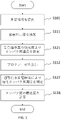
- 1 is a flow chart of a method for assessing the degree of saccharification of proteins, according to one embodiment.
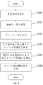
- 1 is a flow chart of a method for assessing the degree of saccharification of proteins, according to one embodiment.
- 4 is a graph showing changes in absorbance over time at multiple albumin concentrations, according to an example.
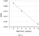
- 1 is a graph showing a calibration curve of albumin concentration based on absorbance, according to one example.
- 4 is a graph showing the relationship of albumin concentrations in real samples between the method according to an example and the conventional method.
- 4 is a graph showing the effect of the presence or absence of BCP on GA concentration measurement, according to one example.
- 4 is a graph showing changes in absorbance over time at multiple albumin concentrations, according to an example.
- FIG. 4 is a graph showing changes in sensor output over time at multiple albumin concentrations, according to one example.
- FIG. 5 is a graph showing the relationship between GA measurement values obtained by a method according to an example and GA values obtained by a conventional method; 4 is a graph showing changes in absorbance over time at multiple albumin concentrations, according to an example. 5 is a graph showing the relationship between GA values obtained by a method according to an example and GA values obtained by a conventional method;
- 1 is a block diagram of an apparatus for evaluating the degree of saccharification of proteins, according to one embodiment.
- FIG. 1 is a block diagram of an apparatus for evaluating the degree of saccharification of proteins, according to one embodiment.
- FIG. 1 is a block diagram of an apparatus for evaluating the degree of saccharification of proteins, according to one embodiment.
- FIG. 1 is a block diagram of an apparatus for evaluating the degree of saccharification of proteins, according to one embodiment.
- FIG. 1 is a block diagram of an apparatus for evaluating the degree of saccharification of proteins, according to one embodiment.
- FIG. 1 is a graph of absorbance showing the effect of the presence or absence of SDS on a modified BCP method, according to one example.
- the subject may include or be human.
- the subject may include non-human animals and may be non-human animals.
- the subject may include or be a mammal.
- Subjects may be, for example, without limitation, working animals, farm animals, companion animals, and wild animals.
- the sample to be measured may be a solution.
- the “solution” may be a body fluid, a solution derived from the body fluid, or a diluted solution of the body fluid.
- the solution may be a solution that is not a bodily fluid (derived from a non-bodily fluid), or a mixture of a bodily fluid or a bodily fluid-derived solution and a non-bodily fluid-derived solution.
- the solution may be the solution used for sample measurements or the solution used for calibration measurements.
- the solution may be a standard solution or a calibrator solution.
- the solution may be a liquid that does not contain the substance to be measured intentionally or intentionally for use in calibration or the like.
- the sample to be measured may be a specimen.
- the solution may be a solution containing chemicals.
- Body fluid may be lymph fluid, interstitial fluid such as interstitial fluid, intercellular fluid, interstitial fluid, body cavity fluid, serous fluid, pleural fluid, ascites, pericardial fluid, cerebrospinal fluid ( cerebrospinal fluid), synovial fluid (synovial fluid), or aqueous humor (aqueous humor).
- the bodily fluid may be a digestive fluid such as saliva, gastric juice, bile, pancreatic juice, intestinal juice, sweat, tears, runny nose, urine, semen, vaginal fluid, amniotic fluid, milk.
- the bodily fluid may be an animal bodily fluid or a human bodily fluid.
- a "bodily fluid” may be a solution.
- the solution may contain a physiological buffer such as phosphate-buffered saline (PBS) or N-tris(hydroxymethyl)methyl-2-aminoethanesulfonic acid buffer (TES) containing the substance to be measured.
- PBS phosphate-buffered saline
- TES N-tris(hydroxymethyl)methyl-2-aminoethanesulfonic acid buffer
- the solution is not particularly limited as long as it contains the substance to be measured.
- the solution may contain the substance to be measured.
- the solution may possibly contain the substance to be measured.
- the substance to be measured may be a molecule, ion, macromolecule, biomolecule, or the like.
- the substance to be measured may comprise a biomolecule.
- the substance to be measured may be protein, glycated protein, or the like.
- the solution may be tears, and the substance to be measured may be albumin, glycoalbumin, hemoglobin, and/or glycated hemoglobin contained in tears.
- the object to be measured may be albumin, glycoalbumin, hemoglobin, or glycohemoglobin in blood, serum, or plasma, or albumin, glycoalbumin, hemoglobin, or glycohemoglobin in interstitial fluid, urine, or saliva.
- Albumin may be oxidized albumin (HNA) or reduced albumin (HMA).
- HNA oxidized albumin
- HMA reduced albumin
- the substance to be measured may be AGE (Advanced Glycation End Products).
- the substance to be measured may be a glycated lipid.
- the sensor may be a chemical sensor, biosensor, ion sensor, etc. (hereinafter sometimes referred to as “sensor”, “biochemical sensor”, “chemical sensor” or “electrochemical sensor”).
- the sensor may comprise multiple sensors.
- the sensor may have electrodes.
- the electrodes may be amperometric electrodes.
- the electrodes may comprise hydrogen peroxide electrodes.
- the electrodes may comprise oxygen electrodes.
- the electrodes may be potentiometric electrodes.
- the electrodes may be electrodes for ion detection (pH electrode, cyanide ion electrode, iodide ion electrode, etc.).
- the senor may output an electrical signal. In some aspects, the sensor may output a current signal. A sensor may output a voltage signal or an electrical charge. The sensor may be electrically connected to an ammeter, voltmeter, or the like.
- the glycated amino acids produced may be reacted with ketoamine oxidase to generate hydrogen peroxide.
- the amount (concentration) of hydrogen peroxide generated may be measured.
- the amount (concentration) of hydrogen peroxide may be measured electronically.
- ketoamine oxidase generally recognizes the ketoamine structure of a peptide or peptide fragment containing glycated amino acids or glycated amino acid residues and oxidizes the glycated amino acids to produce amino acids, glucosone ( ⁇ -ketoaldehyde) and peroxidation.
- An oxidase that produces hydrogen is proportional to or related to the concentration of the peptide or peptide fragment containing the glycated amino acid or glycated amino acid residue that it recognizes.
- a ketoamine oxidase may be a dehydrogenase, a kinase, or an oxidase.
- the ketoamine oxidase may be fructosyl amino acid oxidase (FAOD), fructosyl peptide oxidase, fructosyl valyl histidine oxidase, fructosyl amine oxidase, amadoriase, fructosylamine deglycase or modified forms thereof.
- FOD fructosyl amino acid oxidase
- FOD fructosyl peptide oxidase
- fructosyl valyl histidine oxidase fructosyl amine oxidase
- amadoriase fructosylamine deglycase or modified forms thereof.
- the ketoamine oxidase may be an oxidase (Group I ketoamine oxidase) that acts on amino acids or peptides whose ⁇ -amino groups are glycated.
- the amino acid or peptide may be valine, glycine, valylhistidine.
- a glycated hemoglobin sensor or a glycated hemoglobin A1c (HbA1c) sensor can be constructed by using an oxidase that acts on an amino acid or peptide having a glycated ⁇ -amino group.
- the ketoamine oxidase may be an oxidase (group II ketoamine oxidase) that acts on amino acids or peptides in which the ⁇ -amino group is glycated.
- the amino acid may be lysine.
- the glycated amino acid may be fructosyl lysine.
- a glycoalbumin sensor can be constructed by using an oxidase that selectively acts on amino acids or peptides having glycated ⁇ -amino groups.
- the ketoamine oxidase may be an oxidase (group III ketoamine oxidase) that acts on amino acids or peptides in which the ⁇ -amino group and/or ⁇ -amino group is glycated.
- the amino acid or peptide may be lysine, valine, glycine, valylhistidine.
- a glycated protein sensor can be constructed by using an oxidase that acts on an amino acid or peptide whose ⁇ -amino group and/or ⁇ -amino group is glycated.
- the senor may include a detector.
- the detector may be a hydrogen peroxide detector.
- the "hydrogen peroxide detector” (hydrogen peroxide sensor) may be an electrochemical electrode or a hydrogen peroxide electrode.
- a hydrogen peroxide electrode may have a counter electrode, a reference electrode, and a working electrode.
- the detector may detect oxygen. For example, the amount or concentration of oxygen that decreases in an enzymatic reaction may be detected.
- Oxygen detection is considered to be relatively insensitive to noise sources such as molecules and ions and tolerant to interference. Oxygen consumption may be measured by oxygen sensing. Since the detector is air saturated, it may be used for enzymatic sensing.
- the detection unit may be configured to be able to selectively or combine a plurality of detection methods.
- a sensor may have an ion exchange resin on the detector.
- the sensor may have an ion exchange resin between the ketoamine oxidase layer and the hydrogen peroxide electrode.
- a cation exchange resin such as Nafion (registered trademark)
- an anion exchange resin such as polypyrrole can be used to suppress or prevent permeation of dopamine and the like, particularly positive ions and the like, from reaching the detection unit.
- the ion exchange resin may contain one, multiple or at least one type of ion exchange resin.
- the ion exchange resin may be configured with one, multiple or at least one type of layer.
- the sensor may comprise a Molecular Imprinted Polymer (MIP) membrane. Changes in charge or potential caused by MIP molecular recognition may be detected (potentiometry type). A change in the current flowing through the electrode (for example, mediator current) caused by MIP molecular recognition may be detected (amperometric type).
- MIP Molecular Imprinted Polymer
- protease is a general term for peptide bond hydrolases that hydrolyze and catabolize proteins and polypeptides.
- a protease may be an enzyme that breaks proteins into peptide fragments.
- the peptide fragments generated by the action of protease may include peptide fragments containing glycosylated amino acid residues and peptide fragments that are not glycosylated at all.
- protease may be an animal-derived protease, a plant-derived protease, or a microorganism-derived protease.
- a protease may be an exopeptidase or an endopeptidase.
- the protease may be an aspartic protease, a metalloprotease, a serine protease, or a thiol protease.
- protease may include multiple types or types of protease, or may include one type or type of protease.
- a protease may contain one or both of an endopeptidase and an exopeptidase. Mixing multiple proteases may increase the efficiency of decomposition.
- Animal-derived proteases include trypsin, chymotrypsin, elastase, bovine pancreatic protease, cathepsin, calpain, protease type-I, protease type-XX, aminopeptidase N, carboxypeptidase, pancreatin (a mixture of multiple enzymes such as proteases and amylases). and so on.
- the plant-derived protease may be papain, bromelain, gingipain, kallikrein, ficin, chymopapain, or the like.
- Microorganism-derived proteases include Bacillus-derived proteases, Aspergillus-derived proteases, Penicillium-derived proteases, Streptomyces-derived proteases, Lysobacter-derived proteases, Yeast-derived proteases, Tritirachium-derived protease, Thermus-derived protease, Pseudomonas-derived protease, Achromobacter-derived protease, and the like may be used.
- the protease may be provided in liquid form or mixed with a solution. In some embodiments, the protease may be added or mixed into the solution in solid form (eg, powder). In some embodiments, the protease may be introduced into solution on a support material. The protease may be supported on a plate, curved surface, sphere, bead, biomolecule such as protein, polymer, or the like.
- optical measurement generally refers to determining optical properties of matter using an optical element or device.
- optical measurements of the substance of interest may be measured.
- a substance that is bound to or related to a target substance hereinafter referred to as a substance (e.g., reagent) that is chemically, biologically or physically bound to or related to a target substance, even if it is not the target substance itself) Properties may be measured. Properties of reagents may be measured.
- the reagent may be referred to as a "target substance”.
- optical measurements may include spectroscopic measurements.
- the absorbance of the target substance may be measured.
- a color changing indicator corresponding to the substance of interest may be introduced. A color changing indicator may be detected or measured.
- color changing indicator generally refers to halochromic chemical compounds. Color changes may generally be reversible. Color changing indicators include, for example, without limitation, acid-base indicators (pH indicators), redox indicators, adsorption indicators, TLC color changing indicators, and the like. It changes the color of its solution depending on the pH.
- the pH indicator may be, for example, without limitation, bromocresol purple (BCP), bromocresol green (BCG), and the like.
- BCP or BCG can be used for albumin.
- albumin combines with bromocresol green (BCG) to produce an albumin-bromocresol green complex. Around pH 4, the complex appears blue. Therefore, the albumin concentration can be obtained by measuring the absorbance (BCG method).
- BCG method bromocresol green
- a surfactant may be added to the measurement reagent. Surfactant may not be added to the measurement reagent. For example, protein denaturants (such as SDS) may not be added.
- albumin binds with bromocresol purple (BCP) to generate an albumin-bromocresol purple complex.
- BCP bromocresol purple
- This complex has a blue color. Therefore, the albumin concentration can be obtained by measuring the absorbance (BCP method).
- the albumin concentration can be measured by a color reaction with BCP after converting all of the reduced albumin into oxidized albumin by the action of an oxidizing agent (improved BCP method).
- a surfactant may be added to the measurement reagent.
- protein denaturants such as SDS
- the reaction in which albumin combines with bromocresol purple (BCP) or bromocresol green (BCG) to form a conjugate may be referred to as the "first reaction.”
- the color changing indicator may be provided in liquid form or mixed with a solution. In some embodiments, the color changing indicator may be added or mixed into the solution in solid form (eg, powder).
- an absorption photometer (colorimeter) may be used.
- the absorption photometer has a light source and a light receiver. All or part of the light emitted by the light source enters the target solution. All or part of the light that passes through the solution enters the receiver. The light absorbed by the target substance can be analyzed.
- the wavelength of the light source may be appropriately selected according to the color indicator used.
- Absorbance may be measured, for example, at wavelengths such as dominant wavelength 600-630 nm for BCP and 565-660 nm for BCG.
- the change in absorbance over time of the color indicator may be measured.
- absorbance may be measured at least two (or more) times.
- the time at which protease decomposition starts is measured, and the protein concentration may be determined based on the measured value corresponding to the time from this start time.
- protein concentration may be determined based on measurements at three or more time points. From at least three or more measurements, it may be possible to determine the initial protein concentration even if the time from the start of protease degradation is not clear.
- the measurement of glycated protein may be started after a predetermined or predetermined time has elapsed from the start time of protease degradation.
- glycated protein measurement may begin when the absorbance A is sufficiently small.
- the change in absorbance over time ( ⁇ A/ ⁇ t) becomes substantially zero, it can be determined that sufficient protease degradation has been completed and measurement of glycated proteins can be started, or The measurement result of glycated protein at that time may be adopted.
- the change in absorbance over time reaches a predetermined value, it may be determined that degradation by a predetermined protease has been achieved, and the measurement of glycated protein may be started, or the glycated protein at that time may be measured. Measurement results may be used.
- calibration of absorbance measurements may be performed.
- An optical system including optical measurement cells, light emitters, light receivers, etc. may be calibrated.
- Absorbance may be measured before introduction of the solution into the optical system (e.g., the state in which the optical measurement cell is empty, its initial state, etc.) and after (e.g., the state in which the measurement solution has been introduced into the optical measurement cell).
- Absorbance may be measured before introduction of the solution into the optical system (e.g., the state in which the optical measurement cell is empty, its initial state, etc.) and after (e.g., the state in which the measurement solution has been introduced into the optical measurement cell).
- a difference in absorbance between them may be obtained.
- a method of measuring a protein comprising: providing a solution that may contain a protein of interest; adding a color changing indicator to the solution; and adding a protease.
- the method can be used to measure glycated proteins.
- the application of the method is not so limited. The method can be used in various other methods.
- FIG. 1 shows a flowchart of a method for assessing glycation degree of albumin, according to an embodiment.
- a solution that may contain albumin as a protein to be measured is provided.
- BCP is added as a color indicator.
- the absorbance of BCP is measured. Determine the concentration of albumin from the absorbance.
- protease is added to the measurement solution to which BCP has already been added.
- Albumin begins to be broken down into peptides or amino acids by proteases.
- step S122 hydrogen peroxide is generated by causing ketoamine oxidase, which specifically reacts with saccharified amino acids among the peptides or amino acids produced by the digestion reaction with the protease, to act.
- the hydrogen peroxide is measured using electrodes. This gives the concentration of glycated albumin (GA) in the solution.
- step S131 the ratio of the obtained glycated albumin (GA) to the albumin concentration is obtained as the GA value.
- a color indicator is added (S111), absorbance is measured (S121), and then protease is added (S112).
- the absorbance measurement may be completed by the time the protease is added.
- the protease may be added before completing the absorbance measurement or during the absorbance measurement.
- FIG. 2 shows a flowchart of a method for assessing glycation degree of albumin, according to an embodiment.
- a solution that may contain albumin as a protein to be measured is provided.
- BCP is added as a color indicator.
- protease is added to the measurement solution to which BCP has already been added. Degradation of albumin into amino acids by proteases is initiated.
- the absorbance of BCP is measured. Determine the concentration of albumin from the absorbance. Absorbance decreases with time due to ongoing decomposition of albumin.
- the concentration of albumin contained in the original measurement solution can be obtained by measuring the absorbance at the time when the protease is introduced or the start time of the decomposition and after a predetermined time from the start time.
- step S222 hydrogen peroxide is generated by causing ketoamine oxidase, which specifically reacts with saccharified amino acids among the peptides or amino acids produced by the digestion reaction with the protease, to act.
- the hydrogen peroxide is measured using electrodes.
- step S231 the ratio of the obtained glycated albumin (GA) to the albumin concentration is obtained as the GA value.
- a color indicator is added (S211), and then protease is added (S212).
- the order of steps is not limited to this.
- the addition of the color changing indicator (S211) and the addition of the protease (S212) may occur substantially simultaneously.
- the protease and the color indicator may be mixed in advance, and the mixed solution may be added to the measurement solution.
- the glycated albumin concentration is measured (S222).
- measuring albumin concentration (S221) and measuring glycated albumin concentration (S222) may be performed substantially simultaneously.
- a color changing indicator is added (S211) and a protease is added (S212), after which the solution with both added may be diverted.
- the concentration of albumin may be measured in one of the diverted solutions, and the concentration of glycated albumin in the other.
- the protease and color indicator may not be added separately.
- the protease and the color indicator may be mixed in advance, and the mixed solution may be added to the measurement solution.
- a color-changing indicator is brought into contact with a solution that may contain the target protein, and then the first reaction between the color-changing indicator and the target protein is used to determine the concentration of the target protein in the solution.
- the protease is contacted with a solution that may contain the protein of interest, and then a second reaction between the protease and the protein of interest is used to determine the concentration of the glycated protein of interest (the concentration of the glycated protein of interest concentration, also called concentration of glycated protein).
- the second reaction may comprise reacting the produced glycated amino acid with ketoamine oxidase to generate hydrogen peroxide.
- the order of the four items of introduction of the color-changing indicator, measurement of the first reaction between the color-changing indicator and the target protein, introduction of the protease, and measurement of the second reaction between the protease and the target protein is Not limited.
- protein is measured for the indicator mixed solution after mixing the measurement solution and the indicator, and glycated protein is measured for the protease mixed solution after mixing the measurement solution and the protease.
- the order of steps may not be limited except for the order of performing.
- the protease may be mixed into the indicator mixed solution.
- the measurement solution and the indicator are mixed, the protein is measured using the indicator mixed solution, the protease is mixed with the indicator mixed solution, and the indicator protease mixed solution (the indicator and the protease are mixed solution) may be used to measure glycated protein.
- a measurement solution and an indicator are mixed, a protease is mixed in the indicator mixed solution, and the glycated protein is measured using the indicator protease mixed solution (a solution in which the indicator and the protease are mixed), Proteins may be measured using the protease mixed solution.
- a measurement solution and an indicator are mixed, a protease is mixed in the indicator mixed solution, and the glycated protein is measured using the indicator protease mixed solution (a solution in which the indicator and the protease are mixed), Measurement of protein using the indicator-protease mixed solution may be performed in that order.
- an indicator may be mixed with the protease mixed solution after mixing the measurement solution and the protease.
- a measurement solution and protease are mixed, glycated protein is measured using the protease mixed solution, an indicator is mixed with the protease mixed solution, and protein is measured using the protease indicator mixed solution. , in that order.
- a measurement solution and a protease are mixed, an indicator is mixed with the protease mixed solution, glycated protein is measured using the indicator protease mixed solution, and protein is measured using the indicator protease mixed solution. You can do things in that order.
- a measurement solution and a protease are mixed, an indicator is mixed with the protease mixed solution, protein is measured using the indicator protease mixed solution, and glycated protein is measured using the indicator protease mixed solution.
- the indicator and the protease may be mixed substantially simultaneously with the measurement solution.
- the glycation degree of the protein of interest may be determined based on the concentration of the protein of interest and the concentration of the glycated protein.
- the degree of saccharification of protein may be obtained by dividing the concentration of glycated protein by the concentration of protein.
- the quantity to be measured is not limited to concentration.
- the quantity measured may be an absolute quantity, a relative quantity, a concentration, or any other quantity related to mass or concentration.
- the concentration of protein and the concentration of glycated protein can be measured after addition of a color changing indicator and addition of protease.
- the color changing indicator and/or protease is added in liquid form, changing the volume from the original solution.
- the variability caused by errors in the dosage of color indicator and/or protease occurs at the same rate for both protein concentration and glycated protein concentration. Therefore, it does not substantially affect the calculation of the saccharification degree. Therefore, relatively accurate and reproducible measurements are possible.
- measurement of protein concentration, addition of solid protease, and measurement of glycated protein concentration can be performed after addition of a color indicator.
- variability caused by errors in input amounts of color indicator and/or protease occurs in the same proportions for both protein concentration and glycated protein. Therefore, it does not substantially affect the calculation of the saccharification degree. Therefore, relatively accurate and reproducible measurements are possible.
- the pH may be adjusted.
- the pH may be changed during the addition or reaction of the color indicator (first reaction) and during the decomposition by the protease (second reaction).
- the first reaction and the second reaction may be performed at substantially the same pH.
- the pH value may differ from the general optimum range.
- the first reaction may be followed by the second reaction.
- the pH when the first and second reactions are (at least partially) performed simultaneously, one pH is applied to both reactions.
- the value may be adjusted to give a suitable range for both.
- the pH value may differ from the general optimum range.
- the temperature may be regulated.
- the temperature may be changed between the addition or reaction of the color indicator (first reaction) and the decomposition by protease (second reaction).
- first reaction the temperature was adjusted to 20° C. to 70° C., which is its general optimum temperature. , or the temperature may be adjusted to 55-65°C.
- second reaction the temperature may be once raised to the temperature for protease decomposition before introducing the protease.
- the temperature may be adjusted to 25°C to 75°C, 30°C to 65°C, 35°C to 65°C, around 37°C, or around 55°C. .
- one temperature is applied to both reactions.
- the value may be adjusted to give a suitable range for both.
- the temperature may be adjusted in the range of 25° C. to 50° C., 25° C. to 45° C., or 30° C. to 45° C. for both the first reaction and the second reaction.
- the temperature may be between 25°C and 70°C, between 30°C and 70°C, between 40°C and 70°C, between 50°C and 70°C, or between 55°C and 65°C. You can adjust the range.
- albumin concentration was measured using an absorbance method.
- a commercially available glycoalbumin measurement kit (Lucica (registered trademark) GA-L) calibrator (GA-L calibrator, Asahi Kasei Pharma Corp.) was used, and serial dilutions (7.93 mg/mL, 3 .97 mg/mL, 1.98 mg/mL) were prepared.
- a solution containing only PBS (blank PBS) was also prepared as a blank sample.
- As a protease solution 320 mg of Orientase 22BF (HBI Co., Ltd.) was dissolved in 4 mL of HEPES buffer.
- a BCP solution (0.02 mM, pH 8.0) was used for the color reaction.
- the absorbance was measured over time at intervals of 12 seconds using a diabetes test item automatic analyzer DM-JACK Ex (Hitachi Chemical Diagnostic Systems Co., Ltd.).
- DM-JACK Ex Hagaku Chemical Diagnostic Systems Co., Ltd.
- 180 ⁇ L of BCP solution was added to 18 ⁇ L of sample solution.
- 18 ⁇ L of protease solution was added.
- ⁇ A when it shows a constant value corresponds to the concentration of albumin.
- the concentration of albumin may be determined based on absorbance in a solution without added protease.
- the concentration of albumin may be determined based on the absorbance value (A or ⁇ A) at a predetermined time after protease addition. For example, an endpoint method may be used. In some embodiments, the concentration of albumin may be determined based on the time conversion rate of absorbance (dA/dt, or d( ⁇ A)/dt). For example, a rate assay may be used. In some embodiments, albumin concentration may be determined based on higher order derivatives of absorbance (such as d2A/dt2, or d2( ⁇ A)/dt2).
- the concentration of albumin may be determined based on the absorbance ( ⁇ A) after sufficient time has passed.
- Fig. 4 shows a calibration curve obtained from the relationship between the concentration of albumin in the sample solution and dA/dt. It became clear that a very strong negative correlation is shown.
- Example 2 Measurement of saccharified albumin concentration> As an example, glycated albumin concentration was measured using the enzymatic electrode method.
- glucose was added to HSA (Fujifilm Wako Pure Chemical Industries, Ltd.) and incubated at 37°C to prepare GA solutions (40 mg/mL) with different saccharification rates.
- the saccharification rate of each sample was confirmed using a commercially available glycoalbumin measurement kit (Lucica (registered trademark) GA-L, Asahi Kasei Pharma Corp.).
- a protease solution was prepared by dissolving 2 mg of Amano K (Amano Enzyme Co., Ltd.) in 200 ⁇ L of TES buffer. 8 ⁇ L of BCP solution (1 mM) was added to 20 ⁇ L of protease solution. The solution was heat treated and stored at 4°C.
- This sample was added to an electrochemical sensor with a hydrogen peroxide electrode on which a ketoamine oxidase membrane was fixed.
- the glycated albumin concentration was determined by measuring the output current.
- Fig. 6 shows the relationship between glycated albumin concentration and enzyme sensor output in the presence or absence of BCP.
- the horizontal axis indicates the glycated albumin concentration in the solution, and the vertical axis indicates the measured current value.
- white circles indicate measurements "without BCP”
- black circles indicate measurements "with BCP”.
- Example 3 shows an example of GA value measurement.
- BCP is added to a sample containing glycated albumin, albumin concentration is measured by absorbance, protease is added to the solution, and glycated albumin is measured using an electrochemical sensor having a hydrogen peroxide electrode to which an FAOD membrane is fixed. Densitometry was performed. More specific description will be given below.
- FIG. 7 shows an example of measurement results used to generate a calibration curve for albumin concentration by absorbance measurement. Samples containing 1.3 mg/mL, 2.4 mg/mL, and 4.7 mg/mL albumin were used for absorbance measurements. At 0 seconds in FIG. 7, BCP solution (including Sodium Dodecyl Sulfate (SDS), TritonX-100, 5,5′-Dithiobis (2-nitrobenzoic acid (DTNB), HEPES (pH 8.0), NaCl) was optically measured.
- SDS Sodium Dodecyl Sulfate
- TritonX-100 TritonX-100
- DTNB 5,5′-Dithiobis (2-nitrobenzoic acid
- HEPES pH 8.0
- the albumin sample was added to the BCP solution.Therefore, the absorbance decreased.Then, the solution was mixed by pipetting.The spikes and fluctuations visible until about 40 seconds were It was caused by the movement of the liquid due to mixing and the blockage of the light path by the pipette.After about 50 seconds, the absorbance of the albumin sample was all constant.As is clear from Figure 7, the absorbance showed albumin concentration dependence. , the relationship between albumin concentration and absorbance was determined, and a calibration curve was generated (not shown).
- Fig. 8 shows an example of the measurement results used to generate the calibration curve of glycated albumin concentration by electrochemical sensor measurement.
- Samples containing 0.4 mg/mL, 0.7 mg/mL, and 1.5 mg/mL glycated albumin were used for the measurement. These solutions contain BCP.
- Protease was added to the solution after absorbance measurement, and a digestion reaction was performed at 55° C. for 30 minutes. After that, at 0 seconds in FIG. 8, the solution after the digestion reaction was introduced into the electrochemical sensor.
- the output current of the electrochemical sensor was measured. It was found that any concentration takes a maximum value (inflection point) at about 60 to 80 seconds. At this time, for example, the relationship between the output current at 60 seconds and the saccharified albumin concentration can be obtained. Using such data, the relationship between the saccharified albumin concentration and the output current of the electrochemical sensor was determined, and the calibration curve was generated.
- the GA value of an unknown sample can be determined.
- ⁇ Measurement of GA value of unknown sample> Next, using the same method as above, the GA value of the unknown sample was measured. As measurement samples, a high-concentration GA value sample and a low-concentration GA value sample were mixed at several ratios to prepare a plurality of measurement solutions. A human serum albumin reagent (Sigma-Aldrich) was used for low-concentration GA value samples. A high-concentration GA value sample was prepared by adding glucose to a human serum albumin reagent (Sigma-Aldrich) and incubating. Although these are artificially prepared samples, the actual GA values are unknown.
- the measurement method of this example was applied to these measurement samples to obtain the GA value.
- the GA value (%) was determined for the same measurement sample using a commercially available glycoalbumin measurement kit (Lucica (registered trademark) GA-L) as an existing method.
- Figure 9 shows both measurements obtained for samples of four different unknown concentrations.
- the horizontal axis indicates the GA value measured by Lucica, and the vertical axis indicates the GA value (arbitrary unit; GA value (%) is not shown) obtained by the measurement method of this example.
- GA value can be obtained very satisfactorily by using one of the methods of the present disclosure.
- Example 4 shows an example of GA value (%) measurement.
- BCP and protease mixed solution is added to a sample containing glycated albumin, and then optical measurement is performed to measure the albumin concentration, while using an electrochemical sensor having a hydrogen peroxide electrode to which an FAOD membrane is fixed. Glycated albumin concentration was measured. More specific description will be given below.
- FIG. 10 shows an example of measurement results used to generate a calibration curve for albumin concentration by absorbance measurement.
- Samples containing 1.2 mg/mL, 2.5 mg/mL, and 5.0 mg/mL albumin were used for the measurements.
- BCP and protease mixture SDS, Triton X-100, DTNB, HEPES (pH 8.0), NaCl, protease added
- SDS Triton X-100, DTNB, HEPES (pH 8.0), NaCl, protease added
- values related to absorbance such as the absorbance at 20 seconds, the difference in absorbance at 20 seconds and later time points (e.g., the difference in absorbance at 90 seconds, the change in absorbance over time (time derivative), etc., are derived from the initial albumin This data was used to determine the relationship between albumin concentration and absorbance and generate a calibration curve for it.
- the measurement method of this example was applied to these measurement samples to obtain the GA value (%).
- the GA value (%) was determined for the same measurement sample using a commercially available glycoalbumin measurement kit (Lucica (registered trademark) GA-L) as an existing method.
- Figure 11 shows the relationship between both measurements obtained for samples of five different unknown concentrations.
- the horizontal axis indicates the GA value (%) obtained by the existing measurement method, and the vertical axis indicates the GA value (%) obtained by the measurement method of this embodiment.
- the GA value (%) can be obtained very satisfactorily by using one of the methods of the present disclosure.
- Albumin measurement by the improved BCP method is generally performed under acidic conditions using an oxidizing agent and SDS. Protein denaturants such as SDS are believed to facilitate oxidation of albumin upon introduction of an oxidizing agent such as DTNB. In that state, BCP is introduced.
- the protease reaction as the second reaction generally requires weak alkalinity.
- the first reaction In order to carry out the first reaction and the second reaction simultaneously, continuously or serially, it is preferred that the first reaction also be carried out in the same alkaline environment. No need to change pH.
- the solution does not contain SDS in the modified BCP process which is carried out in mild alkalinity.
- reaction solution containing SDS was adjusted to a concentration of 0.02% SDS.
- concentration of 0.02% SDS was adjusted to a concentration of 0.02% SDS.
- FIG. 16 shows the presence of SDS (SDS+, solid) and no SDS (SDS-, open) for three types of buffer solutions, succinic acid solution, HEPES solution (HEPES, circle), and Tricine solution (Tricine, triangle). , shows the absorbance.
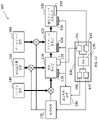
- FIG. 12 schematically shows the configuration of an apparatus 100 according to one embodiment.
- the device 100 comprises an optical measurement section 110 , a protease reaction section 120 and a glycated protein measurement section 130 .
- the sample solution is introduced from the introduction unit 140 into the optical measurement section 110 and then flowed through the protease reaction section 120 and the glycated protein measurement section 130 in sequence.
- a color changing indicator tank 150 is connected to the optical measurement section 110 and is configured to provide a color changing indicator to the optical measurement section 110 .
- a protease solution tank 160 is connected to the protease reaction section 120 and configured to provide the protease reaction section 120 with a protease solution.
- the solution introduction and liquid transfer are controlled by the liquid transfer mechanism 170 .
- the liquid sending mechanism 170 can control the timing and amount of introduction of the sample solution into the optical measurement section 110 .
- the liquid feeding mechanism 170 controls the timing and amount of introduction of the color indicator solution from the color indicator solution tank 150 to the optical measurement section 110, and the introduction of the protease solution from the protease solution tank 160 to the protease reaction section 120. You can control the timing and amount of
- the liquid feeding mechanism 170 may include, for example, a pump.
- the optical measurement section 110, the protease reaction section 120, and the glycated protein measurement section 130 are closed channels, positive pressure can be applied to the upstream side to control the movement of the solution.
- the liquid feeding mechanism 170 may be a pressing mechanism attached to each solution tank. When each solution tank is a deformable container, a predetermined amount of solution can be delivered to the outside by deforming the container.
- Temperature control units 118, 128, 138 having temperature sensors and heaters (not shown) are arranged in the optical measurement section 110, the protease reaction section 120, and the glycated protein measurement section 130, respectively.
- the temperature control units 118, 128, 138 are connected to a temperature control mechanism (driver) 180, which receives measured temperature information from the temperature sensors and can send on/off instructions and power to the heaters.
- Light emitted from the light-emitting element passes through the solution in the optical measurement unit 110, and the light-receiving element senses the emitted light.
- the optical measurement unit 119 can output absorbance information at that time.
- a sensor 139 is arranged in the glycated protein measurement unit 130 .
- the sensor 139 is arranged inside the glycated protein measuring unit 130 and can directly contact the internal solution. This sensor 139 is configured to measure the concentration of glycated proteins.
- the device 100 further comprises a computer system 190.
- Computer system 190 includes at least a central processing unit (CPU) 195, which performs the necessary computations.
- Computer system 190 has communication interfaces 197 and 198 for liquid transfer mechanism 170 and temperature control mechanism 180, respectively.
- Computer system 190 has communication interfaces 191 and 193 with optical measurement unit 119 and sensor 139, respectively.
- the CPU 195 transmits necessary information such as required operation items (solution to be introduced and component to be introduced), its timing, and its amount to the liquid transfer mechanism 170 via the liquid transfer mechanism communication interface 197. send.
- Liquid delivery mechanism 170 performs the necessary operations based on the information received.
- the liquid transfer mechanism 170 can transmit information such as information indicating that each operation has been executed and information such as execution failure to the communication interface 197 .
- the CPU 195 provides the temperature control mechanism 180 with necessary operation items (measurement of temperature, temperature required for each reaction, etc.), their timing, their temperature and time, etc. to the temperature control mechanism 180 via the temperature control mechanism communication interface 198. send information. Temperature control mechanism 180 performs the necessary operations based on the information received. The temperature control mechanism 180 can send information to the communication interface 198 such as information that each operation has been executed, information such as execution failure.
- the optical measurement unit communication interface 191 receives information about absorbance from the optical measurement unit 119 arranged in the optical measurement section 110 .
- the glycated protein sensor communication interface 193 receives information about the glycated protein concentration from the sensor 139 arranged in the glycated protein measurement unit 130 .
- the optical measurement unit communication interface 191 issues instructions such as the start and end of absorbance measurement to the optical measurement unit 119 arranged in the optical measurement section 110, and then receives information on the protein concentration obtained by the measurement.
- the glycated protein sensor communication interface 193 issues instructions such as start and end of current measurement to the sensor 139 arranged in the glycated protein measurement unit 130, and then receives information on the glycated protein concentration obtained by the measurement. .
- the CPU 195 receives information related to absorbance and information related to glycated protein via communication interfaces 191 and 193, performs predetermined conversion, and calculates the degree of saccharification.
- FIG. 13 schematically shows the configuration of an apparatus 200 according to one embodiment.
- the device 200 comprises an optical measurement section 210 and a glycated protein measurement section 230 .
- the sample solution is introduced from the introduction unit 240 into the optical measurement section 210 and flowed to the glycated protein measurement section 230 .
- a measuring liquid (color changing indicator protease solution) tank 250 is connected to the optical measuring section 210 and is configured to provide the optical measuring section 210 with a mixture of color changing indicator and protease.
- the solution introduction and liquid transfer are controlled by the liquid transfer mechanism 270 .
- Temperature control units 218 and 238 having temperature sensors and heaters (not shown) are arranged in the optical measurement section 210 and glycated protein measurement section 230, respectively.
- An optical measurement unit 219 is arranged in the optical measurement section 210 and can measure absorbance and output information about the measured absorbance.
- a sensor 239 is arranged in the glycated protein measurement unit 230 and is configured to measure the concentration of the glycated protein and output information on the measured glycated protein.
- the device 200 further comprises a computer system 290.
- Computer system 290 includes at least a central processing unit (CPU) 295, which performs the necessary computations.
- Computer system 290 can communicate with liquid delivery mechanism 270 and temperature control mechanism 280 via communication interfaces 297 and 298 .
- Computer system 290 can communicate with optical measurement unit 219 and sensor 239 via communication interfaces 291 and 293 .
- the CPU 295 receives information related to absorbance and information related to glycated protein via communication interfaces 291 and 293, performs predetermined conversion, and calculates the degree of saccharification.
- FIG. 14 schematically shows the configuration of an apparatus 300 according to one embodiment.
- the device 300 comprises a color indicator reaction section 305 , an optical measurement section 310 , a protease reaction section 320 and a glycated protein measurement section 330 .
- the sample solution is introduced from the introduction unit 340 into the color indicator reaction section 305 and then passed through the optical measurement section 310 , the protease reaction section 320 and the glycated protein measurement section 330 in sequence.
- the color-changing indicator reaction unit 305 contains a color-changing indicator in advance.
- the solution introduced into the color-changing indicator reaction section 305 is mixed with the color-changing indicator and reacts with it.
- the protease reaction section 320 contains a protease in advance. The solution introduced into the protease reaction section 320 contacts and reacts with the protease.
- the solution introduction and liquid transfer are controlled by the liquid transfer mechanism 370 .
- Temperature control units 318, 328, and 338 having temperature sensors and heaters (not shown) are arranged in the optical measurement section 310, the protease reaction section 320, and the glycated protein measurement section 330, respectively.
- An optical measurement unit 319 is arranged in the optical measurement section 310 and can measure absorbance and output information about the measured absorbance.
- a sensor 339 is arranged in the glycated protein measurement unit 330 and configured to measure the concentration of the glycated protein and output information about the measured glycated protein.
- the device 300 further comprises a computer system 390.
- Computer system 390 includes at least a central processing unit (CPU) 395, which performs the necessary computations.
- Computer system 390 can communicate with liquid delivery mechanism 370 and temperature control mechanism 380 via communication interfaces 397 and 398 .
- Computer system 390 can communicate with optical measurement unit 319 and sensor 339 via communication interfaces 391 and 393 .
- the CPU 395 receives information related to absorbance and information related to glycated protein via communication interfaces 391 and 393, performs predetermined conversion, and calculates the degree of saccharification.
- FIG. 15 schematically shows the configuration of an apparatus 400 according to one embodiment.
- the device 400 comprises a color indicator protease solution reaction section 405 , an optical measurement section 410 and a glycated protein measurement section 430 .
- the sample solution is introduced from the introduction unit 440 into the color indicator reaction section 405 and then passed through the optical measurement section 410 and the glycated protein measurement section 430 in sequence.
- the color-changing indicator protease solution reaction unit 405 contains a color-changing indicator and protease in advance.
- the solution introduced into the color-changing indicator protease solution reaction unit 405 is mixed with and reacting with the color-changing indicator, and also contacts and reacts with the protease.
- the solution introduction and liquid transfer are controlled by the liquid transfer mechanism 470 .
- Temperature control units 418 and 438 having temperature sensors and heaters (not shown) are arranged in the optical measurement section 410 and the glycated protein measurement section 430 .
- An optical measurement unit 419 is arranged in the optical measurement section 410 and can measure absorbance and output information about the measured absorbance.
- a sensor 439 is arranged in the glycated protein measurement unit 430 and is configured to measure the concentration of the glycated protein and output information about the measured glycated protein.
- the device 400 further comprises a computer system 490.
- Computer system 490 includes at least a central processing unit (CPU) 495, which performs the necessary computations.
- Computer system 490 can communicate with liquid delivery mechanism 470 and temperature control mechanism 480 via communication interfaces 497 and 498 .
- Computer system 490 can communicate with optical measurement unit 419 and sensor 439 via communication interfaces 491 and 493 .
- the CPU 495 receives information related to absorbance and information related to glycated protein via communication interfaces 491 and 493, performs predetermined conversion, and calculates the degree of saccharification.
- the device or system may be an automated pipettor or system (sometimes simply referred to as an automated pipettor).
- the automated pipetting device may have wells containing reagents or mixtures of at least two of them.
- a cartridge (microplate) with multiple wells may be provided.
- the device or cartridge may include an electrochemical sensor or may be configured to have an electrochemical sensor attached. Samples and solutions may be quantified, transferred from one well to another, or mixed by pipetting.
- the automated pipetting device may include a heater, and the heater may control the temperature of the wells or solutions within the wells.
- the automated pipetting device may be equipped with an optical measurement system.
- the device may include a waste tank.
- the solution that has passed through the optical measurement section, the glycated protein measurement section, and the like may be collected in a waste liquid tank. This can reduce the risk of a solution containing body fluid leaking to the outside.
- a method for evaluating the degree of saccharification of a protein comprising: providing a solution that may contain the protein of interest; contacting the solution with a color changing indicator; contacting the solution with a protease; determining the concentration of the protein of interest in the solution using a first reaction between the color-changing indicator and the protein of interest; determining the concentration of the glycated protein of interest using a second reaction between the protease and the protein of interest; determining the glycation degree of the target protein based on the concentration of the target protein and the concentration of the glycated target protein; How to prepare.
- A011 The method of embodiment A001, wherein contacting the solution with a protease comprises adding the protease to a color changing indicator mixture produced by contacting the solution with the color changing indicator; Method.
- A012 The method of embodiment A001, wherein contacting the solution with a color-changing indicator and contacting the solution with a protease are performed substantially simultaneously; Method.
- A012b The method of embodiment A001, wherein substantially simultaneously mixing the color changing indicator and the protease with the solution; Method.
- A013 The method of embodiment A001, wherein contacting the solution with a color changing indicator comprises adding the color changing indicator to a protease mixture produced by contacting the solution with the protease; Method.
- A021 The method of any one of embodiments A001 through A013, wherein measuring the first reaction between the color-changing indicator and the protein of interest comprises measuring the absorbance of the color-changing indicator; Method. A022 The method of embodiment A021, wherein Measuring the absorbance of the color indicator comprises measuring a time change of the absorbance; Method. A023 The method of embodiment A022, wherein Measuring the change in absorbance over time includes measuring the absorbance at least two (or more) times, Method. A024 The method of embodiment A022 or A023, wherein determining the concentration of the glycated target protein based on the time change of the absorbance; Method. A031 The method of any one of embodiments A001-A024, wherein the protein of interest is albumin.
- A032 The method of embodiment A031, wherein Determining the concentration of glycated albumin using the second reaction between the protease and the albumin includes: degrading the albumin with the protease to produce peptides, and reacting the produced peptides with ketoamine oxidase to produce hydrogen peroxide; measuring the concentration of the hydrogen peroxide produced; determining the concentration of the glycated albumin based on the concentration of the hydrogen peroxide; comprising a Method.
- A033 The method of embodiment A032, wherein measuring the concentration of hydrogen peroxide comprises using a hydrogen peroxide electrode; Method.
- A034 The method of embodiment A033, wherein measuring the concentration of the hydrogen peroxide includes contacting the hydrogen peroxide that has passed through an ion exchange resin with the hydrogen peroxide electrode; Method.
- A041 The method of any one of embodiments A001 through A034, wherein wherein the color changing indicator is a pH indicator; Method.
- A042 The method of any one of embodiments A031 through A034, wherein The color changing indicator is bromocresol purple (BCP) or bromocresol green (BCG). Method.
- A051 The method of embodiment A042, wherein The color changing indicator is bromocresol purple (BCP) or bromocresol green (BCG), adjusting the pH of the solution to a range of 4.5 to 8.5 during the first reaction and adjusting the pH of the solution to a range of 6.0 to 9.0 during the second reaction; and the method further comprising: A052 The method of any one of embodiments A001 through A042, wherein When the first reaction between the color changing indicator and the protein of interest and the second reaction between the color changing indicator and the protein of interest occur (at least partially) simultaneously, the pH of the solution is adjusted to a range suitable for both the first reaction and the second reaction; Method.
- BCP bromocresol purple
- BCG bromocresol green
- A053 The method of embodiment A052, wherein The color changing indicator is bromocresol purple (BCP) or bromocresol green (BCG), the pH is adjusted to the range of 7.0 to 8.5; Method.
- A054 The method of embodiment A051 or A053, wherein the pH is adjusted to a range of 7.0 to 8.5; During the first reaction, the solution mixture does not contain a protein denaturant (SDS); Method.
- A055 The method of any one of embodiments A032-A054, wherein the color changing indicator is bromocresol purple (BCP) or bromocresol green (BCG), adjusting the temperature of the solution in the range of 20° C. to 70° C.
- a method further comprising: A056 The method of any one of embodiments A001 through A054, wherein When the first reaction between the color changing indicator and the protein of interest and the second reaction between the color changing indicator and the protein of interest occur (at least partially) simultaneously, the temperature of the solution is adjusted to a range suitable for both the first reaction and the second reaction; Method. A057 The method of embodiment A056, wherein the temperature is adjusted in the range of 25°C to 70°C; Method.
- A058 The method of any one of embodiments A032-A034, wherein The method further comprising adjusting the temperature of said ketoamine oxidase to a range of 25°C to 75°C.
- A061 The method of any one of embodiments A032-A058, wherein The ketoamine oxidase is an oxidase (group I ketoamine oxidase) that acts on amino acids or peptides in which the ⁇ -amino group is glycated, and measuring the concentration of the generated hydrogen peroxide is fructosylvaline , calibrating the measurement using a standard solution containing at least one of fructosylglycine and fructosylvalylhistidine; Method.
- ketoamine oxidase is an oxidase (group II ketoamine oxidase) that acts on amino acids or peptides in which the ⁇ -amino group has been glycated, and measuring the concentration of the produced hydrogen peroxide is a way of measuring fructosyl lysine. calibrating the measurement using a reference solution comprising Method.
- ketoamine oxidase is an oxidase (group III ketoamine oxidase) that acts on amino acids or peptides having glycated ⁇ -amino groups and/or ⁇ -amino groups
- Measuring the concentration of the produced hydrogen peroxide includes calibrating the measurement using a standard solution containing at least one of fructosyl lysine, fructosyl valine, fructosyl glycine and fructosyl valyl histidine. be prepared to Method.
- B01 A program for causing a computer system to perform the method of any one of embodiments A001 through A063.
- C01 A device for evaluating the degree of saccharification of proteins, a bodily fluid introduction unit (or inlet); a mechanism for mixing the bodily fluid and a color indicator solution; a protein concentration measurement unit including an optical measurement unit that measures the concentration of protein in a mixture of the body fluid and the color indicator solution; a mechanism for mixing the bodily fluid and a protease solution; a glycated protein measuring unit comprising an electrode unit (sensor) for measuring the concentration of glycated protein in a mixture of the body fluid and the protease; an arithmetic processing unit that communicates with the albumin concentration measurement unit and the glycated protein measurement unit; A device comprising C02 The apparatus of embodiment C01, wherein: mixing the bodily fluid and the protease solution includes mixing the protease solution with a mixture of the bodily fluid and the color indicator solution; Device.
- C11 The apparatus of embodiment C01 or C02, wherein The protein concentration measurement unit and the glycated protein measurement unit are fluidly connected in series, Device.
- C12 The apparatus of embodiment C11, wherein: The bodily fluid introduction unit, the protein concentration measurement unit, and the glycated protein measurement unit are fluidly connected in series in that order, Device.
- C13 The apparatus of embodiment C01 or C02, wherein the protein concentration measurement unit and the glycated protein measurement unit are connected in parallel to each other and to the introduction unit; Device.
- C21 The apparatus of any one of embodiments C01 through C13, wherein: The apparatus further comprises a waste liquid section (tank) fluidly connected downstream of the protein concentration measurement section and the glycated protein measurement section.
- C31 The apparatus of any one of embodiments C01-C21, wherein: the optical measurement unit comprises an absorbance meter; Device.
- C41 The apparatus of any one of embodiments C01-C31, wherein: The electrode unit (sensor) for measuring the concentration of the glycated protein includes a ketoamine oxidase and a hydrogen peroxide electrode, Device.
- C42 The apparatus of embodiment C41, wherein: The apparatus, wherein the sensor further comprises an ion exchange membrane covering the hydrogen peroxide electrode.
- C43 The apparatus of embodiment C41 or C42, wherein The apparatus further comprising an amperometer that measures the current generated by the hydrogen peroxide electrode.
- C51 The apparatus of any one of embodiments C01-C043, further comprising a color changing indicator solution tank containing the BCP solution and a protease solution tank containing the protease solution.
- C52 The apparatus of any one of embodiments C01 through C043, further comprising a treatment liquid tank containing a mixed solution of the color changing indicator solution and the protease solution.
- C61 The apparatus of any one of embodiments C01-C52, wherein: wherein said protein is albumin; Device.
- C62 The apparatus of embodiment C61, wherein: The color changing indicator is BCP or BCG, Device.
- C71 The device of any one of C01 to C62, wherein The apparatus is or comprises an automated pipetting apparatus or device.
Landscapes
- Life Sciences & Earth Sciences (AREA)
- Health & Medical Sciences (AREA)
- Engineering & Computer Science (AREA)
- Molecular Biology (AREA)
- Physics & Mathematics (AREA)
- Urology & Nephrology (AREA)
- Hematology (AREA)
- Chemical & Material Sciences (AREA)
- Immunology (AREA)
- Biomedical Technology (AREA)
- Biophysics (AREA)
- Bioinformatics & Computational Biology (AREA)
- Bioinformatics & Cheminformatics (AREA)
- Analytical Chemistry (AREA)
- Biochemistry (AREA)
- Proteomics, Peptides & Aminoacids (AREA)
- Biotechnology (AREA)
- Food Science & Technology (AREA)
- Medicinal Chemistry (AREA)
- Cell Biology (AREA)
- Microbiology (AREA)
- General Health & Medical Sciences (AREA)
- General Physics & Mathematics (AREA)
- Pathology (AREA)
- Measuring Or Testing Involving Enzymes Or Micro-Organisms (AREA)
- Investigating Or Analysing Materials By The Use Of Chemical Reactions (AREA)
- Investigating Or Analysing Biological Materials (AREA)
Priority Applications (4)
| Application Number | Priority Date | Filing Date | Title |
|---|---|---|---|
| EP22742675.6A EP4283301A4 (en) | 2021-01-23 | 2022-01-21 | METHOD AND DEVICE FOR ASSESSING THE LEVEL OF PROTEIN GLYCATION |
| JP2022576755A JPWO2022158556A1 (https=) | 2021-01-23 | 2022-01-21 | |
| US18/262,043 US20240053352A1 (en) | 2021-01-23 | 2022-01-21 | Method and apparatus for evaluating degree of glycation of protein |
| CN202280011245.7A CN116802498A (zh) | 2021-01-23 | 2022-01-21 | 评价蛋白质的糖化度的方法和装置 |
Applications Claiming Priority (2)
| Application Number | Priority Date | Filing Date | Title |
|---|---|---|---|
| JP2021009215 | 2021-01-23 | ||
| JP2021-009215 | 2021-01-23 |
Publications (1)
| Publication Number | Publication Date |
|---|---|
| WO2022158556A1 true WO2022158556A1 (ja) | 2022-07-28 |
Family
ID=82549491
Family Applications (1)
| Application Number | Title | Priority Date | Filing Date |
|---|---|---|---|
| PCT/JP2022/002141 Ceased WO2022158556A1 (ja) | 2021-01-23 | 2022-01-21 | タンパク質の糖化度を評価する方法及び装置 |
Country Status (5)
| Country | Link |
|---|---|
| US (1) | US20240053352A1 (https=) |
| EP (1) | EP4283301A4 (https=) |
| JP (1) | JPWO2022158556A1 (https=) |
| CN (1) | CN116802498A (https=) |
| WO (1) | WO2022158556A1 (https=) |
Citations (6)
| Publication number | Priority date | Publication date | Assignee | Title |
|---|---|---|---|---|
| JPH11504808A (ja) * | 1995-05-05 | 1999-05-11 | ジェンザイム・リミテッド | 糖化タンパク質の測定 |
| JP2001204495A (ja) * | 2000-01-28 | 2001-07-31 | Asahi Kasei Corp | 糖化タンパク質割合測定方法 |
| WO2001094618A1 (fr) * | 2000-06-07 | 2001-12-13 | Bml, Inc. | Procede de detection d'albumine saccharifiee |
| JP2004333455A (ja) * | 2003-05-12 | 2004-11-25 | Asahi Kasei Pharma Kk | 高分子中のケトアミンの測定方法 |
| JP2013165708A (ja) * | 2012-01-20 | 2013-08-29 | Sanwa Kagaku Kenkyusho Co Ltd | 糖化タンパク質及びタンパク質の測定方法 |
| WO2019221264A1 (ja) * | 2018-05-18 | 2019-11-21 | 株式会社Provigate | 糖化タンパク質センサ、測定方法、プログラム及びセンサの製造方法 |
Family Cites Families (1)
| Publication number | Priority date | Publication date | Assignee | Title |
|---|---|---|---|---|
| CN102505042A (zh) * | 2001-01-31 | 2012-06-20 | 旭化成制药株式会社 | 测定糖化蛋白质的组合物 |
-
2022
- 2022-01-21 CN CN202280011245.7A patent/CN116802498A/zh active Pending
- 2022-01-21 WO PCT/JP2022/002141 patent/WO2022158556A1/ja not_active Ceased
- 2022-01-21 EP EP22742675.6A patent/EP4283301A4/en active Pending
- 2022-01-21 JP JP2022576755A patent/JPWO2022158556A1/ja active Pending
- 2022-01-21 US US18/262,043 patent/US20240053352A1/en active Pending
Patent Citations (6)
| Publication number | Priority date | Publication date | Assignee | Title |
|---|---|---|---|---|
| JPH11504808A (ja) * | 1995-05-05 | 1999-05-11 | ジェンザイム・リミテッド | 糖化タンパク質の測定 |
| JP2001204495A (ja) * | 2000-01-28 | 2001-07-31 | Asahi Kasei Corp | 糖化タンパク質割合測定方法 |
| WO2001094618A1 (fr) * | 2000-06-07 | 2001-12-13 | Bml, Inc. | Procede de detection d'albumine saccharifiee |
| JP2004333455A (ja) * | 2003-05-12 | 2004-11-25 | Asahi Kasei Pharma Kk | 高分子中のケトアミンの測定方法 |
| JP2013165708A (ja) * | 2012-01-20 | 2013-08-29 | Sanwa Kagaku Kenkyusho Co Ltd | 糖化タンパク質及びタンパク質の測定方法 |
| WO2019221264A1 (ja) * | 2018-05-18 | 2019-11-21 | 株式会社Provigate | 糖化タンパク質センサ、測定方法、プログラム及びセンサの製造方法 |
Non-Patent Citations (1)
| Title |
|---|
| See also references of EP4283301A4 * |
Also Published As
| Publication number | Publication date |
|---|---|
| EP4283301A4 (en) | 2024-11-13 |
| JPWO2022158556A1 (https=) | 2022-07-28 |
| EP4283301A1 (en) | 2023-11-29 |
| US20240053352A1 (en) | 2024-02-15 |
| CN116802498A (zh) | 2023-09-22 |
Similar Documents
| Publication | Publication Date | Title |
|---|---|---|
| Hadwan | New method for assessment of serum catalase activity | |
| CN109239059B (zh) | 一种糖化血清蛋白测定试剂盒及其制备方法和应用 | |
| US7816145B2 (en) | Method, device and apparatus for measuring the concentration of creatinine, and method, device and apparatus for measuring the amount of salt using the same | |
| JPWO2007094354A1 (ja) | ヘモグロビンA1cセンサ | |
| EP3589948A1 (en) | Systems and methods for enzymatic a1c detection and quantification | |
| CN109563535B (zh) | HbA1c的测定法 | |
| US8921052B2 (en) | Hemoglobin derivative measurement method, and reagent composition, measurement kit, analysis device and analysis system for use in the method | |
| JP2000333696A (ja) | 糖化アミンの測定方法 | |
| WO2022158556A1 (ja) | タンパク質の糖化度を評価する方法及び装置 | |
| US11262345B2 (en) | Method for measuring glycated protein using interdigitated electrode | |
| JP4393947B2 (ja) | 特定タンパク質の糖化割合測定方法及び特定タンパク質の糖化割合測定用使い捨て検査具 | |
| US20200018766A1 (en) | Reagent composition for measuring glycated albumin and method for measuring glycated albumin using same | |
| US20090081718A1 (en) | Protein cleavage method and thereof | |
| EP2325652B1 (en) | Postprandial hyperglycemia marker, method of measuring the same, and usage thereof | |
| JP2013108872A (ja) | 糖化ヘモグロビンの測定方法、および測定キット | |
| US20240109061A1 (en) | Microplate, measurement method using the same, automatic measurement system, and program | |
| US11703513B2 (en) | Systems and methods for enzymatic A1C detection and quantification | |
| JP2013106572A (ja) | 糖化ヘモグロビンの測定方法、および測定キット | |
| WO2023219148A1 (ja) | 流体デバイス | |
| CN107367507B (zh) | 糖化蛋白质的分析方法、分析试剂、分析试剂盒及分析用试验片 | |
| US20250003991A1 (en) | Method for measurement of hemoprotein in a sample | |
| JP2007155684A (ja) | 分析装置 | |
| Pincus et al. | 27 CHEMICAL BASIS FOR ANALYTE ASSAYS AND COMMON INTERFERENCES | |
| KR20140008609A (ko) | 당화혈색소 측정용 칩 및 이를 이용한 당화혈색소 측정 방법 |
Legal Events
| Date | Code | Title | Description |
|---|---|---|---|
| 121 | Ep: the epo has been informed by wipo that ep was designated in this application |
Ref document number: 22742675 Country of ref document: EP Kind code of ref document: A1 |
|
| ENP | Entry into the national phase |
Ref document number: 2022576755 Country of ref document: JP Kind code of ref document: A |
|
| WWE | Wipo information: entry into national phase |
Ref document number: 18262043 Country of ref document: US |
|
| WWE | Wipo information: entry into national phase |
Ref document number: 202317049365 Country of ref document: IN Ref document number: 202280011245.7 Country of ref document: CN |
|
| NENP | Non-entry into the national phase |
Ref country code: DE |
|
| ENP | Entry into the national phase |
Ref document number: 2022742675 Country of ref document: EP Effective date: 20230823 |