WO2022158556A1 - Method and device for evaluating protein glycation degree - Google Patents
Method and device for evaluating protein glycation degree Download PDFInfo
- Publication number
- WO2022158556A1 WO2022158556A1 PCT/JP2022/002141 JP2022002141W WO2022158556A1 WO 2022158556 A1 WO2022158556 A1 WO 2022158556A1 JP 2022002141 W JP2022002141 W JP 2022002141W WO 2022158556 A1 WO2022158556 A1 WO 2022158556A1
- Authority
- WO
- WIPO (PCT)
- Prior art keywords
- solution
- protein
- reaction
- protease
- concentration
- Prior art date
Links
- 238000000034 method Methods 0.000 title claims abstract description 131
- 102000004169 proteins and genes Human genes 0.000 title claims abstract description 112
- 108090000623 proteins and genes Proteins 0.000 title claims abstract description 112
- 230000036252 glycation Effects 0.000 title claims abstract description 10
- 239000004365 Protease Substances 0.000 claims abstract description 179
- 108091005804 Peptidases Proteins 0.000 claims abstract description 175
- 238000006243 chemical reaction Methods 0.000 claims abstract description 114
- 102100037486 Reverse transcriptase/ribonuclease H Human genes 0.000 claims abstract 9
- 238000005259 measurement Methods 0.000 claims description 178
- 239000000243 solution Substances 0.000 claims description 172
- MHAJPDPJQMAIIY-UHFFFAOYSA-N Hydrogen peroxide Chemical compound OO MHAJPDPJQMAIIY-UHFFFAOYSA-N 0.000 claims description 88
- 238000002835 absorbance Methods 0.000 claims description 86
- 108010088751 Albumins Proteins 0.000 claims description 85
- 102000009027 Albumins Human genes 0.000 claims description 85
- 108091005996 glycated proteins Proteins 0.000 claims description 81
- 108010004903 glycosylated serum albumin Proteins 0.000 claims description 76
- XJRPTMORGOIMMI-UHFFFAOYSA-N ethyl 2-amino-4-(trifluoromethyl)-1,3-thiazole-5-carboxylate Chemical group CCOC(=O)C=1SC(N)=NC=1C(F)(F)F XJRPTMORGOIMMI-UHFFFAOYSA-N 0.000 claims description 62
- 108090000854 Oxidoreductases Proteins 0.000 claims description 45
- 102000004316 Oxidoreductases Human genes 0.000 claims description 45
- 150000001413 amino acids Chemical class 0.000 claims description 28
- FRPHFZCDPYBUAU-UHFFFAOYSA-N Bromocresolgreen Chemical compound CC1=C(Br)C(O)=C(Br)C=C1C1(C=2C(=C(Br)C(O)=C(Br)C=2)C)C2=CC=CC=C2S(=O)(=O)O1 FRPHFZCDPYBUAU-UHFFFAOYSA-N 0.000 claims description 27
- 108090000765 processed proteins & peptides Proteins 0.000 claims description 22
- 230000008859 change Effects 0.000 claims description 14
- 239000000203 mixture Substances 0.000 claims description 14
- 102000004196 processed proteins & peptides Human genes 0.000 claims description 14
- NWUYHJFMYQTDRP-UHFFFAOYSA-N 1,2-bis(ethenyl)benzene;1-ethenyl-2-ethylbenzene;styrene Chemical compound C=CC1=CC=CC=C1.CCC1=CC=CC=C1C=C.C=CC1=CC=CC=C1C=C NWUYHJFMYQTDRP-UHFFFAOYSA-N 0.000 claims description 9
- 239000003456 ion exchange resin Substances 0.000 claims description 8
- 229920003303 ion-exchange polymer Polymers 0.000 claims description 8
- 239000003398 denaturant Substances 0.000 claims description 5
- BFSYFTQDGRDJNV-AYHFEMFVSA-N fructosyllysine Chemical compound OC(=O)[C@@H](N)CCCCNCC(=O)[C@@H](O)[C@H](O)[C@H](O)CO BFSYFTQDGRDJNV-AYHFEMFVSA-N 0.000 claims description 4
- 239000007793 ph indicator Substances 0.000 claims description 4
- 239000012088 reference solution Substances 0.000 claims description 2
- 238000004040 coloring Methods 0.000 abstract 2
- 102000035195 Peptidases Human genes 0.000 description 166
- 235000019419 proteases Nutrition 0.000 description 152
- 235000018102 proteins Nutrition 0.000 description 78
- 230000003287 optical effect Effects 0.000 description 56
- 239000007788 liquid Substances 0.000 description 30
- 230000007246 mechanism Effects 0.000 description 29
- 235000001014 amino acid Nutrition 0.000 description 27
- 239000011259 mixed solution Substances 0.000 description 27
- DBMJMQXJHONAFJ-UHFFFAOYSA-M Sodium laurylsulphate Chemical compound [Na+].CCCCCCCCCCCCOS([O-])(=O)=O DBMJMQXJHONAFJ-UHFFFAOYSA-M 0.000 description 26
- 210000001124 body fluid Anatomy 0.000 description 24
- 239000000523 sample Substances 0.000 description 24
- 238000004891 communication Methods 0.000 description 20
- 239000000126 substance Substances 0.000 description 20
- 238000011088 calibration curve Methods 0.000 description 16
- 238000002156 mixing Methods 0.000 description 13
- SEQKRHFRPICQDD-UHFFFAOYSA-N N-tris(hydroxymethyl)methylglycine Chemical compound OCC(CO)(CO)[NH2+]CC([O-])=O SEQKRHFRPICQDD-UHFFFAOYSA-N 0.000 description 12
- 239000003153 chemical reaction reagent Substances 0.000 description 12
- 238000012546 transfer Methods 0.000 description 12
- 238000000354 decomposition reaction Methods 0.000 description 10
- JKMHFZQWWAIEOD-UHFFFAOYSA-N 2-[4-(2-hydroxyethyl)piperazin-1-yl]ethanesulfonic acid Chemical compound OCC[NH+]1CCN(CCS([O-])(=O)=O)CC1 JKMHFZQWWAIEOD-UHFFFAOYSA-N 0.000 description 9
- 239000007995 HEPES buffer Substances 0.000 description 9
- 238000011481 absorbance measurement Methods 0.000 description 9
- 239000012488 sample solution Substances 0.000 description 9
- 239000010839 body fluid Substances 0.000 description 8
- 230000015556 catabolic process Effects 0.000 description 8
- 238000006731 degradation reaction Methods 0.000 description 8
- 238000000691 measurement method Methods 0.000 description 8
- 241001465754 Metazoa Species 0.000 description 7
- 238000001514 detection method Methods 0.000 description 7
- KDYFGRWQOYBRFD-UHFFFAOYSA-N succinic acid Chemical compound OC(=O)CCC(O)=O KDYFGRWQOYBRFD-UHFFFAOYSA-N 0.000 description 7
- KIUMMUBSPKGMOY-UHFFFAOYSA-N 3,3'-Dithiobis(6-nitrobenzoic acid) Chemical compound C1=C([N+]([O-])=O)C(C(=O)O)=CC(SSC=2C=C(C(=CC=2)[N+]([O-])=O)C(O)=O)=C1 KIUMMUBSPKGMOY-UHFFFAOYSA-N 0.000 description 6
- 108010033276 Peptide Fragments Proteins 0.000 description 6
- 102000007079 Peptide Fragments Human genes 0.000 description 6
- UZMAPBJVXOGOFT-UHFFFAOYSA-N Syringetin Natural products COC1=C(O)C(OC)=CC(C2=C(C(=O)C3=C(O)C=C(O)C=C3O2)O)=C1 UZMAPBJVXOGOFT-UHFFFAOYSA-N 0.000 description 6
- 239000007997 Tricine buffer Substances 0.000 description 6
- KCFYHBSOLOXZIF-UHFFFAOYSA-N dihydrochrysin Natural products COC1=C(O)C(OC)=CC(C2OC3=CC(O)=CC(O)=C3C(=O)C2)=C1 KCFYHBSOLOXZIF-UHFFFAOYSA-N 0.000 description 6
- LOKCTEFSRHRXRJ-UHFFFAOYSA-I dipotassium trisodium dihydrogen phosphate hydrogen phosphate dichloride Chemical compound P(=O)(O)(O)[O-].[K+].P(=O)(O)([O-])[O-].[Na+].[Na+].[Cl-].[K+].[Cl-].[Na+] LOKCTEFSRHRXRJ-UHFFFAOYSA-I 0.000 description 6
- 150000002500 ions Chemical class 0.000 description 6
- 239000002953 phosphate buffered saline Substances 0.000 description 6
- 239000013076 target substance Substances 0.000 description 6
- 102100027211 Albumin Human genes 0.000 description 5
- 238000010521 absorption reaction Methods 0.000 description 5
- 108010078123 amadoriase Proteins 0.000 description 5
- QVGXLLKOCUKJST-UHFFFAOYSA-N atomic oxygen Chemical compound [O] QVGXLLKOCUKJST-UHFFFAOYSA-N 0.000 description 5
- 230000000694 effects Effects 0.000 description 5
- 210000003722 extracellular fluid Anatomy 0.000 description 5
- 239000001301 oxygen Substances 0.000 description 5
- 229910052760 oxygen Inorganic materials 0.000 description 5
- 238000012545 processing Methods 0.000 description 5
- 239000007787 solid Substances 0.000 description 5
- 102000004190 Enzymes Human genes 0.000 description 4
- DHMQDGOQFOQNFH-UHFFFAOYSA-N Glycine Chemical compound NCC(O)=O DHMQDGOQFOQNFH-UHFFFAOYSA-N 0.000 description 4
- FAPWRFPIFSIZLT-UHFFFAOYSA-M Sodium chloride Chemical compound [Na+].[Cl-] FAPWRFPIFSIZLT-UHFFFAOYSA-M 0.000 description 4
- 125000000539 amino acid group Chemical group 0.000 description 4
- 210000004027 cell Anatomy 0.000 description 4
- 238000004737 colorimetric analysis Methods 0.000 description 4
- 238000010586 diagram Methods 0.000 description 4
- 230000029087 digestion Effects 0.000 description 4
- 229940088598 enzyme Drugs 0.000 description 4
- 239000012528 membrane Substances 0.000 description 4
- 229920000344 molecularly imprinted polymer Polymers 0.000 description 4
- 238000007254 oxidation reaction Methods 0.000 description 4
- 230000002829 reductive effect Effects 0.000 description 4
- 108090000790 Enzymes Proteins 0.000 description 3
- 102000017011 Glycated Hemoglobin A Human genes 0.000 description 3
- 108010054147 Hemoglobins Proteins 0.000 description 3
- 102000001554 Hemoglobins Human genes 0.000 description 3
- JOCBASBOOFNAJA-UHFFFAOYSA-N N-tris(hydroxymethyl)methyl-2-aminoethanesulfonic acid Chemical compound OCC(CO)(CO)NCCS(O)(=O)=O JOCBASBOOFNAJA-UHFFFAOYSA-N 0.000 description 3
- BNQVUHQWZGTIBX-IUCAKERBSA-N Val-His Chemical compound CC(C)[C@H]([NH3+])C(=O)N[C@H](C([O-])=O)CC1=CN=CN1 BNQVUHQWZGTIBX-IUCAKERBSA-N 0.000 description 3
- 230000002378 acidificating effect Effects 0.000 description 3
- 239000000872 buffer Substances 0.000 description 3
- 239000007853 buffer solution Substances 0.000 description 3
- 238000007796 conventional method Methods 0.000 description 3
- 238000006911 enzymatic reaction Methods 0.000 description 3
- 239000012530 fluid Substances 0.000 description 3
- 239000007800 oxidant agent Substances 0.000 description 3
- 230000003647 oxidation Effects 0.000 description 3
- 230000009467 reduction Effects 0.000 description 3
- 239000012086 standard solution Substances 0.000 description 3
- 239000001384 succinic acid Substances 0.000 description 3
- 239000004094 surface-active agent Substances 0.000 description 3
- 239000002699 waste material Substances 0.000 description 3
- JEQHVKBNRPNQDY-UTINFBMNSA-N (2s)-3-methyl-2-[[(3s,4r,5r)-3,4,5,6-tetrahydroxy-2-oxohexyl]amino]butanoic acid Chemical group CC(C)[C@@H](C(O)=O)NCC(=O)[C@@H](O)[C@H](O)[C@H](O)CO JEQHVKBNRPNQDY-UTINFBMNSA-N 0.000 description 2
- 108010005094 Advanced Glycation End Products Proteins 0.000 description 2
- 238000008941 Albumin Reagent Methods 0.000 description 2
- CIWBSHSKHKDKBQ-JLAZNSOCSA-N Ascorbic acid Chemical compound OC[C@H](O)[C@H]1OC(=O)C(O)=C1O CIWBSHSKHKDKBQ-JLAZNSOCSA-N 0.000 description 2
- 108010059378 Endopeptidases Proteins 0.000 description 2
- 102000005593 Endopeptidases Human genes 0.000 description 2
- 108010091443 Exopeptidases Proteins 0.000 description 2
- 102000018389 Exopeptidases Human genes 0.000 description 2
- WQZGKKKJIJFFOK-GASJEMHNSA-N Glucose Natural products OC[C@H]1OC(O)[C@H](O)[C@@H](O)[C@@H]1O WQZGKKKJIJFFOK-GASJEMHNSA-N 0.000 description 2
- 239000004471 Glycine Substances 0.000 description 2
- 108091006905 Human Serum Albumin Proteins 0.000 description 2
- 102000008100 Human Serum Albumin Human genes 0.000 description 2
- KZSNJWFQEVHDMF-BYPYZUCNSA-N L-valine Chemical compound CC(C)[C@H](N)C(O)=O KZSNJWFQEVHDMF-BYPYZUCNSA-N 0.000 description 2
- 239000004472 Lysine Substances 0.000 description 2
- KDXKERNSBIXSRK-UHFFFAOYSA-N Lysine Natural products NCCCCC(N)C(O)=O KDXKERNSBIXSRK-UHFFFAOYSA-N 0.000 description 2
- KZSNJWFQEVHDMF-UHFFFAOYSA-N Valine Natural products CC(C)C(N)C(O)=O KZSNJWFQEVHDMF-UHFFFAOYSA-N 0.000 description 2
- 230000009471 action Effects 0.000 description 2
- 210000001742 aqueous humor Anatomy 0.000 description 2
- 210000001175 cerebrospinal fluid Anatomy 0.000 description 2
- 150000001875 compounds Chemical class 0.000 description 2
- 230000007423 decrease Effects 0.000 description 2
- 230000003247 decreasing effect Effects 0.000 description 2
- 238000010790 dilution Methods 0.000 description 2
- 239000012895 dilution Substances 0.000 description 2
- VYFYYTLLBUKUHU-UHFFFAOYSA-N dopamine Chemical compound NCCC1=CC=C(O)C(O)=C1 VYFYYTLLBUKUHU-UHFFFAOYSA-N 0.000 description 2
- 230000002255 enzymatic effect Effects 0.000 description 2
- YGUYJMQMTNJNFS-LPBLVHEISA-N fructosylglycine Chemical compound OC[C@@H](O)[C@@H](O)[C@H](O)C(=O)CNCC(O)=O YGUYJMQMTNJNFS-LPBLVHEISA-N 0.000 description 2
- 239000008103 glucose Substances 0.000 description 2
- 108091005995 glycated hemoglobin Proteins 0.000 description 2
- 238000004128 high performance liquid chromatography Methods 0.000 description 2
- 239000000463 material Substances 0.000 description 2
- 244000005700 microbiome Species 0.000 description 2
- ODUCDPQEXGNKDN-UHFFFAOYSA-N nitroxyl Chemical group O=N ODUCDPQEXGNKDN-UHFFFAOYSA-N 0.000 description 2
- 210000002381 plasma Anatomy 0.000 description 2
- 229920000642 polymer Polymers 0.000 description 2
- 239000000843 powder Substances 0.000 description 2
- 210000003296 saliva Anatomy 0.000 description 2
- 239000011780 sodium chloride Substances 0.000 description 2
- 210000001179 synovial fluid Anatomy 0.000 description 2
- 210000002700 urine Anatomy 0.000 description 2
- 239000004474 valine Substances 0.000 description 2
- YPUDISCJWPUYAX-VRPWFDPXSA-N (3S,4S,5R)-2-amino-2,5-bis(hydroxymethyl)oxolane-3,4-diol Chemical compound OCC1(N)O[C@H](CO)[C@@H](O)[C@@H]1O YPUDISCJWPUYAX-VRPWFDPXSA-N 0.000 description 1
- QZDDFQLIQRYMBV-UHFFFAOYSA-N 2-[3-nitro-2-(2-nitrophenyl)-4-oxochromen-8-yl]acetic acid Chemical compound OC(=O)CC1=CC=CC(C(C=2[N+]([O-])=O)=O)=C1OC=2C1=CC=CC=C1[N+]([O-])=O QZDDFQLIQRYMBV-UHFFFAOYSA-N 0.000 description 1
- SLAMLWHELXOEJZ-UHFFFAOYSA-N 2-nitrobenzoic acid Chemical compound OC(=O)C1=CC=CC=C1[N+]([O-])=O SLAMLWHELXOEJZ-UHFFFAOYSA-N 0.000 description 1
- 241000590020 Achromobacter Species 0.000 description 1
- 102100022749 Aminopeptidase N Human genes 0.000 description 1
- 102000013142 Amylases Human genes 0.000 description 1
- 108010065511 Amylases Proteins 0.000 description 1
- 206010003445 Ascites Diseases 0.000 description 1
- 102000035101 Aspartic proteases Human genes 0.000 description 1
- 108091005502 Aspartic proteases Proteins 0.000 description 1
- 241000228212 Aspergillus Species 0.000 description 1
- 241000193830 Bacillus <bacterium> Species 0.000 description 1
- 241000283690 Bos taurus Species 0.000 description 1
- 108010004032 Bromelains Proteins 0.000 description 1
- 108010049990 CD13 Antigens Proteins 0.000 description 1
- 102000007590 Calpain Human genes 0.000 description 1
- 108010032088 Calpain Proteins 0.000 description 1
- 102000005367 Carboxypeptidases Human genes 0.000 description 1
- 108010006303 Carboxypeptidases Proteins 0.000 description 1
- 102000005600 Cathepsins Human genes 0.000 description 1
- 108010084457 Cathepsins Proteins 0.000 description 1
- 108090001069 Chymopapain Proteins 0.000 description 1
- 108090000317 Chymotrypsin Proteins 0.000 description 1
- XFXPMWWXUTWYJX-UHFFFAOYSA-N Cyanide Chemical compound N#[C-] XFXPMWWXUTWYJX-UHFFFAOYSA-N 0.000 description 1
- DCNMIDLYWOTSGK-HSUXUTPPSA-N D-glucosone Chemical compound OC[C@@H](O)[C@@H](O)[C@H](O)C(=O)C=O DCNMIDLYWOTSGK-HSUXUTPPSA-N 0.000 description 1
- 101710088194 Dehydrogenase Proteins 0.000 description 1
- 108090000270 Ficain Proteins 0.000 description 1
- 108091020100 Gingipain Cysteine Endopeptidases Proteins 0.000 description 1
- 108010014663 Glycated Hemoglobin A Proteins 0.000 description 1
- UFHFLCQGNIYNRP-UHFFFAOYSA-N Hydrogen Chemical compound [H][H] UFHFLCQGNIYNRP-UHFFFAOYSA-N 0.000 description 1
- 102000004157 Hydrolases Human genes 0.000 description 1
- 108090000604 Hydrolases Proteins 0.000 description 1
- 108060005987 Kallikrein Proteins 0.000 description 1
- 102000001399 Kallikrein Human genes 0.000 description 1
- KDXKERNSBIXSRK-YFKPBYRVSA-N L-lysine Chemical compound NCCCC[C@H](N)C(O)=O KDXKERNSBIXSRK-YFKPBYRVSA-N 0.000 description 1
- 241000863031 Lysobacter Species 0.000 description 1
- 241000124008 Mammalia Species 0.000 description 1
- 108010006035 Metalloproteases Proteins 0.000 description 1
- 102000005741 Metalloproteases Human genes 0.000 description 1
- 229920000557 Nafion® Polymers 0.000 description 1
- 108010067372 Pancreatic elastase Proteins 0.000 description 1
- 102000016387 Pancreatic elastase Human genes 0.000 description 1
- 108010019160 Pancreatin Proteins 0.000 description 1
- 108090000526 Papain Proteins 0.000 description 1
- 241000228143 Penicillium Species 0.000 description 1
- 208000005228 Pericardial Effusion Diseases 0.000 description 1
- 102000001253 Protein Kinase Human genes 0.000 description 1
- 241000589516 Pseudomonas Species 0.000 description 1
- 208000036071 Rhinorrhea Diseases 0.000 description 1
- 206010039101 Rhinorrhoea Diseases 0.000 description 1
- 240000004808 Saccharomyces cerevisiae Species 0.000 description 1
- 108010022999 Serine Proteases Proteins 0.000 description 1
- 102000012479 Serine Proteases Human genes 0.000 description 1
- 241000187747 Streptomyces Species 0.000 description 1
- 239000007994 TES buffer Substances 0.000 description 1
- 241000589596 Thermus Species 0.000 description 1
- 101710097834 Thiol protease Proteins 0.000 description 1
- ISWQCIVKKSOKNN-UHFFFAOYSA-L Tiron Chemical compound [Na+].[Na+].OC1=CC(S([O-])(=O)=O)=CC(S([O-])(=O)=O)=C1O ISWQCIVKKSOKNN-UHFFFAOYSA-L 0.000 description 1
- 241001495012 Tritirachium <Pucciniomycotina> Species 0.000 description 1
- 239000013504 Triton X-100 Substances 0.000 description 1
- 229920004890 Triton X-100 Polymers 0.000 description 1
- 108090000631 Trypsin Proteins 0.000 description 1
- 102000004142 Trypsin Human genes 0.000 description 1
- LEHOTFFKMJEONL-UHFFFAOYSA-N Uric Acid Chemical compound N1C(=O)NC(=O)C2=C1NC(=O)N2 LEHOTFFKMJEONL-UHFFFAOYSA-N 0.000 description 1
- TVWHNULVHGKJHS-UHFFFAOYSA-N Uric acid Natural products N1C(=O)NC(=O)C2NC(=O)NC21 TVWHNULVHGKJHS-UHFFFAOYSA-N 0.000 description 1
- 239000002696 acid base indicator Substances 0.000 description 1
- 210000004381 amniotic fluid Anatomy 0.000 description 1
- 235000019418 amylase Nutrition 0.000 description 1
- 229940025131 amylases Drugs 0.000 description 1
- 239000003957 anion exchange resin Substances 0.000 description 1
- 229960005070 ascorbic acid Drugs 0.000 description 1
- 235000010323 ascorbic acid Nutrition 0.000 description 1
- 239000011668 ascorbic acid Substances 0.000 description 1
- 238000003556 assay Methods 0.000 description 1
- 230000002238 attenuated effect Effects 0.000 description 1
- 239000011324 bead Substances 0.000 description 1
- 210000000941 bile Anatomy 0.000 description 1
- 239000012496 blank sample Substances 0.000 description 1
- 238000004061 bleaching Methods 0.000 description 1
- 210000004369 blood Anatomy 0.000 description 1
- 239000008280 blood Substances 0.000 description 1
- 235000019835 bromelain Nutrition 0.000 description 1
- 239000003729 cation exchange resin Substances 0.000 description 1
- 229960002976 chymopapain Drugs 0.000 description 1
- 229960002376 chymotrypsin Drugs 0.000 description 1
- -1 circle) Chemical compound 0.000 description 1
- 125000000151 cysteine group Chemical group N[C@@H](CS)C(=O)* 0.000 description 1
- 230000000593 degrading effect Effects 0.000 description 1
- 238000000326 densiometry Methods 0.000 description 1
- 206010012601 diabetes mellitus Diseases 0.000 description 1
- 230000001079 digestive effect Effects 0.000 description 1
- 238000007865 diluting Methods 0.000 description 1
- 229960003638 dopamine Drugs 0.000 description 1
- 235000019836 ficin Nutrition 0.000 description 1
- POTUGHMKJGOKRI-UHFFFAOYSA-N ficin Chemical compound FI=CI=N POTUGHMKJGOKRI-UHFFFAOYSA-N 0.000 description 1
- 108010006172 fructosyl-peptide oxidase Proteins 0.000 description 1
- 235000011389 fruit/vegetable juice Nutrition 0.000 description 1
- 210000004051 gastric juice Anatomy 0.000 description 1
- 239000001257 hydrogen Substances 0.000 description 1
- 229910052739 hydrogen Inorganic materials 0.000 description 1
- 230000000968 intestinal effect Effects 0.000 description 1
- XMBWDFGMSWQBCA-UHFFFAOYSA-M iodide Chemical compound [I-] XMBWDFGMSWQBCA-UHFFFAOYSA-M 0.000 description 1
- 229940006461 iodide ion Drugs 0.000 description 1
- 239000003014 ion exchange membrane Substances 0.000 description 1
- 230000000670 limiting effect Effects 0.000 description 1
- 150000002632 lipids Chemical class 0.000 description 1
- 210000004880 lymph fluid Anatomy 0.000 description 1
- 229920002521 macromolecule Polymers 0.000 description 1
- 235000013336 milk Nutrition 0.000 description 1
- 239000008267 milk Substances 0.000 description 1
- 210000004080 milk Anatomy 0.000 description 1
- 230000004048 modification Effects 0.000 description 1
- 238000012986 modification Methods 0.000 description 1
- 238000006395 oxidase reaction Methods 0.000 description 1
- 230000001590 oxidative effect Effects 0.000 description 1
- 230000036284 oxygen consumption Effects 0.000 description 1
- 210000001819 pancreatic juice Anatomy 0.000 description 1
- 229940055695 pancreatin Drugs 0.000 description 1
- 229940055729 papain Drugs 0.000 description 1
- 235000019834 papain Nutrition 0.000 description 1
- 210000004912 pericardial fluid Anatomy 0.000 description 1
- 238000005502 peroxidation Methods 0.000 description 1
- 210000004910 pleural fluid Anatomy 0.000 description 1
- 229920001184 polypeptide Polymers 0.000 description 1
- 229920000128 polypyrrole Polymers 0.000 description 1
- 238000004313 potentiometry Methods 0.000 description 1
- 238000003825 pressing Methods 0.000 description 1
- 230000008569 process Effects 0.000 description 1
- 108060006633 protein kinase Proteins 0.000 description 1
- 239000002824 redox indicator Substances 0.000 description 1
- 230000001105 regulatory effect Effects 0.000 description 1
- 230000002441 reversible effect Effects 0.000 description 1
- 229920006395 saturated elastomer Polymers 0.000 description 1
- 210000000582 semen Anatomy 0.000 description 1
- 238000013207 serial dilution Methods 0.000 description 1
- 210000004911 serous fluid Anatomy 0.000 description 1
- 210000002966 serum Anatomy 0.000 description 1
- 238000001179 sorption measurement Methods 0.000 description 1
- 238000003860 storage Methods 0.000 description 1
- 125000001174 sulfone group Chemical group 0.000 description 1
- 210000004243 sweat Anatomy 0.000 description 1
- 238000012360 testing method Methods 0.000 description 1
- 239000012588 trypsin Substances 0.000 description 1
- 238000011144 upstream manufacturing Methods 0.000 description 1
- 229940116269 uric acid Drugs 0.000 description 1
- 238000005303 weighing Methods 0.000 description 1
Images


Classifications
-
- G—PHYSICS
- G01—MEASURING; TESTING
- G01N—INVESTIGATING OR ANALYSING MATERIALS BY DETERMINING THEIR CHEMICAL OR PHYSICAL PROPERTIES
- G01N33/00—Investigating or analysing materials by specific methods not covered by groups G01N1/00 - G01N31/00
- G01N33/48—Biological material, e.g. blood, urine; Haemocytometers
- G01N33/50—Chemical analysis of biological material, e.g. blood, urine; Testing involving biospecific ligand binding methods; Immunological testing
- G01N33/68—Chemical analysis of biological material, e.g. blood, urine; Testing involving biospecific ligand binding methods; Immunological testing involving proteins, peptides or amino acids
- G01N33/6803—General methods of protein analysis not limited to specific proteins or families of proteins
- G01N33/6842—Proteomic analysis of subsets of protein mixtures with reduced complexity, e.g. membrane proteins, phosphoproteins, organelle proteins
-
- G—PHYSICS
- G01—MEASURING; TESTING
- G01N—INVESTIGATING OR ANALYSING MATERIALS BY DETERMINING THEIR CHEMICAL OR PHYSICAL PROPERTIES
- G01N33/00—Investigating or analysing materials by specific methods not covered by groups G01N1/00 - G01N31/00
- G01N33/48—Biological material, e.g. blood, urine; Haemocytometers
- G01N33/50—Chemical analysis of biological material, e.g. blood, urine; Testing involving biospecific ligand binding methods; Immunological testing
- G01N33/68—Chemical analysis of biological material, e.g. blood, urine; Testing involving biospecific ligand binding methods; Immunological testing involving proteins, peptides or amino acids
- G01N33/6803—General methods of protein analysis not limited to specific proteins or families of proteins
- G01N33/6827—Total protein determination, e.g. albumin in urine
-
- G—PHYSICS
- G01—MEASURING; TESTING
- G01N—INVESTIGATING OR ANALYSING MATERIALS BY DETERMINING THEIR CHEMICAL OR PHYSICAL PROPERTIES
- G01N2333/00—Assays involving biological materials from specific organisms or of a specific nature
- G01N2333/435—Assays involving biological materials from specific organisms or of a specific nature from animals; from humans
- G01N2333/76—Assays involving albumins other than in routine use for blocking surfaces or for anchoring haptens during immunisation
-
- G—PHYSICS
- G01—MEASURING; TESTING
- G01N—INVESTIGATING OR ANALYSING MATERIALS BY DETERMINING THEIR CHEMICAL OR PHYSICAL PROPERTIES
- G01N2440/00—Post-translational modifications [PTMs] in chemical analysis of biological material
- G01N2440/38—Post-translational modifications [PTMs] in chemical analysis of biological material addition of carbohydrates, e.g. glycosylation, glycation
Definitions
- the present disclosure relates to methods and devices for evaluating the degree of saccharification of proteins.
- glycated proteins are measured using high-performance liquid chromatography (HPLC), enzymatic methods, and the like.
- HPLC high-performance liquid chromatography
- the enzymatic method is a simple and commonly used measurement method, and different color reagents are used to measure the total amount of protein (protein concentration) and the amount of glycated protein (concentration of glycated protein). and need to be done separately.
- the colorimetric method is used, the absorption peaks of non-glycated protein and glycated protein are close to each other. Therefore, in simultaneous measurements, one absorption peak is affected by the other peak, preventing accurate measurement of both. Therefore, in order to measure non-glycated protein and glycated protein simultaneously by colorimetric method, a step of bleaching after one measurement is required.
- conventional colorimetric methods can be used to separately measure the concentration of non-glycated and glycated proteins. For that, two different measurement methods must be combined. However, the two measurements have different measurement errors. For example, different dilution ratios yield different errors. That is, different errors can occur between the denominator (concentration of protein) and the numerator (concentration of glycated protein) when calculating the glycation degree of protein. Therefore, there is a risk that the measurement error will increase.
- Some embodiments of the present disclosure provide methods for evaluating the degree of saccharification of proteins.
- the method comprises providing a solution that may contain the protein of interest.
- the method comprises contacting a color changing indicator with the solution and using a first reaction between the color changing indicator and the protein of interest to determine the concentration of the protein of interest in the solution. Be prepared to ask.
- the method comprises contacting a protease with the solution and determining the concentration of the protein of interest that is glycated using a second reaction of the protease with the protein of interest.
- the method further comprises determining the glycation degree of the protein of interest based on the concentration of the protein of interest and the concentration of the glycated protein of interest.
- absorbance may be used to measure albumin concentration
- a hydrogen peroxide electrode may be used to measure glycated albumin.
- they can be measured using different detection methods.
- conventional colorimetric methods measure absorbance at close wavelengths (eg, 546 nm and 600 nm) separately, whereas the method allows simultaneous measurements.
- sample weighing and reagent dilution errors can be the same.
- GA value GA %
- GA value the ratio
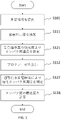
- 1 is a flow chart of a method for assessing the degree of saccharification of proteins, according to one embodiment.
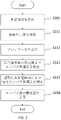
- 1 is a flow chart of a method for assessing the degree of saccharification of proteins, according to one embodiment.
- 4 is a graph showing changes in absorbance over time at multiple albumin concentrations, according to an example.
- 1 is a graph showing a calibration curve of albumin concentration based on absorbance, according to one example.
- 4 is a graph showing the relationship of albumin concentrations in real samples between the method according to an example and the conventional method.
- 4 is a graph showing the effect of the presence or absence of BCP on GA concentration measurement, according to one example.
- 4 is a graph showing changes in absorbance over time at multiple albumin concentrations, according to an example.
- FIG. 4 is a graph showing changes in sensor output over time at multiple albumin concentrations, according to one example.
- FIG. 5 is a graph showing the relationship between GA measurement values obtained by a method according to an example and GA values obtained by a conventional method; 4 is a graph showing changes in absorbance over time at multiple albumin concentrations, according to an example. 5 is a graph showing the relationship between GA values obtained by a method according to an example and GA values obtained by a conventional method;
- 1 is a block diagram of an apparatus for evaluating the degree of saccharification of proteins, according to one embodiment.
- FIG. 1 is a block diagram of an apparatus for evaluating the degree of saccharification of proteins, according to one embodiment.
- FIG. 1 is a block diagram of an apparatus for evaluating the degree of saccharification of proteins, according to one embodiment.
- FIG. 1 is a block diagram of an apparatus for evaluating the degree of saccharification of proteins, according to one embodiment.
- FIG. 1 is a block diagram of an apparatus for evaluating the degree of saccharification of proteins, according to one embodiment.
- FIG. 1 is a graph of absorbance showing the effect of the presence or absence of SDS on a modified BCP method, according to one example.
- the subject may include or be human.
- the subject may include non-human animals and may be non-human animals.
- the subject may include or be a mammal.
- Subjects may be, for example, without limitation, working animals, farm animals, companion animals, and wild animals.
- the sample to be measured may be a solution.
- the “solution” may be a body fluid, a solution derived from the body fluid, or a diluted solution of the body fluid.
- the solution may be a solution that is not a bodily fluid (derived from a non-bodily fluid), or a mixture of a bodily fluid or a bodily fluid-derived solution and a non-bodily fluid-derived solution.
- the solution may be the solution used for sample measurements or the solution used for calibration measurements.
- the solution may be a standard solution or a calibrator solution.
- the solution may be a liquid that does not contain the substance to be measured intentionally or intentionally for use in calibration or the like.
- the sample to be measured may be a specimen.
- the solution may be a solution containing chemicals.
- Body fluid may be lymph fluid, interstitial fluid such as interstitial fluid, intercellular fluid, interstitial fluid, body cavity fluid, serous fluid, pleural fluid, ascites, pericardial fluid, cerebrospinal fluid ( cerebrospinal fluid), synovial fluid (synovial fluid), or aqueous humor (aqueous humor).
- the bodily fluid may be a digestive fluid such as saliva, gastric juice, bile, pancreatic juice, intestinal juice, sweat, tears, runny nose, urine, semen, vaginal fluid, amniotic fluid, milk.
- the bodily fluid may be an animal bodily fluid or a human bodily fluid.
- a "bodily fluid” may be a solution.
- the solution may contain a physiological buffer such as phosphate-buffered saline (PBS) or N-tris(hydroxymethyl)methyl-2-aminoethanesulfonic acid buffer (TES) containing the substance to be measured.
- PBS phosphate-buffered saline
- TES N-tris(hydroxymethyl)methyl-2-aminoethanesulfonic acid buffer
- the solution is not particularly limited as long as it contains the substance to be measured.
- the solution may contain the substance to be measured.
- the solution may possibly contain the substance to be measured.
- the substance to be measured may be a molecule, ion, macromolecule, biomolecule, or the like.
- the substance to be measured may comprise a biomolecule.
- the substance to be measured may be protein, glycated protein, or the like.
- the solution may be tears, and the substance to be measured may be albumin, glycoalbumin, hemoglobin, and/or glycated hemoglobin contained in tears.
- the object to be measured may be albumin, glycoalbumin, hemoglobin, or glycohemoglobin in blood, serum, or plasma, or albumin, glycoalbumin, hemoglobin, or glycohemoglobin in interstitial fluid, urine, or saliva.
- Albumin may be oxidized albumin (HNA) or reduced albumin (HMA).
- HNA oxidized albumin
- HMA reduced albumin
- the substance to be measured may be AGE (Advanced Glycation End Products).
- the substance to be measured may be a glycated lipid.
- the sensor may be a chemical sensor, biosensor, ion sensor, etc. (hereinafter sometimes referred to as “sensor”, “biochemical sensor”, “chemical sensor” or “electrochemical sensor”).
- the sensor may comprise multiple sensors.
- the sensor may have electrodes.
- the electrodes may be amperometric electrodes.
- the electrodes may comprise hydrogen peroxide electrodes.
- the electrodes may comprise oxygen electrodes.
- the electrodes may be potentiometric electrodes.
- the electrodes may be electrodes for ion detection (pH electrode, cyanide ion electrode, iodide ion electrode, etc.).
- the senor may output an electrical signal. In some aspects, the sensor may output a current signal. A sensor may output a voltage signal or an electrical charge. The sensor may be electrically connected to an ammeter, voltmeter, or the like.
- the glycated amino acids produced may be reacted with ketoamine oxidase to generate hydrogen peroxide.
- the amount (concentration) of hydrogen peroxide generated may be measured.
- the amount (concentration) of hydrogen peroxide may be measured electronically.
- ketoamine oxidase generally recognizes the ketoamine structure of a peptide or peptide fragment containing glycated amino acids or glycated amino acid residues and oxidizes the glycated amino acids to produce amino acids, glucosone ( ⁇ -ketoaldehyde) and peroxidation.
- An oxidase that produces hydrogen is proportional to or related to the concentration of the peptide or peptide fragment containing the glycated amino acid or glycated amino acid residue that it recognizes.
- a ketoamine oxidase may be a dehydrogenase, a kinase, or an oxidase.
- the ketoamine oxidase may be fructosyl amino acid oxidase (FAOD), fructosyl peptide oxidase, fructosyl valyl histidine oxidase, fructosyl amine oxidase, amadoriase, fructosylamine deglycase or modified forms thereof.
- FOD fructosyl amino acid oxidase
- FOD fructosyl peptide oxidase
- fructosyl valyl histidine oxidase fructosyl amine oxidase
- amadoriase fructosylamine deglycase or modified forms thereof.
- the ketoamine oxidase may be an oxidase (Group I ketoamine oxidase) that acts on amino acids or peptides whose ⁇ -amino groups are glycated.
- the amino acid or peptide may be valine, glycine, valylhistidine.
- a glycated hemoglobin sensor or a glycated hemoglobin A1c (HbA1c) sensor can be constructed by using an oxidase that acts on an amino acid or peptide having a glycated ⁇ -amino group.
- the ketoamine oxidase may be an oxidase (group II ketoamine oxidase) that acts on amino acids or peptides in which the ⁇ -amino group is glycated.
- the amino acid may be lysine.
- the glycated amino acid may be fructosyl lysine.
- a glycoalbumin sensor can be constructed by using an oxidase that selectively acts on amino acids or peptides having glycated ⁇ -amino groups.
- the ketoamine oxidase may be an oxidase (group III ketoamine oxidase) that acts on amino acids or peptides in which the ⁇ -amino group and/or ⁇ -amino group is glycated.
- the amino acid or peptide may be lysine, valine, glycine, valylhistidine.
- a glycated protein sensor can be constructed by using an oxidase that acts on an amino acid or peptide whose ⁇ -amino group and/or ⁇ -amino group is glycated.
- the senor may include a detector.
- the detector may be a hydrogen peroxide detector.
- the "hydrogen peroxide detector” (hydrogen peroxide sensor) may be an electrochemical electrode or a hydrogen peroxide electrode.
- a hydrogen peroxide electrode may have a counter electrode, a reference electrode, and a working electrode.
- the detector may detect oxygen. For example, the amount or concentration of oxygen that decreases in an enzymatic reaction may be detected.
- Oxygen detection is considered to be relatively insensitive to noise sources such as molecules and ions and tolerant to interference. Oxygen consumption may be measured by oxygen sensing. Since the detector is air saturated, it may be used for enzymatic sensing.
- the detection unit may be configured to be able to selectively or combine a plurality of detection methods.
- a sensor may have an ion exchange resin on the detector.
- the sensor may have an ion exchange resin between the ketoamine oxidase layer and the hydrogen peroxide electrode.
- a cation exchange resin such as Nafion (registered trademark)
- an anion exchange resin such as polypyrrole can be used to suppress or prevent permeation of dopamine and the like, particularly positive ions and the like, from reaching the detection unit.
- the ion exchange resin may contain one, multiple or at least one type of ion exchange resin.
- the ion exchange resin may be configured with one, multiple or at least one type of layer.
- the sensor may comprise a Molecular Imprinted Polymer (MIP) membrane. Changes in charge or potential caused by MIP molecular recognition may be detected (potentiometry type). A change in the current flowing through the electrode (for example, mediator current) caused by MIP molecular recognition may be detected (amperometric type).
- MIP Molecular Imprinted Polymer
- protease is a general term for peptide bond hydrolases that hydrolyze and catabolize proteins and polypeptides.
- a protease may be an enzyme that breaks proteins into peptide fragments.
- the peptide fragments generated by the action of protease may include peptide fragments containing glycosylated amino acid residues and peptide fragments that are not glycosylated at all.
- protease may be an animal-derived protease, a plant-derived protease, or a microorganism-derived protease.
- a protease may be an exopeptidase or an endopeptidase.
- the protease may be an aspartic protease, a metalloprotease, a serine protease, or a thiol protease.
- protease may include multiple types or types of protease, or may include one type or type of protease.
- a protease may contain one or both of an endopeptidase and an exopeptidase. Mixing multiple proteases may increase the efficiency of decomposition.
- Animal-derived proteases include trypsin, chymotrypsin, elastase, bovine pancreatic protease, cathepsin, calpain, protease type-I, protease type-XX, aminopeptidase N, carboxypeptidase, pancreatin (a mixture of multiple enzymes such as proteases and amylases). and so on.
- the plant-derived protease may be papain, bromelain, gingipain, kallikrein, ficin, chymopapain, or the like.
- Microorganism-derived proteases include Bacillus-derived proteases, Aspergillus-derived proteases, Penicillium-derived proteases, Streptomyces-derived proteases, Lysobacter-derived proteases, Yeast-derived proteases, Tritirachium-derived protease, Thermus-derived protease, Pseudomonas-derived protease, Achromobacter-derived protease, and the like may be used.
- the protease may be provided in liquid form or mixed with a solution. In some embodiments, the protease may be added or mixed into the solution in solid form (eg, powder). In some embodiments, the protease may be introduced into solution on a support material. The protease may be supported on a plate, curved surface, sphere, bead, biomolecule such as protein, polymer, or the like.
- optical measurement generally refers to determining optical properties of matter using an optical element or device.
- optical measurements of the substance of interest may be measured.
- a substance that is bound to or related to a target substance hereinafter referred to as a substance (e.g., reagent) that is chemically, biologically or physically bound to or related to a target substance, even if it is not the target substance itself) Properties may be measured. Properties of reagents may be measured.
- the reagent may be referred to as a "target substance”.
- optical measurements may include spectroscopic measurements.
- the absorbance of the target substance may be measured.
- a color changing indicator corresponding to the substance of interest may be introduced. A color changing indicator may be detected or measured.
- color changing indicator generally refers to halochromic chemical compounds. Color changes may generally be reversible. Color changing indicators include, for example, without limitation, acid-base indicators (pH indicators), redox indicators, adsorption indicators, TLC color changing indicators, and the like. It changes the color of its solution depending on the pH.
- the pH indicator may be, for example, without limitation, bromocresol purple (BCP), bromocresol green (BCG), and the like.
- BCP or BCG can be used for albumin.
- albumin combines with bromocresol green (BCG) to produce an albumin-bromocresol green complex. Around pH 4, the complex appears blue. Therefore, the albumin concentration can be obtained by measuring the absorbance (BCG method).
- BCG method bromocresol green
- a surfactant may be added to the measurement reagent. Surfactant may not be added to the measurement reagent. For example, protein denaturants (such as SDS) may not be added.
- albumin binds with bromocresol purple (BCP) to generate an albumin-bromocresol purple complex.
- BCP bromocresol purple
- This complex has a blue color. Therefore, the albumin concentration can be obtained by measuring the absorbance (BCP method).
- the albumin concentration can be measured by a color reaction with BCP after converting all of the reduced albumin into oxidized albumin by the action of an oxidizing agent (improved BCP method).
- a surfactant may be added to the measurement reagent.
- protein denaturants such as SDS
- the reaction in which albumin combines with bromocresol purple (BCP) or bromocresol green (BCG) to form a conjugate may be referred to as the "first reaction.”
- the color changing indicator may be provided in liquid form or mixed with a solution. In some embodiments, the color changing indicator may be added or mixed into the solution in solid form (eg, powder).
- an absorption photometer (colorimeter) may be used.
- the absorption photometer has a light source and a light receiver. All or part of the light emitted by the light source enters the target solution. All or part of the light that passes through the solution enters the receiver. The light absorbed by the target substance can be analyzed.
- the wavelength of the light source may be appropriately selected according to the color indicator used.
- Absorbance may be measured, for example, at wavelengths such as dominant wavelength 600-630 nm for BCP and 565-660 nm for BCG.
- the change in absorbance over time of the color indicator may be measured.
- absorbance may be measured at least two (or more) times.
- the time at which protease decomposition starts is measured, and the protein concentration may be determined based on the measured value corresponding to the time from this start time.
- protein concentration may be determined based on measurements at three or more time points. From at least three or more measurements, it may be possible to determine the initial protein concentration even if the time from the start of protease degradation is not clear.
- the measurement of glycated protein may be started after a predetermined or predetermined time has elapsed from the start time of protease degradation.
- glycated protein measurement may begin when the absorbance A is sufficiently small.
- the change in absorbance over time ( ⁇ A/ ⁇ t) becomes substantially zero, it can be determined that sufficient protease degradation has been completed and measurement of glycated proteins can be started, or The measurement result of glycated protein at that time may be adopted.
- the change in absorbance over time reaches a predetermined value, it may be determined that degradation by a predetermined protease has been achieved, and the measurement of glycated protein may be started, or the glycated protein at that time may be measured. Measurement results may be used.
- calibration of absorbance measurements may be performed.
- An optical system including optical measurement cells, light emitters, light receivers, etc. may be calibrated.
- Absorbance may be measured before introduction of the solution into the optical system (e.g., the state in which the optical measurement cell is empty, its initial state, etc.) and after (e.g., the state in which the measurement solution has been introduced into the optical measurement cell).
- Absorbance may be measured before introduction of the solution into the optical system (e.g., the state in which the optical measurement cell is empty, its initial state, etc.) and after (e.g., the state in which the measurement solution has been introduced into the optical measurement cell).
- a difference in absorbance between them may be obtained.
- a method of measuring a protein comprising: providing a solution that may contain a protein of interest; adding a color changing indicator to the solution; and adding a protease.
- the method can be used to measure glycated proteins.
- the application of the method is not so limited. The method can be used in various other methods.
- FIG. 1 shows a flowchart of a method for assessing glycation degree of albumin, according to an embodiment.
- a solution that may contain albumin as a protein to be measured is provided.
- BCP is added as a color indicator.
- the absorbance of BCP is measured. Determine the concentration of albumin from the absorbance.
- protease is added to the measurement solution to which BCP has already been added.
- Albumin begins to be broken down into peptides or amino acids by proteases.
- step S122 hydrogen peroxide is generated by causing ketoamine oxidase, which specifically reacts with saccharified amino acids among the peptides or amino acids produced by the digestion reaction with the protease, to act.
- the hydrogen peroxide is measured using electrodes. This gives the concentration of glycated albumin (GA) in the solution.
- step S131 the ratio of the obtained glycated albumin (GA) to the albumin concentration is obtained as the GA value.
- a color indicator is added (S111), absorbance is measured (S121), and then protease is added (S112).
- the absorbance measurement may be completed by the time the protease is added.
- the protease may be added before completing the absorbance measurement or during the absorbance measurement.
- FIG. 2 shows a flowchart of a method for assessing glycation degree of albumin, according to an embodiment.
- a solution that may contain albumin as a protein to be measured is provided.
- BCP is added as a color indicator.
- protease is added to the measurement solution to which BCP has already been added. Degradation of albumin into amino acids by proteases is initiated.
- the absorbance of BCP is measured. Determine the concentration of albumin from the absorbance. Absorbance decreases with time due to ongoing decomposition of albumin.
- the concentration of albumin contained in the original measurement solution can be obtained by measuring the absorbance at the time when the protease is introduced or the start time of the decomposition and after a predetermined time from the start time.
- step S222 hydrogen peroxide is generated by causing ketoamine oxidase, which specifically reacts with saccharified amino acids among the peptides or amino acids produced by the digestion reaction with the protease, to act.
- the hydrogen peroxide is measured using electrodes.
- step S231 the ratio of the obtained glycated albumin (GA) to the albumin concentration is obtained as the GA value.
- a color indicator is added (S211), and then protease is added (S212).
- the order of steps is not limited to this.
- the addition of the color changing indicator (S211) and the addition of the protease (S212) may occur substantially simultaneously.
- the protease and the color indicator may be mixed in advance, and the mixed solution may be added to the measurement solution.
- the glycated albumin concentration is measured (S222).
- measuring albumin concentration (S221) and measuring glycated albumin concentration (S222) may be performed substantially simultaneously.
- a color changing indicator is added (S211) and a protease is added (S212), after which the solution with both added may be diverted.
- the concentration of albumin may be measured in one of the diverted solutions, and the concentration of glycated albumin in the other.
- the protease and color indicator may not be added separately.
- the protease and the color indicator may be mixed in advance, and the mixed solution may be added to the measurement solution.
- a color-changing indicator is brought into contact with a solution that may contain the target protein, and then the first reaction between the color-changing indicator and the target protein is used to determine the concentration of the target protein in the solution.
- the protease is contacted with a solution that may contain the protein of interest, and then a second reaction between the protease and the protein of interest is used to determine the concentration of the glycated protein of interest (the concentration of the glycated protein of interest concentration, also called concentration of glycated protein).
- the second reaction may comprise reacting the produced glycated amino acid with ketoamine oxidase to generate hydrogen peroxide.
- the order of the four items of introduction of the color-changing indicator, measurement of the first reaction between the color-changing indicator and the target protein, introduction of the protease, and measurement of the second reaction between the protease and the target protein is Not limited.
- protein is measured for the indicator mixed solution after mixing the measurement solution and the indicator, and glycated protein is measured for the protease mixed solution after mixing the measurement solution and the protease.
- the order of steps may not be limited except for the order of performing.
- the protease may be mixed into the indicator mixed solution.
- the measurement solution and the indicator are mixed, the protein is measured using the indicator mixed solution, the protease is mixed with the indicator mixed solution, and the indicator protease mixed solution (the indicator and the protease are mixed solution) may be used to measure glycated protein.
- a measurement solution and an indicator are mixed, a protease is mixed in the indicator mixed solution, and the glycated protein is measured using the indicator protease mixed solution (a solution in which the indicator and the protease are mixed), Proteins may be measured using the protease mixed solution.
- a measurement solution and an indicator are mixed, a protease is mixed in the indicator mixed solution, and the glycated protein is measured using the indicator protease mixed solution (a solution in which the indicator and the protease are mixed), Measurement of protein using the indicator-protease mixed solution may be performed in that order.
- an indicator may be mixed with the protease mixed solution after mixing the measurement solution and the protease.
- a measurement solution and protease are mixed, glycated protein is measured using the protease mixed solution, an indicator is mixed with the protease mixed solution, and protein is measured using the protease indicator mixed solution. , in that order.
- a measurement solution and a protease are mixed, an indicator is mixed with the protease mixed solution, glycated protein is measured using the indicator protease mixed solution, and protein is measured using the indicator protease mixed solution. You can do things in that order.
- a measurement solution and a protease are mixed, an indicator is mixed with the protease mixed solution, protein is measured using the indicator protease mixed solution, and glycated protein is measured using the indicator protease mixed solution.
- the indicator and the protease may be mixed substantially simultaneously with the measurement solution.
- the glycation degree of the protein of interest may be determined based on the concentration of the protein of interest and the concentration of the glycated protein.
- the degree of saccharification of protein may be obtained by dividing the concentration of glycated protein by the concentration of protein.
- the quantity to be measured is not limited to concentration.
- the quantity measured may be an absolute quantity, a relative quantity, a concentration, or any other quantity related to mass or concentration.
- the concentration of protein and the concentration of glycated protein can be measured after addition of a color changing indicator and addition of protease.
- the color changing indicator and/or protease is added in liquid form, changing the volume from the original solution.
- the variability caused by errors in the dosage of color indicator and/or protease occurs at the same rate for both protein concentration and glycated protein concentration. Therefore, it does not substantially affect the calculation of the saccharification degree. Therefore, relatively accurate and reproducible measurements are possible.
- measurement of protein concentration, addition of solid protease, and measurement of glycated protein concentration can be performed after addition of a color indicator.
- variability caused by errors in input amounts of color indicator and/or protease occurs in the same proportions for both protein concentration and glycated protein. Therefore, it does not substantially affect the calculation of the saccharification degree. Therefore, relatively accurate and reproducible measurements are possible.
- the pH may be adjusted.
- the pH may be changed during the addition or reaction of the color indicator (first reaction) and during the decomposition by the protease (second reaction).
- the first reaction and the second reaction may be performed at substantially the same pH.
- the pH value may differ from the general optimum range.
- the first reaction may be followed by the second reaction.
- the pH when the first and second reactions are (at least partially) performed simultaneously, one pH is applied to both reactions.
- the value may be adjusted to give a suitable range for both.
- the pH value may differ from the general optimum range.
- the temperature may be regulated.
- the temperature may be changed between the addition or reaction of the color indicator (first reaction) and the decomposition by protease (second reaction).
- first reaction the temperature was adjusted to 20° C. to 70° C., which is its general optimum temperature. , or the temperature may be adjusted to 55-65°C.
- second reaction the temperature may be once raised to the temperature for protease decomposition before introducing the protease.
- the temperature may be adjusted to 25°C to 75°C, 30°C to 65°C, 35°C to 65°C, around 37°C, or around 55°C. .
- one temperature is applied to both reactions.
- the value may be adjusted to give a suitable range for both.
- the temperature may be adjusted in the range of 25° C. to 50° C., 25° C. to 45° C., or 30° C. to 45° C. for both the first reaction and the second reaction.
- the temperature may be between 25°C and 70°C, between 30°C and 70°C, between 40°C and 70°C, between 50°C and 70°C, or between 55°C and 65°C. You can adjust the range.
- albumin concentration was measured using an absorbance method.
- a commercially available glycoalbumin measurement kit (Lucica (registered trademark) GA-L) calibrator (GA-L calibrator, Asahi Kasei Pharma Corp.) was used, and serial dilutions (7.93 mg/mL, 3 .97 mg/mL, 1.98 mg/mL) were prepared.
- a solution containing only PBS (blank PBS) was also prepared as a blank sample.
- As a protease solution 320 mg of Orientase 22BF (HBI Co., Ltd.) was dissolved in 4 mL of HEPES buffer.
- a BCP solution (0.02 mM, pH 8.0) was used for the color reaction.
- the absorbance was measured over time at intervals of 12 seconds using a diabetes test item automatic analyzer DM-JACK Ex (Hitachi Chemical Diagnostic Systems Co., Ltd.).
- DM-JACK Ex Hagaku Chemical Diagnostic Systems Co., Ltd.
- 180 ⁇ L of BCP solution was added to 18 ⁇ L of sample solution.
- 18 ⁇ L of protease solution was added.
- ⁇ A when it shows a constant value corresponds to the concentration of albumin.
- the concentration of albumin may be determined based on absorbance in a solution without added protease.
- the concentration of albumin may be determined based on the absorbance value (A or ⁇ A) at a predetermined time after protease addition. For example, an endpoint method may be used. In some embodiments, the concentration of albumin may be determined based on the time conversion rate of absorbance (dA/dt, or d( ⁇ A)/dt). For example, a rate assay may be used. In some embodiments, albumin concentration may be determined based on higher order derivatives of absorbance (such as d2A/dt2, or d2( ⁇ A)/dt2).
- the concentration of albumin may be determined based on the absorbance ( ⁇ A) after sufficient time has passed.
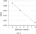
- Fig. 4 shows a calibration curve obtained from the relationship between the concentration of albumin in the sample solution and dA/dt. It became clear that a very strong negative correlation is shown.
- Example 2 Measurement of saccharified albumin concentration> As an example, glycated albumin concentration was measured using the enzymatic electrode method.
- glucose was added to HSA (Fujifilm Wako Pure Chemical Industries, Ltd.) and incubated at 37°C to prepare GA solutions (40 mg/mL) with different saccharification rates.
- the saccharification rate of each sample was confirmed using a commercially available glycoalbumin measurement kit (Lucica (registered trademark) GA-L, Asahi Kasei Pharma Corp.).
- a protease solution was prepared by dissolving 2 mg of Amano K (Amano Enzyme Co., Ltd.) in 200 ⁇ L of TES buffer. 8 ⁇ L of BCP solution (1 mM) was added to 20 ⁇ L of protease solution. The solution was heat treated and stored at 4°C.
- This sample was added to an electrochemical sensor with a hydrogen peroxide electrode on which a ketoamine oxidase membrane was fixed.
- the glycated albumin concentration was determined by measuring the output current.
- Fig. 6 shows the relationship between glycated albumin concentration and enzyme sensor output in the presence or absence of BCP.
- the horizontal axis indicates the glycated albumin concentration in the solution, and the vertical axis indicates the measured current value.
- white circles indicate measurements "without BCP”
- black circles indicate measurements "with BCP”.
- Example 3 shows an example of GA value measurement.
- BCP is added to a sample containing glycated albumin, albumin concentration is measured by absorbance, protease is added to the solution, and glycated albumin is measured using an electrochemical sensor having a hydrogen peroxide electrode to which an FAOD membrane is fixed. Densitometry was performed. More specific description will be given below.
- FIG. 7 shows an example of measurement results used to generate a calibration curve for albumin concentration by absorbance measurement. Samples containing 1.3 mg/mL, 2.4 mg/mL, and 4.7 mg/mL albumin were used for absorbance measurements. At 0 seconds in FIG. 7, BCP solution (including Sodium Dodecyl Sulfate (SDS), TritonX-100, 5,5′-Dithiobis (2-nitrobenzoic acid (DTNB), HEPES (pH 8.0), NaCl) was optically measured.
- SDS Sodium Dodecyl Sulfate
- TritonX-100 TritonX-100
- DTNB 5,5′-Dithiobis (2-nitrobenzoic acid
- HEPES pH 8.0
- the albumin sample was added to the BCP solution.Therefore, the absorbance decreased.Then, the solution was mixed by pipetting.The spikes and fluctuations visible until about 40 seconds were It was caused by the movement of the liquid due to mixing and the blockage of the light path by the pipette.After about 50 seconds, the absorbance of the albumin sample was all constant.As is clear from Figure 7, the absorbance showed albumin concentration dependence. , the relationship between albumin concentration and absorbance was determined, and a calibration curve was generated (not shown).
- Fig. 8 shows an example of the measurement results used to generate the calibration curve of glycated albumin concentration by electrochemical sensor measurement.
- Samples containing 0.4 mg/mL, 0.7 mg/mL, and 1.5 mg/mL glycated albumin were used for the measurement. These solutions contain BCP.
- Protease was added to the solution after absorbance measurement, and a digestion reaction was performed at 55° C. for 30 minutes. After that, at 0 seconds in FIG. 8, the solution after the digestion reaction was introduced into the electrochemical sensor.
- the output current of the electrochemical sensor was measured. It was found that any concentration takes a maximum value (inflection point) at about 60 to 80 seconds. At this time, for example, the relationship between the output current at 60 seconds and the saccharified albumin concentration can be obtained. Using such data, the relationship between the saccharified albumin concentration and the output current of the electrochemical sensor was determined, and the calibration curve was generated.
- the GA value of an unknown sample can be determined.
- ⁇ Measurement of GA value of unknown sample> Next, using the same method as above, the GA value of the unknown sample was measured. As measurement samples, a high-concentration GA value sample and a low-concentration GA value sample were mixed at several ratios to prepare a plurality of measurement solutions. A human serum albumin reagent (Sigma-Aldrich) was used for low-concentration GA value samples. A high-concentration GA value sample was prepared by adding glucose to a human serum albumin reagent (Sigma-Aldrich) and incubating. Although these are artificially prepared samples, the actual GA values are unknown.
- the measurement method of this example was applied to these measurement samples to obtain the GA value.
- the GA value (%) was determined for the same measurement sample using a commercially available glycoalbumin measurement kit (Lucica (registered trademark) GA-L) as an existing method.
- Figure 9 shows both measurements obtained for samples of four different unknown concentrations.
- the horizontal axis indicates the GA value measured by Lucica, and the vertical axis indicates the GA value (arbitrary unit; GA value (%) is not shown) obtained by the measurement method of this example.
- GA value can be obtained very satisfactorily by using one of the methods of the present disclosure.
- Example 4 shows an example of GA value (%) measurement.
- BCP and protease mixed solution is added to a sample containing glycated albumin, and then optical measurement is performed to measure the albumin concentration, while using an electrochemical sensor having a hydrogen peroxide electrode to which an FAOD membrane is fixed. Glycated albumin concentration was measured. More specific description will be given below.
- FIG. 10 shows an example of measurement results used to generate a calibration curve for albumin concentration by absorbance measurement.
- Samples containing 1.2 mg/mL, 2.5 mg/mL, and 5.0 mg/mL albumin were used for the measurements.
- BCP and protease mixture SDS, Triton X-100, DTNB, HEPES (pH 8.0), NaCl, protease added
- SDS Triton X-100, DTNB, HEPES (pH 8.0), NaCl, protease added
- values related to absorbance such as the absorbance at 20 seconds, the difference in absorbance at 20 seconds and later time points (e.g., the difference in absorbance at 90 seconds, the change in absorbance over time (time derivative), etc., are derived from the initial albumin This data was used to determine the relationship between albumin concentration and absorbance and generate a calibration curve for it.
- the measurement method of this example was applied to these measurement samples to obtain the GA value (%).
- the GA value (%) was determined for the same measurement sample using a commercially available glycoalbumin measurement kit (Lucica (registered trademark) GA-L) as an existing method.
- Figure 11 shows the relationship between both measurements obtained for samples of five different unknown concentrations.
- the horizontal axis indicates the GA value (%) obtained by the existing measurement method, and the vertical axis indicates the GA value (%) obtained by the measurement method of this embodiment.
- the GA value (%) can be obtained very satisfactorily by using one of the methods of the present disclosure.
- Albumin measurement by the improved BCP method is generally performed under acidic conditions using an oxidizing agent and SDS. Protein denaturants such as SDS are believed to facilitate oxidation of albumin upon introduction of an oxidizing agent such as DTNB. In that state, BCP is introduced.
- the protease reaction as the second reaction generally requires weak alkalinity.
- the first reaction In order to carry out the first reaction and the second reaction simultaneously, continuously or serially, it is preferred that the first reaction also be carried out in the same alkaline environment. No need to change pH.
- the solution does not contain SDS in the modified BCP process which is carried out in mild alkalinity.
- reaction solution containing SDS was adjusted to a concentration of 0.02% SDS.
- concentration of 0.02% SDS was adjusted to a concentration of 0.02% SDS.
- FIG. 16 shows the presence of SDS (SDS+, solid) and no SDS (SDS-, open) for three types of buffer solutions, succinic acid solution, HEPES solution (HEPES, circle), and Tricine solution (Tricine, triangle). , shows the absorbance.
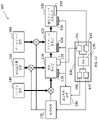
- FIG. 12 schematically shows the configuration of an apparatus 100 according to one embodiment.
- the device 100 comprises an optical measurement section 110 , a protease reaction section 120 and a glycated protein measurement section 130 .
- the sample solution is introduced from the introduction unit 140 into the optical measurement section 110 and then flowed through the protease reaction section 120 and the glycated protein measurement section 130 in sequence.
- a color changing indicator tank 150 is connected to the optical measurement section 110 and is configured to provide a color changing indicator to the optical measurement section 110 .
- a protease solution tank 160 is connected to the protease reaction section 120 and configured to provide the protease reaction section 120 with a protease solution.
- the solution introduction and liquid transfer are controlled by the liquid transfer mechanism 170 .
- the liquid sending mechanism 170 can control the timing and amount of introduction of the sample solution into the optical measurement section 110 .
- the liquid feeding mechanism 170 controls the timing and amount of introduction of the color indicator solution from the color indicator solution tank 150 to the optical measurement section 110, and the introduction of the protease solution from the protease solution tank 160 to the protease reaction section 120. You can control the timing and amount of
- the liquid feeding mechanism 170 may include, for example, a pump.
- the optical measurement section 110, the protease reaction section 120, and the glycated protein measurement section 130 are closed channels, positive pressure can be applied to the upstream side to control the movement of the solution.
- the liquid feeding mechanism 170 may be a pressing mechanism attached to each solution tank. When each solution tank is a deformable container, a predetermined amount of solution can be delivered to the outside by deforming the container.
- Temperature control units 118, 128, 138 having temperature sensors and heaters (not shown) are arranged in the optical measurement section 110, the protease reaction section 120, and the glycated protein measurement section 130, respectively.
- the temperature control units 118, 128, 138 are connected to a temperature control mechanism (driver) 180, which receives measured temperature information from the temperature sensors and can send on/off instructions and power to the heaters.
- Light emitted from the light-emitting element passes through the solution in the optical measurement unit 110, and the light-receiving element senses the emitted light.
- the optical measurement unit 119 can output absorbance information at that time.
- a sensor 139 is arranged in the glycated protein measurement unit 130 .
- the sensor 139 is arranged inside the glycated protein measuring unit 130 and can directly contact the internal solution. This sensor 139 is configured to measure the concentration of glycated proteins.
- the device 100 further comprises a computer system 190.
- Computer system 190 includes at least a central processing unit (CPU) 195, which performs the necessary computations.
- Computer system 190 has communication interfaces 197 and 198 for liquid transfer mechanism 170 and temperature control mechanism 180, respectively.
- Computer system 190 has communication interfaces 191 and 193 with optical measurement unit 119 and sensor 139, respectively.
- the CPU 195 transmits necessary information such as required operation items (solution to be introduced and component to be introduced), its timing, and its amount to the liquid transfer mechanism 170 via the liquid transfer mechanism communication interface 197. send.
- Liquid delivery mechanism 170 performs the necessary operations based on the information received.
- the liquid transfer mechanism 170 can transmit information such as information indicating that each operation has been executed and information such as execution failure to the communication interface 197 .
- the CPU 195 provides the temperature control mechanism 180 with necessary operation items (measurement of temperature, temperature required for each reaction, etc.), their timing, their temperature and time, etc. to the temperature control mechanism 180 via the temperature control mechanism communication interface 198. send information. Temperature control mechanism 180 performs the necessary operations based on the information received. The temperature control mechanism 180 can send information to the communication interface 198 such as information that each operation has been executed, information such as execution failure.
- the optical measurement unit communication interface 191 receives information about absorbance from the optical measurement unit 119 arranged in the optical measurement section 110 .
- the glycated protein sensor communication interface 193 receives information about the glycated protein concentration from the sensor 139 arranged in the glycated protein measurement unit 130 .
- the optical measurement unit communication interface 191 issues instructions such as the start and end of absorbance measurement to the optical measurement unit 119 arranged in the optical measurement section 110, and then receives information on the protein concentration obtained by the measurement.
- the glycated protein sensor communication interface 193 issues instructions such as start and end of current measurement to the sensor 139 arranged in the glycated protein measurement unit 130, and then receives information on the glycated protein concentration obtained by the measurement. .
- the CPU 195 receives information related to absorbance and information related to glycated protein via communication interfaces 191 and 193, performs predetermined conversion, and calculates the degree of saccharification.
- FIG. 13 schematically shows the configuration of an apparatus 200 according to one embodiment.
- the device 200 comprises an optical measurement section 210 and a glycated protein measurement section 230 .
- the sample solution is introduced from the introduction unit 240 into the optical measurement section 210 and flowed to the glycated protein measurement section 230 .
- a measuring liquid (color changing indicator protease solution) tank 250 is connected to the optical measuring section 210 and is configured to provide the optical measuring section 210 with a mixture of color changing indicator and protease.
- the solution introduction and liquid transfer are controlled by the liquid transfer mechanism 270 .
- Temperature control units 218 and 238 having temperature sensors and heaters (not shown) are arranged in the optical measurement section 210 and glycated protein measurement section 230, respectively.
- An optical measurement unit 219 is arranged in the optical measurement section 210 and can measure absorbance and output information about the measured absorbance.
- a sensor 239 is arranged in the glycated protein measurement unit 230 and is configured to measure the concentration of the glycated protein and output information on the measured glycated protein.
- the device 200 further comprises a computer system 290.
- Computer system 290 includes at least a central processing unit (CPU) 295, which performs the necessary computations.
- Computer system 290 can communicate with liquid delivery mechanism 270 and temperature control mechanism 280 via communication interfaces 297 and 298 .
- Computer system 290 can communicate with optical measurement unit 219 and sensor 239 via communication interfaces 291 and 293 .
- the CPU 295 receives information related to absorbance and information related to glycated protein via communication interfaces 291 and 293, performs predetermined conversion, and calculates the degree of saccharification.
- FIG. 14 schematically shows the configuration of an apparatus 300 according to one embodiment.
- the device 300 comprises a color indicator reaction section 305 , an optical measurement section 310 , a protease reaction section 320 and a glycated protein measurement section 330 .
- the sample solution is introduced from the introduction unit 340 into the color indicator reaction section 305 and then passed through the optical measurement section 310 , the protease reaction section 320 and the glycated protein measurement section 330 in sequence.
- the color-changing indicator reaction unit 305 contains a color-changing indicator in advance.
- the solution introduced into the color-changing indicator reaction section 305 is mixed with the color-changing indicator and reacts with it.
- the protease reaction section 320 contains a protease in advance. The solution introduced into the protease reaction section 320 contacts and reacts with the protease.
- the solution introduction and liquid transfer are controlled by the liquid transfer mechanism 370 .
- Temperature control units 318, 328, and 338 having temperature sensors and heaters (not shown) are arranged in the optical measurement section 310, the protease reaction section 320, and the glycated protein measurement section 330, respectively.
- An optical measurement unit 319 is arranged in the optical measurement section 310 and can measure absorbance and output information about the measured absorbance.
- a sensor 339 is arranged in the glycated protein measurement unit 330 and configured to measure the concentration of the glycated protein and output information about the measured glycated protein.
- the device 300 further comprises a computer system 390.
- Computer system 390 includes at least a central processing unit (CPU) 395, which performs the necessary computations.
- Computer system 390 can communicate with liquid delivery mechanism 370 and temperature control mechanism 380 via communication interfaces 397 and 398 .
- Computer system 390 can communicate with optical measurement unit 319 and sensor 339 via communication interfaces 391 and 393 .
- the CPU 395 receives information related to absorbance and information related to glycated protein via communication interfaces 391 and 393, performs predetermined conversion, and calculates the degree of saccharification.
- FIG. 15 schematically shows the configuration of an apparatus 400 according to one embodiment.
- the device 400 comprises a color indicator protease solution reaction section 405 , an optical measurement section 410 and a glycated protein measurement section 430 .
- the sample solution is introduced from the introduction unit 440 into the color indicator reaction section 405 and then passed through the optical measurement section 410 and the glycated protein measurement section 430 in sequence.
- the color-changing indicator protease solution reaction unit 405 contains a color-changing indicator and protease in advance.
- the solution introduced into the color-changing indicator protease solution reaction unit 405 is mixed with and reacting with the color-changing indicator, and also contacts and reacts with the protease.
- the solution introduction and liquid transfer are controlled by the liquid transfer mechanism 470 .
- Temperature control units 418 and 438 having temperature sensors and heaters (not shown) are arranged in the optical measurement section 410 and the glycated protein measurement section 430 .
- An optical measurement unit 419 is arranged in the optical measurement section 410 and can measure absorbance and output information about the measured absorbance.
- a sensor 439 is arranged in the glycated protein measurement unit 430 and is configured to measure the concentration of the glycated protein and output information about the measured glycated protein.
- the device 400 further comprises a computer system 490.
- Computer system 490 includes at least a central processing unit (CPU) 495, which performs the necessary computations.
- Computer system 490 can communicate with liquid delivery mechanism 470 and temperature control mechanism 480 via communication interfaces 497 and 498 .
- Computer system 490 can communicate with optical measurement unit 419 and sensor 439 via communication interfaces 491 and 493 .
- the CPU 495 receives information related to absorbance and information related to glycated protein via communication interfaces 491 and 493, performs predetermined conversion, and calculates the degree of saccharification.
- the device or system may be an automated pipettor or system (sometimes simply referred to as an automated pipettor).
- the automated pipetting device may have wells containing reagents or mixtures of at least two of them.
- a cartridge (microplate) with multiple wells may be provided.
- the device or cartridge may include an electrochemical sensor or may be configured to have an electrochemical sensor attached. Samples and solutions may be quantified, transferred from one well to another, or mixed by pipetting.
- the automated pipetting device may include a heater, and the heater may control the temperature of the wells or solutions within the wells.
- the automated pipetting device may be equipped with an optical measurement system.
- the device may include a waste tank.
- the solution that has passed through the optical measurement section, the glycated protein measurement section, and the like may be collected in a waste liquid tank. This can reduce the risk of a solution containing body fluid leaking to the outside.
- a method for evaluating the degree of saccharification of a protein comprising: providing a solution that may contain the protein of interest; contacting the solution with a color changing indicator; contacting the solution with a protease; determining the concentration of the protein of interest in the solution using a first reaction between the color-changing indicator and the protein of interest; determining the concentration of the glycated protein of interest using a second reaction between the protease and the protein of interest; determining the glycation degree of the target protein based on the concentration of the target protein and the concentration of the glycated target protein; How to prepare.
- A011 The method of embodiment A001, wherein contacting the solution with a protease comprises adding the protease to a color changing indicator mixture produced by contacting the solution with the color changing indicator; Method.
- A012 The method of embodiment A001, wherein contacting the solution with a color-changing indicator and contacting the solution with a protease are performed substantially simultaneously; Method.
- A012b The method of embodiment A001, wherein substantially simultaneously mixing the color changing indicator and the protease with the solution; Method.
- A013 The method of embodiment A001, wherein contacting the solution with a color changing indicator comprises adding the color changing indicator to a protease mixture produced by contacting the solution with the protease; Method.
- A021 The method of any one of embodiments A001 through A013, wherein measuring the first reaction between the color-changing indicator and the protein of interest comprises measuring the absorbance of the color-changing indicator; Method. A022 The method of embodiment A021, wherein Measuring the absorbance of the color indicator comprises measuring a time change of the absorbance; Method. A023 The method of embodiment A022, wherein Measuring the change in absorbance over time includes measuring the absorbance at least two (or more) times, Method. A024 The method of embodiment A022 or A023, wherein determining the concentration of the glycated target protein based on the time change of the absorbance; Method. A031 The method of any one of embodiments A001-A024, wherein the protein of interest is albumin.
- A032 The method of embodiment A031, wherein Determining the concentration of glycated albumin using the second reaction between the protease and the albumin includes: degrading the albumin with the protease to produce peptides, and reacting the produced peptides with ketoamine oxidase to produce hydrogen peroxide; measuring the concentration of the hydrogen peroxide produced; determining the concentration of the glycated albumin based on the concentration of the hydrogen peroxide; comprising a Method.
- A033 The method of embodiment A032, wherein measuring the concentration of hydrogen peroxide comprises using a hydrogen peroxide electrode; Method.
- A034 The method of embodiment A033, wherein measuring the concentration of the hydrogen peroxide includes contacting the hydrogen peroxide that has passed through an ion exchange resin with the hydrogen peroxide electrode; Method.
- A041 The method of any one of embodiments A001 through A034, wherein wherein the color changing indicator is a pH indicator; Method.
- A042 The method of any one of embodiments A031 through A034, wherein The color changing indicator is bromocresol purple (BCP) or bromocresol green (BCG). Method.
- A051 The method of embodiment A042, wherein The color changing indicator is bromocresol purple (BCP) or bromocresol green (BCG), adjusting the pH of the solution to a range of 4.5 to 8.5 during the first reaction and adjusting the pH of the solution to a range of 6.0 to 9.0 during the second reaction; and the method further comprising: A052 The method of any one of embodiments A001 through A042, wherein When the first reaction between the color changing indicator and the protein of interest and the second reaction between the color changing indicator and the protein of interest occur (at least partially) simultaneously, the pH of the solution is adjusted to a range suitable for both the first reaction and the second reaction; Method.
- BCP bromocresol purple
- BCG bromocresol green
- A053 The method of embodiment A052, wherein The color changing indicator is bromocresol purple (BCP) or bromocresol green (BCG), the pH is adjusted to the range of 7.0 to 8.5; Method.
- A054 The method of embodiment A051 or A053, wherein the pH is adjusted to a range of 7.0 to 8.5; During the first reaction, the solution mixture does not contain a protein denaturant (SDS); Method.
- A055 The method of any one of embodiments A032-A054, wherein the color changing indicator is bromocresol purple (BCP) or bromocresol green (BCG), adjusting the temperature of the solution in the range of 20° C. to 70° C.
- a method further comprising: A056 The method of any one of embodiments A001 through A054, wherein When the first reaction between the color changing indicator and the protein of interest and the second reaction between the color changing indicator and the protein of interest occur (at least partially) simultaneously, the temperature of the solution is adjusted to a range suitable for both the first reaction and the second reaction; Method. A057 The method of embodiment A056, wherein the temperature is adjusted in the range of 25°C to 70°C; Method.
- A058 The method of any one of embodiments A032-A034, wherein The method further comprising adjusting the temperature of said ketoamine oxidase to a range of 25°C to 75°C.
- A061 The method of any one of embodiments A032-A058, wherein The ketoamine oxidase is an oxidase (group I ketoamine oxidase) that acts on amino acids or peptides in which the ⁇ -amino group is glycated, and measuring the concentration of the generated hydrogen peroxide is fructosylvaline , calibrating the measurement using a standard solution containing at least one of fructosylglycine and fructosylvalylhistidine; Method.
- ketoamine oxidase is an oxidase (group II ketoamine oxidase) that acts on amino acids or peptides in which the ⁇ -amino group has been glycated, and measuring the concentration of the produced hydrogen peroxide is a way of measuring fructosyl lysine. calibrating the measurement using a reference solution comprising Method.
- ketoamine oxidase is an oxidase (group III ketoamine oxidase) that acts on amino acids or peptides having glycated ⁇ -amino groups and/or ⁇ -amino groups
- Measuring the concentration of the produced hydrogen peroxide includes calibrating the measurement using a standard solution containing at least one of fructosyl lysine, fructosyl valine, fructosyl glycine and fructosyl valyl histidine. be prepared to Method.
- B01 A program for causing a computer system to perform the method of any one of embodiments A001 through A063.
- C01 A device for evaluating the degree of saccharification of proteins, a bodily fluid introduction unit (or inlet); a mechanism for mixing the bodily fluid and a color indicator solution; a protein concentration measurement unit including an optical measurement unit that measures the concentration of protein in a mixture of the body fluid and the color indicator solution; a mechanism for mixing the bodily fluid and a protease solution; a glycated protein measuring unit comprising an electrode unit (sensor) for measuring the concentration of glycated protein in a mixture of the body fluid and the protease; an arithmetic processing unit that communicates with the albumin concentration measurement unit and the glycated protein measurement unit; A device comprising C02 The apparatus of embodiment C01, wherein: mixing the bodily fluid and the protease solution includes mixing the protease solution with a mixture of the bodily fluid and the color indicator solution; Device.
- C11 The apparatus of embodiment C01 or C02, wherein The protein concentration measurement unit and the glycated protein measurement unit are fluidly connected in series, Device.
- C12 The apparatus of embodiment C11, wherein: The bodily fluid introduction unit, the protein concentration measurement unit, and the glycated protein measurement unit are fluidly connected in series in that order, Device.
- C13 The apparatus of embodiment C01 or C02, wherein the protein concentration measurement unit and the glycated protein measurement unit are connected in parallel to each other and to the introduction unit; Device.
- C21 The apparatus of any one of embodiments C01 through C13, wherein: The apparatus further comprises a waste liquid section (tank) fluidly connected downstream of the protein concentration measurement section and the glycated protein measurement section.
- C31 The apparatus of any one of embodiments C01-C21, wherein: the optical measurement unit comprises an absorbance meter; Device.
- C41 The apparatus of any one of embodiments C01-C31, wherein: The electrode unit (sensor) for measuring the concentration of the glycated protein includes a ketoamine oxidase and a hydrogen peroxide electrode, Device.
- C42 The apparatus of embodiment C41, wherein: The apparatus, wherein the sensor further comprises an ion exchange membrane covering the hydrogen peroxide electrode.
- C43 The apparatus of embodiment C41 or C42, wherein The apparatus further comprising an amperometer that measures the current generated by the hydrogen peroxide electrode.
- C51 The apparatus of any one of embodiments C01-C043, further comprising a color changing indicator solution tank containing the BCP solution and a protease solution tank containing the protease solution.
- C52 The apparatus of any one of embodiments C01 through C043, further comprising a treatment liquid tank containing a mixed solution of the color changing indicator solution and the protease solution.
- C61 The apparatus of any one of embodiments C01-C52, wherein: wherein said protein is albumin; Device.
- C62 The apparatus of embodiment C61, wherein: The color changing indicator is BCP or BCG, Device.
- C71 The device of any one of C01 to C62, wherein The apparatus is or comprises an automated pipetting apparatus or device.
Abstract
Description
いくつかの実施形態では、対象者(被検者)は、ヒトを含んでいてもよく、ヒトであってもよい。いくつかの実施形態では、対象者は、ヒト以外の動物を含んでいてもよく、ヒト以外の動物であってもよい。対象者は、哺乳類動物を含んでいてもよく、哺乳類動物であってもよい。対象者は、例えば非限定的に、使役動物、家畜動物、愛玩動物、野生動物であってもよい。 <Measurement target>
In some embodiments, the subject (subject) may include or be human. In some embodiments, the subject may include non-human animals and may be non-human animals. The subject may include or be a mammal. Subjects may be, for example, without limitation, working animals, farm animals, companion animals, and wild animals.
センサは、化学センサ、バイオセンサ、イオンセンサなど(以下、「センサ」、「生化学センサ」、「化学センサ」又は「電気化学センサ」と呼ぶ場合がある。)であってもよい。センサは、複数のセンサを備えていてもよい。 <Sensor>
The sensor may be a chemical sensor, biosensor, ion sensor, etc. (hereinafter sometimes referred to as "sensor", "biochemical sensor", "chemical sensor" or "electrochemical sensor"). The sensor may comprise multiple sensors.
いくつかの実施形態では、生成された糖化アミノ酸をケトアミンオキシダーゼと反応させて過酸化水素を発生させてもよい。いくつかの実施形態では、発生した過酸化水素の量(濃度)を測定してもよい。いくつかの実施形態では、過酸化水素の量(濃度)を電気的に測定してもよい。 <Measurement of hydrogen peroxide>
In some embodiments, the glycated amino acids produced may be reacted with ketoamine oxidase to generate hydrogen peroxide. In some embodiments, the amount (concentration) of hydrogen peroxide generated may be measured. In some embodiments, the amount (concentration) of hydrogen peroxide may be measured electronically.
ある実施形態に係るセンサは検出器上にイオン交換樹脂を有していてもよい。センサは、ケトアミンオキシダーゼ層と過酸化水素電極との間にイオン交換樹脂を有していてもよい。 <Noise reduction using ion exchange resin>
A sensor according to some embodiments may have an ion exchange resin on the detector. The sensor may have an ion exchange resin between the ketoamine oxidase layer and the hydrogen peroxide electrode.
センサは、分子鋳型ポリマ(Molecular Imprinted Polymer,MIP)膜を備えていてもよい。MIPの分子認識により生じる電荷又は電位の変化を検出してもよい(ポテンシオメトリ型)。MIPの分子認識により生じる電極に流れる電流(例えばメディエータの電流)の変化を検出してもよい(アンペロメトリ型)。 <Molecular template polymer sensor>
The sensor may comprise a Molecular Imprinted Polymer (MIP) membrane. Changes in charge or potential caused by MIP molecular recognition may be detected (potentiometry type). A change in the current flowing through the electrode (for example, mediator current) caused by MIP molecular recognition may be detected (amperometric type).
「プロテアーゼ」は一般に、タンパク質やポリペプチドを加水分解して異化する、ペプチド結合加水分解酵素の総称である。プロテアーゼは、タンパク質をペプチド断片に分解する酵素であってもよい。タンパク質が糖化されたアミノ酸残基を含む場合、プロテアーゼの作用に生じたペプチド断片には、糖化されたアミノ酸残基を含むペプチド断片と、全く糖化されていないペプチド断片が存在し得る。 <Protease>
“Protease” is a general term for peptide bond hydrolases that hydrolyze and catabolize proteins and polypeptides. A protease may be an enzyme that breaks proteins into peptide fragments. When a protein contains glycosylated amino acid residues, the peptide fragments generated by the action of protease may include peptide fragments containing glycosylated amino acid residues and peptide fragments that are not glycosylated at all.
本明細書で用いる「光学測定」は、一般に、光学素子又はデバイスを用いて物質の光学的特性を決定することをさす。いくつかの実施形態では、対象物質の光学測定を測定してもよい。いくつかの態様では、対象物質に結合された又は関連する物質(以下、対象物質そのものでなくとも、対象物質に化学的、生物的又は物理的に結合し又は関連する物質(例えば試薬))の特性を測定してもよい。試薬の特性を測定してもよい。その試薬を、「対象物質」という場合がある。 <Optical measurement>
As used herein, "optical measurement" generally refers to determining optical properties of matter using an optical element or device. In some embodiments, optical measurements of the substance of interest may be measured. In some embodiments, a substance that is bound to or related to a target substance (hereinafter referred to as a substance (e.g., reagent) that is chemically, biologically or physically bound to or related to a target substance, even if it is not the target substance itself) Properties may be measured. Properties of reagents may be measured. The reagent may be referred to as a "target substance".
本明細書で用いられる「呈色指示薬」とは、一般に、ハロクロミックな化学合成物をさす。色の変化は一般に可逆的であってもよい。呈色指示薬は、例えば非限定的に、酸塩基指示薬(pH指示薬)、酸化還元指示薬、吸着指示薬、TLC呈色指示薬などを含む。それはpHに応じてその溶液の色を変化させる。pH指示薬は、例えば非限定的に、ブロモクレゾールパープル(BCP)、ブロモクレゾールグリーン(BCG)などであってもよい。 <Color indicator>
As used herein, "color changing indicator" generally refers to halochromic chemical compounds. Color changes may generally be reversible. Color changing indicators include, for example, without limitation, acid-base indicators (pH indicators), redox indicators, adsorption indicators, TLC color changing indicators, and the like. It changes the color of its solution depending on the pH. The pH indicator may be, for example, without limitation, bromocresol purple (BCP), bromocresol green (BCG), and the like.
いくつかの実施形態では、吸光光度計(比色計)を用いてもよい。吸光光度計は、光源と受光器とを有する。光源から出た光の全て又は一部は、対象溶液内に入る。溶液を通った光の全て又は一部は受光器に入る。対象物質が吸収した光を分析することができる。 <Measurement of absorbance>
In some embodiments, an absorption photometer (colorimeter) may be used. The absorption photometer has a light source and a light receiver. All or part of the light emitted by the light source enters the target solution. All or part of the light that passes through the solution enters the receiver. The light absorbed by the target substance can be analyzed.
本開示のいくつかの実施形態では、タンパク質を測定する方法であって、対象タンパク質を含む可能性がある溶液を提供することと、この溶液に呈色指示薬を添加することと、その後又は同時に、プロテアーゼを添加することとを備える方法が提供される。当該方法は、糖化タンパク質の測定に用いることができる。しかし、当該方法の用途はそれに限定されない。当該方法は、その他の種々の方法に用いることができる。 <Measurement steps>
In some embodiments of the present disclosure, a method of measuring a protein, comprising: providing a solution that may contain a protein of interest; adding a color changing indicator to the solution; and adding a protease. The method can be used to measure glycated proteins. However, the application of the method is not so limited. The method can be used in various other methods.
ステップS101で、測定対象であるタンパク質としてのアルブミンを含む可能性がある溶液を提供する。
ステップS111で、呈色指示薬としてBCPを添加する。
ステップS121で、BCPの吸光度測定を行う。吸光度からアルブミンの濃度を求める。
ステップS112で、BCPがすでに添加されている測定溶液にプロテアーゼを添加する。プロテアーゼによって、アルブミンは、ペプチド又はアミノ酸に分解され始める。
ステップS122で、プロテアーゼによる消化反応により生成されたペプチド又はアミノ酸の内、糖化したアミノ酸に特異的に反応するケトアミンオキシダーゼを作用させることによって過酸化水素を発生させる。その過酸化水素を、電極を用いて測定する。これにより、溶液中の糖化アルブミン(GA)の濃度を求める。
ステップS131で、求められた糖化アルブミン(GA)のアルブミンの濃度に対する比をGA値として求める。 FIG. 1 shows a flowchart of a method for assessing glycation degree of albumin, according to an embodiment.
In step S101, a solution that may contain albumin as a protein to be measured is provided.
In step S111, BCP is added as a color indicator.
In step S121, the absorbance of BCP is measured. Determine the concentration of albumin from the absorbance.
In step S112, protease is added to the measurement solution to which BCP has already been added. Albumin begins to be broken down into peptides or amino acids by proteases.
In step S122, hydrogen peroxide is generated by causing ketoamine oxidase, which specifically reacts with saccharified amino acids among the peptides or amino acids produced by the digestion reaction with the protease, to act. The hydrogen peroxide is measured using electrodes. This gives the concentration of glycated albumin (GA) in the solution.
In step S131, the ratio of the obtained glycated albumin (GA) to the albumin concentration is obtained as the GA value.
ステップS201で、測定対象であるタンパク質としてのアルブミンを含む可能性がある溶液を提供する。
ステップS211で、呈色指示薬としてBCPを添加する。
ステップS212で、BCPがすでに添加されている測定溶液にプロテアーゼを添加する。プロテアーゼによるアルブミンのアミノ酸への分解が開始される。
ステップS221で、BCPの吸光度測定を行う。吸光度からアルブミンの濃度を求める。アルブミンの分解が進行しているため、吸光度は時間と共に低下する。例えば、プロテアーゼの導入時刻又は分解の開始時刻と、開始時刻から所定の時間後とでの吸光度を測定することで、もとの測定溶液に含まれていたアルブミンの濃度を求めることができる。
ステップS222で、プロテアーゼによる消化反応により生成されたペプチド又はアミノ酸の内、糖化したアミノ酸に特異的に反応するケトアミンオキシダーゼを作用させることによって過酸化水素を発生させる。その過酸化水素を電極を用いて測定する。これにより、溶液中の糖化アルブミン(GA)の濃度を求める。
ステップS231で、求められた糖化アルブミン(GA)のアルブミンの濃度に対する比をGA値として求める。 FIG. 2 shows a flowchart of a method for assessing glycation degree of albumin, according to an embodiment.
In step S201, a solution that may contain albumin as a protein to be measured is provided.
At step S211, BCP is added as a color indicator.
In step S212, protease is added to the measurement solution to which BCP has already been added. Degradation of albumin into amino acids by proteases is initiated.
In step S221, the absorbance of BCP is measured. Determine the concentration of albumin from the absorbance. Absorbance decreases with time due to ongoing decomposition of albumin. For example, the concentration of albumin contained in the original measurement solution can be obtained by measuring the absorbance at the time when the protease is introduced or the start time of the decomposition and after a predetermined time from the start time.
In step S222, hydrogen peroxide is generated by causing ketoamine oxidase, which specifically reacts with saccharified amino acids among the peptides or amino acids produced by the digestion reaction with the protease, to act. The hydrogen peroxide is measured using electrodes. This gives the concentration of glycated albumin (GA) in the solution.
In step S231, the ratio of the obtained glycated albumin (GA) to the albumin concentration is obtained as the GA value.
いくつかの実施形態では、対象タンパク質について、対象タンパクの濃度と、糖化タンパク質の濃度とに基づいて、対象タンパク質の糖化度を求めてもよい。タンパク質の糖化度は、糖化タンパク質の濃度を、タンパク質の濃度で除することで求められてもよい。測定する量は、濃度に限られない。測定する量は、絶対量であってもよく、相対量であってもよく、濃度であってもよく、質量又は濃度に関連する他の量であってもよい。 <Calculation of saccharification degree>
In some embodiments, the glycation degree of the protein of interest may be determined based on the concentration of the protein of interest and the concentration of the glycated protein. The degree of saccharification of protein may be obtained by dividing the concentration of glycated protein by the concentration of protein. The quantity to be measured is not limited to concentration. The quantity measured may be an absolute quantity, a relative quantity, a concentration, or any other quantity related to mass or concentration.
いくつかの実施形態では、呈色指示薬の添加とプロテアーゼの添加の後に、タンパク質の濃度と糖化タンパク質の濃度とを測定することができる。この場合、呈色指示薬及び/又はプロテアーゼが液状で添加され、当初の溶液から容積が変化する。しかし、呈色指示薬及び・又はプロテアーゼの投入量の誤差が与えるばらつきは、タンパク質の濃度と糖化タンパク質の濃度との両方に対して同じ比率で生じる。そのため、糖化度の計算には実質的に影響を与えない。したがって、比較的正確な再現性のある測定が可能になる。 <Others>
In some embodiments, the concentration of protein and the concentration of glycated protein can be measured after addition of a color changing indicator and addition of protease. In this case, the color changing indicator and/or protease is added in liquid form, changing the volume from the original solution. However, the variability caused by errors in the dosage of color indicator and/or protease occurs at the same rate for both protein concentration and glycated protein concentration. Therefore, it does not substantially affect the calculation of the saccharification degree. Therefore, relatively accurate and reproducible measurements are possible.
いくつかの実施形態では、呈色指示薬の反応又は測定と、プロテアーゼの反応又は測定とにおける種々の条件(例えば、pH、温度など)を調整又は変更してもよい。 <Conditions of color indicator and protease>
In some embodiments, various conditions (eg, pH, temperature, etc.) in the color changing indicator reaction or measurement and the protease reaction or measurement may be adjusted or altered.
一実施例として、吸光度法を用いてアルブミン濃度を測定した。アルブミンサンプル溶液として、市販のグリコアルブミン測定キット(ルシカ(登録商標)GA-L)用キャリブレーター(GA-Lキャリブレーター、旭化成ファーマ株式会社)を用い、PBSでの希釈系列(7.93mg/mL、3.97mg/mL、1.98mg/mL)を準備した。ブランクサンプルとして、PBSだけの溶液(ブランクPBS)も準備した。プロテアーゼ溶液として、オリエンターゼ22BF(エイチビィアイ株式会社)320mgをHEPES緩衝液4mLに溶解させた。呈色反応には、BCP溶液(0.02mM、pH8.0)を用いた。吸光度は、糖尿病検査項目自動分析装置DM-JACK Ex(日立化成ダイアグノスティック・システム株式会社)を用い、12秒間隔で経時的に測定した。時刻t=0分に、サンプル溶液18μLにBCP溶液180μLを添加した。t=約4分に、プロテアーゼ溶液18μLを添加した。t=0分から9分程度まで、吸光度を測定した。 <Example 1: ALB concentration measurement>
As an example, albumin concentration was measured using an absorbance method. As an albumin sample solution, a commercially available glycoalbumin measurement kit (Lucica (registered trademark) GA-L) calibrator (GA-L calibrator, Asahi Kasei Pharma Corp.) was used, and serial dilutions (7.93 mg/mL, 3 .97 mg/mL, 1.98 mg/mL) were prepared. A solution containing only PBS (blank PBS) was also prepared as a blank sample. As a protease solution, 320 mg of Orientase 22BF (HBI Co., Ltd.) was dissolved in 4 mL of HEPES buffer. A BCP solution (0.02 mM, pH 8.0) was used for the color reaction. The absorbance was measured over time at intervals of 12 seconds using a diabetes test item automatic analyzer DM-JACK Ex (Hitachi Chemical Diagnostic Systems Co., Ltd.). At time t=0 minutes, 180 μL of BCP solution was added to 18 μL of sample solution. At t=approximately 4 minutes, 18 μL of protease solution was added. Absorbance was measured from t=0 to about 9 minutes.
図3に、複数のアルブミン濃度について得られた吸光度Aから、ブランクPBSの吸光度A0を引いたΔAの時間変化を示す。t=0分以前にではBCPが存在しないので、吸光度ΔA=0である。t=0分での吸光度(ΔA=0)に対して、BCP添加後に吸光度(ΔA)に変化が認められた。t=4分までの間の吸光度変化量(ΔA)は、実質的に一定であった。これは、安定してBCPの吸光度が測定できることを示している。 <Generation of ALB calibration curve>
FIG. 3 shows the change over time of ΔA obtained by subtracting the absorbance A0 of blank PBS from the absorbance A obtained for multiple albumin concentrations. Since there is no BCP before t=0 minutes, the absorbance ΔA=0. A change in absorbance (ΔA) was observed after the addition of BCP relative to the absorbance at t=0 minutes (ΔA=0). The absorbance change (ΔA) was substantially constant up to t=4 minutes. This indicates that the absorbance of BCP can be stably measured.
上記と同様の方法で、10人分の健常者実検体におけるアルブミンの濃度を測定した。実検体は、健常者より採取した血漿をPBSで10倍希釈して準備した溶液を用いた。この検体について、市販のグリコアルブミン測定キットであるルシカ(登録商標)GA-Lを用いてアルブミン濃度を測定し、比較した。図5に、両者の関係を示す。非常に強い正の相関関係を示すことが明らかとなった。 <ALB measurement in actual sample>
Albumin concentrations in real specimens from 10 healthy subjects were measured in the same manner as above. As the actual sample, a solution prepared by diluting plasma collected from a healthy subject 10-fold with PBS was used. The albumin concentration of this specimen was measured using Lucica (registered trademark) GA-L, which is a commercially available glycoalbumin measurement kit, and compared. FIG. 5 shows the relationship between the two. It became clear that a very strong positive correlation is shown.
一実施例として、酵素電極法を用いて糖化アルブミン濃度を測定した。 <Example 2: Measurement of saccharified albumin concentration>
As an example, glycated albumin concentration was measured using the enzymatic electrode method.
本実施例では、GA値測定の一例を示す。糖化アルブミンを含む試料に対して、BCPを添加し、吸光度によるアルブミン濃度測定を行い、その後、当該溶液にプロテアーゼを添加し、FAOD膜が固定された過酸化水素電極を有する電気化学センサによる糖化アルブミン濃度測定を行った。以下より具体的に説明する。 <Example 3: GA value measurement-1>
This embodiment shows an example of GA value measurement. BCP is added to a sample containing glycated albumin, albumin concentration is measured by absorbance, protease is added to the solution, and glycated albumin is measured using an electrochemical sensor having a hydrogen peroxide electrode to which an FAOD membrane is fixed. Densitometry was performed. More specific description will be given below.
図7に、吸光度測定よるアルブミン濃度の検量線の生成に用いた測定結果の一例を示す。吸光度測定には、1.3mg/mL、2.4mg/mL、及び4.7mg/mLのアルブミンを含む試料を用いた。図7の0秒で、BCP液(Sodium Dodecyl Sulfate(SDS), TritonX―100, 5,5’―Dithiobis(2―nitrobenzoic acid(DTNB), HEPES(pH8.0),NaClを含む)を光学測定用セルに導入した。約30秒で、アルブミン試料をBCP液に添加した。それにより、吸光度は低下した。その後、ピペッティング操作で溶液を混合した。約40秒あたりまで見えるスパイクや変動は、混合による液体の動きやピペットによる光路の遮断により生じた。約50秒後、アルブミン試料の吸光度はいずれも一定していた。図7から明らかなように、吸光度はアルブミン濃度依存性を示したことから、アルブミン濃度と吸光度との関係を求め、その検量線を生成した(不図示)。 <Generation of GA calibration curve>
FIG. 7 shows an example of measurement results used to generate a calibration curve for albumin concentration by absorbance measurement. Samples containing 1.3 mg/mL, 2.4 mg/mL, and 4.7 mg/mL albumin were used for absorbance measurements. At 0 seconds in FIG. 7, BCP solution (including Sodium Dodecyl Sulfate (SDS), TritonX-100, 5,5′-Dithiobis (2-nitrobenzoic acid (DTNB), HEPES (pH 8.0), NaCl) was optically measured. In about 30 seconds, the albumin sample was added to the BCP solution.Therefore, the absorbance decreased.Then, the solution was mixed by pipetting.The spikes and fluctuations visible until about 40 seconds were It was caused by the movement of the liquid due to mixing and the blockage of the light path by the pipette.After about 50 seconds, the absorbance of the albumin sample was all constant.As is clear from Figure 7, the absorbance showed albumin concentration dependence. , the relationship between albumin concentration and absorbance was determined, and a calibration curve was generated (not shown).
次に、上記と同様の方法を用いて、未知試料におけるGA値を測定した。測定試料として、高濃度GA値試料と低濃度GA値試料とをいくつかの割合で混合し、複数の測定溶液を準備した。低濃度GA値試料は、ヒト血清アルブミン試薬(Sigma-Aldrich)を使用した。高濃度GA値試料は、ヒト血清アルブミン試薬(Sigma-Aldrich)にグルコースを加え、インキュベートして作製した。これらは、人工的に調製した試料であるが、実際のGA値は未知である。 <Measurement of GA value of unknown sample>
Next, using the same method as above, the GA value of the unknown sample was measured. As measurement samples, a high-concentration GA value sample and a low-concentration GA value sample were mixed at several ratios to prepare a plurality of measurement solutions. A human serum albumin reagent (Sigma-Aldrich) was used for low-concentration GA value samples. A high-concentration GA value sample was prepared by adding glucose to a human serum albumin reagent (Sigma-Aldrich) and incubating. Although these are artificially prepared samples, the actual GA values are unknown.
本実施例では、GA値(%)測定の一例を示す。糖化アルブミンを含む試料に対して、BCP及びプロテアーゼ混合液を添加し、その後、光学測定を行ってアルブミン濃度を測定しつつ、FAOD膜が固定された過酸化水素電極を有する電気化学センサを用いて糖化アルブミン濃度を測定した。以下より具体的に説明する。 <Example 4: GA value measurement-2>
This embodiment shows an example of GA value (%) measurement. BCP and protease mixed solution is added to a sample containing glycated albumin, and then optical measurement is performed to measure the albumin concentration, while using an electrochemical sensor having a hydrogen peroxide electrode to which an FAOD membrane is fixed. Glycated albumin concentration was measured. More specific description will be given below.
図10に、吸光度測定によるアルブミン濃度の検量線の生成に用いた測定結果の一例を示す。測定には、1.2mg/mL、2.5mg/mL、及び5.0mg/mLのアルブミンを含む試料を用いた。図10の-10秒より前に、BCP及びプロテアーゼ混合液(SDS, TritonX―100, DTNB, HEPES(pH8.0), NaCl,プロテアーゼが添加されている)を光学測定用セルに導入した。0秒で、アルブミン試料をBCP及びプロテアーゼの混合液に添加した。それにより、吸光度は低下した。その後、ピペッティング操作で溶液を混合した。約15秒あたりまで見えるスパイクや変動は、混合による液体の動き、ピペットによる光路の遮断により生じた。約20秒より後は、吸光度は安定した減衰を示した。 <Generation of ALB calibration curve>
FIG. 10 shows an example of measurement results used to generate a calibration curve for albumin concentration by absorbance measurement. Samples containing 1.2 mg/mL, 2.5 mg/mL, and 5.0 mg/mL albumin were used for the measurements. BCP and protease mixture (SDS, Triton X-100, DTNB, HEPES (pH 8.0), NaCl, protease added) were introduced into the optical measurement cell before −10 seconds in FIG. At 0 seconds, the albumin sample was added to the BCP and protease mixture. Thereby, the absorbance decreased. The solutions were then mixed by pipetting. The spikes and fluctuations visible up to about 15 seconds were caused by liquid movement due to mixing and blockage of the light path by the pipette. After about 20 seconds the absorbance showed a steady decay.
実施例3と同様に測定試料を準備した。 <Measurement of GA value (%) of unknown sample>
A measurement sample was prepared in the same manner as in Example 3.
上記実施例では、SDSが含まれているが、他の実施形態では、SDSは含まれていなくてもよい。第一反応と第二反応の両方が弱アルカリ性(本実施例では、pH=8.0)で行われる実施例を示す。以下、第一反応について説明する。 <Example 5: An example of BCP method for improving weak alkalinity>
Although SDS is included in the above example, SDS may not be included in other embodiments. An example is shown in which both the first and second reactions are carried out in weak alkalinity (pH=8.0 in this example). The first reaction will be described below.
図12に、一実施形態に係る装置100の構成を模式的に示す。装置100は、光学測定部110と、プロテアーゼ反応部120と、糖化タンパク質測定部130とを備える。サンプル溶液は、その導入ユニット140から光学測定部110に導入され、順次、プロテアーゼ反応部120、糖化タンパク質測定部130に流される。呈色指示薬タンク150が、光学測定部110に接続され、光学測定部110に呈色指示薬を提供するように構成されている。プロテアーゼ溶液タンク160が、プロテアーゼ反応部120に接続され、プロテアーゼ反応部120にプロテアーゼ溶液を提供するように構成されている。 <Device Configuration Example 1>
FIG. 12 schematically shows the configuration of an
図13に、一実施形態に係る装置200の構成を模式的に示す。装置200は、光学測定部210と、糖化タンパク質測定部230とを備える。サンプル溶液は、その導入ユニット240から光学測定部210に導入され、糖化タンパク質測定部230に流される。測定液(呈色指示薬プロテアーゼ溶液)タンク250が、光学測定部210に接続され、光学測定部210に呈色指示薬とプロテアーゼとの混合液を提供するように構成されている。 <Device Configuration Example 2>
FIG. 13 schematically shows the configuration of an
図14に、一実施形態に係る装置300の構成を模式的に示す。装置300は、呈色指示薬反応部305と、光学測定部310と、プロテアーゼ反応部320と、糖化タンパク質測定部330とを備える。サンプル溶液は、その導入ユニット340から呈色指示薬反応部305に導入され、順次、光学測定部310、プロテアーゼ反応部320、糖化タンパク質測定部330に流される。 <Device Configuration Example 3>
FIG. 14 schematically shows the configuration of an
図15に、一実施形態に係る装置400の構成を模式的に示す。装置400は、呈色指示薬プロテアーゼ溶液反応部405と、光学測定部410と、糖化タンパク質測定部430とを備える。サンプル溶液は、その導入ユニット440から呈色指示薬反応部405に導入され、順次、光学測定部410、糖化タンパク質測定部430に流される。 <Device configuration example 4>
FIG. 15 schematically shows the configuration of an
A001
タンパク質の糖化度を評価する方法であって、
対象タンパク質を含む可能性がある溶液を提供することと、
前記溶液を呈色指示薬と接触させることと、
前記溶液をプロテアーゼと接触させることと、
前記呈色指示薬と前記対象タンパク質との第一反応を用いて、前記溶液内の前記対象タンパク質の濃度を求めることと、
前記プロテアーゼと前記対象タンパク質との第二反応を用いて、糖化された前記対象タンパク質の濃度を求めることと、
前記対象タンパク質の濃度と、前記糖化された前記対象タンパク質の濃度とに基づいて、前記対象タンパク質の糖化度を求めることと、
を備える方法。
A011
実施形態A001に記載の方法であって、
前記溶液をプロテアーゼと接触させることは、前記溶液と前記呈色指示薬とを接触させて生成された呈色指示薬混合液に、前記プロテアーゼを添加することを含む、
方法。
A012
実施形態A001に記載の方法であって、
前記溶液と呈色指示薬とを接触させることと、前記溶液とプロテアーゼとを接触させることとは、実質的に同時に行われる、
方法。
A012b
実施形態A001に記載の方法であって、
前記呈色指示薬と前記プロテアーゼとを実質的に同時に前記溶液と混合させることを含む、
方法。
A013
実施形態A001に記載の方法であって、
前記溶液と呈色指示薬とを接触させることは、前記溶液と前記プロテアーゼとを接触させて生成されたプロテアーゼ混合液に、前記呈色指示薬を添加することを含む、
方法。
A021
実施形態A001からA013のいずれか一項に記載の方法であって、
前記呈色指示薬と前記対象タンパク質との第一反応を測定することは、前記呈色指示薬の吸光度を測定することを備える、
方法。
A022
実施形態A021に記載の方法であって、
前記呈色指示薬の吸収光度を測定することは、前記吸光度の時間変化を測定することを備える、
方法。
A023
実施形態A022に記載の方法であって、
前記吸光度の時間変化を測定することは、少なくとも2つ(又は複数)の時刻で前記吸光度を測定することを含む、
方法。
A024
実施形態A022又はA023に記載の方法であって、
前記吸光度の時間変化に基づいて、糖化された前記対象タンパク質の濃度を求める、
方法。
A031
実施形態A001からA024のいずれか一項に記載の方法であって、前記対象タンパク質はアルブミンである、
方法。
A032
実施形態A031に記載の方法であって、
前記プロテアーゼと前記アルブミンとの第二反応を用いて、糖化アルブミンの濃度を求めることは、
前記プロテアーゼにより前記アルブミンを分解させてペプチドを生成させることと、前記生成されたペプチドをケトアミンオキシダーゼと反応させて過酸化水素を生成させることと、
前記生成された過酸化水素の濃度を測定することと、
前記過酸化水素の濃度に基づいて、前記糖化アルブミンの濃度を求めること、
を備える、
方法。
A033
実施形態A032に記載の方法であって、
前記過酸化水素の濃度を測定することは、過酸化水素電極を用いることを含む、
方法。
A034
実施形態A033に記載の方法であって、
前記過酸化水素の濃度を測定することは、イオン交換樹脂を通過した前記過酸化水素を、前記過酸化水素電極と接触させることを含む、
方法。
A041
実施形態A001からA034のいずれか一項に記載の方法であって、
前記呈色指示薬は、pH指示薬である、
方法。
A042
実施形態A031からA034のいずれか一項に記載の方法であって、
前記呈色指示薬は、ブロモクレゾールパープル(BCP)又はブロモクレゾールグリーン(BCG)である、
方法。
A051
実施形態A042に記載の方法であって、
前記呈色指示薬は、ブロモクレゾールパープル(BCP)又はブロモクレゾールグリーン(BCG)であって、
前記第一反応の間、前記溶液のpHを4.5から8.5の範囲に調整することと、前記第二反応の間、前記溶液のpHを6.0から9.0の範囲に調整することと、を更に備える方法。
A052
実施形態A001からA042のいずれか一項に記載の方法であって、
前記呈色指示薬と前記対象タンパク質との第一反応と、前記呈色指示薬と前記対象タンパク質との第二反応とが(少なくとも部分的には)同時に行われる際に、前記溶液のpHは、前記第一反応と前記第二反応との両方に適した範囲に調整される、
方法。
A053
実施形態A052に記載の方法であって、
前記呈色指示薬は、ブロモクレゾールパープル(BCP)又はブロモクレゾールグリーン(BCG)であって、
前記pHは、7.0から8.5の範囲に調整される、
方法。
A054
実施形態A051又はA053に記載の方法であって、
前記pHは、7.0から8.5の範囲に調整され、
前記第一反応の際に、前記溶液の混合液はタンパク質変性剤(SDS)を含有しない(非含有である)、
方法。
A055
実施形態A032からA054のいずれか一項に記載の方法であって、前記呈色指示薬は、ブロモクレゾールパープル(BCP)又はブロモクレゾールグリーン(BCG)であって、
前記第一反応の間、前記溶液の温度を20℃から70℃の範囲に調整することと、前記第二反応の間、前記溶液の温度を25℃から70℃の範囲に調整することと、を更に備える方法。
A056
実施形態A001からA054のいずれか一項に記載の方法であって、
前記呈色指示薬と前記対象タンパク質との第一反応と、前記呈色指示薬と前記対象タンパク質との第二反応とが(少なくとも部分的には)同時に行われる際に、前記溶液の温度は、前記第一反応と前記第二反応との両方に適した範囲に調整される、
方法。
A057
実施形態A056に記載の方法であって、
前記温度は、25℃から70℃の範囲に調整される、
方法。
A058
実施形態A032からA034のいずれか一項に記載の方法であって、
前記ケトアミンオキシダーゼの温度を、25℃から75℃の範囲に調整することを更に備える方法。
A061
実施形態A032からA058のいずれか一項に記載の方法であって、
前記ケトアミンオキシダーゼは、α-アミノ基が糖化されたアミノ酸又はペプチドに作用するオキシダーゼ(グループIケトアミンオキシダーゼ)であり、前記生成された過酸化水素の濃度を測定することは、フルクトシルバリン、フルクトシルグリシン及びフルクトシルバリルヒスチジンの少なくとも一つを含む基準液を用いて、測定の較正を行うことを備える、
方法。
A062
実施形態A032からA058のいずれか一項に記載の方法であって、
前記ケトアミンオキシダーゼは、ε-アミノ基が糖化されたアミノ酸又はペプチドに作用するオキシダーゼ(グループIIケトアミンオキシダーゼ)であり、前記生成された過酸化水素の濃度を測定することは、フルクトシルリジンを含む基準液を用いて、測定の較正を行うことを備える、
方法。
A063
実施形態A032からA058のいずれか一項に記載の方法であって、
前記ケトアミンオキシダーゼは、α-アミノ基及び/又はε-アミノ基が糖化されたアミノ酸又はペプチドに作用するオキシダーゼ(グループIIIケトアミンオキシダーゼ)であり、
前記生成された過酸化水素の濃度を測定することは、フルクトシルリジン、フルクトシルバリン、フルクトシルグリシン及びフルクトシルバリルヒスチジンの少なくとも一つを含む基準液を用いて、測定の較正を行うことを備える、
方法。
B01
実施形態A001からA063のいずれか一項に記載の方法をコンピュータシステムに実行させるためのプログラム。
B11
実施形態B01に記載のプログラムを格納する記憶媒体。
C01
タンパク質の糖化度を評価するための装置であって、
体液の導入ユニット(又は導入口)と、
前記体液と呈色指示薬溶液とを混合する機構と、
前記体液と前記呈色指示薬溶液との混合液におけるタンパク質の濃度を測定する光学測定ユニットを備えるタンパク質濃度測定部と、
前記体液とプロテアーゼ溶液とを混合する機構と、
前記体液と前記プロテアーゼとの混合液における糖化タンパク質の濃度を測定する電極ユニット(センサ)とを備える糖化タンパク質測定部と、
前記アルブミン濃度測定部と前記糖化タンパク質測定部と通信する演算処理部と、
を備える装置。
C02
実施形態C01に記載の装置であって、
前記体液とプロテアーゼ溶液とを混合することは、前記体液と前記呈色指示薬溶液との混合液に、前記プロテアーゼ溶液を混合することを含む、
装置。
C11
実施形態C01又はC02に記載の装置であって、
前記タンパク質濃度測定部と、前記糖化タンパク質測定部とは、直列に流体連結されている、
装置。
C12
実施形態C11に記載の装置であって、
前記体液の導入ユニットと、前記タンパク質濃度測定部と、前記糖化タンパク質測定部とは、その順番に直列に流体連結されている、
装置。
C13
実施形態C01又はC02に記載の装置であって、
前記タンパク質濃度測定部と、前記糖化タンパク質測定部とは、互いに並列に、そして前記導入ユニットに接続されている、
装置。
C21
実施形態C01からC13のいずれか一項に記載の装置であって、
前記タンパク質濃度測定部と、前記糖化タンパク質測定部との下流に流体連結された廃液部(タンク)を更に備える装置。
C31
実施形態C01からC21のいずれか一項に記載の装置であって、
前記光学測定ユニットは、吸光度計を備える、
装置。
C41
実施形態C01からC31のいずれか一項に記載の装置であって、
前記糖化タンパク質の濃度を測定する電極ユニット(センサ)は、ケトアミンオキシダーゼと過酸化水素電極を含む、
装置。
C42
実施形態C41に記載の装置であって、
前記センサは、前記過酸化水素電極を覆うイオン交換膜を更に備える、装置。
C43
実施形態C41又はC42に記載の装置であって、
前記過酸化水素電極で発生する電流を測定するアンペロメータを更に備える、装置。
C51
実施形態C01からC043のいずれか一項に記載の装置であって、前記BCP溶液を収容する呈色指示薬溶液タンクと、前記プロテアーゼ溶液を収容するプロテアーゼ溶液タンクとを更に備える装置。
C52
実施形態C01からC043のいずれか一項に記載の装置であって、前記呈色指示薬溶液と前記プロテアーゼ溶液との混合溶液を収容する処理液タンクを更に備える装置。
C61
実施形態C01からC52のいずれか一項に記載の装置であって、
前記タンパク質はアルブミンである、
装置。
C62
実施形態C61に記載の装置であって、
前記呈色指示薬は、BCP又はBCGである、
装置。
C71
C01からC62のいずれか一項に記載の装置であって、
前記装置は、自動分注装置又はデバイスである、又は備える装置。 The present disclosure also provides the following embodiments.
A001
A method for evaluating the degree of saccharification of a protein, comprising:
providing a solution that may contain the protein of interest;
contacting the solution with a color changing indicator;
contacting the solution with a protease;
determining the concentration of the protein of interest in the solution using a first reaction between the color-changing indicator and the protein of interest;
determining the concentration of the glycated protein of interest using a second reaction between the protease and the protein of interest;
determining the glycation degree of the target protein based on the concentration of the target protein and the concentration of the glycated target protein;
How to prepare.
A011
The method of embodiment A001, wherein
contacting the solution with a protease comprises adding the protease to a color changing indicator mixture produced by contacting the solution with the color changing indicator;
Method.
A012
The method of embodiment A001, wherein
contacting the solution with a color-changing indicator and contacting the solution with a protease are performed substantially simultaneously;
Method.
A012b
The method of embodiment A001, wherein
substantially simultaneously mixing the color changing indicator and the protease with the solution;
Method.
A013
The method of embodiment A001, wherein
contacting the solution with a color changing indicator comprises adding the color changing indicator to a protease mixture produced by contacting the solution with the protease;
Method.
A021
The method of any one of embodiments A001 through A013, wherein
measuring the first reaction between the color-changing indicator and the protein of interest comprises measuring the absorbance of the color-changing indicator;
Method.
A022
The method of embodiment A021, wherein
Measuring the absorbance of the color indicator comprises measuring a time change of the absorbance;
Method.
A023
The method of embodiment A022, wherein
Measuring the change in absorbance over time includes measuring the absorbance at least two (or more) times,
Method.
A024
The method of embodiment A022 or A023, wherein
determining the concentration of the glycated target protein based on the time change of the absorbance;
Method.
A031
The method of any one of embodiments A001-A024, wherein the protein of interest is albumin.
Method.
A032
The method of embodiment A031, wherein
Determining the concentration of glycated albumin using the second reaction between the protease and the albumin includes:
degrading the albumin with the protease to produce peptides, and reacting the produced peptides with ketoamine oxidase to produce hydrogen peroxide;
measuring the concentration of the hydrogen peroxide produced;
determining the concentration of the glycated albumin based on the concentration of the hydrogen peroxide;
comprising a
Method.
A033
The method of embodiment A032, wherein
measuring the concentration of hydrogen peroxide comprises using a hydrogen peroxide electrode;
Method.
A034
The method of embodiment A033, wherein
measuring the concentration of the hydrogen peroxide includes contacting the hydrogen peroxide that has passed through an ion exchange resin with the hydrogen peroxide electrode;
Method.
A041
The method of any one of embodiments A001 through A034, wherein
wherein the color changing indicator is a pH indicator;
Method.
A042
The method of any one of embodiments A031 through A034, wherein
The color changing indicator is bromocresol purple (BCP) or bromocresol green (BCG).
Method.
A051
The method of embodiment A042, wherein
The color changing indicator is bromocresol purple (BCP) or bromocresol green (BCG),
adjusting the pH of the solution to a range of 4.5 to 8.5 during the first reaction and adjusting the pH of the solution to a range of 6.0 to 9.0 during the second reaction; and the method further comprising:
A052
The method of any one of embodiments A001 through A042, wherein
When the first reaction between the color changing indicator and the protein of interest and the second reaction between the color changing indicator and the protein of interest occur (at least partially) simultaneously, the pH of the solution is adjusted to a range suitable for both the first reaction and the second reaction;
Method.
A053
The method of embodiment A052, wherein
The color changing indicator is bromocresol purple (BCP) or bromocresol green (BCG),
the pH is adjusted to the range of 7.0 to 8.5;
Method.
A054
The method of embodiment A051 or A053, wherein
the pH is adjusted to a range of 7.0 to 8.5;
During the first reaction, the solution mixture does not contain a protein denaturant (SDS);
Method.
A055
The method of any one of embodiments A032-A054, wherein the color changing indicator is bromocresol purple (BCP) or bromocresol green (BCG),
adjusting the temperature of the solution in the range of 20° C. to 70° C. during the first reaction; adjusting the temperature of the solution in the range of 25° C. to 70° C. during the second reaction; A method further comprising:
A056
The method of any one of embodiments A001 through A054, wherein
When the first reaction between the color changing indicator and the protein of interest and the second reaction between the color changing indicator and the protein of interest occur (at least partially) simultaneously, the temperature of the solution is adjusted to a range suitable for both the first reaction and the second reaction;
Method.
A057
The method of embodiment A056, wherein
the temperature is adjusted in the range of 25°C to 70°C;
Method.
A058
The method of any one of embodiments A032-A034, wherein
The method further comprising adjusting the temperature of said ketoamine oxidase to a range of 25°C to 75°C.
A061
The method of any one of embodiments A032-A058, wherein
The ketoamine oxidase is an oxidase (group I ketoamine oxidase) that acts on amino acids or peptides in which the α-amino group is glycated, and measuring the concentration of the generated hydrogen peroxide is fructosylvaline , calibrating the measurement using a standard solution containing at least one of fructosylglycine and fructosylvalylhistidine;
Method.
A062
The method of any one of embodiments A032-A058, wherein
The ketoamine oxidase is an oxidase (group II ketoamine oxidase) that acts on amino acids or peptides in which the ε-amino group has been glycated, and measuring the concentration of the produced hydrogen peroxide is a way of measuring fructosyl lysine. calibrating the measurement using a reference solution comprising
Method.
A063
The method of any one of embodiments A032-A058, wherein
The ketoamine oxidase is an oxidase (group III ketoamine oxidase) that acts on amino acids or peptides having glycated α-amino groups and/or ε-amino groups,
Measuring the concentration of the produced hydrogen peroxide includes calibrating the measurement using a standard solution containing at least one of fructosyl lysine, fructosyl valine, fructosyl glycine and fructosyl valyl histidine. be prepared to
Method.
B01
A program for causing a computer system to perform the method of any one of embodiments A001 through A063.
B11
A storage medium storing the program according to Embodiment B01.
C01
A device for evaluating the degree of saccharification of proteins,
a bodily fluid introduction unit (or inlet);
a mechanism for mixing the bodily fluid and a color indicator solution;
a protein concentration measurement unit including an optical measurement unit that measures the concentration of protein in a mixture of the body fluid and the color indicator solution;
a mechanism for mixing the bodily fluid and a protease solution;
a glycated protein measuring unit comprising an electrode unit (sensor) for measuring the concentration of glycated protein in a mixture of the body fluid and the protease;
an arithmetic processing unit that communicates with the albumin concentration measurement unit and the glycated protein measurement unit;
A device comprising
C02
The apparatus of embodiment C01, wherein:
mixing the bodily fluid and the protease solution includes mixing the protease solution with a mixture of the bodily fluid and the color indicator solution;
Device.
C11
The apparatus of embodiment C01 or C02, wherein
The protein concentration measurement unit and the glycated protein measurement unit are fluidly connected in series,
Device.
C12
The apparatus of embodiment C11, wherein:
The bodily fluid introduction unit, the protein concentration measurement unit, and the glycated protein measurement unit are fluidly connected in series in that order,
Device.
C13
The apparatus of embodiment C01 or C02, wherein
the protein concentration measurement unit and the glycated protein measurement unit are connected in parallel to each other and to the introduction unit;
Device.
C21
The apparatus of any one of embodiments C01 through C13, wherein:
The apparatus further comprises a waste liquid section (tank) fluidly connected downstream of the protein concentration measurement section and the glycated protein measurement section.
C31
The apparatus of any one of embodiments C01-C21, wherein:
the optical measurement unit comprises an absorbance meter;
Device.
C41
The apparatus of any one of embodiments C01-C31, wherein:
The electrode unit (sensor) for measuring the concentration of the glycated protein includes a ketoamine oxidase and a hydrogen peroxide electrode,
Device.
C42
The apparatus of embodiment C41, wherein:
The apparatus, wherein the sensor further comprises an ion exchange membrane covering the hydrogen peroxide electrode.
C43
The apparatus of embodiment C41 or C42, wherein
The apparatus further comprising an amperometer that measures the current generated by the hydrogen peroxide electrode.
C51
The apparatus of any one of embodiments C01-C043, further comprising a color changing indicator solution tank containing the BCP solution and a protease solution tank containing the protease solution.
C52
The apparatus of any one of embodiments C01 through C043, further comprising a treatment liquid tank containing a mixed solution of the color changing indicator solution and the protease solution.
C61
The apparatus of any one of embodiments C01-C52, wherein:
wherein said protein is albumin;
Device.
C62
The apparatus of embodiment C61, wherein:
The color changing indicator is BCP or BCG,
Device.
C71
The device of any one of C01 to C62, wherein
The apparatus is or comprises an automated pipetting apparatus or device.
While some embodiments and examples of the present disclosure have been described above, these embodiments and examples are illustrative of the present disclosure. For example, each of the above embodiments has been described in detail for easy understanding of the present disclosure, and additional changes in dimensions, configurations, materials, and circuits may be made as necessary. Note that embodiments in which one or more of the features of the present disclosure listed above are arbitrarily combined are also included within the scope of the present disclosure. The claims encompass numerous variations to the embodiments without departing from the spirit of the disclosure. Accordingly, the embodiments and examples disclosed herein are presented for purposes of illustration and should not be considered limiting the scope of the disclosure.
Claims (20)
- タンパク質の糖化度を評価する方法であって、
対象タンパク質を含む可能性がある溶液を提供することと、
前記溶液を呈色指示薬と接触させることと、
前記溶液をプロテアーゼと接触させることと、
前記呈色指示薬と前記対象タンパク質との第一反応を用いて、前記溶液内の前記対象タンパク質の濃度を求めることと、
前記プロテアーゼと前記対象タンパク質との第二反応を用いて、糖化された前記対象タンパク質の濃度を求めることと、
前記対象タンパク質の濃度と、前記糖化された前記対象タンパク質の濃度とに基づいて、前記対象タンパク質の糖化度を求めることと、
を備える方法。 A method for evaluating the degree of saccharification of a protein, comprising:
providing a solution that may contain the protein of interest;
contacting the solution with a color changing indicator;
contacting the solution with a protease;
determining the concentration of the protein of interest in the solution using a first reaction between the color-changing indicator and the protein of interest;
determining the concentration of the glycated protein of interest using a second reaction between the protease and the protein of interest;
determining the glycation degree of the target protein based on the concentration of the target protein and the concentration of the glycated target protein;
How to prepare. - 請求項1に記載の方法であって、
前記溶液をプロテアーゼと接触させることは、前記溶液と前記呈色指示薬とを接触させて生成された呈色指示薬混合液に、前記プロテアーゼを添加することを含む、
方法。 2. The method of claim 1, wherein
contacting the solution with a protease comprises adding the protease to a color changing indicator mixture produced by contacting the solution with the color changing indicator;
Method. - 請求項1に記載の方法であって、
前記溶液と呈色指示薬とを接触させることと、前記溶液とプロテアーゼとを接触させることとは、実質的に同時に行われる、
方法。 2. The method of claim 1, wherein
contacting the solution with a color-changing indicator and contacting the solution with a protease are performed substantially simultaneously;
Method. - 請求項1から3のいずれか一項に記載の方法であって、
前記呈色指示薬と前記対象タンパク質との第一反応を測定することは、前記呈色指示薬の吸光度を測定することを備える、
方法。 A method according to any one of claims 1 to 3,
measuring the first reaction between the color-changing indicator and the protein of interest comprises measuring the absorbance of the color-changing indicator;
Method. - 請求項4に記載の方法であって、
前記呈色指示薬の吸収光度を測定することは、前記吸光度の時間変化を測定することを備える、
方法。 5. The method of claim 4, wherein
Measuring the absorbance of the color indicator comprises measuring a time change of the absorbance;
Method. - 請求項1から5のいずれか一項に記載の方法であって、
前記対象タンパク質はアルブミンである、
方法。 6. A method according to any one of claims 1 to 5,
wherein said protein of interest is albumin;
Method. - 請求項6に記載の方法であって、
前記プロテアーゼと前記アルブミンとの第二反応を用いて、糖化アルブミンの濃度を求めることは、
前記プロテアーゼに前記アルブミンを分解させてペプチドを生成させることと、前記生成されたペプチドをケトアミンオキシダーゼと反応させて過酸化水素を生成させることと、
前記生成された過酸化水素の濃度を測定することと、
前記過酸化水素の濃度に基づいて、前記糖化アルブミンの濃度を求めること、
を備える、
方法。 7. The method of claim 6, wherein
Determining the concentration of glycated albumin using the second reaction between the protease and the albumin includes:
allowing the protease to degrade the albumin to produce a peptide; reacting the produced peptide with ketoamine oxidase to produce hydrogen peroxide;
measuring the concentration of the hydrogen peroxide produced;
determining the concentration of the glycated albumin based on the concentration of the hydrogen peroxide;
comprising
Method. - 請求項7に記載の方法であって、
前記過酸化水素の濃度を測定することは、過酸化水素電極を用いることを含む、
方法。 8. The method of claim 7, wherein
measuring the concentration of hydrogen peroxide comprises using a hydrogen peroxide electrode;
Method. - 請求項8に記載の方法であって、
前記過酸化水素の濃度を測定することは、イオン交換樹脂を通過した前記過酸化水素を、前記過酸化水素電極と接触させることを含む、
方法。 9. The method of claim 8, wherein
measuring the concentration of the hydrogen peroxide includes contacting the hydrogen peroxide that has passed through an ion exchange resin with the hydrogen peroxide electrode;
Method. - 請求項1から9のいずれか一項に記載の方法であって、
前記呈色指示薬は、pH指示薬である、
方法。 10. A method according to any one of claims 1 to 9,
wherein the color changing indicator is a pH indicator;
Method. - 請求項6から9のいずれか一項に記載の方法であって、
前記呈色指示薬は、ブロモクレゾールパープル(BCP)又はブロモクレゾールグリーン(BCG)である、
方法。 A method according to any one of claims 6 to 9,
The color changing indicator is bromocresol purple (BCP) or bromocresol green (BCG).
Method. - 請求項7から9のいずれか一項に記載の方法であって、
前記呈色指示薬は、ブロモクレゾールパープル(BCP)又はブロモクレゾールグリーン(BCG)であって、
前記第一反応の間、前記溶液のpHを4.5から8.5の範囲に調整することと、前記第二反応の間、前記溶液のpHを6.0から9.0の範囲に調整することと、を更に備える方法。 A method according to any one of claims 7 to 9,
The color changing indicator is bromocresol purple (BCP) or bromocresol green (BCG),
adjusting the pH of the solution to a range of 4.5 to 8.5 during the first reaction and adjusting the pH of the solution to a range of 6.0 to 9.0 during the second reaction; and the method further comprising: - 請求項1から11のいずれか一項に記載の方法であって、
前記呈色指示薬と前記対象タンパク質との第一反応と、前記呈色指示薬と前記対象タンパク質との第二反応とが同時に行われる際に、前記溶液のpHは、前記第一反応と前記第二反応との両方に適した範囲に調整される、
方法。 12. A method according to any one of claims 1 to 11,
When the first reaction between the color-changing indicator and the target protein and the second reaction between the color-changing indicator and the target protein are performed simultaneously, the pH of the solution is the same as that of the first reaction and the second reaction. adjusted to a range suitable for both the reaction and the
Method. - 請求項13に記載の方法であって、
前記呈色指示薬は、ブロモクレゾールパープル(BCP)又はブロモクレゾールグリーン(BCG)であって、
前記pHは、7.0から8.5の範囲に調整される、
方法。 14. The method of claim 13, wherein
The color changing indicator is bromocresol purple (BCP) or bromocresol green (BCG),
the pH is adjusted to the range of 7.0 to 8.5;
Method. - 請求項12又は14に記載の方法であって、
前記pHは、7.0から8.5の範囲に調整され、
前記第一反応の際に、前記溶液の混合液はタンパク質変性剤を含有しない、
方法。 15. A method according to claim 12 or 14,
the pH is adjusted to a range of 7.0 to 8.5;
During the first reaction, the mixture of solutions does not contain a protein denaturant;
Method. - 請求項7から9のいずれか一項に記載の方法であって、
前記呈色指示薬は、ブロモクレゾールパープル(BCP)又はブロモクレゾールグリーン(BCG)であって、
前記第一反応の間、前記溶液の温度を20℃から70℃の範囲に調整することと、前記第二反応の間、前記溶液の温度を25℃から70℃の範囲に調整することと、を更に備える方法。 A method according to any one of claims 7 to 9,
The color changing indicator is bromocresol purple (BCP) or bromocresol green (BCG),
adjusting the temperature of the solution in the range of 20° C. to 70° C. during the first reaction; adjusting the temperature of the solution in the range of 25° C. to 70° C. during the second reaction; A method further comprising: - 請求項1から16のいずれか一項に記載の方法であって、
前記呈色指示薬と前記対象タンパク質との第一反応と、前記呈色指示薬と前記対象タンパク質との第二反応とが、少なくとも部分的に同時に行われる際に、前記溶液の温度は、前記第一反応と前記第二反応との両方に適した範囲に調整される、
方法。 17. A method according to any one of claims 1 to 16,
When the first reaction between the color changing indicator and the protein of interest and the second reaction between the color changing indicator and the protein of interest occur at least partially simultaneously, the temperature of the solution is adjusted to a range suitable for both the reaction and the second reaction;
Method. - 請求項17に記載の方法であって、
前記温度は、25℃から70℃の範囲に調整される、
方法。 18. The method of claim 17, wherein
the temperature is adjusted in the range of 25° C. to 70° C.
Method. - 請求項7から9のいずれか一項に記載の方法であって、
前記ケトアミンオキシダーゼの温度を、25℃から75℃の範囲に調整することを更に備える方法。 A method according to any one of claims 7 to 9,
The method further comprising adjusting the temperature of said ketoamine oxidase to a range of 25°C to 75°C. - 請求項7から19のいずれか一項に記載の方法であって、
前記ケトアミンオキシダーゼは、ε-アミノ基が糖化されたアミノ酸又はペプチドに作用するオキシダーゼ(グループIIケトアミンオキシダーゼ)であり、前記生成された過酸化水素の濃度を測定することは、フルクトシルリジンを含む基準液を用いて、測定の較正を行うことを備える、
方法。
20. A method according to any one of claims 7 to 19,
The ketoamine oxidase is an oxidase (group II ketoamine oxidase) that acts on amino acids or peptides in which the ε-amino group has been glycated, and measuring the concentration of the produced hydrogen peroxide is a way of measuring fructosyl lysine. calibrating the measurement using a reference solution comprising
Method.
Priority Applications (4)
| Application Number | Priority Date | Filing Date | Title |
|---|---|---|---|
| EP22742675.6A EP4283301A1 (en) | 2021-01-23 | 2022-01-21 | Method and device for evaluating protein glycation degree |
| JP2022576755A JPWO2022158556A1 (en) | 2021-01-23 | 2022-01-21 | |
| US18/262,043 US20240053352A1 (en) | 2021-01-23 | 2022-01-21 | Method and apparatus for evaluating degree of glycation of protein |
| CN202280011245.7A CN116802498A (en) | 2021-01-23 | 2022-01-21 | Method and apparatus for evaluating glycation degree of protein |
Applications Claiming Priority (2)
| Application Number | Priority Date | Filing Date | Title |
|---|---|---|---|
| JP2021-009215 | 2021-01-23 | ||
| JP2021009215 | 2021-01-23 |
Publications (1)
| Publication Number | Publication Date |
|---|---|
| WO2022158556A1 true WO2022158556A1 (en) | 2022-07-28 |
Family
ID=82549491
Family Applications (1)
| Application Number | Title | Priority Date | Filing Date |
|---|---|---|---|
| PCT/JP2022/002141 WO2022158556A1 (en) | 2021-01-23 | 2022-01-21 | Method and device for evaluating protein glycation degree |
Country Status (5)
| Country | Link |
|---|---|
| US (1) | US20240053352A1 (en) |
| EP (1) | EP4283301A1 (en) |
| JP (1) | JPWO2022158556A1 (en) |
| CN (1) | CN116802498A (en) |
| WO (1) | WO2022158556A1 (en) |
Citations (6)
| Publication number | Priority date | Publication date | Assignee | Title |
|---|---|---|---|---|
| JPH11504808A (en) * | 1995-05-05 | 1999-05-11 | ジェンザイム・リミテッド | Measurement of glycated protein |
| JP2001204495A (en) * | 2000-01-28 | 2001-07-31 | Asahi Kasei Corp | Method for assaying proportion of saccharified protein |
| WO2001094618A1 (en) * | 2000-06-07 | 2001-12-13 | Bml, Inc. | Method of detecting saccharified albumin |
| JP2004333455A (en) * | 2003-05-12 | 2004-11-25 | Asahi Kasei Pharma Kk | Method for measuring ketoamine in high polymer |
| JP2013165708A (en) * | 2012-01-20 | 2013-08-29 | Sanwa Kagaku Kenkyusho Co Ltd | Measurement method for glycated protein and protein |
| WO2019221264A1 (en) * | 2018-05-18 | 2019-11-21 | 株式会社Provigate | Glycated protein sensor, measurement method, program, and sensor manufacture method |
-
2022
- 2022-01-21 WO PCT/JP2022/002141 patent/WO2022158556A1/en active Application Filing
- 2022-01-21 EP EP22742675.6A patent/EP4283301A1/en active Pending
- 2022-01-21 CN CN202280011245.7A patent/CN116802498A/en active Pending
- 2022-01-21 JP JP2022576755A patent/JPWO2022158556A1/ja active Pending
- 2022-01-21 US US18/262,043 patent/US20240053352A1/en active Pending
Patent Citations (6)
| Publication number | Priority date | Publication date | Assignee | Title |
|---|---|---|---|---|
| JPH11504808A (en) * | 1995-05-05 | 1999-05-11 | ジェンザイム・リミテッド | Measurement of glycated protein |
| JP2001204495A (en) * | 2000-01-28 | 2001-07-31 | Asahi Kasei Corp | Method for assaying proportion of saccharified protein |
| WO2001094618A1 (en) * | 2000-06-07 | 2001-12-13 | Bml, Inc. | Method of detecting saccharified albumin |
| JP2004333455A (en) * | 2003-05-12 | 2004-11-25 | Asahi Kasei Pharma Kk | Method for measuring ketoamine in high polymer |
| JP2013165708A (en) * | 2012-01-20 | 2013-08-29 | Sanwa Kagaku Kenkyusho Co Ltd | Measurement method for glycated protein and protein |
| WO2019221264A1 (en) * | 2018-05-18 | 2019-11-21 | 株式会社Provigate | Glycated protein sensor, measurement method, program, and sensor manufacture method |
Also Published As
| Publication number | Publication date |
|---|---|
| US20240053352A1 (en) | 2024-02-15 |
| EP4283301A1 (en) | 2023-11-29 |
| JPWO2022158556A1 (en) | 2022-07-28 |
| CN116802498A (en) | 2023-09-22 |
Similar Documents
| Publication | Publication Date | Title |
|---|---|---|
| Marchenko et al. | Application of potentiometric biosensor based on recombinant urease for urea determination in blood serum and hemodialyzate | |
| CN109239059B (en) | Glycated serum protein assay kit and preparation method and application thereof | |
| US7816145B2 (en) | Method, device and apparatus for measuring the concentration of creatinine, and method, device and apparatus for measuring the amount of salt using the same | |
| JP2014170017A (en) | Electrophoretic chip, electrophoresis apparatus, and analytical method of sample by capillary electrophoresis | |
| US8921052B2 (en) | Hemoglobin derivative measurement method, and reagent composition, measurement kit, analysis device and analysis system for use in the method | |
| JPWO2007094354A1 (en) | Hemoglobin A1c sensor | |
| KR101016127B1 (en) | Protein cleavage method and use thereof | |
| JPWO2002027330A1 (en) | Method for measuring hemoglobin and method for measuring saccharification rate of hemoglobin | |
| CN109563535B (en) | Method for measuring HbA1c | |
| US20210208152A1 (en) | Reagent composition for measuring glycated albumin and method for measuring glycated albumin using same | |
| WO2018160216A1 (en) | Systems and methods for enzymatic a1c detection and quantification | |
| JP2000333696A (en) | Measurement of glycosylated amine | |
| WO2022158556A1 (en) | Method and device for evaluating protein glycation degree | |
| JP5395005B2 (en) | Method for measuring glycated protein | |
| EP2325652B1 (en) | Postprandial hyperglycemia marker, method of measuring the same, and usage thereof | |
| JP2013108872A (en) | Glycosylated hemoglobin measurement method and measurement kit | |
| JP2007155684A (en) | Analysis apparatus | |
| JP4393947B2 (en) | Method for measuring glycation ratio of specific protein and disposable inspection tool for measuring glycation ratio of specific protein | |
| US11703513B2 (en) | Systems and methods for enzymatic A1C detection and quantification | |
| WO2022158548A1 (en) | Microplate, measurement method using same, automatic measurement system, and program | |
| US11262345B2 (en) | Method for measuring glycated protein using interdigitated electrode | |
| JP2013106572A (en) | Method for measuring glycated hemoglobin and measuring kit | |
| WO2023219148A1 (en) | Fluid device | |
| JP2006149230A (en) | Method for determining ratio of glycated protein | |
| WO2023036906A1 (en) | Method for measurement of hemoprotein in a sample |
Legal Events
| Date | Code | Title | Description |
|---|---|---|---|
| 121 | Ep: the epo has been informed by wipo that ep was designated in this application |
Ref document number: 22742675 Country of ref document: EP Kind code of ref document: A1 |
|
| ENP | Entry into the national phase |
Ref document number: 2022576755 Country of ref document: JP Kind code of ref document: A |
|
| WWE | Wipo information: entry into national phase |
Ref document number: 18262043 Country of ref document: US |
|
| WWE | Wipo information: entry into national phase |
Ref document number: 202280011245.7 Country of ref document: CN |
|
| NENP | Non-entry into the national phase |
Ref country code: DE |
|
| ENP | Entry into the national phase |
Ref document number: 2022742675 Country of ref document: EP Effective date: 20230823 |