WO2021182306A1 - 三次元培養法、三次元培養構造体、および三次元培養構造体の製造方法 - Google Patents
三次元培養法、三次元培養構造体、および三次元培養構造体の製造方法 Download PDFInfo
- Publication number
- WO2021182306A1 WO2021182306A1 PCT/JP2021/008512 JP2021008512W WO2021182306A1 WO 2021182306 A1 WO2021182306 A1 WO 2021182306A1 JP 2021008512 W JP2021008512 W JP 2021008512W WO 2021182306 A1 WO2021182306 A1 WO 2021182306A1
- Authority
- WO
- WIPO (PCT)
- Prior art keywords
- dimensional culture
- culture method
- curable resin
- perfluoroalkyl
- solid surface
- Prior art date
- Legal status (The legal status is an assumption and is not a legal conclusion. Google has not performed a legal analysis and makes no representation as to the accuracy of the status listed.)
- Ceased
Links
Images


Classifications
-
- C—CHEMISTRY; METALLURGY
- C12—BIOCHEMISTRY; BEER; SPIRITS; WINE; VINEGAR; MICROBIOLOGY; ENZYMOLOGY; MUTATION OR GENETIC ENGINEERING
- C12M—APPARATUS FOR ENZYMOLOGY OR MICROBIOLOGY; APPARATUS FOR CULTURING MICROORGANISMS FOR PRODUCING BIOMASS, FOR GROWING CELLS OR FOR OBTAINING FERMENTATION OR METABOLIC PRODUCTS, i.e. BIOREACTORS OR FERMENTERS
- C12M3/00—Tissue, human, animal or plant cell, or virus culture apparatus
-
- C—CHEMISTRY; METALLURGY
- C12—BIOCHEMISTRY; BEER; SPIRITS; WINE; VINEGAR; MICROBIOLOGY; ENZYMOLOGY; MUTATION OR GENETIC ENGINEERING
- C12N—MICROORGANISMS OR ENZYMES; COMPOSITIONS THEREOF; PROPAGATING, PRESERVING, OR MAINTAINING MICROORGANISMS; MUTATION OR GENETIC ENGINEERING; CULTURE MEDIA
- C12N5/00—Undifferentiated human, animal or plant cells, e.g. cell lines; Tissues; Cultivation or maintenance thereof; Culture media therefor
- C12N5/06—Animal cells or tissues; Human cells or tissues
Definitions
- the present invention relates to a three-dimensional cell culture method (hereinafter referred to as "three-dimensional culture method”), a structure used for three-dimensional culture (including a container), and a method for producing a three-dimensional culture structure.
- three-dimensional culture method has attracted attention as an indispensable technique for drug discovery and regenerative medicine (for example, Patent Documents 1 to 5 and Non-Patent Documents 1 to 1). 3).
- the three-dimensional culture method is a method of culturing cells in vitro while interacting three-dimensionally, and it is possible to obtain spheroids that well reflect the properties of cells in vivo.
- the spheroids obtained by the three-dimensional culture method can express the properties and functions of the biological tissue from which the cultured cells are derived in a manner closer to that of the living body than the cells obtained by the two-dimensional culture method. ..
- tissue reproducibility such a property of spheroids is referred to as "tissue reproducibility”
- tissue reproducibility is higher as the protein produced by intracellular gene expression functions physiologically in a form closer to that of a living body. ..
- a culture method using a surface having a fine uneven structure is described in, for example, Patent Documents 1 to 4 and Non-Patent Document 1.
- a cell suspension containing cells and a medium here, a culture medium or a liquid medium
- a medium here, a culture medium or a liquid medium
- the part is cultured in a liquid in a state where it adheres to the bottom surface of the container.
- Patent Document 5 a hanging drop method for culturing cells in a droplet is described in, for example, Patent Document 5 and Non-Patent Documents 2 and 3.
- the fine uneven structure of the bottom surface acts as a scaffold for cells. If the interaction between the concave-convex structure on the bottom surface and the cells is stronger than the interaction between the cells, the cells cannot proliferate sufficiently in the thickness direction, and the in-plane growth becomes dominant, resulting in three-dimensionality. Tissue structure may not be fully reproduced. Further, in the microadhesive three-dimensional culture method, cells repeatedly contact and adhere to each other while they migrate randomly in the in-plane direction to form spheroids with cell division, so that the number of cells constituting the spheroids is increased. There is also a problem that the shape and size of the spheroids are not reproducible due to large variations.
- the hanging drop method is cultured in droplets, it has the advantages that the number of cells can be easily controlled and the shape and size of spheroids are highly reproducible. However, since there is no surface in the droplet that serves as a scaffold for cells, viability may not be maintained in cell types that are highly scaffold-dependent. Further, the hanging drop method has a problem that the workability is low because the surface to which the droplets are attached faces downward (directs in the direction of gravity).
- tissue reproducibility of the spheroids obtained by any of the above three-dimensional culture methods is higher than the tissue reproducibility of the spheroids obtained by the two-dimensional culture method (plane culture method), further improvement is required. ing.
- an embodiment of the present invention provides a three-dimensional culture method capable of obtaining a spheroid having excellent workability or mass productivity and / or high tissue reproducibility as compared with the conventional three-dimensional culture method. With the goal.
- Another embodiment of the present invention aims to provide a three-dimensional culture structure and / or a method for producing a three-dimensional culture structure that is suitably used for such a three-dimensional culture method.
- a step of preparing a cell suspension containing cells and a medium and A step of preparing a solid surface having a plurality of convex portions having a height of 10 nm or more and 1 mm or less, and A step of adhering droplets of the cell suspension onto the solid surface, Including the step of culturing the cells in the droplets in a state where the direction of gravity acting on the droplets is toward the solid surface.
- the solid surface is formed of a synthetic polymer film formed of a curable resin.
- the curable resin contains at least the perfluoroalkyl-based oligomer.
- [Item 7] The three-dimensional culture method according to item 6, wherein the fluorine segment has a perfluoroalkyl group.
- [Item 8] The three-dimensional culture method according to item 6 or 7, wherein the curable resin contains 1% by mass or more and 5% by mass or less of the block copolymer with respect to the entire curable resin.
- the total mass of the block copolymer contained in the curable resin is 0.25 or more, where 1 is the total mass of the perfluoroalkyl-based oligomer and / or the perfluoroalkyl-based monomer contained in the curable resin.
- the three-dimensional culture method according to any one of items 6 to 8, which is 0.0 or less.
- the curable resin is any one of items 1 to 9, which contains 0.5% by mass or more and 10% by mass or less of the perfluoroalkyl-based oligomer and / or the perfluoroalkyl-based monomer with respect to the entire curable resin.
- the curable resin is described in any one of Items 1 to 10, wherein the curable resin contains 1% by mass or more and 8% by mass or less of the perfluoroalkyl-based oligomer and / or the perfluoroalkyl-based monomer with respect to the entire curable resin. Three-dimensional culture method.
- the curable resin is described in any one of items 1 to 11, wherein the curable resin contains the perfluoroalkyl-based oligomer and / or the perfluoroalkyl-based monomer in an amount of 2% by mass or more and 5% by mass or less based on the entire curable resin.
- Three-dimensional culture method [Item 13]
- the curable resin is a photocurable resin containing a photopolymerization initiator.
- the photopolymerization initiators include etanone, 1- [9-ethyl-6- (2-methylbenzoyl) -9H-carbazole-3-yl]-, 1- (O-acetyloxime), items 1-12.
- the three-dimensional culture method according to any one of.
- the contact angle of the solid surface with respect to the cell suspension (at least 10 seconds after dripping, the contact angle of the solid surface with respect to the cell suspension) may be, for example, 17 ° or more, or 90 ° or more. May be.
- the sliding angle of the solid surface with respect to the cell suspension may be 45 ° or more.
- the sliding angle may be evaluated by the value 20 seconds after the drip is applied.
- [Item 20] The three-dimensional culture method according to any one of items 1 to 19, wherein the plurality of convex portions have a substantially conical tip portion.
- [Item 21] The three-dimensional culture method according to any one of items 1 to 20, wherein the height of the droplet is 1 mm or more.
- [Item 22] The three-dimensional culture method according to any one of items 1 to 21, further comprising the step of applying the medium to the droplets while culturing the cells in the droplets.
- [Item 23] The three-dimensional culture method according to item 22, further comprising a step of sucking a part of the medium from the droplets before applying the medium.
- the volume of the droplet is, for example, 10 ⁇ L or more and 50 ⁇ L or less. The above range is preferable from the viewpoint of forming droplets having an appropriate shape and operability.
- seeding density of the cells contained in the droplet is 107 or less cells / mL for example 10 3 cells / mL or more.
- a 3D culture structure eg, container
- a three-dimensional culture method capable of obtaining a spheroid having excellent workability or mass productivity and / or high tissue reproducibility as compared with the conventional three-dimensional culture method.
- a three-dimensional culture structure that is suitably used for such a three-dimensional culture method.
- a three-dimensional culture structure for example, a container having a spheroid on the surface having higher tissue reproducibility than before.
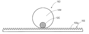
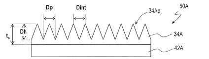
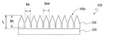
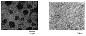
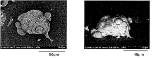
- FIG. 34 It is a figure which shows typically the culture state in the 3D culture method by embodiment of this invention. It is a schematic cross-sectional view of the synthetic polymer film 34A which has a moth-eye structure on the surface used in the 3D culture method by embodiment of this invention. It is a schematic cross-sectional view of the synthetic polymer film 34B which has a moth-eye structure on the surface used in the 3D culture method by embodiment of this invention. It is an optical microscope image (left) of the result of the drop culture of the human liver cancer-derived cell line HepG2, and the optical microscope image (right) of the result of the plane culture. The spheroids of HepG2 obtained by the drop culture method are a top view (left) and a side view (right) of an electron microscope.
- droplets 16D of a cell suspension containing cells 12C and medium 14M are attached onto a solid surface 10S, and the droplets are formed.
- This is a method of culturing cells 12C in droplet 16D in a state where the direction of gravity acting on 16D is toward the solid surface 10S.
- this three-dimensional culture method hereinafter referred to as "drop culture method"
- the cells 12C are cultured in the droplet 16D, the number of cells can be easily controlled as in the hanging drop method, and the shape of the spheroid and the shape of the spheroids can be controlled. The advantage of high size reproducibility is obtained.
- the solid surface 10S acts as a scaffold, so that even a cell type highly dependent on the scaffold is relatively high. Survivability can be maintained. Further, since it is not necessary to turn the surface 10S to which the droplet 16D is attached downward (directed in the direction of gravity), the workability is higher than that of the hanging drop method.
- the solid surface 10S has a plurality of protrusions 10Sp that can act as scaffolds.
- the droplet 16D is in contact with atmospheric gas (for example, air) except for the portion in contact with the solid surface 10S, and forms a closed culture space. Although it is shown in FIG. 1 that the bottom surface of the droplet 16D is in contact with the tip of the convex portion 10Sp and the droplet 16D is present only above the tip of the convex portion 10Sp, the droplet 16D is shown. A part of the bottom of the ridge may be intruded between the adjacent convex portions 10 Sp.
- the volume of the droplet 16D is, for example, 10 ⁇ L or more and 50 ⁇ L or less.
- the solid surface 10S capable of forming stable droplets 16D and efficiently culturing cells is a solid surface having a plurality of convex portions 10Sp having a height of 10 nm or more and 1 mm or less, as shown later in the experimental results. It is also described in Patent Document 1 (registered as Patent No. 4507845) that three-dimensional culture can be performed by using a solid surface having a plurality of convex portions having a height of 10 nm or more and 1 mm or less. However, as described above, the microadhesive three-dimensional culture method using the fine uneven structure of the bottom surface cannot sufficiently reproduce the three-dimensional tissue structure, or the shape and size of the spheroids are poorly reproducible. There is.
- the cells in the droplet 16D are cultured, so that the above-mentioned drawbacks of the finely adherent three-dimensional culture method can be eliminated.
- the cells 12C gather in the three-dimensionally closed droplet 16D so as to be deposited on the bottom surface in contact with the solid surface 10S under the action of gravity. Therefore, there are a certain amount of cells that interact with the plurality of convex portions 10Sp of the solid surface 10S, and on top of that, there are cells that interact only between the cells.
- a spheroid that proliferates appropriately in the thickness direction and has high reproducibility of the three-dimensional tissue structure can be obtained.
- a solid surface having a moth-eye structure which one of the applicants has developed as an antireflection film or a synthetic polymer film having bactericidal properties, is suitably used for the drop culture method.
- the disclosure contents of Patent Documents 6 to 9 (antireflection film) and Patent Document 10 (synthetic polymer film having bactericidal properties) are incorporated herein by reference.
- anodized porous alumina layer when used, it is formed by curing a synthetic polymer film having a moth-eye structure on the surface (for example, a photocurable resin) with high mass productivity. It is possible to produce a photocurable resin film formed, a thermosetting resin film formed by curing a thermosetting resin, or the like).
- the experimental example shown below is an example using a photocurable resin film having a moth-eye structure formed by the above method on the surface, and has the features described in the above items 2 to 9.
- the size and height of a plurality of convex portions and the distance between adjacent convex portions (pitch when regularly arranged) are not limited to these. Conceivable.
- the material forming the moth-eye structure may be either an organic material or an inorganic material.
- Synthetic polymer membranes 34A and 34B are examples of a three-dimensional culture structure according to an embodiment of the present invention.
- 2A and 2B show schematic cross-sectional views of the synthetic polymer films 34A and 34B, respectively.
- the synthetic polymer films 34A and 34B exemplified here are both formed on the base films 42A and 42B, respectively, but are not limited to this, of course.
- Synthetic polymer films 34A and 34B can be formed directly on the surface of any object.
- the film 50A shown in FIG. 2A has a base film 42A and a synthetic polymer film 34A formed on the base film 42A.
- the synthetic polymer film 34A has a plurality of convex portions 34Ap on its surface, and the plurality of convex portions 34Ap form a moth-eye structure.
- 2-dimensional size D p of the convex portion 34Ap is in the range of 10nm or more 500nm or less.
- the "two-dimensional size" of the convex portion 34Ap refers to the diameter corresponding to the area circle of the convex portion 34Ap when viewed from the normal of the surface.
- the two-dimensional size of the convex portion 34Ap corresponds to the diameter of the bottom surface of the cone.
- typical distance between adjacent D int of the convex portion 34Ap is 10nm or more 1000nm or less.
- the convex portions 34Ap The two-dimensional magnitude D p is equal to the inter-adjacent distance D int.
- the typical height D h of the convex portion 34Ap is 10 nm or more and 500 nm or less.
- the thickness t s of the synthetic polymer film 34A is not particularly limited, and may be larger than the height D h of the convex portion 34Ap.
- the synthetic polymer film 34A shown in FIG. 2A has a moth-eye structure similar to that of the antireflection film described in Patent Documents 6 to 9.
- the convex portions 34Ap are densely arranged.
- the convex portion 34Ap has a shape in which the cross-sectional area (cross section parallel to the plane orthogonal to the incident light beam, for example, the cross section parallel to the plane of the base film 42A) increases from the air side toward the base film 42A side, for example. It is preferably conical.
- the convex portions 34Ap preferably randomly so as not to have regularity.
- the protrusions 34Ap do not have to be closely arranged and may be regularly arranged.
- the upper and lower limits of D p , D int , and D h may also exceed the wavelength range of visible light because it is not necessary to prevent reflection of visible light.
- the film 50B shown in FIG. 2B has a base film 42B and a synthetic polymer film 34B formed on the base film 42B.
- the synthetic polymer film 34B has a plurality of convex portions 34Bp on its surface, and the plurality of convex portions 34Bp form a moth-eye structure.
- the structure of the convex portion 34Bp of the synthetic polymer film 34B of the film 50B is different from the structure of the convex portion 34Ap of the synthetic polymer film 34A of the film 50A.
- the description of the features common to the film 50A may be omitted.
- 2-dimensional size D p of protrusions 34Bp is in the range of 10nm or more 500nm or less. Further, the typical distance between adjacent portions D int of the convex portion 34 Bp is 10 nm or more and 1000 nm or less, and D p ⁇ D int . That is, in the synthetic polymer film 34B, a flat portion exists between adjacent convex portions 34Bp.
- the convex portion 34Bp is a columnar shape having a conical portion on the air side, and the typical height D h of the convex portion 34Bp is 10 nm or more and 500 nm or less. Further, the convex portions 34Bp may be arranged regularly or irregularly. If the convex portions 34Bp are regularly arranged, the int will also represent the period of the arrangement. This is, of course, the same for the synthetic polymer membrane 34A.
- the "moss-eye structure" is composed of convex portions having a shape in which the cross-sectional area (cross section parallel to the membrane surface) increases, such as the convex portion 34Ap of the synthetic polymer film 34A shown in FIG. 2A.
- the nano-surface structure having an excellent antireflection function but also a portion having a constant cross-sectional area (cross section parallel to the film surface) like the convex portion 34Bp of the synthetic polymer film 34B shown in FIG. 2B.
- the plurality of convex portions of the solid surface illustrated in the examples have a substantially conical tip portion, but the shape of the plurality of convex portions is not limited to this.
- a shape that is thinner toward the tip of the convex portion is preferable from the viewpoint of mold releasability.
- the tip does not have to be sharp.
- the height of the convex portion exceeds 500 nm, there are disadvantages such as a decrease in mold releasability or a time required for manufacturing the mold.
- the surfaces of the synthetic polymer films 34A and 34B may be treated, if necessary.
- a water-repellent oil-repellent agent or a surface treatment agent may be applied to adjust the surface tension (contact angle of the drop).
- a thin polymer film is formed on the surfaces of the synthetic polymer films 34A and 34B.
- the surfaces of the synthetic polymer films 34A and 34B may be modified by using plasma or the like. For example, lipophilicity can be imparted to the surfaces of the synthetic polymer films 34A and 34B by plasma treatment.
- the mold for forming the moth-eye structure on the surface as illustrated in FIGS. 2A and 2B (hereinafter, referred to as "moth-eye mold") has an inverted moth-eye structure in which the moth-eye structure is inverted. If the anodized porous alumina layer having the inverted moth-eye structure is used as it is as a mold, the moth-eye structure can be manufactured at low cost. In particular, when a cylindrical moth-eye mold is used, a moth-eye structure can be efficiently manufactured by a roll-to-roll method. Such a mold for moth eye can be produced by the method described in Patent Documents 6 to 9.
- the manufacturing method for the moth-eye mold is not limited to the above method.
- various lithography methods such as interference exposure lithography and electron beam lithography, or a method of irradiating a glassy carbon substrate with an oxygen ion beam to form a structure, or a method of forming a known nanostructure can be used.
- the solid surface having the characteristics described in the above items 2 to 9 is preferably used in the drop culture method.
- the synthetic polymer film described in the above patent application has a feature that the pH of the droplets adhering to the surface does not change. Specifically, the pH of the aqueous solution 5 minutes after dropping 200 ⁇ L of water on the surface of the synthetic polymer membrane can be 6.5 or more and 7.5 or less. In a synthetic polymer film made of a photocurable resin, an acid generated by a polymerization initiator may be eluted in water adhering to the surface.
- One or more polymerization initiators selected from the group consisting of [hydroxyethoxy) -phenyl] -2-hydroxy-2-methyl-1-propan-1-one may be used.
- IRGACURE OXE02 BASF
- Omnirad 127 IGM Resins
- Omnirad 2959 IGM Resins
- the synthetic polymer membrane described in the above patent application is preferably used.
- FIG. 3A shows the results of drop culture of the human liver cancer-derived cell line HepG2 (left) and the results of plane culture (right) observed using an inverted phase-contrast microscope. Further, FIG. 3B shows an image of the HepG2 spheroid obtained by the drop culture method observed with an electron microscope. The left side of FIG. 3B is a top view, and the right side of FIG. 3B is a side view.
- human hepatoma-derived cell line HepG2 cells were prepared by adding a final concentration of 10% bovine fetal serum (FBS) to Darbecco-modified eagle medium (D-MEM), which is a general culture condition (cell culture medium).
- FBS bovine fetal serum
- D-MEM Darbecco-modified eagle medium
- the cells were maintained and cultured in an adhesive cell culture medium (for example, MS-11600 manufactured by Sumitomo Bakelite Co., Ltd.) under atmospheric conditions of a temperature of 37 ° C., a carbon dioxide concentration of 5%, and a relative humidity of 95%.
- an adhesive cell culture medium for example, MS-11600 manufactured by Sumitomo Bakelite Co., Ltd.
- the HepG2 cells that had been subjected to this maintenance culture were detached from the culture medium using a trypsin solution, which is a general cell exfoliating solution, and the cell density in the state where the cells were suspended was measured by a fully automatic cell measuring machine (cell counter). ) in measured, as it will become 1 ⁇ 10 5 cells / mL in the above medium to prepare a cell suspension. 25 ⁇ L of this cell suspension was weighed and attached so as to form droplets on the surface of a photocurable resin film having a moth-eye structure on the surface. The optimum number of droplets was 6 to 9 if it was on a nano-projection film attached on a 35 mm ⁇ dish.
- a trypsin solution which is a general cell exfoliating solution
- the droplets (25 ⁇ L) were cultured under the same atmospheric conditions as above (temperature 37 ° C., carbon dioxide concentration 5%, relative humidity 95%) for 3 days.
- the droplets maintained their shape under these atmospheric conditions and even when half or all of the culture medium was replaced to adjust the osmotic pressure in the medium.
- FIG. 3A shows the result by general planar culture. No spheroid formation was observed on the right side of FIG. 3A.
- general plane culture was carried out here as follows.
- a general cell adhesion surface coating for example, MS-3096 or MS-11350 manufactured by Sumitomo Bakelite Co., Ltd., selected according to the test volume.
- the cell suspension was seeded in an appropriate amount (100 ⁇ L for a 96-well plate and 2 mL for a 35 mm dish) and cultured so that the cells could grow flat.
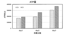
- FIG. 3C shows the results of determining the cell survival number of the liver cancer cell line HepG2 in drop culture. Here, it was quantified by measuring adenosine triphosphate (ATP), which is known to have the same amount of energy in living cells of the same cell line.
- ATP adenosine triphosphate
- 3D shows the result of the drop culture method
- 2D shows the result of the general plane culture method.
- the number of surviving cells by the drop culture method is almost the same as the number of surviving cells by the planar culture method.
- the number of cells alive is smaller than that of the plane culture method, and if 70% to 80% of the number of cells alive by the plane culture method is obtained, the cells can be efficiently produced. It is said to be alive.
- Cell survival is also dependent on the cell density, the seeding density of typical 1 ⁇ 10 5 cells / mL, it found that almost the same cell viability and plane culture method is obtained in drop culture method rice field.
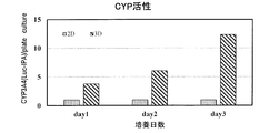
- FIG. 3D shows the results of evaluating the tissue reproducibility (or "gene expression") of the liver cancer cell HepG2 spheroid obtained by the drop culture method.
- 3D shows the result of the drop culture method
- 2D shows the result of the general plane culture method.
- hepatocyte function is hardly maintained in planar culture, but cell coordination according to polarity is possible by performing three-dimensional culture, and drug-metabolizing enzyme, which is one of the characteristic liver functions.
- drug-metabolizing enzyme which is one of the characteristic liver functions.
- CYP activity cytochrome P450
- the tissue reproducibility of hepatocyte spheroids obtained by the drop culture method was evaluated by measuring the P450 activity as follows.
- the enzyme activity in the cells was measured according to the instructions using the P450-Glo TM Luciferin-IPA kit (manufactured by Promega).
- P450-Glo TM Luciferin-IPA kit manufactured by Promega.
- HepG2 cells were subjected to planar culture so as to have the same number of cells as the droplets, and P450 activity was measured by the same method. Since it was expected that the cell growth rate would be different between the planar culture and the drop culture, it is necessary to calculate the corrected value of the enzyme activity per cell.
- ATP was quantified using the Cell Titter Glo R kit (manufactured by Promega).
- the RLU value was measured according to the instructions.
- the relative P450 enzyme activity value of HepG2 cells in the drop culture was obtained by dividing the P450 enzyme activity in the plane culture or the drop culture by the ATP value and setting the enzyme activity value per cell in the plane culture to 1, and graphing it. Is shown in FIG. 3D.
- the CYP activity increased about 10 times after culturing for 3 days, and it is considered that the formed spheroids have high tissue reproducibility. Be done.
- spheroids with high tissue reproducibility can be formed by forming droplets (drops) on the solid surface and culturing.
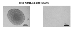
- FIGS. 4A, 4B, 4C, and 4D show the results of drop culture of various cells (left) together with the results of planar culture (right).
- FIG. 4A shows an optical microscope image (left) of the result of drop culture of human fetal kidney epithelial cell HEK293 and an optical microscope image (right) of the result of plane culture
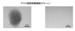
- FIG. 4B shows mouse adipose precursor cells 3T3-L1.
- An optical microscope image of the result of drop culture (left) and an optical microscope image of the result of plane culture (right) are shown.
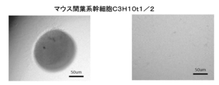
- FIG. 4C shows an optical microscope image of the result of drop culture of mouse mesenchymal stem cells C3H10t1 / 2 (left).
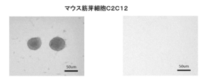
- FIG. 4D shows the light microscope image of the result of drop culture of mouse myoblasts C2C12 (left) and the light microscope image of the result of plane culture (right). ) Is shown.
- the drop culture method confirmed the formation of spheroids, whereas the planar culture did not form spheroids.
- the drop culture method is suitably used for culturing a wide range of cell types.
- each mold was subjected to the mold release treatment shown in Table 3.
- Three types of treatments were carried out in which the concentration of the fluorine-based release agent UD509 (manufactured by Daikin Industries, Ltd., OPTOOL UD509, modified perfluoropolyether) was changed.
- the contact angle was measured as a parameter that characterizes the mold and the surface of the synthetic polymer membrane (solid surface in the drop culture method).
- Table 3 shows the contact angles on the surface of the mold.
- the contact angle of the solid surface with respect to the cell suspension affects the area of contact between the solid surface and the cells (also referred to as the "bottom area of the droplet") and the shape of the droplet. Although it depends on the cell type, the shape of the obtained spheroid changes depending on the contact angle. It is preferable to adjust the contact angle according to the cell type.
- h Droplet height
- the contact angle for characterizing the solid surface refers to the static contact angle 10 seconds after the droplet adheres to the solid surface.
- "without dripping" means that the contact angle is 140 ° or more.
- a droplet of 10 ⁇ L to 70 ⁇ L was used as in the measurement of the contact angle.
- the sliding angle is the inclination angle at which the surface to which the droplets are attached is inclined from the horizontal direction and the droplets start to slide downward.
- D-MEM Low Glucose 1.0 g / L Glucose
- 10% FBS 10% FBS
- Table 4 shows the mold (type of mold release treatment) and resin composition used for producing the synthetic polymer membranes (Comparative Examples 1 to 12 and Examples 1 to 12) used in the experiment, and the surface of each synthetic polymer membrane.
- the result of measuring the contact angle with water is shown.
- the contact angle was measured using 1 ⁇ L of pure water.
- the result of measuring the difference ( ⁇ ) between the contact angle 1 second and 10 seconds after the droplet was attached to the surface and the contact angle 10 seconds later minus the contact angle 1 second is shown.
- the moth-eye surface has a larger contact angle and higher water repellency (Lotus effect).
- the contact angle could not be measured.
- the contact angle exceeds approximately 140 °, the phenomenon that the droplets do not adhere to the surface of the object occurs in this way.
- the time change ( ⁇ ) of the contact angle is small and stable except for Examples 10 and 11. It is probable that the reason why the contact angles of Examples 10 and 11 changed significantly with time was that the concentration of the release agent was low and the water and oil repellent was not uniformly and sufficiently attracted to the surface of the moth eye. This is because the water-repellent and oil-repellent agent contained in the curable resin is attracted to the surface of the moth eye by the mold release treatment.
- Tables 5, 7, 9, and 11 show the results of measuring the contact angle with water and the contact angle with medium by changing the amount of droplets (volume of droplets).
- Table 5 (Comparative Examples 1-1 to 12-1, Examples 1-1 to 12-1) shows the results using 10 ⁇ L droplets
- Table 7 shows the results.
- 1-2 to 12-2) show the results using 30 ⁇ L droplets
- Table 9 (Comparative Examples 1-3 to 12-3, Examples 1-3 to 12-3) uses 50 ⁇ L droplets.
- Table 11 (Comparative Examples 1-4 to 12-4, Examples 1-4 to 12-4) shows the results using 70 ⁇ L droplets.
- the contact angle is larger on the moth-eye surface than on the flat surface for both water and medium, and the water repellency is high.
- the shape of the droplet becomes flatter and the contact angle tends to decrease under the influence of gravity, which is observed for water and medium.
- a synthetic polymer film prepared by using a mold treated with a high-concentration mold release treatment agent tends to have higher water repellency.
- the sliding angle also tends to decrease as the volume of the droplet increases due to the influence of gravity for water and medium.
- the sliding angle is larger on the moth-eye surface than on the flat surface, which is considered to be due to the action of the fine convex portions on the moth-eye surface. That is, it can be seen that the surface of the moth eye can maintain a high sliding angle while having high water repellency.
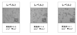
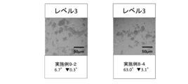
- Example 10-4 is Example 10-4
- the left of FIG. 5B is Example 10-3
- the center is Example 11-4
- the right is Example 10-1
- the left of FIG. 5C is Example 9-2
- the right is Example.
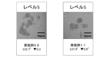
- FIG. 5D is Example 6-2
- the left of FIG. 5E is Example 3-2
- the right is an optical microscope image of the spheroid of Example 1-1, and the contact angle of the medium (after 10 seconds) and , Indicates a change in contact angle ( ⁇ indicates minus).
- the workability of medium exchange was evaluated by the contact angle.
- a contact angle of 110 ° or more was evaluated as ⁇ (excellent), 90 ° or more and less than 110 ° was evaluated as ⁇ (good), and a contact angle of less than 90 ° was evaluated as ⁇ (possible).
- the height of the droplets is less than 1 mm, the workability of the medium exchange is lowered, so the value is x (impossible). Since the drop culture method is highly viable, the culture period may be long (more than several days) depending on the cell type. Then, the medium in the droplet is reduced by evaporation. Also, the amount of waste products in the droplets increases.
- the height of the droplets is preferably 1 mm or more.
- the height h is 1 mm when the contact angle is 17 ° (20 ° when 50 ⁇ L, 26 ° when 30 ⁇ L, 44 when 10 ⁇ L). At °, the height h is 1 mm, respectively). Therefore, only Examples 10-4 in which the contact angles of the medium after 10 seconds were 14.5 ° and less than 17 ° were judged as x.
- Droplets on a solid surface may move (slide or roll) as the solid surface tilts or vibrates. In order to prevent this, it is necessary to maintain a state in which the solid surface is not tilted or vibrated during culturing, so that workability is reduced. For example, by setting the sliding angle of the solid surface with respect to the medium to 45 ° or more, the droplets can be relatively stably held on the solid surface.
- a sliding angle of 90 ° or more was evaluated as ⁇ (excellent), 45 ° or more and less than 90 ° was evaluated as ⁇ (good), 10 ° or more and less than 45 ° was evaluated as ⁇ (possible), and less than 10 ° was evaluated as ⁇ (impossible). ..
- Example 11-4 center of FIG. 5B
- Example 10-1 right of FIG. 5B
- Example 8-4 right of FIG. 5C
- the difference ⁇ of the contact angles is small.
- better spheroids to be obtained. This is because the smaller the amount of change in contact angle within 10 seconds after dripping, the easier it is for the shape of the droplet to be maintained during the culture period (it is difficult for it to become flat), and as a result, the effect of cell accumulation due to the shape of the droplet is achieved. , It is thought that more were obtained.
- the volume of the droplet is preferably 10 ⁇ L or more and 50 ⁇ L or less (see Tables 6, 8 and 10, especially Examples 1 to 6).
- Seeding density of cells contained in the droplets for example, a 10 3 cells / mL or more 10 7 cells / mL or less.
- One of the advantages of the drop culture method is that the number of cells contained in the droplet can be accurately controlled.
- the number of cells is typically in the above range, but can be appropriately adjusted depending on the cell type, the volume of droplets, and the like.
- the sliding angle of the droplet is preferably 45 ° or more, and more preferably 90 ° or more.
- the sliding angle may be evaluated by the value 20 seconds after the drip is applied.
- the present inventor has found that one of the factors contributing to the tissue reproducibility of spheroids obtained by the drop culture method is the degree (strength) of adhesion of cells to the solid surface. According to the study by the present inventor, although it depends on the cell type, if the adhesion of the cell (pseudopodia of the cell) to the solid surface is too weak, the tissue reproducibility of the obtained spheroid tends to be low.
- the synthetic polymer film forming the solid surface is a fluorine-containing oligomer having a perfluoroalkyl group (“Rf-based oligomer” or “perfluoro”). It contains at least one of an alkyl-based oligomer () and a fluorine-containing monomer having a perfluoroalkyl group (sometimes referred to as an “Rf-based monomer” or a perfluoroalkyl-based monomer), and is a perfluoropolyether.
- Rf-based oligomer fluorine-containing oligomer having a perfluoroalkyl group
- the cell When formed from a curable resin that does not contain a fluorine-containing oligomer having a group (sometimes referred to as "PFPE-based oligomer” or “perfluoropolyether-based oligomer"), the cell (cell pseudofoot) has a solid surface. It has been found that a spheroid having high tissue reproducibility can be obtained by adhering to a spheroid with an appropriate strength.
- PFPE-based oligomer sometimes referred to as "PFPE-based oligomer” or “perfluoropolyether-based oligomer”
- Example 2 A solid surface having appropriate adhesion to cells was examined. Drop culture was performed under the same conditions as in Experimental Example 1. Hereinafter, the points different from Experimental Example 1 will be mainly described.
- Example 21 to 31 and Comparative Examples 21 to 25 were prepared in the same manner as in Experimental Example 1.
- Table 1 shows the raw materials used for the ultraviolet curable resin forming the synthetic polymer film 34A of each sample film
- Table 13 shows the composition of each ultraviolet curable resin.
- Examples 21 to 31 and Comparative Examples 21 to 25 were both formed using a mold having an inverted moth-eye structure on the surface. The surface of the mold was subjected to a mold release treatment using a fluorine-based mold release agent UD509 (manufactured by Daikin Industries, Ltd., OPTOOL UD509, modified perfluoropolyether) having a concentration of 0.001% by mass.
- UD509 fluorine-based mold release agent
- a PET film manufactured by Toyobo Co., Ltd., product name: Cosmoshine A4300 (Cosmoshine is a registered trademark), thickness: 100 ⁇ m
- exposure to ultraviolet rays irradiated to cure each ultraviolet curable resin The amount was 1 J / cm 2 .
- the fluorine-containing oligomers and fluorine-containing monomers (MT70, KY1203E, DAC-MO, FAAC4, FAAC6, RS76NS and EB8110) shown in Table 1 are all reactive.
- the chemical structural formulas of MT70, DAC-MO, FAAC4 and FAAC6 are shown in [Chemical formula 1] to [Chemical formula 4] (formulas (1) to (4)), respectively.
- X is a methacrylate group
- m is an integer of 1 to 14
- n is an integer of 1 to 24.
- the chemical formula shown in formula (1) is estimated.
- R1 is a perfluoropolyether group and R2 is an acrylate group.
- M280 and M282 contain ethylene oxide units (EO units) (-CH 2 CH 2 O-).
- EO units ethylene oxide units
- Examples 21 to 31 and Comparative Examples 21 to 25 the contact angle and the sliding angle were measured by the same method as in Experimental Example 1 as parameters for characterizing the surface of the synthetic polymer film.
- a 10 ⁇ L droplet was used for measuring the contact angle and the sliding angle using pure water, and a 10 ⁇ L droplet was used for measuring the contact angle and the sliding angle using a medium.
- the workability, handleability, and spheroidization of medium exchange were evaluated in the same manner as in Experimental Example 1.
- the contact angle of the medium is 110 ° or more, and the workability of the medium exchange is excellent, except for Comparative Example 25.
- Examples 21 to 31 and Comparative Examples 21 to 25 the sliding angle of the medium is 90 ° or more, and the handling property is excellent. Further, in Examples 21 to 31 and Comparative Examples 21 to 25, the formation of spheroids was observed, and as in the result of Experimental Example 1, the larger the contact angle, the better the spheroids in good condition (high density accumulation). There is a tendency to obtain.
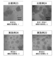
- An example of the morphological observation result by an optical microscope is shown in FIG. 6 (upper left: Comparative Example 23, upper right: Comparative Example 24, lower left: Example 26, lower right: Example 22).
- the degree (strength) of adhesion of cells to the solid surface was evaluated as follows.
- the culture vessel was intentionally vibrated (the culture vessel was shaken) every time the medium was changed (every 24 hours). After culturing for 3 days, the degree of adhesion of the cells to the culture vessel and the presence or absence of pseudopodia of the cells were visually observed. It should be noted that such an evaluation method is for evaluating the degree of adhesion of cells to the solid surface in consideration of the vibration generated during the medium exchange operation, and is based on the vibration generated on the solid surface during the medium exchange operation. , Based on the findings obtained by the inventor that cells may migrate away from the solid surface.
- the degree of cell adhesion to the solid surface was evaluated according to the following criteria, and expressed as the degree of cell adhesion of the solid surface.
- the adhesiveness of the solid surface where the cell adhesion strength is larger than a certain level is practically usable ( ⁇ , ⁇ ), and the adhesiveness of the solid surface where the cell adhesion strength is smaller than a certain level or does not adhere is actually used. It was judged to be impossible ( ⁇ , ⁇ ). Specifically, it was evaluated as follows. X: Pseudopodia were not observed, and when the state in which the culture vessel was vibrated was observed, cell migration was observed. That is, the cells moved away from the solid surface due to the shaking of the medium (droplet) due to vibration.
- ⁇ Some pseudopodia were observed, but when the state in which the culture vessel was vibrated was observed, cell migration was observed.
- ⁇ Pseudopodia was observed, but when the state in which the culture vessel was vibrated was observed, cell migration was observed. However, the cells did not move just by moving the container lightly without forcibly applying vibration.
- ⁇ Pseudopodia were observed, and when the state in which the culture vessel was vibrated was observed, no cell movement was observed.
- Table 14 shows the evaluation results of Examples 21 to 31 and Comparative Examples 21 to 25. As can be seen from Table 14, it is formed from a curable resin containing at least one of a fluorine-containing oligomer having a perfluoroalkyl group and a fluorine-containing monomer having a perfluoroalkyl group and not containing a fluorine-containing oligomer having a perfluoropolyether group.
- the solid The surface has excellent adhesion to cells.
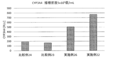
- Example 7 and 15 show the results of evaluating the CYP activity of the liver cancer cell HepG2 spheroids obtained by the drop culture method for Example 22, Example 26, Comparative Example 23 and Comparative Example 24. ..
- the evaluation was performed in the same manner as the evaluation result in FIG. 3D.
- Table 15 also shows the values of Example 22, Example 26, and Comparative Example 24 when Comparative Example 23 is 1. From FIG. 7 and Table 15, it can be seen that in Example 22 and Example 26, since the CYP activity is higher than that in Comparative Example 23 and Comparative Example 24, the tissue reproducibility of the spheroid is improved. From the results of Examples 22 and 26, the state of spheroidization is moderately good (accumulation density is moderately high), and cells (cell pseudopodia) adhere to the solid surface with moderate strength.
- Comparative Example 23 and Comparative Example 24 were inferior in tissue reproducibility of spheroids, although they were comparable to Examples 22 and 26 in the evaluation of spheroidization. It is considered that this is because Comparative Example 23 and Comparative Example 24 are inferior in the adhesiveness of the solid surface to cells as compared with Examples 22 and 26.
- the HepG2 cells used in the experimental examples are scaffold-dependent (that is, they need to adhere to the scaffold for normal cell proliferation), so if the adhesion to the solid surface is weak, spheroids will be formed. It is probable that the tissue reproducibility of the obtained spheroids was low.
- the curable resin contains, for example, 0.5% by mass or more and 10% by mass or less of a fluorine-containing oligomer having a perfluoroalkyl group and / or a fluorine-containing monomer having a perfluoroalkyl group with respect to the entire curable resin. It is preferably contained in an amount of 7% by mass or more and 8% by mass or less, and more preferably 2% by mass or more and 5% by mass or less. If the proportion of the fluorine-containing oligomer and / or monomer having a perfluoroalkyl group is less than 0.5% by mass, the solid surface may not have sufficient water repellency, and if it is more than 10% by mass, it is uniform. The curable resin may not be obtained, and the physical properties of the cured product (synthetic polymer film obtained by curing the curable resin) may be deteriorated.
- fluorine-containing monomer having a perfluoroalkyl group F (CF 2 ) p ⁇ group, p is an integer
- one having p of 4 or more and 6 or less is preferable. If p is less than 4, a sufficient effect may not be obtained from the viewpoint of improving water repellency, and if p is more than 6, a uniform curable resin may not be obtained.
- a fluorine-containing monomer having a perfluoroalkyl group, in which the ratio of the fluorine element to the entire monomer is 50% by mass or more and 60% by mass or less, may be used.
- the curable resin may further contain a block copolymer (sometimes referred to as "fluorine-containing block copolymer”) composed of a fluorine segment and a non-fluorine segment.
- a block copolymer (sometimes referred to as "fluorine-containing block copolymer") composed of a fluorine segment and a non-fluorine segment.
- fluorine segment means a block formed by polymerizing fluorine-containing monomers.
- non-fluorine segment means a block formed by polymerizing fluorine-free monomers. It has been found that when the curable resin further contains a fluorine-containing block copolymer, the tissue reproducibility of the obtained spheroid can be improved by increasing the contact angle of the solid surface with respect to the medium.
- the non-fluorine segment functions as a compatible segment having compatibility with other components in the curable resin, while the fluorine segment is a synthetic polymer film 34A. Is unevenly distributed on the surface of the surface (the surface having the convex portion 34p). As a result, the surface free energy of the synthetic polymer film 34A is considered to be low.
- the curable resin contains only one of a fluorine-containing oligomer having a perfluoroalkyl group and a fluorine-containing monomer having a perfluoroalkyl group, it is preferable that the curable resin further contains a fluorine-containing block copolymer.
- one of the applicants is a perfluoropolyether-based oligomer having a (meth) acryloyl group and / or perfluoro.
- a curable resin (curable resin composition) containing a fluorine-containing block copolymer in addition to an alkyl-based oligomer is used, more fluorine elements are unevenly distributed on the surface (surface having protrusions) of the synthetic polymer film, resulting in a mold.
- the fluorine segment of the fluorine-containing block copolymer preferably has a perfluoroalkyl group.
- the fluorine segment is likely to be unevenly distributed on the surface of the synthetic polymer film 34A (the surface having the convex portion 34p) (the surface is easily arranged so that a rigid molecular chain stands), and the fluorine element is easily arranged.
- the synthetic polymer film 34A is likely to be densely distributed on the surface (the surface having the convex portion 34p). As a result, it is considered that the contact angle of the surface of the synthetic polymer film 34A with respect to the medium on the solid surface is likely to be increased.
- the curable resin preferably contains, for example, 1% by mass or more and 5% by mass or less in total of the fluorine-containing block copolymer with respect to the entire curable resin. If the proportion of the fluorine-containing block copolymer is less than 1% by mass, the solid surface may not have sufficient water repellency, and if it exceeds 5% by mass, the compatibility with other components in the curable resin is high. The physical properties (for example, transparency) of the cured product (synthetic polymer film obtained by curing the curable resin) may be reduced.
- the total mass of the fluorine-containing block copolymer contained in the curable resin is, for example, 0.25 or more and 2.0, where 1 is the total mass of the perfluoroalkyl-based oligomer and / or the perfluoroalkyl-based monomer contained in the curable resin. It is as follows.
- the proportion of fluorine segments in the fluorine-containing block copolymer is preferably 10% by mass or more and 90% by mass or less, and more preferably 35% by mass or more and 65% by mass or less.
- the proportion of fluorine segments in the fluorine-containing block copolymer is lower than 10% by mass, the performance as fluorine segments, for example, water repellency and oil repellency may not be sufficiently obtained.
- the proportion of fluorine segments in the fluorine-containing block copolymer is higher than 90% by mass, the compatibility with other components in the curable resin is lowered, and as a result, the transparency of the synthetic polymer film 34A is lowered. May (whiten).
- the number average molecular weight of the fluorine-containing block copolymer is preferably 5,000 or more and 1,000,000 or less, more preferably 10,000 or more and 300,000 or less, and further preferably 10,000 or more and 100,000 or less.
- the number average molecular weight of the fluorine-containing block copolymer is smaller than 5000, the fluorine segment is short and its performance (for example, water repellency and oil repellency) may not be sufficiently obtained.
- the number average molecular weight of the fluorine-containing block copolymer is larger than 1,000,000, the compatibility with other components in the curable resin is lowered, and as a result, the transparency of the synthetic polymer film 34A is lowered (whitening). Sometimes.
- Examples 21 to 31 are excellent in the adhesiveness of the solid surface to cells.
- Examples 21 to 23 include both a fluorine-containing oligomer having a perfluoroalkyl group and a fluorine-containing monomer having a perfluoroalkyl group. From the results of Examples 21 to 23, the solid surface formed from the curable resin containing both the fluorine-containing oligomer having a perfluoroalkyl group and the fluorine-containing monomer having a perfluoroalkyl group tends to have excellent water repellency. You can see that it is in.
- the curable resin should contain both a fluorine-containing oligomer having a perfluoroalkyl group and a fluorine-containing monomer having a perfluoroalkyl group. Is preferable.
- Examples 27 to 29 contain a fluorine-containing monomer having a perfluoroalkyl group and not containing a fluorine-containing oligomer having a perfluoroalkyl group.
- the contact angle of the solid surface with respect to the medium is low (110 ° or more and less than 120 °), so that although spheroid formation is observed, the spheroid state is slightly inferior (accumulation). (Density is low). Further, in Examples 27 to 29, the contact angle tends to change with time more than in Examples 21 to 23 (the degree of decrease in water repellency with time is large).
- Examples 24 to 26 contain a fluorine-containing oligomer having a perfluoroalkyl group without containing a fluorine-containing monomer having a perfluoroalkyl group. Also in Examples 24 to 26, the contact angle of the solid surface with respect to the medium tends to be lower than that of Examples 21 to 23. Since the fluorine-containing monomers having a small molecular weight are not contained in Examples 24 to 26, it is considered that the fluorine element is difficult to move to the vicinity of the surface of the photocurable resin and is inferior in water repellency as compared with Examples 21 to 23.
- Comparative Examples 21 to 24 contain a fluorine-containing oligomer having a perfluoropolyether group.
- a solid surface formed of a curable resin containing a fluorine-containing monomer and / or a fluorine-containing oligomer tends to have a weak interaction with cells due to its low surface tension, but among the fluorine-containing groups, the perfluoropolyether group is Since it has a longer and more flexible structure than the perfluoroalkyl group, it is considered that the perfluoropolyether group on the surface is easy to move. Therefore, it is presumed that in Comparative Examples 21 to 24, the cells were more easily detached from the solid surface than in Examples 21 to 31.
- Example 30 further contains a fluorine-containing block copolymer in addition to a fluorine-containing oligomer having a perfluoroalkyl group.
- Example 30 contains a fluorine-containing oligomer having a perfluoroalkyl group to the same extent as in Example 24, and further comprises a fluorine-containing block copolymer F606.
- the contact angle of the solid surface with respect to the medium is higher than that of Example 24, and the state of spheroids (accumulation density) is also excellent.
- Example 31 further comprises a fluorine-containing block copolymer in addition to a fluorine-containing monomer having a perfluoroalkyl group.
- Example 31 differs from the composition of Example 27 in that it further comprises the fluorine-containing block copolymer F606.
- the contact angle of the solid surface with respect to the medium is higher than that of Example 27, and the state of spheroids (accumulation density) is also excellent.
- Comparative Example 25 does not contain either a fluorine-containing oligomer having a perfluoroalkyl group or a fluorine-containing monomer having a perfluoroalkyl group, and contains a fluorine-containing block copolymer.
- the contact angle of the solid surface with respect to the medium is low (less than 90 °)
- the workability of the medium exchange is low.
- spheroid formation is observed, it is slightly inferior in the spheroid state (low accumulation density).
- the adhesiveness to the solid surface of the cells was weaker than that in Examples 21 to 31.
- a three-dimensional culture method capable of obtaining a spheroid having excellent workability or mass productivity and / or high tissue reproducibility as compared with the conventional three-dimensional culture method.
- a three-dimensional culture structure having a solid surface having a plurality of protrusions having a height of 10 nm or more and 1 mm or less is suitable for a drop culture method. Used for.
- Such a three-dimensional culture structure can be obtained, for example, by attaching the above-mentioned synthetic polymer membrane to the inner bottom surface of a petri dish. That is, the three-dimensional culture structure can be provided as a container such as a petri dish.
- a three-dimensional culture structure for example, a container
- a three-dimensional culture structure having a spheroid having higher tissue reproducibility than the conventional one can be produced. ..
- Such a three-dimensional culture structure having a spheroid on the surface is suitably used for research and development of drug discovery and regenerative medicine.
- the three-dimensional cell culture method according to the embodiment of the present invention can be widely used for drug discovery, regenerative medicine, and the like.
Landscapes
- Health & Medical Sciences (AREA)
- Engineering & Computer Science (AREA)
- Life Sciences & Earth Sciences (AREA)
- Biomedical Technology (AREA)
- Organic Chemistry (AREA)
- Chemical & Material Sciences (AREA)
- Bioinformatics & Cheminformatics (AREA)
- Wood Science & Technology (AREA)
- Zoology (AREA)
- Genetics & Genomics (AREA)
- Biotechnology (AREA)
- Microbiology (AREA)
- Biochemistry (AREA)
- General Engineering & Computer Science (AREA)
- General Health & Medical Sciences (AREA)
- Cell Biology (AREA)
- Virology (AREA)
- Sustainable Development (AREA)
- Apparatus Associated With Microorganisms And Enzymes (AREA)
- Micro-Organisms Or Cultivation Processes Thereof (AREA)
Priority Applications (1)
| Application Number | Priority Date | Filing Date | Title |
|---|---|---|---|
| JP2022506008A JP7317330B2 (ja) | 2020-03-09 | 2021-03-04 | 三次元培養法、三次元培養構造体、および三次元培養構造体の製造方法 |
Applications Claiming Priority (2)
| Application Number | Priority Date | Filing Date | Title |
|---|---|---|---|
| JP2020039861 | 2020-03-09 | ||
| JP2020-039861 | 2020-03-09 |
Publications (1)
| Publication Number | Publication Date |
|---|---|
| WO2021182306A1 true WO2021182306A1 (ja) | 2021-09-16 |
Family
ID=77670880
Family Applications (1)
| Application Number | Title | Priority Date | Filing Date |
|---|---|---|---|
| PCT/JP2021/008512 Ceased WO2021182306A1 (ja) | 2020-03-09 | 2021-03-04 | 三次元培養法、三次元培養構造体、および三次元培養構造体の製造方法 |
Country Status (2)
| Country | Link |
|---|---|
| JP (1) | JP7317330B2 (cg-RX-API-DMAC7.html) |
| WO (1) | WO2021182306A1 (cg-RX-API-DMAC7.html) |
Citations (2)
| Publication number | Priority date | Publication date | Assignee | Title |
|---|---|---|---|---|
| WO2006106748A1 (ja) * | 2005-03-30 | 2006-10-12 | National University Corporation Nagoya University | 生体有機体の生産方法および培養容器 |
| JP2019156879A (ja) * | 2018-03-07 | 2019-09-19 | シャープ株式会社 | 合成高分子膜および合成高分子膜を有するプラスチック製品 |
-
2021
- 2021-03-04 JP JP2022506008A patent/JP7317330B2/ja active Active
- 2021-03-04 WO PCT/JP2021/008512 patent/WO2021182306A1/ja not_active Ceased
Patent Citations (2)
| Publication number | Priority date | Publication date | Assignee | Title |
|---|---|---|---|---|
| WO2006106748A1 (ja) * | 2005-03-30 | 2006-10-12 | National University Corporation Nagoya University | 生体有機体の生産方法および培養容器 |
| JP2019156879A (ja) * | 2018-03-07 | 2019-09-19 | シャープ株式会社 | 合成高分子膜および合成高分子膜を有するプラスチック製品 |
Non-Patent Citations (1)
| Title |
|---|
| SUGIMOTO, YUKO ET AL.: "Development of drop culture method on scaffold material using nanostructured surface", PROGRAMS AND ABSTRACTS OF THE GENERAL CONFERENCE OF THE JAPANESE SOCIETY FOR REGENERATIVE MEDICINE, vol. 18, 22 February 2019 (2019-02-22), pages 744 * |
Also Published As
| Publication number | Publication date |
|---|---|
| JPWO2021182306A1 (cg-RX-API-DMAC7.html) | 2021-09-16 |
| JP7317330B2 (ja) | 2023-07-31 |
Similar Documents
| Publication | Publication Date | Title |
|---|---|---|
| CA2848875C (en) | Adherent cell culture method | |
| Monge et al. | Engineering muscle tissues on microstructured polyelectrolyte multilayer films | |
| US20130136818A1 (en) | Resin Mold for Nanoimprinting | |
| US12630795B2 (en) | Cell culture substrate, method for producing cell culture substrate, and method for producing spheroids | |
| Axente et al. | Combinatorial MAPLE gradient thin film assemblies signalling to human osteoblasts | |
| WO2017204306A1 (ja) | タンパク質付着防止剤、硬化物、硬化物の製造方法、および物品 | |
| Xia et al. | Cell attachment/detachment behavior on poly (N-isopropylacrylamide)-based microgel films: the effect of microgel structure and swelling ratio | |
| CN107429210B (zh) | 医疗器具、含氟环状烯烃聚合物、含氟环状烯烃聚合物组合物及细胞培养方法 | |
| US20100297675A1 (en) | Substrate and method for culturing breast cells | |
| Deng et al. | Programming colloidal self-assembled patterns (cSAPs) into thermo-responsible hybrid surfaces for controlling human stem cells and macrophages | |
| Balikov et al. | Tunable surface repellency maintains stemness and redox capacity of human mesenchymal stem cells | |
| JP7317330B2 (ja) | 三次元培養法、三次元培養構造体、および三次元培養構造体の製造方法 | |
| Rauer et al. | Cell Adhesion and Local Cytokine Control on Protein‐Functionalized PNIPAM‐co‐AAc Hydrogel Microcarriers | |
| JP6854500B2 (ja) | 三次元培養法、三次元培養構造体、および三次元培養構造体の製造方法 | |
| Jayasinghe et al. | A soft lithography method to generate arrays of microstructures onto hydrogel surfaces | |
| US20120295353A1 (en) | Methods of making and using polymers and compositions | |
| KR20150145253A (ko) | 적층체 | |
| Mattioli et al. | Nanostructured polystyrene films engineered by plasma processes: Surface characterization and stem cell interaction | |
| Kawabata et al. | Two-dimensional cellular patterning on a polymer film based on interfacial stiffness | |
| JP7317331B2 (ja) | 三次元培養法、三次元培養構造体、および三次元培養構造体の製造方法 | |
| WO2016129220A1 (ja) | 細胞培養用バッグ及びそれを用いた細胞培養方法 | |
| Tsai et al. | Ultra-thin porous PDLLA films promote generation, maintenance, and viability of stem cell spheroids | |
| Selezneva et al. | Use of thermosensitive polymer material on the basis of N-isopropylacrylamide and N-tert-butylacrylamide copolymer in cell technologies | |
| JP2023178108A (ja) | 細胞培養基材 | |
| JP7219891B2 (ja) | 細胞培養用積層体、医療器具および医療器具の使用方法 |
Legal Events
| Date | Code | Title | Description |
|---|---|---|---|
| 121 | Ep: the epo has been informed by wipo that ep was designated in this application |
Ref document number: 21768648 Country of ref document: EP Kind code of ref document: A1 |
|
| ENP | Entry into the national phase |
Ref document number: 2022506008 Country of ref document: JP Kind code of ref document: A |
|
| NENP | Non-entry into the national phase |
Ref country code: DE |
|
| 122 | Ep: pct application non-entry in european phase |
Ref document number: 21768648 Country of ref document: EP Kind code of ref document: A1 |