WO2021177157A1 - 医用画像処理装置、医用画像処理方法及びプログラム、医用画像表示システム - Google Patents
医用画像処理装置、医用画像処理方法及びプログラム、医用画像表示システム Download PDFInfo
- Publication number
- WO2021177157A1 WO2021177157A1 PCT/JP2021/007302 JP2021007302W WO2021177157A1 WO 2021177157 A1 WO2021177157 A1 WO 2021177157A1 JP 2021007302 W JP2021007302 W JP 2021007302W WO 2021177157 A1 WO2021177157 A1 WO 2021177157A1
- Authority
- WO
- WIPO (PCT)
- Prior art keywords
- medical image
- image
- medical
- anatomical feature
- feature structure
- Prior art date
- Legal status (The legal status is an assumption and is not a legal conclusion. Google has not performed a legal analysis and makes no representation as to the accuracy of the status listed.)
- Ceased
Links
Images



Classifications
-
- A—HUMAN NECESSITIES
- A61—MEDICAL OR VETERINARY SCIENCE; HYGIENE
- A61B—DIAGNOSIS; SURGERY; IDENTIFICATION
- A61B6/00—Apparatus or devices for radiation diagnosis; Apparatus or devices for radiation diagnosis combined with radiation therapy equipment
- A61B6/02—Arrangements for diagnosis sequentially in different planes; Stereoscopic radiation diagnosis
- A61B6/03—Computed tomography [CT]
-
- G—PHYSICS
- G06—COMPUTING OR CALCULATING; COUNTING
- G06T—IMAGE DATA PROCESSING OR GENERATION, IN GENERAL
- G06T7/00—Image analysis
- G06T7/0002—Inspection of images, e.g. flaw detection
- G06T7/0012—Biomedical image inspection
- G06T7/0014—Biomedical image inspection using an image reference approach
- G06T7/0016—Biomedical image inspection using an image reference approach involving temporal comparison
-
- A—HUMAN NECESSITIES
- A61—MEDICAL OR VETERINARY SCIENCE; HYGIENE
- A61B—DIAGNOSIS; SURGERY; IDENTIFICATION
- A61B5/00—Measuring for diagnostic purposes; Identification of persons
- A61B5/0033—Features or image-related aspects of imaging apparatus, e.g. for MRI, optical tomography or impedance tomography apparatus; Arrangements of imaging apparatus in a room
- A61B5/0035—Features or image-related aspects of imaging apparatus, e.g. for MRI, optical tomography or impedance tomography apparatus; Arrangements of imaging apparatus in a room adapted for acquisition of images from more than one imaging mode, e.g. combining MRI and optical tomography
-
- A—HUMAN NECESSITIES
- A61—MEDICAL OR VETERINARY SCIENCE; HYGIENE
- A61B—DIAGNOSIS; SURGERY; IDENTIFICATION
- A61B5/00—Measuring for diagnostic purposes; Identification of persons
- A61B5/103—Measuring devices for testing the shape, pattern, colour, size or movement of the body or parts thereof, for diagnostic purposes
- A61B5/107—Measuring physical dimensions, e.g. size of the entire body or parts thereof
- A61B5/1075—Measuring physical dimensions, e.g. size of the entire body or parts thereof for measuring dimensions by non-invasive methods, e.g. for determining thickness of tissue layer
-
- A—HUMAN NECESSITIES
- A61—MEDICAL OR VETERINARY SCIENCE; HYGIENE
- A61B—DIAGNOSIS; SURGERY; IDENTIFICATION
- A61B5/00—Measuring for diagnostic purposes; Identification of persons
- A61B5/48—Other medical applications
- A61B5/4887—Locating particular structures in or on the body
-
- A—HUMAN NECESSITIES
- A61—MEDICAL OR VETERINARY SCIENCE; HYGIENE
- A61B—DIAGNOSIS; SURGERY; IDENTIFICATION
- A61B5/00—Measuring for diagnostic purposes; Identification of persons
- A61B5/74—Details of notification to user or communication with user or patient; User input means
- A61B5/742—Details of notification to user or communication with user or patient; User input means using visual displays
- A61B5/7425—Displaying combinations of multiple images regardless of image source, e.g. displaying a reference anatomical image with a live image
-
- A—HUMAN NECESSITIES
- A61—MEDICAL OR VETERINARY SCIENCE; HYGIENE
- A61B—DIAGNOSIS; SURGERY; IDENTIFICATION
- A61B5/00—Measuring for diagnostic purposes; Identification of persons
- A61B5/74—Details of notification to user or communication with user or patient; User input means
- A61B5/742—Details of notification to user or communication with user or patient; User input means using visual displays
- A61B5/743—Displaying an image simultaneously with additional graphical information, e.g. symbols, charts, function plots
-
- G—PHYSICS
- G06—COMPUTING OR CALCULATING; COUNTING
- G06T—IMAGE DATA PROCESSING OR GENERATION, IN GENERAL
- G06T7/00—Image analysis
- G06T7/30—Determination of transform parameters for the alignment of images, i.e. image registration
- G06T7/33—Determination of transform parameters for the alignment of images, i.e. image registration using feature-based methods
-
- G—PHYSICS
- G06—COMPUTING OR CALCULATING; COUNTING
- G06T—IMAGE DATA PROCESSING OR GENERATION, IN GENERAL
- G06T7/00—Image analysis
- G06T7/70—Determining position or orientation of objects or cameras
-
- G—PHYSICS
- G16—INFORMATION AND COMMUNICATION TECHNOLOGY [ICT] SPECIALLY ADAPTED FOR SPECIFIC APPLICATION FIELDS
- G16H—HEALTHCARE INFORMATICS, i.e. INFORMATION AND COMMUNICATION TECHNOLOGY [ICT] SPECIALLY ADAPTED FOR THE HANDLING OR PROCESSING OF MEDICAL OR HEALTHCARE DATA
- G16H30/00—ICT specially adapted for the handling or processing of medical images
- G16H30/20—ICT specially adapted for the handling or processing of medical images for handling medical images, e.g. DICOM, HL7 or PACS
-
- G—PHYSICS
- G16—INFORMATION AND COMMUNICATION TECHNOLOGY [ICT] SPECIALLY ADAPTED FOR SPECIFIC APPLICATION FIELDS
- G16H—HEALTHCARE INFORMATICS, i.e. INFORMATION AND COMMUNICATION TECHNOLOGY [ICT] SPECIALLY ADAPTED FOR THE HANDLING OR PROCESSING OF MEDICAL OR HEALTHCARE DATA
- G16H30/00—ICT specially adapted for the handling or processing of medical images
- G16H30/40—ICT specially adapted for the handling or processing of medical images for processing medical images, e.g. editing
-
- G—PHYSICS
- G16—INFORMATION AND COMMUNICATION TECHNOLOGY [ICT] SPECIALLY ADAPTED FOR SPECIFIC APPLICATION FIELDS
- G16H—HEALTHCARE INFORMATICS, i.e. INFORMATION AND COMMUNICATION TECHNOLOGY [ICT] SPECIALLY ADAPTED FOR THE HANDLING OR PROCESSING OF MEDICAL OR HEALTHCARE DATA
- G16H50/00—ICT specially adapted for medical diagnosis, medical simulation or medical data mining; ICT specially adapted for detecting, monitoring or modelling epidemics or pandemics
- G16H50/20—ICT specially adapted for medical diagnosis, medical simulation or medical data mining; ICT specially adapted for detecting, monitoring or modelling epidemics or pandemics for computer-aided diagnosis, e.g. based on medical expert systems
-
- A—HUMAN NECESSITIES
- A61—MEDICAL OR VETERINARY SCIENCE; HYGIENE
- A61B—DIAGNOSIS; SURGERY; IDENTIFICATION
- A61B5/00—Measuring for diagnostic purposes; Identification of persons
- A61B5/0033—Features or image-related aspects of imaging apparatus, e.g. for MRI, optical tomography or impedance tomography apparatus; Arrangements of imaging apparatus in a room
- A61B5/004—Features or image-related aspects of imaging apparatus, e.g. for MRI, optical tomography or impedance tomography apparatus; Arrangements of imaging apparatus in a room adapted for image acquisition of a particular organ or body part
- A61B5/0042—Features or image-related aspects of imaging apparatus, e.g. for MRI, optical tomography or impedance tomography apparatus; Arrangements of imaging apparatus in a room adapted for image acquisition of a particular organ or body part for the brain
-
- A—HUMAN NECESSITIES
- A61—MEDICAL OR VETERINARY SCIENCE; HYGIENE
- A61B—DIAGNOSIS; SURGERY; IDENTIFICATION
- A61B5/00—Measuring for diagnostic purposes; Identification of persons
- A61B5/0033—Features or image-related aspects of imaging apparatus, e.g. for MRI, optical tomography or impedance tomography apparatus; Arrangements of imaging apparatus in a room
- A61B5/004—Features or image-related aspects of imaging apparatus, e.g. for MRI, optical tomography or impedance tomography apparatus; Arrangements of imaging apparatus in a room adapted for image acquisition of a particular organ or body part
- A61B5/0044—Features or image-related aspects of imaging apparatus, e.g. for MRI, optical tomography or impedance tomography apparatus; Arrangements of imaging apparatus in a room adapted for image acquisition of a particular organ or body part for the heart
-
- A—HUMAN NECESSITIES
- A61—MEDICAL OR VETERINARY SCIENCE; HYGIENE
- A61B—DIAGNOSIS; SURGERY; IDENTIFICATION
- A61B5/00—Measuring for diagnostic purposes; Identification of persons
- A61B5/05—Detecting, measuring or recording for diagnosis by means of electric currents or magnetic fields; Measuring using microwaves or radio waves
- A61B5/055—Detecting, measuring or recording for diagnosis by means of electric currents or magnetic fields; Measuring using microwaves or radio waves involving electronic [EMR] or nuclear [NMR] magnetic resonance, e.g. magnetic resonance imaging
-
- A—HUMAN NECESSITIES
- A61—MEDICAL OR VETERINARY SCIENCE; HYGIENE
- A61B—DIAGNOSIS; SURGERY; IDENTIFICATION
- A61B5/00—Measuring for diagnostic purposes; Identification of persons
- A61B5/72—Signal processing specially adapted for physiological signals or for diagnostic purposes
- A61B5/7235—Details of waveform analysis
- A61B5/7264—Classification of physiological signals or data, e.g. using neural networks, statistical classifiers, expert systems or fuzzy systems
- A61B5/7267—Classification of physiological signals or data, e.g. using neural networks, statistical classifiers, expert systems or fuzzy systems involving training the classification device
-
- A—HUMAN NECESSITIES
- A61—MEDICAL OR VETERINARY SCIENCE; HYGIENE
- A61B—DIAGNOSIS; SURGERY; IDENTIFICATION
- A61B8/00—Diagnosis using ultrasonic, sonic or infrasonic waves
-
- G—PHYSICS
- G06—COMPUTING OR CALCULATING; COUNTING
- G06T—IMAGE DATA PROCESSING OR GENERATION, IN GENERAL
- G06T2200/00—Indexing scheme for image data processing or generation, in general
- G06T2200/24—Indexing scheme for image data processing or generation, in general involving graphical user interfaces [GUIs]
-
- G—PHYSICS
- G06—COMPUTING OR CALCULATING; COUNTING
- G06T—IMAGE DATA PROCESSING OR GENERATION, IN GENERAL
- G06T2207/00—Indexing scheme for image analysis or image enhancement
- G06T2207/10—Image acquisition modality
- G06T2207/10072—Tomographic images
- G06T2207/10088—Magnetic resonance imaging [MRI]
-
- G—PHYSICS
- G06—COMPUTING OR CALCULATING; COUNTING
- G06T—IMAGE DATA PROCESSING OR GENERATION, IN GENERAL
- G06T2207/00—Indexing scheme for image analysis or image enhancement
- G06T2207/20—Special algorithmic details
- G06T2207/20092—Interactive image processing based on input by user
- G06T2207/20101—Interactive definition of point of interest, landmark or seed
Definitions
- the present invention relates to a medical image processing apparatus, a medical image processing method and program, and a medical image display system, and particularly relates to a technique for displaying a plurality of medical images.
- Patent Document 1 discloses an image display device that displays CT (Computed Tomography) images taken at different times in parallel.
- CT Computerputed Tomography
- the present invention has been made in view of such circumstances, and an object of the present invention is to provide a medical image processing device, a medical image processing method and program, and a medical image display system that reduce the amount of movement of the line of sight when observing a medical image. do.
- one aspect of the medical image processing apparatus includes a memory for storing an instruction to be executed by the processor and a processor for executing the instruction stored in the memory, and the processor is a subject.
- Outputs a signal for displaying the first medical image of the above acquires a position in the first medical image specified by the user, and moves to the specified position in the first medical image.
- the closest anatomical feature structure is identified, and a second medical image of the subject taken in the past, including the identified anatomical feature structure, is obtained and the first is obtained.
- It is a medical image processing apparatus that outputs a signal for generating and displaying a second partial region image superimposed on a first medical image.
- a region containing the anatomical feature structure identified from the second medical image is cut out to generate a second partial region image, and the second partial region image is superimposed on the first medical image. Since the signal for displaying is output, it is possible to reduce the amount of movement of the line of sight when observing the medical image.
- the processor estimates the location of the identified anatomical feature structure in the second medical image, and the location of the identified anatomical feature structure in the first medical image and the estimation in the second medical image. It is preferable to align the first medical image with the second medical image by using the position of the anatomical feature structure obtained. As a result, even if there are body movements and respiratory movements when the medical image is taken, the anatomical feature structure can be aligned without deviation.
- the processor cuts out a region containing the anatomical feature structure identified from the first medical image to generate a first subregion image, and displays the first subregion image side by side with the second subregion image. It is preferable to output a signal for As a result, it is possible to reduce the amount of movement of the line of sight when observing the first partial region image and the second partial region image.
- the processor preferably outputs a signal for displaying a frame surrounding the region including the identified anatomical feature structure of the first medical image on the first medical image. This allows the user to recognize the region of the first medical image corresponding to the second partial region image.
- the processor preferably outputs a signal for displaying a difference image showing the difference between the first medical image and the second medical image overlaid on the first medical image.
- the processor adjusts the image quality of the first medical image under the first image quality adjustment condition and adjusts the image quality of the second medical image under the first image quality adjustment condition.
- the processor adjusts the image quality of the first medical image and the second medical image under the same image quality adjustment conditions, it becomes easy to observe the first medical image and the second partial region image.
- the anatomical feature structure preferably comprises at least one of an organ, bone, muscle, and lesion area. This aspect is suitable for observing organs, bones, muscles, and lesion areas.
- the first medical image and the second medical image are CT (Computed Tomography) device, MRI (Magnetic Resonance Imaging) device, PET (Positron Emission Tomography) device, ultrasonic diagnostic device, and CR (Computed Radiography) device. It is preferable to include an image taken by any of these devices. This aspect is suitable for observation using medical images taken by a CT device, an MRI device, a PET device, an ultrasonic diagnostic device, and a CR device.
- one aspect of the medical image display system is the medical image processing apparatus described above, a display that displays an image based on the acquired signal, and a position in the image displayed on the display.
- a medical image display system comprising a designated input device.
- a region containing an anatomical feature structure closest to the position of the first medical image designated by the input device is cut out from the second medical image to generate a second partial region image, and the second is generated. Since it is displayed on the display by superimposing it on the medical image of 1, it is possible to reduce the amount of movement of the line of sight when observing the medical image.
- One aspect of the medical image processing method for achieving the above object is a first medical image display step of outputting a signal for displaying a first medical image of a subject, and a first medical image in the first medical image.
- Medical image processing including a second medical image cropping step of generating an image and a second medical image display step of outputting a signal for displaying a second partial region image superimposed on the first medical image. The method.
- a region containing the anatomical feature structure identified from the second medical image is cut out to generate a second partial region image, and the second partial region image is superimposed on the first medical image. Since the signal for displaying is output, it is possible to reduce the amount of movement of the line of sight when observing the medical image.
- This embodiment also includes a program for causing a computer to execute each step of the medical image processing method described above, and a computer-readable non-temporary recording medium on which the program is recorded.
- FIG. 1 is a diagram showing a comparative display example of conventional medical images.
- FIG. 2 is a block diagram of a medical image display system according to the present embodiment.
- FIG. 3 is a flowchart showing the processing of the method of storing the extraction region data of the anatomical feature structure in the medical image.
- FIG. 4 is a flowchart showing the processing of the medical image display method.
- FIG. 5 is a diagram showing a comparative display example of medical images according to the present embodiment.
- FIG. 6 is a diagram showing a comparative display example of medical images when a tumor is designated.
- FIG. 1 is a diagram showing a comparative display example of conventional medical images.
- the current image I 1 which is the current medical image of the subject and the past image I 2 of the same subject corresponding to the current image I 1 are displayed side by side.
- a user such as a doctor can observe the progress of the region of interest such as the anatomical feature structure by comparing the current image I 1 and the past image I 2 displayed as shown in FIG.
- FIG. 2 is a block diagram of the medical image display system 10. As shown in FIG. 2, the medical image display system 10 includes a medical image processing device 12, a medical image storage device 18, and a medical image viewer device 20.
- the medical image processing device 12 is, for example, a computer used in a hospital.
- the medical image processing device 12 includes a processor 14 and a memory 16.
- the memory 16 stores an instruction to be executed by the processor 14.
- the medical image processing device 12 realizes various functions including an image processing function, an organ recognition function, and a display image generation function by the processor 14 executing a program including instructions read from the memory 16.
- the medical image storage device 18 includes a large-capacity storage for storing medical images.
- the medical image storage device 18 stores medical images to which incidental information defined by the DICOM (Digital Imaging and Communication in Medicine) standard is added.
- DICOM Digital Imaging and Communication in Medicine
- the medical image viewer device 20 is a device for a user to observe a medical image.
- the medical image viewer device 20 includes a display 22 and an input device 24.
- the display 22 is a display device that displays an image based on a signal acquired from the medical image processing device 12.
- the medical image stored in the medical image storage device 18 is displayed on the display 22.
- the input device 24 is a device that allows the user to specify a desired position with respect to the image displayed on the display 22.
- the input device 24 includes, for example, a mouse (not shown) for moving and clicking the pointer.
- the medical image display system 10 is connected to the hospital network 26.
- the hospital network 26 is realized by, for example, a LAN (Local Area Network).
- the medical image display system 10 is connected to a CT (Computed Tomography) imaging device 28 and an MRI (Magnetic Resonance Imaging) imaging device 30 via an in-hospital network 26.
- CT Compputed Tomography
- MRI Magnetic Resonance Imaging
- a PET (Positron Emission Tomography) device, an ultrasonic diagnostic device, a CR (Computed Radiography) device, or the like may be connected to the hospital network 26.
- FIG. 3 is a flowchart showing the processing of the method of storing the extraction region data of the anatomical feature structure in the medical image.
- the anatomical feature structure is, for example, an organ, a bone, a muscle, and a site or region that becomes a landmark in the body such as a lesion area.
- the organs include the brain, heart, lungs, stomach, intestines, pancreas, right kidney, left kidney, spleen, liver and the like.
- Bone includes the spinal column.
- the spinal column includes the cervical spine, the thoracic spine, and the lumbar spine.
- the muscles include the erector spinae muscles, lateral muscles, rectus abdominis muscles, psoas major muscles, quadratus lumborum and the like.
- the lesion area includes a bleeding area and a tumor area.
- the medical image processing device 12 may extract at least one of the anatomical feature structures.
- step S1 the medical image processing device 12 is in a state of waiting for reception of the medical image.
- the captured three-dimensional CT image is transmitted to the medical image processing device 12 via the hospital network 26.
- the MRI imaging device 30 captures a three-dimensional MRI image
- the captured three-dimensional MRI image is transmitted to the medical image processing device 12 via the hospital network 26.
- step S2 the medical image processing device 12 receives the three-dimensional image transmitted from the CT imaging device 28 and the MRI imaging device 30 as a medical image.
- the medical image processing device 12 determines the imaging site of the received medical image.
- the medical image processing device 12 determines the imaging site by a known image recognition process such as pattern matching.
- the medical image processing device 12 may determine the imaging site using a trained machine learning model.
- step S4 the medical image processing device 12 allocates the anatomical feature structure to the extraction process according to the imaging site determined in step S3.
- the medical image processing apparatus 12 extracts the liver region from the medical image in step S5 and extracts the kidney region from the medical image in step S6.
- the medical image processing apparatus 12 extracts the lung region from the medical image in step S7, extracts the heart region from the medical image in step S8, and extracts the heart region from the medical image in step S9. Extract the rib area from.
- the medical image processing apparatus 12 extracts the brain region from the medical image in step S10, and extracts the bleeding region from the medical image in step S11.
- the medical image processing device 12 performs extraction processing of each anatomical feature structure using a trained machine learning model. After finishing the extraction process of the anatomical feature structure, the medical image processing apparatus 12 then performs the process of step S12.
- step S12 the medical image processing device 12 associates the extracted area data of the extracted area with the medical image, and stores each type of the extracted area in the medical image storage device 18.
- the medical image processing device 12 completes the processing of the method of storing the extraction area data of the anatomical feature structure.
- the current image is, for example, a medical image having the latest imaging date and time among the medical images obtained by photographing the subject, and is not limited to the current image at the time when the image is displayed.
- the past image is, for example, a medical image obtained by photographing the same subject as the current image, and is a medical image whose imaging date and time is older than the current image. In the present embodiment, it is sufficient that the first medical image and the second medical image, which are taken at different times, can be displayed, and the past image and the past image older than that may be displayed.
- FIG. 4 is a flowchart showing the processing of the medical image display method.
- step S21 the user specifies the current image of the subject using the input device 24 of the medical image viewer device 20.
- the medical image processing device 12 reads the designated current image from the medical image storage device 18 according to the input from the input device 24, and outputs a signal for displaying the designated current image to the medical image viewer device 20.
- the medical image viewer device 20 acquires this signal and displays a designated current image on the display 22 based on the acquired signal.
- step S22 the user specifies a position in the current image displayed on the display 22 by using the input device 24 of the medical image viewer device 20.
- the user uses the mouse to move the mouse cursor to a desired position, and by performing mouse down and drag operations, the user specifies the position of interest in the current image for which follow-up observation is desired.
- the medical image processing device 12 currently acquires a designated position of interest in the image.
- step S23 anatomical feature structure identification step
- the medical image processing apparatus 12 presents an organ (an example of an anatomical feature structure) anatomically closest to the position of interest specified in step S22 in the image. Identify.
- the medical image stored in the medical image storage device 18 is associated with the extraction region data of the anatomical feature structure including the organ in advance. Therefore, the medical image processing apparatus 12 can identify the organ anatomically closest to the designated position of interest by using this extraction region data. When there are a plurality of organs that are anatomically closest to the designated position of interest, a predetermined organ having a high priority may be selected.
- step S24 the medical image processing device 12 produces a past image of the same subject as the current image displayed on the display 22 and is similar to the current image. Identify and obtain from medical image storage device 18.
- a past image including the organ identified in step S23 is specified.
- the medical image processing apparatus 12 can acquire a past image including the organ identified in step S23 by using the extraction region data of the anatomical feature structure associated with the medical image.
- step S25 the medical image processing apparatus 12 aligns the current image and the past image based on the three-dimensional position of the organ identified in step S23.
- the medical image processing apparatus 12 estimates the position of the organ identified in step S23 in the current image and the position of the organ identified in step S23 in the past image. Further, the medical image processing device 12 aligns the positions of the organs in the current image and the positions of the organs in the past image.
- the organ identified in step S23 is a non-rigid organ such as the liver and lungs
- a known method can be used for non-rigid body alignment.
- the size of the past image may be resized to the size of the current image before alignment.
- step S26 the medical image processing apparatus 12 includes a region including an anatomical feature structure identified in step S23 from a past image based on the result of alignment in step S25. Is cut out to generate a past partial region image (an example of a second partial region image).
- the medical image processing apparatus 12 cuts out a region of interest centered on the position of interest anatomically the same as the position of interest specified in step S22 of the current image from the past image, and generates a past partial region image.
- the medical image processing device 12 adjusts the image quality of the past partial region image by applying the first image quality adjustment condition which is the image quality adjustment condition applied to the current image.
- the image quality adjustment process may include, for example, a gradation conversion process for converting the gradation of the luminance value in the input image into different gradations and a sharpness processing for emphasizing the edges.
- the medical image processing device 12 may apply the first image quality adjustment condition to adjust the image quality of the past image, and then generate the past partial region image.
- the medical image processing device 12 adjusts the image quality of the current image under the first image quality adjustment condition, and the past partial area under the same first image quality adjustment condition. You may adjust the image quality.
- step S28 the medical image processing device 12 outputs a signal for displaying the past partial region image generated in step S26 so as to overlap with the current image.
- the medical image viewer device 20 acquires this signal and displays an image on the display 22.
- the past partial region image is displayed on the display 22 of the medical image viewer device 20 so as to be superimposed on the current image. It is preferable that the past partial region image is superimposed on the current image at a position that does not cover the anatomically the same region of interest as the past partial region image generated in step S26.
- the position where the past partial area image is arranged may be specified with the mouse.
- step S29 the medical image processing apparatus 12 cuts out a region including the anatomical feature structure identified in step S23 from the current image to generate a current partial region image (an example of the first partial region image).
- the medical image processing apparatus 12 cuts out a region of interest that is anatomically the same as the past partial region image generated in step S26.
- step S30 the medical image processing device 12 outputs a signal for overlapping the current partial region image generated in step S29 with the current image and displaying it side by side with the past partial region image.
- the medical image viewer device 20 acquires this signal and displays an image on the display 22.
- the past partial region image and the current partial region image are displayed side by side on the display 22 of the medical image viewer device 20 so as to be superimposed on the current image.
- the current partial region image is preferably displayed at a position that does not cover the region of interest of the current image.
- step S31 the medical image processing device 12 outputs a signal for overlapping the current image with the frame surrounding the region of interest corresponding to the current partial region image generated in step S29 among the current images displayed on the display 22. Output.
- the medical image viewer device 20 acquires this signal and displays an image on the display 22. As a result, the display 22 of the medical image viewer device 20 displays a frame indicating the range of the current partial region image overlaid on the current image.
- step S32 the medical image processing device 12 generates a difference image between the current image displayed on the display 22 and the past image acquired in step S24, and a signal for overlapping the generated difference image with the current image. Is output.
- the medical image viewer device 20 acquires this signal and displays an image on the display 22. As a result, the difference image is displayed on the display 22 of the medical image viewer device 20 so as to be superimposed on the current image.
- the difference image may be, for example, an image in which at least one of the current image and the lightness, saturation, and hue is changed for the difference region.
- the medical image processing device 12 ends the processing of the medical image display method. By repeating the above processing in accordance with the mouse drag operation, it is possible to seamlessly move the region of interest and at the same time perform a comparative display with the past image.
- FIG. 5 is a diagram showing a comparative display example of medical images according to the present embodiment.
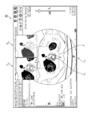
- the display 22 of the medical image viewer apparatus 20 the current image I 11, the current partial area image I 12 generated from the current image I 11, from the past image corresponding to the current image I 11 past generated partial area images I 13, the difference image I 14 of the past image corresponding to the current image I 11, and the frame F surrounding the region of interest of the current image I 11 is displayed.
- the frame F is composed of broken lines, but the color and line type of the frame F may be appropriately selected.
- the current image I 11 is displayed in the center of the display 22.
- the current partial region image I 12 and the past partial region image I 13 are displayed side by side on the current partial region image I 11.
- the current partial region image I 12 and the past partial region image I 13 are displayed at positions that do not overlap with the frame F.
- the difference image I 14 is superimposed and displayed on the current image I 11.
- the difference image I 14 is a difference image of the bone region between the current image I 11 and the corresponding past image. That is, in the example shown in FIG. 5, the bone difference display is performed.
- the difference image I 14 is overlaid on the current image I 11 , not only the past partial area image I 13 but also the current partial area image I 12 excluding the overlay display is displayed. Is preferable.
- the amount of movement of the line of sight during observation is reduced by displaying three images of the same region of interest at the same time and adjacent to each other.
- the current image, the current partial area image, the past partial area image, the difference image, and the frame are all displayed, but only necessary information may be displayed as appropriate.
- the past images may be displayed side by side.
- the past images by switching the past images with at least one of mouse wheel operation, right-click operation, and key operation, a certain display area is elapsed regardless of the number of past images. The observation may be performed smoothly.
- tumor registration has been performed in the past to present examination (tumor tracking)
- tumor tracking when an arbitrary tumor is specified in the current image, the vicinity of the same tumor in the past image is cut out and overlapped with the current image. It is conceivable to display it.
- FIG. 6 is a diagram showing a comparative display example of medical images when a tumor is designated. As shown in FIG. 6, the image I 21 is currently displayed on the display 22 of the medical image viewer device 20. The current image I 21 are included tumor region R 1.
- the medical image processing apparatus 12 acquires the past image including tumor region corresponding to the designated tumor region R 1 from the medical image storage apparatus 18 . Further, the medical image processing apparatus 12 generates a past partial region image I 22 from the past image after the current image I 21 and the past image are aligned. Further, the medical image processing device 12 outputs a signal for displaying the past partial region image I 22 overlaid on the current image I 21.
- the past partial region image I 22 is displayed on the display 22 overlaid on the current image I 21.
- the past partial region image I 22 includes a tumor region R 2 corresponding to the tumor region R 1 .
- the information T 1 of the tumor region R 1 it is displayed over the current image I 21.
- the information T 2 of the tumor region R 2 is superimposed on the past partial region image I 22.
- Information T 1 and information T 2 include the area, major axis, average value, maximum value, and minimum value of tumors R 1 and R 2, respectively. This information is stored in the medical image storage device 18 in association with the medical image. By displaying the tumor information in this way, it is possible to appropriately follow up the tumor.
- the mouse is used as the input device 24, but a keyboard, a touch panel, a touch pad, a trackball, a joystick, or the like may be used.
- the above medical image processing method is configured as a program for realizing each process on a computer, and a non-temporary recording medium such as a CD-ROM (Compact Disk-Read Only Memory) storing this program is configured. Is also possible.
- the processor 14 includes a CPU (Central Processing Unit), which is a general-purpose processor that executes software (program) and functions as various processing units, a GPU (Graphics Processing Unit), which is a processor specialized in image processing, and an FPGA. (Field Programmable Gate Array), etc. Designed exclusively for executing specific processing such as Programmable Logic Device (PLD), ASIC (Application Specific Integrated Circuit), which is a processor whose circuit configuration can be changed after manufacturing.
- a dedicated electric circuit or the like which is a processor having the above-mentioned circuit configuration, is included.
- One processing unit may be composed of one of these various processors, or two or more processors of the same type or different types (for example, a plurality of FPGAs, or a combination of a CPU and an FPGA, or a CPU and a CPU. It may be composed of a combination of GPUs). Further, a plurality of processing units may be configured by one processor. As an example of configuring a plurality of processing units with one processor, first, one processor is configured by a combination of one or more CPUs and software, as represented by a computer such as a server and a client. There is a form in which a processor functions as a plurality of processing units.
- SoC System On Chip
- a processor that realizes the functions of the entire system including a plurality of processing units with one IC (Integrated Circuit) chip is used.
- the various processing units are configured by using one or more various processors as a hardware-like structure.
Landscapes
- Health & Medical Sciences (AREA)
- Engineering & Computer Science (AREA)
- Life Sciences & Earth Sciences (AREA)
- Medical Informatics (AREA)
- Physics & Mathematics (AREA)
- Public Health (AREA)
- General Health & Medical Sciences (AREA)
- Nuclear Medicine, Radiotherapy & Molecular Imaging (AREA)
- Biomedical Technology (AREA)
- Radiology & Medical Imaging (AREA)
- Pathology (AREA)
- Biophysics (AREA)
- Heart & Thoracic Surgery (AREA)
- Molecular Biology (AREA)
- Surgery (AREA)
- Animal Behavior & Ethology (AREA)
- Veterinary Medicine (AREA)
- Theoretical Computer Science (AREA)
- General Physics & Mathematics (AREA)
- Computer Vision & Pattern Recognition (AREA)
- Epidemiology (AREA)
- Primary Health Care (AREA)
- High Energy & Nuclear Physics (AREA)
- Optics & Photonics (AREA)
- Databases & Information Systems (AREA)
- Oral & Maxillofacial Surgery (AREA)
- Dentistry (AREA)
- Data Mining & Analysis (AREA)
- Quality & Reliability (AREA)
- Apparatus For Radiation Diagnosis (AREA)
- Measuring And Recording Apparatus For Diagnosis (AREA)
- Image Analysis (AREA)
Priority Applications (3)
| Application Number | Priority Date | Filing Date | Title |
|---|---|---|---|
| DE112021001411.0T DE112021001411T5 (de) | 2020-03-05 | 2021-02-26 | Verarbeitungsvorrichtung für medizinische bilder, verarbeitungsverfahren für medizinische bilder, programm und anzeigesystem für medizinische bilder |
| JP2022505170A JP7394959B2 (ja) | 2020-03-05 | 2021-02-26 | 医用画像処理装置、医用画像処理方法及びプログラム、医用画像表示システム |
| US17/898,414 US12295757B2 (en) | 2020-03-05 | 2022-08-29 | Medical image processing device, method, and programs that cut out region from second medical image to generate second partial image for superimpose on first medical image, and medical image display system |
Applications Claiming Priority (2)
| Application Number | Priority Date | Filing Date | Title |
|---|---|---|---|
| JP2020037840 | 2020-03-05 | ||
| JP2020-037840 | 2020-03-05 |
Related Child Applications (1)
| Application Number | Title | Priority Date | Filing Date |
|---|---|---|---|
| US17/898,414 Continuation US12295757B2 (en) | 2020-03-05 | 2022-08-29 | Medical image processing device, method, and programs that cut out region from second medical image to generate second partial image for superimpose on first medical image, and medical image display system |
Publications (1)
| Publication Number | Publication Date |
|---|---|
| WO2021177157A1 true WO2021177157A1 (ja) | 2021-09-10 |
Family
ID=77614279
Family Applications (1)
| Application Number | Title | Priority Date | Filing Date |
|---|---|---|---|
| PCT/JP2021/007302 Ceased WO2021177157A1 (ja) | 2020-03-05 | 2021-02-26 | 医用画像処理装置、医用画像処理方法及びプログラム、医用画像表示システム |
Country Status (4)
| Country | Link |
|---|---|
| US (1) | US12295757B2 (https=) |
| JP (1) | JP7394959B2 (https=) |
| DE (1) | DE112021001411T5 (https=) |
| WO (1) | WO2021177157A1 (https=) |
Citations (7)
| Publication number | Priority date | Publication date | Assignee | Title |
|---|---|---|---|---|
| JP2006167169A (ja) * | 2004-12-16 | 2006-06-29 | Hitachi Medical Corp | 医用画像表示装置及びその方法 |
| JP2008212396A (ja) * | 2007-03-05 | 2008-09-18 | Fujifilm Corp | 画像処理装置およびそのプログラム |
| JP2008259622A (ja) * | 2007-04-11 | 2008-10-30 | Fujifilm Corp | レポート作成支援装置およびそのプログラム |
| JP2008289548A (ja) * | 2007-05-22 | 2008-12-04 | Toshiba Corp | 超音波診断装置及び診断パラメータ計測装置 |
| WO2010047324A1 (ja) * | 2008-10-20 | 2010-04-29 | 株式会社 日立メディコ | 医用画像処理装置、及び医用画像処理方法 |
| US20140016845A1 (en) * | 2011-04-08 | 2014-01-16 | Tiferet T. Gazit | Image analysis for specific objects |
| US20180024995A1 (en) * | 2015-02-03 | 2018-01-25 | Pusan National University Industry-University Cooperation Foundation | Medical information providing apparatus and medical information providing method |
Family Cites Families (8)
| Publication number | Priority date | Publication date | Assignee | Title |
|---|---|---|---|---|
| JP3758893B2 (ja) | 1999-04-03 | 2006-03-22 | コニカミノルタホールディングス株式会社 | 画像診断支援装置 |
| WO2005009242A1 (ja) | 2003-07-28 | 2005-02-03 | Hitachi Medical Corporation | 医用画像処理装置及び方法 |
| US7892177B2 (en) * | 2005-02-28 | 2011-02-22 | Scimed Life Systems, Inc. | Systems and methods for estimating the length and position of a stent to be applied within a patient |
| US8953865B2 (en) * | 2011-03-03 | 2015-02-10 | Hitachi Medical Corporation | Medical image processing device and medical image processing method |
| JP6344039B2 (ja) | 2014-04-28 | 2018-06-20 | 富士通株式会社 | 画像表示装置、画像表示方法及びプログラム |
| JP6827706B2 (ja) | 2015-09-09 | 2021-02-10 | キヤノン株式会社 | 情報処理装置及びその方法、情報処理システム、コンピュータプログラム |
| US20190164639A1 (en) * | 2017-07-19 | 2019-05-30 | Vijay Saaraswat | Automated Pharmaceutical Organizer and Dispenser |
| JP7235519B2 (ja) * | 2019-01-29 | 2023-03-08 | ザイオソフト株式会社 | 医用画像処理装置、医用画像処理方法、及び医用画像処理プログラム |
-
2021
- 2021-02-26 DE DE112021001411.0T patent/DE112021001411T5/de active Pending
- 2021-02-26 WO PCT/JP2021/007302 patent/WO2021177157A1/ja not_active Ceased
- 2021-02-26 JP JP2022505170A patent/JP7394959B2/ja active Active
-
2022
- 2022-08-29 US US17/898,414 patent/US12295757B2/en active Active
Patent Citations (7)
| Publication number | Priority date | Publication date | Assignee | Title |
|---|---|---|---|---|
| JP2006167169A (ja) * | 2004-12-16 | 2006-06-29 | Hitachi Medical Corp | 医用画像表示装置及びその方法 |
| JP2008212396A (ja) * | 2007-03-05 | 2008-09-18 | Fujifilm Corp | 画像処理装置およびそのプログラム |
| JP2008259622A (ja) * | 2007-04-11 | 2008-10-30 | Fujifilm Corp | レポート作成支援装置およびそのプログラム |
| JP2008289548A (ja) * | 2007-05-22 | 2008-12-04 | Toshiba Corp | 超音波診断装置及び診断パラメータ計測装置 |
| WO2010047324A1 (ja) * | 2008-10-20 | 2010-04-29 | 株式会社 日立メディコ | 医用画像処理装置、及び医用画像処理方法 |
| US20140016845A1 (en) * | 2011-04-08 | 2014-01-16 | Tiferet T. Gazit | Image analysis for specific objects |
| US20180024995A1 (en) * | 2015-02-03 | 2018-01-25 | Pusan National University Industry-University Cooperation Foundation | Medical information providing apparatus and medical information providing method |
Also Published As
| Publication number | Publication date |
|---|---|
| JPWO2021177157A1 (https=) | 2021-09-10 |
| JP7394959B2 (ja) | 2023-12-08 |
| US12295757B2 (en) | 2025-05-13 |
| US20220409147A1 (en) | 2022-12-29 |
| DE112021001411T5 (de) | 2023-04-13 |
Similar Documents
| Publication | Publication Date | Title |
|---|---|---|
| EP3069318B1 (en) | Registration of medical images | |
| EP3411818B1 (en) | System for image processing to generate three-dimensional (3d) view of an anatomical portion | |
| US9990744B2 (en) | Image registration device, image registration method, and image registration program | |
| US20200242776A1 (en) | Medical image processing apparatus, medical image processing method, and system | |
| JP6967983B2 (ja) | 画像処理装置、画像処理方法、及びプログラム | |
| JP6949535B2 (ja) | 情報処理装置、情報処理システム、情報処理方法及びプログラム | |
| KR101988531B1 (ko) | 증강현실 기술을 이용한 간병변 수술 내비게이션 시스템 및 장기 영상 디스플레이 방법 | |
| CN107633478B (zh) | 图像处理装置、图像处理方法以及计算机可读介质 | |
| US7277567B2 (en) | Medical visible image generating method | |
| US10896501B2 (en) | Rib developed image generation apparatus using a core line, method, and program | |
| WO2017154640A1 (ja) | 画像表示装置、方法およびプログラム | |
| JP7430249B2 (ja) | 画像処理装置、画像表示システム、画像処理方法及びプログラム | |
| CN115461782B (zh) | 用于配准外科手术空间的视觉表示的系统和方法 | |
| JP7039179B2 (ja) | 情報処理装置、情報処理システム、情報処理方法及びプログラム | |
| JP7394959B2 (ja) | 医用画像処理装置、医用画像処理方法及びプログラム、医用画像表示システム | |
| US12033366B2 (en) | Matching apparatus, matching method, and matching program | |
| Schenkenfelder et al. | Elastic registration of abdominal MRI scans and RGB-D images to improve surgical planning of breast reconstruction | |
| JP4099357B2 (ja) | 画像処理方法および装置 | |
| JP2012242938A (ja) | 読影レポート装置、読影レポートプログラム及び読影レポートシステム | |
| US12488479B2 (en) | Image alignment apparatus, method, and program | |
| JP2021097731A (ja) | 医用画像処理装置、及び医用画像処理システム | |
| JP5853785B2 (ja) | 医用画像表示システム | |
| JP2011193997A (ja) | 医用画像変換装置および方法並びにプログラム |
Legal Events
| Date | Code | Title | Description |
|---|---|---|---|
| 121 | Ep: the epo has been informed by wipo that ep was designated in this application |
Ref document number: 21764603 Country of ref document: EP Kind code of ref document: A1 |
|
| ENP | Entry into the national phase |
Ref document number: 2022505170 Country of ref document: JP Kind code of ref document: A |
|
| 122 | Ep: pct application non-entry in european phase |
Ref document number: 21764603 Country of ref document: EP Kind code of ref document: A1 |