WO2021149823A1 - 因子を導入された細胞の培養方法 - Google Patents
因子を導入された細胞の培養方法 Download PDFInfo
- Publication number
- WO2021149823A1 WO2021149823A1 PCT/JP2021/002336 JP2021002336W WO2021149823A1 WO 2021149823 A1 WO2021149823 A1 WO 2021149823A1 JP 2021002336 W JP2021002336 W JP 2021002336W WO 2021149823 A1 WO2021149823 A1 WO 2021149823A1
- Authority
- WO
- WIPO (PCT)
- Prior art keywords
- cells
- factor
- cell
- medium
- seeding
- Prior art date
Links
Images


Classifications
-
- C—CHEMISTRY; METALLURGY
- C12—BIOCHEMISTRY; BEER; SPIRITS; WINE; VINEGAR; MICROBIOLOGY; ENZYMOLOGY; MUTATION OR GENETIC ENGINEERING
- C12N—MICROORGANISMS OR ENZYMES; COMPOSITIONS THEREOF; PROPAGATING, PRESERVING, OR MAINTAINING MICROORGANISMS; MUTATION OR GENETIC ENGINEERING; CULTURE MEDIA
- C12N5/00—Undifferentiated human, animal or plant cells, e.g. cell lines; Tissues; Cultivation or maintenance thereof; Culture media therefor
- C12N5/06—Animal cells or tissues; Human cells or tissues
- C12N5/0602—Vertebrate cells
- C12N5/0696—Artificially induced pluripotent stem cells, e.g. iPS
-
- C—CHEMISTRY; METALLURGY
- C12—BIOCHEMISTRY; BEER; SPIRITS; WINE; VINEGAR; MICROBIOLOGY; ENZYMOLOGY; MUTATION OR GENETIC ENGINEERING
- C12N—MICROORGANISMS OR ENZYMES; COMPOSITIONS THEREOF; PROPAGATING, PRESERVING, OR MAINTAINING MICROORGANISMS; MUTATION OR GENETIC ENGINEERING; CULTURE MEDIA
- C12N2501/00—Active agents used in cell culture processes, e.g. differentation
- C12N2501/60—Transcription factors
- C12N2501/602—Sox-2
-
- C—CHEMISTRY; METALLURGY
- C12—BIOCHEMISTRY; BEER; SPIRITS; WINE; VINEGAR; MICROBIOLOGY; ENZYMOLOGY; MUTATION OR GENETIC ENGINEERING
- C12N—MICROORGANISMS OR ENZYMES; COMPOSITIONS THEREOF; PROPAGATING, PRESERVING, OR MAINTAINING MICROORGANISMS; MUTATION OR GENETIC ENGINEERING; CULTURE MEDIA
- C12N2501/00—Active agents used in cell culture processes, e.g. differentation
- C12N2501/60—Transcription factors
- C12N2501/603—Oct-3/4
-
- C—CHEMISTRY; METALLURGY
- C12—BIOCHEMISTRY; BEER; SPIRITS; WINE; VINEGAR; MICROBIOLOGY; ENZYMOLOGY; MUTATION OR GENETIC ENGINEERING
- C12N—MICROORGANISMS OR ENZYMES; COMPOSITIONS THEREOF; PROPAGATING, PRESERVING, OR MAINTAINING MICROORGANISMS; MUTATION OR GENETIC ENGINEERING; CULTURE MEDIA
- C12N2501/00—Active agents used in cell culture processes, e.g. differentation
- C12N2501/60—Transcription factors
- C12N2501/604—Klf-4
-
- C—CHEMISTRY; METALLURGY
- C12—BIOCHEMISTRY; BEER; SPIRITS; WINE; VINEGAR; MICROBIOLOGY; ENZYMOLOGY; MUTATION OR GENETIC ENGINEERING
- C12N—MICROORGANISMS OR ENZYMES; COMPOSITIONS THEREOF; PROPAGATING, PRESERVING, OR MAINTAINING MICROORGANISMS; MUTATION OR GENETIC ENGINEERING; CULTURE MEDIA
- C12N2501/00—Active agents used in cell culture processes, e.g. differentation
- C12N2501/60—Transcription factors
- C12N2501/606—Transcription factors c-Myc
-
- C—CHEMISTRY; METALLURGY
- C12—BIOCHEMISTRY; BEER; SPIRITS; WINE; VINEGAR; MICROBIOLOGY; ENZYMOLOGY; MUTATION OR GENETIC ENGINEERING
- C12N—MICROORGANISMS OR ENZYMES; COMPOSITIONS THEREOF; PROPAGATING, PRESERVING, OR MAINTAINING MICROORGANISMS; MUTATION OR GENETIC ENGINEERING; CULTURE MEDIA
- C12N2506/00—Differentiation of animal cells from one lineage to another; Differentiation of pluripotent cells
- C12N2506/13—Differentiation of animal cells from one lineage to another; Differentiation of pluripotent cells from connective tissue cells, from mesenchymal cells
- C12N2506/1307—Differentiation of animal cells from one lineage to another; Differentiation of pluripotent cells from connective tissue cells, from mesenchymal cells from adult fibroblasts
-
- C—CHEMISTRY; METALLURGY
- C12—BIOCHEMISTRY; BEER; SPIRITS; WINE; VINEGAR; MICROBIOLOGY; ENZYMOLOGY; MUTATION OR GENETIC ENGINEERING
- C12N—MICROORGANISMS OR ENZYMES; COMPOSITIONS THEREOF; PROPAGATING, PRESERVING, OR MAINTAINING MICROORGANISMS; MUTATION OR GENETIC ENGINEERING; CULTURE MEDIA
- C12N2513/00—3D culture
-
- C—CHEMISTRY; METALLURGY
- C12—BIOCHEMISTRY; BEER; SPIRITS; WINE; VINEGAR; MICROBIOLOGY; ENZYMOLOGY; MUTATION OR GENETIC ENGINEERING
- C12N—MICROORGANISMS OR ENZYMES; COMPOSITIONS THEREOF; PROPAGATING, PRESERVING, OR MAINTAINING MICROORGANISMS; MUTATION OR GENETIC ENGINEERING; CULTURE MEDIA
- C12N2533/00—Supports or coatings for cell culture, characterised by material
- C12N2533/50—Proteins
- C12N2533/52—Fibronectin; Laminin
-
- C—CHEMISTRY; METALLURGY
- C12—BIOCHEMISTRY; BEER; SPIRITS; WINE; VINEGAR; MICROBIOLOGY; ENZYMOLOGY; MUTATION OR GENETIC ENGINEERING
- C12N—MICROORGANISMS OR ENZYMES; COMPOSITIONS THEREOF; PROPAGATING, PRESERVING, OR MAINTAINING MICROORGANISMS; MUTATION OR GENETIC ENGINEERING; CULTURE MEDIA
- C12N2760/00—MICROORGANISMS OR ENZYMES; COMPOSITIONS THEREOF; PROPAGATING, PRESERVING, OR MAINTAINING MICROORGANISMS; MUTATION OR GENETIC ENGINEERING; CULTURE MEDIA ssRNA viruses negative-sense
- C12N2760/00011—Details
- C12N2760/18011—Paramyxoviridae
- C12N2760/18811—Sendai virus
Definitions
- the present invention relates to cell technology and relates to a method for culturing cells into which a factor has been introduced.
- Induced pluripotent stem (iPS) cells are cells that have two characteristic abilities. One is the ability to transform into all the cells that make up the body. The other is that it has a semi-permanent proliferative ability. Since iPS cells have these two abilities, they can be applied to transplantation therapy without rejection by producing iPS cells from their own somatic cells and converting them into target somatic cells. Therefore, iPS cells are considered to be a promising technique for regenerative medicine (see, for example, Patent Documents 1 to 4 and Non-Patent Documents 1 and 2). Conventionally, when a reprogramming factor is introduced into a cell and induced into an iPS cell, a stem cell-like colony is picked up with a pipette and subcultured while observing with a microscope or the like.
- One of the objects of the present invention is to provide an efficient method for culturing cells into which a factor has been introduced.
- a method for culturing a cell into which a factor has been introduced which comprises seeding the cell into which the factor has been introduced without cloning.
- a method for culturing factor-introduced cells which comprises exfoliating the factor-introduced cells from an incubator and mixing and seeding at least a part of the exfoliated cells.
- the detached cells may be mixed.
- a method for culturing factor-introduced cells which comprises recovering the factor-introduced cells from an incubator, mixing and seeding at least a part of the recovered cells. NS.
- the recovered cells may be mixed.
- a method for culturing a cell into which a factor has been introduced which does not pick up each of a plurality of colonies formed by the cell into which the factor has been introduced.
- a method for culturing a cell into which a factor has been introduced which comprises mixing and seeding cells derived from different single cells with each other.
- cells into which a factor has been introduced may be mixed by seeding.
- clones of cells into which the factor has been introduced may be mixed by seeding.
- different clones of the factor-introduced cells may be mixed by seeding.
- the above method does not have to include separating a plurality of colonies formed by the factor-introduced cells from each other before seeding.
- a plurality of colonies formed by cells into which a factor has been introduced may be mixed with each other.
- the above method does not have to involve cloning a single colony formed by the factor-introduced cells prior to seeding.
- the above method does not have to include picking up colonies formed by cells into which the factor has been introduced.
- cells into which a factor has been introduced and which are attached to an incubator may be recovered, and at least a part of the recovered cells may be seeded in a medium and subcultured.
- cells into which a factor has been introduced may be seeded without distinguishing them according to their gene expression status.
- cells into which a factor has been introduced may be seeded without distinction according to the degree of reprogramming.
- the above method may further include expanding the factor-introduced cells in a two-dimensional culture.
- the above method may further include expanding the factor-introduced cells in a three-dimensional culture.
- the above method may further include establishing stem cells from the cells into which the factor has been introduced.
- the seeded cells may be induced into pluripotent stem cells.
- the above method may further include inducing pluripotent stem cells into somatic cells.
- the above method may further comprise freezing the factor-introduced cells after seeding.
- the above method may further include differentiating the factor-introduced cells into at least one selected from endoderm, mesoderm, and ectoderm lines after seeding.
- the above method may further comprise forming at least one selected from embryoid bodies, organoids, and spheres from the factor-introduced cells after seeding.
- the above method may further include inducing the factor-introduced cells into somatic cells different from pluripotent stem cells after seeding.
- the above method may further include cloning the cells that have been treated to induce somatic cells after the treatment that induces them to somatic cells.
- the above method may further include performing gene editing treatment on the cell into which the factor has been introduced.
- the cell into which the factor has been introduced may be derived from blood cells or fibroblasts.
- the cell into which the factor is introduced may be a cell contained in urine.
- the cell into which the factor is introduced may be a bladder epithelial cell.
- the above method may further include collecting cells into which the factor is introduced from urine.
- the cell into which the factor has been introduced may be derived from a plurality of humans or a plurality of non-human animals.
- the cells into which the factor has been introduced may be cultured in a closed incubator.
- sowing may be carried out at a low concentration at the time of subculture.
- the low concentration may be 0.25 ⁇ 10 4 cells / cm 2 or less.
- the low concentration may be a concentration at which 11 or more seeded cells do not come into contact with each other.
- the low concentration may be 5% or less confluent.
- the factor may be RNA.
- the factor may be introduced into cells by the lipofection method.
- a factor may be introduced into cells using a viral vector.
- the viral vector may be an RNA viral vector.
- the RNA virus vector may be a Sendai virus vector.
- culturing the factor-introduced cells and inducing the factor-introduced cells into somatic cells different from pluripotent stem cells without subculture is provided.
- the above method may further include expanding the factor-introduced cells in a two-dimensional culture.
- the above method may further include expanding the factor-introduced cells in a three-dimensional culture.
- the above method may further include freezing the cells into which the factor has been introduced.
- the above method may further comprise differentiating the factor-introduced cells into at least one selected from endoderm, mesoderm, and ectoderm lines.
- the above method may further include performing gene editing treatment on the cell into which the factor has been introduced.
- the cell into which the factor has been introduced may be derived from blood cells or fibroblasts.
- the cell into which the factor has been introduced may be derived from a plurality of humans or a plurality of non-human animals.
- the cells into which the factor has been introduced may be cultured in a closed incubator.
- the factor may be RNA.
- the factor may be introduced into cells by the lipofection method.
- a factor may be introduced into cells using a viral vector.
- the viral vector may be an RNA viral vector.
- the RNA virus vector may be a Sendai virus vector.
- FIG. It is a graph which shows the measurement result by the flow cytometer which concerns on Reference Example 3.
- FIG. It is a graph which shows the measurement result by the flow cytometer which concerns on Reference Example 4.
- FIG. It is a photograph which shows the cell on the 15th day from the infection which concerns on Reference Example 4.
- FIG. It is a graph which shows the measurement result by the flow cytometer which concerns on Reference Example 4.
- FIG. It is a photograph which shows the cell of the 1st passage which concerns on Reference Example 4.
- FIG. 10 is a photograph of the Munch13 positive cell and the vGlut positive cell which concerns on Example 10. It is a photograph of cells derived from urine according to Example 11. It is a photograph of cells derived from urine according to Example 11. 12 is a photograph of urine-derived cells transfected with RNA encoding GFP according to Example 12.
- FIG. 3 is a photograph of urine-derived cells into which a reprogramming factor has been introduced according to Example 13.
- FIG. 6 is a photograph of urine-derived cells into which a reprogramming factor has been introduced according to Example 14.
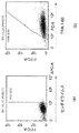
- FIG. 5 is a dot plot by a flow cytometer according to the fifteenth embodiment.
- FIG. 5 is a photograph of urine-derived cells into which a reprogramming factor has been introduced according to Example 16.
- FIG. 5 is a photograph of urine-derived cells into which a reprogramming factor has been introduced according to Example 16.
- the method for culturing the cells into which the reprogramming factor has been introduced according to the embodiment includes culturing the cells into which the reprogramming factor has been introduced, and subculturing the cells into which the reprogramming factor has been introduced without cloning. including.
- the method for culturing the cells into which the reprogramming factor was introduced according to the embodiment was to culture the cells into which the reprogramming factor was introduced and to detach the cells into which the reprogramming factor was introduced from the incubator. Includes mixing and subculturing at least a portion of the cells.
- the method for culturing the cells into which the reprogramming factor has been introduced according to the embodiment includes culturing the cells into which the reprogramming factor has been introduced, and collecting the cells into which the reprogramming factor has been introduced, and at least the recovered cells. Includes: mixing a portion and seeding in a medium for subculture.
- the method for culturing cells into which a reprogramming factor has been introduced according to the embodiment includes cells into which a reprogramming factor has been introduced, in which cells derived from different single cells are mixed and seeded. Cells into which reprogramming factors have been introduced are seeded and induced, for example, into pluripotent stem cells. Pluripotent stem cells are, for example, iPS cells.
- the cells into which the reprogramming factor is introduced are not particularly limited, and examples thereof include fibroblasts, blood cells, dental pulp stem cells, keratinocytes, dermal papilla cells, oral epithelial cells, and somatic stem progenitor cells.
- the cell into which the reprogramming factor is introduced may be a cell contained in urine. Examples of cells contained in urine include bladder epithelial cells.
- the cells into which the reprogramming factor is introduced may be of human or non-human animal origin.
- the cells into which the reprogramming factor is introduced may be of one human origin or of multiple human origins.
- the cell into which the reprogramming factor is introduced may be derived from one non-human animal or may be derived from a plurality of non-human animals.
- Blood cells are separated from the blood.
- Blood is, for example, peripheral blood and umbilical cord blood, but is not limited thereto. Blood may be collected from an adult or a minor.
- an anticoagulant such as ethylenediaminetetraacetic acid (EDTA), heparin, and biopharmacy standard blood preservation solution A (ACD-A) is used.
- EDTA ethylenediaminetetraacetic acid
- ACD-A biopharmacy standard blood preservation solution A
- Blood cells are nucleated cells such as mononuclear cells (Monocytes), neutrophils, macrophages, eosinophils, basophils, and lymphocytes, and do not contain erythrocytes, granulocytes, and platelets. Blood cells may be, for example, vascular endothelial progenitor cells, blood stem / progenitor cells, T cells, or B cells. T cells are, for example, ⁇ T cells.
- Mononuclear cells are separated from blood using a blood cell separation medium, a centrifuge, and the like.
- a blood cell separation medium a centrifuge, and the like.
- Ficoll GE Healthcare
- the method for separating mononuclear cells is as follows.
- the centrifuge To 5 mL of blood, add 5 mL of PBS to dilute, and layer 5 mL each on a medium for human lymphocyte separation in a tube. At this time, diluted blood is slowly added onto the medium along the tube wall of the tube so as not to disturb the interface.
- the method for separating mononuclear cells when using vacutainer (registered trademark, BD) as a blood collection tube is as follows.
- the centrifuge 4 ° C to 42 ° C, preferably 18 ° C.
- 4 ° C to 42 ° C preferably 18 ° C.
- 8 mL of blood is collected using a blood collection tube (vacutainer (registered trademark), BD), mixed by inversion, and mixed with an anticoagulant.
- the balance is then adjusted and the solution is adjusted at 4 ° C to 42 ° C, preferably 18 ° C, 100 ⁇ g to 3000 ⁇ g, preferably 1500 ⁇ g to 1800 ⁇ g with a swing rotor for 1 to 60 minutes, preferably 20 minutes.
- Centrifuge After centrifugation, the upper layer, which is the plasma layer, is removed and pipetting is performed to suspend the mononuclear cell layer and blood cells attached to the gel to obtain a suspension. Transfer the resulting suspension to another 15 mL tube.
- 1 mL to 14 mL, preferably 12 mL of PBS is added to the suspension in a 15 mL tube, and the suspension is prepared at 4 ° C to 42 ° C, preferably 18 ° C, 100 ⁇ g to 3000 ⁇ g, preferably 200 ⁇ g.
- a hemolytic agent PharmLyse (registered trademark), 10-fold concentration, BD
- BD sterile water
- the method for separating mononuclear cells from blood is not limited to the above method, and for example, a dialysis membrane may be used to separate mononuclear cells from blood.
- a pure cell select system for whole blood mononuclear cell concentration (registered trademark, PALL), a purifier for removing blood cells (cellsova E, registered trademark, Asahi Kasei), and a leukapheresis filter for platelet preparation (sepacell PL, registered trademark, Filters such as PLX-5B-SCD, Asahi Kasei) can also be used.
- Mononuclear cells may be separated using an erythrocyte separating agent capable of separating nucleated cells by gravity sedimentation or centrifugation of erythrocytes.
- erythrocyte separating agents include HetaSep (registered trademark, STEMCELL Technologies) and HES40 (NIPRO).
- CTL-UP1 sold by Cellular Technology Limited
- PBMC-001 of Sanguine Biosciences, or the like may be used as the mononuclear cell.
- the cryopreserved blood cells may be thawed and used using a cell cryopreservation solution such as Cerbunker 1, Stem Cerbunker GMP grade, and Stem Cerbunker DMSO-free GMP grade (Zenoac).
- a cell cryopreservation solution such as Cerbunker 1, Stem Cerbunker GMP grade, and Stem Cerbunker DMSO-free GMP grade (Zenoac).
- thawing mononuclear cells When thawing mononuclear cells, first, 1 mL to 15 mL, preferably 8 mL of a serum-free hematopoietic cell medium (X-VIVO® 10, Ronza) having a known composition was placed in a 15 mL tube and frozen. Place the tube containing the mononuclear cells in a warm bath at 4 ° C to 42 ° C, preferably 37 ° C to begin thawing the mononuclear cells. Then, with a little ice remaining, the tube containing the mononuclear cells is pulled out of the warm bath and the mononuclear cells are transferred to the tube containing the serum-free hematopoietic cell medium of known composition. Of these, 10 ⁇ L of mononuclear cell suspension is stained with trypan blue and counted on a hemocytometer.
- X-VIVO® 10 serum-free hematopoietic cell medium
- Blood cells may be isolated based on cell surface markers.
- Blood stem / progenitor cells are positive for CD34.
- the T cells are positive for any of CD3, CD4, and CD8.
- B cells are positive for any of CD10, CD19, and CD20.
- Macrophages are positive for any of CD11b, CD68, and CD163.
- Blood stem / progenitor cells, T cells, or B cells are separated from blood cells using, for example, an automatic magnetic cell separator and immunomagnetic beads. Alternatively, pre-separated mononuclear cells may be prepared. However, blood cells that have not been separated based on cell surface markers may be used.
- CD34-positive cells are stem / progenitor cells and tend to be easily reprogrammed. Further, when iPS cells are prepared using T cells which are CD3 positive cells, the iPS cells derived from the T cells retain the type of TCR recombination, so that they tend to be able to efficiently induce differentiation into T cells.
- the method for separating CD34-positive cells is as follows.
- 10 ⁇ L IL-6 (100 ⁇ g / mL), 10 ⁇ L SCF (300 ⁇ g / mL), 10 ⁇ L TPO (300 ⁇ g / mL), 10 ⁇ L FLT3 ligand (300 ⁇ g / mL) in 10 mL serum-free medium (StemSpan H3000, STEMCELL Technologies).
- 10 ⁇ L of IL-3 (10 ⁇ g / mL) are added to prepare a blood cell medium (blood stem / progenitor cell medium).
- 1 mL to 6 mL preferably 2 mL of blood cell medium in 1 well of a 6-well plate.
- 1 mL to 6 mL and 2 mL of PBS are placed in each of the other 5 wells to prevent evaporation of the medium.
- the 6-well plate is then placed in an incubator at 4 ° C to 42 ° C, preferably 37 ° C for warming.
- a mononuclear cell suspension containing 1 ⁇ 10 4 to 1 ⁇ 10 9 cells, preferably 2 ⁇ 10 7 mononuclear cells was dispensed into a 15 mL tube, and the mononuclear cell suspension was prepared.
- Centrifuge at 4 ° C to 42 ° C, preferably 4 ° C, 100 ⁇ g to 3000 ⁇ g, preferably 300 ⁇ g for 10 minutes. After centrifugation, the supernatant is removed and the mononuclear cells are suspended in 100 ⁇ L to 1 mL, preferably 300 ⁇ L of column buffer.
- 1 mL to 15 mL, preferably 10 mL of column buffer is added to the mononuclear cell suspension in a 15 mL tube to dilute it, and the mixture is diluted at 4 ° C to 42 ° C, preferably 4 ° C, 100 ⁇ g to 1000 ⁇ g. Centrifuge at 300 xg for 1 to 2 hours, preferably 10 minutes. After centrifugation, the supernatant in a 15 mL tube is removed with an ejector, and 10 ⁇ L to 10 mL, preferably 500 ⁇ L of column buffer is added and resuspended.
- a column for an automatic magnetic cell separation device (MS column, Miltenyi Biotec) is attached to an automatic magnetic cell separation device (MiniMACS Separation Unit, Miltenyi Biotec), and a column buffer of 10 ⁇ L to 10 mL, preferably 500 ⁇ L is put in the column and washed.
- a column buffer of 10 ⁇ L to 10 mL, preferably 500 ⁇ L is placed in the column, and the column is washed 1 to 10 times, preferably 3 times.
- the column is then removed from the automatic magnetic cell separator and placed in a 15 mL tube.
- the column is then filled with 10 ⁇ L to 10 mL, preferably 1000 ⁇ L of column buffer, and the syringe is swiftly pushed to allow CD34-positive cells to drain into a 15 mL tube.
- a 10 ⁇ L CD34-positive cell suspension is stained with trypan blue and the cell count is counted on a hemocytometer.
- the CD34-positive cell suspension in a 15 mL tube is centrifuged at 4 ° C to 42 ° C, preferably 4 ° C, 100 ⁇ g to 1000 ⁇ g, preferably 300 ⁇ g for 1 to 2 hours, preferably 10 minutes. .. After centrifugation, remove the supernatant with an ejector.
- the CD34-positive cells are resuspended in the warmed blood cell medium, and the CD34-positive cells are sprinkled on the culture plate. Then, the CD34-positive cells are cultured at 4 ° C. to 42 ° C., preferably 37 ° C., 1% to 20%, preferably 5% CO 2 for 6 days. During this time, the medium does not have to be exchanged.
- the method of isolating cells with a marker other than CD34 is the same as the method of isolating CD34-positive cells.
- RNA is, for example, mRNA.
- Reprogramming factors introduced into cells include, for example, OCT RNA such as OCT3 / 4, SOX RNA such as SOX2, KLF RNA such as KLF4, and MYC RNA such as c-MYC.
- OCT RNA such as OCT3 / 4
- SOX RNA such as SOX2
- KLF RNA such as KLF4
- MYC RNA such as c-MYC.
- reprogramming factor RNA it may be used M 3 O having improved OCT3 / 4.
- RNAs are LIN28A, FOXH1, LIN28B, GLIS1, p53-dominant negative, p53-P275S, L-MYC, NANOG, DPPA2, DPPA4, DPPA5, ZIC3, BCL-2, E-RAS, TPT1, SALL2. , NAC1, DAX1, TERT, ZNF206, FOXD3, REX1, UTF1, KLF2, KLF5, ESRRB, miR-291-3p, miR-294, miR-295, NR5A1, NR5A2, TBX3, MBD3sh, TH2A, TH2 It may further contain RNA of at least one factor selected from the group. These RNAs are available from TriLink.
- gene symbol is described here in humans, it is not intended to limit the species by uppercase or lowercase notation. For example, capitalization of all letters does not exclude the inclusion of mouse or rat genes. However, in the examples, the gene symbols are shown according to the species actually used.
- P53 is a cancer-suppressing protein.
- the dominant negative variant of p53 is not particularly limited as long as it can compete with the wild-type p53 protein endogenous to somatic cells and inhibit the function of the wild-type p53 protein.
- Examples of dominant negative variants of p53 are p53P275S, which is a point mutation of proline at position 275 (position 278 in the case of humans) located in the DNA binding region of mouse p53 to serine, and position 14-301 (human) of mouse p53.
- p53DD lacking the amino acid at positions 11-304, p53S58A in which serine at position 58 (61 in the case of humans) of mouse p53 was point-mutated to alanine, and position 135 of human p53 (mouse).
- p53A135V point-mutated alanine at position 135 (138 in the case of humans) of mouse p53 to valine
- position 172 in the case of humans
- RNA may be modified with psiduridine ( ⁇ ) or 5-methyluridine (5meU). RNA may be polyadenylated.
- the RNA introduced into the cell is, for example, a single-stranded RNA, and the double-stranded RNA may be substantially removed. Further, it is preferable that the RNA introduced into the cell is substantially free of impurities such as short RNA and impurities.
- the single-stranded RNA introduced into the cell may be purified and / or concentrated in order to substantially remove the double-stranded RNA. Examples of the method for purifying the single-stranded RNA to be introduced into cells include a purification method using high performance liquid chromatography (HPLC). For example, HPLC removes 70% or more, 75% or more, 80% or more, 85% or more, or 90% or more of double-stranded RNA.
- HPLC high performance liquid chromatography
- the RNA introduced into the cell may be treated with a ribonuclease that degrades the double-stranded RNA in order to substantially remove the double-stranded RNA.
- the RNA introduced into the cell may further include RNA in the transcriptional activation domain (TAD) of MYOD that is directly linked to the full length of OCT3 / 4 RNA.
- TAD transcriptional activation domain
- the reprogramming factor is introduced into cells by, for example, the lipofection method.
- the lipofection method is a method in which a complex of a negatively charged nucleic acid and a positively charged lipid is formed by electrical interaction, and the complex is incorporated into the cell by endocytosis or membrane fusion. ..
- the lipofection method has advantages such as less damage to cells, excellent introduction efficiency, easy operation, and less time.
- the reprogramming factor is introduced into cells cultured using, for example, an RNA transfection reagent.
- RNA may be introduced into the mononuclear cell immediately after the mononuclear cell is separated from the blood.
- RNA transfection reagent Lipofectamine Messenger MAX (registered trademark, Thermo Fisher SCIENTIFIC) can be used.
- RNA transfection reagent for example, Lipofectamine (registered trademark) RNAiMAX (Thermo Fisher SCIENTIFIC), Lipofectamine StemTransfection Reagent (Thermo Fisher SCIENTIFIC), Transfection (ReproCELL), Jet Messenger (Polyplus), Lipofectamin (registered trademark) 2000, Lipofectamin (registered trademark) 3000, NeonTransfection System (Thermo Fisher SCIENTIFIC), Stemfect RNA transfection reagent (Stemfect), NextFect (registered trademark) RNA Transfection Reagent (BiooSientific ), Amaxa (registered product) Human T cell Nucleector (registered product) kit (Lonza, VAPA-1002), Amaxa (registered product) Human CD34 cell Nucleector (
- a reprogramming factor is introduced into the cell using, for example, a viral vector.
- the viral vector may be an RNA viral vector.
- the RNA viral vector may be a Sendai viral vector.
- the Sendai virus vector may be a temperature-sensitive Sendai virus vector in which the stability of the viral nucleic acid decreases above a predetermined temperature.
- the viral nucleic acid of the temperature-sensitive Sendai virus vector is stable below a predetermined temperature.
- the viral nucleic acid may be viral DNA or viral RNA.
- the viral nucleic acid may be a viral genome. The decrease in the stability of the viral nucleic acid may be at least one of the degradation of the viral nucleic acid and the suppression of the replication or proliferation of the viral nucleic acid.
- Decreased stability of viral nucleic acids reduces at least one of viral nucleic acid proliferation, viral nucleic acid replication rate, and gene expression levels.
- the predetermined temperature is, for example, 36.5 ° C or higher and 37.5 ° C or lower, 36.6 ° C or higher and 37.4 ° C or lower, 36.7 ° C or higher, 37.3 ° C or lower, 36.8 ° C or higher and 37.2.
- the temperature is 36.9 ° C or higher and 37.1 ° C or lower, or 37 ° C.
- At least one of the viral nucleic acid stability of the temperature-sensitive Sendai virus vector, i.e., growth, replication rate and gene expression level is high below a given temperature and low above a given temperature.
- the temperature-sensitive Sendai virus vector has a growth rate or gene expression level of 1/2 or less in cells cultured at 37 ° C. as compared with a growth rate or gene expression level in cells cultured at 32 ° C. , 1/3 or less, 1/5 or less, 1/10 or less, or 1/20 or less.
- Sendai virus encodes N gene, P gene, M gene, F / HN gene, and L gene.
- the HN protein recognizes sialic acid on the cell surface when Sendai virus attaches to the cell and anchors the virus particles to the cell.
- the F protein is cleaved and activated by an extracellular protease to catalyze the fusion of the tethered Sendai virus envelope and the cell membrane of the target cell to establish an infection.
- the L protein, along with its modified protein, the P protein catalyzes the replication of viral nucleic acids in the cytoplasm after infection and transcription from the replicated multi-copy nucleic acids.
- the Sendai virus vector By deleting the F gene in the Sendai virus vector, it is possible to suppress the production of infectious virus particles from the transgenic cells. Moreover, it is possible to make the Sendai virus vector temperature sensitive by introducing a mutation into at least one of the L gene and the P gene.
- TS7 L protein Y942H / L1361C / L1558I mutation
- TS12 P protein D433A / R434A / K437A mutation
- TS13 P protein D433A / R434A / K437A
- Mutations and L protein L1558I mutations TS14 (P protein D433A / R434A / K437A mutations and L protein L1361C mutations)
- TS15 P protein D433A / R434A / K437A mutations and L protein L1361C / L1558I mutations
- Sendai viral vectors have, for example, G69E, T116A, and A183S mutations in the M protein, A262T, G264R, and K461G mutations in the HN protein, L511F mutations in the P protein, and N1197S in the L protein. It is an F gene deletion ( ⁇ F) type Sendai virus vector having the K1795E mutation, and is a Sendai virus vector into which the above-mentioned TS7, TS12, TS13, TS14, or TS15 mutation has been introduced.
- the temperature-sensitive mutation of the Sendai virus vector is not limited to these.
- the Sendai virus vector is, for example, SeV (PM) / TS ⁇ F, SeV18 + / TS ⁇ F, or SeV (HNL) / TS ⁇ F, and is a Sendai virus vector into which the above-mentioned TS7, TS12, TS13, TS14, or TS15 mutation has been introduced.
- the temperature-sensitive mutation of the Sendai virus vector is not limited to these.
- the Sendai virus vector introduced into cells may be a combination of a temperature-sensitive Sendai virus vector and a temperature-insensitive Sendai virus vector.
- the Sendai virus vector introduced into the cell is only the temperature-sensitive Sendai virus vector, and does not have to contain the temperature-insensitive Sendai virus vector.
- the Sendai virus vector introduced into cells is only a temperature-sensitive Sendai virus vector into which a mutation of TS7, TS12, TS13, TS14, or TS15 has been introduced, and does not have to contain a temperature-insensitive Sendai virus vector.
- the Sendai virus vector introduced into cells is only the temperature-sensitive Sendai virus vector having the same or higher temperature sensitivity as the temperature-sensitive Sendai virus vector into which the mutation of TS7, TS12, TS13, TS14, or TS15 has been introduced, and the temperature-insensitive Sendai. It does not have to contain a viral vector.
- the Sendai virus vector introduced into cells is only the Sendai virus vector having the same or higher temperature sensitivity as the temperature sensitive Sendai virus vector into which the mutation of TS7, TS12, TS13, TS14, or TS15 has been introduced, and TS7, TS12, It does not have to contain a Sendai virus vector that is less temperature sensitive than the temperature sensitive Sendai virus vector into which the TS13, TS14, or TS15 mutations have been introduced.
- the Sendai virus vector introduced into cells carries an arbitrary reprogramming factor.
- the Sendai virus vector introduced into the cell contains, for example, a temperature-sensitive Sendai virus vector containing KLF RNA, OCT RNA, and SOX RNA in this order and not containing MYC RNA, and MYC RNA containing KLF. RNA, OCT RNA, and SOX RNA-free temperature-sensitive Sendai virus vector.
- the number, combination, and order of the reprogramming factors loaded on the Sendai virus vector are arbitrary and are not particularly limited.
- the Sendai virus vector introduced into the cell may contain a Sendai virus vector containing KLF RNA and not containing OCT RNA and SOX RNA.
- the Sendai virus vector containing KLF RNA and not OCT RNA and SOX RNA may be a temperature-sensitive Sendai virus vector or a temperature-insensitive Sendai virus vector.
- the Sendai virus vector disappears earlier from the cells into which the Sendai virus vector has been introduced when the temperature-insensitive Sendai virus vector is not introduced.
- Temperature-sensitive Sendai virus vectors containing KLF RNA, OCT RNA, and SOX RNA have, for example, mutations in G69E, T116A, and A183S in the M protein and mutations in A262T, G264R, and K461G in the HN protein.
- An F gene-deficient Sendai virus vector having an L511F mutation in the P protein and N1197S and K1795E mutations in the L protein, and having the above-mentioned TS7, TS12, TS13, TS14, or TS15 mutation. It is a virus vector.
- the temperature sensitive mutation is, for example, TS7 or TS12, or TS12.
- the temperature sensitive Sendai virus vector containing KLF RNA, OCT RNA, and SOX RNA is, for example, SeV (PM) KOS / TS7 ⁇ F or SeV (PM) KOS / TS12 ⁇ F, or SeV (PM) KOS / TS12 ⁇ F. be.
- a temperature-sensitive Sendai virus vector containing MYC RNA has, for example, mutations in G69E, T116A, and A183S in M protein, mutations in A262T, G264R, and K461G in HN protein, and L511F mutation in P protein. It is an F gene-deficient Sendai virus vector having N1197S and K1795E mutations in L protein, and is a Sendai virus vector having the above-mentioned TS7, TS12, TS13, TS14, or TS15 mutations.
- the temperature sensitive mutation is, for example, TS15.
- Temperature sensitive Sendai virus vectors containing MYC RNA are, for example, SeV (HNL) MYC / TS12 ⁇ F, SeV (HNL) MYC / TS13 ⁇ F, or SeV (HNL) MYC / TS15 ⁇ F, or SeV (HNL) MYC / TS15 ⁇ F. be.
- Sendai viral vectors containing KLF RNA but not OCT RNA and SOX RNA have, for example, mutations in G69E, T116A, and A183S in the M protein and mutations in A262T, G264R, and K461G in the HN protein. It is an F gene-deficient Sendai virus vector having an L511F mutation in the P protein and an N1197S and K1795E mutation in the L protein.
- the Sendai virus vector containing KLF RNA and not containing OCT RNA and SOX RNA is, for example, less temperature sensitive than the Sendai virus vector into which the above-mentioned TS7, TS12, TS13, TS14, or TS15 mutation has been introduced, and is predetermined.
- the KLF gene can be expressed even at temperatures above.
- the Sendai virus vector containing KLF RNA but not OCT RNA and SOX RNA is, for example, SeV18 + KLF4 / TS ⁇ F.
- multiple types of Sendai virus vectors are introduced into cells at the same time.
- multiple types of Sendai virus vectors are introduced into cells at the same time.
- the multiplicity of infection (MOI) of the Sendai viral vector when infecting cells is, for example, 0.1 or more, 0.3 or more, 0.5 or more, 1.0 or more, 2.0 or more, 3.0 or more. It is 4.0 or more, or 5.0 or more.
- the MOI is, for example, 100 or less, 90 or less, 80 or less, 70 or less, 60 or less, 50 or less, 40 or less, 30 or less, 20 or less, 10 or less, or 5 or less.
- the temperature at which cells are infected with the Sendai virus vector is below a predetermined temperature at which the stability of the virus nucleic acid of the temperature-sensitive Sendai virus vector decreases, that is, the temperature at which the virus nucleic acid of the temperature-sensitive Sendai virus vector is stable. It may be above a predetermined temperature.
- the temperature at which cells are infected with the Sendai virus vector reduces the stability of the viral nucleic acid of the temperature-sensitive Sendai virus vector. It is preferable that the temperature is lower than the predetermined temperature, that is, the temperature at which the viral nucleic acid of the temperature-sensitive Sendai virus vector is stable.
- the cells into which the reprogramming factor is introduced may be adherently cultured or suspendedly cultured.
- Somatic cells into which reprogramming factors are introduced are feeder-free using basement membrane matrices such as Matrigel (Corning), CELLstart (registered trademark, Thermo Fisher), Laminin 511 (iMatrix-511, nippi), fibronectin, and vitrotin. It may be cultured.
- basement membrane matrices such as Matrigel (Corning), CELLstart (registered trademark, Thermo Fisher), Laminin 511 (iMatrix-511, nippi), fibronectin, and vitrotin. It may be cultured.
- a stem cell medium such as a human ES / iPS medium such as Prime ES Cell Medium (ReproCELL) can be used.
- the stem cell medium is not limited to this, and various stem cell media can be used.
- the stem cell medium is placed in an incubator such as a dish, well, or tube, for example.
- the gel medium is, for example, a stem cell medium containing gellan gum at a final concentration of 0.001% by mass to 0.5% by mass, 0.005% by mass to 0.1% by mass, or 0.01% by mass to 0.05% by mass. It is prepared by adding so as to become.
- Gel media include gellan gum, hyaluronic acid, lambzan gum, dieutan gum, xanthan gum, carrageenan, fucoidan, pectin, pectinic acid, pectinic acid, heparan sulfate, heparan, heparitin sulfate, keratosulfate, chondroitin sulfate, deltaman sulfate, ramnan sulfate, and It may contain at least one polymer compound selected from the group consisting of those salts.
- the gel medium may contain methyl cellulose. By including methyl cellulose, agglutination between cells is further suppressed.
- the gel medium is poly (glycerol monomethacrylate) (PGMA), poly (2-hydroxypropylmethacrylate) (PHPMA), Poly (N-isopropylacrylamide) (PNIPAM), amine terminated, carboxylic acid terminated, maleimide terminated, N-hydroxysuccinimide (N-hydroxysuccinimide).
- PGMA poly (glycerol monomethacrylate)
- PPMA poly (2-hydroxypropylmethacrylate)
- PIPAM Poly (N-isopropylacrylamide)
- amine terminated carboxylic acid terminated
- maleimide terminated N-hydroxysuccinimide (N-hydroxysuccinimide).
- the gel medium may or may not contain a growth factor such as basic fibroblast growth factor (bFGF).
- bFGF basic fibroblast growth factor
- the gel medium may contain a growth factor such as bFGF at a low concentration of 400 ⁇ g / L or less, 40 ⁇ g / L or less, or 10 ⁇ g / L or less.
- the gel medium may or may not contain TGF- ⁇ , or may contain TGF- ⁇ at a low concentration of 600 ⁇ g / L or less, 300 ⁇ g / L or less, or 100 ⁇ g / L or less.
- the gel medium does not have to be agitated. Further, the gel medium does not have to contain feeder cells.
- the gel medium may contain at least one substance selected from the group consisting of cadherin, laminin, fibronectin, and vitronectin.
- a temperature lower than a predetermined temperature at which the stability of the viral nucleic acid of the temperature-sensitive Sendai virus vector decreases that is, a temperature-sensitive Sendai virus.
- the cells may be cultured at a temperature at which the vector viral nucleic acid is stable. Then, the cells may be cultured at a predetermined temperature or higher.
- the medium may be changed, for example, once every two days while culturing the cells above a predetermined temperature.
- At least 2 days, or 2 days or more and 10 days or less for example, 4.0 ° C or higher, 10 ° C or higher, 15 ° C or higher, 20 ° C or higher, 25 ° C or higher, 30 ° C or higher.
- Cells may be cultured at temperatures below 36.6 ° C, below 36.5 ° C, below 36.0 ° C, below 35.0 ° C, or below 34.0 ° C.
- the temperature is raised to 36.5 ° C. or higher, 36.6 ° C. or higher, 36.7 ° C. or higher, 36.8 ° C. or higher, 36.9 ° C. or higher, or 37.0 ° C. or higher, 40.0 ° C.
- the cells may be cultured at a temperature of 39.0 ° C. or lower, or 38.0 ° C. or lower.
- the temperature may be raised once or stepwise. After raising the temperature, the medium may be changed while culturing the cells, for example once every two days.
- the temperature-sensitive Sendai virus vector viral nucleic acid becomes less stable than a predetermined temperature, that is, the temperature-sensitive Sendai viral vector viral nucleic acid
- the cells may be cultured at a stable temperature. After the stem cell-like colonies begin to appear, the cells may be cultured above a predetermined temperature. The medium may be changed, for example, once every two days while culturing the cells above a predetermined temperature.
- stem cell-like colonies After infecting cells with Sendai virus vector, until stem cell-like colonies begin to appear, for example, 4.0 ° C or higher, 10 ° C or higher, 15 ° C or higher, 20 ° C or higher, 25 ° C or higher, 30 ° C or higher, 31.
- ° C or higher 32.0 ° C or higher, 33.0 ° C or higher, 33.1 ° C or higher, 33.2 ° C or higher, 33.3 ° C or higher, 33.4 ° C or higher, 33.5 ° C or higher, 33.6 ° C 33.7 ° C or higher, 33.8 ° C or higher, or 33.9 ° C or higher, less than 37.0 ° C, less than 36.9 ° C, less than 36.8 ° C, less than 36.7 ° C, 36.6 Cells may be cultured at temperatures below ° C., below 36.5 ° C., below 36.0 ° C., below 35.0 ° C., or below 34.0 ° C.
- the temperature is raised to 36.5 ° C or higher, 36.6 ° C or higher, 36.7 ° C or higher, 36.8 ° C or higher, 36.9 ° C or higher, or 37.0 ° C.
- cells may be cultured at a temperature of 40.0 ° C. or lower, 39.0 ° C. or lower, or 38.0 ° C. or lower.
- the temperature may be raised once or stepwise. After raising the temperature, the medium may be changed while culturing the cells, for example once every two days.
- the cells into which the reprogramming factor has been introduced are collected, and at least a passage is performed in which at least a part of the mixed cells is seeded in the medium at least once. do.
- clones of cells into which reprogramming factors have been introduced may be mixed together.
- different clones of cells into which reprogramming factors have been introduced may be mixed together.
- the cells into which the reprogramming factor has been introduced may be collected, and at least a part of the collected and mixed cells may be seeded in a medium and subcultured a plurality of times.
- the cells into which the reprogramming factor has been introduced may be recovered, and at least a part of the recovered mixed cells may be seeded in a medium and subcultured. In addition, all the collected mixed cells may be seeded in the medium.
- the cells into which the reprogramming factor has been introduced are collected, and at least a part of the collected and mixed cells is seeded in a medium and subcultured. It refers to passage without distinction according to the expression state. For example, at the time of passage, cells into which a reprogramming factor has been introduced may be seeded in the same incubator without distinguishing by gene expression status. Alternatively, the cells into which the reprogramming factor has been introduced are collected, and at least a part of the collected and mixed cells is seeded in a medium and subcultured. For example, the cells into which the reprogramming factor has been introduced are reprogrammed. It means to pass on without distinguishing by the degree of. For example, at the time of passage, cells into which a reprogramming factor has been introduced may be seeded in the same incubator without distinction according to the degree of reprogramming.
- the cells into which the reprogramming factor has been introduced are collected, and at least a part of the collected and mixed cells is seeded in a medium and subcultured, for example, the cells into which the reprogramming factor has been introduced are expressed in morphology. It means to succeed without distinction.
- cells into which a reprogramming factor has been introduced may be seeded in the same incubator without distinguishing by morphology.
- the cells into which the reprogramming factor has been introduced are collected, and at least a part of the collected and mixed cells is seeded in a medium and subcultured.
- the cells into which the reprogramming factor has been introduced are sized. It means to succeed without distinction.
- cells into which a reprogramming factor has been introduced may be seeded in the same incubator without distinction by size.
- the cells into which the reprogramming factor has been introduced are recovered, and at least a part of the collected and mixed cells is seeded in a medium and subcultured without cloning the cells into which the reprogramming factor has been introduced. It means to succeed. For example, in the case of passage without cloning, it is not necessary to pick up the colonies formed by the cells into which the reprogramming factor has been introduced. For example, when passaged without cloning, it is not necessary to separate multiple colonies formed by cells into which the reprogramming factor has been introduced. For example, at the time of passage, cells forming a plurality of different colonies may be mixed and seeded in the same incubator.
- colonies may be mixed and seeded in the same incubator.
- cells into which a reprogramming factor has been introduced are adherently cultured, even if the adherently cultured cells are collected, and at least a part of the collected and mixed cells is seeded in a medium and subcultured. good.
- all cells may be exfoliated from the incubator and at least some of the exfoliated and mixed cells may be seeded in the same incubator.
- all cells may be stripped from the incubator with a stripping solution, and the stripped and mixed cells as a whole may be passaged. For example, cells that do not form colonies may be passaged. If the cells into which the reprogramming factor has been introduced are suspension-cultured, the entire suspension-cultured cells may be passaged.
- the cells When subculturing cells that have been introduced with reprogramming factors, the cells are seeded in medium or incubator at low concentrations.
- the low concentration is, for example, 1 cell / cm 2 or more, 0.25 ⁇ 10 4 cells / cm 2 or less, 1.25 ⁇ 10 3 cells / cm 2 or less, 0.25 ⁇ 10 3 cells /. It is cm 2 or less, 0.25 ⁇ 10 2 cells / cm 2 or less, or 0.25 ⁇ 10 1 cells / cm 2 or less.
- low concentration means that 10 or less, 9 or less, 8 or less, 7 or less, 6 or less, 5 or less, 4 or less, 3 or less, or 2 or less cells can come into contact with each other.
- the concentration at which 11 or more cells do not come into contact with each other there is a concentration at which 11 or more cells do not come into contact with each other.
- the state where the entire bottom surface of the cell container is covered with cells is defined as 100% confluent, and low concentration means 5% or less confluent, 4% or less confluent, 3% or less confluent, 2% or less confluent, 1% or less confluent, 0. It is 5.5% or less confluent, 0.1% or less confluent, 0.05% or less confluent, or 0.01% or less confluent.
- the low concentration is, for example, a concentration at which single cells do not come into contact with each other in the seeded cells.
- single cells may be seeded in the wells of the well plate.
- the well plate may be a 12-well plate or a 96-well plate.
- the induced pluripotent stem cells the proportion of cells in which Sendai virus remains is, for example, 4% or less, 3% or less, 2% or less, 1% or less, 0.5% or less, or 0%.
- the cells may be cultured at a temperature higher than a predetermined temperature at which the stability of the viral nucleic acid of the temperature-sensitive Sendai virus vector decreases. After passage, the cells are cultured, for example, at a temperature of 36.5 ° C. or higher and lower than 38.0 ° C. After passage, for example, cells are cultured at a temperature of 36.5 ° C. or higher and lower than 38.0 ° C. until cell-cell adhesion starts, and after cell-cell adhesion starts, a higher temperature until cell-cell adhesion starts. For example, cells may be cultured at a temperature of 37.5 ° C. or higher, 42.0 ° C.
- the temperature is 37.5 ° C or higher, 42.0 ° C or lower, 41.5 ° C or lower, 41.0 ° C or lower, 40.5 ° C or lower, or 40.0 ° C. or lower. ..
- the temperature is 37.5 ° C or higher, 42.0 ° C or lower, 41.5 ° C or lower, 41.0 ° C or lower, 40.5 ° C or lower, or 40.0 ° C.
- the cells may be cultured at the following temperatures.
- Cells into which the reprogramming factor has been introduced may be cultured and subcultured in a closed incubator.
- the closed incubator does not exchange gas, virus, microorganisms, impurities, etc. with the outside, for example.
- cells into which a reprogramming factor has been introduced may be expanded and cultured in a two-dimensional culture, or may be expanded and cultured in a three-dimensional culture.
- the entire cells that have been adherently cultured may be cryopreserved as pluripotent stem cells.
- the entire cells exfoliated from the incubator with the exfoliating solution may be cryopreserved as pluripotent stem cells.
- the entire cells in suspension culture may be cryopreserved as pluripotent stem cells.
- the induced pluripotent stem cells can form flat colonies similar to ES cells and express alkaline phosphatase.
- the induced pluripotent stem cells can express Nanog, OCT4, SOX2, etc., which are undifferentiated cell mer powers.
- Induced pluripotent stem cells can express TERT.
- Induced pluripotent stem cells may exhibit temouth mellase activity.
- a cytoflow meter which is a cell surface marker indicating that the cells were undifferentiated, such as TRA-1-60, TRA-1-81, SSEA-1, and. This may be done by analyzing whether at least one surface marker selected from SSEA5 is positive.
- TRA-1-60 is an antigen specific for iPS / ES cells. Since iPS cells can be produced only from the TRA-1-60 positive fraction, TRA-1-60 positive cells are considered to be the species of iPS cells.
- the induced pluripotent stem cells may be induced into somatic cells in a state different from that of the pluripotent stem cells.
- somatic cells include nerve cells, retinal epithelial cells, hepatocytes, ⁇ cells, renal cells, dental pulp stem cells, mesenchymal stem cells, somatic stem progenitor cells, keratinocytes, dermal papilla cells, oral epithelial cells, cartilage cells, Examples include muscle cells, vascular cells, epithelial cells, myocardial cells, blood cells, and immune cells.
- blood cells include erythroblasts, red blood cells, megakaryocytes, and platelets.
- immune cells examples include monocytes, neutrophils, eosinophils, basophils, B cells, T cells, NK cells, and NKT cells.
- the induced stem cells may be differentiated into endoderm, mesoderm, or ectoderm lines. Stem cells may form embryoid bodies, organoids, and spheres.
- Examples of factors that induce cells into nervous system cells include the ASCL family, DLX family, MYT family, NeuroD family, SOX family, and NGN family.
- An example of the ASCL family is ASCL1.
- An example of the DLX family is DLX2.
- An example of the MYT family is MYT1L.
- An example of the NGN family is NGN2.
- Examples of neural cells include neural cells, neural stem cells and neural progenitor cells.
- Examples of neurons include inhibitory neurons, excitatory neurons and dopamine-producing neurons, cranial nerves, intervening nerves, and optic nerves.
- the nervous system cells may be motor neurons, oligodendrocyte progenitor cells, astrocytes, oligodendrocytes, and the like.
- Examples of factors that induce cells into cardiomyocytes include the GATA family, MEF family, TBX family, MYOCD family, MESS family, and miR-133 family.
- GATA family is GATA4A.
- MEF family is MEF2C.
- TBX family is TBX5.
- MESS family is MESS1.
- induction refers to reprogramming, reprogramming, transformation, transdifferentiation or lineage reprogramming, differentiation induction, cell fate reprogramming, and the like.
- the induced pluripotent cells may be cloned.
- Gene-edited cells may be cloned after gene-editing the established pluripotent stem cells.
- the cells into which the reprogramming factor has been introduced may be induced into somatic cells different from the pluripotent stem cells without substituting the cells into which the reprogramming factor has been introduced at all.
- the method for introducing the reprogramming factor into the cell is as described above.
- a differentiation-inducing factor is introduced into a cell into which a reprogramming factor has been introduced, and a cell into which the reprogramming factor has been introduced is referred to as a pluripotent stem cell, without subculturing the cell into which the reprogramming factor has been introduced. May induce to different somatic cells.
- the cells into which the reprogramming factor has been introduced are given hormones or chemical substances, and the cells into which the reprogramming factor has been introduced are referred to as pluripotent stem cells. May induce to different somatic cells.
- the cells into which the reprogramming factor has been introduced are induced into somatic cells without being cloned.
- the somatic cells induced are as described above.
- Example 1 Comparative Example 1
- the dish coated with laminin 511 was used as a dish for inducing pluripotent stem cells.
- human peripheral blood mononuclear cells were suspended in a blood medium, and the number of mononuclear cells was measured using a hemocytometer to adjust the number of mononuclear cells in the blood medium. Then, mononuclear cells were two-dimensionally cultured at 37 ° C. for 1 to 7 days on a dish for inducing pluripotent stem cells.
- SeV (PM) hKOS / TS12 ⁇ F and SeV (HNL) hC-Myc / TS15 ⁇ F were added to the two-dimensionally cultured mononuclear cells so that the MOI was 5, and a dish for inducing pluripotent stem cells was added.
- a dish for inducing pluripotent stem cells was added.
- a stem cell-like cell mass developed. On the 14th day after infection, almost all cells became TRA1-60 positive cells and showed iPS cell-like morphology.
- triple select a cell stripping agent, was added to the dish and allowed to stand at room temperature for 1 minute, then the cell-containing solution was aspirated and the cell-containing solution was incubated at 37 ° C. for 5 to 10 minutes. .. Then, iPS cell medium was added, and the iPS cell medium containing the cells was collected in a 15 mL tube.
- Example 1 The number of cells was measured using a hemocytometer, the concentration of the solution containing the cells was adjusted, and the cells were seeded on a well plate so that the concentration was 0.25 ⁇ 10 4 cells / cm 2 or less, and the first passage was performed. I made a charge.
- Example 1 at the time of the first passage, all the cells were exfoliated from the well plate, and the exfoliated and mixed cells were seeded on the next well plate without distinction.
- Comparative Example 1 at the time of the first passage, colony picking was performed and cloning was performed. In both Example 1 and Comparative Example 1, cells were seeded so that 11 or more cells did not come into contact with each other during passage.
- the Weldish was then placed in an incubator at 37 ° C. and the cells were two-dimensionally cultured. After the cells began to divide, the culture temperature was raised to 38 ° C. Then, in both Example 1 and Comparative Example 1, all the cells were collected every time the cells became 60% to 80% confluent, and at least a part of the collected and mixed cells was seeded in a medium and subcultured. .. During the second and subsequent passages, the cells were seeded on the well plate so that the concentration was 0.25 ⁇ 10 4 cells / cm 2 or less. Also at this time, 11 or more cells did not come into contact with each other.
- the number of colonies formed 5 days after the intracellular Sendai virus disappeared was counted. Furthermore, the clonal efficiency was calculated by dividing the number of colonies by the number of seeded cells. The results of the three tests are shown in FIG. When all cells were collected at the time of the first passage using mTeSR Plus as the medium, and some of the collected and mixed cells were seeded in the medium and subcultured, the clonal efficiency was about 5%. It was about 8%, and the variation was small. When colony picking and cloning using mTeSR Plus as a medium during the first passage, the clonal efficiency may be less than 1% or about 6%, resulting in clonal efficiency. There was variation.
- the clonal efficiency was about 10% to about. It was 15%, and the variation was small.
- the clonal efficiency may be less than 1% or about 16%, and the clonal efficiency varies. was there.
- the clonal efficiency can be obtained. It was shown to be high and stable.
- Example 2 Comparative Example 2
- the iPS cell-like cells obtained by subculture in the same manner as in Example 1 and Comparative Example 1 were exfoliated and dissociated.
- about 1 ⁇ 10 5 cells were cryopreserved using STEM-CELLBANKER®.
- the frozen cells were thawed, about 1 ⁇ 10 4 cells were seeded in wells, and the cells were cultured and proliferated.
- the number of cells and the number of colonies 7 days after seeding were measured. The results of the three tests are shown in FIGS. 5 and 6.
- the cells were collected at the time of the first passage, and a part of the collected and mixed cells was seeded in a medium and subcultured.
- the cells were cultured for 7 days after freezing and thawing.
- the number was about 10 ⁇ 10 4 to about 15 ⁇ 10 4 , and the variation was small.
- the cells cloned by colony picking at the time of the first passage have a number of cells cultured for 7 days after freezing and thawing, which is about 0.4 ⁇ 10 4 to about 15 ⁇ 10 4 . There was a lot of variation.
- Example 3 Comparative Example 3
- the iPS cell-like cells obtained by subculture in the same manner as in Example 1 and Comparative Example 1 were exfoliated, 2.5 ⁇ 10 5 cells were suspended in a gel medium, and the cells were three-dimensionally cultured. , The cells were allowed to form a clamp. The number of clamps and the number of cells were measured 13 days after seeding the cells in the gel medium. The results of testing the cells obtained in Example 1 twice and the cells obtained in Comparative Example 1 four times are shown in FIG.
- Example 4 Comparative Example 4
- the iPS cell-like cells obtained by subculture in the same manner as in Example 1 and Comparative Example 1 were exfoliated, and iPS cell-like cells were used using a cardiomyocyte differentiation induction kit (PSC Cardiomyocyte Differentiation Kit, Gibco, registered trademark). Cells were differentiated into cardiomyocytes.
- PSC Cardiomyocyte Differentiation Kit Gibco, registered trademark
- iPS cell-like cells were seeded on a 12-well plate whose bottom surface was treated with a basement membrane matrix (Corning Matrigel, registered trademark), and mTeSR1 was placed in the medium.
- the cells were cultured using. Two days after seeding the iPS cell-like cells, the medium was exchanged with Cardiomyocyte Differentiation Medium A. Two days later, the medium was exchanged with Cardiomyocyte Differentiation Medium B. Two days later, the medium was exchanged at Cardiomyocyte Maintenance Medium. Then, every two days, the cells were cultured until the 22nd day after seeding the iPS cell-like cells while exchanging the medium with the Cardiomyocyte Maintenance Medium.
- cTnT myocardial troponin T
- Example 5 Comparative Example 5
- the iPS cell-like cells obtained by subculture in the same manner as in Example 1 and Comparative Example 1 were exfoliated, and 1.5 ⁇ 10 5 cells were round-bottomed bottom having a size suitable for cell mass formation.
- the cells were seeded and cultured on a cell culture plate (Kurare, RB 500 400 NA 6).
- the medium was a selective inhibitor of TGF- ⁇ 1 activin receptor-like kinase (ALK) ALK-4, -5, -7 (500 nmol / L, A-83-01, Stemgent) and BMP Type I receptor (ALK2, 8GMK medium (8% KnockOut Serum Receptors) to which an inhibitor of membrane permeability of ALK3) (100 nmol / L, LDN193189, Stemgent) was added (Life Technologies), 1% non-essential amino acids (NEAA, Life Technologies), 1% non-essential amino acids (NEAA, Life Technologies) Sodium (Sigma), 100 nmol / L 2-mercaptoethanol (2-ME, Life Technologies) was used.
- Pluripotent stem cells cultured in the presence of the above inhibitors may be induced to differentiate into neural precursor cells. Are known.
- the cells were collected in a tube and centrifuged, the cell mass was decomposed into a single cell with a cell dissociator (TripLE Select, ThermoFisher, registered trademark), the number of cells was counted, and then a nerve cell adhesion molecule (N).
- Cells were immunostained using PSA-NCAM antibody, which is an antibody that detects a polysialization molecule of -CAM), and the positive rate of PSA-NCAM was analyzed by flow cytometry.
- PSA-NCAM antibody which is an antibody that detects a polysialization molecule of -CAM
- the cells subcultured by seeding at least a part of the cells in the medium had a positive rate of PSA-NCAM of about 25% to about 30%, and the variation was small.
- cells cloned by colony picking during the first passage after introduction of a reprogramming factor before induction of differentiation into neural progenitor cells have a PSA-NCAM positive rate of about 5%. It was about 15% from, and the variation was large.
- Example 6 Comparative Example 6
- the iPS cell-like cells obtained by subculture in the same manner as in Example 1 and Comparative Example 1 were exfoliated, and 1 ⁇ 10 5 cells were seeded on a non-adhesive dish and cultured.
- As the medium a human ES cell medium to which bFGF was not added was used.
- the medium was changed once every two days.
- the formed embryoid bodies (EB) were reseeded on gelatin-coated 6-well plates. Then, the medium was exchanged once every two days, and the cells were collected by trypsin treatment on the 24th day after reseeding.
- the collected cells were fixed with 4% paraformaldehyde, the cells were stained with each of SOX1 antibody, OTC2 antibody, HAND1 antibody, and SOX17 antibody, and the stained cells were analyzed using a flow cytometer.
- SOX1 and OTX2 are ectoderm markers
- HAND1 is a mesoderm marker
- SOX17 is an endoderm marker.
- SeV (PM) hKOS / TS12 ⁇ F, SeV18 + hKLF4 / TS ⁇ F, and SeV (HNL) hC-Myc / TS15 ⁇ F were added to the two-dimensionally cultured fibroblasts so that the MOI was 5, and pluripotent stem cells were added.
- the induction dish was placed in an incubator at 34 ° C. and the cells were cultured. Two days after infection, the blood medium was replaced with iPS cell medium. Then, the medium was exchanged once every two days using the iPS cell medium. By 14 days after infection, the culture temperature was gradually increased to 37 ° C and 38 ° C.
- the weldish was then placed in an incubator and the cells were two-dimensionally cultured. After the cells began to divide, the culture temperature was adjusted to 38 ° C. The cells were then passaged each time the cells became 60% to 80% confluent. During the second and subsequent passages, the cells were seeded on the well plate so that the concentration was 0.25 ⁇ 10 4 cells / cm 2 or less. Also at this time, 11 or more cells did not come into contact with each other.
- SeV (PM) hKOS / TS12 ⁇ F, SeV18 + hKLF4 / TS ⁇ F, and SeV (HNL) hC-Myc / TS15 ⁇ F were added to the two-dimensionally cultured mononuclear cells so that the MOI was 5, and they were pluripotent.
- the stem cell-inducing dish was placed in an incubator at 37 ° C., and the cells were cultured. Two days after infection, the blood medium was replaced with iPS cell medium. Then, the medium was exchanged once every two days using the iPS cell medium.
- a stem cell-like cell mass developed.
- the cells were two-dimensionally cultured. After the cells began to divide, the culture temperature was raised to 38 ° C. The cells were then passaged each time the cells became 60% to 80% confluent. From the second passage to the fifth passage, the cells were seeded on the well plate so that the concentration was higher than 0.25 ⁇ 10 4 cells / cm 2 so that 11 or more cells adhered to each other. From the 6th passage onward, cells were seeded on a well plate so that the concentration was 0.25 ⁇ 10 4 cells / cm 2 or less. At this time, 11 or more cells did not come into contact with each other.
- CytoTune-iPS2.0 (registered trademark, ID Pharma), which is a Sendai virus vector kit, was prepared.
- CytoTune-iPS2.0 contains SeV (PM) hKOS / TS12 ⁇ F, which is a temperature-sensitive Sendai viral vector carrying the KLF4 gene, Oct3 / 4 gene, and SOX2 gene as reprogramming factors, and the temperature at which KLF4 is loaded as the reprogramming factor. It contains the sensitive Sendai viral vector SeV18 + hKLF4 / TS ⁇ F and the temperature sensitive Sendai viral vector SeV (HNL) hC-Myc / TS15 ⁇ F carrying c-MYC as a reprogramming factor.
- SeV (PM) hKOS / TS12 ⁇ F which is a temperature-sensitive Sendai viral vector carrying the KLF4 gene, Oct3 / 4 gene, and SOX2 gene as reprogramming factors, and the temperature at which KLF4 is loaded as the reprogramm
- SeV (PM) hKOS / TS12 ⁇ F, SeV18 + hKLF4 / TS ⁇ F, and SeV (HNL) hC-Myc / TS15 ⁇ F were added to mononuclear cells so that the MOI was 5, and a dish for inducing pluripotent stem cells was added.
- the cells were cultured in an incubator at 37 ° C. Two days after the infection, the blood medium was replaced with iPS cell medium (mTeSR Plus (STEMCELL Technologies) or StemFit (Ajinomoto)). Then, the medium was exchanged once every two days using the iPS cell medium.
- a stem cell-like cell mass developed. On the 14th day after infection, almost all cells became TRA1-60 positive cells and showed iPS cell-like morphology.
- triple select a cell stripping agent, was added to the dish and allowed to stand at room temperature for 1 minute, then the cell-containing solution was aspirated and the cell-containing solution was incubated at 37 ° C. for 5 to 10 minutes. .. Then, iPS cell medium was added, and the iPS cell medium containing the cells was collected in a 15 mL tube.
- the number of cells was measured using a hemocytometer, the concentration of the solution containing the cells was adjusted, and the cells were seeded on a well plate so that the concentration was 0.25 ⁇ 10 4 cells / cm 2 or less, and the first passage was performed. I made a charge. At this time, 11 or more cells did not come into contact with each other.
- the Weldish was then placed in an incubator at 37 ° C. and the cells were two-dimensionally cultured. The cells were then passaged each time the cells became 60% to 80% confluent. During the second and subsequent passages, the cells were seeded on the well plate so that the concentration was 0.25 ⁇ 10 4 cells / cm 2 or less. Also at this time, 11 or more cells did not come into contact with each other.
- a stem cell-like cell mass developed. On the 14th day after infection, almost all cells became TRA1-60 positive cells and showed iPS cell-like morphology. Fifteen days after infection, triple select, a cell stripping agent, was added to the dish and allowed to stand at room temperature for 1 minute, then the cell-containing solution was aspirated and the cell-containing solution was incubated at 37 ° C. for 5 to 10 minutes. .. Then, iPS cell medium was added, and the iPS cell medium containing the cells was collected in a 15 mL tube.
- the number of cells was measured using a hemocytometer, the concentration of the solution containing the cells was adjusted, and the cells were seeded on a well plate so that the concentration was 0.25 ⁇ 10 4 cells / cm 2 or less, and the first passage was performed. I made a charge. At this time, 11 or more cells did not come into contact with each other.
- the Weldish was then placed in an incubator at 38 ° C. and the cells were two-dimensionally cultured. The cells were then passaged each time the cells became 60% to 80% confluent. During the second and subsequent passages, the cells were seeded on the well plate so that the concentration was 0.25 ⁇ 10 4 cells / cm 2 or less. Also at this time, 11 or more cells did not come into contact with each other.
- FIG. 17 shows the results of staining the cells before passage on the 15th day after infection using an anti-Sendai virus antibody and evaluating the Sendai virus remaining in the cells with a flow cytometer.
- FIG. 19 shows the results of staining the cells after one passage with an anti-Sendai virus antibody and evaluating the Sendai virus remaining in the cells with a flow cytometer. As shown in FIG. 19, the intracellular Sendai virus was almost eliminated in the first passage. A photograph of the cells in the first passage is shown in FIG.
- TRA1-60 positive cells On the 14th day after infection.
- iPS cell-like colonies were formed.
- the cell mass is collected using a mesh, triple select, which is a cell release agent, is added to the collected cells, and the cells are allowed to stand at room temperature for 5 minutes, and then the solution containing the cells is aspirated to obtain the solution containing the cells.
- iPS cell medium was added, and the iPS cell medium containing the cells was collected in a 15 mL tube.
- the number of cells was measured using a hemocytometer, the concentration of the solution containing the cells was adjusted, and the cells were seeded on a well plate so that the concentration was 0.25 ⁇ 10 4 cells / cm 2 or less, and the first passage was performed. I made a charge. At this time, 11 or more cells did not come into contact with each other.
- the Weldish was then placed in an incubator at 37 ° C. and the cells were two-dimensionally cultured. The cells were then passaged each time the cells became 60% to 80% confluent. During the second and subsequent passages, the cells were seeded on the well plate so that the concentration was 0.25 ⁇ 10 4 cells / cm 2 or less. Also at this time, 11 or more cells did not come into contact with each other. The temperature was raised to 38 ° C on the way.
- SeV (PM) hKOS / TS12 ⁇ F and SeV (HNL) hC-Myc / TS15 ⁇ F were added to the two-dimensionally cultured fibroblasts so that the MOI was 5, and a dish for inducing pluripotent stem cells was added.
- the cells were cultured in an incubator at 34 ° C. Two days after infection, the blood medium was replaced with iPS cell medium. Then, the medium was exchanged once every two days using the iPS cell medium.
- a stem cell-like cell mass developed. On the 14th day after infection, almost all cells became TRA1-60 positive cells and showed iPS cell-like morphology.
- triple select a cell stripping agent, was added to the dish and allowed to stand at room temperature for 1 minute, then the cell-containing solution was aspirated and the cell-containing solution was incubated at 37 ° C. for 5 to 10 minutes. .. Then, iPS cell medium was added, and the iPS cell medium containing the cells was collected in a 15 mL tube.
- the number of cells was measured using a hemocytometer, the concentration of the solution containing the cells was adjusted, and the cells were seeded on a well plate so that the concentration was 0.25 ⁇ 10 4 cells / cm 2 or less, and the first passage was performed. I made a charge. At this time, 11 or more cells did not come into contact with each other.
- the well dish was then placed in an incubator at 37 ° C. and the cells were two-dimensionally cultured. The cells were then passaged each time the cells became 60% to 80% confluent. During the second and subsequent passages, the cells were seeded on the well plate so that the concentration was 0.25 ⁇ 10 4 cells / cm 2 or less. Also at this time, 11 or more cells did not come into contact with each other.
- Example 7 DMEM containing 10% FBS was prepared as a medium for fibroblasts. Fibroblasts were suspended in a human adult-derived fibroblast medium to obtain a fibroblast suspension.
- a 6-well dish To 1 well of a 6-well dish, 1.5 mL of PBS and 4.8 ⁇ L of a mixture of silk moth-derived laminin (iMatrix-511 silk, Nippi) were added, and the dish was placed in an incubator at 37 ° C. for 1 hour. Next, the mixture of PBS and laminin was removed from the wells using an ejector, and 1.5 mL of a fibroblast suspension was added to one well. The number of fibroblasts in 1 well was 2.0 ⁇ 10 5 cells from 0.5 ⁇ 10 5. Then, the fibroblasts were cultured for 1 day in an incubator at 37 ° C.
- a mixture of silk moth-derived laminin iMatrix-511 silk, Nippi
- the amount of medium exchanged was also 1.5 mL.
- Tube A and tube B were prepared, and a mixture of OCT4 RNA, SOX2 RNA, KLF4 RNA, and C-MYC RNA (100 ng / ⁇ L) was added to 125 ⁇ L PBS in tube A from 0.1 ⁇ L to 100 ⁇ L. .. These RNAs were concentrated and purified by HPLC. 0.1 ⁇ L to 100 ⁇ L of lipofection reagent was added to 125 ⁇ L of PBS in tube B. Next, the solution in tube A and the solution in tube B were mixed, the mixture was left at room temperature for 10 minutes, and the entire mixture was added to the medium in 1 well. The dishes were then placed in an incubator at 37 ° C. for 1 day to transfect the cells with RNA. After that, RNA transfection by the same procedure was repeated 11 times.
- Example 7 The day after the 11th RNA transfection, the concentration of the solution containing the cells was adjusted and the cells were seeded on a laminin-coated well plate so that the concentration was 0.25 ⁇ 10 4 cells / cm 2 or less. I took over.
- Example 7 at the time of the first passage, all the cells detached from the well plate were collected, and at least a part of the collected and mixed cells was seeded in the next well plate without distinction.
- Comparative Example 7 colony picking and cloning were performed at the time of the first passage. In both Example 7 and Comparative Example 7, cells were seeded so that 11 or more cells did not come into contact with each other during passage. The Weldish was then placed in an incubator at 37 ° C.
- the cells were two-dimensionally cultured.
- the cells were then passaged only once when the cells became 60% to 80% confluent.
- the cells were seeded on the well plate so that the concentration was 0.25 ⁇ 10 4 cells / cm 2 or less. At this time, 11 or more cells did not come into contact with each other.
- the stem cells established by the method according to Example 7 had a small variation in clonal efficiency, as in Example 1.
- the stem cells established by the method according to Example 7 had stable growth rates and colony forming ability, as in Example 2.
- the stem cells established by the method according to Example 7 had a small variation in the number of clamps and the number of cells after three-dimensional culture, as in Example 3.
- the stem cells established by the method according to Example 7 had a stable ability to induce differentiation into cardiomyocytes, as in Example 4.
- the stem cells established by the method according to Example 7 had a stable ability to induce differentiation into neural progenitor cells, as in Example 5.
- the stem cells established by the method according to Example 7 had stable differentiation potential into endoderm, mesoderm, and ectoderm, as in Example 6.
- the stem cells established by the method according to Comparative Example 7 had a large variation in clonal efficiency, as in Comparative Example 1.
- the stem cells established by the method according to Comparative Example 7 had a large variation in proliferation rate and colony forming ability, as in Comparative Example 2.
- the stem cells established by the method according to Comparative Example 7 had a large variation in the number of clamps and the number of cells after three-dimensional culture, as in Comparative Example 3.
- the stem cells established by the method according to Comparative Example 7 had unstable ability to induce differentiation into cardiomyocytes.
- the stem cells established by the method according to Comparative Example 7 had unstable ability to induce differentiation into neural progenitor cells.
- the stem cells established by the method according to Comparative Example 7 had unstable differentiation potential into endoderm, mesoderm, and ectoderm.
- Example 8 Similar to Example 1, a reprogramming factor was introduced into mononuclear cells. On the 14th day after infection, almost all cells became TRA1-60 positive cells and showed iPS cell-like morphology. Then, iPS cell-like cells were infected with Sendai virus capable of expressing Ngn2-Puro RNA so that the MOI became 20 at the stage where the cells were never subcultured. On the second day after infection with Sendai virus, the medium in the well was replaced with a nerve induction medium (N3 medium) containing puromycin at a concentration of 2 ⁇ g / mL, and uninfected cells were killed. The N3 medium was prepared by adding 10 mL of B27, 5 mL of N2, and 1.6 mL of insulin at a concentration of 6.25 mg / mL to 500 mL of DMEMF12.
- N3 medium was prepared by adding 10 mL of B27, 5 mL of N2, and 1.6 mL of insulin at a concentration
- FIG. 28 shows a micrograph of cells 14 days after infection with Sendai virus. As shown in FIG. 28, it was morphologically confirmed that the cells were induced into nervous system cells after infection with Sendai virus.
- Example 9 Similar to Example 1, a reprogramming factor was introduced into mononuclear cells. On the 14th day after infection, almost all cells became TRA1-60 positive cells and showed iPS cell-like morphology. Then, iPS cell-like cells were transplanted into immunodeficient mouse testis at a stage where the cells had never been subcultured. After a few weeks, teratomas were removed from the mice, tissue sections were prepared from the removed teratomas, stained with hematoxylin and eosin (HE), and observed under a microscope. The results are shown in FIG. Secretory tissue-like structure, neural tube-like structure, intestinal-like structure, and cartilage and bone-like structure were observed in the tissue section.
- HE hematoxylin and eosin
- RNA was introduced into the chimpanzee-derived fibroblasts in the same manner as in Example 1 except that the chimpanzee-derived fibroblasts were used.
- iPS cell-like colonization was observed as shown in FIG.
- the maintenance-cultured cells were immunostained with an antibody against Oct3 / 4
- the cells showed Oct3 / 4 positive as shown in FIG. 31 (a).
- the maintenance-cultured cells were immunostained with an antibody against Nanog
- the cells showed Nanog positivity as shown in FIG. 31 (b).
- FIG. 32 it was confirmed that the induced cells were TRA-1-60 positive.
- the established chimpanzee-derived stem cells were induced to differentiate into cardiomyocytes.
- a photograph of the obtained cardiomyocytes is shown in FIG.
- the established chimpanzee-derived stem cells were induced to differentiate into nerve cells.
- a photograph of the obtained cardiomyocytes is shown in FIG. Nerve cells were confirmed to be Munch13 positive and vGlut positive.
- Example 11 300 mL of urine from a healthy subject was collected, 6 urine was dispensed into a 50 mL falcon tube, and the tube was centrifuged at 400 G for 5 minutes. After centrifugation, the supernatant was removed from the tube, 30 mL of PBS was placed in the tube, and the tube was centrifuged at 400 G for 5 minutes. After centrifugation, the supernatant was removed from the tube, 30 mL of primary medium was placed in the tube and the tube was centrifuged at 400 G for 5 minutes.
- the primary medium was DMEM / Ham's F12 (Gibco, 11320-033), fetal bovine serum (Gibco, 10437028, final concentration 10%), SingleQuots Kit CC-4127 REGM (Lonza, 1/1000 amount), and Prepared by adding Antibiotic-Antimycotic (Gibco, 15240062, 1/100 amount). After centrifugation, the supernatant was removed from the tube, the cells were suspended in 1 mL of primary medium, the cells were seeded in 1 well of a gelatin-coated 24-well plate, and the cells were incubated in a 37 ° C. incubator.
- the primary medium was added to a 300 ⁇ L well, and from the 3rd day onward, the medium was replaced with a medium for epithelial cells.
- the medium for epithelial cells was prepared by adding SingleQuots Kit CC-4127 REGM (Lonza) to the renal epithelial cell basal medium (Lonza).
- a microscopic image of the cells after expanded culture for 6 days is shown in FIG. 35. The cells were subcultured for the first time on the 7th day after seeding, and the cells were further expanded and cultured, and the cells were subcultured for the second time on the 7th day from the first passage.
- a microscopic image of the cells 6 days after the 2nd passage is shown in FIG.
- Example 12 The dish coated with laminin 511 was used as a dish for inducing pluripotent stem cells.
- the urine-derived cells prepared in Example 11 were seeded in a dish for inducing pluripotent stem cells from 1 ⁇ 10 4 to 1 ⁇ 10 5 and incubated at 37 ° C.
- the medium used was a medium for epithelial cells.
- a mixture of transfection reagent and RNA encoding green fluorescent protein (GFP) was added to the medium, the medium was exchanged using the medium, and the medium was incubated at 37 ° C.
- a microscopic image of the cells the next day is shown in FIG. 37. Expression of GFP was observed, indicating that urine-derived cells can be transfected.
- GFP green fluorescent protein
- Example 13 The dish coated with laminin 511 was used as a dish for inducing pluripotent stem cells.
- the urine-derived cells prepared in Example 11 were seeded in a dish for inducing pluripotent stem cells from 1 ⁇ 10 4 to 1 ⁇ 10 5 and incubated at 37 ° C.
- the medium used was a medium for epithelial cells.
- the next day prepare the tubes A and tubes B, M 3 O of RNA of PBS 125 ⁇ L in the tube A, SOX2 RNA of, KLF4 of RNA, C-MYC of RNA, and RNA mixtures of LIN28 (100ng / ⁇ L) It was added 10 2 [mu] L from 0.1 [mu] L.
- RNAs were modified with psidouridine ( ⁇ ).
- RNAs were substantially concentrated and purified into single-stranded RNA by HPLC.
- the ratio of the absorbance of RNA at 260 nm to 280 nm was 1.71 to 2.1, confirming that the protein was substantially free of contamination.
- dot blot analysis was performed using the anti-double-stranded RNA antibody J2, 90% or more of the double-stranded RNA was removed.
- 0.1 ⁇ L to 100 ⁇ L of lipofection reagent was added to 125 ⁇ L of PBS in tube B.
- the solution in tube A and the solution in tube B are mixed, the mixed solution is left at room temperature for 10 minutes, and the entire mixed solution is added to the transfection medium without using B18R or the like, and the transfection medium is added.
- the medium was exchanged using the transfection medium and incubated at 37 ° C. Transfection was performed once daily for 10 days. Observation was performed on days 1, 5, 7, and 14 after cell seeding. As shown in FIG. 38, it was observed that the cell morphology changed like ES cells as the day progressed.
- Example 14 Similar to Example 13, urine-derived cells were transfected for 10 days. From the 11th day after seeding the cells, the cells were cultured in a stem cell medium (StemFit, Ajinomoto), and on the 14th day, all the cells were peeled from the dish, and some of the collected and mixed cells were seeded in the medium. And succeeded. At the time of passage, the colonies were not picked up, the entire cells on the dish were collected, and 1 ⁇ 10 2 to 1 ⁇ 10 5 cells were seeded on the dish. A microscopic image of the cells 6 days after passage is shown in FIG. ES cell-like cells were confirmed.
- Example 15 Similar to Example 13, urine-derived cells were transfected for 10 days. The cells were seeded, the cells were cultured in a stem cell medium (StemFit, Ajinomoto) from the 11th day, the cells were peeled from the dish on the 14th day, and some cells were analyzed by flow cytometry. ), It was confirmed that it was TRA-1-60 positive. Further, when the cells detached from the dish were subcultured on the 14th day and analyzed by flow cytometry 7 days later, it was confirmed that they were TRA-1-60 positive as shown in FIG. 40 (b). rice field.
- stem cell medium Stema, Ajinomoto
- Example 16 Similar to Example 13, urine-derived cells were transfected for 10 days. From the 11th day after seeding the cells, the cells were cultured in a stem cell medium (StemFit, Ajinomoto), and on the 14th day, all the cells were peeled from the dish, and a part of the peeled and mixed cells was seeded and subcultured. did. StemFit® was used as the medium after passage. Seven days after passage, cells were fixed and stained with anti-OCT3 / 4 antibody and anti-NANOG antibody. In addition, chemical staining of nuclei with Hoechst® was also performed. As a result, as shown in FIG.
- FIG. 41 (d) shows a photograph of cells stained with an anti-OCT3/4 antibody, a photograph of cells stained with an anti-NANOG antibody, and a photograph of cells stained with Hoechst (registered trademark). And, it is a composite photograph.
- Example 17 Similar to Example 13, urine-derived cells were transfected for 10 days. From the 11th day after seeding the cells, the cells were cultured in a stem cell medium (StemFit, Ajinomoto), and on the 14th day, all the cells were peeled from the dish using Triple Select, and some of the peeled and mixed cells were peeled off. The cells were seeded in a medium and subcultured. StemFit® was used as the medium after passage. Seven days after passage, cells were fixed and stained with anti-LIN28 antibody. In addition, chemical staining of nuclei with Hoechst® was also performed. As a result, as shown in FIG.
- FIG. 42 (d) is a photograph obtained by synthesizing a photograph of cells stained with an anti-LIN28 antibody and a photograph of cells stained with Hoechst (registered trademark).
Landscapes
- Health & Medical Sciences (AREA)
- Engineering & Computer Science (AREA)
- Life Sciences & Earth Sciences (AREA)
- Biomedical Technology (AREA)
- Bioinformatics & Cheminformatics (AREA)
- Wood Science & Technology (AREA)
- Biotechnology (AREA)
- Organic Chemistry (AREA)
- Chemical & Material Sciences (AREA)
- Genetics & Genomics (AREA)
- Zoology (AREA)
- Developmental Biology & Embryology (AREA)
- Microbiology (AREA)
- Transplantation (AREA)
- Biochemistry (AREA)
- General Engineering & Computer Science (AREA)
- General Health & Medical Sciences (AREA)
- Cell Biology (AREA)
- Micro-Organisms Or Cultivation Processes Thereof (AREA)
Abstract
本開示によれば、因子を導入された細胞を培養することと、因子を導入された細胞を回収し、回収した細胞の少なくとも一部を培地に播種して播種することと、を含む、因子を導入された細胞の培養方法が提供される。また、因子を導入された細胞を培養することと、因子を導入された細胞を継代せずに、多能性幹細胞とは異なる体細胞に誘導すること、を含む、因子を導入された細胞の培養方法が提供される。
Description
本発明は、細胞技術に関し、因子を導入された細胞の培養方法に関する。
人工多能性幹(iPS)細胞とは、二つの特徴的な能力を有する細胞である。一つは、体を構成するあらゆる細胞へと変化することができる能力である。もう一つは、半永久的な増殖能を有することである。iPS細胞はこの二つの能力を有するため、自己の体細胞からiPS細胞を作製し、目的の体細胞へ変化させることで、拒絶反応のない移植治療へと応用することができる。そのためiPS細胞は、再生医療の有力な技術になると考えられている(例えば、特許文献1から4及び非特許文献1、2参照。)。従来、細胞にリプログラミング因子を導入し、iPS細胞に誘導する際には、顕微鏡等で観察しながら、幹細胞様コロニーをピペットでピックアップして継代している。
Nature 448, 313-317
Nature Biotechnol 26(3): 313-315, 2008.
因子を導入された細胞の効率的な培養方法が求められている。本発明は、因子を導入された細胞の効率的な培養方法を提供することを目的の一つとする。
本発明の態様によれば、細胞をクローニングしない、因子を導入された細胞の培養方法が提供される。
本発明の態様によれば、因子を導入された細胞をクローニングせずに播種することを含む、因子を導入された細胞の培養方法が提供される。
本発明の態様によれば、因子を導入された細胞を培養器から剥離し、剥離した細胞の少なくとも一部を混合して播種することを含む、因子を導入された細胞の培養方法が提供される。剥離された細胞は、混じり合っていてもよい。
本発明の態様によれば、因子を導入された細胞を培養器から回収し、回収した細胞の少なくとも一部を混合して播種することを含む、因子を導入された細胞の培養方法が提供される。回収された細胞は、混じり合っていてもよい。
本発明の態様によれば、因子を導入された細胞が形成する複数のコロニーのそれぞれをピックアップしない、因子を導入された細胞の培養方法が提供される。
本発明の態様によれば、因子を導入された細胞であって、異なるシングルセル由来の細胞どうし混合して播種することを含む、因子を導入された細胞の培養方法が提供される。
上記の方法において、播種することにおいて、細胞をクローニングしなくともよい。
上記の方法において、播種することにおいて、因子を導入された細胞どうしを混合してもよい。
上記の方法において、播種することにおいて、因子を導入された細胞のクローンどうしを混合してもよい。
上記の方法において、播種することにおいて、因子を導入された細胞の異なるクローンどうしを混合してもよい。
上記の方法において、播種する前に、因子を導入された細胞が形成する複数のコロニーを互いに分離することを含まなくともよい。
上記の方法において、播種する前に、因子を導入された細胞が形成する複数のコロニーのそれぞれをピックアップしなくともよい。
上記の方法において、播種することにおいて、因子を導入された細胞が形成する複数のコロニーを互いに混合してもよい。
上記の方法において、播種する前に、因子を導入された細胞が形成する単一のコロニーをクローニングすることを含まなくともよい。
上記の方法において、因子を導入された細胞が形成するコロニーをピックアップすることを含まなくともよい。
上記の方法において、因子を導入された細胞であって、培養器に付着している細胞を回収し、回収した細胞の少なくとも一部を培地に播種して継代してもよい。
上記の方法において、因子を導入された細胞を遺伝子発現状態で区別することなく播種してもよい。
上記の方法において、因子を導入された細胞をリプログラミングの程度で区別することなく播種してもよい。
上記の方法が、因子を導入された細胞を二次元培養で拡大培養することをさらに含んでもよい。
上記の方法が、因子を導入された細胞を三次元培養で拡大培養することをさらに含んでいてもよい。
上記の方法が、因子を導入された細胞から幹細胞を樹立することをさらに含んでいてもよい。
上記の方法において、播種された細胞を多能性幹細胞に誘導してもよい。
上記の方法が、多能性幹細胞を体細胞に誘導することをさらに含んでいてもよい。
上記の方法が、播種した後に、因子を導入された細胞を凍結することをさらに含んでいてもよい。
上記の方法が、播種した後に、因子を導入された細胞を、内胚葉系、中胚葉系、及び外胚葉系から選択される少なくとも1つに分化させることをさらに含んでいてもよい。
上記の方法が、播種した後に、因子を導入された細胞から胚様体、オルガノイド、及びスフィアから選択される少なくとも1つを形成することをさらに含んでいてもよい。
上記の方法が、播種した後に、因子を導入された細胞を多能性幹細胞とは異なる体細胞に誘導することをさらに含んでいてもよい。
上記の方法が、体細胞に誘導する処理をした後、体細胞に誘導する処理をされた細胞をクローニングすることをさらに含んでいてもよい。
上記の方法が、因子を導入された細胞に遺伝子編集処理をすることをさらに含んでいてもよい。
上記の方法において、因子を導入された細胞が、血液細胞又は線維芽細胞由来であってもよい。
上記の方法において、因子を導入される細胞が、尿に含まれる細胞であってもよい。
上記の方法において、因子を導入される細胞が、膀胱上皮細胞であってもよい。
上記の方法が、尿から因子を導入される細胞を収集することをさらに含んでいてもよい。
上記の方法において、因子を導入された細胞が、複数のヒト又は複数の非ヒト動物由来であってもよい。
上記の方法において、因子を導入された細胞を閉鎖された培養器内で培養してもよい。
上記の方法において、継代時に、低濃度で播種してもよい。
上記の方法において、低濃度が、0.25×10⁴cells/cm2以下であってもよい。
上記の方法において、低濃度が、播種した11個以上の細胞同士が接触しない濃度であってもよい。
上記の方法において、低濃度が、5%以下コンフルエントであってもよい。
上記の方法において、因子がRNAであってもよい。
上記の方法において、細胞にリポフェクション法により因子を導入してもよい。
上記の方法において、細胞にウイルスベクターを用いて因子を導入してもよい。
上記の方法において、ウイルスベクターがRNAウイルスベクターであってもよい。
上記の方法において、RNAウイルスベクターがセンダイウイルスベクターであってもよい。
また、本発明の態様によれば、因子を導入された細胞を培養することと、因子を導入された細胞を継代せずに、多能性幹細胞とは異なる体細胞に誘導すること、を含む、因子を導入された細胞の培養方法が提供される。
上記の方法が、因子を導入された細胞を二次元培養で拡大培養することをさらに含んでいてもよい。
上記の方法が、因子を導入された細胞を三次元培養で拡大培養することをさらに含んでいてもよい。
上記の方法が、因子を導入された細胞を凍結することをさらに含んでいてもよい。
上記の方法が、因子を導入された細胞を、内胚葉系、中胚葉系、及び外胚葉系から選択される少なくとも1つに分化させることをさらに含んでいてもよい。
上記の方法が、因子を導入された細胞に遺伝子編集処理をすることをさらに含んでいてもよい。
上記の方法において、因子を導入された細胞が、血液細胞又は線維芽細胞由来であってもよい。
上記の方法において、因子を導入された細胞が、複数のヒト又は複数の非ヒト動物由来であってもよい。
上記の方法において、因子を導入された細胞を閉鎖された培養器内で培養してもよい。
上記の方法において、因子がRNAであってもよい。
上記の方法において、細胞にリポフェクション法により因子を導入してもよい。
上記の方法において、細胞にウイルスベクターを用いて因子を導入してもよい。
上記の方法において、ウイルスベクターがRNAウイルスベクターであってもよい。
上記の方法において、RNAウイルスベクターがセンダイウイルスベクターであってもよい。
本発明によれば、因子を導入された細胞の効率的な培養方法を提供可能である。
以下、本発明の実施形態について詳細に説明する。なお以下の示す実施形態は、この発明の技術的思想を具体化するための装置や方法を例示するものであって、この発明の技術的思想は構成部材の組み合わせ等を下記のものに特定するものではない。この発明の技術的思想は、特許請求の範囲において種々の変更を加えることができる。
実施形態に係るリプログラミング因子を導入された細胞の培養方法は、リプログラミング因子を導入された細胞を培養することと、リプログラミング因子を導入された細胞をクローニングせずに継代することと、を含む。また、実施形態に係るリプログラミング因子を導入された細胞の培養方法は、リプログラミング因子を導入された細胞を培養することと、リプログラミング因子を導入された細胞を培養器から剥離し、剥離した細胞の少なくとも一部を混合して継代することと、を含む。また、実施形態に係るリプログラミング因子を導入された細胞の培養方法は、リプログラミング因子を導入された細胞を培養することと、リプログラミング因子を導入された細胞を回収し、回収した細胞の少なくとも一部を混合して培地に播種して継代することと、を含む。また、実施形態に係るリプログラミング因子を導入された細胞の培養方法は、リプログラミング因子を導入された細胞であって、異なるシングルセル由来の細胞どうし混合して播種することを含む。リプログラミング因子を導入された細胞は、播種され、例えば、多能性幹細胞に誘導される。多能性幹細胞は、例えば、iPS細胞である。
リプログラミング因子を導入される細胞は特に限定されないが、例としては、繊維芽細胞、血液細胞、歯髄幹細胞、ケラチノサイト、毛乳頭細胞、口腔上皮細胞、及び体性幹前駆細胞等が挙げられる。リプログラミング因子を導入される細胞は、尿に含まれる細胞であってもよい。尿に含まれる細胞の例としては、膀胱上皮細胞が挙げられる。リプログラミング因子を導入される細胞は、ヒト由来であってもよいし、非ヒト動物由来であってもよい。リプログラミング因子を導入される細胞は、一人のヒト由来であってもよいし、複数人のヒト由来であってもよい。リプログラミング因子を導入される細胞は、一匹の非ヒト動物由来であってもよいし、複数匹の非ヒト動物由来であってもよい。
血液細胞は、血液から分離される。血液は、例えば末梢血及び臍帯血であるが、これらに限定されない。血液は、成年から採取されてもよいし、未成年から採取されてもよい。採血の際には、エチレンジアミン四酢酸(EDTA)、ヘパリン、及び生物学的製剤基準血液保存液A液(ACD-A)液等の抗凝固剤を用いる。
血液細胞は、例えば、単核球細胞(Monocyte)、好中球、マクロファージ、好酸球、好塩基球、及びリンパ球等の有核細胞であり、赤血球、顆粒球、及び血小板を含まない。血液細胞は、例えば血管内皮前駆細胞、血液幹・前駆細胞、T細胞、又はB細胞であってもよい。T細胞は、例えばαβT細胞である。
単核球細胞は、血液細胞の分離用媒体、及び遠心分離装置等を用いて、血液から分離される。血液細胞の分離用媒体としてFicoll(GEヘルスケア)を使用する場合の、単核球細胞の分離方法は、以下のとおりである。
低温では単核球細胞の分離精度が悪くなる傾向にあるため、遠心機を4℃から42℃好ましくは18℃に設定する。成年又は未成年のヒトから10μLから50mLの血液を採血し、血液が固まらないように血液にEDTAを含むキレート剤を加えて優しく混ぜる。また、ヒトリンパ球分離用の媒体(Ficoll-Paque PREMIUM、GEヘルスケアジャパン)を5mLずつ2本の15mLチューブに分注する。5mLの血液に対して5mLのPBSを加えて希釈し、チューブ中のヒトリンパ球分離用の媒体の上に5mLずつ重層する。この時、界面を乱さないように、希釈血液をチューブの管壁を伝わらせてゆっくりと媒体上に加える。
チューブ中の溶液を、10×gから1000×g、好ましくは400×gで、4℃から42℃好ましくは18℃で5分から2時間、好ましくは30分間遠心する。遠心後、チューブ中に白く濁った中間層が現れる。この白く濁った中間層は、単核球細胞を含んでいる。チューブ中の白く濁った中間層をピペットマンでゆっくりと回収し、新しい15mLチューブに移す。この際、下層は吸い取らないようにする。白く濁った中間層は、1本のチューブより1mL程度回収できる。2本分の中間層をまとめて1本のチューブに移す。
回収した単核球細胞に対し、1mLから48mL、好ましくは12mLのPBSを加えて、溶液をさらに10×gから1000×g、好ましくは200×g、4℃から42℃、好ましくは18℃で1分から60分、好ましくは10分間遠心する。その後、アスピレータを用いて溶液の上清を吸引して除去し、1mLから12mL、好ましくは3mLの既知組成無血清造血細胞培地(X-VIVO(登録商標)10、ロンザ)を加えて懸濁し、単核球細胞懸濁液を得る。そのうち10μLの単核球細胞懸濁液をトリパンブルーで染色して血球計算盤でカウントする。
採血管としてバキュテイナ(登録商標、BD)を使用する場合の、単核球細胞の分離方法は、以下のとおりである。
低温では単核球細胞の分離精度が悪くなる傾向にあるため、遠心機を4℃から42℃、好ましくは18℃に設定する。成年又は未成年のヒトから、採血管(バキュテイナ(登録商標)、BD)を用いて8mL採血し、転倒混和して抗凝固剤と混和する。その後、バランスを調整し、溶液を4℃から42℃、好ましくは18℃、100×gから3000×g、好ましくは1500×gから1800×gでスイングロータで1分から60分、好ましくは20分間遠心する。遠心後、血漿層である上層を取り除き、ピペッティングして単核球層とゲルに張り付いている血球を懸濁して懸濁液を得る。得られた懸濁液を、別の15mLチューブに移す。
15mLチューブの懸濁液に1mLから14mL、好ましくは12mLのPBSを加えて、懸濁液を4℃から42℃、好ましくは18℃、100×gから3000×g、好ましくは200×gで1分から60分、好ましくは5分間遠心する。遠心後、上清をアスピレータで除去する。また、溶血剤(PharmLyse(登録商標)、10倍濃度、BD)を滅菌水で1倍濃度に希釈する。15mLチューブ中のペレットをタッピングでほぐし、1mLから14mL、好ましくは1mLの溶血剤を加える。その後、室温で遮光し、1分間から60分間、好ましくは1分間溶液を静置する。
次に、15mLチューブに1mLから14mL、好ましくは12mLのPBSを加えて、4℃から42℃、好ましくは室温で、100×gから3000×g、好ましくは200×gで1分から60分、5分間遠心する。遠心後、上清をアスピレータで除去し、1mLから15mL、好ましくは3mLの既知組成無血清造血細胞培地(X-VIVO(登録商標)10、ロンザ)を加えて懸濁し、単核球細胞懸濁液を得る。そのうち10μLの単核球細胞懸濁液をトリパンブルーで染色して血球計算盤でカウントする。
血液から単核球細胞を分離する方法は、上記の方法に限られず、例えば、透析膜を使用して、血液から単核球を分離してもよい。また、全血単核球濃縮用ピュアセルセレクトシステム(登録商標、PALL)、血球細胞除去用浄化器(セルソーバE、登録商標、旭化成)、及び血小板製剤用白血球除去フィルター(セパセルPL、登録商標、PLX-5B-SCD、旭化成)等のフィルターも使用可能である。
単核球細胞は、赤血球を重力沈降又は遠心分離することにより有核細胞を分離することが可能な赤血球分離剤を用いて分離されてもよい。赤血球分離剤の例としては、HetaSep(登録商標、STEMCELL Technologies)及びHES40(NIPRO)が挙げられる。
また、単核球細胞としては、Cellular Technology Limited社から販売されているCTL-UP1や、Sanguine Biosciences社のPBMC-001等を使用してもよい。
あるいは、血液細胞としては、セルバンカー1、ステムセルバンカー GMPグレード、及びステムセルバンカー DMSOフリー GMPグレード(ゼノアック)等の細胞凍結保存液を用いて凍結保存された血液細胞を解凍して用いてもよい。
単核球細胞を解凍する際には、まず、15mLチューブに1mLから15mL、好ましくは8mLの既知組成無血清造血細胞培地(X-VIVO(登録商標)10、ロンザ)を入れておき、凍結した単核球細胞の入ったチューブを4℃から42℃、好ましくは37℃の温浴槽にいれて、単核球細胞を溶かし始める。その後、少し氷が残っている状態で、単核球細胞の入ったチューブを温浴槽から引きあげ、単核球細胞を既知組成無血清造血細胞培地の入ったチューブに移す。そのうち10μLの単核球細胞懸濁液をトリパンブルーで染色して血球計算盤でカウントする。
血液細胞は、細胞表面マーカーに基づいて分離されてもよい。血液幹・前駆細胞は、CD34が陽性である。T細胞は、CD3、CD4、CD8のいずれかが陽性である。B細胞は、CD10、CD19、CD20のいずれかが陽性である。マクロファージはCD11b、CD68、CD163のいずれかが陽性である。血液幹・前駆細胞、T細胞、又はB細胞は、例えば、自動磁気細胞分離装置及び免疫磁気ビーズを用いて、血液細胞から分離される。あるいは、予め分離された単核球細胞を用意してもよい。ただし、細胞表面マーカーに基づいて分離されていない血液細胞を用いてもよい。
CD34陽性細胞は、幹・前駆細胞であり、リプログラミングされやすい傾向にある。また、CD3陽性細胞であるT細胞を用いてiPS細胞を作製すると、T細胞由来のiPS細胞はTCRリコンビネーションの型を保持しているので、T細胞に効率的に分化誘導できる傾向にある。
CD34陽性細胞の分離方法は、以下のとおりである。
10mLの無血清培地(StemSpan H3000、STEMCELLTechnologies)に、10μLのIL-6(100μg/mL)、10μLのSCF(300μg/mL)、10μLのTPO(300μg/mL)、10μLのFLT3リガンド(300μg/mL)、及び10μLのIL-3(10μg/mL)を加えて血球培地(血液幹・前駆細胞培地)を調製する。
6ウェルプレートの1ウェルに1mLから6mL、好ましくは2mLの血球培地を入れる。また、培地の蒸発を防ぐために、他の5ウェルのそれぞれに1mLから6mL、2mLのPBSを入れる。その後、6ウェルプレートを4℃から42℃、好ましくは37℃のインキュベーターに入れて温める。
20mLのPBSに対し、10μLから1mL、好ましくは80μLのEDTA(500mmol/L)及び10μLから1mL、好ましくは200μLのFBSを加えたカラムバッファを用意する。1×104から1×109個、好ましくは2×107個の単核球細胞を含有する単核球細胞懸濁液を15mLチューブに分注し、単核球細胞懸濁液を、4℃から42℃、好ましくは4℃、100×gから3000×g、好ましくは300×gで10分間遠心する。遠心後、上清を除き、単核球細胞を100μLから1mL、好ましくは300μLのカラムバッファで懸濁する。
15mLチューブ中の単核球細胞懸濁液に、10μLから1mL、好ましくは100μLのFcRブロッキング試薬(Miltenyi Biotec)及び10μLから1mL、好ましくは100μLのCD34マイクロビーズキット(Miltenyi Biotec)を加える。FcRブロッキング試薬は、マイクロビーズ標識の特異性を高めるために用いられる。その後、単核球細胞懸濁液を混和して、4℃から42℃、好ましくは4℃で1分から2時間、好ましくは30分間静置する。
次に、15mLチューブ中の単核球細胞懸濁液に1mLから15mL、好ましくは10mLのカラムバッファを加えて希釈し、4℃から42℃、好ましくは4℃、100×gから1000×g、好ましくは300×gで1分から2時間、好ましくは10分間遠心する。遠心後、15mLチューブ中の上清をアスピレータで除き、10μLから10mL、好ましくは500μLのカラムバッファを加えて再懸濁する。
自動磁気細胞分離装置用のカラム(MSカラム、Miltenyi Biotec)を自動磁気細胞分離装置(MiniMACS Separation Unit、Miltenyi Biotec)に取り付け、カラムに10μLから10mL、好ましくは500μLのカラムバッファを入れて洗浄する。次に、単核球細胞をカラムに入れる。さらに、カラムに10μLから10mL、好ましくは500μLのカラムバッファを入れて、カラムを1回から10回、好ましくは3回洗浄する。その後、カラムを自動磁気細胞分離装置から外し、15mLチューブに入れる。次に、カラムに10μLから10mL、好ましくは1000μLのカラムバッファを入れ、速やかにシリンジを押してCD34陽性細胞を15mLチューブに流出させる。
10μLのCD34陽性細胞懸濁液をトリパンブルーで染めて、細胞数を血球計算版でカウントする。また、15mLチューブ中のCD34陽性細胞懸濁液を、4℃から42℃、好ましくは4℃、100×gから1000×g、好ましくは300×gで1分から2時間、好ましくは10分間遠心する。遠心後、上清をアスピレータで除く。さらに、温めておいた血球培地でCD34陽性細胞を再懸濁し、CD34陽性細胞を培養プレートにまく。その後、4℃から42℃、好ましくは37℃、1%から20%、好ましくは5%CO2でCD34陽性細胞を6日間培養する。この間、培地交換はしなくてもよい。
CD34以外のマーカーで細胞を単離する方法は、CD34陽性細胞を単離する方法と同様である。
細胞に導入されるリプログラミング因子は、例えば、RNAである。RNAは、例えば、mRNAである。細胞に導入されるリプログラミング因子は、例えば、OCT3/4等のOCTのRNA、SOX2等のSOXのRNA、KLF4等のKLFのRNA、及びc-MYC等のMYCのRNAを含む。リプログラミング因子RNAとして、OCT3/4を改良したM3Oを使用してもよい。また、リプログラミング因子RNAは、LIN28A、FOXH1、LIN28B、GLIS1、p53-dominant negative、p53-P275S、L-MYC、NANOG、DPPA2、DPPA4、DPPA5、ZIC3、BCL-2、E-RAS、TPT1、SALL2、NAC1、DAX1、TERT、ZNF206、FOXD3、REX1、UTF1、KLF2、KLF5、ESRRB、miR-291-3p、miR-294、miR-295、NR5A1、NR5A2、TBX3、MBD3sh、TH2A、TH2B、及びP53DDからなる群から選択される少なくとも一つの因子のRNAをさらに含んでいてもよい。これらのRNAは、TriLinkから入手可能である。なお、ここでは遺伝子記号はヒトで記載しているが、大文字・小文字表記によって、種を制限することを意図するものではない。例えば、全て大文字表記していても、マウス又はラットの遺伝子を含むことを排除するものではない。ただし、実施例においては、実際に使用した生物種に則って、遺伝子記号を表記している。
p53は、ガン抑制タンパク質である。p53のドミナントネガティブ変異体は、体細胞に内在する野生型p53タンパク質と競合的に作用して、野生型p53タンパク質の機能を阻害し得る限り特に限定されない。p53のドミナントネガティブ変異体の例としては、マウスp53のDNA結合領域に位置する275位(ヒトの場合は278位)のプロリンをセリンに点変異させたp53P275S、マウスp53の14-301位(ヒトp53では11-304位に対応)のアミノ酸を欠失させたp53DD、マウスp53の58位(ヒトの場合は61位)のセリンをアラニンに点変異させたp53S58A、ヒトp53の135位(マウスの場合は132位)のシステインをチロシンに点変異させたp53C135Y、マウスp53の135位(ヒトの場合は138位)のアラニンをバリンに点変異させたp53A135V、マウスp53の172位(ヒトの場合は175位)のアルギニンをヒスチジンに点変異させたp53R172H、マウスp53の270位(ヒトの場合は273位)のアルギニンをヒスチジンに点変異させたp53R270H、及びマウスp53の278位(ヒトの場合は281位)のアスパラギン酸をアスパラギンに点変異させたp53D278Nが挙げられる。
RNAは、プソイドウリジン(Ψ)又は5-メチルウリジン(5meU)で修飾されていてもよい。RNAは、ポリアデニル化されていてもよい。
細胞に導入されるRNAは、例えば、1本鎖RNAであり、2本鎖RNAが実質的に除去されていてもよい。また、細胞に導入されるRNAは、短鎖RNA及び夾雑物等の不純物が実質的に除去されていることが好ましい。2本鎖RNAを実質的に除去するために、細胞に導入する1本鎖RNAを、精製及び/又は濃縮してもよい。細胞に導入する1本鎖RNAを精製する方法としては、高速液体クロマトグラフィー(HPLC)を用いる精製方法が挙げられる。例えば、HPLCによって、2本鎖RNAが、70%以上、75%以上、80%以上、85%以上、あるいは90%以上除去される。あるいは、2本鎖RNAを実質的に除去するために、細胞に導入するRNAを、2本鎖RNAを分解するリボヌクレアーゼで処理してもよい。
細胞に導入されるRNAは、OCT3/4のRNAの完全長に直接接続されたMYODの転写活性化ドメイン(TAD)のRNAをさらに含んでいてもよい。
リプログラミング因子は、例えば、リポフェクション法により細胞に導入される。リポフェクション法とは、陰性荷電物質である核酸と、陽性荷電脂質と、の複合体を、電気的な相互作用により形成し、複合体をエンドサイトーシスや膜融合により細胞内に取り込ませる方法である。リポフェクション法は、細胞へのダメージが少なく、導入効率に優れており、操作が簡便であり、時間がかからない等の利点を有する。
リプログラミング因子は、例えば、RNAトランスフェクション試薬を用いて培養されている細胞に導入される。例えば、細胞が単核球である場合、血液から単核球を分離した直後に、単核球にRNAを導入してもよい。
RNAトランスフェクション試薬としては、Lipofectamine MessengerMAX(登録商標、Thermo Fisher SCIENTIFIC)が使用可能である。あるいは、RNAトランスフェクション試薬としては、例えば、Lipofectamine(登録商標)RNAiMAX(Thermo Fisher SCIENTIFIC)、Lipofectamine StemTransfection Reagent(Thermo Fisher SCIENTIFIC)、TransIT(Mirus)、mRNA-In(MTI-GlobalStem)、Stemfect RNA Transfection Kit (ReproCELL)、Jet Messenger (Polyplus)、Lipofectamin(登録商標)2000、Lipofectamin(登録商標)3000、NeonTransfection System(Thermo Fisher SCIENTIFIC)、Stemfect RNA transfection reagent(Stemfect)、NextFect(登録商標)RNA Transfection Reagent(BiooSientific)、Amaxa(登録商品)Human T cell Nucleofector(登録商品)kit(Lonza社、VAPA-1002)、Amaxa(登録商品)Human CD34 cell Nucleofector(登録商品)kit(Lonza社、VAPA-1003)、及びReproRNA(登録商標)トランスフェクション試薬(STEMCELL Technologies)等のリポフェクション試薬を利用してもよい。
あるいは、細胞には、例えば、ウイルスベクターを用いてリプログラミング因子を導入する。ウイルスベクターは、RNAウイルスベクターであってもよい。RNAウイルスベクターは、センダイウイルスベクターであってもよい。センダイウイルスベクターは、所定の温度以上でウイルス核酸の安定性が低下する温度感受性センダイウイルスベクターであってもよい。温度感受性センダイウイルスベクターのウイルス核酸は、所定の温度未満で安定である。ウイルス核酸は、ウイルスDNAであってもよいし、ウイルスRNAであってもよい。ウイルス核酸はウイルスゲノムであってもよい。ウイルス核酸の安定性の低下とは、ウイルス核酸が分解すること、及びウイルス核酸の複製又は増殖が抑制されることと、の少なくとも一方であってもよい。ウイルス核酸の安定性が低下すると、ウイルス核酸の増殖、ウイルス核酸の複製速度及び遺伝子発現レベルの少なくとも一方が低下する。所定の温度とは、例えば、36.5℃以上37.5℃以下、36.6℃以上37.4℃以下、36.7℃以上、37.3℃以下、36.8℃以上37.2℃以下、36.9℃以上37.1℃以下、あるいは37℃である。温度感受性センダイウイルスベクターのウイルス核酸の安定性、すなわち、増殖、複製速度及び遺伝子発現レベルの少なくとも一方は、所定の温度未満で高く、所定の温度以上で低くなる。例えば、温度感受性センダイウイルスベクターは、32℃で培養されている細胞における増殖速度又は遺伝子発現レベルと比較して、37℃で培養されている細胞における増殖速度又は遺伝子発現レベルが、1/2以下、1/3以下、1/5以下、1/10以下、あるいは1/20以下である。
センダイウイルスは、N遺伝子、P遺伝子、M遺伝子、F/HN遺伝子、及びL遺伝子をコードする。HNタンパク質は、センダイウイルスが細胞に付着する際に細胞表面のシアル酸を認識してウイルス粒子を細胞に繋留させる。Fタンパク質は、細胞外のプロテアーゼにより切断活性化されて、繋留されているセンダイウイルスのエンベロープと標的細胞の細胞膜の融合を触媒して感染を成立させる。Lタンパク質は、その修飾タンパク質であるPタンパク質とともに、感染後に細胞質でウイルス核酸の複製と複製された多コピーの核酸からの転写を触媒する。
センダイウイルスベクターにおいてF遺伝子を欠失させることにより、遺伝子導入細胞から感染可能なウイルス粒子が産生されることを抑制することが可能である。また、L遺伝子及びP遺伝子の少なくとも一方に変異を導入することにより、センダイウイルスベクターを温度感受性にすることが可能である。
センダイウイルスの温度感受性(TS)変異の例としては、TS7(Lタンパク質のY942H/L1361C/L1558I変異)、TS12(Pタンパク質のD433A/R434A/K437A変異)、TS13(Pタンパク質のD433A/R434A/K437A変異及びLタンパク質のL1558I変異)、TS14(Pタンパク質のD433A/R434A/K437A変異及びLタンパク質のL1361C変異)、TS15(Pタンパク質のD433A/R434A/K437A変異及びLタンパク質のL1361C/L1558I変異)等が挙げられる。
センダイウイルスベクターは、例えば、Mタンパク質にG69E、T116A、及びA183Sの変異を有し、HNタンパク質にA262T、G264R、及びK461Gの変異を有し、Pタンパク質にL511F変異を有し、Lタンパク質にN1197S及びK1795E変異を有するF遺伝子欠失(ΔF)型センダイウイルスベクターであって、上記のTS7、TS12、TS13、TS14、又はTS15の変異を導入したセンダイウイルスベクターである。ただし、センダイウイルスベクターの温度感受性変異は、これらに限定されない。
センダイウイルスベクターは、例えば、SeV(PM)/TSΔF、SeV18+/TSΔF、又はSeV(HNL)/TSΔFであって、上記のTS7、TS12、TS13、TS14、又はTS15の変異を導入したセンダイウイルスベクターである。ただし、センダイウイルスベクターの温度感受性変異は、これらに限定されない。
細胞に導入されるセンダイウイルスベクターは、温度感受性センダイウイルスベクターと温度非感受性センダイウイルスベクターとの組み合わせであってもよい。あるいは、細胞に導入されるセンダイウイルスベクターは、温度感受性センダイウイルスベクターのみであり、温度非感受性センダイウイルスベクターを含まなくともよい。例えば、細胞に導入されるセンダイウイルスベクターは、TS7、TS12、TS13、TS14、又はTS15の変異を導入した温度感受性センダイウイルスベクターのみであり、温度非感受性センダイウイルスベクターを含まなくともよい。例えば、細胞に導入されるセンダイウイルスベクターは、TS7、TS12、TS13、TS14、又はTS15の変異を導入した温度感受性センダイウイルスベクターと同じ以上温度感受性を有するセンダイウイルスベクターのみであり、温度非感受性センダイウイルスベクターを含まなくともよい。例えば、細胞に導入されるセンダイウイルスベクターは、TS7、TS12、TS13、TS14、又はTS15の変異を導入した温度感受性センダイウイルスベクターと同じ以上温度感受性を有するセンダイウイルスベクターのみであり、TS7、TS12、TS13、TS14、又はTS15の変異を導入した温度感受性センダイウイルスベクターより温度感受性が低いセンダイウイルスベクターを含まなくともよい。
細胞に導入されるセンダイウイルスベクターは、任意のリプログラミング因子を搭載する。細胞に導入されるセンダイウイルスベクターは、例えば、KLFのRNA、OCTのRNA、及びSOXのRNAをこの順序で含み、MYCのRNAを含まない温度感受性センダイウイルスベクターと、MYCのRNAを含み、KLFのRNA、OCTのRNA、及びSOXのRNAを含まない温度感受性センダイウイルスベクターと、の組み合わせである。ただし、センダイウイルスベクターに搭載するリプログラム因子の数、組み合わせ、及び順序は任意であり、特に限定されない。
細胞に導入されるセンダイウイルスベクターは、KLFのRNAを含み、OCTのRNA及びSOXのRNAを含まないセンダイウイルスベクターを含んでいてもよい。KLFのRNAを含み、OCTのRNA及びSOXのRNAを含まないセンダイウイルスベクターは、温度感受性センダイウイルスベクターであってもよいし、温度非感受性センダイウイルスベクターであってもよい。ただし、本発明者らの知見によれば、温度非感受性センダイウイルスベクターを導入しないほうが、センダイウイルスベクターを導入された細胞から早期にセンダイウイルスベクターが消滅する。
KLFのRNA、OCTのRNA、及びSOXのRNAを含む温度感受性センダイウイルスベクターは、例えば、Mタンパク質にG69E、T116A、及びA183Sの変異を有し、HNタンパク質にA262T、G264R、及びK461Gの変異を有し、Pタンパク質にL511F変異を有し、Lタンパク質にN1197S及びK1795E変異を有するF遺伝子欠失型センダイウイルスベクターであって、上記のTS7、TS12、TS13、TS14、又はTS15の変異を有するセンダイウイルスベクターである。温度感受性変異は、例えばTS7又はTS12、あるいはTS12である。
KLFのRNA、OCTのRNA、及びSOXのRNAを含む温度感受性センダイウイルスベクターは、例えば、SeV(PM)KOS/TS7ΔF又はSeV(PM)KOS/TS12ΔFであり、あるいはSeV(PM)KOS/TS12ΔFである。
MYCのRNAを含む温度感受性センダイウイルスベクターは、例えば、Mタンパク質にG69E、T116A、及びA183Sの変異を有し、HNタンパク質にA262T、G264R、及びK461Gの変異を有し、Pタンパク質にL511F変異を有し、Lタンパク質にN1197S及びK1795E変異を有するF遺伝子欠失型センダイウイルスべクターであって、上記のTS7、TS12、TS13、TS14、又はTS15の変異を有するセンダイウイルスベクターである。温度感受性変異は、例えばTS15である。
MYCのRNAを含む温度感受性センダイウイルスベクターは、例えば、SeV(HNL)MYC/TS12ΔF、SeV(HNL)MYC/TS13ΔF、又はSeV(HNL)MYC/TS15ΔFであり、あるいはSeV(HNL)MYC/TS15ΔFである。
KLFのRNAを含み、OCTのRNA及びSOXのRNAを含まないセンダイウイルスベクターは、例えば、Mタンパク質にG69E、T116A、及びA183Sの変異を有し、HNタンパク質にA262T、G264R、及びK461Gの変異を有し、Pタンパク質にL511F変異を有し、Lタンパク質にN1197S及びK1795E変異を有するF遺伝子欠失型センダイウイルスベクターである。KLFのRNAを含み、OCTのRNA及びSOXのRNAを含まないセンダイウイルスベクターは、例えば、上記のTS7、TS12、TS13、TS14、又はTS15の変異を導入したセンダイウイルスベクターより温度感受性が弱く、所定の温度以上でもKLF遺伝子を発現可能である。
KLFのRNAを含み、OCTのRNA及びSOXのRNAを含まないセンダイウイルスベクターは、例えば、SeV18+KLF4/TSΔFである。
複数種類のセンダイウイルスベクターを細胞に導入する際には、例えば、複数種類のセンダイウイルスベクターを同時に細胞に導入する。あるいは、ある種類のセンダイウイルスベクターを細胞に導入してから、48時間以内、36時間以内、24時間以内、18時間以内、12時間以内、10時間以内、8時間以内、6時間以内、3時間以内、2時間以内、あるいは1時間以内に、全ての種類のセンダイウイルスベクターを細胞に導入することが好ましい。
細胞を感染させる際のセンダイウイルスベクターの感染多重度(MOI)は、例えば、0.1以上、0.3以上、0.5以上、1.0以上、2.0以上、3.0以上、4.0以上、又は5.0以上である。また、MOIは、例えば、100以下、90以下、80以下、70以下、60以下、50以下、40以下、30以下、20以下、10以下、又は5以下である。
細胞をセンダイウイルスベクターに感染させる際の温度は、温度感受性センダイウイルスベクターのウイルス核酸の安定性が低下する所定の温度未満、つまり、温度感受性センダイウイルスベクターのウイルス核酸が安定である温度であってもよいし、所定の温度以上であってもよい。センダイウイルスベクターが温度感受性センダイウイルスベクターのみであり、温度非感受性センダイウイルスベクターを含まない場合、細胞をセンダイウイルスベクターに感染させる際の温度は、温度感受性センダイウイルスベクターのウイルス核酸の安定性が低下する所定の温度より低い温度、つまり、つまり、温度感受性センダイウイルスベクターのウイルス核酸が安定である温度であることが好ましい。
リプログラミング因子を導入される細胞は、接着培養されていてもよいし、浮遊培養されていてもよい。
リプログラミング因子を導入される体細胞は、Matrigel(Corning)、CELLstart(登録商標、ThermoFisher)、Laminin511(iMatrix-511、nippi)、ファイブロネクチン、及びビトロチン等の基底膜マトリックスを用いて、フィーダーフリーで培養されていてもよい。
リプログラミング因子を導入される細胞が培養される培地としては、例えば、Primate ES Cell Medium(ReproCELL)等のヒトES/iPS培地等の幹細胞培地を使用可能である。
ただし、幹細胞培地は、これに限定されず、種々の幹細胞培地が使用可能である。例えばPrimate ES Cell Medium、Reprostem、ReproFF、ReproFF2、ReproXF(Reprocell)、mTeSR1、TeSR2、TeSRE8、ReproTeSR(STEMCELL Technologies)、PluriSTEM(登録商標)Human ES/iPS Medium(Merck)、NutriStem (登録商標)XF/FF Culture Medium for Human iPS and ES Cells、Pluriton reprogramming medium(Stemgent)、PluriSTEM(登録商標)、Stemfit AK02N、Stemfit AK03(Ajinomoto)、ESC-Sure(登録商標)serum and feeder free medium for hESC/iPS(Applied StemCell)、L7(登録商標)hPSC Culture System (LONZA)、及びPluriQ(MTI-GlobalStem)等を利用してもよい。幹細胞培地は、例えば、ディッシュ、ウェル、又はチューブ等の培養器に入れられる。
細胞を浮遊培養あるいは三次元培養する場合、例えば、ゲル培地が使用される。ゲル培地は、例えば、幹細胞培地にジェランガムを終濃度が0.001質量%から0.5質量%、0.005質量%から0.1質量%、あるいは0.01質量%から0.05質量%となるよう添加することにより調製される。
ゲル培地は、ジェランガム、ヒアルロン酸、ラムザンガム、ダイユータンガム、キサンタンガム、カラギーナン、フコイダン、ペクチン、ペクチン酸、ペクチニン酸、ヘパラン硫酸、ヘパリン、ヘパリチン硫酸、ケラト硫酸、コンドロイチン硫酸、デルタマン硫酸、ラムナン硫酸、及びそれらの塩からなる群から選択される少なくとも1種の高分子化合物を含んでいてもよい。また、ゲル培地は、メチルセルロースを含んでいてもよい。メチルセルロースを含むことにより、細胞同士の凝集がより抑制される。
あるいは、ゲル培地は、poly(glycerol monomethacrylate) (PGMA)、poly(2-hydroxypropyl methacrylate) (PHPMA)、Poly (N-isopropylacrylamide) (PNIPAM)、amine terminated、carboxylic acid terminated、maleimide terminated、N-hydroxysuccinimide (NHS) ester terminated、triethoxysilane terminated、Poly (N-isopropylacrylamide-co-acrylamide)、Poly (N-isopropylacrylamide-co-acrylic acid)、Poly (N-isopropylacrylamide-co-butylacrylate)、Poly (N-isopropylacrylamide-co-methacrylic acid)、Poly (N-isopropylacrylamide-co-methacrylic acid-co-octadecyl acrylate)、及びN-Isopropylacrylamideから選択される少なくの温度感受性ゲルを含んでいてもよい。
ゲル培地は、例えば、basic fibroblast growth factor(bFGF)等の成長因子を含んでいてもよいし、含まなくともよい。あるいは、ゲル培地は、bFGF等の成長因子を、400μg/L以下、40μg/L以下、あるいは10μg/L以下の低濃度で含んでいてもよい。
また、ゲル培地は、TGF-βを含んでいてもよいし、含まないか、TGF-βを600μg/L以下、300μg/L以下、あるいは100μg/L以下の低濃度で含んでいてもよい。
ゲル培地は、撹拌されなくともよい。また、ゲル培地は、フィーダー細胞を含まなくともよい。
ゲル培地は、カドヘリン、ラミニン、フィブロネクチン、及びビトロネクチンからなる群から選択される少なくとも1種の物質を含んでいてもよい。
細胞をセンダイウイルスベクターに感染させた後、少なくとも2日、あるいは2日以上10日以下、温度感受性センダイウイルスベクターのウイルス核酸の安定性が低下する所定の温度より低い温度、つまり、温度感受性センダイウイルスベクターのウイルス核酸が安定である温度で細胞を培養してもよい。その後、所定の温度以上で細胞を培養してもよい。所定の温度以上で細胞を培養する間、例えば2日に1回、培地を交換してもよい。
細胞をセンダイウイルスベクターに感染させた後、少なくとも2日、あるいは2日以上10日以下、例えば、4.0℃以上、10℃以上、15℃以上、20℃以上、25℃以上、30℃以上、31.0℃以上、32.0℃以上、33.0℃以上、33.1℃以上、33.2℃以上、33.3℃以上、33.4℃以上、33.5℃以上、33.6℃以上、33.7℃以上、33.8℃以上、あるいは33.9℃以上であり、37.0℃未満、36.9℃未満、36.8℃未満、36.7℃未満、36.6℃未満、36.5℃未満、36.0℃以下、35.0℃以下、あるいは34.0℃以下の温度で細胞を培養してもよい。その後、温度を上昇させ、36.5℃以上、36.6℃以上、36.7℃以上、36.8℃以上、36.9℃以上、あるいは37.0℃以上であり、40.0℃以下、39.0℃以下、あるいは38.0℃以下の温度で細胞を培養してもよい。温度は1回で上げてもよいし、段階的に上げてもよい。温度を上昇させた後、細胞を培養する間、例えば2日に1回、培地を交換してもよい。
細胞をセンダイウイルスベクターに感染させた後、幹細胞様のコロニーが現れ始めるまで、温度感受性センダイウイルスベクターのウイルス核酸の安定性が低下する所定の温度未満、つまり、温度感受性センダイウイルスベクターのウイルス核酸が安定である温度で細胞を培養してもよい。幹細胞様のコロニーが現れ始めた後、所定の温度以上で細胞を培養してもよい。所定の温度以上で細胞を培養する間、例えば2日に1回、培地を交換してもよい。
細胞をセンダイウイルスベクターに感染させた後、幹細胞様のコロニーが現れ始めるまで、例えば、4.0℃以上、10℃以上、15℃以上、20℃以上、25℃以上、30℃以上、31.0℃以上、32.0℃以上、33.0℃以上、33.1℃以上、33.2℃以上、33.3℃以上、33.4℃以上、33.5℃以上、33.6℃以上、33.7℃以上、33.8℃以上、あるいは33.9℃以上であり、37.0℃未満、36.9℃未満、36.8℃未満、36.7℃未満、36.6℃未満、36.5℃未満、36.0℃以下、35.0℃以下、あるいは34.0℃以下の温度で細胞を培養してもよい。幹細胞様のコロニーが現れ始めた後、温度を上昇させ、36.5℃以上、36.6℃以上、36.7℃以上、36.8℃以上、36.9℃以上、あるいは37.0℃以上であり、40.0℃以下、39.0℃以下、あるいは38.0℃以下の温度で細胞を培養してもよい。温度は1回で上げてもよいし、段階的に上げてもよい。温度を上昇させた後、細胞を培養する間、例えば2日に1回、培地を交換してもよい。
細胞にリプログラミング因子を導入し、細胞を培養した後、リプログラミング因子を導入された細胞を回収し、回収して混じり合った細胞の少なくとも一部を培地に播種する継代を少なくとも1回実行する。継代することにおいて、リプログラミング因子を導入された細胞のクローンどうしを混合してもよい。継代することにおいて、リプログラミング因子を導入された細胞の異なるクローンどうしを混合してもよい。その後、リプログラミング因子を導入された細胞を回収し、回収して混じり合った細胞の少なくとも一部を培地に播種して継代することを、複数回実施してもよい。幹細胞が樹立するまで、リプログラミング因子を導入された細胞を回収し、回収した混じり合った細胞の少なくとも一部を培地に播種して継代してもよい。なお、回収した混じり合った細胞の全てを培地に播種してもよい。
ここで、リプログラミング因子を導入された細胞を回収し、回収して混じり合った細胞の少なくとも一部を培地に播種して継代するとは、例えば、リプログラミング因子を導入された細胞を、遺伝子発現状態で区別することなく継代することをいう。例えば、継代時に、リプログラミング因子を導入された細胞を、遺伝子発現状態で区別することなく、同じ培養器に播種してもよい。あるいは、リプログラミング因子を導入された細胞を回収し、回収して混じり合った細胞の少なくとも一部を培地に播種して継代するとは、例えば、リプログラミング因子を導入された細胞を、リプログラミングの程度で区別することなく継代することをいう。例えば、継代時に、リプログラミング因子を導入された細胞を、リプログラミングの程度で区別することなく、同じ培養器に播種してもよい。
あるいは、リプログラミング因子を導入された細胞を回収し、回収して混じり合った細胞の少なくとも一部を培地に播種して継代するとは、例えば、リプログラミング因子を導入された細胞を、形態で区別することなく継代することをいう。例えば、継代時に、リプログラミング因子を導入された細胞を、形態で区別することなく、同じ培養器に播種してもよい。あるいは、リプログラミング因子を導入された細胞を回収し、回収して混じり合った細胞の少なくとも一部を培地に播種して継代するとは、例えば、リプログラミング因子を導入された細胞を、大きさで区別することなく継代することをいう。例えば、継代時に、リプログラミング因子を導入された細胞を、大きさで区別することなく、同じ培養器に播種してもよい。
またあるいは、リプログラミング因子を導入された細胞を回収し、回収して混じり合った細胞の少なくとも一部を培地に播種して継代するとは、リプログラミング因子を導入された細胞をクローニングすることなく継代することをいう。例えば、クローニングすることなく継代する場合、リプログラミング因子を導入された細胞が形成するコロニーをピックアップしなくともよい。例えば、クローニングすることなく継代する場合、リプログラミング因子を導入された細胞が形成する複数のコロニーを互いに分離しなくともよい。例えば、継代時に、複数の異なるコロニーを形成した細胞同士を混合して、同じ培養器に播種してもよい。また、例えば、クローニングすることなく継代する場合、リプログラミング因子を導入された細胞が形成する単一のコロニーをクローニングしなくともよい。例えば、継代時に、コロニーどうしを混合して、同じ培養器に播種してもよい。
例えば、リプログラミング因子を導入された細胞が接着培養されている場合、接着培養されている細胞を回収し、回収して混じり合った細胞の少なくとも一部を培地に播種して継代してもよい。例えば、継代時に、培養器から全ての細胞を剥離し、剥離して混じり合った細胞の少なくとも一部を同じ培養器に播種してもよい。例えば、剥離液で培養器から全ての細胞を剥がし、剥がして混じり合った細胞全体を継代してもよい。例えば、コロニーを形成していない細胞を継代してもよい。リプログラミング因子を導入された細胞が浮遊培養されている場合、浮遊培養されている細胞全体を継代してもよい。
リプログラミング因子を導入された細胞を継代する際、細胞は低濃度で培地あるいは培養器に播種される。ここで、低濃度とは、例えば、1cell/cm2以上であり、0.25×104cells/cm2以下、1.25×103cells/cm2以下、0.25×103cells/cm2以下、0.25×102cells/cm2以下、あるいは0.25×101cells/cm2以下である。あるいは、低濃度とは、10個以下、9個以下、8個以下、7個以下、6個以下、5個以下、4個以下、3個以下、あるいは2個以下の細胞同士が接触可能であり、11個以上の細胞同士が接触しない濃度である。なお、10個以下の細胞同士が接触した細胞塊が複数あってもよい。あるいは、細胞容器底面全体が細胞で覆われた状態を100%コンフルエントとして、低濃度とは、5%以下コンフルエント、4%以下コンフルエント、3%以下コンフルエント、2%以下コンフルエント、1%以下コンフルエント、0.5%以下コンフルエント、0.1%以下コンフルエント、0.05%以下コンフルエント、あるいは0.01%以下コンフルエントである。またあるいは、低濃度とは、例えば、播種された細胞においてシングルセル同士が接触しない濃度である。例えば、ウェルプレートのウェルに、シングルセルを播種してもよい。ウェルプレートは、12ウェルプレートや96ウェルプレートであってもよい。本発明者らの知見によれば、リプログラミング因子を導入された細胞を継代する際、低濃度で細胞を培地に播種することにより、細胞から誘導される多能性幹細胞におけるセンダイウイルスの残存を抑制することが可能である。誘導される多能性幹細胞において、センダイウイルスが残存する細胞の割合は、例えば4%以下、3%以下、2%以下、1%以下、0.5%以下あるいは0%である。
温度感受性センダイウイルスベクターを用いた場合、継代後、温度感受性センダイウイルスベクターのウイルス核酸の安定性が低下する所定の温度以上で細胞を培養してよい。継代後、例えば、36.5℃以上38.0℃未満の温度で細胞を培養する。継代後、例えば、細胞間接着が開始するまで36.5℃以上38.0℃未満の温度で細胞を培養し、細胞間接着が開始した後、細胞間接着が開始するまでより高い温度、例えば、37.5℃以上であり、42.0℃以下、41.5℃以下、41.0℃以下、40.5℃以下、あるいは40.0℃以下の温度で細胞を培養してもよい。継代後、細胞間接着が開始する前から、37.5℃以上であり、42.0℃以下、41.5℃以下、41.0℃以下、40.5℃以下、あるいは40.0℃以下の温度で細胞を培養してもよい。
リプログラミング因子を導入された細胞は、閉鎖された培養器内で培養され、継代されてもよい。閉鎖された培養器は、例えば、外部と気体、ウイルス、微生物及び不純物等の交換をしない。また、リプログラミング因子を導入された細胞を二次元培養で拡大培養してもよいし、三次元培養で拡大培養してもよい。
リプログラミング因子を導入された細胞が多能性幹細胞に誘導され、多能性幹細胞が樹立された後、接着培養されている細胞全体を、多能性幹細胞として凍結保存してもよい。例えば、剥離液で培養器から剥がれた細胞全体を多能性幹細胞として凍結保存してもよい。また、リプログラミング因子を導入された細胞が多能性幹細胞に誘導された後、浮遊培養されている細胞全体を、多能性幹細胞として凍結保存してもよい。
誘導された多能性幹細胞は、ES細胞に類似する肩平なコロニーを形成し、アルカリホスファターゼを発現し得る。誘導された多能性幹細胞は、未分化細胞マー力一であるNanog、OCT4、及びSOX2等を発現し得る。誘導された多能性幹細胞は、TERTを発現し得る。誘導された多能性幹細胞は、テ口メラーゼ活性を示し得る。
また、細胞が多能性幹細胞に誘導されたか否かは、サイトフローメータで、未分化であることを示す細胞表面マーカーであるTRA-1-60、TRA-1-81、SSEA-1、及びSSEA5から選択される少なくとも一つの表面マーカーが陽性であるか否かを分析することにより行ってもよい。TRA-1-60は、iPS/ES細胞に特異的な抗原である。iPS細胞はTRA-1-60陽性画分からのみできることから、TRA-1-60陽性細胞はiPS細胞の種と考えられる。
誘導された多能性幹細胞を、多能性幹細胞の状態とは異なる状態の体細胞に誘導してもよい。体細胞の例としては、神経細胞、網膜上皮細胞、肝細胞、β細胞、腎細胞、歯髄幹細胞、間葉系幹細胞、体性幹前駆細胞、ケラチノサイト、毛乳頭細胞、口腔上皮細胞、軟骨細胞、筋細胞、血管細胞、上皮細胞、心筋細胞、血液細胞、及び免疫細胞が挙げられる。血液細胞の例としては、赤芽球、赤血球、巨核球、及び血小板が挙げられる。免疫細胞の例としては、単球、好中球、好酸球、好塩基球、B細胞、T細胞、NK細胞、及びNKT細胞が挙げられる。誘導された幹細胞を、内胚葉系、中胚葉系、又は外胚葉系に分化させてもよい。幹細胞に、胚様体、オルガノイド、及びスフィアを形成させてもよい。
細胞を神経系細胞に誘導する因子の例としては、ASCLファミリー、DLXファミリー、MYTファミリー、NeuroDファミリー、SOXファミリー、及びNGNファミリーが挙げられる。ASCLファミリーの例としては、ASCL1が挙げられる。DLXファミリーの例としては、DLX2が挙げられる。MYTファミリーの例としては、MYT1Lが挙げられる。NGNファミリーの例としては、NGN2が挙げられる。神経系細胞の例としては、神経細胞、神経幹細胞及び神経前駆細胞が挙げられる。神経細胞の例としては、抑制性神経細胞、興奮性神経細胞及びドーパミン産生神経細胞、大脳神経、介在性神経、及び視神経が挙げられる。あるいは、神経系細胞は、運動神経細胞、オリゴデンドロサイト前駆細胞、アストロサイト、及びオリゴデンロドサイト等であってもよい。
細胞を心筋細胞に誘導する因子の例としては、GATAファミリー、MEFファミリー、TBXファミリー、MYOCDファミリー、MESPファミリー、及びmiR-133ファミリーが挙げられる。GATAファミリーの例としては、GATA4Aが挙げられる。MEFファミリーの例としては、MEF2Cが挙げられる。TBXファミリーの例としては、TBX5が挙げられる。MESPファミリーの例としては、MESP1が挙げられる。
なお、本開示において、誘導とは、リプログラミング、初期化、形質転換、分化転換(Transdifferentiation or Lineage reprogramming)、分化誘導及び細胞の運命変更(Cell fate reprogramming)等を指す。
樹立した多能性幹細胞を、多能性幹細胞とは異なる体細胞に誘導する処理をした後、誘導処理された細胞をクローニングしてもよい。
樹立した多能性幹細胞に遺伝子編集処理をした後、遺伝子編集処理された細胞をクローニングしてもよい。
なお、リプログラミング因子を導入された細胞を全く継代せずに、リプログラミング因子を導入された細胞を、多能性幹細胞とは異なる体細胞に誘導してもよい。細胞へのリプログラミング因子の導入方法は、上述の通りである。例えば、リプログラミング因子を導入された細胞を全く継代せずに、リプログラミング因子を導入された細胞に分化誘導因子を導入して、リプログラミング因子を導入された細胞を、多能性幹細胞とは異なる体細胞に誘導してもよい。あるいは、リプログラミング因子を導入された細胞を全く継代せずに、リプログラミング因子を導入された細胞にホルモン又は化学物質を与えて、リプログラミング因子を導入された細胞を、多能性幹細胞とは異なる体細胞に誘導してもよい。この場合、リプログラミング因子を導入された細胞は、クローニングされずに、体細胞に誘導される。誘導される体細胞は上述のとおりである。
(実施例1、比較例1)
ラミニン511をコートしたディッシュを、多能性幹細胞誘導用のディッシュとした。また、ヒト末梢血単核球細胞を血液培地に懸濁し、血球計算版を用いて単核球細胞の数を測定して、血液培地における単核球細胞数を調整した。その後、単核球細胞を、37℃で1日間から7日間、多能性幹細胞誘導用のディッシュ上で二次元培養した。
ラミニン511をコートしたディッシュを、多能性幹細胞誘導用のディッシュとした。また、ヒト末梢血単核球細胞を血液培地に懸濁し、血球計算版を用いて単核球細胞の数を測定して、血液培地における単核球細胞数を調整した。その後、単核球細胞を、37℃で1日間から7日間、多能性幹細胞誘導用のディッシュ上で二次元培養した。
二次元培養されている単核球細胞に、SeV(PM)hKOS/TS12ΔFと、SeV(HNL)hC-Myc/TS15ΔFと、をMOIが5になるよう添加し、多能性幹細胞誘導用のディッシュを34℃のインキュベーターに収容して、細胞を培養した。インフェクションから2日後、血液培地をiPS細胞培地に交換した。その後、iPS細胞培地を用いて培地交換を2日に1回行った。途中から、温度を37℃、38℃に段階的に上げた。
インフェクションから8日後、幹細胞様の細胞塊が生じた。インフェクションから14日目にほぼ全ての細胞がTRA1-60陽性細胞となり、iPS細胞様の形態を示した。インフェクションから14日後、ディッシュに細胞剥離剤であるトリプルセレクトを添加し、室温で1分静置してから、細胞を含む溶液を吸引し、細胞を含む溶液を37℃で5分から10分インキュベートした。その後、iPS細胞培地を添加し、細胞を含むiPS細胞培地を15mLチューブに回収した。血球計算版を用いて細胞数を測定し、細胞を含む溶液の濃度を調整し、濃度が0.25×10⁴cells/cm2以下となるよう細胞をウェルプレートに播種して1回目の継代をした。実施例1では、1回目の継代の際、ウェルプレートから全ての細胞を剥離させ、剥離して混じり合った細胞を、区別なく次のウェルプレートに播種した。これに対し、比較例1では、1回目の継代の際、コロニーピックして、クローニングした。なお、実施例1及び比較例1ともに、継代の際、11個以上の細胞同士が接触しないよう、細胞を播種した。
次に、ウェルディッシュを37℃のインキュベーターに収容して、細胞を二次元培養した。細胞が分裂し始めてから、培養温度を38℃に上げた。その後、実施例1及び比較例1ともに、細胞が60%から80%コンフルエントになるごとに全ての細胞を回収し、回収して混じり合った細胞の少なくとも一部を培地に播種して継代した。2回目以降の継代時も、濃度が0.25×10⁴cells/cm2以下となるよう細胞をウェルプレートに播種した。この際も、11個以上の細胞同士が接触しなかった。
図1に示すように、抗センダイウイルス抗体を用いて継代を1回のみ行った細胞を染色し、実施例1に係る細胞に残存するセンダイウイルスをフローサイトメーターで評価したところ、細胞内のセンダイウイルスは消滅していた。図2に示すように、PCRによっても、実施例1に係る細胞に残存するセンダイウイルスは検出されたなかった。従来のように、11個以上の細胞同士が接着する高い濃度で細胞を播種した場合は、細胞にセンダイウイルスが残存していた。得らえたTRA1-60陽性細胞の免疫染色写真を図3に示す。
また、細胞内のセンダイウイルスが消滅してから5日後に形成されたコロニー数を数えた。さらに、コロニー数を播種した細胞数で割り、クローナルエフィシエンシーを算出した。3回試験した結果を図4に示す。培地にmTeSR Plusを用いて1回目の継代時に全ての細胞を回収し、回収して混じり合った細胞の一部を培地に播種して継代した場合、クローナルエフィシエンシーは約5%から約8%であり、ばらつきが小さかった。培地にmTeSR Plusを用いて1回目の継代の際にコロニーピックしてクローニングした場合、クローナルエフィシエンシーは1%未満になるときと、約6%になるときがあり、クローナルエフィシエンシーにばらつきがあった。培地にStemFitを用いて1回目の継代時に全ての細胞を回収し、回収して混じり合った細胞の一部を培地に播種して継代した場合、クローナルエフィシエンシーは約10%から約15%であり、ばらつきが小さかった。培地にStemFitを用いて1回目の継代の際にコロニーピックしてクローニングした場合、クローナルエフィシエンシーは1%未満になるときと、約16%になるときがあり、クローナルエフィシエンシーにばらつきがあった。
したがって、1回目の継代時にリプログラミング因子を導入された全ての細胞を回収し、回収して混じり合った細胞の少なくとも一部を培地に播種して継代することにより、クローナルエフィシエンシーが高くなり、かつ安定することが示された。
(実施例2、比較例2)
実施例1及び比較例1と同様に継代培養して得られたiPS細胞様の細胞を剥離し、解離した。次に、約1×10⁵個の細胞を、STEM-CELLBANKER(登録商標、タカラ)を用いて凍結保存した。その後、凍結細胞を融解させ、約1×10⁴個の細胞をウェルに播種し、細胞を培養して増殖させた。播種から7日後の細胞数とコロニー数を計測した。3回試験した結果を図5と図6に示す。
実施例1及び比較例1と同様に継代培養して得られたiPS細胞様の細胞を剥離し、解離した。次に、約1×10⁵個の細胞を、STEM-CELLBANKER(登録商標、タカラ)を用いて凍結保存した。その後、凍結細胞を融解させ、約1×10⁴個の細胞をウェルに播種し、細胞を培養して増殖させた。播種から7日後の細胞数とコロニー数を計測した。3回試験した結果を図5と図6に示す。
図5に示すように、1回目の継代時に全ての細胞を回収し、回収して混じり合った細胞の一部を培地に播種して継代すると、凍結及び融解後の7日間培養した細胞数が約10×104個から約15×104個であり、ばらつきが小さかった。一方、1回目の継代の際にコロニーピックしてクローニングされた細胞は、凍結及び融解後の7日間培養した細胞数が約0.4×10⁴個から約15×104個であり、ばらつきが大きかった。したがって、1回目の継代時にリプログラミング因子を導入された全ての細胞を回収し、回収して混じり合った細胞の少なくとも一部を培地に播種して継代することにより、凍結及び融解後の細胞の増殖率が高くなり、かつ安定することが示された。
図6に示すように、1回目の継代時に全ての細胞を回収し、回収して混じり合った細胞の少なくとも一部を培地に播種して継代すると、凍結及び融解後の7日間培養した細胞のコロニー数が約500個から約800個であり、ばらつきが小さかった。一方、1回目の継代の際にコロニーピックしてクローニングされた細胞は、凍結及び融解後の7日間培養した細胞のコロニー数が約30から約600であり、ばらつきが大きかった。したがって、1回目の継代時にリプログラミング因子を導入された全ての細胞を回収し、回収して混じり合った細胞の少なくとも一部を培地に播種して継代することにより、凍結及び融解後の細胞のコロニー形成率が高くなり、かつ安定することが示された。
(実施例3、比較例3)
実施例1及び比較例1と同様に継代培養して得られたiPS細胞様の細胞を剥離し、2.5×10⁵個の細胞をゲル培地に懸濁し、細胞を三次元培養して、細胞にクランプを形成させた。ゲル培地に細胞を播種してから13日後のクランプ数と細胞数を計測した。実施例1で得た細胞を2回、比較例1で得た細胞を4回試験した結果を図7に示す。
実施例1及び比較例1と同様に継代培養して得られたiPS細胞様の細胞を剥離し、2.5×10⁵個の細胞をゲル培地に懸濁し、細胞を三次元培養して、細胞にクランプを形成させた。ゲル培地に細胞を播種してから13日後のクランプ数と細胞数を計測した。実施例1で得た細胞を2回、比較例1で得た細胞を4回試験した結果を図7に示す。
1回目の継代時に全ての細胞を回収し、回収して混じり合った細胞の一部を培地に播種して継代すると、細胞は三次元培養されて3000個前後のクランプを形成し、試験ごとによるばらつきが小さかった。また、1回目の継代時に全ての細胞を回収し、回収して混じり合った細胞の一部を培地に播種して継代すると、細胞は、三次元培養されて約2000×102個になり、試験ごとによるばらつきが小さかった。一方、1回目の継代の際にコロニーピックしてクローニングされた細胞は、約7000個のクランプを形成した試験もあったが、死滅してほぼクランプを形成しない試験もあり、試験ごとによるばらつきがおおきかった。また、1回目の継代の際にコロニーピックしてクローニングされた細胞は、三次元培養されて約5000×102個になる試験もあったが、死滅してほぼ0個になる試験もあり、試験ごとによるばらつきがおおきかった。
したがって、1回目の継代時にリプログラミング因子を導入された全ての細胞を回収し、回収して混じり合った細胞の少なくとも一部を培地に播種して継代することにより、その後の三次元培養における細胞の増殖率及びクランプ形成能が高くなり、かつ安定することが示された。
(実施例4、比較例4)
実施例1及び比較例1と同様に継代培養して得られたiPS細胞様の細胞を剥離し、心筋細胞分化誘導キット(PSC Cardiomyocyte Differentiation Kit、Gibco、登録商標)を用いて、iPS細胞様の細胞を心筋細胞に分化させた。
実施例1及び比較例1と同様に継代培養して得られたiPS細胞様の細胞を剥離し、心筋細胞分化誘導キット(PSC Cardiomyocyte Differentiation Kit、Gibco、登録商標)を用いて、iPS細胞様の細胞を心筋細胞に分化させた。
具体的には、基底膜マトリックス(コーニング マトリゲル、登録商標)で底面を処理した12ウェルプレートに、iPS細胞様の細胞を約2×10⁴個から約6×10⁴個播種し、培地にmTeSR1を用いて細胞を培養した。iPS細胞様の細胞を播種してから2日後、Cardiomyocyte Differentiation Medium Aで培地交換を行った。その2日後、Cardiomyocyte Differentiation Medium Bで培地交換を行った。その2日後、Cardiomyocyte Maintenance Mediumで培地交換を行った。その後、2日おきに、Cardiomyocyte Maintenance Mediumで培地交換を行いながら、iPS細胞様の細胞を播種してから22日目まで細胞を培養した。
その結果、図8に示すように、心筋細胞に分化誘導される前に、リプログラミング因子を導入された後、1回目の継代時に全て回収され、回収して混じり合った細胞の一部を培地に播種して継代された細胞は、全ての試験において22日目に拍動を示した。一方、心筋細胞に分化誘導される前に、リプログラミング因子を導入された後、1回目の継代の際にコロニーピックしてクローニングされた細胞は、半分以下の試験においてのみ22日目に拍動を示した。
また、FACSで、心筋細胞のマーカーである心筋トロポニンT(cTnT)の陽性率を検査したところ、図9に示すように、心筋細胞に分化誘導される前に、リプログラミング因子を導入された後、1回目の継代時に全て回収され、回収して混じり合った細胞の一部を培地に播種して継代された細胞は、cTnTの陽性率が約20%前後であり、安定していた。一方、心筋細胞に分化誘導される前に、リプログラミング因子を導入された後、1回目の継代の際にコロニーピックしてクローニングされた細胞は、cTnTの陽性率が約1%から約37%とばらつきが大きかった。
したがって、1回目の継代時にリプログラミング因子を導入された全ての細胞を回収し、回収して混じり合った細胞の少なくとも一部を培地に播種して継代し、幹細胞を樹立することにより、その後の心筋細胞のような体細胞への分化誘導能が高くなり、かつ安定することが示された。
(実施例5、比較例5)
実施例1及び比較例1と同様に継代培養して得られたiPS細胞様の細胞を剥離し、1.5×10⁵個の細胞を細胞塊形成に適した大きさの丸底型ボトムを有する細胞培養プレート(クラレ、RB 500 400 NA 6)に播種して培養した。
実施例1及び比較例1と同様に継代培養して得られたiPS細胞様の細胞を剥離し、1.5×10⁵個の細胞を細胞塊形成に適した大きさの丸底型ボトムを有する細胞培養プレート(クラレ、RB 500 400 NA 6)に播種して培養した。
培地は、TGF-β1アクチビン受容体様キナーゼ(ALK)のALK-4、-5、-7の選択的阻害剤(500nmol/L、A-83-01、Stemgent)及びBMP Type I レセプター(ALK2、ALK3)の膜透過性の阻害剤(100nmol/L、LDN193189、Stemgent)を添加した8GMK培地(8% KnockOut Serum Replacement (Life Technologies)、1%非必須アミノ酸(NEAA、Life Technologies)、1%ピルビン酸ナトリウム(Sigma)、100nmol/L 2-メルカプトエタノール(2-ME、Life Technologies)を用いた。上記阻害剤の存在下で培養された多能性幹細胞は、神経前駆細胞に分化誘導されることが知られている。
細胞を播種して5日後、8日後、及び11日後に培地交換を行った。14日後、細胞塊数を計測したところ、図10に示すように、神経前駆細胞に分化誘導される前に、リプログラミング因子を導入された後、1回目の継代時に全ての細胞を回収し、回収して混じり合った細胞の少なくとも一部を培地に播種して継代された細胞は、約25個から約55個のニューロスフィアを形成し、ばらつきが小さかった。一方、神経前駆細胞に分化誘導される前に、リプログラミング因子を導入された後、1回目の継代の際にコロニーピックしてクローニングされた細胞は、約10個から約80個の細胞塊を形成し、ばらつきが大きかった。
また、14日後の細胞をチューブに回収して遠心を行い、細胞解離剤(TrypLE Select、ThermoFisher、登録商標)で細胞塊をシングルセルに分解し、細胞数を計測後、神経細胞接着分子(N-CAM)のポリシアル化分子を検出する抗体であるPSA-NCAM抗体を利用して、細胞を免疫染色し、フローサイトメトリーでPSA-NCAMの陽性率を解析した。その結果、図10に示すように、神経前駆細胞に分化誘導される前に、リプログラミング因子を導入された後、1回目の継代時に全ての細胞を回収し、回収して混じり合った細胞の少なくとも一部を培地に播種して継代された細胞は、PSA-NCAMの陽性率が約25%から約30%であり、ばらつきが小さかった。一方、神経前駆細胞に分化誘導される前に、リプログラミング因子を導入された後、1回目の継代の際にコロニーピックしてクローニングされた細胞は、PSA-NCAMの陽性率が約5%から約15%であり、ばらつきが大きかった。
したがって、1回目の継代時に、リプログラミング因子を導入された全ての細胞を回収し、回収して混じり合った細胞の少なくとも一部を培地に播種して継代し、幹細胞を樹立することにより、その後の神経細胞のような体細胞への分化誘導能が高くなり、かつ安定することが示された。
(実施例6、比較例6)
実施例1及び比較例1と同様に継代培養して得られたiPS細胞様の細胞を剥離し、細胞を非接着ディッシュに1×10⁵個播種して培養した。培地は、bFGFを添加していないヒトES細胞培地を用いた。また、2日に1度、培地交換を行った。播種してから9日後、形成された胚様体(EB)をジェラチンコートされた6ウェルプレートに再播種した。その後、2日に一度培地交換を行い、再播種してから24日目にトリプシン処理をして細胞を回収した。回収した細胞を4%パラホルムアルデヒドで固定し、SOX1抗体、OTC2抗体、HAND1抗体、及びSOX17抗体のそれぞれを用いて細胞を染色し、染色された細胞をフローサイトメーターを用いて解析した。なお、SOX1及びOTX2は外胚葉のマーカーであり、HAND1は中胚葉のマーカーであり、SOX17は内胚葉のマーカーである。
実施例1及び比較例1と同様に継代培養して得られたiPS細胞様の細胞を剥離し、細胞を非接着ディッシュに1×10⁵個播種して培養した。培地は、bFGFを添加していないヒトES細胞培地を用いた。また、2日に1度、培地交換を行った。播種してから9日後、形成された胚様体(EB)をジェラチンコートされた6ウェルプレートに再播種した。その後、2日に一度培地交換を行い、再播種してから24日目にトリプシン処理をして細胞を回収した。回収した細胞を4%パラホルムアルデヒドで固定し、SOX1抗体、OTC2抗体、HAND1抗体、及びSOX17抗体のそれぞれを用いて細胞を染色し、染色された細胞をフローサイトメーターを用いて解析した。なお、SOX1及びOTX2は外胚葉のマーカーであり、HAND1は中胚葉のマーカーであり、SOX17は内胚葉のマーカーである。
その結果、図11及び図12に示すように、リプログラミング因子を導入された後、1回目の継代時に全ての細胞を回収し、回収して混じり合った細胞の一部を培地に播種して継代された細胞は、各マーカーの陽性率のばらつきが小さかった。一方、リプログラミング因子を導入された後、1回目の継代の際にコロニーピックしてクローニングされた細胞は、各マーカーの陽性率のばらつきが大きかった。したがって、1回目の継代時に、リプログラミング因子を導入された全ての細胞を回収し、回収して混じり合った細胞の少なくとも一部を培地に播種して継代することにより、その後の内胚葉、中胚葉、及び外胚葉への分化能が高くなり、かつ安定することが示された。
(参考実施例1)
二次元培養されている繊維芽細胞に、SeV(PM)hKOS/TS12ΔFと、SeV18+hKLF4/TSΔFと、SeV(HNL)hC-Myc/TS15ΔFと、をMOIが5になるよう添加し、多能性幹細胞誘導用のディッシュを34℃のインキュベーターに収容して、細胞を培養した。インフェクションから2日後、血液培地をiPS細胞培地に交換した。その後、iPS細胞培地を用いて培地交換を2日に1回行った。インフェクションから14日までに、培養温度を37℃、38℃に段階的に上げた。
二次元培養されている繊維芽細胞に、SeV(PM)hKOS/TS12ΔFと、SeV18+hKLF4/TSΔFと、SeV(HNL)hC-Myc/TS15ΔFと、をMOIが5になるよう添加し、多能性幹細胞誘導用のディッシュを34℃のインキュベーターに収容して、細胞を培養した。インフェクションから2日後、血液培地をiPS細胞培地に交換した。その後、iPS細胞培地を用いて培地交換を2日に1回行った。インフェクションから14日までに、培養温度を37℃、38℃に段階的に上げた。
インフェクションから10日後、幹細胞様の細胞塊が生じた。インフェクションから14日目にほぼ全ての細胞がTRA1-60陽性細胞となり、iPS細胞様の形態を示した。インフェクションから14日後、ディッシュに細胞剥離剤であるトリプルセレクトを添加し細胞を含む溶液を37℃で5分から10分インキュベートした。その後、iPS細胞培地を添加し、細胞を含むiPS細胞培地を15mLチューブに回収した。血球計算版を用いて細胞数を測定し、細胞を含む溶液の濃度を調整し、濃度が0.25×10⁴cells/cm2以下となるよう細胞をウェルプレートに播種して1回目の継代をした。この際、11個以上の細胞同士が接触しなかった。次に、ウェルディッシュをインキュベーターに収容して、細胞を二次元培養した。細胞が分裂し始めてから、培養温度を38℃にした。その後、細胞が60%から80%コンフルエントになるごとに細胞を継代した。2回目以降の継代時も、濃度が0.25×104cells/cm2以下となるよう細胞をウェルプレートに播種した。この際も、11個以上の細胞同士が接触しなかった。
図13に示すように、抗センダイウイルス抗体を用いて継代を1回のみ行った細胞を染色し、細胞に残存するセンダイウイルスをフローサイトメーターで評価したところ、細胞内のセンダイウイルスはほぼ消滅していた。得られたTRA1-60陽性細胞の写真を図14に示す。
(参考実施例2)
二次元培養されている単核球細胞に、SeV(PM)hKOS/TS12ΔFと、SeV18+hKLF4/TSΔFと、SeV(HNL)hC-Myc/TS15ΔFと、をMOIが5になるよう添加し、多能性幹細胞誘導用のディッシュを37℃のインキュベーターに収容して、細胞を培養した。インフェクションから2日後、血液培地をiPS細胞培地に交換した。その後、iPS細胞培地を用いて培地交換を2日に1回行った。
二次元培養されている単核球細胞に、SeV(PM)hKOS/TS12ΔFと、SeV18+hKLF4/TSΔFと、SeV(HNL)hC-Myc/TS15ΔFと、をMOIが5になるよう添加し、多能性幹細胞誘導用のディッシュを37℃のインキュベーターに収容して、細胞を培養した。インフェクションから2日後、血液培地をiPS細胞培地に交換した。その後、iPS細胞培地を用いて培地交換を2日に1回行った。
インフェクションから8日後、幹細胞様の細胞塊が生じた。インフェクションから14日後、ディッシュに細胞剥離剤であるトリプルセレクトを添加し、室温で1分静置してから、細胞を含む溶液を吸引し、細胞を含む溶液を37℃で5分から10分インキュベートした。その後、iPS細胞培地を添加し、細胞を含むiPS細胞培地を15mLチューブに回収した。細胞を含む溶液の濃度を調整し、11個以上の細胞同士が接着するよう、濃度が0.25×10⁴cells/cm2より高くとなるように細胞をウェルプレートに播種して1回目の継代をした。次に、ウェルディッシュを37℃のインキュベーターに収容して、細胞を二次元培養した。細胞が分裂し始めてから、培養温度を38℃に上げた。その後、細胞が60%から80%コンフルエントになるごとに細胞を継代した。継代2回目から継代5回目も、11個以上の細胞同士が接着するよう、濃度が0.25×104cells/cm2より高くとなるように細胞をウェルプレートに播種した。継代6回目以降は、濃度が0.25×104cells/cm2以下となるよう細胞をウェルプレートに播種した。この際、11個以上の細胞同士が接触しなかった。
図15に示すように、抗センダイウイルス抗体を用いて細胞を染色し、細胞に残存するセンダイウイルスをフローサイトメーターで評価したところ、6回目の継代の前までは、細胞内にセンダイウイルスが残存していた。しかし、濃度が0.25×10⁴cells/cm2以下となるよう細胞をウェルプレートに播種した継代6回目以降は、細胞内のセンダイウイルスは急激に消滅した。
(参考実施例3)
センダイウイルスベクターキットであるCytoTune-iPS2.0(登録商標、IDファーマ)を用意した。CytoTune-iPS2.0は、リプログラミング因子としてKLF4遺伝子、OCT3/4遺伝子、及びSOX2遺伝子を搭載する温度感受性センダイウイルスベクターであるSeV(PM)hKOS/TS12ΔFと、リプログラミング因子としてKLF4を搭載する温度感受性センダイウイルスベクターであるSeV18+hKLF4/TSΔFと、リプログラミング因子としてc-MYCを搭載する温度感受性センダイウイルスベクターであるSeV(HNL)hC-Myc/TS15ΔFと、を含む。
センダイウイルスベクターキットであるCytoTune-iPS2.0(登録商標、IDファーマ)を用意した。CytoTune-iPS2.0は、リプログラミング因子としてKLF4遺伝子、OCT3/4遺伝子、及びSOX2遺伝子を搭載する温度感受性センダイウイルスベクターであるSeV(PM)hKOS/TS12ΔFと、リプログラミング因子としてKLF4を搭載する温度感受性センダイウイルスベクターであるSeV18+hKLF4/TSΔFと、リプログラミング因子としてc-MYCを搭載する温度感受性センダイウイルスベクターであるSeV(HNL)hC-Myc/TS15ΔFと、を含む。
単核球細胞に、SeV(PM)hKOS/TS12ΔFと、SeV18+hKLF4/TSΔFと、SeV(HNL)hC-Myc/TS15ΔFと、をMOIが5になるよう添加し、多能性幹細胞誘導用のディッシュを37℃のインキュベーターに収容して、細胞を培養した。インフェクションから2日後、血液培地をiPS細胞培地(mTeSR Plus(STEMCELL Technologies)又はStemFit(味の素))に交換した。その後、iPS細胞培地を用いて培地交換を2日に1回行った。
インフェクションから8日後、幹細胞様の細胞塊が生じた。インフェクションから14日目にほぼ全ての細胞がTRA1-60陽性細胞となり、iPS細胞様の形態を示した。インフェクションから14日後、ディッシュに細胞剥離剤であるトリプルセレクトを添加し、室温で1分静置してから、細胞を含む溶液を吸引し、細胞を含む溶液を37℃で5分から10分インキュベートした。その後、iPS細胞培地を添加し、細胞を含むiPS細胞培地を15mLチューブに回収した。血球計算版を用いて細胞数を測定し、細胞を含む溶液の濃度を調整し、濃度が0.25×10⁴cells/cm2以下となるよう細胞をウェルプレートに播種して1回目の継代をした。この際、11個以上の細胞同士が接触しなかった。
次に、ウェルディッシュを37℃のインキュベーターに収容して、細胞を二次元培養した。その後、細胞が60%から80%コンフルエントになるごとに細胞を継代した。2回目以降の継代時も、濃度が0.25×10⁴cells/cm2以下となるよう細胞をウェルプレートに播種した。この際も、11個以上の細胞同士が接触しなかった。
図16に示すように、抗センダイウイルス抗体を用いて細胞を染色し、細胞に残存するセンダイウイルスをフローサイトメーターで評価したところ、継代8回目で、細胞内のセンダイウイルスはほぼ消滅した。
(参考実施例4)
二次元培養されている単核球細胞に、SeV(PM)hKOS/TS12ΔFと、SeV(HNL)hC-Myc/TS15ΔFと、をMOIが5になるよう添加し、多能性幹細胞誘導用のディッシュを34℃のインキュベーターに収容して、細胞を培養した。インフェクションから2日後、血液培地をiPS細胞培地に交換した。その後、iPS細胞培地を用いて培地交換を2日に1回行った。途中から、温度を38℃に上げた。
二次元培養されている単核球細胞に、SeV(PM)hKOS/TS12ΔFと、SeV(HNL)hC-Myc/TS15ΔFと、をMOIが5になるよう添加し、多能性幹細胞誘導用のディッシュを34℃のインキュベーターに収容して、細胞を培養した。インフェクションから2日後、血液培地をiPS細胞培地に交換した。その後、iPS細胞培地を用いて培地交換を2日に1回行った。途中から、温度を38℃に上げた。
インフェクションから8日後、幹細胞様の細胞塊が生じた。インフェクションから14日目にほぼ全ての細胞がTRA1-60陽性細胞となり、iPS細胞様の形態を示した。インフェクションから15日後、ディッシュに細胞剥離剤であるトリプルセレクトを添加し、室温で1分静置してから、細胞を含む溶液を吸引し、細胞を含む溶液を37℃で5分から10分インキュベートした。その後、iPS細胞培地を添加し、細胞を含むiPS細胞培地を15mLチューブに回収した。血球計算版を用いて細胞数を測定し、細胞を含む溶液の濃度を調整し、濃度が0.25×10⁴cells/cm2以下となるよう細胞をウェルプレートに播種して1回目の継代をした。この際、11個以上の細胞同士が接触しなかった。次に、ウェルディッシュを38℃のインキュベーターに収容して、細胞を二次元培養した。その後、細胞が60%から80%コンフルエントになるごとに細胞を継代した。2回目以降の継代時も、濃度が0.25×104cells/cm2以下となるよう細胞をウェルプレートに播種した。この際も、11個以上の細胞同士が接触しなかった。
抗センダイウイルス抗体を用いて、インフェクションから15日目、継代をする前の細胞を染色し、細胞に残存するセンダイウイルスをフローサイトメーターで評価した結果を図17に示す。また、インフェクションから15日目の細胞の写真を図18に示す。抗センダイウイルス抗体を用いて、継代を1回した後の細胞を染色し、細胞に残存するセンダイウイルスをフローサイトメーターで評価した結果を図19に示す。図19に示すように、継代1回目で、細胞内のセンダイウイルスはほぼ消滅した。継代1回目の細胞の写真を図20に示す。
(参考実施例5)
ポリマー含有血液培地中で三次元培養されている単核球細胞に、SeV(PM)hKOS/TS12ΔFと、SeV18+hKLF4/TSΔFと、SeV(HNL)hC-Myc/TS15ΔFと、をMOIが5になるよう添加し、多能性幹細胞誘導用のディッシュを37℃のインキュベーターに収容して、細胞を培養した。インフェクションから2日後、ポリマー含有血液培地をポリマー含有iPS細胞培地に交換した。その後、ポリマー含有iPS細胞培地を用いて培地交換を2日に1回行った。
ポリマー含有血液培地中で三次元培養されている単核球細胞に、SeV(PM)hKOS/TS12ΔFと、SeV18+hKLF4/TSΔFと、SeV(HNL)hC-Myc/TS15ΔFと、をMOIが5になるよう添加し、多能性幹細胞誘導用のディッシュを37℃のインキュベーターに収容して、細胞を培養した。インフェクションから2日後、ポリマー含有血液培地をポリマー含有iPS細胞培地に交換した。その後、ポリマー含有iPS細胞培地を用いて培地交換を2日に1回行った。
インフェクションから14日後、幹細胞様の細胞塊が生じた。インフェクションから14日目にほぼ全ての細胞がTRA1-60陽性細胞となった。なお、得られたTRA1-60陽性細胞の一部を培養器に播種して二次元培養したところ、iPS細胞様のコロニーが形成された。また、メッシュを使って細胞塊を回収し、回収された細胞に細胞剥離剤であるトリプルセレクトを添加し、室温で5分静置してから、細胞を含む溶液を吸引し、細胞を含む溶液を37℃で5分から10分インキュベートした。その後、iPS細胞培地を添加し、細胞を含むiPS細胞培地を15mLチューブに回収した。血球計算版を用いて細胞数を測定し、細胞を含む溶液の濃度を調整し、濃度が0.25×10⁴cells/cm2以下となるよう細胞をウェルプレートに播種して1回目の継代をした。この際、11個以上の細胞同士が接触しなかった。次に、ウェルディッシュを37℃のインキュベーターに収容して、細胞を二次元培養した。その後、細胞が60%から80%コンフルエントになるごとに細胞を継代した。2回目以降の継代時も、濃度が0.25×104cells/cm2以下となるよう細胞をウェルプレートに播種した。この際も、11個以上の細胞同士が接触しなかった。途中で温度を38℃に上げた。
抗センダイウイルス抗体を用いて、継代を2回した細胞を染色し、細胞に残存するセンダイウイルスをフローサイトメーターで評価したところ、図21に示すように、細胞内のセンダイウイルスはほぼ消滅した。継代を2回した細胞の写真を図22に示す。
(参考実施例6)
二次元培養されている線維芽細胞に、SeV(PM)hKOS/TS12ΔFと、SeV(HNL)hC-Myc/TS15ΔFと、をMOIが5になるよう添加し、多能性幹細胞誘導用のディッシュを34℃のインキュベーターに収容して、細胞を培養した。インフェクションから2日後、血液培地をiPS細胞培地に交換した。その後、iPS細胞培地を用いて培地交換を2日に1回行った。
二次元培養されている線維芽細胞に、SeV(PM)hKOS/TS12ΔFと、SeV(HNL)hC-Myc/TS15ΔFと、をMOIが5になるよう添加し、多能性幹細胞誘導用のディッシュを34℃のインキュベーターに収容して、細胞を培養した。インフェクションから2日後、血液培地をiPS細胞培地に交換した。その後、iPS細胞培地を用いて培地交換を2日に1回行った。
インフェクションから8日後、幹細胞様の細胞塊が生じた。インフェクションから14日目にほぼ全ての細胞がTRA1-60陽性細胞となり、iPS細胞様の形態を示した。インフェクションから14日後、ディッシュに細胞剥離剤であるトリプルセレクトを添加し、室温で1分静置してから、細胞を含む溶液を吸引し、細胞を含む溶液を37℃で5分から10分インキュベートした。その後、iPS細胞培地を添加し、細胞を含むiPS細胞培地を15mLチューブに回収した。血球計算版を用いて細胞数を測定し、細胞を含む溶液の濃度を調整し、濃度が0.25×10⁴cells/cm2以下となるよう細胞をウェルプレートに播種して1回目の継代をした。この際、11個以上の細胞同士が接触しなかった。その後、ウェルディッシュを37℃のインキュベーターに収容して、細胞を二次元培養した。その後、細胞が60%から80%コンフルエントになるごとに細胞を継代した。2回目以降の継代時も、濃度が0.25×104cells/cm2以下となるよう細胞をウェルプレートに播種した。この際も、11個以上の細胞同士が接触しなかった。
図23に示すように、抗センダイウイルス抗体を用いて細胞を染色し、細胞に残存するセンダイウイルスをフローサイトメーターで評価したところ、1回目の継代の後、細胞内のセンダイウイルスは消滅していた。図24に示すように、PCRによっても、細胞に残存するセンダイウイルスは検出されたなかった。得られたTRA1-60陽性細胞の写真を図25に示す。
(実施例7、比較例7)
10%FBSを含むDMEMを線維芽細胞用培地として用意した。ヒトの成人由来の線維芽細胞用培地に、線維芽細胞を懸濁して、線維芽細胞懸濁液を得た。
10%FBSを含むDMEMを線維芽細胞用培地として用意した。ヒトの成人由来の線維芽細胞用培地に、線維芽細胞を懸濁して、線維芽細胞懸濁液を得た。
6ウェルディッシュの1ウェルに対して、1.5mLのPBSと4.8μLのカイコ由来ラミニン(iMatrix-511 silk、ニッピ)の混合液を加え、37℃のインキュベーター中にディッシュを1時間置いた。次に、アスピレータを用いてPBSとラミニンの混合液をウェルから除去し、1ウェルに1.5mLの線維芽細胞懸濁液を加えた。1ウェルにおける線維芽細胞の数は、0.5×10⁵から2.0×10⁵個であった。その後、37℃のインキュベーター中で線維芽細胞を1日間培養した。
次に、培地を幹細胞誘導培地に交換した。交換した培地の量も1.5mLであった。
チューブAとチューブBを用意し、チューブA中の125μLのPBSにOCT4のRNA、SOX2のRNA、KLF4のRNA、及びC-MYCのRNAの混合物(100ng/μL)を0.1μLから100μL加えた。これらのRNAは、HPLCで濃縮精製されていた。チューブB中の125μLのPBSに0.1μLから100μLのリポフェクション試薬を加えた。次に、チューブA中の溶液とチューブB中の溶液を混合し、混合液を10分間室温で放置し、全量の混合液を1ウェル中の培地に加えた。その後、37℃のインキュベーター中にディッシュを1日間置いて、細胞にRNAをトランスフェクトした。以後、同様の手順によるRNAトランスフェクションを11回繰り返した。
11回目のRNAトランスフェクションの翌日、細胞を含む溶液の濃度を調整し、濃度が0.25×10⁴cells/cm2以下となるよう細胞をラミニンでコートされたウェルプレートに播種して1回目の継代をした。実施例7では、1回目の継代の際、ウェルプレートから剥離させた細胞全てを回収し、回収して混じり合った細胞の少なくとも一部を、区別なく次のウェルプレートに播種した。これに対し、比較例7では、1回目の継代の際、コロニーピックしてクローニングした。なお、実施例7及び比較例7ともに、継代の際、11個以上の細胞同士が接触しなかいよう、細胞を播種した。次に、ウェルディッシュを37℃のインキュベーターに収容して、細胞を二次元培養した。その後、細胞が60%から80%コンフルエントになったときに細胞を1回のみ継代した。2回目以降の継代時も、濃度が0.25×104cells/cm2以下となるよう細胞をウェルプレートに播種した。この際、11個以上の細胞同士が接触しなかった。
図26に示すように、1回目の継代から1日目で、リプログラミング因子が細胞からほぼ消滅し、1回目の継代から2日目で、リプログラミング因子が細胞から完全に消滅した。インフェクションから10日目にほぼ全ての細胞がTRA1-60陽性細胞となり、iPS細胞様の形態を示した。TRA1-60陽性細胞の免疫染色写真を図27に示す。
実施例7に係る方法で樹立された幹細胞は、実施例1と同様に、クローナルエフィシエンシーのばらつきが小さかった。実施例7に係る方法で樹立された幹細胞は、実施例2と同様に、増殖率及びコロニー形成能が安定していた。実施例7に係る方法で樹立された幹細胞は、実施例3と同様に、三次元培養後のクランプ数と細胞数にばらつきが小さかった。実施例7に係る方法で樹立された幹細胞は、実施例4と同様に、心筋細胞への分化誘導能が安定していた。実施例7に係る方法で樹立された幹細胞は、実施例5と同様に、神経前駆細胞への分化誘導能が安定していた。実施例7に係る方法で樹立された幹細胞は、実施例6と同様に、内胚葉、中胚葉、及び外胚葉への分化能が安定していた。
比較例7に係る方法で樹立された幹細胞は、比較例1と同様に、クローナルエフィシエンシーのばらつきが大きかった。比較例7に係る方法で樹立された幹細胞は、比較例2と同様に、増殖率及びコロニー形成能のばらつきが大きかった。比較例7に係る方法で樹立された幹細胞は、比較例3と同様に、三次元培養後のクランプ数と細胞数にばらつきが大きかった。比較例7に係る方法で樹立された幹細胞は、比較例4と同様に、心筋細胞への分化誘導能が安定していなかった。比較例7に係る方法で樹立された幹細胞は、比較例5と同様に、神経前駆細胞への分化誘導能が安定していなかった。比較例7に係る方法で樹立された幹細胞は、比較例6と同様に、内胚葉、中胚葉、及び外胚葉への分化能が安定していなかった。
(実施例8)
実施例1と同様に、単核球細胞にリプログラミング因子を導入した。インフェクションから14日目にほぼ全ての細胞がTRA1-60陽性細胞となり、iPS細胞様の形態を示した。その後、細胞を一度も継代していない段階で、MOIが20になるよう、Ngn2-Puro RNAを発現させることができるセンダイウイルスにiPS細胞様の細胞を感染させた。センダイウイルス感染後2日目に、ウェル内の培地を、2μg/mLの濃度でピューロマイシンを含む神経誘導培地(N3培地)に置換し、未感染の細胞を死滅させた。N3培地は、500mLのDMEMF12に、10mLのB27と、5mLのN2と、濃度が6.25mg/mLの1.6mLのインスリンを加えて調製した。
実施例1と同様に、単核球細胞にリプログラミング因子を導入した。インフェクションから14日目にほぼ全ての細胞がTRA1-60陽性細胞となり、iPS細胞様の形態を示した。その後、細胞を一度も継代していない段階で、MOIが20になるよう、Ngn2-Puro RNAを発現させることができるセンダイウイルスにiPS細胞様の細胞を感染させた。センダイウイルス感染後2日目に、ウェル内の培地を、2μg/mLの濃度でピューロマイシンを含む神経誘導培地(N3培地)に置換し、未感染の細胞を死滅させた。N3培地は、500mLのDMEMF12に、10mLのB27と、5mLのN2と、濃度が6.25mg/mLの1.6mLのインスリンを加えて調製した。
センダイウイルス感染後14日目の細胞の顕微鏡写真を図28に示す。図28に示すように、センダイウイルス感染後、細胞が神経系細胞に誘導されていることが、形態的に確認された。
(実施例9)
実施例1と同様に、単核球細胞にリプログラミング因子を導入した。インフェクションから14日目にほぼ全ての細胞がTRA1-60陽性細胞となり、iPS細胞様の形態を示した。その後、細胞を一度も継代していない段階で、iPS細胞様の細胞を免疫不全マウス精巣に移植した。数週間後、マウスからテラトーマを摘出し、摘出したテラトーマから組織切片を作製して、ヘマトキシリン・エオジン(HE)染色して、顕微鏡観察した。結果を図29に示す。組織切片中に、分泌組織様構造、神経管様構造、腸管様構造、及び軟骨、骨様構造が観察された。
実施例1と同様に、単核球細胞にリプログラミング因子を導入した。インフェクションから14日目にほぼ全ての細胞がTRA1-60陽性細胞となり、iPS細胞様の形態を示した。その後、細胞を一度も継代していない段階で、iPS細胞様の細胞を免疫不全マウス精巣に移植した。数週間後、マウスからテラトーマを摘出し、摘出したテラトーマから組織切片を作製して、ヘマトキシリン・エオジン(HE)染色して、顕微鏡観察した。結果を図29に示す。組織切片中に、分泌組織様構造、神経管様構造、腸管様構造、及び軟骨、骨様構造が観察された。
(実施例10)
チンパンジー由来の繊維芽細胞を用いた以外は、実施例1と同様の方法で、チンパンジー由来の繊維芽細胞にRNAを導入した。その結果、図30に示すようにiPS細胞様のコロニー形成が認められた。維持培養された細胞をOct3/4に対する抗体で免疫染色したところ、図31(a)に示すように、細胞はOct3/4陽性を示した。また、維持培養された細胞をNanogに対する抗体で免疫染色したところ、図31(b)に示すように、細胞はNanog陽性を示した。また、図32に示すように、誘導細胞がTRA-1-60陽性であることを確認した。
チンパンジー由来の繊維芽細胞を用いた以外は、実施例1と同様の方法で、チンパンジー由来の繊維芽細胞にRNAを導入した。その結果、図30に示すようにiPS細胞様のコロニー形成が認められた。維持培養された細胞をOct3/4に対する抗体で免疫染色したところ、図31(a)に示すように、細胞はOct3/4陽性を示した。また、維持培養された細胞をNanogに対する抗体で免疫染色したところ、図31(b)に示すように、細胞はNanog陽性を示した。また、図32に示すように、誘導細胞がTRA-1-60陽性であることを確認した。
樹立されたチンパンジー由来の幹細胞を、心筋細胞へ分化誘導した。得られた心筋細胞の写真を図33に示す。
また、樹立されたチンパンジー由来の幹細胞を、神経細胞へ分化誘導した。得られた心筋細胞の写真を図34に示す。神経細胞は、Munch13陽性及びvGlut陽性であることが確認された。
(実施例11)
健常者の尿を300mL採取し、50mLファルコンチューブに6本ずつ尿を分注して、400Gで5分間、チューブを遠心した。遠心後、チューブから上清を除き、30mLのPBSをチューブに入れ、400Gで5分間、チューブを遠心した。遠心後、チューブから上清を除き、30mLのプライマリー培地をチューブに入れ、400Gで5分間、チューブを遠心した。プライマリー培地は、DMEM/Ham's F12(Gibco、11320-033)に、ウシ胎仔血清(Gibco、10437028、終濃度10%)、SingleQuots Kit CC-4127 REGM(Lonza、1000分の1量)、及びAntibiotic-Antimycotic(Gibco、15240062、100分の1量)を添加して調製した。遠心後、チューブから上清を除き、1mLのプライマリー培地で細胞を懸濁し、ジェラチンコートした24ウェルプレートの1ウェルに細胞を播種し、37℃のインキュベーターで細胞をインキュベートした。細胞播種後2日間はプライマリー培地を300μLウェルに添加し、3日目以降からは上皮細胞用培地を用いて培地交換した。上皮細胞用培地は、腎上皮細胞基本培地(Lonza)にSingleQuots Kit CC-4127 REGM (Lonza)を添加して調製した。6日間拡大培養した後の細胞の顕微鏡画像を図35に示す。播種から7日目に細胞を1回目の継代をし、さらに細胞を拡大培養し、1回目の継代から7日目に細胞を2回目の継代をした。2回目の継代から6日目の細胞の顕微鏡画像を図36に示す。
健常者の尿を300mL採取し、50mLファルコンチューブに6本ずつ尿を分注して、400Gで5分間、チューブを遠心した。遠心後、チューブから上清を除き、30mLのPBSをチューブに入れ、400Gで5分間、チューブを遠心した。遠心後、チューブから上清を除き、30mLのプライマリー培地をチューブに入れ、400Gで5分間、チューブを遠心した。プライマリー培地は、DMEM/Ham's F12(Gibco、11320-033)に、ウシ胎仔血清(Gibco、10437028、終濃度10%)、SingleQuots Kit CC-4127 REGM(Lonza、1000分の1量)、及びAntibiotic-Antimycotic(Gibco、15240062、100分の1量)を添加して調製した。遠心後、チューブから上清を除き、1mLのプライマリー培地で細胞を懸濁し、ジェラチンコートした24ウェルプレートの1ウェルに細胞を播種し、37℃のインキュベーターで細胞をインキュベートした。細胞播種後2日間はプライマリー培地を300μLウェルに添加し、3日目以降からは上皮細胞用培地を用いて培地交換した。上皮細胞用培地は、腎上皮細胞基本培地(Lonza)にSingleQuots Kit CC-4127 REGM (Lonza)を添加して調製した。6日間拡大培養した後の細胞の顕微鏡画像を図35に示す。播種から7日目に細胞を1回目の継代をし、さらに細胞を拡大培養し、1回目の継代から7日目に細胞を2回目の継代をした。2回目の継代から6日目の細胞の顕微鏡画像を図36に示す。
(実施例12)
ラミニン511をコートしたディッシュを、多能性幹細胞誘導用のディッシュとした。多能性幹細胞誘導用のディッシュに、実施例11で用意した尿由来の細胞を1×10⁴個から1×10⁵個播種し、37℃でインキュベートした。培地は上皮細胞用培地を用いた。翌日、トランスフェクション試薬と緑色蛍光タンパク質(GFP)をコードするRNAとの混合物を培地に添加し、当該培地を用いて培地交換を行い、37℃でインキュベートした。その翌日の細胞の顕微鏡画像を図37に示す。GFPの発現が認められたことから、尿由来の細胞にトランスフェクションを行えることが示された。
ラミニン511をコートしたディッシュを、多能性幹細胞誘導用のディッシュとした。多能性幹細胞誘導用のディッシュに、実施例11で用意した尿由来の細胞を1×10⁴個から1×10⁵個播種し、37℃でインキュベートした。培地は上皮細胞用培地を用いた。翌日、トランスフェクション試薬と緑色蛍光タンパク質(GFP)をコードするRNAとの混合物を培地に添加し、当該培地を用いて培地交換を行い、37℃でインキュベートした。その翌日の細胞の顕微鏡画像を図37に示す。GFPの発現が認められたことから、尿由来の細胞にトランスフェクションを行えることが示された。
(実施例13)
ラミニン511をコートしたディッシュを、多能性幹細胞誘導用のディッシュとした。多能性幹細胞誘導用のディッシュに、実施例11で用意した尿由来の細胞を1×104個から1×105個播種し、37℃でインキュベートした。培地は上皮細胞用培地を用いた。翌日、チューブAとチューブBを用意し、チューブA中の125μLのPBSにM3OのRNA、SOX2のRNA、KLF4のRNA、C-MYCのRNA、及びLIN28のRNAの混合物(100ng/μL)を0.1μLから102μL加えた。これらのRNAは、プソイドウリジン(Ψ)で修飾されていた。また、これらのRNAは、HPLCで実質的に1本鎖RNAに濃縮精製されていた。RNAの260nmと280nmの吸光度の比(A260/A280)は、1.71から2.1であり、タンパク質が実質的に混入していないことが確認された。また、抗2本鎖RNA抗体J2を用いてドットブロット分析したところ、2本鎖RNAは90%以上除去されていた。チューブB中の125μLのPBSに0.1μLから100μLのリポフェクション試薬を加えた。次に、チューブA中の溶液とチューブB中の溶液を混合し、混合液を10分間室温で放置し、全量の混合液を、B18R等を利用せずトランスフェクション用培地に添加し、当該トランスフェクション用培地を用いて培地交換を行い、37℃でインキュベートした。トランスフェクションは10日間、1日に1回行った。細胞播種後、1、5、7、14日目に観察を行った。図38に示すように、日が進むにつれてES細胞様に細胞の形態が変化したことが認められた。
ラミニン511をコートしたディッシュを、多能性幹細胞誘導用のディッシュとした。多能性幹細胞誘導用のディッシュに、実施例11で用意した尿由来の細胞を1×104個から1×105個播種し、37℃でインキュベートした。培地は上皮細胞用培地を用いた。翌日、チューブAとチューブBを用意し、チューブA中の125μLのPBSにM3OのRNA、SOX2のRNA、KLF4のRNA、C-MYCのRNA、及びLIN28のRNAの混合物(100ng/μL)を0.1μLから102μL加えた。これらのRNAは、プソイドウリジン(Ψ)で修飾されていた。また、これらのRNAは、HPLCで実質的に1本鎖RNAに濃縮精製されていた。RNAの260nmと280nmの吸光度の比(A260/A280)は、1.71から2.1であり、タンパク質が実質的に混入していないことが確認された。また、抗2本鎖RNA抗体J2を用いてドットブロット分析したところ、2本鎖RNAは90%以上除去されていた。チューブB中の125μLのPBSに0.1μLから100μLのリポフェクション試薬を加えた。次に、チューブA中の溶液とチューブB中の溶液を混合し、混合液を10分間室温で放置し、全量の混合液を、B18R等を利用せずトランスフェクション用培地に添加し、当該トランスフェクション用培地を用いて培地交換を行い、37℃でインキュベートした。トランスフェクションは10日間、1日に1回行った。細胞播種後、1、5、7、14日目に観察を行った。図38に示すように、日が進むにつれてES細胞様に細胞の形態が変化したことが認められた。
(実施例14)
実施例13と同様に、尿由来の細胞にトランスフェクションを10日間行った。細胞を播種して11日目から幹細胞用培地(StemFit、味の素)で細胞を培養し、14日目に全ての細胞をディッシュから剥がし、回収して混じり合った細胞の一部を培地に播種して継代した。継代の際には、コロニーをピックアップせず、ディッシュ上の細胞全体を回収し、1×10²個から1×10⁵個の細胞をディッシュに播種した。継代してから6日目の細胞の顕微鏡画像を図39に示す。ES細胞様の細胞が確認された。
実施例13と同様に、尿由来の細胞にトランスフェクションを10日間行った。細胞を播種して11日目から幹細胞用培地(StemFit、味の素)で細胞を培養し、14日目に全ての細胞をディッシュから剥がし、回収して混じり合った細胞の一部を培地に播種して継代した。継代の際には、コロニーをピックアップせず、ディッシュ上の細胞全体を回収し、1×10²個から1×10⁵個の細胞をディッシュに播種した。継代してから6日目の細胞の顕微鏡画像を図39に示す。ES細胞様の細胞が確認された。
(実施例15)
実施例13と同様に、尿由来の細胞にトランスフェクションを10日間行った。細胞を播種して11日目から幹細胞用培地(StemFit、味の素)で細胞を培養し、14日目に細胞をディッシュから剥がし、一部の細胞をフローサイトメトリーで分析したところ、図40(a)に示すように、TRA-1-60陽性であることが確認された。また、14日目にディッシュから剥がされた細胞を継代し、7日後にフローサイトメトリーで分析したところ、図40(b)に示すように、TRA-1-60陽性であることが確認された。
実施例13と同様に、尿由来の細胞にトランスフェクションを10日間行った。細胞を播種して11日目から幹細胞用培地(StemFit、味の素)で細胞を培養し、14日目に細胞をディッシュから剥がし、一部の細胞をフローサイトメトリーで分析したところ、図40(a)に示すように、TRA-1-60陽性であることが確認された。また、14日目にディッシュから剥がされた細胞を継代し、7日後にフローサイトメトリーで分析したところ、図40(b)に示すように、TRA-1-60陽性であることが確認された。
(実施例16)
実施例13と同様に、尿由来の細胞にトランスフェクションを10日間行った。細胞を播種して11日目から幹細胞用培地(StemFit、味の素)で細胞を培養し、14日目に全ての細胞をディッシュから剥がし、剥がして混じり合った細胞の一部を播種して継代した。継代後の培地にはStemFit(登録商標)を用いた。継代してから7日後、細胞を固定し、抗OCT3/4抗体及び抗NANOG抗体を用いて、細胞を染色した。また、Hoechst(登録商標)による核の化学染色も行った。その結果、図41に示すように、多能性幹細胞の特異的マーカーであるOCT3/4及びNANOGの発現が細胞核で確認された。したがって、尿由来の細胞から、RNAを利用して多能性幹細胞を誘導できることが示された。なお、図41(d)は、抗OCT3/4抗体を用いて染色した細胞の写真と、抗NANOG抗体を用いて染色した細胞の写真と、Hoechst(登録商標)を用いて染色した細胞の写真と、を合成した写真である。
実施例13と同様に、尿由来の細胞にトランスフェクションを10日間行った。細胞を播種して11日目から幹細胞用培地(StemFit、味の素)で細胞を培養し、14日目に全ての細胞をディッシュから剥がし、剥がして混じり合った細胞の一部を播種して継代した。継代後の培地にはStemFit(登録商標)を用いた。継代してから7日後、細胞を固定し、抗OCT3/4抗体及び抗NANOG抗体を用いて、細胞を染色した。また、Hoechst(登録商標)による核の化学染色も行った。その結果、図41に示すように、多能性幹細胞の特異的マーカーであるOCT3/4及びNANOGの発現が細胞核で確認された。したがって、尿由来の細胞から、RNAを利用して多能性幹細胞を誘導できることが示された。なお、図41(d)は、抗OCT3/4抗体を用いて染色した細胞の写真と、抗NANOG抗体を用いて染色した細胞の写真と、Hoechst(登録商標)を用いて染色した細胞の写真と、を合成した写真である。
(実施例17)
実施例13と同様に、尿由来の細胞にトランスフェクションを10日間行った。細胞を播種して11日目から幹細胞用培地(StemFit、味の素)で細胞を培養し、14日目にトリプルセレクトを用いて全ての細胞をディッシュから剥がし、剥がして混じり合った細胞の一部を培地に播種して継代した。継代後の培地にはStemFit(登録商標)を用いた。継代してから7日後、細胞を固定し、抗LIN28抗体を用いて、細胞を染色した。また、Hoechst(登録商標)による核の化学染色も行った。その結果、図42に示すように、多能性幹細胞の特異的マーカーであるLIN28の発現が細胞核で確認された。したがって、尿由来の細胞から、RNAを利用して多能性幹細胞を誘導できることが示された。なお、図42(d)は、抗LIN28抗体を用いて染色した細胞の写真と、Hoechst(登録商標)を用いて染色した細胞の写真と、を合成した写真である。
実施例13と同様に、尿由来の細胞にトランスフェクションを10日間行った。細胞を播種して11日目から幹細胞用培地(StemFit、味の素)で細胞を培養し、14日目にトリプルセレクトを用いて全ての細胞をディッシュから剥がし、剥がして混じり合った細胞の一部を培地に播種して継代した。継代後の培地にはStemFit(登録商標)を用いた。継代してから7日後、細胞を固定し、抗LIN28抗体を用いて、細胞を染色した。また、Hoechst(登録商標)による核の化学染色も行った。その結果、図42に示すように、多能性幹細胞の特異的マーカーであるLIN28の発現が細胞核で確認された。したがって、尿由来の細胞から、RNAを利用して多能性幹細胞を誘導できることが示された。なお、図42(d)は、抗LIN28抗体を用いて染色した細胞の写真と、Hoechst(登録商標)を用いて染色した細胞の写真と、を合成した写真である。
Claims (30)
- 細胞をクローニングしない、因子を導入された細胞の培養方法。
- 因子を導入された細胞をクローニングせずに播種することを含む、因子を導入された細胞の培養方法。
- 因子を導入された細胞を培養器から剥離し、剥離した細胞の少なくとも一部を混合して播種することを含む、因子を導入された細胞の培養方法。
- 因子を導入された細胞を培養器から回収し、回収した細胞の少なくとも一部を混合して播種することを含む、因子を導入された細胞の培養方法。
- 因子を導入された細胞が形成する複数のコロニーのそれぞれをピックアップしない、因子を導入された細胞の培養方法。
- 因子を導入された細胞であって、異なるシングルセル由来の細胞どうし混合して播種することを含む、因子を導入された細胞の培養方法。
- 前記播種することにおいて、前記因子を導入された細胞どうしを混合する、請求項1から6のいずれか1項に記載の方法。
- 前記播種することにおいて、前記因子を導入された細胞のクローンどうしを混合する、請求項1から7のいずれか1項に記載の方法。
- 前記播種することにおいて、前記因子を導入された細胞の異なるクローンどうしを混合する、請求項1から8のいずれか1項に記載の方法。
- 前記播種する前に、前記因子を導入された細胞が形成する複数のコロニーを互いに分離することを含まない、請求項1から9のいずれか1項に記載の方法。
- 前記播種することにおいて、前記因子を導入された細胞が形成する複数のコロニーを互いに混合する、請求項1から10のいずれか1項に記載の方法。
- 前記播種する前に、前記因子を導入された細胞が形成する単一のコロニーをクローニングすることを含まない、請求項1から11のいずれか1項に記載の方法。
- 前記因子を導入された細胞であって、培養器に付着している細胞を回収し、回収した細胞の少なくとも一部を培地に播種する、請求項1から12のいずれか1項に記載の方法。
- 前記因子を導入された細胞を遺伝子発現状態で区別することなく播種する、請求項1から13のいずれか1項に記載の方法。
- 前記因子を導入された細胞をリプログラミングの程度で区別することなく播種する、請求項1から14のいずれか1項に記載の方法。
- 前記播種された細胞を多能性幹細胞に誘導する、請求項1から15のいずれか1項に記載の方法。
- 前記多能性幹細胞を体細胞に誘導することをさらに含む、請求項16に記載の方法。
- 前記因子を導入された細胞を凍結することをさらに含む、請求項1から17のいずれか1項に記載の方法。
- 前記因子を導入された細胞を、内胚葉系、中胚葉系、及び外胚葉系から選択される少なくとも1つに分化させることをさらに含む、請求項1から18のいずれか1項に記載の方法。
- 前記因子を導入された細胞から胚様体、オルガノイド、及びスフィアから選択される少なくとも1つを形成することをさらに含む、請求項1から18のいずれか1項に記載の方法。
- 前記因子を導入された細胞を多能性幹細胞とは異なる体細胞に誘導することをさらに含む、請求項1から18のいずれか1項に記載の方法。
- 前記体細胞に誘導する処理をした後、前記誘導する処理をされた細胞をクローニングすることをさらに含む、請求項21に記載の方法。
- 前記因子を導入された細胞に遺伝子編集処理をすることをさらに含む、請求項1から22のいずれか1項に記載の方法。
- 前記因子を導入された細胞が、血液細胞、線維芽細胞、又は尿に含まれる細胞由来である、請求項1から23のいずれか1項に記載の方法。
- 前記因子を導入された細胞が、複数のヒト又は複数の非ヒト動物由来である、請求項1から24のいずれか1項に記載の方法。
- 前記因子を導入された細胞を閉鎖された培養器内で培養する、請求項1から25のいずれか1項に記載の方法。
- 因子を導入された細胞を培養することと、
前記因子を導入された細胞を継代せずに、多能性幹細胞とは異なる体細胞に誘導すること、
を含む、請求項1に記載の方法。 - 前記因子を導入された細胞を凍結することをさらに含む、請求項27に記載の方法。
- 前記因子を導入された細胞を、内胚葉系、中胚葉系、及び外胚葉系から選択される少なくとも1つに誘導することをさらに含む、請求項27又は28に記載の方法。
- 前記因子を導入された細胞を閉鎖された培養器内で培養する、請求項27から29のいずれか1項に記載の方法。
Priority Applications (2)
| Application Number | Priority Date | Filing Date | Title |
|---|---|---|---|
| US17/759,327 US20230061798A1 (en) | 2020-01-24 | 2021-01-22 | Method for culturing factor-introduced cells |
| JP2021572828A JPWO2021149823A1 (ja) | 2020-01-24 | 2021-01-22 |
Applications Claiming Priority (8)
| Application Number | Priority Date | Filing Date | Title |
|---|---|---|---|
| US202062965669P | 2020-01-24 | 2020-01-24 | |
| US62/965,669 | 2020-01-24 | ||
| US202062988131P | 2020-03-11 | 2020-03-11 | |
| US62/988,131 | 2020-03-11 | ||
| US202063025814P | 2020-05-15 | 2020-05-15 | |
| US63/025,814 | 2020-05-15 | ||
| US202063131451P | 2020-12-29 | 2020-12-29 | |
| US63/131,451 | 2020-12-29 |
Publications (1)
| Publication Number | Publication Date |
|---|---|
| WO2021149823A1 true WO2021149823A1 (ja) | 2021-07-29 |
Family
ID=76993008
Family Applications (1)
| Application Number | Title | Priority Date | Filing Date |
|---|---|---|---|
| PCT/JP2021/002336 WO2021149823A1 (ja) | 2020-01-24 | 2021-01-22 | 因子を導入された細胞の培養方法 |
Country Status (3)
| Country | Link |
|---|---|
| US (1) | US20230061798A1 (ja) |
| JP (1) | JPWO2021149823A1 (ja) |
| WO (1) | WO2021149823A1 (ja) |
Citations (3)
| Publication number | Priority date | Publication date | Assignee | Title |
|---|---|---|---|---|
| US20140242695A1 (en) * | 2011-05-02 | 2014-08-28 | Wayne State University | Protein-induced pluripotent cell technology and uses thereof |
| JP2015522257A (ja) * | 2012-06-13 | 2015-08-06 | ステムジェント, インコーポレイテッドStemgent, Inc. | 多能性幹細胞を調製する方法 |
| JP2017503849A (ja) * | 2013-10-25 | 2017-02-02 | ウェイン ステート ユニバーシティー | タンパク質誘導生体内細胞リプログラミングによる細胞転換に関する方法、システム、及び組成物 |
Family Cites Families (3)
| Publication number | Priority date | Publication date | Assignee | Title |
|---|---|---|---|---|
| US9127256B2 (en) * | 2008-07-16 | 2015-09-08 | Dnavec Corporation | Method for production of reprogrammed cell using chromosomally unintegrated virus vector |
| CN110352238A (zh) * | 2017-02-24 | 2019-10-18 | 田边刚士 | 诱导性多能干细胞的制作方法 |
| WO2021149822A1 (ja) * | 2020-01-24 | 2021-07-29 | アイ ピース, インコーポレイテッド | 人工多能性幹細胞の作製方法 |
-
2021
- 2021-01-22 WO PCT/JP2021/002336 patent/WO2021149823A1/ja active Application Filing
- 2021-01-22 US US17/759,327 patent/US20230061798A1/en active Pending
- 2021-01-22 JP JP2021572828A patent/JPWO2021149823A1/ja active Pending
Patent Citations (3)
| Publication number | Priority date | Publication date | Assignee | Title |
|---|---|---|---|---|
| US20140242695A1 (en) * | 2011-05-02 | 2014-08-28 | Wayne State University | Protein-induced pluripotent cell technology and uses thereof |
| JP2015522257A (ja) * | 2012-06-13 | 2015-08-06 | ステムジェント, インコーポレイテッドStemgent, Inc. | 多能性幹細胞を調製する方法 |
| JP2017503849A (ja) * | 2013-10-25 | 2017-02-02 | ウェイン ステート ユニバーシティー | タンパク質誘導生体内細胞リプログラミングによる細胞転換に関する方法、システム、及び組成物 |
Also Published As
| Publication number | Publication date |
|---|---|
| US20230061798A1 (en) | 2023-03-02 |
| JPWO2021149823A1 (ja) | 2021-07-29 |
Similar Documents
| Publication | Publication Date | Title |
|---|---|---|
| US11286454B2 (en) | Pluripotent stem cell manufacturing system and method for producing induced pluripotent stem cells | |
| US11970714B2 (en) | Method for reprogramming blood to induced pluripotent stem cells | |
| JP2021035399A (ja) | 少量の末梢血からの人工多能性幹細胞の作製 | |
| JP5185443B2 (ja) | 生体組織から分離できる多能性幹細胞画分 | |
| JP2021176315A (ja) | 神経系細胞の作製方法 | |
| JP7146211B2 (ja) | 人工多能性幹細胞の作製方法 | |
| WO2021149822A1 (ja) | 人工多能性幹細胞の作製方法 | |
| WO2021149823A1 (ja) | 因子を導入された細胞の培養方法 | |
| US20230055044A1 (en) | Method for manufacturing induced pluripotent stem cells | |
| EP2794856A1 (en) | Induced pluripotent stem cells prepared from human kidney-derived cells | |
| US20220213446A1 (en) | Method for culturing cells into which reprogramming factor is introduced | |
| WO2022163466A1 (ja) | オリゴデンドロサイトの作製方法 | |
| JP2020036608A (ja) | 多能性幹細胞製造システム、幹細胞の誘導方法、幹細胞の浮遊培養方法、幹細胞の浮遊培養器、人工多能性幹細胞の作製方法、及び動物細胞から特定の体細胞を作製する方法 | |
| KR101077042B1 (ko) | 중배엽 줄기세포의 증식을 촉진하기 위한 cd45+ 세포 또는 cd45+ 세포 배양액의 용도 | |
| KR20220016010A (ko) | 줄기세포 유래 조혈전구세포의 제조 방법 | |
| Kimura et al. | Deoxythymidine is the DNA nucleoside T, which pairs with deoxyadenosine (A) in double-stranded DNA. Menu |
Legal Events
| Date | Code | Title | Description |
|---|---|---|---|
| 121 | Ep: the epo has been informed by wipo that ep was designated in this application |
Ref document number: 21744722 Country of ref document: EP Kind code of ref document: A1 |
|
| ENP | Entry into the national phase |
Ref document number: 2021572828 Country of ref document: JP Kind code of ref document: A |
|
| NENP | Non-entry into the national phase |
Ref country code: DE |
|
| 122 | Ep: pct application non-entry in european phase |
Ref document number: 21744722 Country of ref document: EP Kind code of ref document: A1 |