WO2021149823A1 - Procédé pour la culture de cellules à facteur introduit - Google Patents
Procédé pour la culture de cellules à facteur introduit Download PDFInfo
- Publication number
- WO2021149823A1 WO2021149823A1 PCT/JP2021/002336 JP2021002336W WO2021149823A1 WO 2021149823 A1 WO2021149823 A1 WO 2021149823A1 JP 2021002336 W JP2021002336 W JP 2021002336W WO 2021149823 A1 WO2021149823 A1 WO 2021149823A1
- Authority
- WO
- WIPO (PCT)
- Prior art keywords
- cells
- factor
- cell
- medium
- seeding
- Prior art date
Links
Images


Classifications
-
- C—CHEMISTRY; METALLURGY
- C12—BIOCHEMISTRY; BEER; SPIRITS; WINE; VINEGAR; MICROBIOLOGY; ENZYMOLOGY; MUTATION OR GENETIC ENGINEERING
- C12N—MICROORGANISMS OR ENZYMES; COMPOSITIONS THEREOF; PROPAGATING, PRESERVING, OR MAINTAINING MICROORGANISMS; MUTATION OR GENETIC ENGINEERING; CULTURE MEDIA
- C12N5/00—Undifferentiated human, animal or plant cells, e.g. cell lines; Tissues; Cultivation or maintenance thereof; Culture media therefor
- C12N5/06—Animal cells or tissues; Human cells or tissues
- C12N5/0602—Vertebrate cells
- C12N5/0696—Artificially induced pluripotent stem cells, e.g. iPS
-
- C—CHEMISTRY; METALLURGY
- C12—BIOCHEMISTRY; BEER; SPIRITS; WINE; VINEGAR; MICROBIOLOGY; ENZYMOLOGY; MUTATION OR GENETIC ENGINEERING
- C12N—MICROORGANISMS OR ENZYMES; COMPOSITIONS THEREOF; PROPAGATING, PRESERVING, OR MAINTAINING MICROORGANISMS; MUTATION OR GENETIC ENGINEERING; CULTURE MEDIA
- C12N2501/00—Active agents used in cell culture processes, e.g. differentation
- C12N2501/60—Transcription factors
- C12N2501/602—Sox-2
-
- C—CHEMISTRY; METALLURGY
- C12—BIOCHEMISTRY; BEER; SPIRITS; WINE; VINEGAR; MICROBIOLOGY; ENZYMOLOGY; MUTATION OR GENETIC ENGINEERING
- C12N—MICROORGANISMS OR ENZYMES; COMPOSITIONS THEREOF; PROPAGATING, PRESERVING, OR MAINTAINING MICROORGANISMS; MUTATION OR GENETIC ENGINEERING; CULTURE MEDIA
- C12N2501/00—Active agents used in cell culture processes, e.g. differentation
- C12N2501/60—Transcription factors
- C12N2501/603—Oct-3/4
-
- C—CHEMISTRY; METALLURGY
- C12—BIOCHEMISTRY; BEER; SPIRITS; WINE; VINEGAR; MICROBIOLOGY; ENZYMOLOGY; MUTATION OR GENETIC ENGINEERING
- C12N—MICROORGANISMS OR ENZYMES; COMPOSITIONS THEREOF; PROPAGATING, PRESERVING, OR MAINTAINING MICROORGANISMS; MUTATION OR GENETIC ENGINEERING; CULTURE MEDIA
- C12N2501/00—Active agents used in cell culture processes, e.g. differentation
- C12N2501/60—Transcription factors
- C12N2501/604—Klf-4
-
- C—CHEMISTRY; METALLURGY
- C12—BIOCHEMISTRY; BEER; SPIRITS; WINE; VINEGAR; MICROBIOLOGY; ENZYMOLOGY; MUTATION OR GENETIC ENGINEERING
- C12N—MICROORGANISMS OR ENZYMES; COMPOSITIONS THEREOF; PROPAGATING, PRESERVING, OR MAINTAINING MICROORGANISMS; MUTATION OR GENETIC ENGINEERING; CULTURE MEDIA
- C12N2501/00—Active agents used in cell culture processes, e.g. differentation
- C12N2501/60—Transcription factors
- C12N2501/606—Transcription factors c-Myc
-
- C—CHEMISTRY; METALLURGY
- C12—BIOCHEMISTRY; BEER; SPIRITS; WINE; VINEGAR; MICROBIOLOGY; ENZYMOLOGY; MUTATION OR GENETIC ENGINEERING
- C12N—MICROORGANISMS OR ENZYMES; COMPOSITIONS THEREOF; PROPAGATING, PRESERVING, OR MAINTAINING MICROORGANISMS; MUTATION OR GENETIC ENGINEERING; CULTURE MEDIA
- C12N2506/00—Differentiation of animal cells from one lineage to another; Differentiation of pluripotent cells
- C12N2506/13—Differentiation of animal cells from one lineage to another; Differentiation of pluripotent cells from connective tissue cells, from mesenchymal cells
- C12N2506/1307—Differentiation of animal cells from one lineage to another; Differentiation of pluripotent cells from connective tissue cells, from mesenchymal cells from adult fibroblasts
-
- C—CHEMISTRY; METALLURGY
- C12—BIOCHEMISTRY; BEER; SPIRITS; WINE; VINEGAR; MICROBIOLOGY; ENZYMOLOGY; MUTATION OR GENETIC ENGINEERING
- C12N—MICROORGANISMS OR ENZYMES; COMPOSITIONS THEREOF; PROPAGATING, PRESERVING, OR MAINTAINING MICROORGANISMS; MUTATION OR GENETIC ENGINEERING; CULTURE MEDIA
- C12N2513/00—3D culture
-
- C—CHEMISTRY; METALLURGY
- C12—BIOCHEMISTRY; BEER; SPIRITS; WINE; VINEGAR; MICROBIOLOGY; ENZYMOLOGY; MUTATION OR GENETIC ENGINEERING
- C12N—MICROORGANISMS OR ENZYMES; COMPOSITIONS THEREOF; PROPAGATING, PRESERVING, OR MAINTAINING MICROORGANISMS; MUTATION OR GENETIC ENGINEERING; CULTURE MEDIA
- C12N2533/00—Supports or coatings for cell culture, characterised by material
- C12N2533/50—Proteins
- C12N2533/52—Fibronectin; Laminin
-
- C—CHEMISTRY; METALLURGY
- C12—BIOCHEMISTRY; BEER; SPIRITS; WINE; VINEGAR; MICROBIOLOGY; ENZYMOLOGY; MUTATION OR GENETIC ENGINEERING
- C12N—MICROORGANISMS OR ENZYMES; COMPOSITIONS THEREOF; PROPAGATING, PRESERVING, OR MAINTAINING MICROORGANISMS; MUTATION OR GENETIC ENGINEERING; CULTURE MEDIA
- C12N2760/00—MICROORGANISMS OR ENZYMES; COMPOSITIONS THEREOF; PROPAGATING, PRESERVING, OR MAINTAINING MICROORGANISMS; MUTATION OR GENETIC ENGINEERING; CULTURE MEDIA ssRNA viruses negative-sense
- C12N2760/00011—Details
- C12N2760/18011—Paramyxoviridae
- C12N2760/18811—Sendai virus
Definitions
- the present invention relates to cell technology and relates to a method for culturing cells into which a factor has been introduced.
- Induced pluripotent stem (iPS) cells are cells that have two characteristic abilities. One is the ability to transform into all the cells that make up the body. The other is that it has a semi-permanent proliferative ability. Since iPS cells have these two abilities, they can be applied to transplantation therapy without rejection by producing iPS cells from their own somatic cells and converting them into target somatic cells. Therefore, iPS cells are considered to be a promising technique for regenerative medicine (see, for example, Patent Documents 1 to 4 and Non-Patent Documents 1 and 2). Conventionally, when a reprogramming factor is introduced into a cell and induced into an iPS cell, a stem cell-like colony is picked up with a pipette and subcultured while observing with a microscope or the like.
- One of the objects of the present invention is to provide an efficient method for culturing cells into which a factor has been introduced.
- a method for culturing a cell into which a factor has been introduced which comprises seeding the cell into which the factor has been introduced without cloning.
- a method for culturing factor-introduced cells which comprises exfoliating the factor-introduced cells from an incubator and mixing and seeding at least a part of the exfoliated cells.
- the detached cells may be mixed.
- a method for culturing factor-introduced cells which comprises recovering the factor-introduced cells from an incubator, mixing and seeding at least a part of the recovered cells. NS.
- the recovered cells may be mixed.
- a method for culturing a cell into which a factor has been introduced which does not pick up each of a plurality of colonies formed by the cell into which the factor has been introduced.
- a method for culturing a cell into which a factor has been introduced which comprises mixing and seeding cells derived from different single cells with each other.
- cells into which a factor has been introduced may be mixed by seeding.
- clones of cells into which the factor has been introduced may be mixed by seeding.
- different clones of the factor-introduced cells may be mixed by seeding.
- the above method does not have to include separating a plurality of colonies formed by the factor-introduced cells from each other before seeding.
- a plurality of colonies formed by cells into which a factor has been introduced may be mixed with each other.
- the above method does not have to involve cloning a single colony formed by the factor-introduced cells prior to seeding.
- the above method does not have to include picking up colonies formed by cells into which the factor has been introduced.
- cells into which a factor has been introduced and which are attached to an incubator may be recovered, and at least a part of the recovered cells may be seeded in a medium and subcultured.
- cells into which a factor has been introduced may be seeded without distinguishing them according to their gene expression status.
- cells into which a factor has been introduced may be seeded without distinction according to the degree of reprogramming.
- the above method may further include expanding the factor-introduced cells in a two-dimensional culture.
- the above method may further include expanding the factor-introduced cells in a three-dimensional culture.
- the above method may further include establishing stem cells from the cells into which the factor has been introduced.
- the seeded cells may be induced into pluripotent stem cells.
- the above method may further include inducing pluripotent stem cells into somatic cells.
- the above method may further comprise freezing the factor-introduced cells after seeding.
- the above method may further include differentiating the factor-introduced cells into at least one selected from endoderm, mesoderm, and ectoderm lines after seeding.
- the above method may further comprise forming at least one selected from embryoid bodies, organoids, and spheres from the factor-introduced cells after seeding.
- the above method may further include inducing the factor-introduced cells into somatic cells different from pluripotent stem cells after seeding.
- the above method may further include cloning the cells that have been treated to induce somatic cells after the treatment that induces them to somatic cells.
- the above method may further include performing gene editing treatment on the cell into which the factor has been introduced.
- the cell into which the factor has been introduced may be derived from blood cells or fibroblasts.
- the cell into which the factor is introduced may be a cell contained in urine.
- the cell into which the factor is introduced may be a bladder epithelial cell.
- the above method may further include collecting cells into which the factor is introduced from urine.
- the cell into which the factor has been introduced may be derived from a plurality of humans or a plurality of non-human animals.
- the cells into which the factor has been introduced may be cultured in a closed incubator.
- sowing may be carried out at a low concentration at the time of subculture.
- the low concentration may be 0.25 ⁇ 10 4 cells / cm 2 or less.
- the low concentration may be a concentration at which 11 or more seeded cells do not come into contact with each other.
- the low concentration may be 5% or less confluent.
- the factor may be RNA.
- the factor may be introduced into cells by the lipofection method.
- a factor may be introduced into cells using a viral vector.
- the viral vector may be an RNA viral vector.
- the RNA virus vector may be a Sendai virus vector.
- culturing the factor-introduced cells and inducing the factor-introduced cells into somatic cells different from pluripotent stem cells without subculture is provided.
- the above method may further include expanding the factor-introduced cells in a two-dimensional culture.
- the above method may further include expanding the factor-introduced cells in a three-dimensional culture.
- the above method may further include freezing the cells into which the factor has been introduced.
- the above method may further comprise differentiating the factor-introduced cells into at least one selected from endoderm, mesoderm, and ectoderm lines.
- the above method may further include performing gene editing treatment on the cell into which the factor has been introduced.
- the cell into which the factor has been introduced may be derived from blood cells or fibroblasts.
- the cell into which the factor has been introduced may be derived from a plurality of humans or a plurality of non-human animals.
- the cells into which the factor has been introduced may be cultured in a closed incubator.
- the factor may be RNA.
- the factor may be introduced into cells by the lipofection method.
- a factor may be introduced into cells using a viral vector.
- the viral vector may be an RNA viral vector.
- the RNA virus vector may be a Sendai virus vector.
- FIG. It is a graph which shows the measurement result by the flow cytometer which concerns on Reference Example 3.
- FIG. It is a graph which shows the measurement result by the flow cytometer which concerns on Reference Example 4.
- FIG. It is a photograph which shows the cell on the 15th day from the infection which concerns on Reference Example 4.
- FIG. It is a graph which shows the measurement result by the flow cytometer which concerns on Reference Example 4.
- FIG. It is a photograph which shows the cell of the 1st passage which concerns on Reference Example 4.
- FIG. 10 is a photograph of the Munch13 positive cell and the vGlut positive cell which concerns on Example 10. It is a photograph of cells derived from urine according to Example 11. It is a photograph of cells derived from urine according to Example 11. 12 is a photograph of urine-derived cells transfected with RNA encoding GFP according to Example 12.
- FIG. 3 is a photograph of urine-derived cells into which a reprogramming factor has been introduced according to Example 13.
- FIG. 6 is a photograph of urine-derived cells into which a reprogramming factor has been introduced according to Example 14.
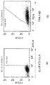
- FIG. 5 is a dot plot by a flow cytometer according to the fifteenth embodiment.
- FIG. 5 is a photograph of urine-derived cells into which a reprogramming factor has been introduced according to Example 16.
- FIG. 5 is a photograph of urine-derived cells into which a reprogramming factor has been introduced according to Example 16.
- the method for culturing the cells into which the reprogramming factor has been introduced according to the embodiment includes culturing the cells into which the reprogramming factor has been introduced, and subculturing the cells into which the reprogramming factor has been introduced without cloning. including.
- the method for culturing the cells into which the reprogramming factor was introduced according to the embodiment was to culture the cells into which the reprogramming factor was introduced and to detach the cells into which the reprogramming factor was introduced from the incubator. Includes mixing and subculturing at least a portion of the cells.
- the method for culturing the cells into which the reprogramming factor has been introduced according to the embodiment includes culturing the cells into which the reprogramming factor has been introduced, and collecting the cells into which the reprogramming factor has been introduced, and at least the recovered cells. Includes: mixing a portion and seeding in a medium for subculture.
- the method for culturing cells into which a reprogramming factor has been introduced according to the embodiment includes cells into which a reprogramming factor has been introduced, in which cells derived from different single cells are mixed and seeded. Cells into which reprogramming factors have been introduced are seeded and induced, for example, into pluripotent stem cells. Pluripotent stem cells are, for example, iPS cells.
- the cells into which the reprogramming factor is introduced are not particularly limited, and examples thereof include fibroblasts, blood cells, dental pulp stem cells, keratinocytes, dermal papilla cells, oral epithelial cells, and somatic stem progenitor cells.
- the cell into which the reprogramming factor is introduced may be a cell contained in urine. Examples of cells contained in urine include bladder epithelial cells.
- the cells into which the reprogramming factor is introduced may be of human or non-human animal origin.
- the cells into which the reprogramming factor is introduced may be of one human origin or of multiple human origins.
- the cell into which the reprogramming factor is introduced may be derived from one non-human animal or may be derived from a plurality of non-human animals.
- Blood cells are separated from the blood.
- Blood is, for example, peripheral blood and umbilical cord blood, but is not limited thereto. Blood may be collected from an adult or a minor.
- an anticoagulant such as ethylenediaminetetraacetic acid (EDTA), heparin, and biopharmacy standard blood preservation solution A (ACD-A) is used.
- EDTA ethylenediaminetetraacetic acid
- ACD-A biopharmacy standard blood preservation solution A
- Blood cells are nucleated cells such as mononuclear cells (Monocytes), neutrophils, macrophages, eosinophils, basophils, and lymphocytes, and do not contain erythrocytes, granulocytes, and platelets. Blood cells may be, for example, vascular endothelial progenitor cells, blood stem / progenitor cells, T cells, or B cells. T cells are, for example, ⁇ T cells.
- Mononuclear cells are separated from blood using a blood cell separation medium, a centrifuge, and the like.
- a blood cell separation medium a centrifuge, and the like.
- Ficoll GE Healthcare
- the method for separating mononuclear cells is as follows.
- the centrifuge To 5 mL of blood, add 5 mL of PBS to dilute, and layer 5 mL each on a medium for human lymphocyte separation in a tube. At this time, diluted blood is slowly added onto the medium along the tube wall of the tube so as not to disturb the interface.
- the method for separating mononuclear cells when using vacutainer (registered trademark, BD) as a blood collection tube is as follows.
- the centrifuge 4 ° C to 42 ° C, preferably 18 ° C.
- 4 ° C to 42 ° C preferably 18 ° C.
- 8 mL of blood is collected using a blood collection tube (vacutainer (registered trademark), BD), mixed by inversion, and mixed with an anticoagulant.
- the balance is then adjusted and the solution is adjusted at 4 ° C to 42 ° C, preferably 18 ° C, 100 ⁇ g to 3000 ⁇ g, preferably 1500 ⁇ g to 1800 ⁇ g with a swing rotor for 1 to 60 minutes, preferably 20 minutes.
- Centrifuge After centrifugation, the upper layer, which is the plasma layer, is removed and pipetting is performed to suspend the mononuclear cell layer and blood cells attached to the gel to obtain a suspension. Transfer the resulting suspension to another 15 mL tube.
- 1 mL to 14 mL, preferably 12 mL of PBS is added to the suspension in a 15 mL tube, and the suspension is prepared at 4 ° C to 42 ° C, preferably 18 ° C, 100 ⁇ g to 3000 ⁇ g, preferably 200 ⁇ g.
- a hemolytic agent PharmLyse (registered trademark), 10-fold concentration, BD
- BD sterile water
- the method for separating mononuclear cells from blood is not limited to the above method, and for example, a dialysis membrane may be used to separate mononuclear cells from blood.
- a pure cell select system for whole blood mononuclear cell concentration (registered trademark, PALL), a purifier for removing blood cells (cellsova E, registered trademark, Asahi Kasei), and a leukapheresis filter for platelet preparation (sepacell PL, registered trademark, Filters such as PLX-5B-SCD, Asahi Kasei) can also be used.
- Mononuclear cells may be separated using an erythrocyte separating agent capable of separating nucleated cells by gravity sedimentation or centrifugation of erythrocytes.
- erythrocyte separating agents include HetaSep (registered trademark, STEMCELL Technologies) and HES40 (NIPRO).
- CTL-UP1 sold by Cellular Technology Limited
- PBMC-001 of Sanguine Biosciences, or the like may be used as the mononuclear cell.
- the cryopreserved blood cells may be thawed and used using a cell cryopreservation solution such as Cerbunker 1, Stem Cerbunker GMP grade, and Stem Cerbunker DMSO-free GMP grade (Zenoac).
- a cell cryopreservation solution such as Cerbunker 1, Stem Cerbunker GMP grade, and Stem Cerbunker DMSO-free GMP grade (Zenoac).
- thawing mononuclear cells When thawing mononuclear cells, first, 1 mL to 15 mL, preferably 8 mL of a serum-free hematopoietic cell medium (X-VIVO® 10, Ronza) having a known composition was placed in a 15 mL tube and frozen. Place the tube containing the mononuclear cells in a warm bath at 4 ° C to 42 ° C, preferably 37 ° C to begin thawing the mononuclear cells. Then, with a little ice remaining, the tube containing the mononuclear cells is pulled out of the warm bath and the mononuclear cells are transferred to the tube containing the serum-free hematopoietic cell medium of known composition. Of these, 10 ⁇ L of mononuclear cell suspension is stained with trypan blue and counted on a hemocytometer.
- X-VIVO® 10 serum-free hematopoietic cell medium
- Blood cells may be isolated based on cell surface markers.
- Blood stem / progenitor cells are positive for CD34.
- the T cells are positive for any of CD3, CD4, and CD8.
- B cells are positive for any of CD10, CD19, and CD20.
- Macrophages are positive for any of CD11b, CD68, and CD163.
- Blood stem / progenitor cells, T cells, or B cells are separated from blood cells using, for example, an automatic magnetic cell separator and immunomagnetic beads. Alternatively, pre-separated mononuclear cells may be prepared. However, blood cells that have not been separated based on cell surface markers may be used.
- CD34-positive cells are stem / progenitor cells and tend to be easily reprogrammed. Further, when iPS cells are prepared using T cells which are CD3 positive cells, the iPS cells derived from the T cells retain the type of TCR recombination, so that they tend to be able to efficiently induce differentiation into T cells.
- the method for separating CD34-positive cells is as follows.
- 10 ⁇ L IL-6 (100 ⁇ g / mL), 10 ⁇ L SCF (300 ⁇ g / mL), 10 ⁇ L TPO (300 ⁇ g / mL), 10 ⁇ L FLT3 ligand (300 ⁇ g / mL) in 10 mL serum-free medium (StemSpan H3000, STEMCELL Technologies).
- 10 ⁇ L of IL-3 (10 ⁇ g / mL) are added to prepare a blood cell medium (blood stem / progenitor cell medium).
- 1 mL to 6 mL preferably 2 mL of blood cell medium in 1 well of a 6-well plate.
- 1 mL to 6 mL and 2 mL of PBS are placed in each of the other 5 wells to prevent evaporation of the medium.
- the 6-well plate is then placed in an incubator at 4 ° C to 42 ° C, preferably 37 ° C for warming.
- a mononuclear cell suspension containing 1 ⁇ 10 4 to 1 ⁇ 10 9 cells, preferably 2 ⁇ 10 7 mononuclear cells was dispensed into a 15 mL tube, and the mononuclear cell suspension was prepared.
- Centrifuge at 4 ° C to 42 ° C, preferably 4 ° C, 100 ⁇ g to 3000 ⁇ g, preferably 300 ⁇ g for 10 minutes. After centrifugation, the supernatant is removed and the mononuclear cells are suspended in 100 ⁇ L to 1 mL, preferably 300 ⁇ L of column buffer.
- 1 mL to 15 mL, preferably 10 mL of column buffer is added to the mononuclear cell suspension in a 15 mL tube to dilute it, and the mixture is diluted at 4 ° C to 42 ° C, preferably 4 ° C, 100 ⁇ g to 1000 ⁇ g. Centrifuge at 300 xg for 1 to 2 hours, preferably 10 minutes. After centrifugation, the supernatant in a 15 mL tube is removed with an ejector, and 10 ⁇ L to 10 mL, preferably 500 ⁇ L of column buffer is added and resuspended.
- a column for an automatic magnetic cell separation device (MS column, Miltenyi Biotec) is attached to an automatic magnetic cell separation device (MiniMACS Separation Unit, Miltenyi Biotec), and a column buffer of 10 ⁇ L to 10 mL, preferably 500 ⁇ L is put in the column and washed.
- a column buffer of 10 ⁇ L to 10 mL, preferably 500 ⁇ L is placed in the column, and the column is washed 1 to 10 times, preferably 3 times.
- the column is then removed from the automatic magnetic cell separator and placed in a 15 mL tube.
- the column is then filled with 10 ⁇ L to 10 mL, preferably 1000 ⁇ L of column buffer, and the syringe is swiftly pushed to allow CD34-positive cells to drain into a 15 mL tube.
- a 10 ⁇ L CD34-positive cell suspension is stained with trypan blue and the cell count is counted on a hemocytometer.
- the CD34-positive cell suspension in a 15 mL tube is centrifuged at 4 ° C to 42 ° C, preferably 4 ° C, 100 ⁇ g to 1000 ⁇ g, preferably 300 ⁇ g for 1 to 2 hours, preferably 10 minutes. .. After centrifugation, remove the supernatant with an ejector.
- the CD34-positive cells are resuspended in the warmed blood cell medium, and the CD34-positive cells are sprinkled on the culture plate. Then, the CD34-positive cells are cultured at 4 ° C. to 42 ° C., preferably 37 ° C., 1% to 20%, preferably 5% CO 2 for 6 days. During this time, the medium does not have to be exchanged.
- the method of isolating cells with a marker other than CD34 is the same as the method of isolating CD34-positive cells.
- RNA is, for example, mRNA.
- Reprogramming factors introduced into cells include, for example, OCT RNA such as OCT3 / 4, SOX RNA such as SOX2, KLF RNA such as KLF4, and MYC RNA such as c-MYC.
- OCT RNA such as OCT3 / 4
- SOX RNA such as SOX2
- KLF RNA such as KLF4
- MYC RNA such as c-MYC.
- reprogramming factor RNA it may be used M 3 O having improved OCT3 / 4.
- RNAs are LIN28A, FOXH1, LIN28B, GLIS1, p53-dominant negative, p53-P275S, L-MYC, NANOG, DPPA2, DPPA4, DPPA5, ZIC3, BCL-2, E-RAS, TPT1, SALL2. , NAC1, DAX1, TERT, ZNF206, FOXD3, REX1, UTF1, KLF2, KLF5, ESRRB, miR-291-3p, miR-294, miR-295, NR5A1, NR5A2, TBX3, MBD3sh, TH2A, TH2 It may further contain RNA of at least one factor selected from the group. These RNAs are available from TriLink.
- gene symbol is described here in humans, it is not intended to limit the species by uppercase or lowercase notation. For example, capitalization of all letters does not exclude the inclusion of mouse or rat genes. However, in the examples, the gene symbols are shown according to the species actually used.
- P53 is a cancer-suppressing protein.
- the dominant negative variant of p53 is not particularly limited as long as it can compete with the wild-type p53 protein endogenous to somatic cells and inhibit the function of the wild-type p53 protein.
- Examples of dominant negative variants of p53 are p53P275S, which is a point mutation of proline at position 275 (position 278 in the case of humans) located in the DNA binding region of mouse p53 to serine, and position 14-301 (human) of mouse p53.
- p53DD lacking the amino acid at positions 11-304, p53S58A in which serine at position 58 (61 in the case of humans) of mouse p53 was point-mutated to alanine, and position 135 of human p53 (mouse).
- p53A135V point-mutated alanine at position 135 (138 in the case of humans) of mouse p53 to valine
- position 172 in the case of humans
- RNA may be modified with psiduridine ( ⁇ ) or 5-methyluridine (5meU). RNA may be polyadenylated.
- the RNA introduced into the cell is, for example, a single-stranded RNA, and the double-stranded RNA may be substantially removed. Further, it is preferable that the RNA introduced into the cell is substantially free of impurities such as short RNA and impurities.
- the single-stranded RNA introduced into the cell may be purified and / or concentrated in order to substantially remove the double-stranded RNA. Examples of the method for purifying the single-stranded RNA to be introduced into cells include a purification method using high performance liquid chromatography (HPLC). For example, HPLC removes 70% or more, 75% or more, 80% or more, 85% or more, or 90% or more of double-stranded RNA.
- HPLC high performance liquid chromatography
- the RNA introduced into the cell may be treated with a ribonuclease that degrades the double-stranded RNA in order to substantially remove the double-stranded RNA.
- the RNA introduced into the cell may further include RNA in the transcriptional activation domain (TAD) of MYOD that is directly linked to the full length of OCT3 / 4 RNA.
- TAD transcriptional activation domain
- the reprogramming factor is introduced into cells by, for example, the lipofection method.
- the lipofection method is a method in which a complex of a negatively charged nucleic acid and a positively charged lipid is formed by electrical interaction, and the complex is incorporated into the cell by endocytosis or membrane fusion. ..
- the lipofection method has advantages such as less damage to cells, excellent introduction efficiency, easy operation, and less time.
- the reprogramming factor is introduced into cells cultured using, for example, an RNA transfection reagent.
- RNA may be introduced into the mononuclear cell immediately after the mononuclear cell is separated from the blood.
- RNA transfection reagent Lipofectamine Messenger MAX (registered trademark, Thermo Fisher SCIENTIFIC) can be used.
- RNA transfection reagent for example, Lipofectamine (registered trademark) RNAiMAX (Thermo Fisher SCIENTIFIC), Lipofectamine StemTransfection Reagent (Thermo Fisher SCIENTIFIC), Transfection (ReproCELL), Jet Messenger (Polyplus), Lipofectamin (registered trademark) 2000, Lipofectamin (registered trademark) 3000, NeonTransfection System (Thermo Fisher SCIENTIFIC), Stemfect RNA transfection reagent (Stemfect), NextFect (registered trademark) RNA Transfection Reagent (BiooSientific ), Amaxa (registered product) Human T cell Nucleector (registered product) kit (Lonza, VAPA-1002), Amaxa (registered product) Human CD34 cell Nucleector (
- a reprogramming factor is introduced into the cell using, for example, a viral vector.
- the viral vector may be an RNA viral vector.
- the RNA viral vector may be a Sendai viral vector.
- the Sendai virus vector may be a temperature-sensitive Sendai virus vector in which the stability of the viral nucleic acid decreases above a predetermined temperature.
- the viral nucleic acid of the temperature-sensitive Sendai virus vector is stable below a predetermined temperature.
- the viral nucleic acid may be viral DNA or viral RNA.
- the viral nucleic acid may be a viral genome. The decrease in the stability of the viral nucleic acid may be at least one of the degradation of the viral nucleic acid and the suppression of the replication or proliferation of the viral nucleic acid.
- Decreased stability of viral nucleic acids reduces at least one of viral nucleic acid proliferation, viral nucleic acid replication rate, and gene expression levels.
- the predetermined temperature is, for example, 36.5 ° C or higher and 37.5 ° C or lower, 36.6 ° C or higher and 37.4 ° C or lower, 36.7 ° C or higher, 37.3 ° C or lower, 36.8 ° C or higher and 37.2.
- the temperature is 36.9 ° C or higher and 37.1 ° C or lower, or 37 ° C.
- At least one of the viral nucleic acid stability of the temperature-sensitive Sendai virus vector, i.e., growth, replication rate and gene expression level is high below a given temperature and low above a given temperature.
- the temperature-sensitive Sendai virus vector has a growth rate or gene expression level of 1/2 or less in cells cultured at 37 ° C. as compared with a growth rate or gene expression level in cells cultured at 32 ° C. , 1/3 or less, 1/5 or less, 1/10 or less, or 1/20 or less.
- Sendai virus encodes N gene, P gene, M gene, F / HN gene, and L gene.
- the HN protein recognizes sialic acid on the cell surface when Sendai virus attaches to the cell and anchors the virus particles to the cell.
- the F protein is cleaved and activated by an extracellular protease to catalyze the fusion of the tethered Sendai virus envelope and the cell membrane of the target cell to establish an infection.
- the L protein, along with its modified protein, the P protein catalyzes the replication of viral nucleic acids in the cytoplasm after infection and transcription from the replicated multi-copy nucleic acids.
- the Sendai virus vector By deleting the F gene in the Sendai virus vector, it is possible to suppress the production of infectious virus particles from the transgenic cells. Moreover, it is possible to make the Sendai virus vector temperature sensitive by introducing a mutation into at least one of the L gene and the P gene.
- TS7 L protein Y942H / L1361C / L1558I mutation
- TS12 P protein D433A / R434A / K437A mutation
- TS13 P protein D433A / R434A / K437A
- Mutations and L protein L1558I mutations TS14 (P protein D433A / R434A / K437A mutations and L protein L1361C mutations)
- TS15 P protein D433A / R434A / K437A mutations and L protein L1361C / L1558I mutations
- Sendai viral vectors have, for example, G69E, T116A, and A183S mutations in the M protein, A262T, G264R, and K461G mutations in the HN protein, L511F mutations in the P protein, and N1197S in the L protein. It is an F gene deletion ( ⁇ F) type Sendai virus vector having the K1795E mutation, and is a Sendai virus vector into which the above-mentioned TS7, TS12, TS13, TS14, or TS15 mutation has been introduced.
- the temperature-sensitive mutation of the Sendai virus vector is not limited to these.
- the Sendai virus vector is, for example, SeV (PM) / TS ⁇ F, SeV18 + / TS ⁇ F, or SeV (HNL) / TS ⁇ F, and is a Sendai virus vector into which the above-mentioned TS7, TS12, TS13, TS14, or TS15 mutation has been introduced.
- the temperature-sensitive mutation of the Sendai virus vector is not limited to these.
- the Sendai virus vector introduced into cells may be a combination of a temperature-sensitive Sendai virus vector and a temperature-insensitive Sendai virus vector.
- the Sendai virus vector introduced into the cell is only the temperature-sensitive Sendai virus vector, and does not have to contain the temperature-insensitive Sendai virus vector.
- the Sendai virus vector introduced into cells is only a temperature-sensitive Sendai virus vector into which a mutation of TS7, TS12, TS13, TS14, or TS15 has been introduced, and does not have to contain a temperature-insensitive Sendai virus vector.
- the Sendai virus vector introduced into cells is only the temperature-sensitive Sendai virus vector having the same or higher temperature sensitivity as the temperature-sensitive Sendai virus vector into which the mutation of TS7, TS12, TS13, TS14, or TS15 has been introduced, and the temperature-insensitive Sendai. It does not have to contain a viral vector.
- the Sendai virus vector introduced into cells is only the Sendai virus vector having the same or higher temperature sensitivity as the temperature sensitive Sendai virus vector into which the mutation of TS7, TS12, TS13, TS14, or TS15 has been introduced, and TS7, TS12, It does not have to contain a Sendai virus vector that is less temperature sensitive than the temperature sensitive Sendai virus vector into which the TS13, TS14, or TS15 mutations have been introduced.
- the Sendai virus vector introduced into cells carries an arbitrary reprogramming factor.
- the Sendai virus vector introduced into the cell contains, for example, a temperature-sensitive Sendai virus vector containing KLF RNA, OCT RNA, and SOX RNA in this order and not containing MYC RNA, and MYC RNA containing KLF. RNA, OCT RNA, and SOX RNA-free temperature-sensitive Sendai virus vector.
- the number, combination, and order of the reprogramming factors loaded on the Sendai virus vector are arbitrary and are not particularly limited.
- the Sendai virus vector introduced into the cell may contain a Sendai virus vector containing KLF RNA and not containing OCT RNA and SOX RNA.
- the Sendai virus vector containing KLF RNA and not OCT RNA and SOX RNA may be a temperature-sensitive Sendai virus vector or a temperature-insensitive Sendai virus vector.
- the Sendai virus vector disappears earlier from the cells into which the Sendai virus vector has been introduced when the temperature-insensitive Sendai virus vector is not introduced.
- Temperature-sensitive Sendai virus vectors containing KLF RNA, OCT RNA, and SOX RNA have, for example, mutations in G69E, T116A, and A183S in the M protein and mutations in A262T, G264R, and K461G in the HN protein.
- An F gene-deficient Sendai virus vector having an L511F mutation in the P protein and N1197S and K1795E mutations in the L protein, and having the above-mentioned TS7, TS12, TS13, TS14, or TS15 mutation. It is a virus vector.
- the temperature sensitive mutation is, for example, TS7 or TS12, or TS12.
- the temperature sensitive Sendai virus vector containing KLF RNA, OCT RNA, and SOX RNA is, for example, SeV (PM) KOS / TS7 ⁇ F or SeV (PM) KOS / TS12 ⁇ F, or SeV (PM) KOS / TS12 ⁇ F. be.
- a temperature-sensitive Sendai virus vector containing MYC RNA has, for example, mutations in G69E, T116A, and A183S in M protein, mutations in A262T, G264R, and K461G in HN protein, and L511F mutation in P protein. It is an F gene-deficient Sendai virus vector having N1197S and K1795E mutations in L protein, and is a Sendai virus vector having the above-mentioned TS7, TS12, TS13, TS14, or TS15 mutations.
- the temperature sensitive mutation is, for example, TS15.
- Temperature sensitive Sendai virus vectors containing MYC RNA are, for example, SeV (HNL) MYC / TS12 ⁇ F, SeV (HNL) MYC / TS13 ⁇ F, or SeV (HNL) MYC / TS15 ⁇ F, or SeV (HNL) MYC / TS15 ⁇ F. be.
- Sendai viral vectors containing KLF RNA but not OCT RNA and SOX RNA have, for example, mutations in G69E, T116A, and A183S in the M protein and mutations in A262T, G264R, and K461G in the HN protein. It is an F gene-deficient Sendai virus vector having an L511F mutation in the P protein and an N1197S and K1795E mutation in the L protein.
- the Sendai virus vector containing KLF RNA and not containing OCT RNA and SOX RNA is, for example, less temperature sensitive than the Sendai virus vector into which the above-mentioned TS7, TS12, TS13, TS14, or TS15 mutation has been introduced, and is predetermined.
- the KLF gene can be expressed even at temperatures above.
- the Sendai virus vector containing KLF RNA but not OCT RNA and SOX RNA is, for example, SeV18 + KLF4 / TS ⁇ F.
- multiple types of Sendai virus vectors are introduced into cells at the same time.
- multiple types of Sendai virus vectors are introduced into cells at the same time.
- the multiplicity of infection (MOI) of the Sendai viral vector when infecting cells is, for example, 0.1 or more, 0.3 or more, 0.5 or more, 1.0 or more, 2.0 or more, 3.0 or more. It is 4.0 or more, or 5.0 or more.
- the MOI is, for example, 100 or less, 90 or less, 80 or less, 70 or less, 60 or less, 50 or less, 40 or less, 30 or less, 20 or less, 10 or less, or 5 or less.
- the temperature at which cells are infected with the Sendai virus vector is below a predetermined temperature at which the stability of the virus nucleic acid of the temperature-sensitive Sendai virus vector decreases, that is, the temperature at which the virus nucleic acid of the temperature-sensitive Sendai virus vector is stable. It may be above a predetermined temperature.
- the temperature at which cells are infected with the Sendai virus vector reduces the stability of the viral nucleic acid of the temperature-sensitive Sendai virus vector. It is preferable that the temperature is lower than the predetermined temperature, that is, the temperature at which the viral nucleic acid of the temperature-sensitive Sendai virus vector is stable.
- the cells into which the reprogramming factor is introduced may be adherently cultured or suspendedly cultured.
- Somatic cells into which reprogramming factors are introduced are feeder-free using basement membrane matrices such as Matrigel (Corning), CELLstart (registered trademark, Thermo Fisher), Laminin 511 (iMatrix-511, nippi), fibronectin, and vitrotin. It may be cultured.
- basement membrane matrices such as Matrigel (Corning), CELLstart (registered trademark, Thermo Fisher), Laminin 511 (iMatrix-511, nippi), fibronectin, and vitrotin. It may be cultured.
- a stem cell medium such as a human ES / iPS medium such as Prime ES Cell Medium (ReproCELL) can be used.
- the stem cell medium is not limited to this, and various stem cell media can be used.
- the stem cell medium is placed in an incubator such as a dish, well, or tube, for example.
- the gel medium is, for example, a stem cell medium containing gellan gum at a final concentration of 0.001% by mass to 0.5% by mass, 0.005% by mass to 0.1% by mass, or 0.01% by mass to 0.05% by mass. It is prepared by adding so as to become.
- Gel media include gellan gum, hyaluronic acid, lambzan gum, dieutan gum, xanthan gum, carrageenan, fucoidan, pectin, pectinic acid, pectinic acid, heparan sulfate, heparan, heparitin sulfate, keratosulfate, chondroitin sulfate, deltaman sulfate, ramnan sulfate, and It may contain at least one polymer compound selected from the group consisting of those salts.
- the gel medium may contain methyl cellulose. By including methyl cellulose, agglutination between cells is further suppressed.
- the gel medium is poly (glycerol monomethacrylate) (PGMA), poly (2-hydroxypropylmethacrylate) (PHPMA), Poly (N-isopropylacrylamide) (PNIPAM), amine terminated, carboxylic acid terminated, maleimide terminated, N-hydroxysuccinimide (N-hydroxysuccinimide).
- PGMA poly (glycerol monomethacrylate)
- PPMA poly (2-hydroxypropylmethacrylate)
- PIPAM Poly (N-isopropylacrylamide)
- amine terminated carboxylic acid terminated
- maleimide terminated N-hydroxysuccinimide (N-hydroxysuccinimide).
- the gel medium may or may not contain a growth factor such as basic fibroblast growth factor (bFGF).
- bFGF basic fibroblast growth factor
- the gel medium may contain a growth factor such as bFGF at a low concentration of 400 ⁇ g / L or less, 40 ⁇ g / L or less, or 10 ⁇ g / L or less.
- the gel medium may or may not contain TGF- ⁇ , or may contain TGF- ⁇ at a low concentration of 600 ⁇ g / L or less, 300 ⁇ g / L or less, or 100 ⁇ g / L or less.
- the gel medium does not have to be agitated. Further, the gel medium does not have to contain feeder cells.
- the gel medium may contain at least one substance selected from the group consisting of cadherin, laminin, fibronectin, and vitronectin.
- a temperature lower than a predetermined temperature at which the stability of the viral nucleic acid of the temperature-sensitive Sendai virus vector decreases that is, a temperature-sensitive Sendai virus.
- the cells may be cultured at a temperature at which the vector viral nucleic acid is stable. Then, the cells may be cultured at a predetermined temperature or higher.
- the medium may be changed, for example, once every two days while culturing the cells above a predetermined temperature.
- At least 2 days, or 2 days or more and 10 days or less for example, 4.0 ° C or higher, 10 ° C or higher, 15 ° C or higher, 20 ° C or higher, 25 ° C or higher, 30 ° C or higher.
- Cells may be cultured at temperatures below 36.6 ° C, below 36.5 ° C, below 36.0 ° C, below 35.0 ° C, or below 34.0 ° C.
- the temperature is raised to 36.5 ° C. or higher, 36.6 ° C. or higher, 36.7 ° C. or higher, 36.8 ° C. or higher, 36.9 ° C. or higher, or 37.0 ° C. or higher, 40.0 ° C.
- the cells may be cultured at a temperature of 39.0 ° C. or lower, or 38.0 ° C. or lower.
- the temperature may be raised once or stepwise. After raising the temperature, the medium may be changed while culturing the cells, for example once every two days.
- the temperature-sensitive Sendai virus vector viral nucleic acid becomes less stable than a predetermined temperature, that is, the temperature-sensitive Sendai viral vector viral nucleic acid
- the cells may be cultured at a stable temperature. After the stem cell-like colonies begin to appear, the cells may be cultured above a predetermined temperature. The medium may be changed, for example, once every two days while culturing the cells above a predetermined temperature.
- stem cell-like colonies After infecting cells with Sendai virus vector, until stem cell-like colonies begin to appear, for example, 4.0 ° C or higher, 10 ° C or higher, 15 ° C or higher, 20 ° C or higher, 25 ° C or higher, 30 ° C or higher, 31.
- ° C or higher 32.0 ° C or higher, 33.0 ° C or higher, 33.1 ° C or higher, 33.2 ° C or higher, 33.3 ° C or higher, 33.4 ° C or higher, 33.5 ° C or higher, 33.6 ° C 33.7 ° C or higher, 33.8 ° C or higher, or 33.9 ° C or higher, less than 37.0 ° C, less than 36.9 ° C, less than 36.8 ° C, less than 36.7 ° C, 36.6 Cells may be cultured at temperatures below ° C., below 36.5 ° C., below 36.0 ° C., below 35.0 ° C., or below 34.0 ° C.
- the temperature is raised to 36.5 ° C or higher, 36.6 ° C or higher, 36.7 ° C or higher, 36.8 ° C or higher, 36.9 ° C or higher, or 37.0 ° C.
- cells may be cultured at a temperature of 40.0 ° C. or lower, 39.0 ° C. or lower, or 38.0 ° C. or lower.
- the temperature may be raised once or stepwise. After raising the temperature, the medium may be changed while culturing the cells, for example once every two days.
- the cells into which the reprogramming factor has been introduced are collected, and at least a passage is performed in which at least a part of the mixed cells is seeded in the medium at least once. do.
- clones of cells into which reprogramming factors have been introduced may be mixed together.
- different clones of cells into which reprogramming factors have been introduced may be mixed together.
- the cells into which the reprogramming factor has been introduced may be collected, and at least a part of the collected and mixed cells may be seeded in a medium and subcultured a plurality of times.
- the cells into which the reprogramming factor has been introduced may be recovered, and at least a part of the recovered mixed cells may be seeded in a medium and subcultured. In addition, all the collected mixed cells may be seeded in the medium.
- the cells into which the reprogramming factor has been introduced are collected, and at least a part of the collected and mixed cells is seeded in a medium and subcultured. It refers to passage without distinction according to the expression state. For example, at the time of passage, cells into which a reprogramming factor has been introduced may be seeded in the same incubator without distinguishing by gene expression status. Alternatively, the cells into which the reprogramming factor has been introduced are collected, and at least a part of the collected and mixed cells is seeded in a medium and subcultured. For example, the cells into which the reprogramming factor has been introduced are reprogrammed. It means to pass on without distinguishing by the degree of. For example, at the time of passage, cells into which a reprogramming factor has been introduced may be seeded in the same incubator without distinction according to the degree of reprogramming.
- the cells into which the reprogramming factor has been introduced are collected, and at least a part of the collected and mixed cells is seeded in a medium and subcultured, for example, the cells into which the reprogramming factor has been introduced are expressed in morphology. It means to succeed without distinction.
- cells into which a reprogramming factor has been introduced may be seeded in the same incubator without distinguishing by morphology.
- the cells into which the reprogramming factor has been introduced are collected, and at least a part of the collected and mixed cells is seeded in a medium and subcultured.
- the cells into which the reprogramming factor has been introduced are sized. It means to succeed without distinction.
- cells into which a reprogramming factor has been introduced may be seeded in the same incubator without distinction by size.
- the cells into which the reprogramming factor has been introduced are recovered, and at least a part of the collected and mixed cells is seeded in a medium and subcultured without cloning the cells into which the reprogramming factor has been introduced. It means to succeed. For example, in the case of passage without cloning, it is not necessary to pick up the colonies formed by the cells into which the reprogramming factor has been introduced. For example, when passaged without cloning, it is not necessary to separate multiple colonies formed by cells into which the reprogramming factor has been introduced. For example, at the time of passage, cells forming a plurality of different colonies may be mixed and seeded in the same incubator.
- colonies may be mixed and seeded in the same incubator.
- cells into which a reprogramming factor has been introduced are adherently cultured, even if the adherently cultured cells are collected, and at least a part of the collected and mixed cells is seeded in a medium and subcultured. good.
- all cells may be exfoliated from the incubator and at least some of the exfoliated and mixed cells may be seeded in the same incubator.
- all cells may be stripped from the incubator with a stripping solution, and the stripped and mixed cells as a whole may be passaged. For example, cells that do not form colonies may be passaged. If the cells into which the reprogramming factor has been introduced are suspension-cultured, the entire suspension-cultured cells may be passaged.
- the cells When subculturing cells that have been introduced with reprogramming factors, the cells are seeded in medium or incubator at low concentrations.
- the low concentration is, for example, 1 cell / cm 2 or more, 0.25 ⁇ 10 4 cells / cm 2 or less, 1.25 ⁇ 10 3 cells / cm 2 or less, 0.25 ⁇ 10 3 cells /. It is cm 2 or less, 0.25 ⁇ 10 2 cells / cm 2 or less, or 0.25 ⁇ 10 1 cells / cm 2 or less.
- low concentration means that 10 or less, 9 or less, 8 or less, 7 or less, 6 or less, 5 or less, 4 or less, 3 or less, or 2 or less cells can come into contact with each other.
- the concentration at which 11 or more cells do not come into contact with each other there is a concentration at which 11 or more cells do not come into contact with each other.
- the state where the entire bottom surface of the cell container is covered with cells is defined as 100% confluent, and low concentration means 5% or less confluent, 4% or less confluent, 3% or less confluent, 2% or less confluent, 1% or less confluent, 0. It is 5.5% or less confluent, 0.1% or less confluent, 0.05% or less confluent, or 0.01% or less confluent.
- the low concentration is, for example, a concentration at which single cells do not come into contact with each other in the seeded cells.
- single cells may be seeded in the wells of the well plate.
- the well plate may be a 12-well plate or a 96-well plate.
- the induced pluripotent stem cells the proportion of cells in which Sendai virus remains is, for example, 4% or less, 3% or less, 2% or less, 1% or less, 0.5% or less, or 0%.
- the cells may be cultured at a temperature higher than a predetermined temperature at which the stability of the viral nucleic acid of the temperature-sensitive Sendai virus vector decreases. After passage, the cells are cultured, for example, at a temperature of 36.5 ° C. or higher and lower than 38.0 ° C. After passage, for example, cells are cultured at a temperature of 36.5 ° C. or higher and lower than 38.0 ° C. until cell-cell adhesion starts, and after cell-cell adhesion starts, a higher temperature until cell-cell adhesion starts. For example, cells may be cultured at a temperature of 37.5 ° C. or higher, 42.0 ° C.
- the temperature is 37.5 ° C or higher, 42.0 ° C or lower, 41.5 ° C or lower, 41.0 ° C or lower, 40.5 ° C or lower, or 40.0 ° C. or lower. ..
- the temperature is 37.5 ° C or higher, 42.0 ° C or lower, 41.5 ° C or lower, 41.0 ° C or lower, 40.5 ° C or lower, or 40.0 ° C.
- the cells may be cultured at the following temperatures.
- Cells into which the reprogramming factor has been introduced may be cultured and subcultured in a closed incubator.
- the closed incubator does not exchange gas, virus, microorganisms, impurities, etc. with the outside, for example.
- cells into which a reprogramming factor has been introduced may be expanded and cultured in a two-dimensional culture, or may be expanded and cultured in a three-dimensional culture.
- the entire cells that have been adherently cultured may be cryopreserved as pluripotent stem cells.
- the entire cells exfoliated from the incubator with the exfoliating solution may be cryopreserved as pluripotent stem cells.
- the entire cells in suspension culture may be cryopreserved as pluripotent stem cells.
- the induced pluripotent stem cells can form flat colonies similar to ES cells and express alkaline phosphatase.
- the induced pluripotent stem cells can express Nanog, OCT4, SOX2, etc., which are undifferentiated cell mer powers.
- Induced pluripotent stem cells can express TERT.
- Induced pluripotent stem cells may exhibit temouth mellase activity.
- a cytoflow meter which is a cell surface marker indicating that the cells were undifferentiated, such as TRA-1-60, TRA-1-81, SSEA-1, and. This may be done by analyzing whether at least one surface marker selected from SSEA5 is positive.
- TRA-1-60 is an antigen specific for iPS / ES cells. Since iPS cells can be produced only from the TRA-1-60 positive fraction, TRA-1-60 positive cells are considered to be the species of iPS cells.
- the induced pluripotent stem cells may be induced into somatic cells in a state different from that of the pluripotent stem cells.
- somatic cells include nerve cells, retinal epithelial cells, hepatocytes, ⁇ cells, renal cells, dental pulp stem cells, mesenchymal stem cells, somatic stem progenitor cells, keratinocytes, dermal papilla cells, oral epithelial cells, cartilage cells, Examples include muscle cells, vascular cells, epithelial cells, myocardial cells, blood cells, and immune cells.
- blood cells include erythroblasts, red blood cells, megakaryocytes, and platelets.
- immune cells examples include monocytes, neutrophils, eosinophils, basophils, B cells, T cells, NK cells, and NKT cells.
- the induced stem cells may be differentiated into endoderm, mesoderm, or ectoderm lines. Stem cells may form embryoid bodies, organoids, and spheres.
- Examples of factors that induce cells into nervous system cells include the ASCL family, DLX family, MYT family, NeuroD family, SOX family, and NGN family.
- An example of the ASCL family is ASCL1.
- An example of the DLX family is DLX2.
- An example of the MYT family is MYT1L.
- An example of the NGN family is NGN2.
- Examples of neural cells include neural cells, neural stem cells and neural progenitor cells.
- Examples of neurons include inhibitory neurons, excitatory neurons and dopamine-producing neurons, cranial nerves, intervening nerves, and optic nerves.
- the nervous system cells may be motor neurons, oligodendrocyte progenitor cells, astrocytes, oligodendrocytes, and the like.
- Examples of factors that induce cells into cardiomyocytes include the GATA family, MEF family, TBX family, MYOCD family, MESS family, and miR-133 family.
- GATA family is GATA4A.
- MEF family is MEF2C.
- TBX family is TBX5.
- MESS family is MESS1.
- induction refers to reprogramming, reprogramming, transformation, transdifferentiation or lineage reprogramming, differentiation induction, cell fate reprogramming, and the like.
- the induced pluripotent cells may be cloned.
- Gene-edited cells may be cloned after gene-editing the established pluripotent stem cells.
- the cells into which the reprogramming factor has been introduced may be induced into somatic cells different from the pluripotent stem cells without substituting the cells into which the reprogramming factor has been introduced at all.
- the method for introducing the reprogramming factor into the cell is as described above.
- a differentiation-inducing factor is introduced into a cell into which a reprogramming factor has been introduced, and a cell into which the reprogramming factor has been introduced is referred to as a pluripotent stem cell, without subculturing the cell into which the reprogramming factor has been introduced. May induce to different somatic cells.
- the cells into which the reprogramming factor has been introduced are given hormones or chemical substances, and the cells into which the reprogramming factor has been introduced are referred to as pluripotent stem cells. May induce to different somatic cells.
- the cells into which the reprogramming factor has been introduced are induced into somatic cells without being cloned.
- the somatic cells induced are as described above.
- Example 1 Comparative Example 1
- the dish coated with laminin 511 was used as a dish for inducing pluripotent stem cells.
- human peripheral blood mononuclear cells were suspended in a blood medium, and the number of mononuclear cells was measured using a hemocytometer to adjust the number of mononuclear cells in the blood medium. Then, mononuclear cells were two-dimensionally cultured at 37 ° C. for 1 to 7 days on a dish for inducing pluripotent stem cells.
- SeV (PM) hKOS / TS12 ⁇ F and SeV (HNL) hC-Myc / TS15 ⁇ F were added to the two-dimensionally cultured mononuclear cells so that the MOI was 5, and a dish for inducing pluripotent stem cells was added.
- a dish for inducing pluripotent stem cells was added.
- a stem cell-like cell mass developed. On the 14th day after infection, almost all cells became TRA1-60 positive cells and showed iPS cell-like morphology.
- triple select a cell stripping agent, was added to the dish and allowed to stand at room temperature for 1 minute, then the cell-containing solution was aspirated and the cell-containing solution was incubated at 37 ° C. for 5 to 10 minutes. .. Then, iPS cell medium was added, and the iPS cell medium containing the cells was collected in a 15 mL tube.
- Example 1 The number of cells was measured using a hemocytometer, the concentration of the solution containing the cells was adjusted, and the cells were seeded on a well plate so that the concentration was 0.25 ⁇ 10 4 cells / cm 2 or less, and the first passage was performed. I made a charge.
- Example 1 at the time of the first passage, all the cells were exfoliated from the well plate, and the exfoliated and mixed cells were seeded on the next well plate without distinction.
- Comparative Example 1 at the time of the first passage, colony picking was performed and cloning was performed. In both Example 1 and Comparative Example 1, cells were seeded so that 11 or more cells did not come into contact with each other during passage.
- the Weldish was then placed in an incubator at 37 ° C. and the cells were two-dimensionally cultured. After the cells began to divide, the culture temperature was raised to 38 ° C. Then, in both Example 1 and Comparative Example 1, all the cells were collected every time the cells became 60% to 80% confluent, and at least a part of the collected and mixed cells was seeded in a medium and subcultured. .. During the second and subsequent passages, the cells were seeded on the well plate so that the concentration was 0.25 ⁇ 10 4 cells / cm 2 or less. Also at this time, 11 or more cells did not come into contact with each other.
- the number of colonies formed 5 days after the intracellular Sendai virus disappeared was counted. Furthermore, the clonal efficiency was calculated by dividing the number of colonies by the number of seeded cells. The results of the three tests are shown in FIG. When all cells were collected at the time of the first passage using mTeSR Plus as the medium, and some of the collected and mixed cells were seeded in the medium and subcultured, the clonal efficiency was about 5%. It was about 8%, and the variation was small. When colony picking and cloning using mTeSR Plus as a medium during the first passage, the clonal efficiency may be less than 1% or about 6%, resulting in clonal efficiency. There was variation.
- the clonal efficiency was about 10% to about. It was 15%, and the variation was small.
- the clonal efficiency may be less than 1% or about 16%, and the clonal efficiency varies. was there.
- the clonal efficiency can be obtained. It was shown to be high and stable.
- Example 2 Comparative Example 2
- the iPS cell-like cells obtained by subculture in the same manner as in Example 1 and Comparative Example 1 were exfoliated and dissociated.
- about 1 ⁇ 10 5 cells were cryopreserved using STEM-CELLBANKER®.
- the frozen cells were thawed, about 1 ⁇ 10 4 cells were seeded in wells, and the cells were cultured and proliferated.
- the number of cells and the number of colonies 7 days after seeding were measured. The results of the three tests are shown in FIGS. 5 and 6.
- the cells were collected at the time of the first passage, and a part of the collected and mixed cells was seeded in a medium and subcultured.
- the cells were cultured for 7 days after freezing and thawing.
- the number was about 10 ⁇ 10 4 to about 15 ⁇ 10 4 , and the variation was small.
- the cells cloned by colony picking at the time of the first passage have a number of cells cultured for 7 days after freezing and thawing, which is about 0.4 ⁇ 10 4 to about 15 ⁇ 10 4 . There was a lot of variation.
- Example 3 Comparative Example 3
- the iPS cell-like cells obtained by subculture in the same manner as in Example 1 and Comparative Example 1 were exfoliated, 2.5 ⁇ 10 5 cells were suspended in a gel medium, and the cells were three-dimensionally cultured. , The cells were allowed to form a clamp. The number of clamps and the number of cells were measured 13 days after seeding the cells in the gel medium. The results of testing the cells obtained in Example 1 twice and the cells obtained in Comparative Example 1 four times are shown in FIG.
- Example 4 Comparative Example 4
- the iPS cell-like cells obtained by subculture in the same manner as in Example 1 and Comparative Example 1 were exfoliated, and iPS cell-like cells were used using a cardiomyocyte differentiation induction kit (PSC Cardiomyocyte Differentiation Kit, Gibco, registered trademark). Cells were differentiated into cardiomyocytes.
- PSC Cardiomyocyte Differentiation Kit Gibco, registered trademark
- iPS cell-like cells were seeded on a 12-well plate whose bottom surface was treated with a basement membrane matrix (Corning Matrigel, registered trademark), and mTeSR1 was placed in the medium.
- the cells were cultured using. Two days after seeding the iPS cell-like cells, the medium was exchanged with Cardiomyocyte Differentiation Medium A. Two days later, the medium was exchanged with Cardiomyocyte Differentiation Medium B. Two days later, the medium was exchanged at Cardiomyocyte Maintenance Medium. Then, every two days, the cells were cultured until the 22nd day after seeding the iPS cell-like cells while exchanging the medium with the Cardiomyocyte Maintenance Medium.
- cTnT myocardial troponin T
- Example 5 Comparative Example 5
- the iPS cell-like cells obtained by subculture in the same manner as in Example 1 and Comparative Example 1 were exfoliated, and 1.5 ⁇ 10 5 cells were round-bottomed bottom having a size suitable for cell mass formation.
- the cells were seeded and cultured on a cell culture plate (Kurare, RB 500 400 NA 6).
- the medium was a selective inhibitor of TGF- ⁇ 1 activin receptor-like kinase (ALK) ALK-4, -5, -7 (500 nmol / L, A-83-01, Stemgent) and BMP Type I receptor (ALK2, 8GMK medium (8% KnockOut Serum Receptors) to which an inhibitor of membrane permeability of ALK3) (100 nmol / L, LDN193189, Stemgent) was added (Life Technologies), 1% non-essential amino acids (NEAA, Life Technologies), 1% non-essential amino acids (NEAA, Life Technologies) Sodium (Sigma), 100 nmol / L 2-mercaptoethanol (2-ME, Life Technologies) was used.
- Pluripotent stem cells cultured in the presence of the above inhibitors may be induced to differentiate into neural precursor cells. Are known.
- the cells were collected in a tube and centrifuged, the cell mass was decomposed into a single cell with a cell dissociator (TripLE Select, ThermoFisher, registered trademark), the number of cells was counted, and then a nerve cell adhesion molecule (N).
- Cells were immunostained using PSA-NCAM antibody, which is an antibody that detects a polysialization molecule of -CAM), and the positive rate of PSA-NCAM was analyzed by flow cytometry.
- PSA-NCAM antibody which is an antibody that detects a polysialization molecule of -CAM
- the cells subcultured by seeding at least a part of the cells in the medium had a positive rate of PSA-NCAM of about 25% to about 30%, and the variation was small.
- cells cloned by colony picking during the first passage after introduction of a reprogramming factor before induction of differentiation into neural progenitor cells have a PSA-NCAM positive rate of about 5%. It was about 15% from, and the variation was large.
- Example 6 Comparative Example 6
- the iPS cell-like cells obtained by subculture in the same manner as in Example 1 and Comparative Example 1 were exfoliated, and 1 ⁇ 10 5 cells were seeded on a non-adhesive dish and cultured.
- As the medium a human ES cell medium to which bFGF was not added was used.
- the medium was changed once every two days.
- the formed embryoid bodies (EB) were reseeded on gelatin-coated 6-well plates. Then, the medium was exchanged once every two days, and the cells were collected by trypsin treatment on the 24th day after reseeding.
- the collected cells were fixed with 4% paraformaldehyde, the cells were stained with each of SOX1 antibody, OTC2 antibody, HAND1 antibody, and SOX17 antibody, and the stained cells were analyzed using a flow cytometer.
- SOX1 and OTX2 are ectoderm markers
- HAND1 is a mesoderm marker
- SOX17 is an endoderm marker.
- SeV (PM) hKOS / TS12 ⁇ F, SeV18 + hKLF4 / TS ⁇ F, and SeV (HNL) hC-Myc / TS15 ⁇ F were added to the two-dimensionally cultured fibroblasts so that the MOI was 5, and pluripotent stem cells were added.
- the induction dish was placed in an incubator at 34 ° C. and the cells were cultured. Two days after infection, the blood medium was replaced with iPS cell medium. Then, the medium was exchanged once every two days using the iPS cell medium. By 14 days after infection, the culture temperature was gradually increased to 37 ° C and 38 ° C.
- the weldish was then placed in an incubator and the cells were two-dimensionally cultured. After the cells began to divide, the culture temperature was adjusted to 38 ° C. The cells were then passaged each time the cells became 60% to 80% confluent. During the second and subsequent passages, the cells were seeded on the well plate so that the concentration was 0.25 ⁇ 10 4 cells / cm 2 or less. Also at this time, 11 or more cells did not come into contact with each other.
- SeV (PM) hKOS / TS12 ⁇ F, SeV18 + hKLF4 / TS ⁇ F, and SeV (HNL) hC-Myc / TS15 ⁇ F were added to the two-dimensionally cultured mononuclear cells so that the MOI was 5, and they were pluripotent.
- the stem cell-inducing dish was placed in an incubator at 37 ° C., and the cells were cultured. Two days after infection, the blood medium was replaced with iPS cell medium. Then, the medium was exchanged once every two days using the iPS cell medium.
- a stem cell-like cell mass developed.
- the cells were two-dimensionally cultured. After the cells began to divide, the culture temperature was raised to 38 ° C. The cells were then passaged each time the cells became 60% to 80% confluent. From the second passage to the fifth passage, the cells were seeded on the well plate so that the concentration was higher than 0.25 ⁇ 10 4 cells / cm 2 so that 11 or more cells adhered to each other. From the 6th passage onward, cells were seeded on a well plate so that the concentration was 0.25 ⁇ 10 4 cells / cm 2 or less. At this time, 11 or more cells did not come into contact with each other.
- CytoTune-iPS2.0 (registered trademark, ID Pharma), which is a Sendai virus vector kit, was prepared.
- CytoTune-iPS2.0 contains SeV (PM) hKOS / TS12 ⁇ F, which is a temperature-sensitive Sendai viral vector carrying the KLF4 gene, Oct3 / 4 gene, and SOX2 gene as reprogramming factors, and the temperature at which KLF4 is loaded as the reprogramming factor. It contains the sensitive Sendai viral vector SeV18 + hKLF4 / TS ⁇ F and the temperature sensitive Sendai viral vector SeV (HNL) hC-Myc / TS15 ⁇ F carrying c-MYC as a reprogramming factor.
- SeV (PM) hKOS / TS12 ⁇ F which is a temperature-sensitive Sendai viral vector carrying the KLF4 gene, Oct3 / 4 gene, and SOX2 gene as reprogramming factors, and the temperature at which KLF4 is loaded as the reprogramm
- SeV (PM) hKOS / TS12 ⁇ F, SeV18 + hKLF4 / TS ⁇ F, and SeV (HNL) hC-Myc / TS15 ⁇ F were added to mononuclear cells so that the MOI was 5, and a dish for inducing pluripotent stem cells was added.
- the cells were cultured in an incubator at 37 ° C. Two days after the infection, the blood medium was replaced with iPS cell medium (mTeSR Plus (STEMCELL Technologies) or StemFit (Ajinomoto)). Then, the medium was exchanged once every two days using the iPS cell medium.
- a stem cell-like cell mass developed. On the 14th day after infection, almost all cells became TRA1-60 positive cells and showed iPS cell-like morphology.
- triple select a cell stripping agent, was added to the dish and allowed to stand at room temperature for 1 minute, then the cell-containing solution was aspirated and the cell-containing solution was incubated at 37 ° C. for 5 to 10 minutes. .. Then, iPS cell medium was added, and the iPS cell medium containing the cells was collected in a 15 mL tube.
- the number of cells was measured using a hemocytometer, the concentration of the solution containing the cells was adjusted, and the cells were seeded on a well plate so that the concentration was 0.25 ⁇ 10 4 cells / cm 2 or less, and the first passage was performed. I made a charge. At this time, 11 or more cells did not come into contact with each other.
- the Weldish was then placed in an incubator at 37 ° C. and the cells were two-dimensionally cultured. The cells were then passaged each time the cells became 60% to 80% confluent. During the second and subsequent passages, the cells were seeded on the well plate so that the concentration was 0.25 ⁇ 10 4 cells / cm 2 or less. Also at this time, 11 or more cells did not come into contact with each other.
- a stem cell-like cell mass developed. On the 14th day after infection, almost all cells became TRA1-60 positive cells and showed iPS cell-like morphology. Fifteen days after infection, triple select, a cell stripping agent, was added to the dish and allowed to stand at room temperature for 1 minute, then the cell-containing solution was aspirated and the cell-containing solution was incubated at 37 ° C. for 5 to 10 minutes. .. Then, iPS cell medium was added, and the iPS cell medium containing the cells was collected in a 15 mL tube.
- the number of cells was measured using a hemocytometer, the concentration of the solution containing the cells was adjusted, and the cells were seeded on a well plate so that the concentration was 0.25 ⁇ 10 4 cells / cm 2 or less, and the first passage was performed. I made a charge. At this time, 11 or more cells did not come into contact with each other.
- the Weldish was then placed in an incubator at 38 ° C. and the cells were two-dimensionally cultured. The cells were then passaged each time the cells became 60% to 80% confluent. During the second and subsequent passages, the cells were seeded on the well plate so that the concentration was 0.25 ⁇ 10 4 cells / cm 2 or less. Also at this time, 11 or more cells did not come into contact with each other.
- FIG. 17 shows the results of staining the cells before passage on the 15th day after infection using an anti-Sendai virus antibody and evaluating the Sendai virus remaining in the cells with a flow cytometer.
- FIG. 19 shows the results of staining the cells after one passage with an anti-Sendai virus antibody and evaluating the Sendai virus remaining in the cells with a flow cytometer. As shown in FIG. 19, the intracellular Sendai virus was almost eliminated in the first passage. A photograph of the cells in the first passage is shown in FIG.
- TRA1-60 positive cells On the 14th day after infection.
- iPS cell-like colonies were formed.
- the cell mass is collected using a mesh, triple select, which is a cell release agent, is added to the collected cells, and the cells are allowed to stand at room temperature for 5 minutes, and then the solution containing the cells is aspirated to obtain the solution containing the cells.
- iPS cell medium was added, and the iPS cell medium containing the cells was collected in a 15 mL tube.
- the number of cells was measured using a hemocytometer, the concentration of the solution containing the cells was adjusted, and the cells were seeded on a well plate so that the concentration was 0.25 ⁇ 10 4 cells / cm 2 or less, and the first passage was performed. I made a charge. At this time, 11 or more cells did not come into contact with each other.
- the Weldish was then placed in an incubator at 37 ° C. and the cells were two-dimensionally cultured. The cells were then passaged each time the cells became 60% to 80% confluent. During the second and subsequent passages, the cells were seeded on the well plate so that the concentration was 0.25 ⁇ 10 4 cells / cm 2 or less. Also at this time, 11 or more cells did not come into contact with each other. The temperature was raised to 38 ° C on the way.
- SeV (PM) hKOS / TS12 ⁇ F and SeV (HNL) hC-Myc / TS15 ⁇ F were added to the two-dimensionally cultured fibroblasts so that the MOI was 5, and a dish for inducing pluripotent stem cells was added.
- the cells were cultured in an incubator at 34 ° C. Two days after infection, the blood medium was replaced with iPS cell medium. Then, the medium was exchanged once every two days using the iPS cell medium.
- a stem cell-like cell mass developed. On the 14th day after infection, almost all cells became TRA1-60 positive cells and showed iPS cell-like morphology.
- triple select a cell stripping agent, was added to the dish and allowed to stand at room temperature for 1 minute, then the cell-containing solution was aspirated and the cell-containing solution was incubated at 37 ° C. for 5 to 10 minutes. .. Then, iPS cell medium was added, and the iPS cell medium containing the cells was collected in a 15 mL tube.
- the number of cells was measured using a hemocytometer, the concentration of the solution containing the cells was adjusted, and the cells were seeded on a well plate so that the concentration was 0.25 ⁇ 10 4 cells / cm 2 or less, and the first passage was performed. I made a charge. At this time, 11 or more cells did not come into contact with each other.
- the well dish was then placed in an incubator at 37 ° C. and the cells were two-dimensionally cultured. The cells were then passaged each time the cells became 60% to 80% confluent. During the second and subsequent passages, the cells were seeded on the well plate so that the concentration was 0.25 ⁇ 10 4 cells / cm 2 or less. Also at this time, 11 or more cells did not come into contact with each other.
- Example 7 DMEM containing 10% FBS was prepared as a medium for fibroblasts. Fibroblasts were suspended in a human adult-derived fibroblast medium to obtain a fibroblast suspension.
- a 6-well dish To 1 well of a 6-well dish, 1.5 mL of PBS and 4.8 ⁇ L of a mixture of silk moth-derived laminin (iMatrix-511 silk, Nippi) were added, and the dish was placed in an incubator at 37 ° C. for 1 hour. Next, the mixture of PBS and laminin was removed from the wells using an ejector, and 1.5 mL of a fibroblast suspension was added to one well. The number of fibroblasts in 1 well was 2.0 ⁇ 10 5 cells from 0.5 ⁇ 10 5. Then, the fibroblasts were cultured for 1 day in an incubator at 37 ° C.
- a mixture of silk moth-derived laminin iMatrix-511 silk, Nippi
- the amount of medium exchanged was also 1.5 mL.
- Tube A and tube B were prepared, and a mixture of OCT4 RNA, SOX2 RNA, KLF4 RNA, and C-MYC RNA (100 ng / ⁇ L) was added to 125 ⁇ L PBS in tube A from 0.1 ⁇ L to 100 ⁇ L. .. These RNAs were concentrated and purified by HPLC. 0.1 ⁇ L to 100 ⁇ L of lipofection reagent was added to 125 ⁇ L of PBS in tube B. Next, the solution in tube A and the solution in tube B were mixed, the mixture was left at room temperature for 10 minutes, and the entire mixture was added to the medium in 1 well. The dishes were then placed in an incubator at 37 ° C. for 1 day to transfect the cells with RNA. After that, RNA transfection by the same procedure was repeated 11 times.
- Example 7 The day after the 11th RNA transfection, the concentration of the solution containing the cells was adjusted and the cells were seeded on a laminin-coated well plate so that the concentration was 0.25 ⁇ 10 4 cells / cm 2 or less. I took over.
- Example 7 at the time of the first passage, all the cells detached from the well plate were collected, and at least a part of the collected and mixed cells was seeded in the next well plate without distinction.
- Comparative Example 7 colony picking and cloning were performed at the time of the first passage. In both Example 7 and Comparative Example 7, cells were seeded so that 11 or more cells did not come into contact with each other during passage. The Weldish was then placed in an incubator at 37 ° C.
- the cells were two-dimensionally cultured.
- the cells were then passaged only once when the cells became 60% to 80% confluent.
- the cells were seeded on the well plate so that the concentration was 0.25 ⁇ 10 4 cells / cm 2 or less. At this time, 11 or more cells did not come into contact with each other.
- the stem cells established by the method according to Example 7 had a small variation in clonal efficiency, as in Example 1.
- the stem cells established by the method according to Example 7 had stable growth rates and colony forming ability, as in Example 2.
- the stem cells established by the method according to Example 7 had a small variation in the number of clamps and the number of cells after three-dimensional culture, as in Example 3.
- the stem cells established by the method according to Example 7 had a stable ability to induce differentiation into cardiomyocytes, as in Example 4.
- the stem cells established by the method according to Example 7 had a stable ability to induce differentiation into neural progenitor cells, as in Example 5.
- the stem cells established by the method according to Example 7 had stable differentiation potential into endoderm, mesoderm, and ectoderm, as in Example 6.
- the stem cells established by the method according to Comparative Example 7 had a large variation in clonal efficiency, as in Comparative Example 1.
- the stem cells established by the method according to Comparative Example 7 had a large variation in proliferation rate and colony forming ability, as in Comparative Example 2.
- the stem cells established by the method according to Comparative Example 7 had a large variation in the number of clamps and the number of cells after three-dimensional culture, as in Comparative Example 3.
- the stem cells established by the method according to Comparative Example 7 had unstable ability to induce differentiation into cardiomyocytes.
- the stem cells established by the method according to Comparative Example 7 had unstable ability to induce differentiation into neural progenitor cells.
- the stem cells established by the method according to Comparative Example 7 had unstable differentiation potential into endoderm, mesoderm, and ectoderm.
- Example 8 Similar to Example 1, a reprogramming factor was introduced into mononuclear cells. On the 14th day after infection, almost all cells became TRA1-60 positive cells and showed iPS cell-like morphology. Then, iPS cell-like cells were infected with Sendai virus capable of expressing Ngn2-Puro RNA so that the MOI became 20 at the stage where the cells were never subcultured. On the second day after infection with Sendai virus, the medium in the well was replaced with a nerve induction medium (N3 medium) containing puromycin at a concentration of 2 ⁇ g / mL, and uninfected cells were killed. The N3 medium was prepared by adding 10 mL of B27, 5 mL of N2, and 1.6 mL of insulin at a concentration of 6.25 mg / mL to 500 mL of DMEMF12.
- N3 medium was prepared by adding 10 mL of B27, 5 mL of N2, and 1.6 mL of insulin at a concentration
- FIG. 28 shows a micrograph of cells 14 days after infection with Sendai virus. As shown in FIG. 28, it was morphologically confirmed that the cells were induced into nervous system cells after infection with Sendai virus.
- Example 9 Similar to Example 1, a reprogramming factor was introduced into mononuclear cells. On the 14th day after infection, almost all cells became TRA1-60 positive cells and showed iPS cell-like morphology. Then, iPS cell-like cells were transplanted into immunodeficient mouse testis at a stage where the cells had never been subcultured. After a few weeks, teratomas were removed from the mice, tissue sections were prepared from the removed teratomas, stained with hematoxylin and eosin (HE), and observed under a microscope. The results are shown in FIG. Secretory tissue-like structure, neural tube-like structure, intestinal-like structure, and cartilage and bone-like structure were observed in the tissue section.
- HE hematoxylin and eosin
- RNA was introduced into the chimpanzee-derived fibroblasts in the same manner as in Example 1 except that the chimpanzee-derived fibroblasts were used.
- iPS cell-like colonization was observed as shown in FIG.
- the maintenance-cultured cells were immunostained with an antibody against Oct3 / 4
- the cells showed Oct3 / 4 positive as shown in FIG. 31 (a).
- the maintenance-cultured cells were immunostained with an antibody against Nanog
- the cells showed Nanog positivity as shown in FIG. 31 (b).
- FIG. 32 it was confirmed that the induced cells were TRA-1-60 positive.
- the established chimpanzee-derived stem cells were induced to differentiate into cardiomyocytes.
- a photograph of the obtained cardiomyocytes is shown in FIG.
- the established chimpanzee-derived stem cells were induced to differentiate into nerve cells.
- a photograph of the obtained cardiomyocytes is shown in FIG. Nerve cells were confirmed to be Munch13 positive and vGlut positive.
- Example 11 300 mL of urine from a healthy subject was collected, 6 urine was dispensed into a 50 mL falcon tube, and the tube was centrifuged at 400 G for 5 minutes. After centrifugation, the supernatant was removed from the tube, 30 mL of PBS was placed in the tube, and the tube was centrifuged at 400 G for 5 minutes. After centrifugation, the supernatant was removed from the tube, 30 mL of primary medium was placed in the tube and the tube was centrifuged at 400 G for 5 minutes.
- the primary medium was DMEM / Ham's F12 (Gibco, 11320-033), fetal bovine serum (Gibco, 10437028, final concentration 10%), SingleQuots Kit CC-4127 REGM (Lonza, 1/1000 amount), and Prepared by adding Antibiotic-Antimycotic (Gibco, 15240062, 1/100 amount). After centrifugation, the supernatant was removed from the tube, the cells were suspended in 1 mL of primary medium, the cells were seeded in 1 well of a gelatin-coated 24-well plate, and the cells were incubated in a 37 ° C. incubator.
- the primary medium was added to a 300 ⁇ L well, and from the 3rd day onward, the medium was replaced with a medium for epithelial cells.
- the medium for epithelial cells was prepared by adding SingleQuots Kit CC-4127 REGM (Lonza) to the renal epithelial cell basal medium (Lonza).
- a microscopic image of the cells after expanded culture for 6 days is shown in FIG. 35. The cells were subcultured for the first time on the 7th day after seeding, and the cells were further expanded and cultured, and the cells were subcultured for the second time on the 7th day from the first passage.
- a microscopic image of the cells 6 days after the 2nd passage is shown in FIG.
- Example 12 The dish coated with laminin 511 was used as a dish for inducing pluripotent stem cells.
- the urine-derived cells prepared in Example 11 were seeded in a dish for inducing pluripotent stem cells from 1 ⁇ 10 4 to 1 ⁇ 10 5 and incubated at 37 ° C.
- the medium used was a medium for epithelial cells.
- a mixture of transfection reagent and RNA encoding green fluorescent protein (GFP) was added to the medium, the medium was exchanged using the medium, and the medium was incubated at 37 ° C.
- a microscopic image of the cells the next day is shown in FIG. 37. Expression of GFP was observed, indicating that urine-derived cells can be transfected.
- GFP green fluorescent protein
- Example 13 The dish coated with laminin 511 was used as a dish for inducing pluripotent stem cells.
- the urine-derived cells prepared in Example 11 were seeded in a dish for inducing pluripotent stem cells from 1 ⁇ 10 4 to 1 ⁇ 10 5 and incubated at 37 ° C.
- the medium used was a medium for epithelial cells.
- the next day prepare the tubes A and tubes B, M 3 O of RNA of PBS 125 ⁇ L in the tube A, SOX2 RNA of, KLF4 of RNA, C-MYC of RNA, and RNA mixtures of LIN28 (100ng / ⁇ L) It was added 10 2 [mu] L from 0.1 [mu] L.
- RNAs were modified with psidouridine ( ⁇ ).
- RNAs were substantially concentrated and purified into single-stranded RNA by HPLC.
- the ratio of the absorbance of RNA at 260 nm to 280 nm was 1.71 to 2.1, confirming that the protein was substantially free of contamination.
- dot blot analysis was performed using the anti-double-stranded RNA antibody J2, 90% or more of the double-stranded RNA was removed.
- 0.1 ⁇ L to 100 ⁇ L of lipofection reagent was added to 125 ⁇ L of PBS in tube B.
- the solution in tube A and the solution in tube B are mixed, the mixed solution is left at room temperature for 10 minutes, and the entire mixed solution is added to the transfection medium without using B18R or the like, and the transfection medium is added.
- the medium was exchanged using the transfection medium and incubated at 37 ° C. Transfection was performed once daily for 10 days. Observation was performed on days 1, 5, 7, and 14 after cell seeding. As shown in FIG. 38, it was observed that the cell morphology changed like ES cells as the day progressed.
- Example 14 Similar to Example 13, urine-derived cells were transfected for 10 days. From the 11th day after seeding the cells, the cells were cultured in a stem cell medium (StemFit, Ajinomoto), and on the 14th day, all the cells were peeled from the dish, and some of the collected and mixed cells were seeded in the medium. And succeeded. At the time of passage, the colonies were not picked up, the entire cells on the dish were collected, and 1 ⁇ 10 2 to 1 ⁇ 10 5 cells were seeded on the dish. A microscopic image of the cells 6 days after passage is shown in FIG. ES cell-like cells were confirmed.
- Example 15 Similar to Example 13, urine-derived cells were transfected for 10 days. The cells were seeded, the cells were cultured in a stem cell medium (StemFit, Ajinomoto) from the 11th day, the cells were peeled from the dish on the 14th day, and some cells were analyzed by flow cytometry. ), It was confirmed that it was TRA-1-60 positive. Further, when the cells detached from the dish were subcultured on the 14th day and analyzed by flow cytometry 7 days later, it was confirmed that they were TRA-1-60 positive as shown in FIG. 40 (b). rice field.
- stem cell medium Stema, Ajinomoto
- Example 16 Similar to Example 13, urine-derived cells were transfected for 10 days. From the 11th day after seeding the cells, the cells were cultured in a stem cell medium (StemFit, Ajinomoto), and on the 14th day, all the cells were peeled from the dish, and a part of the peeled and mixed cells was seeded and subcultured. did. StemFit® was used as the medium after passage. Seven days after passage, cells were fixed and stained with anti-OCT3 / 4 antibody and anti-NANOG antibody. In addition, chemical staining of nuclei with Hoechst® was also performed. As a result, as shown in FIG.
- FIG. 41 (d) shows a photograph of cells stained with an anti-OCT3/4 antibody, a photograph of cells stained with an anti-NANOG antibody, and a photograph of cells stained with Hoechst (registered trademark). And, it is a composite photograph.
- Example 17 Similar to Example 13, urine-derived cells were transfected for 10 days. From the 11th day after seeding the cells, the cells were cultured in a stem cell medium (StemFit, Ajinomoto), and on the 14th day, all the cells were peeled from the dish using Triple Select, and some of the peeled and mixed cells were peeled off. The cells were seeded in a medium and subcultured. StemFit® was used as the medium after passage. Seven days after passage, cells were fixed and stained with anti-LIN28 antibody. In addition, chemical staining of nuclei with Hoechst® was also performed. As a result, as shown in FIG.
- FIG. 42 (d) is a photograph obtained by synthesizing a photograph of cells stained with an anti-LIN28 antibody and a photograph of cells stained with Hoechst (registered trademark).
Landscapes
- Health & Medical Sciences (AREA)
- Engineering & Computer Science (AREA)
- Life Sciences & Earth Sciences (AREA)
- Biomedical Technology (AREA)
- Bioinformatics & Cheminformatics (AREA)
- Wood Science & Technology (AREA)
- Biotechnology (AREA)
- Organic Chemistry (AREA)
- Chemical & Material Sciences (AREA)
- Genetics & Genomics (AREA)
- Zoology (AREA)
- Developmental Biology & Embryology (AREA)
- Microbiology (AREA)
- Transplantation (AREA)
- Biochemistry (AREA)
- General Engineering & Computer Science (AREA)
- General Health & Medical Sciences (AREA)
- Cell Biology (AREA)
- Micro-Organisms Or Cultivation Processes Thereof (AREA)
Abstract
La présente invention concerne un procédé de culture de cellules à facteur introduit, ledit procédé comprenant la culture de cellules à facteur introduit, la collecte des cellules à facteur introduit, puis l'ensemencement d'au moins une partie des cellules collectées dans un milieu. L'invention concerne également un procédé de culture de cellules à facteur introduit, ledit procédé comprenant la culture de cellules à facteur introduit et la non-culture des cellules à facteur introduit mais leur induction dans des cellules somatiques qui sont différentes des cellules souches pluripotentes.
Priority Applications (2)
| Application Number | Priority Date | Filing Date | Title |
|---|---|---|---|
| JP2021572828A JPWO2021149823A1 (fr) | 2020-01-24 | 2021-01-22 | |
| US17/759,327 US20230061798A1 (en) | 2020-01-24 | 2021-01-22 | Method for culturing factor-introduced cells |
Applications Claiming Priority (8)
| Application Number | Priority Date | Filing Date | Title |
|---|---|---|---|
| US202062965669P | 2020-01-24 | 2020-01-24 | |
| US62/965,669 | 2020-01-24 | ||
| US202062988131P | 2020-03-11 | 2020-03-11 | |
| US62/988,131 | 2020-03-11 | ||
| US202063025814P | 2020-05-15 | 2020-05-15 | |
| US63/025,814 | 2020-05-15 | ||
| US202063131451P | 2020-12-29 | 2020-12-29 | |
| US63/131,451 | 2020-12-29 |
Publications (1)
| Publication Number | Publication Date |
|---|---|
| WO2021149823A1 true WO2021149823A1 (fr) | 2021-07-29 |
Family
ID=76993008
Family Applications (1)
| Application Number | Title | Priority Date | Filing Date |
|---|---|---|---|
| PCT/JP2021/002336 WO2021149823A1 (fr) | 2020-01-24 | 2021-01-22 | Procédé pour la culture de cellules à facteur introduit |
Country Status (3)
| Country | Link |
|---|---|
| US (1) | US20230061798A1 (fr) |
| JP (1) | JPWO2021149823A1 (fr) |
| WO (1) | WO2021149823A1 (fr) |
Citations (3)
| Publication number | Priority date | Publication date | Assignee | Title |
|---|---|---|---|---|
| US20140242695A1 (en) * | 2011-05-02 | 2014-08-28 | Wayne State University | Protein-induced pluripotent cell technology and uses thereof |
| JP2015522257A (ja) * | 2012-06-13 | 2015-08-06 | ステムジェント, インコーポレイテッドStemgent, Inc. | 多能性幹細胞を調製する方法 |
| JP2017503849A (ja) * | 2013-10-25 | 2017-02-02 | ウェイン ステート ユニバーシティー | タンパク質誘導生体内細胞リプログラミングによる細胞転換に関する方法、システム、及び組成物 |
Family Cites Families (3)
| Publication number | Priority date | Publication date | Assignee | Title |
|---|---|---|---|---|
| EP3075850B1 (fr) * | 2008-07-16 | 2019-02-06 | ID Pharma Co., Ltd. | Procédé de production d'une cellule reprogrammée utilisant un vecteur viral chromosomiquement non intégré |
| CN110352238A (zh) * | 2017-02-24 | 2019-10-18 | 田边刚士 | 诱导性多能干细胞的制作方法 |
| US20230053060A1 (en) * | 2020-01-24 | 2023-02-16 | I Peace, Inc. | Method for manufacturing induced pluripotent stem cells |
-
2021
- 2021-01-22 US US17/759,327 patent/US20230061798A1/en active Pending
- 2021-01-22 JP JP2021572828A patent/JPWO2021149823A1/ja active Pending
- 2021-01-22 WO PCT/JP2021/002336 patent/WO2021149823A1/fr active Application Filing
Patent Citations (3)
| Publication number | Priority date | Publication date | Assignee | Title |
|---|---|---|---|---|
| US20140242695A1 (en) * | 2011-05-02 | 2014-08-28 | Wayne State University | Protein-induced pluripotent cell technology and uses thereof |
| JP2015522257A (ja) * | 2012-06-13 | 2015-08-06 | ステムジェント, インコーポレイテッドStemgent, Inc. | 多能性幹細胞を調製する方法 |
| JP2017503849A (ja) * | 2013-10-25 | 2017-02-02 | ウェイン ステート ユニバーシティー | タンパク質誘導生体内細胞リプログラミングによる細胞転換に関する方法、システム、及び組成物 |
Also Published As
| Publication number | Publication date |
|---|---|
| US20230061798A1 (en) | 2023-03-02 |
| JPWO2021149823A1 (fr) | 2021-07-29 |
Similar Documents
| Publication | Publication Date | Title |
|---|---|---|
| US11286454B2 (en) | Pluripotent stem cell manufacturing system and method for producing induced pluripotent stem cells | |
| US11970714B2 (en) | Method for reprogramming blood to induced pluripotent stem cells | |
| JP2021035399A (ja) | 少量の末梢血からの人工多能性幹細胞の作製 | |
| JP5185443B2 (ja) | 生体組織から分離できる多能性幹細胞画分 | |
| JP2021176315A (ja) | 神経系細胞の作製方法 | |
| JP7146211B2 (ja) | 人工多能性幹細胞の作製方法 | |
| WO2021149822A1 (fr) | Procédé de production de cellules souches pluripotentes induites | |
| WO2021149823A1 (fr) | Procédé pour la culture de cellules à facteur introduit | |
| US20230055044A1 (en) | Method for manufacturing induced pluripotent stem cells | |
| EP2794856A1 (fr) | Cellules souches pluripotentes induites préparées à partir de cellules issues de rein humain | |
| US20220213446A1 (en) | Method for culturing cells into which reprogramming factor is introduced | |
| WO2022163466A1 (fr) | Procédé de fabrication d'oligodendrocytes | |
| JP2020036608A (ja) | 多能性幹細胞製造システム、幹細胞の誘導方法、幹細胞の浮遊培養方法、幹細胞の浮遊培養器、人工多能性幹細胞の作製方法、及び動物細胞から特定の体細胞を作製する方法 | |
| KR101077042B1 (ko) | 중배엽 줄기세포의 증식을 촉진하기 위한 cd45+ 세포 또는 cd45+ 세포 배양액의 용도 | |
| KR20220016010A (ko) | 줄기세포 유래 조혈전구세포의 제조 방법 |
Legal Events
| Date | Code | Title | Description |
|---|---|---|---|
| 121 | Ep: the epo has been informed by wipo that ep was designated in this application |
Ref document number: 21744722 Country of ref document: EP Kind code of ref document: A1 |
|
| ENP | Entry into the national phase |
Ref document number: 2021572828 Country of ref document: JP Kind code of ref document: A |
|
| NENP | Non-entry into the national phase |
Ref country code: DE |
|
| 122 | Ep: pct application non-entry in european phase |
Ref document number: 21744722 Country of ref document: EP Kind code of ref document: A1 |