WO2021065877A1 - Procédé de pcr quantitative et kit associé - Google Patents
Procédé de pcr quantitative et kit associé Download PDFInfo
- Publication number
- WO2021065877A1 WO2021065877A1 PCT/JP2020/036819 JP2020036819W WO2021065877A1 WO 2021065877 A1 WO2021065877 A1 WO 2021065877A1 JP 2020036819 W JP2020036819 W JP 2020036819W WO 2021065877 A1 WO2021065877 A1 WO 2021065877A1
- Authority
- WO
- WIPO (PCT)
- Prior art keywords
- target gene
- gene
- cells
- amount
- sample
- Prior art date
Links
- 238000000034 method Methods 0.000 title claims abstract description 114
- 238000003753 real-time PCR Methods 0.000 title claims abstract description 84
- 108090000623 proteins and genes Proteins 0.000 claims abstract description 282
- 239000012472 biological sample Substances 0.000 claims abstract description 44
- 108091029865 Exogenous DNA Proteins 0.000 claims abstract description 33
- 210000005260 human cell Anatomy 0.000 claims abstract description 11
- 238000011529 RT qPCR Methods 0.000 claims abstract 12
- 210000001744 T-lymphocyte Anatomy 0.000 claims description 53
- 210000000265 leukocyte Anatomy 0.000 claims description 13
- 238000012937 correction Methods 0.000 claims description 11
- 230000007423 decrease Effects 0.000 claims description 9
- 238000011084 recovery Methods 0.000 claims description 8
- 201000010099 disease Diseases 0.000 claims description 3
- 208000037265 diseases, disorders, signs and symptoms Diseases 0.000 claims description 3
- 210000004027 cell Anatomy 0.000 abstract description 65
- 238000002659 cell therapy Methods 0.000 abstract description 8
- 238000012933 kinetic analysis Methods 0.000 abstract 1
- 239000000523 sample Substances 0.000 description 107
- 108020004414 DNA Proteins 0.000 description 82
- 210000004369 blood Anatomy 0.000 description 81
- 239000008280 blood Substances 0.000 description 81
- 238000011088 calibration curve Methods 0.000 description 59
- 238000011002 quantification Methods 0.000 description 32
- 241000282465 Canis Species 0.000 description 24
- 230000000052 comparative effect Effects 0.000 description 18
- 230000002441 reversible effect Effects 0.000 description 16
- 239000013598 vector Substances 0.000 description 13
- 108010019670 Chimeric Antigen Receptors Proteins 0.000 description 12
- 239000000284 extract Substances 0.000 description 11
- 239000013612 plasmid Substances 0.000 description 11
- 101001134060 Homo sapiens Melanocyte-stimulating hormone receptor Proteins 0.000 description 10
- 102100034216 Melanocyte-stimulating hormone receptor Human genes 0.000 description 10
- 239000003153 chemical reaction reagent Substances 0.000 description 10
- 241000699666 Mus <mouse, genus> Species 0.000 description 9
- 230000003321 amplification Effects 0.000 description 8
- 238000003199 nucleic acid amplification method Methods 0.000 description 8
- 239000012086 standard solution Substances 0.000 description 8
- 238000007400 DNA extraction Methods 0.000 description 7
- 238000001727 in vivo Methods 0.000 description 6
- 241001465754 Metazoa Species 0.000 description 5
- 206010028980 Neoplasm Diseases 0.000 description 5
- 230000000694 effects Effects 0.000 description 5
- 238000004519 manufacturing process Methods 0.000 description 5
- 230000004544 DNA amplification Effects 0.000 description 4
- 241000699670 Mus sp. Species 0.000 description 4
- 102000004167 Ribonuclease P Human genes 0.000 description 4
- 108090000621 Ribonuclease P Proteins 0.000 description 4
- 239000007853 buffer solution Substances 0.000 description 4
- 238000000684 flow cytometry Methods 0.000 description 4
- 239000007850 fluorescent dye Substances 0.000 description 4
- 238000005259 measurement Methods 0.000 description 4
- 108020004707 nucleic acids Proteins 0.000 description 4
- 102000039446 nucleic acids Human genes 0.000 description 4
- 150000007523 nucleic acids Chemical class 0.000 description 4
- 238000002360 preparation method Methods 0.000 description 4
- 230000001177 retroviral effect Effects 0.000 description 4
- 108700026220 vif Genes Proteins 0.000 description 4
- 241000282412 Homo Species 0.000 description 3
- 241000124008 Mammalia Species 0.000 description 3
- 108091008874 T cell receptors Proteins 0.000 description 3
- 102000016266 T-Cell Antigen Receptors Human genes 0.000 description 3
- 101150058049 car gene Proteins 0.000 description 3
- 238000007796 conventional method Methods 0.000 description 3
- 238000012239 gene modification Methods 0.000 description 3
- 238000009396 hybridization Methods 0.000 description 3
- 210000004698 lymphocyte Anatomy 0.000 description 3
- 239000000047 product Substances 0.000 description 3
- 239000000126 substance Substances 0.000 description 3
- 239000013603 viral vector Substances 0.000 description 3
- 208000024893 Acute lymphoblastic leukemia Diseases 0.000 description 2
- 208000010839 B-cell chronic lymphocytic leukemia Diseases 0.000 description 2
- 108010012236 Chemokines Proteins 0.000 description 2
- 108090000695 Cytokines Proteins 0.000 description 2
- 102000004127 Cytokines Human genes 0.000 description 2
- 108091028043 Nucleic acid sequence Proteins 0.000 description 2
- 108700019146 Transgenes Proteins 0.000 description 2
- 239000000872 buffer Substances 0.000 description 2
- 201000011510 cancer Diseases 0.000 description 2
- 238000002512 chemotherapy Methods 0.000 description 2
- 206010052015 cytokine release syndrome Diseases 0.000 description 2
- 239000013604 expression vector Substances 0.000 description 2
- 230000006870 function Effects 0.000 description 2
- 210000002865 immune cell Anatomy 0.000 description 2
- 238000000691 measurement method Methods 0.000 description 2
- 230000035755 proliferation Effects 0.000 description 2
- 239000013062 quality control Sample Substances 0.000 description 2
- 230000008685 targeting Effects 0.000 description 2
- 230000001225 therapeutic effect Effects 0.000 description 2
- 238000002560 therapeutic procedure Methods 0.000 description 2
- 108010078373 tisagenlecleucel Proteins 0.000 description 2
- 241001430294 unidentified retrovirus Species 0.000 description 2
- 210000003462 vein Anatomy 0.000 description 2
- FWMNVWWHGCHHJJ-SKKKGAJSSA-N 4-amino-1-[(2r)-6-amino-2-[[(2r)-2-[[(2r)-2-[[(2r)-2-amino-3-phenylpropanoyl]amino]-3-phenylpropanoyl]amino]-4-methylpentanoyl]amino]hexanoyl]piperidine-4-carboxylic acid Chemical compound C([C@H](C(=O)N[C@H](CC(C)C)C(=O)N[C@H](CCCCN)C(=O)N1CCC(N)(CC1)C(O)=O)NC(=O)[C@H](N)CC=1C=CC=CC=1)C1=CC=CC=C1 FWMNVWWHGCHHJJ-SKKKGAJSSA-N 0.000 description 1
- 208000014697 Acute lymphocytic leukaemia Diseases 0.000 description 1
- 206010003445 Ascites Diseases 0.000 description 1
- 102100024222 B-lymphocyte antigen CD19 Human genes 0.000 description 1
- 241000283690 Bos taurus Species 0.000 description 1
- 241000282472 Canis lupus familiaris Species 0.000 description 1
- 241000283707 Capra Species 0.000 description 1
- 102000019034 Chemokines Human genes 0.000 description 1
- 102000053602 DNA Human genes 0.000 description 1
- 102000016928 DNA-directed DNA polymerase Human genes 0.000 description 1
- 108010014303 DNA-directed DNA polymerase Proteins 0.000 description 1
- 241000702421 Dependoparvovirus Species 0.000 description 1
- 108060002716 Exonuclease Proteins 0.000 description 1
- 241000282326 Felis catus Species 0.000 description 1
- 108020005004 Guide RNA Proteins 0.000 description 1
- 101000980825 Homo sapiens B-lymphocyte antigen CD19 Proteins 0.000 description 1
- 208000031422 Lymphocytic Chronic B-Cell Leukemia Diseases 0.000 description 1
- 206010025327 Lymphopenia Diseases 0.000 description 1
- 206010027905 Monocytopenia Diseases 0.000 description 1
- 241000283973 Oryctolagus cuniculus Species 0.000 description 1
- 241001494479 Pecora Species 0.000 description 1
- 208000006664 Precursor Cell Lymphoblastic Leukemia-Lymphoma Diseases 0.000 description 1
- 241000288906 Primates Species 0.000 description 1
- 241000700159 Rattus Species 0.000 description 1
- 241000283984 Rodentia Species 0.000 description 1
- 241000282887 Suidae Species 0.000 description 1
- 238000002835 absorbance Methods 0.000 description 1
- 230000003213 activating effect Effects 0.000 description 1
- 230000004913 activation Effects 0.000 description 1
- 238000004458 analytical method Methods 0.000 description 1
- 238000000137 annealing Methods 0.000 description 1
- 239000000427 antigen Substances 0.000 description 1
- 108091007433 antigens Proteins 0.000 description 1
- 102000036639 antigens Human genes 0.000 description 1
- 210000003719 b-lymphocyte Anatomy 0.000 description 1
- 210000001185 bone marrow Anatomy 0.000 description 1
- 210000001175 cerebrospinal fluid Anatomy 0.000 description 1
- 239000007795 chemical reaction product Substances 0.000 description 1
- 208000032852 chronic lymphocytic leukemia Diseases 0.000 description 1
- 230000000295 complement effect Effects 0.000 description 1
- 125000004122 cyclic group Chemical group 0.000 description 1
- 230000001351 cycling effect Effects 0.000 description 1
- 230000002548 cytokinetic effect Effects 0.000 description 1
- 230000003247 decreasing effect Effects 0.000 description 1
- 210000004443 dendritic cell Anatomy 0.000 description 1
- 230000000779 depleting effect Effects 0.000 description 1
- 238000011161 development Methods 0.000 description 1
- 238000010586 diagram Methods 0.000 description 1
- 238000010790 dilution Methods 0.000 description 1
- 239000012895 dilution Substances 0.000 description 1
- 238000009826 distribution Methods 0.000 description 1
- 229940079593 drug Drugs 0.000 description 1
- 239000003814 drug Substances 0.000 description 1
- 238000011156 evaluation Methods 0.000 description 1
- 102000013165 exonuclease Human genes 0.000 description 1
- 238000000605 extraction Methods 0.000 description 1
- 238000002866 fluorescence resonance energy transfer Methods 0.000 description 1
- 238000001415 gene therapy Methods 0.000 description 1
- 238000003505 heat denaturation Methods 0.000 description 1
- 201000002364 leukopenia Diseases 0.000 description 1
- 231100001022 leukopenia Toxicity 0.000 description 1
- 231100001023 lymphopenia Toxicity 0.000 description 1
- 210000002540 macrophage Anatomy 0.000 description 1
- 210000003071 memory t lymphocyte Anatomy 0.000 description 1
- 206010061289 metastatic neoplasm Diseases 0.000 description 1
- 210000001616 monocyte Anatomy 0.000 description 1
- 210000000581 natural killer T-cell Anatomy 0.000 description 1
- 210000000822 natural killer cell Anatomy 0.000 description 1
- 208000004235 neutropenia Diseases 0.000 description 1
- 230000008569 process Effects 0.000 description 1
- 108090000765 processed proteins & peptides Proteins 0.000 description 1
- 102000004196 processed proteins & peptides Human genes 0.000 description 1
- 230000019491 signal transduction Effects 0.000 description 1
- 125000006850 spacer group Chemical group 0.000 description 1
- 230000004083 survival effect Effects 0.000 description 1
- 208000024891 symptom Diseases 0.000 description 1
- 229950007137 tisagenlecleucel Drugs 0.000 description 1
- 231100000419 toxicity Toxicity 0.000 description 1
- 230000001988 toxicity Effects 0.000 description 1
- 238000002054 transplantation Methods 0.000 description 1
- 210000003171 tumor-infiltrating lymphocyte Anatomy 0.000 description 1
- 241000701161 unidentified adenovirus Species 0.000 description 1
Images


Classifications
-
- C—CHEMISTRY; METALLURGY
- C12—BIOCHEMISTRY; BEER; SPIRITS; WINE; VINEGAR; MICROBIOLOGY; ENZYMOLOGY; MUTATION OR GENETIC ENGINEERING
- C12Q—MEASURING OR TESTING PROCESSES INVOLVING ENZYMES, NUCLEIC ACIDS OR MICROORGANISMS; COMPOSITIONS OR TEST PAPERS THEREFOR; PROCESSES OF PREPARING SUCH COMPOSITIONS; CONDITION-RESPONSIVE CONTROL IN MICROBIOLOGICAL OR ENZYMOLOGICAL PROCESSES
- C12Q1/00—Measuring or testing processes involving enzymes, nucleic acids or microorganisms; Compositions therefor; Processes of preparing such compositions
- C12Q1/68—Measuring or testing processes involving enzymes, nucleic acids or microorganisms; Compositions therefor; Processes of preparing such compositions involving nucleic acids
- C12Q1/6844—Nucleic acid amplification reactions
- C12Q1/6851—Quantitative amplification
-
- C—CHEMISTRY; METALLURGY
- C12—BIOCHEMISTRY; BEER; SPIRITS; WINE; VINEGAR; MICROBIOLOGY; ENZYMOLOGY; MUTATION OR GENETIC ENGINEERING
- C12Q—MEASURING OR TESTING PROCESSES INVOLVING ENZYMES, NUCLEIC ACIDS OR MICROORGANISMS; COMPOSITIONS OR TEST PAPERS THEREFOR; PROCESSES OF PREPARING SUCH COMPOSITIONS; CONDITION-RESPONSIVE CONTROL IN MICROBIOLOGICAL OR ENZYMOLOGICAL PROCESSES
- C12Q1/00—Measuring or testing processes involving enzymes, nucleic acids or microorganisms; Compositions therefor; Processes of preparing such compositions
- C12Q1/68—Measuring or testing processes involving enzymes, nucleic acids or microorganisms; Compositions therefor; Processes of preparing such compositions involving nucleic acids
- C12Q1/6844—Nucleic acid amplification reactions
- C12Q1/686—Polymerase chain reaction [PCR]
-
- C—CHEMISTRY; METALLURGY
- C12—BIOCHEMISTRY; BEER; SPIRITS; WINE; VINEGAR; MICROBIOLOGY; ENZYMOLOGY; MUTATION OR GENETIC ENGINEERING
- C12Q—MEASURING OR TESTING PROCESSES INVOLVING ENZYMES, NUCLEIC ACIDS OR MICROORGANISMS; COMPOSITIONS OR TEST PAPERS THEREFOR; PROCESSES OF PREPARING SUCH COMPOSITIONS; CONDITION-RESPONSIVE CONTROL IN MICROBIOLOGICAL OR ENZYMOLOGICAL PROCESSES
- C12Q2537/00—Reactions characterised by the reaction format or use of a specific feature
- C12Q2537/10—Reactions characterised by the reaction format or use of a specific feature the purpose or use of
- C12Q2537/16—Assays for determining copy number or wherein the copy number is of special importance
-
- C—CHEMISTRY; METALLURGY
- C12—BIOCHEMISTRY; BEER; SPIRITS; WINE; VINEGAR; MICROBIOLOGY; ENZYMOLOGY; MUTATION OR GENETIC ENGINEERING
- C12Q—MEASURING OR TESTING PROCESSES INVOLVING ENZYMES, NUCLEIC ACIDS OR MICROORGANISMS; COMPOSITIONS OR TEST PAPERS THEREFOR; PROCESSES OF PREPARING SUCH COMPOSITIONS; CONDITION-RESPONSIVE CONTROL IN MICROBIOLOGICAL OR ENZYMOLOGICAL PROCESSES
- C12Q2545/00—Reactions characterised by their quantitative nature
- C12Q2545/10—Reactions characterised by their quantitative nature the purpose being quantitative analysis
- C12Q2545/114—Reactions characterised by their quantitative nature the purpose being quantitative analysis involving a quantitation step
Definitions
- the present invention relates to a quantitative PCR (quantitative polymerase chain reaction: qPCR) method and a kit for it. More specifically, the present invention relates to a method for correcting the quantification result of a target gene in the qPCR method and a kit for performing the method.
- quantitative PCR quantitative polymerase chain reaction
- CAR-T cells T cells
- CAR chimeric antigen receptor
- Non-Patent Document 1 states Tisagenlecleucel (development) for the treatment of acute lymphoblastic leukemia (ALL) and chronic lymphocytic leukemia (CLL).
- Code CTL019 that is, the cytokinetics in vivo when a preparation containing CAR-T cells targeting CD19-positive B cells is administered. It is expected that the administered CAR-T cells will proliferate in vivo and maintain a constant number of cells.
- the number of CAR-T cells in the blood varies in relation to the symptoms of ALL and CLL being treated, but also with the severity of cytokine release syndrome (CRS), which can be caused as toxicity of the preparation. It is considered to be related.
- CRS cytokine release syndrome
- Non-Patent Document 1 the copy number (copies / ⁇ g gDNA) of the CAR-T gene per 1 ⁇ g of genomic DNA (gDNA) is used as an index reflecting the number of CAR-T cells.
- Figure 1 shows that the copy number drops sharply within a few days (about 10 1 copies / ⁇ g gDNA) from the time of cell transplantation (about 10 3 copies / ⁇ g gDNA) and then rises sharply (about 10 4 ⁇ ). 10 6 copies / ⁇ g gDNA) The situation is shown.
- the number of cells in a biological sample prepared from a sample such as blood is determined by a quantitative PCR (qPCR) method targeting a gene specific to the cell, as is the case with Non-Patent Document 1, for example. It is quantified as the copy number of.
- the copy number of the target gene is generally determined by the amount of gDNA converted from the quantitative value of a gene (for example, RNaseP gene) whose copy number in the genome is well known, using genomic DNA (gDNA) as an internal standard. It is standardized and is expressed using a unit such as "copy number / ⁇ g gDNA" as the number of copies per unit amount of gDNA.
- An object of the present invention is to provide an improved qPCR method that enables analysis of cell dynamics with higher accuracy in cell therapy, for example.
- the present inventors use gDNA as an internal standard, and the measured value of the target gene expressed in the unit of "copy / ⁇ g gDNA" is the CAR-T cell administered by cell therapy. It has been found that it may not be accurate enough from some viewpoints to be used as information for analyzing the dynamics of such.
- the amount of gDNA used for standardization is considered to fluctuate relatively large due to various factors.
- the amount of gDNA is generally in cell therapy.
- a chemistry for depleting lymphocytes in the blood in order to enhance the effect of the transplant. Get therapy.
- the amount of gDNA in the blood also temporarily decreases remarkably, and eventually the amount of gDNA increases as the lymphocytes recover.
- the amount of gDNA will also increase due to the proliferated CAR-T.
- the amount of gRNA is not always constant between individuals (patients) and between biological samples (samples) at different collection times even if they are derived from the same individual, and fluctuations and variations are compared. It may be big. If the amount of gDNA used as the internal standard fluctuates, the quantitative value (copy number / ⁇ g gDNA) of the target gene expressed relative to the amount of gDNA also fluctuates compared to the case where the amount of gDNA is always assumed to be constant. Therefore, it may not be an index that accurately reflects the dynamics of CAR-T cells such as proliferation and survival in blood.
- fluctuations in the recovery rate of the target gene are also considered to affect the quantitative value of the copy number of the target gene. That is, the recovery rate of the target gene from the DNA extract (the number of copies of the true target gene contained in the total DNA contained in the biological sample, which is contained in the extract obtained from the total DNA, and is included in the total DNA.
- the ratio of the number of copies of the target gene that can be amplified by qPCR also fluctuates between biological samples, and if the amount of gDNA is used as the internal standard, the effect of fluctuations in the recovery rate of the target gene may not be sufficiently mitigated. is there.
- the present inventors By adding exogenous DNA such as canine gDNA to a biological sample and using its quantitative value for standardization, the present inventors suppress various effects on the copy number of the target gene as described above. In addition, they have found that more accurate quantitative values can be obtained, and have completed the present invention.
- the number of copies to the target gene is obtained as a numerical value per unit amount of a biological sample (for example, blood), for example, as a measured value expressed in the unit of "copy / ⁇ L blood".
- the present invention provides the following [1] to [6] as means for solving the above problems.
- It is a quantitative PCR method (qPCR method) for measuring the amount of target gene contained in human cells.
- a qPCR method comprising the step of adding a predetermined amount of exogenous DNA that does not cross the target gene as an external standard for correcting the measured amount of the target gene to the biological sample containing the human cells.
- the qPCR method is a real-time PCR method, The correction of the measured amount of the target gene determines the difference ( ⁇ Ct) between the Ct value measured for the target gene and the Ct value measured for the exogenous DNA, and converts the ⁇ Ct into the amount of the target gene.
- Item 2 The qPCR method according to Item 1.
- the correction of the measured amount of the target gene includes reflecting the recovery rate obtained by comparing the measured amount of the exogenous DNA by the qPCR method with the addition amount of the exogenous DNA to the biological sample.
- Item 2. The qPCR method according to Item 1.
- Item 2. The qPCR method according to Item 1, wherein the qPCR method is a real-time PCR method, and the measured amount of the target gene is represented by the number of copies per unit amount of the biological sample.
- Item 2. The qPCR method according to Item 1, wherein the human is a human who has been administered a genetically modified T cell and / or has a disease causing a decrease in white blood cells or has been treated.
- a kit for qPCR containing a predetermined amount of exogenous DNA that does not cross the target gene as an external standard for correcting the measured amount of the target gene.
- the qPCR method of the present invention which corrects the measured value using exogenous DNA as the external standard, can quantify the target gene with higher accuracy and accuracy than the conventional qPCR method using gDNA as the internal standard.
- Such a qPCR method of the present invention is a highly effective and safe treatment by accurately grasping the dynamics of the number of transplanted cells including the target gene in a patient whose gDNA amount fluctuates greatly due to, for example, a decrease in leukocytes. It makes it possible to make plans and so on.
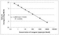
- FIG. 1A shows the difference ( ⁇ Ct; vertical axis) between the Ct value of the target gene and the Ct value of the canine genomic DNA (external standard gene) in the calibration curve sample (standard solution) using immunodeficient mouse blood as a sample in Example 1. ) And the concentration of the target gene (horizontal axis).
- FIG. 1B represents a calibration curve created based on the relationship between the Ct value (vertical axis) of the target gene in the calibration curve sample and the concentration (horizontal axis) of the target gene in Comparative Example 1.
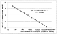
- FIG. 2A shows the difference ( ⁇ Ct; vertical axis) between the Ct value of the target gene in the calibration curve sample using pooled human blood as a sample and the Ct value of canine genomic DNA (external standard gene) in Example 2 and the target gene. Represents a calibration curve created based on the relationship with the concentration (horizontal axis).
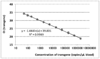
- FIG. 2B shows the difference ( ⁇ Ct; vertical axis) between the Ct value of the target gene and the Ct value of RNaseP (internal standard gene) in the calibration curve sample using pooled human blood as a sample in Comparative Example 2 and the concentration of the target gene. Represents a calibration curve created based on the relationship with (horizontal axis).
- FIG. 2A shows the difference ( ⁇ Ct; vertical axis) between the Ct value of the target gene in the calibration curve sample using pooled human blood as a sample and the Ct value of canine genomic DNA (external standard gene) in Example 2 and the target gene. Represents
- FIG. 2C shows the accuracy of the target gene copy number in the QC sample prepared from the blood sample of each donor in Comparative Example 2 (vertical axis, see Table 2B) and the DNA concentration measured by the absorptiometry (horizontal axis). It is a correlation diagram plotting.
- FIG. 2D plots the trueness of the target gene copy number in QC samples prepared from the blood of individual donors in Example 2 (left side “External control gene”) and Comparative Example 2 (right side “Internal control gene”). It is a scatter plot.
- FIG. 2D plots the trueness of the target gene copy number in QC samples prepared from the blood of individual donors in Example 2 (left side “External control gene”) and Comparative Example 2 (right side “Internal control gene”). It is a scatter plot.
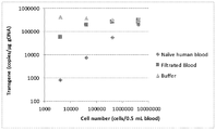
- FIG. 3A shows a sample in Example 3 in which a human gene-modified T cell containing a target gene was added to each of human blood (marked with ⁇ ), human blood from which leukocytes had been removed (marked with ⁇ ), and buffer solution (marked with ⁇ ).
- FIG. 3B shows a sample in Comparative Example 3 in which human gene-modified T cells containing a target gene were added to each of human blood (marked with ⁇ ), white blood cell-depleted human blood (marked with ⁇ ), and buffer solution (marked with ⁇ ).
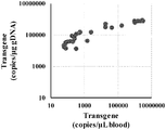
- FIG. 4A shows a quantification method using an external standard gene (canine genomic DNA) according to the present invention for a target gene 1 in a blood sample collected from a Xenograft mouse to which a first human gene-modified T cell was administered in Example 6. It is a graph showing the relationship between the result expressed by copies / ⁇ L blood (horizontal axis) and the result expressed by copies / ⁇ g gDNA by the quantification method using the internal standard gene (RNAseP).
- RNAseP internal standard gene
- FIG. 4B shows copies / ⁇ L of the target gene 2 in the blood sample collected from the Xenograft mouse to which the second human gene-modified T cells were administered in Example 6 by the quantification method using the external standard gene according to the present invention. It is a graph showing the relationship between the result expressed by blood (horizontal axis) and the result expressed by copies / ⁇ g gDNA by a quantification method using an internal standard gene (vertical axis).
- FIG. 4C shows the number of second human gene-modified T cells (measured by horizontal axis, cells / ⁇ L blood, flow cytometry) in the blood sample of second human gene-modified T cells in Example 6 and the target gene 2. It is a graph showing the correlation of the introduction level (measured by the quantification method of the present invention using the vertical axis, copies / ⁇ g gDNA, and internal standard gene).
- FIG. 4D shows the number of second human gene-modified T cells (measured by horizontal axis, cells / ⁇ L blood, flow cytometry) in the blood sample of second human gene-modified T cells in Example 6 and the target gene 2. It is a graph showing the correlation of the introduction level (vertical axis, copies / ⁇ L blood, measured by the quantification method of the present invention using an external standard gene).
- the qPCR method according to the present invention is for measuring the amount of a target gene contained in human cells, and is used as an external standard for correcting the measured amount of the target gene in a biological sample containing human cells.
- the step of adding a predetermined amount of exogenous DNA that does not cross the target gene is included.
- target gene in the present invention is not particularly limited, and can be selected from genes contained in human cells, which are quantified by a general qPCR method.
- the target gene is a gene contained in "gene-modified T cells".
- T cells include ⁇ T cells, ⁇ T cells, CD8 + T cells, CD4 + T cells, tumor infiltrating T cells, memory T cells, naive T cells, NKT cells and the like.
- the "gene modification” includes not only gene modification by introduction of CAR but also gene modification by introduction of TCR (T cell receptor). Typical gene-modified T cells include CAR-T cells and TCR-T cells.
- the target gene is a gene contained in "gene-modified immune cells" other than gene-modified T cells, for example, immune cells modified by introducing genes such as CAR into NK cells, monocytes, macrophages, dendritic cells, and the like. It may be.
- the target gene may be, for example, a gene contained in an expression vector introduced into a host cell such as a T cell in order to prepare a genetically modified cell such as a CAR-T cell.
- the expression vector may be linear or cyclic, and may be a non-viral vector such as a plasmid, a viral vector, or a transposon vector.
- the viral vector include a retrovirus vector, a lentiviral vector, an adenovirus vector, and an adeno-associated virus vector.
- Preferred retroviral vectors include, for example, pMSGV vector (Tamada k et al., ClinCancer Res 18: 6436-6445 (2002)) and pMSCV vector (manufactured by Takara Bio Inc.).
- pMSGV vector Temada k et al., ClinCancer Res 18: 6436-6445 (2002)
- pMSCV vector manufactured by Takara Bio Inc.
- the gene contained in the vector is incorporated into the genome of a host cell such as a T cell, so that the gene can be stably expressed for a long period of time.
- the T cells that serve as host cells are basically (i) single-stranded antibodies that recognize the cell surface antigens of attacked cells such as cancer cells. , (Ii) Transmembrane region, and (iii) Signal transduction region that induces activation of T cells, are configured by linking peptides at each site via spacers, if necessary, CAR expression. Genes are introduced.
- the CAR expression gene has a function of the suicide gene (the function of the suicide gene depends on the course of treatment after administration of the gene-modified T cell, for example, when the cancer cell to be attacked disappears.
- Genes that make it possible to control the number of gene-modified T cells in the body by administering an activating drug may be used together.
- cytokine / chemokine genes and linkers that connect the genes (P2A, F2A, T2A, E2A) (Sequence, etc.) may be used together.
- the target gene is a gene included in the CAR-T cell production gene group. It can be a portion (gene or a part thereof) containing at least a possible base sequence.
- the target gene when TCR-T cells are prepared as gene-modified T cells, the target gene includes a TCR gene, a suicide gene, a cytokine / chemokine gene, a linker that connects the genes, and the like as needed, and CAR-T. It can be a portion (gene or a part thereof) containing at least a base sequence that can be specified to be a TCR-T cell production gene group selected from the cell production gene group.
- the exogenous DNA in the present invention is DNA used as an external standard for correcting the quantitative value of the target gene in the qPCR method, and does not cross the target gene, that is, is not amplified by the primer for the target gene (as such).
- a DNA having a base sequence where a primer for a suitable target sequence can be designed.
- the exogenous DNA also needs to be a base sequence that does not cross the target gene or other genes in the target cell or biological sample (for which primers for such exogenous DNA can be designed). .. It is desirable that the exogenous DNA contains a base sequence (gene or the like) that is not possessed by humans.
- the extrinsic DNA is not particularly limited as long as it is a DNA having a base sequence having the above characteristics.
- Typical exogenous DNA includes mammals other than humans, preferably mammals other than primates, such as cows, pigs, goats, sheep, dogs, cats, rabbits, rodents (mouses, rats, etc.).
- Genomic DNA can be mentioned. It is also possible to use an artificial nucleic acid containing an artificially designed and synthesized sequence as an exogenous DNA (an exogenous nucleic acid when a nucleic acid other than DNA such as LNA is contained).
- the exogenous DNA may be DNA that has been isolated in advance so that the amount of addition can be easily adjusted, or DNA that is contained in the cells of mammals other than humans (the amount of addition is adjusted as the number of cells). May be.
- DNA may be extracted from the cell together with the target gene contained in the cell.
- the amount of the exogenous DNA added to the biological sample is not particularly limited and can be appropriately adjusted.
- the Ct value is obtained for at least the extrinsic DNA ( ⁇ Ct)
- the addition amount may be such that the amount of amplification product of exogenous DNA reaches the threshold in the PCR cycle of a predetermined number of times or less.
- biological sample As the "biological sample” in the present invention, the same as the biological sample for quantifying the target gene contained in human cells by a general qPCR method can be used.
- biological samples may include cells containing, for example, blood, cerebrospinal fluid, bone marrow, tumors (primary tumors, metastatic tumors, cancerous ascites, etc.), and other target genes. Examples include samples prepared from samples collected from.
- the biological sample is prepared from a sample taken from a human who has been administered genetically modified T cells and / or who has suffered or has been treated for a disease that causes a decrease in white blood cells.
- a disease that causes a decrease in white blood cells.
- it is particularly important to analyze the kinetics of the cell number of the cells after administration by the quantitative value improved from the conventional method by the qPCR method of the present invention. is there.
- patients with leukopenia neutropenia, lymphopenia, monocytopenia
- patients who have undergone chemotherapy to deplete white blood cells (lymphocytes) for cell therapy have also been treated with conventional techniques. Since the quantitative value of the target gene is greatly affected by the significant decrease in the amount of gDNA as an internal standard, it is particularly preferable to apply the qPCR method of the present invention.
- the qPCR method refers to various PCR methods capable of quantitatively measuring the copy number of a gene in a biological sample. Such a qPCR method is well known and used by those skilled in the art, and its basic technical matters, preferable technical matters, and the like can also be applied to the present invention.
- One of the typical embodiments of the qPCR method is the "real-time PCR method".
- the fluorescence (signal) that is emitted as the amplification product increases due to PCR is monitored in real time, the number of cycles until the amplification product (signal) reaches a certain level is measured, and a standard sample is used. The number of copies of the target gene in the original biological sample is estimated with reference to the calibration curve produced using the sample.
- Specific examples of the real-time PCR method include a fluorescent probe method using a fluorescently labeled probe (for example, TaqMan method, molecular beacon method, cycling probe method), and an inter using a reagent that emits fluorescence by binding to double-stranded DNA.
- the cullator method can be mentioned, and either of them can be used in the present invention.
- ddPCR method digital droplet PCR method
- PCR was performed after limiting dilution of the biological sample so that the target gene was contained in 0 or 1 (or a plurality) in the micro-compartment (well), and the amplification signal was negative for the total number of micro-compartments (well).
- the absolute copy number of the target gene in the biological sample is estimated from the proportion of the number of microcompartments (which did not contain the target gene). In making this estimation, the Poisson distribution is used to correct for the possibility of microsections containing multiple target genes.
- the amplified signal in the ddPCR method can be based on the same fluorescent probe method as exemplified for the real-time PCR method.
- Basic reagents, kits, systems, etc. for carrying out the qPCR method are also commercially available, and they can also be used in the present invention.
- Nucleotide sequences and bases of primers (forward primer and reverse primer) for target genes and exogenous DNA which are necessary for carrying out the qPCR method of the present invention, and probes used when adopting the fluorescent probe method.
- the length can be appropriately designed by those skilled in the art so that each of the target gene and the exogenous DNA can be specifically amplified (not crossed with other genes).
- the probes and primers may be sequences that are completely complementary to the target sequence, may be sequences that partially contain a mismatch, or may be highly stringent to those sequences. It may be a base sequence having homology within a range that allows hybridization under (hybridization conditions).
- the fluorescent substance used when the fluorescent probe method is adopted and the quencher forming a FRET pair with the fluorescent substance are not particularly limited and can be appropriately selected by those skilled in the art.
- the qPCR method of the present invention includes a step of adding a predetermined amount of exogenous DNA that does not cross the target gene as an external standard for correcting the measured amount of the target gene to the biological sample, and other steps, for example, , Preparation step of biological sample from sample, DNA extraction step from human cells, etc., nucleic acid amplification step by PCR (heat denaturation, primer annealing, probe hybridization, extension reaction, etc.), etc. are general qPCR methods. Can be similar to.
- the number of PCR reaction cycles (Ct value) until crossing (threshold) is measured, and the number of copies (concentration) of the target gene contained in the initial biological sample is reflected as a measured value.
- the larger the Ct value the lower the copy number (concentration) of the initial DNA. Since DNA increases exponentially in PCR, the difference between the two Ct values is related to the logarithmic difference, i.e. the ratio (index) of the original number of copies of DNA.
- the measured value (Ct value) and the final measured amount (copy number) of the target gene are corrected by the Ct value for gDNA as an internal standard. That is, the following operations [B1] to [B3] were performed: [B1] The initial measured value (Ct) is corrected by measuring the Ct value of the gDNA used as the internal standard and calculating the difference ( ⁇ Ct (i)) between the Ct value of the target gene and the Ct value of the internal standard.
- the Ct value is also measured for a plurality of standard samples in which the copy number (concentration) of the target gene is known, the difference from the internal standard Ct value ( ⁇ Ct (i_s)) is calculated, and the plurality of ⁇ Ct.
- the corrected measurement value ( ⁇ Ct (i)) is converted, and the corrected measurement amount of the copy number of the target gene contained in the initial biological sample is obtained. ..
- the final measured amount is expressed in units of, for example, "copy number / ⁇ g gDNA".
- the measured value (Ct value) and the final measured amount (copy number) of the target gene are corrected by the Ct value of the extrinsic DNA as an external standard. .. That is, the following operations [A1] to [A3] were performed: [A1]
- the initial measured value (Ct) is obtained by measuring the Ct value of the extrinsic DNA used as the external standard and calculating the difference ( ⁇ Ct (o)) between the Ct value of the target gene and the Ct value of the external standard.
- the Ct value is also measured for a plurality of standard samples whose copy number (concentration) of the target gene is known, the difference from the Ct value of the external standard ( ⁇ Ct (o_s)) is calculated, and the plurality of ⁇ Ct Create a calibration curve based on the relationship between (o_s) and the copy number of the corresponding known target gene; [A3] By referring to the calibration curve, the corrected measured value ( ⁇ Ct (o)) is converted, and the corrected measured amount of the copy number of the target gene contained in the initial biological sample is obtained. .. The final measured amount is the number of copies per unit amount (weight) of the biological sample, and can be expressed in units such as "copy / ⁇ g blood".
- the method of correcting the measured amount of the target gene using exogenous DNA in the qPCR method according to the present invention is not limited to the correction for ⁇ Ct in the real-time PCR method as described above. If the copy number of the target gene in the biological sample can be quantified more accurately than using the quantitative value measured for the target gene itself, exogenous DNA is used as an external standard in other ways, for example, in the ddPCR method. It is possible to correct it.
- the measured amount of the target gene measured by the real-time PCR method, the ddPCR method, or the like can be further corrected (multiplied by 100 / recovery rate) depending on how much the recovery rate deviates from 100%. ..
- Such further corrections can also be made in combination with the corrections relating to ⁇ Ct in the real-time PCR method described above.
- the qPCR kit according to the present invention contains a predetermined amount of exogenous DNA that does not cross the target gene as an external standard for correcting the measured amount of the target gene, and other components (for example, 5' ⁇ 3').
- DNA polymerase having exonuclease activity, other reagents, instruments, etc. necessary for carrying out the qPCR method can include the same as the conventional kit for qPCR. Primers and probes may be prepared separately depending on the target gene and the exogenous gene.
- Probes, forward and reverse primers for canine genomic DNA-specific gene (MC1R) used in the following examples and comparative examples; probe and forward primer for target gene 1 (transgene 1 for CAR-T cell production). And the reverse primer; and the base sequences of the probe, the forward primer and the reverse primer for the target gene 2 (transgene 2 for producing CAR-T cells) are as follows. It should be noted that the probe, forward primer and reverse primer for the target gene 1 and the probe, forward primer and reverse primer for the canine genomic DNA-specific gene (MC1R) have a base sequence that does not cross each other; and the target.
- MC1R probe SEQ ID NO: 1) 5'-GCCTTGGCTGCGCAGGCTGCTGTGGTGCAG -3' Forward primer for MC1R (SEQ ID NO: 2) 5'-CGCCCATGTATTACTTCATCTGTTGCC -3' Reverse primer for MC1R (SEQ ID NO: 3) 5'-CACGGCGATGGCGCCCAGGAA -3' Probe for target gene 1 (SEQ ID NO: 4) 5'-CGACTACAGGGCCTACT -3' Forward primer for target gene 1 (SEQ ID NO: 5) 5'- GGAGCTGAGGTCCCTGAGAAG -3' Target gene 1 reverse primer (SEQ ID NO: 6) 5'-CCTGGCCCAGTAGTCGAA -3' Probe for target gene 2 (SEQ ID NO: 7)
- the "target gene” in the description of this example and this comparative example refers to "target gene 1".
- the plasmid containing the target gene should be 50, 100, 200, 600, 2,000, 6,000, 20,000, 60,000, 200,000, 600,000, and 2,000,000 copies / ⁇ L blood.
- the added sample was prepared and used as a calibration curve sample (standard solution).
- a sample in which a plasmid containing the target gene was added so as to be 50, 100, 200, 600, 4,000, 40,000, 400,000, and 2,000,000 copies / ⁇ L blood was prepared and used as a quality control sample (QC sample).
- Genomic DNA was extracted from each of these samples using a commercially available DNA extraction reagent (QIAampDNA Blood Midi Kits, QIAGEN), followed by probes, forward primers and probes for the target gene and canine genomic DNA-specific gene (MC1R), respectively.
- QPCR quantification was performed using a reverse primer.
- the qPCR was carried out using a QuantStudio7Flex real-time PCR system (Thermo Fisher Scientific) under the condition that one cycle was 95 ° C. for 10 minutes / (95 ° C. for 15 seconds and 60 ° C. for 1 minute).
- Tables 1A and B show the results of calculating the target gene copy number in the QC sample using the calibration curve of Delta Ct and Ct values, respectively.
- the average trueness (Accuracy, Mean) of the value (Observed) calculated by the calibration curve of Delta Ct was 83.0-108.1%, and the accuracy of variation (CV) was 2.4-17.5%, which was good.
- the average trueness of the values calculated by the Ct calibration curve is 46.9-92.7%, and the accuracy is 5.0-26.6%, which is compared with the results of the Delta Ct (Example 1) calibration curve using an external standard gene.
- the quantitativeness of the method using the Ct calibration curve (Comparative Example 1) was low. From the above results, it was considered that correction of the target gene copy number between samples by an external standard gene is useful in quantifying the copy number per unit amount of a biological sample.
- target gene in the description of this example refers to "target gene 1".
- a sample containing 1.8 mL of pooled human blood mixed with the blood of multiple people was used as a sample, and 600, 2,000, 6,000, 20,000, 60,000, 200,000, 2,000,000, and 20,000,000 copies of plasmid containing the target gene were added to the sample. , Used as a calibration curve sample (standard solution).
- a sample was prepared by adding 2,000 copies of a plasmid containing the target gene to 1.8 mL of blood of each of the 8 donors, and used as a QC sample. To each of these samples, 1,000 ng of canine genomic DNA (Zyagen) was added as an external standard gene.
- Genomic DNA is extracted from each of these samples using a commercially available DNA extraction reagent (QIAampDNA Blood Midi Kits, QIAGEN), followed by probes, forward primers and reverses for the target gene and canine genomic DNA-specific gene (MC1R).
- QPCR quantification was performed using primers.
- the qPCR was carried out using a QuantStudio7Flex real-time PCR system (Thermo Fisher Scientific) under the condition that one cycle was 95 ° C. for 10 minutes / (95 ° C. for 15 seconds and 60 ° C. for 1 minute).
- a calibration curve was created from the difference between the Ct value of the target gene and the Ct value of canine genomic DNA (Delta Ct) and the concentration of the target gene added to each calibration curve sample (Fig. 2A). Since the linearity of the calibration curve was observed in the range of 0.33 to 11,111 copies / ⁇ L blood of the target gene, it is considered that the number of copies per unit amount of the biological sample can be quantitatively measured by this method. ..
- target gene in the description of this comparative example refers to "target gene 1".
- correction of target gene copy number between samples by internal standard gene by subtracting Ct of internal standard gene from Ct of target gene, target gene between samples is generally used. How to correct the copy number) is performed. Therefore, RNase P was selected as the internal standard gene, and qPCR was used for all the DNA extracts of Example 2 using probes for human RNase P, forward primers, and reverse primers (TaqMan RNase P Control Reagents Kit, Thermo Fisher Scientific). Quantitative was performed.
- the qPCR was carried out using a QuantStudio7Flex real-time PCR system (Thermo Fisher Scientific) under the condition that one cycle was 95 ° C. 20 seconds / (95 ° C. 1 second and 60 ° C. 20 seconds).
- a calibration curve was created from the difference between the Ct value of the target gene and the Ct value of RNase P (Delta Ct) and the concentration of each calibration curve sample (Fig. 2B).
- the linearity of the calibration curve was observed in the range of copy number of the target gene from 0.33 to 11,111 copies / ⁇ L blood.
- target gene in the description of this example and this comparative example refers to "target gene 1".
- human blood from which leukocytes have been removed with a commercially available leukocyte depletion filter (PLASMODIPUR, EuroProxima), and a buffer solution as samples 4,000, 40,000, 400,000, and 4,000,000 human gene-modified T cells were added thereto. Samples to which each was added were prepared.
- the above-mentioned human gene-modified T cells were prepared by introducing a CAR gene containing a target gene into T cells activated by a retroviral vector.
- a calibration curve sample (standard solution) a sample in which a plasmid containing a target gene was added to 0.5 mL of human blood for 375, 625, 1,250, 3,750, 12,500, 37,500, 125,000, 1,250,000, 12,500,000, and 125,000,000 copies was added.
- Canine genomic DNA (Zyagen) was added to each of these samples at a rate of 2,000 ng / mL.
- Genomic DNA is extracted from each of these samples using a commercially available DNA extraction reagent (QIAampDNA Blood Midi Kits, QIAGEN), and then qPCR quantification is performed using probes, forward and reverse primers for the target gene and canine genomic DNA.
- a commercially available DNA extraction reagent QIAampDNA Blood Midi Kits, QIAGEN
- the qPCR was carried out using a QuantStudio7Flex real-time PCR system (Thermo Fisher Scientific) under the condition that one cycle was 95 ° C. for 10 minutes / (95 ° C. for 15 seconds and 60 ° C. for 1 minute).
- a calibration curve is created from the difference between the Ct value of the target gene and the Ct value of canine genomic DNA (Delta Ct) and the concentration of the target gene added to each calibration curve sample, and the concentration of the target gene in the sample to which the genetically modified T cells are added is measured.
- the result is shown in FIG. 3A.
- an increase in the copy number of the target gene was confirmed in proportion to the number of added cells.
- the copy number of the target gene derived by this method was the same regardless of the type of sample. From these results, it is considered that this method can quantitatively measure the fluctuation of the number of target gene copies per unit amount of the biological sample due to the increase or decrease of the gene-modified T cells in the living body.
- target gene in the description of this example and this comparative example refers to "target gene 1".
- the ratio of the target gene copy number to the amount of genomic DNA is generally used. Therefore, the human genomic DNA and the target gene copy number in the DNA extract extracted from the gene-modified T cell-added sample prepared in Example 3 were measured, and the ratio of the target gene copy number to the genomic DNA amount was calculated.
- Samples in which the plasmid containing the target gene was added to the extract AE of the DNA extraction reagent so that the results were 1.5, 5, 15, 25, 50, 150, 500, 1,500, 5,000, 50,000, 500,000, and 5,000,000 copies / ⁇ L. was prepared and used as a calibration curve sample (standard solution) of the target gene.
- qPCR quantification was performed using a probe, a forward primer, and a reverse primer for the target gene and the human genomic DNA-specific gene sequence (RNaseP).
- RNaseP human genomic DNA-specific gene sequence
- a QuantStudio7Flex real-time PCR system was used. In the qPCR measurement for the target gene, 40 cycles were performed under the condition that 95 ° C. 10 minutes / (95 ° C. 15 seconds and 60 ° C. 1 minute) was one cycle. In the qPCR measurement on human genomic DNA, 40 cycles were performed under the condition that 95 ° C. 20 seconds / (95 ° C. 1 second and 60 ° C. 20 seconds) was one cycle.
- Calibration curve of target gene A calibration curve is created from the Ct value of the target gene amplification curve at each concentration (copy number) of the sample and the concentration of each calibration curve sample, and the target gene copy in the DNA extract of the gene-modified T cell-added sample. The number was calculated.
- a calibration curve was created from the Ct value of the target gene amplification curve at each concentration of the calibration curve sample of human genomic DNA (amount of human genomic DNA) and the concentration of each calibration curve sample, and the calibration curve was prepared in the DNA extract of the gene-modified T cell-added sample. The amount of human genomic DNA was calculated.
- the ratio of the target gene copy number to the amount of human genomic DNA in the DNA extract of the gene-modified T cell-added sample was calculated and compared with the number of added cells, and the result is shown in FIG. 3B.
- the copy number of the target gene was confirmed according to the number of added cells, but in the human blood and buffer sample from which leukocytes were removed, the copy number was increased according to the number of added cells. No increase in the copy number of the target gene was clearly observed.
- the human genomic DNA derived from the genetically modified T cell increases as the number of target gene copies increases, so that the leukocyte content of the human genomic DNA is significantly lower than that of untreated human blood. It is considered that the removed human blood and buffer did not show an increase in the copy number of the target gene according to the number of added cells. In this way, when the ratio of the number of target gene copies to the amount of genomic DNA is used as a unit for quantifying the number of target gene copies, the target gene copy for the increase or decrease of gene-modified T cells in vivo depends on the amount of genome contained in the sample.
- target gene in human blood by correction using an external standard gene
- the "target gene” in the description of this example refers to "target gene 1”.
- a pooled human blood mixed with 0.5 mL of blood collected from multiple persons was used as a sample, and 0.75, 1.3, 2.5, 7.5, 25, 75, 250, 2,500, 25,000 and 250,000 copies / ⁇ L of plasmid containing the target gene were used as a sample.
- a sample added at a blood concentration was prepared and used as a calibration curve sample (standard solution).
- a sample in which a plasmid containing a target gene was added at concentrations of 7.5, 200 and 200,000 copies / ⁇ L blood to a similar pooled human blood sample was prepared and used as a QC sample.
- 1,000 ng of canine genomic DNA Zyagen was added as an external standard gene.
- Genomic DNA is extracted from each of these samples using a commercially available DNA extraction reagent (QIAamp DNA Blood Midi Kits, QIAGEN), followed by probes, forward primers and reverses for the target gene and canine genomic DNA-specific gene (MC1R). QPCR quantification was performed using primers.
- the qPCR was performed using a QuantStudio 7 Flex real-time PCR system (Thermo Fisher Scientific) under the condition that one cycle was 95 ° C. for 10 minutes / (95 ° C. for 15 seconds and 60 ° C. for 1 minute).
- a calibration curve was created from the difference between the Ct value of the target gene and the Ct value of canine genomic DNA (Delta Ct) and the concentration of the target gene added to each calibration curve sample.
- the target gene copy number was measured three times, each on a different day. The results are shown in Table 2. The robustness of the quantification method of the present invention, which is corrected using an external standard gene, can be confirmed.
- Target gene 1 Quantification of target gene in mouse blood by correction using an external standard gene
- a sample was prepared using 30 ⁇ L of immunodeficient mouse blood as a sample, to which a plasmid containing the target gene was added so as to have 50, 200, 600, 2,000, 6,000, 20,000, 60,000, 200,000 and 2,500,000 copies / ⁇ L blood. Then, it was used as a calibration curve sample (standard solution).
- a sample in which a plasmid containing the target gene was added so as to be 50, 100, 200, 600, 4,000, 40,000, 400,000, and 2,000,000 copies / ⁇ L blood was prepared and used as a quality control sample (QC sample).
- QC sample quality control sample
- 1,200 ng of canine genomic DNA (Zyagen) was added as an external standard gene.
- Genomic DNA was extracted from each of these samples using a commercially available DNA extraction reagent (QIAamp DNA Blood Midi Kits, QIAGEN), followed by probes, forward primers and probes for the target gene and canine genomic DNA-specific gene (MC1R), respectively.
- QPCR quantification was performed using a reverse primer.
- the qPCR was performed using a QuantStudio 7 Flex real-time PCR system (Thermo Fisher Scientific) under the condition that one cycle was 95 ° C. for 10 minutes / (95 ° C. for 15 seconds and 60 ° C. for 1 minute).
- Human gene-modified T cells (hereinafter referred to as "first human gene-modified T cells") prepared by introducing a CAR gene containing a target gene 1 into T cells activated by a retroviral vector, and target gene 2 are introduced.
- Human gene-modified T cells (hereinafter referred to as "second human gene-modified T cells”) prepared by introducing the containing CAR gene into T cells activated by a retroviral vector were prepared.
- First human gene-modified T cells were intravenously administered to Xenograft mice at a dose of 5 ⁇ 10 6 CAR + cells / animal or 10 ⁇ 10 6 CAR + cells / animal. Blood was collected from the submandibular veins of mice 1, 3, 7, 10, 14, 17, 21 and 28 days after administration. In addition, second human gene-modified T cells were intravenously administered to Xenograft mice at a dose of 10 ⁇ 10 6 CAR + cells / animal. Blood was drawn from the submandibular veins of mice 1, 4, 8, 11, 15, 22 and 29 days after administration.
- Example 2 and Comparative Example 2 Prepared using blood samples collected at each of the above time points (excluding the last day) using an external standard gene (canine genomic DNA) and an internal standard gene (RNaseP) in the same manner as in Example 2 and Comparative Example 2, respectively. Based on the calibration curve, the copy numbers of the target genes 1 and 2 in the blood sample were quantified by the same measurement method as in Example 2 and Comparative Example 2. The results are shown in FIGS. 4A, B, C and D. In the blood sample of the first human gene-modified T cell, when the copy number of the target gene 1 is expressed by the first copies / ⁇ g gDNA by the quantification method using the internal standard gene (vertical axis), saturation is observed.
- an external standard gene canine genomic DNA
- RNaseP internal standard gene
- the copy number is calculated by the quantification method using the internal standard gene. It appears more clearly than when expressed per ⁇ g gDNA (Fig. 4C).
- the quantification method according to the present invention is considered to be a method more suitable for evaluation of cell kinetics, which has a low risk of underestimating the proliferation of CAR-T cells in vivo.
Landscapes
- Chemical & Material Sciences (AREA)
- Organic Chemistry (AREA)
- Life Sciences & Earth Sciences (AREA)
- Zoology (AREA)
- Wood Science & Technology (AREA)
- Proteomics, Peptides & Aminoacids (AREA)
- Health & Medical Sciences (AREA)
- Engineering & Computer Science (AREA)
- Chemical Kinetics & Catalysis (AREA)
- Biophysics (AREA)
- Immunology (AREA)
- Microbiology (AREA)
- Molecular Biology (AREA)
- Biotechnology (AREA)
- Analytical Chemistry (AREA)
- Physics & Mathematics (AREA)
- Biochemistry (AREA)
- Bioinformatics & Cheminformatics (AREA)
- General Engineering & Computer Science (AREA)
- General Health & Medical Sciences (AREA)
- Genetics & Genomics (AREA)
- Measuring Or Testing Involving Enzymes Or Micro-Organisms (AREA)
Abstract
Priority Applications (5)
| Application Number | Priority Date | Filing Date | Title |
|---|---|---|---|
| CN202080068474.3A CN114502743A (zh) | 2019-09-30 | 2020-09-29 | 定量pcr方法及用于其的试剂盒 |
| JP2021551296A JPWO2021065877A1 (fr) | 2019-09-30 | 2020-09-29 | |
| EP20871606.8A EP4039802A4 (fr) | 2019-09-30 | 2020-09-29 | Procédé de pcr quantitative et kit associé |
| US17/764,463 US20220356516A1 (en) | 2019-09-30 | 2020-09-29 | Quantitative pcr method and kit therefor |
| KR1020227011613A KR20220066298A (ko) | 2019-09-30 | 2020-09-29 | 정량적 pcr법 및 이를 위한 키트 |
Applications Claiming Priority (4)
| Application Number | Priority Date | Filing Date | Title |
|---|---|---|---|
| JP2019-178755 | 2019-09-30 | ||
| JP2019178755 | 2019-09-30 | ||
| JP2020-127716 | 2020-07-28 | ||
| JP2020127716 | 2020-07-28 |
Publications (1)
| Publication Number | Publication Date |
|---|---|
| WO2021065877A1 true WO2021065877A1 (fr) | 2021-04-08 |
Family
ID=75336418
Family Applications (1)
| Application Number | Title | Priority Date | Filing Date |
|---|---|---|---|
| PCT/JP2020/036819 WO2021065877A1 (fr) | 2019-09-30 | 2020-09-29 | Procédé de pcr quantitative et kit associé |
Country Status (6)
| Country | Link |
|---|---|
| US (1) | US20220356516A1 (fr) |
| EP (1) | EP4039802A4 (fr) |
| JP (1) | JPWO2021065877A1 (fr) |
| KR (1) | KR20220066298A (fr) |
| CN (1) | CN114502743A (fr) |
| WO (1) | WO2021065877A1 (fr) |
Cited By (3)
| Publication number | Priority date | Publication date | Assignee | Title |
|---|---|---|---|---|
| CN113957174A (zh) * | 2021-06-18 | 2022-01-21 | 重庆天科雅生物科技有限公司 | 一种特异性识别免疫分型为hlaa11的ebv病毒肽段的tcr引物组及其应用 |
| WO2023027073A1 (fr) * | 2021-08-27 | 2023-03-02 | 株式会社島津製作所 | Procédé de pcr quantitative utilisant une commande interne |
| WO2023054501A1 (fr) * | 2021-09-29 | 2023-04-06 | 武田薬品工業株式会社 | Procédé de mesure de la concentration cellulaire |
Families Citing this family (1)
| Publication number | Priority date | Publication date | Assignee | Title |
|---|---|---|---|---|
| JP7274033B1 (ja) * | 2022-10-27 | 2023-05-15 | 積水メディカル株式会社 | 蛍光検出の補正方法 |
Citations (2)
| Publication number | Priority date | Publication date | Assignee | Title |
|---|---|---|---|---|
| JP2005168408A (ja) * | 2003-12-11 | 2005-06-30 | Ebara Corp | 定量的pcr法による標的微生物又は遺伝子の定量方法 |
| JP2018512871A (ja) * | 2015-04-20 | 2018-05-24 | キム・ソンチョン | 標準物質として外部生体分子を用いた生体分子分析方法およびそのキット |
Family Cites Families (1)
| Publication number | Priority date | Publication date | Assignee | Title |
|---|---|---|---|---|
| EP2721173A1 (fr) * | 2011-06-14 | 2014-04-23 | INSERM (Institut National de la Santé et de la Recherche Médicale) | Procédés de détermination du niveau d'expression d'un gène d'intérêt comprenant la correction des données de rt-pcrq concernant les signaux dérivés de l'adn génomique |
-
2020
- 2020-09-29 US US17/764,463 patent/US20220356516A1/en active Pending
- 2020-09-29 JP JP2021551296A patent/JPWO2021065877A1/ja active Pending
- 2020-09-29 KR KR1020227011613A patent/KR20220066298A/ko unknown
- 2020-09-29 EP EP20871606.8A patent/EP4039802A4/fr active Pending
- 2020-09-29 CN CN202080068474.3A patent/CN114502743A/zh active Pending
- 2020-09-29 WO PCT/JP2020/036819 patent/WO2021065877A1/fr unknown
Patent Citations (2)
| Publication number | Priority date | Publication date | Assignee | Title |
|---|---|---|---|---|
| JP2005168408A (ja) * | 2003-12-11 | 2005-06-30 | Ebara Corp | 定量的pcr法による標的微生物又は遺伝子の定量方法 |
| JP2018512871A (ja) * | 2015-04-20 | 2018-05-24 | キム・ソンチョン | 標準物質として外部生体分子を用いた生体分子分析方法およびそのキット |
Non-Patent Citations (5)
| Title |
|---|
| FU, JIE ET AL.: "Absolute Quantification of Plasmid DNA by Real-time PCR with Genomic DNA as External Standard and Its Application to a Biodistribution Study of an HIV DNA Vaccine", ANALYTICAL SCIENCES, vol. 25, 2009, pages 675 - 680, XP055815450, DOI: 10. 2116/analsci.25.675 * |
| MUELLER ET AL., BLOOD, vol. 130, 2017, pages 2317 - 2325 |
| See also references of EP4039802A4 |
| TAMADA K ET AL., CLINCANCER RES, vol. 18, 2002, pages 6436 - 6445 |
| YUN, JAMES J. ET AL.: "Genomic DNA functions as a universal external standard in quantitative real- time PCR", NUCLEIC ACIDS RESEARCH, vol. 34, no. 12, 2006, pages e85, XP055219779, DOI: 10.1093/nar/gkl400 * |
Cited By (3)
| Publication number | Priority date | Publication date | Assignee | Title |
|---|---|---|---|---|
| CN113957174A (zh) * | 2021-06-18 | 2022-01-21 | 重庆天科雅生物科技有限公司 | 一种特异性识别免疫分型为hlaa11的ebv病毒肽段的tcr引物组及其应用 |
| WO2023027073A1 (fr) * | 2021-08-27 | 2023-03-02 | 株式会社島津製作所 | Procédé de pcr quantitative utilisant une commande interne |
| WO2023054501A1 (fr) * | 2021-09-29 | 2023-04-06 | 武田薬品工業株式会社 | Procédé de mesure de la concentration cellulaire |
Also Published As
| Publication number | Publication date |
|---|---|
| JPWO2021065877A1 (fr) | 2021-04-08 |
| CN114502743A (zh) | 2022-05-13 |
| EP4039802A4 (fr) | 2023-12-27 |
| US20220356516A1 (en) | 2022-11-10 |
| EP4039802A1 (fr) | 2022-08-10 |
| KR20220066298A (ko) | 2022-05-24 |
Similar Documents
| Publication | Publication Date | Title |
|---|---|---|
| WO2021065877A1 (fr) | Procédé de pcr quantitative et kit associé | |
| US11806555B2 (en) | Methods for treating hair loss disorders | |
| CN109790583B (zh) | 对肺腺癌亚型分型的方法 | |
| KR101913293B1 (ko) | 탈모 질환의 치료 방법 | |
| RU2766004C1 (ru) | Способ оценки нормального состояния иммунного репертуара и его применение | |
| KR20140044341A (ko) | 암에 대한 분자적 진단 검사 | |
| EA020795B1 (ru) | Генетические маркеры, ассоциированные с ответом на интерферон-альфа | |
| KR102067327B1 (ko) | 최적의 암 치료를 위한 Her2 단백질 정량 | |
| US20230135171A1 (en) | Methods and systems for molecular disease assessment via analysis of circulating tumor dna | |
| US20230223105A1 (en) | Mitigation of statistical bias in genetic sampling | |
| KR20200037796A (ko) | 암 치료를 위한 염색체 불안정성 및 다운스트림 사이토졸 dna 시그널링의 표적화 | |
| CN111670364B (zh) | 用于将癌症患者分类到合适的癌症治疗组的系统和方法以及治疗癌症患者的组合物 | |
| EP0290144A1 (fr) | Méthode de détection de résistance naissante à des agents thérapeutiques chez des malades cancéreux | |
| CN101784674A (zh) | Egfr抑制剂治疗的预测性标记物 | |
| CN104650214A (zh) | 肺动脉高压致病基因acvrl1突变位点及其应用 | |
| US20220392638A1 (en) | Precision enrichment of pathology specimens | |
| CN112608995A (zh) | 一种肺结核病特异性标志物及应用和试剂盒 | |
| WO2022272309A1 (fr) | Méthodes d'utilisation de hla-i loh somatique pour prédire la réponse de patients traités par un inhibiteur de points de contrôle immunitaires atteints d'un cancer du poumon |
Legal Events
| Date | Code | Title | Description |
|---|---|---|---|
| 121 | Ep: the epo has been informed by wipo that ep was designated in this application |
Ref document number: 20871606 Country of ref document: EP Kind code of ref document: A1 |
|
| ENP | Entry into the national phase |
Ref document number: 2021551296 Country of ref document: JP Kind code of ref document: A |
|
| NENP | Non-entry into the national phase |
Ref country code: DE |
|
| ENP | Entry into the national phase |
Ref document number: 20227011613 Country of ref document: KR Kind code of ref document: A |
|
| ENP | Entry into the national phase |
Ref document number: 2020871606 Country of ref document: EP Effective date: 20220502 |