WO2021054090A1 - エネルギーサブトラクション処理装置、方法およびプログラム - Google Patents
エネルギーサブトラクション処理装置、方法およびプログラム Download PDFInfo
- Publication number
- WO2021054090A1 WO2021054090A1 PCT/JP2020/032748 JP2020032748W WO2021054090A1 WO 2021054090 A1 WO2021054090 A1 WO 2021054090A1 JP 2020032748 W JP2020032748 W JP 2020032748W WO 2021054090 A1 WO2021054090 A1 WO 2021054090A1
- Authority
- WO
- WIPO (PCT)
- Prior art keywords
- image
- radiation
- bone
- new
- subject
- Prior art date
- Legal status (The legal status is an assumption and is not a legal conclusion. Google has not performed a legal analysis and makes no representation as to the accuracy of the status listed.)
- Ceased
Links
Images


Classifications
-
- A—HUMAN NECESSITIES
- A61—MEDICAL OR VETERINARY SCIENCE; HYGIENE
- A61B—DIAGNOSIS; SURGERY; IDENTIFICATION
- A61B6/00—Apparatus or devices for radiation diagnosis; Apparatus or devices for radiation diagnosis combined with radiation therapy equipment
- A61B6/48—Diagnostic techniques
- A61B6/482—Diagnostic techniques involving multiple energy imaging
-
- G—PHYSICS
- G06—COMPUTING OR CALCULATING; COUNTING
- G06T—IMAGE DATA PROCESSING OR GENERATION, IN GENERAL
- G06T7/00—Image analysis
- G06T7/0002—Inspection of images, e.g. flaw detection
- G06T7/0012—Biomedical image inspection
-
- A—HUMAN NECESSITIES
- A61—MEDICAL OR VETERINARY SCIENCE; HYGIENE
- A61B—DIAGNOSIS; SURGERY; IDENTIFICATION
- A61B6/00—Apparatus or devices for radiation diagnosis; Apparatus or devices for radiation diagnosis combined with radiation therapy equipment
- A61B6/40—Arrangements for generating radiation specially adapted for radiation diagnosis
- A61B6/4035—Arrangements for generating radiation specially adapted for radiation diagnosis the source being combined with a filter or grating
-
- A—HUMAN NECESSITIES
- A61—MEDICAL OR VETERINARY SCIENCE; HYGIENE
- A61B—DIAGNOSIS; SURGERY; IDENTIFICATION
- A61B6/00—Apparatus or devices for radiation diagnosis; Apparatus or devices for radiation diagnosis combined with radiation therapy equipment
- A61B6/50—Apparatus or devices for radiation diagnosis; Apparatus or devices for radiation diagnosis combined with radiation therapy equipment specially adapted for specific body parts; specially adapted for specific clinical applications
- A61B6/505—Apparatus or devices for radiation diagnosis; Apparatus or devices for radiation diagnosis combined with radiation therapy equipment specially adapted for specific body parts; specially adapted for specific clinical applications for diagnosis of bone
-
- A—HUMAN NECESSITIES
- A61—MEDICAL OR VETERINARY SCIENCE; HYGIENE
- A61B—DIAGNOSIS; SURGERY; IDENTIFICATION
- A61B6/00—Apparatus or devices for radiation diagnosis; Apparatus or devices for radiation diagnosis combined with radiation therapy equipment
- A61B6/52—Devices using data or image processing specially adapted for radiation diagnosis
- A61B6/5211—Devices using data or image processing specially adapted for radiation diagnosis involving processing of medical diagnostic data
- A61B6/5217—Devices using data or image processing specially adapted for radiation diagnosis involving processing of medical diagnostic data extracting a diagnostic or physiological parameter from medical diagnostic data
-
- A—HUMAN NECESSITIES
- A61—MEDICAL OR VETERINARY SCIENCE; HYGIENE
- A61B—DIAGNOSIS; SURGERY; IDENTIFICATION
- A61B6/00—Apparatus or devices for radiation diagnosis; Apparatus or devices for radiation diagnosis combined with radiation therapy equipment
- A61B6/52—Devices using data or image processing specially adapted for radiation diagnosis
- A61B6/5211—Devices using data or image processing specially adapted for radiation diagnosis involving processing of medical diagnostic data
- A61B6/5229—Devices using data or image processing specially adapted for radiation diagnosis involving processing of medical diagnostic data combining image data of a patient, e.g. combining a functional image with an anatomical image
- A61B6/5235—Devices using data or image processing specially adapted for radiation diagnosis involving processing of medical diagnostic data combining image data of a patient, e.g. combining a functional image with an anatomical image combining images from the same or different ionising radiation imaging techniques, e.g. PET and CT
-
- A—HUMAN NECESSITIES
- A61—MEDICAL OR VETERINARY SCIENCE; HYGIENE
- A61B—DIAGNOSIS; SURGERY; IDENTIFICATION
- A61B6/00—Apparatus or devices for radiation diagnosis; Apparatus or devices for radiation diagnosis combined with radiation therapy equipment
- A61B6/52—Devices using data or image processing specially adapted for radiation diagnosis
- A61B6/5258—Devices using data or image processing specially adapted for radiation diagnosis involving detection or reduction of artifacts or noise
- A61B6/5282—Devices using data or image processing specially adapted for radiation diagnosis involving detection or reduction of artifacts or noise due to scatter
-
- G—PHYSICS
- G06—COMPUTING OR CALCULATING; COUNTING
- G06T—IMAGE DATA PROCESSING OR GENERATION, IN GENERAL
- G06T5/00—Image enhancement or restoration
- G06T5/50—Image enhancement or restoration using two or more images, e.g. averaging or subtraction
-
- G—PHYSICS
- G06—COMPUTING OR CALCULATING; COUNTING
- G06T—IMAGE DATA PROCESSING OR GENERATION, IN GENERAL
- G06T5/00—Image enhancement or restoration
- G06T5/90—Dynamic range modification of images or parts thereof
- G06T5/92—Dynamic range modification of images or parts thereof based on global image properties
-
- A—HUMAN NECESSITIES
- A61—MEDICAL OR VETERINARY SCIENCE; HYGIENE
- A61B—DIAGNOSIS; SURGERY; IDENTIFICATION
- A61B6/00—Apparatus or devices for radiation diagnosis; Apparatus or devices for radiation diagnosis combined with radiation therapy equipment
- A61B6/40—Arrangements for generating radiation specially adapted for radiation diagnosis
- A61B6/4035—Arrangements for generating radiation specially adapted for radiation diagnosis the source being combined with a filter or grating
- A61B6/4042—K-edge filters
-
- G—PHYSICS
- G06—COMPUTING OR CALCULATING; COUNTING
- G06T—IMAGE DATA PROCESSING OR GENERATION, IN GENERAL
- G06T2207/00—Indexing scheme for image analysis or image enhancement
- G06T2207/10—Image acquisition modality
- G06T2207/10016—Video; Image sequence
-
- G—PHYSICS
- G06—COMPUTING OR CALCULATING; COUNTING
- G06T—IMAGE DATA PROCESSING OR GENERATION, IN GENERAL
- G06T2207/00—Indexing scheme for image analysis or image enhancement
- G06T2207/10—Image acquisition modality
- G06T2207/10116—X-ray image
-
- G—PHYSICS
- G06—COMPUTING OR CALCULATING; COUNTING
- G06T—IMAGE DATA PROCESSING OR GENERATION, IN GENERAL
- G06T2207/00—Indexing scheme for image analysis or image enhancement
- G06T2207/30—Subject of image; Context of image processing
- G06T2207/30004—Biomedical image processing
- G06T2207/30008—Bone
-
- G—PHYSICS
- G06—COMPUTING OR CALCULATING; COUNTING
- G06T—IMAGE DATA PROCESSING OR GENERATION, IN GENERAL
- G06T2207/00—Indexing scheme for image analysis or image enhancement
- G06T2207/30—Subject of image; Context of image processing
- G06T2207/30004—Biomedical image processing
- G06T2207/30061—Lung
Definitions
- This disclosure relates to energy subtraction processing equipment, methods and programs.
- the energy subtraction processing is a specific structure in which each pixel of the two radiation images obtained as described above is associated with each other, multiplied by an appropriate weighting coefficient between the pixels, and then subtracted (subtract). This is a method of acquiring the extracted image.
- energy subtraction processing for example, if a soft tissue image obtained by extracting the soft tissue from a radiographic image acquired by photographing the chest is derived, the shadow appearing on the soft tissue can be observed without being disturbed by the bone. .. On the contrary, if the bone image obtained by extracting the bone is derived, the shadow appearing on the bone can be observed without being disturbed by the soft tissue.
- Japanese Patent Application Laid-Open No. 2018-15453 includes two radiation detectors including a plurality of pixels that accumulate charges according to the irradiated radiation, and these two radiation detectors are arranged in a stacked manner. Radiation imaging devices are known. Further, in this type of radioimaging apparatus, there is known a technique of measuring the amount of bone mineral in a subject by using each of the electric signals corresponding to the dose of radiation radiated to each radiation detector.
- the weighting coefficients for two radiographic images acquired from radiations having different energy distributions when performing energy subtraction processing are derived based on the radiation attenuation coefficients of the soft tissue and the bone part for each of the radiations having different energy distributions.
- the substance has a radiation attenuation coefficient depending on the radiation energy.
- the energy distribution of the detected radiation is the thickness of the substance (bone and soft part in the case of the human body) contained in the subject.
- a phenomenon called beam hardening occurs, which changes depending on the energy. That is, the attenuation coefficient is dependent on the energy of radiation, and the higher the energy component, the smaller the attenuation coefficient.
- the attenuation coefficient obtained by weighting and averaging the radiation attenuation coefficient of the substance with the detected radiation energy distribution is used.
- the average attenuation coefficient differs depending on the thickness of the substance.
- the attenuation coefficient for deriving the weighting coefficient used when performing the energy subtraction processing is obtained by, for example, estimating based on the low energy image acquired by the low energy radiation having the lower energy distribution. There is. Therefore, when performing the energy subtraction process, the same attenuation coefficient is used as the weighting coefficient in all the pixels.
- the thickness of the substance in the subject varies depending on the location of the subject. Further, as described above, the attenuation coefficient differs depending on the thickness of the substance in the subject. Therefore, for example, when the subject is a human body, the thickness of the soft part and the bone part is not constant depending on the part, and if the same attenuation coefficient is used as the weighting coefficient in all the pixels, it is unnecessary in the difference image. Structure cannot be completely removed. As a result, there is a problem that the bone part remains in the soft part image and the soft part remains in the bone part image.
- the difference in logarithmic value of the radiation amount in each pixel between two radiation images acquired by radiation having different energy distributions, or the ratio or difference of the radiation amount in each pixel between each radiation image is performed using the derived attenuation coefficient as the weighting coefficient.
- JP-A-2002-152593 The ratio or difference of the radiation amount used in the method described in JP-A-2002-152593 reflects the influence of beam hardening due to the difference in the thickness of the subject, and the weighting coefficient is set to the degree of beam hardening.
- the ratio or difference in radiation dose in the method described in JP-A-2002-152595 takes into consideration that the radiation attenuation characteristics differ depending on the composition (soft part and bone part) constituting the body in the subject. Absent. Therefore, in the method described in JP-A-2002-152593, the weighting coefficient may not be optimal when the composition ratios are different. In addition, since the energy characteristics of the radiation source and the sensitivity characteristics of the radiation detector change over time, the difference or ratio of the radiation amount is easily affected by the aging of the photographing apparatus. Therefore, even if the weighting coefficient derived by the method described in JP-A-2002-152593 is used, the structure cannot be separated accurately, so that unnecessary structures included in the difference image may be included. There is.
- the present disclosure has been made in view of the above circumstances, and an object of the present disclosure is to enable more accurate removal of unnecessary structures in a difference image derived by energy subtraction processing.
- the energy subtraction processing apparatus includes an image acquisition unit that acquires two radiation images based on radiation having different energy distributions that have passed through a subject including a soft part and a bone part. By performing weighting and subtraction using a predetermined initial weighting coefficient between the corresponding pixels of the two radiographic images, a soft tissue image from which the soft tissue of the subject is extracted and a bone image from which the bone of the subject is extracted are derived.
- Subtraction section and It is equipped with a weighting coefficient derivation unit that derives a new weighting coefficient based on the pixel value of the bone part included in the bone part image.
- the subtraction unit derives a new soft tissue image and a new bone image by performing weighting and subtraction on the two radiographic images using a new weighting coefficient.
- the weighting coefficient deriving unit may derive a new weighting coefficient based on a predetermined relationship between the pixel value of the bone portion and the weighting coefficient. ..
- the weight coefficient derivation unit obtains a new weight coefficient by modifying the new weight coefficient based on the pixel value of the bone included in the new bone image.
- Derived and The subtraction section derives a new soft tissue image and a new bone image by performing weighting subtraction using a new weighting coefficient.
- the energy subtraction processing apparatus is further provided with a scattered radiation removing unit for removing scattered radiation components included in the two radiographic images.
- the subtraction unit may perform weighting and subtraction based on the two radiographic images from which the scattered radiation component has been removed.
- the scattered radiation removing unit has two radiation images based on the radiation characteristics of an object interposed between the subject and the detection unit for acquiring the two radiation images. It may be the one which removes the scattered radiation component contained in.
- the two radiation images are acquired by the two detection units by simultaneously irradiating the two detection units superimposed on each other with the radiation transmitted through the subject. There may be.
- the energy subtraction processing apparatus may further include an initial weighting coefficient setting unit that sets an initial weighting coefficient based on the body thickness of the subject.
- a body thickness deriving unit for deriving the body thickness of the subject may be further provided.
- the energy subtraction processing method acquires two radiographic images based on radiation having different energy distributions that have passed through a subject including a soft part and a bone part.
- a soft tissue image in which the soft tissue of the subject is extracted and a bone image in which the bone of the subject is extracted are derived.
- a new weighting factor is derived based on the pixel value of the bone included in the bone image.
- a new soft tissue image and a new bone image are derived by performing weighting and subtraction on the two radiographic images using a new weighting coefficient.
- Other energy subtraction processing devices include a memory for storing instructions to be executed by a computer and a memory.
- the processor comprises a processor configured to execute a stored instruction.
- Two radiographic images based on radiation with different energy distributions transmitted through a subject including soft tissue and bone are acquired.
- a soft tissue image in which the soft tissue of the subject is extracted and a bone image in which the bone of the subject is extracted are derived.
- a new weighting factor is derived based on the pixel value of the bone included in the bone image.
- a process of deriving a new soft tissue image and a new bone image is executed by weighting and subtracting the two radiographic images using a new weighting coefficient.
- unnecessary structures can be removed more accurately in the difference image derived by the energy subtraction processing without being affected by the aged deterioration of the photographing apparatus.
- FIG. 1 Schematic configuration diagram of a radiation imaging system to which the energy subtraction processing apparatus according to the embodiment of the present disclosure is applied.
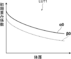
- the figure which shows the schematic structure of the energy subtraction processing apparatus by this embodiment. A diagram showing a table that defines the relationship between body thickness and initial weighting factor.
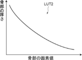
- the figure which shows the soft tissue image and the bone image The figure which shows the table which defined the relationship between the pixel value of a bone part and the thickness of a bone part.
- FIG. 1 is a schematic block diagram showing a configuration of a radiation imaging system to which the energy subtraction processing apparatus according to the embodiment of the present disclosure is applied.
- the radiographic imaging system according to the present embodiment is for capturing two radiographic images having different energy distributions and performing energy subtraction processing using the two radiographic images.
- the photographing device 1 irradiates the first radiation detector 5 and the second radiation detector 6 with radiation such as X-rays emitted from the radiation source 3 and transmitted through the subject H with different energies.
- This is a photographing device for performing shot energy subtraction.
- the first radiation detector 5, the radiation energy conversion filter 7 made of a copper plate or the like, and the second radiation detector 6 are arranged in order from the side closer to the radiation source 3.
- the radiation source 3 is driven.
- the first and second radiation detectors 5 and 6 are in close contact with the radiation energy conversion filter 7.
- the first radiation image G1 of the subject H by low-energy radiation including so-called soft lines is acquired.
- a second radiation image G2 of the subject H by high-energy radiation from which soft lines have been removed is acquired.
- the first and second radiographic images are input to the console 2.
- the scattered radiation removing grid for removing the scattered radiation component of the radiation transmitted through the subject H is not used. Therefore, the first radiation image G1 and the second radiation image G2 include a primary line component and a scattered line component of the radiation transmitted through the subject H.
- the first and second radiation detectors 5 and 6 are so-called direct type radiation detectors that can repeatedly record and read radiation images and generate charges by directly receiving radiation irradiation. Or, a so-called indirect radiation detector that once converts radiation into visible light and then converts the visible light into a charge signal may be used.
- the radiation image signal can be read by turning the TFT (thin film transistor) switch on and off, that is, the so-called TFT reading method, or by irradiating the reading light with the radiation image signal. It is desirable to use a so-called optical reading method in which
- the display unit 8 and the input unit 9 are connected to the console 2.
- the display unit 8 is composed of a display such as a CRT (Cathode Ray Tube) or a liquid crystal display, and includes a radiation image acquired by photographing, a soft part image and a bone part image described later, and various inputs required for processing performed on the console 2. Assist.
- CTR Cathode Ray Tube
- liquid crystal display includes a radiation image acquired by photographing, a soft part image and a bone part image described later, and various inputs required for processing performed on the console 2. Assist.
- the input unit 9 is composed of an input device such as a keyboard, a mouse, or a touch panel method, and receives an operation instruction of the photographing device 1 by the operator. It also accepts instructions for inputting various information such as shooting conditions and correcting the information necessary for shooting. In the present embodiment, each unit of the photographing device 1 operates according to the information input by the operator from the input unit 9.
- the energy subtraction processing program according to this embodiment is installed on the console 2.
- the console 2 corresponds to the energy subtraction processing apparatus according to the present embodiment.
- the console 2 may be a workstation or a personal computer directly operated by the operator, or may be a server computer connected to them via a network.
- the photographing program is stored in the storage device of the server computer connected to the network or in the network storage in a state of being accessible from the outside, and is downloaded and installed in the computer upon request. Alternatively, it is recorded and distributed on a recording medium such as a DVD (Digital Versatile Disc) or a CD-ROM (Compact Disc Read Only Memory), and installed on a computer from the recording medium.
- a recording medium such as a DVD (Digital Versatile Disc) or a CD-ROM (Compact Disc Read Only Memory)
- FIG. 2 is a diagram showing a schematic configuration of an energy subtraction processing device realized by installing an energy subtraction processing program on a computer constituting the console 2.
- the energy subtraction processing apparatus includes a CPU (Central Processing Unit) 21, a memory 22, a storage 23, and a communication unit 24 as a standard computer configuration.
- CPU Central Processing Unit
- the storage 23 is composed of a storage device such as a hard disk drive or an SSD (Solid State Drive), and stores various information including a program for driving each part of the photographing apparatus 1 and an energy subtraction processing program. In addition, the radiographic image acquired by photographing is also stored.
- a storage device such as a hard disk drive or an SSD (Solid State Drive)
- various information including a program for driving each part of the photographing apparatus 1 and an energy subtraction processing program.
- the radiographic image acquired by photographing is also stored.
- the communication unit 24 is a network interface that controls transmission of various information with an external device via a network (not shown).
- the memory 22 temporarily stores a program or the like stored in the storage 23 in order to cause the CPU 21 to execute various processes.
- the energy subtraction processing program is an image acquisition process for causing the imaging device 1 to perform imaging to acquire first and second radiation images G1 and G2 having different energy distributions, and a body thickness of the subject H as a process to be executed by the CPU 21.
- the console 2 has the image acquisition unit 31, the body thickness derivation unit 32, the scattered radiation removal unit 33, the initial weight coefficient setting unit 34, the subtraction unit 35, and the like. And functions as a weighting coefficient deriving unit 36.
- the image acquisition unit 31 drives the radiation source 3 to irradiate the subject H with radiation, detects the radiation transmitted through the subject H by the first and second radiation detectors 5 and 6, and causes the first and second radiation detectors 31 to detect the radiation.
- Radiation images G1 and G2 of the above are acquired.
- imaging conditions such as imaging dose, energy distribution, tube voltage, and SID are set.
- the shooting conditions may be set by input from the input unit 9 by the operator.
- the set shooting conditions are stored in the storage 23.
- the first and second radiographic images G1 and G2 may be acquired by a program separate from the energy subtraction processing program and stored in the storage 23. In this case, the image acquisition unit 31 reads the first and second radiation images G1 and G2 stored in the storage 23 from the storage 23 for processing.
- the abdomen is photographed from the chest of the subject H, and the first and second radiographic images G1 and G2 from the chest to the abdomen are acquired.
- the body thickness deriving unit 32 derives the body thickness of the subject H for each pixel of the first and second radiation images G1 and G2 based on at least one image of the first and second radiation images G1 and G2. .. Since the body thickness is derived for each pixel of the first and second radiation images G1 and G2, the body thickness derivation unit 32 derives the body thickness distribution in at least one of the first and second radiation images G1 and G2. Will be done.
- the body thickness deriving unit 32 uses the first radiation image G1 acquired by the radiation detector 5 on the side closer to the subject H. However, a second radiographic image G2 may be used.
- a low-frequency image representing the low-frequency component of the image may be derived, and the body thickness may be derived using the low-frequency image.
- the body thickness deriving unit 32 assumes that the luminance distribution in the first radiation image G1 matches the distribution of the body thickness of the subject H, and sets the pixel value of the first radiation image G1 as the subject.
- the body thickness of the subject H is derived by converting it into a thickness using the attenuation coefficient in the soft part of H.
- the body thickness deriving unit 32 may measure the thickness of the subject H using a sensor or the like.
- the body thickness deriving unit 32 may derive the body thickness by approximating the body thickness of the subject H with a model such as a cube or an elliptical pillar.
- the body thickness deriving unit 32 may derive the body thickness of the subject H by an arbitrary method such as the method described in Japanese Patent Application Laid-Open No. 2015-043959.
- the scattered radiation removing unit 33 removes the scattered radiation component contained in the first and second radiation images G1 and G2 caused by the scattering of radiation in the subject.
- a method for removing the scattered radiation component for example, any method described in JP-A-2014-207958 and JP-A-2015-043959 can be used.
- the method described in Japanese Patent Application Laid-Open No. 2014-207958 acquires the characteristics of a grid that is expected to be used to remove scattered radiation when taking a radiographic image, and based on this characteristic, the scattered radiation contained in the radiographic image. This is a method of deriving a component and performing a scattered radiation removal process using the derived scattered radiation component.
- 2015-043959 is a method of deriving a scattered radiation component using the derived body thickness and performing a scattered radiation removing process of a radiation image.
- G1 and G2 will be used as reference numerals for the first and second radiographic images from which the scattered radiation component has been removed, respectively.
- the scattering ray removal when the method described in Japanese Patent Application Laid-Open No. 2015-043959 is used will be described.
- the body thickness derivation unit 32 and the scattered radiation removal unit 33 acquire a virtual model of the subject H having an initial body thickness distribution, and estimate a primary line image obtained by photographing the virtual model, and a virtual primary line image.
- An estimated scattered radiation image obtained by estimating the scattered radiation image obtained by photographing the model is derived.
- the estimated primary line image and the estimated scattered line image are derived by using the first radiation image G1.
- the body thickness derivation unit 32 and the scattered radiation removing unit 33 add the estimated primary line image and the estimated scattered line image to derive the estimated image. Further, the body thickness deriving unit 32 and the scattered radiation removing unit 33 modify the initial body thickness distribution so that the difference between the estimated image and the first radiation image G1 becomes small.
- the body thickness deriving unit 32 and the scattered radiation removing unit 33 derive an estimated image using the modified body thickness distribution, and the difference between the estimated image and the first radiation image G1 satisfies a predetermined termination condition. Until then, the generation of the estimated image using the corrected body thickness distribution and the correction of the body thickness distribution are repeated.
- the body thickness deriving unit 32 derives the body thickness distribution when the end condition is satisfied as the body thickness of the subject H.
- the scattered radiation removing unit 33 removes the scattered radiation component from the first radiation image G1 by subtracting the estimated scattered radiation image when the end condition is satisfied from the first radiation image G1.
- the scattered radiation removing unit 33 derives an estimated scattered radiation image for the second radiation image G2 in the same manner as the first radiation image G1, and subtracts the derived estimated scattered radiation image from the second radiation image G2. Thereby, the scattered radiation component is removed from the second radiation image G2.
- the initial weighting coefficient setting unit 34 sets the initial weighting coefficient, which is the initial value of the weighting coefficient when the subtraction unit 35 performs the subtraction processing, based on the body thickness of the subject H derived by the body thickness deriving unit 32.
- the subtraction unit 35 uses the initial weight coefficient set by the initial weight coefficient setting unit 34 and the weight coefficient derived by the weight coefficient derivation unit 36, and the following equations (1) and (2) are used.
- the soft part in the subject H is extracted by performing subtraction processing in which the first and second radiation images G1 and G2 from which the scattered ray component is removed are weighted and subtracted between the corresponding pixels.
- the image Gs and the bone image Gb from which the bone is extracted are derived.
- ⁇ and ⁇ are weighting coefficients.
- Gs (x, y) ⁇ ⁇ G2 (x, y) -G1 (x, y) (1)
- Gb (x, y) ⁇ ⁇ G2 (x, y) -G1 (x, y) (2)
- the initial weight coefficient setting unit 34 sets the initial weight coefficients ⁇ 0 and ⁇ 0, which are the initial values of the weight coefficients ⁇ and ⁇ , based on the body thickness derived by the body thickness derivation unit 32.
- a table LUT1 that defines the relationship between the body thickness and the initial weighting coefficients ⁇ 0 and ⁇ 0 is stored in the storage 23.
- the initial weight coefficient setting unit 34 sets the initial weight coefficients ⁇ 0 and ⁇ 0 based on the body thickness with reference to the table LUT1.
- the relationship between the weighting coefficients ⁇ and ⁇ and the radiation attenuation coefficient will be described.
- the radiation emitted from the radiation source 3 has an energy distribution, and the attenuation coefficient also depends on the energy of the radiation, and the higher the energy component, the smaller the attenuation coefficient. For this reason, radiation loses a relatively large amount of low-energy components in the process of penetrating substances, and the proportion of high-energy components increases, resulting in a phenomenon called beam hardening. Since the degree of beam hardening depends on the soft tissue thickness ts and the bone thickness tb in the subject H, the soft tissue attenuation coefficient ⁇ s and the bone attenuation coefficient ⁇ b are ⁇ s as a function of ts and tb. It can be defined as (ts, tb) and ⁇ b (ts, tb).
- the attenuation coefficient of the soft part of the low energy image is ⁇ ls (ts, tb), and that of the bone part.
- the attenuation coefficient can be expressed as ⁇ lb (ts, tb).
- the attenuation coefficient of the soft part of the high-energy image can be expressed as ⁇ hs (ts, tb)
- the attenuation coefficient of the bone part can be expressed as ⁇ hb (ts, tb).
- the subtraction unit 35 derives the soft part image Gs from which the soft part of the subject H is extracted and the bone part image Gb from which the bone part is extracted by using the above equations (1) and (2).
- the subtraction unit 35 first uses the initial weighting coefficients ⁇ 0 and ⁇ 0 set by the initial weighting coefficient setting unit 34 to display the first and second radiation images G1 and G2 between the corresponding pixels. Performs subtraction processing for weighting and subtraction. After that, as will be described later, subtraction processing is performed using the weighting coefficients ⁇ new and ⁇ new derived by the weighting coefficient deriving unit 36.
- FIG. 4 is a diagram showing a soft tissue image Gs and a bone image Gb. As shown in FIG. 4, in the soft part image Gs, the soft part in the subject H is extracted. Further, in the bone image Gb, the bone in the subject H is extracted.
- the weight coefficient deriving unit 36 derives new weighting coefficients ⁇ new and ⁇ new based on the pixel values Gb (x, y) of the bone portion included in the bone portion image Gb.
- the pixel value Gb (x, y) of the bone portion corresponds to the thickness of the bone portion of the subject H. Therefore, in the present embodiment, the radiation image of the reference object is acquired as the reference radiation image by photographing the reference object simulating the bone portion having various thicknesses in advance. Then, using the relationship between the pixel value of the region of the reference object and the thickness of the reference object in the reference radiation image, a table that defines the relationship between the pixel value of the bone and the thickness is derived in advance and stored in the storage 23. I will do it.
- FIG. 5 is a diagram showing a table that defines the relationship between the pixel value of the bone portion and the thickness of the bone portion.
- the pixel value Gb (x, y) of the bone portion that is, the higher the brightness
- the weight coefficient deriving unit 36 derives the bone thickness tb in each pixel of the bone image Gb from each pixel value Gb (x, y) of the bone image Gb with reference to the table LUT2. Since the region in the bone image Gb where the bone does not exist consists of only the soft part, the thickness tb of the bone is 0. On the other hand, in the bone image Gb, the weight coefficient deriving unit 36 subtracts the bone thickness tb from the body thickness derived by the body thickness deriving unit 32 in the pixel where the bone thickness tb is not 0. , The thickness ts of the soft part is derived.
- the weighting coefficients ⁇ and ⁇ can be expressed as a function of the soft tissue thickness ts and the bone thickness tb.
- the storage 23 stores a table that defines the relationship between the soft tissue thickness ts and the bone thickness tb and the weighting coefficients ⁇ and ⁇ .
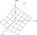
- FIG. 6 is a diagram showing a table defining the relationship between the thickness ts of the soft part and the thickness tb of the bone part and the weighting coefficients ⁇ and ⁇ .
- the table LUT3 three-dimensionally represents the relationship between the soft tissue thickness ts and the bone thickness tb and the weighting coefficient ⁇ (or ⁇ ).
- the larger the soft tissue thickness ts and the bone thickness tb the smaller the weighting coefficient ⁇ (or ⁇ ).
- a plurality of tables LUT3 are prepared and stored in the storage 23 according to the energy distribution of the radiation used at the time of photographing.
- the weighting coefficient derivation unit 36 acquires information on the energy distribution of radiation used at the time of imaging based on the imaging conditions, reads out the table LUT3 corresponding to the acquired energy distribution information from the storage 23, and uses it for deriving the weighting coefficient. To do. Then, the weighting coefficient deriving unit 36 derives new weighting coefficients ⁇ new and ⁇ new with reference to the table LUT3 based on the derived bone thickness tb and soft tissue thickness ts.
- the subtraction unit 35 derives a new soft tissue image Gsnew and a new bone image Gbnew by the above equations (1) and (2) using the new weighting coefficients ⁇ new and ⁇ new derived by the weighting coefficient deriving unit 36. ..
- the new soft tissue image Gsnew and the new bone image Gbnew may be stored in the storage 23 as the final soft tissue image Gs and the bone image Gb, or may be displayed on the display unit 8. In the form, the derivation of the weight coefficients ⁇ and ⁇ and the subtraction process are repeated.
- the weight coefficient deriving unit 36 derives a new bone thickness tbnew with reference to the table LUT2 based on the pixel value of the bone in the new bone image Gbnew. Then, the weighting coefficient deriving unit 36 derives the difference ⁇ tb between the new bone thickness tbnew and the bone thickness tb obtained in the previous process, and the difference ⁇ tb is determined in advance. It is determined whether or not the value is less than Th1. When the difference ⁇ tb is equal to or greater than the threshold value Th1, the weighting coefficient deriving unit 36 derives a new soft tissue thickness tsnew from the new bone thickness tbnew, and the new bone thickness tbnew and the new bone thickness tbnew. Based on the new soft tissue thickness tsnew, new weighting coefficients ⁇ new and ⁇ new are derived with reference to the table LUT3.
- the subtraction unit 35 further performs subtraction processing using the new weighting coefficients ⁇ new and ⁇ new, and further derives a new bone image Gbnew and a new soft tissue image Gsnew.
- the weight coefficient deriving unit 36 derives a new bone thickness tbnew based on the new bone image Gbnew, and further obtains the new bone thickness tbnew in the previous process.
- the difference ⁇ tb from the bone thickness tb is derived.
- the subtraction unit 35 and the weight coefficient deriving unit 36 perform subtraction processing and weighting coefficients ⁇ new and ⁇ new until the difference ⁇ tb derived by the weighting coefficient deriving unit 36 becomes less than the predetermined threshold value Th1. Repeat the derivation of.
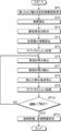
- FIG. 7 is a flowchart showing the processing performed in the present embodiment. It is assumed that the first and second radiation images G1 and G2 are acquired by photographing and stored in the storage 23.
- the image acquisition unit 31 acquires the first and second radiation images G1 and G2 from the storage 23 (step ST1).
- the body thickness deriving unit 32 derives the body thickness of the subject H (step ST2)
- the scattered radiation removing unit 33 removes the scattered radiation components from the first and second radiation images G1 and G2 (step ST3). ).
- the initial weighting coefficient setting unit 34 sets the initial weighting coefficients ⁇ 0 and ⁇ 0 when performing the subtraction processing (step ST4). Then, the subtraction unit 35 performs the subtraction process according to the above equations (1) and (2) using the initial weighting coefficients ⁇ 0 and ⁇ 0 (step ST5). As a result, the soft tissue image Gs and the bone image Gb are derived.
- the weight coefficient deriving unit 36 derives the bone thickness tb based on the bone image Gb (step ST6). Further, the weight coefficient deriving unit 36 subtracts the bone thickness tb from the body thickness derived by the body thickness deriving unit 32 to derive the soft tissue thickness ts (step ST7). Further, the weighting coefficient deriving unit 36 derives new weighting coefficients ⁇ new and ⁇ new with reference to the table LUT3 based on the bone thickness tb and the soft tissue thickness ts (step ST8).
- the subtraction unit 35 performs the subtraction process according to the above equations (1) and (2) using the new weighting coefficients ⁇ new and ⁇ new (step ST9).
- a new soft tissue image Gsnew and a bone bone image Gbnew are derived.
- the weight coefficient deriving unit 36 derives a new bone thickness tbnew based on the new bone image Gbnew, and obtains a new bone thickness tbnew and the bone obtained in the previous process. It is determined whether or not the difference ⁇ tb from the thickness tb of the portion is less than the predetermined threshold value Th1 (step ST10).
- step ST10 is denied, the process returns to step ST6, and the processing of step ST6 to step ST10 is repeated.
- a new bone thickness tbnew, a new soft tissue thickness tsnew are derived, and new weighting coefficients ⁇ new and ⁇ new are derived.
- subtraction processing is performed by the above equations (1) and (2) using the new weighting coefficients ⁇ new and ⁇ new, and new soft tissue image Gsnew and bone image Gbnew are derived, and a new ⁇ tb is obtained. It is determined whether or not the threshold value is less than Th1.
- step ST10 When step ST10 is affirmed, the last derived soft tissue image Gs and bone image Gb are stored in the storage 23 (step ST11), and the process ends.
- the soft part image Gs and the bone part image Gb may be displayed on the display unit 8.
- the composition of the human body includes a soft part and a bone part, but the bone part has a greater attenuation of radiation than the soft part, and the radiation after transmission shifts to a higher energy side. Therefore, in order to accurately separate the soft part and the bone part by the subtraction processing, it is desirable to derive the weighting coefficients ⁇ and ⁇ in consideration of the amount of bone in the subject H.
- the first and second radiation images G1 and G2 are acquired by radiation having different energy distributions transmitted through the subject H including the soft part and the bone part, and a predetermined initial weighting coefficient ⁇ 0
- the soft tissue image Gs and the bone image Gb are derived by performing weighting subtraction using ⁇ 0. Then, new weighting coefficients ⁇ new and ⁇ new are derived based on the pixel values of the bone part included in the bone part image Gb, subtraction processing is performed using the new weighting coefficients ⁇ new and ⁇ new, and a new soft tissue image Gsnew is performed. And a new bone image Gbnew was derived.
- the pixel value of the bone part reflects the amount of bone. Therefore, according to the present embodiment, new weighting coefficients ⁇ new and ⁇ new that reflect the amount of bone can be derived. Further, since the pixel value of the bone portion is used, the relative weighting coefficient does not change even if the energy characteristic of the radiation source and the sensitivity characteristic of the radiation detector fluctuate. Therefore, according to the present embodiment, the weighting coefficient can be derived without being affected by the aged deterioration of the photographing apparatus, and as a result, in the soft tissue image Gs and the bone image Gb derived by the energy subtraction processing, Unnecessary structures can be removed more accurately. That is, in the soft part image Gs, the bone part can be removed with high accuracy, and in the bone part image Gb, the soft part can be removed more accurately.
- the body thickness of the subject H is derived by the body thickness extraction unit 32, but the present invention is not limited to this.
- a predetermined average body thickness may be used without deriving the body thickness.
- the initial weight coefficient setting unit 34 sets the initial weight coefficients ⁇ 0 and ⁇ 0 using the average body thickness.
- the scattered radiation removing unit 33 removes the scattered radiation components from the first and second radiation images G1 and G2, but the present invention is not limited to this.
- the scattered radiation removing grid is used at the time of photographing, the scattered radiation components can be removed from the first and second radiation images G1 and G2. Therefore, when the scattered radiation removing grid is used at the time of photographing, the subtraction processing and the weighting coefficient derivation processing may be performed without removing the scattered radiation components from the first and second radiation images G1 and G2. ..
- the process is repeated until the process of step ST10 is affirmed, but the process is not limited to this.
- the soft tissue image Gs and the bone image Gb may be derived by repeating the process a predetermined number of times.
- the first and second radiographic images G1 and G2 are acquired by the one-shot method, but the first and second radiographic images G1 and G2 are obtained by the so-called two-shot method in which imaging is performed twice. G2 may be acquired.
- the position of the subject H included in the first radiation image G1 and the second radiation image G2 may shift due to the body movement of the subject H. Therefore, it is preferable to perform the processing of the present embodiment after aligning the subjects in the first radiation image G1 and the second radiation image G2.
- the alignment process for example, the method described in Japanese Patent Application Laid-Open No. 2011-255060 can be used.
- the method described in JP-A-2011-255060 is a plurality of first band images and a plurality of second band images representing structures having different frequency bands for each of the first and second radiographic images G1 and G2.
- the band image of the above is generated, the amount of misalignment of the positions corresponding to each other in the first band image and the second band image of the corresponding frequency band is acquired, and based on the amount of misalignment, the first radiation image G1 and The alignment with the second radiographic image G2 is performed.
- the energy subtraction process is performed using the radiation image acquired in the system for capturing the radiation image of the subject using the first and second radiation detectors 5 and 6, but the detection unit
- the present disclosure can be applied even when the first and second radiographic images G1 and G2 are acquired by using the accumulative phosphor sheet.
- two accumulative phosphor sheets are stacked and irradiated with radiation transmitted through the subject H, and the radiation image information of the subject H is accumulated and recorded on each accumulator phosphor sheet, and from each accumulator phosphor sheet.
- the first and second radiographic images G1 and G2 may be acquired by reading the radiographic image information photoelectrically.
- the two-shot method may also be used when the first and second radiographic images G1 and G2 are acquired using the accumulative phosphor sheet.
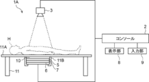
- the photographing device 1A in the radiation image photographing system shown in FIG. 8 is a photographing device for acquiring a radiation image of the subject H lying on the photographing table 11.
- the first radiation detector 5, the radiation energy conversion filter 7, and the second radiation detector 6 are arranged in order from the side closer to the radiation source 3.
- a scattered radiation removing grid for removing the scattered radiation component scattered by the subject H among the radiation transmitted through the subject H between the top plate 11A of the photographing table 11 and the first radiation detector 5 (Hereinafter referred to simply as a grid) 10 are arranged.
- the grid 10, the first radiation detector 5, the radiation energy conversion filter 7, and the second radiation detector 6 can be removed from the photographing table 11 by the mounting portion 11B provided on the lower surface of the top plate 11A of the photographing table 11. It is attached to.
- the top plate 11A and the grid 10 of the photographing table 11 are interposed between the subject H and the first radiation detector 5. Further, in the photographing device 1 shown in FIG. 1 and the photographing device 1A shown in FIG. 8, air may be interposed between the subject H and the first radiation detector 5 at the time of photographing. In such a case, the radiation transmitted through the subject H is transmitted to the top plate 11A, the grid 10, and the air layer to be applied to the first radiation detector 5.
- objects such as the top plate 11A, the grid 10, and air have unique radiation characteristics. Therefore, by transmitting the object, the quality of the primary ray component and the scattered ray component transmitted through the subject H changes according to the radiation characteristics of the object.
- the scattered radiation cannot be completely removed, so that the radiation transmitted through the subject H contains a scattered radiation component. Therefore, in the present embodiment, an object intervening between the subject H and the first radiation detector 5 when estimating the body thickness distribution and removing the scattered radiation component using the first radiation image G1. It is preferable to consider the radiation characteristics of.
- the primary radiation transmittance and the scattered radiation transmittance according to the type of the object intervening between the subject H and the first radiation detector 5 are determined by various imaging conditions and the body thickness distribution of the subject H. It is generated as a table or the like in advance according to the above, and is stored in the storage 23. Then, when the body thickness derivation unit 32 and the scattered radiation removing unit 33 estimate the body thickness distribution of the subject H and remove the scattered radiation, the radiation characteristics of the object according to the body thickness distribution, that is, Obtain the primary ray transmittance and scattered ray transmittance of radiation.
- the scattered radiation removing unit 33 acquires an estimated primary line image and an estimated scattered radiation image using the acquired radiation characteristics, imaging conditions, and body thickness distribution, and adds the estimated primary line image and the estimated scattered radiation image. To generate an estimated image. Further, the body thickness deriving unit 32 and the scattered radiation removing unit 33 repeatedly generate the estimated image and correct the body thickness distribution until the difference between the estimated image and the first radiation image G1 satisfies a predetermined termination condition. .. Then, the body thickness deriving unit 32 derives the body thickness distribution when the end condition is satisfied as the body thickness of the subject H.
- the scattered radiation removing unit 33 subtracts the estimated scattered radiation image when the body thickness distribution satisfying the end condition is acquired from the first radiation image G1 to remove the scattered radiation component from the first radiation image G1. Remove. Thereby, the scattered radiation component can be removed from the first radiation image G1 in consideration of the radiation characteristics of the object interposed between the subject H and the first radiation detector. Similarly, the scattered radiation component can be removed from the second radiation image G2.
- the radiation in the above embodiment is not particularly limited, and in addition to X-rays, ⁇ -rays, ⁇ -rays and the like can be applied.
- the image acquisition unit 31, the body thickness derivation unit 32, the scattered radiation removal unit 33, the initial weight coefficient setting unit 34, the subtraction unit, and the weight coefficient derivation unit 36 of the console 2 which is an energy subtraction processor.
- the various processors include CPUs, which are general-purpose processors that execute software (programs) and function as various processing units, as well as circuits after manufacturing FPGAs (Field Programmable Gate Arrays) and the like.
- Dedicated electricity which is a processor with a circuit configuration specially designed to execute specific processing such as programmable logic device (PLD), ASIC (Application Specific Integrated Circuit), which is a processor whose configuration can be changed. Circuits and the like are included.
- One processing unit may be composed of one of these various processors, or a combination of two or more processors of the same type or different types (for example, a combination of a plurality of FPGAs or a combination of a CPU and an FPGA). ) May be configured. Further, a plurality of processing units may be configured by one processor.
- one processor is configured by combining one or more CPUs and software. There is a form in which this processor functions as a plurality of processing units.
- SoC System On Chip
- the various processing units are configured by using one or more of the above-mentioned various processors as a hardware structure.
- circuitry in which circuit elements such as semiconductor elements are combined can be used.
Landscapes
- Engineering & Computer Science (AREA)
- Health & Medical Sciences (AREA)
- Life Sciences & Earth Sciences (AREA)
- Medical Informatics (AREA)
- Physics & Mathematics (AREA)
- General Health & Medical Sciences (AREA)
- Radiology & Medical Imaging (AREA)
- Nuclear Medicine, Radiotherapy & Molecular Imaging (AREA)
- Biomedical Technology (AREA)
- Veterinary Medicine (AREA)
- Biophysics (AREA)
- Optics & Photonics (AREA)
- Pathology (AREA)
- High Energy & Nuclear Physics (AREA)
- Public Health (AREA)
- Heart & Thoracic Surgery (AREA)
- Molecular Biology (AREA)
- Surgery (AREA)
- Animal Behavior & Ethology (AREA)
- Computer Vision & Pattern Recognition (AREA)
- General Physics & Mathematics (AREA)
- Theoretical Computer Science (AREA)
- Quality & Reliability (AREA)
- Orthopedic Medicine & Surgery (AREA)
- Dentistry (AREA)
- Oral & Maxillofacial Surgery (AREA)
- Physiology (AREA)
- Apparatus For Radiation Diagnosis (AREA)
Priority Applications (2)
| Application Number | Priority Date | Filing Date | Title |
|---|---|---|---|
| JP2021546577A JP7289922B2 (ja) | 2019-09-18 | 2020-08-28 | エネルギーサブトラクション処理装置、方法およびプログラム |
| US17/673,734 US12039727B2 (en) | 2019-09-18 | 2022-02-16 | Energy subtraction processing device, energy subtraction processing method, and energy subtraction processing program |
Applications Claiming Priority (4)
| Application Number | Priority Date | Filing Date | Title |
|---|---|---|---|
| JP2019-169025 | 2019-09-18 | ||
| JP2019169025 | 2019-09-18 | ||
| JP2020-129559 | 2020-07-30 | ||
| JP2020129559 | 2020-07-30 |
Related Child Applications (1)
| Application Number | Title | Priority Date | Filing Date |
|---|---|---|---|
| US17/673,734 Continuation US12039727B2 (en) | 2019-09-18 | 2022-02-16 | Energy subtraction processing device, energy subtraction processing method, and energy subtraction processing program |
Publications (1)
| Publication Number | Publication Date |
|---|---|
| WO2021054090A1 true WO2021054090A1 (ja) | 2021-03-25 |
Family
ID=74883524
Family Applications (1)
| Application Number | Title | Priority Date | Filing Date |
|---|---|---|---|
| PCT/JP2020/032748 Ceased WO2021054090A1 (ja) | 2019-09-18 | 2020-08-28 | エネルギーサブトラクション処理装置、方法およびプログラム |
Country Status (3)
| Country | Link |
|---|---|
| US (1) | US12039727B2 (https=) |
| JP (1) | JP7289922B2 (https=) |
| WO (1) | WO2021054090A1 (https=) |
Cited By (3)
| Publication number | Priority date | Publication date | Assignee | Title |
|---|---|---|---|---|
| JP2024013138A (ja) * | 2022-07-19 | 2024-01-31 | 富士フイルム株式会社 | 放射線画像処理装置、方法およびプログラム |
| EP4535280A1 (en) | 2023-09-20 | 2025-04-09 | FUJI-FILM Corporation | Radiation image processing device, radiation image processing method, and radiation image processing program |
| US12524878B2 (en) | 2022-09-22 | 2026-01-13 | Fujifilm Corporation | Radiation image processing device, radiation image processing method, and radiation image processing program |
Citations (9)
| Publication number | Priority date | Publication date | Assignee | Title |
|---|---|---|---|---|
| JPH11205682A (ja) * | 1998-01-13 | 1999-07-30 | Fuji Photo Film Co Ltd | エネルギーサブトラクション画像生成方法 |
| JP2000232611A (ja) * | 1999-02-12 | 2000-08-22 | Fuji Photo Film Co Ltd | エネルギーサブトラクション画像生成方法および生成装置 |
| JP2002152593A (ja) * | 2000-11-08 | 2002-05-24 | Fuji Photo Film Co Ltd | エネルギーサブトラクション方法および装置並びに記録媒体 |
| WO2009004678A1 (ja) * | 2007-06-29 | 2009-01-08 | Shimadzu Corporation | 放射線撮像装置 |
| JP2011152280A (ja) * | 2010-01-27 | 2011-08-11 | Canon Inc | 放射線撮影装置、その制御方法及びプログラム |
| JP2013046774A (ja) * | 2012-10-05 | 2013-03-07 | Ge Medical Systems Global Technology Co Llc | X線断層撮影装置 |
| JP2018038646A (ja) * | 2016-09-08 | 2018-03-15 | 富士フイルム株式会社 | 画像処理装置、方法およびプログラム |
| JP2018068800A (ja) * | 2016-11-01 | 2018-05-10 | 国立研究開発法人量子科学技術研究開発機構 | 画像処理装置および画像処理装置を用いた照射システム |
| JP2018166652A (ja) * | 2017-03-29 | 2018-11-01 | 富士フイルム株式会社 | 乳腺量取得装置、方法およびプログラム |
Family Cites Families (4)
| Publication number | Priority date | Publication date | Assignee | Title |
|---|---|---|---|---|
| US6125166A (en) | 1998-01-13 | 2000-09-26 | Fuji Photo Film Co., Ltd. | Method of forming energy subtraction images |
| US6421419B1 (en) | 2000-11-08 | 2002-07-16 | Fuji Photo Film Co., Ltd. | Energy subtraction processing method and apparatus |
| US9610057B2 (en) * | 2014-06-16 | 2017-04-04 | General Electric Company | System and method for determining X-ray exposure parameters |
| JP6549535B2 (ja) | 2016-07-29 | 2019-07-24 | 富士フイルム株式会社 | 放射線画像撮影システム、画像処理方法、及び画像処理プログラム |
-
2020
- 2020-08-28 JP JP2021546577A patent/JP7289922B2/ja active Active
- 2020-08-28 WO PCT/JP2020/032748 patent/WO2021054090A1/ja not_active Ceased
-
2022
- 2022-02-16 US US17/673,734 patent/US12039727B2/en active Active
Patent Citations (9)
| Publication number | Priority date | Publication date | Assignee | Title |
|---|---|---|---|---|
| JPH11205682A (ja) * | 1998-01-13 | 1999-07-30 | Fuji Photo Film Co Ltd | エネルギーサブトラクション画像生成方法 |
| JP2000232611A (ja) * | 1999-02-12 | 2000-08-22 | Fuji Photo Film Co Ltd | エネルギーサブトラクション画像生成方法および生成装置 |
| JP2002152593A (ja) * | 2000-11-08 | 2002-05-24 | Fuji Photo Film Co Ltd | エネルギーサブトラクション方法および装置並びに記録媒体 |
| WO2009004678A1 (ja) * | 2007-06-29 | 2009-01-08 | Shimadzu Corporation | 放射線撮像装置 |
| JP2011152280A (ja) * | 2010-01-27 | 2011-08-11 | Canon Inc | 放射線撮影装置、その制御方法及びプログラム |
| JP2013046774A (ja) * | 2012-10-05 | 2013-03-07 | Ge Medical Systems Global Technology Co Llc | X線断層撮影装置 |
| JP2018038646A (ja) * | 2016-09-08 | 2018-03-15 | 富士フイルム株式会社 | 画像処理装置、方法およびプログラム |
| JP2018068800A (ja) * | 2016-11-01 | 2018-05-10 | 国立研究開発法人量子科学技術研究開発機構 | 画像処理装置および画像処理装置を用いた照射システム |
| JP2018166652A (ja) * | 2017-03-29 | 2018-11-01 | 富士フイルム株式会社 | 乳腺量取得装置、方法およびプログラム |
Cited By (3)
| Publication number | Priority date | Publication date | Assignee | Title |
|---|---|---|---|---|
| JP2024013138A (ja) * | 2022-07-19 | 2024-01-31 | 富士フイルム株式会社 | 放射線画像処理装置、方法およびプログラム |
| US12524878B2 (en) | 2022-09-22 | 2026-01-13 | Fujifilm Corporation | Radiation image processing device, radiation image processing method, and radiation image processing program |
| EP4535280A1 (en) | 2023-09-20 | 2025-04-09 | FUJI-FILM Corporation | Radiation image processing device, radiation image processing method, and radiation image processing program |
Also Published As
| Publication number | Publication date |
|---|---|
| JP7289922B2 (ja) | 2023-06-12 |
| JPWO2021054090A1 (https=) | 2021-03-25 |
| US20220172365A1 (en) | 2022-06-02 |
| US12039727B2 (en) | 2024-07-16 |
Similar Documents
| Publication | Publication Date | Title |
|---|---|---|
| JP6906479B2 (ja) | 骨塩情報取得装置、方法およびプログラム | |
| US11234641B2 (en) | Body fat percentage measurement device, method and program | |
| WO2020166561A1 (ja) | 骨折リスク評価値取得装置及びその作動方法並びに骨折リスク評価値取得プログラム | |
| US12039727B2 (en) | Energy subtraction processing device, energy subtraction processing method, and energy subtraction processing program | |
| JP7408867B2 (ja) | 画像処理装置、方法およびプログラム | |
| JP2019209027A (ja) | 骨塩情報取得装置、方法およびプログラム | |
| JP7220643B2 (ja) | 画像処理装置、方法およびプログラム | |
| JP7258165B2 (ja) | 画像処理装置、方法およびプログラム | |
| US12611157B2 (en) | Radiation image processing device, radiation image processing method, and radiation image processing program | |
| JPWO2021054090A5 (https=) | ||
| US12064278B2 (en) | Learning device, learning method, and learning program, radiation image processing device, radiation image processing method, and radiation image processing program | |
| US11763501B2 (en) | Radiographic image processing device, radiographic image processing method, and radiographic image processing program | |
| JP2024000885A (ja) | 放射線画像処理装置、方法およびプログラム | |
| JP7284768B2 (ja) | 被写体情報取得装置及びその作動方法並びに被写体情報取得プログラム | |
| US20240023919A1 (en) | Radiation image processing device, radiation image processing method, and radiation image processing program | |
| US12433559B2 (en) | Radiation image processing device, radiation image processing method, and radiation image processing program | |
| US11911204B2 (en) | Scattered ray model derivation device, scattered ray model derivation method, scattered ray model derivation program, radiation image processing device, radiation image processing method, and radiation image processing program | |
| US20240016465A1 (en) | Radiation image processing device, radiation image processing method, and radiation image processing program | |
| US12533098B2 (en) | Radiation image processing device, radiation image processing method, and radiation image processing program | |
| JP2023177980A (ja) | 放射線画像処理装置、方法およびプログラム | |
| JP2024042609A (ja) | 放射線画像処理装置、その作動方法、及び放射線画像処理プログラム |
Legal Events
| Date | Code | Title | Description |
|---|---|---|---|
| 121 | Ep: the epo has been informed by wipo that ep was designated in this application |
Ref document number: 20865337 Country of ref document: EP Kind code of ref document: A1 |
|
| ENP | Entry into the national phase |
Ref document number: 2021546577 Country of ref document: JP Kind code of ref document: A |
|
| NENP | Non-entry into the national phase |
Ref country code: DE |
|
| 122 | Ep: pct application non-entry in european phase |
Ref document number: 20865337 Country of ref document: EP Kind code of ref document: A1 |