WO2021045122A1 - 網膜色素上皮細胞の三次元組織集合物の凍結体を製造する方法 - Google Patents
網膜色素上皮細胞の三次元組織集合物の凍結体を製造する方法 Download PDFInfo
- Publication number
- WO2021045122A1 WO2021045122A1 PCT/JP2020/033295 JP2020033295W WO2021045122A1 WO 2021045122 A1 WO2021045122 A1 WO 2021045122A1 JP 2020033295 W JP2020033295 W JP 2020033295W WO 2021045122 A1 WO2021045122 A1 WO 2021045122A1
- Authority
- WO
- WIPO (PCT)
- Prior art keywords
- dimensional tissue
- freezing
- retinal pigment
- pigment epithelial
- cells
- Prior art date
- Legal status (The legal status is an assumption and is not a legal conclusion. Google has not performed a legal analysis and makes no representation as to the accuracy of the status listed.)
- Ceased
Links
Images




Classifications
-
- A—HUMAN NECESSITIES
- A01—AGRICULTURE; FORESTRY; ANIMAL HUSBANDRY; HUNTING; TRAPPING; FISHING
- A01N—PRESERVATION OF BODIES OF HUMANS OR ANIMALS OR PLANTS OR PARTS THEREOF; BIOCIDES, e.g. AS DISINFECTANTS, AS PESTICIDES OR AS HERBICIDES; PEST REPELLANTS OR ATTRACTANTS; PLANT GROWTH REGULATORS
- A01N1/00—Preservation of bodies of humans or animals, or parts thereof
- A01N1/10—Preservation of living parts
- A01N1/12—Chemical aspects of preservation
- A01N1/122—Preservation or perfusion media
- A01N1/125—Freeze protecting agents, e.g. cryoprotectants or osmolarity regulators
-
- A—HUMAN NECESSITIES
- A01—AGRICULTURE; FORESTRY; ANIMAL HUSBANDRY; HUNTING; TRAPPING; FISHING
- A01N—PRESERVATION OF BODIES OF HUMANS OR ANIMALS OR PLANTS OR PARTS THEREOF; BIOCIDES, e.g. AS DISINFECTANTS, AS PESTICIDES OR AS HERBICIDES; PEST REPELLANTS OR ATTRACTANTS; PLANT GROWTH REGULATORS
- A01N1/00—Preservation of bodies of humans or animals, or parts thereof
- A01N1/10—Preservation of living parts
- A01N1/12—Chemical aspects of preservation
- A01N1/128—Chemically defined matrices for immobilising, holding or storing living parts, e.g. alginate gels; Chemically altering living parts, e.g. by cross-linking
-
- A—HUMAN NECESSITIES
- A01—AGRICULTURE; FORESTRY; ANIMAL HUSBANDRY; HUNTING; TRAPPING; FISHING
- A01N—PRESERVATION OF BODIES OF HUMANS OR ANIMALS OR PLANTS OR PARTS THEREOF; BIOCIDES, e.g. AS DISINFECTANTS, AS PESTICIDES OR AS HERBICIDES; PEST REPELLANTS OR ATTRACTANTS; PLANT GROWTH REGULATORS
- A01N1/00—Preservation of bodies of humans or animals, or parts thereof
- A01N1/10—Preservation of living parts
- A01N1/14—Mechanical aspects of preservation; Apparatus or containers therefor
- A01N1/146—Non-refrigerated containers specially adapted for transporting or storing living parts whilst preserving
- A01N1/147—Carriers for immersion in cryogenic fluid for slow freezing or vitrification
-
- A—HUMAN NECESSITIES
- A01—AGRICULTURE; FORESTRY; ANIMAL HUSBANDRY; HUNTING; TRAPPING; FISHING
- A01N—PRESERVATION OF BODIES OF HUMANS OR ANIMALS OR PLANTS OR PARTS THEREOF; BIOCIDES, e.g. AS DISINFECTANTS, AS PESTICIDES OR AS HERBICIDES; PEST REPELLANTS OR ATTRACTANTS; PLANT GROWTH REGULATORS
- A01N1/00—Preservation of bodies of humans or animals, or parts thereof
- A01N1/10—Preservation of living parts
- A01N1/16—Physical preservation processes
- A01N1/162—Temperature processes, e.g. following predefined temperature changes over time
-
- A—HUMAN NECESSITIES
- A61—MEDICAL OR VETERINARY SCIENCE; HYGIENE
- A61K—PREPARATIONS FOR MEDICAL, DENTAL OR TOILETRY PURPOSES
- A61K35/00—Medicinal preparations containing materials or reaction products thereof with undetermined constitution
- A61K35/12—Materials from mammals; Compositions comprising non-specified tissues or cells; Compositions comprising non-embryonic stem cells; Genetically modified cells
- A61K35/30—Nerves; Brain; Eyes; Corneal cells; Cerebrospinal fluid; Neuronal stem cells; Neuronal precursor cells; Glial cells; Oligodendrocytes; Schwann cells; Astroglia; Astrocytes; Choroid plexus; Spinal cord tissue
-
- C—CHEMISTRY; METALLURGY
- C12—BIOCHEMISTRY; BEER; SPIRITS; WINE; VINEGAR; MICROBIOLOGY; ENZYMOLOGY; MUTATION OR GENETIC ENGINEERING
- C12N—MICROORGANISMS OR ENZYMES; COMPOSITIONS THEREOF; PROPAGATING, PRESERVING, OR MAINTAINING MICROORGANISMS; MUTATION OR GENETIC ENGINEERING; CULTURE MEDIA
- C12N11/00—Carrier-bound or immobilised enzymes; Carrier-bound or immobilised microbial cells; Preparation thereof
- C12N11/02—Enzymes or microbial cells immobilised on or in an organic carrier
-
- C—CHEMISTRY; METALLURGY
- C12—BIOCHEMISTRY; BEER; SPIRITS; WINE; VINEGAR; MICROBIOLOGY; ENZYMOLOGY; MUTATION OR GENETIC ENGINEERING
- C12N—MICROORGANISMS OR ENZYMES; COMPOSITIONS THEREOF; PROPAGATING, PRESERVING, OR MAINTAINING MICROORGANISMS; MUTATION OR GENETIC ENGINEERING; CULTURE MEDIA
- C12N5/00—Undifferentiated human, animal or plant cells, e.g. cell lines; Tissues; Cultivation or maintenance thereof; Culture media therefor
- C12N5/06—Animal cells or tissues; Human cells or tissues
- C12N5/0602—Vertebrate cells
- C12N5/0618—Cells of the nervous system
- C12N5/0621—Eye cells, e.g. cornea, iris pigmented cells
-
- C—CHEMISTRY; METALLURGY
- C12—BIOCHEMISTRY; BEER; SPIRITS; WINE; VINEGAR; MICROBIOLOGY; ENZYMOLOGY; MUTATION OR GENETIC ENGINEERING
- C12N—MICROORGANISMS OR ENZYMES; COMPOSITIONS THEREOF; PROPAGATING, PRESERVING, OR MAINTAINING MICROORGANISMS; MUTATION OR GENETIC ENGINEERING; CULTURE MEDIA
- C12N2501/00—Active agents used in cell culture processes, e.g. differentation
- C12N2501/10—Growth factors
- C12N2501/115—Basic fibroblast growth factor (bFGF, FGF-2)
-
- C—CHEMISTRY; METALLURGY
- C12—BIOCHEMISTRY; BEER; SPIRITS; WINE; VINEGAR; MICROBIOLOGY; ENZYMOLOGY; MUTATION OR GENETIC ENGINEERING
- C12N—MICROORGANISMS OR ENZYMES; COMPOSITIONS THEREOF; PROPAGATING, PRESERVING, OR MAINTAINING MICROORGANISMS; MUTATION OR GENETIC ENGINEERING; CULTURE MEDIA
- C12N2501/00—Active agents used in cell culture processes, e.g. differentation
- C12N2501/10—Growth factors
- C12N2501/15—Transforming growth factor beta (TGF-β)
-
- C—CHEMISTRY; METALLURGY
- C12—BIOCHEMISTRY; BEER; SPIRITS; WINE; VINEGAR; MICROBIOLOGY; ENZYMOLOGY; MUTATION OR GENETIC ENGINEERING
- C12N—MICROORGANISMS OR ENZYMES; COMPOSITIONS THEREOF; PROPAGATING, PRESERVING, OR MAINTAINING MICROORGANISMS; MUTATION OR GENETIC ENGINEERING; CULTURE MEDIA
- C12N2513/00—3D culture
-
- C—CHEMISTRY; METALLURGY
- C12—BIOCHEMISTRY; BEER; SPIRITS; WINE; VINEGAR; MICROBIOLOGY; ENZYMOLOGY; MUTATION OR GENETIC ENGINEERING
- C12N—MICROORGANISMS OR ENZYMES; COMPOSITIONS THEREOF; PROPAGATING, PRESERVING, OR MAINTAINING MICROORGANISMS; MUTATION OR GENETIC ENGINEERING; CULTURE MEDIA
- C12N2533/00—Supports or coatings for cell culture, characterised by material
- C12N2533/50—Proteins
- C12N2533/54—Collagen; Gelatin
Definitions
- the present disclosure relates to a method for producing a frozen body of a three-dimensional tissue aggregate of retinal pigment epithelial cells.
- the RPE (Retinal Pigment Epithelium) cell sheet is a three-dimensional tissue produced by culturing retinal pigment epithelial cells (Retinal Pigment Epithelial Cells) in the form of a sheet.
- RPE cell sheets are prepared using Transwell TM inserts. More specifically, RPE cells are cultured in a maintenance medium to prepare a collagen gel that serves as a scaffold in an insert, and then RPE cells are seeded on the collagen gel and cultured for about 2 months. You can get a sheet.
- the RPE cell sheet is composed of monolayer cells like a living body, has apical-basal polarity characteristic of epithelial tissue, and forms a basement membrane.
- tight junctions are formed between the cells of the RPE cell sheet, and the RPE cell sheet can exhibit a barrier function such as transepithelial electrical resistance (TER).
- RPE cell sheets are being developed for the purpose of being used for autologous transplantation treatment of exudative age-related macular degeneration, which is one of the ophthalmic diseases.
- autologous transplantation it takes about 10 months to collect skin tissue from the patient, establish iPS cells from it, induce differentiation and purify RPE cells, and prepare a sheet, so it takes an enormous amount of time to prepare for transplantation. And costs are required.
- allogeneic transplantation in which an RPE cell sheet is prepared from a sample collected from a donor and transplanted, can be expected to shorten the culture period until transplantation and reduce the cost required for preparation. This merit can be obtained by stocking and preparing a pre-prepared RPE cell sheet in allogeneic transplantation. In order to stock the RPE cell sheet, it is required to establish a method for stably cryopreserving the RPE cell sheet for a long period of time.
- cryopreserving cells usually, the maintenance medium of the cultured cells is removed, replaced with a preservation solution, and then frozen.
- the preservative solution and freezing method used differ depending on the subject, and in particular, the freezing method is roughly classified into quick freezing (vitrification freezing) and slow freezing.
- the slow freezing method is a method of slowly freezing cells at a relatively high temperature (1 ° C / min at -80 ° C).
- water freezes from the outside of the cell first, so that an osmotic pressure difference is created inside and outside the cell, and dehydration occurs due to the osmotic pressure difference. Since it passes through the temperature at which ice is formed (maximum ice crystal formation temperature range) over a long period of time, extracellular ice crystals may grow and damage the cells during that time.
- slow freezing has low cytotoxicity due to cryopreservation solutions, large cells and tissues cannot be completely dehydrated by the cooling process alone, and are generally considered unsuitable.
- the quick freezing method is a method in which cells are rapidly cooled using a cryogen such as liquid nitrogen and frozen (for example, 3600 ° C./min in liquid nitrogen immersion).
- a cryogen such as liquid nitrogen and frozen
- the quick freezing method is often used for freezing tissues.
- quick freezing uses a high-concentration cryopreservation solution, so cytotoxicity may generally be high. It is also necessary to thaw rapidly to prevent recrystallization, which may require skillful techniques.
- Non-Patent Document 1 discloses cryopreservation of a two-dimensional monolayer tissue in which cells are treated with a preservation solution containing a carboxyl group-introduced polylysine and then vitrified with liquid nitrogen.
- Patent Document 1 describes a step and medium for substituting a culture medium in the form of a cell sheet or a three-dimensional cell culture medium with a vitrification solution containing ⁇ -poly-L-lysine having a carboxyl group introduced therein. It is a form of a cell sheet or a three-dimensional cell culture medium, which comprises a step of cooling cultured cells replaced with a vitrification solution in a gas atmosphere at a temperature lowering rate in the range of 0.08 to 1.00 ° C./sec. A method of slow vitrification of cultured cells is disclosed.
- Patent Document 1 and Non-Patent Document 1 are applied to an RPE cell sheet, cell contraction and cell detachment are observed on the entire surface of the RPE cell sheet, and the sheet structure cannot be maintained. It became clear from the examination.
- the present disclosers have now obtained a frozen product that can be stably stored while maintaining a high-quality state by subjecting the three-dimensional tissue aggregate of retinal pigment epithelial cells to a specific treatment. I found that it was possible.
- the present disclosure provides a method for producing a frozen product of a three-dimensional tissue aggregate of retinal pigment epithelial cells that can be stably stored.
- a method for producing a frozen body of a three-dimensional tissue aggregate of retinal pigment epithelial cells A predetermined freeze set in advance using the step of preparing a three-dimensional tissue aggregate of retinal pigment epithelial cells and the generation rate of linear aggregates of dead cells in the three-dimensional tissue aggregate of retinal pigment epithelial cells after thawing as an index.
- a frozen body of a three-dimensional tissue aggregate of retinal pigment epithelial cells can be stably produced while maintaining high quality.
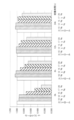
- Example 1 ethylene glycol concentrations of 10 (v / v)%, 12.5 (v / v)%, 15 (v / v)%, 17.5 (v / v)% and 20 (v) as pretreatment solutions.
- FIG. 5 is a graph showing the metabolic activity of cells when a / v)% solution was added to retinal pigment epithelial cells and cultured for 0 to 24 hours.
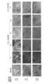
- a photograph of the RPE cell sheet is shown. Each photograph was taken at the time of culture 3, 7 and 14 days after each treatment (observation magnification ⁇ 100 on culture 3 and 7 days, observation magnification ⁇ 200 on culture 14 days).
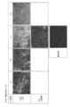
- Example 2 it is a photograph when Cytox TM green staining was performed with a microscope (observation magnification ⁇ 100) (3rd day of culture).
- the upper photograph is a micrograph of the RPE cell sheet obtained by the one-time treatment method (10 minutes ⁇ one pretreatment; EG concentration 5, 10, 15 or 20 (v / v)%).
- the middle photograph is a photomicrograph of the RPE cell sheet obtained by step treatment (2 minutes x 5 pretreatment; EG concentration; EG concentration 3, 6, 9, 12, 15 (v / v)%). ..
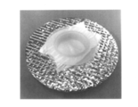
- the lower photo is a photomicrograph of the untreated RPE cell sheet. It is a photograph of the apparatus (float for freezing) used when freezing or thawing the laminate of collagen gel and RPE cell sheet using liquid nitrogen. Photograph of RPE cell sheet subjected to freezing treatment by metal contact (upper), gas phase freezing (liquid surface of liquid nitrogen) (middle) or gas phase freezing (5 mm above liquid nitrogen) (lower) in Example 6. Is. For each sample, photographs are shown at the time of vitrification freezing, immediately after thawing, and 120 hours after thawing.
- FIG. 6 is a photograph of an RPE cell sheet subjected to freezing treatment by metal contact (upper row) or gas phase freezing (5 mm above the liquid surface of liquid nitrogen) (lower row) and stained with Sysox TM green in Example 6.
- Example 6 a photograph and a TER value of an RPE cell sheet subjected to freezing treatment by metal contact (upper) or gas phase freezing (5 mm above the liquid surface of liquid nitrogen) (lower) and stained with Sysox TM green. The result is shown.
- 6 is a photograph of a fully vitrified laminate of collagen gel and RPE cell sheet obtained in Example 7 (7-3).
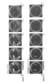
- Example 8-1 metal contact method, gas phase freezing (10 mm from liquid nitrogen liquid surface), gas phase freezing (25 mm from liquid nitrogen liquid surface), gas phase freezing (40 mm from liquid nitrogen liquid surface) or gas. It is a photograph when the RPE cell sheet obtained by phase freezing (80 mm from the liquid nitrogen liquid surface) was thawed and stained with Sysox TM green. In the metal contact method, a linear aggregate (fold) of dead cells is generated.
- Example 8 (8-3) a photograph of the laminated body of the thinned collagen gel and the RPE cell sheet at the time of vitrification freezing when the immersion time in the preservation solution was 0.01 seconds or 30 seconds (upper row). ) And the photograph (lower) when the Second TM green dyeing was performed after thawing.
- Example 9 (9-1) it is a photograph of the RPE cell sheet held at ⁇ 130 ° C. (10 minutes, 20 minutes and 30 minutes) before being immersed in the thaw solution.
- Example 9 (9-2) the predetermined temperature (-170 ° C, -150 ° C, -140 ° C, -130 ° C, -120 ° C or-" before immersing the laminate of collagen gel and RPE cell sheet in the thawing solution. It is a photograph when it was held at 100 ° C.), thawed, and then stained with Sysox TM green. Cracks occur at ⁇ 170 ° C. and ⁇ 100 ° C.
- a method for producing a frozen body of a three-dimensional tissue aggregate of retinal pigment epithelial cells prepares a three-dimensional tissue aggregate of retinal pigment epithelial cells.
- the three-dimensional tissue assembly of retinal pigment epithelial cells at a predetermined freezing temperature set in advance using the rate of occurrence of linear aggregates of dead cells in the three-dimensional tissue assembly of retinal pigment epithelial cells after thawing as an index. It is characterized by including a step of obtaining a frozen product by freezing treatment.
- the details of the present disclosure will be described for each step.
- a three-dimensional tissue assembly of retinal pigment epithelial cells is prepared.
- the retina of the eye contains photoreceptors that sense various levels of light and interneurons that relay signals from the photoreceptors to retinal ganglion cells. These photoreceptors are the most metabolically active cells in the eye and are metabolically and functionally supported by retinal pigment epithelial cells.
- the ability to grasp the visual image of the case may be diminished or lost altogether (eg, eye trauma, infection, degeneration, vascular abnormalities and inflammatory diseases).
- the central part of the retina known as the macula, is responsible for central vision, fine visualization and color distinction.
- Macular function is due to age-related macular degeneration (wet or atrophic), diabetic macular edema, idiopathic choroidal neovascularization, severe myopic macular degeneration, or advanced retinitis pigmentosa, among other medical conditions. Can be adversely affected.
- Macular degeneration is a major cause of visual impairment in the elderly (> 50 years).
- the developmental types of macular degeneration include the "exudative" type and the "atrophic" type.
- cell debris drusen
- neovascularization from the choroid oozes into the space posterior to the macula, resulting in the death of photoreceptors and their supporting cells. Loss of functional cells in the eye weakens new blood vessels and causes frequent leakage of blood and interstitial fluid, which can result in further damage to the macula.
- RPE cells are a source of therapeutic cells for late-stage cell therapy of atrophic age-related macular degeneration (AMD).
- RPE cells can function as differentiated polarized monolayers.
- Successful cryopreservation of stem cell-derived differentiation and polarization RPE monolayers provides clinical product homogeneity, product yield maximization, quality control assurance within clinical product lots, and resemblance to remote clinical settings There is a benefit in the distribution of the final product.
- cGMP Good Manufacturing Practices
- GTP Good Manufacturing Practices for Human Cells and Tissues
- RPE cells are a material for autologous or allogeneic transplantation.
- the RPE cells form a sheet-like cell group from the viewpoint of being used as an eye transplant material. Therefore, according to a preferred embodiment of the present disclosure, the three-dimensional tissue assembly of retinal pigment epithelial cells is an RPE cell sheet.
- the three-dimensional tissue aggregate of RPE cells may be the target of freezing treatment alone, but if necessary, it is frozen together with a structure (cell aggregate, tissue, scaffold, etc.) other than the three-dimensional tissue aggregate, a preservation solution, or the like. It can be the target of processing.
- RPE cells or a three-dimensional tissue assembly thereof are placed on a carrier as a scaffold.
- Cell carriers are advantageous in imparting mechanical stability to cells and producing high viability after the thawing process.
- a biocompatible component known as a cell carrier for culturing and / or cryopreserving cells or tissues can be used as the carrier.
- biocompatible components include hyaluronic acid, alginate, agarose, fibrin, chitin / chitosan, polylactide (PLA), polyglycolide (PGA), polyL-lactic acid (PPLA), gelatin or collagen.
- PLA polylactide
- PGA polyglycolide
- PPLA polyL-lactic acid
- gelatin or collagen Hyaluronic acid, fibrin, gelatin or collagen, more preferably collagen, because it does not produce toxic degradation products and is biocompatible.
- collagen include porcine type I collagen obtained from porcine tendon and bovine type I collagen obtained from bovine skin or skin.
- the biocompatible component may also be a mixture of type I collagen and type III collagen, or a mixture of type I collagen and / or type III collagen and elastin.
- the content of the biocompatible component in the carrier is not particularly limited as long as it can be used as a scaffold for cryopreservation, for example, 0.1 to 90 (w / v) with respect to the total mass of the raw materials constituting the carrier. %, Preferably 0.1 to 80 (w / v)%, more preferably 0.1 to 50 (w / v)%.
- the additives in the carrier other than the biocompatible components described above are not particularly limited as long as they can be used as a scaffold for cryopreservation, and for example, polyols (glycerol, ethylene glycol, butenediol, propenediol, sorbitol, etc.). , Lipids (vegetable oil, etc.), water, etc.
- the carrier is preferably layered from the viewpoint of holding the PRE cell sheet.
- the carrier is usually a monolayer, for example, in the case of a collagen carrier, cells or tissues are inoculated directly in a culture dish coated with a simple collagen gel monolayer. Therefore, according to a preferred embodiment of the present disclosure, the step of preparing a three-dimensional tissue aggregate of RPE cells includes a step of seeding and culturing retinal pigment epithelial cells on a carrier layer.
- Another widely used arrangement for cryopreservation of cells or tissues in collagen-based hydrogels is a sandwich arrangement, for example, cells may be cryopreserved between two layers of collagen gel. The disclosure also includes such aspects.
- the thickness of the carrier layer is preferably thin from the viewpoint of mechanical strength, efficient penetration of the freeze-protecting substance into the inside, and achievement of complete vitrification.
- the average value of the thickness of the carrier layer is, for example, 200 ⁇ m or less, preferably 150 to 10 ⁇ m, more preferably 100 to 15 ⁇ m, and even more preferably 70 to 20 ⁇ m.
- the method for forming the carrier layer is not particularly limited, but from the viewpoint of forming the thin-film carrier layer, it is preferable to combine the centrifugation method and the drying method (air drying or the like). Therefore, according to one embodiment, the carrier layer is obtained by centrifuging the carrier raw material into layers and drying.
- the conditions for centrifugation when forming the carrier layer are not particularly limited, but are, for example, 100 to 4,000 ⁇ g, preferably 1,000 to 2,500 ⁇ g for 0.5 to 4 hours.
- the temperature in centrifugation is usually 1 to 40 ° C, preferably room temperature (1 to 30 ° C).
- the drying conditions after centrifugation are not particularly limited, but can be, for example, usually 1 to 40 ° C., preferably 10 to 36 hours at room temperature (1 to 30 ° C.). Drying after centrifugation can be performed, for example, by allowing the carrier to stand in a clean bench.
- a pretreatment step of dehydrating RPE cells or a three-dimensional tissue assembly thereof is carried out from the viewpoint of reducing the load of abrupt osmotic pressure changes on the cells. Performing the pretreatment step is also preferable in order to prevent the cell structure from being destroyed by the formation of ice crystals in the freezing and thawing treatment.
- the pretreatment step comprises contacting the RPE cells or a three-dimensional tissue assembly thereof with a dehydrating agent.
- the contact between the RPE cells or the three-dimensional tissue aggregate thereof and the dehydrating agent is carried out by immersing the RPE cells or the three-dimensional tissue aggregate thereof and the dehydrating agent in a pretreatment solution containing the dehydrating agent.
- RPE cells or a three-dimensional tissue assembly thereof may be immersed in a pretreatment solution together with a carrier.
- the dehydrating agent is not particularly limited as long as it has a dehydrating function, but is preferably a polyhydric alcohol, more preferably polyethylene glycol, dextran, hydroxyethyl starch, polyvinyl alcohol, polyvinylpyrrolidone, Percoll, ethylene glycol, and further. Ethylene glycol is preferred.
- a polyhydric alcohol such as ethylene glycol can also be used as a freezing agent in the preservative solution immersion step described later.
- the medium in the pretreatment solution is preferably a medium (DMEM medium, F12 HAM medium, B27 medium, EMEM medium, RPMI1640 medium, F12 medium, TC199 medium, GMEM medium, ⁇ MEM medium, etc.), phosphate buffered physiological saline (PBS).
- DMEM medium F12 HAM medium, B27 medium, EMEM medium, RPMI1640 medium, F12 medium, TC199 medium, GMEM medium, ⁇ MEM medium, etc.
- PBS phosphate buffered physiological saline
- the medium may contain amino acids, cell growth factors, and the like.
- the concentration of the polyhydric alcohol in the pretreatment solution may be appropriately adjusted according to the state of cells and the like, but is 1 to 30 (w / v)%, more preferably 2 to 25 (w / v)%. Yes, even more preferably 2 to 20 (w / v)%, and even more preferably 3 to 20 (w / v)%.
- the pretreatment liquid may contain additives such as sugars, polyamino acids or derivatives thereof in addition to the dehydrating agent.
- additives such as sugars, polyamino acids or derivatives thereof in addition to the dehydrating agent.
- the specific type of sugar, polyamino acid or derivative thereof is preferably the same as that of the preservation solution described later.
- the content of sugar and polyamino acid or a derivative thereof in the preservation solution is preferably lower than that in the preservation solution from the viewpoint of avoiding cytotoxicity, and is 1/128 to 1/128 to the content in the preservation solution described later. It is more preferably 2.
- the number of times the cells are immersed in the pretreatment solution may be once or multiple times, but from the viewpoint of efficient dehydration, it is preferable to carry out the cells a plurality of times. More specifically, the number of times the cells are immersed in the pretreatment solution is preferably 3 times or more, more preferably 3 to 8 times, and even more preferably 3 to 5 times.
- the total time for immersing the RPE cells or their three-dimensional tissue aggregate in the pretreatment solution is, for example, 1 to 120 minutes, preferably 1 to 60 minutes, and more preferably 1 to 50 minutes. Minutes.
- the time of each pretreatment step is, for example, 1 to 20 minutes, preferably 1 to 15 minutes, and preferably 1 to 10 minutes.
- the temperature of immersion in the pretreatment liquid used in the pretreatment step is not particularly limited, but is usually 1 to 40 ° C, preferably room temperature (1 to 30 ° C).
- the specific immersion method in the pretreatment step is not particularly limited, but for example, RPE cells are seeded on the membrane on the bottom surface of a commercially available Transwell TM insert via a carrier to form a three-dimensional tissue aggregate.
- RPE cells or their three-dimensional tissue aggregates can be carried out by pouring the pretreatment solution inside and outside the Transwell TM insert and allowing it to stand so that it is immersed in the pretreatment solution.
- Preservative solution immersion (vitrification solution treatment) process After the pretreatment step, from the viewpoint of imparting freezing resistance, it is preferable to immerse the RPE cells or their three-dimensional tissue aggregate in a preservation solution (also referred to as “vitrification solution”). RPE cells or a three-dimensional tissue assembly thereof may be immersed in a preservation solution together with a carrier.
- a preservation solution also referred to as “vitrification solution”.
- known antifreeze agents such as polyethylene glycol, dimethyl sulfoxide, glycerin, ethylene glycol, propylene glycol, and propanediol can be used as the storage solution.
- the antifreeze agent used in the preservation solution may contain one kind or two or more kinds.
- a preferred antifreeze is ethylene glycol.
- the concentration of the antifreeze in the preservation solution is, for example, 1 to 60 (w / v)%, preferably 3 to 50 (w / v)%, and more preferably 5 to 40 (w / v)%. is there.
- Examples of other components contained in the preservation solution of the present disclosure include sugars such as sucrose, trehalose, glucose, raffinose, lactose, maltose, mannose, galactose, and fructose.
- sugars such as sucrose, trehalose, glucose, raffinose, lactose, maltose, mannose, galactose, and fructose.
- the saccharide is sucrose.
- the preservation solution contains saccharides one type may be contained, or two or more types of saccharides may be contained.
- the concentration of saccharides in the preservation solution is, for example, 0.05 to 1.5 M, preferably 0.1 to 1.2 M, and more preferably 0.15 to 1.0 M.
- the composition of a preservation solution for polyamino acids or derivatives thereof including the carboxyl group-introduced polylysine ( ⁇ -poly-L-lysine in which a carboxyl group is introduced) described in Patent Document 1. It can be preferably used as an ingredient.
- the ratio of the carboxyl group to the amino group is, for example, in the range of 0.8 to 19, preferably in the range of 1.0 to 18, and more preferably in the range of 1.5 to 15. Can be a range.
- the concentration of the polyamino acid or its derivative in the preservation solution is, for example, 1 to 20 (w / v)%, preferably 2 to 17 (w / v)%, and more preferably 3 to 15 (w / v)%. )%.
- Examples of the medium in the preservation solution include media (DMEM medium, F12 HAM medium, B27 medium, EMEM medium, RPMI1640 medium, F12 medium, TC199 medium, GMEM medium, ⁇ MEM medium, etc.) and physiological saline (PBS, etc.). It is preferably a medium.
- the preservation solution contains ethylene glycol, sucrose, a polyamino acid or a derivative thereof (preferably a carboxyl group-introduced polylysine).
- the time for immersing the RPE cells in the preservation solution depends on the composition of the vitrification solution used, but is often within 60 seconds from the viewpoint of avoiding the chemical toxicity of the preservation solution. Is preferable. It is more preferably 0.01 to 60 seconds, and even more preferably 0.01 to 30 seconds.
- the three-dimensional tissue aggregate of RPE cells subjected to the above-mentioned dehydration treatment and storage solution immersion treatment is frozen.
- freezing conditions such as the freezing temperature at which the three-dimensional tissue aggregate of RPE cells is frozen are the linear assembly of dead cells in the three-dimensional tissue aggregate of RPE cells after thawing.
- the rate of occurrence of the body is set in advance as an index.
- a linear aggregate of dead cells (hereinafter, also referred to as "fold”) may appear in the three-dimensional tissue aggregate of RPE cells after thawing, and transplantation including transepithelial electrical resistance value (TER value). It is a surprising fact that it affects the quality of the material and the resistance to freezing.
- Freezing conditions can be set by the following procedure. Such a procedure is also used in the examples described later. 1) Although the freezing conditions are different, test samples of three-dimensional tissue aggregates of a plurality of RPE cells obtained under the same freezing and thawing conditions are prepared.
- the freezing conditions are not particularly limited, and examples thereof include a freezing temperature, a temperature lowering rate, a freezing method, and the like, but the freezing temperature is preferable.
- Fluorescence is photographed by a CCD camera mounted on a fluorescence microscope.
- imaging shall be performed from one side of the sheet.
- the magnification of the microscope is 4 to 10 times for the objective lens and 10 times for the eyepiece.
- an image can be acquired using image analysis software (OLYMPUS cellSensDemension: exposure time 200 ms, resolution (number of pixels) 1360 x 1024).
- the “linear aggregate of dead cells” is an aggregate of three or more dead cells arranged linearly, and the distance between dead cells adjacent to each other is less than 10 ⁇ m.
- Adjacent dead cells may be distant or in contact as long as they are within a distance range of less than 10 ⁇ m, but are preferably in contact.
- the term “linear” includes not only a straight line but also a curved line and a combination thereof (a combination of a straight line and a straight line, a combination of a straight line and a curved line, and a combination of a curved line and a curved line).
- Dead cells and their linear aggregates can be detected using the fluorescence as an index, and the distance between adjacent dead cells is the center point of fluorescence on the image acquired by imaging using the image analysis software. It can be determined by measuring the distance between them. In addition, the presence or absence of contact between adjacent dead cells can be determined using the overlap of fluorescence corresponding to the dead cells as an index.
- the rate of occurrence of dead cell linear aggregates in the three-dimensional tissue aggregates of retinal pigment epithelial cells after thawing is preset using the total length of the linear aggregates present in the three-dimensional tissue aggregates as an index. It is preferable to do so. Specifically, the total length of the linear aggregates recognized using fluorescence as an index on the photographed image of the three-dimensional tissue aggregates of RPE cells is measured, and the total length of the linear aggregates is calculated. For example, freezing conditions of 100 ⁇ m or less, preferably 50 ⁇ m or less, and more preferably 30 ⁇ m or less can be selected.
- the freezing treatment step adjusts the freezing temperature within a predetermined range so that linear aggregates of dead cells do not occur in the frozen body of the three-dimensional tissue aggregate after thawing. Includes steps. Such adjustment of freezing conditions is advantageous in efficiently producing a three-dimensional tissue assembly of high-quality RPE cells.
- the freezing temperature of the three-dimensional tissue aggregate of RPE cells is usually -191 ° C. to ⁇ 105 ° C., preferably -185 ° C. to ⁇ 105 ° C. from the viewpoint of avoiding damage to the cells due to a sudden volume change. It is more preferably -185 ° C. to ⁇ 110 ° C., and even more preferably ⁇ 180 ° C. to ⁇ 130 ° C.
- the freezing temperature is advantageous for efficiently producing a high-quality three-dimensional tissue aggregate of RPE cells.
- the freezing temperature of the three-dimensional tissue aggregate of RPE cells is preferably the glass transition temperature of water or a region close thereto, and more preferably ⁇ 140 ° C. ⁇ 40 ° C. ..
- freezing near the glass transition temperature of water is considered to be preferable in order to avoid damaging cells due to volume change of water.
- the temperature lowering rate in the freezing treatment is, for example, exceeding 1 ° C./sec, preferably 1.5 ° C./sec to 144 ° C./sec, and more preferably in the range of 1.5 ° C./sec to 45 ° C./sec. is there.
- the freezing treatment is a vitrification freezing method. It is preferable to completely vitrify the three-dimensional tissue aggregate of RPE cells by completely vitrifying in order to maintain the same structure or morphology as before freezing even after thawing.
- coolant used for the freezing treatment examples include liquid nitrogen, slush nitrogen, liquefied ethane and the like, but liquid nitrogen is preferable.
- the freezing treatment is preferably carried out in the gas phase using a vaporized coolant.
- the freezing temperature may be adjusted by adjusting the distance from the liquid surface of the coolant to the cells.
- the distance to the liquid surface may be, for example, 0 to 70 mm, preferably 5 to 70 mm, and more preferably 5 to 40 mm.
- the carrier is the coolant even if the three-dimensional tissue aggregate of RPE cells is close to the liquid surface of the coolant. It may be close to the liquid level of.
- a frozen body of a three-dimensional tissue aggregate of RPE cells can be produced.
- a three-dimensional tissue aggregate of RPE cells can be frozen and stably stored or transported in a frozen environment.
- the frozen environment is a storage (freezer, etc.) or a means of transportation (vehicle, aircraft, etc.) equipped with a freezing facility under a temperature at which frozen bodies of three-dimensional tissue aggregates of RPE cells are stored in a frozen state.
- the temperature at which the frozen body is stored in the frozen state can be, for example, about ⁇ 150 ° C. to -196 ° C.
- a method for preserving a three-dimensional tissue aggregate of RPE cells which comprises a step of preserving a frozen body of the three-dimensional tissue aggregate of RPE cells in a cryopreservation environment. ..
- a method for transporting a three-dimensional tissue aggregate of RPE cells which comprises a step of transporting a frozen body of the three-dimensional tissue aggregate of RPE cells in a cryopreservation environment. ..
- a frozen body of a three-dimensional tissue aggregate of RPE cells may be thawed and transported according to a method described later. Therefore, according to a preferred embodiment of the present disclosure, the three-dimensional tissue assembly of RPE cells comprises the step of thawing the frozen body of the three-dimensional tissue assembly of RPE cells and transporting the thawed product obtained. Transportation method is provided.
- the transport temperature of the thawed product may be low temperature or room temperature, and more specifically, it may be in the range of 0 to 38 ° C., preferably 10 to 38 ° C.
- ⁇ Decompression process> Frozen bodies of three-dimensional tissue aggregates of RPE cells are thawed at the time of use. According to a preferred embodiment of the present disclosure, it is preferable to keep the frozen body within a predetermined temperature range from the viewpoint of suppressing a sudden volume change in the thawing process. Performing such a thawing treatment is advantageous in avoiding cracking in the three-dimensional tissue aggregate of RPE cells at the time of thawing.
- the temperature at which the three-dimensional tissue aggregate of RPE cells is retained during thawing is usually -195 to ⁇ 105 ° C., preferably -165 ° C. to ⁇ 105 ° C. from the viewpoint of avoiding cracking during thawing. It is more preferably ⁇ 160 ° C. to ⁇ 110 ° C., and even more preferably -155 ° C. to -115 ° C.
- the temperature at which the three-dimensional tissue aggregate of RPE cells is retained at the time of freezing is preferably the glass transition temperature of water or a region close thereto, and more preferably ⁇ 140 ° C. ⁇ 40. °C.
- thawing near the glass transition temperature of water is considered to be preferable in order to avoid damaging cells due to volume change of water, as in the case of freezing.
- the time for retaining the three-dimensional tissue aggregate of RPE cells at the time of thawing is, for example, 1 to 60 minutes, preferably 1 to 50 minutes, and more preferably 5 to 40 minutes.
- the step of retaining the three-dimensional tissue aggregate of RPE cells at the time of thawing may be carried out in the gas phase using, for example, a cooling agent similar to the freezing treatment such as liquid nitrogen as in the freezing treatment.
- the cooling temperature can be adjusted, for example, by referring to the distance and temperature between the coolant and the three-dimensional tissue aggregate, according to the method described in Example 10.
- the thawing treatment can be carried out by adding a thawing liquid to the frozen body.
- the addition of the thawing solution can be performed before, after, or at the same time as the step of holding the three-dimensional tissue aggregate of RPE cells within the predetermined temperature range at the time of thawing, but it is carried out after the step of holding the three-dimensional tissue aggregate within the predetermined temperature range. Is preferable.
- a thaw solution containing sugar or sugar alcohol can be used as the thaw solution to be added to the frozen body.
- the thaw solution is a medium containing sugar or sugar alcohol.
- sugar or sugar alcohol for example, sugar or sugar alcohol selected from the group consisting of sucrose, trehalose, glucose, raffinose, maltose, fructose, inulin, and fructan can be used, but sucrose is preferable.
- concentration of sugar or sugar alcohol for example, a concentration in the range of 0.1 to 2 M, preferably 0.2 to 1.5 M, and more preferably 0.5 to 1.2 M can be used.
- concentration in the range of 0.1 to 2 M, preferably 0.2 to 1.5 M, and more preferably 0.5 to 1.2 M can be used.
- thawing solution a solution containing a medium or its constituents and containing sugar or sugar alcohol is used.
- medium the usual medium used for RPE cells can be used, and these are as described above for the pretreatment solution and the preservation solution.
- thawed solution can be added directly to the frozen product.
- a thaw solution heated to a temperature in the range of, for example, 20 to 38 ° C., preferably 30 to 38 ° C. is prepared, for example, in the range of 20 to 38 ° C., preferably in the range of 30 to 38 ° C. It can be added at ambient temperature and thawed.
- the cells are washed by adding a culture medium (maintenance medium) to the three-dimensional tissue aggregate of RPE cells thawed by adding the thaw solution, after discarding the thaw solution. can do.
- a culture medium maintenance medium
- the culture medium heated to, for example, a temperature in the range of 20 to 38 ° C., preferably 30 to 38 ° C. is prepared, for example, in the above range of 20 to 38 ° C., preferably 30 to 38 ° C. It can be added at ambient temperatures in the range.
- the medium the usual medium used for RPE cells can be used, and these are as described above for the pretreatment solution and the preservation solution.
- a frozen body of a three-dimensional tissue aggregate of RPE cells or a thawed product thereof obtained by the above method is provided.
- the area of the three-dimensional tissue aggregate of RPE cells and the frozen body thereof is not particularly limited and may be determined in consideration of the size used as a graft, for example, 0.001 to 50 cm 2 , preferably 0. It is 0.01 to 5 cm 2 .
- the TER value before freezing and the TER value after freezing of the three-dimensional tissue aggregate of RPE cells are substantially the same, and there is no significant difference.
- the TER value of the three-dimensional tissue aggregate of RPE cells is, for example, 70 to 1000, preferably 130 to 900, and more preferably 150 to 850.
- the frozen body or the three-dimensional tissue aggregate obtained by thawing it can be used as a medicine in the treatment of age-related macular degeneration and the like. Therefore, according to one embodiment, a pharmaceutical composition comprising a frozen body of a three-dimensional tissue aggregate of RPE cells or a thawed product thereof is provided. According to another embodiment, the frozen product, thawed product and pharmaceutical composition are used for regenerative medicine. Further, according to a preferred embodiment, the frozen product, thawed product and pharmaceutical composition are materials for transplantation, preferably cell sheets.
- the frozen product, thawed product and pharmaceutical composition are used for allogeneic transplantation. Since the frozen product of the present disclosure can store a three-dimensional tissue aggregate of RPE cells while maintaining stable high quality for a long period of time, it is necessary to collect and store it from a donor in advance in allogeneic transplantation. It can be used advantageously.
- a method for producing a frozen body of a three-dimensional tissue aggregate of retinal pigment epithelial cells A predetermined freeze set in advance using the step of preparing a three-dimensional tissue aggregate of retinal pigment epithelial cells and the generation rate of linear aggregates of dead cells in the three-dimensional tissue aggregate of retinal pigment epithelial cells after thawing as an index.
- the step of preparing the three-dimensional tissue aggregate includes the step of dehydrating the retinal pigment epithelial cells.
- the dehydration treatment comprises a step of bringing the retinal pigment epithelial cells into contact with a dehydrating agent.
- the dehydration treatment is performed a plurality of times.
- the dehydrating agent contains a polyhydric alcohol.
- the concentration of the dehydrating agent for each dehydration treatment is increased with time.
- the step of preparing the three-dimensional tissue aggregate includes a step of immersing the retinal pigment epithelial cells in a preservation solution after the pre-dehydration treatment.
- the immersion time is 0.01 to 60 seconds.
- the preservation solution contains at least an antifreeze agent.
- the three-dimensional tissue aggregate is arranged on a carrier layer.
- the step of preparing the three-dimensional tissue aggregate includes a step of seeding and culturing the retinal pigment epithelial cells on the carrier layer. .. (19) The method according to (17) or (18), wherein the carrier layer contains collagen as a main component. (20) The method according to any one of (17) to (19), wherein the average thickness of the carrier layer is 200 ⁇ m or less. (21) The method according to any one of (1) to (20), wherein the three-dimensional tissue aggregate of the retinal pigment epithelial cells is the retinal pigment epithelial cell sheet. (22) The method according to (21), wherein the retinal pigment epithelial cell sheet is a single layer.
- a method for preserving a three-dimensional tissue aggregate of retinal pigment epithelial cells which comprises the step of preserving the frozen product according to (23) in a cryopreservation environment.
- a method for transporting a three-dimensional tissue aggregate of retinal pigment epithelial cells which comprises the step of transporting the frozen product according to (23) in a cryopreservation environment.
- a method for producing a three-dimensional tissue aggregate of retinal pigment epithelial cells which comprises the step of thawing the frozen body according to (23).
- cryopreservation and use of RPE cells can be roughly divided into four stages: pretreatment (dehydration), immersion in a preservative solution, freezing, and thawing.
- Example 1 Examination of pretreatment solution (dehydrated solution) To examine the effects of ethylene glycol (EG) concentration and diluting solvent on the pretreatment solution for freezing RPE cells, toxicity evaluation was performed according to the following procedure.
- EG ethylene glycol
- RPE cells (Lonza, 00194987) were seeded in 96-well plates (Falcon, 353072) at 3.3 ⁇ 10 4 cells / well and maintained medium (DMEM (Sigma, D6046) 350 mL, F12 HAM (DMEM (Sigma, D6046)) until confluent was reached. Sigma, N6658) 150 mL, B27 (Invitrogen, 17504-044) 10 mL, 200 mM L-glutamine (Sigma, G7513) 5 mL, bFGF (Wako, 060-04543) 10 ng / mL, SB431542 hydrate (Sigma) 43 Was cultured in.
- Cultured cells were washed with PBS ( ⁇ ) at room temperature and then exposed to the pretreatment solution for 10 or 30 minutes. The cultured cells were then washed twice with maintenance medium and tetrazolium salt (MTS:). Treatment with [3- (4,5-dimethylthiasol-e-yl) -5-(3carboxymethoxyphenyl-2- (4-sulfophenyl) -2H-tetraxolium, inner salt]) for 1.5 hours and 0 hours of post-exposure culture The metabolic activity of the cells was confirmed by absorbance measurement (MTS test). In the other wells, the same pretreatment and washing with PBS ( ⁇ ) were performed as described above, the cultured cells were continued to be cultured in the maintenance medium, and the MTS test was carried out at the specified time.
- MTS tetrazolium salt
- the pretreatment solution includes EG concentrations of 10 (v / v)%, 12.5 (v / v)%, 15 (v / v)%, 17.5 (v / v)% and 20 (v / v /. v)% solution was used. Further, as the solvent of the pretreatment solution, 2 mM L-glutamine-added Dulbecco-modified Eagle (DMEM) medium or PBS (-) was used.
- DMEM Dulbecco-modified Eagle
- Example 2 From the results of Example 1, since cytotoxicity can be minimized and a high dehydration effect can be expected, RPE cells are used even after the thawing step using a pretreatment solution (dMEM medium containing glutamine) having an EG concentration of 15 (v / v)%. Examined a pretreatment method that can maintain the sheet structure. Specifically, a stepwise treatment method in which the diluted solution of the pretreatment solution was used a plurality of times stepwise and a one-time treatment method in which the pretreatment solution was used once were compared.
- dMEM medium containing glutamine having an EG concentration of 15 (v / v)%.
- test sample preparation of collagen gel mixture
- Step Treatment Procedures RPE cell sheets were prepared in Transwell TM (Corning, # 3460) inserts according to the procedure described above. Next, five pretreatment solutions (EG concentrations 3, 6, 9, 12, 15 (v / v)%) with different concentrations were prepared by 5-step dilution. Next, 500 uL of 3 (v / v)% pretreatment solution was added into the Transwell TM insert and 1000 uL outside the insert, and the product was allowed to stand at room temperature for 2 minutes for dehydration, followed by EG concentration. It was replaced with a 6 (v / v)% pretreatment solution. Further, the replacement of the pretreatment liquid was repeated in the same procedure. The pretreatment solution was used in order of concentration from 3 (v / v)% to 15 (v / v)%, and a total of 2 minutes ⁇ 5 stepwise dehydration treatment was performed.
- An RPE cell sheet was prepared in a Transwell TM (Corning, # 3460) insert into the RPE cell sheet according to the procedure described above. Next, a pretreatment solution (EG concentration 5, 10, 15 or 20 (v / v)%) is added to 500 uL inside and 1000 uL outside the Transwell TM insert and dehydrated by allowing it to stand at room temperature for 10 minutes. Processing was performed.
- a pretreatment solution EG concentration 5, 10, 15 or 20 (v / v)% is added to 500 uL inside and 1000 uL outside the Transwell TM insert and dehydrated by allowing it to stand at room temperature for 10 minutes. Processing was performed.
- the following preservation solution immersion, freezing and thawing step treatment method or the one-time treatment method were carried out in the same manner. Specifically, the pretreatment solution was replaced with the same volume of storage solution Stem Cell Keep (Bioverde) (0 ° C.), the preservation solution was removed as much as possible, and the mixture was allowed to stand for 5 minutes while cooling with ice.
- the pretreatment solution was replaced with the same volume of storage solution Stem Cell Keep (Bioverde) (0 ° C.)
- the preservation solution was removed as much as possible, and the mixture was allowed to stand for 5 minutes while cooling with ice.
- the preservation solution was removed, and the RPE cell sheet was frozen in a gas phase of liquid nitrogen and allowed to stand for 10 minutes.
- the frozen RPE cell sheet was then immersed in a 1 M sucrose (Suc) solution at 37 ° C. with Transwell TM and its inserts to thaw. After confirming that the cells had been thawed, the RPE cell sheet was transferred to a 12-well plate and allowed to stand. After 1 minute from thawing, the existing Suc solution was discarded, 500 uL of 0.5 M Suc solution was added into the insert and 1000 uL outside the insert at room temperature, and the mixture was allowed to stand for 3 minutes. Next, the medium was replaced with the same volume of maintenance medium at room temperature, and the medium was allowed to stand at room temperature for 5 minutes. Next, the medium was replaced with a fresh maintenance medium at room temperature, and the medium was allowed to stand at room temperature for 5 minutes. The medium was then replaced with fresh room temperature maintenance medium and the RPE cell sheets were incubated at 37 ° C.
- sucrose sucrose
- the cells were cultured in maintenance medium for 3, 7 or 14 days.
- Sysox TM green was added into the insert of Transwell TM and incubated at 37 ° C., and the dead cells were fluorescently stained.
- FIG. 2 is a photograph of untreated (no pretreatment, freezing, thawing) RPE cell sheets, one-time treatment (10 minutes x one pretreatment; EG concentrations 5, 10, 15 or 20 (v / v). )%) Photographs of RPE cell sheets and RPE cells obtained by stepwise treatment (2 minutes x 5 pretreatments; EG concentrations 3, 6, 9, 12, 15 (v / v)%) Shown is a photo of the sheet.
- the cell morphology was maintained in the step treatment method at any of the time points of cultures 3, 7, and 14.
- FIG. 3 is a photograph of an RPE cell sheet stained with Syntox TM green on the third day of culture (observation magnification ⁇ 100).
- the upper photo is a fluorescence-stained photograph of the RPE cell sheet obtained by the one-time treatment method (10 minutes x 1 pretreatment; EG concentration 5, 10, 15 or 20 (v / v)%), and the middle photo.
- Is a fluorescence-stained photograph of an RPE cell sheet obtained by a stepwise treatment method (2 minutes ⁇ 5 times pretreatment; EG concentration; EG concentration 3, 6, 9, 12, 15 (v / v)%), and the lower row.
- the photograph is a fluorescence-stained photograph of an untreated RPE cell sheet.
- the number of dead cells in the stepwise RPE cell sheet was smaller than that in the single treatment RPE cell sheet. It is considered that the stepwise treatment suppressed the rapid change in osmotic pressure to RPE cells, and prevented the development of dead cells and the deformation of cell morphology.
- Example 3 3-1 The effect of adding Suc and a frost damage protective substance (carboxyl group-introduced polylysine; COOH-PLL) to the pretreatment liquid was investigated.
- the 6 groups shown below were prepared.
- Stem Cell Keep TM Bioverde
- Toxicity evaluation was performed using the above 6 groups according to the description of Example 1. As a result, it was confirmed that the EG10% + Suc + COOH-PLL group and the EG20% + Suc + COOH-PLL group suppressed the decrease in cell metabolism more than the EG10% group and the EG concentration 20%, respectively. In addition, the EG20% + Suc + COOH-PLL group showed almost the same cytotoxicity as the EG10% group, although it contained an EG concentration of 20 (w / v)%.
- the combined use of Suc and COOH-PLL is more suitable for maintaining the cell morphology even if the EG concentration is the same.
- the EG concentration is 20 (w / v)%
- the Suc concentration is 0.375M
- the COOH-PLL concentration is 5 (w / v)%, which are expected to have effective dehydration and cytotoxicity alleviation effects. It was decided to use the treatment solution in the fifth step of the serial dilution method.
- Example 4 After the pretreatment step (dehydration step), a toxicity test (MTS test) is performed according to the procedure described below to select a preservation solution used for immersing RPE cells (manufactured by Lonza) before freezing. carried out.
- MTS test a toxicity test
- ⁇ Preservative solution> The following reagents were selected as the slow cryopreservation solution or the quick cryopreservation solution.
- CELLBANKER TM was the least toxic, followed by Stem Cell Keep TM and Freezing Medium TM in that order.
- the toxicity of the slow frozen solution CELLBANKER TM is considered to be weak, the metabolic activity 48 hours after exposure tended to decrease at any exposure time, so it is considered that the metabolism after recovery may be difficult to stabilize. Therefore, for freezing RPE cells, we decided to select a quick freezing method, which has stable metabolic activity after exposure to the preservation solution and is considered to be suitable for freezing three-dimensional tissues.
- Example 5 In order to evaluate the damage after thawing, a thawing test was carried out on a suspension of RPE cells (manufactured by Lonza).
- the RPE cells were cultured in maintenance medium were collected by trypsinization and put 1.0x10 6 cells / tube in centrifuge tubes, suspended in storage solution, after immersion in liquid nitrogen, thawed at bath at 37 ° C. , Trypan blue staining was performed, and the number of viable cells was calculated.
- the viable cell rate was higher when the immersion time was shorter in any of the cryopreservation solutions.
- Stem Cell Keep (trademark) had a higher viable cell rate and maintained about 90% of the control.
- the viable cell rate was higher when thawing was performed with a 1M Suc solution instead of a medium.
- Example 6 By adopting a stepwise treatment as the pretreatment step and selecting a quick-freezing preservation solution for the preservation solution used in the preservation solution immersion step, toxicity to the RPE cells constituting the sheet could be suppressed, but the toxicity to the sheet structure could be suppressed.
- the occurrence of damage was a challenge. Specifically, when the RPE cell sheet was vitrified, frozen, thawed, and then stained with Sysox TM green, the distribution of fine linear or concentric dead cells was observed. The distribution of dead cells is clearly different from the distribution of Systox TM -positive cells as seen in the toxicity of cryopreservation solutions, and is considered to be a shape formed by various causes.
- the freezing method we examined the metal contact method, which has an excellent freezing speed, and vapor phase freezing for the purpose of slowing down the speed during vitrification freezing. Further, when the gas phase was frozen, the height from the liquid nitrogen liquid level in the gas phase was changed, and the speed of the gas phase freezing was set to two stages. Further, as the test sample, from the viewpoint of confirming the freeze-thaw resistance of the RPE cell sheet itself, a single RPE cell sheet without collagen gel, which is usually used as a scaffold for culturing the RPE cell sheet, was used.
- Aqueous protein substrate solution CELLStart TM was coated in the insert of Transwell TM (Corning, # 3460).
- As the protein substrate aqueous solution 20 ⁇ L of CELLStart TM was diluted with PBS (+) (SIGMA, D8662) 980 ⁇ L, and 300 ⁇ L of this protein substrate aqueous solution was added into the insert. After allowing to stand in a 37 ° C. incubator for 30 minutes, the supernatant of the aqueous protein substrate solution was removed and used as an insert for the culture substrate coat.
- RPE cells (Lonza, 001994987) were seeded in 5.0 ⁇ 10 5 cells (10 (v / v)% FBS-added F-10 medium, 500 ⁇ L) in an insert coated with an aqueous protein substrate solution. 1500 ⁇ L of 10 (v / v)% FBS-added F-10 medium was added to the outside of the insert. Culturing was carried out in F-10 medium supplemented with 10 (v / v)% FBS until the confluence was reached, and when the confluence was reached, the medium was replaced with the above-mentioned maintenance medium and the culture was continued. Medium exchange was performed 2-3 times / week.
- Preservative solution immersion step The diluted solution was replaced with the same amount of Stem Cell Keep TM (Bioverde) (0 ° C.), and allowed to stand for 30 seconds to 5 minutes while cooling with ice.
- Stem Cell Keep TM Bioverde
- the distance from the liquid nitrogen liquid surface was set to about 5 mm above, and the freezing treatment was performed.
- the change in the distance from the liquid nitrogen level was dealt with by adjusting the height of the PTFE membrane filter (POREFLON WP-500-100, manufactured by Sumitomo Electric Industries, Ltd.) attached to the freezing float.
- the freezing float refers to a donut-shaped styrofoam formed and a PTFE membrane filter is stretched in the central cavity as shown in FIG.
- Liquid nitrogen in the gas phase can pass through a central cavity and contact and freeze an RPE cell sheet placed on a membrane filter with a Transwell TM insert.
- the height of the gas phase to be frozen from the liquid nitrogen liquid level was set.
- the RPE cell sheet was allowed to stand for 10 minutes and frozen. Next, the RPE cell sheet was allowed to stand at ⁇ 130 ° C. for 10 minutes using a temperature keeper.
- This temperature keeper is a device that sets the aluminum plate on which the Transwell TM insert containing the RPE cell sheet is placed so that the temperature is maintained at ⁇ 130 ° C. by a temperature controller. The temperature set by the temperature controller was measured with a thermocouple, which will be described later. Next, a Transwell TM insert was quickly placed in 100 mL of a 1M Suc solution at 37 ° C., soaked for 1 minute and thawed.
- a 0.5 M Suc solution was placed in 500 uL inside and 1000 uL outside the Transwell TM insert, replaced, and allowed to stand for 3 minutes.
- the medium was replaced with the same amount of maintenance medium at room temperature, and the mixture was allowed to stand for 5 minutes, which was repeated twice. It was then replaced with the same amount of maintenance medium and incubated at 37 ° C.
- the freeze-thawed RPE cell sheet was recultured for 2 weeks.
- thermocouple (wire diameter 0.32 mm: + pole (alloy mainly composed of nickel and chromium):-pole (alloy mainly composed of copper and nickel)) is used. The measured temperature was monitored with a data logger (HIOKI, LR8431). A thermocouple was placed at the part to be measured, waited until the temperature became stable, and the temperature was recorded in a stable state.
- FIGS. 5A to 5C As a result, it was as shown in FIGS. 5A to 5C. As shown in FIG. 5A, complete vitrification was confirmed in both the metal contact and the gas phase freezing. Both the metal contact and the vapor-frozen cell sheet maintained the cell morphology and sheet structure before thawing both immediately after thawing and during follow-up culture (120 hours later).
- the TER value when the TER value was measured before and after thawing, the TER value recovered quickly in the sheet without the fold, and recovered to the TER value before freezing in 2 weeks after the follow-up culture.
- the TER value was at a level that could be sufficiently used as a transplant material.
- the folded sheet showed recovery of the TER value, but the recovery rate was lower than that of the unfolded sheet, and the TER value before freezing could not be recovered by the additional culture for 2 weeks.
- Example 7 7-1 In Example 6, it was confirmed that the cell morphology and the sheet structure could be maintained even when the RPE cell sheet without collagen gel, which is usually used as a scaffold, was thawed. On the other hand, in the case of an RPE cell sheet that does not have collagen gel as a scaffold, the RPE cell sheet cannot be recovered by collagenase, and there is no means for isolating the RPE cell sheet from the insert when it is used in transplantation therapy.
- Collagen gel which is a scaffold, generally has a meniscus structure, the central portion is thin, the peripheral portion is thick, and the peripheral portion has a thickness of about 1 to 2 mm in the insert to be cultured.
- a collagen gel having such a thickness is completely vitrified, it takes 15 to 30 minutes as a storage solution immersion time, which is a preliminary test carried out according to the method of Example 6 (a metal contact method is adopted as the freezing method). It became clear from the result of.
- Transwell TM bottom membrane diameter 12 mm, culture area 1.12 cm 2 , Corning, # 3460, 12 well plate
- gel volume 200 ⁇ L
- Transwell TM was placed vertically in a 50 mL tube and centrifuged (1,500 xg, room temperature, 2 hours 20 minutes). Centrifugation changed the angle every 20 minutes to prevent gel bias.
- the resulting centrifuged collagen gel solution was air-dried overnight in a clean bench (UV off) with the plate lid closed. After drying, the presence or absence of cracks on the surface of the gel was confirmed.
- the collagen gel was then swollen in culture medium and allowed to stand overnight in a 37 ° C. incubator. Next, after exchanging the medium inside and outside the insert, RPE cells were seeded in the obtained collagen gel and cultured until the color and morphology of the RPE cells became appropriate.
- the photograph after freezing was as shown in FIG. It was confirmed that the laminate of collagen gel and RPE cell sheet was completely vitrified.
- Example 8 8-1 Examination of freezing method in the laminated body of the thinned collagen gel and the RPE cell sheet
- the laminated body of the thinned collagen gel and the RPE cell sheet obtained in 7-2 was subjected to the method of Example 7 according to the method of Example 7.
- a thawing test was conducted under various conditions, and the thawing conditions for complete vitrification when a thinned collagen gel was used were examined.
- 1) metal contact method (copper plate) (-192 ° C), 2) liquid nitrogen (LN 2 ) gas phase freezing at the liquid level, and 3) gas phase freezing about 10 mm above the LN 2 liquid level.
- 2) the gas phase freezing at 25mm about the top of LN 2 liquid surface 5) vapor freezing at about 40mm upper from LN 2 liquid surface, 6) employing vapor phase freezing at 80mm about the top of LN 2 liquid surface did.
- the relationship between the distance from the liquid nitrogen liquid surface and the temperature was as follows. Met.
- the positive pole of the E thermocouple was an alloy of nickel and chromium, the negative pole was an alloy of copper and nickel, and the measurement range was ⁇ 200 to + 900 ° C.
- Liquid level -191 ° C Above the liquid level 5 mm: -185 ° C Above the liquid level 10 mm: -180 ° C 30 mm above the liquid level: -150 ° C 40 mm above the liquid level: -132 ° C Above the liquid level 60 mm: -114 ° C 80 mm above the liquid level: -105 ° C
- thermocouple measurement points were arranged so as to come into contact with the collagen gel surface in the insert. 2) After confirming that the temperature before freezing was room temperature, the temperature measurement was started. The collagen gel contained in the insert was vitrified and frozen by each freezing method while the thermocouple was in contact with the collagen gel. 3) After freezing, the mixture was allowed to stand until the temperature dropped and there was almost no change in temperature, and the temperature data was recorded. 4) The measurement was completed when 3 minutes had passed after the temperature change disappeared. 5) Data was collected from the data logger, the time required for the temperature to drop from 10 ° C to -40 ° C was calculated, and the temperature drop rate was calculated.
- Example 8-3 Examination of storage solution immersion time in a laminated body of thinned collagen gel and RPE cell sheet Next, in order to minimize cytotoxicity, the storage solution immersion time was adjusted according to the method described in Example 7. investigated.
- the immersion time of the preservation solution was set to 0.01 to 30 seconds.
- "0.01 second" immersion means that the preservative solution is removed immediately after being added.
- the freezing method was vapor phase freezing (freezing 10 mm above the liquid nitrogen surface). The time from the storage solution dipping step to the freezing step was about 20 seconds.
- Example 9 9-1 When the methods described in Patent Document 1 and Non-Patent Document 1 are applied to an RPE cell sheet, the RPE cell sheet may be cracked (hereinafter, also referred to as “crack”) when the RPE cell sheet is thawed. It became clear as a result of preliminary experiments.
- a crack is a crack that can be formed in a straight line or a circle, and is a structural damage in which a crack is formed together with the collagen gel that is a scaffold together with the RPE cell sheet.
- the cells in the cracked portion (boundary portion) of the sheet after thawing are dead, and it is observed that cell detachment spreads from around the cracks during the recovery culture. Since it becomes impossible to maintain the sheet structure after thawing when cracks occur, the conditions under which cracks do not occur in the thinned collagen gel and RPE cell sheet obtained in Example 7 were examined as described below.
- Example 6 the laminate of the thinned collagen gel obtained in Example 7 and the RPE cell sheet was allowed to stand at ⁇ 130 ° C. for 10 to 30 minutes before being immersed in the thawing solution. did.
Landscapes
- Health & Medical Sciences (AREA)
- Life Sciences & Earth Sciences (AREA)
- Engineering & Computer Science (AREA)
- Zoology (AREA)
- Biomedical Technology (AREA)
- Wood Science & Technology (AREA)
- General Health & Medical Sciences (AREA)
- Chemical & Material Sciences (AREA)
- Organic Chemistry (AREA)
- Genetics & Genomics (AREA)
- Bioinformatics & Cheminformatics (AREA)
- Biotechnology (AREA)
- Dentistry (AREA)
- Environmental Sciences (AREA)
- Cell Biology (AREA)
- General Engineering & Computer Science (AREA)
- Biochemistry (AREA)
- Microbiology (AREA)
- Ophthalmology & Optometry (AREA)
- Neurosurgery (AREA)
- Neurology (AREA)
- Developmental Biology & Embryology (AREA)
- Virology (AREA)
- Pharmacology & Pharmacy (AREA)
- Medicinal Chemistry (AREA)
- Public Health (AREA)
- Immunology (AREA)
- Epidemiology (AREA)
- Animal Behavior & Ethology (AREA)
- Veterinary Medicine (AREA)
- Micro-Organisms Or Cultivation Processes Thereof (AREA)
- Dispersion Chemistry (AREA)
Priority Applications (3)
| Application Number | Priority Date | Filing Date | Title |
|---|---|---|---|
| JP2021544009A JPWO2021045122A1 (https=) | 2019-09-02 | 2020-09-02 | |
| US17/639,565 US20220295782A1 (en) | 2019-09-02 | 2020-09-02 | Method for producing frozen body of three-dimensional tissue aggregate of retinal pigment epithelial cells |
| EP20860174.0A EP4026897A4 (en) | 2019-09-02 | 2020-09-02 | METHOD FOR PRODUCING FROZEN BODY OF THREE-DIMENSIONAL TISSUE AGGREGATE OF RETINAL EPITHELIAL PIGMENT CELLS |
Applications Claiming Priority (2)
| Application Number | Priority Date | Filing Date | Title |
|---|---|---|---|
| JP2019159895 | 2019-09-02 | ||
| JP2019-159895 | 2019-09-02 |
Publications (1)
| Publication Number | Publication Date |
|---|---|
| WO2021045122A1 true WO2021045122A1 (ja) | 2021-03-11 |
Family
ID=74853221
Family Applications (1)
| Application Number | Title | Priority Date | Filing Date |
|---|---|---|---|
| PCT/JP2020/033295 Ceased WO2021045122A1 (ja) | 2019-09-02 | 2020-09-02 | 網膜色素上皮細胞の三次元組織集合物の凍結体を製造する方法 |
Country Status (4)
| Country | Link |
|---|---|
| US (1) | US20220295782A1 (https=) |
| EP (1) | EP4026897A4 (https=) |
| JP (1) | JPWO2021045122A1 (https=) |
| WO (1) | WO2021045122A1 (https=) |
Cited By (1)
| Publication number | Priority date | Publication date | Assignee | Title |
|---|---|---|---|---|
| WO2022230977A1 (ja) * | 2021-04-30 | 2022-11-03 | 国立研究開発法人理化学研究所 | 網膜色素上皮細胞のひも状凝集体、それを製造するためのデバイスおよび製造方法、ならびに該ひも状凝集体を含有する治療薬 |
Citations (4)
| Publication number | Priority date | Publication date | Assignee | Title |
|---|---|---|---|---|
| JP2013110988A (ja) * | 2011-11-25 | 2013-06-10 | Sumitomo Chemical Co Ltd | 多能性幹細胞由来の組織の凍結保存方法 |
| JP2013111017A (ja) * | 2011-11-29 | 2013-06-10 | Meiji Univ | 凍結細胞シートの製造方法 |
| JP2015149905A (ja) * | 2014-02-10 | 2015-08-24 | 国立大学法人北陸先端科学技術大学院大学 | 細胞シート及び三次元細胞培養体の凍結保存のための緩慢ガラス化方法 |
| WO2016167332A1 (ja) * | 2015-04-17 | 2016-10-20 | 国立大学法人大阪大学 | シート状細胞培養物の凍結保存方法 |
Family Cites Families (2)
| Publication number | Priority date | Publication date | Assignee | Title |
|---|---|---|---|---|
| JP3024367B2 (ja) * | 1992-07-07 | 2000-03-21 | 富士電機株式会社 | 太陽電池装置の製造方法 |
| ES2722207T3 (es) * | 2011-04-29 | 2019-08-08 | Univ Southern California | Procedimientos para la crioconservación de células epiteliales de pigmento retiniano derivadas de citoblastos crecidas sobre un sustrato polimérico |
-
2020
- 2020-09-02 US US17/639,565 patent/US20220295782A1/en not_active Abandoned
- 2020-09-02 WO PCT/JP2020/033295 patent/WO2021045122A1/ja not_active Ceased
- 2020-09-02 JP JP2021544009A patent/JPWO2021045122A1/ja active Pending
- 2020-09-02 EP EP20860174.0A patent/EP4026897A4/en not_active Withdrawn
Patent Citations (5)
| Publication number | Priority date | Publication date | Assignee | Title |
|---|---|---|---|---|
| JP2013110988A (ja) * | 2011-11-25 | 2013-06-10 | Sumitomo Chemical Co Ltd | 多能性幹細胞由来の組織の凍結保存方法 |
| JP2013111017A (ja) * | 2011-11-29 | 2013-06-10 | Meiji Univ | 凍結細胞シートの製造方法 |
| JP2015149905A (ja) * | 2014-02-10 | 2015-08-24 | 国立大学法人北陸先端科学技術大学院大学 | 細胞シート及び三次元細胞培養体の凍結保存のための緩慢ガラス化方法 |
| JP6482052B2 (ja) | 2014-02-10 | 2019-03-13 | 国立大学法人北陸先端科学技術大学院大学 | 細胞シート及び三次元細胞培養体の凍結保存のための緩慢ガラス化方法 |
| WO2016167332A1 (ja) * | 2015-04-17 | 2016-10-20 | 国立大学法人大阪大学 | シート状細胞培養物の凍結保存方法 |
Non-Patent Citations (2)
| Title |
|---|
| K. MATSUMURA: "Cryopreservation of a two-dimensional monolayer using a slow vitrification method with polyampholyte to inhibit ice crystal formation", ACS BIOMATER. SCI. ENG., vol. 2, 2016, pages 1023 - 1029 |
| See also references of EP4026897A4 |
Cited By (1)
| Publication number | Priority date | Publication date | Assignee | Title |
|---|---|---|---|---|
| WO2022230977A1 (ja) * | 2021-04-30 | 2022-11-03 | 国立研究開発法人理化学研究所 | 網膜色素上皮細胞のひも状凝集体、それを製造するためのデバイスおよび製造方法、ならびに該ひも状凝集体を含有する治療薬 |
Also Published As
| Publication number | Publication date |
|---|---|
| EP4026897A1 (en) | 2022-07-13 |
| US20220295782A1 (en) | 2022-09-22 |
| JPWO2021045122A1 (https=) | 2021-03-11 |
| EP4026897A4 (en) | 2023-10-11 |
Similar Documents
| Publication | Publication Date | Title |
|---|---|---|
| US10815456B2 (en) | Composition, kit and method for cryopreserving cells | |
| Hennerbichler et al. | The influence of various storage conditions on cell viability in amniotic membrane | |
| US20150017628A1 (en) | Cryopreservation of cells in absence of vitrification inducing agents | |
| JP2008501320A (ja) | 細胞保存方法 | |
| WO2020045642A1 (ja) | 眼細胞を保存または培養するための組成物および方法 | |
| CN110024775A (zh) | 临床细胞制剂用保存液的配方和用途 | |
| JPWO2001070021A1 (ja) | 組織等価物の凍結保存方法および凍結保存された組織等価物 | |
| KR20020086678A (ko) | 조직 등가물의 동결 보존 방법 및 동결 보존된 조직 등가물 | |
| WO2021045122A1 (ja) | 網膜色素上皮細胞の三次元組織集合物の凍結体を製造する方法 | |
| CN116267890B (zh) | 一种含硫氨基酸的冻存保护剂及生物样本低温保存方法 | |
| González Porto et al. | The addition of albumin improves Schwann cells viability in nerve cryopreservation | |
| Halberstadt et al. | Corneal cryopreservation with dextran | |
| Volkova et al. | Cryopreservation of rat seminiferous tubules using biopolymers and slow non-controlled rate cooling | |
| JP2002503678A (ja) | コラーゲンベース組織の処理および保存方法 | |
| Liu et al. | Freezing osteoblast cells attached to hydroxyapatite discs and glass coverslips: Mechanisms of damage | |
| Jing | Changes on membrane integrity and ultrastructure of human sperm after freeze-drying | |
| JP2024515581A (ja) | 多層羊膜組織移植片及びその使用 | |
| JP2024513140A (ja) | 細胞浸透性凍結保護剤の必要性を排除する、効率的な生体適合性凍結保存培地 | |
| CN113249324A (zh) | 一种猪眼睛角膜的培养及保存方法 | |
| Liu et al. | Effects of different cryopreservation methods on the ultrastructure and viability of amniotic membrane | |
| Krabcova et al. | Rapid cooling of the amniotic membrane as a model system for the vitrification of posterior corneal lamellae | |
| Mutsenko | Cryopreservation of mesenchymal stromal cells within tissue engineering approaches | |
| CN121730280A (zh) | 一种以丙二醇为低温保护剂的新型保存方案 | |
| CN121426887A (zh) | 抗冻多肽的设计方法、多肽基冻存保护剂及其制备方法和应用 | |
| Sugishita et al. | 221 by Vitrification |
Legal Events
| Date | Code | Title | Description |
|---|---|---|---|
| 121 | Ep: the epo has been informed by wipo that ep was designated in this application |
Ref document number: 20860174 Country of ref document: EP Kind code of ref document: A1 |
|
| ENP | Entry into the national phase |
Ref document number: 2021544009 Country of ref document: JP Kind code of ref document: A |
|
| NENP | Non-entry into the national phase |
Ref country code: DE |
|
| ENP | Entry into the national phase |
Ref document number: 2020860174 Country of ref document: EP Effective date: 20220404 |