WO2020162422A1 - アデノ随伴ウイルスを用いたcx3cl1遺伝子の導入による網膜変性疾患の治療 - Google Patents
アデノ随伴ウイルスを用いたcx3cl1遺伝子の導入による網膜変性疾患の治療 Download PDFInfo
- Publication number
- WO2020162422A1 WO2020162422A1 PCT/JP2020/004029 JP2020004029W WO2020162422A1 WO 2020162422 A1 WO2020162422 A1 WO 2020162422A1 JP 2020004029 W JP2020004029 W JP 2020004029W WO 2020162422 A1 WO2020162422 A1 WO 2020162422A1
- Authority
- WO
- WIPO (PCT)
- Prior art keywords
- cx3cl1
- vector
- administration
- pharmaceutical composition
- aav
- Prior art date
- Legal status (The legal status is an assumption and is not a legal conclusion. Google has not performed a legal analysis and makes no representation as to the accuracy of the status listed.)
- Ceased
Links
Images








Classifications
-
- A—HUMAN NECESSITIES
- A61—MEDICAL OR VETERINARY SCIENCE; HYGIENE
- A61K—PREPARATIONS FOR MEDICAL, DENTAL OR TOILETRY PURPOSES
- A61K35/00—Medicinal preparations containing materials or reaction products thereof with undetermined constitution
- A61K35/66—Microorganisms or materials therefrom
- A61K35/76—Viruses; Subviral particles; Bacteriophages
-
- A—HUMAN NECESSITIES
- A61—MEDICAL OR VETERINARY SCIENCE; HYGIENE
- A61K—PREPARATIONS FOR MEDICAL, DENTAL OR TOILETRY PURPOSES
- A61K38/00—Medicinal preparations containing peptides
- A61K38/16—Peptides having more than 20 amino acids; Gastrins; Somatostatins; Melanotropins; Derivatives thereof
- A61K38/17—Peptides having more than 20 amino acids; Gastrins; Somatostatins; Melanotropins; Derivatives thereof from animals; from humans
- A61K38/19—Cytokines; Lymphokines; Interferons
-
- A—HUMAN NECESSITIES
- A61—MEDICAL OR VETERINARY SCIENCE; HYGIENE
- A61K—PREPARATIONS FOR MEDICAL, DENTAL OR TOILETRY PURPOSES
- A61K48/00—Medicinal preparations containing genetic material which is inserted into cells of the living body to treat genetic diseases; Gene therapy
-
- A—HUMAN NECESSITIES
- A61—MEDICAL OR VETERINARY SCIENCE; HYGIENE
- A61P—SPECIFIC THERAPEUTIC ACTIVITY OF CHEMICAL COMPOUNDS OR MEDICINAL PREPARATIONS
- A61P27/00—Drugs for disorders of the senses
- A61P27/02—Ophthalmic agents
-
- A—HUMAN NECESSITIES
- A61—MEDICAL OR VETERINARY SCIENCE; HYGIENE
- A61P—SPECIFIC THERAPEUTIC ACTIVITY OF CHEMICAL COMPOUNDS OR MEDICINAL PREPARATIONS
- A61P43/00—Drugs for specific purposes, not provided for in groups A61P1/00-A61P41/00
-
- C—CHEMISTRY; METALLURGY
- C12—BIOCHEMISTRY; BEER; SPIRITS; WINE; VINEGAR; MICROBIOLOGY; ENZYMOLOGY; MUTATION OR GENETIC ENGINEERING
- C12N—MICROORGANISMS OR ENZYMES; COMPOSITIONS THEREOF; PROPAGATING, PRESERVING, OR MAINTAINING MICROORGANISMS; MUTATION OR GENETIC ENGINEERING; CULTURE MEDIA
- C12N15/00—Mutation or genetic engineering; DNA or RNA concerning genetic engineering, vectors, e.g. plasmids, or their isolation, preparation or purification; Use of hosts therefor
- C12N15/09—Recombinant DNA-technology
- C12N15/63—Introduction of foreign genetic material using vectors; Vectors; Use of hosts therefor; Regulation of expression
- C12N15/79—Vectors or expression systems specially adapted for eukaryotic hosts
- C12N15/85—Vectors or expression systems specially adapted for eukaryotic hosts for animal cells
- C12N15/86—Viral vectors
- C12N15/864—Parvoviral vectors, e.g. parvovirus, densovirus
Definitions
- the present invention provides a method for preventing or treating retinal degenerative disease by introducing a CX3CL1 gene using adeno-associated virus (AAV), and a retinal degenerative disease containing an AAV vector carrying the CX3CL1 gene as an active ingredient.
- AAV adeno-associated virus
- the present invention relates to a pharmaceutical composition for preventing or treating.
- the retina is a tissue located in the fundus, and has the role of converting light information into electrical signals and transmitting stimulation to the brain. Histologically, it can be divided into several layers. From the outside, retinal pigment epithelium layer, inner segment/outer segment layer, outer granular layer, outer plexiform layer, inner granule layer, inner plexiform layer, ganglion cell layer, nerve It is divided into fibrous layers.
- the photoreceptor cells which are responsible for converting light stimuli into electrical signals, have a nucleus in the outer granular layer and express the light-responsive protein opsin in the outer segment. In addition, photoreceptor cells can be classified into rod cells and cone cells that function in the dark and in the light, respectively.
- rod cells are known to be located in the peripheral region, and cone cells are densely clustered in the macula (most often in the fovea). Therefore, rod cells show peripheral vision and cones. It is known that cells control the central visual field and visual acuity.
- retinal degenerative diseases such as retinitis pigmentosa are known as diseases that cause abnormalities in visual function due to the occurrence of damage to cells that make up retinal tissue. These diseases include retinal diseases involving rod cell degeneration and cone cell degeneration.
- Retinitis Pigmentosa develops at a frequency of one in several thousand, where photoreceptor cells and retinal pigment epithelial cells degenerate due to gene mutations in rod cells, pyramidal cells, or retinal pigment epithelial cells. I have a illness. In many cases, some mutations in the rod cells are observed, the rod cells first degenerate, and the patient experiences night blindness and visual field narrowing. Further disease progression leads to secondary degeneration of cone cells leading to loss of central vision and eventual blindness. Currently, no cure is established, and a cure for restoring or maintaining the visual function of the patient is desired.
- Macular dystrophy is a hereditary progressive disease in which the macula, which is a cluster of pyramidal cells, is gradually impaired, resulting in decreased visual acuity.
- Macular dystrophy includes Stargardt's disease, also known as Stargardt's macular dystrophy or macular fundus.
- Macular dystrophy is characterized by color blindness, central vision loss, or central visual field abnormality associated with cone cell dysfunction.
- CRD Cone-rod dystrophy
- RCD rod-cone dystrophy
- Age-related macular degeneration also called macular degeneration
- Age-related macular degeneration is a condition that causes blindness in the elderly.
- Age-related macular degeneration includes both exudative and non-exudative AMD.
- Nonexudative AMD is also known as dry, atrophic, or drusenoid (age-related) macular degeneration.
- non-exudative AMD drusen typically accumulate in Bruch's membrane adjacent to retinal pigment epithelial cells. Subsequently, vision loss can occur when drusen and impaired retinal pigment epithelial cells interfere with the function of pyramidal cells in the macula. Nonexudative AMD causes gradual loss of vision over the years.
- Non-exudative AMD can result in exudative AMD.
- Exudative AMD also known as wet or neovascular maculopathy, can progress rapidly and cause severe damage to central vision.
- Microglia are one of the glial cells present in the retina. In recent years, multiple adverse effects of microglia on the retina have been reported and attracted attention as new target cells (Silverman et al., Annu. Rev. Vis. Sci., 2018, Vol. 15, No. 4, 45). ⁇ P. 77). In normal retina, microglia are localized in the inner and outer plexiform layers, but in retinitis pigmentosa, it has been reported to invade the outer granular layer. The infiltrated microglia have an activated form and have a high phagocytic function.
- Microglia are generally known to phagocytose and remove cell debris that has undergone cell death, but in retinitis pigmentosa, microglia also excessively phagocytose photoreceptor cells. It has been reported to accelerate the progression of pathological conditions (Non-Patent Document 1). It is also known that activated microglia release cytokines such as TNF ⁇ , which themselves induce further photoreceptor cell degeneration. In fact, it has been reported that suppression of the activity of microglia in rd10 mouse, which is a model mouse for retinitis pigmentosa, can delay the progression of the disease state (Peng et al., J. Neurosci., 2014, 34, 34). 8139-8150). Therefore, the search for a new drug targeting microglia may be a new therapeutic option for suppressing the progression of retinal degenerative diseases such as retinitis pigmentosa.
- CX3CL1 (FKN, fractalkine) is the only gene belonging to the CX3C subfamily and is a chemokine that is constantly expressed in normal nerves. In microglia, its only receptor, CX3CR1, is expressed. CX3CL1 is divided into a signal peptide, a chemokine domain, a mucin-like stalk domain, a transmembrane domain, and an intracellular domain from the N terminus. CX3CL1 is expressed as a transmembrane protein, but it is known that the extracellular domain root is cleaved by extracellular cleaving enzymes ADAM-10, ADAM-17, cathepsin S, etc., and it exists as a soluble protein. There is. Further, the CX3CL1/CX3CR1 signal pathway is known to have a function of keeping microglia in an inactivated state.
- Non-Patent Document 1 In a retinitis pigmentosa model, it has been reported that intravitreal administration of recombinant CX3CL1 protein induces suppression of microglial activity and progression of disease state (Non-Patent Document 1). It has been reported that, even in a model of retinal degeneration induced by active oxygen, intravitreal administration of the recombinant CX3CL1 protein induces a retinal protective action (Patent Document 1). It has also been reported that the recombinant soluble CX3CL1 protein also exhibits a retinal cell protective action in vitro (Non-patent Document 2).
- An AAV vector carrying a nucleic acid encoding soluble CX3CL1 has been reported to target Parkinson's disease and Alzheimer's disease. Specifically, it has been reported that an AAV9 vector carrying a nucleic acid encoding soluble CX3CL1 suppresses neuron loss (Patent Document 2, Non-Patent Documents 2, 3 and 4). It is also described that secretion of soluble CX3CL1 was confirmed as a result of administration of the AAV4 vector carrying a nucleic acid encoding soluble CX3CL1 into the ventricles (Non-Patent Documents 5 and 6).
- a method for preventing or treating a retinal degenerative disease by introducing a CX3CL1 gene using an adeno-associated virus (AAV), and a pharmaceutical composition for preventing or treating a retinal degenerative disease, comprising an AAV vector carrying a CX3CL1 gene as an active ingredient. provide.
- the present inventors have shown that the introduction of the CX3CL1 gene using an AAV vector, in particular, the AAV8 vector exhibits a pyramidal cell protective effect, and that an AAV vector carrying the CX3CL1 gene, particularly The present invention has been completed by finding that the AAV8 vector can be used as a prophylactic or therapeutic agent for retinal degenerative diseases.
- a pharmaceutical composition for preventing or treating retinal degenerative disease which comprises an adeno-associated virus (AAV) vector containing a nucleic acid molecule encoding CX3CL1 and a promoter operably linked to the nucleic acid molecule.
- AAV adeno-associated virus
- the pharmaceutical composition according to (2), wherein the soluble CX3CL1 is human soluble CX3CL1.
- the pharmaceutical composition according to (3), wherein the human soluble CX3CL1 is a polypeptide having the amino acid sequence represented by SEQ ID NO: 4.
- A-1) Prevention of retinal degenerative disease, which comprises administering to a subject a therapeutically effective amount of an adeno-associated virus (AAV) vector containing a nucleic acid molecule encoding CX3CL1 and a promoter operably linked to the nucleic acid molecule Or a treatment method.
- AAV adeno-associated virus
- A-2) The method according to (a-1), wherein the CX3CL1 is a soluble CX3CL1.
- A-3) The method according to (a-2), wherein the soluble CX3CL1 is human soluble CX3CL1.
- A-4) The method according to (a-3), wherein the human soluble CX3CL1 is a polypeptide having the amino acid sequence shown by SEQ ID NO:4.
- A-5) The method according to any one of (a-1) to (a-4), wherein the AAV vector is an AAV8 vector.
- A-6) The AAV vector is an AAV8 vector containing a nucleic acid molecule encoding a polypeptide consisting of the amino acid sequence shown in SEQ ID NO: 4 and a promoter operably linked to the nucleic acid molecule, (a-1) ).
- the retinal degenerative disease is retinitis pigmentosa, age-related macular degeneration, macular dystrophy, cone-rod dystrophy, or rod-cone dystrophy, (a-1) to (a-6) The method according to any one.
- A-8) The method according to (a-7) for the treatment of advanced stage retinitis pigmentosa.
- A-10) The method according to any one of (a-1) to (a-8), wherein the administration is intravitreal administration of the AAV vector.
- B-1 Use of an adeno-associated virus (AAV) vector containing a nucleic acid molecule encoding CX3CL1 and a promoter operably linked to the nucleic acid molecule for producing a pharmaceutical composition for preventing or treating retinal degenerative disease ..
- B-2 The use according to (b-1), wherein the CX3CL1 is a soluble CX3CL1.
- B-3) The use according to (b-2), wherein the soluble CX3CL1 is human soluble CX3CL1.
- B-4) The use according to (b-3), wherein the human soluble CX3CL1 is a polypeptide having the amino acid sequence shown by SEQ ID NO:4.
- the AAV8 vector is an AAV8 vector containing a nucleic acid molecule encoding a polypeptide consisting of the amino acid sequence shown in SEQ ID NO: 4 and a promoter operably linked to the nucleic acid molecule, (b-1) ) Use.
- the retinal degenerative disease is retinitis pigmentosa, age-related macular degeneration, macular dystrophy, cone-rod dystrophy, or rod-cone dystrophy, (b-1) to (b-6) Use according to any one.
- (B-8) The use according to (b-7) for the manufacture of a pharmaceutical composition for treating advanced stage retinitis pigmentosa.
- (B-9) The use according to any one of (b-1) to (b-8), wherein the pharmaceutical composition is for subretinal administration.
- (B-10) Use according to any one of (b-1) to (b-8), wherein the pharmaceutical composition is for intravitreal administration.
- C-1 Use of an adeno-associated virus (AAV) vector comprising a nucleic acid molecule encoding CX3CL1 and a promoter operably linked to the nucleic acid molecule for the prevention or treatment of retinal degenerative disease.
- AAV adeno-associated virus
- C-2 The use according to (c-1), wherein the CX3CL1 is a soluble CX3CL1.
- C-3 The use according to (c-2), wherein the soluble CX3CL1 is human soluble CX3CL1.
- C-4) The use according to (c-3), wherein the human soluble CX3CL1 is a polypeptide consisting of the amino acid sequence shown by SEQ ID NO:4.
- the AAV8 vector is an AAV8 vector comprising a nucleic acid molecule encoding a polypeptide consisting of the amino acid sequence represented by SEQ ID NO: 4 and a promoter operably linked to the nucleic acid molecule, (c-1) Use as described in.
- the retinal degenerative disease is retinitis pigmentosa, age-related macular degeneration, macular dystrophy, cone-rod dystrophy, or rod-cone dystrophy, (c-1) to (c-6) Use according to any one.
- (C-8) The use according to (c-7) for the treatment of advanced stage retinitis pigmentosa.
- C-9 The use according to any one of (c-1) to (c-8), wherein the AAV vector is administered subretinally for prevention or treatment.
- C-10) The use according to any one of (c-1) to (c-8), wherein the AAV vector is intravitreally administered for prevention or treatment.
- (D-1) An adeno-associated virus (AAV) 8 vector comprising a nucleic acid molecule encoding CX3CL1 and a promoter operably linked to the nucleic acid molecule.
- (D-2) The AAV8 vector according to (d-1), wherein the CX3CL1 is a soluble CX3CL1.
- (D-3) The AAV8 vector according to (d-2), wherein the soluble CX3CL1 is human soluble CX3CL1.
- (D-4) The AAV8 vector according to (d-3), wherein the human soluble CX3CL1 is a polypeptide consisting of the amino acid sequence shown in SEQ ID NO: 4.
- (D-5) The AAV8 vector according to any one of (d-1) to (d-4), wherein the AAV8 vector is a self-complementary AAV8 vector.
- (D-6) The AAV8 vector according to (d-1), which comprises a nucleic acid molecule encoding a polypeptide consisting of the amino acid sequence shown in SEQ ID NO: 4 and a promoter operably linked to the nucleic acid molecule.
- (D-7) The AAV8 vector according to (d-6), wherein the AAV8 vector is a self-complementary AAV8 vector.
- a pharmaceutical composition comprising the AAV8 vector according to any one of (d-1) to (d-7) and one or more excipients.
- the pharmaceutical composition according to (d-9) The pharmaceutical composition according to (d-8), which is used for preventing or treating retinal degenerative diseases.
- the pharmaceutical composition according to (d-10) The pharmaceutical composition according to (d-9), wherein the retinal degenerative disease is retinitis pigmentosa, age-related macular degeneration, macular dystrophy, cone-rod dystrophy, or rod-cone dystrophy.
- the pharmaceutical composition according to (d-10) which is used for treating advanced stage retinitis pigmentosa.
- (D-13) The pharmaceutical composition according to any one of (d-8) to (d-11), which is for intravitreal administration.
- a method for preventing or treating retinal degenerative diseases which comprises: (D-15) The method according to (d-14), wherein the retinal degenerative disease is retinitis pigmentosa, age-related macular degeneration, macular dystrophy, cone-rod dystrophy, or rod-cone dystrophy. (D-16) The method according to (d-15), for treating advanced stage retinitis pigmentosa.
- the AAV vector carrying the CX3CL1 gene can be used as a preventive and/or therapeutic agent for retinal degenerative diseases.
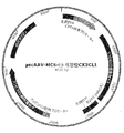
- FIG. 1 shows a vector map of pscAAV-MCS-mouse soluble Cx3cl1 which is an AAV vector plasmid carrying a mouse soluble Cx3cl1 gene.
- FIG. 2 shows a vector map of psAAV-MCS-human soluble CX3CL1 which is an AAV vector plasmid carrying the human soluble CX3CL1 gene.
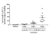
- FIG. 3 shows the expression of mouse soluble Cx3cl1 in retinal tissue by administration of the AAV8 vector (rAAV8-mouse soluble Cx3cl1) carrying the mouse soluble Cx3cl1 gene to rd10 mice at various doses.
- the vertical axis represents the relative expression value when the average of Cx3cl1 gene expression in the retinal tissue of the control vehicle (Vehicle) administration group was set to 1.
- the horizontal axis represents the administration group. Bars in the graph show the mean value ⁇ standard error of the mean value in each group.
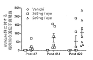
- FIG. 4 shows changes in gene expression of mouse soluble Cx3cl1 in retinal tissue with administration of rAAV8-mouse soluble Cx3cl1 to rd10 mice. Circles, squares, and triangles in the figure show the respective administration groups.
- the vertical axis represents the relative expression value when the average of the gene expression of Cx3cl1 in the retina of the control vehicle (Vehicle) administration group on each evaluation day was set to 1.
- FIG. 5 shows the amount of mouse soluble CX3CL1 protein in the vitreous body by administration of rAAV8-mouse soluble Cx3cl1 to rd10 mice.
- the vertical axis represents the concentration of CX3CL1 protein in the vitreous.
- the horizontal axis represents the administration group. Bars in the graph show the mean value ⁇ standard error of the mean value in each group.
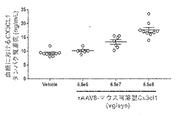
- FIG. 6 shows the amount of mouse CX3CL1 protein in plasma by administration of rAAV8-mouse soluble Cx3cl1 to rd10 mice.
- the vertical axis represents the concentration of CX3CL1 protein in plasma.
- the horizontal axis represents the administration group. Bars in the graph show the mean value ⁇ standard error of the mean value in each group.
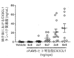
- FIG. 7 shows the expression of human soluble CX3CL1 protein in retinal tissue by administration of the AAV8 vector carrying the human soluble CX3CL1 gene (rAAV8-human soluble CX3CL1) to rd10 mice.
- the vertical axis represents the value obtained by correcting the amount of CX3CL1 protein in the retinal tissue with the tissue weight.
- the horizontal axis represents the administration group. Bars in the graph show the mean value ⁇ standard error of the mean value in each group.
- FIG. 8 shows expression of human soluble CX3CL1 protein in RPE/choroid/scleral tissue by administration of rAAV8-human soluble CX3CL1 to rd10 mice.
- the vertical axis represents the value of the protein amount of CX3CL1 in RPE/choroid/scleral tissue corrected by the tissue weight.
- the horizontal axis represents the administration group. Bars in the graph show the mean value ⁇ standard error of the mean value in each group.
- FIG. 9 shows the amount of human soluble CX3CL1 protein in the vitreous body when rAAV8-human soluble CX3CL1 was administered to rd10 mice.
- the vertical axis represents the concentration of CX3CL1 protein in the vitreous.
- the horizontal axis represents the administration group.
- FIG. 10 shows the amount of human soluble CX3CL1 protein in plasma when rAAV8-human soluble CX3CL1 was administered to rd10 mice.
- the vertical axis represents the concentration of CX3CL1 protein in plasma.
- the horizontal axis represents the administration group.
- Bars in the graph show the mean value ⁇ standard error of the mean value in each group.
- FIG. 11 shows changes in expression of human soluble CX3CL1 protein in retinal tissue over time due to administration of rAAV8-human soluble CX3CL1 to rd10 mice. Circles and squares in the figure indicate the respective administration groups.
- the vertical axis represents the value obtained by correcting the amount of CX3CL1 protein in the retinal tissue with the tissue weight.
- the horizontal axis indicates the number of days after administration. Bars in the graph show the mean value ⁇ standard error of the mean value in each group.
- FIG. 12 shows the time-dependent changes in expression of human soluble CX3CL1 protein in RPE/choroid/scleral tissue due to administration of rAAV8-human soluble CX3CL1 to rd10 mice. Circles and squares in the figure indicate the respective administration groups.
- the vertical axis represents the value of the protein amount of CX3CL1 in RPE/choroid/scleral tissue corrected by the tissue weight.
- the horizontal axis indicates the number of days after administration.
- FIG. 13 shows time-course changes in the amount of human soluble CX3CL1 protein in plasma due to administration of rAAV8-human soluble CX3CL1 to rd10 mice.
- the vertical axis represents the concentration of CX3CL1 protein in plasma.
- the horizontal axis indicates the number of days after administration.
- Bars in the graph show the mean value ⁇ standard error of the mean value in each group.
- FIG. 14 shows the protective effect on the visual motor response by administration of rAAV8-mouse soluble Cx3cl1 to rd10 mice.
- the vertical axis represents the spatial frequency limit (c/d) that can be recognized by the mouse based on the visual motor response.
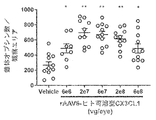
- FIG. 15 shows the protective effect of the visual motor response by administration of rAAV8-human soluble CX3CL1 to rd10 mice.
- the vertical axis represents the spatial frequency limit (c/d) that can be recognized by the mouse based on the visual motor response.
- the horizontal axis represents the administration group.
- Each bar in the graph represents the average value ⁇ standard error of the average value. *P ⁇ 0.05, Dunnett's multiple comparison test.
- FIG. 16 shows the pyramidal cell protective effect by administration of rAAV8-mouse soluble Cx3cl1 to rd10 mice.
- the vertical axis represents the average number of cells (outer segments) positive for anti-cone opsin antibody (cone opsin count) in the observation area.
- the horizontal axis represents the administration group. Each bar in the graph represents the average value ⁇ standard error of the average value. **P ⁇ 0.01, Dunnett's multiple comparison test.
- FIG. 17 shows the pyramidal cell protective effect by administration of rAAV8-human soluble CX3CL1 to rd10 mice.
- the vertical axis represents the average number of cells (outer segments) positive for anti-cone opsin antibody (cone opsin count) in the observation area.
- the horizontal axis represents the administration group. Each bar in the graph represents the average value ⁇ standard error of the average value.
- FIG. 18 shows the pyramidal cell protective effect of rAAV8-mouse soluble Cx3cl1 administration to rd10 mice.
- the vertical axis represents the average number of cells (outer segments) positive for anti-cone opsin antibody (cone opsin count) in the observation area.
- the horizontal axis represents the administration group.
- Each bar in the graph represents the average value ⁇ standard error of the average value.
- FIG. 19 shows the pyramidal cell protective effect by administration of rAAV8-human soluble CX3CL1 to rd10 mice.
- the vertical axis represents the average number of cells (outer segments) positive for anti-cone opsin antibody (cone opsin count) in the observation area.
- the horizontal axis represents the administration group and the age of mice at the time of tissue sampling.
- the retinas of rd10 mice that were collected without administration on the day of administration (29 days after birth) are represented as “Nive”.
- Each bar in the graph represents the average value ⁇ standard error of the average value. **P ⁇ 0.01, Dunnett's multiple comparison test.
- the present invention provides an adeno-associated virus (AAV) vector (hereinafter also referred to as “AAV vector of the present invention”) comprising a nucleic acid molecule encoding CX3CL1 and a promoter operably linked to the nucleic acid molecule, and Provided is a pharmaceutical composition for preventing or treating retinal degenerative disease (hereinafter, also referred to as "pharmaceutical composition of the present invention”), which comprises an AAV vector.
- AAV vector of the present invention adeno-associated virus
- Nucleic acid molecule encoding CX3CL1 The AAV vector of the present invention comprises a nucleic acid molecule encoding CX3CL1.
- CX3CL1 is a chemokine that is constitutively expressed in normal nerves, and is composed of a signal peptide, a chemokine domain, a mucin-like stalk domain, a transmembrane domain, and an intracellular domain from the N-terminus.
- CX3CL1 is expressed as a transmembrane protein, but it can be cleaved with an extracellular cleaving enzyme to exist as soluble CX3CL1.
- CX3CL1 includes naturally-occurring CX3CL1 and variants having its function.
- the CX3CL1 is CX3CL1 from a mammal, and in one aspect, human CX3CL1.
- human CX3CL1 includes naturally-occurring human CX3CL1 and variants having its function.
- CX3CL1 includes full-length (transmembrane type) CX3CL1 and soluble CX3CL1.
- the CX3CL1 is a soluble CX3CL1.
- “soluble CX3CL1” refers to a CX3CL1 polypeptide containing a chemokine domain and a mucin-like stalk domain, but not a transmembrane domain and an intracellular domain.
- Soluble CX3CL1 includes naturally-occurring soluble CX3CL1 and variants having its function.
- the CX3CL1 is human soluble CX3CL1.
- human soluble CX3CL1 includes naturally-occurring human soluble CX3CL1 and variants having the function thereof.
- the human soluble CX3CL1 comprises the amino acid sequence of amino acid numbers 1 to 338 (SEQ ID NO: 4) of the translated sequence (SEQ ID NO: 2) of the coding region (SEQ ID NO: 1) of accession number NM_002996.6. Is a polypeptide.
- the human soluble CX3CL1 has 1 to several, preferably 1 to 10, more preferably 1 to 7, still more preferably 1 to 5, 1 to several amino acid sequences shown in SEQ ID NO: 4. It is a polypeptide having an amino acid sequence in which 3 to 3 or 1 to 2 amino acids are deleted, substituted, inserted, and/or added, and having the function of human soluble CX3CL1.
- the human soluble CX3CL1 comprises an amino acid sequence having at least 90%, 95%, or 98% identity to the amino acid sequence set forth in SEQ ID NO:4, and has the function of human soluble CX3CL1. It is a polypeptide having.
- CX3CL1 is mouse CX3CL1 and in one aspect, mouse soluble CX3CL1.
- the mouse soluble CX3CL1 comprises the amino acid sequence of amino acid numbers 1 to 336 (SEQ ID NO: 8) of the translated sequence (SEQ ID NO: 6) of the coding region of SEQ ID NO: NM_009142.3 (SEQ ID NO: 6). Is a polypeptide.
- identity refers to the parameters prepared by default in the EMBOSS NEEDLE program (Needleman et al., J. Mol. Biol., 1970, 48, 443-453). It means the obtained value Identity.
- the function of CX3CL1 or soluble CX3CL1 includes the ability to bind to CX3CR1.
- the function of CX3CL1 or soluble CX3CL1 includes the ability to suppress the activity of microglia. Such a function of CX3CL1 can be easily evaluated by those skilled in the art using a known method.
- the nucleic acid molecule encoding CX3CL1 is a nucleic acid molecule encoding a polypeptide consisting of the amino acid sequence shown in SEQ ID NO:4, and in one aspect consisting of a polynucleotide consisting of the base sequence shown in SEQ ID NO:3. It is a nucleic acid molecule.
- Nucleic acid molecule encoding CX3CL1 includes DNA molecules (eg, cDNA or genomic DNA) and RNA molecules (eg, mRNA), and analogs of DNA or RNA produced using nucleotide analogs.
- the nucleic acid molecule may be single-stranded or double-stranded, and in one embodiment is single-stranded DNA.
- a CX3CL1-encoding nucleic acid molecule can be synthesized using standard polynucleotide synthesis methods known in the art based on publicly available sequence information. Further, once a nucleic acid molecule encoding CX3CL1 is obtained, it is known to those skilled in the art such as site-directed mutagenesis (Current Protocols in Molecular Biology edition, 1987, John Wiley & Sons Section 8.1-8.5). It is also possible to prepare a variant having the function of CX3CL1 by introducing a mutation into a predetermined site using the method.
- an adeno-associated virus (AAV) vector is used as a vector carrying a nucleic acid molecule encoding CX3CL1.
- AAV is a genome consisting of a single-stranded DNA molecule containing a gene (Rep gene and Cap gene) encoding a function required for infection and replication of AAV to a host cell and an inverted terminal repeat sequence (Inverted terminal repeats: ITR). It is an icosahedral virus without an envelope having (AAV genome).
- AAV is known to be non-pathogenic and able to infect non-dividing cells.
- serotypes of AAV There are over 100 serotypes of AAV, and it is known that the characteristics of the host range and viruses differ depending on the serotype.
- AAV vectors include a capsid and a genome (AAV vector genome) that includes the heterologous nucleic acid molecule sequence flanked by one or more ITRs encapsulated therein.
- AAV vectors used in the present invention are described, for example, in US Pat. No. 7,056,502 and Yan et al. Virol. 2002, 76, 2043-2053, specifically, AAV1 vector, AAV2 vector, AAV3 vector, AAV4 vector, AAV5 vector, AAV8 vector, AAV9 vector and the like.
- the AAV vector used in the present invention is an AAV8 vector (eg, WO2003/052051).
- the AAV vector used in the present invention may be a modified AAV vector in which the amino acid sequence of capsid is modified.
- the AAV vector used in the present invention is a modified AAV8 vector.
- the ITR contained in the AAV vector used in the present invention may be a wild-type sequence, or a variant having an ITR function or an ITR containing a mutation used in a self-complementary vector described later (mutation). ITR).
- the ITR used in the present invention is an ITR derived from AAV1, AAV2, AAV3, AAV4, AAV5, AAV8, AAV9, etc., or a mutant ITR thereof.
- the ITR used in the present invention is an AAV2-derived ITR or a mutant ITR thereof.
- the AAV vector used in the present invention is a self-complementary AAV (Self complementary (sc) AAV) vector.
- the AAV vector used in the present invention is a self-complementary AAV8 vector.
- the self-complementary AAV vector is an AAV vector having a self-complementary AAV vector genome (eg, WO 2001/092551 and WO 2001/011034). In a normal AAV vector, since the vector genome is single-stranded DNA, it takes time to form a double-stranded genome from a single-stranded genome after infecting a host cell.
- the self-complementary AAV vector contains a linked heterologous nucleic acid molecule sequence and a vector genome having its complementary sequence (self-complementary vector genome), and rapidly forms a double-stranded genome in a host cell.
- the self-complementary AAV vector comprises a heterologous nucleic acid molecule sequence linked by a mutant ITR and its complement, and further has two ITRs at each end.
- the mutant ITR has a structure in which the D sequence (packaging signal) is deleted and ⁇ trs (mutation of the terminal truncation region) is included. The structure can prevent Rep-mediated nicking and induce packaging of the self-complementary vector genome.
- the AAV vector of the present invention comprises a promoter operably linked to a nucleic acid molecule encoding CX3CL1.
- "Operably linked” means that one or more promoters are linked to a nucleic acid molecule of interest such that the protein encoded by the nucleic acid molecule of interest can be expressed in a host cell.
- Promoters used in the present invention include, but are not limited to, the SV40 promoter, cytomegalovirus (CMV) derived promoters, and other viral and eukaryotic promoters known to those of skill in the art.
- the promoter used in the present invention is a CMV-derived promoter.
- the promoter used in the present invention is a tissue-specific promoter.
- tissue-specific promoters include retinal pigment epithelium cell-specific promoters, photoreceptor cell-specific promoters, rod cell-specific promoters, and cone cell-specific promoters. More specific examples include Nrl, Crx, Rax (Matsuda and Cepko, 2007, Proc. Natl. Acad. Sci. USA, 104, 1027-1032), opsin promoter, photoreception. Intrabody retinoid binding protein promoter (IRBP156), rhodopsin kinase (RK) promoter, neural leucine zipper (NRLL) promoter (see, for example, Simple-Rowland et al., Mol. Vis., 2010, 16, 916-934). Are listed.
- IRBP156 Intrabody retinoid binding protein promoter
- RK rhodopsin kinase
- NRLL neural leucine zipper
- a cone arrestin promoter in another embodiment, a cone arrestin promoter, a Cabp5 promoter, a Cralbp promoter, an Ndrg4 promoter, a bestrophin 1 promoter, a chicken beta actin (CBA promoter), a clusterin promoter, a Hes1 promoter, a vimentin promoter, a surface antigen classification (CD44) promoter.
- GFAP glial fibrillary acidic protein
- the AAV vector of the present invention includes an enhancer, a polyadenylation signal (poly(A)), a Kozak sequence, etc. (eg, Goeddel, Gene Expression Technology, Methods in Enzymology, 1990, Volume 185, Academic, Academia). San Diego, Calif.) expression control sequences may be included.
- poly(A) polyadenylation signal
- Kozak sequence etc.
- the AAV vector of the present invention is an AAV8 vector comprising a nucleic acid molecule encoding a polypeptide consisting of the amino acid sequence shown in SEQ ID NO: 4 and a CMV-derived promoter operably linked to the nucleic acid molecule.
- the AAV vector of the invention is a self-complementary AAV8 vector comprising a nucleic acid molecule encoding a polypeptide consisting of the amino acid sequence set forth in SEQ ID NO:4 and a CMV-derived promoter operably linked to the nucleic acid molecule. Is.
- the AAV vector of the present invention can be produced using a method known in the art.
- a heterologous nucleic acid molecule for example, a nucleic acid molecule encoding CX3CL1
- a heterologous nucleic acid molecule for example, a nucleic acid molecule encoding CX3CL1
- a plasmid containing the AAV Rep gene and Cap gene complementing the AAV helper function
- a plasmid containing a helper virus accessory gene eg, region VA, E2A, E4
- AAV vector recombinant AAV
- the packaging cells are then cultured to produce the AAV vector and the AAV vector is purified using standard techniques known in the art.
- Examples of packaging cells used include mammalian cells, specifically A549 cells, HeLa cells, HEK293 cells, HEK293T17 cells.
- cesium chloride density gradient centrifugation method digestion of DNA and RNA using benzonase, sucrose gradient centrifugation method, iodixanol density gradient centrifugation method, ultrafiltration method, diafiltration method , Affinity chromatography method, ion exchange chromatography method, polyethylene glycol precipitation method, ammonium sulfate precipitation method and the like can be used.
- the AAV vector genome may be packaged as a baculovirus and introduced into an insect cell such as Sf9 cell.
- Rep and Cap genes are also similarly introduced into insect cells by another baculovirus.
- the baculovirus may contain the genes necessary for efficient production of the AAV vector.
- the insect cells infected with the two baculoviruses can then be cultured to produce an AAV vector and the AAV vector purified and formulated using methods known in the art (eg, US Patent Nos. 5436146, 5753500, 6093570, and 6548286, US2002/0168342).
- the pharmaceutical composition of the present invention is a pharmaceutical composition for preventing or treating retinal degenerative disease, which comprises the AAV vector of the present invention.
- Retinal degenerative disease is a general term for diseases that cause abnormalities in visual function due to the occurrence of disorders in cells that make up retinal tissue, and includes retinal diseases involving rod cell degeneration and cone cell degeneration. including.
- the retinal degenerative disease is retinitis pigmentosa, age-related macular degeneration, macular dystrophy, cone-rod dystrophy, or rod-cone dystrophy.
- the retinal degenerative disease is retinitis pigmentosa.
- the pharmaceutical composition of the invention is a pharmaceutical composition for the treatment of advanced stage retinitis pigmentosa.
- advanced stage retinitis pigmentosa means retinitis pigmentosa after the period when the loss of rod cell function is observed, and retinitis pigmentosa which is observed to have thinning of the outer granular layer in optical coherence tomography, It refers to retinitis pigmentosa in which corrected visual acuity is 20/80 to 20/400 in logMAR visual acuity, or retinitis pigmentosa in which night blindness is observed.
- Loss of rod cell function can be judged by recognizing the loss of amplitude in the dark adaptation electroretinogram. It can also be judged from the fact that the central visual field is narrowed to 20 degrees or less.
- the pharmaceutical composition of the present invention can be prepared by a commonly used method using an excipient usually used in the art, that is, a pharmaceutical excipient, a pharmaceutical carrier and the like.
- a pharmaceutical composition of the invention comprises an AAV vector of the invention and one or more excipients.
- dosage forms of these pharmaceutical compositions include injectable pharmaceutical compositions and other parenteral pharmaceutical compositions, which are administered by intraocular administration, subretinal administration, intravitreal administration, suprachoroidal administration, etc. can do.
- the pharmaceutical compositions of the invention are administered subretinally or intravitreally.
- excipients, carriers, additives, etc. depending on these dosage forms can be used within a pharmaceutically acceptable range.
- the pharmaceutical composition of the present invention may further contain small amounts of auxiliary substances such as wetting agents, emulsifying agents, pH buffering agents, adjuvants or immunostimulants.
- the present invention also provides a method for preventing or treating retinal degenerative disease (hereinafter, also referred to as “the preventive or therapeutic method of the present invention”), which comprises administering a therapeutically effective amount of the AAV vector of the present invention to a subject.
- the method for preventing or treating the present invention is a method for preventing or treating retinitis pigmentosa, age-related macular degeneration, macular dystrophy, cone-rod dystrophy, or rod-cone dystrophy.
- the preventive or therapeutic method of the present invention is a preventive or therapeutic method for retinitis pigmentosa.
- the prophylactic or therapeutic method of the present invention is a method of treating advanced stage retinitis pigmentosa.
- the “subject” is a human or other animal in need of the prevention or treatment, and in one aspect, a human in need of the prevention or treatment.
- “Administration” to a subject includes intraocular administration, intravitreal administration, subretinal administration, suprachoroidal administration and the like. Methods of administration include injection (eg, intravitreal or subretinal or suprachoroidal injection), gene gun, gel, electroporation, use of lipid-based transfection reagents, implantation of drug delivery devices, or any other. Suitable transfection methods are mentioned.
- the AAV vector of the invention is administered intravitreally.
- the AAV vector of the invention may be delivered in dosage forms such as suspensions or solutions.
- a local anesthetic is applied to the surface of the eye and then a local antiseptic solution is applied. Inject the vector through the sclera into the vitreous cavity of the subject's eye with a thin, short (eg, 30 gauge) needle under direct observation, keeping the eye open with or without the device .
- Intravitreal administration is characterized by being relatively less invasive than other intraocular administration methods.
- the AAV vector of the invention is administered subretinally. It is known that the AAV vector administered under the retina is generally introduced into cells such as photoreceptor cells and retinal pigment epithelium (RPE) (Allocca et al., J. Virol., 2007, 81, 11372-11380).
- RPE retinal pigment epithelium
- the AAV vector of the present invention is injected subretinally in a dosage form such as a suspension or a solution using a surgical microscope under direct observation.
- Certain embodiments of subretinal administration include post-vitrectomy, followed by injection into the subretinal space through one or more small retinal incisions using a fine cannula.
- the AAV vector of the present invention is administered suprachoroidal injection.
- the vector is administered in the form of a suspension or solution.
- a topical anesthetic is applied to the surface of the eye, followed by a topical antiseptic solution.
- AAV is placed on the choroid of the subject's eye with a thin, short (eg, 30 gauge) needle under direct observation, with the eye open with or without the use of a device. Inject the vector.
- the preventive or therapeutic method of the present invention comprises introducing the nucleic acid molecule of the present invention into photoreceptor cells and/or retinal pigment epithelial cells using the AAV vector of the present invention.
- the therapeutically effective amount of the AAV vector of the present invention can be appropriately optimized in consideration of severity of disease, previous treatment, general health condition of subject, age, administration method, other diseases and the like.
- a therapeutically effective amount of an AAV vector of the invention is about 0.001-30 mg/kg body weight, in some embodiments about 0.01-25 mg/kg body weight, in another embodiment about 0.1-20 mg/kg.
- Body weight in another embodiment about 1-10 mg/kg, 2-9 mg/kg, 3-8 mg/kg, 4-7 mg/kg or 5-6 mg/kg body weight.
- the therapeutically effective amount of the AAV vector of the present invention can also be expressed as the amount of vector genome administered per eye (vg/eye). vg can also be described as a genome copy (GC).
- the therapeutically effective amount of the AAV vector of the invention is about 1 ⁇ 10 6 to 1 ⁇ 10 12 vg/eye, and in another embodiment, about 1 ⁇ 10 7 to 4 ⁇ 10 10 vg/eye. is there.
- Administration of a therapeutically effective amount of an AAV vector of the present invention includes single doses, multiple doses, tapering doses and titrating doses.
- the AAV vectors of the invention can be administered at least 1, 2, 3, or 4 times daily.
- the administration period for prevention or treatment is at least about 2, 3, 4, 5, 6, 7, 8, 9, 10, 11, 12, 13, 14, 15, 16, 17, 18, 19, 20, It may range for 21, 22, 23 or 24 weeks.
- the AAV vector of the present invention can be used in combination with various therapeutic or prophylactic agents for diseases for which it is considered effective.
- the combination may be administered simultaneously, or separately and continuously or at desired time intervals.
- the preparation for co-administration may be a combination drug or separately formulated.
- the present invention also provides the use of the AAV vector of the present invention for the manufacture of a pharmaceutical composition for preventing or treating retinal degenerative disease.
- the present invention also provides the use of the AAV vector of the present invention for the prevention or treatment of retinal degenerative diseases.
- Example 1 Construction of Construct Cloning, Plasmid Construction, rAAV8-Mouse Soluble Cx3cl1 Production
- AAV vector plasmid carrying the mouse soluble Cx3cl1 nucleic acid molecule sequence was designated as pscAAV-MCS plasmid (Cell BioLabs, Catalog No. VPK-).
- pscAAV-MCS plasmid Cell BioLabs, Catalog No. VPK-.
- SEQ ID NO: 7 mouse soluble Cx3cl1 nucleic acid molecule sequence
- SEQ ID NO: 7 was inserted by using BamHI (5' side) and XbaI (3' side) restriction enzymes.
- the resulting plasmid is called pscAAV-MCS-mouse soluble Cx3cl1.
- the mouse soluble Cx3cl1 gene is located downstream of the CMV-derived promoter and upstream of SV40 poly(A), and has a Kozak sequence (GCCACC) before the start codon.
- GCCACC Kozak sequence
- the length between ITRs including the ITR is 1969b. p., which is a size that can be packaged in an AAV vector.
- FIG. 1 A vector map of pscAAV-MCS-mouse soluble Cx3cl1 is shown in FIG.
- This plasmid was amplified using Stbl3 E. coli strain (Thermo Fisher Scientific, Catalog No. C737303).
- the plasmid pHelper AAVpro Helper Free System (AAV2), Takara Bio Inc., Catalog No. 6230
- pRCAAV8 necessary for producing recombinant AAV were prepared using DH5 ⁇ E. coli strain (Takara Bio Inc., Catalog No. 9057). Amplified.
- pRCAAV8 converted the Cap sequence portion of pRC5 (AAV pro Packaging Plasmid (AAV5), Takara Bio Inc., Catalog No.
- AAV8 (Accession No. NC_006261.1, 2121b.p.-4337b.p.). It is a plasmid.
- the HEK293T17 cell line was seeded at a density of 2500 cells per square centimeter, and 4 days later, pscAAV-MCS-mouse soluble Cx3cl1, pHelper, and pRCAAV8 were added to polyethyleneimine (Polysciences "Max", Polysciences, catalog number 24765-. According to the attached protocol, 1) was used for transient transfection, and the cell culture supernatant was recovered by culturing at 37° C. and 5% CO 2 for 3 days and 6 days.
- RAAV8 contained in the culture supernatant was concentrated by tangential flow filtration using mPES MiniKros Sampler 300 kDa (Spectrum Labs, Catalog No. S-02-E300-05-N). The concentrated sample was filtered through a 0.22 ⁇ m filter, and affinity column POROS (registered trademark) CaptureSelect (registered trademark) AAV8 Affinity Resin (Thermo Fisher Scientific 7), catalog number A30 using AKTAexplorer100 (GE Healthcare). The purified sample was subjected to ultracentrifugation using a cesium chloride concentration gradient. A vertical rotor (VTi50 (Beckman Coulter, Catalog No. 362758)) was used in this ultracentrifugation.
- VTi50 Beckman Coulter, Catalog No. 362758
- the fractions with high absorbance measurement values at 280 nm and 260 nm were collected.
- the collected sample was dialyzed three times using phosphate buffered saline and used as a stock of the prepared AAV8 vector carrying the mouse soluble Cx3cl1 gene (referred to as rAAV8-mouse soluble Cx3cl1).
- the virus titer measurement was performed using the QuickTiter AAV Quantation kit (Cell Biolabs, catalog number VPK-145).
- Example 2 Construction of Construct Cloning, Plasmid Construction, rAAV8-Human Soluble CX3CL1 Production
- AAV vector plasmid carrying human soluble CX3CL1 nucleic acid molecule sequence was used as pscAAV-MCS plasmid (Cell BioLabs, Catalog No. VPK-).
- pscAAV-MCS plasmid Cell BioLabs, Catalog No. VPK-.
- SEQ ID NO: 3 a human soluble CX3CL1 nucleic acid molecule sequence
- SEQ ID NO: 3 was inserted using BamHI (5′ side) and XbaI (3′ side) restriction enzymes to prepare.
- the resulting plasmid is called pscAAV-MCS-human soluble CX3CL1.
- the human soluble CX3CL1 gene is located downstream of a CMV-derived promoter and upstream of SV40 poly(A), and has a Kozak sequence (GCCACC) before the start codon. Further, a human soluble CX3CL1 having a stop codon located downstream of the human soluble CX3CL1 gene and having an amino acid sequence of amino acid numbers 1 to 338 (SEQ ID NO: 4) of the human full length CX3CL1 sequence (SEQ ID NO: 2). was designed to be expressed. The length between ITRs including ITRs is 1975b. p., which is a size that can be packaged in an AAV vector.
- a vector map of pscAAV-MCS-human soluble CX3CL1 is shown in FIG.
- This plasmid was amplified using the Stbl3 E. coli strain.
- the plasmids pHelper and pRCAAV8 required for rAAV production were amplified using the DH5 ⁇ E. coli strain.
- the HEK293T cell line was seeded at a density of 2500 cells per square centimeter, and 5 days later, pscAAV-MCS-human soluble CX3CL1, pHelper, and pRCAAV8 were added to polyethyleneimine (Polysciences, Polysciences, catalog number 24765-. According to the attached protocol, 1) was used for transient transfection, and the culture supernatant of cells cultured for 4 days at 37° C. and 5% CO 2 was collected.
- the rAAV8 contained in the culture supernatant was concentrated by tangential flow filtration using mPES MiniKros Sampler 300 kDa (Spectrum, catalog number S-02-E300-05-N).
- the concentrated sample was filtered through a 0.22 ⁇ M filter and purified by an affinity column using AKTAexplorer100 (GE Healthcare) as in Example 1.
- the purified sample was subjected to ultracentrifugation using a cesium chloride concentration gradient. A vertical rotor (VTi50) was used for this ultracentrifugation. In the ultracentrifuged sample, the fractions with high absorbance measurement values at 280 nm and 260 nm were collected.
- VTi50 vertical rotor
- the collected sample was dialyzed three times using phosphate buffered saline and used as a stock of the prepared AAV8 vector carrying the human soluble CX3CL1 gene (referred to as rAAV8-human soluble CX3CL1).
- the virus titer measurement was performed using the QuickTiter AAV Quantation kit (Cell Biolabs, catalog number VPK-145).
- Example 3 Virus Infection Protocol
- the protocol for administering rAAV8-mouse soluble Cx3cl1 or rAAV8-human soluble CX3CL1 at 1 day of age is the method of Xiong et al. (J. Clin. Invest. 2015, 125, 1433-143). 1445).
- the rAAV8-mouse soluble Cx3cl1 prepared in Example 1 or the rAAV8-human soluble CX3CL1 prepared in Example 2 was administered to rd10 mice, which are model mice for retinitis pigmentosa.
- rd10 mice which are model mice for retinitis pigmentosa.
- One-day-old postnatal mice were anesthetized on ice, the eyeballs were exposed, and administration was performed under the retina with a glass syringe.
- rAAV8-mouse soluble Cx3cl1 vehicle (phosphate) was added to a final concentration of 2.2 ⁇ 10 10 vg/mL, 2.2 ⁇ 10 11 vg/mL or 2.2 ⁇ 10 12 vg/mL.
- Buffered saline with 0.25% Fast Green FCF (Nacalai Tesque, Catalog No. 15939-54) and 0.001% Pluronic F-68 (Thermo Fisher Scientific, Catalog No. 24040032).
- 6.5 ⁇ 10 6 (6.5e6) vg/0.3 ⁇ L
- 6.5 ⁇ 10 7 (6.5e7) vg/0.3 ⁇ L or 6.5 ⁇ 10 8 (6.5e8) per eye.
- the final concentration was 2 ⁇ 10 10 vg/mL, 6 ⁇ 10 10 vg/mL, 2 ⁇ 10 11 vg/mL, 6 ⁇ 10 11 vg/mL or 2 ⁇ 10 12 vg. /ML, 6 ⁇ 10 6 (6e6) vg/0.3 ⁇ L/eye, 1.8 ⁇ 10 7 (rounded to the nearest whole number in the figure as 2e7) vg/0.
- mice 3 ⁇ L, 6 ⁇ 10 7 (6e7) vg/0.3 ⁇ L, 1.8 ⁇ 10 8 (in the figure, rounded off to the nearest whole number and shown as 2e8) vg/0.3 ⁇ L or 6 ⁇ 10 8 (6e8) vg/0 It was administered at 3 ⁇ L.
- the administered mice were allowed to return to body temperature and awakened, and were bred with parent mice and allowed to survive at 28 days old to 35 days old after weaning and from 50 days old to 51 days old after birth. Similar to patients with retinitis pigmentosa, in rd10 mice, pyramidal cell death occurs after rod cell death (20 days old to 28 days old) (Gargini et al., J. Comp. Neurol., 2007).
- Non-Patent Document 1 the direct protection effect of the drug on the pyramidal cells could not be verified.
- the drug was administered and verified.
- the administration method at that time is as follows.
- Midrin (registered trademark) P ophthalmic solution (Santen Pharmaceutical Co., Ltd.) was applied to the rd10 mouse to cause dilation and anesthesia was performed by intramuscular injection of a ketamine/xylazine mixed anesthetic solution. After anesthesia, Hyalerin (registered trademark) eye drop 0.3% (Santen Pharmaceutical Co., Ltd.) was applied to the eye to protect the cornea.
- the sclera on the ear side was perforated with a 30-gauge needle, passed through the needle of a Hamilton syringe filled with 1 ⁇ L of the administration solution, passed through the vitreous body, and inserted into the retina on the nose side for subretinal administration.
- rAAV8-mouse soluble Cx3cl1 was prepared in a vehicle so that the final concentration was 2 ⁇ 10 11 vg/mL or 2 ⁇ 10 12 vg/mL, and 2 ⁇ 10 8 (2e8) vg/1 ⁇ L per eye was prepared. Alternatively, it was administered at 2 ⁇ 10 9 (2e9) vg/1 ⁇ L.
- rAAV8-human soluble CX3CL1 in vehicle such that the final concentration was 2 ⁇ 10 9 vg/mL, 2 ⁇ 10 10 vg/mL, 2 ⁇ 10 11 vg/mL or 2 ⁇ 10 12 vg/mL.
- mice Prepared at 2 ⁇ 10 6 (2e6) vg/1 ⁇ L, 2 ⁇ 10 7 (2e7) vg/1 ⁇ L, 2 ⁇ 10 8 (2e8) vg/1 ⁇ L or 2 ⁇ 10 9 (2e9) vg/1 ⁇ L per eye was administered. 1 ⁇ L of vehicle was administered to the control vehicle group. The administered mice were intramuscularly injected with antisedan (registered trademark) (Nippon Zenyaku Kogyo Co., Ltd.) to awaken them, and allowed to survive up to 51 days old.
- antisedan registered trademark
- Example 4 Evaluation of Cx3cl1 Gene Expression in Retinal Tissue
- rd10 mice to which rAAV8-mouse soluble Cx3cl1 or vehicle was administered subretinally at the age of 1 day after birth were comforted by exsanguination under isoflurane anesthesia at 51 days old. It was killed and the eyeball was extracted. The cornea and lens were removed, the retina was collected so as to be isolated from other tissues, and gene expression of Cx3cl1 in the retinal tissues was evaluated. RNA was extracted from the retina and cDNA was prepared by reverse transcription.
- Taqman gene expression assay was performed using the following primers and probe for Cx3cl1 (Thermo Fisher Scientific, Catalog No. 4324034). It was carried out according to the attached protocol of Taqman Gene Expression Master Mix (Thermo Fisher Scientific, Catalog No. 4369016). A Taqman gene expression assay was similarly performed on the housekeeping gene (beta actin) (Thermo Fisher Scientific, Catalog No. Mm00607939_s1).
- Example 3 rd10 mice that were subretinally administered rAAV8-mouse soluble Cx3cl1 or vehicle between 28 days old and 29 days old were 7 days (Post d7), 14 days (Post d14), and After 22 days (Post d22), cDNA was prepared from the retina as described above, and Cx3cl1 gene expression was evaluated by the Taqman gene expression assay as described above (FIG. 4).
- the expression of Cx3cl1 mRNA was higher than that in the control vehicle group 7 days after the administration, and was further increased 14 days and 22 days later.
- the 2 ⁇ 10 9 (2e9) vg/eye administration group was evaluated only 22 days after administration, and at that time point, a higher expression tendency than that of the 2 ⁇ 10 8 vg/eye administration group was observed.
- Example 5 Evaluation of CX3CL1 protein level in retinal tissue, RPE/choroid/scleral tissue, vitreous and plasma
- rAAV8-mouse soluble Cx3cl1 or vehicle was subretinally administered at 1 day of age. The mice were allowed to survive to the age of 50 days after birth, and plasma and vitreous were sampled. For the vitreous body, the cornea and lens were excised from the eyeball, and the exposed vitreous body was dipped in phosphate buffered saline to collect the material. Plasma was collected from the supernatant obtained by collecting blood from the abdominal vena cava into a heparin blood collection tube under isoflurane anesthesia and centrifuging.
- the amount of CX3CL1 protein in the vitreous and plasma was evaluated by an ELISA kit (R&D Systems, catalog number MCX310) using an anti-mouse CX3CL1 antibody. The results are shown in FIGS. 5 and 6, respectively.
- 6.5 ⁇ 10 6 6.5e6
- 6.5 ⁇ 10 7 6.5e7
- 6.5 ⁇ 10 8 6.5e8
- a dose-dependent increase in the amount of CX3CL1 protein was confirmed in the vg/eye administration group, and in particular, a large increase was observed in the 6.5 ⁇ 10 7 vg/eye administration group and the 6.5 ⁇ 10 8 vg/eye administration group.
- Example 3 rd10 mice in which rAAV8-human soluble CX3CL1 or vehicle was administered subretinally at 1 day of age were allowed to survive until 50 days of age, and plasma, vitreous, retinal tissue, and RPE/choroid/strong were established. Membrane tissue was sampled. The retina was collected by picking up tissue with tweezers from the rest of the eyeball from which the vitreous was collected as described above.
- RPE/choroid/scleral tissue refers to the remaining tissue obtained by removing the retina from the eyeball after removing the vitreous with tweezers.
- the retinal tissue and RPE/choroid/scleral tissue were sampled in a Radioimmunoprecipitation Assay (RIPA) buffer (Merck, catalog number R0278-50ML), and the tissue was dissolved using a grinder.
- the amount of CX3CL1 protein in retinal tissue, RPE/choroid/scleral tissue, vitreous and plasma was evaluated by an ELISA kit (R&D Systems, catalog number DCX310) using an anti-human CX3CL1 antibody. The results are shown in FIGS. 7, 8, 9 and 10, respectively.
- Example 3 For the retinal tissue and RPE/choroid/scleral tissue, the values of the CX3CL1 protein amount in the tissue corrected by the tissue weight are shown. Dose-dependent expression of CX3CL1 protein was confirmed in all tissues.
- ret10 tissue, RPE/choroid/scleral tissue, and plasma were administered from rd10 mouse that underwent subretinal administration of rAAV8-human soluble CX3CL1 (2 ⁇ 10 8 vg/eye) or vehicle at 28 days of age.
- the samples were collected on the 7th, 14th, 21st, and 49th days, and the amount of CX3CL1 protein in each tissue was evaluated by a protein quantification kit (MSD, catalog number K151VCK) using an anti-human CX3CL1 antibody.
- MSD protein quantification kit
- FIGS. 11, 12 and 13 respectively.
- the values obtained by correcting the CX3CL1 protein amount with the tissue weight are shown.
- Time-dependent expression of CX3CL1 protein was confirmed in all tissues, and it was confirmed that the expression reached a plateau 14 days after administration.
- Example 6 Visual Motor Response Test The visual motor response test was carried out according to the method of Xiong et al. (J. Clin. Invest., 2015, 125:1433-1445). In Example 3, a visual motor response test was carried out at the age of 44 days to the age of 45 days after rd10 mice to which rAAV8-mouse soluble Cx3cl1 or vehicle had been administered subretinally at the age of 1 day after birth. In this test, OptoMotry HD manufactured by CerebralMechanics was used. All mice were placed on the platform for about 5 minutes under wakefulness approximately one week before evaluation to acclimate to the chamber.
- the mouse On the day of the evaluation, the mouse was placed on the platform under awakening, and when the experimenter judged that the animal stopped the exploratory behavior and did not fall off the platform, the experimenter started the test.
- the contrast of the black and white grid was set to 100%
- the rotation speed was set to 12 degrees/second
- the grid screen was rotated, and the visual movement reaction of the mouse to the rotated grid was observed.
- the blind mouse did not respond, and the visible mouse also turned its head as if following the movement of a rotated grid.
- the visual motor response was evaluated using the minimum thickness of the lattice that can be recognized by the mouse, that is, the spatial frequency threshold (represented by cycles/degree (c/d)) as an index.
- the experimenter conducted the test blindly so that the administration content of each individual was unknown.
- Fig. 14 The results are shown in Fig. 14.
- the 6.5 ⁇ 10 6 (6.5e6) vg/eye administration group showed no change compared with the control vehicle group, but the 6.5 ⁇ 10 7 (6.5e7) vg/eye administration group and 6.5. It was confirmed that the visual motor reaction was retained in the group administered with ⁇ 10 8 (6.5e8) vg/eye.
- a visual motor response test was carried out in the same manner as described above, and 42 days after the birth to 43 days after the birth of rd10 mice in which the rAAV8-human soluble CX3CL1 or the vehicle was administered subretinally at the age of 1 day after birth in Example 3. It was carried out. The results are shown in Fig. 15.
- Example 7 Evaluation of cone cell protective effect
- the outer segment of the cone cells was stained by immunohistochemistry and became positive.
- Each outer segment was regarded as one cone cell, and the number of remaining cone cells was evaluated. Specifically, it is as follows.
- rd10 mice which were administered with rAAV8-mouse soluble Cx3cl1 or vehicle under the retina at 1 day of age, were euthanized by exsanguination under isoflurane anesthesia at 50 days of age, and eyeballs were collected to obtain 4%. It was fixed with paraformaldehyde for 30 minutes.
- the cornea, lens, iris, sclera, choroid, and retinal pigment epithelial cells were removed from the fixed eyeball to isolate the retina.
- a blocking agent for immunohistochemical staining Alexa Fluoro (registered trademark) 647-labeled anti-red/green opsin antibody (Merck, catalog number AB5405) and DyLight (registered trademark) 488 were labeled. It was stained with anti-blue opsin antibody (Merck, catalog number AB5407).
- the stained retina was incised at four places and placed on a slide with the photoreceptor cell side facing up to prepare a flat mount.
- the 6.5 ⁇ 10 8 (6.5e8)vg/eye administration group also shows an increasing tendency.
- These data indicate that in rd10 mice, subretinal administration of rAAV8-mouse soluble Cx3cl1 preserves mouse cone cell numbers.
- Example 3 the retinas of rd10 mice in which rAAV8-human soluble CX3CL1 or vehicle was administered subretinally at 1 day of age were sampled at 50 days of age, and the number of pyramidal opsin was determined by the same method as described above. The evaluation results are shown in FIG.
- Example 3 rd10 mice that were subretinally administered rAAV8-mouse soluble Cx3cl1 or vehicle at 28 days of age were sampled at 22 days (50 days of age) after administration, and the same method as above was used.
- FIG. 18 shows the result of evaluation of the cone opsin number by. A significant increase was observed in the 2 ⁇ 10 8 (2e8) vg/eye administration group and the 2 ⁇ 10 9 (2e9) vg/eye administration group compared to the control vehicle group (P ⁇ 0.01 and P ⁇ 0.05, respectively). , Dunnett's multiple comparison test).
- Example 3 the retinas of rd10 mice to which rAAV8-human soluble CX3CL1 or vehicle was administered subretinally at 29 days of age were sampled at 51 days of age, and the number of pyramidal opsin was determined by the same method as described above. evaluated.
- the retina (Naive) of the rd10 mouse which was collected without administration on the day of administration (29 days after birth), was also included in the evaluation target. The results are shown in Fig. 19.
- the number of pyramidal opsins at the age of 51 days after birth in the vehicle-administered group was lower than that of pyramidal opsin on the day of administration, which indicates the progress of pyramidal cell degeneration from the day of administration.
- a dose-dependent increase in the number of cone opsin was observed as compared with the vehicle administration group.
- the 2 ⁇ 10 8 (2e8) vg/eye administration group and the 2 ⁇ 10 9 (2e9) vg/eye administration group showed a significant increase compared with the control vehicle group (P ⁇ 0.01, Dunnet's multiple comparison). Certification).
- rAAV8-soluble CX3CL1 has an effect of retaining a visual motor response and a cone cell protective effect. Therefore, the AAV vector carrying the CX3CL1 gene of the present invention is considered to be preventable or treatable by maintaining the visual motor response and protecting the pyramidal cells.
- Retinitis pigmentosa age-related macular degeneration, macular dystrophy, cone It is expected as a preventive and/or therapeutic agent for retinal degenerative diseases such as rod dystrophy or rod cone dystrophy.
- the AAV vector equipped with the CX3CL1 gene of the present invention is a preventive and/or therapeutic agent for retinal degenerative diseases such as retinitis pigmentosa, age-related macular degeneration, macular dystrophy, cone-rod dystrophy, and rod-cone dystrophy. As expected.
Landscapes
- Health & Medical Sciences (AREA)
- Life Sciences & Earth Sciences (AREA)
- Chemical & Material Sciences (AREA)
- Engineering & Computer Science (AREA)
- General Health & Medical Sciences (AREA)
- Veterinary Medicine (AREA)
- Genetics & Genomics (AREA)
- Bioinformatics & Cheminformatics (AREA)
- Medicinal Chemistry (AREA)
- Pharmacology & Pharmacy (AREA)
- Animal Behavior & Ethology (AREA)
- Public Health (AREA)
- Organic Chemistry (AREA)
- Zoology (AREA)
- Biotechnology (AREA)
- Epidemiology (AREA)
- Biomedical Technology (AREA)
- Nuclear Medicine, Radiotherapy & Molecular Imaging (AREA)
- Microbiology (AREA)
- Virology (AREA)
- Molecular Biology (AREA)
- General Engineering & Computer Science (AREA)
- Chemical Kinetics & Catalysis (AREA)
- Wood Science & Technology (AREA)
- General Chemical & Material Sciences (AREA)
- Physics & Mathematics (AREA)
- Plant Pathology (AREA)
- Mycology (AREA)
- Biophysics (AREA)
- Biochemistry (AREA)
- Gastroenterology & Hepatology (AREA)
- Immunology (AREA)
- Proteomics, Peptides & Aminoacids (AREA)
- Ophthalmology & Optometry (AREA)
- Medicines Containing Material From Animals Or Micro-Organisms (AREA)
- Medicines That Contain Protein Lipid Enzymes And Other Medicines (AREA)
Abstract
【課題】 アデノ随伴ウイルス(AAV)を用いたCX3CL1遺伝子の導入による網膜変性疾患の予防又は治療方法、及び、CX3CL1遺伝子を搭載したAAVベクターを有効成分として含む、網膜変性疾患の予防又は治療用医薬組成物を提供する。 【解決手段】 本発明者らは、網膜変性疾患の遺伝子治療について鋭意検討した結果、AAVベクター、特にAAV8ベクターを用いたCX3CL1遺伝子の導入が錐体細胞保護作用を示すこと、CX3CL1遺伝子を搭載したAAVベクター、特にAAV8ベクターが、網膜変性疾患の治療剤として使用できることを知見して本発明を完成した。CX3CL1遺伝子を搭載したAAVベクターは、網膜変性疾患の予防及び/又は治療剤として使用できる。
Description
本発明は、アデノ随伴ウイルス(adeno-associated virus:AAV)を用いたCX3CL1遺伝子の導入による網膜変性疾患の予防又は治療方法、及び、CX3CL1遺伝子を搭載したAAVベクターを有効成分として含む、網膜変性疾患の予防又は治療用医薬組成物に関する。
網膜は眼底に位置する組織であり、光の情報を電気信号に変え、脳に刺激を伝える役割を持つ。組織学的に幾つかの層に分けることができ、外側から網膜色素上皮層、内節・外節層、外顆粒層、外網状層、内顆粒層、内網状層、神経節細胞層、神経線維層に分けられる。光刺激を電気信号に変換する役割を担う視細胞は核を外顆粒層に持ち、外節に光に反応するタンパク質オプシンを発現する。また、視細胞はそれぞれ暗所と明所で機能する杆体細胞と錐体細胞に分類できる。ヒトの網膜においては、杆体細胞は周辺部に位置し、錐体細胞は黄斑に密集している(中心窩に一番多い)ことが知られ、このため、杆体細胞は周辺視野を、錐体細胞は中心視野及び視力をつかさどることが知られている。
網膜組織を構成する細胞に障害が発生することにより、視機能に異常をきたす疾患として、網膜色素変性症等の種々の網膜変性疾患が知られている。これらの疾患には、杆体細胞の変性を伴う網膜疾患及び錐体細胞の変性を伴う網膜疾患が含まれる。
網膜色素変性症(Retinitis Pigmentosa:RP)は、杆体細胞、錐体細胞、又は網膜色素上皮細胞における遺伝子変異が原因で視細胞及び網膜色素上皮細胞が変性する、数千人に一人の頻度で発症する病気である。多くの場合は杆体細胞に何らかの変異が認められ、まず杆体細胞の変性が発生し、患者は夜盲及び視野狭窄を経験する。更に病気が進行すると、二次的に錐体細胞の変性が起き、中心視力の低下及び最終的な失明に至る。現在、治療法は確立されておらず、患者の視覚機能を回復又は維持させる治療法が望まれている。
黄斑ジストロフィーは、錐体細胞が密集する黄斑が徐々に障害を受けることにより視力低下をきたす、遺伝性進行性疾患である。黄斑ジストロフィーは、スタルガルト黄斑ジストロフィー又は黄色斑眼底としても公知のスタルガルト病を含む。黄斑ジストロフィーは錐体細胞機能障害にともなう色覚異常、中心視力低下又は中心視野異常によって特徴付けられる。
錐体杆体ジストロフィー(Cone-rod dystrophy:CRD)及び杆体錐体ジストロフィー(Rod-cone dystrophy:RCD)は、錐体細胞及び杆体細胞の変性を引き起こし、多くの場合盲目をもたらす、遺伝性進行性疾患である。CRDは、錐体細胞の生存性低下又は細胞死と、続く杆体細胞の生存性低下又は細胞死によって特徴付けられる。対照的に、RCDは、杆体細胞の生存性低下又は細胞死と、続く錐体細胞の生存性低下又は細胞死によって特徴付けられる。
加齢性黄斑変性症(Age-related macular degeneration:AMD)は、黄斑変性症とも称され、高齢者に盲目を引き起こす病態である。加齢性黄斑変性症は、滲出性及び非滲出性のAMDの両方を含む。非滲出性のAMDは、ドライ型、萎縮性、又はドルーゼノイド(drusenoid)(加齢性)黄斑変性症としても公知である。非滲出性のAMDにおいて、ドルーゼンは、網膜色素上皮細胞に隣接するブルッフ膜に典型的に蓄積する。続いて、ドルーゼン及び障害された網膜色素上皮細胞が、黄斑における錐体細胞の機能に干渉すると、視力喪失が起こり得る。非滲出性のAMDは、長年にわたり漸進的な視力喪失を生じる。非滲出性のAMDは、滲出性のAMDをもたらし得る。滲出性のAMDは、ウェット型又は血管新生黄斑症としても知られ、急速に進行し、中心視力に重度の損傷を引き起こし得る。
マイクログリアは網膜内に存在するグリア細胞の一つである。近年、マイクログリアの網膜における有害な作用が複数報告されており、新たな標的細胞として注目されている(Silvermanら、Annu. Rev. Vis. Sci.、2018年、第15巻、4号、45~77頁)。正常な網膜ではマイクログリアは内網状層及び外網状層に局在するが、網膜色素変性症においては、外顆粒層に浸潤することが報告されている。浸潤したマイクログリアは活性化した形態をとっており、高い貪食機能を持っている。マイクログリアは一般的に、細胞死を起こした細胞の残骸を貪食し、除去することが知られているが、網膜色素変性症においては、マイクログリアが生きている視細胞も過剰に貪食してしまい、病態の進行を速めることが報告されている(非特許文献1)。また、活性化したマイクログリアはTNFα等のサイトカインを放出し、それら自体が更なる視細胞の変性を誘導することも知られている。実際に、網膜色素変性症のモデルマウスであるrd10マウスにおいてマイクログリアの活性を抑制させると、病態進行を遅延できることが報告されている(Pengら、J.Neurosci.、2014年、第34巻、8139~8150頁)。よって、マイクログリアを標的とする新規薬剤探索は網膜色素変性症等の網膜変性疾患の進行を抑制する新たな治療選択肢になる可能性がある。
CX3CL1(FKN,フラクタルカイン)は、CX3Cサブファミリーに属する唯一の遺伝子であり、正常な神経において恒常的に発現しているケモカインである。マイクログリアではその唯一の受容体であるCX3CR1が発現している。CX3CL1はN末端からシグナルペプチド、ケモカインドメイン、ムチン様ストークドメイン、膜貫通ドメイン、細胞内ドメインに分けられる。CX3CL1は膜貫通タンパク質として発現されるが、細胞外切断酵素であるADAM-10やADAM-17、カテプシンSなどにより細胞外領域の根元が切断され、可溶型タンパク質として存在することが知られている。また、CX3CL1/CX3CR1シグナル経路はマイクログリアを非活性化状態に保つ機能を持つことが知られている。
網膜色素変性症モデルにおいては、リコンビナントCX3CL1タンパク質を硝子体内投与することにより、マイクログリアの活性抑制及び病態の進行抑制を誘導することが報告されている(非特許文献1)。活性酸素により誘導された網膜変性症モデルにおいても、リコンビナントCX3CL1タンパク質を硝子体内投与することにより、網膜保護作用を誘導することが報告されている(特許文献1)。リコンビナント可溶型CX3CL1タンパク質についても、in vitroで網膜細胞保護作用を示すことが報告されている(非特許文献2)。
そして、AAV8を用いた網膜下投与での遺伝子導入が複数報告されている(例えば、Vandenbergheら、Sci. Transl. Med.、2011年、第3巻、88ra54頁、Igarashiら、Hum. Gene Ther.、2010年、第21巻、631~7頁、Igarashiら、Curr. Eye Res.、2013年、第38巻、569~77頁)。
可溶型CX3CL1をコードする核酸を搭載したAAVベクターは、パーキンソン病及びアルツハイマー病をターゲットした報告がされている。具体的には、可溶型CX3CL1をコードする核酸を搭載したAAV9ベクターが、神経細胞脱落を抑制することが報告されている(特許文献2、非特許文献2、3及び4)。また、可溶型CX3CL1をコードする核酸を搭載したAAV4ベクターを脳室内に投与した結果、可溶型CX3CL1の分泌が確認されたことが記載されている(非特許文献5及び6)。
Zabelら、Glia、2016年、第64巻、1479~1491頁
Rocheら、Sci. Rep.2017年、第7巻、記事番号43067
Nashら、Mol. Ther.、2015年、第23巻、17~23頁
Nashら、Neurobiol. Aging、2013年、第34巻、1540~1548頁
Morgantiら、J.Neurosci.、2012年、第32巻、14592~14601頁
Finneranら、Cell Transplantation、2015年、第24巻、757頁
アデノ随伴ウイルス(AAV)を用いたCX3CL1遺伝子の導入による網膜変性疾患の予防又は治療方法、及び、CX3CL1遺伝子を搭載したAAVベクターを有効成分として含む、網膜変性疾患の予防又は治療用医薬組成物を提供する。
本発明者らは、網膜変性疾患の遺伝子治療について鋭意検討した結果、AAVベクター、特にAAV8ベクターを用いたCX3CL1遺伝子の導入が錐体細胞保護作用を示すこと、CX3CL1遺伝子を搭載したAAVベクター、特にAAV8ベクターが、網膜変性疾患の予防又は治療剤として使用できることを知見して本発明を完成した。
本発明のある態様を以下に示す。
(1)CX3CL1をコードする核酸分子及び該核酸分子に作動可能に連結されたプロモーターを含むアデノ随伴ウイルス(AAV)ベクターを含む、網膜変性疾患の予防又は治療用医薬組成物。
(2)前記CX3CL1が可溶型CX3CL1である、(1)に記載の医薬組成物。
(3)前記可溶型CX3CL1がヒト可溶型CX3CL1である、(2)に記載の医薬組成物。
(4)前記ヒト可溶型CX3CL1が配列番号4に示されるアミノ酸配列からなるポリペプチドである、(3)に記載の医薬組成物。
(5)前記AAVベクターが、AAV8ベクターである、(1)乃至(4)のいずれか1つに記載の医薬組成物。
(6)前記AAVベクターが、配列番号4に示されるアミノ酸配列からなるポリペプチドをコードする核酸分子及び該核酸分子に作動可能に連結されたプロモーターを含むAAV8ベクターである、(1)に記載の医薬組成物。
(7)網膜変性疾患が、網膜色素変性症、加齢性黄斑変性症、黄斑ジストロフィー、錐体杆体ジストロフィー、又は杆体錐体ジストロフィーである、(1)乃至(6)のいずれか1つに記載の医薬組成物。
(8)進行期の網膜色素変性症の治療用である(7)に記載の医薬組成物。
(9)網膜下に投与するための、(1)乃至(8)のいずれか1つに記載の医薬組成物。
(10)硝子体内に投与するための、(1)乃至(8)のいずれか1つに記載の医薬組成物。
(1)CX3CL1をコードする核酸分子及び該核酸分子に作動可能に連結されたプロモーターを含むアデノ随伴ウイルス(AAV)ベクターを含む、網膜変性疾患の予防又は治療用医薬組成物。
(2)前記CX3CL1が可溶型CX3CL1である、(1)に記載の医薬組成物。
(3)前記可溶型CX3CL1がヒト可溶型CX3CL1である、(2)に記載の医薬組成物。
(4)前記ヒト可溶型CX3CL1が配列番号4に示されるアミノ酸配列からなるポリペプチドである、(3)に記載の医薬組成物。
(5)前記AAVベクターが、AAV8ベクターである、(1)乃至(4)のいずれか1つに記載の医薬組成物。
(6)前記AAVベクターが、配列番号4に示されるアミノ酸配列からなるポリペプチドをコードする核酸分子及び該核酸分子に作動可能に連結されたプロモーターを含むAAV8ベクターである、(1)に記載の医薬組成物。
(7)網膜変性疾患が、網膜色素変性症、加齢性黄斑変性症、黄斑ジストロフィー、錐体杆体ジストロフィー、又は杆体錐体ジストロフィーである、(1)乃至(6)のいずれか1つに記載の医薬組成物。
(8)進行期の網膜色素変性症の治療用である(7)に記載の医薬組成物。
(9)網膜下に投与するための、(1)乃至(8)のいずれか1つに記載の医薬組成物。
(10)硝子体内に投与するための、(1)乃至(8)のいずれか1つに記載の医薬組成物。
本発明の別の態様を以下に示す。
(a-1)CX3CL1をコードする核酸分子及び該核酸分子に作動可能に連結されたプロモーターを含むアデノ随伴ウイルス(AAV)ベクターの治療有効量を対象に投与することを含む、網膜変性疾患の予防又は治療方法。
(a-2)前記CX3CL1が可溶型CX3CL1である、(a-1)に記載の方法。
(a-3)前記可溶型CX3CL1がヒト可溶型CX3CL1である、(a-2)に記載の方法。
(a-4)前記ヒト可溶型CX3CL1が配列番号4に示されるアミノ酸配列からなるポリペプチドである、(a-3)に記載の方法。
(a-5)前記AAVベクターが、AAV8ベクターである、(a-1)乃至(a-4)のいずれか1つに記載の方法。
(a-6)前記AAVベクターが、配列番号4に示されるアミノ酸配列からなるポリペプチドをコードする核酸分子及び該核酸分子に作動可能に連結されたプロモーターを含むAAV8ベクターである、(a-1)に記載の方法。
(a-7)網膜変性疾患が、網膜色素変性症、加齢性黄斑変性症、黄斑ジストロフィー、錐体杆体ジストロフィー、又は杆体錐体ジストロフィーである、(a-1)乃至(a-6)のいずれか1つに記載の方法。
(a-8)進行期の網膜色素変性症の治療のための(a-7)に記載の方法。
(a-9)投与がAAVベクターの網膜下への投与である、(a-1)乃至(a-8)のいずれか1つに記載の方法。
(a-10)投与がAAVベクターの硝子体内への投与である、(a-1)乃至(a-8)のいずれか1つに記載の方法。
(a-1)CX3CL1をコードする核酸分子及び該核酸分子に作動可能に連結されたプロモーターを含むアデノ随伴ウイルス(AAV)ベクターの治療有効量を対象に投与することを含む、網膜変性疾患の予防又は治療方法。
(a-2)前記CX3CL1が可溶型CX3CL1である、(a-1)に記載の方法。
(a-3)前記可溶型CX3CL1がヒト可溶型CX3CL1である、(a-2)に記載の方法。
(a-4)前記ヒト可溶型CX3CL1が配列番号4に示されるアミノ酸配列からなるポリペプチドである、(a-3)に記載の方法。
(a-5)前記AAVベクターが、AAV8ベクターである、(a-1)乃至(a-4)のいずれか1つに記載の方法。
(a-6)前記AAVベクターが、配列番号4に示されるアミノ酸配列からなるポリペプチドをコードする核酸分子及び該核酸分子に作動可能に連結されたプロモーターを含むAAV8ベクターである、(a-1)に記載の方法。
(a-7)網膜変性疾患が、網膜色素変性症、加齢性黄斑変性症、黄斑ジストロフィー、錐体杆体ジストロフィー、又は杆体錐体ジストロフィーである、(a-1)乃至(a-6)のいずれか1つに記載の方法。
(a-8)進行期の網膜色素変性症の治療のための(a-7)に記載の方法。
(a-9)投与がAAVベクターの網膜下への投与である、(a-1)乃至(a-8)のいずれか1つに記載の方法。
(a-10)投与がAAVベクターの硝子体内への投与である、(a-1)乃至(a-8)のいずれか1つに記載の方法。
本発明の別の態様を以下に示す。
(b-1)網膜変性疾患の予防又は治療用医薬組成物の製造のためのCX3CL1をコードする核酸分子及び該核酸分子に作動可能に連結されたプロモーターを含むアデノ随伴ウイルス(AAV)ベクターの使用。
(b-2)前記CX3CL1が可溶型CX3CL1である、(b-1)に記載の使用。
(b-3)前記可溶型CX3CL1がヒト可溶型CX3CL1である、(b-2)に記載の使用。
(b-4)前記ヒト可溶型CX3CL1が配列番号4に示されるアミノ酸配列からなるポリペプチドである、(b-3)に記載の使用。
(b-5)前記AAVベクターが、AAV8ベクターである、(b-1)乃至(b-4)のいずれか1つに記載の使用。
(b-6)前記AAVベクターが、配列番号4に示されるアミノ酸配列からなるポリペプチドをコードする核酸分子及び該核酸分子に作動可能に連結されたプロモーターを含むAAV8ベクターである、(b-1)に記載の使用。
(b-7)網膜変性疾患が、網膜色素変性症、加齢性黄斑変性症、黄斑ジストロフィー、錐体杆体ジストロフィー、又は杆体錐体ジストロフィーである、(b-1)乃至(b-6)のいずれか1つに記載の使用。
(b-8)進行期の網膜色素変性症の治療用医薬組成物の製造のための(b-7)に記載の使用。
(b-9)医薬組成物が網膜下に投与するための医薬組成物である、(b-1)乃至(b-8)のいずれか1つに記載の使用。
(b-10)医薬組成物が硝子体内に投与するための医薬組成物である、(b-1)乃至(b-8)のいずれか1つに記載の使用。
(b-1)網膜変性疾患の予防又は治療用医薬組成物の製造のためのCX3CL1をコードする核酸分子及び該核酸分子に作動可能に連結されたプロモーターを含むアデノ随伴ウイルス(AAV)ベクターの使用。
(b-2)前記CX3CL1が可溶型CX3CL1である、(b-1)に記載の使用。
(b-3)前記可溶型CX3CL1がヒト可溶型CX3CL1である、(b-2)に記載の使用。
(b-4)前記ヒト可溶型CX3CL1が配列番号4に示されるアミノ酸配列からなるポリペプチドである、(b-3)に記載の使用。
(b-5)前記AAVベクターが、AAV8ベクターである、(b-1)乃至(b-4)のいずれか1つに記載の使用。
(b-6)前記AAVベクターが、配列番号4に示されるアミノ酸配列からなるポリペプチドをコードする核酸分子及び該核酸分子に作動可能に連結されたプロモーターを含むAAV8ベクターである、(b-1)に記載の使用。
(b-7)網膜変性疾患が、網膜色素変性症、加齢性黄斑変性症、黄斑ジストロフィー、錐体杆体ジストロフィー、又は杆体錐体ジストロフィーである、(b-1)乃至(b-6)のいずれか1つに記載の使用。
(b-8)進行期の網膜色素変性症の治療用医薬組成物の製造のための(b-7)に記載の使用。
(b-9)医薬組成物が網膜下に投与するための医薬組成物である、(b-1)乃至(b-8)のいずれか1つに記載の使用。
(b-10)医薬組成物が硝子体内に投与するための医薬組成物である、(b-1)乃至(b-8)のいずれか1つに記載の使用。
本発明の別の態様を以下に示す。
(c-1)網膜変性疾患の予防又は治療のためのCX3CL1をコードする核酸分子及び該核酸分子に作動可能に連結されたプロモーターを含むアデノ随伴ウイルス(AAV)ベクターの使用。
(c-2)前記CX3CL1が可溶型CX3CL1である、(c-1)に記載の使用。
(c-3)前記可溶型CX3CL1がヒト可溶型CX3CL1である、(c-2)に記載の使用。
(c-4)前記ヒト可溶型CX3CL1が配列番号4に示されるアミノ酸配列からなるポリペプチドである、(c-3)に記載の使用。
(c-5)前記AAVベクターが、AAV8ベクターである、(c-1)乃至(c-4)のいずれか1つに記載の使用。
(c-6)前記AAVベクターが配列番号4に示されるアミノ酸配列からなるポリペプチドをコードする核酸分子及び該核酸分子に作動可能に連結されたプロモーターを含むAAV8ベクターである、(c-1)に記載の使用。
(c-7)網膜変性疾患が、網膜色素変性症、加齢性黄斑変性症、黄斑ジストロフィー、錐体杆体ジストロフィー、又は杆体錐体ジストロフィーである、(c-1)乃至(c-6)のいずれか1つに記載の使用。
(c-8)進行期の網膜色素変性症の治療のための(c-7)に記載の使用。
(c-9)予防又は治療がAAVベクターを網膜下に投与することによる、(c-1)乃至(c-8)のいずれか1つに記載の使用。
(c-10)予防又は治療がAAVベクターを硝子体内に投与することによる、(c-1)乃至(c-8)のいずれか1つに記載の使用。
(c-1)網膜変性疾患の予防又は治療のためのCX3CL1をコードする核酸分子及び該核酸分子に作動可能に連結されたプロモーターを含むアデノ随伴ウイルス(AAV)ベクターの使用。
(c-2)前記CX3CL1が可溶型CX3CL1である、(c-1)に記載の使用。
(c-3)前記可溶型CX3CL1がヒト可溶型CX3CL1である、(c-2)に記載の使用。
(c-4)前記ヒト可溶型CX3CL1が配列番号4に示されるアミノ酸配列からなるポリペプチドである、(c-3)に記載の使用。
(c-5)前記AAVベクターが、AAV8ベクターである、(c-1)乃至(c-4)のいずれか1つに記載の使用。
(c-6)前記AAVベクターが配列番号4に示されるアミノ酸配列からなるポリペプチドをコードする核酸分子及び該核酸分子に作動可能に連結されたプロモーターを含むAAV8ベクターである、(c-1)に記載の使用。
(c-7)網膜変性疾患が、網膜色素変性症、加齢性黄斑変性症、黄斑ジストロフィー、錐体杆体ジストロフィー、又は杆体錐体ジストロフィーである、(c-1)乃至(c-6)のいずれか1つに記載の使用。
(c-8)進行期の網膜色素変性症の治療のための(c-7)に記載の使用。
(c-9)予防又は治療がAAVベクターを網膜下に投与することによる、(c-1)乃至(c-8)のいずれか1つに記載の使用。
(c-10)予防又は治療がAAVベクターを硝子体内に投与することによる、(c-1)乃至(c-8)のいずれか1つに記載の使用。
本発明の別の態様を以下に示す。
(d-1)CX3CL1をコードする核酸分子及び該核酸分子に作動可能に連結されたプロモーターを含むアデノ随伴ウイルス(AAV)8ベクター。
(d-2)前記CX3CL1が可溶型CX3CL1である、(d-1)に記載のAAV8ベクター。
(d-3)前記可溶型CX3CL1がヒト可溶型CX3CL1である、(d-2)に記載のAAV8ベクター。
(d-4)前記ヒト可溶型CX3CL1が配列番号4に示されるアミノ酸配列からなるポリペプチドである、(d-3)に記載のAAV8ベクター。
(d-5)AAV8ベクターが、自己相補型AAV8ベクターである、(d-1)乃至(d-4)のいずれか1つに記載のAAV8ベクター。
(d-6)配列番号4に示されるアミノ酸配列からなるポリペプチドをコードする核酸分子及び該核酸分子に作動可能に連結されたプロモーターを含む、(d-1)に記載のAAV8ベクター。
(d-7)前記AAV8ベクターが、自己相補型AAV8ベクターである、(d-6)に記載のAAV8ベクター。
(d-8)(d-1)乃至(d-7)のいずれか1つに記載のAAV8ベクター、及び一以上の賦形剤を含有する医薬組成物。
(d-9)網膜変性疾患の予防又は治療用である(d-8)に記載の医薬組成物。
(d-10)網膜変性疾患が、網膜色素変性症、加齢性黄斑変性症、黄斑ジストロフィー、錐体杆体ジストロフィー、又は杆体錐体ジストロフィーである、(d-9)に記載の医薬組成物。
(d-11)進行期の網膜色素変性症の治療用である(d-10)に記載の医薬組成物。
(d-12)網膜下に投与するための、(d-8)乃至(d-11)のいずれか1つに記載の医薬組成物。
(d-13)硝子体内に投与するための、(d-8)乃至(d-11)のいずれか1つに記載の医薬組成物。
(d-14)(d-1)乃至(d-7)のいずれか1つに記載のAAV8ベクターを用いて、CX3CL1をコードする核酸分子を視細胞及び/又は網膜色素上皮細胞に導入することを含む、網膜変性疾患の予防又は治療方法。
(d-15)網膜変性疾患が、網膜色素変性症、加齢性黄斑変性症、黄斑ジストロフィー、錐体杆体ジストロフィー、又は杆体錐体ジストロフィーである、(d-14)に記載の方法。
(d-16)進行期の網膜色素変性症の治療のための(d-15)に記載の方法。
(d-1)CX3CL1をコードする核酸分子及び該核酸分子に作動可能に連結されたプロモーターを含むアデノ随伴ウイルス(AAV)8ベクター。
(d-2)前記CX3CL1が可溶型CX3CL1である、(d-1)に記載のAAV8ベクター。
(d-3)前記可溶型CX3CL1がヒト可溶型CX3CL1である、(d-2)に記載のAAV8ベクター。
(d-4)前記ヒト可溶型CX3CL1が配列番号4に示されるアミノ酸配列からなるポリペプチドである、(d-3)に記載のAAV8ベクター。
(d-5)AAV8ベクターが、自己相補型AAV8ベクターである、(d-1)乃至(d-4)のいずれか1つに記載のAAV8ベクター。
(d-6)配列番号4に示されるアミノ酸配列からなるポリペプチドをコードする核酸分子及び該核酸分子に作動可能に連結されたプロモーターを含む、(d-1)に記載のAAV8ベクター。
(d-7)前記AAV8ベクターが、自己相補型AAV8ベクターである、(d-6)に記載のAAV8ベクター。
(d-8)(d-1)乃至(d-7)のいずれか1つに記載のAAV8ベクター、及び一以上の賦形剤を含有する医薬組成物。
(d-9)網膜変性疾患の予防又は治療用である(d-8)に記載の医薬組成物。
(d-10)網膜変性疾患が、網膜色素変性症、加齢性黄斑変性症、黄斑ジストロフィー、錐体杆体ジストロフィー、又は杆体錐体ジストロフィーである、(d-9)に記載の医薬組成物。
(d-11)進行期の網膜色素変性症の治療用である(d-10)に記載の医薬組成物。
(d-12)網膜下に投与するための、(d-8)乃至(d-11)のいずれか1つに記載の医薬組成物。
(d-13)硝子体内に投与するための、(d-8)乃至(d-11)のいずれか1つに記載の医薬組成物。
(d-14)(d-1)乃至(d-7)のいずれか1つに記載のAAV8ベクターを用いて、CX3CL1をコードする核酸分子を視細胞及び/又は網膜色素上皮細胞に導入することを含む、網膜変性疾患の予防又は治療方法。
(d-15)網膜変性疾患が、網膜色素変性症、加齢性黄斑変性症、黄斑ジストロフィー、錐体杆体ジストロフィー、又は杆体錐体ジストロフィーである、(d-14)に記載の方法。
(d-16)進行期の網膜色素変性症の治療のための(d-15)に記載の方法。
CX3CL1遺伝子を搭載したAAVベクターは、網膜変性疾患の予防及び/又は治療剤として使用できる。
以下、本発明について詳細に説明するが、本発明はこれらに限定されるものではない。本明細書中で使用される用語が具体的に定義されていない場合、当該用語は、当業者に一般的に受け入れられている意味で使用される。
本発明は、CX3CL1をコードする核酸分子及び該核酸分子に作動可能に連結されたプロモーターを含むアデノ随伴ウイルス(AAV)ベクター(以下、「本発明のAAVベクター」とも称する)、並びに、本発明のAAVベクターを含む、網膜変性疾患の予防又は治療用医薬組成物(以下、「本発明の医薬組成物」とも称する)を提供する。
<本発明のAAVベクター>
1.CX3CL1をコードする核酸分子
本発明のAAVベクターは、CX3CL1をコードする核酸分子を含む。CX3CL1は、正常な神経において恒常的に発現しているケモカインであり、N末端からシグナルペプチド、ケモカインドメイン、ムチン様ストークドメイン、膜貫通ドメイン、及び細胞内ドメインから構成される。天然において、CX3CL1は膜貫通タンパク質として発現されるが、細胞外切断酵素で切断され、可溶型CX3CL1としても存在できる。
1.CX3CL1をコードする核酸分子
本発明のAAVベクターは、CX3CL1をコードする核酸分子を含む。CX3CL1は、正常な神経において恒常的に発現しているケモカインであり、N末端からシグナルペプチド、ケモカインドメイン、ムチン様ストークドメイン、膜貫通ドメイン、及び細胞内ドメインから構成される。天然において、CX3CL1は膜貫通タンパク質として発現されるが、細胞外切断酵素で切断され、可溶型CX3CL1としても存在できる。
本発明において、CX3CL1には、天然に存在するCX3CL1及びその機能を有する改変体が含まれる。ある態様では、CX3CL1は、哺乳動物由来のCX3CL1であり、ある態様では、ヒトCX3CL1である。本発明において、ヒトCX3CL1には、天然に存在するヒトCX3CL1及びその機能を有する改変体が含まれる。
本発明において、CX3CL1には、全長型(膜貫通型)CX3CL1及び可溶型CX3CL1が含まれる。ある態様では、CX3CL1は、可溶型CX3CL1である。本発明において、「可溶型CX3CL1」とは、ケモカインドメイン及びムチン様ストークドメインを含み、かつ、膜貫通ドメインと細胞内ドメインを含まないCX3CL1ポリペプチドをいう。可溶型CX3CL1には、天然に存在する可溶型CX3CL1及びその機能を有する改変体が含まれる。ある態様では、CX3CL1は、ヒト可溶型CX3CL1である。本発明において、ヒト可溶型CX3CL1には、天然に存在するヒト可溶型CX3CL1及びその機能を有する改変体が含まれる。
ある態様では、ヒト可溶型CX3CL1は、アクセッション番号NM_002996.6のコーディング領域(配列番号1)を翻訳した配列(配列番号2)のアミノ酸番号1から338までのアミノ酸配列(配列番号4)からなるポリペプチドである。別の態様では、ヒト可溶型CX3CL1は、配列番号4に示されるアミノ酸配列において1~数個、好ましくは1~10個、より好ましくは1~7個、さらに好ましくは1~5個、1~3個、又は1~2個のアミノ酸が欠失、置換、挿入、及び/又は付加されたアミノ酸配列からなり、かつヒト可溶型CX3CL1の機能を有するポリペプチドである。別の態様では、ヒト可溶型CX3CL1は、配列番号4に示されるアミノ酸配列と少なくとも90%、95%、又は98%の同一性を有するアミノ酸配列を含み、かつヒト可溶型CX3CL1の機能を有するポリペプチドである。
ある態様では、CX3CL1は、マウスCX3CL1であり、ある態様では、マウス可溶型CX3CL1である。ある態様では、マウス可溶型CX3CL1は、アクセッション番号NM_009142.3のコーディング領域(配列番号5)を翻訳した配列(配列番号6)のアミノ酸番号1から336までのアミノ酸配列(配列番号8)からなるポリペプチドである。
本明細書において、「同一性」とは、EMBOSS NEEDLE program(Needlemanら、J.Mol.Biol.、1970年、第48巻、443~453頁)検索によりデフォルトで用意されているパラメータを用いて得られた値Identityを意味する。前記のパラメータは以下の通りである。
Gap Open penalty=10
Gap Extend penalty=0.5
Matrix=EBLOSUM62
Gap Open penalty=10
Gap Extend penalty=0.5
Matrix=EBLOSUM62
本発明において、CX3CL1又は可溶型CX3CL1の機能として、CX3CR1に結合する能力が挙げられる。また、CX3CL1又は可溶型CX3CL1の機能として、マイクログリアの活性を抑制する能力が挙げられる。このようなCX3CL1の機能は、公知の方法を用いて当業者に容易に評価され得る。
ある態様では、CX3CL1をコードする核酸分子は、配列番号4に示されるアミノ酸配列からなるポリペプチドをコードする核酸分子であり、ある態様では、配列番号3に示される塩基配列からなるポリヌクレオチドからなる核酸分子である。
CX3CL1をコードする「核酸分子」には、DNA分子(例えば、cDNA又はゲノムDNA)及びRNA分子(例えば、mRNA)並びにヌクレオチドアナログを使用して生成されるDNA又はRNAのアナログが含まれる。当該核酸分子は、一本鎖であっても二本鎖であってもよく、ある態様としては、一本鎖DNAである。
CX3CL1をコードする核酸分子は、公的に利用可能な配列情報に基づき、当該分野で公知の標準的なポリヌクレオチド合成法を使用して合成することができる。また、CX3CL1をコードする核酸分子を一旦取得すれば、部位特異的変異誘発法(Current Protocols in Molecular Biology edition、1987、John Wiley & Sons Section 8.1-8.5)等の当業者に公知の方法を使用して、所定の部位に変異を導入することによって、CX3CL1の機能を有する改変体を作製することも可能である。
2.AAVベクター
本発明では、CX3CL1をコードする核酸分子を搭載するベクターとしてアデノ随伴ウイルス(AAV)ベクターを使用する。AAVは、AAVの宿主細胞への感染や複製に必要な機能をコードする遺伝子(Rep遺伝子及びCap遺伝子)及び末端逆位反復配列(Inverted terminal repeats:ITR)を含む一本鎖DNA分子からなるゲノム(AAVゲノム)を有するエンベロープを持たない正十二面体ウイルスである。AAVは、非病原性であり、非分裂期の細胞へも感染できることが知られている。AAVには100を超える血清型が存在しており、血清型の違いによって宿主域やウイルスの持つ特徴が異なることが知られている。AAVベクターは、キャプシド、及びそれに封入された、1つ以上のITRが隣接した異種核酸分子配列を含むゲノム(AAVベクターゲノム)を含む。
本発明では、CX3CL1をコードする核酸分子を搭載するベクターとしてアデノ随伴ウイルス(AAV)ベクターを使用する。AAVは、AAVの宿主細胞への感染や複製に必要な機能をコードする遺伝子(Rep遺伝子及びCap遺伝子)及び末端逆位反復配列(Inverted terminal repeats:ITR)を含む一本鎖DNA分子からなるゲノム(AAVゲノム)を有するエンベロープを持たない正十二面体ウイルスである。AAVは、非病原性であり、非分裂期の細胞へも感染できることが知られている。AAVには100を超える血清型が存在しており、血清型の違いによって宿主域やウイルスの持つ特徴が異なることが知られている。AAVベクターは、キャプシド、及びそれに封入された、1つ以上のITRが隣接した異種核酸分子配列を含むゲノム(AAVベクターゲノム)を含む。
本発明で使用されるAAVベクターは、例えば、米国特許第7,056,502号及びYanら、J. Virol.、2002年、76巻、2043~2053頁に記載されているAAVベクターであり、具体的には、AAV1ベクター、AAV2ベクター、AAV3ベクター、AAV4ベクター、AAV5ベクター、AAV8ベクター、AAV9ベクター等である。ある態様では、本発明で使用されるAAVベクターは、AAV8ベクターである(例えば、国際公開第2003/052051号)。本発明で使用されるAAVベクターは、キャプシドのアミノ酸配列を改変した改変AAVベクターであってもよい。ある態様では、本発明で使用されるAAVベクターは、改変AAV8ベクターである。本発明で使用されるAAVベクターに含まれるITRは、野生型の配列であってもよく、或いは、ITRの機能を有する改変体又は後述の自己相補型ベクターで使用される変異を含むITR(変異ITR)であってもよい。ある態様では、本発明で使用されるITRは、AAV1、AAV2、AAV3、AAV4、AAV5、AAV8、AAV9等に由来のITR又はその変異ITRである。ある態様では、本発明で使用されるITRは、AAV2由来のITR又はその変異ITRである。
ある態様では、本発明で使用されるAAVベクターは、自己相補型AAV(Self complementary(sc))AAV)ベクターである。ある態様では、本発明で使用されるAAVベクターは、自己相補型AAV8ベクターである。自己相補型AAVベクターは、自己相補性のAAVベクターゲノムを有するAAVベクターである(例えば、国際公開第2001/092551号、国際公開第2001/011034号)。通常のAAVベクターでは、ベクターゲノムが一本鎖DNAであるために宿主細胞への感染後に一本鎖ゲノムから二本鎖ゲノムを形成するためには時間がかかる。一方、自己相補型AAVベクターでは、連結された異種核酸分子配列とその相補配列を有するベクターゲノム(自己相補性ベクターゲノム)を含み、宿主細胞内で速やかに二本鎖ゲノムを形成する。ある態様では、自己相補型AAVベクターは、変異ITRによって連結された異種核酸分子配列とその相補配列を含み、さらに両端に2つのITRを有する。ある態様では、変異ITRはDシーケンス(パッケージングシグナル)が欠損し、Δtrs(末端切断領域(terminal resolution site)の変異)が含まれる構造を有する。当該構造はRepを介したニッキングを防ぎ、自己相補性ベクターゲノムのパッケージングを誘導することができる。
3.プロモーター
本発明のAAVベクターは、CX3CL1をコードする核酸分子に作動可能に連結されたプロモーターを含む。「作動可能に連結された」とは、目的の核酸分子にコードされたタンパク質が宿主細胞において発現可能なように1以上のプロモーターが目的の核酸分子に連結されていることを意味する。本発明において使用されるプロモーターとしては、SV40プロモーター、サイトメガロウイルス(CMV)由来プロモーター、並びに当業者に公知の他のウイルス及び真核細胞プロモーターが挙げられるがこれらに限定されない。ある態様として、本発明において使用されるプロモーターは、CMV由来プロモーターである。ある態様として、本発明において使用されるプロモーターは、組織特異的プロモーターである。組織特異的プロモーターの例としては、網膜色素上皮細胞特異的プロモーター、視細胞特異的プロモーター、杆体細胞特異的プロモーター、及び錐体細胞特異的プロモーターが挙げられる。より具体的な例としては、Nrl、Crx、Rax(Matsuda及びCepko、2007年、Proc. Natl. Acad. Sci. U.S.A.、104巻、1027~1032頁)、オプシンプロモーター、光受容体間レチノイド結合タンパク質プロモーター(IRBP156)、ロドプシンキナーゼ(RK)プロモーター、神経ロイシンジッパー(NRLL)プロモーター(例えば、Semple-Rowlandら、Mol. Vis.、2010年、16巻、916~934頁を参照)が挙げられる。別の態様としては、錐体アレスチンプロモーター、Cabp5プロモーター、Cralbpプロモーター、Ndrg4プロモーター、ベストロフィン1プロモーター、チキンベータアクチン(CBAプロモーター)、クラスタリンプロモーター、Hes1プロモーター、ビメンチンプロモーター、表面抗原分類(CD44)プロモーター及びグリア線維酸性タンパク質(GFAP)プロモーターが挙げられる。
本発明のAAVベクターは、CX3CL1をコードする核酸分子に作動可能に連結されたプロモーターを含む。「作動可能に連結された」とは、目的の核酸分子にコードされたタンパク質が宿主細胞において発現可能なように1以上のプロモーターが目的の核酸分子に連結されていることを意味する。本発明において使用されるプロモーターとしては、SV40プロモーター、サイトメガロウイルス(CMV)由来プロモーター、並びに当業者に公知の他のウイルス及び真核細胞プロモーターが挙げられるがこれらに限定されない。ある態様として、本発明において使用されるプロモーターは、CMV由来プロモーターである。ある態様として、本発明において使用されるプロモーターは、組織特異的プロモーターである。組織特異的プロモーターの例としては、網膜色素上皮細胞特異的プロモーター、視細胞特異的プロモーター、杆体細胞特異的プロモーター、及び錐体細胞特異的プロモーターが挙げられる。より具体的な例としては、Nrl、Crx、Rax(Matsuda及びCepko、2007年、Proc. Natl. Acad. Sci. U.S.A.、104巻、1027~1032頁)、オプシンプロモーター、光受容体間レチノイド結合タンパク質プロモーター(IRBP156)、ロドプシンキナーゼ(RK)プロモーター、神経ロイシンジッパー(NRLL)プロモーター(例えば、Semple-Rowlandら、Mol. Vis.、2010年、16巻、916~934頁を参照)が挙げられる。別の態様としては、錐体アレスチンプロモーター、Cabp5プロモーター、Cralbpプロモーター、Ndrg4プロモーター、ベストロフィン1プロモーター、チキンベータアクチン(CBAプロモーター)、クラスタリンプロモーター、Hes1プロモーター、ビメンチンプロモーター、表面抗原分類(CD44)プロモーター及びグリア線維酸性タンパク質(GFAP)プロモーターが挙げられる。
本発明のAAVベクターは、プロモーターの他にも、エンハンサー、ポリアデニル化シグナル(poly(A))、コザック配列等(例えば、Goeddel、Gene Expression Technology、Methods in Enzymology、1990年、185巻、Academic Press、San Diego、Calif.)の発現調節配列を含んでもよい。
ある態様では、本発明のAAVベクターは、配列番号4に示されるアミノ酸配列からなるポリペプチドをコードする核酸分子及び該核酸分子に作動可能に連結されたCMV由来プロモーターを含む、AAV8ベクターである。ある態様では、本発明のAAVベクターは、配列番号4に示されるアミノ酸配列からなるポリペプチドをコードする核酸分子及び該核酸分子に作動可能に連結されたCMV由来プロモーターを含む、自己相補型AAV8ベクターである。
<本発明のAAVベクターを製造する方法>
本発明のAAVベクターは、当該分野で公知の方法を使用して製造することができる。製造方法の一例としては、第一に、野生型AAVゲノムから、両端のITRを残して、Rep遺伝子及びCap遺伝子の代わりに、異種核酸分子(例えば、CX3CL1をコードする核酸分子)配列を挿入したAAVベクタープラスミドを作製する。また、AAVヘルパー機能を補完するAAVのRep遺伝子及びCap遺伝子を含むプラスミド、及び、AAVの複製を促進するヘルパーウイルス付属遺伝子(例えば、領域VA、E2A、E4)を含むプラスミドを準備する。これらの3つのプラスミドをパッケージング細胞にコトランスフェクトすることで該細胞内に組換AAV(即ち、AAVベクター)が産生される。その後、パッケージング細胞を培養して、AAVベクターを生成し、当該分野において公知の標準的技術を使用して、AAVベクターを精製する。使用されるパッケージング細胞の例としては、哺乳動物細胞、具体的には、A549細胞、HeLa細胞、HEK293細胞、HEK293T17細胞が挙げられる。精製方法としては、塩化セシウム密度勾配遠心分離法、ベンゾナーゼを用いたDNA及びRNAの消化、ショ糖勾配遠心分離法、イオジキサノール(Iodixanol)密度勾配遠心分離法、限外ろ過法、ダイアフィルトレーション法、アフィニティークロマトグラフィー法、イオン交換クロマトグラフィー法、ポリエチレングリコール沈殿法や硫酸アンモニウム沈殿法などを用いることができる。
本発明のAAVベクターは、当該分野で公知の方法を使用して製造することができる。製造方法の一例としては、第一に、野生型AAVゲノムから、両端のITRを残して、Rep遺伝子及びCap遺伝子の代わりに、異種核酸分子(例えば、CX3CL1をコードする核酸分子)配列を挿入したAAVベクタープラスミドを作製する。また、AAVヘルパー機能を補完するAAVのRep遺伝子及びCap遺伝子を含むプラスミド、及び、AAVの複製を促進するヘルパーウイルス付属遺伝子(例えば、領域VA、E2A、E4)を含むプラスミドを準備する。これらの3つのプラスミドをパッケージング細胞にコトランスフェクトすることで該細胞内に組換AAV(即ち、AAVベクター)が産生される。その後、パッケージング細胞を培養して、AAVベクターを生成し、当該分野において公知の標準的技術を使用して、AAVベクターを精製する。使用されるパッケージング細胞の例としては、哺乳動物細胞、具体的には、A549細胞、HeLa細胞、HEK293細胞、HEK293T17細胞が挙げられる。精製方法としては、塩化セシウム密度勾配遠心分離法、ベンゾナーゼを用いたDNA及びRNAの消化、ショ糖勾配遠心分離法、イオジキサノール(Iodixanol)密度勾配遠心分離法、限外ろ過法、ダイアフィルトレーション法、アフィニティークロマトグラフィー法、イオン交換クロマトグラフィー法、ポリエチレングリコール沈殿法や硫酸アンモニウム沈殿法などを用いることができる。
本発明のAAVベクターの製造方法として、別の態様では、AAVベクターゲノムは、バキュロウイルスとしてパッケージングされ、昆虫細胞、例えば、Sf9細胞に導入されてもよい。この際、Rep及びCap遺伝子も別のバキュロウイルスによって同様に昆虫細胞に導入される。バキュロウイルスは、AAVベクターの効率的な産生のために必要な遺伝子を含んでいてもよい。次に、2種のバキュロウイルスを感染させた昆虫細胞を培養してAAVベクターを生成し、当該分野で公知の方法を使用してAAVベクターを精製し、製剤化することができる(例えば、米国特許第5436146号、同第5753500号、同第6093570号、及び同第6548286号、US2002/0168342)。
<本発明の医薬組成物>
本発明の医薬組成物は、本発明のAAVベクターを含む、網膜変性疾患の予防又は治療用医薬組成物である。網膜変性疾患とは、網膜組織を構成する細胞に障害が発生することにより、視機能に異常をきたす疾患の総称であり、杆体細胞の変性を伴う網膜疾患及び錐体細胞の変性を伴う網膜疾患を含む。ある態様では、網膜変性疾患は、網膜色素変性症、加齢性黄斑変性症、黄斑ジストロフィー、錐体杆体ジストロフィー、又は杆体錐体ジストロフィーである。ある態様では、網膜変性疾患は、網膜色素変性症である。ある態様では、本発明の医薬組成物は、進行期の網膜色素変性症の治療用医薬組成物である。本明細書において「進行期の網膜色素変性症」とは、杆体細胞機能の消失が認められる時期以降の網膜色素変性症、光干渉断層撮影において外顆粒層の菲薄化を認める網膜色素変性症、矯正視力がlogMAR視力において20/80から20/400であることを認める網膜色素変性症、又は夜盲を認める網膜色素変性症をいう。杆体細胞機能の消失は暗順応網膜電図における振幅の消失を認めることで判断できる。また、中心視野が20度以下に狭まることからも判断できる。
本発明の医薬組成物は、本発明のAAVベクターを含む、網膜変性疾患の予防又は治療用医薬組成物である。網膜変性疾患とは、網膜組織を構成する細胞に障害が発生することにより、視機能に異常をきたす疾患の総称であり、杆体細胞の変性を伴う網膜疾患及び錐体細胞の変性を伴う網膜疾患を含む。ある態様では、網膜変性疾患は、網膜色素変性症、加齢性黄斑変性症、黄斑ジストロフィー、錐体杆体ジストロフィー、又は杆体錐体ジストロフィーである。ある態様では、網膜変性疾患は、網膜色素変性症である。ある態様では、本発明の医薬組成物は、進行期の網膜色素変性症の治療用医薬組成物である。本明細書において「進行期の網膜色素変性症」とは、杆体細胞機能の消失が認められる時期以降の網膜色素変性症、光干渉断層撮影において外顆粒層の菲薄化を認める網膜色素変性症、矯正視力がlogMAR視力において20/80から20/400であることを認める網膜色素変性症、又は夜盲を認める網膜色素変性症をいう。杆体細胞機能の消失は暗順応網膜電図における振幅の消失を認めることで判断できる。また、中心視野が20度以下に狭まることからも判断できる。
本発明の医薬組成物は、当該分野において通常用いられている賦形剤、すなわち、薬剤用賦形剤や薬剤用担体等を用いて、通常使用される方法によって調製することができる。ある態様では、本発明の医薬組成物は、本発明のAAVベクター、及び一以上の賦形剤を含有する。これらの医薬組成物の剤型としては、例えば、注射用医薬組成物、及びその他の非経口用医薬組成物が挙げられ、眼内投与、網膜下投与、硝子体内投与、脈絡膜上投与等により投与することができる。ある態様では、本発明の医薬組成物は、網膜下又は硝子体内に投与される。本発明のAAVベクターの製剤化にあたっては、薬学的に許容される範囲で、これら剤型に応じた賦形剤、担体、又は添加剤等を使用することができる。本発明の医薬組成物は、さらに、湿潤剤、乳化剤、pH緩衝剤、アジュバント又は免疫賦活薬等、少量の補助的物質を含有することができる。
<本発明の予防又は治療方法>
本発明はまた、本発明のAAVベクターの治療有効量を対象に投与することを含む、網膜変性疾患の予防又は治療方法(以下、「本発明の予防又は治療方法」ともいう)を提供する。ある態様では、本発明の予防又は治療方法は、網膜色素変性症、加齢性黄斑変性症、黄斑ジストロフィー、錐体杆体ジストロフィー、又は杆体錐体ジストロフィーの予防又は治療方法である。ある態様では、本発明の予防又は治療方法は、網膜色素変性症の予防又は治療方法である。ある態様では、本発明の予防又は治療方法は、進行期の網膜色素変性症を治療する方法である。
本発明はまた、本発明のAAVベクターの治療有効量を対象に投与することを含む、網膜変性疾患の予防又は治療方法(以下、「本発明の予防又は治療方法」ともいう)を提供する。ある態様では、本発明の予防又は治療方法は、網膜色素変性症、加齢性黄斑変性症、黄斑ジストロフィー、錐体杆体ジストロフィー、又は杆体錐体ジストロフィーの予防又は治療方法である。ある態様では、本発明の予防又は治療方法は、網膜色素変性症の予防又は治療方法である。ある態様では、本発明の予防又は治療方法は、進行期の網膜色素変性症を治療する方法である。
本発明の予防又は治療方法において、「対象」とは、その予防又は治療を必要とするヒト又はその他の動物であり、ある態様では、その予防又は治療を必要とするヒトである。対象への「投与」には、眼内投与、硝子体内投与、網膜下投与、脈絡膜上投与等が含まれる。投与方法としては、注射(例えば、硝子体内又は網膜下又は脈絡膜上注射)、遺伝子銃、ゲル、エレクトロポレーション、脂質に基づくトランスフェクション試薬の使用、薬物送達デバイスの埋め込み、又は他のいずれかの適したトランスフェクション方法が挙げられる。
ある態様において、本発明のAAVベクターは、硝子体内に投与される。硝子体内投与において、本発明のAAVベクターは、懸濁液又は溶液等の剤型で送達され得る。硝子体内投与に際しては、例えば、最初に、眼の表面に局所麻酔剤を適用し、その後局所消毒溶液を適用する。器具を使用するか、又は使用せずに眼を開いたまま維持し、直接観察下、細く短い(例えば、30ゲージの)針によって、強膜を通して対象の眼の硝子体腔内にベクターを注射する。硝子体内投与は他の眼内投与方法に比べて、比較的侵襲性が低いことが特徴である。
ある態様において、本発明のAAVベクターは、網膜下に投与される。網膜下に投与されたAAVベクターは、一般的に、視細胞や網膜色素上皮細胞(Retinal pigment epithelium:RPE)等の細胞に導入されることが知られている(Alloccaら、J. Virol.、2007年、81巻、11372~11380頁)。網膜下投与において、本発明のAAVベクターは、懸濁液や溶液等の剤型で、直接観察下、手術用顕微鏡を使用して網膜下注射される。網膜下投与のある態様としては、硝子体切開後、微細なカニューレを使用して、1つ以上の小さな網膜切開を通して網膜下の空間に注射することが挙げられる。
ある態様において、本発明のAAVベクターは、脈絡膜上投与(suprachoroidal injection)される。脈絡膜上投与において、ベクターは、懸濁液又は溶液の剤型で投与される。最初に、眼の表面に局所麻酔剤を適用し、その後、局所消毒溶液を適用する。脈絡膜上投与に際しては、器具を使用するか、又は使用せずに、眼を開いたまま維持し、直接観察下、細く短い(例えば、30ゲージの)針によって、対象の眼の脈絡膜上にAAVベクターを注射する。
ある態様において、本発明の予防又は治療方法は、本発明のAAVベクターを用いて本発明の核酸分子を視細胞及び/又は網膜色素上皮細胞に導入することを含む。
本発明のAAVベクターの治療有効量は、疾患の重症度、以前の処置、対象の一般的な健康状態、年齢、投与方法、他の疾患等を考慮して適宜最適化され得る。ある態様では、本発明のAAVベクターの治療有効量は、約0.001~30mg/kg体重、ある態様では約0.01~25mg/kg体重、別の態様では約0.1~20mg/kg体重、別の態様では約1~10mg/kg、2~9mg/kg、3~8mg/kg、4~7mg/kg又は5~6mg/kg体重である。本発明のAAVベクターの治療有効量は1眼あたりに投与するベクターゲノムの量(vg/eye)でも表すことができる。vgは、ゲノムコピー(GC)とも表記できる。ある態様では、本発明のAAVベクターの治療有効量は、約1×106~1×1012vg/eyeであり、別の態様では、約1×107~4×1010vg/eyeである。本発明のAAVベクターの治療有効量の投与には、単回投与、複数回投与、漸減投与、漸増投与が含まれる。ある態様では、本発明のAAVベクターは、1日に少なくとも1、2、3、又は4回投与することができる。予防又は治療のための投与期間は、少なくとも約2、3、4、5、6、7、8、9、10、11、12、13、14、15、16、17、18、19、20、21、22、23又は24週間の期間に及び得る。
本発明のAAVベクターは、それが有効性を示すと考えられる疾患の種々の治療剤又は予防剤と併用することができる。当該併用は、同時投与、或いは別個に連続して、若しくは所望の時間間隔をおいて投与してもよい。同時投与製剤は、配合剤であっても別個に製剤化されていてもよい。
本発明はまた、網膜変性疾患の予防又は治療用医薬組成物の製造のための本発明のAAVベクターの使用を提供する。本発明はまた、網膜変性疾患の予防又は治療のための本発明のAAVベクターの使用を提供する。
本発明についてさらに理解を得るために参照する特定の実施例をここに提供するが、これらは例示目的とするものであって、本発明を限定するものではない。
実施例1 コンストラクトの作製:クローニング、プラスミド作製、rAAV8-マウス可溶型Cx3cl1作製
マウス可溶型Cx3cl1の核酸分子配列を搭載したAAVベクタープラスミドを、pscAAV-MCSプラスミド(Cell BioLabs社、カタログ番号VPK-430)にマウス可溶型Cx3cl1核酸分子配列(配列番号7)をBamHI(5’側)とXbaI(3’側)制限酵素を使用し挿入して、作製した。得られたプラスミドをpscAAV-MCS-マウス可溶型Cx3cl1と称する。マウス可溶型Cx3cl1遺伝子はCMV由来プロモーターの下流、かつ、SV40 poly(A)の上流に位置しており、開始コドンの前にコザック配列(GCCACC)が存在する。また、マウス可溶型Cx3cl1遺伝子の下流に終止コドンを配置し、マウス全長CX3CL1アミノ酸配列(配列番号6)の内のアミノ酸番号1から336までのアミノ酸配列(配列番号8)を有するマウス可溶型CX3CL1が発現されるようにデザインした。ITRを含むITR間の長さは1969b.p.であり、AAVベクターにパッケージできるサイズである。pscAAV-MCS-マウス可溶型Cx3cl1のベクターマップを図1に示す。このプラスミドをStbl3大腸菌株(Thermo Fisher Scientific社、カタログ番号C737303)を用いて増幅した。この他、組換AAV(rAAV)作製に必要なプラスミドpHelper(AAVpro Helper Free System (AAV2)、タカラバイオ社、カタログ番号6230)及びpRCAAV8をDH5α大腸菌株(タカラバイオ社、カタログ番号9057)を用いて増幅した。pRCAAV8は、pRC5(AAVpro Packaging Plasmid (AAV5)、タカラバイオ社、カタログ番号6664)のCap配列部分をAAV8のCap配列(アクセッション番号NC_006261.1の2121b.p.-4337b.p.)に変換したプラスミドである。1平方センチメートルあたり2500細胞の密度でHEK293T17細胞株を播種し、4日後にpscAAV-MCS-マウス可溶型Cx3cl1、pHelper、及びpRCAAV8をポリエチレンイミン(Polyethylenimine‘‘Max’’、Polysciences社、カタログ番号24765-1)を用いて、添付のプロトコールに従い、一過性にトランスフェクションし、37℃及び5%CO2条件下で3日間及び6日間培養した細胞培養上清を回収した。培養上清中に含まれるrAAV8をmPES MiniKros Sampler 300kDa(Spectrum Labs社、カタログ番号 S-02-E300-05-N)を用いてタンジェンシャルフロー・フィルトレーション(Tangential Flow Filtration)で濃縮した。濃縮されたサンプルを0.22μmフィルターにかけ、AKTAexplorer100(GE Healthcare社)を用いたアフィニティーカラムPOROS(登録商標)CaptureSelect(登録商標)AAV8 Affinity Resin (Thermo Fisher Scientific社、カタログ番号A30790)で精製した。精製したサンプルを塩化セシウムの濃度勾配を利用した超遠心にかけた。この超遠心では垂直ローター(VTi50(ベックマン・コールター社、カタログ番号362758))を使用した。超遠心したサンプルにおいて、280nm及び260nmの吸光測定値が高いフラクションを回収した。回収したサンプルをりん酸緩衝生理食塩水を用いて3回透析したものを、作製されたマウス可溶型Cx3cl1遺伝子を搭載したAAV8ベクター(rAAV8-マウス可溶型Cx3cl1と称する)のストックとした。ウイルスタイター測定はQuickTiter AAV Quantitation kit(Cell Biolabs社、カタログ番号 VPK-145)を用いて実施した。
マウス可溶型Cx3cl1の核酸分子配列を搭載したAAVベクタープラスミドを、pscAAV-MCSプラスミド(Cell BioLabs社、カタログ番号VPK-430)にマウス可溶型Cx3cl1核酸分子配列(配列番号7)をBamHI(5’側)とXbaI(3’側)制限酵素を使用し挿入して、作製した。得られたプラスミドをpscAAV-MCS-マウス可溶型Cx3cl1と称する。マウス可溶型Cx3cl1遺伝子はCMV由来プロモーターの下流、かつ、SV40 poly(A)の上流に位置しており、開始コドンの前にコザック配列(GCCACC)が存在する。また、マウス可溶型Cx3cl1遺伝子の下流に終止コドンを配置し、マウス全長CX3CL1アミノ酸配列(配列番号6)の内のアミノ酸番号1から336までのアミノ酸配列(配列番号8)を有するマウス可溶型CX3CL1が発現されるようにデザインした。ITRを含むITR間の長さは1969b.p.であり、AAVベクターにパッケージできるサイズである。pscAAV-MCS-マウス可溶型Cx3cl1のベクターマップを図1に示す。このプラスミドをStbl3大腸菌株(Thermo Fisher Scientific社、カタログ番号C737303)を用いて増幅した。この他、組換AAV(rAAV)作製に必要なプラスミドpHelper(AAVpro Helper Free System (AAV2)、タカラバイオ社、カタログ番号6230)及びpRCAAV8をDH5α大腸菌株(タカラバイオ社、カタログ番号9057)を用いて増幅した。pRCAAV8は、pRC5(AAVpro Packaging Plasmid (AAV5)、タカラバイオ社、カタログ番号6664)のCap配列部分をAAV8のCap配列(アクセッション番号NC_006261.1の2121b.p.-4337b.p.)に変換したプラスミドである。1平方センチメートルあたり2500細胞の密度でHEK293T17細胞株を播種し、4日後にpscAAV-MCS-マウス可溶型Cx3cl1、pHelper、及びpRCAAV8をポリエチレンイミン(Polyethylenimine‘‘Max’’、Polysciences社、カタログ番号24765-1)を用いて、添付のプロトコールに従い、一過性にトランスフェクションし、37℃及び5%CO2条件下で3日間及び6日間培養した細胞培養上清を回収した。培養上清中に含まれるrAAV8をmPES MiniKros Sampler 300kDa(Spectrum Labs社、カタログ番号 S-02-E300-05-N)を用いてタンジェンシャルフロー・フィルトレーション(Tangential Flow Filtration)で濃縮した。濃縮されたサンプルを0.22μmフィルターにかけ、AKTAexplorer100(GE Healthcare社)を用いたアフィニティーカラムPOROS(登録商標)CaptureSelect(登録商標)AAV8 Affinity Resin (Thermo Fisher Scientific社、カタログ番号A30790)で精製した。精製したサンプルを塩化セシウムの濃度勾配を利用した超遠心にかけた。この超遠心では垂直ローター(VTi50(ベックマン・コールター社、カタログ番号362758))を使用した。超遠心したサンプルにおいて、280nm及び260nmの吸光測定値が高いフラクションを回収した。回収したサンプルをりん酸緩衝生理食塩水を用いて3回透析したものを、作製されたマウス可溶型Cx3cl1遺伝子を搭載したAAV8ベクター(rAAV8-マウス可溶型Cx3cl1と称する)のストックとした。ウイルスタイター測定はQuickTiter AAV Quantitation kit(Cell Biolabs社、カタログ番号 VPK-145)を用いて実施した。
実施例2 コンストラクトの作製:クローニング、プラスミド作製、rAAV8-ヒト可溶型CX3CL1作製
ヒト可溶型CX3CL1の核酸分子配列を搭載したAAVベクタープラスミドを、pscAAV-MCSプラスミド(Cell BioLabs社、カタログ番号VPK-430)にヒト可溶型CX3CL1核酸分子配列(配列番号3)をBamHI(5’側)とXbaI(3’側)制限酵素を使用し挿入して、作製した。得られたプラスミドをpscAAV-MCS-ヒト可溶型CX3CL1と称する。ヒト可溶型CX3CL1遺伝子はCMV由来プロモーターの下流、かつ、SV40 poly(A)の上流に位置しており、開始コドンの前にコザック配列(GCCACC)が存在する。また、ヒト可溶型CX3CL1遺伝子の下流に終止コドンを配置し、ヒト全長CX3CL1配列(配列番号2)の内のアミノ酸番号1から338までのアミノ酸配列(配列番号4)を有するヒト可溶型CX3CL1が発現されるようにデザインした。ITRを含むITR間の長さは1975b.p.であり、AAVベクターにパッケージできるサイズである。pscAAV-MCS-ヒト可溶型CX3CL1のベクターマップを図2に示す。このプラスミドをStbl3大腸菌株を用いて増幅した。この他、rAAV作製に必要なプラスミドpHelper及びpRCAAV8をDH5α大腸菌株を用いて増幅した。1平方センチメートルあたり2500細胞の密度でHEK293T細胞株を播種し、5日後にpscAAV-MCS-ヒト可溶型CX3CL1、pHelper、及びpRCAAV8をポリエチレンイミン(Polyethylenimine‘‘Max’’、Polysciences社、カタログ番号24765-1)を用いて、添付のプロトコールに従い、一過性にトランスフェクションし、37℃及び5%CO2条件下で4日間培養した細胞の培養上清を回収した。培養上清中に含まれるrAAV8をmPES MiniKros Sampler 300kDa(Spectrum社、カタログ番号 S-02-E300-05-N)を用いてTangential Flow Filtrationで濃縮した。濃縮されたサンプルを0.22μMフィルターにかけ、実施例1と同様に、AKTAexplorer100(GE Healthcare)を用いたアフィニティーカラムで精製した。精製したサンプルを塩化セシウムの濃度勾配を利用した超遠心にかけた。この超遠心では垂直ローター(VTi50)を使用した。超遠心したサンプルにおいて、280nm及び260nmの吸光測定値が高いフラクションを回収した。回収したサンプルをりん酸緩衝生理食塩水を用いて3回透析したものを、作製されたヒト可溶型CX3CL1遺伝子を搭載したAAV8ベクター(rAAV8-ヒト可溶型CX3CL1と称する)のストックとした。ウイルスタイター測定はQuickTiter AAV Quantitation kit(Cell Biolabs社、カタログ番号 VPK-145)を用いて実施した。
ヒト可溶型CX3CL1の核酸分子配列を搭載したAAVベクタープラスミドを、pscAAV-MCSプラスミド(Cell BioLabs社、カタログ番号VPK-430)にヒト可溶型CX3CL1核酸分子配列(配列番号3)をBamHI(5’側)とXbaI(3’側)制限酵素を使用し挿入して、作製した。得られたプラスミドをpscAAV-MCS-ヒト可溶型CX3CL1と称する。ヒト可溶型CX3CL1遺伝子はCMV由来プロモーターの下流、かつ、SV40 poly(A)の上流に位置しており、開始コドンの前にコザック配列(GCCACC)が存在する。また、ヒト可溶型CX3CL1遺伝子の下流に終止コドンを配置し、ヒト全長CX3CL1配列(配列番号2)の内のアミノ酸番号1から338までのアミノ酸配列(配列番号4)を有するヒト可溶型CX3CL1が発現されるようにデザインした。ITRを含むITR間の長さは1975b.p.であり、AAVベクターにパッケージできるサイズである。pscAAV-MCS-ヒト可溶型CX3CL1のベクターマップを図2に示す。このプラスミドをStbl3大腸菌株を用いて増幅した。この他、rAAV作製に必要なプラスミドpHelper及びpRCAAV8をDH5α大腸菌株を用いて増幅した。1平方センチメートルあたり2500細胞の密度でHEK293T細胞株を播種し、5日後にpscAAV-MCS-ヒト可溶型CX3CL1、pHelper、及びpRCAAV8をポリエチレンイミン(Polyethylenimine‘‘Max’’、Polysciences社、カタログ番号24765-1)を用いて、添付のプロトコールに従い、一過性にトランスフェクションし、37℃及び5%CO2条件下で4日間培養した細胞の培養上清を回収した。培養上清中に含まれるrAAV8をmPES MiniKros Sampler 300kDa(Spectrum社、カタログ番号 S-02-E300-05-N)を用いてTangential Flow Filtrationで濃縮した。濃縮されたサンプルを0.22μMフィルターにかけ、実施例1と同様に、AKTAexplorer100(GE Healthcare)を用いたアフィニティーカラムで精製した。精製したサンプルを塩化セシウムの濃度勾配を利用した超遠心にかけた。この超遠心では垂直ローター(VTi50)を使用した。超遠心したサンプルにおいて、280nm及び260nmの吸光測定値が高いフラクションを回収した。回収したサンプルをりん酸緩衝生理食塩水を用いて3回透析したものを、作製されたヒト可溶型CX3CL1遺伝子を搭載したAAV8ベクター(rAAV8-ヒト可溶型CX3CL1と称する)のストックとした。ウイルスタイター測定はQuickTiter AAV Quantitation kit(Cell Biolabs社、カタログ番号 VPK-145)を用いて実施した。
実施例3 ウイルス感染プロトコル
生後1日齢にrAAV8-マウス可溶型Cx3cl1又はrAAV8-ヒト可溶型CX3CL1を投与するプロトコルはXiongらの方法(J. Clin. Invest. 2015年、125巻、1433~1445頁)に従ったものである。実施例1で作製したrAAV8-マウス可溶型Cx3cl1又は実施例2で作製したrAAV8-ヒト可溶型CX3CL1を網膜色素変性症のモデルマウスであるrd10マウスに投与した。生後1日齢のマウスを氷上麻酔させ、眼球を露出し、ガラスシリンジで網膜下に投与を行った。rAAV8-マウス可溶型Cx3cl1に関しては、最終濃度が2.2×10¹⁰vg/mL、2.2×1011vg/mL又は2.2×1012vg/mLになるようにビヒクル(りん酸緩衝生理食塩水に0.25%のFast Green FCF(ナカライテスク社、カタログ番号15939-54)及び0.001%のプルロニック F―68(Thermo Fisher Scientific社、カタログ番号 24040032)を添加した溶液)で調製し、1眼あたり6.5×10⁶(6.5e6)vg/0.3μL、6.5×107(6.5e7)vg/0.3μL又は6.5×108(6.5e8)vg/0.3μLで投与した。rAAV8-ヒト可溶型CX3CL1に関しては、最終濃度が2×1010vg/mL、6×1010vg/mL、2×1011vg/mL、6×1011vg/mL又は2×1012vg/mLになるようにビヒクルで調製し、1眼あたり6×106(6e6)vg/0.3μL、1.8×107(図中では小数点以下を四捨五入し2e7として示す)vg/0.3μL、6×107(6e7)vg/0.3μL、1.8×108(図中では小数点以下を四捨五入し2e8として示す)vg/0.3μL又は6×108(6e8)vg/0.3μLで投与した。投与したマウスは体温を戻し覚醒させ、親マウスと共に飼育し、生後28日齢~生後35日齢で離乳後、生後50日齢~生後51日齢まで生存させた。
rd10マウスでは網膜色素変性症患者同様、杆体細胞の細胞死(生後20日齢~生後28日齢)が発生した後に錐体細胞の細胞死が起こる(Garginiら、J. Comp. Neurol.、2007年、500巻、222~238頁)。また、杆体細胞の保護は間接的に錐体細胞の保護に繋がることも知られている(Leveillardら、C. R. Biol.、2014年、337巻、207~213頁)。rd10マウスにおける網膜保護効果を検証する場合は、杆体細胞変性前又はそれと同時に薬剤を投与する方法が一般的である(非特許文献1)。しかし、その場合は薬剤の錐体細胞直接的な保護効果が検証できないため、杆体細胞非依存的に錐体細胞保護効果が期待できるかを、生後28日齢~生後29日齢のrd10マウスに薬剤を投与し検証した。その際の投与方法は次の通りである。rd10マウスにミドリン(登録商標)P点眼液(参天製薬社)を点眼し、散瞳させ、ケタミン・キシラジン混合麻酔液を筋肉内注射し麻酔した。麻酔後にヒアレイン(登録商標)点眼液0.3%(参天製薬社)を点眼し、角膜を保護した。耳側の強膜に30ゲージの針で穴をあけ、投与液を1μL充填したハミルトンシリンジの針を通し、硝子体を通り、鼻側の網膜に挿して網膜下投与した。rAAV8-マウス可溶型Cx3cl1に関しては、最終濃度が2×10¹¹vg/mL又は2×1012vg/mLになるようにビヒクルで調製し、1眼あたり2×108(2e8)vg/1μL又は2×109(2e9)vg/1μLで投与した。rAAV8-ヒト可溶型CX3CL1に関しては、最終濃度が2×109vg/mL、2×1010vg/mL、2×1011vg/mL又は2×1012vg/mLになるようにビヒクルで調製し、1眼あたり2×106(2e6)vg/1μL、2×107(2e7)vg/1μL、2×108(2e8)vg/1μL又は2×109(2e9)vg/1μLで投与した。対象ビヒクル群には、ビヒクルを1μL投与した。投与したマウスはアンチセダン(登録商標)(日本全薬工業社)を筋肉内注射し覚醒させ、最長生後51日齢まで生存させた。
生後1日齢にrAAV8-マウス可溶型Cx3cl1又はrAAV8-ヒト可溶型CX3CL1を投与するプロトコルはXiongらの方法(J. Clin. Invest. 2015年、125巻、1433~1445頁)に従ったものである。実施例1で作製したrAAV8-マウス可溶型Cx3cl1又は実施例2で作製したrAAV8-ヒト可溶型CX3CL1を網膜色素変性症のモデルマウスであるrd10マウスに投与した。生後1日齢のマウスを氷上麻酔させ、眼球を露出し、ガラスシリンジで網膜下に投与を行った。rAAV8-マウス可溶型Cx3cl1に関しては、最終濃度が2.2×10¹⁰vg/mL、2.2×1011vg/mL又は2.2×1012vg/mLになるようにビヒクル(りん酸緩衝生理食塩水に0.25%のFast Green FCF(ナカライテスク社、カタログ番号15939-54)及び0.001%のプルロニック F―68(Thermo Fisher Scientific社、カタログ番号 24040032)を添加した溶液)で調製し、1眼あたり6.5×10⁶(6.5e6)vg/0.3μL、6.5×107(6.5e7)vg/0.3μL又は6.5×108(6.5e8)vg/0.3μLで投与した。rAAV8-ヒト可溶型CX3CL1に関しては、最終濃度が2×1010vg/mL、6×1010vg/mL、2×1011vg/mL、6×1011vg/mL又は2×1012vg/mLになるようにビヒクルで調製し、1眼あたり6×106(6e6)vg/0.3μL、1.8×107(図中では小数点以下を四捨五入し2e7として示す)vg/0.3μL、6×107(6e7)vg/0.3μL、1.8×108(図中では小数点以下を四捨五入し2e8として示す)vg/0.3μL又は6×108(6e8)vg/0.3μLで投与した。投与したマウスは体温を戻し覚醒させ、親マウスと共に飼育し、生後28日齢~生後35日齢で離乳後、生後50日齢~生後51日齢まで生存させた。
rd10マウスでは網膜色素変性症患者同様、杆体細胞の細胞死(生後20日齢~生後28日齢)が発生した後に錐体細胞の細胞死が起こる(Garginiら、J. Comp. Neurol.、2007年、500巻、222~238頁)。また、杆体細胞の保護は間接的に錐体細胞の保護に繋がることも知られている(Leveillardら、C. R. Biol.、2014年、337巻、207~213頁)。rd10マウスにおける網膜保護効果を検証する場合は、杆体細胞変性前又はそれと同時に薬剤を投与する方法が一般的である(非特許文献1)。しかし、その場合は薬剤の錐体細胞直接的な保護効果が検証できないため、杆体細胞非依存的に錐体細胞保護効果が期待できるかを、生後28日齢~生後29日齢のrd10マウスに薬剤を投与し検証した。その際の投与方法は次の通りである。rd10マウスにミドリン(登録商標)P点眼液(参天製薬社)を点眼し、散瞳させ、ケタミン・キシラジン混合麻酔液を筋肉内注射し麻酔した。麻酔後にヒアレイン(登録商標)点眼液0.3%(参天製薬社)を点眼し、角膜を保護した。耳側の強膜に30ゲージの針で穴をあけ、投与液を1μL充填したハミルトンシリンジの針を通し、硝子体を通り、鼻側の網膜に挿して網膜下投与した。rAAV8-マウス可溶型Cx3cl1に関しては、最終濃度が2×10¹¹vg/mL又は2×1012vg/mLになるようにビヒクルで調製し、1眼あたり2×108(2e8)vg/1μL又は2×109(2e9)vg/1μLで投与した。rAAV8-ヒト可溶型CX3CL1に関しては、最終濃度が2×109vg/mL、2×1010vg/mL、2×1011vg/mL又は2×1012vg/mLになるようにビヒクルで調製し、1眼あたり2×106(2e6)vg/1μL、2×107(2e7)vg/1μL、2×108(2e8)vg/1μL又は2×109(2e9)vg/1μLで投与した。対象ビヒクル群には、ビヒクルを1μL投与した。投与したマウスはアンチセダン(登録商標)(日本全薬工業社)を筋肉内注射し覚醒させ、最長生後51日齢まで生存させた。
実施例4 網膜組織のCx3cl1遺伝子発現評価
実施例3において生後1日齢にrAAV8-マウス可溶型Cx3cl1又はビヒクルを網膜下に投与したrd10マウスを、生後51日齢でイソフルラン麻酔下で放血により安楽死させ、眼球を摘出した。角膜及び水晶体を除去し、網膜を他組織から単離するように採材し、網膜組織におけるCx3cl1の遺伝子発現評価を実施した。網膜からRNAを抽出し、逆転写でcDNAを作製した。得られたcDNAを鋳型として、Cx3cl1に対する下記プライマー及びプローブ(Thermo Fisher Scientific社、カタログ番号4324034)を用いて、Taqman遺伝子発現アッセイを行った。Taqman Gene Expression Master Mix(Thermo Fisher Scientific社、カタログ番号4369016)の添付プロトコールに従い実施した。ハウスキーピング遺伝子(ベータアクチン)(Thermo Fisher Scientific社、カタログ番号Mm00607939_s1)に対しても同様にTaqman遺伝子発現アッセイを行った。
フォワードプライマー:CGGCAAGCGTGCCATT(配列番号9)
リバースプライマー配列:CCTTCGGGTCAGCACAGAA(配列番号10)
プローブ配列:CTGGAGACGACACAGC(配列番号11)
Cx3cl1のCt値をベータアクチンのCt値で補正し、比較Ct法により対照ビヒクル群と各投与群の比較を行った。結果を図3に示す。6.5×10⁶(6.5e6)vg/eye投与群、6.5×107(6.5e7)vg/eye投与群及び6.5×108(6.5e8)vg/eye投与群において用量依存的なCx3cl1 mRNAの発現上昇が見られた。
実施例3において生後1日齢にrAAV8-マウス可溶型Cx3cl1又はビヒクルを網膜下に投与したrd10マウスを、生後51日齢でイソフルラン麻酔下で放血により安楽死させ、眼球を摘出した。角膜及び水晶体を除去し、網膜を他組織から単離するように採材し、網膜組織におけるCx3cl1の遺伝子発現評価を実施した。網膜からRNAを抽出し、逆転写でcDNAを作製した。得られたcDNAを鋳型として、Cx3cl1に対する下記プライマー及びプローブ(Thermo Fisher Scientific社、カタログ番号4324034)を用いて、Taqman遺伝子発現アッセイを行った。Taqman Gene Expression Master Mix(Thermo Fisher Scientific社、カタログ番号4369016)の添付プロトコールに従い実施した。ハウスキーピング遺伝子(ベータアクチン)(Thermo Fisher Scientific社、カタログ番号Mm00607939_s1)に対しても同様にTaqman遺伝子発現アッセイを行った。
フォワードプライマー:CGGCAAGCGTGCCATT(配列番号9)
リバースプライマー配列:CCTTCGGGTCAGCACAGAA(配列番号10)
プローブ配列:CTGGAGACGACACAGC(配列番号11)
Cx3cl1のCt値をベータアクチンのCt値で補正し、比較Ct法により対照ビヒクル群と各投与群の比較を行った。結果を図3に示す。6.5×10⁶(6.5e6)vg/eye投与群、6.5×107(6.5e7)vg/eye投与群及び6.5×108(6.5e8)vg/eye投与群において用量依存的なCx3cl1 mRNAの発現上昇が見られた。
実施例3において生後28日齢~生後29日齢にrAAV8-マウス可溶型Cx3cl1又はビヒクルを網膜下に投与したrd10マウスについては、投与7日後(Post d7)、14日後(Post d14)、及び22日後(Post d22)に上記に記載の通り網膜からcDNAを作製し、前記と同様にTaqman遺伝子発現アッセイでCx3cl1遺伝子発現を評価した(図4)。2×10⁸(2e8)vg/eye投与群において、Cx3cl1 mRNAの発現は投与7日後に対照ビヒクル群に比べ上昇しており、14日後及び22日後では更に上昇していた。なお、2×10⁹(2e9)vg/eye投与群は投与22日後のみで評価しており、その時点において、2×108vg/eye投与群より高い発現傾向が見られた。
実施例5 網膜組織、RPE/脈絡膜/強膜組織、硝子体中及び血漿中のCX3CL1タンパク質量評価
実施例3において生後1日齢にrAAV8-マウス可溶型Cx3cl1又はビヒクルを網膜下に投与したrd10マウスを生後50日齢まで生存させ、血漿及び硝子体を採材した。硝子体は、眼球から角膜及び水晶体を切除し、露出させた硝子体をりん酸緩衝生理食塩水に浸して採材した。血漿は、イソフルラン麻酔下で腹部大静脈から血液をヘパリン採血管に採取し、遠心して分離した上清から採取した。抗マウスCX3CL1抗体を用いたELISAキット(R&D Systems社、カタログ番号 MCX310)により硝子体及び血漿中のCX3CL1タンパク質量を評価した。結果をそれぞれ図5及び図6に示す。硝子体においては、6.5×10⁶(6.5e6)vg/eye投与群、6.5×107(6.5e7)vg/eye投与群、6.5×108(6.5e8)vg/eye投与群において用量依存的なCX3CL1タンパク質量の上昇が確認でき、特に6.5×107vg/eye投与群及び6.5×108vg/eye投与群において大幅な上昇が見られた。血漿においては対照ビヒクル群に比べ、6.5×107vg/eye投与群及び6.5×108vg/eye投与群において用量依存的な上昇が確認できた。これらは、網膜組織で発現したCX3CL1タンパク質が一部全身血流に移行していることを示唆する。
実施例3において生後1日齢にrAAV8-ヒト可溶型CX3CL1又はビヒクルを網膜下に投与したrd10マウスを生後50日齢まで生存させ、血漿、硝子体、網膜組織、及び、RPE/脈絡膜/強膜組織を採材した。網膜は、上記の通りに硝子体を採材した眼球の残りからピンセットで組織をつまみ出して採材した。RPE/脈絡膜/強膜組織は、硝子体採材後の眼球から網膜をピンセットで取り除いた残りの組織を指す。網膜組織及びRPE/脈絡膜/強膜組織はRadioimmunoprecipitation Assay(RIPA)バッファー(Merck社、カタログ番号R0278-50ML)に採材し、グラインダーを用いて組織を溶解した。抗ヒトCX3CL1抗体を用いたELISAキット(R&D Systems社、カタログ番号 DCX310)により網膜組織、RPE/脈絡膜/強膜組織、硝子体中及び血漿中のCX3CL1タンパク質量を評価した。結果をそれぞれ図7、図8、図9及び図10に示す。尚、網膜組織及びRPE/脈絡膜/強膜組織に関しては組織中のCX3CL1タンパク質量を組織重量で補正した値を示す。いずれの組織においても用量依存的なCX3CL1タンパク質の発現が確認された。
実施例3において生後28日齢にrAAV8-ヒト可溶型CX3CL1(2×10⁸vg/eye)又はビヒクルを網膜下に投与したrd10マウスから網膜組織、RPE/脈絡膜/強膜組織及び血漿を投与後7日、14日、21日及び49日に採材し、抗ヒトCX3CL1抗体を用いたタンパク質定量キット(MSD社、カタログ番号 K151VCK)によりそれぞれの組織におけるCX3CL1タンパク質量を評価した。結果をそれぞれ図11、図12及び図13に示す。尚、網膜組織及びRPE/脈絡膜/強膜組織に関してはCX3CL1タンパク質量を組織重量で補正した値を示す。いずれの組織においても時間依存的なCX3CL1タンパク質の発現が確認され、投与14日後に発現がプラトーに達することが確認された。
実施例3において生後1日齢にrAAV8-マウス可溶型Cx3cl1又はビヒクルを網膜下に投与したrd10マウスを生後50日齢まで生存させ、血漿及び硝子体を採材した。硝子体は、眼球から角膜及び水晶体を切除し、露出させた硝子体をりん酸緩衝生理食塩水に浸して採材した。血漿は、イソフルラン麻酔下で腹部大静脈から血液をヘパリン採血管に採取し、遠心して分離した上清から採取した。抗マウスCX3CL1抗体を用いたELISAキット(R&D Systems社、カタログ番号 MCX310)により硝子体及び血漿中のCX3CL1タンパク質量を評価した。結果をそれぞれ図5及び図6に示す。硝子体においては、6.5×10⁶(6.5e6)vg/eye投与群、6.5×107(6.5e7)vg/eye投与群、6.5×108(6.5e8)vg/eye投与群において用量依存的なCX3CL1タンパク質量の上昇が確認でき、特に6.5×107vg/eye投与群及び6.5×108vg/eye投与群において大幅な上昇が見られた。血漿においては対照ビヒクル群に比べ、6.5×107vg/eye投与群及び6.5×108vg/eye投与群において用量依存的な上昇が確認できた。これらは、網膜組織で発現したCX3CL1タンパク質が一部全身血流に移行していることを示唆する。
実施例3において生後1日齢にrAAV8-ヒト可溶型CX3CL1又はビヒクルを網膜下に投与したrd10マウスを生後50日齢まで生存させ、血漿、硝子体、網膜組織、及び、RPE/脈絡膜/強膜組織を採材した。網膜は、上記の通りに硝子体を採材した眼球の残りからピンセットで組織をつまみ出して採材した。RPE/脈絡膜/強膜組織は、硝子体採材後の眼球から網膜をピンセットで取り除いた残りの組織を指す。網膜組織及びRPE/脈絡膜/強膜組織はRadioimmunoprecipitation Assay(RIPA)バッファー(Merck社、カタログ番号R0278-50ML)に採材し、グラインダーを用いて組織を溶解した。抗ヒトCX3CL1抗体を用いたELISAキット(R&D Systems社、カタログ番号 DCX310)により網膜組織、RPE/脈絡膜/強膜組織、硝子体中及び血漿中のCX3CL1タンパク質量を評価した。結果をそれぞれ図7、図8、図9及び図10に示す。尚、網膜組織及びRPE/脈絡膜/強膜組織に関しては組織中のCX3CL1タンパク質量を組織重量で補正した値を示す。いずれの組織においても用量依存的なCX3CL1タンパク質の発現が確認された。
実施例3において生後28日齢にrAAV8-ヒト可溶型CX3CL1(2×10⁸vg/eye)又はビヒクルを網膜下に投与したrd10マウスから網膜組織、RPE/脈絡膜/強膜組織及び血漿を投与後7日、14日、21日及び49日に採材し、抗ヒトCX3CL1抗体を用いたタンパク質定量キット(MSD社、カタログ番号 K151VCK)によりそれぞれの組織におけるCX3CL1タンパク質量を評価した。結果をそれぞれ図11、図12及び図13に示す。尚、網膜組織及びRPE/脈絡膜/強膜組織に関してはCX3CL1タンパク質量を組織重量で補正した値を示す。いずれの組織においても時間依存的なCX3CL1タンパク質の発現が確認され、投与14日後に発現がプラトーに達することが確認された。
実施例6 視覚運動反応試験
Xiongらの方法(J. Clin. Invest.、2015年、125巻 1433~1445頁)に従って視覚運動反応試験を実施した。実施例3において生後1日齢にrAAV8-マウス可溶型Cx3cl1又はビヒクルを網膜下に投与したrd10マウスを対象に生後44日齢~生後45日齢に視覚運動反応試験を実施した。本試験にはCerebralMechanics社のOptoMotry HDを使用した。全てのマウスは評価のおよそ1週間前に覚醒下でプラットフォームに5分ほど置き、チャンバーに順化させた。評価当日、マウスを覚醒下でプラットフォームに乗せ、動物が探索行動をやめ、プラットフォームから落ちることなく、落ち着いたと実験者が判断したら検査を開始した。白黒の格子のコントラストを100%、回転速度を12度/秒に設定し、格子画面を回転させ、回転させた格子に対するマウスの視覚運動反応を観察した。盲目マウスは応答せず、目の見えるマウスは回転させた格子の動きを追跡するかのようにその頭部も回した。視覚運動反応は、マウスが認識できる格子の最小の厚さ、すなわち空間周波数の限界(spatial frequency threshold;cycles/degree(c/d)で表される)を指標に評価した。なお、実験者はそれぞれの個体の投与内容が分からないよう、ブラインドで試験を実施した。結果を図14に示す。6.5×106(6.5e6)vg/eye投与群では対照ビヒクル群に比べ変化は見られなかったが、6.5×107(6.5e7)vg/eye投与群及び6.5×108(6.5e8)vg/eye投与群において視覚運動反応が保持される傾向が確認された。
前記と同様の方法で視覚運動反応試験を、実施例3において生後1日齢にrAAV8-ヒト可溶型CX3CL1又はビヒクルを網膜下に投与したrd10マウスを対象に生後42日齢~生後43日齢に実施した。結果を図15に示す。rAAV8-ヒト可溶型CX3CL1を1.8×10⁷(図中では小数点以下を四捨五入し2e7として示す)vg/eye以上投与した群において視覚運動反応が保持される傾向が見られ、6×107(6e7)vg/eye投与群では対照ビヒクル群に比べ有意な上昇が見られた(P<0.05、ダネット多重比較検定)。
Xiongらの方法(J. Clin. Invest.、2015年、125巻 1433~1445頁)に従って視覚運動反応試験を実施した。実施例3において生後1日齢にrAAV8-マウス可溶型Cx3cl1又はビヒクルを網膜下に投与したrd10マウスを対象に生後44日齢~生後45日齢に視覚運動反応試験を実施した。本試験にはCerebralMechanics社のOptoMotry HDを使用した。全てのマウスは評価のおよそ1週間前に覚醒下でプラットフォームに5分ほど置き、チャンバーに順化させた。評価当日、マウスを覚醒下でプラットフォームに乗せ、動物が探索行動をやめ、プラットフォームから落ちることなく、落ち着いたと実験者が判断したら検査を開始した。白黒の格子のコントラストを100%、回転速度を12度/秒に設定し、格子画面を回転させ、回転させた格子に対するマウスの視覚運動反応を観察した。盲目マウスは応答せず、目の見えるマウスは回転させた格子の動きを追跡するかのようにその頭部も回した。視覚運動反応は、マウスが認識できる格子の最小の厚さ、すなわち空間周波数の限界(spatial frequency threshold;cycles/degree(c/d)で表される)を指標に評価した。なお、実験者はそれぞれの個体の投与内容が分からないよう、ブラインドで試験を実施した。結果を図14に示す。6.5×106(6.5e6)vg/eye投与群では対照ビヒクル群に比べ変化は見られなかったが、6.5×107(6.5e7)vg/eye投与群及び6.5×108(6.5e8)vg/eye投与群において視覚運動反応が保持される傾向が確認された。
前記と同様の方法で視覚運動反応試験を、実施例3において生後1日齢にrAAV8-ヒト可溶型CX3CL1又はビヒクルを網膜下に投与したrd10マウスを対象に生後42日齢~生後43日齢に実施した。結果を図15に示す。rAAV8-ヒト可溶型CX3CL1を1.8×10⁷(図中では小数点以下を四捨五入し2e7として示す)vg/eye以上投与した群において視覚運動反応が保持される傾向が見られ、6×107(6e7)vg/eye投与群では対照ビヒクル群に比べ有意な上昇が見られた(P<0.05、ダネット多重比較検定)。
実施例7 錐体細胞保護効果の評価
rd10マウスにおいて、rAAV8-マウス可溶型Cx3cl1による錐体細胞保護効果を検証するため、錐体細胞の外節を免疫組織化学法で染色し、陽性となった各外節1つを各錐体細胞1つとみなし、残存する錐体細胞数の評価を実施した。具体的には以下の通りである。実施例3において生後1日齢に網膜下にrAAV8-マウス可溶型Cx3cl1又はビヒクルを投与したrd10マウスを生後50日齢にイソフルラン麻酔下で放血により安楽死させ、眼球を採材し、4%パラホルムアルデヒドで30分間固定した。固定した眼球から角膜・水晶体・虹彩・強膜・脈絡膜・網膜色素上皮細胞を除き、網膜を単離した。単離した網膜を免疫組織染色用ブロッキング剤でブロッキング後、Alexa Fluoro(登録商標)647がラベルされた抗赤色/緑色オプシン抗体(メルク社、カタログ番号AB5405)及びDyLight(登録商標)488がラベルされた抗青色オプシン抗体(メルク社、カタログ番号AB5407)で染色した。染色した網膜は4か所に切れ込みを入れ、視細胞側を上にするようにスライドに乗せ、フラットマウントを作製した。視神経乳頭部位から1.5mmの位置をKEYENCE BZ-X710蛍光顕微鏡の40倍レンズを用いて画像を取得した。撮影部位に存在する抗錐体オプシン抗体で陽性となった細胞(外節)数はImage J(ver 1.49)ソフトウェアを用いて定量化した。1画面(観察エリア)における錐体オプシンの数を残存する錐体細胞の数の指標として評価した。結果を図16に示す。6.5×10⁶(6.5e6)vg/eye投与群では対照ビヒクル群に比べ変化は見られなかったが、6.5×107(6.5e7)vg/eye投与群では有意な上昇を示した(P<0.01、ダネット多重比較検定)。また、6.5×108(6.5e8)vg/eye投与群でも上昇傾向を示している。これらのデータはrd10マウスにおいて、rAAV8-マウス可溶型Cx3cl1の網膜下投与によりマウス錐体細胞数が保持されることを示している。
実施例3において生後1日齢にrAAV8-ヒト可溶型CX3CL1又はビヒクルを網膜下に投与したrd10マウスの網膜を生後50日齢に採材し、前記と同様の方法で錐体オプシンの数を評価した結果を図17に示す。全てのrAAV8-ヒト可溶型CX3CL1投与群において対照ビヒクル群に比べ有意な上昇を示した(6×10⁶(6e6)vg/eye及び6×108(6e8)vg/eye投与群ではP<0.05、2×107(2e7)vg/eye、6×107(6e7)vg/eye及び2×108(2e8)vg/eye投与群ではP<0.01、共にダネット多重比較検定)。これらのデータはrd10マウスにおいて、rAAV8-ヒト可溶型CX3CL1の網膜下投与によりマウス錐体細胞数が保持されることを示している。
従って、rAAV8-可溶型CX3CL1の網膜下投与により、錐体細胞保護効果が認められた。
rd10マウスにおいて、rAAV8-マウス可溶型Cx3cl1による錐体細胞保護効果を検証するため、錐体細胞の外節を免疫組織化学法で染色し、陽性となった各外節1つを各錐体細胞1つとみなし、残存する錐体細胞数の評価を実施した。具体的には以下の通りである。実施例3において生後1日齢に網膜下にrAAV8-マウス可溶型Cx3cl1又はビヒクルを投与したrd10マウスを生後50日齢にイソフルラン麻酔下で放血により安楽死させ、眼球を採材し、4%パラホルムアルデヒドで30分間固定した。固定した眼球から角膜・水晶体・虹彩・強膜・脈絡膜・網膜色素上皮細胞を除き、網膜を単離した。単離した網膜を免疫組織染色用ブロッキング剤でブロッキング後、Alexa Fluoro(登録商標)647がラベルされた抗赤色/緑色オプシン抗体(メルク社、カタログ番号AB5405)及びDyLight(登録商標)488がラベルされた抗青色オプシン抗体(メルク社、カタログ番号AB5407)で染色した。染色した網膜は4か所に切れ込みを入れ、視細胞側を上にするようにスライドに乗せ、フラットマウントを作製した。視神経乳頭部位から1.5mmの位置をKEYENCE BZ-X710蛍光顕微鏡の40倍レンズを用いて画像を取得した。撮影部位に存在する抗錐体オプシン抗体で陽性となった細胞(外節)数はImage J(ver 1.49)ソフトウェアを用いて定量化した。1画面(観察エリア)における錐体オプシンの数を残存する錐体細胞の数の指標として評価した。結果を図16に示す。6.5×10⁶(6.5e6)vg/eye投与群では対照ビヒクル群に比べ変化は見られなかったが、6.5×107(6.5e7)vg/eye投与群では有意な上昇を示した(P<0.01、ダネット多重比較検定)。また、6.5×108(6.5e8)vg/eye投与群でも上昇傾向を示している。これらのデータはrd10マウスにおいて、rAAV8-マウス可溶型Cx3cl1の網膜下投与によりマウス錐体細胞数が保持されることを示している。
実施例3において生後1日齢にrAAV8-ヒト可溶型CX3CL1又はビヒクルを網膜下に投与したrd10マウスの網膜を生後50日齢に採材し、前記と同様の方法で錐体オプシンの数を評価した結果を図17に示す。全てのrAAV8-ヒト可溶型CX3CL1投与群において対照ビヒクル群に比べ有意な上昇を示した(6×10⁶(6e6)vg/eye及び6×108(6e8)vg/eye投与群ではP<0.05、2×107(2e7)vg/eye、6×107(6e7)vg/eye及び2×108(2e8)vg/eye投与群ではP<0.01、共にダネット多重比較検定)。これらのデータはrd10マウスにおいて、rAAV8-ヒト可溶型CX3CL1の網膜下投与によりマウス錐体細胞数が保持されることを示している。
従って、rAAV8-可溶型CX3CL1の網膜下投与により、錐体細胞保護効果が認められた。
実施例3において、生後28日齢に網膜下にrAAV8-マウス可溶型Cx3cl1又はビヒクルを投与したrd10マウスを投与後22日(生後50日齢)で網膜を採材し、前記と同様の方法で錐体オプシン数を評価した結果を図18に示す。2×10⁸(2e8)vg/eye投与群及び2×109(2e9)vg/eye投与群では対照ビヒクル群に比べ有意な上昇を示した(それぞれP<0.01、P<0.05、ダネット多重比較検定)。これらのデータは、病態がある程度進行し、杆体細胞死が起こった後にrAAV8-マウス可溶型Cx3cl1を網膜下投与してもrd10マウスの錐体細胞数が保持されることを示している。
実施例3において生後29日齢にrAAV8-ヒト可溶型CX3CL1又はビヒクルを網膜下に投与したrd10マウスの網膜を生後51日齢に採材し、前記と同様の方法で錐体オプシンの数を評価した。尚、本実験には投与日(生後29日齢)に未投与で採材したrd10マウスの網膜(Naive)も評価対象に含めた。結果を図19に示す。投与日の錐体オプシン数に比べて、ビヒクル投与群の生後51日齢での錐体オプシン数は減少しており、投与日からの錐体細胞変性の進行を表している。しかし、rAAV8-ヒト可溶型CX3CL1投与群ではビヒクル投与群に比べて用量依存的な錐体オプシン数の上昇が見られた。このうち、2×10⁸(2e8)vg/eye投与群及び2×109(2e9)vg/eye投与群では対照ビヒクル群に比べ有意な上昇を示した(P<0.01、ダネット多重比較検定)。これらのデータは、病態がある程度進行し、杆体細胞死が起こった後にrAAV8-ヒト可溶型CX3CL1を網膜下投与してもrd10マウスの錐体細胞数が保持されることを示している。
従って、rAAV8-可溶型CX3CL1を、病態がある程度進行し、杆体細胞死が起こった後に投与しても、錐体細胞保護効果が認められることが示された。
実施例3において生後29日齢にrAAV8-ヒト可溶型CX3CL1又はビヒクルを網膜下に投与したrd10マウスの網膜を生後51日齢に採材し、前記と同様の方法で錐体オプシンの数を評価した。尚、本実験には投与日(生後29日齢)に未投与で採材したrd10マウスの網膜(Naive)も評価対象に含めた。結果を図19に示す。投与日の錐体オプシン数に比べて、ビヒクル投与群の生後51日齢での錐体オプシン数は減少しており、投与日からの錐体細胞変性の進行を表している。しかし、rAAV8-ヒト可溶型CX3CL1投与群ではビヒクル投与群に比べて用量依存的な錐体オプシン数の上昇が見られた。このうち、2×10⁸(2e8)vg/eye投与群及び2×109(2e9)vg/eye投与群では対照ビヒクル群に比べ有意な上昇を示した(P<0.01、ダネット多重比較検定)。これらのデータは、病態がある程度進行し、杆体細胞死が起こった後にrAAV8-ヒト可溶型CX3CL1を網膜下投与してもrd10マウスの錐体細胞数が保持されることを示している。
従って、rAAV8-可溶型CX3CL1を、病態がある程度進行し、杆体細胞死が起こった後に投与しても、錐体細胞保護効果が認められることが示された。
以上の結果から、rAAV8-可溶型CX3CL1は、視覚運動反応を保持する効果及び錐体細胞保護効果を有することが示された。従って、本発明のCX3CL1遺伝子を搭載したAAVベクターは、視覚運動反応の保持や錐体細胞保護により予防又は治療できると考えられる、網膜色素変性症、加齢性黄斑変性症、黄斑ジストロフィー、錐体杆体ジストロフィー、又は杆体錐体ジストロフィー等の網膜変性疾患の予防及び/又は治療剤として期待される。
本発明のCX3CL1遺伝子を搭載したAAVベクターは、網膜変性疾患、例えば、網膜色素変性症、加齢性黄斑変性症、黄斑ジストロフィー、錐体杆体ジストロフィー、杆体錐体ジストロフィー等の予防及び/又は治療剤として期待される。
配列番号9~11:DNA配列
Claims (10)
- CX3CL1をコードする核酸分子及び該核酸分子に作動可能に連結されたプロモーターを含むアデノ随伴ウイルス(AAV)ベクターを含む、網膜変性疾患の予防又は治療用医薬組成物。
- 前記CX3CL1が可溶型CX3CL1である、請求項1に記載の医薬組成物。
- 前記可溶型CX3CL1がヒト可溶型CX3CL1である、請求項2に記載の医薬組成物。
- 前記ヒト可溶型CX3CL1が配列番号4に示されるアミノ酸配列からなるポリペプチドである、請求項3に記載の医薬組成物。
- 前記AAVベクターが、AAV8ベクターである、請求項1乃至4のいずれか1項に記載の医薬組成物。
- 前記AAVベクターが、配列番号4に示されるアミノ酸配列からなるポリペプチドをコードする核酸分子及び該核酸分子に作動可能に連結されたプロモーターを含むAAV8ベクターである、請求項1に記載の医薬組成物。
- 網膜変性疾患が、網膜色素変性症、加齢性黄斑変性症、黄斑ジストロフィー、錐体杆体ジストロフィー、又は杆体錐体ジストロフィーである、請求項1乃至6のいずれか1項に記載の医薬組成物。
- 進行期の網膜色素変性症の治療用である、請求項7に記載の医薬組成物。
- 網膜下に投与するための、請求項1乃至8のいずれか1項に記載の医薬組成物。
- 硝子体内に投与するための、請求項1乃至8のいずれか1項に記載の医薬組成物。
Applications Claiming Priority (2)
| Application Number | Priority Date | Filing Date | Title |
|---|---|---|---|
| JP2019-018559 | 2019-02-05 | ||
| JP2019018559A JP2022049718A (ja) | 2019-02-05 | 2019-02-05 | アデノ随伴ウイルスを用いたcx3cl1遺伝子の導入による網膜変性疾患の治療 |
Publications (1)
| Publication Number | Publication Date |
|---|---|
| WO2020162422A1 true WO2020162422A1 (ja) | 2020-08-13 |
Family
ID=71947776
Family Applications (1)
| Application Number | Title | Priority Date | Filing Date |
|---|---|---|---|
| PCT/JP2020/004029 Ceased WO2020162422A1 (ja) | 2019-02-05 | 2020-02-04 | アデノ随伴ウイルスを用いたcx3cl1遺伝子の導入による網膜変性疾患の治療 |
Country Status (2)
| Country | Link |
|---|---|
| JP (1) | JP2022049718A (ja) |
| WO (1) | WO2020162422A1 (ja) |
Citations (4)
| Publication number | Priority date | Publication date | Assignee | Title |
|---|---|---|---|---|
| JP2012506538A (ja) * | 2008-10-21 | 2012-03-15 | アスチュート メディカル,インコーポレイテッド | 腎損傷および腎不全の診断および予後のための方法および組成物 |
| JP2012511921A (ja) * | 2008-12-17 | 2012-05-31 | ユニヴェルシテ ピエール エ マリ キューリ (パリ 6) | Cx3cr1受容体モジュレータ及びその治療的使用 |
| WO2017144611A1 (en) * | 2016-02-23 | 2017-08-31 | Eyeserv Gmbh | Gene therapy for the treatment of a retinal degeneration disease |
| WO2017180936A1 (en) * | 2016-04-15 | 2017-10-19 | The Trustees Of The University Of Pennsylvania | Compositions for treatment of wet age-related macular degeneration |
-
2019
- 2019-02-05 JP JP2019018559A patent/JP2022049718A/ja active Pending
-
2020
- 2020-02-04 WO PCT/JP2020/004029 patent/WO2020162422A1/ja not_active Ceased
Patent Citations (4)
| Publication number | Priority date | Publication date | Assignee | Title |
|---|---|---|---|---|
| JP2012506538A (ja) * | 2008-10-21 | 2012-03-15 | アスチュート メディカル,インコーポレイテッド | 腎損傷および腎不全の診断および予後のための方法および組成物 |
| JP2012511921A (ja) * | 2008-12-17 | 2012-05-31 | ユニヴェルシテ ピエール エ マリ キューリ (パリ 6) | Cx3cr1受容体モジュレータ及びその治療的使用 |
| WO2017144611A1 (en) * | 2016-02-23 | 2017-08-31 | Eyeserv Gmbh | Gene therapy for the treatment of a retinal degeneration disease |
| WO2017180936A1 (en) * | 2016-04-15 | 2017-10-19 | The Trustees Of The University Of Pennsylvania | Compositions for treatment of wet age-related macular degeneration |
Non-Patent Citations (4)
| Title |
|---|
| IGARASHI, TSUTOMU ET AL.: "New innovations for ocular gene therapy", NIHON IKA DAIGAKU IGAKKAI ZASSHI, vol. 13, no. 2, 2017, pages 88 - 96 * |
| ROCHE, SARAH, L. ET AL.: "Fractalkine-CX3CR1 signaling is critical for progesterone-mediated neuroprotection in the retina", SCI REP, vol. 7, no. 43067, 20 February 2017 (2017-02-20), pages 1 - 15, XP055729790 * |
| WANG, SEAN, K. ET AL.: "Soluble CX3CL1 gene therapy improves cone survival and function in mouse models of rerinitis pigmentosa", PNAS, vol. 116, no. 20, 29 April 2019 (2019-04-29), pages 10140 - 10149, XP055729791 * |
| ZABEL, MATTHEW, K. ET AL.: "Microglial phagocytosis and activation underlying photoreceptor degeneration is regulated by CX3CL1-CX3CR1 signaling in a mouse model of retinitis pigmentosa", GLIA, vol. 64, 17 June 2016 (2016-06-17), pages 1479 - 1491, XP055729788 * |
Also Published As
| Publication number | Publication date |
|---|---|
| JP2022049718A (ja) | 2022-03-30 |
Similar Documents
| Publication | Publication Date | Title |
|---|---|---|
| JP6449175B2 (ja) | 眼の遺伝子関連疾患の治療のための方法及び組成物 | |
| JP5766389B2 (ja) | 脊髄疾患の遺伝子療法 | |
| AU2025271467A1 (en) | AAV-mediated gene therapy restoring the Otoferlin gene | |
| JP7289306B2 (ja) | 網膜障害を治療するための組成物及び方法 | |
| US12589136B2 (en) | Viral vectors comprising RDH12 coding regions and methods of treating retinal dystrophies | |
| CN105980569A (zh) | 重组aav-crumbs同源物组合物及治疗lca-8和进行性rp的方法 | |
| JP2019523648A (ja) | Abc4aを発現させるためのデュアル重複アデノ随伴ウイルスベクターシステム | |
| CA2873628A1 (en) | Treatment of amd using aav sflt-1 | |
| JP7618287B2 (ja) | 医薬の製造におけるCYP4V2およびRdCVFの使用 | |
| JP7287611B2 (ja) | 改良型アデノ随伴ウイルスベクター | |
| CN113840915A (zh) | 腺相关病毒及其用于内耳疗法的用途 | |
| JP7663504B2 (ja) | 遺伝子置換による希少な眼疾患のための改善された治療方法 | |
| CN119752819A (zh) | 用于眼部病症的基因疗法 | |
| US20230049217A1 (en) | Compositions and methods for enhancing visual function | |
| CN113347962B (zh) | 通过合理设计增强的新型aav病毒在视网膜中的高效转导和侧向扩散 | |
| US20240175051A1 (en) | Cd47 compositions and methods for the treatment of degenerative ocular diseases | |
| JP2022049718A (ja) | アデノ随伴ウイルスを用いたcx3cl1遺伝子の導入による網膜変性疾患の治療 | |
| CN113795279B (zh) | 靶向akt通路的神经保护性基因疗法 | |
| WO2020163250A1 (en) | Cx3cl1 compositions and methods for the treatment of degenerative ocular diseases | |
| CN117899199A (zh) | 一种用于治疗感光细胞变性的药物 | |
| CA3082586C (en) | Viral vectors comprising rdh12 coding regions and methods of treating retinal dystrophies | |
| HK40106588A (en) | Viral vectors comprising rdh12 coding regions and methods of treating retinal dystrophies | |
| JP2026503581A (ja) | 新規なカプシドタンパク質バリアント及びその使用 | |
| HK40037202A (en) | Viral vectors comprising rdh12 coding regions and methods of treating retinal dystrophies | |
| HK40037202B (en) | Viral vectors comprising rdh12 coding regions and methods of treating retinal dystrophies |
Legal Events
| Date | Code | Title | Description |
|---|---|---|---|
| 121 | Ep: the epo has been informed by wipo that ep was designated in this application |
Ref document number: 20752559 Country of ref document: EP Kind code of ref document: A1 |
|
| NENP | Non-entry into the national phase |
Ref country code: DE |
|
| 122 | Ep: pct application non-entry in european phase |
Ref document number: 20752559 Country of ref document: EP Kind code of ref document: A1 |
|
| NENP | Non-entry into the national phase |
Ref country code: JP |