WO2020122185A1 - 血液試料中の微生物の検出方法 - Google Patents
血液試料中の微生物の検出方法 Download PDFInfo
- Publication number
- WO2020122185A1 WO2020122185A1 PCT/JP2019/048731 JP2019048731W WO2020122185A1 WO 2020122185 A1 WO2020122185 A1 WO 2020122185A1 JP 2019048731 W JP2019048731 W JP 2019048731W WO 2020122185 A1 WO2020122185 A1 WO 2020122185A1
- Authority
- WO
- WIPO (PCT)
- Prior art keywords
- fraction
- microorganism
- sample
- microbial
- bacteria
- Prior art date
- Legal status (The legal status is an assumption and is not a legal conclusion. Google has not performed a legal analysis and makes no representation as to the accuracy of the status listed.)
- Ceased
Links
Classifications
-
- C—CHEMISTRY; METALLURGY
- C12—BIOCHEMISTRY; BEER; SPIRITS; WINE; VINEGAR; MICROBIOLOGY; ENZYMOLOGY; MUTATION OR GENETIC ENGINEERING
- C12M—APPARATUS FOR ENZYMOLOGY OR MICROBIOLOGY; APPARATUS FOR CULTURING MICROORGANISMS FOR PRODUCING BIOMASS, FOR GROWING CELLS OR FOR OBTAINING FERMENTATION OR METABOLIC PRODUCTS, i.e. BIOREACTORS OR FERMENTERS
- C12M1/00—Apparatus for enzymology or microbiology
- C12M1/34—Measuring or testing with condition measuring or sensing means, e.g. colony counters
-
- C—CHEMISTRY; METALLURGY
- C12—BIOCHEMISTRY; BEER; SPIRITS; WINE; VINEGAR; MICROBIOLOGY; ENZYMOLOGY; MUTATION OR GENETIC ENGINEERING
- C12Q—MEASURING OR TESTING PROCESSES INVOLVING ENZYMES, NUCLEIC ACIDS OR MICROORGANISMS; COMPOSITIONS OR TEST PAPERS THEREFOR; PROCESSES OF PREPARING SUCH COMPOSITIONS; CONDITION-RESPONSIVE CONTROL IN MICROBIOLOGICAL OR ENZYMOLOGICAL PROCESSES
- C12Q1/00—Measuring or testing processes involving enzymes, nucleic acids or microorganisms; Compositions therefor; Processes of preparing such compositions
- C12Q1/02—Measuring or testing processes involving enzymes, nucleic acids or microorganisms; Compositions therefor; Processes of preparing such compositions involving viable microorganisms
- C12Q1/04—Determining presence or kind of microorganism; Use of selective media for testing antibiotics or bacteriocides; Compositions containing a chemical indicator therefor
-
- G—PHYSICS
- G01—MEASURING; TESTING
- G01N—INVESTIGATING OR ANALYSING MATERIALS BY DETERMINING THEIR CHEMICAL OR PHYSICAL PROPERTIES
- G01N1/00—Sampling; Preparing specimens for investigation
- G01N1/28—Preparing specimens for investigation including physical details of (bio-)chemical methods covered elsewhere, e.g. G01N33/50, C12Q
-
- G—PHYSICS
- G01—MEASURING; TESTING
- G01N—INVESTIGATING OR ANALYSING MATERIALS BY DETERMINING THEIR CHEMICAL OR PHYSICAL PROPERTIES
- G01N33/00—Investigating or analysing materials by specific methods not covered by groups G01N1/00 - G01N31/00
- G01N33/48—Biological material, e.g. blood, urine; Haemocytometers
-
- G—PHYSICS
- G01—MEASURING; TESTING
- G01N—INVESTIGATING OR ANALYSING MATERIALS BY DETERMINING THEIR CHEMICAL OR PHYSICAL PROPERTIES
- G01N33/00—Investigating or analysing materials by specific methods not covered by groups G01N1/00 - G01N31/00
- G01N33/48—Biological material, e.g. blood, urine; Haemocytometers
- G01N33/50—Chemical analysis of biological material, e.g. blood, urine; Testing involving biospecific ligand binding methods; Immunological testing
- G01N33/53—Immunoassay; Biospecific binding assay; Materials therefor
- G01N33/569—Immunoassay; Biospecific binding assay; Materials therefor for microorganisms, e.g. protozoa, bacteria, viruses
Definitions
- the present invention provides a method for detecting the presence or absence of microorganisms in a blood sample derived from a subject; a method for preparing a sample containing microorganisms derived from a subject; a system for detecting the presence or absence of microorganisms in a blood sample derived from a subject (such as an apparatus).
- bacteria such as microbial infections, especially sepsis
- bacteria microbial infections
- empirical treatment is performed by selecting an antibacterial agent that is effective assuming a causative agent.
- a blood culture test is performed to clarify the causative bacteria, but it takes several days until the causative bacteria are revealed. For this reason, it has been demanded that the causative bacteria be promptly identified and empirical treatment be used as a proper use of antibacterial agents.
- Non-Patent Document 2 A method of using an ultrasonic generator for the purpose of eliminating formation is known (Non-Patent Document 2).
- the present inventors have obtained a residue obtained by subjecting a blood culture derived from a subject, which has been hemolyzed with ammonium chloride, to a filtration treatment with a filtration membrane having a pore size that does not allow bacteria to pass through, and collecting the residue captured by the filtration membrane.
- a mass spectrometer compared with the case where hemolysis treatment was performed using another hemolytic agent (eg, NP-40, Tween 20, Triton X-100), It has been found that it is possible to detect with high sensitivity whether or not bacteria are contained (Japanese Patent Application No. 2018-61903).
- An object of the present invention is to provide a method and the like which is excellent in simplicity and speed and which can accurately detect microorganisms in a blood sample.
- the present inventors have examined a method for separating a microorganism and a blood cell component in a blood culture without using a hemolytic agent. Many bacteria are expected to precipitate together with blood cell components in high-speed centrifugation, so it is expected that they cannot be separated.In addition, low-speed centrifugation has larger specific gravity and size than other bacteria as described above. It was feared that bacteria and fungi with remarkable chain and cluster formation could not be separated from blood cell components. However, after further examination of the relative centrifugal force and centrifugation time, the blood culture was subjected to relative centrifugal force of around 213 ⁇ g.
- Centrifuge for about 1 minute to determine whether the supernatant fraction after centrifugation contains bacteria (particularly bacteria with striking staphylococci or streptococci or other bacteria with marked chain or cluster formation) and fungi. , And found that it can be detected accurately by mass spectrometry.
- a method for detecting a microorganism in a blood sample which comprises the following steps (a) to (c).
- a culture of a blood sample collected from a subject (hereinafter, also referred to as “subject-derived blood culture”) is centrifuged at a relative centrifugal force of 170 to 800 ⁇ g for 40 to 80 seconds, and Separating into a clear fraction and a precipitate fraction;
- the method according to [1] above, wherein in the step (c), analysis is performed using a mass spectrometer.
- step (A) a step of centrifuging a culture of a blood sample collected from a subject with a relative centrifugal force of 170 to 800 ⁇ g for 40 to 80 seconds to separate a supernatant fraction and a precipitation fraction;
- step (B) obtaining a microbial fraction from the supernatant fraction obtained in step (a);
- [5] The method according to [4] above, wherein the sample is a sample for mass spectrometry.
- step (b) a microbial fraction is obtained by carrying out the following steps (p) and (q).
- step (P) a step of filtering the supernatant fraction obtained in step (a) with a filtration membrane having a pore size that does not allow passage of microorganisms;
- step (Q) a step of collecting the residue captured by the filtration membrane after the filtration treatment to obtain a microbial fraction;
- step (q) the filtration membrane after the filtration treatment is immersed in pure water and the residue is recovered by centrifugation to obtain a microbial fraction.
- step (r) between the step (p) and the step (q) of washing the filter membrane after the filtration treatment with pure water.
- a system for detecting a microorganism in a blood sample comprising the following means (A) to (C).
- A Centrifugal means for separating a culture of a blood sample collected from a subject into a supernatant fraction and a precipitation fraction under conditions of a relative centrifugal force of 170 to 800 ⁇ g for 40 to 80 seconds;
- B means for obtaining a microbial fraction based on the supernatant fraction;
- C Analytical means for analyzing whether or not the microorganism fraction contains microorganisms; [11] The system according to [10] above, wherein the analysis means is a mass analysis means. [12] The system according to the above [11], wherein the mass spectrometric means is a matrix-assisted laser desorption/ionization time-of-flight mass spectrometer (MALDI-TOF MS).
- MALDI-TOF MS matrix-assisted laser desorption/ionization time-of-flight mass spectrometer
- a system for preparing a sample containing a microorganism comprising the following means (A) and (B).
- the subject is highly likely to be infected with the microorganisms. If the subject is infected with a microorganism, the subject is diagnosed as being unlikely to be infected with the microorganism, and the subject is infected with the microorganism. How to diagnose whether or not; or Use of a drug such as an antibiotic or a vaccine against the microorganism for a subject including the above steps (a) to (c) and (x) and further diagnosed as likely to be infected with the microorganism.
- a method of preventing or treating a microbial infection by diagnosing whether or not a subject is infected with a microorganism which comprises the step (y) of applying a therapeutic treatment by Can be mentioned.
- the blood culture is subjected to a centrifugal treatment with a relative centrifugal force of about 213 ⁇ g for about 1 minute, which is a relatively short time, using the method of the present invention.
- a centrifugal treatment with a relative centrifugal force of about 213 ⁇ g for about 1 minute, which is a relatively short time, using the method of the present invention.
- a centrifugation it is possible to accurately separate the microorganisms in the blood culture (particularly, bacteria and fungi with remarkable chain and/or cluster formation such as Staphylococcus bacteria and Streptococcus bacteria) from blood cell components.
- the blood culture contains specific microorganisms. Whether or not it can be accurately determined and evaluated.
- the method of the present invention is carried out without using a hemolytic agent, there is no damage given to the microorganism by the hemolytic agent, so that the microorganism can be recovered more efficiently and the microorganism can be identified more accurately.
- the filtration treatment can be carried out by a manual operation using a syringe. Therefore, the present invention can be carried out without a large-scale device such as a vacuum pump.
- the blood culture test for diagnosing sepsis is performed by culturing a blood sample collected from a subject in a culture medium, and using the changes in the turbidity of the blood culture, gas generation in the culture medium due to bacteria, etc. as an index. Then, the presence or absence of proliferation of bacteria and the like is confirmed for 1 to 7 days. Next, when the growth of bacteria is confirmed, the blood culture is collected, Gram stain and subculture (secondary culture) are performed, and the empirical treatment is performed based on the rough classification of the bacteria and other species by Gram stain. Evaluation of the antibacterial drug used in. On the other hand, when the detection method of the present invention is used, bacteria and the like can be efficiently collected, and species of bacteria and the like can be identified quickly and accurately. Will be possible and lead to proper treatment of antibacterial drugs. Therefore, the present invention can contribute to improving the prognosis of patients with infectious diseases such as bacteria, suppressing increase in resistant bacteria, and reducing costs.
- a subject-derived blood culture is centrifuged at a relative centrifugal force (RCF; also referred to as a relative centrifugal acceleration) of 170 to 800 ⁇ g for 40 to 80 seconds.
- RCF relative centrifugal force
- the present detection method is a method of sequentially including steps (a) to (c) of the step (c) of analyzing whether or not is included (hereinafter, sometimes referred to as “the present detection method”).
- the detection method is a method of detecting whether or not a microorganism is present in the blood sample derived from the subject, and does not include a diagnostic action by a doctor.
- a method for preparing a sample containing the microorganism of the present invention a method for preparing a sample containing the microorganism in the order including the above steps (a) and (b) (hereinafter, may be referred to as “the present preparation method”) If there is no particular limitation.
- the detection method and the preparation method of the present case may be collectively referred to as the “method of the present case” below.
- a blood culture derived from a subject is separated into a supernatant fraction and a precipitate fraction under conditions of a relative centrifugal force of 170 to 800 ⁇ g for 40 to 80 seconds.
- Centrifugal means for separation A
- means for obtaining a microbial fraction based on the supernatant fraction B
- means for analyzing whether or not the microbial fraction contains a microorganism C
- To (C) is not particularly limited as long as it is a system (hereinafter sometimes referred to as “the present detection system”), and the sample preparation system containing the microorganism of the present invention includes the above-mentioned step (A ) And (B) are included in the system (hereinafter, may be referred to as “the present preparation system”) without any particular limitation.
- the case detection system and the case preparation system may be collectively referred to as “the case system” hereinafter.
- At least 1 ⁇ 10 5 Staphylococcus bacteria means, for example, at least 1 ⁇ 10 6 Staphylococcus bacteria, at least 1 ⁇ 10 7 Staphylococcus bacteria, at least 1 ⁇ It includes 10 8 Staphylococcus bacteria and the like.
- substantially free of hemoglobin means almost free of hemoglobin, or more specifically, a part of a sample containing a microorganism (that is, a microbial fraction) or When all are detected/quantified by mass spectrometry using MALDI-TOFMS, one or more peaks derived from hemoglobin are hardly detected (for example, detection of 5 mg/mL or less) or not detected at all. It means that it is not performed (that is, it is below the detection sensitivity).
- the above-mentioned subject is not particularly limited, and for example, a person whose presence or absence of a microbial infection is unknown (for example, a healthy person; a non-microbial infection patient such as a cancer patient, a diabetic patient, etc.) or a specific microorganism is infected Those who are unclear (for example, healthy persons; patients with microbial infections such as patients with sepsis and patients with upper respiratory tract infections) can be mentioned.
- step (a) in the present method a part or most of the microorganisms contained in the subject-derived blood culture and a part of the blood cell component are obtained by centrifugation at 170 to 800 ⁇ g RCF for 40 to 80 seconds. Alternatively, most of them are separated into a supernatant fraction and a precipitation fraction, respectively.
- the blood culture derived from the subject is an arbitrary centrifuge (for example, an angle rotor centrifuge in which the centrifuge tubes are arranged at a predetermined angle; or the centrifuge tube is variable in angle during the centrifugal treatment). Centrifugation can be performed using a horizontal swing rotor centrifuge ;).
- centrifuge examples include a tabletop centrifuge 4000 (manufactured by Kubota Corporation), Beckman GPR (himac CF7D2, manufactured by HITACHI), and a tabletop low speed centrifuge (LT-015, manufactured by TOMY) used in Examples described later.
- a commercial item can be used.
- the centrifugation means in the present system a means capable of separating a subject-derived blood culture into a supernatant fraction and a precipitation fraction under conditions of a relative centrifugal force of 170 to 800 ⁇ g for 40 to 80 seconds,
- the above-mentioned arbitrary centrifuge or a commercially available centrifuge can be used.
- RCF can be calculated based on the following formula (1).
- RCF( ⁇ g) 1118 ⁇ R ⁇ N 2 ⁇ 10 ⁇ 8 (1)
- R represents a radius of rotation (cm)
- the rotation speed N is set so that
- the RCF may be in the range of 170 to 800 ⁇ g, for example, 170 to 700 ⁇ g, 170 to 600 ⁇ g, 170 to 500 ⁇ g, 170 to 400 ⁇ g, 170 to 300 ⁇ g, 170-250 ⁇ g, 170-230 ⁇ g, 170-220 ⁇ g, 175-800 ⁇ g, 180-800 ⁇ g, 185-800 ⁇ g, 190-800 ⁇ g, 195-800 ⁇ g, 200- 800 ⁇ g, 175-700 ⁇ g, 180-700 ⁇ g, 185-700 ⁇ g, 190-700 ⁇ g, 195-700 ⁇ g, 200-700 ⁇ g, 175-700 ⁇ g, 175-600 ⁇ g, 175 to 500 ⁇ g, 175 to 400 ⁇ g, 175 to 300 ⁇ g, 175 to 250 ⁇ g, 175 to 230 ⁇ g, 175 to 220 ⁇ g, 180 to 700 ⁇ g, 180 to 600 ⁇ g, 180-500 ⁇ g, 180-400 ⁇ g, 180-300 ⁇ g, 180-250 ⁇ g, 180-230 ⁇ g, 180-220 ⁇
- the centrifugal treatment time in the step (a) may be in the range of 40 to 80 seconds, for example, 40 to 76 seconds, 40 to 73 seconds, 40 to 70 seconds, 40 to 66 seconds, 40 to 63 seconds. , 43-80 seconds, 46-80 seconds, 50-80 seconds, 53-80 seconds, 56-80 seconds, 43-76 seconds, 46-76 seconds, 50-76 seconds, 53-76 seconds, 56-76 seconds , 43-73 seconds, 46-73 seconds, 50-73 seconds, 53-73 seconds, 56-73 seconds, 43-70 seconds, 46-70 seconds, 50-70 seconds, 53-70 seconds, 56-70 seconds , 43 to 66 seconds, 46 to 66 seconds, 50 to 66 seconds, 53 to 66 seconds, 56 to 66 seconds, 43 to 63 seconds, 46 to 63 seconds, 50 to 63 seconds, 53 to 63 seconds, 56 to 63 seconds Etc. can be mentioned.
- the temperature during centrifugation is usually in the range of 0 to 40° C., and room temperature (for example, 10 to 30° C.) can be suitably exemplified.
- the subject-derived blood culture can be obtained, for example, by culturing a blood sample collected from the subject under the condition of a culture temperature of 35° C. for 12 to 48 hours.
- the supernatant fraction obtained in the step (a) does not need to be hemolyzed with a hemolytic agent, but may be contained in the supernatant fraction obtained in the step (a).
- the supernatant fraction obtained in the above step (a) is used as a hemolytic agent before the above step (b). It may be mixed and hemolyzed.
- the subject-derived blood culture that has been hemolyzed with a hemolytic agent in advance may be subjected to centrifugation.
- hemolytic agent examples include ammonium chloride described in the prior application of the present inventors (Japanese Patent Application No. 2018-61903), a surfactant, a hypotonic solution, and the like.
- examples of the hemolytic agent other than ammonium chloride include a surfactant and a hypotonic solution.
- Triton X-100 Triton is a registered trademark
- Triton X-114 polyoxyethylene (8) octyl phenyl ether
- Triton X-405 Polyoxyethylene pt-octyl phenyl ether
- Triton surfactant such as polyoxyethylene (40) isooctyl phenyl ether), NP-40 (Nonidet P-40) (polyoxyethylene (9) octyl phenyl ether); Tween20 (Tween is a registered trademark), Tween40, Tween60, Tween80, Tween65, Tween85, and other polyoxyethylene sorbitan fatty acid esters (Tween-based surfactants); Briji35 (Briji is a registered trademark) (Polyoxyethylene (23) Lauryl ether) and other polyoxyethylene alkyl
- hypotonic solution is not particularly limited as long as it is a hypotonic solution whose salt or sugar concentration is adjusted with sodium or potassium so as to be lower than the osmotic pressure of body fluid (eg plasma) or cell fluid.
- body fluid eg plasma
- hypotonic as used herein means that the osmotic pressure is lower than 250 mOsm/L.
- the supernatant fraction obtained in the above step (a) is a subject-derived blood culture in which a part or most of the blood cell components have been removed.
- microorganisms contained in the supernatant fraction are separated to obtain a microorganism fraction (that is, a sample containing the microorganism).
- the method for separating the microorganisms from the supernatant fraction may be any method as long as it can separate the microorganisms that are non-dissolved components and the dissolved components.
- the supernatant fraction obtained in the above step (a) is subjected to a centrifugal treatment.
- a precipitate fraction and a supernatant fraction are separated to obtain a precipitate fraction as a microorganism fraction, and the supernatant fraction obtained in the above step (a) has a pore size that does not allow passage of microorganisms.
- a method for obtaining a microbial fraction by carrying out a step (p) of performing a filtration treatment with a filtration membrane and a step (q) of collecting the residue captured by the filtration membrane after the filtration treatment to obtain a microbial fraction a method for obtaining a microbial fraction by carrying out the steps (p) and (q) can be preferably exemplified.
- the “sample containing microorganisms” may be a liquid sample or a solid sample (for example, a liquid sample dried).
- means for obtaining the microbial fraction in the present system means for obtaining the microbial fraction from the supernatant fraction, for example, the above-mentioned arbitrary centrifuge or commercially available centrifuge, and the pore size that does not allow passage of microorganisms And a filtration membrane having a pore size that does not allow microorganisms to pass therethrough.
- the RCF in the centrifugal treatment is, for example, 3000 ⁇ g or more, preferably 6000 ⁇ g or more, more preferably 9000 ⁇ g or more.
- the centrifugation time is, for example, 3 minutes or longer, preferably 5 minutes or longer, and more preferably 10 minutes or longer.
- step (p) in the present method By the filtration treatment in the step (p), the dissolved component passes through the filtration membrane. Therefore, when a microorganism is contained in the supernatant fraction obtained in the above step (a), the microorganism is captured as a residue by the filtration membrane and the supernatant fraction obtained in the above step (a) is dissolved. It can be separated from the components.
- the supernatant fraction obtained in the step (a) is transferred into a container equipped with a filtration membrane and made of a material such as plastic, metal or glass, and a syringe or vacuum is used.
- the filtration treatment is preferably performed in a closed system, and the filtration membrane is preferably disposable.
- the pore size of the filtration membrane may be any pore size that does not allow passage of microorganisms.
- the microorganism is a bacterium, it is usually 1.5 ⁇ m or less, preferably 1.2 ⁇ m or less, more preferably 0.8 ⁇ m or less, and further preferably 0.7 ⁇ m or less, more preferably 0.6 ⁇ m or less, most preferably 0.5 ⁇ m or less, and usually 0.1 ⁇ m or more, preferably 0.2 ⁇ m or more from the viewpoint of avoiding clogging by blood cell components such as hemoglobin.
- the pore size of the filtration membrane used for the filtration treatment of bacteria is usually within the range of 0.1 to 1.5 ⁇ m, preferably 0.2 to 1.2 ⁇ m, more preferably 0.25 to 0.8 ⁇ m, and further It is preferably 0.3 to 0.7 ⁇ m, even more preferably 0.35 to 0.6 ⁇ m, and most preferably 0.4 to 0.5 ⁇ m.
- the pore size of the filtration membrane is usually 15 ⁇ m or less, preferably 12 ⁇ m or less, more preferably 8 ⁇ m or less, even more preferably 7 ⁇ m or less, still more preferably 6 ⁇ m or less, most preferably 5 ⁇ m or less. From the viewpoint of avoiding clogging due to blood cell components such as hemoglobin, it is usually 0.1 ⁇ m or more, preferably 0.2 ⁇ m or more, more preferably 0.25 ⁇ m or more, still more preferably 0.3 ⁇ m or more, even more preferably 0. It is at least 0.35 ⁇ m, most preferably at least 0.4 ⁇ m.
- the pore size of the filtration membrane used for the filtration treatment of fungi is usually in the range of 0.1 to 15 ⁇ m, preferably 0.2 to 12 ⁇ m, more preferably 0.25 to 8 ⁇ m, and further preferably 0.3 to It is 7 ⁇ m, even more preferably 0.35 to 6 ⁇ m, and most preferably 0.4 to 5 ⁇ m.
- Examples of the material of the filtration membrane include cellulose mixed ester (MCE; Mixed cellulose esters), polyvinylidene fluoride (PVDF), polytetrafluoroethylene (PTFE), hydrophilic PTFE, polyether sulfone (PES), hydrophilicity.
- MCE cellulose mixed ester
- PVDF polyvinylidene fluoride
- PTFE polytetrafluoroethylene
- PES polyether sulfone
- hydrophilicity Polyethersulfone, hydrophilic polypropylene (GHP), nylon (NYL), cellulose acetate (CA), polysulfone (PSF), acrylic copolymer, polyamide, nylon 6, 6, polyester, polycarbonate, nitrocellulose, a mixture of nitrocellulose and cellulose ester, and the like.
- the method for collecting the residue and obtaining the microorganism fraction may be any method as long as the residue can be released and recovered from the filtration membrane after the filtration treatment in the step (p), such as a scoop. It may be a method of scraping off the residue captured on the filtration membrane using, but from the viewpoint of the recovery efficiency of the microorganisms contained in the residue, part or all of the filtration membrane is immersed in pure water and filtered. After stirring the membrane and/or pure water, a method is preferred in which the residue is recovered from water containing the residue by centrifugation.
- the centrifugation is preferably performed in a closed system from the viewpoint of preventing contamination of microorganisms and the like. .. Further, it is preferable to wash the filtration membrane with pure water before collecting the residue.
- the number of times of washing may be at least once, and may be plural times (2, 3, 4, etc.), but once considering the time-effectiveness and cost-effectiveness, it is preferable.
- the filtration membrane is washed by adding pure water to the filtration membrane after the filtration treatment in step (p) and passing the pure water through the filtration membrane by the same suction or pressure method as in the filtration treatment. be able to.
- pure water means water from which impurities and ionic substances have been removed, and specifically, water having an electric conductivity of 10 ⁇ S/cm or less under conditions of 24 to 26° C.
- Means Pure water can be prepared by subjecting tap water to distillation and/or ion exchange.
- the method of analysis is not particularly limited as long as it can detect and quantify the presence or absence of microorganisms.
- analysis using a mass spectrometer (more specifically, proteins derived from microorganisms A method of detecting and quantifying) (a more specific method of detecting and quantifying genomic DNA derived from a microorganism) using a DNA analysis means such as a real-time PCR device and a next-generation sequencer [NGS; Next Generation Sequencer]
- a method of analyzing more specifically, detecting and quantifying a microbial-derived component
- a component analysis means such as a near-infrared spectrophotometer; among these, a mass spectrometer is used.
- the method of analysis can be preferably exemplified.
- the microbial fraction obtained in the above step (b) is converted into gaseous ions using an ion source (ionization), and in a vacuum in the analysis section.
- an analysis method using a mass spectrometer capable of separating and detecting the microbial fraction ionized by moving or using electromagnetic force or by a time difference of flight according to the mass-to-charge ratio.
- EI electron ionization
- CI chemical ionization
- FD field desorption ionization
- FAB fast atom bombardment
- MALDI ionization
- ESI electrospray ionization
- a magnetic field deflection type, a quadrupole type , Ion trap type, time of flight (TOF) type, Fourier transform ion cyclotron resonance type, etc. can be selected as appropriate.
- tandem mass spectrometry in which two or more mass spectrometry methods are combined can be used.
- mass spectrometer a matrix-assisted laser desorption/ionization time-of-flight mass spectrometer (MALDI-TOFMS) is preferable.
- the “next-generation sequencer” is also called a second-generation sequencer, and means a nucleotide sequence determination device capable of simultaneously determining the nucleotide sequences of tens of millions of DNA fragments in parallel.
- the sequencing principle in the next-generation sequencer for example, the principle of amplifying the DNA to be detected on the flow cell by the bridge PCR method and the Sequencing-by-synthesis method and performing the sequencing while synthesizing, the emulsion PCR method and the DNA
- the principle of performing sequencing by the pyrosequencing method which determines the sequence by measuring the amount of pyrophosphate released during synthesis, can be mentioned. More specific examples of the next-generation sequencer include MiniSeq, MiSeq, NextSeq, HiSeq and HiSeqX series manufactured by Illumina, and Roche454GSGSFLX sequencer manufactured by Roche.
- an analysis means capable of analyzing whether or not a microorganism is contained in the microbial fraction for example, a mass analysis means (for example, the above-mentioned mass spectrometer), a DNA analysis means (for example, a next-generation sequencer) , A real-time PCR apparatus) and a spectroscopic analysis means (for example, a near-infrared spectroscopic measurement apparatus), a mass spectrometric means is preferable, and MALDI-TOFMS can be preferably exemplified.
- step (c) above whether or not a microorganism is contained in the microbial fraction is determined by preparing a sample suitable for analysis from the microbial fraction.
- the substance to be analyzed in the microbial fraction may be any substance in the microorganism (eg, protein, peptide, compound), which has a large expression level in the microorganism; is easily ionized; and is subjected to peptide fragmentation treatment with a proteolytic enzyme such as trypsin Without being easily detected by TOFMS within a molecular weight range of about 4000 to 15,000 Da; mass spectrum patterns capable of discriminating the types of microorganisms are already known for many types of microorganisms; Therefore, when the microorganism is an in vitro-derived cell such as a bacterium or a fungus, the ribosomal protein in the in vitro-derived cell is preferable.
- the subject when a specific microorganism is detected in the microbial fraction, the subject can obtain data for diagnosing that the subject is highly likely to be infected with the specific microorganism. When a specific microorganism is not detected in the microorganism fraction, the subject can obtain data for diagnosing that the possibility of being infected with the specific microorganism is low. Therefore, as another embodiment of the present invention, the step (a) to (c) described above is further included, and the step of acquiring these data is further performed to diagnose whether or not the subject is infected with a microorganism. The method of collecting the data can be mentioned.
- microorganism examples include cells and viruses derived from in vitro such as bacteria and fungi, and bacteria and fungi can be preferably exemplified. Specific examples of such bacteria and fungi include the bacteria and fungi described in Examples below.
- microorganism-containing sample of the present invention can be used for analysis (eg, mass spectrometry, DNA analysis, component analysis) after preparation, and is preferably used for mass spectrometry.
- the subject-derived blood culture is centrifuged at a relatively low relative centrifugal force of about 213 ⁇ g and at a relatively short centrifugation time of about 1 minute.
- the ultrasonic generator can be used to suppress the number of microbial cells that precipitate with blood cell components without eliminating the bacterial chain or cluster formation, and to detect many detectable microbial cells. Can be fractionated into a supernatant fraction. Therefore, the method generally does not include the step of treating the subject-derived blood culture using an ultrasonic generator, and the system does not include ultrasonic generating means.
- Example 1 Examination of separation conditions for blood cultures 1
- the present inventors when separating bacteria and blood cell components in a blood culture, are expected to be fractionated into a precipitate fraction together with blood cell components in a high-speed centrifugation operation in which the relative centrifugal force is strong. Therefore, an examination was performed in a low-speed centrifugal operation in which the relative centrifugal force is relatively weak (specifically, the relative centrifugal force is 37 to 213 ⁇ g).
- the relative centrifugal force is calculated by using the average radius (Rav) as the radius of gyration.
- Benzoic acid 80 mg/mL, 30% acetonitrile, 0.1% trifluoroacetic acid
- Benzoic acid 80 mg/mL, 30% acetonitrile, 0.1% trifluoroacetic acid
- crystallization was performed by a drying treatment.
- 1 ⁇ L of ⁇ -Cyano-4-hydroxycinnamic Acid (CHCA) matrix reagent manufactured by Bruker Daltonics
- MALDI-TOF MS Microflex [manufactured by Bruker Daltonics]
- double measurement was performed and the average value was calculated. Acquisition of mass spectra was performed using Flex Control software (manufactured by Bruker Daltonics), and three types of staphylococcus bacteria (S.
- aureus S. epidermidis, based on fingerprints (mass spectrum patterns) of ribosomal proteins were used. And S. capitis) were identified using MALDI Biotyper 3.1 software (Bruker Daltonics). If the score value is 2.0 or more, the bacterial species level coincides with a high evaluation, and if the score value is 1.7 or more and less than 2.0, the genus level coincides with a high evaluation, and the score value is 1. When it was less than 0.7, it was evaluated as unidentifiable.
- Example 2 Examination of separation conditions for blood cultures 2 As a centrifugal treatment condition for separating bacteria and blood cell components in a blood culture, a relative centrifugal force stronger than 213 ⁇ g (specifically, 1337 ⁇ g) was examined.
- the score value is 2.0 or more, the bacterial species level coincides with a high evaluation, and if the score value is 1.7 or more and less than 2.0, the genus level coincides with a high evaluation, and the score value is 1. When it was less than 0.7, it was evaluated as unidentifiable.
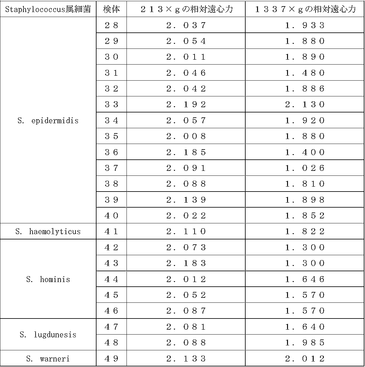
- the identification probability of the species level (score value ⁇ 2.0) of the above eight kinds of Staphylococcus bacteria was obtained by centrifuging a blood culture at a relative centrifugal force of 213 ⁇ g for 1 minute. 100% (S. aureus [16/16], S. capitis [7/7], S. caprae [4/4], S. epidermidis [13] in all species when detected using the supernatant fraction. /13], S. haemolyticus [1/1], S. hominis [5/5], S. lugdunesis [2/2], and S.
- the species-level identification probabilities of the above-mentioned three types of Streptococcus bacteria are detected when using a supernatant fraction obtained by centrifuging a blood culture at a relative centrifugal force of 213 ⁇ g for 1 minute. , 100% in all species (S.dysgalactiae [1/1], S.mitis [1/1], and S.pneumoniae [2/2]), whereas blood culture was subjected to relative centrifugation. 0% (S.dysgalactiae [0/1], S. mitis [0/1] in all species when detected using the supernatant fraction obtained by centrifugation at 1337 xg for 1 minute. ], and S. pneumoniae [0/2]) (see Table 3).
- the species-level identification probabilities of the above-mentioned two types of Bacteroides bacteria are determined by using a supernatant fraction obtained by centrifuging a blood culture at a relative centrifugal force of 213 ⁇ g for 1 minute, 100% (B. fragilis [1/1] and B. vulgatus [1/1]) in all species, whereas blood cultures were centrifuged at a relative centrifugal force of 1337 xg for 1 minute. It was 0% (B. fragilis [0/1] and B. vulgatus [0/1]) in all the species when detected using the supernatant fraction thus obtained (see Table 4).
- the species-level identification probability of the above-mentioned one type of Propionibacterium is the supernatant fraction obtained by centrifuging a blood culture at a relative centrifugal force of 213 ⁇ g for 1 minute.
- the supernatant fraction obtained by centrifuging the blood culture at a relative centrifugal force of 1337 ⁇ g for 1 minute It was 0% (0/1) when detected using (see Table 5).
- the identification probability at the species level of the above-mentioned one type of C. freundii bacterium is the supernatant fraction obtained by centrifuging a blood culture at a relative centrifugal force of 213 ⁇ g for 1 minute. It was 100% (2/2) when detected by using the supernatant fraction obtained by centrifuging the blood culture at a relative centrifugal force of 1337 ⁇ g for 1 minute. When detected, it was 50% (1/2) (see Table 6).
- the species-level identification probabilities of the above three kinds of Enterobacter bacteria are detected when using a supernatant fraction obtained by centrifuging a blood culture at a relative centrifugal force of 213 ⁇ g for 1 minute. , 100% (E.cloacae [1/1], E.faecalis [2/2], and E.faecium [1/1]) in all species, whereas the blood culture was subjected to relative centrifugation.
- the species level identification probability of E.cloacae is 100% (1/1).
- the species-level identification probability of faecalis was 100% (2/2), but the species-level identification probability of E.faecium was 0% (0/1) (see Table 7).
- said 1 type of Clostridium bacterium (C. septicum) said 1 type of Corynebacterium genus (C. striatum), said 1 type of Klebsiella genus bacterium (K. oxytoca), said 1 type of Proteus bacterium (P.mirabilis), the above-mentioned one kind of Providencia bacteria (P. rettgeri), and the above-mentioned one kind of Pseudomonas bacteria (P.
- the identification probability at the species level is that the relative centrifugal force of the blood culture is 213 ⁇ . and the supernatant fraction obtained by centrifuging the blood culture at a relative centrifugal force of 1337 ⁇ g for 1 minute, as detected using the supernatant fraction obtained by centrifuging at 1 g for 1 minute. It was 100% the same as when it was detected using (see Tables 8 to 13).
- Example 3 Comparison between the present detection method and the conventional method The blood culture was hemolyzed when the blood culture was prepared by subjecting the blood culture to a microbial fraction by centrifuging the blood culture at a relative centrifugal force of about 213 ⁇ g for about 1 minute. It was examined whether there is a difference in the level of microorganisms detected by subsequent mass spectrometry as compared with the case where the conventional method of preparing a microbial fraction with an agent is used.
- 3-1 Method 3-1-1 The present detection method The present preparation method was performed according to the following procedures [1] to [5], and then the present detection method was performed according to the following procedures [6] and [7]. .. [1] 3 mL of blood culture was transferred to a 15 mL conical tube, diluted with 3 mL of PBS solution, and then a table top centrifuge 4000 (rotor radius: 13.29, 15 mL conical tube ⁇ 16 tube rack, manufactured by Kubota Shoji Co., Ltd.) was subjected to centrifugal treatment under the conditions of a relative centrifugal force value of 213 ⁇ g (1200 rpm in terms of rotation speed) and room temperature for 1 minute.
- a table top centrifuge 4000 rotor radius: 13.29, 15 mL conical tube ⁇ 16 tube rack, manufactured by Kubota Shoji Co., Ltd.
- Streptococcus bacteria S. agalactiae, S. anginosus, S. dysgalactiae, S. mitis, And S. sanguinis
- P. aeruginosa glucose non-fermenting bacterium
- enterococci E. faecium and E. faecalis
- E. aerogenes K. pneumoniae, P. mirabilis, R. ornithinolytica, and C. freundii
- Bacillus bacteria B.
- C. striatum Corynebacterium
- C. perfringens one type of bacterium belonging to the genus Clostridium
- MALDI Biotyper 3.1 software manufactured by Bruker Daltonics. If the score value is 2.0 or more, the bacterial species level coincides with a high evaluation, and if the score value is 1.7 or more and less than 2.0, the genus level coincides with a high evaluation, and the score value is 1. When it was less than 0.7, it was evaluated as unidentifiable.
- the identification probability at the species level (score value ⁇ 2.0) of the above 8 kinds of Staphylococcus bacteria is 100% (24/24; 100% in S. aureus when the present detection method is used. 10/10], S. epidermidis 100% [6/6], S. capitis 100% [1/1], S. caprae 100% [1/1], S. hominis 100% [1/ 1], 100% [2/2] for S. haemolyticus, 100% [2/2] for S. lugdunesis, and 100% [1/1] for S. warneri. 58.4% (14/24; S. aureus 80% [8/10], S. epidermidis 50% [3/6], S. capitis 100% [1/1], 0% [0/1] for S.
- the amount of Staphylococcus bacteria contained in the microbial fraction was 1 ⁇ 10 5 to 1 ⁇ 10 8 .
- the identification probabilities of the above-mentioned 5 kinds of Streptococcus species at the species level are 71.4% (5/7; S. agalactiae 100% [1/1], S. anginosus when the present detection method is used. Was 100% [1/1], S. dysgalactiae was 100% [2/2], S. mitis was 50% [1/2], and S. sanguinis was 0% [0/1]).
- the conventional method 42.9% (3/7; 100% [1/1] for S. agalactiae, 100% [1/1] for S. anginosus, and 0% for S. dysgalactiae. [0/2], 50% [1/2] for S.
- the identification probability of the genus level (score value ⁇ 1.7) of the above-mentioned 5 kinds of Streptococcus bacteria is 100% (7/7; 100% [1/7 in S. agalactiae when the present detection method is used. 1], 100% [1/1] for S. anginosus, 100% [2/2] for S. dysgalactiae, 100% [2/2] for S. zymitis, and 100% [1/1 for S. sanguinis. ]), when using the conventional method, 71.4% (5/7; 100% [1/1] for S. agalactiae, 100% [1/1] for S. anginosus, It was 50% [1/2] for S. dysgalactiae, 100% [2/2] for S. mitis, and 0% [0/1] for S. sanguinis (see Table 15).
- the identification probability of the above-mentioned one type of glucose non-fermenting bacterium (P. aeruginosa) at the species level was 100% (1/1) when the present detection method was used, whereas the conventional method was used. If it was, it was 0% (0/1) (see Table 16).
- the identification probabilities at the species level of the above-mentioned two types of Enterococcus are 100% (3/3; 100% [2/2] in E.faecium, and E.faecalis in the case of using the present detection method. While it was 100% [1/1]), 66.7% (2/3; 100% [2/2] in E.faecium and 0 in E.faecalis when using the conventional method. % [0/1]) (see Table 17).
- the identification probability of the species level of the bacteria belonging to the above six types of Enterobacteriaceae is 100% (17/17; 100% [10/10] in E. coli, E. coli, when using the present detection method).
- 100% [1/1] for aerogenes, 100% [2/2] for K.pneumoniae, 100% [1/1] for P.mirabilis, 100% [1/1] for R. ornithinolytica, and C. freundii Was 100% [2/2])
- 94.1% (16/17; 17/17; E.
- the identification rate of all bacteria was 90% at the genus level and 72% at the species level when the conventional method was used, whereas when the present detection method was used, the genus level was found. Was 100% and seed level was 96%. Further, hemoglobin, which is one of the blood cell components, was detected when the conventional method was used, whereas it was not detected when the present detection method was used (that is, it was below the detection sensitivity).
- the present preparation method can reduce the presence of microorganisms in blood cultures (especially bacteria with remarkable chain or cluster formation such as Staphylococcus bacteria and Streptococcus bacteria) in comparison with the conventional method. It shows that the microorganisms can be efficiently recovered, and as a result, the microorganisms can be detected with high accuracy.
- the present preparation method can separate the blood cell component and the microorganism by centrifugation for about 1 minute, which is a relatively short time, without using a hemolytic agent. Since it can also be prepared in a relatively short time, it is shown to be excellent in convenience and speed.
- Example 4 Detection of Fungus Using the detection method of the present invention described in Example 3, three types of fungi (Candida albicans], Candida glabrata] contained in the blood culture, When Candida tropcalis was detected, the identification probability at the species level was 75% (67% [2/3] for Candida albicans, 75% [3/4] for Candida glabrata, and 100 for Candida tropcalis). % [1/1]) (see Table 22). This result shows that the detection method of the present invention can also detect a fungus having a specific gravity or a size larger than that of a bacterium, similarly.
- the present invention contributes to diagnosis of an infectious disease of a microorganism in a subject (particularly, a bacterium or fungus having a striking chain or cluster formation such as a staphylococcal bacterium or a streptococcal bacterium), and early prevention or early treatment of the infectious disease. Is.
Landscapes
- Health & Medical Sciences (AREA)
- Life Sciences & Earth Sciences (AREA)
- Chemical & Material Sciences (AREA)
- Engineering & Computer Science (AREA)
- Immunology (AREA)
- Biochemistry (AREA)
- General Health & Medical Sciences (AREA)
- Organic Chemistry (AREA)
- Analytical Chemistry (AREA)
- Biomedical Technology (AREA)
- Biotechnology (AREA)
- Wood Science & Technology (AREA)
- Zoology (AREA)
- Molecular Biology (AREA)
- Physics & Mathematics (AREA)
- Bioinformatics & Cheminformatics (AREA)
- Microbiology (AREA)
- Medicinal Chemistry (AREA)
- Urology & Nephrology (AREA)
- Hematology (AREA)
- General Physics & Mathematics (AREA)
- Pathology (AREA)
- Proteomics, Peptides & Aminoacids (AREA)
- Food Science & Technology (AREA)
- General Engineering & Computer Science (AREA)
- Genetics & Genomics (AREA)
- Biophysics (AREA)
- Toxicology (AREA)
- Cell Biology (AREA)
- Virology (AREA)
- Tropical Medicine & Parasitology (AREA)
- Sustainable Development (AREA)
- Investigating Or Analysing Biological Materials (AREA)
- Measuring Or Testing Involving Enzymes Or Micro-Organisms (AREA)
- Other Investigation Or Analysis Of Materials By Electrical Means (AREA)
Abstract
本発明の課題は、簡便性や迅速性に優れ、かつ、血液試料中の微生物を精度よく検出できる方法を提供することにある。 被験者から採取された血液試料の培養物を、213×g前後の相対遠心力で1分間前後、遠心処理し、上清画分と沈殿画分とに分離する工程(a);工程(a)で得られた上清画分から微生物画分を得る工程(b);及び前記微生物画分に微生物が含まれているか否かを、質量分析計を用いて解析する工程(c);の工程(a)~(c)を順次含む方法を用いると、前記被験者が特定の微生物に感染しているか否かを精度よく検出することができる。
Description
本発明は、被験者由来血液試料中の微生物の有無を検出する方法;被験者由来の、微生物を含む試料を調製する方法;被験者由来血液試料中の微生物の有無を検出するためのシステム(装置等の手段の組合せ、以下同じ);被験者由来の、微生物を含む試料を調製するためのシステム;及び、微生物を含む試料;に関する。
微生物感染症、特に敗血症のように、細菌や真菌(以下、「細菌等」ということがある)による重度の感染症の場合、早期に適切な抗菌薬を投与することが、感染症の治癒や良好な予後の面から重要である。細菌等による感染症に対しては、起炎菌を想定し効果のある抗菌薬を選択し、経験的治療が施される。それと同時に、起炎菌を明らかにするための血液培養検査が実施されるが、起炎菌が明らかになるまで数日間を要する。このため、迅速に起炎菌を明らかにし、経験的治療を根拠ある抗菌薬適正使用とすることが求められていた。
最近、細菌タンパク質又は全細菌について、マトリックス支援レーザー脱離イオン化飛行時間型質量分析計(MALDI-TOF MS)を用いた質量分析法を行うと、細菌の属、種、及び株固有のバイオマーカーを生成できることが報告されている(特許文献1)。また、尿を2000×gで30秒間の低速回転の遠心処理を行うことにより、細菌と、生体内由来の細胞(白血球)とを比重の違いでそれぞれ上清画分と沈殿画分に分離し、MALDI-TOFMSを用いた質量分析法を行い、細菌を同定する方法が報告されている(非特許文献1)。さらに、比重やサイズが他の細菌よりも大きい連鎖やクラスター形成の著しい細菌は、低速回転の遠心処理では生体内由来の細胞と分離することができないため、遠心処理前に、細菌の連鎖やクラスター形成を解消することを目的とし、超音波発生装置を使用する方法が知られている(非特許文献2)。
一方、本発明者らは、塩化アンモニウムにより溶血処理した被験者由来血液培養物を、細菌を通過させない孔径を有する濾過膜で濾過処理し、濾過膜に捕捉された残渣を回収して得られた残渣試料中の細菌を、質量分析計を用いて解析すると、他の溶血剤(例えば、NP-40、Tween 20、Triton X-100)を用いて溶血処理した場合と比べ、前記残渣試料中に、細菌が含まれているか否かを、感度よく検出することができることを見出している(特願2018-61903)。
J Clin Microbiol 48: 2110-2115.
日本臨床微生物学会誌 Vol.26 No.2 79-89 2016
本発明の課題は、簡便性や迅速性に優れ、かつ、血液試料中の微生物を精度よく検出できる方法等を提供することにある。
本発明者らは、溶血剤を用いずに、血液培養物中の微生物と血球成分との分離する方法について検討を行った。高速回転の遠心操作では、多くの細菌は血球成分とともに沈殿するため、両者を分離できないことが予想され、また、低速回転の遠心操作では、前述のとおり、比重やサイズが他の細菌よりも大きい連鎖やクラスター形成の著しい細菌や真菌は、血球成分と分離できないことが懸念されたが、相対遠心力と遠心時間の検討を重ねたところ、血液培養物を、213×g前後の相対遠心力で1分間前後、遠心処理すると、遠心処理後の上清画分に細菌(特に、ブドウ球菌属細菌や連鎖球菌属細菌等の連鎖やクラスター形成の著しい細菌)や真菌が含まれているか否かを、質量分析により精度よく検出することができることを見出した。
すなわち、本発明は、以下のとおりである。
〔1〕以下の工程(a)~(c)を含むことを特徴とする、血液試料中の微生物の検出方法。
(a)被験者から採取された血液試料の培養物(以下、「被験者由来血液培養物」ということがある)を、170~800×gの相対遠心力で40~80秒間、遠心処理し、上清画分と沈殿画分とに分離する工程;
(b)工程(a)で得られた上清画分から微生物画分を得る工程;
(c)前記微生物画分に微生物が含まれているか否かを解析する工程;
〔2〕工程(c)において、質量分析計を用いて解析することを特徴とする上記〔1〕に記載の方法。
〔3〕質量分析計が、マトリックス支援レーザー脱離イオン化飛行時間型質量分析計(MALDI-TOF MS)であることを特徴とする上記〔2〕に記載の方法。
〔4〕以下の工程(a)及び(b)を含むことを特徴とする、微生物を含む試料の調製方法。
(a)被験者から採取された血液試料の培養物を、170~800×gの相対遠心力で40~80秒間、遠心処理し、上清画分と沈殿画分とに分離する工程;
(b)工程(a)で得られた上清画分から微生物画分を得る工程;
〔5〕試料が、質量分析用試料であることを特徴とする上記〔4〕に記載の方法。
〔6〕工程(b)において、以下の工程(p)及び(q)を実施することにより微生物画分を得ることを特徴とする上記〔1〕~〔5〕のいずれかに記載の方法。
(p)工程(a)で得られた上清画分を、微生物を通過させない孔径を有する濾過膜で濾過処理する工程;
(q)前記濾過処理後の濾過膜に捕捉された残渣を回収し、微生物画分として得る工程;
〔7〕工程(q)において、濾過処理後の濾過膜を純水中に浸し、遠心分離により残渣を回収し、微生物画分として得ることを特徴とする上記〔6〕に記載の方法。
〔8〕工程(p)と工程(q)の間に、濾過処理後の濾過膜を、純水を用いて洗浄する工程(r)を含むことを特徴とする上記〔6〕又は〔7〕に記載の方法。
〔9〕微生物が、細菌又は真菌であることを特徴とする上記〔1〕~〔8〕のいずれかに記載の方法。
〔10〕以下の(A)~(C)の手段を備えることを特徴とする、血液試料中の微生物の検出システム。
(A)被験者から採取された血液試料の培養物を、170~800×gの相対遠心力で40~80秒間の条件下で、上清画分と沈殿画分とに分離する遠心手段;
(B)上清画分に基づく、微生物画分の取得手段;
(C)微生物画分に微生物が含まれているか否かを解析する分析手段;
〔11〕分析手段が、質量分析手段であることを特徴とする上記〔10〕に記載のシステム。
〔12〕質量分析手段が、マトリックス支援レーザー脱離イオン化飛行時間型質量分析計(MALDI-TOF MS)であることを特徴とする上記〔11〕に記載のシステム。
〔13〕以下の(A)及び(B)の手段を備えることを特徴とする、微生物を含む試料の調製システム。
(A)被験者から採取された血液試料の培養物を、170~800×gの相対遠心力で40~80秒間の条件下で、上清画分と沈殿画分とに分離する遠心手段;
(B)上清画分に基づく、微生物画分の取得手段;
〔14〕試料が、質量分析用試料であることを特徴とする上記〔13〕に記載のシステム。
〔15〕微生物画分の取得手段が、微生物を通過させない孔径を有する濾過膜であることを特徴とする上記〔10〕~〔14〕に記載のシステム。
〔16〕微生物が、細菌又は真菌であることを特徴とする上記〔10〕~〔15〕のいずれかに記載のシステム。
〔17〕少なくとも1×10⁵個のブドウ球菌属細菌を含み、かつ、ヘモグロビンを実質的に含まないことを特徴とする、微生物を含む試料。
〔18〕質量分析用試料であることを特徴とする上記〔17〕に記載の試料。
〔1〕以下の工程(a)~(c)を含むことを特徴とする、血液試料中の微生物の検出方法。
(a)被験者から採取された血液試料の培養物(以下、「被験者由来血液培養物」ということがある)を、170~800×gの相対遠心力で40~80秒間、遠心処理し、上清画分と沈殿画分とに分離する工程;
(b)工程(a)で得られた上清画分から微生物画分を得る工程;
(c)前記微生物画分に微生物が含まれているか否かを解析する工程;
〔2〕工程(c)において、質量分析計を用いて解析することを特徴とする上記〔1〕に記載の方法。
〔3〕質量分析計が、マトリックス支援レーザー脱離イオン化飛行時間型質量分析計(MALDI-TOF MS)であることを特徴とする上記〔2〕に記載の方法。
〔4〕以下の工程(a)及び(b)を含むことを特徴とする、微生物を含む試料の調製方法。
(a)被験者から採取された血液試料の培養物を、170~800×gの相対遠心力で40~80秒間、遠心処理し、上清画分と沈殿画分とに分離する工程;
(b)工程(a)で得られた上清画分から微生物画分を得る工程;
〔5〕試料が、質量分析用試料であることを特徴とする上記〔4〕に記載の方法。
〔6〕工程(b)において、以下の工程(p)及び(q)を実施することにより微生物画分を得ることを特徴とする上記〔1〕~〔5〕のいずれかに記載の方法。
(p)工程(a)で得られた上清画分を、微生物を通過させない孔径を有する濾過膜で濾過処理する工程;
(q)前記濾過処理後の濾過膜に捕捉された残渣を回収し、微生物画分として得る工程;
〔7〕工程(q)において、濾過処理後の濾過膜を純水中に浸し、遠心分離により残渣を回収し、微生物画分として得ることを特徴とする上記〔6〕に記載の方法。
〔8〕工程(p)と工程(q)の間に、濾過処理後の濾過膜を、純水を用いて洗浄する工程(r)を含むことを特徴とする上記〔6〕又は〔7〕に記載の方法。
〔9〕微生物が、細菌又は真菌であることを特徴とする上記〔1〕~〔8〕のいずれかに記載の方法。
〔10〕以下の(A)~(C)の手段を備えることを特徴とする、血液試料中の微生物の検出システム。
(A)被験者から採取された血液試料の培養物を、170~800×gの相対遠心力で40~80秒間の条件下で、上清画分と沈殿画分とに分離する遠心手段;
(B)上清画分に基づく、微生物画分の取得手段;
(C)微生物画分に微生物が含まれているか否かを解析する分析手段;
〔11〕分析手段が、質量分析手段であることを特徴とする上記〔10〕に記載のシステム。
〔12〕質量分析手段が、マトリックス支援レーザー脱離イオン化飛行時間型質量分析計(MALDI-TOF MS)であることを特徴とする上記〔11〕に記載のシステム。
〔13〕以下の(A)及び(B)の手段を備えることを特徴とする、微生物を含む試料の調製システム。
(A)被験者から採取された血液試料の培養物を、170~800×gの相対遠心力で40~80秒間の条件下で、上清画分と沈殿画分とに分離する遠心手段;
(B)上清画分に基づく、微生物画分の取得手段;
〔14〕試料が、質量分析用試料であることを特徴とする上記〔13〕に記載のシステム。
〔15〕微生物画分の取得手段が、微生物を通過させない孔径を有する濾過膜であることを特徴とする上記〔10〕~〔14〕に記載のシステム。
〔16〕微生物が、細菌又は真菌であることを特徴とする上記〔10〕~〔15〕のいずれかに記載のシステム。
〔17〕少なくとも1×10⁵個のブドウ球菌属細菌を含み、かつ、ヘモグロビンを実質的に含まないことを特徴とする、微生物を含む試料。
〔18〕質量分析用試料であることを特徴とする上記〔17〕に記載の試料。
また本発明の実施の他の形態として、
上記工程(a)~(c)を含み、さらに、上記工程(c)の後、前記微生物画分に微生物が含まれていた場合、前記被験者は、前記微生物に感染している可能性が高いと診断し、前記微生物画分に微生物が含まれていない場合、前記被験者は、前記微生物に感染している可能性が低いと診断する工程(x);を含む、被験者が微生物に感染しているか否かを診断する方法;や、
上記工程(a)~(c)、及び(x)を含み、さらに、微生物に感染している可能性が高いと診断された被験者に対して、当該微生物に対する抗生物質やワクチン等の薬剤を用いて治療処理を施す工程(y)を含む、被験者が微生物に感染しているか否かを診断し、微生物感染症を予防又は治療する方法;
を挙げることができる。
上記工程(a)~(c)を含み、さらに、上記工程(c)の後、前記微生物画分に微生物が含まれていた場合、前記被験者は、前記微生物に感染している可能性が高いと診断し、前記微生物画分に微生物が含まれていない場合、前記被験者は、前記微生物に感染している可能性が低いと診断する工程(x);を含む、被験者が微生物に感染しているか否かを診断する方法;や、
上記工程(a)~(c)、及び(x)を含み、さらに、微生物に感染している可能性が高いと診断された被験者に対して、当該微生物に対する抗生物質やワクチン等の薬剤を用いて治療処理を施す工程(y)を含む、被験者が微生物に感染しているか否かを診断し、微生物感染症を予防又は治療する方法;
を挙げることができる。
近年、血液培養物の検体数が増加傾向ある状況において、血液培養物を、本発明の方法を用いて、213×g前後の相対遠心力で1分間前後と比較的短時間、遠心処理することにより、多検体の血液培養物であっても、迅速かつ効率的に処理することができる。また、上記遠心処理により、血液培養物中の微生物(特に、ブドウ球菌属細菌、連鎖球菌属細菌等の連鎖及び/又はクラスター形成の著しい細菌や真菌)と、血球成分とを精度よく分離することができるため、分離後の微生物を含む上清画分を基に、分析用サンプルを調製し、質量分析計等の分析手段を用いて解析すると、血液培養物中に特定の微生物が含まれているか否かを精度よく判定・評価することができる。特に、溶血剤を用いずに本発明の方法を実施すると、溶血剤により微生物に与えるダメージがないため、より高効率に微生物を回収し、より精度よく微生物を同定することができる。以上のことから、本発明によると、被験者が特定の微生物に感染しているか否かを診断する上で有用なデータを提供することができるため、微生物感染症の診断の他、微生物感染症の早期予防や早期治療が期待される。
また、本発明において、遠心処理後の微生物を含む上清画分を、微生物を通過させない孔径を有する濾過膜で濾過処理すると、濾過処理は、シリンジを用いた手動操作により行うことが可能であるため、真空ポンプ等の大掛かりな機器がなくても、本発明を実施することができる。
敗血症を診断する血液培養検査は、被験者から採取された血液試料を、培養液中で培養し、血液培養物の濁度の変化、細菌等による培養液中のガス発生、pHの変化等を指標にして、1~7日間、細菌等の増殖の有無を確認することから行われる。次いで、細菌等の増殖が確認された場合、血液培養物を採取し、グラム染色とサブカルチャー(二次培養)が行われ、グラム染色による大まかな細菌等の種の分類結果から、経験的治療に使用した抗菌薬の評価が行われる。一方、本発明の検出法を用いると、細菌等を効率よく集菌し、細菌等の種同定を迅速かつ正確に行うことができるため、起炎菌の細菌等の種を正確に同定することが可能となり、抗菌薬適正治療につながる。このため、本発明は、細菌等の感染症患者の予後の改善、耐性細菌等の増加抑制、コスト削減等に貢献できる。
本発明の血液試料中の微生物の検出方法としては、被験者由来血液培養物を、170~800×gの相対遠心力(RCF;relative centrifugal force;相対遠心加速度ともいう)で40~80秒間、遠心処理し、上清画分と沈殿画分とに分離する工程(a);前記工程(a)で得られた上清画分から微生物画分を得る工程(b);及び前記微生物画分に微生物が含まれているか否かを解析する工程(c);の工程(a)~(c)を順次含む方法(以下、「本件検出法」ということがある)であれば特に制限されず、本件検出法は、前記被験者由来の血液試料中に、微生物が存在するか否かを検出する方法であって、医師による診断行為を含まない。また、本発明の微生物を含む試料の調製方法としては、上記工程(a)及び(b)を順次含み、微生物を含む試料を調製する方法(以下、「本件調製法」ということがある)であれば特に制限されない。なお、本件検出法及び本件調製法を総称して、以下、「本件方法」ということがある。
本発明の血液試料中の微生物の検出システムとしては、被験者由来血液培養物を、170~800×gの相対遠心力で40~80秒間の条件下で、上清画分と沈殿画分とに分離する遠心手段(A);上清画分に基づく、微生物画分の取得手段(B);及び微生物画分に微生物が含まれているか否かを解析する分析手段(C);の(A)~(C)の手段を備えるシステム(以下、「本件検出システム」ということがある)であれば特に制限されず、また、本発明の微生物を含む試料の調製システムとしては、上記工程(A)及び(B)の手段を備えるシステム(以下、「本件調製システム」ということがある)であれば特に制限されない。なお、本件検出システム及び本件調製システムを総称して、以下、「本件システム」ということがある。
本明細書において、「少なくとも1×10⁵個のブドウ球菌属細菌」には、例えば、少なくとも1×10⁶個のブドウ球菌属細菌、少なくとも1×10⁷個のブドウ球菌属細菌、少なくとも1×10⁸個のブドウ球菌属細菌等が含まれる。
本明細書において、「ヘモグロビンを実質的に含まない」とは、ヘモグロビンをほとんど又は全く含まないことを意味し、より具体的には、微生物を含む試料(すなわち、微生物画分)の一部又は全部を、MALDI-TOFMSを用いた質量分析法を行いて検出・定量した場合、1又は2種以上のヘモグロビン由来のピークが、ほとんど検出されないこと(例えば、5mg/mL以下の検出)又は全く検出されないこと(すなわち、検出感度以下であること)を意味する。
上記被験者としては、特に制限されず、例えば、微生物感染の有無が不明な者(例えば、健常者;がん患者、糖尿病患者等の非微生物感染症患者)や、特定の微生物に感染しているか否かが不明な者(例えば、健常者;敗血症患者、上気道感染症患者等の微生物感染症患者)を挙げることができる。
[本件方法における工程(a)について]
上記工程(a)において、170~800×gのRCFで40~80秒間、遠心処理を行うことにより、被験者由来血液培養物中に含まれる微生物の一部又は大部分と、血球成分の一部又は大部分とが、それぞれ上清画分と沈殿画分とに分離される。被験者由来血液培養物は任意の遠心機(例えば、遠心管が所定の角度で配置されているアングルロータ式遠心機;や、遠心管の角度が可変であり遠心処理中に遠心管が水平又はほぼ水平にあるスイングロータ式遠心機;)を用いて遠心処理することができる。遠心機は、後述する本実施例で用いたテーブルトップ遠心機4000(久保田商事社製)、ベックマンGPR(himac CF7D2、HITACHI社製)、卓上低速遠心機(LT-015、TOMY社製)等の市販品を用いることができる。そして、本件システムにおける遠心手段としては、被験者由来血液培養物を、170~800×gの相対遠心力で40~80秒間の条件下で、上清画分と沈殿画分とに分離できる手段、例えば、前述した、任意の遠心機や市販品の遠心機を挙げることができる。
上記工程(a)において、170~800×gのRCFで40~80秒間、遠心処理を行うことにより、被験者由来血液培養物中に含まれる微生物の一部又は大部分と、血球成分の一部又は大部分とが、それぞれ上清画分と沈殿画分とに分離される。被験者由来血液培養物は任意の遠心機(例えば、遠心管が所定の角度で配置されているアングルロータ式遠心機;や、遠心管の角度が可変であり遠心処理中に遠心管が水平又はほぼ水平にあるスイングロータ式遠心機;)を用いて遠心処理することができる。遠心機は、後述する本実施例で用いたテーブルトップ遠心機4000(久保田商事社製)、ベックマンGPR(himac CF7D2、HITACHI社製)、卓上低速遠心機(LT-015、TOMY社製)等の市販品を用いることができる。そして、本件システムにおける遠心手段としては、被験者由来血液培養物を、170~800×gの相対遠心力で40~80秒間の条件下で、上清画分と沈殿画分とに分離できる手段、例えば、前述した、任意の遠心機や市販品の遠心機を挙げることができる。
本明細書において、RCFは、下記式(1)を基に算出することができる。
RCF(×g)=1118×R×N2×10-8・・・(1)
[式中、「R」は回転半径(cm)を示し、「N」は1分間当たりの回転数(rpm=min-1)を示す。]
RCF(×g)=1118×R×N2×10-8・・・(1)
[式中、「R」は回転半径(cm)を示し、「N」は1分間当たりの回転数(rpm=min-1)を示す。]
上記工程(a)において、式(1)中の回転半径Rとして、回転軸から遠心処理中の遠心管までの半径のうち、最大となる半径(すなわち、最大半径[Rmax])と、最小となる半径(すなわち、最小半径[Rmin])とを基に算出した平均半径(Rav=[最大Rmax+Rmin]/2)の値を用いて、RCFが170~800×gの範囲内となるように回転数Nを設定し、遠心処理を行う。
上記RCFとしては、170~800×gの範囲内であればよく、例えば、170~700×g、170~600×g、170~500×g、170~400×g、170~300×g、170~250×g、170~230×g、170~220×g、175~800×g、180~800×g、185~800×g、190~800×g、195~800×g、200~800×g、175~700×g、180~700×g、185~700×g、190~700×g、195~700×g、200~700×g、175~700×g、175~600×g、175~500×g、175~400×g、175~300×g、175~250×g、175~230×g、175~220×g、180~700×g、180~600×g、180~500×g、180~400×g、180~300×g、180~250×g、180~230×g、180~220×g、185~700×g、185~600×g、185~500×g、185~400×g、185~300×g、185~250×g、185~230×g、185~220×g、190~700×g、190~600×g、190~500×g、190~400×g、190~300×g、190~250×g、190~230×g、190~220×g、195~700×g、195~600×g、195~500×g、195~400×g、195~300×g、195~250×g、195~230×g、195~220×g、200~700×g、200~600×g、200~500×g、200~400×g、200~300×g、200~250×g、200~230×g、200~220×g等を挙げることができる。
上記工程(a)における遠心処理時間としては、40~80秒間の範囲内であればよく、例えば、40~76秒間、40~73秒間、40~70秒間、40~66秒間、40~63秒間、43~80秒間、46~80秒間、50~80秒間、53~80秒間、56~80秒間、43~76秒間、46~76秒間、50~76秒間、53~76秒間、56~76秒間、43~73秒間、46~73秒間、50~73秒間、53~73秒間、56~73秒間、43~70秒間、46~70秒間、50~70秒間、53~70秒間、56~70秒間、43~66秒間、46~66秒間、50~66秒間、53~66秒間、56~66秒間、43~63秒間、46~63秒間、50~63秒間、53~63秒間、56~63秒間等を挙げることができる。
遠心処理中の温度は、通常0~40℃の範囲内であり、室温(例えば、10~30℃)を好適に例示することができる。
被験者由来血液培養物は、例えば、被験者から採取された血液試料を、培養温度35℃の条件下で12~48時間培養することにより得ることができる。
上記工程(a)における遠心処理により、被験者由来血液培養物中の血球成分(例えば、ヘモグロビン)は、その後の工程(b)及び/又は工程(c)の操作に妨げのならない程度まで除去することができる。このため、上記工程(a)で得られた上清画分を、溶血剤を用いて溶血処理しなくてもよいが、上記工程(a)で得られた上清画分中に含まれるおそれがある血球成分を溶血処理し、血液試料中の微生物の検出精度をより高めるために、上記工程(b)の前に、上記工程(a)で得られた上清画分を、溶血剤と混合し、溶血処理してもよい。また、上記工程(a)において、予め溶血剤で溶血処理した被験者由来血液培養物を遠心処理してもよい。
上記溶血剤としては、例えば、本発明者らの先願(特願2018-61903)に記載の塩化アンモニウムの他、界面活性剤、低張液等を挙げることができる。ここで、塩化アンモニウム以外の溶血剤としては、例えば、界面活性剤、低張液等を挙げることができる。かかる界面活性剤としては、Triton X-100(Tritonは登録商標)(ポリオキシエチレン(10)オクチルフェニルエーテル)、Triton X-114(ポリオキシエチレン(8)オクチルフェニルエーテル)、Triton X-405(ポリオキシエチレン(40)イソオクチルフェニルエーテル)、NP-40(Nonidet P-40)(ポリオキシエチレン(9)オクチルフェニルエーテル)等のポリオキシエチレンp-t-オクチルフェニルエーテル(Triton系界面活性剤);Tween 20(Tweenは登録商標)、Tween 40、Tween 60、Tween 80、Tween65、Tween 85等のポリオキシエチレンソルビタン脂肪酸エステル(Tween系界面活性剤);Briji 35(Brijiは登録商標)(ポリオキシエチレン(23)ラウリルエーテル)等のポリオキシエチレンアルキルエーテル(Briji系界面活性剤);Dodecyl-β-D-maltose;Octyl-β-D-glucoside等の非イオン性界面活性剤やドデシル硫酸ナトリウム(SDS)等の陰イオン性界面活性剤や塩化ベンザルコニウム、塩化ベンゼトニウム、ジデシルジメチルアンモニウム塩、ドデシルトリメチルアンモニウムクロリド等の陽イオン性界面活性剤やCHAPS(3-(3-cholamidepropyl)dimethylammonio-1-propanesulphonate)、塩化アルキルポリアミノエチルグリシン等の両性界面活性剤を挙げることができる。
上記低張液としては、体液(例えば血漿)や細胞液の浸透圧よりも低くなるようにナトリウムやカリウムなどによって塩や糖濃度等を調整した低張液であれば特に制限されない。本明細書において「低張」とは、浸透圧が250mOsm/Lよりも低いことを意味する。
[本件方法における工程(b)について]
上記工程(a)で得られた上清画分は、血球成分の一部又は大部分が除去された被験者由来血液培養物である。上記工程(b)においては、かかる上清画分中に含まれる微生物を分離し、微生物画分(すなわち、微生物を含む試料)を得る。上清画分から微生物を分離する方法としては、非溶解成分である微生物と、溶解成分とを分離できる方法であればよく、例えば、上記工程(a)で得られた上清画分を遠心処理により、沈殿画分と、上清画分とに分離し、沈殿画分を微生物画分として得る方法や、上記工程(a)で得られた上清画分を、微生物を通過させない孔径を有する濾過膜で濾過処理する工程(p)と、前記濾過処理後の濾過膜に捕捉された残渣を回収し、微生物画分として得る工程(q)とを実施することにより微生物画分を得る方法を挙げることができ、これらの中でも上記工程(p)及び(q)を実施することにより微生物画分を得る方法を好適に例示することができる。本明細書において、「微生物を含む試料」は、液体試料であっても、固体試料(例えば、液体試料を乾燥処理したもの)あってもよい。そして、本件システムにおける微生物画分の取得手段としては、上清画分から微生物画分を得ることができる手段、例えば、前述した、任意の遠心機や市販品の遠心機や、微生物を通過させない孔径を有する濾過膜を挙げることができ、微生物を通過させない孔径を有する濾過膜を好適に例示することができる。
上記工程(a)で得られた上清画分は、血球成分の一部又は大部分が除去された被験者由来血液培養物である。上記工程(b)においては、かかる上清画分中に含まれる微生物を分離し、微生物画分(すなわち、微生物を含む試料)を得る。上清画分から微生物を分離する方法としては、非溶解成分である微生物と、溶解成分とを分離できる方法であればよく、例えば、上記工程(a)で得られた上清画分を遠心処理により、沈殿画分と、上清画分とに分離し、沈殿画分を微生物画分として得る方法や、上記工程(a)で得られた上清画分を、微生物を通過させない孔径を有する濾過膜で濾過処理する工程(p)と、前記濾過処理後の濾過膜に捕捉された残渣を回収し、微生物画分として得る工程(q)とを実施することにより微生物画分を得る方法を挙げることができ、これらの中でも上記工程(p)及び(q)を実施することにより微生物画分を得る方法を好適に例示することができる。本明細書において、「微生物を含む試料」は、液体試料であっても、固体試料(例えば、液体試料を乾燥処理したもの)あってもよい。そして、本件システムにおける微生物画分の取得手段としては、上清画分から微生物画分を得ることができる手段、例えば、前述した、任意の遠心機や市販品の遠心機や、微生物を通過させない孔径を有する濾過膜を挙げることができ、微生物を通過させない孔径を有する濾過膜を好適に例示することができる。
遠心処理により微生物画分を得る場合、上清画分中に含まれる微生物が十分沈殿するように、RCF及び時間を設定し、遠心処理を行う。遠心処理におけるRCFは、例えば、3000×g以上であり、好ましくは6000×g以上、より好ましくは9000×g以上である。また、遠心時間は、例えば、3分以上であり、好ましくは5分以上、より好ましくは10分以上である。
[本件方法における工程(p)について]
上記工程(p)の濾過処理により、溶解成分は、濾過膜を通過する。このため、上記工程(a)で得られた上清画分中に微生物が含まれる場合、微生物は、残渣として濾過膜に捕捉され、上記工程(a)で得られた上清画分の溶解成分と分離することができる。
上記工程(p)の濾過処理により、溶解成分は、濾過膜を通過する。このため、上記工程(a)で得られた上清画分中に微生物が含まれる場合、微生物は、残渣として濾過膜に捕捉され、上記工程(a)で得られた上清画分の溶解成分と分離することができる。
上記濾過処理する方法としては、例えば、上記工程(a)で得られた上清画分を、濾過膜を備え、プラスチック、金属、ガラス等の材質により製造された容器内に移し、シリンジ、真空ポンプ、アスピレーター、コンプレッサー等の機器を用いて吸引又は加圧することにより濾過処理する方法や、濾過膜を備え、プラスチック、金属、ガラス等の材質により製造された遠沈管又は試験管内に移し、遠心することにより濾過処理する方法を挙げることができ、大掛かりな機器を使用しないで、より簡便に濾過処理する観点から、シリンジを手動操作することにより加圧又は減圧し、濾過処理する方法や、遠心することにより濾過処理する方法が好ましい。また、微生物のコンタミネーションを防ぐ観点から、濾過処理は密閉系で行うことが好ましく、濾過膜は、ディスポーザブルのものが好ましい。
上記濾過膜の孔径としては、微生物を通過させない孔径であればよく、例えば、微生物が細菌の場合、通常1.5μm以下、好ましくは1.2μm以下、より好ましくは0.8μm以下、さらに好ましくは0.7μm以下、さらにより好ましくは0.6μm以下、最も好ましくは0.5μm以下であり、ヘモグロビン等の血球成分による目詰まりを回避する観点から、通常0.1μm以上、好ましくは0.2μm以上、より好ましくは0.25μm以上、さらに好ましくは0.3μm以上、さらにより好ましくは0.35μm以上、最も好ましくは0.4μm以上である。したがって、細菌の濾過処理に使用する濾過膜の孔径としては、通常0.1~1.5μmの範囲内、好ましくは0.2~1.2μm、より好ましくは0.25~0.8μm、さらに好ましくは0.3~0.7μm、さらにより好ましくは0.35~0.6μm、最も好ましくは0.4~0.5μmである。また、微生物が真菌の場合、上記濾過膜の孔径としては、通常15μm以下、好ましくは12μm以下、より好ましくは8μm以下、さらに好ましくは7μm以下、さらにより好ましくは6μm以下、最も好ましくは5μm以下であり、ヘモグロビン等の血球成分による目詰まりを回避する観点から、通常0.1μm以上、好ましくは0.2μm以上、より好ましくは0.25μm以上、さらに好ましくは0.3μm以上、さらにより好ましくは0.35μm以上、最も好ましくは0.4μm以上である。したがって、真菌の濾過処理に使用する濾過膜の孔径としては、通常0.1~15μmの範囲内、好ましくは0.2~12μm、より好ましくは0.25~8μm、さらに好ましくは0.3~7μm、さらにより好ましくは0.35~6μm、最も好ましくは0.4~5μmである。
上記濾過膜の材質としては、例えば、セルロース混合エステル(MCE;Mixed cellulose esters)、ポリビニリデンフロライド(PVDF)、ポリテトラフルオロエチレン(PTFE)、親水性PTFE、ポリエーテルスルホン(PES)、親水性ポリエーテルスルホン(hydrophilic polyethersulfone)、親水性ポリプロピレン(GHP)、ナイロン(NYL)、セルロースアセテート(CA;cellulose acetate)、ポリスルフォン(PSF)、アクリル系共重合体(acrylic copolymer)、ポリアミド、ナイロン6,6、ポリエステル、ポリカーボネート、ニトロセルロース、ニトロセルロースとセルロースエステルの混合物等を挙げることができる。
[本件方法における工程(q)について]
上記工程(q)において、残渣を回収し、微生物画分として得る方法としては、工程(p)の濾過処理後の濾過膜から、残渣を遊離させ、回収できる方法であればよく、薬さじ等を用いて上記濾過膜に捕捉された残渣を掻き取る方法であってもよいが、残渣に含まれる微生物の回収効率の観点から、上記濾過膜の一部又は全部を純水中に浸し、濾過膜及び/又は純水を撹拌した後、残渣を含む水から遠心分離により残渣を回収する方法が好ましく、ここで遠心分離は、微生物等のコンタミネーションを防ぐ観点から、密閉系で行うことが好ましい。また、残渣を回収する前に、上記濾過膜を、純水を用いて洗浄することが好ましい。洗浄回数としては、少なくとも1回であればよく、複数回(2、3、4回等)であってもよいが、時間対効果及び費用対効果を考慮すると、1回が好ましい。また、濾過膜の洗浄は、工程(p)の濾過処理後の濾過膜に純水を添加し、濾過処理と同様の吸引又は加圧方法により、純水を濾過膜内に通過させることにより行うことができる。
上記工程(q)において、残渣を回収し、微生物画分として得る方法としては、工程(p)の濾過処理後の濾過膜から、残渣を遊離させ、回収できる方法であればよく、薬さじ等を用いて上記濾過膜に捕捉された残渣を掻き取る方法であってもよいが、残渣に含まれる微生物の回収効率の観点から、上記濾過膜の一部又は全部を純水中に浸し、濾過膜及び/又は純水を撹拌した後、残渣を含む水から遠心分離により残渣を回収する方法が好ましく、ここで遠心分離は、微生物等のコンタミネーションを防ぐ観点から、密閉系で行うことが好ましい。また、残渣を回収する前に、上記濾過膜を、純水を用いて洗浄することが好ましい。洗浄回数としては、少なくとも1回であればよく、複数回(2、3、4回等)であってもよいが、時間対効果及び費用対効果を考慮すると、1回が好ましい。また、濾過膜の洗浄は、工程(p)の濾過処理後の濾過膜に純水を添加し、濾過処理と同様の吸引又は加圧方法により、純水を濾過膜内に通過させることにより行うことができる。
本明細書において、「純水」とは、不純物やイオン性物質を除去した水のことを意味し、具体的には、電気伝導率が24~26℃条件下で、10μS/cm以下の水を意味する。純水は、水道水を、蒸留及び/又はイオン交換することにより、調製することができる。
[本件検出法における工程(c)について]
上記工程(c)において、解析する方法としては、微生物の有無を検出・定量できる方法であれば特に制限されず、例えば、質量分析計を用いて解析(より具体的には、微生物由来のタンパク質を検出・定量)する方法;リアルタイムPCR装置、次世代シークエンサー[NGS;Next Generation Sequencer]等のDNA分析手段を用いて解析(より具体的には、微生物由来のゲノムDNAを検出・定量)する方法;近赤外分光測定装置等の成分分析手段を用いて解析(より具体的には、微生物由来の成分を検出・定量)する方法;などを挙げることができ、これらの中でも質量分析計を用いて解析する方法を好適に例示することができる。
上記工程(c)において、解析する方法としては、微生物の有無を検出・定量できる方法であれば特に制限されず、例えば、質量分析計を用いて解析(より具体的には、微生物由来のタンパク質を検出・定量)する方法;リアルタイムPCR装置、次世代シークエンサー[NGS;Next Generation Sequencer]等のDNA分析手段を用いて解析(より具体的には、微生物由来のゲノムDNAを検出・定量)する方法;近赤外分光測定装置等の成分分析手段を用いて解析(より具体的には、微生物由来の成分を検出・定量)する方法;などを挙げることができ、これらの中でも質量分析計を用いて解析する方法を好適に例示することができる。
上記「質量分析計を用いて解析する方法」としては、上記工程(b)で得られた微生物画分を、イオン源を用いて気体状のイオンとし(イオン化)、分析部において、真空中で運動させ電磁気力を用いて、あるいは飛行時間差によりイオン化した微生物画分を質量電荷比に応じて分離し、検出できる質量分析計を用いた解析方法であれば特に制限されない。ここで、イオン源を用いてイオン化する方法としては、電子イオン化(EI)法、化学イオン化(CI)法、電界脱離イオン化(FD)法、高速原子衝撃(FAB)法、マトリックス支援レーザー脱離イオン化(MALDI)法、エレクトロスプレーイオン化(ESI)法等の方法を適宜選択することができ、また、分析部において、イオン化した微生物画分を分離する方法としては、磁場偏向型、四重極型、イオントラップ型、飛行時間(TOF)型、フーリエ変換イオンサイクロトロン共鳴型等の分離方法を適宜選択することができる。また、2以上の質量分析法を組み合わせたタンデム型質量分析(MS/MS)を利用することができる。また、ガスクロマトグラフィー(GC)や液体クロマトグラフィー(LC)や高速液体クロマトグラフィー(HPLC)により、SBPやメチルシステインを夾雑物から分離・精製して分析することができる。質量分析計としては、マトリックス支援レーザー脱離イオン化飛行時間型質量分析計(MALDI-TOFMS)が好ましい。
本明細書において、「次世代シークエンサー」とは、第2世代シークエンサーとも呼称され、数千万のDNA断片のヌクレオチド配列を同時並行的に決定できるヌクレオチド配列決定装置を意味する。次世代シークエンサーにおけるシークエンシング原理としては、例えばブリッジPCR法とSequencing-by-synthesis法により、フローセル上で検出対象のDNAを増幅させ、合成しながらシークエンシングを行うといった原理や、エマルションPCR法とDNA合成時に放出されるピロリン酸の量を測定することで配列決定を行なうパイロシークエンス法とによりシークエンシングを行うといった原理を挙げることができる。次世代シークエンサーとしては、より具体的には、イルミナ(Illumina)社製のMiniSeq、MiSeq、NextSeq、HiSeq及びHiSeq Xシリーズ、ロシュ社製のRoche 454 GS FLXシークエンサー等を挙げることができる。
本件システムにおける分析手段としては、微生物画分に微生物が含まれているか否かを解析できる分析手段、例えば、質量分析手段(例えば、前述した質量分析計)、DNA分析手段(例えば、次世代シークエンサー、リアルタイムPCR装置)、分光分析手段(例えば、近赤外分光測定装置)を挙げることができ、質量分析手段が好ましく、MALDI-TOFMSを好適に例示することができる。
上記工程(c)において、微生物画分中に微生物が含まれているか否かは、微生物画分から解析に適したサンプルを調製し、解析する。微生物画分における解析対象物としては、微生物における物質(例えば、タンパク質、ペプチド、化合物)であればよく、微生物における発現量が多く;イオン化されやすく;トリプシン等のタンパク質分解酵素によりペプチド断片化処理することなく、分子量が約4000~15,000Daの範囲内でTOFMSに検出されやすい;微生物の種類を判別することができるマススペクトルパターンが、多数の種類の微生物について既に知られている;等の理由から、微生物が、細菌や真菌等の生体外由来の細胞の場合、当該生体外由来の細胞におけるリボソームタンパク質が好ましい。
上記工程(c)において、微生物画分中に特定の微生物が検出された場合、上記被験者は、前記特定の微生物に感染している可能性が高いと診断するためのデータを取得することができ、微生物画分中に特定の微生物が検出されない場合、上記被験者は、前記特定の微生物に感染している可能性が低いと診断するためのデータを取得することができる。このため、本発明の実施の他の形態として、上記工程(a)~(c)を含み、さらに、これらのデータを取得する工程を含む、被験者が微生物に感染しているか否かを診断するためのデータを収集する方法を挙げることができる。
上記微生物としては、例えば、細菌、真菌等の生体外由来の細胞やウイルスを挙げることができ、細菌又は真菌を好適に例示することができる。かかる細菌や真菌の具体例としては、例えば、後述する実施例に記載の細菌や真菌を挙げることができる。
本発明の「微生物を含む試料」は、調製後の解析(例えば、質量分析、DNA分析、成分分析)用途に用いることができ、質量分析のために用いることが好ましい。
本発明において、被験者由来血液培養物を、213×g前後と比較的弱い相対遠心力で、かつ、1分間前後と比較的短い遠心処理時間で遠心処理を行う。かかる特定の条件での遠心処理により、血液培養物中に含まれる微生物、特に、比重やサイズが他の細菌よりも大きい連鎖及び/又はクラスター形成の著しい細菌(例えば、ブドウ球菌属細菌、連鎖球菌属細菌)や真菌であっても、超音波発生装置を使用して、細菌の連鎖やクラスター形成を解消することなく、血球成分とともに沈殿する菌体数が抑えられ、検出可能な多くの菌体を上清画分に分画することができる。このため、本件方法には、通常、被験者由来血液培養物を、超音波発生装置を使用して処理する工程は含まれず、また、本件システムには、超音波発生手段は含まれない。
以下、実施例により本発明をより具体的に説明するが、本発明の技術的範囲はこれらの例示に限定されるものではない。なお、本実施例で用いる血液培養物は、2016年6月~2017年4月の間に、重症感染症患者から採取された血液試料を、培養温度35℃の条件下で12~48時間培養することにより得られた培養物のうち、血液培養自動分析装置(BACTEC FX SYSTEM、BD Bioscience社製)により培養陽性となった培養物(n=120)である。
実施例1.血液培養物の分離条件の検討1
本発明者らは、血液培養物中の細菌と血球成分との分離する場合、相対遠心力が強い高速遠心操作では、菌体が血球成分とともに沈殿画分に分画されることが予想されるため、相対遠心力が比較的弱い(具体的には、相対遠心力が37~213×g)低速遠心操作での検討を行った。なお、本実施例において、相対遠心力は、回転半径として平均半径(Rav)をの値を用いて算出したものである。
本発明者らは、血液培養物中の細菌と血球成分との分離する場合、相対遠心力が強い高速遠心操作では、菌体が血球成分とともに沈殿画分に分画されることが予想されるため、相対遠心力が比較的弱い(具体的には、相対遠心力が37~213×g)低速遠心操作での検討を行った。なお、本実施例において、相対遠心力は、回転半径として平均半径(Rav)をの値を用いて算出したものである。
1-1 方法
以下の手順〔1〕~〔7〕に従って行った。
〔1〕3mLの血液培養物を15mLコニカル管に移し、3mLのPBS溶液で希釈した後、テーブルトップ遠心機 4000(ロータ半径:13.29、15mLコニカル管×16チューブラック、スイングロータ、久保田商事社製)を用いて、相対遠心力が5種類の条件(213×g[回転数換算で1200rpm]、148×g[回転数換算で1000rpm]、95×g[回転数換算で800rpm]、53×g[回転数換算で600rpm]、又は37×g[回転数換算で500rpm])、室温で1分間、遠心処理した。
〔2〕遠心処理後の上清画分を、孔径が0.45μmの濾過膜(37mmクオリティモニター[日本PALL社製])上に添加した。
〔3〕上清画分を、37mmクオリティモニターに装着したシリンジによる手動操作により濾過した。なお、上記5種類の条件うち、3種類の条件(95×g、53×g、及び37×g)下での遠心処理により得られた上清画分では、濾過処理により濾過膜が目詰まりした。このため、以降の操作は、2種類の条件(213×g及び148×g)下での遠心処理により得られた上清画分についてのみ行った。
〔4〕2mLの蒸留水を37mmクオリティモニター上に添加し、37mmクオリティモニターに装着したシリンジによる手動操作により濾過洗浄した。
〔5〕洗浄後の37mmクオリティモニターを取り出し、1mLの蒸留水中に浸し、30秒間ボルテックスミキサーを用いて撹拌した後、遠心処理(20630×g、3分間、室温)し、上清を除去した。
〔6〕得られた沈殿物(すなわち、微生物画分)のうち、数μgをMALDIサンプルターゲット(BrukerDaltonics社製)上に載せ、1μLの70%ギ酸と、1μLのマトリックス溶液(2,5-ジヒドロキシ安息香酸、80mg/mL、30%アセトニトリル、0.1%トリフルオロ酢酸)を添加し、乾燥処理による結晶化を行った。
〔7〕乾燥後のサンプルに、α-Cyano-4-hydroxycinnamic Acid(CHCA)マトリックス試薬(Bruker Daltonics社製)を1μL添加し、乾燥後にMALDI-TOF MS(Microflex[Bruker Daltonics社製])に導入し、2重測定を行い、その平均値を算出した。マススペクトルの取得は、Flex Controlソフトウェア(Bruker Daltonics社製)を用いて行い、リボソームタンパク質のフィンガープリント(マススペクトルパターン)を基にした3種類のブドウ球菌属細菌(S. aureus、S. epidermidis、及びS.capitis)の同定は、MALDIバイオタイパー3.1ソフトウェア(Bruker Daltonics社製)を用いて行った。スコア値が2.0以上の場合、菌種レベルの一致が高いと評価し、スコア値が1.7以上~2.0未満の場合、属レベルの一致が高いと評価し、スコア値が1.7未満の場合、同定不能と評価した。
以下の手順〔1〕~〔7〕に従って行った。
〔1〕3mLの血液培養物を15mLコニカル管に移し、3mLのPBS溶液で希釈した後、テーブルトップ遠心機 4000(ロータ半径:13.29、15mLコニカル管×16チューブラック、スイングロータ、久保田商事社製)を用いて、相対遠心力が5種類の条件(213×g[回転数換算で1200rpm]、148×g[回転数換算で1000rpm]、95×g[回転数換算で800rpm]、53×g[回転数換算で600rpm]、又は37×g[回転数換算で500rpm])、室温で1分間、遠心処理した。
〔2〕遠心処理後の上清画分を、孔径が0.45μmの濾過膜(37mmクオリティモニター[日本PALL社製])上に添加した。
〔3〕上清画分を、37mmクオリティモニターに装着したシリンジによる手動操作により濾過した。なお、上記5種類の条件うち、3種類の条件(95×g、53×g、及び37×g)下での遠心処理により得られた上清画分では、濾過処理により濾過膜が目詰まりした。このため、以降の操作は、2種類の条件(213×g及び148×g)下での遠心処理により得られた上清画分についてのみ行った。
〔4〕2mLの蒸留水を37mmクオリティモニター上に添加し、37mmクオリティモニターに装着したシリンジによる手動操作により濾過洗浄した。
〔5〕洗浄後の37mmクオリティモニターを取り出し、1mLの蒸留水中に浸し、30秒間ボルテックスミキサーを用いて撹拌した後、遠心処理(20630×g、3分間、室温)し、上清を除去した。
〔6〕得られた沈殿物(すなわち、微生物画分)のうち、数μgをMALDIサンプルターゲット(BrukerDaltonics社製)上に載せ、1μLの70%ギ酸と、1μLのマトリックス溶液(2,5-ジヒドロキシ安息香酸、80mg/mL、30%アセトニトリル、0.1%トリフルオロ酢酸)を添加し、乾燥処理による結晶化を行った。
〔7〕乾燥後のサンプルに、α-Cyano-4-hydroxycinnamic Acid(CHCA)マトリックス試薬(Bruker Daltonics社製)を1μL添加し、乾燥後にMALDI-TOF MS(Microflex[Bruker Daltonics社製])に導入し、2重測定を行い、その平均値を算出した。マススペクトルの取得は、Flex Controlソフトウェア(Bruker Daltonics社製)を用いて行い、リボソームタンパク質のフィンガープリント(マススペクトルパターン)を基にした3種類のブドウ球菌属細菌(S. aureus、S. epidermidis、及びS.capitis)の同定は、MALDIバイオタイパー3.1ソフトウェア(Bruker Daltonics社製)を用いて行った。スコア値が2.0以上の場合、菌種レベルの一致が高いと評価し、スコア値が1.7以上~2.0未満の場合、属レベルの一致が高いと評価し、スコア値が1.7未満の場合、同定不能と評価した。
1-2 結果
血液培養物を相対遠心力が213×gで1分間遠心処理することにより得られた上清画分を用いて上記3種類のブドウ球菌属細菌を検出した場合、種レベル(スコア値≧2.0)の同定確率は100%(7/7)であったのに対して、血液培養物を相対遠心力が148×gで1分間遠心処理することにより得られた上清画分を用いて上記3種類のブドウ球菌属細菌を検出した場合、種レベルの同定確率は0%(0/7)であり、すべて属レベル(1.7≦スコア値<2.0)での同定にとどまった(表1参照)。
この結果は、血液培養物中に含まれる微生物(特に、ブドウ球菌属細菌等のクラスター形成の著しい細菌)を精度よく検出するためには、血液培養物を1分間前後の時間で遠心処理するときの相対遠心力は、少なくとも148×gよりも大きく、かつ213×gに近い値とすることが必要であることを示している。
血液培養物を相対遠心力が213×gで1分間遠心処理することにより得られた上清画分を用いて上記3種類のブドウ球菌属細菌を検出した場合、種レベル(スコア値≧2.0)の同定確率は100%(7/7)であったのに対して、血液培養物を相対遠心力が148×gで1分間遠心処理することにより得られた上清画分を用いて上記3種類のブドウ球菌属細菌を検出した場合、種レベルの同定確率は0%(0/7)であり、すべて属レベル(1.7≦スコア値<2.0)での同定にとどまった(表1参照)。
この結果は、血液培養物中に含まれる微生物(特に、ブドウ球菌属細菌等のクラスター形成の著しい細菌)を精度よく検出するためには、血液培養物を1分間前後の時間で遠心処理するときの相対遠心力は、少なくとも148×gよりも大きく、かつ213×gに近い値とすることが必要であることを示している。

実施例2.血液培養物の分離条件の検討2
血液培養物中の細菌と血球成分との分離する遠心処理条件として、213×gよりも強い相対遠心力(具体的には、1337×g)について検討を行った。
血液培養物中の細菌と血球成分との分離する遠心処理条件として、213×gよりも強い相対遠心力(具体的には、1337×g)について検討を行った。
2-1 方法
以下の手順〔1〕~〔7〕に従って行った。
〔1〕3mLの血液培養物を15mLコニカル管に移し、3mLのPBS溶液で希釈した後、テーブルトップ遠心機 4000(ロータ半径:13.29、15mLコニカル管×16チューブラック、久保田商事社製)を用いて、相対遠心力が2種類の条件(213×g[回転数換算で1200rpm]、又は1337×g[回転数換算で3000rpm])、室温で1分間、遠心処理した。
〔2〕遠心処理後の上清画分を、孔径が0.45μmの濾過膜(37mmクオリティモニター[日本PALL社製])上に添加した。
〔3〕上清画分を、37mmクオリティモニターに装着したシリンジによる手動操作により濾過した。
〔4〕2mLの蒸留水を37mmクオリティモニター上に添加し、37mmクオリティモニターに装着したシリンジによる手動操作により濾過洗浄した。
〔5〕洗浄後の37mmクオリティモニターを取り出し、1mLの蒸留水中に浸し、30秒間ボルテックスミキサーを用いて撹拌した後、遠心処理(20630×g、3分間、室温)し、上清を除去した。
〔6〕得られた沈殿物(すなわち、微生物画分)のうち、数μgをMALDIサンプルターゲット(BrukerDaltonics社製)上に載せ、1μLの70%ギ酸と、1μLのマトリックス溶液(2,5-ジヒドロキシ安息香酸、80mg/mL、30%アセトニトリル、0.1%トリフルオロ酢酸)を添加し、乾燥処理による結晶化を行った。
〔7〕乾燥後のサンプルに、CHCAマトリックス試薬(Bruker Daltonics社製)を1μL添加し、乾燥後にMALDI-TOFMS(Microflex[Bruker Daltonics社製])に導入し、2重測定を行い、その平均値を算出した。マススペクトルの取得は、Flex Controlソフトウェア(Bruker Daltonics社製)を用いて行い、リボソームタンパク質のフィンガープリント(マススペクトルパターン)を基にした8種類のブドウ球菌属細菌(S. aureus、S. capitis、S. caprae、 S.epidermidis、S. haemolyticus、S. hominis、S. lugdunesis、及びS. warneri)、3種類の連鎖球菌属細菌(S. dysgalactiae、S. mitis、及びS. pneumoniae)、2種類のバクテロイデス属細菌(B. fragilis及びB. vulgatus)、1種類のプロピオニバクテリウム属細菌(P. acnes)、1種類のシトロバクター属細菌(C. freundii)、3種類のエンテロバクター属細菌(E. cloacae、E. faecalis、及びE. faecium)、1種類のクロストリジウム属細菌(C. septicum)、1種類のコリネバクテリウム属細菌(C. striatum)、1種類のクレブシエラ属細菌(K. oxytoca)、1種類のプロテウス属細菌(P. mirabilis)、1種類のプロビデンシア属細菌(P. rettgeri)、及び1種類のシュードモナス属細菌(P. aeruginosa)の同定は、MALDIバイオタイパー3.1ソフトウェア(Bruker Daltonics社製)を用いて行った。スコア値が2.0以上の場合、菌種レベルの一致が高いと評価し、スコア値が1.7以上~2.0未満の場合、属レベルの一致が高いと評価し、スコア値が1.7未満の場合、同定不能と評価した。
以下の手順〔1〕~〔7〕に従って行った。
〔1〕3mLの血液培養物を15mLコニカル管に移し、3mLのPBS溶液で希釈した後、テーブルトップ遠心機 4000(ロータ半径:13.29、15mLコニカル管×16チューブラック、久保田商事社製)を用いて、相対遠心力が2種類の条件(213×g[回転数換算で1200rpm]、又は1337×g[回転数換算で3000rpm])、室温で1分間、遠心処理した。
〔2〕遠心処理後の上清画分を、孔径が0.45μmの濾過膜(37mmクオリティモニター[日本PALL社製])上に添加した。
〔3〕上清画分を、37mmクオリティモニターに装着したシリンジによる手動操作により濾過した。
〔4〕2mLの蒸留水を37mmクオリティモニター上に添加し、37mmクオリティモニターに装着したシリンジによる手動操作により濾過洗浄した。
〔5〕洗浄後の37mmクオリティモニターを取り出し、1mLの蒸留水中に浸し、30秒間ボルテックスミキサーを用いて撹拌した後、遠心処理(20630×g、3分間、室温)し、上清を除去した。
〔6〕得られた沈殿物(すなわち、微生物画分)のうち、数μgをMALDIサンプルターゲット(BrukerDaltonics社製)上に載せ、1μLの70%ギ酸と、1μLのマトリックス溶液(2,5-ジヒドロキシ安息香酸、80mg/mL、30%アセトニトリル、0.1%トリフルオロ酢酸)を添加し、乾燥処理による結晶化を行った。
〔7〕乾燥後のサンプルに、CHCAマトリックス試薬(Bruker Daltonics社製)を1μL添加し、乾燥後にMALDI-TOFMS(Microflex[Bruker Daltonics社製])に導入し、2重測定を行い、その平均値を算出した。マススペクトルの取得は、Flex Controlソフトウェア(Bruker Daltonics社製)を用いて行い、リボソームタンパク質のフィンガープリント(マススペクトルパターン)を基にした8種類のブドウ球菌属細菌(S. aureus、S. capitis、S. caprae、 S.epidermidis、S. haemolyticus、S. hominis、S. lugdunesis、及びS. warneri)、3種類の連鎖球菌属細菌(S. dysgalactiae、S. mitis、及びS. pneumoniae)、2種類のバクテロイデス属細菌(B. fragilis及びB. vulgatus)、1種類のプロピオニバクテリウム属細菌(P. acnes)、1種類のシトロバクター属細菌(C. freundii)、3種類のエンテロバクター属細菌(E. cloacae、E. faecalis、及びE. faecium)、1種類のクロストリジウム属細菌(C. septicum)、1種類のコリネバクテリウム属細菌(C. striatum)、1種類のクレブシエラ属細菌(K. oxytoca)、1種類のプロテウス属細菌(P. mirabilis)、1種類のプロビデンシア属細菌(P. rettgeri)、及び1種類のシュードモナス属細菌(P. aeruginosa)の同定は、MALDIバイオタイパー3.1ソフトウェア(Bruker Daltonics社製)を用いて行った。スコア値が2.0以上の場合、菌種レベルの一致が高いと評価し、スコア値が1.7以上~2.0未満の場合、属レベルの一致が高いと評価し、スコア値が1.7未満の場合、同定不能と評価した。
2-2 結果
上記8種類のブドウ球菌属細菌の種レベル(スコア値≧2.0)の同定確率は、血液培養物を相対遠心力が213×gで1分間遠心処理することにより得られた上清画分を用いて検出した場合、すべての種において100%(S. aureus[16/16]、S. capitis[7/7]、S. caprae[4/4]、S. epidermidis[13/13]、S. haemolyticus[1/1]、S. hominis[5/5]、S. lugdunesis[2/2]、及びS. warneri[1/1])であったのに対して、血液培養物を相対遠心力が1337×gで1分間遠心処理することにより得られた上清画分を用いて検出した場合、S. warneriでは100%(1/1)であったものの、S. aureusでは37.5%(6/16)、S. capitisでは28.6%(2/7)、S. capraeでは25%(1/4)、S. epidermidisでは7.70%(1/13)、S. haemolyticusでは0%(0/1)、S. hominisでは0%(0/5)、S. lugdunesisでは0%(0/2)と大幅に低下していた(表2参照)。
上記8種類のブドウ球菌属細菌の種レベル(スコア値≧2.0)の同定確率は、血液培養物を相対遠心力が213×gで1分間遠心処理することにより得られた上清画分を用いて検出した場合、すべての種において100%(S. aureus[16/16]、S. capitis[7/7]、S. caprae[4/4]、S. epidermidis[13/13]、S. haemolyticus[1/1]、S. hominis[5/5]、S. lugdunesis[2/2]、及びS. warneri[1/1])であったのに対して、血液培養物を相対遠心力が1337×gで1分間遠心処理することにより得られた上清画分を用いて検出した場合、S. warneriでは100%(1/1)であったものの、S. aureusでは37.5%(6/16)、S. capitisでは28.6%(2/7)、S. capraeでは25%(1/4)、S. epidermidisでは7.70%(1/13)、S. haemolyticusでは0%(0/1)、S. hominisでは0%(0/5)、S. lugdunesisでは0%(0/2)と大幅に低下していた(表2参照)。
また、上記3種類の連鎖球菌属細菌の種レベルの同定確率は、血液培養物を相対遠心力が213×gで1分間遠心処理することにより得られた上清画分を用いて検出した場合、すべての種において100%(S. dysgalactiae[1/1]、S. mitis[1/1]、及びS. pneumoniae[2/2])であったのに対して、血液培養物を相対遠心力が1337×gで1分間遠心処理することにより得られた上清画分を用いて検出した場合、すべての種において0%(S. dysgalactiae[0/1]、S. mitis[0/1]、及びS. pneumoniae[0/2])であった(表3参照)。
また、上記2種類のバクテロイデス属細菌の種レベルの同定確率は、血液培養物を相対遠心力が213×gで1分間遠心処理することにより得られた上清画分を用いて検出した場合、すべての種において100%(B. fragilis[1/1]及びB. vulgatus[1/1])であったのに対して、血液培養物を相対遠心力が1337×gで1分間遠心処理することにより得られた上清画分を用いて検出した場合、すべての種において0%(B. fragilis[0/1]及びB. vulgatus[0/1])であった(表4参照)。
また、上記1種類のプロピオニバクテリウム属細菌(P. acnes)の種レベルの同定確率は、血液培養物を相対遠心力が213×gで1分間遠心処理することにより得られた上清画分を用いて検出した場合、100%(1/1)であったのに対して、血液培養物を相対遠心力が1337×gで1分間遠心処理することにより得られた上清画分を用いて検出した場合、0%(0/1)であった(表5参照)。
また、上記1種類のシトロバクター属細菌(C. freundii)の種レベルの同定確率は、血液培養物を相対遠心力が213×gで1分間遠心処理することにより得られた上清画分を用いて検出した場合、100%(2/2)であったのに対して、血液培養物を相対遠心力が1337×gで1分間遠心処理することにより得られた上清画分を用いて検出した場合、50%(1/2)であった(表6参照)。
また、上記3種類のエンテロバクター属細菌の種レベルの同定確率は、血液培養物を相対遠心力が213×gで1分間遠心処理することにより得られた上清画分を用いて検出した場合、すべての種において100%(E. cloacae[1/1]、E. faecalis[2/2]、及びE. faecium[1/1])であったのに対して、血液培養物を相対遠心力が1337×gで1分間遠心処理することにより得られた上清画分を用いて検出した場合、E. cloacaeの種レベルの同定確率は、100%(1/1)であり、E. faecalisの種レベルの同定確率は、100%(2/2)であったものの、E. faeciumの種レベルの同定確率は、0%(0/1)であった(表7参照)。なお、上記1種類のクロストリジウム属細菌(C. septicum)、上記1種類のコリネバクテリウム属細菌(C. striatum)、上記1種類のクレブシエラ属細菌(K. oxytoca)、上記1種類のプロテウス属細菌(P. mirabilis)、上記1種類のプロビデンシア属細菌(P. rettgeri)、及び上記1種類のシュードモナス属細菌(P. aeruginosa)の種レベルの同定確率は、血液培養物を相対遠心力が213×gで1分間遠心処理することにより得られた上清画分を用いて検出した場合と、血液培養物を相対遠心力が1337×gで1分間遠心処理することにより得られた上清画分を用いて検出した場合とで、ともに100%と変わらなかった(表8~13参照)。
以上の結果は、血液培養物中に含まれる微生物(特に、ブドウ球菌属細菌や連鎖球菌属細菌等の連鎖やクラスター形成の著しい細菌;バクテロイデス属細菌;プロピオニバクテリウム属細菌)を精度よく検出するためには、血液培養物を1分間前後の時間で遠心処理するときの相対遠心力は、少なくとも1337×gよりも小さく、かつ213×gに近い値とすることが必要であることを示している。











実施例3.本件検出法と従来法との比較
血液培養物を213×g前後の相対遠心力で1分間前後、遠心処理し、微生物画分を調製する本件調製法を用いた場合と、血液培養物を溶血剤により微生物画分を調製する従来法を用いた場合とで、その後の質量分析により検出される微生物レベルに違いがあるかどうかを検討した。
血液培養物を213×g前後の相対遠心力で1分間前後、遠心処理し、微生物画分を調製する本件調製法を用いた場合と、血液培養物を溶血剤により微生物画分を調製する従来法を用いた場合とで、その後の質量分析により検出される微生物レベルに違いがあるかどうかを検討した。
3-1 方法
3-1-1 本件検出法
本件調製法は、以下の手順〔1〕~〔5〕に従って行い、その後、以下の手順〔6〕及び〔7〕に従って、本件検出法を行った。
〔1〕3mLの血液培養物を15mLコニカル管に移し、3mLのPBS溶液で希釈した後、テーブルトップ遠心機 4000(ロータ半径:13.29、15mLコニカル管×16チューブラック、久保田商事社製)を用いて、相対遠心力の値が213×g(回転数換算で1200rpm)、室温で1分間の条件下で遠心処理を行った。
〔2〕遠心処理後の上清画分を、孔径が0.45μmの濾過膜(37mmクオリティモニター[日本PALL社製])上に添加した。
〔3〕上清画分を、37mmクオリティモニターに装着したシリンジによる手動操作により濾過した。
〔4〕2mLの蒸留水を37mmクオリティモニター上に添加し、37mmクオリティモニターに装着したシリンジによる手動操作により濾過洗浄した。
〔5〕洗浄後の37mmクオリティモニターを取り出し、1mLの蒸留水中に浸し、30秒間ボルテックスミキサーを用いて撹拌した後、遠心処理(20630×g、3分間、室温)し、上清を除去し、沈殿物(すなわち、微生物画分)を得た。
〔6〕得られた沈殿物のうち、数μgをMALDIサンプルターゲット(BrukerDaltonics社製)上に載せ、1μLの70%ギ酸と、1μLのマトリックス溶液(2,5-ジヒドロキシ安息香酸、80mg/mL、30%アセトニトリル、0.1%トリフルオロ酢酸)を添加し、乾燥処理による結晶化を行った。
〔7〕乾燥後のサンプルに、CHCAマトリックス試薬(Bruker Daltonics社製)を1μL添加し、乾燥後にMALDI-TOFMS(Microflex[Bruker Daltonics社製])に導入し、2重測定を行い、その平均値を算出した。マススペクトルの取得は、Flex Controlソフトウェア(Bruker Daltonics社製)を用いて行い、リボソームタンパク質のフィンガープリント(マススペクトルパターン)を基にした8種類のブドウ球菌属細菌(S. aureus、S. epidermidis、S.capitis、S. caprae、S. hominis、S. haemolyticus、S. lugdunesis、及びS. warneri)、5種類の連鎖球菌属細菌(S. agalactiae、S. anginosus、S.dysgalactiae、S. mitis、及びS. sanguinis)、1種類のブドウ糖非発酵菌(P. aeruginosa)、2種類の腸球菌属細菌(E. faecium及びE. faecalis)、6種類の腸内細菌科に属する細菌(E. coli、E. aerogenes、K. pneumoniae、P. mirabilis、R. ornithinolytica、及びC. freundii)、2種類のバシラス属細菌(B. cereus及びB. Subtilis)、1種類のコリネバクテリウム属細菌(C. striatum)、及び1種類のクロストリジウム属細菌(C.perfringens)の同定は、MALDIバイオタイパー3.1ソフトウェア(Bruker Daltonics社製)を用いて行った。スコア値が2.0以上の場合、菌種レベルの一致が高いと評価し、スコア値が1.7以上~2.0未満の場合、属レベルの一致が高いと評価し、スコア値が1.7未満の場合、同定不能と評価した。
3-1-1 本件検出法
本件調製法は、以下の手順〔1〕~〔5〕に従って行い、その後、以下の手順〔6〕及び〔7〕に従って、本件検出法を行った。
〔1〕3mLの血液培養物を15mLコニカル管に移し、3mLのPBS溶液で希釈した後、テーブルトップ遠心機 4000(ロータ半径:13.29、15mLコニカル管×16チューブラック、久保田商事社製)を用いて、相対遠心力の値が213×g(回転数換算で1200rpm)、室温で1分間の条件下で遠心処理を行った。
〔2〕遠心処理後の上清画分を、孔径が0.45μmの濾過膜(37mmクオリティモニター[日本PALL社製])上に添加した。
〔3〕上清画分を、37mmクオリティモニターに装着したシリンジによる手動操作により濾過した。
〔4〕2mLの蒸留水を37mmクオリティモニター上に添加し、37mmクオリティモニターに装着したシリンジによる手動操作により濾過洗浄した。
〔5〕洗浄後の37mmクオリティモニターを取り出し、1mLの蒸留水中に浸し、30秒間ボルテックスミキサーを用いて撹拌した後、遠心処理(20630×g、3分間、室温)し、上清を除去し、沈殿物(すなわち、微生物画分)を得た。
〔6〕得られた沈殿物のうち、数μgをMALDIサンプルターゲット(BrukerDaltonics社製)上に載せ、1μLの70%ギ酸と、1μLのマトリックス溶液(2,5-ジヒドロキシ安息香酸、80mg/mL、30%アセトニトリル、0.1%トリフルオロ酢酸)を添加し、乾燥処理による結晶化を行った。
〔7〕乾燥後のサンプルに、CHCAマトリックス試薬(Bruker Daltonics社製)を1μL添加し、乾燥後にMALDI-TOFMS(Microflex[Bruker Daltonics社製])に導入し、2重測定を行い、その平均値を算出した。マススペクトルの取得は、Flex Controlソフトウェア(Bruker Daltonics社製)を用いて行い、リボソームタンパク質のフィンガープリント(マススペクトルパターン)を基にした8種類のブドウ球菌属細菌(S. aureus、S. epidermidis、S.capitis、S. caprae、S. hominis、S. haemolyticus、S. lugdunesis、及びS. warneri)、5種類の連鎖球菌属細菌(S. agalactiae、S. anginosus、S.dysgalactiae、S. mitis、及びS. sanguinis)、1種類のブドウ糖非発酵菌(P. aeruginosa)、2種類の腸球菌属細菌(E. faecium及びE. faecalis)、6種類の腸内細菌科に属する細菌(E. coli、E. aerogenes、K. pneumoniae、P. mirabilis、R. ornithinolytica、及びC. freundii)、2種類のバシラス属細菌(B. cereus及びB. Subtilis)、1種類のコリネバクテリウム属細菌(C. striatum)、及び1種類のクロストリジウム属細菌(C.perfringens)の同定は、MALDIバイオタイパー3.1ソフトウェア(Bruker Daltonics社製)を用いて行った。スコア値が2.0以上の場合、菌種レベルの一致が高いと評価し、スコア値が1.7以上~2.0未満の場合、属レベルの一致が高いと評価し、スコア値が1.7未満の場合、同定不能と評価した。
3-1-2 従来法
従来法は、以下の手順〔1〕~〔6〕に従って行い、その後、以下の手順〔7〕及び〔8〕に従って、細菌の検出を行った。
〔1〕1mLの血液培養物を1.5mLチューブに移し、200μLのSepsityper lysis buffer(Bruker社製)と混和した後、卓上マイクロ遠心機セントリック3300 アングルロータ(ロータ半径:8.20cm、1.5/2mL×24本、久保田商事社製)を用いて15000rpm(20630×g)で2分間遠心処理を行った。
〔2〕遠心処理後の上清画分を除去し、沈殿画分に1mLのSepsityper Washing buffer(Bruker社製)を加え、混和した後、卓上マイクロ遠心機セントリック3300 アングルロータを用いて15000rpm(20630×g)で1分間遠心処理を行った。
〔3〕上清画分を除去し、沈殿画分に300μLの精製水を加え、混和した。
〔4〕900μLの100%エタノールを加え、混和した後、卓上マイクロ遠心機セントリック3300 アングルロータを用いて15000rpm(20630×g)で2分間遠心処理を行った。
〔5〕上清画分を除去した後、卓上マイクロ遠心機セントリック3300 アングルロータを用いて15000rpm(20630×g)で2分間遠心処理を行った。
〔6〕チューブの蓋を開けた状態で5分間乾燥させ、沈殿物(すなわち、微生物画分)を得た。
〔7〕得られた沈殿物に、30μLの70%ギ酸及び30μLのアセトニトリルを添加し、分散・混和した後、卓上マイクロ遠心機セントリック3300 アングルロータを用いて15000rpm(20630×g)で3分間遠心処理した。
〔8〕上清を回収し、MALDI-TOF MS(Microflex[Bruker Daltonics社製])に導入し、2重測定を行い、その平均値を算出した。マススペクトルの取得や、細菌種の同定は、上記「3-1-1 本件検出法」の項目の手順〔7〕に記載の方法に従って行った。
従来法は、以下の手順〔1〕~〔6〕に従って行い、その後、以下の手順〔7〕及び〔8〕に従って、細菌の検出を行った。
〔1〕1mLの血液培養物を1.5mLチューブに移し、200μLのSepsityper lysis buffer(Bruker社製)と混和した後、卓上マイクロ遠心機セントリック3300 アングルロータ(ロータ半径:8.20cm、1.5/2mL×24本、久保田商事社製)を用いて15000rpm(20630×g)で2分間遠心処理を行った。
〔2〕遠心処理後の上清画分を除去し、沈殿画分に1mLのSepsityper Washing buffer(Bruker社製)を加え、混和した後、卓上マイクロ遠心機セントリック3300 アングルロータを用いて15000rpm(20630×g)で1分間遠心処理を行った。
〔3〕上清画分を除去し、沈殿画分に300μLの精製水を加え、混和した。
〔4〕900μLの100%エタノールを加え、混和した後、卓上マイクロ遠心機セントリック3300 アングルロータを用いて15000rpm(20630×g)で2分間遠心処理を行った。
〔5〕上清画分を除去した後、卓上マイクロ遠心機セントリック3300 アングルロータを用いて15000rpm(20630×g)で2分間遠心処理を行った。
〔6〕チューブの蓋を開けた状態で5分間乾燥させ、沈殿物(すなわち、微生物画分)を得た。
〔7〕得られた沈殿物に、30μLの70%ギ酸及び30μLのアセトニトリルを添加し、分散・混和した後、卓上マイクロ遠心機セントリック3300 アングルロータを用いて15000rpm(20630×g)で3分間遠心処理した。
〔8〕上清を回収し、MALDI-TOF MS(Microflex[Bruker Daltonics社製])に導入し、2重測定を行い、その平均値を算出した。マススペクトルの取得や、細菌種の同定は、上記「3-1-1 本件検出法」の項目の手順〔7〕に記載の方法に従って行った。
3-2 結果
上記8種類のブドウ球菌属細菌の種レベル(スコア値≧2.0)の同定確率は、本件検出法を用いた場合、100%(24/24;S. aureusでは100%[10/10]、S. epidermidisでは100%[6/6]、S. capitisでは100%[1/1]、S. capraeでは100%[1/1]、S. hominisでは100%[1/1]、S. haemolyticusでは100%[2/2]、S. lugdunesisでは100%[2/2]、及びS. warneriでは100%[1/1])であったのに対して、従来法を用いた場合、58.4%(14/24;S. aureusでは80%[8/10]、S.epidermidisでは50%[3/6]、S. capitisでは100%[1/1]、S. capraeでは0%[0/1]、S. hominisでは100%[1/1]、S. haemolyticusでは0%[0/2]、及びS. warneriでは100%[1/1])であった(表14参照)。なお、微生物画分中に含まれるブドウ球菌属細菌の菌体量は、1×105~1×108個であった。
上記8種類のブドウ球菌属細菌の種レベル(スコア値≧2.0)の同定確率は、本件検出法を用いた場合、100%(24/24;S. aureusでは100%[10/10]、S. epidermidisでは100%[6/6]、S. capitisでは100%[1/1]、S. capraeでは100%[1/1]、S. hominisでは100%[1/1]、S. haemolyticusでは100%[2/2]、S. lugdunesisでは100%[2/2]、及びS. warneriでは100%[1/1])であったのに対して、従来法を用いた場合、58.4%(14/24;S. aureusでは80%[8/10]、S.epidermidisでは50%[3/6]、S. capitisでは100%[1/1]、S. capraeでは0%[0/1]、S. hominisでは100%[1/1]、S. haemolyticusでは0%[0/2]、及びS. warneriでは100%[1/1])であった(表14参照)。なお、微生物画分中に含まれるブドウ球菌属細菌の菌体量は、1×105~1×108個であった。
また、上記5種類の連鎖球菌属細菌の種レベルの同定確率は、本件検出法を用いた場合、71.4%(5/7;S. agalactiaeでは100%[1/1]、S. anginosusでは100%[1/1]、S. dysgalactiaeでは100%[2/2]、S. mitisでは50%[1/2]、及びS. sanguinisでは0%[0/1])であったのに対して、従来法を用いた場合、42.9%(3/7;S. agalactiaeでは100%[1/1]、S. anginosusでは100%[1/1]、S. dysgalactiaeでは0%[0/2]、S. mitisでは50%[1/2]、及びS. sanguinisでは0%[0/1])であった(表15参照)。また、上記5種類の連鎖球菌属細菌の属レベル(スコア値≧1.7)の同定確率は、本件検出法を用いた場合、100%(7/7;S. agalactiaeでは100%[1/1]、S. anginosusでは100%[1/1]、S. dysgalactiaeでは100%[2/2]、S. mitisでは100%[2/2]、及びS. sanguinisでは100%[1/1])であったのに対して、従来法を用いた場合、71.4%(5/7;S. agalactiaeでは100%[1/1]、S. anginosusでは100%[1/1]、S. dysgalactiaeでは50%[1/2]、S. mitisでは100%[2/2]、及びS. sanguinisでは0%[0/1])であった(表15参照)。
また、上記1種類のブドウ糖非発酵菌(P. aeruginosa)の種レベルの同定確率は、本件検出法を用いた場合、100%(1/1)であったのに対して、従来法を用いた場合、0%(0/1)であった(表16参照)。
また、上記2種類の腸球菌属細菌の種レベルの同定確率は、本件検出法を用いた場合、100%(3/3;E. faeciumでは100%[2/2]、及びE. faecalisでは100%[1/1])であったのに対して、従来法を用いた場合、66.7%(2/3;E. faeciumでは100%[2/2]、及びE. faecalisでは0%[0/1])であった(表17参照)。
また、上記6種類の腸内細菌科に属する細菌の種レベルの同定確率は、本件検出法を用いた場合、100%(17/17;E. coliでは100%[10/10]、E. aerogenesでは100%[1/1]、K. pneumoniaeでは100%[2/2]、P. mirabilisでは100%[1/1]、R. ornithinolyticaでは100%[1/1]、及びC. freundiiでは100%[2/2])であったのに対して、従来法を用いた場合、94.1%(16/17;17/17;E. coliでは100%[10/10]、E. aerogenesでは100%[1/1]、K. pneumoniaeでは100%[2/2]、P. mirabilisでは100%[1/1]、R. ornithinolyticaでは100%[1/1]、及びC. freundiiでは50%[1/2])であった(表18参照)。なお、上記2種類のバシラス属細菌(B. cereus及びB. Subtilis)、上記1種類のコリネバクテリウム属細菌(C. striatum)、及び上記1種類のクロストリジウム属細菌(C.perfringens)の種レベルの同定確率は、本件検出法を用いた場合と、従来法を用いた場合とで、ともに100%と変わらなかった(表19~21参照)。
以上の結果を総合すると、全細菌の同定率は、従来法を用いた場合、属レベルでは90%、種レベルでは72%にとどまったのに対して、本件検出法を用いた場合、属レベルでは100%、種レベルでは96%と高かった。
また、血球成分の1つであるヘモグロビンは、従来法を用いた場合、検出されたのに対して、本件検出法を用いた場合、検出されなかった(すなわち、検出感度以下であった)。
また、血球成分の1つであるヘモグロビンは、従来法を用いた場合、検出されたのに対して、本件検出法を用いた場合、検出されなかった(すなわち、検出感度以下であった)。
以上の結果は、本件調製法を用いると、従来法を用いた場合と比べ、血液培養物中の微生物(特に、ブドウ球菌属細菌や連鎖球菌属細菌等の連鎖やクラスター形成の著しい細菌)を効率よく回収することができ、その結果、当該微生物を高い精度で検出できることを示す。また、本件調製法は、従来法とは異なり、溶血剤を用いることなく、1分前後と比較的短時間の遠心処理により、血球成分と微生物とを分離することができ、その後の微生物画分の調製も比較的短時間で行うことができるため、簡便性及び迅速性にも優れていることを示している。
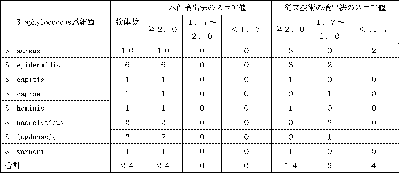


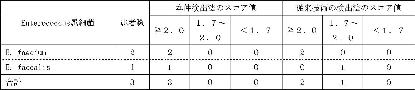
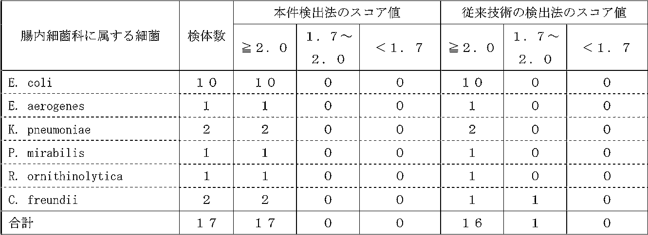



実施例4.本件検出法を用いた真菌の検出
実施例3に記載の本件検出法を用いて、血液培養物中に含まれる3種類の真菌(カンジダ・アルビカンス[Candida albicans]、カンジダ・グラブラータ[Candida glabrata]、カンジダ・トロピカリス[Candida tropcalis])を検出したところ、種レベルの同定確率は75%(Candida albicansでは67%[2/3]、Candida glabrataでは75%[3/4]、及びCandida tropcalisでは100%[1/1])と高かった(表22参照)。
この結果は、本件検出法により、細菌よりも比重やサイズが大きい真菌についても同様に、精度よく検出できることを示している。
実施例3に記載の本件検出法を用いて、血液培養物中に含まれる3種類の真菌(カンジダ・アルビカンス[Candida albicans]、カンジダ・グラブラータ[Candida glabrata]、カンジダ・トロピカリス[Candida tropcalis])を検出したところ、種レベルの同定確率は75%(Candida albicansでは67%[2/3]、Candida glabrataでは75%[3/4]、及びCandida tropcalisでは100%[1/1])と高かった(表22参照)。
この結果は、本件検出法により、細菌よりも比重やサイズが大きい真菌についても同様に、精度よく検出できることを示している。

本発明は、被験者における微生物(特に、ブドウ球菌属細菌や連鎖球菌属細菌等の連鎖やクラスター形成の著しい細菌や真菌)の感染症の診断や、当該感染症の早期予防又は早期治療に資するものである。
Claims (18)
- 以下の工程(a)~(c)を含むことを特徴とする、血液試料中の微生物の検出方法。
(a)被験者から採取された血液試料の培養物を、170~800×gの相対遠心力で40~80秒間、遠心処理し、上清画分と沈殿画分とに分離する工程;
(b)工程(a)で得られた上清画分から微生物画分を得る工程;
(c)前記微生物画分に微生物が含まれているか否かを解析する工程; - 工程(c)において、質量分析計を用いて解析することを特徴とする請求項1に記載の方法。
- 質量分析計が、マトリックス支援レーザー脱離イオン化飛行時間型質量分析計(MALDI-TOF MS)であることを特徴とする請求項2に記載の方法。
- 以下の工程(a)及び(b)を含むことを特徴とする、微生物を含む試料の調製方法。
(a)被験者から採取された血液試料の培養物を、170~800×gの相対遠心力で40~80秒間、遠心処理し、上清画分と沈殿画分とに分離する工程;
(b)工程(a)で得られた上清画分から微生物画分を得る工程; - 試料が、質量分析用試料であることを特徴とする請求項4に記載の方法。
- 工程(b)において、以下の工程(p)及び(q)を実施することにより微生物画分を得ることを特徴とする請求項1~5のいずれかに記載の方法。
(p)工程(a)で得られた上清画分を、微生物を通過させない孔径を有する濾過膜で濾過処理する工程;
(q)前記濾過処理後の濾過膜に捕捉された残渣を回収し、微生物画分として得る工程; - 工程(q)において、濾過処理後の濾過膜を純水中に浸し、遠心分離により残渣を回収し、微生物画分として得ることを特徴とする請求項6に記載の方法。
- 工程(p)と工程(q)の間に、濾過処理後の濾過膜を、純水を用いて洗浄する工程(r)を含むことを特徴とする請求項6又は7に記載の方法。
- 微生物が、細菌又は真菌であることを特徴とする請求項1~8のいずれかに記載の方法。
- 以下の(A)~(C)の手段を備えることを特徴とする、血液試料中の微生物の検出システム。
(A)被験者から採取された血液試料の培養物を、170~800×gの相対遠心力で40~80秒間の条件下で、上清画分と沈殿画分とに分離する遠心手段;
(B)上清画分に基づく、微生物画分の取得手段;
(C)微生物画分に微生物が含まれているか否かを解析する分析手段; - 分析手段が、質量分析手段であることを特徴とする請求項10に記載のシステム。
- 質量分析手段が、マトリックス支援レーザー脱離イオン化飛行時間型質量分析計(MALDI-TOF MS)であることを特徴とする請求項11に記載のシステム。
- 以下の(A)及び(B)の手段を備えることを特徴とする、微生物を含む試料の調製システム。
(A)被験者から採取された血液試料の培養物を、170~800×gの相対遠心力で40~80秒間の条件下で、上清画分と沈殿画分とに分離する遠心手段;
(B)上清画分に基づく、微生物画分の取得手段; - 試料が、質量分析用試料であることを特徴とする請求項13に記載のシステム。
- 微生物画分の取得手段が、微生物を通過させない孔径を有する濾過膜であることを特徴とする請求項10~14に記載のシステム。
- 微生物が、細菌又は真菌であることを特徴とする請求項10~15のいずれかに記載のシステム。
- 少なくとも1×10⁵個のブドウ球菌属細菌を含み、かつ、ヘモグロビンを実質的に含まないことを特徴とする、微生物を含む試料。
- 質量分析用試料であることを特徴とする請求項17に記載の試料。
Applications Claiming Priority (2)
| Application Number | Priority Date | Filing Date | Title |
|---|---|---|---|
| JP2018-233784 | 2018-12-13 | ||
| JP2018233784A JP2022049707A (ja) | 2018-12-13 | 2018-12-13 | 血液試料中の細菌の検出方法 |
Publications (1)
| Publication Number | Publication Date |
|---|---|
| WO2020122185A1 true WO2020122185A1 (ja) | 2020-06-18 |
Family
ID=71076451
Family Applications (1)
| Application Number | Title | Priority Date | Filing Date |
|---|---|---|---|
| PCT/JP2019/048731 Ceased WO2020122185A1 (ja) | 2018-12-13 | 2019-12-12 | 血液試料中の微生物の検出方法 |
Country Status (3)
| Country | Link |
|---|---|
| JP (1) | JP2022049707A (ja) |
| TW (1) | TW202028738A (ja) |
| WO (1) | WO2020122185A1 (ja) |
Cited By (1)
| Publication number | Priority date | Publication date | Assignee | Title |
|---|---|---|---|---|
| CN114414341A (zh) * | 2022-01-25 | 2022-04-29 | 厦门元谱生物科技有限公司 | 一种血液培养报阳的检测方法 |
Citations (2)
| Publication number | Priority date | Publication date | Assignee | Title |
|---|---|---|---|---|
| JP2012526996A (ja) * | 2009-05-15 | 2012-11-01 | ビオメリュー・インコーポレイテッド | 試料内の微生物因子の迅速な同定および/または特徴付けのためのシステムおよび方法 |
| US20130089886A1 (en) * | 2011-10-07 | 2013-04-11 | Becton, Dickinson And Company | Method for direct and rapid identification of microorganisms and antimicrobial susceptibility testing from positive blood cultures |
-
2018
- 2018-12-13 JP JP2018233784A patent/JP2022049707A/ja active Pending
-
2019
- 2019-12-12 WO PCT/JP2019/048731 patent/WO2020122185A1/ja not_active Ceased
- 2019-12-12 TW TW108145592A patent/TW202028738A/zh unknown
Patent Citations (2)
| Publication number | Priority date | Publication date | Assignee | Title |
|---|---|---|---|---|
| JP2012526996A (ja) * | 2009-05-15 | 2012-11-01 | ビオメリュー・インコーポレイテッド | 試料内の微生物因子の迅速な同定および/または特徴付けのためのシステムおよび方法 |
| US20130089886A1 (en) * | 2011-10-07 | 2013-04-11 | Becton, Dickinson And Company | Method for direct and rapid identification of microorganisms and antimicrobial susceptibility testing from positive blood cultures |
Non-Patent Citations (2)
| Title |
|---|
| ASHIZAWA, K. ET AL.: "Applications of copolymer for rapid identification of bacteria in blood culture broths using matrix-assisted laser desorption ionization time-of-flight mass spectrometry", J. MICROBIOL. METHODS, vol. 139, 2017, pages 54 - 60, XP085110893, DOI: 10.1016/j.mimet.2017.04.013 * |
| BERNHARDT M, ET AL: "DETECTION OF BACTERIA IN BLOOD BY CENTRIFUGATION AND FILTRATION", JOURNAL OF CLINICAL MICROBIOLOGY, AMERICAN SOCIETY FOR MICROBIOLOGY, US, vol. 29, no. 3, 1 March 1991 (1991-03-01), US, pages 422 - 425, XP002565155, ISSN: 0095-1137 * |
Cited By (1)
| Publication number | Priority date | Publication date | Assignee | Title |
|---|---|---|---|---|
| CN114414341A (zh) * | 2022-01-25 | 2022-04-29 | 厦门元谱生物科技有限公司 | 一种血液培养报阳的检测方法 |
Also Published As
| Publication number | Publication date |
|---|---|
| JP2022049707A (ja) | 2022-03-30 |
| TW202028738A (zh) | 2020-08-01 |
Similar Documents
| Publication | Publication Date | Title |
|---|---|---|
| US11473122B2 (en) | Mass spectrometric diagnosis of septicemia | |
| Veron et al. | Rapid urine preparation prior to identification of uropathogens by MALDI-TOF MS | |
| Conville et al. | Nocardia, rhodococcus, gordonia, actinomadura, streptomyces, and other aerobic actinomycetes | |
| Wang et al. | Direct identification of bacteria causing urinary tract infections by combining matrix-assisted laser desorption ionization-time of flight mass spectrometry with UF-1000i urine flow cytometry | |
| Pulcrano et al. | Rapid and reliable MALDI-TOF mass spectrometry identification of Candida non-albicans isolates from bloodstream infections | |
| EP3024942B1 (fr) | Méthode de détection de la contamination par un microbe d'un échantillon de produit sanguin par analyse de spectres de masse | |
| FR2951548A1 (fr) | Procede de caracterisation d'au moins un microorganisme par spectrometrie de masse | |
| CN102395680A (zh) | 用于鉴别液体培养基中的微生物的方法 | |
| JP2015021960A (ja) | インサイチュマルディトフ質量分析器を用いた胞子を形成するバチルス菌の同定方法および分析システム | |
| US20170204448A1 (en) | Identification of microorganisms using maldi-tof-ms on-plate extraction | |
| WO2020122185A1 (ja) | 血液試料中の微生物の検出方法 | |
| WO2012069768A1 (fr) | Procede d'identification d'un champignon, notamment d'un champignon filamenteux, et utilisations | |
| JP2021093913A (ja) | 血液試料中の細菌の検出方法 | |
| TWI775205B (zh) | 鑑定抗甲氧西林金黃色葡萄球菌的方法 | |
| TWI415943B (zh) | 用於鑑定具萬古黴素抗藥性之金黃色葡萄球菌的胜肽生物標記 | |
| CN107860819B (zh) | 适用于maldi-tof ms检测的病原体样品前处理方法及其应用 | |
| Gudlavalleti et al. | Application of atmospheric pressure matrix-assisted laser desorption/ionization mass spectrometry for rapid identification of Neisseria species | |
| Singh et al. | Detection of Aspergillus species by polymerase chain reaction | |
| CN110878374A (zh) | 药品质量控制中常见产毒真菌的特征序列鉴定方法 | |
| US20240248016A1 (en) | Hemolysis reagent | |
| US20170211120A1 (en) | Pathogen Identification in Complex Biological Fluids | |
| Chaturvedi et al. | Mucormycosis Infection and Methods of Detection: An Overview | |
| TWI416105B (zh) | 用於區分社區型抗甲氧苯青黴素金黃色葡萄球菌與醫院型抗甲氧苯青黴素金黃色葡萄球菌的胜肽生物標記 | |
| Anderson | Developing a Method for Rapid and Accurate Identification of Bacillus Species in Clinical Isolates Using Polymerase Chain Reaction | |
| CN114354946A (zh) | 地域性人源病原菌多肽质量参考谱库的建立方法 |
Legal Events
| Date | Code | Title | Description |
|---|---|---|---|
| 121 | Ep: the epo has been informed by wipo that ep was designated in this application |
Ref document number: 19894789 Country of ref document: EP Kind code of ref document: A1 |
|
| DPE1 | Request for preliminary examination filed after expiration of 19th month from priority date (pct application filed from 20040101) | ||
| NENP | Non-entry into the national phase |
Ref country code: DE |
|
| 122 | Ep: pct application non-entry in european phase |
Ref document number: 19894789 Country of ref document: EP Kind code of ref document: A1 |
|
| NENP | Non-entry into the national phase |
Ref country code: JP |