WO2020095946A1 - 新規蛍光プローブ - Google Patents
新規蛍光プローブ Download PDFInfo
- Publication number
- WO2020095946A1 WO2020095946A1 PCT/JP2019/043494 JP2019043494W WO2020095946A1 WO 2020095946 A1 WO2020095946 A1 WO 2020095946A1 JP 2019043494 W JP2019043494 W JP 2019043494W WO 2020095946 A1 WO2020095946 A1 WO 2020095946A1
- Authority
- WO
- WIPO (PCT)
- Prior art keywords
- group
- fluorescent probe
- groups
- psa
- hydrogen atom
- Prior art date
- Legal status (The legal status is an assumption and is not a legal conclusion. Google has not performed a legal analysis and makes no representation as to the accuracy of the status listed.)
- Ceased
Links
Images




Classifications
-
- A—HUMAN NECESSITIES
- A61—MEDICAL OR VETERINARY SCIENCE; HYGIENE
- A61K—PREPARATIONS FOR MEDICAL, DENTAL OR TOILETRY PURPOSES
- A61K49/00—Preparations for testing in vivo
-
- C—CHEMISTRY; METALLURGY
- C07—ORGANIC CHEMISTRY
- C07F—ACYCLIC, CARBOCYCLIC OR HETEROCYCLIC COMPOUNDS CONTAINING ELEMENTS OTHER THAN CARBON, HYDROGEN, HALOGEN, OXYGEN, NITROGEN, SULFUR, SELENIUM OR TELLURIUM
- C07F7/00—Compounds containing elements of Groups 4 or 14 of the Periodic Table
- C07F7/02—Silicon compounds
- C07F7/08—Compounds having one or more C—Si linkages
- C07F7/10—Compounds having one or more C—Si linkages containing nitrogen having a Si-N linkage
-
- C—CHEMISTRY; METALLURGY
- C09—DYES; PAINTS; POLISHES; NATURAL RESINS; ADHESIVES; COMPOSITIONS NOT OTHERWISE PROVIDED FOR; APPLICATIONS OF MATERIALS NOT OTHERWISE PROVIDED FOR
- C09K—MATERIALS FOR MISCELLANEOUS APPLICATIONS, NOT PROVIDED FOR ELSEWHERE
- C09K11/00—Luminescent materials, e.g. electroluminescent or chemiluminescent
- C09K11/06—Luminescent materials, e.g. electroluminescent or chemiluminescent containing organic luminescent materials
-
- C—CHEMISTRY; METALLURGY
- C12—BIOCHEMISTRY; BEER; SPIRITS; WINE; VINEGAR; MICROBIOLOGY; ENZYMOLOGY; MUTATION OR GENETIC ENGINEERING
- C12Q—MEASURING OR TESTING PROCESSES INVOLVING ENZYMES, NUCLEIC ACIDS OR MICROORGANISMS; COMPOSITIONS OR TEST PAPERS THEREFOR; PROCESSES OF PREPARING SUCH COMPOSITIONS; CONDITION-RESPONSIVE CONTROL IN MICROBIOLOGICAL OR ENZYMOLOGICAL PROCESSES
- C12Q1/00—Measuring or testing processes involving enzymes, nucleic acids or microorganisms; Compositions therefor; Processes of preparing such compositions
- C12Q1/34—Measuring or testing processes involving enzymes, nucleic acids or microorganisms; Compositions therefor; Processes of preparing such compositions involving hydrolase
- C12Q1/37—Measuring or testing processes involving enzymes, nucleic acids or microorganisms; Compositions therefor; Processes of preparing such compositions involving hydrolase involving peptidase or proteinase
-
- G—PHYSICS
- G01—MEASURING; TESTING
- G01N—INVESTIGATING OR ANALYSING MATERIALS BY DETERMINING THEIR CHEMICAL OR PHYSICAL PROPERTIES
- G01N21/00—Investigating or analysing materials by the use of optical means, i.e. using sub-millimetre waves, infrared, visible or ultraviolet light
- G01N21/62—Systems in which the material investigated is excited whereby it emits light or causes a change in wavelength of the incident light
- G01N21/63—Systems in which the material investigated is excited whereby it emits light or causes a change in wavelength of the incident light optically excited
- G01N21/64—Fluorescence; Phosphorescence
-
- G—PHYSICS
- G01—MEASURING; TESTING
- G01N—INVESTIGATING OR ANALYSING MATERIALS BY DETERMINING THEIR CHEMICAL OR PHYSICAL PROPERTIES
- G01N33/00—Investigating or analysing materials by specific methods not covered by groups G01N1/00 - G01N31/00
- G01N33/48—Biological material, e.g. blood, urine; Haemocytometers
- G01N33/50—Chemical analysis of biological material, e.g. blood, urine; Testing involving biospecific ligand binding methods; Immunological testing
- G01N33/53—Immunoassay; Biospecific binding assay; Materials therefor
-
- G—PHYSICS
- G01—MEASURING; TESTING
- G01N—INVESTIGATING OR ANALYSING MATERIALS BY DETERMINING THEIR CHEMICAL OR PHYSICAL PROPERTIES
- G01N33/00—Investigating or analysing materials by specific methods not covered by groups G01N1/00 - G01N31/00
- G01N33/48—Biological material, e.g. blood, urine; Haemocytometers
- G01N33/50—Chemical analysis of biological material, e.g. blood, urine; Testing involving biospecific ligand binding methods; Immunological testing
- G01N33/53—Immunoassay; Biospecific binding assay; Materials therefor
- G01N33/531—Production of immunochemical test materials
- G01N33/532—Production of labelled immunochemicals
- G01N33/533—Production of labelled immunochemicals with fluorescent label
-
- G—PHYSICS
- G01—MEASURING; TESTING
- G01N—INVESTIGATING OR ANALYSING MATERIALS BY DETERMINING THEIR CHEMICAL OR PHYSICAL PROPERTIES
- G01N33/00—Investigating or analysing materials by specific methods not covered by groups G01N1/00 - G01N31/00
- G01N33/48—Biological material, e.g. blood, urine; Haemocytometers
- G01N33/50—Chemical analysis of biological material, e.g. blood, urine; Testing involving biospecific ligand binding methods; Immunological testing
- G01N33/53—Immunoassay; Biospecific binding assay; Materials therefor
- G01N33/575—Immunoassay; Biospecific binding assay; Materials therefor for cancer
Definitions
- the present invention relates to a novel fluorescent probe for detecting puromycin-sensitive aminopeptidase (PSA), a detection method using the fluorescent probe, and a detection kit containing the probe.
- PSA puromycin-sensitive aminopeptidase
- imaging technologies such as X-ray CT, magnetic resonance imaging (MRI), and positron emission tomography (PET) are extremely useful in cancer diagnosis. It has become an important technology. Above all, fluorescence imaging is expected as a simple and safe method having high sensitivity and excellent temporal / spatial resolution.
- the inventors of the present invention have developed a fluorescent probe for ⁇ -glutamyl transpeptidase (GGT), which has been enhanced in breast cancer cells, and can detect a minute tumor with high sensitivity and specificity in a short time. This has been demonstrated (for example, Non-Patent Documents 1 and 2).
- GGT fluorescent probe of Non-Patent Document 1 uses a fluorescent response in the green region. Therefore, if a probe showing a fluorescent response in a red region or the like different from this can be developed, a plurality of fluorescent probes should be used in combination. Therefore, it is expected that breast cancer can be detected with higher sensitivity and specificity.
- the present invention provides a novel fluorescent probe that can detect a biomarker enzyme activity having high specificity for breast cancer with high sensitivity and high selectivity, and can detect it as a response of red fluorescence having a longer wavelength. This is an issue.
- the present inventors have found puromycin-sensitive aminopeptidase (PSA) as a new biomarker enzyme for detecting breast cancer cells, and have characteristics different from conventional green probes ( Breast cancer cells can be detected as a response to red fluorescence by using a compound in which a fluorophore having wavelength, charge, and lipophilicity) is used as a nucleus and an amino acid residue that is specifically cleaved by PSA is introduced into the side chain.
- PSA puromycin-sensitive aminopeptidase
- a fluorescent probe for detecting puromycin-sensitive aminopeptidase which comprises a compound represented by the following formula (I) or a salt thereof: [In the formula, A is an alanine residue, B is an amino acid residue, wherein A is connected to an adjacent N atom by forming an amide bond and B is connected to A by forming an amide bond.
- X is Si (R a ) (R b ), Ge (R a ) (R b ), Sn (R a ) (R b ), C (R a ) (R b ), P ( ⁇ O) (R a ) or O
- R a and R b each independently represent a hydrogen atom, an alkyl group, or an aryl group
- R 1 is a hydrogen atom, or 1 to 4 same or different independently selected from the group consisting of an optionally substituted alkyl group, a carboxyl group, an ester group, an alkoxy group, an amide group, and an azido group.
- R 2 represents a hydrogen atom, a hydroxyl group, a cyano group, an optionally substituted alkyl group, an alkoxy group, aryl, or heteroaryl
- R 3 and R 4 are each independently, independently of the group consisting of a hydrogen atom, a hydroxyl group, a halogen atom, an optionally substituted alkyl group, a sulfo group, a carboxyl group, an ester group, an amide group and an azido group.
- R 5 , R 6 and R 7 each independently represent a hydrogen atom or an alkyl group, Here, R 6 or R 7 may together with R 4 form a ring structure containing a nitrogen atom to which they are bonded.
- B is an asparagine residue or a lysine residue
- R 3 is an optionally substituted alkyl group having 1 to 6 carbon atoms or an optionally substituted alkoxy group having 1 to 6 carbon atoms.
- a kit for detecting a mycin-sensitive aminopeptidase (PSA) is provided.
- the present invention provides ⁇ 9> A step of bringing the fluorescent probe according to any one of the above ⁇ 1> to ⁇ 5> into contact with a sample to be measured, and puromycin-sensitive aminopeptidase (PSA) contained in the sample and the fluorescent probe.
- a method for detecting PSA which comprises a step of observing a fluorescence response or a change in absorbance due to the reaction of ⁇ 10> The detection method according to ⁇ 9> above, wherein the fluorescence response is visualized using a fluorescence imaging means.
- ⁇ 11> The method according to ⁇ 9> or ⁇ 10>, wherein the sample to be measured is a cell expressing puromycin-sensitive aminopeptidase (PSA); ⁇ 12> The method according to ⁇ 11> above, wherein the cells are cancer cells; ⁇ 13> The method according to ⁇ 12> above, wherein the cancer cells are breast cancer cells.
- PSA puromycin-sensitive aminopeptidase
- puromycin-sensitive aminopeptidase which is expressed at a high level in breast cancer tissue, can be detected by a fluorescence response, and thereby the presence of breast cancer can be identified accurately, quickly, and with high sensitivity. It also has an excellent effect that imaging can be performed.
- the fluorescent probe of the present invention detects the activity of PSA as a fluorescent response as a new breast cancer biomarker enzyme different from the conventionally known ⁇ -glutamyl transpeptidase (GGT), the conventional GGT is used. It is also possible to identify breast cancer specimens that could not be detected by the detection fluorescent probe.
- GGT ⁇ -glutamyl transpeptidase
- the fluorescent probe of the present invention can detect PSA activity as a fluorescent response in the red region, by using it in combination with a conventional green fluorescent probe, it is possible to detect breast cancer cells with higher specificity as compared with individual use of individual probes. In addition to enabling detection, multicolor imaging using a plurality of fluorescent response regions is also possible. Thereby, cancer cells such as breast cancer can be visualized and detected more precisely and with high sensitivity.
- the fluorescent probe of the present invention is expected to be used not only for medical applications such as detecting or diagnosing cancer cells such as breast cancer and normal cells in real time, but also as a basic research tool for elucidating disease states.
- FIG. 1 shows fluorescence imaging images (a and b) of a tumor sample and a normal sample treated with the fluorescent probe 1, and a temporal change (c) of fluorescence intensity.
- FIG. 2 is a graph showing the time change of the T / N ratio with the addition of the fluorescent probe of the present invention to the tumor site.
- FIG. 3 is a graph showing the fluorescence intensity change due to the reaction between the fluorescent probe 1 and the tumor site in the DEG assay (a) and PSA.
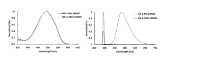
- FIG. 4 is an absorption spectrum (left) and a fluorescence spectrum (right) of the fluorescent probes 1 and 2.
- FIG. 5 is a table showing the fluorescence quantum yields of the fluorescent probes 1 and 2.
- FIG. 6 is a graph showing changes in fluorescence intensity due to the reaction of the fluorescent probe 2 with PSA and GGT.
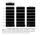
- FIG. 7 is a multicolor imaging image using the fluorescent probe 2 and gGlu-HMRG.
- the “amino acid” can be any compound as long as it is a compound having both an amino group and a carboxyl group, and includes natural and unnatural ones. It may be a neutral amino acid, a basic amino acid, or an acidic amino acid, and in addition to an amino acid which itself functions as a transmitter such as a neurotransmitter, a physiologically active peptide (dipeptide, tripeptide, tetrapeptide, Amino acids that are constituents of polypeptide compounds (including oligopeptides) and proteins can be used, and may be, for example, ⁇ -amino acids, ⁇ -amino acids, ⁇ -amino acids and the like.
- amino acid residue refers to a structure corresponding to the remaining partial structure (acyl group) obtained by removing a hydroxyl group from a carboxyl group of an amino acid.
- halogen atom means a fluorine atom, a chlorine atom, a bromine atom or an iodine atom.
- alkyl may be any of linear, branched, cyclic, or a combination thereof, which is an aliphatic hydrocarbon group.
- the number of carbon atoms of the alkyl group is not particularly limited, and examples thereof include 1 to 6 carbon atoms (C 1 to 6 ), 1 to 10 carbon atoms (C 1 to 10 ), and 1 to 15 carbon atoms (C 1 to 15). ) And having 1 to 20 carbon atoms (C 1 to 20 ). When the number of carbons is specified, it means “alkyl” having the number of carbons in the range.
- C 1-8 alkyl includes methyl, ethyl, n-propyl, isopropyl, n-butyl, isobutyl, sec-butyl, tert-butyl, n-pentyl, isopentyl, neo-pentyl, n-hexyl, isohexyl, It includes n-heptyl, n-octyl and the like.
- the alkyl group may have one or more optional substituents.
- substituents include, but are not limited to, an alkoxy group, a halogen atom, an amino group, a mono- or di-substituted amino group, a substituted silyl group, and acyl.
- alkyl group has two or more substituents, they may be the same or different. The same applies to the alkyl moiety of other substituents containing an alkyl moiety (eg, alkoxy group, arylalkyl group, etc.).
- a functional group when a functional group is defined as “optionally substituted”, the type of the substituent, the substitution position, and the number of the substituents are not particularly limited, and the substitution of two or more substituents is not limited. If they have groups, they may be identical or different.
- the substituent include, but are not limited to, an alkyl group, an alkoxy group, a hydroxyl group, a carboxyl group, a halogen atom, a sulfo group, an amino group, an alkoxycarbonyl group, and an oxo group.
- Substituents may be further present in these substituents. Examples of such a group include, but are not limited to, a halogenated alkyl group and a dialkylamino group.
- alkenyl refers to a straight or branched chain hydrocarbon group having at least one carbon-carbon double bond.
- non-limiting examples thereof are vinyl, allyl, 1-propenyl, isopropenyl, 1-butenyl, 2-butenyl, 3-butenyl, 1,3-butanedienyl, 1-pentenyl, 2-pentenyl, 3-pentenyl.
- the double bond may be in either the cis or trans conformation.
- alkynyl refers to a straight or branched chain hydrocarbon group having at least one carbon-carbon triple bond.
- non-limiting examples include ethynyl, propynyl, 2-butynyl, 3-methylbutynyl and the like.
- cycloalkyl refers to a monocyclic or polycyclic non-aromatic ring system consisting of the above alkyl.
- the cycloalkyl may be unsubstituted or substituted by one or more substituents which may be the same or different and non-limiting examples of monocyclic cycloalkyl include cyclopropyl, cyclopentyl, cyclohexyl. And cycloheptyl and the like, non-limiting examples of polycyclic cycloalkyl include 1-decalinyl, 2-decalinyl, norbornyl, adamantyl and the like.
- the cycloalkyl may be a heterocycloalkyl containing at least one hetero atom (eg, an oxygen atom, a nitrogen atom, or a sulfur atom) as a ring-constituting atom.
- Any —NH in the heterocycloalkyl ring may be protected, such as, for example, as a —N (Boc), —N (CBz) and —N (Tos) group, a nitrogen atom in the ring or
- the sulfur atom may be oxidized to the corresponding N-oxide, S-oxide or S, S-dioxide.
- non-limiting examples of monocyclic heterocycloalkyl include diazapanyl, piperidinyl, pyrrolidinyl, piperazinyl, morpholinyl, thiomorpholinyl, thiazolidinyl, 1,4-dioxanyl, tetrahydrofuranyl, tetrahydropyranyl, tetrahydrothiophenyl, lactam and Examples include lactones.
- cycloalkenyl refers to a monocyclic or polycyclic non-aromatic ring system containing at least one carbon-carbon double bond.
- the cycloalkenyl may be unsubstituted or substituted by one or more substituents which may be the same or different and non-limiting examples of monocyclic cycloalkenyl include cyclopentenyl, cyclohexenyl. And cyclohepta-1,3-dienyl and the like, and non-limiting examples of polycyclic cycloalkenyl include norbornylenyl and the like.
- the cycloalkyl may be a heterocycloalkenyl containing one or more hetero atoms (eg, oxygen atom, nitrogen atom, or sulfur atom) as a ring-constituting atom. Atoms may be oxidized to the corresponding N-oxide, S-oxide or S, S-dioxide.
- hetero atoms eg, oxygen atom, nitrogen atom, or sulfur atom
- aryl may be either a monocyclic or a condensed polycyclic aromatic hydrocarbon group, and a hetero atom (for example, an oxygen atom, a nitrogen atom, or a sulfur atom) as a ring-constituting atom. And the like) may be included in the aromatic heterocycle. In this case, it may be referred to as “heteroaryl” or “heteroaromatic”. Whether the aryl is a single ring or a fused ring, it may be attached at all possible positions.
- Non-limiting examples of monocyclic aryl include phenyl group (Ph), thienyl group (2- or 3-thienyl group), pyridyl group, furyl group, thiazolyl group, oxazolyl group, pyrazolyl group, 2-pyrazinyl group.
- Non-limiting examples of the fused polycyclic aryl include 1-naphthyl group, 2-naphthyl group, 1-indenyl group, 2-indenyl group, 2,3-dihydroinden-1-yl group, 2,3 -Dihydroinden-2-yl group, 2-anthryl group, indazolyl group, quinolyl group, isoquinolyl group, 1,2-dihydroisoquinolyl group, 1,2,3,4-tetrahydroisoquinolyl group, indolyl group, Isoindolyl group, phthalazinyl group, quinoxalinyl group, benzofuranyl group, 2,3-dihydrobenzofuran-1-yl group, 2,3-dihydrobenzofuran-2-yl group, 2,3-dihydrobenzothiophen-1-yl group, 2 , 3-dihydrobenzothiophen-2-yl group, benzothiazolyl group
- the aryl group may have one or more optional substituents on its ring.
- substituents include, but are not limited to, an alkoxy group, a halogen atom, an amino group, a mono- or di-substituted amino group, a substituted silyl group, and acyl.
- the aryl group has two or more substituents, they may be the same or different. The same applies to the aryl moiety of other substituents containing an aryl moiety (for example, an aryloxy group and an arylalkyl group).
- arylalkyl represents alkyl substituted with the above aryl.
- the arylalkyl may have one or more optional substituents.
- substituents include, but are not limited to, an alkoxy group, a halogen atom, an amino group, a mono- or di-substituted amino group, a substituted silyl group, and an acyl group.
- the acyl group has two or more substituents, they may be the same or different.
- Non-limiting examples of arylalkyl include benzyl group, 2-thienylmethyl group, 3-thienylmethyl group, 2-pyridylmethyl group, 3-pyridylmethyl group, 4-pyridylmethyl group, 2-furylmethyl group, 3-furylmethyl group, 2-thiazolylmethyl group, 4-thiazolylmethyl group, 5-thiazolylmethyl group, 2-oxazolylmethyl group, 4-oxazolylmethyl group, 5-oxazolylmethyl group, 1-pyrazolylmethyl group , 3-pyrazolylmethyl group, 4-pyrazolylmethyl group, 2-pyrazinylmethyl group, 2-pyrimidinylmethyl group, 4-pyrimidinylmethyl group, 5-pyrimidinylmethyl group, 1-pyrrolylmethyl group, 2-pyrrolylmethyl group, 3-pyrrolylmethyl group , 1-imidazolylmethyl group, 2-imidazolylmethyl group, 4-imidazolylmethyl group , 3-pyridazinylmethyl group, 4-pyri
- arylalkenyl refers to an alkenyl substituted with the above aryl.
- the “alkoxy group” has a structure in which the alkyl group is bonded to an oxygen atom, and examples thereof include a saturated alkoxy group having a linear, branched, or cyclic structure or a combination thereof.
- alkylene is a linear or branched saturated hydrocarbon divalent group such as methylene, 1-methylmethylene, 1,1-dimethylmethylene, ethylene, and the like.
- alkenylene is a divalent group consisting of an unsaturated hydrocarbon having at least one linear or branched carbon-carbon double bond, and examples thereof include ethenylene. , 1-methylethenylene, 1-ethylethenylene, 1,2-dimethylethenylene, 1,2-diethylethenylene, 1-ethyl-2-methylethenylene, propenylene, 1-methyl-2-propenylene, 2-methyl -2-propenylene, 1,1-dimethyl-2-propenylene, 1,2-dimethyl-2-propenylene, 1-ethyl-2-propenylene, 2-ethyl-2-propenylene, 1,1-diethyl-2-propenylene , 1,2-diethyl-2-propenylene, 1-butenylene, 2-butenylene, 1-methyl-2-butenylene, 2-methyl-2-butenylene 1,1-dimethyl-2-butenylene, 1,2-dimethyl
- arylene and arylalkylene mean a divalent group based on the above “aryl” and “arylalkyl”, respectively.
- oxyalkylene and aryleneoxy mean a divalent group based on the above “alkoxy” and “aryloxy”, respectively.
- ring structure when formed by the combination of two substituents, means a heterocyclic or carbocyclic group, such group being saturated, unsaturated, or aromatic.
- it includes cycloalkyl, cycloalkenyl, aryl, and heteroaryl as defined above. Examples include cycloalkyl, phenyl, naphthyl, morpholinyl, piperdinyl, imidazolyl, pyrrolidinyl, pyridyl and the like.
- a substituent can form a ring structure with another substituent, and when such substituents are bonded to each other, those skilled in the art can perform a specific substitution, for example, bonding to hydrogen.
- the fluorescent probe of the present invention contains a compound having a structure represented by the following formula (I).
- a and B have a structure in which amino acid residues are linked, and are sites that can be selectively hydrolyzed by puromycin-sensitive aminopeptidase (PSA). More specifically, A represents an alanine residue and B represents an amino acid residue.
- A is linked with N (R 5 ) in the formula by forming an amide bond, that is, with the N atom of N (R 5 ) with the carbonyl moiety (acyl group) of the alanine residue A.
- A can be linked to amino acid residue B like a normal peptide chain, so that B forms an amide bond with A and links them. Therefore, B has a structure similar to that of a so-called N-terminal residue, and the intermediate amino acid residue A can be linked to B in the same manner as a normal peptide chain.
- puromycin-sensitive aminopeptidase is a cytoplasmic alanylaminopeptidase that uses Ala-AMC as a standard substrate, and it has been reported that it is highly expressed particularly in the central nervous system. It is also used in clinical medicine as a biomarker for estimating renal function, and its function is known to be inhibited by puromycin.
- a and B are substrate peptides of PSA and are combinations of amino acid residues that are easily hydrolyzed selectively. preferable. More specifically, as described above, A is an alanine residue (“Ala” or “A”) and B is an asparagine residue (“Asn” or “N”) or a lysine residue (“ Lys "or” K ”) is preferred. More preferably, A is an alanine residue and B is an asparagine residue.
- R a and R b each independently represent a hydrogen atom, an alkyl group, or an aryl group.
- R a and R b are alkyl groups, they can have one or more substituents, and examples of such substituents include an alkyl group, an alkoxy group, a halogen atom, a hydroxyl group, a carboxyl group, You may have 1 or 2 or more amino groups, a sulfo group, etc.
- Each of R a and R b may be an optionally substituted alkyl group having 1 to 6 carbon atoms, and both are preferably methyl groups. Further, in some cases, R a and R b may be bonded to each other to form a ring structure.
- R a and R b can combine with each other to form a spiro carbocycle.
- the ring formed is preferably a 5- to 8-membered ring, for example.
- X is preferably Si (R a ) (R b ), and more preferably Si (CH 3 ) 2 .
- R 1 represents a hydrogen atom or 1 to 4 substituents bonded to the benzene ring.
- R 1 is a hydrogen atom, or 1 to 4 independently selected from the group consisting of an optionally substituted alkyl group, a carboxyl group, an ester group, an alkoxy group, an amide group, and an azido group. The same or different substituents are represented.
- R 1 is other than a hydrogen atom, its position on the benzene ring is not particularly limited, but it is preferably meta to R 2 .
- the benzene ring has two or more substituents, they may be the same or different.
- R 1 is a hydrogen atom.
- R 2 represents a hydrogen atom, a hydroxyl group, a cyano group, an optionally substituted alkyl group, an alkoxy group, aryl, or heteroaryl.
- it is a hydroxyl group, a cyano group, an optionally substituted C 1 -C 4 alkyl group, a C 1 -C 4 alkoxy group, or a phenyl group. More preferably, it is a methyl group or a methoxy group.
- R 3 and R 4 are each independently selected from the group consisting of a hydrogen atom, a hydroxyl group, a halogen atom, an optionally substituted alkyl group, a sulfo group, a carboxyl group, an ester group, an amide group and an azido group. 1 to 3 of the same or different substituents. Preferably, both R 3 and R 4 are hydrogen atoms. Further, similar to R 1 described above, R 3 or R 4 may have a fluorophore that serves as a donor of fluorescence resonance energy transfer (FRET).
- FRET fluorescence resonance energy transfer
- R 5 , R 6 and R 7 each independently represent a hydrogen atom or an alkyl group.
- R 5 , R 6 and R 7 both represent an alkyl group, they may be the same or different.
- R 5 , R 6 , and R 7 can each independently be a methyl group or an ethyl group.
- R 5 , R 6 and R 7 are all hydrogen atoms.
- R 6 or R 7 may together with R 4 form a ring structure containing a nitrogen atom to which they are bonded.
- the ring structure is a 5-8 membered heterocyclic structure.
- the ring structure may further include a heteroatom other than the nitrogen atom to which R 6 and R 7 are bonded.
- R 1 , R 3 , R 4 , R 5 , R 6 , and R 7 are all hydrogen atoms.
- the compound represented by the above formula (I) has a monovalent positive charge at the N atom to which R 6 and R 7 are linked, it usually exists as a salt.
- salts include base addition salts, acid addition salts, amino acid salts and the like.
- base addition salt include metal salts such as sodium salt, potassium salt, calcium salt, and magnesium salt, ammonium salt, and organic amine salts such as triethylamine salt, piperidine salt, and morpholine salt.
- mineral acid salts such as hydrochlorides, sulfates, nitrates, etc.
- organic acid salts such as carboxylates, trifluoroacetates, methanesulfonates, paratoluenesulfonates, citrates, oxalates, etc.
- amino acid salts include glycine salts. However, it is not limited to these salts.
- the compound represented by the formula (I) may have one or two or more asymmetric carbon atoms depending on the kind of the substituent, and stereoisomers such as optical isomers or diastereoisomers exist. There are cases. Any stereoisomers in pure form, any mixture of stereoisomers, racemates and the like are included within the scope of the present invention.
- the compound represented by the formula (I) or a salt thereof may exist as a hydrate or a solvate, and all of these substances are included in the scope of the present invention.
- the type of solvent that forms the solvate is not particularly limited, and examples thereof include solvents such as ethanol, acetone, and isopropanol.
- the above-mentioned fluorescent probe may be used as a composition by adding an additive usually used for the preparation of reagents, if necessary.
- an additive such as a solubilizing agent, a pH adjusting agent, a buffering agent, an isotonicity agent, etc. can be used, and the blending amount thereof can be appropriately selected by those skilled in the art. is there.
- These compositions can be provided as a composition in an appropriate form such as a powder-form mixture, a lyophilized product, a granule, a tablet, and a liquid.
- PSA enzyme activity is detected by fluorescence control using photoinduced electron transfer (PET: Photoinduced electron transfer). More specifically, as shown in the following scheme, when the side chain at the 3-position of the xanthene ring is linked to the amino acid residue “AB” (peptide chain), it can be irradiated with excitation light. Since the xanthene ring portion has a high electron accepting property, it is in a state of being quenched by electron transfer from the benzene ring portion. On the other hand, when the fluorescent probe molecule reacts with PSA, the side chain “AB” peptide chain is cleaved by hydrolysis and the 3-position is changed to an amino group. As a result, fluorescence emission is observed.
- PET Photoinduced electron transfer
- fluorescence is quenched by PET from the benzene ring site before the reaction with PSA, but when the peptide chain is cleaved by PSA, the electron density of the xanthene ring site is increased. Changes and the fluorescence quenching by PET is suppressed, so that the activity of PSA can be detected as a fluorescence response.
- the fluorescence emission due to the “AB” cleavage can be fluorescence in the red region with a fluorescence wavelength of 600 nm or more. This makes it possible to detect the presence of cancer cells and the like as a red fluorescence response.
- visible light having a wavelength of about 400 to 600 nm may be irradiated as excitation light.
- the fluorescence wavelength to be observed is usually in the range of about 580 to 700 nm.
- the presence of PSA is specifically detected or visualized by observing the fluorescence response or the change in absorbance due to the reaction between PSA and the fluorescent probe.
- detection should be construed in the broadest sense including measurement for various purposes such as quantification and qualification.
- A) a step of bringing the fluorescent probe of the present invention into contact with a sample to be measured; and B) Only the target cells expressing PSA are specifically fluorescent by including a step of observing a fluorescence response or a change in absorbance due to a reaction between puromycin-sensitive aminopeptidase (PSA) contained in the sample and the fluorescent probe. It can be detected or visualized in response.
- PSA puromycin-sensitive aminopeptidase
- the method of the present invention can further include observing the fluorescence response using a fluorescence imaging means.
- a fluorescence photometer having a wide measurement wavelength can be used, but the fluorescence response can also be visualized using a fluorescence imaging means capable of displaying a two-dimensional image.
- the fluorescence imaging means By using the fluorescence imaging means, the fluorescence response can be visualized in two dimensions, and the PSA can be instantly visually recognized.
- a device known in the art can be used as the fluorescence imaging device.
- the reaction between the sample to be measured and the fluorescent probe can be detected by a change in the UV-visible absorption spectrum (for example, a change in absorbance at a specific absorption wavelength).
- a solution containing the fluorescent probe is added to the sample, applied, or sprayed. It can be appropriately selected according to the measurement environment and the like. Such contacting is preferably done in an in vitro environment.
- the fluorescent probe of the present invention is applied to diagnosis or diagnosis assistance in animal individuals, or detection of specific cells or tissues, as a means for bringing the fluorescent probe into contact with the target cells or tissues, without being limited, for example, intravenous administration and the like, which is a general administration means in the art, can be used.
- the application concentration of the fluorescent probe of the present invention is not particularly limited, but for example, a solution having a concentration of about 0.1 to 10 ⁇ M can be applied.
- the light irradiation performed on the target cells can be performed by irradiating the cells with light directly or via a waveguide (optical fiber etc.).
- a waveguide optical fiber etc.
- any light source can be used as long as it can irradiate the light having the absorption wavelength of the fluorescent probe of the present invention, and it can be used in the environment for carrying out the method of the present invention. It can be selected accordingly.
- the compound represented by the above formula (I) or a salt thereof may be used as it is, but if necessary, an additive usually used for the preparation of a reagent may be blended to obtain a composition. You may use.
- an additive for using the reagent in a physiological environment an additive such as a solubilizing agent, a pH adjusting agent, a buffering agent, an isotonicity agent, etc. can be used, and the blending amount thereof can be appropriately selected by those skilled in the art. It is possible.
- compositions are generally provided as a composition in an appropriate form such as a mixture in powder form, a lyophilized product, a granule, a tablet, a solution, etc., but distilled water for injection or an appropriate buffer solution at the time of use. It is possible to apply by dissolving in.
- the sample to be measured in the above step A) can be cells expressing puromycin-sensitive aminopeptidase (PSA), but when such cells are cancer cells or cancer tissues expressing PSA
- PSA puromycin-sensitive aminopeptidase
- cancer cells and cancer tissues can be detected or visualized by the detection method of the present invention. That is, the fluorescent probe of the present invention, the composition containing the fluorescent probe, and the detection method of the present invention can also be used for cancer diagnosis.
- cancer tissue means any tissue containing cancer cells.
- tissue should be interpreted in its broadest sense including a part or whole of an organ, and should not be limitedly interpreted in any sense. Since the fluorescent probe or the composition for detecting cancer cells containing the fluorescent probe of the present invention has an action of detecting PSA which is specifically and strongly expressed in cancer tissue, PSA is regarded as cancer tissue. A tissue that highly expresses is preferred. Further, in this specification, the term “diagnosis” should be interpreted in its broadest sense, including the confirmation of the presence of cancerous tissue at any part of a living body visually or under a microscope.
- the sample to be measured in the above step A) is preferably a cancer cell, more preferably a breast cancer cell.
- a fluorescent probe for ⁇ -glutamyl transpeptidase can be used in combination with the fluorescent probe of the present invention.
- GGT ⁇ -glutamyl transpeptidase
- the fluorescent probe of the present invention is usually prepared as a solution, but for example, it is provided as a composition in an appropriate form such as a mixture in a powder form, a lyophilized product, a granule, a tablet, and a liquid, and at the time of use. It can also be applied by dissolving in distilled water for injection or an appropriate buffer solution.
- the kit may optionally contain other reagents and the like as necessary.
- a solubilizing agent, a pH adjusting agent, a buffering agent, an isotonicity agent and the like can be used, and the amount of these can be appropriately selected by those skilled in the art.
- a green fluorescent probe for GGT detection can be included in addition to the fluorescent probe of the present invention.
- the reaction solution was filtered off. 900 ⁇ L of DMF was added to the resin, shaken for 1 minute, and DMF was removed by filtration. This washing operation was repeated twice more.
- 800 ⁇ L of 40% piperidine in DMF was added, shaken for 3 minutes, and the reaction solution was removed by filtration.
- 400 ⁇ L of 40% piperidine in DMF was added, shaken for 12 minutes and the reaction solution was removed by filtration.
- 900 ⁇ L of DMF was added to the resin, shaken for 1 minute, and DMF was removed by filtration. This washing operation was repeated 5 more times.
- a / B 70/30 to 0/100 in 40 minutes (eluent A: H 2 containing 0.1% TFA. O, eluent B: 0.1% TFA in 80% acetonitrile and 20% H 2 O) to give compound 6 of sufficient purity.
- Fluorescent Probe 2 (NA-2-OMe SiR600) of the Present Invention
- the fluorescent probe 2 of the present invention (NA-2-OMe SiR600) having Asn-Ala (NA) in the side chain was synthesized as follows.
- Fluorescent probe 2 differs from fluorescent probe 1 (NA-2-Me SiR600) in that the substituent on the benzene ring corresponding to R 2 in formula (I) is methoxy.
- N, N, N ', N'-Tetraallyldiamino-Si-xanthone was synthesized according to the literature (Kushida, Y. et al., Bioorganic & medicinal chemistry letters 2012, 22 (12), 3908-3911.).
- N, N, N ', N'-Tetraallyldiamino-Si-xanthone (1.28 g, 2.97 mmol) dissolved in anhydrous THF (10 mL) was added slowly and the mixture was warmed to room temperature. It was then stirred for 1 hour. The reaction was quenched by addition of 2N HCl and the mixture was stirred at room temperature for 5 minutes. Saturated NaHCO 3 was added and the whole was extracted with CH 2 Cl 2 . The organic layer was washed with brine, dried over Na 2 SO 4 and evaporated. The residue was dissolved in MeOH, stirred at 0 ° C. and NaBH 4 (340 mg, 8.99 mmol) was added.
- Example 2 Screening Using Human Breast Cancer Surgical Specimens Using the library of fluorescent probes synthesized in Example 1, screening was performed on human breast cancer surgical specimens. First, a lysate is prepared from the surgical specimen by dividing it into a tumor site and a normal site, and each probe with a different peptide chain at the AB site is reacted with each lysate to measure the change in fluorescence intensity. Thirty-six fluorescent probes showing a sufficient increase in fluorescence and a high T / N ratio (Tumor to Normal ratio) were selected as candidate probes.
- T / N ratio Tumor to Normal ratio
- the probe solution was dropped on the excised specimen and fluorescence imaging was performed to measure the change in fluorescence intensity.
- fluorescence imaging was performed to measure the change in fluorescence intensity.
- a large number of 3-5 mm square pieces were cut from a human breast cancer specimen with a scalpel and placed in each well of an 8-well chamber slide (Thermo Fisher 155411), and each fluorescent probe solution (50 ⁇ M) was placed there.
- each fluorescent probe solution 50 ⁇ M
- the fluorescent probe 1 (compound 8: NA-2-MeSiR600) in which the AB site in formula (I) is “Asn-Ala” (NA) shows a rapid increase in fluorescence at the tumor site. , It was found that the normal site showed almost no increase.
- the fluorescent probes with AB sites of “Lys-Ala” (KA) and “Met-Ala” (MA) also showed high sensitivity.
- FIG. 1 shows fluorescence imaging images (a and b) of a tumor sample and a normal sample treated with the fluorescent probe 1, and a temporal change (c) of fluorescence intensity.
- the time change of T / N ratio with addition to the tumor site is shown in FIG.
- the T / N ratio at each time was higher for fluorescent probe 1 (NA) except after 1 minute.
- a lysate prepared from the tumor site of the surgical specimen was used to perform a DEG (diced electrophoresis gel) assay, and a single highly active spot was observed (Fig. 3 ( a)). Focusing on this spot, PMF (Peptide Mass Fingerprinting) analysis was performed. As a result, puromycin-sensitive aminopeptidase (PSA) was identified as a metabolic enzyme of fluorescent probe 1. When the identified PSA was reacted with fluorescent probe 1 using a purified enzyme, a rapid increase in fluorescence was shown (FIG. 3 (b)). From these results, it was found that the fluorescent probe 1 is effective as a breast cancer detection fluorescent probe using PSA as a biomarker enzyme.
- DEG cold electrophoresis gel
- Fluorescence Assay for PSA The absorption and fluorescence spectra of the fluorescent probe 1 (Compound 8: NA-2-Me SiR600) and the fluorescent probe 2 (Compound 11: NA-2-OMe SiR600) synthesized in Example 1 were measured, and , The quantum yield was calculated. In each case, the fluorescent probe concentration was 1 ⁇ M, and a 0.1 M Na phosphate buffer (pH 7.4, 0.1% DMSO) solution was used.
- the fluorescent probe 2 showed a rapid increase in fluorescence with the addition of PSA. This is probably because "Asn-Ala" at the AB site in the formula (I) was cleaved by the reaction with PSA, whereby PET was suppressed and the fluorescence emission of the xanthene ring was restored. ..
- GGT was added, no increase in fluorescence was observed, indicating that fluorescent probe 2 was not cleaved at the AB site by GGT. This result demonstrates that the fluorescent probe 2 can specifically detect the presence of PSA.
- gGlu-HMRG breast cancer detection green fluorescent probe
- a tumor site and a normal site were separately cut out in a size of about 5 mm square from a breast cancer sample, and a mixed solution of fluorescent probe 2 and gGlu-HMRG was dropped on each. Merged images were created by acquiring green and red fluorescence images 30 minutes after dropping 1, 3, 5, 10, 15, 20, and 30 minutes, and superimposing them. A total of 14 cases were imaged by the same method. An image of one of these cases is shown in FIG. 7 as a representative example. In FIG. 7, both of the fluorescent probe 2 and gGlu-HMRG showed an increase in fluorescence only at the tumor site, but did not show an increase in fluorescence at the normal site. The tumor site could be detected in 13 of 14 cases (93%). This demonstrates that the combined use of fluorescent probe 2 for PSA and gGlu-HMRG for GGT can detect a tumor site of breast cancer with higher specificity.
- the fluorescent probe of the present invention can selectively detect PSA and is a highly practical fluorescent probe for breast cancer.
Landscapes
- Health & Medical Sciences (AREA)
- Life Sciences & Earth Sciences (AREA)
- Chemical & Material Sciences (AREA)
- Immunology (AREA)
- Engineering & Computer Science (AREA)
- Molecular Biology (AREA)
- Organic Chemistry (AREA)
- Biomedical Technology (AREA)
- Hematology (AREA)
- General Health & Medical Sciences (AREA)
- Urology & Nephrology (AREA)
- Physics & Mathematics (AREA)
- Biochemistry (AREA)
- Analytical Chemistry (AREA)
- General Physics & Mathematics (AREA)
- Biotechnology (AREA)
- Microbiology (AREA)
- Pathology (AREA)
- Food Science & Technology (AREA)
- Cell Biology (AREA)
- Medicinal Chemistry (AREA)
- Zoology (AREA)
- Wood Science & Technology (AREA)
- Proteomics, Peptides & Aminoacids (AREA)
- Epidemiology (AREA)
- Materials Engineering (AREA)
- Nuclear Medicine, Radiotherapy & Molecular Imaging (AREA)
- Biophysics (AREA)
- Veterinary Medicine (AREA)
- Public Health (AREA)
- Animal Behavior & Ethology (AREA)
- Bioinformatics & Cheminformatics (AREA)
- General Engineering & Computer Science (AREA)
- Genetics & Genomics (AREA)
- Measuring Or Testing Involving Enzymes Or Micro-Organisms (AREA)
- Investigating, Analyzing Materials By Fluorescence Or Luminescence (AREA)
- Medicines Containing Antibodies Or Antigens For Use As Internal Diagnostic Agents (AREA)
Abstract
【課題】 乳がんに特異性の高いバイオマーカー酵素活性を高感度・高選択的に検出できるとともに、より長波長の赤色蛍光の応答として検出することができる新規蛍光プローブを提供すること 【解決手段】 以下の式(I)で表される化合物又はその塩を含む、ピューロマイシン感受性アミノペプチダーゼ(PSA)検出用蛍光プローブ:〔式中、 Aは、アラニン残基であり、Bは、アミノ酸残基を表し、ここで、Aは隣接するN原子とアミド結合を形成して連結し、BはAとアミド結合を形成して連結しており、 Xは、Si(Ra)(Rb)、Ge(Ra)(Rb)、Sn(Ra)(Rb)、C(Ra)(Rb)、P(=O)(Ra)又はOを表し(ここで、Ra及びRbは、それぞれ独立に水素原子、アルキル基、又はアリール基を表す); R1は、水素原子、又はそれぞれ置換されていてもよいアルキル基、カルボキシル基、エステル基、アルコキシ基、アミド基、及びアジド基よりなる群から独立に選択される1~4個の同一又は異なる置換基を表し; R2は、水素原子、ヒドロキシル基、シアノ基、それぞれ置換されていてもよいアルキル基、アルコキシ基、アリール、又はヘテロアリールを表し; R3及びR4は、それぞれ独立に、水素原子、ヒドロキシル基、ハロゲン原子、それぞれ置換されていてもよいアルキル基、スルホ基、カルボキシル基、エステル基、アミド基及びアジド基よりなる群から独立に選択される1~3個の同一又は異なる置換基を表し; R5、R6、及びR7は、それぞれ独立に水素原子又はアルキル基を表し、 ここで、R6又はR7は、それぞれR4と一緒になって、それらが結合する窒素原子を含む環構造を形成してもよい。〕。
Description
本発明は、ピューロマイシン感受性アミノペプチダーゼ(PSA)検出用の新規な蛍光プローブ、当該蛍光プローブを用いた検出方法、及び当該プローブを含む検出用キットに関する。
がんの罹患者や死亡者が年々増加している現在、その治療方法の開発が期待され続けている。現時点で、最も確実と考えられているがん治療法の一つは、がんの早期発見とその確実な外科的摘出であるが、目視が完全には難しい微小ながん組織を完全に除去することは難しく再発の原因となっている。
一方、このような微小ながん組織を特定する観点から、X線CTや磁気共鳴画像法(MRI)、陽電子放射断層撮像(PET)などをはじめとするイメージング技術が、がん診断において非常に重要な技術となっている。なかでも、蛍光イメージングは、高い感度および優れた時間・空間分解能を有する簡便且つ安全な手法として期待されている。
本願発明者らは、これまで、乳がん細胞で亢進しているγ-グルタミルトランスペプチダーゼ(GGT)に対する蛍光プローブを開発し、これにより短時間で微小な腫瘍を高感度及び特異的に検出可能であることを実証している(例えば、非特許文献1及び2)。しかしながら、このGGT蛍光プローブでは偽陽性及び偽陰性となる乳がん検体が存在することから、GGTの酵素活性とは異なる判断基準を有する新たなプローブの開発が求められていた。特に、非特許文献1のGGT蛍光プローブは、緑色領域の蛍光応答を用いるものであるため、これとは異なる赤色等の領域に蛍光応答を示すプローブを開発できれば、複数の蛍光プローブを併用することで、さらに高感度・特異度での乳がん検出が可能となることが期待される。
Y.Uranoら、Sci. Transl. Med.,vol.3, pp.110ra119(2011年)
H.Ueoら、Sci. Rep. 5, 12080 (2015年)
そこで、本発明は、乳がんに特異性の高いバイオマーカー酵素活性を高感度・高選択的に検出できるとともに、より長波長の赤色蛍光の応答として検出することができる新規蛍光プローブを提供することを課題とするものである。
本発明者らは、上記課題を解決するべく鋭意検討を行った結果、乳がん細胞検出用の新たなバイオマーカー酵素としてピューロマイシン感受性アミノペプチダーゼ(PSA)を見出し、従来の緑色プローブとは異なる特性(波長、電荷、脂溶性)を有する蛍光団を母核とし、側鎖にPSAによって特異的に切断されるアミノ酸残基を導入した化合物を用いることで、赤色蛍光の応答として乳がん細胞を検出可能な新規蛍光プローブが得られることを見出し、本発明を完成するに至った。
すなわち、本発明は、一態様において、
<1>以下の式(I)で表される化合物又はその塩を含む、ピューロマイシン感受性アミノペプチダーゼ(PSA)検出用蛍光プローブ:

〔式中、
Aは、アラニン残基であり、Bは、アミノ酸残基を表し、ここで、Aは隣接するN原子とアミド結合を形成して連結し、BはAとアミド結合を形成して連結しており、
Xは、Si(Ra)(Rb)、Ge(Ra)(Rb)、Sn(Ra)(Rb)、C(Ra)(Rb)、P(=O)(Ra)又はOを表し(ここで、Ra及びRbは、それぞれ独立に水素原子、アルキル基、又はアリール基を表す);
R1は、水素原子、又はそれぞれ置換されていてもよいアルキル基、カルボキシル基、エステル基、アルコキシ基、アミド基、及びアジド基よりなる群から独立に選択される1~4個の同一又は異なる置換基を表し;
R2は、水素原子、ヒドロキシル基、シアノ基、それぞれ置換されていてもよいアルキル基、アルコキシ基、アリール、又はヘテロアリールを表し;
R3及びR4は、それぞれ独立に、水素原子、ヒドロキシル基、ハロゲン原子、それぞれ置換されていてもよいアルキル基、スルホ基、カルボキシル基、エステル基、アミド基及びアジド基よりなる群から独立に選択される1~3個の同一又は異なる置換基を表し;
R5、R6、及びR7は、それぞれ独立に水素原子又はアルキル基を表し、
ここで、R6又はR7は、それぞれR4と一緒になって、それらが結合する窒素原子を含む環構造を形成してもよい。〕;
<2> Bが、アスパラギン残基又はリシン残基である、上記<1>に記載の蛍光プローブ;
<3>R2が、置換されていてもよい炭素数1~6個のアルキル基、又は置換されていてもよい炭素数1~6個のアルコキシ基である、上記<1>又は<2>に記載の蛍光プローブ;
<4>R2が、メチル基又はメトキシ基である、上記<1>~<3>のいずれか1に記載の蛍光プローブ;
<5>R1、R3、R4、R5、R6、及びR7が、いずれも水素原子である、上記<1>~<4>のいずれか1に記載の蛍光プローブ;
<6>上記<1>~<5>のいずれか1に記載の蛍光プローブを含む、がん細胞検出用組成物;
<7>前記がん細胞が、乳がん細胞である、上記<6>に記載の組成物;及び
<8>上記<1>~<5>のいずれか1項に記載の蛍光プローブを含む、ピューロマイシン感受性アミノペプチダーゼ(PSA)検出用キット
を提供するものである。
<1>以下の式(I)で表される化合物又はその塩を含む、ピューロマイシン感受性アミノペプチダーゼ(PSA)検出用蛍光プローブ:
Aは、アラニン残基であり、Bは、アミノ酸残基を表し、ここで、Aは隣接するN原子とアミド結合を形成して連結し、BはAとアミド結合を形成して連結しており、
Xは、Si(Ra)(Rb)、Ge(Ra)(Rb)、Sn(Ra)(Rb)、C(Ra)(Rb)、P(=O)(Ra)又はOを表し(ここで、Ra及びRbは、それぞれ独立に水素原子、アルキル基、又はアリール基を表す);
R1は、水素原子、又はそれぞれ置換されていてもよいアルキル基、カルボキシル基、エステル基、アルコキシ基、アミド基、及びアジド基よりなる群から独立に選択される1~4個の同一又は異なる置換基を表し;
R2は、水素原子、ヒドロキシル基、シアノ基、それぞれ置換されていてもよいアルキル基、アルコキシ基、アリール、又はヘテロアリールを表し;
R3及びR4は、それぞれ独立に、水素原子、ヒドロキシル基、ハロゲン原子、それぞれ置換されていてもよいアルキル基、スルホ基、カルボキシル基、エステル基、アミド基及びアジド基よりなる群から独立に選択される1~3個の同一又は異なる置換基を表し;
R5、R6、及びR7は、それぞれ独立に水素原子又はアルキル基を表し、
ここで、R6又はR7は、それぞれR4と一緒になって、それらが結合する窒素原子を含む環構造を形成してもよい。〕;
<2> Bが、アスパラギン残基又はリシン残基である、上記<1>に記載の蛍光プローブ;
<3>R2が、置換されていてもよい炭素数1~6個のアルキル基、又は置換されていてもよい炭素数1~6個のアルコキシ基である、上記<1>又は<2>に記載の蛍光プローブ;
<4>R2が、メチル基又はメトキシ基である、上記<1>~<3>のいずれか1に記載の蛍光プローブ;
<5>R1、R3、R4、R5、R6、及びR7が、いずれも水素原子である、上記<1>~<4>のいずれか1に記載の蛍光プローブ;
<6>上記<1>~<5>のいずれか1に記載の蛍光プローブを含む、がん細胞検出用組成物;
<7>前記がん細胞が、乳がん細胞である、上記<6>に記載の組成物;及び
<8>上記<1>~<5>のいずれか1項に記載の蛍光プローブを含む、ピューロマイシン感受性アミノペプチダーゼ(PSA)検出用キット
を提供するものである。
また、別の態様において、本発明は、
<9>上記<1>~<5>のいずれか1項に記載の蛍光プローブを測定対象試料と接触させる工程、及び、当該試料中に含まれるピューロマイシン感受性アミノペプチダーゼ(PSA)と前記蛍光プローブの反応による蛍光応答又は吸光度変化観測する工程を含むことを特徴とする、PSAの検出方法;
<10>蛍光イメージング手段を用いて前記蛍光応答を可視化することを特徴とする、上記<9>に記載の検出方法;
<11>前記測定対象試料が、ピューロマイシン感受性アミノペプチダーゼ(PSA)が発現している細胞である、上記<9>又は<10>に記載の方法;
<12>前記細胞が、がん細胞である、上記<11>に記載の方法;
<13>前記がん細胞が、乳がん細胞である、上記<12>に記載の方法
を提供するものである。
<9>上記<1>~<5>のいずれか1項に記載の蛍光プローブを測定対象試料と接触させる工程、及び、当該試料中に含まれるピューロマイシン感受性アミノペプチダーゼ(PSA)と前記蛍光プローブの反応による蛍光応答又は吸光度変化観測する工程を含むことを特徴とする、PSAの検出方法;
<10>蛍光イメージング手段を用いて前記蛍光応答を可視化することを特徴とする、上記<9>に記載の検出方法;
<11>前記測定対象試料が、ピューロマイシン感受性アミノペプチダーゼ(PSA)が発現している細胞である、上記<9>又は<10>に記載の方法;
<12>前記細胞が、がん細胞である、上記<11>に記載の方法;
<13>前記がん細胞が、乳がん細胞である、上記<12>に記載の方法
を提供するものである。
本発明によれば、乳がん組織中において高レベルで発現しているピューロマイシン感受性アミノペプチダーゼ(PSA)を蛍光応答で検出することができ、それによって、乳がんの存在を正確、迅速、高感度に特定及びイメージングすることできるという優れた効果を奏するものである。
また、本発明の蛍光プローブは、従来知られていたγ-グルタミルトランスペプチダーゼ(GGT)とは異なる新たな乳がんバイオマーカー酵素として、PSAの活性を蛍光応答として検出するものであるため、従来のGGT検出蛍光プローブでは検出できなかった乳がん検体の特定も可能となる。
さらに、本発明の蛍光プローブは、PSA活性を赤色領域における蛍光応答として検出できるため、従来の緑色蛍光プローブと併用することによって、個々のプローブの単独使用に比べてより高い特異度で乳がん細胞の検出が可能となるとともに、複数の蛍光応答領域を用いるマルチカラーイメージングも可能となる。これにより、より精密かつ高感度に乳がん等のがん細胞を可視化及び検出することができる。
本発明の蛍光プローブは、乳がん等のがん細胞と正常細胞を区別してをリアルタイムで検出又は診断するといった医学的用途のみならず、病態解明などの基礎研究ツールとしても利用できることが期待される。
以下、本発明の実施形態について説明する。本発明の範囲はこれらの説明に拘束されることはなく、以下の例示以外についても、本発明の趣旨を損なわない範囲で適宜変更し実施することができる。
1.定義
本明細書において、「アミノ酸」は、アミノ基とカルボキシル基の両方を有する化合物であれば任意の化合物を用いることができ、天然及非天然のものを含む。中性アミノ酸、塩基性アミノ酸、又は酸性アミノ酸のいずれであってもよく、それ自体が神経伝達物質などの伝達物質として機能するアミノ酸のほか、生理活性ペプチド(ジペプチド、トリペプチド、テトラペプチドのほか、オリゴペプチドを含む)やタンパク質などのポリペプチド化合物の構成成分であるアミノ酸を用いることができ、例えばαアミノ酸、βアミノ酸、γアミノ酸などであってもよい。アミノ酸としては、光学活性アミノ酸を用いることが好ましい。例えば、αアミノ酸についてはD-又はL-アミノ酸のいずれを用いてもよいが、生体において機能する光学活性アミノ酸を選択することが好ましい場合がある。また、本明細書において、「アミノ酸残基」とは、アミノ酸のカルボキシル基からヒドロキシル基を除去した残りの部分構造(アシル基)に対応する構造をいう。
本明細書中において、「ハロゲン原子」とは、フッ素原子、塩素原子、臭素原子、又はヨウ素原子を意味する。
本明細書中において、「アルキル」は直鎖状、分枝鎖状、環状、又はそれらの組み合わせからなる脂肪族炭化水素基のいずれであってもよい。アルキル基の炭素数は特に限定されないが、例えば、炭素数1~6個(C1~6)、炭素数1~10個(C1~10)、炭素数1~15個(C1~15)、炭素数1~20個(C1~20)である。炭素数を指定した場合は、その数の範囲の炭素数を有する「アルキル」を意味する。例えば、C1~8アルキルには、メチル、エチル、n-プロピル、イソプロピル、n-ブチル、イソブチル、sec-ブチル、tert-ブチル、n-ペンチル、イソペンチル、neo-ペンチル、n-ヘキシル、イソヘキシル、n-ヘプチル、n-オクチル等が含まれる。本明細書において、アルキル基は任意の置換基を1個以上有していてもよい。そのような置換基としては、例えば、アルコキシ基、ハロゲン原子、アミノ基、モノ若しくはジ置換アミノ基、置換シリル基、又はアシルなどを挙げることができるが、これらに限定されることはない。アルキル基が2個以上の置換基を有する場合には、それらは同一でも異なっていてもよい。アルキル部分を含む他の置換基(例えばアルコシ基、アリールアルキル基など)のアルキル部分についても同様である。
本明細書において、ある官能基について「置換されていてもよい」と定義されている場合には、置換基の種類、置換位置、及び置換基の個数は特に限定されず、2個以上の置換基を有する場合には、それらは同一でも異なっていてもよい。置換基としては、例えば、アルキル基、アルコキシ基、水酸基、カルボキシル基、ハロゲン原子、スルホ基、アミノ基、アルコキシカルボニル基、オキソ基などを挙げることができるが、これらに限定されることはない。これらの置換基にはさらに置換基が存在していてもよい。このような例として、例えば、ハロゲン化アルキル基、ジアルキルアミノ基などを挙げることができるが、これらに限定されることはない。
本明細書中において、「アルケニル」は、少なくとも1つの炭素-炭素二重結合を有している直鎖又は分枝鎖の炭化水素基をいう。例えば、その非限定的な例として、ビニル、アリル、1-プロペニル、イソプロペニル、1-ブテニル、2-ブテニル、3-ブテニル、1,3-ブタンジエニル、1-ペンテニル、2-ペンテニル、3-ペンテニル、4-ペンテニル、1,3-ペンタンジエニル、1-ヘキセニル、2-ヘキセニル、3-ヘキセニル、4-ヘキセニル、5-ヘキセニル及び1,4-ヘキサンジエニル)を含む。二重結合についてシス配座またはトランス配座のいずれであってもよい。
本明細書中において、「アルキニル」は、少なくとも1つの炭素-炭素三重結合を有している直鎖又は分枝鎖の炭化水素基をいう。例えば、その非限定的な例として、エチニル、プロピニル、2-ブチニルおよび3-メチルブチニルなどがあるを含む。
本明細書中において、「シクロアルキル」は、上記のアルキルよりなる単環または多環式の非芳香環系をいう。当該シクロアルキルは、置換されていないか同一もしくは異なっても良い1以上の置換基によって置換されていることができ、単環式シクロアルキルの非限定的な例には、シクロプロピル、シクロペンチル、シクロヘキシルおよびシクロヘプチルなどがあり、多環式のシクロアルキルの非限定的な例には、1-デカリニル、2-デカリニル、ノルボルニル、アダマンチルなどが挙げられる。また、当該シクロアルキルは、環構成原子としてヘテロ原子(例えば、酸素原子、窒素原子、又は硫黄原子など)を1個以上含むヘテロシクロアルキルであってもよい。ヘテロシクロアルキル環中の任意の-NHは、例えば-N(Boc)基、-N(CBz)基および-N(Tos)基としてのように保護されていてもよく、環中の窒素原子または硫黄原子が対応するN-オキシド、S-オキシドまたはS,S-ジオキシドへ酸化されたものであってもよい。例えば、単環式ヘテロシクロアルキルの非限定的な例には、ジアザパニル、ピペリジニル、ピロリジニル、ピペラジニル、モルホリニル、チオモルホリニル、チアゾリジニル、1,4-ジオキサニル、テトラヒドロフラニル、テトラヒドロピラニル、テトラヒドロチオフェニル、ラクタムおよびラクトン等が挙げられる。
本明細書中において、「シクロアルケニル」は、少なくとも1つの炭素-炭素二重結合を含む、単環または多環式の非芳香環系をいう。当該シクロアルケニルは、置換されていないか同一もしくは異なっても良い1以上の置換基によって置換されていることができ、単環式のシクロアルケニルの非限定的な例には、シクロペンテニル、シクロヘキセニルおよびシクロヘプタ-1,3-ジエニルなどがあり、多環式のシクロアルケニルの非限定的な例には、ノルボルニレニル等が挙げられる。また、当該シクロアルキルは、環構成原子としてヘテロ原子(例えば、酸素原子、窒素原子、又は硫黄原子など)を1個以上含むヘテロシクロアルケニルであってもよいヘテロシクロアルケニル環中の窒素原子または硫黄原子を、対応するN-オキシド、S-オキシドまたはS,S-ジオキシドへ酸化してもよい。
本明細書中において、「アリール」は単環式又は縮合多環式の芳香族炭化水素基のいずれであってもよく、環構成原子としてヘテロ原子(例えば、酸素原子、窒素原子、又は硫黄原子など)を1個以上含む芳香族複素環であってもよい。この場合、これを「ヘテロアリール」または「ヘテロ芳香族」と呼ぶ場合もある。アリールが単環および縮合環のいずれである場合も、すべての可能な位置で結合しうる。単環式のアリールの非限定的な例としては、フェニル基(Ph)、チエニル基(2-又は3-チエニル基)、ピリジル基、フリル基、チアゾリル基、オキサゾリル基、ピラゾリル基、2-ピラジニル基、ピリミジニル基、ピロリル基、イミダゾリル基、ピリダジニル基、3-イソチアゾリル基、3-イソオキサゾリル基、1,2,4-オキサジアゾール-5-イル基又は1,2,4-オキサジアゾール-3-イル基等が挙げられる。縮合多環式のアリールの非限定的な例としては、1-ナフチル基、2-ナフチル基、1-インデニル基、2-インデニル基、2,3-ジヒドロインデン-1-イル基、2,3-ジヒドロインデン-2-イル基、2-アンスリル基、インダゾリル基、キノリル基、イソキノリル基、1,2-ジヒドロイソキノリル基、1,2,3,4-テトラヒドロイソキノリル基、インドリル基、イソインドリル基、フタラジニル基、キノキサリニル基、ベンゾフラニル基、2,3-ジヒドロベンゾフラン-1-イル基、2,3-ジヒドロベンゾフラン-2-イル基、2,3-ジヒドロベンゾチオフェン-1-イル基、2,3-ジヒドロベンゾチオフェン-2-イル基、ベンゾチアゾリル基、ベンズイミダゾリル基、フルオレニル基又はチオキサンテニル基等が挙げられる。本明細書において、アリール基はその環上に任意の置換基を1個以上有していてもよい。該置換基としては、例えば、アルコキシ基、ハロゲン原子、アミノ基、モノ若しくはジ置換アミノ基、置換シリル基、又はアシルなどを挙げることができるが、これらに限定されることはない。アリール基が2個以上の置換基を有する場合には、それらは同一でも異なっていてもよい。アリール部分を含む他の置換基(例えばアリールオキシ基やアリールアルキル基など)のアリール部分についても同様である。
本明細書中において、「アリールアルキル」は、上記アリールで置換されたアルキルを表す。アリールアルキルは、任意の置換基を1個以上有していてもよい。該置換基としては、例えば、アルコキシ基、ハロゲン原子、アミノ基、モノ若しくはジ置換アミノ基、置換シリル基、又はアシル基などを挙げることができるが、これらに限定されることはない。アシル基が2個以上の置換基を有する場合には、それらは同一でも異なっていてもよい。アリールアルキルの非限定的な例としては、ベンジル基、2-チエニルメチル基、3-チエニルメチル基、2-ピリジルメチル基、3-ピリジルメチル基、4-ピリジルメチル基、2-フリルメチル基、3-フリルメチル基、2-チアゾリルメチル基、4-チアゾリルメチル基、5-チアゾリルメチル基、2-オキサゾリルメチル基、4-オキサゾリルメチル基、5-オキサゾリルメチル基、1-ピラゾリルメチル基、3-ピラゾリルメチル基、4-ピラゾリルメチル基、2-ピラジニルメチル基、2-ピリミジニルメチル基、4-ピリミジニルメチル基、5-ピリミジニルメチル基、1-ピロリルメチル基、2-ピロリルメチル基、3-ピロリルメチル基、1-イミダゾリルメチル基、2-イミダゾリルメチル基、4-イミダゾリルメチル基、3-ピリダジニルメチル基、4-ピリダジニルメチル基、3-イソチアゾリルメチル基、3-イソオキサゾリルメチル基、1,2,4-オキサジアゾール-5-イルメチル基又は1,2,4-オキサジアゾール-3-イルメチル基等が挙げられる。
同様に、本明細書中において、「アリールアルケニル」は、上記アリールで置換されたアルケニルを表す。
本明細書中において、「アルコキシ基」とは、前記アルキル基が酸素原子に結合した構造であり、例えば直鎖状、分枝状、環状又はそれらの組み合わせである飽和アルコキシ基が挙げられる。例えば、メトキシ基、エトキシ基、n-プロポキシ基、イソプロポキシ基、シクロプロポキシ基、n-ブトキシ基、イソブトキシ基、s-ブトキシ基、t-ブトキシ基、シクロブトキシ基、シクロプロピルメトキシ基、n-ペンチルオキシ基、シクロペンチルオキシ基、シクロプロピルエチルオキシ基、シクロブチルメチルオキシ基、n-ヘキシルオキシ基、シクロヘキシルオキシ基、シクロプロピルプロピルオキシ基、シクロブチルエチルオキシ基又はシクロペンチルメチルオキシ基等が好適な例として挙げられる。
本明細書中において、「アルキレン」とは、直鎖状または分枝状の飽和炭化水素からなる二価の基であり、例えば、メチレン、1-メチルメチレン、1,1-ジメチルメチレン、エチレン、1-メチルエチレン、1-エチルエチレン、1,1-ジメチルエチレン、1,2-ジメチルエチレン、1,1-ジエチルエチレン、1,2-ジエチルエチレン、1-エチル-2-メチルエチレン、トリメチレン、1-メチルトリメチレン、2-メチルトリメチレン、1,1-ジメチルトリメチレン、1,2-ジメチルトリメチレン、2,2-ジメチルトリメチレン、1-エチルトリメチレン、2-エチルトリメチレン、1,1-ジエチルトリメチレン、1,2-ジエチルトリメチレン、2,2-ジエチルトリメチレン、2-エチル-2-メチルトリメチレン、テトラメチレン、1-メチルテトラメチレン、2-メチルテトラメチレン、1,1-ジメチルテトラメチレン、1,2-ジメチルテトラメチレン、2,2-ジメチルテトラメチレン、2,2-ジ-n-プロピルトリメチレン等が挙げられる。
本明細書中において、「アルケニレン」とは、直鎖状または分枝状の少なくとも1つの炭素-炭素二重結合を有している不飽和炭化水素からなる二価の基であり、例えば、エテニレン、1-メチルエテニレン、1-エチルエテニレン、1,2-ジメチルエテニレン、1,2-ジエチルエテニレン、1-エチル-2-メチルエテニレン、プロペニレン、1-メチル-2-プロペニレン、2-メチル-2-プロペニレン、1,1-ジメチル-2-プロペニレン、1,2-ジメチル-2-プロペニレン、1-エチル-2-プロペニレン、2-エチル-2-プロペニレン、1,1-ジエチル-2-プロペニレン、1,2-ジエチル-2-プロペニレン、1-ブテニレン、2-ブテニレン、1-メチル-2-ブテニレン、2-メチル-2-ブテニレン、1,1-ジメチル-2-ブテニレン、1,2-ジメチル-2-ブテニレン等が挙げられる。
本明細書中において、「アリーレン」及び「アリールアルキレン」は、それぞれ上記「アリール」及び「アリールアルキル」に基づく二価の基を意味する。同様に、「オキシアルキレン」及び「アリーレンオキシ」は、それぞれ上記「アルコキシ」及び「アリールオキシ」に基づく二価の基を意味する。
本明細書中において用いられる「アミド」とは、RNR’CO-(R=アルキルの場合、アルカミノカルボニル-)およびRCONR’-(R=アルキルの場合、アルキルカルボニルアミノ-)の両方を含む。
本明細書中において用いられる「エステル」とは、ROCO-(R=アルキルの場合、アルコキシカルボニル-)およびRCOO-(R=アルキルの場合、アルキルカルボニルオキシ-)の両方を含む。
本明細書中において、「環構造」という用語は、二つの置換基の組み合わせによって形成される場合、複素環または炭素環基を意味し、そのような基は飽和、不飽和、または芳香族であることができる。従って、上記において定義した、シクロアルキル、シクロアルケニル、アリール、及びヘテロアリールを含むものである。例えば、シクロアルキル、フェニル、ナフチル、モルホリニル、ピペルジニル、イミダゾリル、ピロリジニル、およびピリジルなどが挙げられる。本明細書中において、置換基は、別の置換基と環構造を形成することができ、そのような置換基同士が結合する場合、当業者であれば、特定の置換、例えば水素への結合が形成されることを理解できる。従って、特定の置換基が共に環構造を形成すると記載されている場合、当業者であれば、当該環構造は通常の化学反応によって形成することができ、また容易に生成することを理解できる。かかる環構造およびそれらの形成過程はいずれも、当業者の認識範囲内である。
2.本発明のPSA検出用蛍光プローブ
本発明の蛍光プローブは、一態様において、以下の式(I)で表される構造を有する化合物を含むものである。
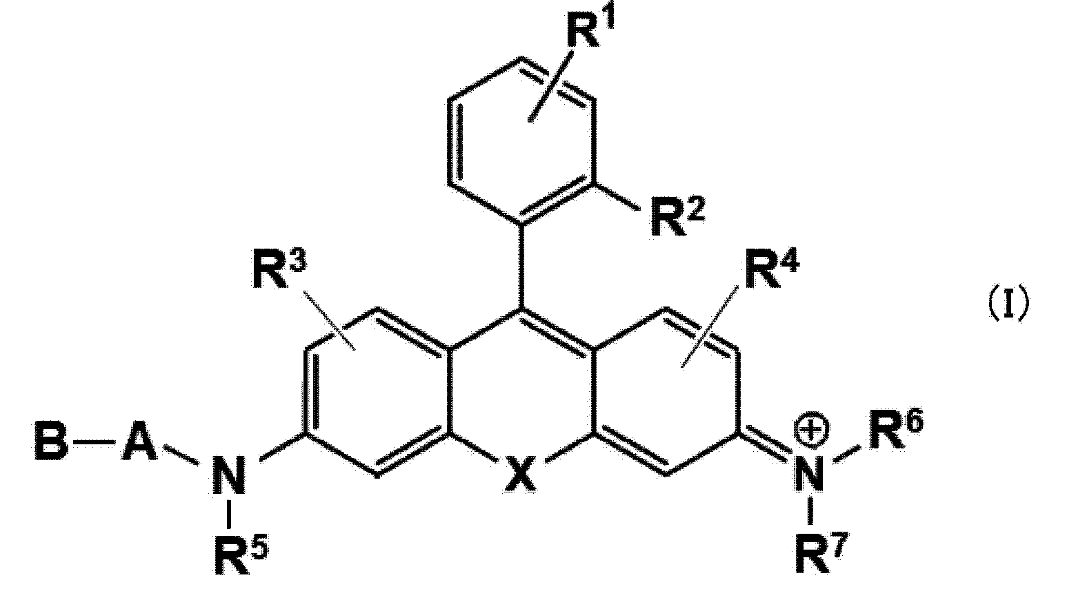
本発明の蛍光プローブは、一態様において、以下の式(I)で表される構造を有する化合物を含むものである。
上記式(I)において、A及びBは、アミノ酸残基が連結した構造を有し、ピューロマイシン感受性アミノペプチダーゼ(puromycin-sensitive aminopeptidase;PSA)によって選択的に加水分解され得る部位である。より詳細には、Aは、アラニン残基であり、Bは、アミノ酸残基を表す。ここで、Aは、式中のN(R5)とアミド結合を形成して連結しており、すなわち、アラニン残基Aのカルボニル部分(アシル基)とのN(R5)のN原子とがアミド結合を形成することでキサンテン骨格と連結している。さらに、Aは、通常のペプチド鎖と同様にアミノ酸残基Bと連結することができ、その結果、BはAとアミド結合を形成して連結する。従って、Bは、いわゆるN-末端残基と同様の構造を有し、中間のアミノ酸残基であるAは通常のペプチド鎖と同様にBと連結することができる。
なお、ピューロマイシン感受性アミノペプチダーゼ(PSA)は、Ala-AMCを標準基質とする、細胞質アラニルアミノペプチダーゼであり、特に中枢神経系において高発現となることが報告されている。また、臨床医療においては、腎機能を推定するためのバイオマーカーとして用いられ、その機能はピューロマイシンによって阻害されることが知られている。
ピューロマイシン感受性アミノペプチダーゼ(PSA)を検出するという本発明の目的の観点からは、上記A及びBは、PSAの基質ペプチドであって選択的に加水分解されやすいアミノ酸残基の組み合わせであることが好ましい。より具体的には、上述のように、Aは、アラニン残基(「Ala」或いは「A」)であり、Bは、アスパラギン残基(「Asn」或いは「N」)又はリシン残基(「Lys」或いは「K」)であることが好ましい。より好ましくは、Aがアラニン残基であり、Bがアスパラギン残基である。
上記式(I)において、Xは、Si(Ra)(Rb)、Ge(Ra)(Rb)、Sn(Ra)(Rb)、C(Ra)(Rb)、P(=O)(Ra)又はOを表す。ここで、Ra及びRbは、それぞれ独立に水素原子、アルキル基、又はアリール基を表す。Ra及びRbが、アルキル基である場合、それらは1以上の置換基を有することができ、そのような置換基としては、例えば、アルキル基、アルコキシ基、ハロゲン原子、水酸基、カルボキシル基、アミノ基、スルホ基などを1個又は2個以上有していてもよい。Ra及びRbは、それぞれ置換されていてもよい炭素数1~6個のアルキル基であることができ、いずれもメチル基であることが好ましい。また、場合によっては、Ra及びRbは互いに結合して環構造を形成していてもよい。例えば、Ra及びRbがともにアルキル基である場合に、Ra及びRbが互いに結合してスピロ炭素環を形成することができる。形成される環は、例えば5ないし8員環程度であることが好ましい。Xは、好ましくは、Si(Ra)(Rb)であり、より好ましくはSi(CH3)2である。
R1は、水素原子又はベンゼン環に結合する1個ないし4個の置換基を表す。好ましくは、R1は、水素原子、又はそれぞれ置換されていてもよいアルキル基、カルボキシル基、エステル基、アルコキシ基、アミド基、及びアジド基よりなる群から独立に選択される1~4個の同一又は異なる置換基を表す。R1が水素原子以外である場合、そのベンゼン環における位置は特に限定されないが、R2に対してメタ位であることが好ましい。また、ベンゼン環上に2個以上の置換基を有する場合には、それらは同一でも異なっていてもよい。好ましくは、R1は、水素原子である。
R2は、水素原子、ヒドロキシル基、シアノ基、それぞれ置換されていてもよいアルキル基、アルコキシ基、アリール、又はヘテロアリールを表す。好ましくは、ヒドロキシル基、シアノ基、それぞれ置換されていてもよいC1-C4アルキル基、C1-C4アルコキシ基、又はフェニル基である。より好ましくは、メチル基又はメトキシ基である。
R3及びR4は、それぞれ独立に、水素原子、ヒドロキシル基、ハロゲン原子、それぞれ置換されていてもよいアルキル基、スルホ基、カルボキシル基、エステル基、アミド基及びアジド基よりなる群から選択される1~3個の同一又は異なる置換基を表す。好ましくは、R3及びR4が、いずれも水素原子である。また、上記R1と同様に、R3又はR4が、蛍光共鳴エネルギー移動(FRET)のドナーとなる蛍光団を有することもできる。
R5、R6、及びR7は、それぞれ独立に水素原子又はアルキル基を表す。R5、R6、及びR7がともにアルキル基を示す場合には、それらは同一でも異なっていてもよい。例えば、R5、R6、及びR7はそれぞれ独立に、メチル基又はエチル基であることができる。好ましくは、R5、R6、及びR7が、いずれも水素原子である。
ここで、R6又はR7は、それぞれR4と一緒になって、それらが結合する窒素原子を含む環構造を形成してもよい。好ましくは、当該環構造は、5~8員のヘテロ環構造である。また、当該環構造は、R6及びR7が結合している窒素原子以外のヘテロ原子をさらに含むことができる。
好ましくは、R1、R3、R4、R5、R6、及びR7が、いずれも水素原子である。
本発明のPSA検出用蛍光プローブとして特に適切な式(I)の化合物の具体例(トリフルオロ酢酸塩)としては、
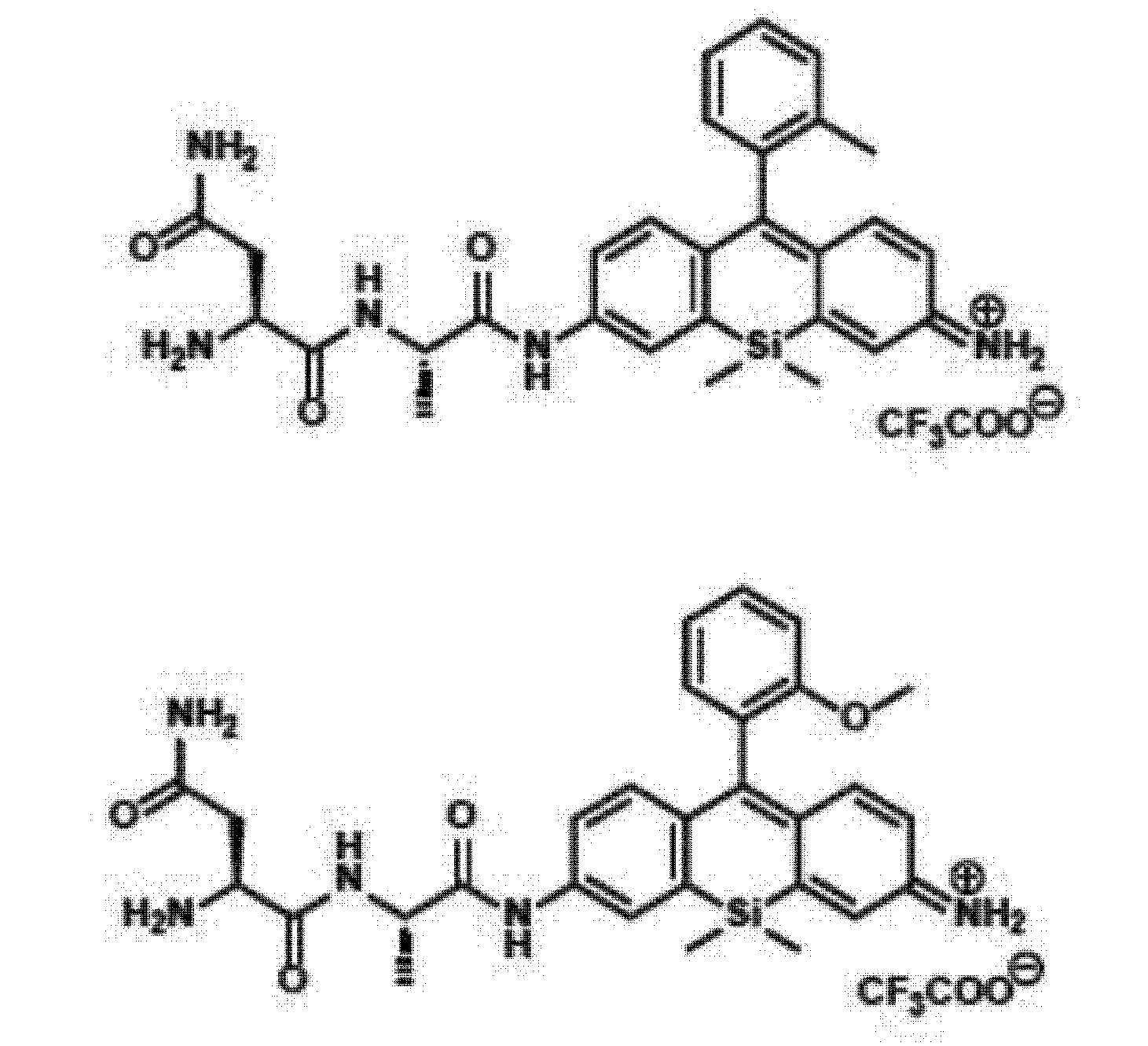
が挙げられる。ただし、これらに限定されるものではない。これらの化合物では、式(I)中の「A-B」は、いずれもアラニン残基-アスパラギン残基(Ala-Asn)となっている例である。
上記式(I)で表される化合物は、R6及びR7が連結するN原子において1価の正電荷を有するため、通常は塩として存在する。そのような塩としては、塩基付加塩、酸付加塩、アミノ酸塩などを挙げることができる。塩基付加塩としては、例えば、ナトリウム塩、カリウム塩、カルシウム塩、マグネシウム塩などの金属塩、アンモニウム塩、又はトリエチルアミン塩、ピペリジン塩、モルホリン塩などの有機アミン塩を挙げることができ、酸付加塩としては、例えば、塩酸塩、硫酸塩、硝酸塩などの鉱酸塩、カルボン酸塩、トリフルオロ酢酸塩、メタンスルホン酸塩、パラトルエンスルホン酸塩、クエン酸塩、シュウ酸塩などの有機酸塩を挙げることができる。アミノ酸塩としてはグリシン塩などを例示することができる。もっとも、これらの塩に限定されることはない。
式(I)で表される化合物は、置換基の種類に応じて1個または2個以上の不斉炭素を有する場合があり、光学異性体又はジアステレオ異性体などの立体異性体が存在する場合がある。純粋な形態の立体異性体、立体異性体の任意の混合物、ラセミ体などはいずれも本発明の範囲に包含される。
式(I)で表される化合物又はその塩は、水和物又は溶媒和物として存在する場合もあるが、これらの物質はいずれも本発明の範囲に包含される。溶媒和物を形成する溶媒の種類は特に限定されないが、例えば、エタノール、アセトン、イソプロパノールなどの溶媒を例示することができる。
上記の蛍光プローブは、必要に応じて試薬の調製に通常用いられる添加剤を配合して組成物として用いてもよい。例えば、生理的環境で用いるための添加剤として、溶解補助剤、pH調節剤、緩衝剤、等張化剤などの添加剤を用いることができ、これらの配合量は当業者に適宜選択可能である。これらの組成物は、粉末形態の混合物、凍結乾燥物、顆粒剤、錠剤、液剤など適宜の形態の組成物として提供され得る。
本明細書の実施例には、式(I)で表される本発明の化合物に包含される代表的化合物についての製造方法が具体的に示されているので、当業者は本明細書の開示を参照することにより、及び必要に応じて出発原料や試薬、反応条件などを適宜選択することにより、式(I)に包含される任意の化合物を容易に製造することができる。
3.本発明の蛍光プローブを用いたPSAの検出方法
本発明の蛍光プローブでは、光誘起電子移動(PET:Photoinduced electron transfer)を用いた蛍光制御によって、PSA酵素活性を検出するものである。より詳細には、以下のスキームに示すように、キサンテン環の3位の側鎖に「A-B」のアミノ酸残基(ペプチド鎖)が連結している状態では、励起光を照射してもキサンテン環部位の電子受容性が高いため、ベンゼン環部位からの電子移動によって消光している状態である。これに対し、蛍光プローブ分子がPSAと反応することによって、側鎖の「A-B」ペプチド鎖が加水分解により切断され、3位がアミノ基に変化した場合には、キサンテン環部位の電子受容性が低下し、その結果、蛍光発光が観測されることとなる。

本発明の蛍光プローブでは、光誘起電子移動(PET:Photoinduced electron transfer)を用いた蛍光制御によって、PSA酵素活性を検出するものである。より詳細には、以下のスキームに示すように、キサンテン環の3位の側鎖に「A-B」のアミノ酸残基(ペプチド鎖)が連結している状態では、励起光を照射してもキサンテン環部位の電子受容性が高いため、ベンゼン環部位からの電子移動によって消光している状態である。これに対し、蛍光プローブ分子がPSAと反応することによって、側鎖の「A-B」ペプチド鎖が加水分解により切断され、3位がアミノ基に変化した場合には、キサンテン環部位の電子受容性が低下し、その結果、蛍光発光が観測されることとなる。
すなわち、式(I)で表される化合物は、PSAとの反応前はベンゼン環部位からのPETにより蛍光が消光しているが、PSAによってペプチド鎖が切断されると、キサンテン環部位の電子密度が変化し、PETによる蛍光消光が抑制されるため、PSAの活性を蛍光応答として検出することができるという機構である。これにより、式(I)で表される蛍光プローブを取り込んだ細胞が、「A-B」を加水分解して切断可能なペプチダーゼ(PSA)を発現していない場合には、蛍光物質が当該細胞内で生成することはないが、そのようなペプチダーゼが存在する場合には、「A-B」が切断されることで強い蛍光発光が得られるため、これにより、標的とするペプチダーゼの存在を蛍光強度のon/off変化により観測し、PSA等のペプチダーゼを発現するがん細胞等の存在を検出することが可能となる。
また、式(I)で表される化合物では、キサンテン環の10位元素であるXの種類及び当該キサンテン骨格に連結するベンゼン環の置換基(R1及びR2)の種類を調整することで、「A-B」切断による蛍光発光を、蛍光波長が600nm以上の赤色領域の蛍光とすることができるという特徴を有するものである。これにより、がん細胞等の存在を赤色蛍光応答として検出することが可能となる。
本発明の蛍光プローブを用いて検出を行う場合には、励起光として、通常は400~600nm程度の可視光を照射すればよい。観測すべき蛍光波長は通常は580~700nm程度の領域である。
以上の発光機構に従い、本発明の蛍光プローブを用いるPSA検出方法では、PSAと前記蛍光プローブとの反応による蛍光応答又は吸光度変化を観測することにより、PSAの存在を特異的に検出又は可視化することができる。本明細書において「検出」という用語は、定量、定性など種々の目的の測定を含めて最も広義に解釈されるべきである。
具体的には、
A)本発明の蛍光プローブを測定対象試料と接触させる工程;及び、
B)当該試料中に含まれるピューロマイシン感受性アミノペプチダーゼ(PSA)と前記蛍光プローブの反応による蛍光応答又は吸光度変化観測する工程
を含むことによって、PSAを発現している標的細胞のみを特異的に蛍光応答として検出又は可視化することができる。
A)本発明の蛍光プローブを測定対象試料と接触させる工程;及び、
B)当該試料中に含まれるピューロマイシン感受性アミノペプチダーゼ(PSA)と前記蛍光プローブの反応による蛍光応答又は吸光度変化観測する工程
を含むことによって、PSAを発現している標的細胞のみを特異的に蛍光応答として検出又は可視化することができる。
また、本発明の方法は、さらに蛍光イメージング手段を用いて前記蛍光応答を観測することを含むことができる。上記蛍光応答を観測する手段は、広い測定波長を有する蛍光光度計を用いることができるが、前記蛍光応答を2次元画像として表示可能な蛍光イメージング手段を用いて可視化することもできる。蛍光イメージングの手段を用いることによって、蛍光応答を二次元で可視化できるため、PSAを瞬時に視認することが可能となる。蛍光イメージング装置としては、当該技術分野において公知の装置を用いることができる。なお、場合によって、紫外可視吸光スペクトルの変化(例えば、特定の吸収波長における吸光度の変化)によって上記測定対象試料と蛍光プローブの反応を検出することも可能である。
上記工程A)において、測定対象である試料と蛍光プローブを接触させる手段としては、代表的には、蛍光プローブを含む溶液を試料添加、塗布、或いは噴霧することが挙げられるが、上記試料の形態や測定環境等に応じて適宜選択することが可能である。かかる接触は、好ましくは生体外の環境で行われる。また、本発明の蛍光プローブを、動物個体における診断又は診断の補助、若しくは特定の細胞又は組織の検出に適用する際に、当該蛍光プローブと、標的細胞又は組織とを接触させる手段としては、特に限定されることなく、例えば、静脈内投与等、当該分野において一般的な投与手段を用いることができる。
本発明の蛍光プローブの適用濃度は特に限定されないが、例えば0.1~10μM程度の濃度の溶液を適用することができる。
また、標的細胞に行う光照射は、当該細胞に対して光を直接或いは導波管(光ファイバー等)を介して照射することができる。光源としては、酵素切断を受けた後の、本発明の蛍光プローブの吸収波長を含む光を照射できるものであれば、任意の光源を用いることができ、本発明の方法を実施する環境等に応じて適宜選択され得る。
本発明の蛍光プローブとしては、上記式(I)で表される化合物又はその塩をそのまま用いてもよいが、必要に応じて、試薬の調製に通常用いられる添加剤を配合して組成物として用いてもよい。例えば、生理的環境で試薬を用いるための添加剤として、溶解補助剤、pH調節剤、緩衝剤、等張化剤などの添加剤を用いることができ、これらの配合量は当業者に適宜選択可能である。これらの組成物は、一般的には、粉末形態の混合物、凍結乾燥物、顆粒剤、錠剤、液剤など適宜の形態の組成物として提供されるが、使用時に注射用蒸留水や適宜の緩衝液に溶解して適用することが可能である。
上記工程A)における測定対象試料は、ピューロマイシン感受性アミノペプチダーゼ(PSA)が発現している細胞であることができるが、かかる細胞がPSAを発現しているがん細胞やがん組織である場合には、本発明の検出方法によって、がん細胞やがん組織を検出又は可視化することができる。すなわち、本発明の蛍光プローブ、当該蛍光プローブを含む組成物、及び本発明の検出方法は、がんの診断に用いることもできる。
本明細書において、「がん組織」の用語はがん細胞を含む任意の組織を意味している。「組織」の用語は臓器の一部又は全体を含めて最も広義に解釈しなければならず、いかなる意味においても限定的に解釈してはならない。本発明の蛍光プローブ又は蛍光プローブを含むがん細胞検出用組成物は、がん組織において特異的に強発現しているPSAを検出する作用を有していることから、がん組織としてはPSAを高発現している組織が好ましい。また、本明細書において「診断」の用語は任意の生体部位においてがん組織の存在を肉眼的又は顕微鏡下に確認することを含めて最も広義に解釈する必要がある。上記工程A)における測定対象試料は、好ましくは、がん細胞であり、より好ましくは乳がん細胞である。
また、本発明の検出方法の一態様として、γ-グルタミルトランスペプチダーゼ(GGT)に対する蛍光プローブを、本発明の蛍光プローブと併用して用いることができる。これにより、乳がん細胞に発現しているGGTとPSAの両方をバイオマーカー酵素とした検出を行うことができるため、個々のプローブの単独使用に比べてより高い特異度で乳がん細胞の検出が可能となる。特に、本発明の蛍光プローブが赤色領域における蛍光発光を有する場合には、従来の緑色蛍光プローブと併用することによって、複数の蛍光応答領域を用いるマルチカラーイメージングも可能となる。
4.本発明の蛍光プローブを用いたPSA検出用キット
本発明の検出方法においては、上記蛍光プローブを含むPSA検出用キットを用いることが好ましい。当該キットにおいて、通常、本発明の蛍光プローブは溶液として調製されているが、例えば、粉末形態の混合物、凍結乾燥物、顆粒剤、錠剤、液剤など適宜の形態の組成物として提供され、使用時に注射用蒸留水や適宜の緩衝液に溶解して適用することもできる。
本発明の検出方法においては、上記蛍光プローブを含むPSA検出用キットを用いることが好ましい。当該キットにおいて、通常、本発明の蛍光プローブは溶液として調製されているが、例えば、粉末形態の混合物、凍結乾燥物、顆粒剤、錠剤、液剤など適宜の形態の組成物として提供され、使用時に注射用蒸留水や適宜の緩衝液に溶解して適用することもできる。
また、当該キットには、必要に応じてそれ以外の試薬等を適宜含んでいてもよい。例えば、添加剤として、溶解補助剤、pH調節剤、緩衝剤、等張化剤などの添加剤を用いることができ、これらの配合量は当業者に適宜選択可能である。特に、上述のように、本発明の蛍光プローブに加えて、GGT検出用の緑色蛍光プローブを含むものとすることができる。
以下、実施例により本発明をさらに詳細に説明するが、本発明はこれらによって限定されるものではない。
1.蛍光プローブの合成
以下に示す反応スキームに従い、キサンテン環の3位に種々の組み合わせのジペプチドを導入した、蛍光プローブ化合物を合成した。ここでは、まず反応スキーム(General Procedure)について説明した後、特定のジペプチドを有する個々の化合物の合成について説明する。
以下に示す反応スキームに従い、キサンテン環の3位に種々の組み合わせのジペプチドを導入した、蛍光プローブ化合物を合成した。ここでは、まず反応スキーム(General Procedure)について説明した後、特定のジペプチドを有する個々の化合物の合成について説明する。
[規則26に基づく補充 22.11.2019]
[反応スキーム(General Procedure)]
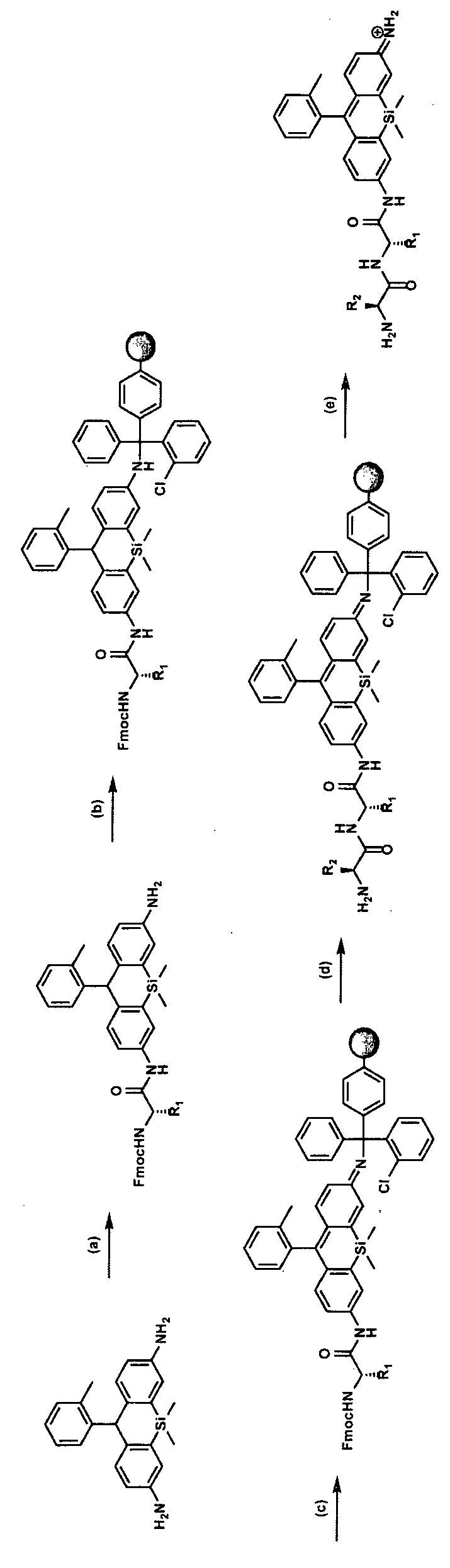
(a) Fmoc-A.A., HATU, DIEA, DMF, r.t. y. # Fmoc-A.A. = Gly 76.4 %, Glu(OtBu) 52.0 %, Lys(Boc) 46.6 %, Tyr(tBu) 42.6 %, Leu 52.4 %, Pro 49.6 %, Ile 63.2 %, Ala 42.2 %, Val 71.6 %, Trp(Boc) 63.5 %, Thr(tBu) 76.0 %, His(Trt) 53.8 %, Ser(tBu) 64.4 %, Phe 67.5 %, Cys(Trt) 66.6 %, Met 55.3 %, Asn(Trt) 64.4 %, Gln(Trt) 71.3 %, Asp(OtBu) 66.3 %, Arg(Pbf) 69.3 %, (b) 2-Chlorotrityl resin, DIEA, DMF/CH2Cl2, r.t. (c’) DDQ, DMF, r.t. (d) (i) piperidine, DMF/NMP, r.t. (ii) Fmoc-A.A., HATU, DIEA, DMF/NMP, r.t. (iii) piperidine, DMF/NMP, r.t. (e) TFA, Anisole, H2O, r.t.
[反応スキーム(General Procedure)]
(a) Fmoc-A.A., HATU, DIEA, DMF, r.t. y. # Fmoc-A.A. = Gly 76.4 %, Glu(OtBu) 52.0 %, Lys(Boc) 46.6 %, Tyr(tBu) 42.6 %, Leu 52.4 %, Pro 49.6 %, Ile 63.2 %, Ala 42.2 %, Val 71.6 %, Trp(Boc) 63.5 %, Thr(tBu) 76.0 %, His(Trt) 53.8 %, Ser(tBu) 64.4 %, Phe 67.5 %, Cys(Trt) 66.6 %, Met 55.3 %, Asn(Trt) 64.4 %, Gln(Trt) 71.3 %, Asp(OtBu) 66.3 %, Arg(Pbf) 69.3 %, (b) 2-Chlorotrityl resin, DIEA, DMF/CH2Cl2, r.t. (c’) DDQ, DMF, r.t. (d) (i) piperidine, DMF/NMP, r.t. (ii) Fmoc-A.A., HATU, DIEA, DMF/NMP, r.t. (iii) piperidine, DMF/NMP, r.t. (e) TFA, Anisole, H2O, r.t.
[化合物1(Leuco-2-Me SiR600 )の合成]

文献(Kushida, Y.ら、 Bioorganic & medicinal chemistry letters 2012, 22 (12), 3908-3911.)に従い、出発物質である化合物1を合成した。
[化合物2の合成]

DMF中の化合物1(2当量)の溶液に、Fmoc-アミノ酸(1当量)、HATU(1当量)およびDIEA(5当量)を加え、室温で1.5時間撹拌した。反応混合物を溶媒留去し、H2Oを加え、次いで水層をCH2Cl2で抽出した。有機層を塩水で洗浄し、Na2SO4で乾燥し、蒸発させた。残渣をカラムクロマトグラフィーまたはMPLCで精製した。不純物が含まれている場合は、溶離液としてクロロホルムを用いたGPCリサイクルカラムによる追加精製を行い、純粋な化合物を得た。
[化合物3の合成]

DMF 0.4mLおよびCH2Cl2 1.6mL中の化合物2(1-1.1当量)の溶液に、塩化2-クロロトリチル樹脂(1当量)およびDIEA 200μLを加えた。反応混合物を室温で撹拌した。Ar雰囲気下で一晩放置した。 反応溶液を濾過により除去し、樹脂をCH2Cl2で複数回洗浄した。 この樹脂を分割し、ペプチド合成機で次の反応に使用した。
[化合物4の合成]
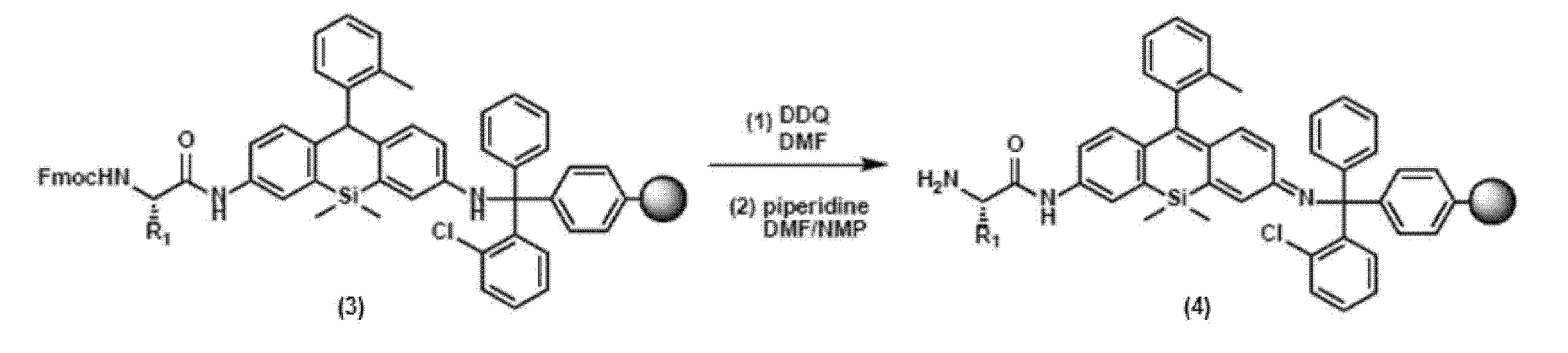
上記で得られた樹脂(0.0245mmol)にDMF 1.1mLを加え、1時間振とうして樹脂を膨潤させた。DMFを濾過により除去し、DMF中のDDQ(4当量)を加え、1時間振とうした。反応液をろ別した。樹脂に、DMF中の40%ピペリジンを加え、800μLを添加し、3分間振とうし、反応溶液を濾過により除去した。DMF中の40%ピペリジン400μLを添加し、12分間振とうし、反応溶液を濾過により除去した。樹脂にDMF900μLを加え、1分間振とうし、DMFを濾過により除去した。この洗浄操作をさらに5回繰り返した。
[化合物5の合成]

上記で得られた樹脂(0.0245mmol)にDMF中のFmoc-アミノ酸(4当量)、DMF中のHATU(4当量)およびNMP中の2M DIEA 200μLを加え、2時間振とうした。反応液をろ別した。樹脂にDMF 900μLを加え、1分間振とうし、DMFを濾過により除去した。この洗浄操作をさらに2回繰り返した。この凝縮操作をもう一度繰り返した。DMF中のFmoc-アミノ酸(4当量)、DMF中のHATU(4当量)およびNMP中の2M DIEA 200μLを樹脂(0.0245mmol)に添加し、1時間振とうした。反応液をろ別した。樹脂にDMF 900μLを加え、1分間振とうし、DMFを濾過により除去した。この洗浄操作をさらに2回繰り返した。樹脂に、DMF中の40%ピペリジン800μLを加え、3分間振とうし、反応溶液を濾過により除去した。DMF中の40%ピペリジン400μLを添加し、12分間振とうし、反応溶液を濾過により除去した。樹脂にDMF900μLを加え、1分間振とうし、DMFを濾過により除去した。この洗浄操作をさらに5回繰り返した。
[化合物6の合成]
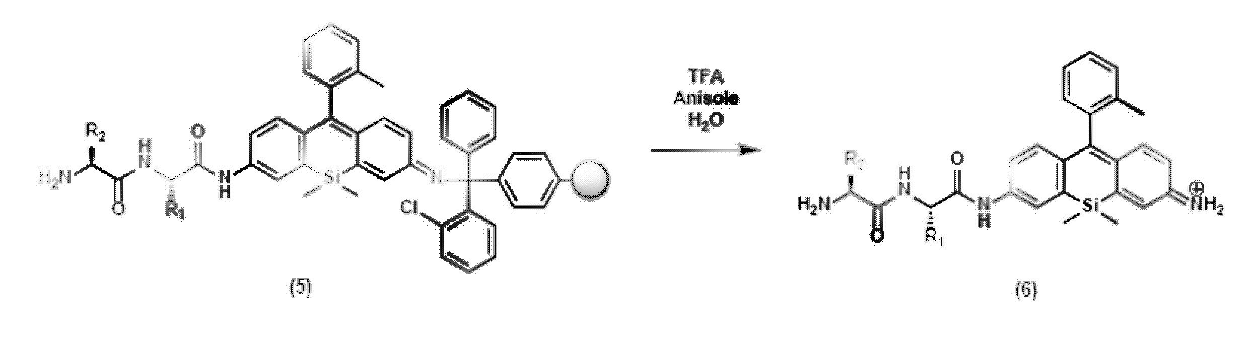
ペプチド合成機との反応後の樹脂(0.024mmol)に、TFA 2mL、アニソール50μL、水50μLを加え、2時間攪拌した。反応混合物を蒸発乾固させた。残渣を4℃で50mLのチューブ中の40mLのEt2O中に注ぎ、沈殿させ、遠心分離(3,000rpm、4℃で5分間)を行った。 上清を除去し、残渣を風乾して粗生成物(化合物6)を得た。粗生成物をDMSOに溶解し、LC-MSで分析した。254nmまたは490nmのいずれかでの粗生成物の純度が80%未満である場合、40分間でA / B = 70 / 30~0 / 100(溶出液A :0.1%TFAを含有するH2O、溶出液B:80%アセトニトリルおよび20%H2O中の0.1%TFA)で精製して十分な純度の化合物6を得た。
[本発明の蛍光プローブ1(NA-2-Me SiR600)の合成]
上述の反応スキームに従い、側鎖にAsn-Ala(NA)を有する本発明の蛍光プローブ化合物(NA-2-Me SiR600)を以下のとおり合成した。
上述の反応スキームに従い、側鎖にAsn-Ala(NA)を有する本発明の蛍光プローブ化合物(NA-2-Me SiR600)を以下のとおり合成した。
[化合物7の合成]

上記化合物2に相当する化合物7を化合物1から合成した。上記反応スキームに従い、化合物1(417mg、1.21mmol)、Fmoc-Ala-OH(380mg、1.22mmol)、HATU(232mg、0.611mmol)およびDIEA(515μL、3.03mmol)から、化合物7を合成した。AcOEt / n-ヘキサン(1/1)を溶出液としてとするMPLC、及びクロロホルムを溶出液とするGPCリサイクルカラムを用いて精製した後、化合物7を得た(163mg、42.2%)。
1H-NMR (300 MHz, CD2Cl2) δ: 0.40 (d, 3H, J = 2.9 Hz), 0.56 (d, 3H, J = 5.9 Hz), 1.46 (d, 3H, J = 6.6 Hz), 2.36 (s, 3H), 3.58 (br, 2H), 4.20 (t, 1H, J = 6.6 Hz), 4.42 (d, 2H, J = 7.3 Hz), 4.43-4.46 (m, 1H), 5.66 (s, 1H), 5.95 (d, 1H, J = 7.3 Hz), 6.60 (dd, 1H, J = 8.4, 2.6 Hz), 6.88 (d, 1H, J = 8.1 Hz), 6.92 (d, 1H, J = 2.2 Hz), 7.00-7.10 (m, 4H), 7.17-7.18 (m, 1H), 7.26-7.28 (m, 2H), 7.31-7.34 (m, 1H), 7.38 (t, 2H, J = 7.0 Hz), 7.55-7.61 (m, 2H), 7.76-7.81 (m, 2H), 7.89 (s, 1H), 8.58 (s, 1H).
13C-NMR (75 MHz, CD2Cl2, isomer mixture) δ: -0.57, -0.53, -0.33, 18.72, 20.77, 47.46, 50.35, 51.71, 67.43, 117.29, 119.16, 120.31, 121.63, 124.50, 125.35, 125.41, 126.27, 126.56, 127.44, 128.08, 130.12, 130.43, 130.95, 131.58, 133.93, 134.55, 135.24, 135.28, 135.66, 138.65, 141.61, 144.01, 144.22, 144.46, 145.50, 146.39, 156.73, 171.21.
HRMS (ESI+): calcd for [M+H]+, 638.28389 ; found, 638.28361 (-0.28 mmu)
1H-NMR (300 MHz, CD2Cl2) δ: 0.40 (d, 3H, J = 2.9 Hz), 0.56 (d, 3H, J = 5.9 Hz), 1.46 (d, 3H, J = 6.6 Hz), 2.36 (s, 3H), 3.58 (br, 2H), 4.20 (t, 1H, J = 6.6 Hz), 4.42 (d, 2H, J = 7.3 Hz), 4.43-4.46 (m, 1H), 5.66 (s, 1H), 5.95 (d, 1H, J = 7.3 Hz), 6.60 (dd, 1H, J = 8.4, 2.6 Hz), 6.88 (d, 1H, J = 8.1 Hz), 6.92 (d, 1H, J = 2.2 Hz), 7.00-7.10 (m, 4H), 7.17-7.18 (m, 1H), 7.26-7.28 (m, 2H), 7.31-7.34 (m, 1H), 7.38 (t, 2H, J = 7.0 Hz), 7.55-7.61 (m, 2H), 7.76-7.81 (m, 2H), 7.89 (s, 1H), 8.58 (s, 1H).
13C-NMR (75 MHz, CD2Cl2, isomer mixture) δ: -0.57, -0.53, -0.33, 18.72, 20.77, 47.46, 50.35, 51.71, 67.43, 117.29, 119.16, 120.31, 121.63, 124.50, 125.35, 125.41, 126.27, 126.56, 127.44, 128.08, 130.12, 130.43, 130.95, 131.58, 133.93, 134.55, 135.24, 135.28, 135.66, 138.65, 141.61, 144.01, 144.22, 144.46, 145.50, 146.39, 156.73, 171.21.
HRMS (ESI+): calcd for [M+H]+, 638.28389 ; found, 638.28361 (-0.28 mmu)
[蛍光プローブ1(化合物8:NA-2-Me SiR600)の合成]
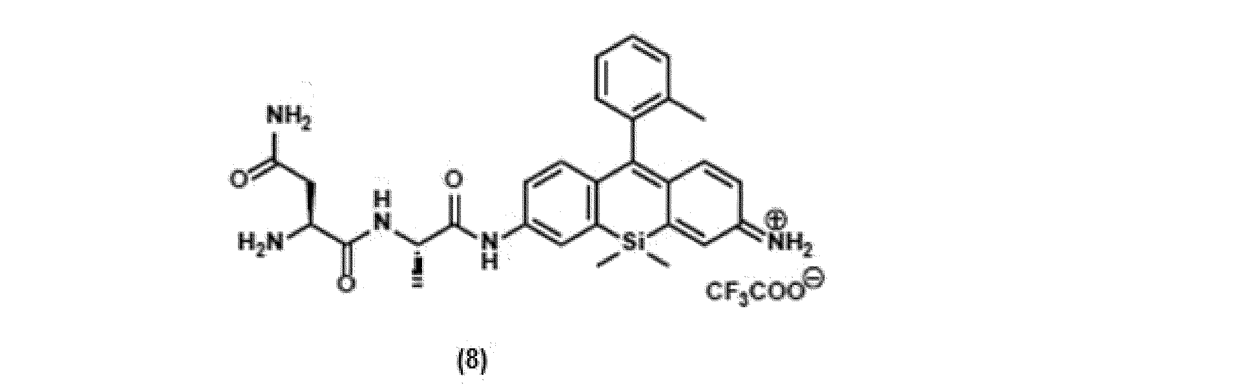
上記反応スキームに従い、化合物6に相当する化合物8を、上記反応スキームに従い化合物7(19.0mg、0.0298mmol)から合成した。粗生成物を以下の条件下でHPLCで精製し、化合物8を得た(10.2mg、53.3%)。40分でA / B = 70 / 30~0 / 100(溶出液A:0.1%TFAを含有するH2O、溶離液B:80%アセトニトリルおよび20%H2O中の0.1%TFA)。
1H-NMR (400 MHz, CD3OD) δ: 0.58 (s, 3H), 0.60 (s, 3H), 1.49 (d, 3H, J = 7.3 Hz), 2.04 (s, 3H), 2.78-2.87 (m, 1H), 2.94-3.02 (m, 1H), 4.22 (dd, 1H, J = 8.5, 4.8 Hz), 4.54 (q, 1H, J = 7.2 Hz), 6.77 (dd, 1H, J = 9.8, 2.5 Hz), 7.09 (d, 1H, J = 9.1 Hz), 7.17 (d, 1H, J = 7.3 Hz), 7.31 (d, 1H, J = 10.1 Hz), 7.38-7.46 (m, 3H), 7.51 (t, 1H, J = 7.3 Hz), 7.60-7.66 (m, 1H), 8.24-8.29 (m, 1H)
13C-NMR (100 MHz, CD3OD) δ: -1.96, -1.68, 17.80, 19.56, 36.23, 51.15, 51.57, 120.29, 122.07, 126.75, 127.01, 128.38, 130.20, 130.51, 131.16, 131.58, 136.06, 137.00, 138.71, 139.28, 144.01, 144.51, 147.36, 156.11, 162.41, 169.62, 169.76, 173.25, 173.65.
HRMS (ESI+): calcd for [M+H]+, 528.24309 ; found, 528.24304 (-0.05 mmu)
1H-NMR (400 MHz, CD3OD) δ: 0.58 (s, 3H), 0.60 (s, 3H), 1.49 (d, 3H, J = 7.3 Hz), 2.04 (s, 3H), 2.78-2.87 (m, 1H), 2.94-3.02 (m, 1H), 4.22 (dd, 1H, J = 8.5, 4.8 Hz), 4.54 (q, 1H, J = 7.2 Hz), 6.77 (dd, 1H, J = 9.8, 2.5 Hz), 7.09 (d, 1H, J = 9.1 Hz), 7.17 (d, 1H, J = 7.3 Hz), 7.31 (d, 1H, J = 10.1 Hz), 7.38-7.46 (m, 3H), 7.51 (t, 1H, J = 7.3 Hz), 7.60-7.66 (m, 1H), 8.24-8.29 (m, 1H)
13C-NMR (100 MHz, CD3OD) δ: -1.96, -1.68, 17.80, 19.56, 36.23, 51.15, 51.57, 120.29, 122.07, 126.75, 127.01, 128.38, 130.20, 130.51, 131.16, 131.58, 136.06, 137.00, 138.71, 139.28, 144.01, 144.51, 147.36, 156.11, 162.41, 169.62, 169.76, 173.25, 173.65.
HRMS (ESI+): calcd for [M+H]+, 528.24309 ; found, 528.24304 (-0.05 mmu)
[本発明の蛍光プローブ2(NA-2-OMe SiR600)の合成]
上述の反応スキームに従い、側鎖にAsn-Ala(NA)を有する本発明の蛍光プローブ2(NA-2-OMe SiR600)を以下のとおり合成した。蛍光プローブ2は、式(I)におけるR2に相当するベンゼン環上の置換基がメトキシである点で、蛍光プローブ1(NA-2-Me SiR600)と相違する。

(a) Fmoc-Ala-OH, HATU, DIEA, DMF, r.t. (b) 2-Chlorotrityl resin, DIEA, DMF/CH2Cl2, r.t. (c) DDQ, DMF, r.t. (d) (i) piperidine, DMF/NMP, r.t. (ii) Fmoc-Asn(Trt)-OH, HATU, DIEA, DMF/NMP, r.t. (iii) piperidine, DMF/NMP, r.t. (e) TFA, Anisole, H2O, r.t.
上述の反応スキームに従い、側鎖にAsn-Ala(NA)を有する本発明の蛍光プローブ2(NA-2-OMe SiR600)を以下のとおり合成した。蛍光プローブ2は、式(I)におけるR2に相当するベンゼン環上の置換基がメトキシである点で、蛍光プローブ1(NA-2-Me SiR600)と相違する。
[N,N,N',N'-Tetraallyldiamino-Si-xanthoneの合成]

N,N,N',N'-Tetraallyldiamino-Si-xanthoneを、文献(Kushida, Y.ら、 Bioorganic & medicinal chemistry letters 2012, 22 (12), 3908-3911.)に従い合成した。
[化合物9の合成]
上記化合物1におけるベンゼン環上の置換基をメチルからメトキシに変えた
化合物9(Leuco-2-OMe SiR600)を以下のとおり合成した。
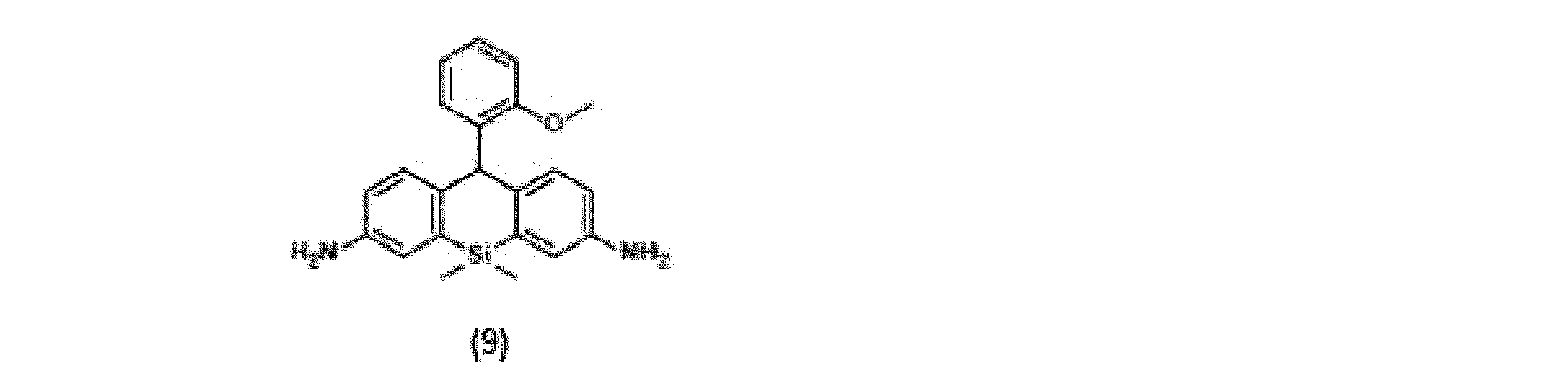
アルゴンでフラッシュした乾燥フラスコに、2-ブロモアニソール(5.57g、29.8mmol)および無水THF(30mL)を添加した。溶液を-78℃に冷却し、1M s-BuLi(26.7mmol)を加え、混合物を20分間撹拌した。同じ温度で、無水THF(10mL)に溶解したN,N,N',N'-Tetraallyldiamino-Si-xanthone(1.28g、2.97mmol)をゆっくり加え、混合物を室温に温めた。次いで、1時間撹拌した。2N HClの添加により反応を停止させ、混合物を室温で5分間撹拌した。飽和NaHCO3を加え、全体をCH2Cl2で抽出した。有機層を塩水で洗浄し、Na2SO4で乾燥させ、溶媒留去した。残渣をMeOHに溶解し、0℃で撹拌し、NaBH4(340mg、8.99mmol)を加えた。反応混合物を0℃で10分間撹拌した。混合物を蒸発させ、H2Oを加え、次いで水層をCH2Cl2で抽出した。有機層を塩水で洗浄し、Na2SO4で乾燥させ、溶媒留去した。残渣をMPLC(シリカゲル、ヘキサン/ AcOEt = 100:0~87:13)で精製し、粗生成物を得た(2.58g)。粗生成物のCH2Cl2(50mL)溶液に1,3-ジメチルバルビツール酸(3.28g、21.0mmol)とPd(PPh3)4(0.94g、0.81mmol)を加え、35℃でAr雰囲気中で22時間加熱した。NaHCO3水溶液を加えて反応を停止させ、水層をCH2Cl2で抽出した。有機層を塩水で洗浄し、Na2SO4で乾燥させ、溶媒留去した。残渣をMPLC(シリカゲル、ヘキサン/ AcOEt = 71:29~50:50)で精製し、化合物9を得た(708mg、66.2%)。
1H-NMR (300 MHz, CD2Cl2) δ: 0.50 (s, 3H), 0.57 (s, 3H), 3.65 (s, 4H), 3.98 (s, 3H), 6.04 (s, 1H), 6.64 (dd, 2H, J = 8.1, 2.2 Hz), 6.73-6.84 (m, 2H), 6.90-6.94 (m, 1H), 6.94 (d, 2H, J = 2.2 Hz), 7.06-7.12 (m, 1H), 7.20 (d, 2H, J = 8.1 Hz).
13C-NMR (75 MHz, CD2Cl2) δ: -0.69, 0.71, 45.91, 55.88, 111.55, 117.22, 119.35, 120.91, 126.87, 129.99, 130.82, 134.79, 137.68, 139.98, 144.43, 156.16
HRMS (ESI+): calcd for [M+H]+, 361.17361 ; found, 361.17301 (-0.60 mmu)
上記化合物1におけるベンゼン環上の置換基をメチルからメトキシに変えた
化合物9(Leuco-2-OMe SiR600)を以下のとおり合成した。
1H-NMR (300 MHz, CD2Cl2) δ: 0.50 (s, 3H), 0.57 (s, 3H), 3.65 (s, 4H), 3.98 (s, 3H), 6.04 (s, 1H), 6.64 (dd, 2H, J = 8.1, 2.2 Hz), 6.73-6.84 (m, 2H), 6.90-6.94 (m, 1H), 6.94 (d, 2H, J = 2.2 Hz), 7.06-7.12 (m, 1H), 7.20 (d, 2H, J = 8.1 Hz).
13C-NMR (75 MHz, CD2Cl2) δ: -0.69, 0.71, 45.91, 55.88, 111.55, 117.22, 119.35, 120.91, 126.87, 129.99, 130.82, 134.79, 137.68, 139.98, 144.43, 156.16
HRMS (ESI+): calcd for [M+H]+, 361.17361 ; found, 361.17301 (-0.60 mmu)
[化合物10の合成]

DMF(10mL)中の化合物9(127mg、0.353mmol)に、Fmoc-Ala-OH(55mg、0.177mmol)、HATU(67mg、0.176mmol)およびDIEA(114μL、0.670mmol)を加え、Ar雰囲気中で1.5時間室温で撹拌した。反応混合物を溶媒留去し、H2Oを加え、次いで水層をCH2Cl2で抽出した。有機層を塩水で洗浄し、Na2SO4で乾燥し、溶媒留去した。溶離液としてAcOEt / n-ヘキサン(1/1)を用いたMPLCで精製した後、クロロホルムを溶離液として用いるGPCリサイクルカラムで精製し、化合物10を得た(81mg、70.3%)。
1H-NMR (400 MHz, CD2Cl2) δ: 0.46 (d, 3H, J = 1.8 Hz), 0.53 (d, 3H, J = 3.2 Hz), 1.45 (d, 3H, J = 6.9 Hz), 3.67 (br, 2H), 3.95 (s, 3H), 4.23 (t, 1H, J = 6.9 Hz), 4.44 (d, 3H, J = 6.9 Hz), 5.71 (s, 1H), 6.08 (s, 1H), 6.64 (d, 1H, J = 7.3 Hz), 6.71-6.79 (m, 2H), 6.91 (d, 2H, J = 7.8 Hz), 7.06-7.10 (m, 1H), 7.19 (d, 1H, J = 8.2 Hz), 7.28 (d, 2H, J = 6.4 Hz), 7.34-7.46 (m, 4H), 7.57-7.61 (m, 2H), 7.74-7.79 (m, 3H), 8.32 (s, 1H)
13C-NMR (100 MHz, CD2Cl2, isomer mixture) δ: -0.74, 0.63, 18.58, 46.39, 47.54, 51.75, 55.87, 67.44, 111.58, 117.38, 119.35, 120.33, 120.95, 121.73, 124.65, 125.38, 125.42, 127.15, 127.47, 128.10, 130.04, 130.56, 130.85, 134.50, 135.04, 135.67, 136.89, 139.37, 141.66, 144.17, 144.25, 144.55, 146.23, 156.15, 156.71, 170.96.
HRMS (ESI+): calcd for [M+Na]+, 676.26075 ; found, 676.26088 (0.13 mmu)
1H-NMR (400 MHz, CD2Cl2) δ: 0.46 (d, 3H, J = 1.8 Hz), 0.53 (d, 3H, J = 3.2 Hz), 1.45 (d, 3H, J = 6.9 Hz), 3.67 (br, 2H), 3.95 (s, 3H), 4.23 (t, 1H, J = 6.9 Hz), 4.44 (d, 3H, J = 6.9 Hz), 5.71 (s, 1H), 6.08 (s, 1H), 6.64 (d, 1H, J = 7.3 Hz), 6.71-6.79 (m, 2H), 6.91 (d, 2H, J = 7.8 Hz), 7.06-7.10 (m, 1H), 7.19 (d, 1H, J = 8.2 Hz), 7.28 (d, 2H, J = 6.4 Hz), 7.34-7.46 (m, 4H), 7.57-7.61 (m, 2H), 7.74-7.79 (m, 3H), 8.32 (s, 1H)
13C-NMR (100 MHz, CD2Cl2, isomer mixture) δ: -0.74, 0.63, 18.58, 46.39, 47.54, 51.75, 55.87, 67.44, 111.58, 117.38, 119.35, 120.33, 120.95, 121.73, 124.65, 125.38, 125.42, 127.15, 127.47, 128.10, 130.04, 130.56, 130.85, 134.50, 135.04, 135.67, 136.89, 139.37, 141.66, 144.17, 144.25, 144.55, 146.23, 156.15, 156.71, 170.96.
HRMS (ESI+): calcd for [M+Na]+, 676.26075 ; found, 676.26088 (0.13 mmu)
[蛍光プローブ2(化合物11:NA-2-OMe SiR600)の合成]
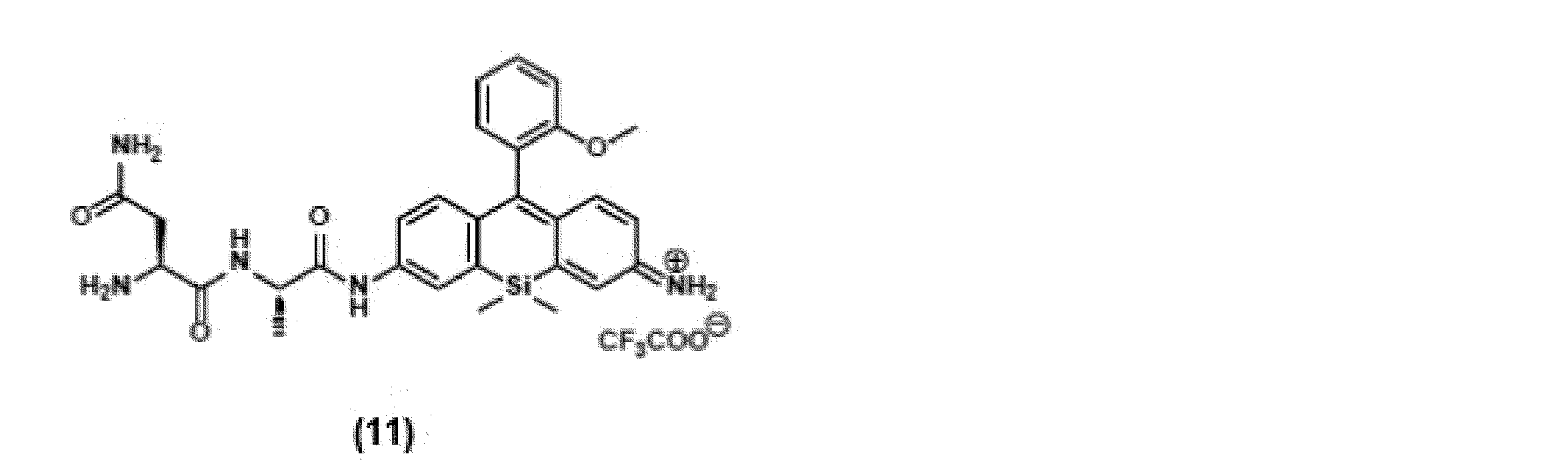
上記反応スキーム(General Procedure)に従い、化合物10(18.0mg、0.0276mmol)から、化合物11を合成した。粗生成物を以下の条件下でHPLCで精製し、化合物11を得た(10.9mg、62.3%)。40分でA / B = 70 / 30~0 / 100(溶出液A:0.1%TFAを含有するH2O、溶離液B:80%アセトニトリルおよび20%H2O中の0.1%TFA)。
1H-NMR (400 MHz, CD3OD, isomer mixture) δ: 0.47 (s, 0.6H), 0.57 (s, 2.4H), 0.60 (d, 3H, J = 3.2 Hz), 1.49 (d, 2.7H, J = 7.3 Hz), 1.62 (d, 0.3H, J = 6.8 Hz), 2.66 (br, 0.2H), 2.77-2.87 (m, 0.9H), 2.96-3.02 (m, 0.9H), 3.09 (s, 0.6H), 3.73 (s, 2.4H), 4.06-4.25 (m, 1H), 4.49-4.57 (m, 1H), 6.69 (d, 0.2H, J = 4.0 Hz), 6.76 (d, 0.8H, J = 4.8Hz), 7.09-7.26 (m, 4H), 7.32 (d, 0.2H, J = 4.2 Hz), 7.38-7.42 (m, 1.8H), 7.53 (s, 0.2H), 7.59-7.68 (m, 1.6 H), 7.87-7.91 (m, 0.2H), 8.14 (s, 0.1H), 8.23 (br, 1H)
13C-NMR (100 MHz, CD3OD) δ: -1.91, -1.66, 17.83, 36.23, 51.17, 51.57, 56.25, 112.51, 119.88, 121.64, 121.90, 126.52, 128.09, 128.26, 131.46, 131.67, 132.27, 136.56, 138.76, 143.68, 144.28, 147.93, 156.11, 158.10, 162.39, 168.37, 169.59, 173.27, 173.60
HRMS (ESI+): calcd for [M+H]+, 544.23800 ; found, 544.23644 (-1.57 mmu)
1H-NMR (400 MHz, CD3OD, isomer mixture) δ: 0.47 (s, 0.6H), 0.57 (s, 2.4H), 0.60 (d, 3H, J = 3.2 Hz), 1.49 (d, 2.7H, J = 7.3 Hz), 1.62 (d, 0.3H, J = 6.8 Hz), 2.66 (br, 0.2H), 2.77-2.87 (m, 0.9H), 2.96-3.02 (m, 0.9H), 3.09 (s, 0.6H), 3.73 (s, 2.4H), 4.06-4.25 (m, 1H), 4.49-4.57 (m, 1H), 6.69 (d, 0.2H, J = 4.0 Hz), 6.76 (d, 0.8H, J = 4.8Hz), 7.09-7.26 (m, 4H), 7.32 (d, 0.2H, J = 4.2 Hz), 7.38-7.42 (m, 1.8H), 7.53 (s, 0.2H), 7.59-7.68 (m, 1.6 H), 7.87-7.91 (m, 0.2H), 8.14 (s, 0.1H), 8.23 (br, 1H)
13C-NMR (100 MHz, CD3OD) δ: -1.91, -1.66, 17.83, 36.23, 51.17, 51.57, 56.25, 112.51, 119.88, 121.64, 121.90, 126.52, 128.09, 128.26, 131.46, 131.67, 132.27, 136.56, 138.76, 143.68, 144.28, 147.93, 156.11, 158.10, 162.39, 168.37, 169.59, 173.27, 173.60
HRMS (ESI+): calcd for [M+H]+, 544.23800 ; found, 544.23644 (-1.57 mmu)
これら蛍光プローブ1及び2の他に、上述の反応スキームに従い、式(I)のA-B部位として、天然アミノ酸20種×20種、合計400種のジペプチドを導入した400種の蛍光プローブからなるライブラリーを構築した。
ヒト乳がん手術検体を用いたスクリーニング
実施例1で合成した蛍光プローブのライブラリーを用いて、ヒト乳がん手術検体におけるスクリーニングを実施した。初めに手術検体から腫瘍部位と正常部位に分けてライセートを作成し、A-B部位のペプチド鎖が異なる各プローブをそれぞれのライセートと反応させて蛍光強度変化を測定し、腫瘍部位由来のライセートで充分な蛍光上昇を示し、かつ高いT/N比(Tumor to Normal ratio)を示した36種の蛍光プローブを候補プローブとして選出した。
実施例1で合成した蛍光プローブのライブラリーを用いて、ヒト乳がん手術検体におけるスクリーニングを実施した。初めに手術検体から腫瘍部位と正常部位に分けてライセートを作成し、A-B部位のペプチド鎖が異なる各プローブをそれぞれのライセートと反応させて蛍光強度変化を測定し、腫瘍部位由来のライセートで充分な蛍光上昇を示し、かつ高いT/N比(Tumor to Normal ratio)を示した36種の蛍光プローブを候補プローブとして選出した。
続いて、候補となったプローブ群について検体切除片にプローブ溶液を滴下して蛍光イメージングを行い、蛍光強度変化を測定した。具体的には、ヒト乳がん検体から3~5 mm角の片をメスによって多数切り出し、それらを8-well chamber slide(Thermo Fisher 155411)の各ウェルに入れ、そこにそれぞれの蛍光プローブ溶液(50μM)を滴下し、蛍光強度の変化をMaestro In-Vivo Imaging Systemで測定した。
その結果、式(I)中のA-B部位が「Asn-Ala」(NA)である蛍光プローブ1(化合物8:NA-2-Me SiR600)が腫瘍部位において迅速な蛍光上昇を示す一方で、正常部位においてはほとんど上昇を示さないことが判明した。その他、A-B部位が「Lys-Ala」(KA)及び「Met-Ala」(MA)とした蛍光プローブも高い感度を示した。
さらに、蛍光プローブ1について、同様のイメージングを他の13症例の乳がん検体でも行ったところ、いずれも腫瘍部位で有意な蛍光上昇を示し、高いT/N比で腫瘍部位を蛍光検出可能であることが示唆された。図1に、蛍光プローブ1で処理した腫瘍検体と正常検体の蛍光イメージング画像(a及びb)、及び蛍光強度の時間変化(c)を示す。
また、蛍光プローブ1(NA)とA-B部位が「Lys-Ala」である化合物(KA)について、腫瘍部位への添加に伴うT/N比の時間変化を図2に示す。各時間におけるT/N比は1分後を除いて、蛍光プローブ1(NA)の方が高い値を示した。
バイオマーカー酵素の特定
蛍光プローブ1について、手術検体の腫瘍部位から作成したライセートを用いて、DEG(diced electrophoresis gel)アッセイを行ったところ、活性の高い単一のスポットが観測された(図3(a))。このスポットに注目してPMF (Peptide Mass Fingerprinting)解析を行った。その結果、蛍光プローブ1の代謝酵素としてピューロマイシン感受性アミノペプチダーゼ(PSA)が同定された。同定されたPSAについて精製酵素を用いて蛍光プローブ1と反応させたところ、迅速な蛍光上昇を示した(図3(b))。これらの結果より、蛍光プローブ1がPSAをバイオマーカー酵素とする乳がん検出蛍光プローブとして有効であることが分かった。
蛍光プローブ1について、手術検体の腫瘍部位から作成したライセートを用いて、DEG(diced electrophoresis gel)アッセイを行ったところ、活性の高い単一のスポットが観測された(図3(a))。このスポットに注目してPMF (Peptide Mass Fingerprinting)解析を行った。その結果、蛍光プローブ1の代謝酵素としてピューロマイシン感受性アミノペプチダーゼ(PSA)が同定された。同定されたPSAについて精製酵素を用いて蛍光プローブ1と反応させたところ、迅速な蛍光上昇を示した(図3(b))。これらの結果より、蛍光プローブ1がPSAをバイオマーカー酵素とする乳がん検出蛍光プローブとして有効であることが分かった。
PSAに対する蛍光アッセイ
実施例1で合成した蛍光プローブ1(化合物8:NA-2-Me SiR600)及び蛍光プローブ2(化合物11:NA-2-OMe SiR600)について、吸収及び蛍光スペクトルを測定し、また、量子収率を算出した。いずれも、蛍光プローブ濃度は1μMとし、0.1M リン酸Naバッファー(pH7.4、0.1%DMSO)溶液を用いた。
実施例1で合成した蛍光プローブ1(化合物8:NA-2-Me SiR600)及び蛍光プローブ2(化合物11:NA-2-OMe SiR600)について、吸収及び蛍光スペクトルを測定し、また、量子収率を算出した。いずれも、蛍光プローブ濃度は1μMとし、0.1M リン酸Naバッファー(pH7.4、0.1%DMSO)溶液を用いた。
得られた吸収及び蛍光スペクトルを図4に示し、算出した量子収率を図5に示す。吸収スペクトルに大きな違いはないが、一方で蛍光スペクトルに着目すると、蛍光プローブ2は蛍光プローブ1と比較して大幅に消光していることが分かった。蛍光プローブ2は、蛍光量子収率もほぼ0にまで低下していることが分かった。これは、式(I)におけるR2の置換基をメトキシ基としたことにより、A-B部位が切断される前の状態において、光誘起電子移動による蛍光消光が大きくなり、無蛍光性となっているものと考えられる。したがって、蛍光プローブ2は、高いT/N比での蛍光応答が期待でき、生体内でのイメージングに適した蛍光プローブであることが分かった。
次に、蛍光プローブ2とPSAとの反応性を確認するため、PSAの精製酵素を用いて蛍光強度変化を測定した。リン酸緩衝生理食塩水(PBS)中、1μMの蛍光プローブ2溶液に、酵素溶液として、0.0015%のDMSO及び0.1%のCHAPSを含むPSA(0.55μg/ウェル)溶液を添加した際の蛍光強度変化を図6に示す。また、比較例として、γ-グルタミルトランスペプチダーゼ(GGT)溶液(150μg/ウェル)を用いた場合の蛍光強度変化も併せて示す。励起波長は544nm、発光波長は615nmである。
図6の結果から、蛍光プローブ2は、PSAの添加に伴い、速やかに蛍光上昇を示した。このことは、式(I)中のA-B部位の「Asn-Ala」がPSAとの反応により切断されることによって、PETが抑制され、キサンテン環の蛍光発光が回復したためであると考えられる。一方、GGTを添加した場合には、蛍光上昇が認められないことから、蛍光プローブ2は、GGTによってA-B部位が切断されないことが分かった。この結果は、蛍光プローブ2は、PSAの存在を特異的に検出可能であることを実証するものである。
乳がん細胞に対するマルチカラーイメージング
PSAに対する赤色蛍光プローブとして機能することが示された蛍光プローブ2と、GGTに対する蛍光応答を示すことが知られている乳がん検出緑色蛍光プローブ(gGlu-HMRG)とを併用して、乳がんのマルチカラーイメージングを試みた。gGlu-HMRGは、以下の構造を有する蛍光プローブであり、その合成法等は、Y.Uranoら、Sci. Transl. Med.,vol.3, pp.110ra119、2011年(非特許文献1)に開示されている。
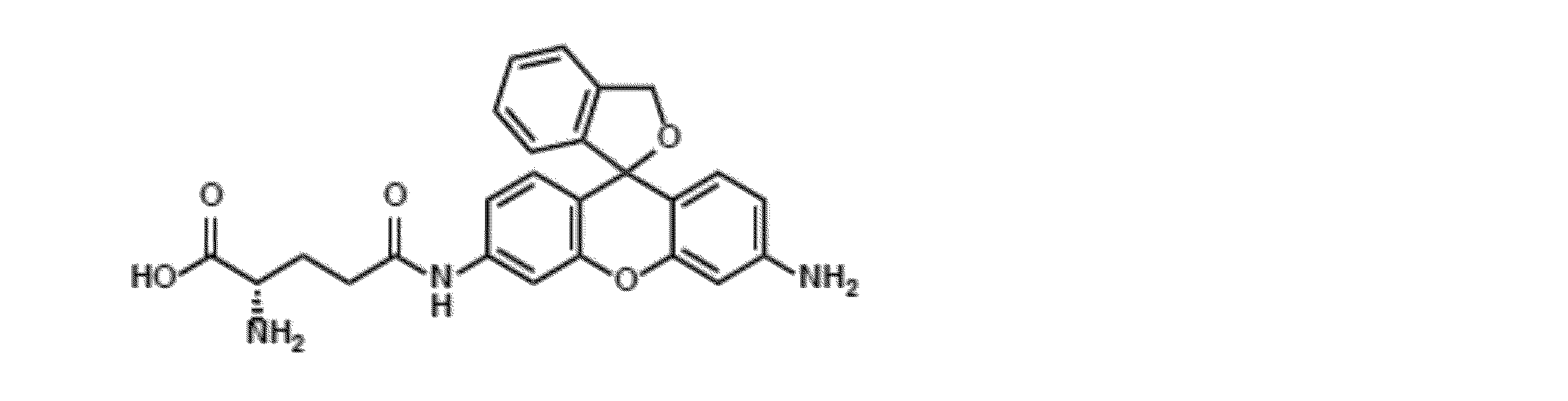
PSAに対する赤色蛍光プローブとして機能することが示された蛍光プローブ2と、GGTに対する蛍光応答を示すことが知られている乳がん検出緑色蛍光プローブ(gGlu-HMRG)とを併用して、乳がんのマルチカラーイメージングを試みた。gGlu-HMRGは、以下の構造を有する蛍光プローブであり、その合成法等は、Y.Uranoら、Sci. Transl. Med.,vol.3, pp.110ra119、2011年(非特許文献1)に開示されている。
乳がん検体に対して、5 mm角程度の大きさで腫瘍部位、正常部位を別々に切り出し、それぞれに蛍光プローブ2とgGlu-HMRGの混合溶液を滴下した。滴下1, 3, 5, 10, 15, 20, 30分後の緑色及び赤色の蛍光像を取得し、それらを重ね合わせることでマージ画像を作成した。同様の手法で合計14症例についてイメージングを行った。これらのうち1症例の画像を代表例として、図7に示す。図7では、蛍光プローブ2とgGlu-HMRGのいずれについても、腫瘍部位においてのみ蛍光上昇が認められ、正常部位については蛍光上昇が見られなかった。14例中13例(93 %)で腫瘍部位の検出ができた。これは、PSAに対する蛍光プローブ2と、GGTに対するgGlu-HMRGを併用することで、乳がんの腫瘍部位をより高い特異度で検出可能であることを実証するものである。
以上の結果は、本発明の蛍光プローブは、PSAを選択的に検出可能であり、乳がんに対して極めて実用性の高い蛍光プローブであることを示すものである。
Claims (13)
- 以下の式(I)で表される化合物又はその塩を含む、ピューロマイシン感受性アミノペプチダーゼ(PSA)検出用蛍光プローブ:

Aは、アラニン残基であり、Bは、アミノ酸残基を表し、ここで、Aは隣接するN原子とアミド結合を形成して連結し、BはAとアミド結合を形成して連結しており、
Xは、Si(Ra)(Rb)、Ge(Ra)(Rb)、Sn(Ra)(Rb)、C(Ra)(Rb)、P(=O)(Ra)又はOを表し(ここで、Ra及びRbは、それぞれ独立に水素原子、アルキル基、又はアリール基を表す);
R1は、水素原子、又はそれぞれ置換されていてもよいアルキル基、カルボキシル基、エステル基、アルコキシ基、アミド基、及びアジド基よりなる群から独立に選択される1~4個の同一又は異なる置換基を表し;
R2は、水素原子、ヒドロキシル基、シアノ基、それぞれ置換されていてもよいアルキル基、アルコキシ基、アリール、又はヘテロアリールを表し;
R3及びR4は、それぞれ独立に、水素原子、ヒドロキシル基、ハロゲン原子、それぞれ置換されていてもよいアルキル基、スルホ基、カルボキシル基、エステル基、アミド基及びアジド基よりなる群から独立に選択される1~3個の同一又は異なる置換基を表し;
R5、R6、及びR7は、それぞれ独立に水素原子又はアルキル基を表し、
ここで、R6又はR7は、それぞれR4と一緒になって、それらが結合する窒素原子を含む環構造を形成してもよい。〕。 - Bが、アスパラギン残基又はリシン残基である、請求項1に記載の蛍光プローブ。
- R2が、置換されていてもよい炭素数1~6個のアルキル基、又は置換されていてもよい炭素数1~6個のアルコキシ基である、請求項1又は2に記載の蛍光プローブ。
- R2が、メチル基又はメトキシ基である、請求項1~3のいずれか1に記載の蛍光プローブ。
- R1、R3、R4、R5、R6、及びR7が、いずれも水素原子である、請求項1~4のいずれか1に記載の蛍光プローブ。
- 請求項1~5のいずれか1に記載の蛍光プローブを含む、がん細胞検出用組成物。
- 前記がん細胞が、乳がん細胞である、請求項6に記載の組成物。
- 請求項1~5のいずれか1項に記載の蛍光プローブを含む、ピューロマイシン感受性アミノペプチダーゼ(PSA)検出用キット。
- 請求項1~5のいずれか1項に記載の蛍光プローブを測定対象試料と接触させる工程、及び、当該試料中に含まれるピューロマイシン感受性アミノペプチダーゼ(PSA)と前記蛍光プローブの反応による蛍光応答又は吸光度変化観測する工程を含むことを特徴とする、PSAの検出方法。
- 蛍光イメージング手段を用いて前記蛍光応答を可視化することを特徴とする、請求項9に記載の検出方法。
- 前記測定対象試料が、ピューロマイシン感受性アミノペプチダーゼ(PSA)が発現している細胞である、請求項9又は10に記載の方法。
- 前記細胞が、がん細胞である、請求項11に記載の方法。
- 前記がん細胞が、乳がん細胞である、請求項12に記載の方法。
Applications Claiming Priority (2)
| Application Number | Priority Date | Filing Date | Title |
|---|---|---|---|
| JP2018210101A JP7303532B2 (ja) | 2018-11-07 | 2018-11-07 | 新規蛍光プローブ |
| JP2018-210101 | 2018-11-07 |
Publications (1)
| Publication Number | Publication Date |
|---|---|
| WO2020095946A1 true WO2020095946A1 (ja) | 2020-05-14 |
Family
ID=70611910
Family Applications (1)
| Application Number | Title | Priority Date | Filing Date |
|---|---|---|---|
| PCT/JP2019/043494 Ceased WO2020095946A1 (ja) | 2018-11-07 | 2019-11-06 | 新規蛍光プローブ |
Country Status (2)
| Country | Link |
|---|---|
| JP (1) | JP7303532B2 (ja) |
| WO (1) | WO2020095946A1 (ja) |
Cited By (2)
| Publication number | Priority date | Publication date | Assignee | Title |
|---|---|---|---|---|
| JPWO2021070906A1 (ja) * | 2019-10-09 | 2021-04-15 | ||
| RU234589U1 (ru) * | 2025-03-11 | 2025-06-03 | федеральное государственное автономное образовательное учреждение высшего образования "Московский физико-технический институт (национальный исследовательский университет)" | Устройство для детектирования одиночных флуоресцентных молекул |
Citations (3)
| Publication number | Priority date | Publication date | Assignee | Title |
|---|---|---|---|---|
| WO2012111818A1 (ja) * | 2011-02-18 | 2012-08-23 | 国立大学法人 東京大学 | 蛍光プローブ |
| WO2014136780A1 (ja) * | 2013-03-04 | 2014-09-12 | 国立大学法人 東京大学 | カルパイン活性検出蛍光プローブ |
| JP2018145126A (ja) * | 2017-03-03 | 2018-09-20 | 国立大学法人 東京大学 | カルボキシペプチダーゼ活性検出用蛍光プローブ |
Family Cites Families (1)
| Publication number | Priority date | Publication date | Assignee | Title |
|---|---|---|---|---|
| EP3636653A1 (en) | 2013-01-07 | 2020-04-15 | The University of Tokyo | Asymmetrical si rhodamine and rhodol synthesis |
-
2018
- 2018-11-07 JP JP2018210101A patent/JP7303532B2/ja active Active
-
2019
- 2019-11-06 WO PCT/JP2019/043494 patent/WO2020095946A1/ja not_active Ceased
Patent Citations (3)
| Publication number | Priority date | Publication date | Assignee | Title |
|---|---|---|---|---|
| WO2012111818A1 (ja) * | 2011-02-18 | 2012-08-23 | 国立大学法人 東京大学 | 蛍光プローブ |
| WO2014136780A1 (ja) * | 2013-03-04 | 2014-09-12 | 国立大学法人 東京大学 | カルパイン活性検出蛍光プローブ |
| JP2018145126A (ja) * | 2017-03-03 | 2018-09-20 | 国立大学法人 東京大学 | カルボキシペプチダーゼ活性検出用蛍光プローブ |
Non-Patent Citations (5)
| Title |
|---|
| KAMIYA M. ET AL.: "Rapid and sensitive fluorescent imaging of tiny tumors in vivo and in clinical specimens.", CURR. OPIN. CHEM. BIOL., vol. 33, 2016, pages 9 - 15, XP029723106 * |
| KAMIYA MAKO: "Rapid cancer imaging by rationally designed fluorescence probes", vol. 11, no. 2, 10 May 2018 (2018-05-10), pages 58, XP055705914 * |
| LARRINAGA G. ET AL.: "Altered peptidase activities in thyroid neoplasia and hyperplasia.", DIS. MARKERS, vol. 35, no. 6, 2013, pages 825 - 832, XP055705959 * |
| PEREZ I. ET AL.: "Altered activity and expression of cytosolic peptidases in colorectal cancer.", INT. J. MED. SCI., vol. 12, no. 6, 2015, pages 458 - 467, XP055705964 * |
| SEVERINI G. ET AL.: "Diagnostic evaluation of alanine aminopeptidase as serum marker for detecting cancer.", CANCER BIOCHEM. BIOPHYS., vol. 12, no. 3, 1991, pages 199 - 204 * |
Cited By (3)
| Publication number | Priority date | Publication date | Assignee | Title |
|---|---|---|---|---|
| JPWO2021070906A1 (ja) * | 2019-10-09 | 2021-04-15 | ||
| WO2021070906A1 (ja) * | 2019-10-09 | 2021-04-15 | 国立大学法人 東京大学 | ピューロマイシン感受性アミノペプチダーゼ又はブレオマイシン加水分解酵素検出用蛍光プローブ。 |
| RU234589U1 (ru) * | 2025-03-11 | 2025-06-03 | федеральное государственное автономное образовательное учреждение высшего образования "Московский физико-технический институт (национальный исследовательский университет)" | Устройство для детектирования одиночных флуоресцентных молекул |
Also Published As
| Publication number | Publication date |
|---|---|
| JP2020075883A (ja) | 2020-05-21 |
| JP7303532B2 (ja) | 2023-07-05 |
Similar Documents
| Publication | Publication Date | Title |
|---|---|---|
| JP7100034B2 (ja) | 多環式ペプチド及びそれらの調製方法 | |
| CN107001374B (zh) | 作为髓过氧化物酶抑制剂的1-[2-(氨基甲基)苄基]-2-硫代-1,2,3,5-四氢-4h-吡咯并[3,2-d]嘧啶-4-酮 | |
| WO2014106957A1 (ja) | 非対称Siローダミン及びロドールの合成 | |
| CN110746410A (zh) | 一种亮氨酸氨肽酶和单胺氧化酶激活的近红外荧光探针、合成方法及生物应用 | |
| WO2012050393A2 (ko) | 리버스-턴 유사체의 신규한 화합물 및 그 제조방법과 용도 | |
| Chiotellis et al. | Synthesis and biological evaluation of 18F-labeled fluoropropyl tryptophan analogs as potential PET probes for tumor imaging | |
| US6403625B1 (en) | Fluorescent labeling reagents | |
| JPWO2014136780A1 (ja) | カルパイン活性検出蛍光プローブ | |
| JP6731669B2 (ja) | ジペプチジルペプチダーゼiv検出用蛍光プローブ | |
| JP7070882B2 (ja) | ペプチダーゼ活性検出用近赤外蛍光プローブ | |
| JP7303532B2 (ja) | 新規蛍光プローブ | |
| JP7008339B2 (ja) | ペプチダーゼ活性検出用赤色蛍光プローブ | |
| JP2018025399A (ja) | ハイドロポリスルフィド検出用蛍光プローブ | |
| WO2021177050A1 (ja) | Activatable型ラマンプローブ | |
| JP7339675B2 (ja) | カルボキシペプチダーゼ活性検出用蛍光プローブ | |
| JP2018145126A (ja) | カルボキシペプチダーゼ活性検出用蛍光プローブ | |
| CN100402512C (zh) | 可用作组织病理学染色剂、显象剂和生物标记物的苯并[1,2,5]二唑和苯并[1,2,5]噻二唑 | |
| JP2007513928A (ja) | ソマトスタチン受容体サブタイプ1(sstr1)活性化合物及び治療におけるその使用 | |
| JP2009519305A (ja) | Pea−15と相互作用する新規化合物 | |
| US20220341922A1 (en) | Fluorescent probe for use in detection of brain tumor | |
| AU2023246242A1 (en) | Tetrahydronaphthalene derivative | |
| JP7595354B2 (ja) | 癌検出蛍光プローブ | |
| EP4204089A1 (en) | Chemical probes and inhibitors for polypeptides of sars coronaviruses | |
| JP5360609B2 (ja) | 低酸素環境測定用試薬 | |
| JP2021063038A (ja) | グルカゴン認識ペプチド並びにグルカゴン検出法 |
Legal Events
| Date | Code | Title | Description |
|---|---|---|---|
| 121 | Ep: the epo has been informed by wipo that ep was designated in this application |
Ref document number: 19882739 Country of ref document: EP Kind code of ref document: A1 |
|
| NENP | Non-entry into the national phase |
Ref country code: DE |
|
| 122 | Ep: pct application non-entry in european phase |
Ref document number: 19882739 Country of ref document: EP Kind code of ref document: A1 |