WO2020059772A1 - Traitement d'un cancer à her3 mutant par l'administration d'un conjugué anticorps anti-her3-médicament - Google Patents
Traitement d'un cancer à her3 mutant par l'administration d'un conjugué anticorps anti-her3-médicament Download PDFInfo
- Publication number
- WO2020059772A1 WO2020059772A1 PCT/JP2019/036668 JP2019036668W WO2020059772A1 WO 2020059772 A1 WO2020059772 A1 WO 2020059772A1 JP 2019036668 W JP2019036668 W JP 2019036668W WO 2020059772 A1 WO2020059772 A1 WO 2020059772A1
- Authority
- WO
- WIPO (PCT)
- Prior art keywords
- her3
- cancer
- antibody
- seq
- amino acid
- Prior art date
Links
- 0 Cc(c(F)c1)c(CC[C@@](c2c3CN45)NC(COCNC(CNC([C@](Cc6ccccc6)NC(CNC(CNC(CCCCCN(C(CC6*)=O)C6=O)=O)=O)=O)=O)=O)=O)c2c1nc3C4=CC([C@](C(OC1)=O)(C#CC)O)=C1C5=O Chemical compound Cc(c(F)c1)c(CC[C@@](c2c3CN45)NC(COCNC(CNC([C@](Cc6ccccc6)NC(CNC(CNC(CCCCCN(C(CC6*)=O)C6=O)=O)=O)=O)=O)=O)=O)c2c1nc3C4=CC([C@](C(OC1)=O)(C#CC)O)=C1C5=O 0.000 description 3
- UVOIKYSVNLYKSV-AOMKIAJQSA-N Cc(c(F)c1)c(CC[C@@H](c2c3CN45)N)c2c1nc3C4=CC([C@@H](C(OC1)=O)[O]#CC)=C1C5=O Chemical compound Cc(c(F)c1)c(CC[C@@H](c2c3CN45)N)c2c1nc3C4=CC([C@@H](C(OC1)=O)[O]#CC)=C1C5=O UVOIKYSVNLYKSV-AOMKIAJQSA-N 0.000 description 1
Images


Classifications
-
- A—HUMAN NECESSITIES
- A61—MEDICAL OR VETERINARY SCIENCE; HYGIENE
- A61P—SPECIFIC THERAPEUTIC ACTIVITY OF CHEMICAL COMPOUNDS OR MEDICINAL PREPARATIONS
- A61P1/00—Drugs for disorders of the alimentary tract or the digestive system
-
- A—HUMAN NECESSITIES
- A61—MEDICAL OR VETERINARY SCIENCE; HYGIENE
- A61K—PREPARATIONS FOR MEDICAL, DENTAL OR TOILETRY PURPOSES
- A61K47/00—Medicinal preparations characterised by the non-active ingredients used, e.g. carriers or inert additives; Targeting or modifying agents chemically bound to the active ingredient
- A61K47/50—Medicinal preparations characterised by the non-active ingredients used, e.g. carriers or inert additives; Targeting or modifying agents chemically bound to the active ingredient the non-active ingredient being chemically bound to the active ingredient, e.g. polymer-drug conjugates
- A61K47/51—Medicinal preparations characterised by the non-active ingredients used, e.g. carriers or inert additives; Targeting or modifying agents chemically bound to the active ingredient the non-active ingredient being chemically bound to the active ingredient, e.g. polymer-drug conjugates the non-active ingredient being a modifying agent
- A61K47/68—Medicinal preparations characterised by the non-active ingredients used, e.g. carriers or inert additives; Targeting or modifying agents chemically bound to the active ingredient the non-active ingredient being chemically bound to the active ingredient, e.g. polymer-drug conjugates the non-active ingredient being a modifying agent the modifying agent being an antibody, an immunoglobulin or a fragment thereof, e.g. an Fc-fragment
- A61K47/6835—Medicinal preparations characterised by the non-active ingredients used, e.g. carriers or inert additives; Targeting or modifying agents chemically bound to the active ingredient the non-active ingredient being chemically bound to the active ingredient, e.g. polymer-drug conjugates the non-active ingredient being a modifying agent the modifying agent being an antibody, an immunoglobulin or a fragment thereof, e.g. an Fc-fragment the modifying agent being an antibody or an immunoglobulin bearing at least one antigen-binding site
- A61K47/6851—Medicinal preparations characterised by the non-active ingredients used, e.g. carriers or inert additives; Targeting or modifying agents chemically bound to the active ingredient the non-active ingredient being chemically bound to the active ingredient, e.g. polymer-drug conjugates the non-active ingredient being a modifying agent the modifying agent being an antibody, an immunoglobulin or a fragment thereof, e.g. an Fc-fragment the modifying agent being an antibody or an immunoglobulin bearing at least one antigen-binding site the antibody targeting a determinant of a tumour cell
-
- A—HUMAN NECESSITIES
- A61—MEDICAL OR VETERINARY SCIENCE; HYGIENE
- A61K—PREPARATIONS FOR MEDICAL, DENTAL OR TOILETRY PURPOSES
- A61K47/00—Medicinal preparations characterised by the non-active ingredients used, e.g. carriers or inert additives; Targeting or modifying agents chemically bound to the active ingredient
- A61K47/50—Medicinal preparations characterised by the non-active ingredients used, e.g. carriers or inert additives; Targeting or modifying agents chemically bound to the active ingredient the non-active ingredient being chemically bound to the active ingredient, e.g. polymer-drug conjugates
- A61K47/51—Medicinal preparations characterised by the non-active ingredients used, e.g. carriers or inert additives; Targeting or modifying agents chemically bound to the active ingredient the non-active ingredient being chemically bound to the active ingredient, e.g. polymer-drug conjugates the non-active ingredient being a modifying agent
- A61K47/68—Medicinal preparations characterised by the non-active ingredients used, e.g. carriers or inert additives; Targeting or modifying agents chemically bound to the active ingredient the non-active ingredient being chemically bound to the active ingredient, e.g. polymer-drug conjugates the non-active ingredient being a modifying agent the modifying agent being an antibody, an immunoglobulin or a fragment thereof, e.g. an Fc-fragment
- A61K47/6801—Drug-antibody or immunoglobulin conjugates defined by the pharmacologically or therapeutically active agent
- A61K47/6803—Drugs conjugated to an antibody or immunoglobulin, e.g. cisplatin-antibody conjugates
-
- A—HUMAN NECESSITIES
- A61—MEDICAL OR VETERINARY SCIENCE; HYGIENE
- A61K—PREPARATIONS FOR MEDICAL, DENTAL OR TOILETRY PURPOSES
- A61K31/00—Medicinal preparations containing organic active ingredients
- A61K31/33—Heterocyclic compounds
- A61K31/395—Heterocyclic compounds having nitrogen as a ring hetero atom, e.g. guanethidine or rifamycins
- A61K31/435—Heterocyclic compounds having nitrogen as a ring hetero atom, e.g. guanethidine or rifamycins having six-membered rings with one nitrogen as the only ring hetero atom
- A61K31/4353—Heterocyclic compounds having nitrogen as a ring hetero atom, e.g. guanethidine or rifamycins having six-membered rings with one nitrogen as the only ring hetero atom ortho- or peri-condensed with heterocyclic ring systems
- A61K31/437—Heterocyclic compounds having nitrogen as a ring hetero atom, e.g. guanethidine or rifamycins having six-membered rings with one nitrogen as the only ring hetero atom ortho- or peri-condensed with heterocyclic ring systems the heterocyclic ring system containing a five-membered ring having nitrogen as a ring hetero atom, e.g. indolizine, beta-carboline
-
- A—HUMAN NECESSITIES
- A61—MEDICAL OR VETERINARY SCIENCE; HYGIENE
- A61K—PREPARATIONS FOR MEDICAL, DENTAL OR TOILETRY PURPOSES
- A61K47/00—Medicinal preparations characterised by the non-active ingredients used, e.g. carriers or inert additives; Targeting or modifying agents chemically bound to the active ingredient
- A61K47/50—Medicinal preparations characterised by the non-active ingredients used, e.g. carriers or inert additives; Targeting or modifying agents chemically bound to the active ingredient the non-active ingredient being chemically bound to the active ingredient, e.g. polymer-drug conjugates
- A61K47/51—Medicinal preparations characterised by the non-active ingredients used, e.g. carriers or inert additives; Targeting or modifying agents chemically bound to the active ingredient the non-active ingredient being chemically bound to the active ingredient, e.g. polymer-drug conjugates the non-active ingredient being a modifying agent
- A61K47/68—Medicinal preparations characterised by the non-active ingredients used, e.g. carriers or inert additives; Targeting or modifying agents chemically bound to the active ingredient the non-active ingredient being chemically bound to the active ingredient, e.g. polymer-drug conjugates the non-active ingredient being a modifying agent the modifying agent being an antibody, an immunoglobulin or a fragment thereof, e.g. an Fc-fragment
- A61K47/6835—Medicinal preparations characterised by the non-active ingredients used, e.g. carriers or inert additives; Targeting or modifying agents chemically bound to the active ingredient the non-active ingredient being chemically bound to the active ingredient, e.g. polymer-drug conjugates the non-active ingredient being a modifying agent the modifying agent being an antibody, an immunoglobulin or a fragment thereof, e.g. an Fc-fragment the modifying agent being an antibody or an immunoglobulin bearing at least one antigen-binding site
- A61K47/6851—Medicinal preparations characterised by the non-active ingredients used, e.g. carriers or inert additives; Targeting or modifying agents chemically bound to the active ingredient the non-active ingredient being chemically bound to the active ingredient, e.g. polymer-drug conjugates the non-active ingredient being a modifying agent the modifying agent being an antibody, an immunoglobulin or a fragment thereof, e.g. an Fc-fragment the modifying agent being an antibody or an immunoglobulin bearing at least one antigen-binding site the antibody targeting a determinant of a tumour cell
- A61K47/6855—Medicinal preparations characterised by the non-active ingredients used, e.g. carriers or inert additives; Targeting or modifying agents chemically bound to the active ingredient the non-active ingredient being chemically bound to the active ingredient, e.g. polymer-drug conjugates the non-active ingredient being a modifying agent the modifying agent being an antibody, an immunoglobulin or a fragment thereof, e.g. an Fc-fragment the modifying agent being an antibody or an immunoglobulin bearing at least one antigen-binding site the antibody targeting a determinant of a tumour cell the tumour determinant being from breast cancer cell
-
- A—HUMAN NECESSITIES
- A61—MEDICAL OR VETERINARY SCIENCE; HYGIENE
- A61P—SPECIFIC THERAPEUTIC ACTIVITY OF CHEMICAL COMPOUNDS OR MEDICINAL PREPARATIONS
- A61P11/00—Drugs for disorders of the respiratory system
-
- A—HUMAN NECESSITIES
- A61—MEDICAL OR VETERINARY SCIENCE; HYGIENE
- A61P—SPECIFIC THERAPEUTIC ACTIVITY OF CHEMICAL COMPOUNDS OR MEDICINAL PREPARATIONS
- A61P15/00—Drugs for genital or sexual disorders; Contraceptives
-
- A—HUMAN NECESSITIES
- A61—MEDICAL OR VETERINARY SCIENCE; HYGIENE
- A61P—SPECIFIC THERAPEUTIC ACTIVITY OF CHEMICAL COMPOUNDS OR MEDICINAL PREPARATIONS
- A61P17/00—Drugs for dermatological disorders
-
- A—HUMAN NECESSITIES
- A61—MEDICAL OR VETERINARY SCIENCE; HYGIENE
- A61P—SPECIFIC THERAPEUTIC ACTIVITY OF CHEMICAL COMPOUNDS OR MEDICINAL PREPARATIONS
- A61P25/00—Drugs for disorders of the nervous system
-
- A—HUMAN NECESSITIES
- A61—MEDICAL OR VETERINARY SCIENCE; HYGIENE
- A61P—SPECIFIC THERAPEUTIC ACTIVITY OF CHEMICAL COMPOUNDS OR MEDICINAL PREPARATIONS
- A61P35/00—Antineoplastic agents
-
- C—CHEMISTRY; METALLURGY
- C07—ORGANIC CHEMISTRY
- C07K—PEPTIDES
- C07K16/00—Immunoglobulins [IGs], e.g. monoclonal or polyclonal antibodies
- C07K16/18—Immunoglobulins [IGs], e.g. monoclonal or polyclonal antibodies against material from animals or humans
- C07K16/32—Immunoglobulins [IGs], e.g. monoclonal or polyclonal antibodies against material from animals or humans against translation products of oncogenes
Definitions
- the present invention provides a therapeutic agent for HER3 mutant cancer containing an anti-HER3 antibody-drug conjugate and / or an anti-HER3 antibody-drug conjugate administered to a patient confirmed to have HER3 mutant cancer. And a method for treating cancer.
- HER3 Human epidermal growth factor receptor 3
- ErbB3 is a transmembrane receptor belonging to the epidermal growth factor receptor subfamily of receptor protein tyrosine kinases.
- HER3 is expressed in various types of cancer, such as breast cancer, lung cancer, and colon cancer, and undergoes phosphorylation by forming a heterodimer with tyrosine kinase receptors such as HER2 and EGFR, and It is known to induce cancer cell proliferation and apoptosis inhibitory signals (Non-patent Documents 1 to 3).
- HER3 has a mutant, which is known to be one of cancer driver mutations (Non-patent Documents 4 to 6).
- Such HER3 mutant cancers include, for example, 4% of breast cancer (Non-patent document 7), 10% of gastric cancer (Non-patent document 8), 1% of ovarian cancer (Non-patent document 9), and colorectal cancer. It is reported to be present in 1% (Non-Patent Document 5) and 1% of head and neck cancers (Non-Patent Document 10).
- anti-HER2 drugs such as trastuzumab, pertuzumab, and lapatinib (Lapatinib) show efficacy in HER3 mutant cancer in vitro and in vivo.
- Non-Patent Document 12 it has been reported that in a clinical test using neratinib (Neratinib), which is an anti-HER2 drug, no efficacy against HER3 mutant cancer was observed (Non-Patent Document 12).
- HER3 mutant cancer functions independently of HER2 overexpression. That is, it has been reported that HER3 mutant cancer can induce the growth of cancer cells even under the condition where HER2 is not overexpressed (Non-patent Documents 13 and 14), and the situation where HER2 is not overexpressed. Below, it is believed that anti-HER2 drugs cannot exert an antitumor effect on HER3 mutant cancers.
- Non-Patent Document 11 a study for verifying the effectiveness of an anti-HER3 antibody against HER3 mutant cancer is known.
- Non-Patent Document 11 a study for verifying the effectiveness of an anti-HER3 antibody against HER3 mutant cancer is known.
- the anti-HER3 antibody showed clear efficacy against HER3 mutant cancer regardless of the presence or absence of HER2 overexpression.
- it is generally assumed that the binding of an anti-HER3 antibody to HER3 is reduced due to the mutation of HER3, and it is necessary to obtain an anti-HER3 antibody that exhibits constantly excellent antitumor activity against various HER3 mutations. Is considered difficult. Therefore, an effective therapy for HER3 mutant cancer has not yet been established.
- ADC Antibody-drug conjugate in which a cytotoxic drug is bound to an antibody capable of binding to an antigen expressed on the surface of a cancer cell and internalizing the cell is selected for cancer cells.
- an anti-HER3 antibody-drug conjugate comprising a derivative of an anti-HER3 antibody and an exotecan which is a topoisomerase I inhibitor is known (Patent Document 1). There is no known report on the efficacy of anti-HER3 antibody-drug conjugates against HER3 mutant cancer.
- the present invention provides a therapeutic agent for HER3 mutant cancer containing an anti-HER3 antibody-drug conjugate and / or an anti-HER3 antibody-drug conjugate administered to a patient confirmed to have HER3 mutant cancer.
- An object of the present invention is to provide a method for treating cancer, characterized in that:
- the present inventors have conducted intensive studies to solve the above problems, and found that an anti-HER3 antibody-drug conjugate shows excellent antitumor activity against HER3 mutant cancer, and completed the present invention. .
- the present invention provides the following [1] to [85].
- a therapeutic agent for HER3 mutant cancer comprising an anti-HER3 antibody-drug conjugate as an active ingredient.
- the HER3 mutation in the HER3 mutant cancer is at least one selected from the group consisting of V104L, V104M, A232V, P262H, G284R, D297Y, G325R, T355I, Q809R, S846I, and E928G, [1].
- Therapeutic agent [3] The therapeutic agent according to [1], wherein the HER3 mutation in the HER3 mutant cancer is Q809R.
- Lysosomal translocation of the anti-HER3 antibody-drug conjugate is not substantially different between HER2 overexpressing HER3 expressing cells and HER2 overexpressing HER3 expressing cells [1]-[ 6] The therapeutic agent according to any one of the above. [8] An anti-HER3 antibody-drug conjugate has the formula
- A indicates the binding position with the anti-HER3 antibody
- the therapeutic agent according to any one of [1] to [7], which is an anti-HER3 antibody-drug conjugate in which the drug linker represented by and the anti-HER3 antibody are bound by a thioether bond.
- An anti-HER3 antibody-drug conjugate has the formula
- the therapeutic agent according to any one of [1] to [7], which is an anti-HER3 antibody-drug conjugate represented by the formula: [10]
- a heavy chain comprising CDRH1 consisting of the amino acid sequence represented by SEQ ID NO: 1, CDRH2 consisting of the amino acid sequence represented by SEQ ID NO: 2 and CDRH3 consisting of the amino acid sequence represented by SEQ ID NO: 3, and an anti-HER3 antibody comprising:
- CDRL2 consisting of the amino acid sequence represented by SEQ ID NO: 5
- CDRL3 consisting of the amino acid sequence represented by SEQ ID NO: 6, [1] to [1].
- the therapeutic agent according to any one of [1] to [9].
- the anti-HER3 antibody is an antibody comprising a heavy chain comprising a heavy chain variable region consisting of the amino acid sequence represented by SEQ ID NO: 7, and a light chain comprising a light chain variable region consisting of the amino acid sequence represented by SEQ ID NO: 8.
- the therapeutic agent according to any one of [1] to [9].
- Any one of [1] to [9], wherein the anti-HER3 antibody is an antibody comprising a heavy chain consisting of the amino acid sequence represented by SEQ ID NO: 9 and a light chain consisting of the amino acid sequence represented by SEQ ID NO: 10
- the therapeutic agent according to any one of the above.
- the cancer is at least one selected from the group consisting of breast cancer, non-small cell lung cancer, colon cancer, stomach cancer, ovarian cancer, head and neck cancer, glioblastoma multiforme, and melanoma, [1 ] The therapeutic agent according to any one of [15].
- a method for treating cancer comprising administering an anti-HER3 antibody-drug conjugate to a patient confirmed to have HER3 mutant cancer.
- the HER3 mutation in the HER3 mutant cancer is at least one selected from the group consisting of V104L, V104M, A232V, P262H, G284R, D297Y, G325R, T355I, Q809R, S846I, and E928G, [18].
- Method of treatment [20] The method according to [18], wherein the HER3 mutation in the HER3 mutant cancer is Q809R.
- Lysosomal translocation of the anti-HER3 antibody-drug conjugate is not substantially different between HER2 overexpressing HER3 expressing cells and HER2 overexpressing HER3 expressing cells, [18]-[ 23] The treatment method according to any one of the above. [25] An anti-HER3 antibody-drug conjugate has the formula
- A indicates the binding position with the anti-HER3 antibody
- the drug linker is an anti-HER3 antibody-drug conjugate in which the anti-HER3 antibody is bound to the anti-HER3 antibody by a thioether bond.
- An anti-HER3 antibody-drug conjugate has the formula
- the anti-HER3 antibody is an antibody comprising a heavy chain comprising a heavy chain variable region consisting of the amino acid sequence represented by SEQ ID NO: 7, and a light chain comprising a light chain variable region consisting of the amino acid sequence represented by SEQ ID NO: 8.
- the therapeutic method according to [29] wherein a lysine residue at the carboxyl terminal of the heavy chain of the anti-HER3 antibody is deleted.
- the cancer is breast, lung, colon, gastric, ovarian, head and neck, glioblastoma multiforme, melanoma, kidney, urothelial, prostate, pancreatic, Bladder cancer, gastrointestinal stromal tumor, cervical cancer, esophageal cancer, squamous cell carcinoma, peritoneal cancer, glioblastoma multiforme, liver cancer, hepatocellular carcinoma, endometrial cancer, uterus Any one of [18] to [32], which is at least one selected from the group consisting of cancer, salivary gland cancer, vulvar cancer, thyroid cancer, liver carcinoma, anal carcinoma, and penis cancer
- the treatment method according to Item is at least one selected from the group consisting of cancer, salivary gland cancer, vulvar cancer, thyroid cancer, liver carcinoma, anal carcinoma, and penis cancer
- the cancer is at least one selected from the group consisting of breast cancer, non-small cell lung cancer, colon cancer, stomach cancer, ovarian cancer, head and neck cancer, glioblastoma multiforme, and melanoma, [18] ] The treatment method according to any one of [32].
- Anti-HER3 antibody-drug conjugate for the treatment of HER3 mutant cancer is at least one selected from the group consisting of V104L, V104M, A232V, P262H, G284R, D297Y, G325R, T355I, Q809R, S846I, and E928G, [35].
- Anti-HER3 antibody-drug conjugate is at least one selected from the group consisting of V104L, V104M, A232V, P262H, G284R, D297Y, G325R, T355I, Q809R, S846I, and E928G.
- Anti-HER3 antibody-drug conjugate [41] Lysosomal translocation of the anti-HER3 antibody-drug conjugate is not substantially different between HER2 overexpressing HER3 expressing cells and HER2 overexpressing HER3 expressing cells, [35]-[ 40] The anti-HER3 antibody-drug conjugate according to any one of the above items. [42] An anti-HER3 antibody-drug conjugate has the formula
- A indicates the binding position with the anti-HER3 antibody
- the anti-HER3 antibody-drug conjugate according to any one of [35] to [41], which is an anti-HER3 antibody-drug conjugate in which the drug linker represented by and the anti-HER3 antibody are bound by a thioether bond.
- An anti-HER3 antibody-drug conjugate has the formula
- the anti-HER3 antibody-drug conjugate according to any one of [35] to [41], which is an anti-HER3 antibody-drug conjugate represented by the formula: [44]
- a heavy chain comprising CDRH1 consisting of the amino acid sequence represented by SEQ ID NO: 1, CDRH2 consisting of the amino acid sequence represented by SEQ ID NO: 2 and CDRH3 consisting of the amino acid sequence represented by SEQ ID NO: 3, and an anti-HER3 antibody comprising: [35] to [35] to [35] to [35], wherein the antibody comprises CDRL1 comprising the amino acid sequence represented by SEQ ID NO: 4, CDRL2 comprising the amino acid sequence represented by SEQ ID NO: 5, and a light chain comprising CDRL3 comprising the amino acid sequence represented by SEQ ID NO: 6.
- the anti-HER3 antibody-drug conjugate according to any one of the above items.
- the anti-HER3 antibody is an antibody comprising a heavy chain comprising a heavy chain variable region consisting of the amino acid sequence represented by SEQ ID NO: 7, and a light chain comprising a light chain variable region consisting of the amino acid sequence represented by SEQ ID NO: 8.
- anti-HER3 antibody is an antibody comprising a heavy chain consisting of the amino acid sequence represented by SEQ ID NO: 9 and a light chain consisting of the amino acid sequence represented by SEQ ID NO: 10
- the anti-HER3 antibody-drug conjugate according to [46] wherein the lysine residue at the carboxyl terminal of the heavy chain of the anti-HER3 antibody is deleted.
- the anti-HER3 antibody-drug according to any one of [35] to [47], wherein the average number of drug linkers per antibody in the anti-HER3 antibody-drug conjugate ranges from 7.5 to 8. Conjugate.
- the cancer is breast, lung, colon, stomach, ovary, head and neck, glioblastoma multiforme, melanoma, kidney, urothelial, prostate, pancreatic, Bladder cancer, gastrointestinal stromal tumor, cervical cancer, esophageal cancer, squamous cell carcinoma, peritoneal cancer, glioblastoma multiforme, liver cancer, hepatocellular carcinoma, endometrial cancer, uterus
- Any one of [35] to [49] which is at least one selected from the group consisting of cancer, salivary gland cancer, vulvar cancer, thyroid cancer, liver carcinoma, anal carcinoma, and penis cancer
- the cancer is at least one selected from the group consisting of breast cancer, non-small cell lung cancer, colon cancer, stomach cancer, ovarian cancer, head and neck cancer, glioblastoma multiforme, and melanoma, [35 [49]
- the anti-HER3 antibody-drug conjugate according to any one of [49].
- the HER3 mutation in the HER3 mutant cancer is at least one selected from the group consisting of V104L, V104M, A232V, P262H, G284R, D297Y, G325R, T355I, Q809R, S846I, and E928G, [52]. use. [54] The use according to [52], wherein the HER3 mutation in the HER3 mutant cancer is Q809R. [55] The use according to any one of [52] to [54], wherein the HER3 mutant cancer overexpresses HER2. [56] The use according to any one of [52] to [54], wherein the HER3 mutant cancer does not overexpress HER2.
- the anti-HER3 antibody is an antibody comprising a heavy chain comprising a heavy chain variable region consisting of the amino acid sequence represented by SEQ ID NO: 7, and a light chain comprising a light chain variable region consisting of the amino acid sequence represented by SEQ ID NO: 8. Use according to any one of [52] to [60].
- cancer is breast, lung, colon, stomach, ovary, head and neck, glioblastoma multiforme, melanoma, kidney, urothelial, prostate, pancreatic, Bladder cancer, gastrointestinal stromal tumor, cervical cancer, esophageal cancer, squamous cell carcinoma, peritoneal cancer, glioblastoma multiforme, liver cancer, hepatocellular carcinoma, endometrial cancer, uterus
- Any one of [52] to [66] which is at least one selected from the group consisting of cancer, salivary gland cancer, vulvar cancer, thyroid cancer, liver carcinoma, anal carcinoma, and penis cancer. Use as described in section.
- the cancer is at least one selected from the group consisting of breast cancer, non-small cell lung cancer, colorectal cancer, gastric cancer, ovarian cancer, head and neck cancer, glioblastoma multiforme, and melanoma, [52 ] The use according to any one of [66] to [66].
- a method for treating HER3 mutant cancer comprising administering an anti-HER3 antibody-drug conjugate to a patient in need of treatment for HER3 mutant cancer.
- the HER3 mutation in the HER3 mutant cancer is at least one selected from the group consisting of V104L, V104M, A232V, P262H, G284R, D297Y, G325R, T355I, Q809R, S846I, and E928G, [69]. Method of treatment.
- [71] The treatment method according to [69], wherein the HER3 mutation in the HER3 mutant cancer is Q809R. [72] The method according to any one of [69] to [71], wherein the HER3 mutant cancer overexpresses HER2. [73] The treatment method according to any one of [69] to [71], wherein the HER3 mutant cancer does not overexpress HER2. [74] The method according to any one of [69] to [73], wherein the lysosomal translocation of the anti-HER3 antibody-drug conjugate is not substantially different between wild-type HER3-expressing cells and mutant HER3-expressing cells. Method of treatment.
- Lysosomal translocation of the anti-HER3 antibody-drug conjugate is not substantially different between HER3 overexpressing cells overexpressing HER2 and HER3 expressing cells not overexpressing HER2, [69]-[ 74] The treatment method according to any one of the above. [76] An anti-HER3 antibody-drug conjugate has the formula
- A indicates the binding position with the anti-HER3 antibody
- the therapeutic method according to any one of [69] to [75], which is an anti-HER3 antibody-drug conjugate in which the drug linker represented by and the anti-HER3 antibody are bound by a thioether bond.
- An anti-HER3 antibody-drug conjugate has the formula
- the anti-HER3 antibody is an antibody comprising a heavy chain comprising a heavy chain variable region consisting of the amino acid sequence represented by SEQ ID NO: 7, and a light chain comprising a light chain variable region consisting of the amino acid sequence represented by SEQ ID NO: 8.
- the treatment method according to any one of [69] to [77].
- Any one of [69] to [77], wherein the anti-HER3 antibody is an antibody comprising a heavy chain consisting of the amino acid sequence represented by SEQ ID NO: 9 and a light chain consisting of the amino acid sequence represented by SEQ ID NO: 10 The method of treatment described in 1.
- cancer is breast, lung, colon, gastric, ovarian, head and neck, glioblastoma multiforme, melanoma, kidney, urothelial, prostate, pancreatic, Bladder cancer, gastrointestinal stromal tumor, cervical cancer, esophageal cancer, squamous cell carcinoma, peritoneal cancer, glioblastoma multiforme, liver cancer, hepatocellular carcinoma, endometrial cancer, uterus
- Any one of [69] to [83] which is at least one selected from the group consisting of cancer, salivary gland cancer, vulvar cancer, thyroid cancer, liver carcinoma, anal carcinoma, and penis cancer
- the treatment method according to Item is at least one selected from the group consisting of cancer, salivary gland cancer, vulvar cancer, thyroid cancer, liver carcinoma, anal carcinoma, and penis cancer
- the cancer is at least one selected from the group consisting of breast cancer, non-small cell lung cancer, colon cancer, stomach cancer, ovarian cancer, head and neck cancer, glioblastoma multiforme, and melanoma, [69] ]
- the treatment method according to any one of [83] to [83].
- the present invention can also be represented as the following (1) to (48).
- a therapeutic agent for HER3 gene-mutated cancer comprising an anti-HER3 antibody-drug conjugate as an active ingredient.
- An anti-HER3 antibody-drug conjugate has the formula
- A indicates the binding position with the anti-HER3 antibody
- the therapeutic agent according to any one of 4).
- the anti-HER3 antibody is an antibody comprising a heavy chain comprising a heavy chain variable region consisting of the amino acid sequence represented by SEQ ID NO: 7, and a light chain comprising a light chain variable region consisting of the amino acid sequence represented by SEQ ID NO: 8.
- the therapeutic agent according to any one of (1) to (5).
- Any one of (1) to (6), wherein the anti-HER3 antibody is an antibody comprising a heavy chain consisting of the amino acid sequence represented by SEQ ID NO: 9 and a light chain consisting of the amino acid sequence represented by SEQ ID NO: 10
- the therapeutic agent according to any one of the above.
- (8) The therapeutic agent according to (7), wherein the lysine residue at the carboxyl terminal of the heavy chain of the anti-HER3 antibody is deleted.
- the cancer is breast, lung, colon, stomach, ovary, head and neck, glioblastoma multiforme, melanoma, kidney, urothelial, prostate, pancreatic, Bladder cancer, gastrointestinal stromal tumor, cervical cancer, esophageal cancer, squamous cell carcinoma, peritoneal cancer, glioblastoma multiforme, liver cancer, hepatocellular carcinoma, endometrial cancer, uterus Any one of (1) to (10), which is at least one selected from the group consisting of cancer, salivary gland cancer, vulvar cancer, thyroid cancer, liver carcinoma, anal carcinoma, and penis cancer The therapeutic agent according to Item.
- the cancer is at least one selected from the group consisting of breast cancer, non-small cell lung cancer, colorectal cancer, gastric cancer, ovarian cancer, head and neck cancer, glioblastoma multiforme, and melanoma; (1 )) The therapeutic agent according to any one of (1) to (10). (13) A method for treating cancer, comprising administering an anti-HER3 antibody-drug conjugate to a patient confirmed to have a HER3 gene-mutated cancer. (14) (13) The treatment method according to (13), wherein the HER3 gene mutant cancer overexpresses HER2. (15) The therapeutic method according to (13), wherein the HER3 gene mutant cancer does not overexpress HER2. (16) An anti-HER3 antibody-drug conjugate has the formula
- A indicates the binding position with the anti-HER3 antibody
- a heavy chain comprising CDRH1 consisting of the amino acid sequence represented by SEQ ID NO: 1, CDRH2 consisting of the amino acid sequence represented by SEQ ID NO: 2 and CDRH3 consisting of the amino acid sequence represented by SEQ ID NO: 3, and an anti-HER3 antibody comprising: (13) to (13) which are antibodies comprising CDRL1 consisting of the amino acid sequence represented by SEQ ID NO: 4, CDRL2 consisting of the amino acid sequence represented by SEQ ID NO: 5, and a light chain comprising CDRL3 consisting of the amino acid sequence represented by SEQ ID NO: 6.
- the treatment method according to any one of 16).
- the anti-HER3 antibody is an antibody comprising a heavy chain comprising a heavy chain variable region consisting of the amino acid sequence represented by SEQ ID NO: 7, and a light chain comprising a light chain variable region consisting of the amino acid sequence represented by SEQ ID NO: 8. (13) The method according to any one of (17) to (17). (19) Any one of (13) to (18), wherein the anti-HER3 antibody is an antibody comprising a heavy chain consisting of the amino acid sequence represented by SEQ ID NO: 9 and a light chain consisting of the amino acid sequence represented by SEQ ID NO: 10 The method of treatment described in 1. (20) The therapeutic method according to (19), wherein the anti-HER3 antibody has a deletion of a lysine residue at the carboxyl terminus of the heavy chain.
- the cancer is breast, lung, colon, stomach, ovary, head and neck, glioblastoma multiforme, melanoma, kidney, urothelial, prostate, pancreatic, Bladder cancer, gastrointestinal stromal tumor, cervical cancer, esophageal cancer, squamous cell carcinoma, peritoneal cancer, glioblastoma multiforme, liver cancer, hepatocellular carcinoma, endometrial cancer, uterus Any one of (13) to (22), which is at least one selected from the group consisting of cancer, salivary gland cancer, vulvar cancer, thyroid cancer, liver carcinoma, anal carcinoma, and penis cancer
- the treatment method according to Item is at least one selected from the group consisting of cancer, salivary gland cancer, vulvar cancer, thyroid cancer, liver carcinoma, anal carcinoma, and penis cancer
- the cancer is at least one selected from the group consisting of breast cancer, non-small cell lung cancer, colorectal cancer, gastric cancer, ovarian cancer, head and neck cancer, glioblastoma multiforme, and melanoma; (13 )) The treatment method according to any one of (22) to (22).
- Anti-HER3 antibody-drug conjugate for the treatment of HER3 gene mutation cancer (26)
- the anti-HER3 antibody-drug conjugate according to (25), wherein the HER3 gene mutant cancer overexpresses HER2.
- (27) The anti-HER3 antibody-drug conjugate according to (25), wherein the HER3 gene mutant cancer does not overexpress HER2.
- An anti-HER3 antibody-drug conjugate has the formula
- A indicates the binding position with the anti-HER3 antibody
- the anti-HER3 antibody-drug conjugate according to any one of 28).
- the anti-HER3 antibody is an antibody comprising a heavy chain comprising a heavy chain variable region consisting of the amino acid sequence represented by SEQ ID NO: 7, and a light chain comprising a light chain variable region consisting of the amino acid sequence represented by SEQ ID NO: 8.
- cancer is breast, lung, colon, stomach, ovary, head and neck, glioblastoma multiforme, melanoma, kidney, urothelial, prostate, pancreatic, Bladder cancer, gastrointestinal stromal tumor, cervical cancer, esophageal cancer, squamous cell carcinoma, peritoneal cancer, glioblastoma multiforme, liver cancer, hepatocellular carcinoma, endometrial cancer, uterus Any one of (25) to (34), which is at least one selected from the group consisting of cancer, salivary gland cancer, vulvar cancer, thyroid cancer, liver carcinoma, anal carcinoma, and penis cancer An anti-HER3 antibody-drug conjugate according to item 8.
- the cancer is at least one selected from the group consisting of breast cancer, non-small cell lung cancer, colon cancer, stomach cancer, ovarian cancer, head and neck cancer, glioblastoma multiforme, and melanoma; (25 ) The anti-HER3 antibody-drug conjugate of any one of (34). (37) Use of an anti-HER3 antibody-drug conjugate for the manufacture of a medicament for treating a HER3 gene mutant cancer. (38) The use according to (37), wherein the HER3 gene mutation cancer overexpresses HER2. (39) The use according to (37), wherein the HER3 gene mutant cancer does not overexpress HER2. (40) An anti-HER3 antibody-drug conjugate has the formula
- the anti-HER3 antibody is an antibody comprising a heavy chain comprising a heavy chain variable region consisting of the amino acid sequence represented by SEQ ID NO: 7, and a light chain comprising a light chain variable region consisting of the amino acid sequence represented by SEQ ID NO: 8. Use according to any one of (37) to (41). (43) Any one of (37) to (42), wherein the anti-HER3 antibody is an antibody comprising a heavy chain consisting of the amino acid sequence represented by SEQ ID NO: 9 and a light chain consisting of the amino acid sequence represented by SEQ ID NO: 10 Use as described in. (44) The use according to (43), wherein the lysine residue at the carboxyl terminus of the heavy chain of the anti-HER3 antibody is deleted.
- cancer is breast, lung, colon, stomach, ovary, head and neck, glioblastoma multiforme, melanoma, kidney, urothelial, prostate, pancreatic, Bladder cancer, gastrointestinal stromal tumor, cervical cancer, esophageal cancer, squamous cell carcinoma, peritoneal cancer, glioblastoma multiforme, liver cancer, hepatocellular carcinoma, endometrial cancer, uterus
- the cancer is at least one selected from the group consisting of breast cancer, non-small cell lung cancer, colon cancer, stomach cancer, ovarian cancer, head and neck cancer, glioblastoma multiforme, and melanoma; (37 ) Use according to any one of the above (46).
- a therapeutic agent for HER3 mutant cancer containing an anti-HER3 antibody-drug conjugate and / or an anti-HER3 antibody-drug conjugate is administered to a patient confirmed to have HER3 mutant cancer And a method for treating cancer.
- Fig. 9 shows the amino acid sequence of the heavy chain of the anti-HER3 antibody (SEQ ID NO: 9).
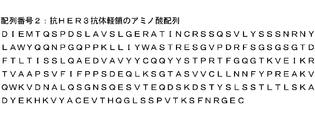
- Fig. 3 shows the amino acid sequence of an anti-HER3 antibody light chain (SEQ ID NO: 10).
- the results of 1% agarose gel electrophoresis of the RT-PCR products of various HER3 stably expressing cells (HER3 expression) are shown.
- the results of 1% agarose gel electrophoresis of RT-PCR products of various HER2 overexpressing HER3 stably expressing cells (HER3 expression) are shown.
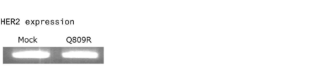
- 1 shows the results of 1% agarose gel electrophoresis (HER2 expression) of RT-PCR products of various HER2 overexpressing HER3 stably expressing cells.
- FIG. 2 shows the binding of HER3-ADC (1) to HER3 stably expressing cells.
- FIG. 9 shows the binding of HER3-ADC (1) in HER2-overexpressing HER3 stably expressing cells.
- FIG. 9 shows the cell growth inhibitory activity of HER3-ADC (1), HER3-Ab (1), and IgG-ADC (1) on empty vector-transfected cells (without overexpression of HER2).
- FIG. 4 shows the cell growth inhibitory activity of HER3-ADC (1), HER3-Ab (1), and IgG-ADC (1) on wild-type HER3-transfected cells (without overexpression of HER2).
- FIG. 3 shows the cell growth inhibitory activity of HER3-ADC (1), HER3-Ab (1), and IgG-ADC (1) on mutant HER3 (V104L) -introduced cells (without overexpression of HER2).
- FIG. 9 shows the cell growth inhibitory activity of HER3-ADC (1), HER3-Ab (1), and IgG-ADC (1) on mutant HER3 (V104M) -introduced cells (without overexpression of HER2).
- FIG. 9 shows the cell growth inhibitory activity of HER3-ADC (1), HER3-Ab (1), and IgG-ADC (1) on mutant HER3 (A232V) -introduced cells (without overexpression of HER2).
- FIG. 4 shows the cell growth inhibitory activity of HER3-ADC (1), HER3-Ab (1), and IgG-ADC (1) on mutant HER3 (P262H) -introduced cells (without overexpression of HER2).
- FIG. 9 shows the cell growth inhibitory activity of HER3-ADC (1), HER3-Ab (1), and IgG-ADC (1) on mutant HER3 (P262H) -introduced cells (without overexpression of HER2).
- FIG. 9 shows the cell growth inhibitory activity of HER3-ADC (1), HER3-Ab (1), and IgG-ADC (1) on mutant HER3 (G284R) -introduced cells (without overexpression of HER2).
- FIG. 9 shows the cell growth inhibitory activity of HER3-ADC (1), HER3-Ab (1), and IgG-ADC (1) on mutant HER3 (D297Y) -introduced cells (without overexpression of HER2).
- FIG. 4 shows the cell growth inhibitory activity of HER3-ADC (1), HER3-Ab (1), and IgG-ADC (1) on mutant HER3 (G325R) -introduced cells (without overexpression of HER2).
- FIG. 9 shows the cell growth inhibitory activity of HER3-ADC (1), HER3-Ab (1), and IgG-ADC (1) on mutant HER3 (G325R) -introduced cells (without overexpression of HER2).
- FIG. 3 shows the cell growth inhibitory activity of HER3-ADC (1), HER3-Ab (1), and IgG-ADC (1) on mutant HER3 (T355I) -introduced cells (without overexpression of HER2).
- FIG. 9 shows the cell growth inhibitory activity of HER3-ADC (1), HER3-Ab (1), and IgG-ADC (1) on mutant HER3 (S846I) -introduced cells (without overexpression of HER2).
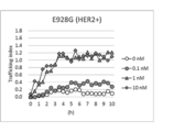
- FIG. 9 shows the cell growth inhibitory activity of HER3-ADC (1), HER3-Ab (1), and IgG-ADC (1) on mutant HER3 (E928G) -introduced cells (without overexpression of HER2).
- FIG. 4 shows the cell growth inhibitory activity of HER3-ADC (1), HER3-Ab (1), and IgG-ADC (1) on empty vector-introduced cells (with HER2 overexpression).
- FIG. 4 shows the cell growth inhibitory activity of HER3-ADC (1), HER3-Ab (1), and IgG-ADC (1) on wild-type HER3-introduced cells (HER2 overexpression).
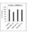
- FIG. 9 shows the cell growth inhibitory activity of HER3-ADC (1), HER3-Ab (1), and IgG-ADC (1) on mutant HER3 (V104L) -introduced cells (HER2 overexpression).
- FIG. 3 shows the cell growth inhibitory activity of HER3-ADC (1), HER3-Ab (1), and IgG-ADC (1) on mutant HER3 (V104M) -transfected cells (HER2 overexpression).
- FIG. 9 shows the cell growth inhibitory activity of HER3-ADC (1), HER3-Ab (1), and IgG-ADC (1) on mutant HER3 (V104M) -transfected cells (HER2 overexpression).
- FIG. 9 shows the cell growth inhibitory activity of HER3-ADC (1), HER3-Ab (1), and IgG-ADC (1) on mutant HER3 (A232V) -introduced cells (HER2 overexpression).
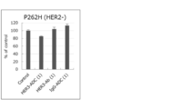
- FIG. 9 shows the cell growth inhibitory activity of HER3-ADC (1), HER3-Ab (1), and IgG-ADC (1) on mutant HER3 (P262H) -introduced cells (HER2 overexpression).
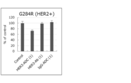
- FIG. 9 shows the cell growth inhibitory activity of HER3-ADC (1), HER3-Ab (1), and IgG-ADC (1) on mutant HER3 (G284R) -introduced cells (HER2 overexpression).
- FIG. 9 shows the cell growth inhibitory activity of HER3-ADC (1), HER3-Ab (1), and IgG-ADC (1) on mutant HER3 (G284R) -introduced cells (HER2 overexpression).
- FIG. 9 shows the cell growth inhibitory activity of HER3-ADC (1), HER3-Ab (1), and IgG-ADC (1) on mutant HER3 (D297Y) -introduced cells (HER2 overexpression).
- FIG. 9 shows the cell growth inhibitory activity of HER3-ADC (1), HER3-Ab (1), and IgG-ADC (1) on mutant HER3 (G325R) -introduced cells (with HER2 overexpression).
- FIG. 9 shows the cell growth inhibitory activity of HER3-ADC (1), HER3-Ab (1), and IgG-ADC (1) on mutant HER3 (T355I) -introduced cells (HER2 overexpression).
- FIG. 3 shows the cell growth inhibitory activity of HER3-ADC (1), HER3-Ab (1), and IgG-ADC (1) on mutant HER3 (S846I) -introduced cells (HER2 overexpression).
- FIG. 9 shows the cell growth inhibitory activity of HER3-ADC (1), HER3-Ab (1), and IgG-ADC (1) on mutant HER3 (E928G) -introduced cells (with HER2 overexpression).
- FIG. 9 shows the cell growth inhibitory activity of HER3-ADC (1), HER3-Ab (1), and IgG-ADC (1) on empty vector-transfected cells (without overexpression of HER2).
- FIG. 4 shows the cell growth inhibitory activity of HER3-ADC (1), HER3-Ab (1), and IgG-ADC (1) on wild-type HER3-transfected cells (without overexpression of HER2).
- FIG. 1 shows the binding of HER3-ADC (1) in mutant HER3 (Q809R) -introduced cells (HER2 overexpression).
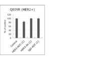
- FIG. 9 shows the cell growth inhibitory activity of HER3-ADC (1), HER3-Ab (1), and IgG-ADC (1) on mutant HER3 (Q809R) -introduced cells (HER2 overexpression).
- Fig. 4 shows the lysosomal translocation of HER3-ADC (1) to empty vector-transfected cells (without overexpression of HER2).
- FIG. 4 shows the lysosomal translocation of HER3-ADC (1) to wild-type HER3-introduced cells (without HER2 overexpression).
- FIG. 4 shows the lysosomal translocation of HER3-ADC (1) to mutant HER3 (V104L) -introduced cells (without overexpression of HER2).
- FIG. 4 shows lysosomal translocation of HER3-ADC (1) to mutant HER3 (V104M) -introduced cells (without HER2 overexpression).
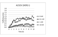
- FIG. 4 shows the lysosomal translocation of HER3-ADC (1) to mutant HER3 (A232V) -introduced cells (without HER2 overexpression).
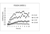
- FIG. 9 shows the lysosomal translocation of HER3-ADC (1) to mutant HER3 (P262H) -introduced cells (without overexpression of HER2).
- FIG. 4 shows the lysosomal translocation of HER3-ADC (1) to mutant HER3 (G284R) -introduced cells (without overexpression of HER2).
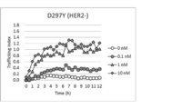
- FIG. 9 shows the lysosomal translocation of HER3-ADC (1) to mutant HER3 (D297Y) -introduced cells (without overexpression of HER2).
- FIG. 4 shows the lysosomal translocation of HER3-ADC (1) to mutant HER3 (G325R) -introduced cells (without overexpression of HER2).
- FIG. 9 shows the lysosomal translocation of HER3-ADC (1) to mutant HER3 (T355I) -introduced cells (without HER2 overexpression).
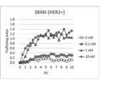
- FIG. 4 shows the lysosomal translocation of HER3-ADC (1) to mutant HER3 (S846I) -introduced cells (without overexpression of HER2).
- FIG. 4 shows lysosomal translocation of HER3-ADC (1) to mutant HER3 (E928G) -introduced cells (without HER2 overexpression).
- FIG. 4 shows the lysosomal translocation of HER3-ADC (1) to empty vector-introduced cells (HER2 overexpression).
- FIG. 4 shows the lysosomal translocation of HER3-ADC (1) to wild-type HER3-introduced cells (HER2 over-expression).
- FIG. 4 shows lysosomal translocation of HER3-ADC (1) to mutant HER3 (V104L) -introduced cells (HER2 overexpression).
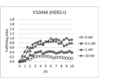
- FIG. 4 shows the lysosomal translocation of HER3-ADC (1) to mutant HER3 (V104M) -introduced cells (HER2 overexpression).
- FIG. 4 shows the lysosomal translocation of HER3-ADC (1) to mutant HER3 (A232V) -introduced cells (HER2 overexpression).
- FIG. 4 shows the lysosomal translocation of HER3-ADC (1) to wild-type HER3-introduced cells (HER2 over-expression).
- FIG. 4 shows lysosomal translocation of HER3-ADC (1) to mutant HER3 (V104L)
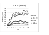
- FIG. 4 shows lysosomal translocation of HER3-ADC (1) to mutant HER3 (P262H) -introduced cells (HER2 overexpression).
- FIG. 4 shows the lysosomal translocation of HER3-ADC (1) to mutant HER3 (G284R) -introduced cells (HER2 overexpression).
- FIG. 4 shows lysosomal translocation of HER3-ADC (1) to mutant HER3 (D297Y) -introduced cells (HER2 overexpression).
- FIG. 9 shows the lysosomal translocation of HER3-ADC (1) to mutant HER3 (G325R) -introduced cells (HER2 overexpression).
- FIG. 7 shows the lysosomal translocation of HER3-ADC (1) to mutant HER3 (T355I) -introduced cells (HER2 overexpression).
- FIG. 4 shows the lysosomal translocation of HER3-ADC (1) to mutant HER3 (Q809R) -introduced cells (HER2 overexpression).
- FIG. 9 shows the lysosomal translocation of HER3-ADC (1) to mutant HER3 (E928G) -introduced cells (with HER2 overexpression).
- Fig. 4 shows the lysosomal translocation of HER3-ADC (1) to empty vector-transfected cells (without overexpression of HER2).
- FIG. 4 shows the lysosomal translocation of HER3-ADC (1) to wild-type HER3-introduced cells (without HER2 overexpression).
- Fig. 3 shows the amino acid sequence of HER3 protein (SEQ ID NO: 69).
- HER3 is synonymous with human epidermal growth factor receptor 3 (HER3; also known as ErbB3) and is a receptor together with HER1 (EGFR, ErbB1), HER2 (ErbB2) and HER4 (ErbB4). It is a transmembrane receptor belonging to the epidermal growth factor receptor subfamily of protein tyrosine kinases.
- HER3 is expressed in various types of cancers, such as breast cancer, lung cancer, and colorectal cancer, and undergoes phosphorylation by forming a heterodimer with tyrosine kinase receptors such as HER2 and EGFR, and It is known to induce cancer cell proliferation and apoptosis inhibitory signals (Alimandi et al., Oncogene (1995) 10, 1813-1821, deFazio et al., Int. J. Cancer (2000) 87, 487- 498, Naidu et al., Br. J. Cancer (1998) 78, 1385-1390).
- HER3 protein is used in the same meaning as HER3.
- HER3 protein expression can be detected using a method well known to those skilled in the art, such as immunohistochemistry (IHC).
- HER3 protein expression can be detected by introducing a Flag peptide into the HER3 protein and using an anti-Flag peptide antibody.
- HER3 gene means a gene encoding a HER3 protein.
- HER3 protein is a gene product of the HER3 gene.
- HER3 mutation means having a mutation in the amino acid sequence of HER3 protein.
- HER3 mutant cancer means a cancer having a mutation in the amino acid sequence of HER3 protein.
- a cancer that does not have a HER3 mutation in the entire tumor tissue but includes a cancer cell having a HER3 mutation is included in a HER3 mutant cancer.
- HER3 gene mutation means having a mutation in the HER3 gene.
- HER3 gene mutation cancer means a cancer having a mutation in the HER3 gene.
- a cancer containing a cancer cell having a HER3 gene mutation even if it does not have a HER3 gene mutation in the entire tumor tissue is included in the HER3 gene-mutated cancer.
- the HER3 gene mutation causes a mutation in the amino acid sequence of the HER3 protein which is a gene product, resulting in a HER3 mutation.
- V valine
- L leucine
- V valine, the 104th amino acid of the HER3 protein
- M methionine
- G glycine
- R arginine
- G glycine
- R arginine
- T threonine
- I isoleucine
- Q the 809th amino acid of the HER3 protein
- cancers having Q809R in particular exhibit strong resistance to existing anti-HER2 drugs and anti-HER3 antibodies (Jaiswal et al., Cancer cell (2013 ) 23, 603-17).
- the HER3 mutation according to the present invention is not particularly limited as long as it has a mutation in the amino acid sequence of the HER3 protein. And E928G, and more preferably Q809R.
- the presence or absence of the HER3 mutation is confirmed by, for example, collecting a tumor tissue from a cancer patient and performing a method such as real-time quantitative PCR (qRT-PCR) or microarray analysis on a formalin-fixed paraffin-embedded specimen (FFPE). be able to.
- qRT-PCR real-time quantitative PCR
- FFPE formalin-fixed paraffin-embedded specimen
- the presence or absence of the HER3 mutation can also be confirmed by collecting cell-free blood circulating tumor DNA (ctDNA) from a cancer patient and performing a method such as next-generation sequencing (NGS) (Sergina et al., Nature (2007) 445, 437-41, Jeong et al., Int.J. Cancer (2006) 119, 2986-7, Ding et al., Nature (2008) 455, 1069-75, Kan et al., Nature (2010) 466, 869-73, Wang et al., Nat. Genet.
- NGS next-generation sequencing
- HER3 mutation is used in the same meaning as the HER3 gene mutation.
- wild-type HER3 means a HER3 protein having no HER3 mutation.
- the wild type is sometimes referred to as “WT”.
- mutant HER3 means a HER3 protein having a HER3 mutation.
- HER3 stably expressing cells can be obtained by transfection using cationic lipid, cationic polymer, calcium phosphate, etc., chemical gene transfer method, electroporation, microinjection, sonoporation, physical gene transfer method by laser irradiation, virus, etc. It can be prepared by a biological gene transfer method using a vector or the like.
- a lentivirus among the biological gene transfer methods when a lentivirus protein and an envelope protein expression plasmid and a HER3 expression plasmid are both introduced into packaging cells such as Lenti-X @ 293T cells, the cells are cultured.
- It can be prepared by preparing a lentivirus solution from the supernatant and culturing the tumor cells using this.
- wild-type or mutant-type HER3-expressing cells can be produced depending on the type of HER3-expression plasmid used.
- the mutant HER3 expression plasmid can be prepared using HER3 mutation-introducing primers.
- the “anti-HER3 antibody” refers to an antibody that specifically binds to HER3 and preferably has an activity of being internalized in a HER3 expression cell by binding to HER3, in other words, after binding to HER3, 1 shows an antibody having an activity of migrating into HER3-expressing cells.
- HER2 is synonymous with human epidermal growth factor receptor 2 (sometimes called neu, ErbB-2), and together with HER1, HER3 and HER4, epidermal growth factor of receptor protein tyrosine kinase It is a transmembrane receptor belonging to the receptor subfamily. HER2 is important for cell growth, differentiation and survival in normal cells and tumor cells by activating self-phosphorylation of intracellular tyrosine residues by heterodimer formation with HER1, HER3 or HER4 to activate them. It is known to play a role.
- HER2 protein is used in the same meaning as HER2.
- “overexpression” of HER2 indicates that the expression of HER2 is determined to be positive, for example, that the expression of HER2 is determined to be 3+ by immunohistochemistry (IHC), This indicates that the expression of HER2 is determined to be 2+ by the histochemistry method and the expression of HER2 is determined to be positive by the in situ hybridization (ISH) method.
- IHC immunohistochemistry
- ISH in situ hybridization
- HER2 overexpressing cells can be obtained by transfection using cationic lipid, cationic polymer, calcium phosphate, etc., chemical gene transfer method, electroporation, microinjection, sonoporation, physical gene transfer method by laser irradiation, etc., virus It can be prepared by a biological gene transfer method using a vector or the like.
- lentivirus when lentivirus is used in the biological gene transfer method, after lentiviral protein and envelope protein expression plasmid and HER2 expression plasmid are both introduced into packaging cells such as Lenti-X @ 293T cells, the cells are cultured. It can be prepared by preparing a lentivirus solution from the supernatant and culturing the tumor cells using this.
- HER2 overexpressing HER3 stable expression cells can be produced by culturing the HER2 overexpressing cells using the lentivirus solution obtained by the introduction of the HER3 expression plasmid.
- antibody-drug conjugate refers to a complex in which a cytotoxic drug is bound to an antibody via a linker.
- Antibody-drug conjugates include, for example, US Pat. No. 6,214,345, WO 2002/083067, WO 2003/026577, WO 2004/054622, WO 2005/112919, WO WO 2006/135371, WO 2007121193, WO 2008/033891, WO 2009/100194, WO 2009/134977, WO 2009/134977, WO 2010/093395.
- These antibody-drug conjugates can be produced by the method described in the above-mentioned literature.
- the drug having cytotoxicity is not particularly limited as long as it has an antitumor effect and has a substituent or a partial structure capable of binding to a linker.
- camptothecin Campetothecin
- calicheamicin Calicheamicin
- Doxorubicin Daunorubicin
- Mitomycin C Mitomycin @ C
- Bleomycin Bleomycin
- Cyclocytidine Vincristine
- Vinblastine Vinblastine
- Vinblastine E Auristatin @ E
- Maytansine M ytansine
- paclitaxel pyrrolobenzodiazepine
- derivatives thereof preferably a camptothecin derivative, more preferably an exatecan derivative. it can.
- Exatecan which is a topoisomerase I inhibitor (IUPAC name: (1S, 9S) -1-amino-9-ethyl-5-fluoro-1,2,3,9,12,15-hexahydro-9-hydroxy-4-methyl -10H, 13H-benzo [de] pyrano [3 ', 4': 6,7] indolizino [1,2-b] quinoline-10,13-dione, (chemical name: (1S, 9S) -1-amino -9-ethyl-5-fluoro-2,3-dihydro-9-hydroxy-4-methyl-1H, 12H-benzo [de] pyrano [3 ′, 4 ′: 6,7] indolidino [1,2-b Quinoline-10,13 (9H, 15H) -dione)) has the formula
- the “drug linker” refers to a drug and a linker portion in the antibody-drug conjugate, in other words, a partial structure other than the antibody in the antibody-drug conjugate.
- anti-HER3 antibody-drug conjugate refers to an antibody-drug conjugate in which the antibody in the antibody-drug conjugate is an anti-HER3 antibody.
- Anti-HER3 antibody-drug conjugates include, for example, those described in WO2012 / 019042, WO2012 / 064733, and WO2015 / 155998, preferably WO 2015/155998 can be mentioned. These anti-HER3 antibody-drug conjugates can be produced by the method described in the above-mentioned literature.
- the anti-HER3 antibody-drug conjugate suitably used in the present invention has the formula
- A indicates a binding position with the antibody
- This drug linker is formed by a thiol group (in other words, a sulfur atom of a cysteine residue) generated at a disulfide bond site between antibody chains (two heavy chains and heavy chains and two heavy chains and light chains). ).
- anti-HER3 antibody-drug conjugate suitably used in the present invention can also be represented by the following formula.
- n has the same meaning as the so-called average number of drug bonds (DAR; Drug-to-Antibody Ratio), and indicates the average number of drug linker bonds per antibody.
- the average number of drug linker bonds per antibody of the anti-HER3 antibody-drug conjugate preferably used in the present invention is preferably 2 to 8, more preferably 3 to 8, and even more. Preferably from 7 to 8, even more preferably from 7.5 to 8, and even more preferably about 8.
- the anti-HER3 antibody portion of the anti-HER3 antibody-drug conjugate used in the present invention is preferably CDRH1 consisting of the amino acid sequence shown in SEQ ID NO: 1, CDRH2 consisting of the amino acid sequence shown in SEQ ID NO: 2, and the sequence A heavy chain containing CDRH3 consisting of the amino acid sequence represented by SEQ ID NO: 3, CDRL1 consisting of the amino acid sequence represented by SEQ ID NO: 4, CDRL2 consisting of the amino acid sequence represented by SEQ ID NO: 5, and the amino acid sequence represented by SEQ ID NO: 6
- a light chain comprising CDRL3 consisting of: More preferably, an antibody comprising a heavy chain comprising a heavy chain variable region consisting of the amino acid sequence represented by SEQ ID NO: 7, and a light chain comprising a light chain variable region consisting of the amino acid sequence represented by SEQ ID NO: 8, Even more preferably, an antibody comprising a heavy chain consisting of the amino acid sequence represented by SEQ ID NO: 9 and a light chain consisting of
- the anti-HER3 antibody-drug conjugate suitably used in the present invention has the formula
- the above compound is considered to be the main antitumor activity of the above anti-HER3 antibody-drug conjugate suitably used in the present invention, and has been confirmed to have topoisomerase I inhibitory activity (Ogitani Y. et al., Clinical Cancer Research, 2016, Oct 15; 22 (20): 5097-5108, Epub 2016 Mar 29).
- the above compound is obtained by cleavage of the linker portion of the anti-HER3 antibody-drug conjugate described above.
- the HER3 protein used in the present invention can be used after directly purified from human HER3-expressing cells, or when used as an antigen, the cell membrane fraction of the cells can be used as the HER3 protein.
- HER3 can be synthesized in vitro, or produced in host cells by genetic engineering. In the genetic manipulation, specifically, HER3 is synthesized by incorporating a HER3 cDNA into a vector capable of expressing and incubating the vector in a solution containing enzymes, substrates and energy substances necessary for transcription and translation. be able to.
- the protein can be obtained by transforming another prokaryotic or eukaryotic host cell with the vector and expressing HER3.
- HER3-expressing cells or cell lines expressing HER3 by the above-described genetic manipulation can be used as HER3 protein antigens.
- the RNA sequence, cDNA sequence and amino acid sequence of HER3 are publicly available on public databases. Can be referenced.
- HER3 includes a protein having an amino acid sequence in which 1 to 10 amino acids have been substituted, deleted, added and / or inserted in the amino acid sequence of HER3, and having the same biological activity as the protein.
- the anti-HER3 antibody used in the present invention can be obtained by known means. For example, immunizing an animal with HER3 as an antigen or an arbitrary polypeptide selected from the amino acid sequence of HER3, and collecting and purifying an antibody produced in a living body, using a method usually used in this field. Can be obtained by The origin of the antigen is not limited to humans, and animals can be immunized with antigens derived from non-human animals such as mice and rats. In this case, an anti-HER3 antibody applicable to human disease can be selected by testing the cross-reactivity between the obtained antibody that binds to the heterologous antigen and the human antigen.
- a hybridoma can be established by fusing an antibody-producing cell that produces an antibody against an antigen with a myeloma cell to obtain a monoclonal antibody.
- the antigen can be obtained by causing a host cell to produce a gene encoding an antigen protein by genetic manipulation. Specifically, a vector capable of expressing an antigen gene may be prepared, introduced into a host cell to express the gene, and the expressed antigen may be purified. Antibodies can also be obtained by using a method of immunizing an animal with an antigen-expressing cell or a cell line expressing the antigen by the above-described genetic manipulation.
- the anti-HER3 antibody used in the present invention is a recombinant antibody artificially modified for the purpose of, for example, reducing the antigenicity to humans, such as a chimeric antibody or a humanized (Humanized) antibody. It is preferable that the antibody has only the gene sequence of a human-derived antibody, that is, a human antibody. These antibodies can be produced using known methods.
- chimeric antibody examples include antibodies in which the variable region and the constant region of the antibody are different from each other, for example, a chimeric antibody in which the variable region of a mouse or rat-derived antibody is conjugated to a human-derived constant region (Proc. Natl. Acad . Sci. USA, 81, 6851-6855, (1984)).
- humanized antibody an antibody (Nature (1986) @ 321, @ p.522-525) in which only the complementarity determining region (CDR; complementarity determining region) of a heterologous antibody is incorporated into a human-derived antibody, In addition to the CDR sequences of the antibody, the amino acid residues of some of the frameworks of the heterologous antibody were also transplanted into a human antibody (WO 90/07861), a gene conversion mutagenesis strategy. And humanized antibodies (US Pat. No. 5,821,337).
- CDR complementarity determining region
- an antibody As a human antibody, an antibody (Tomizuka, K. et al., Nature Genetics (1997) 16, p.1) prepared using a human antibody-producing mouse having a human chromosome fragment containing human antibody heavy and light chain genes. 133-143; Kuroiwa, Y. et. Al., Nucl. Acids Res. (1998) 26, p.3447-3448; Yoshida, H. et. Al., Animal Cell Technology: Basic and Applied Aspects vol.10, p.69-73 (Kitagawa, Y., atsuMatsuda, T. and Iijima, S. eds.), Kluwer Academic Publishers, 1999; Tomizuka, K. et.
- antibodies obtained by phage display selected from a human antibody library (Wormstone, I. M. et. Al, Investigative Ophthalmology & Visual Science. (2002) 43 (7), p.2301-2308; Mé, S. et al., Briefings in Functional Genomics and Proteomics (2002), 1 (2), p. 189-203; Siriwardena, D. et.al, Ophthalmology (2002) 109 (3), p. 427-431, etc. See also).
- the anti-HER3 antibody used in the present invention also includes modified antibodies.
- modified product means that the antibody of the present invention has been subjected to chemical or biological modification.
- Chemical modifications include those having a chemical moiety attached to the amino acid backbone, a chemical moiety attached to an N-linked or O-linked carbohydrate chain, and the like.
- Biologically modified forms include post-translational modifications (eg, addition of N-linked or O-linked sugar chains, N-terminal or C-terminal processing, deamidation, aspartic acid isomerization, methionine oxidation, etc. ) And those in which a methionine residue is added to the N-terminus by expression using a prokaryotic host cell.
- modified anti-HER3 antibody or antigen used in the present invention for example, an enzyme label, a fluorescent label, and an affinity label are also included in the meaning of such a modified form. It is.
- a modified anti-HER3 antibody used in the present invention is useful for improving antibody stability and blood retention, reducing antigenicity, detecting or isolating an antibody or antigen, and the like.
- the anti-HER3 antibody used in the present invention it is possible to enhance the antibody-dependent cytotoxic activity by controlling the modification of the sugar chain bound to the anti-HER3 antibody used in the present invention (glycosylation, defucosification, etc.).
- Techniques for controlling the sugar chain modification of antibodies include WO 99/54342, WO 00/61739, WO 02/31140, WO 2007/133855, and WO 2013/120066. Nos. Are known, but not limited to these.
- the anti-HER3 antibody used in the present invention also includes an antibody whose sugar chain modification is regulated.
- the anti-HER3 antibody used in the present invention also includes the modified antibody and a functional fragment of the antibody, a deletion form in which one or two amino acids are deleted at the heavy chain carboxyl terminus, and
- the amidated deletion for example, a heavy chain in which a proline residue at the carboxyl terminal site is amidated
- the carboxyl-terminal deletion of the heavy chain of the anti-HER3 antibody used in the present invention is not limited to the above types.
- the two heavy chains constituting the anti-HER3 antibody used in the present invention may be any one of the heavy chains selected from the group consisting of full-length and the above-described deletions, or any one of the heavy chains.
- the amount ratio of each deletion can be affected by the type and culture conditions of the cultured mammalian cells producing the anti-HER3 antibody used in the present invention, but the anti-HER3 antibody used in the present invention is preferably 2%.
- One in which one amino acid residue at the carboxyl terminus is deleted in both heavy chains of the book can be mentioned.
- IgG IgG1, IgG2, IgG3, IgG4
- IgG1 or IgG2 IgG2
- IgG1 or IgG2 IgG2
- Anti-HER3 antibodies that can be used in the present invention include patrizumab (Patritumab, @ U3-1287), U1-59 (WO 2007/077028), AV-203 (WO 2011/136911), LJM-716 (International Publication No. 2012/022814), Duligotumab (MEHD-7945A) (International Publication No. 2010/108127), Istratumab (MM-141) (International Publication No. 2011/047180), Lumretuzumab (RG-7116) (International Publication No.
- the drug linker intermediate used for preparing the anti-HER3 antibody-drug conjugate according to the present invention is represented by the following formula.
- the above drug linker intermediate is N- [6- (2,5-dioxo-2,5-dihydro-1H-pyrrol-1-yl) hexanoyl] glycylglycyl-L-phenylalanyl-N-[(2- ⁇ [(1S, 9S) -9-ethyl-5-fluoro-9-hydroxy-4-methyl-10,13-dioxo-2,3,9,10,13,15-hexahydro-1H, 12H-benzo [ de] pyrano [3 ′, 4 ′: 6,7] indolizino [1,2-b] quinolin-1-yl] amino ⁇ -2-oxoethoxy) methyl] glycinamide, It can be produced by referring to the description of WO 2014/057687, WO 2015/155998, WO 2019/049477 and the like.
- the anti-HER3 antibody-drug conjugate used in the present invention can be produced by reacting the aforementioned drug linker intermediate with an anti-HER3 antibody having a thiol group (or sulfhydryl group).
- An anti-HER3 antibody having a sulfhydryl group can be obtained by a method well known to those skilled in the art (Hermanson, G. T, Bioconjugate Techniques, pp.56-136, pp.456-493, Academic Press (1996)).
- a reducing agent such as tris (2-carboxyethyl) phosphine hydrochloride (TCEP) is used in an amount of 0.3 to 3 molar equivalents per one intrachain disulfide in an antibody, and a chelating agent such as ethylenediaminetetraacetic acid (EDTA) is used.
- TCEP tris (2-carboxyethyl) phosphine hydrochloride
- EDTA ethylenediaminetetraacetic acid
- an anti-HER3 antibody-drug conjugate bound with 2 to 8 drugs per antibody was prepared using 2 to 20 molar equivalents of the drug linker intermediate per anti-HER3 antibody having a sulfhydryl group. Can be manufactured.
- the average number of drug bonds per antibody molecule of the manufactured anti-HER3 antibody-drug conjugate is calculated, for example, by measuring the UV absorbance of the anti-HER3 antibody-drug conjugate and its conjugate precursor at two wavelengths of 280 nm and 370 nm. (UV method), or a method of quantifying and calculating each fragment obtained by treating the antibody-drug conjugate with a reducing agent by HPLC measurement (HPLC method).
- the therapeutic agent and / or method of the present invention is characterized by administering an anti-HER3 antibody-drug conjugate, and can be used for treating HER3 mutant cancer.
- the cancer in the HER3 mutant cancer to which the therapeutic agent and / or the therapeutic method of the present invention can be used is preferably breast cancer, lung cancer (including small cell lung cancer and non-small cell lung cancer), colon cancer (colon) Sometimes called rectal cancer, including colon and rectal cancer), stomach cancer (sometimes called gastric adenocarcinoma), ovarian cancer, head and neck cancer, glioblastoma multiforme, melanoma, Kidney, urothelial, prostate, pancreatic, bladder, gastrointestinal stromal, cervical, esophageal, squamous, peritoneal, glioblastoma Tumor, liver, hepatocellular, endometrial, uterine, salivary gland, vulvar, thyroid, hepatocarcinoma, anal carcinoma, and penis cancer
- At least one, more preferably breast cancer, non-small cell lung cancer, colon cancer, stomach cancer, ovarian cancer, Cervical cancer is at least one selected from the group consisting of
- the therapeutic agent and the therapeutic method of the present invention can be suitably used for mammals, but can be more preferably used for humans.
- the antitumor effect of the therapeutic agent and the therapeutic method of the present invention can be determined, for example, by expressing a mutant HER3 in tumor cells and not overexpressing HER2 and / or overexpressing HER2. It can be confirmed by measuring the cell growth inhibitory activity of the conjugate. Alternatively, it can be confirmed by preparing a model in which a tumor expressing the mutant HER3 is transplanted into a test animal and applying the therapeutic agent or the therapeutic method of the present invention.
- a therapeutic agent or a therapeutic method of the present invention is administered to a cancer patient in which a HER3 mutation has been confirmed, and a Response Evaluation Criteria in Solid Tumors (RECIST) evaluation method, a WHO evaluation method, a Macdonald evaluation method, weight measurement, and other methods are used.
- RECIST Response Evaluation Criteria in Solid Tumors
- a complete response (Complete response; CR), a partial response (Partial response; PR), progression (Progressive disease; PD), objective response rate (Objective Response rate, ORR), response time of Response (DoR), Progression-Free Survival (PFS), Overall Survival It can be determined by OS) index, such as; Overall Survival.
- the therapeutic agent and the therapeutic method of the present invention can delay the growth of cancer cells, suppress the proliferation, and can destroy cancer cells. By these actions, in a cancer patient, it is possible to achieve a relief from symptoms caused by the cancer and an improvement in QOL, thereby achieving a therapeutic effect while maintaining the life of the cancer patient. Even in cases where the cancer cells are not destroyed, cancer patients can achieve longer QOL while achieving higher QOL by suppressing and controlling the proliferation of cancer cells.
- the therapeutic agent of the present invention can be applied to a patient as a systemic therapy, or applied locally to a cancer tissue to expect a therapeutic effect.
- Therapeutic agents of the invention can be administered as a pharmaceutical composition comprising one or more pharmaceutically compatible ingredients.
- Pharmaceutically compatible components are appropriately selected and applied from pharmaceutical additives and the like usually used in this field according to the dosage and concentration of the anti-HER3 antibody-drug conjugate used in the present invention. can do.
- the therapeutic agent of the present invention comprises a pharmaceutical composition comprising a buffer such as a histidine buffer, an excipient such as sucrose or trehalose, and a surfactant such as polysorbate 80 or 20 (hereinafter referred to as “the pharmaceutical composition of the present invention”).
- Object The pharmaceutical composition of the present invention can be suitably used as an injection, more preferably as an aqueous injection or a lyophilized injection, and even more preferably a lyophilized injection. It can be used as an agent.
- the pharmaceutical composition of the present invention is an aqueous injection, it can be suitably administered by intravenous infusion after dilution with an appropriate diluent.
- the diluting solution include a glucose solution and a physiological saline solution.
- the pharmaceutical composition of the present invention when it is a freeze-dried injection, it can be preferably dissolved in water for injection, diluted with a suitable diluent to a required amount, and then administered by intravenous infusion.
- a suitable diluent examples include a glucose solution and a physiological saline solution.
- Introduction routes that can be used to administer the pharmaceutical compositions of the present invention include, for example, intravenous, intradermal, subcutaneous, intramuscular, and intraperitoneal routes, preferably intravenous. Routes can be mentioned.
- the anti-HER3 antibody-drug conjugate used in the present invention can be administered to a human, preferably at intervals of one week, two weeks, three weeks, or four weeks, and even more. Preferably, it can be administered once every three weeks.
- the anti-HER3 antibody-drug conjugate used in the present invention can be suitably administered to a human at a dose of 1.6 mg / kg to 12.8 mg / kg per dose, More preferably, 1.6 mg / kg, 3.2 mg / kg, 4.8 mg / kg, 5.6 mg / kg, 6.4 mg / kg, 8.0 mg / kg, 9.6 mg / kg per dose, Or a dose of 12.8 mg / kg, and even more preferably a dose of 4.8 mg / kg, 5.6 mg / kg, or 6.4 mg / kg per dose. Can be.
- the therapeutic agent of the present invention can also be administered in combination with a cancer therapeutic agent other than the anti-HER3 antibody-drug conjugate used in the present invention, whereby the antitumor effect can be enhanced.
- a cancer therapeutic agent other than the anti-HER3 antibody-drug conjugate used in the present invention
- the other therapeutic agent for cancer used for such a purpose may be administered to an individual simultaneously or separately with the therapeutic agent of the present invention, or may be administered at different administration intervals. You may.
- Such a cancer therapeutic agent is not limited as long as it has an antitumor activity.
- irinotecan (Irinotecan, CPT-11), cisplatin, carboplatin, oxaliplatin (Oxaliplatin), Fluorouracil (Fluorouracil, 5-FU), Gemcitabine (Gemcitabine), Capecitabine (Capecitabine), Paclitaxel (Paclitaxel), Docetaxi (Doctorubixin), Doxorubicin , Mitomycin C (Mitomycin @ C), Tegafu (Tegafur) / Gimeracil / Oteracil combination drug, Cetuximab (Cetuximab), Panitumumab (Panituumab), Bevacizumab (Bevacizumab), Ramucirafarib Tiframefiranib, Ramucilirab Nitrabirafifarib, Ramucirumab, Rumucirumab, Rumucirumab, Ramucirumab Tipiracil, Gefitinib, Gelotinib, Er
- trastuzumab Trastuzumab, Pertuzumab, Lapatinib, Lapatinib, Nivolumab, Nivolumab, Pembrolizumab, Atezolizumab, Averozumab, Vervalumaval, evalu lumab), ipilimumab, and at least one selected from the group consisting of tremelimumab.
- the therapeutic agent of the present invention can also be used in combination with radiation therapy.
- a cancer patient receives radiation therapy before and / or after receiving treatment with the therapeutic agent of the present invention, or at the same time.
- the therapeutic agent of the present invention can also be used as adjuvant chemotherapy combined with surgery. Surgery is performed, for example, by physically removing all or part of a brain tumor.
- the therapeutic agent of the present invention may be administered for the purpose of reducing the size of a brain tumor before surgery (referred to as preoperative adjuvant chemotherapy or neoadjuvant therapy), or for preventing the recurrence of brain tumor after surgery. (Referred to as postoperative adjuvant chemotherapy or adjuvant therapy).
- Example 1 Production of anti-HER3 antibody-drug conjugate According to the production method described in WO 2015/155998, a heavy chain consisting of the amino acid sequence represented by SEQ ID NO: 9 and the amino acid sequence represented by SEQ ID NO: 10 Using an anti-HER3 antibody (hereinafter referred to as “HER3-Ab (1)” in the present invention) comprising a light chain
- drug linker intermediate (1) A conjugation reaction with a drug linker intermediate (hereinafter referred to as “drug linker intermediate (1)”) represented by
- HER3-ADC (1) An anti-HER3 antibody-drug conjugate (hereinafter referred to as “HER3-ADC (1)” in the present invention) in which the drug linker represented by and the anti-HER3 antibody were bound by a thioether bond was produced.
- the average number of drug bonds per antibody in HER3-ADC (1) ranges from 7-8.
- Example 2 Preparation of HER3 stable expression cell
- Example 2-1 Preparation of HER3 mutant expression lentivirus vector
- HER3 cDNA As a template, amplification was carried out with a HER3 IF primer (Fw) shown in SEQ ID NO: 11 and a HER3 IF primer (Rev) shown in SEQ ID NO: 12, and InFusion System (CLONTECH) was used.
- Fw HER3 IF primer
- Rev HER3 IF primer
- CLONTECH InFusion System
- HER3 mutant expression plasmids pLVSIN EF1 ⁇ flag-HER3 (V104L) Pur Using pLVSIN EF1 ⁇ flag-HER3 (WT) as a template, a full-length plasmid was amplified using a HER3 mutation-introducing primer (Fw) shown in SEQ ID NO: 17 and a HER3 mutation-introducing primer (Rev) shown in SEQ ID NO: 18, and the InFusion System was used.
- a HER3 mutant expression plasmid (pLVSIN EF1 ⁇ flag-HER3 (V104L) Pur) was obtained by self-ligation (manufactured by CLONTECH). The sequence was confirmed by DNA sequencing. The nucleotide sequence of the cDNA encoding Flag-HER3 (V104L) is shown in SEQ ID NO: 19, and the amino acid sequence of Flag-HER3 (V104L) is shown in SEQ ID NO: 20.
- pLVSIN EF1 ⁇ flag-HER3 V104M
- WT pLVSIN EF1 ⁇ flag-HER3
- a HER3 mutant expression plasmid (pLVSIN EF1 ⁇ flag-HER3 (V104M) Pur) was obtained by self-ligation (manufactured by CLONTECH). The sequence was confirmed by DNA sequencing.
- the nucleotide sequence of the cDNA encoding Flag-HER3 (V104M) is shown in SEQ ID NO: 23, and the amino acid sequence of Flag-HER3 (V104M) is shown in SEQ ID NO: 24.
- pLVSIN EF1 ⁇ flag-HER3 (A232V) Pur Using pLVSIN EF1 ⁇ flag-HER3 (WT) as a template, the full-length plasmid was amplified using a HER3 mutation-introducing primer (Fw) shown in SEQ ID NO: 25 and a HER3 mutation-introducing primer (Rev) shown in SEQ ID NO: 26, and InFusion System was used.
- a HER3 mutant expression plasmid (pLVSIN EF1 ⁇ flag-HER3 (A232V) Pur) was obtained by self-ligation (manufactured by CLONTECH). The sequence was confirmed by DNA sequencing.
- the nucleotide sequence of the cDNA encoding Flag-HER3 (A232V) is shown in SEQ ID NO: 27, and the amino acid sequence of Flag-HER3 (A232V) is shown in SEQ ID NO: 28.
- pLVSIN EF1 ⁇ flag-HER3 Pur Using pLVSIN EF1 ⁇ flag-HER3 (WT) as a template, the full-length plasmid was amplified with a HER3 mutation-introducing primer (Fw) represented by SEQ ID NO: 29 and a HER3 mutation-introducing primer (Rev) represented by SEQ ID NO: 30 to obtain InFusion System.
- Fw HER3 mutation-introducing primer
- Rev HER3 mutation-introducing primer
- SEQ ID NO: 30 A HER3 mutant expression plasmid (pLVSIN EF1 ⁇ flag-HER3 (P262H) Pur) was obtained by self-ligation (manufactured by CLONTECH). The sequence was confirmed by DNA sequencing.
- the nucleotide sequence of cDNA encoding Flag-HER3 (P262H) is shown in SEQ ID NO: 31, and the amino acid sequence of Flag-HER3 (P262H) is shown in SEQ ID NO: 32.
- pLVSIN EF1 ⁇ flag-HER3 (G284R) Pur Using pLVSIN EF1 ⁇ flag-HER3 (WT) as a template, the full-length plasmid was amplified using a HER3 mutation-introducing primer (Fw) shown in SEQ ID NO: 33 and a HER3 mutation-introducing primer (Rev) shown in SEQ ID NO: 34, and InFusion System was used.
- An HER3 mutant expression plasmid (pLVSIN EF1 ⁇ flag-HER3 (G284R) Pur) was obtained by self-ligation (manufactured by CLONTECH). The sequence was confirmed by DNA sequencing.
- the nucleotide sequence of cDNA encoding Flag-HER3 (G284R) is shown in SEQ ID NO: 35, and the amino acid sequence of Flag-HER3 (G284R) is shown in SEQ ID NO: 36.
- pLVSIN EF1 ⁇ flag-HER3 (D297Y) Pur Using pLVSIN EF1 ⁇ flag-HER3 (WT) as a template, the full-length plasmid was amplified using a HER3 mutation-introducing primer (Fw) shown in SEQ ID NO: 37 and a HER3 mutation-introducing primer (Rev) shown in SEQ ID NO: 38, and InFusion System was used.
- a HER3 mutant expression plasmid (pLVSIN EF1 ⁇ flag-HER3 (D297Y) Pur) was obtained by self-ligation (manufactured by CLONTECH). The sequence was confirmed by DNA sequencing.
- the nucleotide sequence of cDNA encoding Flag-HER3 (D297Y) is shown in SEQ ID NO: 39, and the amino acid sequence of Flag-HER3 (D297Y) is shown in SEQ ID NO: 40.
- pLVSIN EF1 ⁇ flag-HER3 (G325R) Pur Using pLVSIN EF1 ⁇ flag-HER3 (WT) as a template, the full-length plasmid was amplified with a HER3 mutation-introducing primer (Fw) shown in SEQ ID NO: 41 and a HER3 mutation-introducing primer (Rev) shown in SEQ ID NO: 42, and the InFusion System was used.
- a HER3 mutant expression plasmid (pLVSIN EF1 ⁇ flag-HER3 (G325R) Pur) was obtained by self-ligation (manufactured by CLONTECH). The sequence was confirmed by DNA sequencing.
- the nucleotide sequence of the cDNA encoding Flag-HER3 (G325R) is shown in SEQ ID NO: 43
- the amino acid sequence of Flag-HER3 (G325R) is shown in SEQ ID NO: 44.
- pLVSIN EF1 ⁇ flag-HER3 (T355I) Pur Using pLVSIN EF1 ⁇ flag-HER3 (WT) as a template, the full-length plasmid was amplified with a HER3 mutation-introducing primer (Fw) shown in SEQ ID NO: 45 and a HER3 mutation-introducing primer (Rev) shown in SEQ ID NO: 46, and InFusion System was used.
- An HER3 mutant expression plasmid (pLVSIN EF1 ⁇ flag-HER3 (T355I) Pur) was obtained by self-ligation (manufactured by CLONTECH). The sequence was confirmed by DNA sequencing. The nucleotide sequence of the cDNA encoding Flag-HER3 (T355I) is shown in SEQ ID NO: 47, and the amino acid sequence of Flag-HER3 (T355I) is shown in SEQ ID NO: 48.
- pLVSIN EF1 ⁇ flag-HER3 Pur Using pLVSIN EF1 ⁇ flag-HER3 (WT) as a template, a full-length plasmid was amplified with a HER3 mutation-introducing primer (Fw) shown in SEQ ID NO: 49 and a HER3 mutation-introducing primer (Rev) shown in SEQ ID NO: 50, and InFusion System was used.
- a HER3 mutant expression plasmid (pLVSIN EF1 ⁇ flag-HER3 (S846I) Pur) was obtained by self-ligation (manufactured by CLONTECH). The sequence was confirmed by DNA sequencing. The nucleotide sequence of the cDNA encoding Flag-HER3 (S846I) is shown in SEQ ID NO: 51, and the amino acid sequence of Flag-HER3 (S846I) is shown in SEQ ID NO: 52.
- pLVSIN EF1 ⁇ flag-HER3 (E928G) Pur Using pLVSIN EF1 ⁇ flag-HER3 (WT) as a template, the full-length plasmid was amplified using a HER3 mutation-introducing primer (Fw) represented by SEQ ID NO: 53 and a HER3 mutation-introducing primer (Rev) represented by SEQ ID NO: 54, and InFusion System was used.
- a HER3 mutant expression plasmid (pLVSIN EF1 ⁇ flag-HER3 (E928G) Pur) was obtained by self-ligation (manufactured by CLONTECH). The sequence was confirmed by DNA sequencing.
- the nucleotide sequence of cDNA encoding Flag-HER3 (E928G) is shown in SEQ ID NO: 55
- the amino acid sequence of Flag-HER3 (E928G) is shown in SEQ ID NO: 56.
- Example 2-2 Preparation of vector for lentivirus expressing HER2
- HER2 cDNA As a template, amplify with HER2 IF primer (Fw) shown in SEQ ID NO: 57 and HER2 IF primer (Rev) shown in SEQ ID NO: 58, and use InFusion System (manufactured by CLONTECH).
- Fw HER2 IF primer
- Rev HER2 IF primer
- SEQ ID NO: 58 amplify with HER2 IF primer
- Rev HER2 IF primer
- InFusion System manufactured by CLONTECH
- the inserted sequence was confirmed by DNA sequencing.
- the nucleotide sequence of the cDNA encoding HER2 is shown in SEQ ID NO: 59, and the amino acid sequence of HER2
- Example 2-3 Preparation of HER3 stable expression cells
- Lenti-X 293T cells CLONTECH
- ViraPower manufactured by Thermofisher
- pLVSIN EF1 ⁇ flag-HER3 WT
- pLVSIN EF1 ⁇ flag-HER3 V104L
- pLVSINF1H1F1E3H1F1 ⁇ F1H3E1H1 Pur, pLVSINF1H1F1E3H1F1 ⁇ F1H3E1H1
- a plasmid (pLVSIN EF1 ⁇ Pur) was introduced into Lenti-X 293T cells by the same method as described above for the purpose of producing an empty vector-introduced cell.
- the culture supernatant was collected and passed through a MILLEX-HP 0.45 UM filter (manufactured by Merck) to prepare a lentivirus solution.
- One half of the prepared lentivirus solution was transferred to a RetroNectin Coating plate (12 well plate) and left at 37 ° C. overnight. After washing with PBS, MDA-MB-231 cells were seeded at 1 ⁇ 10 5 cells / well and cultured for 3 days.
- a PCR reaction was performed using 1/20 volume of the reverse transcription reaction product as a template and a HER3 primer (Fw) represented by SEQ ID NO: 61 and a HER3 primer (Rev) represented by SEQ ID NO: 62.
- the PCR was performed under the conditions of 25 cycles of heating at 94 ° C. for 2 minutes, 98 ° C. for 10 seconds and 68 ° C. for 1 minute.
- the RT-PCR product was subjected to 1% agarose gel electrophoresis to confirm HER3 gene expression.
- Example 2-4 Preparation of HER2 overexpressing HER3 stable expression strain
- Lenti-X 293T cells CLONTECH
- ViraPower manufactured by Thermofisher
- HER2 expression plasmid pLVSIN EF1 ⁇ HER2 Neo
- the culture supernatant was collected and passed through a MILLEX-HP 0.45 UM filter (manufactured by Merck) to prepare a lentivirus solution.
- the prepared lentivirus solution was transferred to a plate (6 well plate) coated with RetroNextin (TAKARA) and allowed to stand at 37 ° C. overnight. After washing with PBS, MDA-MB-231 cells were seeded at 4 ⁇ 10 5 cells / well and cultured for 3 days.
- HER2 stably expressing cells were obtained using drug resistance in the presence of 800 ⁇ g / mL geneticin (manufactured by Thermofisher) as an index.
- Total RNA was extracted from the obtained cells, and a reverse transcription reaction was performed using 600 ng of total RNA.
- a PCR reaction was performed using 1/20 volume of the reverse transcription reaction product as a template and a HER2 primer (Fw) shown by SEQ ID NO: 63 and a HER2 primer (Rev) shown by SEQ ID NO: 64.
- PCR was carried out under the conditions of 23 cycles of heating at 94 ° C. for 2 minutes, 98 ° C. for 10 seconds and 68 ° C. for 1 minute.
- the RT-PCR product was subjected to 1% agarose gel electrophoresis to confirm HER2 gene expression.
- a plasmid (pLVSIN EF1 ⁇ Pur) was introduced into Lenti-X 293T cells by the same method as described above for the purpose of producing an empty vector-introduced cell.
- the culture supernatant was collected and passed through a MILLEX-HP 0.45 UM filter (manufactured by Merck) to prepare a lentivirus solution.
- a half of the prepared lentivirus solution was transferred to a RetroNextin (TAKARA) -coated plate (12 well plate), and left at 37 ° C. overnight. After washing with PBS, MDA-MB-231 cells overexpressing HER2 were seeded at 1 ⁇ 10 5 cells / well and cultured for 3 days.
- TAKARA RetroNextin
- a PCR reaction was performed using 1/20 volume of the reverse transcription reaction product as a template and a HER3 primer (Fw) represented by SEQ ID NO: 61 and a HER3 primer (Rev) represented by SEQ ID NO: 62. The PCR was performed under the conditions of 25 cycles of heating at 94 ° C.
- HER2 gene expression was confirmed by the method described above.
- FIGS. 3 shows 1% agarose gel electrophoresis of RT-PCR products of various HER3 stably expressing cells
- FIGS. 4 and 5 show results of 1% agarose gel electrophoresis of RT-PCR products of various HER2 overexpressing HER3 stably expressing cells. Show.
- HER3 gene expression was observed in both HER3 wild type and mutant types.
- HER2 overexpressing HER3 stable expression cells HER2 overexpression was observed, and HER3 gene expression was observed in both HER3 wild type and mutant types.
- Example 2-5 Confirmation of Positive Rate of Cell Surface HER3 Expression Positive HER3 expression MDA-MB-231 cells were cultured in RPMI1640 medium (manufactured by ThermoFisher) containing 10% FBS (manufactured by HyClone). The cells were detached from the culture plate using TrypLE (registered trademark) Express Enzyme (manufactured by ThermoFisher), and the number of viable cells was measured by trypan blue treatment. The same number of living cells was added to a 96-well U-bottom plate, and the cells were precipitated by centrifugation, and the medium was replaced with Stain Buffer (manufactured by BD).
- the cells were again precipitated by centrifugation, and 100 ⁇ L of PE anti-DYKDDDDK Tag Antibody (BioLegend, # 637310) diluted to 1 ⁇ g / mL was added to suspend the cells.
- Cells treated with Stain Buffer without addition of PE anti-DYKDDDDK Tag Antibody were used as a control group. After allowing each cell to react on dark ice for 60 minutes, it was washed with Stain Buffer. After suspending again in 100 ⁇ L of Stain Buffer, an equal amount of 4% paraformaldehyde phosphate buffer (manufactured by Wako Pure Chemical Industries, Ltd.) was added, and the mixture was allowed to react on dark ice for 20 minutes.
- 4% paraformaldehyde phosphate buffer manufactured by Wako Pure Chemical Industries, Ltd.
- the HER3 expression rate on the cell surface was 65.0% to 83.9%, which was judged to be a range that would not hinder comparison of the properties of the anti-HER3 antibody-drug conjugate between these cells.
- Example 3 Confirmation of binding of HER3-ADC (1) to wild-type and various mutant HER3-introduced cell lines
- Wild-type and various mutant HER3-introduced MDA-MB-231 cells were 10% FBS (HyClone).
- the cells were cultured in the contained RPMI1640 medium (manufactured by ThermoFisher).
- the cells were detached from the culture plate using TrypLE (registered trademark) Express Enzyme (manufactured by ThermoFisher), and the number of viable cells was measured by trypan blue treatment.
- the same number of living cells was added to a 96-well U-bottom plate, and the cells were precipitated by centrifugation, and the medium was replaced with Stain Buffer (manufactured by BD).
- the cells were precipitated again by centrifugation, and suspended in 100 ⁇ L of ice-cooled HER3-ADC (1) diluent (prepared to 100 nM to 0.1 nM by serial dilution of 1/10 using Stain Buffer).
- Cells treated with Stain Buffer to which HER3-ADC (1) was not added were used as a control group. After reacting on each cell ice for 60 minutes, it was washed with Stain Buffer.
- the cells were supplemented with Stain Buffer or a secondary antibody (Goat anti-Human IgG (H + L) Cross-Adsorbed Secondary Antibody, Alexa Fluor647, ThermoFisher 45 supplemented with ThermoFisher # 1002) and diluted with 10 ⁇ g / mL. .
- Stain Buffer or a secondary antibody
- Alexa Fluor647 Alexa Fluor647
- ThermoFisher 45 supplemented with ThermoFisher # 1002
- the plate was washed with Stain Buffer.
- an equal volume of 4% paraformaldehyde phosphate buffer manufactured by Wako Pure Chemical Industries, Ltd.
- the fluorescence signal was measured using an Attune NxT Flow Cytometer (manufactured by ThermoFisher), and the measurement results were analyzed using FlowJosoftware (BD, Version 10.5.0).
- BD FlowJosoftware
- a value obtained by subtracting the signal from the control group treated with Stain Buffer was used.
- the vertical axis indicates the binding of HER3-ADC (1) to wild-type and various mutant HER3-introduced cell lines at each concentration (average fluorescence intensity of HER3-positive cells).
- HER3-ADC (1) The binding of HER3-ADC (1) to various mutant HER3 stably expressing cells increased in a concentration-dependent manner, and showed the same binding properties as wild-type HER3 (FIG. 6).
- HER3-ADC (1) binding in HER2-overexpressing HER3 stably expressing cells also increased in a concentration-dependent manner, and showed the same binding property as wild-type HER3 (FIG. 7).
- the binding was measured again using cells without HER2 overexpression, and it was confirmed that there was no difference in the binding of HER3-ADC (1) depending on the HER2 expression level (FIG. 7).
- Example 4 Inhibition of In Vitro Cell Proliferation in Wild-Type and Various Mutant HER3-Introduced Cell Lines by HER3-ADC (1)
- the growth inhibitory activity was measured in the presence of RPMI 1640 medium (manufactured by ThermoFisher) containing 10% FBS (manufactured by HyClone).
- Cell proliferation was assessed by measuring adenosine triphosphate (ATP) activity in untreated, HER3-ADC (1), and HER3-Ab (1) treated groups.
- ATP adenosine triphosphate
- As a negative control an IgG antibody-drug conjugate (referred to as “IgG-ADC (1)” in the present invention) produced by a conjugation reaction between an IgG antibody and a drug linker intermediate (1) was used.
- Example 4-1 Cell treatment The wild-type and various mutant HER3-introduced MDA-MB-231 cell lines were prepared as a cell suspension of 500 cells / 100 ⁇ L / well in a 96-microwell plate (Corning, # 3904, black wall). (Transparent bottom). The next day, a 10-fold concentrated solution of HER3-ADC (1), HER3-Ab (1), and IgG-ADC (1) as a negative control was prepared in RPMI1640 medium containing 10% FBS, and 10 ⁇ L of the solution was added. Culturing was carried out at a temperature of 5% CO2 for 6 days. Only 10 ⁇ L of medium was added to control wells. The measurement was performed in 3 to 5 wells per condition.
- CellTiter-Glo® reagent Promega, # G7753
- ATPlite 1-step Luminescence Assay System PerkinElmer, # 6016739
- FIGS. 8 to 19 are diagrams showing the cell growth inhibitory activities of various drugs in wild-type and various mutant HER3-introduced cells without HER2 overexpression.
- FIG. 20 to FIG. 31 are diagrams showing cell proliferation inhibitory activities of various drugs in wild type and various mutant HER3 transfected cells with HER2 overexpression.
- HER3-ADC (1) was found to exhibit cell growth inhibitory activity against various mutant HER3-introduced cells as well as wild-type HER3-introduced cells (FIGS. 8 to 19).
- HER3-ADC (1) also exhibited cell growth inhibitory activity against various mutant HER3-introduced cells as well as wild-type HER3-introduced cells even in HER2-overexpressing HER3 stably expressing cells (FIG. 20 to FIG. 20). ( Figure 31). At this time, the cell growth inhibitory activities of various drugs in cells without HER2 overexpression were again measured (FIGS. 32 and 33), and the cell growth inhibitory activity of HER3-ADC (1) was determined based on the HER2 expression level. No difference was confirmed.
- Example 5 Preparation of mutant HER3 (Q809R) -introduced cells and pharmacological evaluation
- Example 5-1 Preparation of HER3 mutant expression plasmid HER3 represented by SEQ ID NO: 65 using pLVSIN EF1 ⁇ flag-HER3 (WT) as a template The full length of the plasmid was amplified using the mutation-introducing primer (Fw) and the HER3 mutation-introducing primer (Rev) represented by SEQ ID NO: 66, and the HER3 mutant expression plasmid (pLVSIN EF1 ⁇ ) was subjected to self-ligation using InFusion System (manufactured by CLONTECH). flag-HER3 (Q809R) Pur) was obtained.
- the sequence was confirmed by DNA sequencing.
- the nucleotide sequence of cDNA encoding Flag-HER3 (Q809R) is shown in SEQ ID NO: 67, and the amino acid sequence of Flag-HER3 (Q809R) is shown in SEQ ID NO: 68.
- Example 5-2 Preparation of mutant HER3 (Q809R) -introduced cells (HER2 over-expression) Using HER3 mutant expression plasmid (pLVSIN EF1 ⁇ flag-HER3 (Q809R) Pur) prepared in Example 5-1
- mutant HER3 (Q809R) -introduced cells were obtained as HER2-overexpressing HER3 stably expressing cells (polyclone).
- total RNA was extracted from the obtained cells, and the RT-PCR product was subjected to 1% agarose gel electrophoresis to express HER3 gene (FIG. 34) and HER2 gene (FIG. 35). It was confirmed.
- Example 5-3 Confirmation of Positive Rate of Cell Surface HER3 Expression
- the mutant HER3 (Q809R) -introduced cells (with HER2 overexpression) prepared in Example 5-2 were obtained in the same manner as in Example 2-5.
- the cell surface HER3 expression positive rate was confirmed.
- the HER3 expression rate on the cell surface was 58.8%, which was judged to be a value that would not hinder the comparison of the properties of the anti-HER3 antibody-drug conjugate.
- Example 5-4 Confirmation of binding property to mutant HER3 (Q809R) -introduced cells (HER2 over-expressed) About mutant HER3 (Q809R) -introduced cells (HER2 over-expressed) prepared in Example 5-2, In the same manner as in Example 3, the binding of HER3-ADC (1) to wild-type and mutant HER3 (Q809R) -transfected cell lines at each concentration was evaluated. The binding of HER3-ADC (1) to the mutant HER3 (Q809R) -introduced cell line increased in a concentration-dependent manner and showed the same binding as wild-type HER3 (FIG. 36).
- Example 5-5 Inhibition of In Vitro Cell Growth in Mutant HER3 (Q809R) -Introduced Cells (HER2 Overexpression) About Mutant HER3 (Q809R) -Introduced Cells (HER2 Overexpression) Prepared in Example 5-2 In vitro cell growth suppression of HER3-ADC (1), HER3-Ab (1), and IgG-ADC (1) -introduced wild-type and mutant HER3 (Q809R) -transfected cell lines in the same manner as in Example 4. An evaluation was performed. HER3-ADC (1) was found to exhibit cytostatic activity against mutant HER3 (Q809R) -transfected cells as well as wild-type HER3-transfected cells (FIG. 37).
- Example 6 Confirmation of lysosomal translocation of HER3-ADC (1) in wild-type and various mutant HER3-introduced cells
- the lysosomal translocation of HER3-ADC (1) in wild-type and various mutant HER3-introduced cells was 10% The measurement was performed in the presence of RPMI1640 medium (manufactured by ThermoFisher) containing FBS (manufactured by HyClone).
- HER3-ADC (1) was labeled with a pH-sensitive fluorescent dye pHrodo, and evaluated by measuring a fluorescent signal generated by lysosomal translocation.
- Example 6-1 Preparation of pHrodo-labeled HER3-ADC (1) pHrodo (registered trademark) iFL Microscale Protein Labeling Kit was used for labeling. HER3-ADC (1) was adjusted to 1 mg / mL with 10 mM Acetate / 5% sorbitol (pH 5.5, manufactured by Nacalai Tesque). 100 ⁇ L (corresponding to 100 ⁇ g) was added to the reaction tube. Dissolve pHrodo (registered trademark) iFL Red STP ester in DMSO, add to a reaction tube an amount equivalent to 10 times the amount of HER3-ADC (1) protein moles, mix and react at room temperature for 60 minutes. I let it.
- the reaction solution was centrifuged using a Zeba-spin (registered trademark) desalting column (manufactured by ThermoFisher) and Amicon Ultra-0.5 (manufactured by Merk-Millipore) to purify pHrodo-labeled HER3-ADC (1). did.
- the absorbance at 280 nm and 566 nm was measured using a NanoDrop® 8000 Spectrophotometer to calculate the labeling efficiency.
- Example 6-2 Lysosomal transfer in cultured cells Wild-type and various mutant HER3-introduced cells were placed in a 96 microwell plate (Cellcarrier-96 Ultra, manufactured by ThermoFisher) as a cell suspension of 5 ⁇ 10 4 cells / 100 ⁇ L / well. Seeded. On the next day, the medium was aspirated, and 50 ⁇ L of Hoechst 33342 (manufactured by Life Technologies) diluted to 100 ng / mL with RPMI1640 medium containing 10% FBS was added.
- Hoechst 33342 manufactured by Life Technologies
- a 2-fold concentrated solution of pHrodo-labeled HER3-ADC (1) prepared in RPMI1640 medium containing 10% FBS containing 100 ng / mL Hoechst 33342, and a pHrodo-labeled HER3-ADC as a negative control Medium was added instead of (1).
- Live cell imaging images were obtained from the fluorescent signals every 30 minutes for 10 hours using an Opera Phoenix® high throughput high content imaging system (Perkin Elmer). The measurement was performed at 3 spots / well per condition. The obtained images were quantitatively analyzed using image analysis software Harmony (registered trademark) (manufactured by Perkin Elmer). The quantitative value was shown as a value obtained by multiplying the number of fluorescent dots per cell by the fluorescent intensity per dot (Trafficking index).
- FIGS. 38 to 49 show the results of HER3 stably expressing cells (without HER2 overexpression), and FIGS. 50 to 62 show the results of HER3 stably expressing cells (with HER2 overexpression).
- PHrodo-labeled HER3-ADC (1) showed concentration-dependent lysosomal translocation to various mutant HER3-introduced cells as well as wild-type HER3-introduced cells (FIGS. 38 to 49). This revealed that the lysosomal translocation of HER3-ADC (1) was not substantially different between wild-type HER3-expressing cells and mutant HER3-expressing cells.
- pHrodo-labeled HER3-ADC (1) exhibited concentration-dependent lysosomal translocation even in HER2-overexpressing HER3 stably expressing cells (FIGS. 50 to 62). Further, at this time, as a control, the lysosomal translocation of HER3-ADC (1) in cells without HER2 overexpression was again confirmed (FIGS. 63 and 64). This revealed that the lysosomal translocation of HER3-ADC (1) was not substantially different between HER3-expressing cells overexpressing HER2 and HER3-expressing cells not overexpressing HER2. .
- HER3-ADC (1) exhibited cytostatic activity against various mutant HER3-introduced cells regardless of the presence or absence of HER2 overexpression.
- SEQ ID NO: 1 amino acid sequence of CDRH1 of anti-HER3 antibody
- SEQ ID NO: 2 amino acid sequence of CDRH2 of anti-HER3 antibody
- SEQ ID NO: 3 amino acid sequence of CDRH3 of anti-HER3 antibody
- SEQ ID NO: 4 amino acid sequence of CDRL1 of anti-HER3 antibody 5: Amino acid sequence of CDRL2 of anti-HER3 antibody
- SEQ ID NO: 6 Amino acid sequence of CDRL3 of anti-HER3 antibody
- SEQ ID NO: 7 Amino acid sequence of heavy chain variable region of anti-HER3 antibody
- SEQ ID NO: 8 Light chain variable region of anti-HER3 antibody
- Amino acid sequence SEQ ID NO: 9 amino acid sequence of heavy chain of anti-HER3 antibody
- SEQ ID NO: 10 amino acid sequence of light chain of anti-HER3 antibody
- SEQ ID NO: 11 nucleotide sequence of HER3 IF primer (Fw)
- SEQ ID NO: 12 HER3 IF primer ( Rev)
- SEQ ID NO: 31 Nucleotide sequence of cDNA encoding Flag-HER3 (P262H) SEQ ID NO: 32: Amino acid sequence of Flag-HER3 (P262H) SEQ ID NO: 33: Nucleotide sequence of HER3 mutagenesis primer (Fw) SEQ ID NO: 34: HER3 mutation Nucleotide sequence of introduction primer (Rev) SEQ ID NO: 35: Nucleotide sequence of cDNA encoding Flag-HER3 (G284R) SEQ ID NO: 36: Amino acid sequence of Flag-HER3 (G284R) SEQ ID NO: 37: HER3 mutation introduction primer (Fw) Nucleotide sequence SEQ ID NO: 38: Nucleotide sequence of HER3 mutation-introducing primer (Rev) SEQ ID NO: 39: Nucleotide sequence of cDNA encoding Flag-HER3 (D297Y) SEQ ID NO: 40: Flag-H Amino acid sequence of R3 (D2
- SEQ ID NO: 51 Nucleotide sequence of cDNA encoding Flag-HER3 (S846I)
- SEQ ID NO: 52 Amino acid sequence of Flag-HER3 (S846I)
- SEQ ID NO: 53 Nucleotide sequence of HER3 mutagenesis primer (Fw)
- SEQ ID NO: 54 HER3 mutation
- SEQ ID NO: 55 nucleotide sequence of cDNA encoding Flag-HER3 (E928G)
Abstract
Priority Applications (7)
| Application Number | Priority Date | Filing Date | Title |
|---|---|---|---|
| CA3113207A CA3113207A1 (fr) | 2018-09-20 | 2019-09-19 | Traitement d'un cancer a her3 mutant par l'administration d'un conjugue anticorps anti-her3-medicament |
| CN201980061665.4A CN112912109A (zh) | 2018-09-20 | 2019-09-19 | 通过施用抗her3抗体-药物缀合物治疗her3-突变的癌症 |
| EP19863647.4A EP3854418A4 (fr) | 2018-09-20 | 2019-09-19 | Traitement d'un cancer à her3 mutant par l'administration d'un conjugué anticorps anti-her3-médicament |
| KR1020217007188A KR20210062005A (ko) | 2018-09-20 | 2019-09-19 | 항 her3 항체-약물 콘쥬게이트 투여에 의한 her3 변이암의 치료 |
| US17/276,394 US20220072144A1 (en) | 2018-09-20 | 2019-09-19 | Treatment of her3-mutated cancer by administration of anti-her3 antibody-drug conjugate |
| JP2020548575A JPWO2020059772A1 (ja) | 2018-09-20 | 2019-09-19 | 抗her3抗体−薬物コンジュゲート投与によるher3変異がんの治療 |
| BR112021005246-1A BR112021005246A2 (pt) | 2018-09-20 | 2019-09-19 | agente terapêutico, e, método de tratamento para câncer |
Applications Claiming Priority (2)
| Application Number | Priority Date | Filing Date | Title |
|---|---|---|---|
| JP2018-175510 | 2018-09-20 | ||
| JP2018175510 | 2018-09-20 |
Publications (1)
| Publication Number | Publication Date |
|---|---|
| WO2020059772A1 true WO2020059772A1 (fr) | 2020-03-26 |
Family
ID=69887630
Family Applications (1)
| Application Number | Title | Priority Date | Filing Date |
|---|---|---|---|
| PCT/JP2019/036668 WO2020059772A1 (fr) | 2018-09-20 | 2019-09-19 | Traitement d'un cancer à her3 mutant par l'administration d'un conjugué anticorps anti-her3-médicament |
Country Status (9)
| Country | Link |
|---|---|
| US (1) | US20220072144A1 (fr) |
| EP (1) | EP3854418A4 (fr) |
| JP (1) | JPWO2020059772A1 (fr) |
| KR (1) | KR20210062005A (fr) |
| CN (1) | CN112912109A (fr) |
| BR (1) | BR112021005246A2 (fr) |
| CA (1) | CA3113207A1 (fr) |
| TW (1) | TW202024133A (fr) |
| WO (1) | WO2020059772A1 (fr) |
Cited By (1)
| Publication number | Priority date | Publication date | Assignee | Title |
|---|---|---|---|---|
| WO2022099762A1 (fr) * | 2020-11-12 | 2022-05-19 | 博瑞生物医药(苏州)股份有限公司 | Intermédiaire de conjugué d'anticorps et son procédé de préparation |
Families Citing this family (3)
| Publication number | Priority date | Publication date | Assignee | Title |
|---|---|---|---|---|
| CA3196940A1 (fr) * | 2020-10-14 | 2022-04-21 | Jiangsu Hengrui Pharmaceuticals Co., Ltd. | Anticorps anti-her3 et conjugue anticorps anti-her3-medicament et leur utilisation medicale |
| WO2023143365A1 (fr) * | 2022-01-28 | 2023-08-03 | 映恩生物制药(苏州)有限公司 | Conjugué anticorps-médicament anti-her3 et son utilisation |
| WO2023208216A1 (fr) * | 2022-04-29 | 2023-11-02 | Sichuan Kelun-Biotech Biopharmaceutical Co., Ltd. | Conjugués anticorps-médicament, procédés de préparation et utilisation associées |
Citations (45)
| Publication number | Priority date | Publication date | Assignee | Title |
|---|---|---|---|---|
| WO1990007861A1 (fr) | 1988-12-28 | 1990-07-26 | Protein Design Labs, Inc. | IMMUNOGLOBULINES CHIMERIQUES SPECIFIQUES CONTRE LA PROTEINE TAC p55 DU RECEPTEUR D'IL-2 |
| US5821337A (en) | 1991-06-14 | 1998-10-13 | Genentech, Inc. | Immunoglobulin variants |
| WO1999054342A1 (fr) | 1998-04-20 | 1999-10-28 | Pablo Umana | Modification par glycosylation d'anticorps aux fins d'amelioration de la cytotoxicite cellulaire dependant des anticorps |
| WO2000061739A1 (fr) | 1999-04-09 | 2000-10-19 | Kyowa Hakko Kogyo Co., Ltd. | Methode de regulation de l'activite d'une molecule immunologiquement fonctionnelle |
| US6214345B1 (en) | 1993-05-14 | 2001-04-10 | Bristol-Myers Squibb Co. | Lysosomal enzyme-cleavable antitumor drug conjugates |
| WO2002031140A1 (fr) | 2000-10-06 | 2002-04-18 | Kyowa Hakko Kogyo Co., Ltd. | Cellules produisant des compositions d'anticorps |
| WO2002083067A2 (fr) | 2001-04-17 | 2002-10-24 | Cryolife, Inc. | Promedicaments obtenus par acylation avec cinnamate |
| WO2003026577A2 (fr) | 2001-09-24 | 2003-04-03 | Seattle Genetics, Inc. | P-aminobenzyl ether dans des agents d'administration de medicaments |
| WO2004054622A1 (fr) | 2002-12-13 | 2004-07-01 | Immunomedics, Inc. | Immunoconjugues comprenant une liaison intracellulaire clivable |
| WO2005112919A2 (fr) | 2004-05-19 | 2005-12-01 | Medarex, Inc. | Bras de liaison chimiques et conjugues associes |
| WO2006135371A1 (fr) | 2005-06-09 | 2006-12-21 | Kosan Biosciences Incorporated | Conjugués à effets adverses systémiques réduits |
| WO2007077028A2 (fr) | 2005-12-30 | 2007-07-12 | U3 Pharma Ag | Anticorps dirigés contre le her-3 et leurs utilisations |
| WO2007112193A2 (fr) | 2006-03-23 | 2007-10-04 | Immunomedics, Inc. | Conjugués de groupements se liant à la camptothécine |
| WO2007133855A2 (fr) | 2006-03-27 | 2007-11-22 | University Of Maryland Biotechnology Institute | Synthèse de glycoprotéines et remodelage par transglycosylation enzymatique |
| WO2008033891A2 (fr) | 2006-09-12 | 2008-03-20 | Government Of The United States Of America, Represented By The Secretary, Department Of Health And Human Services | Composés dérivés d'azonafide pour le ciblage de tumeurs et de cancers |
| WO2008100624A2 (fr) | 2007-02-16 | 2008-08-21 | Merrimack Pharmaceuticals, Inc. | Anticorps contre erbb3 et leur utilisation |
| WO2009100194A2 (fr) | 2008-02-06 | 2009-08-13 | Immunomedics, Inc. | Conjugués à fraction de liaison à la camptothécine |
| WO2009134976A1 (fr) | 2008-04-30 | 2009-11-05 | Immunogen, Inc | Conjugués puissants et coupleurs hydrophiles |
| WO2009134977A1 (fr) | 2008-04-30 | 2009-11-05 | Immunogen, Inc. | Agents de réticulation et leurs utilisations |
| WO2010093395A1 (fr) | 2009-02-13 | 2010-08-19 | Immunomedics, Inc. | Immunoconjugués avec une liaison intracellulaire clivable |
| WO2010108127A1 (fr) | 2009-03-20 | 2010-09-23 | Genentech, Inc. | Anticorps anti-her di-spécifiques |
| WO2011047180A1 (fr) | 2009-10-14 | 2011-04-21 | Merrimack Pharmaceuticals, Inc. | Agents de liaison bispécifiques qui ciblent la signalisation igf-1r et erbb3, et leurs utilisations |
| WO2011130616A1 (fr) | 2010-04-15 | 2011-10-20 | Spirogen Limited | Pyrrolobenzodiazépines utilisées pour traiter des maladies prolifératives |
| WO2011130613A1 (fr) | 2010-04-15 | 2011-10-20 | Seattle Genetics, Inc. | Conjugués de pyrrolobenzodiazapine ciblés |
| WO2011136911A2 (fr) | 2010-04-09 | 2011-11-03 | Aveo Pharmaceuticals, Inc. | Anticorps anti-erbb3 |
| WO2012019024A2 (fr) | 2010-08-04 | 2012-02-09 | Immunogen, Inc. | Molécules se liant à her3 et leurs immunoconjugués |
| WO2012022814A1 (fr) | 2010-08-20 | 2012-02-23 | Novartis Ag | Anticorps pour le récepteur 3 du facteur de croissance épidermique (her3) |
| WO2012064733A2 (fr) | 2010-11-09 | 2012-05-18 | Medimmune, Llc | Échafaudage d'anticorps pour conjugaison homogène |
| WO2013048883A2 (fr) | 2011-09-30 | 2013-04-04 | Regeneron Pharmaceuticals, Inc. | Anticorps anti-erbb3 et leurs utilisations |
| WO2013055993A1 (fr) | 2011-10-14 | 2013-04-18 | Seattle Genetics, Inc. | Pyrrolobenzodiazépines et conjugués ciblés |
| WO2013063702A1 (fr) | 2011-11-04 | 2013-05-10 | Zymeworks Inc. | Structure d'anticorps hétérodimérique stable comprenant des mutations dans le domaine fc |
| WO2013120066A1 (fr) | 2012-02-10 | 2013-08-15 | University Of Maryland, Baltimore | Glyco-ingénierie chimio-enzymatique d'anticorps et de leurs fragments fc |
| WO2014057687A1 (fr) | 2012-10-11 | 2014-04-17 | 第一三共株式会社 | Conjugué anticorps-médicament |
| WO2014061277A1 (fr) | 2012-10-19 | 2014-04-24 | 第一三共株式会社 | Conjugué anticorps-médicament produit par liaison par l'intermédiaire d'un lieur ayant une structure hydrophile |
| WO2014107024A1 (fr) | 2013-01-03 | 2014-07-10 | 셀트리온 | Conjugué anticorps-lieur-médicament, son procédé de préparation, et composition médicamenteuse anticancéreuse le contenant |
| WO2014108484A2 (fr) | 2013-01-11 | 2014-07-17 | F. Hoffmann-La Roche Ag | Thérapie de combinaison avec des anticorps anti-her3 |
| WO2014134457A2 (fr) | 2013-02-28 | 2014-09-04 | Immunogen, Inc. | Conjugués comprenant des agents de liaison cellulaire (cba) et des agents cytotoxiques |
| WO2014145090A1 (fr) | 2013-03-15 | 2014-09-18 | Regeneron Pharmaceuticals, Inc. | Molécules biologiquement actives, leurs conjugués, et utilisations thérapeutiques |
| JP2015500638A (ja) * | 2011-11-30 | 2015-01-08 | ジェネンテック, インコーポレイテッド | 癌におけるerbb3変異 |
| WO2015155998A1 (fr) | 2014-04-10 | 2015-10-15 | Daiichi Sankyo Company, Limited | Conjugué médicament-anticorps anti-her3 |
| JP2015536336A (ja) * | 2012-11-08 | 2015-12-21 | エフ・ホフマン−ラ・ロシュ・アクチェンゲゼルシャフト | Her3のベータヘアピン及びher4のベータヘアピンに結合する抗her3/her4抗原結合タンパク質 |
| JP2017521050A (ja) * | 2014-05-14 | 2017-08-03 | エフ・ホフマン−ラ・ロシュ・アクチェンゲゼルシャフト | Her3のベータヘアピンに結合する抗her3抗体 |
| JP2018522822A (ja) * | 2015-05-20 | 2018-08-16 | ザ・ブロード・インスティテュート・インコーポレイテッド | 共通ネオ抗原 |
| WO2018159582A1 (fr) * | 2017-02-28 | 2018-09-07 | 学校法人近畿大学 | Méthode de traitement du cancer du poumon non à petites cellules résistant à l'egfr-tki par administration d'un conjugué anticorps anti-her3-médicament |
| WO2019044947A1 (fr) | 2017-08-31 | 2019-03-07 | 第一三共株式会社 | Procédé amélioré de production d'un conjugué anticorps-médicament |
Family Cites Families (1)
| Publication number | Priority date | Publication date | Assignee | Title |
|---|---|---|---|---|
| US10516432B2 (en) * | 2016-12-01 | 2019-12-24 | Mediatek Inc. | Communication system with switchable devices |
-
2019
- 2019-09-19 US US17/276,394 patent/US20220072144A1/en active Pending
- 2019-09-19 WO PCT/JP2019/036668 patent/WO2020059772A1/fr unknown
- 2019-09-19 TW TW108133829A patent/TW202024133A/zh unknown
- 2019-09-19 CA CA3113207A patent/CA3113207A1/fr active Pending
- 2019-09-19 CN CN201980061665.4A patent/CN112912109A/zh active Pending
- 2019-09-19 EP EP19863647.4A patent/EP3854418A4/fr active Pending
- 2019-09-19 JP JP2020548575A patent/JPWO2020059772A1/ja active Pending
- 2019-09-19 BR BR112021005246-1A patent/BR112021005246A2/pt unknown
- 2019-09-19 KR KR1020217007188A patent/KR20210062005A/ko unknown
Patent Citations (46)
| Publication number | Priority date | Publication date | Assignee | Title |
|---|---|---|---|---|
| WO1990007861A1 (fr) | 1988-12-28 | 1990-07-26 | Protein Design Labs, Inc. | IMMUNOGLOBULINES CHIMERIQUES SPECIFIQUES CONTRE LA PROTEINE TAC p55 DU RECEPTEUR D'IL-2 |
| US5821337A (en) | 1991-06-14 | 1998-10-13 | Genentech, Inc. | Immunoglobulin variants |
| US6214345B1 (en) | 1993-05-14 | 2001-04-10 | Bristol-Myers Squibb Co. | Lysosomal enzyme-cleavable antitumor drug conjugates |
| WO1999054342A1 (fr) | 1998-04-20 | 1999-10-28 | Pablo Umana | Modification par glycosylation d'anticorps aux fins d'amelioration de la cytotoxicite cellulaire dependant des anticorps |
| WO2000061739A1 (fr) | 1999-04-09 | 2000-10-19 | Kyowa Hakko Kogyo Co., Ltd. | Methode de regulation de l'activite d'une molecule immunologiquement fonctionnelle |
| WO2002031140A1 (fr) | 2000-10-06 | 2002-04-18 | Kyowa Hakko Kogyo Co., Ltd. | Cellules produisant des compositions d'anticorps |
| WO2002083067A2 (fr) | 2001-04-17 | 2002-10-24 | Cryolife, Inc. | Promedicaments obtenus par acylation avec cinnamate |
| WO2003026577A2 (fr) | 2001-09-24 | 2003-04-03 | Seattle Genetics, Inc. | P-aminobenzyl ether dans des agents d'administration de medicaments |
| WO2004054622A1 (fr) | 2002-12-13 | 2004-07-01 | Immunomedics, Inc. | Immunoconjugues comprenant une liaison intracellulaire clivable |
| WO2005112919A2 (fr) | 2004-05-19 | 2005-12-01 | Medarex, Inc. | Bras de liaison chimiques et conjugues associes |
| WO2006135371A1 (fr) | 2005-06-09 | 2006-12-21 | Kosan Biosciences Incorporated | Conjugués à effets adverses systémiques réduits |
| WO2007077028A2 (fr) | 2005-12-30 | 2007-07-12 | U3 Pharma Ag | Anticorps dirigés contre le her-3 et leurs utilisations |
| WO2007112193A2 (fr) | 2006-03-23 | 2007-10-04 | Immunomedics, Inc. | Conjugués de groupements se liant à la camptothécine |
| WO2007133855A2 (fr) | 2006-03-27 | 2007-11-22 | University Of Maryland Biotechnology Institute | Synthèse de glycoprotéines et remodelage par transglycosylation enzymatique |
| WO2008033891A2 (fr) | 2006-09-12 | 2008-03-20 | Government Of The United States Of America, Represented By The Secretary, Department Of Health And Human Services | Composés dérivés d'azonafide pour le ciblage de tumeurs et de cancers |
| WO2008100624A2 (fr) | 2007-02-16 | 2008-08-21 | Merrimack Pharmaceuticals, Inc. | Anticorps contre erbb3 et leur utilisation |
| WO2009100194A2 (fr) | 2008-02-06 | 2009-08-13 | Immunomedics, Inc. | Conjugués à fraction de liaison à la camptothécine |
| WO2009134976A1 (fr) | 2008-04-30 | 2009-11-05 | Immunogen, Inc | Conjugués puissants et coupleurs hydrophiles |
| WO2009134977A1 (fr) | 2008-04-30 | 2009-11-05 | Immunogen, Inc. | Agents de réticulation et leurs utilisations |
| WO2010093395A1 (fr) | 2009-02-13 | 2010-08-19 | Immunomedics, Inc. | Immunoconjugués avec une liaison intracellulaire clivable |
| WO2010108127A1 (fr) | 2009-03-20 | 2010-09-23 | Genentech, Inc. | Anticorps anti-her di-spécifiques |
| WO2011047180A1 (fr) | 2009-10-14 | 2011-04-21 | Merrimack Pharmaceuticals, Inc. | Agents de liaison bispécifiques qui ciblent la signalisation igf-1r et erbb3, et leurs utilisations |
| WO2011136911A2 (fr) | 2010-04-09 | 2011-11-03 | Aveo Pharmaceuticals, Inc. | Anticorps anti-erbb3 |
| WO2011130616A1 (fr) | 2010-04-15 | 2011-10-20 | Spirogen Limited | Pyrrolobenzodiazépines utilisées pour traiter des maladies prolifératives |
| WO2011130613A1 (fr) | 2010-04-15 | 2011-10-20 | Seattle Genetics, Inc. | Conjugués de pyrrolobenzodiazapine ciblés |
| WO2012019024A2 (fr) | 2010-08-04 | 2012-02-09 | Immunogen, Inc. | Molécules se liant à her3 et leurs immunoconjugués |
| WO2012022814A1 (fr) | 2010-08-20 | 2012-02-23 | Novartis Ag | Anticorps pour le récepteur 3 du facteur de croissance épidermique (her3) |
| WO2012064733A2 (fr) | 2010-11-09 | 2012-05-18 | Medimmune, Llc | Échafaudage d'anticorps pour conjugaison homogène |
| WO2013048883A2 (fr) | 2011-09-30 | 2013-04-04 | Regeneron Pharmaceuticals, Inc. | Anticorps anti-erbb3 et leurs utilisations |
| WO2013055993A1 (fr) | 2011-10-14 | 2013-04-18 | Seattle Genetics, Inc. | Pyrrolobenzodiazépines et conjugués ciblés |
| WO2013063702A1 (fr) | 2011-11-04 | 2013-05-10 | Zymeworks Inc. | Structure d'anticorps hétérodimérique stable comprenant des mutations dans le domaine fc |
| JP2015500638A (ja) * | 2011-11-30 | 2015-01-08 | ジェネンテック, インコーポレイテッド | 癌におけるerbb3変異 |
| WO2013120066A1 (fr) | 2012-02-10 | 2013-08-15 | University Of Maryland, Baltimore | Glyco-ingénierie chimio-enzymatique d'anticorps et de leurs fragments fc |
| WO2014057687A1 (fr) | 2012-10-11 | 2014-04-17 | 第一三共株式会社 | Conjugué anticorps-médicament |
| WO2014061277A1 (fr) | 2012-10-19 | 2014-04-24 | 第一三共株式会社 | Conjugué anticorps-médicament produit par liaison par l'intermédiaire d'un lieur ayant une structure hydrophile |
| JP2015536336A (ja) * | 2012-11-08 | 2015-12-21 | エフ・ホフマン−ラ・ロシュ・アクチェンゲゼルシャフト | Her3のベータヘアピン及びher4のベータヘアピンに結合する抗her3/her4抗原結合タンパク質 |
| WO2014107024A1 (fr) | 2013-01-03 | 2014-07-10 | 셀트리온 | Conjugué anticorps-lieur-médicament, son procédé de préparation, et composition médicamenteuse anticancéreuse le contenant |
| WO2014108484A2 (fr) | 2013-01-11 | 2014-07-17 | F. Hoffmann-La Roche Ag | Thérapie de combinaison avec des anticorps anti-her3 |
| WO2014134457A2 (fr) | 2013-02-28 | 2014-09-04 | Immunogen, Inc. | Conjugués comprenant des agents de liaison cellulaire (cba) et des agents cytotoxiques |
| WO2014145090A1 (fr) | 2013-03-15 | 2014-09-18 | Regeneron Pharmaceuticals, Inc. | Molécules biologiquement actives, leurs conjugués, et utilisations thérapeutiques |
| WO2015155998A1 (fr) | 2014-04-10 | 2015-10-15 | Daiichi Sankyo Company, Limited | Conjugué médicament-anticorps anti-her3 |
| JP2017503784A (ja) * | 2014-04-10 | 2017-02-02 | 第一三共株式会社 | 抗her3抗体−薬物コンジュゲート |
| JP2017521050A (ja) * | 2014-05-14 | 2017-08-03 | エフ・ホフマン−ラ・ロシュ・アクチェンゲゼルシャフト | Her3のベータヘアピンに結合する抗her3抗体 |
| JP2018522822A (ja) * | 2015-05-20 | 2018-08-16 | ザ・ブロード・インスティテュート・インコーポレイテッド | 共通ネオ抗原 |
| WO2018159582A1 (fr) * | 2017-02-28 | 2018-09-07 | 学校法人近畿大学 | Méthode de traitement du cancer du poumon non à petites cellules résistant à l'egfr-tki par administration d'un conjugué anticorps anti-her3-médicament |
| WO2019044947A1 (fr) | 2017-08-31 | 2019-03-07 | 第一三共株式会社 | Procédé amélioré de production d'un conjugué anticorps-médicament |
Non-Patent Citations (63)
| Title |
|---|
| "HER2 Test Guide", BREAST CANCER |
| "Monoclonal Antibodies", 1980, PLENUM PRESS, pages: 365 - 367 |
| ALIMANDI ET AL., ONCOGENE, vol. 10, 1995, pages 1813 - 1821 |
| ALLEY ET AL., CURRENT OPINION IN CHEMICAL BIOLOGY, vol. 14, 2010, pages 529 - 537 |
| ANALYTICAL BIOCHEMISTRY, vol. 360, 2007, pages 75 - 83 |
| ANN ONCOL, vol. 26, no. 8, August 2015 (2015-08-01), pages 1704 - 9 |
| ANN ONCOL, vol. 27, no. 1, January 2016 (2016-01-01), pages 127 - 33 |
| ANONYMOUS: "About starting of second phase test for non-small- cell lung cancer patients of DS-8201", NEWS RELEASE, 31 May 2018 (2018-05-31), pages 1 - 2, XP055693665, Retrieved from the Internet <URL:www.daiichisankyo.co.jp/news/detail/006860.html> [retrieved on 20191030] * |
| CANCER CELL, vol. 23, no. 5, 13 May 2013 (2013-05-13), pages 603 - 17 |
| CANCER CELL, vol. 29, no. 2, 8 February 2016 (2016-02-08), pages 229 - 40 |
| CANCER RES, vol. 74, no. 21, 1 November 2014 (2014-11-01), pages 6071 - 81 |
| CANCER RES, vol. 76, no. 20, 15 October 2016 (2016-10-15), pages 5954 - 5961 |
| CANCER RES., vol. 74, no. 12, 15 June 2014 (2014-06-15), pages 3238 - 47 |
| CANCER, vol. 122, no. 17, 1 September 2016 (2016-09-01), pages 2654 - 62 |
| CARMEN, S., BRIEFINGS IN FUNCTIONAL GENOMICS AND PROTEOMICS, vol. 1, no. 2, 2002, pages 189 - 203 |
| CELL REP, vol. 15, no. 4, 26 April 2016 (2016-04-26), pages 857 - 865 |
| CELL REP., vol. 15, no. 4, 26 April 2016 (2016-04-26), pages 857 - 865 |
| CLIN CANCER RES, vol. 22, no. 24, 15 December 2016 (2016-12-15), pages 6061 - 6068 |
| CLIN CANCER RES, vol. 22, no. 7, 1 April 2016 (2016-04-01), pages 1583 - 91 |
| DAMLE, EXPERT OPIN. BIOL. THER., vol. 4, 2004, pages 1445 - 1452 |
| DEFAZIO ET AL., INT. J. CANCER, vol. 87, 2000, pages 487 - 498 |
| DING ET AL., NATURE, vol. 455, 2008, pages 1069 - 75 |
| DUCRY ET AL., BIOCONJUGATE CHEM, vol. 21, 2010, pages 5 - 13 |
| GASTROENTEROLOGY, vol. 146, no. 2, February 2014 (2014-02-01), pages 530 - 38 |
| GENOME BIOL, vol. 15, no. 4, 1 April 2014 (2014-04-01), pages R55 |
| GREENMAN ET AL., NATURE, vol. 446, 2007, pages 153 - 41 |
| HERMANSON, G.T: "Bioconjugate Techniques", ACADEMIC PRESS, pages: 56 - 136,456-493 |
| HOWARD ET AL., J CLIN ONCOL, vol. 29, 2011, pages 398 - 405 |
| HUM MUTAT, vol. 29, no. 3, March 2008 (2008-03-01), pages 441 - 50 |
| HYMAN ET AL., CANCER RES, 2017 |
| INT J CANCER, vol. 119, no. 12, 15 December 2006 (2006-12-15), pages 2986 - 7 |
| JAISWAL ET AL., CANCER CELL, vol. 23, 2013, pages 603 - 17 |
| JEONG ET AL., INT. J. CANCER, vol. 119, 2006, pages 2986 - 7 |
| JOURNAL OF CHROMATOGRAPHY A, vol. 705, 1995, pages 129 - 134 |
| KAN ET AL., NATURE, vol. 466, 2010, pages 869 - 73 |
| KOHLERMILSTEIN, NATURE, vol. 256, 1975, pages 495 - 497 |
| KUROIWA, Y., NUCL. ACIDS RES., vol. 26, 1998, pages 3447 - 3448 |
| MISHRA ET AL., ONCOTARGET, vol. 45, 2018, pages 27773 - 27788 |
| MISHRA ET AL., ONCOTARGET, vol. 69, 2017, pages 114371 - 114392 |
| NAIDU ET AL., BR. J. CANCER, vol. 78, 1998, pages 1385 - 1390 |
| NAT GENET, vol. 43, no. 12, 30 October 2011 (2011-10-30), pages 1219 - 23 |
| NAT GENET, vol. 45, no. 5, May 2013 (2013-05-01), pages 478 - 86 |
| NAT GENET, vol. 46, no. 10, October 2014 (2014-10-01), pages 1097 - 102 |
| NAT GENET, vol. 46, no. 12, December 2014 (2014-12-01), pages 1264 - 6 |
| NAT GENET, vol. 46, no. 6, June 2014 (2014-06-01), pages 573 - 82 |
| NAT GENET, vol. 46, no. 8, August 2014 (2014-08-01), pages 872 - 6 |
| NAT MED, vol. 23, no. 6, June 2017 (2017-06-01), pages 703 - 713 |
| NATURE, vol. 321, 1986, pages 522 - 525 |
| NATURE, vol. 455, no. 7216, 23 October 2008 (2008-10-23), pages 1069 - 75 |
| NATURE, vol. 554, no. 7691, 8 February 2018 (2018-02-08), pages 189 - 194 |
| OGITANI Y. ET AL., CLINICAL CANCER RESEARCH, vol. 22, no. 20, 29 March 2016 (2016-03-29), pages 5097 - 5108 |
| PLOS MED, vol. 13, no. 12, 27 December 2016 (2016-12-27), pages e1002201 |
| PLOS ONE, vol. 9, no. 3, 5 March 2014 (2014-03-05), pages e90459 |
| PROC. NATL. ACAD. SCI. U.S.A., vol. 81, 1984, pages 6851 - 6855 |
| See also references of EP3854418A4 |
| SENTER ET AL., NATURE BIOTECHNOLOGY, vol. 30, 2012, pages 631 - 637 |
| SIRIWARDENA, D., OPHTHALMOLOGY, vol. 109, no. 3, 2002, pages 427 - 431 |
| STRANSKY ET AL., SCIENCE, vol. 333, 2011, pages 1157 - 60 |
| TOMIZUKA, K. ET AL., NATURE GENETICS, vol. 16, 1997, pages 133 - 143 |
| TOMIZUKA, K., PROC. NATL. ACAD. SCI. USA, vol. 97, 2000, pages 722 - 727 |
| WANG ET AL., NAT. GENET., vol. 43, 2011, pages 1219 - 23 |
| WORMSTONE, I. M., INVESTIGATIVE OPHTHALMOLOGY & VISUAL SCIENCE, vol. 43, no. 7, 2002, pages 2301 - 2308 |
| YOSHIDA, H.: "Animal Cell Technology: Basic and Applied Aspects", vol. 10, 1999, KLUWER ACADEMIC PUBLISHERS, pages: 69 - 73 |
Cited By (1)
| Publication number | Priority date | Publication date | Assignee | Title |
|---|---|---|---|---|
| WO2022099762A1 (fr) * | 2020-11-12 | 2022-05-19 | 博瑞生物医药(苏州)股份有限公司 | Intermédiaire de conjugué d'anticorps et son procédé de préparation |
Also Published As
| Publication number | Publication date |
|---|---|
| BR112021005246A2 (pt) | 2021-06-15 |
| EP3854418A4 (fr) | 2022-07-27 |
| KR20210062005A (ko) | 2021-05-28 |
| US20220072144A1 (en) | 2022-03-10 |
| CA3113207A1 (fr) | 2020-03-26 |
| EP3854418A1 (fr) | 2021-07-28 |
| JPWO2020059772A1 (ja) | 2021-08-30 |
| TW202024133A (zh) | 2020-07-01 |
| CN112912109A (zh) | 2021-06-04 |
Similar Documents
| Publication | Publication Date | Title |
|---|---|---|
| JP6914380B2 (ja) | 抗her2抗体−薬物コンジュゲート | |
| TWI704928B (zh) | 抗her3抗體-藥物結合物 | |
| WO2020059772A1 (fr) | Traitement d'un cancer à her3 mutant par l'administration d'un conjugué anticorps anti-her3-médicament | |
| WO2020031936A1 (fr) | Association d'un conjugué anticorps-médicament et d'un inhibiteur de tubuline | |
| CN110475569B (zh) | Egfr-tki耐受性的非小细胞肺癌的治疗剂以及抗her3抗体-药物偶联物的应用 | |
| JP7441165B2 (ja) | 抗her2抗体-薬物コンジュゲート投与によるher2変異がんの治療 | |
| WO2020122034A1 (fr) | Combinaison d'un conjugué anticorps-médicament et d'un inhibiteur de parp | |
| US20210290777A1 (en) | Treatment of metastatic brain tumor by administration of antibody-drug conjugate | |
| US20230414778A1 (en) | COMBINATION OF ANTIBODY-DRUG CONJUGATE WITH ANTI-SIRPalpha ANTIBODY | |
| WO2022102695A1 (fr) | Traitement du mésothéliome par l'administration d'un conjugué anticorps-médicament anti-b7-h3 | |
| WO2022019259A1 (fr) | Combinaison d'un conjugué (anticorps anti-her2)-médicament et d'un inhibiteur de la dimérisation de her |
Legal Events
| Date | Code | Title | Description |
|---|---|---|---|
| 121 | Ep: the epo has been informed by wipo that ep was designated in this application |
Ref document number: 19863647 Country of ref document: EP Kind code of ref document: A1 |
|
| ENP | Entry into the national phase |
Ref document number: 2020548575 Country of ref document: JP Kind code of ref document: A |
|
| ENP | Entry into the national phase |
Ref document number: 3113207 Country of ref document: CA |
|
| NENP | Non-entry into the national phase |
Ref country code: DE |
|
| REG | Reference to national code |
Ref country code: BR Ref legal event code: B01A Ref document number: 112021005246 Country of ref document: BR |
|
| ENP | Entry into the national phase |
Ref document number: 2019863647 Country of ref document: EP Effective date: 20210420 |
|
| ENP | Entry into the national phase |
Ref document number: 112021005246 Country of ref document: BR Kind code of ref document: A2 Effective date: 20210319 |