WO2019007033A1 - 一种难以降解cs-e的糖胺聚糖裂解酶及其编码基因与应用 - Google Patents
一种难以降解cs-e的糖胺聚糖裂解酶及其编码基因与应用 Download PDFInfo
- Publication number
- WO2019007033A1 WO2019007033A1 PCT/CN2018/073401 CN2018073401W WO2019007033A1 WO 2019007033 A1 WO2019007033 A1 WO 2019007033A1 CN 2018073401 W CN2018073401 W CN 2018073401W WO 2019007033 A1 WO2019007033 A1 WO 2019007033A1
- Authority
- WO
- WIPO (PCT)
- Prior art keywords
- expression vector
- glycosaminoglycan lyase
- glycosaminoglycan
- degrade
- seq
- Prior art date
- Legal status (The legal status is an assumption and is not a legal conclusion. Google has not performed a legal analysis and makes no representation as to the accuracy of the status listed.)
- Ceased
Links
Images




Classifications
-
- C—CHEMISTRY; METALLURGY
- C12—BIOCHEMISTRY; BEER; SPIRITS; WINE; VINEGAR; MICROBIOLOGY; ENZYMOLOGY; MUTATION OR GENETIC ENGINEERING
- C12N—MICROORGANISMS OR ENZYMES; COMPOSITIONS THEREOF; PROPAGATING, PRESERVING, OR MAINTAINING MICROORGANISMS; MUTATION OR GENETIC ENGINEERING; CULTURE MEDIA
- C12N9/00—Enzymes; Proenzymes; Compositions thereof; Processes for preparing, activating, inhibiting, separating or purifying enzymes
- C12N9/88—Lyases (4.)
-
- A—HUMAN NECESSITIES
- A61—MEDICAL OR VETERINARY SCIENCE; HYGIENE
- A61K—PREPARATIONS FOR MEDICAL, DENTAL OR TOILETRY PURPOSES
- A61K47/00—Medicinal preparations characterised by the non-active ingredients used, e.g. carriers or inert additives; Targeting or modifying agents chemically bound to the active ingredient
- A61K47/30—Macromolecular organic or inorganic compounds, e.g. inorganic polyphosphates
- A61K47/42—Proteins; Polypeptides; Degradation products thereof; Derivatives thereof, e.g. albumin, gelatin or zein
-
- C—CHEMISTRY; METALLURGY
- C12—BIOCHEMISTRY; BEER; SPIRITS; WINE; VINEGAR; MICROBIOLOGY; ENZYMOLOGY; MUTATION OR GENETIC ENGINEERING
- C12P—FERMENTATION OR ENZYME-USING PROCESSES TO SYNTHESISE A DESIRED CHEMICAL COMPOUND OR COMPOSITION OR TO SEPARATE OPTICAL ISOMERS FROM A RACEMIC MIXTURE
- C12P19/00—Preparation of compounds containing saccharide radicals
-
- C—CHEMISTRY; METALLURGY
- C12—BIOCHEMISTRY; BEER; SPIRITS; WINE; VINEGAR; MICROBIOLOGY; ENZYMOLOGY; MUTATION OR GENETIC ENGINEERING
- C12P—FERMENTATION OR ENZYME-USING PROCESSES TO SYNTHESISE A DESIRED CHEMICAL COMPOUND OR COMPOSITION OR TO SEPARATE OPTICAL ISOMERS FROM A RACEMIC MIXTURE
- C12P19/00—Preparation of compounds containing saccharide radicals
- C12P19/04—Polysaccharides, i.e. compounds containing more than five saccharide radicals attached to each other by glycosidic bonds
Definitions
- the invention relates to a glycosaminoglycan lyase which is difficult to degrade CS-E, a coding gene thereof and application thereof, and belongs to the technical field of genetic engineering.
- Glycosaminoglycan is a linear linear polysaccharide composed of repeating disaccharide units.
- the disaccharide unit consists of hexosamine (N-acetylglucosamine/N-acetylgalactosamine) and hexuronic acid (D-glucuronic acid/L-iduronic acid) or D-galactose by glycosidic linkage .
- GAG was also called mucopolysaccharide in the early stage.
- the structure contains a sulfate group and/or a carboxyl group, which is negatively charged and has polyanionic properties.
- Glycosaminoglycans can be classified into two major categories, sulfated and non-sulfated, depending on whether or not sulfation occurs.
- Non-sulfated GAG mainly referred to as hyaluronic acid (HA); sulfated GAG, including chondroitin Sulfate/Dermatan Sulfate (CS/DS), heparin/heparan sulfate (Heparin/Heparan) Sulfate, Hep/HS), Keratan Sulfate (KS).
- HA hyaluronic acid
- sulfated GAG including chondroitin Sulfate/Dermatan Sulfate (CS/DS), heparin/heparan sulfate (Heparin/Heparan) Sulfate, Hep/HS), Keratan Sulfate (KS).
- the keratan sulfate does not contain hexouronic acid, the repeat disaccharide unit consists of D-galactose and hexosamine, and the repeating disaccharide units of other GAGs are composed of hexuronic acid and hexosamine.
- the hyaluronic acid structure is relatively simple, and the disaccharide units composed of D-glucuronic acid and N-acetylglucosamine are linked by ⁇ -1,4-glycosidic bonds; other glycosaminoglycans are modified by various enzymes.
- the action makes the sugar chain extremely complicated, mainly manifested in the sulfation of hydroxyl (-OH) and amino (-NH 2 ) in different parts of the sugar chain, and the conversion of D-glucuronic acid under the action of C5-epimerase It is an acetylation of L-iduronic acid and a di-amino group of hexosamine.
- the high complexity of glycosaminoglycan sugar chain structure is not random, but has a high temporal and spatial specificity, which is caused by the expression level of various sugar chain synthesis related enzymes in different developmental stages of different cell tissues and organs.
- the structure makes it have different functions, and the complexity of the structure gives it a variety of functions.
- GAG is a polysaccharide side chain of proteoglycan (PG), which is widely distributed in the extracellular matrix and cell surface of animal tissues. It participates in central nervous system development, wound repair, virus infection, tissue by interacting with a series of proteins. Morphogenesis, signal transduction, cell division and many other physiological and pathological processes [1-3]. Glycosaminoglycans have a series of important biological functions that make them important biologically active molecules, and are widely used in medicines and functional foods such as heparin, chondroitin sulfate, hyaluronic acid, etc. [4, 5].
- glycosaminoglycan structure due to the heterogeneity of the glycosaminoglycan structure, especially the high complexity of the sulfated glycosaminoglycan structure, the same glycosaminoglycans from different sources and different batches have great differences in activity [6]. .
- a large number of studies in recent years have shown that the biological function of glycosaminoglycans is achieved through the specific interaction of functional regions with specific structures in polysaccharide chains with specific proteins. Functional regions with different structures exist in the same polysaccharide chain.
- Protein interactions can perform different functions, so it is possible to selectively degrade the glycosaminoglycan polysaccharide chain to prepare a structurally uniform or relatively uniform specific functional oligosaccharide for pharmaceutical and functional foods, which is not only determined in structure and activity. Moreover, the utilization rate is greatly improved because the molecular weight is small for absorption.
- Glycosaminoglycan degrading enzymes are important tool enzymes for studying the structure-activity relationship of glycosaminoglycans and preparing bioactive oligosaccharides, mainly including microbial-derived lyases and animal-derived hydrolase systems. According to the different substrates, it can be divided into four categories: hyaluronidase, chondroitin sulfate/dermatan sulfate degrading enzyme, heparin/heparan sulfate degrading enzyme and keratan sulfate degrading enzyme. No specific chondroitin sulfate/dermatan sulfate degrading enzyme family has been found in animals.
- chondroitin sulfate/sulphate in mammals is mainly caused by some enzymes of the hyaluronidase family.
- HYAL4 the recent hyaluronidase family member HYAL4 was identified as a chondroitin sulfate hydrolase [7].
- the microbial-derived glycosaminoglycan degrading enzyme has important application value due to its high activity, stable nature, easy to recombine expression, and suitable for large-scale preparation.
- chondroitin sulfate degrading enzyme mainly includes CsaseABC derived from Proteus vulgaris, which has extensive substrate degradation activity, which can degrade HA and It can degrade CS and DS [8].
- CsaseABC chondroitin sulfate degrading enzyme
- CSaseAC chondroitin sulfate degrading enzyme
- CSaseAC mainly includes endogenous CSaseAC I [9] derived from Flavobacterium heparinulm and exo-CSase AC II derived from Arthrobacter aurescens [10]; and CsaseB derived from Flavobacterium heparinum It is a highly specific dermatan sulfate degrading enzyme with no degradation activity against chondroitin sulfate and hyaluronic acid [11].
- the structural and functional studies of complex glycosaminoglycans can be studied, combined with various separation and analysis methods for the polysaccharide chain.
- CS-E from squid cartilage is chondroitin sulfate rich in E unit (4GlcUA ⁇ 1-3GalNAc(4S,6S)), and the content of E unit in the polysaccharide chain reaches 40-60%, N-acetyl-D- of E unit
- the 4 and 6 positions of aminogalactose are sulfated with a high negative charge, giving them a specific molecular structure.
- This highly sulfated disaccharide-rich CS can bind to growth factors to promote neurite outgrowth [12,13]; at the same time, this property is more conducive to specific binding to cytokines, chemokines, etc., to regulate various physiological and Pathological process [14].
- Chondroitin sulfate E polysaccharide possesses unique pharmacological activities not possessed by other types of chondroitin sulfate. When used against herpes simplex virus, it is less likely to cause allergies than hormonal drugs [15]; highly sulfated CS-E inhibits neuronal cell death [16], promotes neurite outgrowth, and inhibits cancer cells. Metastasis and inhibition of osteoporosis induced by estrogen deficiency [17,18]; these studies indicate that CS-E can be used as a precursor for the development of novel glycosaminoglycans.
- E unit-rich oligosaccharides By selective enzymatic hydrolysis, the non-sulfated and low-sulfated fragments in CS-E are removed to prepare E unit-rich oligosaccharides, which is more conducive to enhancing the prevention of cancer, viral infection, neuralgia and other related diseases by CS. treatment effect. Therefore, the preparation structure is uniform or relatively uniform, and the activity is determined.
- the E-rich CS-E oligosaccharide is particularly important for medical research and development. However, there has been a long-standing lack of glycosaminoglycan-degrading enzymes that selectively retain E-rich oligosaccharide fragments during degradation.
- Chinese patent document CN103923857A discloses a Vibrio strain FC509 strain and a culture method and application thereof.
- the strain was deposited with the General Microbiology Center of the China Microbial Culture Collection Management Committee on March 12, 2014, and the deposit number is CGMCC NO.8913. Address: Institute of Microbiology, Chinese Academy of Sciences, No. 3, Beichen West Road, Chaoyang District, Beijing.
- the Vibrio sp. FC509 strain isolated for the first time in the invention can prepare a glycosaminoglycan lyase having a specific activity of 110,000 U/mg for hyaluronic acid and 45,000 U for chondroitin sulfate. /mg; Enzyme activity is known to be tens to hundreds of times of commercial glycosaminoglycan lyase, and can be applied to fields such as medicine and cosmetics, and has broad application prospects.
- the present invention is directed to the deficiencies of the prior art, and provides a glycosaminoglycan lyase which is difficult to degrade CS-E, a coding gene thereof and an application thereof.
- a glycosaminoglycan lyase that is difficult to degrade CS-E, and the amino acid sequence is as shown in (a) or (b):
- (b) A protein derived from (a) wherein the amino acid sequence in (a) is substituted, deleted or added with one or more amino acids and has glycosaminoglycan lyase activity.
- amino acid sequence (b) is represented by SEQ ID NO. 3 or SEQ ID NO.
- a gene encoding a glycosaminoglycan lyase that is difficult to degrade CS-E is as shown in (i) or (ii):
- nucleotide sequence (ii) under the stringent conditions in the nucleotide sequence (ii), it is meant:
- Hybridization was carried out in 6 ⁇ SSC buffer containing 0.5% SDS in a mass concentration at 65 ° C, and then washed with 2 ⁇ SSC buffer containing 0.1% SDS in mass concentration and 1 ⁇ SSC buffer containing 0.1% SDS in mass. The membrane is once.
- nucleotide sequence (ii) is represented by SEQ ID NO. 5 or SEQ ID NO.
- a recombinant expression vector in which the above-described gene encoding a glycosaminoglycan lyase which is difficult to degrade CS-E is inserted into an expression vector.
- the expression vector is selected from the group consisting of an E. coli expression vector, a yeast expression vector, a Bacillus subtilis expression vector, a lactic acid bacteria expression vector, a Streptomyces expression vector, a phage vector, a filamentous fungal expression vector, a plant expression vector, an insect expression vector or a mammalian cell expression vector. .
- the above cells are selected from the group consisting of an Escherichia coli host cell, a yeast host cell, a Bacillus subtilis host cell, a lactic acid bacteria host cell, an actinomycete host cell, a filamentous fungal host cell, an insect cell or a mammalian cell.
- Recombinant cells for recombinant expression of glycosaminoglycan lyase HCLase Ei selected from E. coli host cells (such as Escherichia coli BL21, Escherichia coli JM109, Escherichia coli DH5 ⁇ , etc.), yeast host cells (such as Saccharomyces cerevisiae, Pichia pastoris, Kluyveromyces Iactis, etc., Bacillus subtilis host cells (such as Bacillus subtilis R25, Bacillus subtilis 9920, etc.), lactic acid bacteria host cells (such as Lactic acid bacteria C0CC101, etc.), actinomycete host cells (such as Streptomyces spp., etc.), filamentous fungal hosts Cells (such as Trichoderma viride, Trichoderma reesei, Aspergillus niger, Aspergillus nidulans, etc.), insect cells (such as Bombyxmori, Anthara
- glycosaminoglycan lyase which is difficult to degrade CS-E serves as an agent for enhancing the absorption or delivery of a drug in the preparation of a medicament.
- glycosaminoglycan lyase HCLase Ei which is difficult to degrade CS-E, degrades squid-derived chondroitin sulfate CS-E to prepare a relatively uniform structure of oligosaccharides of different degrees of polymerization.
- the above-mentioned structure is relatively uniform in the use of different degrees of polymerization oligosaccharides in the preparation of a medicament for inhibiting tumor cell metastasis.
- the present invention finds and identifies a glycosaminoglycan lyase (HCLase Ei) which is difficult to degrade CS-E from the marine microbial genome, and the enzyme can degrade HA, bovine cartilage-derived CS-A, and shark cartilage.
- CS-C and CS-D produce disaccharide end products, but only partially degrade CS-E to produce a large amount of E unit-rich high molecular weight oligosaccharides; the specific activity of the enzyme for hyaluronic acid was 13.8 U/mg.
- the specific activities for CS-A, CS-C, CS-D, and CS-E were 7.2, 6.7, 5.9, and 5.3 U/mg, respectively.
- the present invention utilizes a glycosaminoglycan lyase (HCLase Ei) which is difficult to degrade CS-E for the first time to prepare a series of relatively uniform polymerization degree oligosaccharides with a low-sulfation CS-E polysaccharide as a substrate.
- the content is much higher than that of CS-E polysaccharide, and the high polymerization degree oligosaccharide can significantly inhibit the metastasis of tumor cells, and can be applied to the fields of medicine and cosmetics, and has broad application prospects; and enriched E unit for enzymatic hydrolysis preparation.
- the CS-E active oligosaccharide provides an important tool enzyme.
- Figure 1 Three-dimensional structure model of glycosaminoglycan lyase protein
- M protein molecular weight standard, strip size from top to bottom 116kD, 66.2kD, 45kD, 35kD, 25kD, 18.4kD, 14.4kD;
- lane 1 control strain before cell wall breaking, loading volume 6 ⁇ L, lane 2.
- the bacterial solution after breaking the wall of the recombinant bacteria the sample volume is 6 ⁇ L, the lane 3, the supernatant of the recombinant bacteria is broken, the sample volume is 6 ⁇ L, the lane 4, the HCLase Ei purified by the nickel column, and the sample volume is 6 ⁇ L.
- the following examples are set forth to fully disclose some of the common techniques of the present invention, and are not intended to limit the scope of application of the present invention.
- the inventors have made every effort to ensure the accuracy of the parameters in the examples (eg, amount, temperature, etc.), but some experimental errors and deviations should also be considered.
- the molecular weight in the present invention means the weight average molecular weight, and the temperature is Celsius.
- Vibrio sp. FC509 is a known strain available from the General Microbiology Center of the China Microbial Culture Collection Management Committee, under the accession number CGMCC No. 8913.
- lysozyme buffer (10 mM Tris-HCl pH 8.0
- composition of the above liquid medium per liter is as follows:
- Example 2 Genomic scanning and sequence analysis of Vibrio sp. FC509 strain.
- the large molecular weight genomic DNA prepared in Example 1 was sequenced (Mike Biotech). The sequencing results were analyzed using software from NCBI (National Center for Biotechnology Information, http://www.ncb1.nlm.nih.gov/). The NCBI analysis software used is the Open Reading Frame Finder (ORF Finder, http://www.ncb1.nlm.nih.gov/gorf/gorf.html) and the Basic Local Alignment Search Tool (BLAST, http://blast. Ncbi.nlm.nih.gov/Blast.cgi ).
- the NCBI analysis showed that the Vibrio sp. FC509 strain carries a glycosaminoglycan lyase gene hclase ei on the genome, and the coding region of this gene is 2973 bp in length, and its nucleotide sequence is shown in SEQ ID NO. 36% homology to the 238 amino acids of the chondroitinase gene (NCBI SEQ ID NO: WP039547743.1) in the genome-wide sequence of Vibrio vulnificus.
- HCLase Ei consists of 990 amino acids, the amino acid sequence of which is shown in SEQ ID NO. 2, and the theoretical molecular weight of the protein is about 107 kD.
- the structural information of glycosaminoglycan lyase HCLase Ei was analyzed by Simple Modular Architecture Research Tool (SMART, http://smart.embl_heidelberg.de/), and the results showed that the first to the 21st amino acids of the N-terminus are signal peptide sequences, The 23-702 amino acid sequence belongs to the glycosaminoglycan lyase superfamily.
- the three-dimensional structure of the glycosaminoglycan lyase HCLase Ei protein was homologously modeled by the SWISS-MODEL homology modeling server ( http://swissmodel.expasy.org ), and the resulting three-dimensional structure model of HCLase Ei protein, such as Figure 1 shows.
- Example 3 Recombinant expression of the gene of rHCLase Ei in Escherichia coli
- the large molecular weight genomic DNA prepared in Example 1 was used as a template to carry out PCR amplification.
- the primers are as follows:
- Reverse primer HCLase Ei-R CCG CTCGAG TTTCACCGGCTCTTGCAAC;
- the forward primer is underlined with the restriction endonuclease NdeI
- the reverse primer is underlined with the restriction endonuclease Xho I site.
- Primerstar HS DNA polymerase was purchased from Biotech, and the PCR reaction system was operated according to the product instructions provided by the company.
- PCR reaction conditions pre-denaturation at 95 ° C for 5 min; denaturation at 95 ° C for 10 s, annealing at 68 ° C for 2.5 min, 30 cycles; extension at 72 ° C for 10 min.
- the PCR product was digested with Nde I and XhoI, and the digested PCR product was recovered by agarose gel electrophoresis.
- the product pET-30a vector purchased from Novagen, USA was digested with NdeI and XhoI, and a large fragment of the vector was obtained by agarose gel electrophoresis.
- Both NdeI and Xho I are purchased from Bao Bio, and the system, temperature and time of reaction between the enzyme and the substrate are operated according to the product description provided by the company.
- the double-digested PCR product was ligated to the same double-digested pET-30a vector, and the ligated product was transformed into E. coli DH5 ⁇ strain and then coated on a Luria-Bertani medium solid plate containing 50 ⁇ g/mL kanamycin.
- pET30a-HCLase Ei was transformed into E. coli strain BL21 (DE3) (purchased from Novagen, USA), and then induced by recombinant glycosaminoglycan lyase HCLase Ei according to the procedure provided by the company.
- the HCLase Ei was purified using a Ni Sepharose 6Fast Flow (GE) gel, and the purification conditions were carried out according to GE's product manual.
- the purification of recombinant glycosaminoglycan lyase HCLase Ei was detected by polyacrylamide gel electrophoresis. As shown in Fig. 2, the purified recombinant glycosaminoglycan lyase HCLase Ei showed a single band on the electrophoresis gel. The position is consistent with the predicted molecular weight.
- the mass concentration is 1% hyaluronic acid or chondroitin sulfate substrate, HCLase Ei enzyme solution, 150 mM HAc-NaAc, NaH 2 PO 4 -Na 2 HPO 4 , Tris-HCl buffer and water (pH range) It is 5.0 to 10.0), and mixed at a ratio of 2:1:3:4 (volume ratio), and then reacted at 30 ° C for 60 minutes, and the enzyme activity is measured by the aforementioned ultraviolet method.
- the results showed that HCLase Ei reached its maximum viability at pH 8.0, indicating that the optimum pH of HCLase Ei was 8.0 (Fig. 3).
- the mass concentration is 1% hyaluronic acid or chondroitin sulfate substrate, HCLase Ei enzyme solution, 150 mM Tris-HCl buffer and water (pH 8.0) at 2:1:3:4 (volume)
- the ratio of the ratio is mixed, and reacted at different temperatures (0 ° C to 90 ° C) for 10 min, respectively, and the enzyme activity is measured according to the aforementioned ultraviolet method.
- the results showed that HCLase Ei reached its maximum viability at 30 ° C, indicating that the optimum reaction temperature of HCLase Ei was 30 ° C (Fig. 4).
- the HCLase Ei enzyme solution after heat treatment at 30 ° C for a different time is mixed with a mass concentration of 1% hyaluronic acid or chondroitin sulfate substrate solution in a ratio of 2:3 (volume ratio), and then at an optimum temperature and optimum temperature.
- the residual enzyme activity was determined at pH, and the enzymatic activity without heat treatment was defined as 100% relative activity (relativie activity).
- the results showed that HCLase Ei still had more than 60% activity after 12 h at 30 °C (Fig. 5). ).
- the specific activity was 13.8 U/mg, and the specific activities for CS-A, CS-C, CS-D, and CS-E were 7.2, 6.7, 5.9, and 5.3 U/mg, respectively.
- the substrate HA and bovine cartilage source CS-A from Sigma-Aldrich, the shark cartilage source CS-C and CS-D were purchased from Seikagaku Company, and the squid cartilage source was low-sulfation CS-E according to the specification of Chinese Patent Document CN105924544A (Application No. 201610323540.2)
- the method of Example 1 prepared CS-E (E unit content was 39.3%).
- the specific activity of the protein represented by SEQ ID NO. 3 for hyaluronic acid is 0.09 U/mg, and the specific activity for chondroitin sulfate A is 0.1 U/mg;
- the specific activity of the protein represented by SEQ ID NO. 4 for hyaluronic acid is 0.07 U/mg, and the specific activity for chondroitin sulfate A is 0.09 U/mg;
- Enzyme Activity Unit Definition The amount of enzyme required to catalyze the production of 1 ⁇ mol of unsaturated double bonds per minute by glycosaminoglycans. A method for measuring glycosaminoglycan lyase activity according to the ultraviolet method [8].
- HPLC analysis conditions were a gel column: superdex peptide 10/300 GL (GE); mobile phase: 0.2 M ammonium hydrogencarbonate; flow rate: 0.4 mL/min; detection conditions: UV 232 nm.
- HCLase Ei can completely degrade HA, CS-A, CS-C and CS-D into different degrees of sulphate disaccharide, but it is difficult to degrade the source of carp cartilage rich in Eunit.
- CS-E indicating that the presence of Eunit inhibits the activity of the enzyme.
- the time gradient degradation test of CS-A showed that as the degradation time prolonged, the degree of polymerization of the product gradually decreased and eventually turned into disaccharide (Fig. 8).
- This result indicates that HCLase Ei belongs to the endo-glycosaminoglycan lyase and is difficult to degrade E-rich CS-E. Therefore, it can be used for the preparation of glycosaminoglycan oligosaccharides, especially E-rich oligosaccharides and for the preparation of sugar amines. Study on the structure-activity relationship of glycans.
- the mass concentration of 1% crude CS-E, HCLase Ei enzyme solution, 150 mM Tris-HCl buffer solution and water were mixed at a ratio of 2:1:3:4 (volume ratio) at pH 8.0 and 30 ° C.
- the products of different enzymatic hydrolysis time (0, 4, 8, 12, 24, 72 h) were selected for HPLC analysis.
- the HPLC analysis conditions were a gel column: superdex peptide 10/300 GL (GE); mobile phase: 0.2 M ammonium hydrogencarbonate; flow rate: 0.4 mL/min; detection conditions: UV 232 nm.
- HPLC analysis conditions were: HPLC conditions: column: YMC-Pack PA-G column, mobile phase: NaH 2 PO 4 linear elution, NaH 2 PO 4 concentration increased from 16 mM to 460 mM in 60 min. Flow rate: 1 mL/min, detection conditions: Ex330 nm, Em 420 nm. The results are shown in Figure 9 and Table 1.
- Table 1 shows the disaccharide composition analysis of the large molecular weight component of tetrasaccharide or higher prepared by glycosaminoglycan lyase HCLase Ei degradation of CS-E:
- Example 8 Effect of anti-tumor metastasis of different degrees of polymerization oligosaccharides
- breast cancer cell line 4T1 was subcultured in DMEM medium containing 10% fetal bovine serum, 100 U/ml penicillin and streptomycin double antibody at 37 ° C in a 5% CO 2 incubator;
- mice (about 8 weeks) were injected with PBS, 20 ⁇ g CS-E 8 sugar, 20 ⁇ g CS-E 10 sugar, 20 ⁇ g CS-E 12 sugar, and injected into 200 ⁇ l tumor cells 30 min later, and dissected after 3 weeks.
- the number of tumors in mice was counted in the lesions of mice, and the anti-tumor effect was examined.
- the CS-E 10 sugar and the higher molecular weight oligosaccharide had a remarkable antitumor effect, and the CS-E10 sugar effect was superior to the CS-E12 sugar.
- the glycosaminoglycan lyase HCLase Ei can be used to enhance absorption or/and delivery of the drug, including but not limited to, dermal route assisted drug penetration.
- exemplary drugs include corticosteroids such as hydrocortisone, prednisolone, beclomethasone propionate, flumetasone, triamcinolone, clobetasol propionate, and the like; analgesics and/or anti-inflammatory agents Such as acetaminophen, mefenamic acid, flufenamic acid, diclofenac, diclofenac sodium, aclofenac, hydroxybuten, phenylbutazone, ibuprofen, flurbiprofen, salicylic acid, menthol, camphor , naproxen, etc.; antihypertensive drugs such as guanolol, ⁇ norol, heart pain, etc.; antibiotics such as penicillin, tetracycl
- the glycosaminoglycan lyase is applied by a compress, gauze, or other skin application route.
- the glycosaminoglycan lyase solution can be applied to a suitable application (e.g., 5-6 layers of gauze).
- the application should cover the damaged area and the application itself is fixed with wax paper or a bandage.
- the amount of the formulation to be applied depends on the area of the damage, and is usually from 5 to 100 IU/cm 2 .
- the application should be used for 12 to 24 hours per day for 10 to 100 days.
- solutions, suspensions, gels, pastes, ointments, creams or other formulations of glycosaminoglycan lyase can be used.
- Formulations such as solutions include acceptable diluents, adjuvants and excipients for topical use in the pharmaceutical and cosmetic fields.
- These other formulations mainly include buffers such as phosphates, citrates, acetates and other organic acid salts.
- antioxidants such as ascorbic acid, peptides, proteins such as serum albumin, gelatin or immunoglobulins; hydrophilic polymers such as polyvinylpyrrolidone, natural or synthetic oils; amino acids such as glycine, glutamic acid, aspartic acid, Or arginine; monosaccharides, disaccharides and other carbohydrates, including cellulose or its derivatives, glucose, lactose, mannose or dextrin; chelating agents such as EDTA; sugar alcohols such as mannitol or sorbitol; Salts such as sodium chloride, and nonionic surfactants such as Tween, polyethylene glycol.
- hydrophilic polymers such as polyvinylpyrrolidone, natural or synthetic oils
- amino acids such as glycine, glutamic acid, aspartic acid, Or arginine
- monosaccharides, disaccharides and other carbohydrates including cellulose or its derivatives, glucose, lactose, mannose or dextrin
- a preferred glycosaminoglycan lyase preparation is a lyophilized powder of glycosaminoglycan lyase HCLase Ei suspended or dissolved in a solvent containing 0.1% to 10% sucrose, 1% to 20% glycerol, the pH of the solution Keep it within the range of 6.5 to 8.5.
- glycosaminoglycan lyase composition can be administered orally or parenterally, and the glycosaminoglycan lyase composition can be administered in combination with a suitable delivery vehicle.
- a glycosaminoglycan lyase pharmaceutical preparation for oral use can be obtained by combining the active compound with a solid excipient.
- the above mixture is selected and added to a suitable auxiliary agent for mixing.
- Suitable excipients are carbohydrates or proteins, such as sugar, cellulose, bovine serum albumin, and the like, if desired.
- glycosaminoglycan lyase enzyme is combined with a drug such as hydrocortisone, an antibiotic, a vitamin.
- a drug such as hydrocortisone, an antibiotic, a vitamin.
- glycosaminoglycan lyase-HCLase prepared by the Chinese patent document CN103923857A (Application No. 201410159983.3) and the commercially available CSaseABC, and the HCLase Ei prepared by the present invention sufficiently degrade the commercialized CS under respective optimal conditions.
- E E unit content: 61%
- the reaction product was detected by HPLC according to ultraviolet absorption. Results As shown in Fig. 11, it can be seen from the results that both HCLase and commercial CSaseABC can completely degrade CS-E to produce disaccharides.
- HCLase Ei described in the present application is difficult to degrade the commercialized CS-E, but can degrade the low-sulfation CS-E to a certain extent (Fig. 9), indicating that the E unit may inhibit the activity of HCLase Ei and is difficult to degrade.
- CS-E especially CS-E rich in E unit. Therefore, E unit-rich oligosaccharides were accumulated when degrading the low sulfated CS-E, which was consistent with the results of Table 1.
- HCLase and commercial CSaseABC can completely degrade CS-E. Although it is possible to prepare oligosaccharides of different molecular weights, it is impossible to prepare E unit-rich oligosaccharides.
Landscapes
- Chemical & Material Sciences (AREA)
- Life Sciences & Earth Sciences (AREA)
- Organic Chemistry (AREA)
- Health & Medical Sciences (AREA)
- Wood Science & Technology (AREA)
- Zoology (AREA)
- Engineering & Computer Science (AREA)
- Bioinformatics & Cheminformatics (AREA)
- Genetics & Genomics (AREA)
- General Health & Medical Sciences (AREA)
- Microbiology (AREA)
- Biotechnology (AREA)
- Biochemistry (AREA)
- General Engineering & Computer Science (AREA)
- Chemical Kinetics & Catalysis (AREA)
- General Chemical & Material Sciences (AREA)
- Medicinal Chemistry (AREA)
- Molecular Biology (AREA)
- Biomedical Technology (AREA)
- Proteomics, Peptides & Aminoacids (AREA)
- Inorganic Chemistry (AREA)
- Pharmacology & Pharmacy (AREA)
- Epidemiology (AREA)
- Animal Behavior & Ethology (AREA)
- Public Health (AREA)
- Veterinary Medicine (AREA)
- Enzymes And Modification Thereof (AREA)
Abstract
一种难以降解CS-E的糖胺聚糖裂解酶及其编码基因与应用。糖胺聚糖裂解酶的氨基酸序列如SEQ ID NO.2所示,糖胺聚糖裂解酶的编码基因和核苷酸序列如SEQ ID NO.1所示,该糖胺聚糖裂解酶对HA和CS-A、CS-C、CS-D、CS-E的比活分别是13.8、7.2、6.7、5.9、5.3U/mg,并以低硫酸化的CS-E多糖为底物制备高硫酸化的不同聚合度的寡糖,制备的高硫酸化的寡糖可用于抗肿瘤转移。该酶也可应用于医药及化妆品等领域,具有广阔的应用前景。
Description
本发明涉及一种难以降解CS-E的糖胺聚糖裂解酶及其编码基因与应用,属于基因工程技术领域。
糖胺聚糖(glycosaminoglycan,GAG)是重复二糖单位组成的直链线性多糖。二糖单位由己糖胺(N-乙酰氨葡萄糖胺/N-乙酰半乳糖胺)和己糖醛酸(D-葡萄糖醛酸/L-艾杜糖醛酸)或D-半乳糖通过糖苷键连接组成。GAG早期又称粘多糖,结构中含有硫酸基和(或)羧基,带负电荷,具有聚阴离子特性。
根据是否发生硫酸化,糖胺聚糖可分为硫酸化和非硫酸化两大类。非硫酸化GAG,主要指透明质酸(Hyaluronic Acid,HA);硫酸化GAG,包括硫酸软骨素/硫酸皮肤素(Chondroitin Sulfate/Dermatan Sulfate,CS/DS),肝素/硫酸乙酰肝素(Heparin/Heparan Sulfate,Hep/HS),硫酸角质素(Keratan Sulfate,KS)三大类。其中硫酸角质素不含己糖醛酸,重复二糖单位由D-半乳糖和己糖胺组成,而其它GAG的重复二糖单位均由己糖醛酸和己糖胺组成。透明质酸结构相对简单,由D-葡萄糖醛酸和N-乙酰葡糖胺组成的二糖单位经β-1,4-糖苷键连接而成;其它糖胺聚糖则由于各种修饰酶的作用使糖链变得异常复杂,主要表现在糖链中不同部位羟基(-OH)和氨基(-NH
2)的硫酸化,D-葡萄糖醛酸在C5-差向异构酶的作用下转变为L-艾杜糖醛酸以及己糖胺二位氨基的乙酰化等。糖胺聚糖糖链结构的高度复杂性不是随机发生的,而是具有高度的时空特异性,是各种糖链合成相关酶在不同细胞组织和器官的不同发育阶段表达调控水平所致,不同的结构使其具有不同的功能,结构的复杂性赋予其功能的多样性。
GAG是蛋白聚糖(proteoglycan,PG)的多糖侧链,广泛分布于动物组织的细胞外基质和细胞表面,通过与一系列蛋白相互作用参与了中枢神经系统发育,伤口修复,病毒侵染,组织形态发生,信号传导,细胞分裂等多种生理和病理过程[1-3]。糖胺聚糖所具有的一系列重要的生物学功能使其成为重要的生物活性分子,在医药及功能食品中得到广泛应用,如肝素、硫酸软骨素、透明质酸等[4,5]。但由于糖胺聚糖结构的不均一性,特别是硫酸化糖胺聚糖结构的高度复杂性,使不同来源和不同批次的同一种糖胺聚糖在活性上存在很大差异[6]。近年来的大量研究表明,糖胺聚糖的生物功能是通过多糖链中具有特殊结构的功能区与特定蛋白的特异相互作用来实现的,同一多糖链中存在不同结构的功能区,与不同的蛋白相互作用可以行使不同的功能,因此可以通过选择性部分降解糖胺聚糖多糖链,制备结构均一或相对均一的特定功能区寡糖用于医药及功能食品,该类产品不但结构和活性确定,而且还由于分子量小利于吸收而大 大提高了利用率。
糖胺聚糖降解酶是研究糖胺聚糖构效关系和制备生物活性寡糖的重要工具酶,主要包括微生物来源的裂解酶系和动物来源的水解酶系。根据底物的不同可分为透明质酸酶、硫酸软骨素/硫酸皮肤素降解酶、肝素/硫酸乙酰肝素降解酶、硫酸角质素降解酶四大类。在动物体内一直未发现专一的硫酸软骨素/硫酸皮肤素降解酶家族,越来越多的研究显示哺乳动物体内硫酸软骨素/硫酸皮素的降解主要是由透明质酸酶家族的一些酶来完成的,最近透明质酸酶家族成员HYAL4被鉴定为一个硫酸软骨素水解酶[7]。微生物来源的糖胺聚糖降解酶,由于活力高,性质稳定,易于重组表达,适合大规模制备等特点,具有重要的应用价值。
目前,可以利用的商品化糖胺聚糖降解酶极为有限,就硫酸软骨素降解酶(CSase)而言,主要包括Proteus vulgaris来源的CsaseABC,其具有广泛的底物降解活性,既可以降解HA又可以降解CS和DS[8],进一步研究表明商品化CSase ABC包含两种酶,内切型CSaseABC I和外切型CSaseABC II;CSaseAC对GAG链中GlcUA残基C5位的差向异构化非常敏感,故只能降解HA和CS,目前商品化的CSaseAC主要包括Flavobacterium heparinulm来源的内切型CSaseAC I[9]和Arthrobacter aurescens来源的外切型CSase AC II[10];而Flavobacterium heparinum来源的CsaseB是高度专一性硫酸皮肤素降解酶,对硫酸软骨素和透明质酸均无降解活性[11]。利用各种糖胺聚糖降解酶所具有的不同底物选择性和降解产物多样性,可以对结构复杂的糖胺聚糖进行组成结构和功能研究,结合各种分离分析手段对多糖链中与靶蛋白结合的特定功能区进行鉴定,并最终用于高活性药用及功能寡糖的酶法制备,但是目前所鉴定的为数不多的糖胺聚糖降解酶还远远不能满足复杂糖胺聚糖构效关系研究和结构特异活性寡糖制备的需要。
来自鱿鱼软骨的CS-E是富含E unit(4GlcUAβ1-3GalNAc(4S,6S))的硫酸软骨素,其多糖链中E unit的含量达到40-60%,E unit的N-乙酰-D-氨基半乳糖的4位和6位发生硫酸化,带有高负电荷,使其具有特定的分子结构。这种富含高硫酸化二糖的CS可以与生长因子结合促进神经突生长[12,13];同时这种特性更利于与细胞因子、趋化因子等特异性结合,以调节各种生理和病理过程[14]。硫酸软骨素E多糖,拥有其它种类硫酸软骨素所不具备的独特药理活性。当用于抗单疱疹病毒时,其引起过敏的可能性比激素类药物低[15];高硫酸化的CS-E可抑制神经元细胞死亡[16]、促进神经突增生、抑制癌细胞的转移、抑制雌激素缺乏诱发的骨质疏松[17,18];这些研究表明CS-E可以作为新型糖胺聚糖类药物研发的前体。通过选择性酶解作用,去除CS-E中的非硫酸化和低硫酸化片段,制备富含E unit的寡糖,更有利于增强CS对癌症、病毒感染、神经痛等相关疾病的预防、治疗效果。因此制备结构均一或相对均一,活性确定,富含E unit的CS-E寡糖对于医药研发尤为重要。可是,长期以来始终缺乏在降解过程中可以选择性保留富含E unit寡糖片段的糖胺聚糖降解酶。
中国专利文献CN103923857A(申请号201410159983.3)公开了一株弧菌FC509菌株及其培养方法与应用。该菌株于2014年03月12日保藏于中国微生物菌种保藏管理委员会普通微生物中心,保藏编号CGMCC NO.8913,地址:北京市朝阳区北辰西路1号院3号中国科学院微生物研究所。该发明首次分离得到的弧菌(Vibrio sp.)FC509菌株,可以制备糖胺聚糖裂解酶,该酶对 透明质酸的比活为110,000U/mg、对硫酸软骨素的比活为45,000U/mg;酶活力是已知商品化糖胺聚糖裂解酶的几十到上百倍,可应用于医药及化妆品等领域,具有广阔的应用前景。
发明内容
本发明针对现有技术的不足,提供一种难以降解CS-E的糖胺聚糖裂解酶及其编码基因与应用。
一种难以降解CS-E的糖胺聚糖裂解酶,氨基酸序列如(a)或(b)所示:
(a)氨基酸序列如SEQ ID NO.2所示;
(b)在(a)中的氨基酸序列经过取代、缺失或添加一个或一个以上氨基酸且具有糖胺聚糖裂解酶活性的由(a)衍生的蛋白质。
根据本发明优选的,所述氨基酸序列(b)如SEQ ID NO.3或SEQ ID NO.4所示。
一种难以降解CS-E的糖胺聚糖裂解酶的编码基因,核苷酸序列如(i)或(ii)所示:
(i)核苷酸序列如SEQ ID NO.1所示;
(ii)在严格条件下与(i)限定的DNA序列杂交且编码具有糖胺聚糖裂解酶活性的蛋白质的DNA分子。
根据本发明优选的,所述核苷酸序列(ii)中所述严格条件下,是指:
在含质量浓度0.5%SDS的6×SSC缓冲液中,在65℃下杂交,然后用含质量浓度0.1%SDS的2×SSC缓冲液和含质量浓度0.1%SDS的1×SSC缓冲液各洗膜一次。
根据本发明进一步优选的,所述核苷酸序列(ii)如SEQ ID NO.5或SEQ ID NO.6所示。
一种重组表达载体,在表达载体中插入了上述难以降解CS-E的糖胺聚糖裂解酶的编码基因。
上述表达载体选自大肠杆菌表达载体、酵母表达载体、枯草杆菌表达载体、乳酸菌表达载体、链霉菌表达载体、噬菌体载体、丝状真菌表达载体、植物表达载体、昆虫表达载体或哺乳动物细胞表达载体。
一种重组细胞,在细胞中插入了上述难以降解CS-E的糖胺聚糖裂解酶的编码基因。
上述细胞选自大肠杆菌宿主细胞、酵母菌宿主细胞、枯草杆菌宿主细胞、乳酸菌宿主细胞、放线菌宿主细胞、丝状真菌宿主细胞、昆虫细胞或哺乳动物细胞。
用于重组表达糖胺聚糖裂解酶HCLase Ei的重组细胞,选自大肠杆菌宿主细胞(如Escherichia coli BL21、Escherichia coli JM109、Escherichia coli DH5α等)、酵母菌宿主细胞(如Saccharomyces cerevisiae、Pichia pastoris、Kluyveromyces Iactis等)、枯草杆菌宿主细胞(如Bacillus subtilis R25、Bacillus subtilis 9920等)、乳酸菌宿主细胞(如Lactic acid bacteria C0CC101等)、放线菌宿主细胞(如Streptomyces spp.等)、丝状真菌宿主细胞(如Trichoderma viride,Trichoderma reesei,Aspergillus niger、Aspergillus nidulans等)、昆虫细胞(如Bombyxmori,Antharaea eucalypti等)或哺乳动物细胞(如中国仓鼠卵巢细胞CHO,幼小仓鼠肾脏细胞BHK、中国仓鼠肺细胞CHL等)。
上述难以降解CS-E的糖胺聚糖裂解酶做为增强药物的吸收或递送成分在制备药物中的应用。
一种利用难以降解CS-E的糖胺聚糖裂解酶HCLase Ei降解鱿鱼来源的硫酸软骨素CS-E制备结构相对均一的不同聚合度寡糖中的应用。
上述结构相对均一的不同聚合度寡糖在制备抑制肿瘤细胞转移药物中的应用。
1、本发明从海洋微生物基因组中发现和鉴定了一种难以降解CS-E的糖胺聚糖裂解酶(HCLase Ei),该酶可以降解HA、牛软骨来源的CS-A、鲨鱼软骨来源的CS-C和CS-D产生二糖终产物,但只能部分降解CS-E产生大量富含E unit的高分子量寡糖;经检测,该酶对透明质酸的比活为13.8U/mg、对CS-A、CS-C、CS-D、CS-E的比活分别是7.2、6.7、5.9、5.3U/mg。
2、本发明首次利用难以降解CS-E的糖胺聚糖裂解酶(HCLase Ei)以低硫酸化的CS-E多糖为底物制备出系列结构相对均一的不同聚合度寡糖,其E unit含量远远高于CS-E多糖,且其中的高聚合度寡糖可以显著抑制肿瘤细胞的转移,可应用于医药及化妆品等领域,具有广阔的应用前景;且为酶解制备富含E unit的CS-E活性寡糖提供了重要工具酶。
图1、糖胺聚糖裂解酶的蛋白质三维结构模型;
图2、难以降解CS-E的糖胺聚糖裂解酶HCLase Ei表达及纯化情况的聚丙烯酰胺凝胶电泳图(SDS-PAGE);
其中:M、蛋白质分子量标准,条带自上至下大小为 116kD,66.2kD,45kD,35kD,25kD,18.4kD,14.4kD;泳道1、对照菌株破壁前菌体,上样量6μL,泳道2、重组菌破壁后菌液,上样量 6μL,泳道3、重组菌破壁后上清,上样量6μL,泳道4、经镍柱纯化的HCLase Ei,上样量6μL。
图3、pΗ值对难以降解CS-E的糖胺聚糖裂解酶HCLase Ei的活性影响曲线;
图4、温度对难以降解CS-E的糖胺聚糖裂解酶HCLase Ei的活性影响曲线;
图5、温度对难以降解CS-E的糖胺聚糖裂解酶HCLase Ei的稳定性影响曲线;
图6、金属离子对难以降解CS-E的糖胺聚糖裂解酶HCLase Ei活性的影响柱形图;
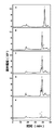
图7、难以降解CS-E的糖胺聚糖裂解酶HCLase Ei降解商品化HA(A)和CS-A(B)、CS-C(C)、CS-D(D)、CS-E(E)终产物的HPLC分析图;
图中:1:二硫酸化二糖,2:单硫酸化二糖,3:非硫酸化二糖;
图8、难以降解CS-E的糖胺聚糖裂解酶HCLase Ei不同时间降解CS-A所得产物的HPLC分析图;
图中:1:四糖,2:单硫酸化二糖,3:非硫酸化二糖;
图9、难以降解CS-E的糖胺聚糖裂解酶HCLase Ei降解低硫酸化CS-E所得终产物的HPLC分析图;
图10、难以降解CS-E的糖胺聚糖裂解酶HCLase Ei降解CS-E制备的寡糖抗肿瘤效果分析;
图11、三个酶降解商品化CS-E的比较曲线图;
图中:1:二硫酸化二糖,2:单硫酸化二糖。
以下实施例的阐述,是为了全面公开本发明如何实施的一些常用技术,而不是为了限制本发明的应用范围。发明人已经尽最大努力确保实施例中个参数的准确性(例如量,温度,等等),但是一些实验误差和偏差也应该予以考虑。除非另有说明,本发明中分子量是指重均分子量,温度是摄氏度。
生物材料来源
弧菌(Vibrio sp.)FC509为现有已知菌株,购自中国微生物菌种保藏管理委员会普通微生物中心,保藏编号CGMCC NO.8913。
实施例1、弧菌(Vibrio sp.)FC509菌株基因组DNA的提取
将弧菌(Vibrio sp.)FC509菌株接种至液体培养基中,在30℃、200rpm的条件下,振荡培养至OD
600=0.8;取培养菌液40mL,在12,000rmp条件下离心25min,收集菌体沉淀,用20mL的溶菌酶缓冲液(10mM Tris-HCl pH 8.0)洗涤,在12,000rmp条件下离心25min,收集菌体沉淀。
上述菌体沉淀中,每管加入溶菌酶缓冲液12.0mL,得到约14.0mL的菌液,分别加入浓度为20mg/mL的溶菌酶各560μL,其终浓度约800μg/mL;冰浴1.0h后,37℃温浴2h,至溶液粘稠;加入10wt%的SDS 0.82mL,100mg/mL的蛋白酶K溶液60μL,52℃水浴1.0h;加入Tris-平衡过的酚/氯仿/异戊醇(体积比25:24:1)15mL,轻轻颠倒混匀,至充分乳化;10,000g、4℃条件下离心10min,转移上清,加入2.0mL的NaAc-HAc(pH 5.2,3.0M)缓冲液,以及17.0mL的无水乙醇,混匀;用1.0mL的枪头挑出丝状DNA,转移至1.5mL的EP离心管中,以体积百分比70%乙醇溶液(贮于-20℃),洗涤2次,微离心后弃上清;10,000g、4℃条件下离心3min,彻底弃掉上清;样品于无菌工作台中,酒精灯下风吹干燥;用无菌去离子水重悬溶解DNA样品,4℃过夜,得到大分子量基因组DNA。
上述液体培养基每升组分如下:
胰蛋白胨10g、酵母提取物5g、NaCl 30g、KH
2PO
4 3g、K
2HPO
4 7g、(NH
4)
2SO
4 2g、MgSO
4·7H
20 5mg、FeSO
4·7H
20 2mg,pH值为7.2。
实施例2、弧菌(Vibrio sp.)FC509菌株基因组扫描及其序列分析。
将实施例1制得的大分子量基因组DNA进行测序(美吉生物公司)。用NCBI(National Center for Biotechnology Information,http://www.ncb1.nlm.nih.gov/)上的软件对测序结果进行分析。所用到的NCBI分析软件是Open Reading Frame Finder(ORF Finder,http://www.ncb1.nlm.nih.gov/gorf/gorf.html)和Basic Local Alignment Search Tool(BLAST,
http://blast.ncbi.nlm.nih.gov/Blast.cgi)。
NCBI分析结果显示Vibrio sp.FC509菌株基因组上携带一个糖胺聚糖裂解酶基因hclase ei,该基因编码区长2973bp,其核苷酸序列如SEQ ID NO.1所示。与Vibrio vulnificus的全基因组序列中chondroitinase基因(NCBI序列号:WP039547743.1)的238个氨基酸有36%的同源性。
HCLase Ei由990个氨基酸组成,其氨基酸序列如SEQ ID NO.2所示,蛋白质的理论分子量约为107kD。用Simple Modular Architecture Research Tool(SMART,http://smart.embl_heidelberg.de/)分析糖胺聚糖裂解酶HCLase Ei的结构信息,结果显示N端第 1至第21个氨基酸是信号肽序列,第23-702位氨基酸序列属于糖胺聚糖裂解酶超家族。用SWISS-MODEL同源建模服务器(
http://swissmodel.expasy.org)对糖胺聚糖裂解酶HCLase Ei的蛋白质三维结构进行同源建模,最终得到的HCLase Ei蛋白质三维结构模型,如图1所示。
实施例3、rHCLase Ei的基因在大肠杆菌中的重组表达
以实施例1制得的大分子量基因组DNA为模板,进行PCR扩增。
引物如下:
正向引物HCLase Ei-F:GGGAATTC
CATATGATAATCAAAGAAGTCGAAAATC;
反向引物HCLase Ei-R:CCG
CTCGAGTTTCACCGGCTCTTGCAAC;
正向引物下划线标注的是限制性内切酶NdeI,反向引物下划线标注的是限制性内切酶Xho I位点。Primerstar HS DNA聚合酶购自宝生物公司,PCR反应体系按照公司提供的产品说明操作。
PCR反应条件:95℃预变性5min;95℃变性10s,68℃退火2.5min,30个循环;72℃延伸10min。
将PCR产物用Nde I和XhoI双酶切,琼脂糖凝胶电泳回收酶切的PCR产物。将购于美国Novagen公司的产物pET-30a载体用NdeI和XhoI双酶切,琼脂糖凝胶电泳回收酶切载体大片段。NdeI和Xho I均购于宝生物公司,酶与底物反应的体系、温度和时间均按照公司提供的产品说明操作。
将经过双酶切的PCR产物与同样经过双酶切pET-30a载体连接,连接产物转化大肠杆菌DH5α菌株后涂布于含有50μg/mL卡那青霉素的Luria-Bertani培养基固体平板(按常规技术配制)上,37℃培养14h,挑取单克隆;将单克隆接入含有50μg/mL卡那青霉素的液体Luria-Bertani培养基(按常规技术配制)中培养,提取质粒;将质粒用正向引物HCLase Ei-F和反向引物HCLase Ei-R进行菌液PCR验证,结果得到大小正确的扩增产物,初步证明构建的重组质粒正确;重组质粒测序结果表明,在pET-30a的NdeI和Xho I酶切位点之间插入SEQ ID NO.1所示的HCLase Ei基因,且插入方向正确,所以进一步证明构建的重组质粒正确,将该重组质粒命名为pET30a-HCLase Ei。
将pET30a-HCLase Ei转化大肠杆菌菌株BL21(DE3)(购自美国Novagen公司),然后按照该公司提供的操作步骤进行重组糖胺聚糖裂解酶HCLase Ei诱导表达。并用Ni Sepharose 6Fast Flow(GE)凝胶对HCLase Ei进行纯化,纯化条件按照GE公司的产品手册操作。用聚丙烯酰胺凝胶电泳检测重组糖胺聚糖裂解酶HCLase Ei的纯化情况,结果如图2所示,纯化后的重组糖胺聚糖裂解酶HCLase Ei在电泳胶上呈单一条带,且位置与预测的分子量相吻合。
实施例4、重组糖胺聚糖裂解酶HCLase Ei的酶学性质分析
pH和温度对酶活性的影响
将质量浓度为1%透明质酸或硫酸软骨素底物、HCLase Ei酶液、不同pH值的150mM HAc-NaAc、NaH
2PO
4-Na
2HPO
4、Tris-HCl缓冲液以及水(pH范围为5.0~10.0),按2:1:3:4(体积比)的比例混合后,在30℃反应60min,按前述的紫外法测酶活力。结果显示HCLase Ei在pH 8.0时达到最大活力,表明HCLase Ei的最适反应pH为8.0(如图3)。
在最适pH下,将质量浓度为1%透明质酸或硫酸软骨素底物、HCLase Ei酶液、150mM Tris-HCl缓冲液和水(pH8.0)按2:1:3:4(体积比)的比例混合,分别在不同温度(0℃~90℃)反应10min,按前述的紫外法测酶活力。结果显示HCLase Ei在30℃时达到最大活力,表明HCLase Ei的最适反应温度为30℃(如图4)。
温度对酶稳定性的影响
将在30℃下热处理不同时间后的HCLase Ei酶液与质量浓度为1%透明质酸或硫酸软骨素底物溶液按2:3(体积比)的比例混合,然后在最适温度和最适pH下测定剩余酶活,以不经过热处理的酶液酶活定义为100%相对活力(relativie activity),结果显示HCLase Ei在30℃的温度下,12h后仍具有大于60%活性(如图5)。
金属离子对HCLsae Ei活性的影响
将质量浓度为1%透明质酸或硫酸软骨素底物、HCLase Ei酶液、150mM Tris-HCl缓冲液以及水按2:1:3:4(体积比)的比例混合后,接着向反应体系中添加不同的金属离子,添加的离子终浓度为5mM,然后在30℃反应60min,按前述的紫外法测酶活力。对照组为不加任何金属离子时HCLase Ei的活性(设定为100%),结果图7所示。实验结果显示,Li
+、EDTA,Na
+K
+、Ca
2+、Mg
2+对HCLase Ei活性基本无影响,Ag
+、Cu
2+、Co
2+、Ni
2+、Cr
3+、Fe
3+、Fe
2+、Hg
2+、Pb
2+、Zn
2+等其他离子对酶活呈现抑制作用(图6)。
实施例5、HCLase Ei的酶活测定
将质量浓度为1%透明质酸或硫酸软骨素底物、HCLase Ei酶液、150mM Tris-HCl缓冲液以及水按2:1:3:4(体积比)的比例混合后,在最适温度和最适pH下反应1~10min,按前述的紫外法测酶活力,同时用购于康为世纪公司的蛋白质定量试剂盒测定HCLase Ei酶液的蛋白含量,结果表明重组HCLase Ei对透明质酸的比活为13.8U/mg、对CS-A、CS-C、CS-D、CS-E的比活分别是7.2、6.7、5.9、5.3U/mg。其中底物HA和牛软骨来源CS-A来自Sigma-Aldrich、鲨鱼软骨来源CS-C和CS-D购自Seikagaku公司,鱿鱼软骨来源低硫酸化CS-E根据中国专利文献CN105924544A(申请号201610323540.2)说明书实施例1的方法制备CS-E(E unit含量为39.3%)。
按上述方法检测SEQ ID NO.3所示的蛋白和SEQ ID NO.4所示的蛋白:
SEQ ID NO.3所示的蛋白对透明质酸的比活为0.09U/mg、对硫酸软骨素A的比活为0.1U/mg;
SEQ ID NO.4所示的蛋白对透明质酸的比活为0.07U/mg、对硫酸软骨素A的比活为0.09U/mg;
酶活力单位定义:每分钟催化糖胺聚糖产生1μmoL不饱和双键所需要的酶量。根据紫外法测糖胺聚糖裂酶酶活的方法[8]。
实施例6、HCLase Ei降解糖胺聚糖降解产物的高效液相(HPLC)分析
将质量浓度为1%透明质酸或硫酸软骨素底物、HCLase Ei酶液、150mM Tris-HCl缓冲液以及水按2:1:3:4(体积比)的比例混合后,在pH8.0,30℃条件下反应,选取不同酶解时间 (0、1、2、8、12、24、72h)的产物进行HPLC分析。HPLC分析条件为凝胶柱:superdex peptide10/300GL(GE);流动相:0.2M碳酸氢铵;流速:0.4mL/min;检测条件:UV232nm。
结果如图7和图8所示,图7结果表明HCLase Ei可以将HA,CS-A,CS-C和CS-D彻底降解为不同硫酸化度二糖,但难于降解鱿鱼软骨来源富含Eunit的CS-E,预示着Eunit的存在抑制了该酶的活性。对CS-A的时间梯度降解试验表明,随着降解时间的延长,产物聚合度逐渐降低,最终转变成二糖(图8)。该结果表明HCLase Ei属于内切糖胺聚糖裂解酶且难于降解富含Eunit的CS-E,因此,可被用于糖胺聚糖寡糖特别是富含E unit寡糖的制备以及糖胺聚糖构效关系研究。
实施例7、HCLase Ei降解CS-E多糖后的高效液相(HPLC)及各个组分的二糖分析
将质量浓度为1%粗品CS-E、HCLase Ei酶液、150mM Tris-HCl缓冲液以及水按2:1:3:4(体积比)的比例混合后,在pH8.0,30℃条件下反应,选取不同酶解时间(0、4、8、12、24、72h)的产物进行HPLC分析。HPLC分析条件为凝胶柱:superdex peptide 10/300GL(GE);流动相:0.2M碳酸氢铵;流速:0.4mL/min;检测条件:UV232nm。
收集组分1~6,脱盐后用商品化CSaseABC降解,降解条件参照说明书,产物进行HPLC分析。HPLC分析条件为:HPLC条件:色谱柱:YMC-Pack PA-G column,流动相:NaH
2PO
4线性洗脱,NaH
2PO
4浓度在60min内从16mM升到460mM。流速:1mL/min,检测条件:Ex330nm,Em 420nm。结果如图9和表1所示,表1为糖胺聚糖裂解酶HCLase Ei降解CS-E制备的大于等于四糖的大分子量组分的二糖组成分析:
表1

由表1可以看出HCLase Ei通过降解CS-E制备的各个组分的寡糖其E unit含量明显提高,远远高于CS-E多糖。
实施例8、不同聚合度寡糖的抗肿瘤转移的效果
(1)培养肿瘤细胞:乳腺癌细胞株4T1在含10%胎牛血清,100U/ml青霉素、链霉素双抗的DMEM培养液中,于37℃,5%CO
2培养箱传代培养;
(2)收集细胞:离心收集肿瘤细胞用PBS稀释到2×10
6个/ml待用;
(3)对BALB/c小鼠(8周左右)分别注射PBS、20μg CS-E 8糖、20μg CS-E 10糖、20μg CS-E 12糖,30min后注入200μl肿瘤细胞,3周后解剖小鼠病变部位,统计小鼠体内肿瘤个 数,检验抗肿瘤效果。结果如图10所示,CS-E 10糖及更高分子量寡糖具有显著的抗肿瘤转移效果,并且CS-E10糖效果优于CS-E12糖。
实施例9、糖胺聚糖裂解酶HCLase Ei在药物中的应用
糖胺聚糖裂解酶HCLase Ei可以用于增强药物的吸收或/和递送,包括但不限于经过皮肤途径辅助药物渗透。示例性的药物包括皮质类固醇如氢化可的松、泼尼松龙、倍氯米松丙酸酯、氟米松、去炎松、氯倍他索丙酸酯等;镇痛药和/或抗炎剂如对乙酰氨基酚、甲灭酸、氟芬那酸、双氯芬酸、双氯芬酸钠、阿氯芬酸、羟布宗、保泰松、布洛芬、氟比洛芬、水杨酸、薄荷醇、樟脑、萘普生等;抗高血压药如吲哚洛尔、茚诺洛尔、心痛定等;抗生素如青霉素、四环素、土霉素、新霉素、红霉素、氯霉素等;麻醉剂如利多卡因、苯佐卡因等以及其他药物。
通过敷布,纱布,或其它皮肤涂抹途径施用糖胺聚糖裂解酶。糖胺聚糖裂解酶溶液可以涂敷到合适的敷贴上(如5-6层纱布)。敷贴应覆盖受损区域,且敷贴本身用蜡纸或绷带固定。应用的制剂的量取决于损伤的面积,通常为5~100IU/cm
2。敷贴应每天使用12~24小时,连续使用10~100天。
对于局部使用,溶液,悬浮液,凝胶剂,糊剂,软膏,乳膏或糖胺聚糖裂解酶的其他配方(有或没有另外的活性剂,都可以使用)。溶液等配方中包含制药和化妆品领域用于局部使用可接受的稀释剂,辅助剂和赋形剂的,这些其它配方主要包括缓冲剂如磷酸盐,柠檬酸盐,醋酸盐和其它有机酸盐、抗氧化剂如抗坏血酸、肽、蛋白质如血清白蛋白,明胶或免疫球蛋白;亲水性聚合物如聚乙烯吡咯烷酮,天然的或合成的油;氨基酸如甘氨酸,谷氨酸,天冬氨酸,或精氨酸;单糖,二糖和其它碳水化合物,包括纤维素或其衍生物,葡萄糖,乳糖,甘露糖或糊精;螯合剂如EDTA;糖醇如甘露糖醇或山梨糖醇;无机盐,如氯化钠,和非离子表面活性剂如吐温、聚乙二醇。
一种优选的,糖胺聚糖裂解酶制剂是糖胺聚糖裂解酶HCLase Ei的冻干粉末悬浮或溶解在含有0.1%~10%蔗糖、1%~20%甘油的溶剂中,溶液的pH保持在6.5~8.5范围内。
糖胺聚糖裂解酶组合物可以通过口服给药或肠胃外给药,并且糖胺聚糖裂解酶组合物可以与合适的递送载体结合后给药。
可通过活性化合物与固体赋形剂的组合获得用于口服使用的糖胺聚糖裂解酶药物制剂。选择上述的混合物加入合适的附助剂混合使用。如果需要的话,合适的赋形剂是碳水化合物或蛋白质,例如糖、纤维素、牛血清白蛋白等。
糖胺聚糖裂解酶酶与药物(如氢化可的松,抗生素,维生素)组合。以口服凝胶剂,膏剂,或悬浮液的形式通过静脉内,皮下或肌内注射给药;或以软膏剂,乳膏剂,面罩局部给药;以及用作化妆品辅助剂,用于增强皮肤的吸收。
实验例10
利用中国专利文献CN103923857A(申请号201410159983.3)中制备的糖胺聚糖裂解酶-HCLase及商品化购买的CSaseABC,以及本发明实施例制备的HCLase Ei在各自最优的条件下充分降解商品化CS-E(E unit含量为61%),利用HPLC根据紫外吸收检测反应产物。结果 如图11所示,通过结果可以看出,HCLase和商品化CSaseABC均可以完全降解CS-E产生二糖。
由上可以看出,本申请中所述HCLase Ei难以降解商品化CS-E,却可以一定程度地降解低硫酸化的CS-E(图9),说明E unit可能抑制HCLase Ei活力,难以降解CS-E,尤其富含E unit的CS-E。因此在降解低硫酸化CS-E时聚集了富含E unit的寡糖,这与表1的结果相符。HCLase和商品化CSaseABC可完全降解CS-E,虽然可以制备不同分子量的寡糖,却不能制备富含E unit的寡糖。
说明书中涉及的参考文献
[1].Bradbury,E.J.,et al.,Chondroitinase ABC promotes functional recovery after spinal cord injury.Nature,2002.416(6881):p.636-40.
[2].Kamhi,E.,et al.,Glycosaminoglycans in infectious disease.Biol Rev Camb Philos Soc,2013.88(4):p.928-43.
[3].Linhardt,R.J.,et al.,CS lyases:structure,activity,and applications in analysis and the treatment of diseases.Adv Pharmacol,2006.53:p.187-215.
[4].Jiang,D.,J.Liang and P.W.Noble,Hyaluronan in tissue injury and repair.Annu Rev Cell Dev Biol,2007.23:p.435-61.
[5].Noble,P.W.,Hyaluronan and its catabolic products in tissue injury and repair.Matrix Biol,2002.21(1):p.25-9.
[6].Sugahara,K.,et al.,Recent advances in the structural biology of chondroitin sulfate and dermatan sulfate.Curr Opin Struct Biol,2003.13(5):p.612-20.
[7].Yamada,S.,K.Sugahara and S.Ozbek,Evolution of glycosaminoglycans:Comparative biochemical study.Commun Integr Biol,2011.4(2):p.150-8.
[8].Yamagata,T.,et al.,Purification and properties of bacterial chondroitinases and chondrosulfatases.J Biol Chem,1968.243(7):p.1523-35.
[9].Hiyama,K.and S.Okada,Crystallization and some properties of chondroitinase from Arthrobacter aurescens.J Biol Chem,1975.250(5):p.1824-8.
[10].Yin,F.X.,F.S.Wang and J.Z.Sheng,Uncovering the Catalytic Direction of Chondroitin AC Exolyase:FROM THE REDUCING END TOWARDS THE NON-REDUCING END.J Biol Chem,2016.291(9):p.4399-406.
[11].Gu,K.,et al.,Purification,characterization and specificity of chondroitin lyases and glycuronidase from Flavobacterium heparinum.Biochem J,1995.312(Pt 2):p.569-77.
[12].Deepa,S.S.,et al.,Specific molecular interactions of oversulfated chondroitin sulfate E with various heparin-binding growth factors.Implications as a physiological binding partner in the brain and other tissues.J Biol Chem,2002.277(46):p.43707-16.
[13].Clement, A. M. , K. Sugahara and A. Faissner, Chondroitin sulfate E promotes neurite outgrowth of rat embryonic day 18 hippocampal neurons. Neurosci Lett, 1999.269 (3) : p. 125-8.
[14].Bishop, J. R. , M. Schuksz and J. D. Esko, Heparan sulphate proteoglycans fine-tune mammalian physiology. Nature, 2007.446 (7139) : p. 1030-7.
[15].Bergefall, K. , et al. , Chondroitin sulfate characterized by the E-disaccharide unit is a potent inhibitor of herpes simplex virus infectivity and provides the virus binding sites on gro2C cells. J Biol Chem, 2005.280 (37) : p. 32193-9.
[16].Sato, Y. , et al. , A highly sulfated chondroitin sulfate preparation, CS-E, prevents excitatory amino acid-induced neuronal cell death. J Neurochem, 2008.104 (6) : p. 1565-76.
[17].Li, F. , et al. , Involvement of Highly Sulfated Chondroitin Sulfate in the Metastasis of the Lewis Lung Carcinoma Cells. Journal of Biological Chemistry, 2008.283 (49) : p. 34294-34304.
[18].Koike, T. , et al. , Chondroitin sulfate-E mediates estrogen-induced osteoanabolism. Sci Rep, 2015.5: p. 8994.
Claims (10)
- 一种难以降解CS-E的糖胺聚糖裂解酶,其特征在于,氨基酸序列如(a)或(b)所示:(a)氨基酸序列如SEQ ID NO.2所示;(b)在(a)中的氨基酸序列经过取代、缺失或添加一个或一个以上氨基酸且具有糖胺聚糖裂解酶活性的由(a)衍生的蛋白质。
- 如权利要求1所述的糖胺聚糖裂解酶,其特征在于,所述氨基酸序列(b)如SEQ ID NO.3或SEQ ID NO.4所示。
- 一种难以降解CS-E的糖胺聚糖裂解酶的编码基因,其特征在于,核苷酸序列如(i)或(ii)所示:(i)核苷酸序列如SEQ ID NO.1所示;(ii)在严格条件下与(i)限定的DNA序列杂交且编码具有糖胺聚糖裂解酶活性的蛋白质的DNA分子。
- 如权利要求3所述的编码基因,其特征在于,所述核苷酸序列(ii)中所述严格条件下,是指:在含质量浓度0.5%SDS的6×SSC缓冲液中,在65℃下杂交,然后用含质量浓度0.1%SDS的2×SSC缓冲液和含质量浓度0.1%SDS的1×SSC缓冲液各洗膜一次。
- 如权利要求4所述的编码基因,其特征在于,所述核苷酸序列(ii)如SEQ ID NO.5或SEQ ID NO.6所示。
- 一种重组表达载体,在表达载体中插入了权利要求3所述的难以降解CS-E的糖胺聚糖裂解酶的编码基因;优选的,上述表达载体选自大肠杆菌表达载体、酵母表达载体、枯草杆菌表达载体、乳酸菌表达载体、链霉菌表达载体、噬菌体载体、丝状真菌表达载体、植物表达载体、昆虫表达载体或哺乳动物细胞表达载体。
- 一种重组细胞,在细胞中插入了权利要求3所述的难以降解CS-E的糖胺聚糖裂解酶的编码基因;优选的,上述细胞选自大肠杆菌宿主细胞、酵母菌宿主细胞、枯草杆菌宿主细胞、乳酸菌宿主细胞、放线菌宿主细胞、丝状真菌宿主细胞、昆虫细胞或哺乳动物细胞。
- 权利要求1所述难以降解CS-E的糖胺聚糖裂解酶做为增强药物的吸收或递送成分在制备药物中的应用。
- 一种利用难以降解CS-E的糖胺聚糖裂解酶HCLase Ei在降解鱿鱼来源的硫酸软骨素CS-E制备结构相对均一的不同聚合度寡糖中的应用。
- 权利要求9制备的结构相对均一的不同聚合度寡糖在制备抑制肿瘤细胞转移药物中的应用。
Applications Claiming Priority (2)
| Application Number | Priority Date | Filing Date | Title |
|---|---|---|---|
| CN201710544077.9A CN107312765B (zh) | 2017-07-05 | 2017-07-05 | 一种糖胺聚糖裂解酶及其编码基因与应用 |
| CN201710544077.9 | 2017-07-05 |
Publications (1)
| Publication Number | Publication Date |
|---|---|
| WO2019007033A1 true WO2019007033A1 (zh) | 2019-01-10 |
Family
ID=60181334
Family Applications (1)
| Application Number | Title | Priority Date | Filing Date |
|---|---|---|---|
| PCT/CN2018/073401 Ceased WO2019007033A1 (zh) | 2017-07-05 | 2018-01-19 | 一种难以降解cs-e的糖胺聚糖裂解酶及其编码基因与应用 |
Country Status (2)
| Country | Link |
|---|---|
| CN (1) | CN107312765B (zh) |
| WO (1) | WO2019007033A1 (zh) |
Families Citing this family (4)
| Publication number | Priority date | Publication date | Assignee | Title |
|---|---|---|---|---|
| CN107312765B (zh) * | 2017-07-05 | 2020-01-21 | 山东大学 | 一种糖胺聚糖裂解酶及其编码基因与应用 |
| CN107460179B (zh) * | 2017-09-22 | 2021-06-29 | 青岛农业大学 | 一种多糖降解酶及其编码基因与应用 |
| CN110408566B (zh) * | 2019-07-29 | 2022-09-30 | 山东大学 | 一种外切型硫酸软骨素降解酶及其编码基因与应用 |
| CN112553179B (zh) * | 2020-12-04 | 2022-07-01 | 北京理工大学 | 一种耐热型聚酯塑料降解酶及其应用 |
Citations (5)
| Publication number | Priority date | Publication date | Assignee | Title |
|---|---|---|---|---|
| CN103923857A (zh) * | 2014-04-21 | 2014-07-16 | 山东大学 | 一株弧菌fc509菌株及其培养方法与应用 |
| CN103923899A (zh) * | 2014-04-21 | 2014-07-16 | 山东大学 | 一种糖胺聚糖裂解酶及其编码基因与应用 |
| CN105713890A (zh) * | 2016-04-13 | 2016-06-29 | 山东大学 | 一种外切型糖胺聚糖裂解酶及其编码基因与应用 |
| CN105924544A (zh) * | 2016-05-16 | 2016-09-07 | 山东大学 | 一种高硫酸化硫酸软骨素及其制备方法与应用 |
| CN107312765A (zh) * | 2017-07-05 | 2017-11-03 | 山东大学 | 一种难以降解cs‑e的糖胺聚糖裂解酶及其编码基因与应用 |
-
2017
- 2017-07-05 CN CN201710544077.9A patent/CN107312765B/zh active Active
-
2018
- 2018-01-19 WO PCT/CN2018/073401 patent/WO2019007033A1/zh not_active Ceased
Patent Citations (5)
| Publication number | Priority date | Publication date | Assignee | Title |
|---|---|---|---|---|
| CN103923857A (zh) * | 2014-04-21 | 2014-07-16 | 山东大学 | 一株弧菌fc509菌株及其培养方法与应用 |
| CN103923899A (zh) * | 2014-04-21 | 2014-07-16 | 山东大学 | 一种糖胺聚糖裂解酶及其编码基因与应用 |
| CN105713890A (zh) * | 2016-04-13 | 2016-06-29 | 山东大学 | 一种外切型糖胺聚糖裂解酶及其编码基因与应用 |
| CN105924544A (zh) * | 2016-05-16 | 2016-09-07 | 山东大学 | 一种高硫酸化硫酸软骨素及其制备方法与应用 |
| CN107312765A (zh) * | 2017-07-05 | 2017-11-03 | 山东大学 | 一种难以降解cs‑e的糖胺聚糖裂解酶及其编码基因与应用 |
Non-Patent Citations (4)
| Title |
|---|
| DATABASE NCBI 18 May 2013 (2013-05-18), "hypothetical protein[Vibrio sp. EJY3", Database accession no. WP 014232125. 1 * |
| DATABASE NCBI 18 May 2013 (2013-05-18), "hypothetical protein[Vibrio sp. EJY3", Database accession no. WP_014232125.1 * |
| DATABASE NCBI 3 July 2013 (2013-07-03), "hypothetical protein[Vibrio natriegens", Database accession no. WP_020334673.1 * |
| HAN, W.J.: "Novel Eliminase from a Marine Bacterium That Degrades Hyalu- ronan and Chondroitin Sulfate", THE JOURNAL OF BIOLOGICAL CHEMISTRY, 3 October 2014 (2014-10-03), pages 27886 - 27898, XP055677897, DOI: 10.1074/jbc.M114.590752 * |
Also Published As
| Publication number | Publication date |
|---|---|
| CN107312765B (zh) | 2020-01-21 |
| CN107312765A (zh) | 2017-11-03 |
Similar Documents
| Publication | Publication Date | Title |
|---|---|---|
| CN103923857B (zh) | 一株弧菌fc509菌株及其培养方法与应用 | |
| CN103923899B (zh) | 一种糖胺聚糖裂解酶及其编码基因与应用 | |
| JP5957096B2 (ja) | バチルス、ヒアルロン酸酵素、及びそれらの使用 | |
| US11203772B2 (en) | Chemoenzymatic synthesis of structurally homogeneous ultra-low molecular weight heparins | |
| CA2493509A1 (en) | Rationally designed polysaccharide lyases derived from chondroitinase b | |
| WO2019007033A1 (zh) | 一种难以降解cs-e的糖胺聚糖裂解酶及其编码基因与应用 | |
| US8193166B2 (en) | Epimerized derivatives of K5 polysaccharide with a very high degree of sulfation | |
| JPH10508204A (ja) | イズロン酸を高含有量有する多糖類 | |
| CN106906161B (zh) | 一种微杆菌、该菌表达的广谱糖胺聚糖裂解酶及其编码基因与应用 | |
| JP2006504437A5 (zh) | ||
| CN113603732B (zh) | 一种非动物源硫酸软骨素寡糖及其制备方法 | |
| CN1244702C (zh) | 一种鞘氨醇杆菌及用其生产的肝素酶生产肝素寡糖的方法 | |
| EP2707396B1 (en) | Biotechnological sulphated chondroitin sulphate at position 4 or 6 on the same polysaccharide chain, and process for the preparation thereof | |
| CN109628347B (zh) | 一株发光杆菌fc615及其培养方法与应用 | |
| CN112126606B (zh) | 一株发光杆菌qa16及其培养方法与应用 | |
| CN103952384B (zh) | 内切型硫酸软骨素/硫酸皮肤素4-o-硫酸酯酶及其编码基因与应用 | |
| CN103635491B (zh) | 类鲨鱼硫酸软骨素及其制备方法 | |
| US8513407B2 (en) | Process for the preparation of N-acyl-(epi)K5-amine-O-sulfate-derivatives and products thus obtained | |
| CN1422283A (zh) | K5多糖衍生的具有高抗凝和抗血栓形成活性的葡糖胺聚糖及其制备方法 | |
| CN110408566B (zh) | 一种外切型硫酸软骨素降解酶及其编码基因与应用 | |
| US9718947B2 (en) | Biotechnological sulphated chondroitin sulphate at position 4 or 6 on the same polysaccharide chain, and process for the preparation thereof | |
| CN110643586B (zh) | 一种肝素骨架合酶及其编码基因与应用 | |
| CN118852492A (zh) | 一种岩藻糖化硫酸乙酰肝素及其制备方法与应用 | |
| CN118240788A (zh) | 酶活性提高的硫酸软骨素合酶及其突变体编码基因与应用 | |
| US20090118230A1 (en) | Fucose containing proteoglycan or acidic glycan and their pharmaceutical use |
Legal Events
| Date | Code | Title | Description |
|---|---|---|---|
| 121 | Ep: the epo has been informed by wipo that ep was designated in this application |
Ref document number: 18829050 Country of ref document: EP Kind code of ref document: A1 |
|
| NENP | Non-entry into the national phase |
Ref country code: DE |
|
| 122 | Ep: pct application non-entry in european phase |
Ref document number: 18829050 Country of ref document: EP Kind code of ref document: A1 |