WO2018066701A1 - Ftldの予防又は治療剤及びそのスクリーニング方法 - Google Patents
Ftldの予防又は治療剤及びそのスクリーニング方法 Download PDFInfo
- Publication number
- WO2018066701A1 WO2018066701A1 PCT/JP2017/036520 JP2017036520W WO2018066701A1 WO 2018066701 A1 WO2018066701 A1 WO 2018066701A1 JP 2017036520 W JP2017036520 W JP 2017036520W WO 2018066701 A1 WO2018066701 A1 WO 2018066701A1
- Authority
- WO
- WIPO (PCT)
- Prior art keywords
- tau
- test substance
- cells
- ftld
- cell
- Prior art date
- Legal status (The legal status is an assumption and is not a legal conclusion. Google has not performed a legal analysis and makes no representation as to the accuracy of the status listed.)
- Ceased
Links
Images


Classifications
-
- A—HUMAN NECESSITIES
- A61—MEDICAL OR VETERINARY SCIENCE; HYGIENE
- A61P—SPECIFIC THERAPEUTIC ACTIVITY OF CHEMICAL COMPOUNDS OR MEDICINAL PREPARATIONS
- A61P25/00—Drugs for disorders of the nervous system
-
- C—CHEMISTRY; METALLURGY
- C12—BIOCHEMISTRY; BEER; SPIRITS; WINE; VINEGAR; MICROBIOLOGY; ENZYMOLOGY; MUTATION OR GENETIC ENGINEERING
- C12N—MICROORGANISMS OR ENZYMES; COMPOSITIONS THEREOF; PROPAGATING, PRESERVING, OR MAINTAINING MICROORGANISMS; MUTATION OR GENETIC ENGINEERING; CULTURE MEDIA
- C12N5/00—Undifferentiated human, animal or plant cells, e.g. cell lines; Tissues; Cultivation or maintenance thereof; Culture media therefor
- C12N5/06—Animal cells or tissues; Human cells or tissues
- C12N5/0602—Vertebrate cells
- C12N5/0618—Cells of the nervous system
- C12N5/0619—Neurons
-
- C—CHEMISTRY; METALLURGY
- C12—BIOCHEMISTRY; BEER; SPIRITS; WINE; VINEGAR; MICROBIOLOGY; ENZYMOLOGY; MUTATION OR GENETIC ENGINEERING
- C12N—MICROORGANISMS OR ENZYMES; COMPOSITIONS THEREOF; PROPAGATING, PRESERVING, OR MAINTAINING MICROORGANISMS; MUTATION OR GENETIC ENGINEERING; CULTURE MEDIA
- C12N5/00—Undifferentiated human, animal or plant cells, e.g. cell lines; Tissues; Cultivation or maintenance thereof; Culture media therefor
- C12N5/06—Animal cells or tissues; Human cells or tissues
- C12N5/0602—Vertebrate cells
- C12N5/0693—Tumour cells; Cancer cells
-
- G—PHYSICS
- G01—MEASURING; TESTING
- G01N—INVESTIGATING OR ANALYSING MATERIALS BY DETERMINING THEIR CHEMICAL OR PHYSICAL PROPERTIES
- G01N33/00—Investigating or analysing materials by specific methods not covered by groups G01N1/00 - G01N31/00
- G01N33/48—Biological material, e.g. blood, urine; Haemocytometers
- G01N33/50—Chemical analysis of biological material, e.g. blood, urine; Testing involving biospecific ligand binding methods; Immunological testing
- G01N33/5005—Chemical analysis of biological material, e.g. blood, urine; Testing involving biospecific ligand binding methods; Immunological testing involving human or animal cells
- G01N33/5008—Chemical analysis of biological material, e.g. blood, urine; Testing involving biospecific ligand binding methods; Immunological testing involving human or animal cells for testing or evaluating the effect of chemical or biological compounds, e.g. drugs, cosmetics
- G01N33/5044—Chemical analysis of biological material, e.g. blood, urine; Testing involving biospecific ligand binding methods; Immunological testing involving human or animal cells for testing or evaluating the effect of chemical or biological compounds, e.g. drugs, cosmetics involving specific cell types
- G01N33/5058—Neurological cells
-
- G—PHYSICS
- G01—MEASURING; TESTING
- G01N—INVESTIGATING OR ANALYSING MATERIALS BY DETERMINING THEIR CHEMICAL OR PHYSICAL PROPERTIES
- G01N33/00—Investigating or analysing materials by specific methods not covered by groups G01N1/00 - G01N31/00
- G01N33/48—Biological material, e.g. blood, urine; Haemocytometers
- G01N33/50—Chemical analysis of biological material, e.g. blood, urine; Testing involving biospecific ligand binding methods; Immunological testing
- G01N33/68—Chemical analysis of biological material, e.g. blood, urine; Testing involving biospecific ligand binding methods; Immunological testing involving proteins, peptides or amino acids
- G01N33/6893—Chemical analysis of biological material, e.g. blood, urine; Testing involving biospecific ligand binding methods; Immunological testing involving proteins, peptides or amino acids related to diseases not provided for elsewhere
- G01N33/6896—Neurological disorders, e.g. Alzheimer's disease
-
- C—CHEMISTRY; METALLURGY
- C12—BIOCHEMISTRY; BEER; SPIRITS; WINE; VINEGAR; MICROBIOLOGY; ENZYMOLOGY; MUTATION OR GENETIC ENGINEERING
- C12N—MICROORGANISMS OR ENZYMES; COMPOSITIONS THEREOF; PROPAGATING, PRESERVING, OR MAINTAINING MICROORGANISMS; MUTATION OR GENETIC ENGINEERING; CULTURE MEDIA
- C12N2500/00—Specific components of cell culture medium
- C12N2500/05—Inorganic components
- C12N2500/10—Metals; Metal chelators
- C12N2500/12—Light metals, i.e. alkali, alkaline earth, Be, Al, Mg
- C12N2500/14—Calcium; Ca chelators; Calcitonin
-
- C—CHEMISTRY; METALLURGY
- C12—BIOCHEMISTRY; BEER; SPIRITS; WINE; VINEGAR; MICROBIOLOGY; ENZYMOLOGY; MUTATION OR GENETIC ENGINEERING
- C12N—MICROORGANISMS OR ENZYMES; COMPOSITIONS THEREOF; PROPAGATING, PRESERVING, OR MAINTAINING MICROORGANISMS; MUTATION OR GENETIC ENGINEERING; CULTURE MEDIA
- C12N2506/00—Differentiation of animal cells from one lineage to another; Differentiation of pluripotent cells
- C12N2506/45—Differentiation of animal cells from one lineage to another; Differentiation of pluripotent cells from artificially induced pluripotent stem cells
-
- C—CHEMISTRY; METALLURGY
- C12—BIOCHEMISTRY; BEER; SPIRITS; WINE; VINEGAR; MICROBIOLOGY; ENZYMOLOGY; MUTATION OR GENETIC ENGINEERING
- C12N—MICROORGANISMS OR ENZYMES; COMPOSITIONS THEREOF; PROPAGATING, PRESERVING, OR MAINTAINING MICROORGANISMS; MUTATION OR GENETIC ENGINEERING; CULTURE MEDIA
- C12N2529/00—Culture process characterised by the use of electromagnetic stimulation
- C12N2529/10—Stimulation by light
Definitions
- the present invention relates to a novel therapeutic agent for frontotemporal lobar degeneration. More specifically, the present invention relates to a preventive or therapeutic agent for frontotemporal lobar degeneration, comprising an inhibitor of calcium ion influx into cells.
- neurodegenerative diseases due to the gradual increase in life expectancy and a dramatic increase in the prevalence of these diseases, late-onset neurodegenerative diseases are increasing the burden on society. Based on accumulated evidence, most neurodegenerative diseases (non-patent literature 1), including neurodegenerative tauopathy (non-patent literature 1-5), which is a disease associated with pathological aggregation of tau, a microtubule-related protein in the brain. Misfolded proteins are strongly associated as causative agents of 6-10). Mutations in the tau gene (MAPT) cause a familial disease known as frontotemporal lobar degeneration tauopathy or FTLD-Tau, which presents with dementia and is prominent in the frontal and temporal lobes of the human brain Characterized by atrophy.
- FTLD-Tau frontotemporal lobar degeneration tauopathy
- Tau is an axon microtubule-related protein that contributes to microtubule stabilization.
- Alternative splicing of exon 10 encoding a microtubule binding domain (MBD) generates 3 repeat tau with 3 MBDs or 4 repeat tau with 4 MBDs.
- Mutations in exons of MAPT are known to weaken the ability of tau to bind to microtubules, promote self-assembly, and change MAPT splicing (Non-Patent Documents 11 to 13).
- Intron mutations in MAPT have been shown to affect splicing of exon 10 and increase 4 repeat tau accumulated in the postmortem brain of patients with intron mutations (Non-Patent Documents 14 and 15).
- FTLD induced pluripotent stem cell
- an object of the present invention is to elucidate one end of a mechanism involved in neurodegeneration in nerve cells of FTLD-Tau patients and provide a novel preventive or therapeutic means for frontotemporal lobar degeneration based on the mechanism. That is.
- Another object of the present invention is to provide a method for screening a preventive or therapeutic agent for frontotemporal lobar degeneration using the mechanism.
- the present inventors have introduced a mutation in the intron or exon MAPT (Mackenzie, IR, et al. Nomenclature and nosology for neuropathologic subtypes of frontotemporal lobar degeneration: an update. Acta Neuropathol 119, 1-4 (2010)).
- the FTLD-Tau iPS cell model was established using neuronal differentiation induced by Neurogenin2 (Ngn2) from FTLD-Tau patients with any of the above. This direct conversion method produces a stable amount of cortical neurons.
- FTLD-Tau FTLD-Tau iPS cells that constitutively express DREADD (designer receptors exclusivelyactivated byrugdesigner drug). It was found to contribute to neurodegeneration.
- DREADD designer receptors exclusivelyactivated byrugdesigner drug
- a prophylactic or therapeutic agent for frontotemporal lobar degeneration comprising an inhibitor of calcium ion influx into cells.
- the calcium ion influx inhibitor is an antagonist or expression inhibitor for NMDA type glutamate receptor, AMPA type glutamate receptor, group I metabotropic glutamate receptor or voltage-dependent calcium channel,
- Agent is an agonist for a group II metabotropic glutamate receptor or a group III metabotropic glutamate receptor.
- [5] The agent according to any one of [1] to [4], which is an inhibitor of tau protein misfolding.
- [6] The agent according to any one of [1] to [5], wherein the frontotemporal lobar degeneration is frontotemporal lobar degeneration tauopathy (FTLD-Tau).
- FTLD-Tau frontotemporal lobar degeneration tauopathy
- [7] The agent according to [6], wherein the frontotemporal lobar degeneration tauopathy is caused by a MAPT gene mutation.
- a screening method for a prophylactic or therapeutic agent for frontotemporal lobar degeneration comprising the following steps.
- a step of bringing a test substance into contact with a cell (2) a step of measuring the inflow of calcium ions into the cell, (3) a step of comparing the degree of inflow of calcium ions in (2) above with the degree of inflow of calcium ions in cells not contacted with the test substance; (4) a step of adding a test substance having a reduced level of calcium ion influx to a nerve cell associated with a pathological condition of FTLD and detecting or measuring the accumulation or release of misfolded tau protein, and (5) A step of selecting the test substance as a candidate for a prophylactic or therapeutic agent for frontotemporal lobar degeneration when accumulation of misfolded tau protein or release to the outside of the cell decreases.
- a screening method for a prophylactic or therapeutic agent for frontotemporal lobar degeneration comprising the following steps. (1) A step of bringing a test substance into contact with a nerve cell associated with a pathological condition of FTLD, (2) a step of measuring the inflow of calcium ions into the cells and the degree of tau protein misfolding, (3) a step of comparing the degree of calcium ion influx and tau protein misfolding in (2) above with the degree of calcium ion influx and tau protein misfold in cells not contacted with the test substance; 4) A step of selecting a test substance that has reduced both the inflow of calcium ions and the degree of tau protein misfolding as a candidate for a prophylactic or therapeutic agent for frontotemporal lobar degeneration.
- a screening method for a prophylactic or therapeutic agent for frontotemporal lobar degeneration comprising the following steps. (1) a step of contacting a test substance with a cell in which the exogenous muscarinic receptor subtype M4 is constitutively expressed; (2) a step of measuring the amount of cAMP in the cell, (3) The step of comparing the amount of cAMP in (2) above with the amount of intracellular cAMP that was not contacted with the test substance, and (4) the test substance with a reduced cAMP amount was determined for frontotemporal lobar degeneration. Step of selecting as a prophylactic or therapeutic agent candidate [11] A screening method for a prophylactic or therapeutic agent for frontotemporal lobar degeneration, comprising the following steps.
- IP3 concentration or DAG concentration in (2) above with the intracellular IP3 concentration or DAG concentration not contacted with the test substance, and (4) the test substance with reduced IP3 concentration or DAG concentration
- a candidate for a prophylactic or therapeutic agent for frontotemporal lobar degeneration [12] A method for preventing or treating frontotemporal lobar degeneration in a mammal, comprising administering an effective amount of an inhibitor of calcium ion influx into cells to a mammal.
- An inhibitor of calcium ion influx into cells for use in the
- FIG. 1 shows the generation of iPS cells from FTLD-Tau patients with either intron or exon MAPT mutations, and gene editing for isogenic control.
- the FTLD-Tau1 iPS cell line carried a MAPT intron mutation (intron 10 + 14C ⁇ T), and the FTLD-Tau2 iPS cell line carried a MAPT exon mutation (R406W). .
- FTLD-Tau1 corrected The mutation of (c, d) FTLD-Tau1 was corrected using CRISPR / Cas9 system (FTLD-Tau1 corrected). Sanger sequence analysis revealed the correction of mutations after gene editing.
- RT-PCR showing FTLD-Tau1 neurons with increased expression of 4 repeat tau and FTLD-Tau1 corrected neurons with normal expression ratio of 4 repeat tau and 3 repeat tau. A full-length image of the gel is shown in FIG. 8 (a).
- Vaginal cell lysates were dephosphorylated with lambda phosphatase, separated by SDS-PAGE for comparison with recombinant tau ladder, and then detected using anti-total tau antibody (Tau12) .
- FIG. 8 shows misfolded tau accumulation in FTLD-Tau neurons.
- FIG. 8 shows misfolded tau accumulation in FTLD-Tau neurons.
- FIG. 8 shows misfolded tau accumulation in FTLD-Tau neurons.
- FIGS. 8 (c) -8 (f) Full length images of the blots are shown in FIGS. 8 (c) -8 (f).
- FIG. 3 shows calcium dysregulation and neuronal loss in FTLD-Tau neurons.
- (a, b) Calcium imaging after electrical stimulation. Intracellular Ca 2+ levels increased by electrical stimulation were higher in FTLD-Tau neurons than in controls (n 6; one-way ANOVA, p ⁇ 0.05; post hoc test, * p ⁇ 0.05).
- (c, d) Immunofluorescence staining of ⁇ III tubulin and NeuN on days 8 and 21 in control and FTLD-Tau neurons. Survival of FTLD-Tau neurons from day 8 to day 21 was reduced compared to control neurons (n 6; one-way ANOVA, p ⁇ 0.05; post hoc test, * p ⁇ 0.05 ).
- FIG. 3 shows calcium dysregulation and neuronal loss in FTLD-Tau neurons.
- FIG. 1 shows the characterization of iPS cells and the generated neurons.
- iPS cells show normal karyotype.
- FIG. 6 relates to FIG. 1 and shows an electrophysiological analysis of iPS cell-derived neurons. To confirm whether iPS cell-derived neurons are functional, the electrophysiological properties of 3 weeks differentiated neurons derived from control iPS cells were evaluated by patch clamp analysis.
- the membrane potential was recorded from a resting membrane potential of -60 mV, following a 75 pA depolarization current injection. The continuity of action potential was recorded by current injection.
- Neurons derived from iPS cells showed paired-pulse depression in response to paired pulse stimulation.
- FIG. 8 shows a full-length image of the gel and blot shown in the main figure in relation to FIGS. 1, 2 and 4.
- A Full length image of the gel shown in FIG. 1 (e).
- Agent of the present invention provides a preventive or therapeutic agent for frontotemporal lobar degeneration, comprising an inhibitor of calcium ion influx into cells (hereinafter abbreviated as “agent of the present invention”). May be).
- the “calcium ion influx inhibitor” of the present invention means a drug in which the inflow of calcium ions into cells is reduced before the drug is introduced or compared with a control group to which no drug is administered.
- FTLD Frontotemporal lobar degeneration
- FTLD pathological classification includes frontotemporal lobar degeneration tauopathy (FTLD-tau), FTLD-TDP, FTLD-FUS, FTLD-UPS, and FTLD-without inclusion.
- the FTLD patient to whom the calcium ion influx inhibitor is administered is not particularly limited, but is desirably an FTLD-tau patient, and particularly preferably an FTLD patient with a MAPT gene mutation.
- mutations in introns of MAPT for example, mutations in intron 10 + 14C ⁇ T, in which cytosine, the 14th base of intron 10 of the MAPT gene, is replaced with thymine
- FTLD-Tau patients with abnormally folded (misfolded) tau protein accumulation due to (mutation) etc. are particularly desirable.
- compositions When using calcium ion inflow inhibitors as pharmaceuticals, they are used as is or known pharmaceutically acceptable carriers (excipients, diluents, extenders, binders, lubricants, flow aids, disintegrants, surfactants). Or a conventional additive and the like, and can be prepared as a pharmaceutical composition.
- the pharmaceutical composition is prepared in an orally administered form such as tablets, pills, capsules, powders, granules, syrups, emulsions and suspensions; injections, drops, external preparations, suppositories, etc. Depending on the parenteral administration agent, etc., it can be systemically or locally administered orally or parenterally.
- intravenous administration In the case of parenteral administration, intravenous administration, intradermal administration, subcutaneous administration, rectal administration, transdermal administration, and the like are possible.
- the above-mentioned appropriate dosage form can be produced by blending the active ingredient with an acceptable ordinary carrier, excipient, binder, stabilizer, diluent and the like.
- acceptable buffering agents, solubilizing agents, isotonic agents and the like can be added.
- the dose varies depending on the type of active ingredient, the route of administration, age of the subject or patient, body weight, symptoms, etc., and cannot be specified unconditionally.
- About mg to 2 g, preferably about 5 mg to several tens mg can be administered once to several times a day. In the case of injection, it is sufficient to administer about 0.1 mg to about 500 mg as an active ingredient in an adult, and the daily dose can be administered once or divided into several times.
- ⁇ G i / o signaling In one embodiment of the invention, inhibition of calcium ion influx is effected via Gi signaling.
- ⁇ G i / o signaling '' means that G protein G i / o is shared to inhibit adenylyl cyclase, reduce cAMP production, and downstream protein kinase A (PKA)
- PKA protein kinase A
- the term “performed via Gi signaling” means that activation of Gi / o or inhibition of influx by inhibiting adenylate cyclase or PKA. means.
- inhibitors of adenylate cyclase include Adenosine 3 ', 5'-cyclic Monophosphate, 8- (4-Chlorophenylthio)-, Sodium Salt 116812 (8-CPT-cAMP, Na), Adenosine 3', 5'- cyclic Monophosphate, 8-Bromo-, Sodium Salt 203800 (8-Bromo-cAMP, Na), Adenylyl Cyclase Toxins Inhibitor 116845 (Ethyl-5-aminopyrazolo [1,5-a] quinazoline-3-carboxylate), Adenylyl Cyclase Type V Inhibitor, NKY80 116850 [2-Amino-7- (furanyl) -7,8-dihydro-5 (6H) -quinazolinone], 2 ', 5'-Dideoxyadenosine 288104 (2', 5'-dd-Ado), MDL -12,330A, Hydro
- PKA inhibitors include, but are not limited to, H89, KT5720, ML-9, K-252a, PKI (6-22) amide, and the like. Further, an antibody or aptamer against adenylate cyclase or PKA may be used.
- NMDA type glutamate receptor NMDA receptor
- AMPA type glutamate receptor AMPA type glutamate receptor
- mGluR metabotropic glutamate receptor
- voltage-dependent calcium Examples include channels (VGCC) and calcium release channels existing on the surface of the endoplasmic reticulum (ER).
- NMDA-type glutamate receptors mediate calcium influx in postsynaptic cells on dendritic spines that are present in various neurons and cortex.
- AMPA-type glutamate receptors are ion-coupled receptors, and are known to localize mainly in GABAergic neurons that are almost free of spines and mediate calcium influx.
- Metabotropic glutamate receptor is a type of G protein-coupled receptor (GPCR) that is known to be widely distributed in the central and peripheral nervous systems. It is classified into three types, II and Group III. Group I mGluR is mainly localized in the postsynaptic membrane, and when it receives a signal, it couples with G aq to activate PLC, and the intracellular diacylglycerol (Diacylglycerol) and inositol triphosphate (Inositol triphosphate) Increase concentration. As a result, PKC is activated, and Ca 2+ stored in the endoplasmic reticulum is released into the cell to increase the intracellular calcium concentration.
- GPCR G protein-coupled receptor
- Group II mGluR and Group III mGluR are mainly localized in the presynaptic membrane and inhibit adenylate cyclase activity, thereby inhibiting calcium ion influx into cells.
- VGCC is a channel that opens in a voltage-dependent manner by depolarization of membrane potential, and calcium ions flow into nerve cells by opening.
- VGCC forms a complex composed of multiple subunits, and its structure is rich in diversity.
- On the surface of the endoplasmic reticulum there are calcium release channels such as inositol triphosphate receptor (IP3R) and ryanodine receptor (RyR).
- the term “agonist” means a substance that binds to a specific receptor and induces a response in a cell
- positive allosteric modulators (PAM) Positive Allosteric Modulator
- An allosteric modulator means a molecule that binds to an allosteric site and enhances or inhibits the binding or signal transduction of an orthosteric ligand to exert its action.
- An allosteric modulator is an orthosteric ligand.
- PAM Physical Allosteric Modulator
- NAM Negative Allosteric Modulator
- SAM Neutral Allosteric Modulator
- Antagonist means a substance that decreases, inhibits or prevents the action of another molecule or the activity of a receptor site.
- Antagonists also include, but are not limited to, competitive antagonists, non-competitive antagonists, uncompetitive antagonists, inhibitors of ion channels, NAMs.
- a competitive antagonist binds reversibly to the receptor at the same binding site (active site) as the endogenous ligand or agonist, but does not activate the receptor.
- Non-competitive antagonists also known as allosteric antagonists
- Non-competitive antagonists do not compete with agonists for binding.
- the bound antagonist may reduce the affinity of the receptor agonist or prevent changes in the receptor conformation that are required for receptor activation after agonist binding.
- Uncompetitive antagonists differ from non-competitive antagonists in that they require receptor activation by an agonist before binding to an allosteric binding site.
- antibodies, aptamers, dominant negative mutants and the like that inhibit the function of receptors and calcium channels are also included as antagonists.
- NMDA receptor antagonists include AP5 (APV, R-2-amino-5-phosphonopentanoate), AP7 (2-amino-7-phosphonoheptanoic acid), CPPene (3-[(R) -2-carboxypiperazin-4-yl ] -prop-2-enyl-1-phosphonic acid), Selfotel, Amantadine, Atomoxetine, AZD6765, Agmatine, Chloroform, Dextrallorphan, Dextromethorphan, Dextrorphan, Diphenidine, Dizocilpine (MK-801), Ethanol, Eticyclidine, Gacyclidine, Ibogaine, Magnesium , Memantine, Methoxetamine, Nitromemantine, Nitrous oxide, Phencyclidine, Rolicyclidine, Tenocyclidine, Methoxydine, Tiletamine, Neramexane, Eliprodil, Etoxadrol, Dexox
- AMPA receptor antagonists include CNQX (6-cyano-7-nitroquinoxaline-2,3-dione), DNQX (6,7-Dinitroquinoxaline-2,3-dione), NBQX (2,3-Dioxo-6-nitro -1,2,3,4-tetrahydrobenzo [f] quinoxaline-7-sulfonamide), Kynurenic acid, Ethanol, GYKI-52466 (4- (8-Methyl-9H-1,3-dioxolo [4,5-h] [2,3] benzodiazepin-5-yl) -benzenamine hydrochloride), GYKI-53655 (1- (4-Aminophenyl) -3-methylcarbamyl-4-methyl-3,4-dihydro-7,8-methylenedioxy-5H- 2,3-benzodiazepine hydrochloride), Perampanel, Talampanel, etc., but are not limited thereto.
- Voltage-gated calcium channel antagonists include Amlodipine (Norvasc), dihydropyridine (DHP), phenylalkylamine (PAA), benzothiazepine (BTZ), Aranidipine (Sapresta), Azelnidipine (Calblock), Barnidipine (HypoCa), Benidipine (Coniel), Cilnidipine (Atelec, Cinalong, Siscard), Clevidipine (Cleviprex), Isradipine (DynaCirc, Prescal), Efonidipine (Landel), Felodipine (Plendil), Lacidipine (Motens, Lacipil), Lercanidipine (Zanidip), Manidipine (Calslot, cardipine) (Cardene, Carden SR), Nifedipine (Procardia, Adalat), Nilvadipine (Nivadil), Nimodipine
- Group I mGluR antagonists include JNJ-16259685, R-214127, YM-202074, YM-230888, YM-298198, FTIDC, A-841720, Cyclothiazide, Lithium, LY-344545, Mavoglurant, Remeglurant, SIB-1893, Basimglurant , Dipraglurant, Fenobam, GRN-529, MPEP, MTEP and Raseglurant, but are not limited thereto.
- Group II mGluR agonists include MGS-0028, LY404040, LY379268, LY354740, (R) -2-amino-4- (4-hydroxy [1,2,5] thiadiazol-3-yl) butyric acid, (S) -aminoadipic acid, JNJ-46356479, JNJ-40411813, GSK-1331258, Imidazo [1,2-a] pyridines, 3-Aryl-5-phenoxymethyl-1,3-oxazolidin-2-ones, 3- (Imidazolyl methyl) -3-aza-bicyclo [3.1.0] hexan-6-yl) methyl ethers, BINA and LY-487379, but are not limited thereto.
- Group III mGluR agonists include Cinnabarinic acid, LSP1-2111, LSP4-2022, L-AP4 (L-2-amino-4-phosphonobutyrate), ML-128, VU-0418506, VU-001171, VU-0155041, PHCCC , ADX88178, (S) -aminoadipic acid, AMN082 (N, N'-dibenzhydrylethane-1,2-diamine dihydrochloride), (S) -3,4-DCPG ((S) -3,4-Dicarboxyphenylglycine) and AZ 12216052 (2-[[(4-Bromophenyl) methyl] thio] -N- [4- (1-methylpropyl) phenyl] acetamide) and the like, but is not limited thereto.
- Antisense nucleic acids, siRNA, shRNA, miRNA, ribozyme, antigene, etc. for receptors etc. are designed appropriately using known design software based on the base sequence of genes such as known receptors and mRNA, etc. It can be easily synthesized using an automatic DNA / RNA synthesizer.
- the present invention also provides a screening method for a preventive or therapeutic agent for frontotemporal lobar degeneration (hereinafter sometimes abbreviated as “method of the present invention”), comprising the following steps. (1) A step of bringing a test substance into contact with a cell, (2) a step of measuring the inflow of calcium ions into the cell, (3) a step of comparing the degree of inflow of calcium ions in (2) above with the degree of inflow of calcium ions in cells not contacted with the test substance; (4) a step of adding a test substance having a reduced level of calcium ion influx to a nerve cell associated with a pathological condition of FTLD and detecting or measuring the accumulation or release of misfolded tau protein, and (5) When the accumulation or release of misfolded tau protein is reduced before or after the addition of the test substance, the test substance is removed from frontotemporal lobar degeneration. Selecting as a candidate for a prophylactic or therapeutic agent.
- a screening method for a prophylactic or therapeutic agent for frontotemporal lobar degeneration comprising the following steps. (1 ′) a step of bringing a test substance into contact with a neuron with a pathological condition of FTLD, (2 ′) a step of measuring the inflow of calcium ions into the cells and the degree of tau protein misfolding, (3 ′) comparing the degree of calcium ion influx and tau protein misfolding in (2 ′) with the degree of calcium ion influx and tau protein misfold in cells not contacted with the test substance, And (4 ′) a step of selecting a test substance that has reduced both the influx of calcium ions and the degree of tau protein misfolding as a candidate for a prophylactic or therapeutic agent for frontotemporal lobar degeneration.
- the nerve cell with the pathological condition of FTLD may be a nerve cell collected from an FTLD patient, or a pluripotent stem cell (iPS cell) is prepared using a somatic cell collected from an FTLD patient. It may be a nerve cell obtained by differentiating a cell.
- iPS cells can be appropriately produced by methods known per se using somatic cells collected from FTLD patients. As a method for inducing differentiation of iPS cells into neurons, various known differentiation induction methods can be appropriately selected and used.
- a method of forcibly expressing Ngn2 in cells and differentiating into cortical neurons can be mentioned. It is done.
- the FTLD patient is not particularly limited, but an FTLD-Tau patient having a specific mutation in the MAPT gene as described above is particularly desirable.
- test substance used in the screening method of the present invention is not particularly limited, and examples thereof include proteins, peptides, nucleic acids, inorganic compounds, natural or synthetically prepared organic compounds, and the like.
- the test substance is a peptide library of 3 to 50 amino acids, preferably 5 to 20 amino acids, or a molecular weight of 100 to 2000, preferably prepared using combinatorial chemistry techniques known to those skilled in the art.
- Can include 200 to 800 low molecular weight organic compound libraries.
- concentration of the test substance to be brought into contact with the cell is not particularly limited, and is usually about 0.1 ⁇ M to about 100 ⁇ M, preferably 1 ⁇ M to 50 ⁇ M.
- the time for contacting the cell and the test substance is not particularly limited and is set as appropriate. For example, it is about 5 to 30 minutes, preferably about 10 to 20 minutes.
- the test substance can be used by appropriately dissolving or suspending in a solvent such as water, a buffer such as phosphate buffer or Tris buffer, ethanol, acetone, dimethyl sulfoxide, or a mixture thereof.
- the inflow of calcium ions into the cell and the accompanying membrane potential depolarization can be measured by well-known methods such as the calcium imaging method and the patch clamp method, respectively.
- the inflow of calcium ions into the cell can be detected using, for example, a calcium fluorescent probe.
- the calcium fluorescent probe that can be used in the present invention includes 1- [6-amino-2- (5-carboxy-2-oxazolyl) -5-benzofuranyloxy] -2- (2-amino-5-methyl).
- Examples include, but are not limited to, phenoxy) ethane-N, N, N ′, N′-tetraacetic acid, pentaacetoxymethyl ester (Fura 2-AM), and the like.
- the membrane potential can be measured, for example, by a nerve recording method such as a microelectrode method or a patch clamp method, or may be measured using a membrane potential measurement fluorescent probe.
- Fluorescent probes for membrane potential measurement include 4- (4- (didecylamino) styryl) -N-methylpyridinium iodide (4-Di-10-ASP), bis- (1,3-dibutylbarbituric acid trimethine oxo Nord (DiSBAC2 (3)), 3,3′-dipropylthiadicarbocyanine iodide (DiSC3 (5)), 5,5 ′, 6,6′-tetrachloro-1,1 ′, 3,3 ′ -Tetraethylbenzimidazolyl carbocyanine iodide (JC-1) and rhodamine 123, but are not limited to these.
- misfolded tau protein is immunostained using, for example, an antibody that specifically recognizes the protein (for example, TOC1 antibody), or dot blot analysis, as shown in the examples below. However, it is not limited to these methods as long as misfolded proteins can be detected or measured.
- a screening method for a prophylactic or therapeutic agent for frontotemporal lobar degeneration comprising the following steps.
- D the test substance with a decreased amount of cAMP Selecting as a prophylactic or therapeutic agent candidate.
- a screening method for a prophylactic or therapeutic agent for frontotemporal lobar degeneration comprising the following steps is also provided.
- A contacting the test substance with a cell in which the exogenous muscarinic receptor subtype M3 is constitutively expressed;
- B measuring the amount of inositol trisphosphate (IP3) or diacylglycerol (DAG) in the cell;
- C The step of comparing the amount of IP3 or DAG in (b) above with the amount of intracellular IP3 or DAG that was not contacted with the test substance, and (d) the test substance in which the amount of IP3 or DAG was reduced Is selected as a candidate for a prophylactic or therapeutic agent for frontotemporal lobar degeneration.
- the method described above may further include a step corresponding to step (4) of the method of the present invention in order to confirm the therapeutic effect of the candidate substance more reliably.
- Constant expression of exogenous muscarinic receptor subtype M3 or M4 should be carried out by introducing into the cell an expression vector containing nucleic acids whose sequences encoding these receptor subtypes are known per se. Can do.
- expression vectors include retrovirus, lentivirus, adenovirus, adeno-associated virus, herpes virus, Sendai virus and other viral vectors, animal cell expression plasmids (eg, pA1-11, pXT1, pRc / CMV, pRc / RSV). , PcDNAI / Neo) and the like.
- Intracellular cAMP is measured, for example, by incubating the cells in the presence and absence of the test substance for an appropriate period of time and then performing a competitive immunoassay on the extract obtained by disrupting the cells. Can do. It can also be evaluated by labeling cells with [ 3 H] adenine and measuring the radioactivity of the produced [ 3 H] cAMP, but any other known method can be used. In another embodiment, there is a method for evaluating the amount of cAMP by measuring the expression level of a reporter gene under the control of a cAMP response element (CRE).
- CRE cAMP response element
- cells introduced with a vector containing an expression cassette linked to a DNA encoding a reporter protein downstream of a promoter containing CRE are cultured for an appropriate time in the presence and absence of a test substance.
- the amount of intracellular cAMP is evaluated by measuring and comparing the expression of the reporter gene in the extract obtained by crushing using a known technique.
- IP3 content Measurements of inositol triphosphate amount of intracellular added [3 H] inositol in the cell, or measuring the generated [3 H] inositol 3 radioactive phosphate, measure Ca 2+ of intracellular You can also evaluate it.
- the measurement can be performed using a fluorescent probe that specifically binds to IP3, but any other known method can be used.
- the DAG amount can be measured, for example, by the following method. Using the cell extract sample, phosphatidine produced by phosphorylating DAG of the sample with a kinase is hydrolyzed by lipase into glycerol-3-phosphate. Subsequently, glycerol-3-phosphate oxidase is oxidized with glycerol-3-phosphate oxidase, and the resulting hydrogen peroxide is reacted with a fluorescent probe that reacts specifically with hydrogen peroxide, and excited at 530-560 nm / fluorescence By detecting at 585-595 nm, the amount of DAG in the sample can be measured. Alternatively, measurement may be performed using a DAG fluorescent probe, but any other known method may be used.
- iPS cells As described before the preparation and cell culture of iPS cells (Okita, K., et al. An efficient nonviral method to generate integration-free human-induced pluripotent stem cells from cord blood and peripheral blood cells.Stem Cells 31, 458-466 (2013).), Epithelial vectors for OCT3 / 4, Sox2, Klf4, L-Myc, Lin28, p53-shRNA and EBNA1, and human iPS cells with MAPT mutations from fibroblasts or monocytes And cultured on a SNL feeder layer using a human iPS cell medium (ReproCELL) supplemented with 4 ng / ml basic FGF (Wako Chemicals) and penicillin / streptomycin. As a control, 201B7 iPS cell line (Takahashi, K., et al. Induction of pluripotent stem cells from adult human fibroblasts by defined factors. Cell 131, 861-872 (2007).) was used.
- a guide RNA was designed to target the 5′-GCAACGTCCAGTCCAAGTGTGG-3 ′ (SEQ ID NO: 1) site. Oligonucleotides of guide RNA were ligated to human H1 polymerase III promoter (Li, HL, et al. Precise correction of the dystrophin gene in duchenne muscular dystrophy patient induced pluripotent stem cells by TALEN and CRISPR-Cas9.
- NEPA21 electroporator (Nepagene) was applied to 1 million iPS cells, 10 ⁇ g pHL-Hi guide RNA expression plasmid, 10 ⁇ g pHL-EF1a-hcSpCas9 plasmid, and 10 ⁇ g donor The gene was introduced by electroporation with the plasmid. Four days after gene transfer, puromycin selection was performed for 10 days.
- Puromycin resistant colonies were selected and expanded for genomic DNA extraction and genotyping by PCR using the following primers: forward primer, 5'-AAATGCAGTCGTGGGAGACC-3 '(SEQ ID NO: 2); reverse primer, 5' -GAGTCCCGAATCTCACGGAGACA-3 '(SEQ ID NO: 3). All amplified PCR bands were further analyzed by Sanger sequencing to confirm that there were no sequence changes in the homology arms.
- the transposase vector pHL-EF1 ⁇ -hcPBase (Matsui, H., et al. Delivery of full-length factor VIII using a piggyBac transposon vector to correct a mouse model of hemophilia A.
- PCR of the puromycin cassette was performed and non-integration was confirmed using the following primers: forward primer, 5'-CTGCTGCAACTTACCTCCGGGATG-3 '(SEQ ID NO: 4 ); Reverse primer, 5′-CCAATCCTCCCCCTTGCTGTCCTG-3 ′ (SEQ ID NO: 5).
- iPS cell lines are shown in Table 1.
- piggyBac vector for expression of Ngn2 and introduction into iPS cells
- Ngn2 transcription factor was introduced into mouse or human iPS cells using the piggyBac vector.
- This vector containing the Ngn2 and neomycin resistance genes under the control of the tetracycline operator rtTA is the KW110_PB_TA_ERN (Ef1a_rtTA_neo) vector backbone (Kim, SI, et al. Inducible Transgene Expression in Human iPS Cells Using Versatile All-in-One piggyBac Transposons. Methods Mol Biol 1357, 111-131 (2015).).
- the prepared vector was co-introduced into iPS cells using Lipofectamine LTX (Invitrogen) together with the pCyL43 vector encoding transposase. After clone selection with neomycin, iPS cells harboring a tetracycline-inducible Ngn2 construct were established.
- Preparation of iPS cell-derived cortical neurons by induction of Ngn2 iPS cells were dissociated into single cells using actase, and a 1: 1 ratio of DMEM / F12 (Life Technologies) and Neurobasal (Life Technologies), 1% N2 supplement, 2% B27 supplement, 10 ng / ml brain-derived neurotrophic factor (BDNF, R & D Systems), 10 ng / ml glial cell-derived neurotrophic factor (GDNF, R & D Systems) and 10 ng / ml neurotrophin-3 (NT-3; R & D Systems) was used to seed on plastic plates or coverslips coated with matrigel with 1 ⁇ g / ml doxycycline (Clontech).
- BDNF brain-derived neurotrophic factor
- GDNF ng / ml glial cell-derived neurotrophic factor
- NT-3 10 ng / ml neurotrophin-3
- the tol2 vector containing M4D and hygromycin resistance gene under CAG promoter control was transformed into pT2AL200R175-CAGGS-EGFP vector backbone (Kawakami, K. & Noda, T. Transposition of the Tol2 element, an Ac-like element from the Japanese medaka fish Oryzias latipes, in mouse embryonic stem cells. Genetics 166, 895-899 (2004).).
- the prepared vector was simultaneously introduced into iPS cells into which Ngn2 had been introduced together with a vector encoding transposase using Lipofectamine LTX. After clone selection using hygromycin, an iPS cell line carrying M4D was established.
- the prepared iPS cells were differentiated into neurons with doxycycline and nerve medium for 8 days, and then 100 nM pharmacologically inactive designer drug clozapine-N-oxide (CNO) was added to the culture medium to add DREADD. I was stimulated. On the 14th day, Western blot analysis and dot blot analysis were performed.
- CNO pharmacologically inactive designer drug clozapine-N-oxide
- RNA of cultured cells was extracted using RNeasy Plus Mini kit (QIAGEN). One microgram of RNA was reverse transcribed using ReverTra Ace (TOYOBO). PCR analysis was performed using TAKARA Ex Taq (TAKARA). As previously reported, primer sets for 3 repeat tau and 4 repeat tau were used: forward 5'-AAGTCGCCGTCTTCCGCCAAG-3 '(SEQ ID NO: 6); reverse 5'-GTCCAGGGACCCAATCTTCGA-3' (SEQ ID NO: 7) . RT-PCR products were evaluated on 3% agarose gels: 3 repeat tau and 4 repeat tau RT-PCR products were 288 and 381 bp, respectively (Iovino, M., et al. Early maturation and distinct tau pathology in induced pluripotent stem cell-derived neurons from patients with MAPT mutations. Brain 138, 3345-3359 (2015).).
- RNA of the cultured cells on day 7 was extracted using the RNeasy Plus Mini kit (QIAGEN). One microgram of RNA was reverse transcribed using ReverTra Ace (TOYOBO). Quantitative PCR analysis was performed by reverse transcription reaction with SYBR Premix Ex Taq II (TAKARA) using Step One Plus (Applied Biosystems). The primer set is shown in Table 2.
- Immunocytochemical cells were fixed with 4% paraformaldehyde for 30 minutes at room temperature, washed with PBS, and permeated into PBS containing 0.2% Triton X-100 for 10 minutes at room temperature. Subsequently, blocking was performed with Block Ace (Yukijirushi) for 30 minutes. After overnight incubation with the primary antibody at 4 ° C., the cells were washed 3 times with PBS and incubated with the appropriate secondary antibody for 1 hour at room temperature.
- Cell images were acquired from Delta Vision (Applied Precision) or IN Cell Analyzer 6000 (GE Healthcare). Cell numbers were quantified with IN Cell Analyzer 6000 and IN CELL Developer toolbox software 1.9 (GE Healthcare).
- ⁇ III tubulin (1: 2,000, Covance), NeuN (1: 500, Millipore), TOC1 (1: 1,000), Nanog (1: 200, Abcam), SSEA4 ( 1: 500, Millipore).
- the total protein extract (20 ⁇ g per lane) is separated by size on a 10% polyacrylamide gel and transferred to an Immobilon-P membrane (Millipore). did. Membranes were blocked with 5% skim milk, hybridized with the appropriate antibody and visualized using the ECL Prime detection kit (GE Healthcare). Images were acquired from ImageQuant LAS 4000 (GE Healthcare). The following primary antibodies were used: MAP2 (1: 1,000, CST), ⁇ III tubulin (1: 5,000, Covance), Tau12 (1: 10,000, Millipore), TOC1 (1: 5,000) and ⁇ -actin (1: 5,000), Sigma).
- the TOC1 antibody has a linear epitope displayed in the oligomer compared to the monomer or filament in the oligomer (Ward, SM, et al. TOC1: characterization of a selective oligomeric tau antibody. J Alzheimers Dis 37, 593-602 (2013).) Therefore, it is noteworthy that non-denaturing conditions are required to evaluate oligomeric tau using TOC1 antibody. The use of denaturing conditions makes no distinction between these different tau species, oligomers, monomers and / or filaments using TOC1. Table 3 shows the tau antibodies used in this paper.
- the membrane was blocked with 5% skim milk, hybridized with the appropriate antibody, and visualized using Western Lightning Plus-ECL (PerkinElmer). Images were acquired from ImageQuant LAS 4000 (GE Healthcare). The following primary antibodies were used: Tau12 (1: 10,000, Millipore), Tau46 (1: 1,000), TOC1 (1: 5,000).
- Cell viability assay Dissociated iPS cells transfected with Ngn2 into single cells, 5 ⁇ 10 4 cells / well on a 96-well plate (BD Bioscience) coated with Matrigel using nerve medium containing 1 ⁇ g / ml doxycycline Sowing. Cells were fixed and stained on days 8 and 21. The number of viable neurons stained with NeuN was quantified with IN Cell Analyzer 6000 (GE Healthcare) and expressed as the number of neurons on day 21/8. D-2-amino-5-phosphonopentanoate (AP-5) (SIGMA) and 6-cyano-7-nitroquinoxaline-2,3-dione (CNQX) (SIGMA) from day 8 to day 21 The number of surviving neurons was evaluated on day 21.
- AP-5 SIGMA
- CNQX 6-cyano-7-nitroquinoxaline-2,3-dione
- PI stained cells were incubated with 1.5 ⁇ M PI (Nacalai Tesque) for 30 minutes at 37 ° C. and fixed with 4% paraformaldehyde. The number of PI stained cells was quantified by IN Cell Analyzer 6000.
- Enzyme-linked immunosorbent assay (ELISA) of oligomeric tau A 96-well plate (Greiner) was coated overnight at 4 ° C. with 3 ⁇ g / ml TOC1 antibody in 0.05 M sodium carbonate buffer. After washing and blocking with TBS-T containing 1% BSA, 100 ⁇ l of culture medium was added and incubated at room temperature for 2 hours. A standard curve was obtained using recombinant oligomeric tau protein. For detection, plates were incubated with 2 ⁇ g / ml affinity purified rabbit polyclonal anti-human tau antibody followed by sheep anti-rabbit IgG F (ab) ′ 2 fragment conjugated to horseradish peroxidase (1: 3,000; GE Healthcare) did.
- iPS cells were dissociated into single cells and seeded at 5 ⁇ 10 4 cells / well on a 96-well plate (BD Bioscience) coated with Matrigel using nerve medium containing 1 ⁇ g / ml doxycycline.
- e-FDSS / ⁇ CELL Haamamatsu Photonics
- Electrophysiological recordings Whole cell patch clamp recordings were performed from iPS cell-derived neurons.
- the recording micropipette was filled with an intracellular solution composed of 140 mM KCl, 2 mM MgCl 2 , 10 mM HEPES and 1 mM EGTA, adjusted to pH 7.4 with NaOH. Cells are maintained at 30 ° C throughout the experiment with an oxidized Krebs Ringer solution consisting of 125 mM NaCl, 2.5 mM KCl, 1.25 mM NaH 2 PO 4 , 26 mM NaHCO 3 , 1 mM MgCl 2 , 2 mM CaCl 2 and 20 mM glucose. Continuously perfused. Voltage clamp and current clamp recordings were performed using an EPC9 amplifier (HEKA) and the data were analyzed using Patchmaster software (HEKA).
- HEKA EPC9 amplifier
- the intron 10 + 14C ⁇ T mutation was corrected using CRISPR-Cas9 (FIGS. 1 (c) and 1 (d)).
- Ngn2 was introduced into these iPS cells by a piggyBac vector having a tet-on expression system, followed by drug selection for stable cell line establishment.
- the established iPS cells were converted to cortical neurons after 7 days of culture in nerve medium containing doxycycline. There was no difference in the differentiation tendency of the obtained strains.
- the prepared neurons express mRNA of neurotransmitter receptor (Fig. 5 (c)), and their functional properties were shown by electrophysiological analysis (Fig. 6 (a) -6 (h) )). As previously reported (Hutton, M., et al.
- FTLD-Tau neurons show typical misfolded tau spots and dots, and control neurons, including genetically modified strains, have misfolded tau spots or dots. Most were negative.
- Dot blot analysis showed the accumulation of misfolded forms of tau in cells in a non-denatured state using the TOC1 antibody (FIGS. 2 (d), (e)). Also, as previously shown (Ward, SM, et al. TOC1: characterization of a selective oligomeric tau antibody. J Alzheimers Dis 37, 593-602 (2013)), to detect tau species under denaturing conditions. Misfolded tau was analyzed by Western blot analysis using TOC1 antibody.
- DREADD muscarinic acetylcholine M4 receptor
- D-2-amino-5-phosphonopentanoate (AP-5) and ⁇ -amino-3-hydroxy-5-methyl which are inhibitors of N-methyl-D-aspartate (NMDA) receptors 4-Cyano-7-nitroquinoxaline-2,3-dione (CNQX), an inhibitor of the 4-isoxazolepropionic acid (AMPA) receptor, increased the survival of FTLD-Tau neurons (Fig. 4 (e) and 4 (f)).
- the inflow inhibitor of calcium ions into cells of the present invention is useful for the prevention and / or treatment of frontotemporal lobar degeneration, and the screening method using the inflow of calcium ions into cells as an index is It is useful for screening for preventive and / or therapeutic agents for temporal lobe degeneration.
Landscapes
- Health & Medical Sciences (AREA)
- Engineering & Computer Science (AREA)
- Life Sciences & Earth Sciences (AREA)
- Biomedical Technology (AREA)
- Chemical & Material Sciences (AREA)
- Biotechnology (AREA)
- Bioinformatics & Cheminformatics (AREA)
- Neurology (AREA)
- Immunology (AREA)
- Cell Biology (AREA)
- Organic Chemistry (AREA)
- General Health & Medical Sciences (AREA)
- Microbiology (AREA)
- Urology & Nephrology (AREA)
- Biochemistry (AREA)
- Zoology (AREA)
- Wood Science & Technology (AREA)
- Genetics & Genomics (AREA)
- Hematology (AREA)
- Neurosurgery (AREA)
- Molecular Biology (AREA)
- Medicinal Chemistry (AREA)
- General Physics & Mathematics (AREA)
- Pathology (AREA)
- Food Science & Technology (AREA)
- Physics & Mathematics (AREA)
- Analytical Chemistry (AREA)
- General Engineering & Computer Science (AREA)
- Nuclear Medicine, Radiotherapy & Molecular Imaging (AREA)
- Animal Behavior & Ethology (AREA)
- Tropical Medicine & Parasitology (AREA)
- Chemical Kinetics & Catalysis (AREA)
- General Chemical & Material Sciences (AREA)
- Proteomics, Peptides & Aminoacids (AREA)
- Pharmacology & Pharmacy (AREA)
- Toxicology (AREA)
- Public Health (AREA)
- Veterinary Medicine (AREA)
- Oncology (AREA)
- Measuring Or Testing Involving Enzymes Or Micro-Organisms (AREA)
Abstract
本発明は、細胞内へのカルシウムイオンの流入阻害薬を含有してなる、前頭側頭葉変性症の予防又は治療剤を提供する。また、本発明は以下の工程を含む、前頭側頭葉変性症の予防又は治療剤のスクリーニング方法を提供する。 (1)被験物質を、細胞と接触させる工程、 (2)該細胞内へのカルシウムイオンの流入を測定する工程、 (3)上記(2)のカルシウムイオンの流入の程度を、被験物質を接触させなかった細胞におけるカルシウムイオンの流入の程度と比較する工程、 (4)カルシウムイオンの流入の程度を低下させた被験物質を、FTLDの病態を伴う神経細胞に添加し、ミスフォールドされたタウタンパク質の蓄積又は細胞外への放出を検出又は測定する工程、並びに (5)ミスフォールドされたタウタンパク質の蓄積又は細胞外への放出が減少した場合に、該被験物質を前頭側頭葉変性症の予防又は治療剤の候補として選択する工程。
Description
本発明は、新規の前頭側頭葉変性症の治療剤に関する。より具体的には、本発明は細胞内へのカルシウムイオンの流入阻害薬を含有してなる、前頭側頭葉変性症の予防又は治療剤に関する。
平均余命の漸増及びこれらの疾患の有病率の劇的な上昇のため、遅発性の神経変性疾患は、社会への負担を増大させている。蓄積した証拠によれば、脳における微小管関連タンパク質であるタウの病理学的な凝集に関連する疾患である神経変性タウオパチー(非特許文献1~5)を含むほとんどの神経変性疾患(非特許文献6~10)の原因物質として、ミスフォールドされたタンパク質が強く関連付けられている。タウ遺伝子(MAPT)の変異は、前頭側頭葉変性症タウオパチー又はFTLD-Tauとして知られる家族性疾患の原因となり、この疾患は認知症を呈し、ヒトの脳の前頭葉及び側頭葉における顕著な萎縮を特徴とする。
タウは、微小管の安定化に寄与する軸索の微小管関連タンパク質である。6種類のタウアイソフォームがあり、これはエクソン2、3及び10のスプライシングの結果である。微小管結合ドメイン(MBD)をコードするエクソン10の選択的スプライシングにより、3つのMBDを有する3リピートタウ又は4つのMBDを有する4リピートタウが生成される。MAPTのエクソンの変異は、微小管に結合するタウの能力を弱め、自己集合を促進し、MAPTスプライシングを変化させることが知られている(非特許文献11~13)。MAPTのイントロンの変異は、エクソン10のスプライシングに影響を及ぼし、イントロンの変異を有する患者の死後脳に蓄積される4リピートタウを増加させることが示されている(非特許文献14、15)。
人工多能性幹細胞(iPS細胞)技術の開発により、インビトロで、FTLD-Tau患者を含む神経変性疾患の神経細胞の表現型の研究が促進されているが(非特許文献16~19)、FTLD-Tau患者の神経細胞における神経変性に関与するメカニズムは依然として不明である。
Iba, M., et al. Synthetic tau fibrils mediate transmission of neurofibrillary tangles in a transgenic mouse model of Alzheimer's-like tauopathy. J Neurosci 33, 1024-1037 (2013).
Sanders, D.W., et al. Distinct tau prion strains propagate in cells and mice and define different tauopathies. Neuron 82, 1271-1288 (2014).
Liu, L., et al. Trans-synaptic spread of tau pathology in vivo. PLoS One 7, e31302 (2012).
Irwin, D.J., et al. Frontotemporal lobar degeneration: defining phenotypic diversity through personalized medicine. Acta Neuropathol 129, 469-491 (2015).
Nukina, N. & Ihara, Y. One of the antigenic determinants of paired helical filaments is related to tau protein. J Biochem 99, 1541-1544 (1986).
Guo, J.L. & Lee, V.M. Cell-to-cell transmission of pathogenic proteins in neurodegenerative diseases. Nat Med 20, 130-138 (2014).
Tran, H.T., et al. Alpha-synuclein immunotherapy blocks uptake and templated propagation of misfolded alpha-synuclein and neurodegeneration. Cell Rep 7, 2054-2065 (2014).
Jucker, M. & Walker, L.C. Self-propagation of pathogenic protein aggregates in neurodegenerative diseases. Nature 501, 45-51 (2013).
Weissmann, C., Li, J., Mahal, S.P. & Browning, S. Prions on the move. EMBO Rep 12, 1109-1117 (2011).
Hansen, C., et al. alpha-Synuclein propagates from mouse brain to grafted dopaminergic neurons and seeds aggregation in cultured human cells. J Clin Invest 121, 715-725 (2011).
Hasegawa, M., Smith, M.J. & Goedert, M. Tau proteins with FTDP-17 mutations have a reduced ability to promote microtubule assembly. FEBS Lett 437, 207-210 (1998).
Hong, M., et al. Mutation-specific functional impairments in distinct tau isoforms of hereditary FTDP-17. Science 282, 1914-1917 (1998).
Nacharaju, P., et al. Accelerated filament formation from tau protein with specific FTDP-17 missense mutations. FEBS Lett 447, 195-199 (1999).
Hutton, M., et al. Association of missense and 5'-splice-site mutations in tau with the inherited dementia FTDP-17. Nature 393, 702-705 (1998).
McCarthy, A., et al. Closing the tau loop: the missing tau mutation. Brain 138, 3100-3109 (2015).
Fong, H., et al. Genetic correction of tauopathy phenotypes in neurons derived from human induced pluripotent stem cells. Stem Cell Reports 1, 226-234 (2013).
Iovino, M., et al. Early maturation and distinct tau pathology in induced pluripotent stem cell-derived neurons from patients with MAPT mutations. Brain 138, 3345-3359 (2015).
Ehrlich, M., et al. Distinct Neurodegenerative Changes in an Induced Pluripotent Stem Cell Model of Frontotemporal Dementia Linked to Mutant TAU Protein. Stem Cell Reports 5, 83-96 (2015).
Sposito, T., et al. Developmental regulation of tau splicing is disrupted in stem cell-derived neurons from frontotemporal dementia patients with the 10 + 16 splice-site mutation in MAPT. Hum Mol Genet 24, 5260-5269 (2015).
従って、本発明の課題は、FTLD-Tau患者の神経細胞における神経変性に関与するメカニズムの一端を解明し、該メカニズムに基づいた、新規な前頭側頭葉変性症の予防又は治療手段を提供することである。また、該メカニズムを利用した、前頭側頭葉変性症の予防又は治療剤のスクリーニング方法を提供することも課題とする。
ここで、本発明者らは、イントロン又はエクソンのMAPTの変異(Mackenzie, I.R., et al. Nomenclature and nosology for neuropathologic subtypes of frontotemporal lobar degeneration: an update. Acta Neuropathol 119, 1-4 (2010).)のいずれかを有するFTLD-Tau患者由来のNeurogenin2(Ngn2)で誘導される直接的な神経細胞の分化を用いて、FTLD-Tau iPS細胞モデルを樹立した。このダイレクトコンバージョン法は、安定的な量の皮質神経細胞を産生する。本発明者らはまた、CRISPR-Cas9により、同質遺伝子的な対照のiPS細胞を作製し、MAPTのイントロン及びエクソン変異の収束した表現型を再現する結果となる、ミスフォールドされたタウ(misfolded tau)タンパク質の蓄積及び細胞外への放出、その後の神経細胞死を観察した。さらに、FTLD-Tauにおける神経変性のメカニズムを調べるために、本発明者らは、DREADD(designer receptors exclusively activated by designer drug)を恒常的に発現するFTLD-Tau iPS細胞を作製し、カルシウム調節異常が神経変性に寄与することを見出した。
即ち、本発明は以下の通りである。
[1] 細胞内へのカルシウムイオンの流入阻害薬を含有してなる、前頭側頭葉変性症の予防又は治療剤。
[2] 前記カルシウムイオンの流入阻害が、Gi/oシグナリングを介して生じる、[1]記載の剤。
[3] 前記カルシウムイオンの流入阻害薬が、NMDA型グルタミン酸受容体、AMPA型グルタミン酸受容体、グループI 代謝型グルタミン酸受容体又は電位依存性カルシウムチャネルに対するアンタゴニスト又は発現阻害薬である、[1]記載の剤。
[4] 前記カルシウムイオンの流入阻害薬が、グループII 代謝型グルタミン酸受容体又はグループIII代謝型グルタミン酸受容体に対するアゴニストである、[1]記載の剤。
[5] タウタンパク質のミスフォールディングの阻害剤である、[1]~[4]のいずれかに記載の剤。
[6] 前頭側頭葉変性症が前頭側頭葉変性症タウオパチー(FTLD-Tau)である、[1]~[5]のいずれかに記載の剤。
[7] 前頭側頭葉変性症タウオパチーがMAPT遺伝子変異に起因するものである、[6]記載の剤。
[8] 以下の工程を含む、前頭側頭葉変性症の予防又は治療剤のスクリーニング方法。
(1)被験物質を、細胞と接触させる工程、
(2)該細胞内へのカルシウムイオンの流入を測定する工程、
(3)上記(2)のカルシウムイオンの流入の程度を、被験物質を接触させなかった細胞におけるカルシウムイオンの流入の程度と比較する工程、
(4)カルシウムイオンの流入の程度を低下させた被験物質を、FTLDの病態を伴う神経細胞に添加し、ミスフォールドされたタウタンパク質の蓄積又は細胞外への放出を検出又は測定する工程、並びに
(5)ミスフォールドされたタウタンパク質の蓄積又は細胞外への放出が減少した場合に、該被験物質を前頭側頭葉変性症の予防又は治療剤の候補として選択する工程
[9] 以下の工程を含む、前頭側頭葉変性症の予防又は治療剤のスクリーニング方法。
(1)被験物質を、FTLDの病態を伴う神経細胞と接触させる工程、
(2)該細胞内へのカルシウムイオンの流入及びタウタンパク質のミスフォールドの程度を測定する工程、
(3)上記(2)のカルシウムイオンの流入及びタウタンパク質のミスフォールドの程度を、被験物質を接触させなかった細胞におけるカルシウムイオンの流入及びタウタンパク質のミスフォールドの程度と比較する工程、並びに
(4)カルシウムイオンの流入及びタウタンパク質のミスフォールドの程度を両方とも低下させた被験物質を、前頭側頭葉変性症の予防又は治療剤の候補として選択する工程
[10] 以下の工程を含む、前頭側頭葉変性症の予防又は治療剤のスクリーニング方法。
(1)被験物質を、外因性のムスカリン受容体のサブタイプM4を恒常的に発現させた細胞と接触させる工程、
(2)該細胞内のcAMP量を測定する工程、
(3)上記(2)のcAMP量を、被験物質を接触させなかった細胞内のcAMP量と比較する工程、並びに
(4)cAMP量を低下させた被験物質を、前頭側頭葉変性症の予防又は治療剤の候補として選択する工程
[11] 以下の工程を含む、前頭側頭葉変性症の予防又は治療剤のスクリーニング方法。
(1)被験物質を、外因性のムスカリン受容体のサブタイプM3を恒常的に発現させた細胞と接触させる工程、
(2)該細胞内のイノシトール3リン酸(IP3)濃度又はジアシルグリセロール(DAG)濃度を測定する工程、
(3)上記(2)のIP3濃度又はDAG濃度を、被験物質を接触させなかった細胞内のIP3濃度又はDAG濃度と比較する工程、並びに
(4)IP3濃度又はDAG濃度を低下させた被験物質を、前頭側頭葉変性症の予防又は治療剤の候補として選択する工程
[12] 哺乳動物に対し、細胞内へのカルシウムイオンの流入阻害薬の有効量を投与することを特徴とする、該哺乳動物における前頭側頭葉変性症の予防または治療方法。
[13] 前頭側頭葉変性症の予防又は治療に使用するための、細胞内へのカルシウムイオンの流入阻害薬。
[1] 細胞内へのカルシウムイオンの流入阻害薬を含有してなる、前頭側頭葉変性症の予防又は治療剤。
[2] 前記カルシウムイオンの流入阻害が、Gi/oシグナリングを介して生じる、[1]記載の剤。
[3] 前記カルシウムイオンの流入阻害薬が、NMDA型グルタミン酸受容体、AMPA型グルタミン酸受容体、グループI 代謝型グルタミン酸受容体又は電位依存性カルシウムチャネルに対するアンタゴニスト又は発現阻害薬である、[1]記載の剤。
[4] 前記カルシウムイオンの流入阻害薬が、グループII 代謝型グルタミン酸受容体又はグループIII代謝型グルタミン酸受容体に対するアゴニストである、[1]記載の剤。
[5] タウタンパク質のミスフォールディングの阻害剤である、[1]~[4]のいずれかに記載の剤。
[6] 前頭側頭葉変性症が前頭側頭葉変性症タウオパチー(FTLD-Tau)である、[1]~[5]のいずれかに記載の剤。
[7] 前頭側頭葉変性症タウオパチーがMAPT遺伝子変異に起因するものである、[6]記載の剤。
[8] 以下の工程を含む、前頭側頭葉変性症の予防又は治療剤のスクリーニング方法。
(1)被験物質を、細胞と接触させる工程、
(2)該細胞内へのカルシウムイオンの流入を測定する工程、
(3)上記(2)のカルシウムイオンの流入の程度を、被験物質を接触させなかった細胞におけるカルシウムイオンの流入の程度と比較する工程、
(4)カルシウムイオンの流入の程度を低下させた被験物質を、FTLDの病態を伴う神経細胞に添加し、ミスフォールドされたタウタンパク質の蓄積又は細胞外への放出を検出又は測定する工程、並びに
(5)ミスフォールドされたタウタンパク質の蓄積又は細胞外への放出が減少した場合に、該被験物質を前頭側頭葉変性症の予防又は治療剤の候補として選択する工程
[9] 以下の工程を含む、前頭側頭葉変性症の予防又は治療剤のスクリーニング方法。
(1)被験物質を、FTLDの病態を伴う神経細胞と接触させる工程、
(2)該細胞内へのカルシウムイオンの流入及びタウタンパク質のミスフォールドの程度を測定する工程、
(3)上記(2)のカルシウムイオンの流入及びタウタンパク質のミスフォールドの程度を、被験物質を接触させなかった細胞におけるカルシウムイオンの流入及びタウタンパク質のミスフォールドの程度と比較する工程、並びに
(4)カルシウムイオンの流入及びタウタンパク質のミスフォールドの程度を両方とも低下させた被験物質を、前頭側頭葉変性症の予防又は治療剤の候補として選択する工程
[10] 以下の工程を含む、前頭側頭葉変性症の予防又は治療剤のスクリーニング方法。
(1)被験物質を、外因性のムスカリン受容体のサブタイプM4を恒常的に発現させた細胞と接触させる工程、
(2)該細胞内のcAMP量を測定する工程、
(3)上記(2)のcAMP量を、被験物質を接触させなかった細胞内のcAMP量と比較する工程、並びに
(4)cAMP量を低下させた被験物質を、前頭側頭葉変性症の予防又は治療剤の候補として選択する工程
[11] 以下の工程を含む、前頭側頭葉変性症の予防又は治療剤のスクリーニング方法。
(1)被験物質を、外因性のムスカリン受容体のサブタイプM3を恒常的に発現させた細胞と接触させる工程、
(2)該細胞内のイノシトール3リン酸(IP3)濃度又はジアシルグリセロール(DAG)濃度を測定する工程、
(3)上記(2)のIP3濃度又はDAG濃度を、被験物質を接触させなかった細胞内のIP3濃度又はDAG濃度と比較する工程、並びに
(4)IP3濃度又はDAG濃度を低下させた被験物質を、前頭側頭葉変性症の予防又は治療剤の候補として選択する工程
[12] 哺乳動物に対し、細胞内へのカルシウムイオンの流入阻害薬の有効量を投与することを特徴とする、該哺乳動物における前頭側頭葉変性症の予防または治療方法。
[13] 前頭側頭葉変性症の予防又は治療に使用するための、細胞内へのカルシウムイオンの流入阻害薬。
本発明によれば、前頭側頭葉変性症に対する予防及び/又は治療が可能となる。また、前頭側頭葉変性症に対する予防及び/又は治療剤のスクリーニングも可能となる。
1. 本発明の剤
本発明は、細胞内へのカルシウムイオンの流入阻害薬を含有してなる、前頭側頭葉変性症の予防又は治療剤を提供する(以下「本発明の剤」と省略する場合がある)。本発明の「カルシウムイオンの流入阻害薬」とは、細胞内へのカルシウムイオンの流入が、薬剤を投入する前や薬剤を投与しない対照群と比較して減少する薬剤を意味する。
本発明は、細胞内へのカルシウムイオンの流入阻害薬を含有してなる、前頭側頭葉変性症の予防又は治療剤を提供する(以下「本発明の剤」と省略する場合がある)。本発明の「カルシウムイオンの流入阻害薬」とは、細胞内へのカルシウムイオンの流入が、薬剤を投入する前や薬剤を投与しない対照群と比較して減少する薬剤を意味する。
<<前頭側頭葉変性症(FTLD)>>
本発明において、「前頭側頭葉変性症(FTLD)」とは、著明な人格変化や行動障害、言語障害を主徴とし、前頭葉、前部側頭葉に病変の首座を有する古典的ピック病をプロトタイプとした変性性認知症を意味し、FTLDの病理分類としては、frontotemporal lobar degeneration tauopathy(FTLD-tau)、FTLD-TDP、FTLD-FUS、FTLD-UPS、FTLD-without inclusionが挙げられる。カルシウムイオンの流入阻害薬の投与対象となるFTLD患者は、特に限定されないが、望ましくは、FTLD-tau患者であり、特にMAPT遺伝子変異を伴うFTLD患者が好ましい。中でも、MAPTのイントロンの変異(例えば、MAPT遺伝子のイントロン10の14番目の塩基であるシトシンがチミンに置換した、イントロン10 +14C →Tの変異)や、MAPTのエクソンの変異(例えば、R406Wの変異)などにより、異常に折りたたまれた(ミスフォールドされた)タウタンパク質の蓄積が認められるFTLD-Tau患者が特に望ましい。
本発明において、「前頭側頭葉変性症(FTLD)」とは、著明な人格変化や行動障害、言語障害を主徴とし、前頭葉、前部側頭葉に病変の首座を有する古典的ピック病をプロトタイプとした変性性認知症を意味し、FTLDの病理分類としては、frontotemporal lobar degeneration tauopathy(FTLD-tau)、FTLD-TDP、FTLD-FUS、FTLD-UPS、FTLD-without inclusionが挙げられる。カルシウムイオンの流入阻害薬の投与対象となるFTLD患者は、特に限定されないが、望ましくは、FTLD-tau患者であり、特にMAPT遺伝子変異を伴うFTLD患者が好ましい。中でも、MAPTのイントロンの変異(例えば、MAPT遺伝子のイントロン10の14番目の塩基であるシトシンがチミンに置換した、イントロン10 +14C →Tの変異)や、MAPTのエクソンの変異(例えば、R406Wの変異)などにより、異常に折りたたまれた(ミスフォールドされた)タウタンパク質の蓄積が認められるFTLD-Tau患者が特に望ましい。
<<医薬組成物>>
カルシウムイオンの流入阻害薬を医薬品として用いるにあたり、そのままもしくは公知の薬学的に許容される担体(賦形剤、希釈剤、増量剤、結合剤、滑沢剤、流動助剤、崩壊剤、界面活性剤等などが含まれる)や慣用の添加剤などと混合して医薬組成物として調製することができる。当該医薬組成物は、調製する形態(錠剤、丸剤、カプセル剤、散剤、顆粒剤、シロップ剤、乳剤、懸濁液などの経口投与剤;注射剤、点滴剤、外用剤、坐剤などの非経口投与剤)等に応じて、全身的に又は局所的に、経口投与又は非経口投与することができる。非経口投与する場合には、静脈投与、皮内投与、皮下投与、直腸投与、経皮投与すること等が可能である。
前記の適当な投与剤型は許容される通常の担体、賦型剤、結合剤、安定剤、希釈剤等に有効成分を配合することにより製造することができる。また注射剤型で用いる場合には、許容される緩衝剤、溶解補助剤、等張剤等を添加することもできる。
カルシウムイオンの流入阻害薬を医薬品として用いるにあたり、そのままもしくは公知の薬学的に許容される担体(賦形剤、希釈剤、増量剤、結合剤、滑沢剤、流動助剤、崩壊剤、界面活性剤等などが含まれる)や慣用の添加剤などと混合して医薬組成物として調製することができる。当該医薬組成物は、調製する形態(錠剤、丸剤、カプセル剤、散剤、顆粒剤、シロップ剤、乳剤、懸濁液などの経口投与剤;注射剤、点滴剤、外用剤、坐剤などの非経口投与剤)等に応じて、全身的に又は局所的に、経口投与又は非経口投与することができる。非経口投与する場合には、静脈投与、皮内投与、皮下投与、直腸投与、経皮投与すること等が可能である。
前記の適当な投与剤型は許容される通常の担体、賦型剤、結合剤、安定剤、希釈剤等に有効成分を配合することにより製造することができる。また注射剤型で用いる場合には、許容される緩衝剤、溶解補助剤、等張剤等を添加することもできる。
また投与量は、有効成分の種類、投与経路、投与対象又は患者の年齢、体重、症状などによって異なり一概に規定できないが、通常、経口の場合には成人で1日あたり有効成分量として、数mg~2g程度、好ましくは5mg~数十mg程度を、1日1~数回にわけて投与することができる。注射の場合には成人で有効成分量として約0.1mg~約500mgを投与すればよく、1日の投与量を1回又は数回に分けて投与することができる。
<<Gi/o シグナリング>>
本発明の一実施態様において、カルシウムイオンの流入阻害は、Giシグナリングを介して行われる。本発明において、「Gi/oシグナリング」とは、Gタンパク質のGi/oと共益してアデニル酸シクラーゼ(Adenylyl cyclase)を阻害し、cAMP産生を低下させ、下流のプロテインキナーゼA(PKA)の活性化を阻害する経路を意味し、「Giシグナリングを介して行われる」とは、Gi/oを活性化すること、又はアデニル酸シクラーゼやPKAを阻害することにより流入を阻害することを意味する。アデニル酸シクラーゼの阻害剤としては、例えば、Adenosine 3',5'-cyclic Monophosphate, 8-(4-Chlorophenylthio)-, Sodium Salt 116812 (8-CPT-cAMP, Na)、 Adenosine 3',5'-cyclic Monophosphate, 8-Bromo-, Sodium Salt 203800 (8-Bromo-cAMP, Na)、 Adenylyl Cyclase Toxins Inhibitor 116845 (Ethyl-5-aminopyrazolo[1,5-a]quinazoline-3-carboxylate)、Adenylyl Cyclase Type V Inhibitor, NKY80 116850 [2-Amino-7-(furanyl)-7,8-dihydro-5(6H)-quinazolinone]、 2',5'-Dideoxyadenosine 288104 (2',5'-dd-Ado)、 MDL-12,330A, Hydrochloride 444200 [cis-N-(2-Phenylcyclopentyl)azacyclotridec-1-en-2-amine, HCl]などが挙げられるが、これらに限定されない。また、PKAの阻害剤としては、例えば、H89、KT5720、ML-9、K-252a、PKI (6-22) amideなどが挙げられるが、これらに限定されない。また、アデニル酸シクラーゼやPKAに対する抗体やアプタマーを用いてもよい。
本発明の一実施態様において、カルシウムイオンの流入阻害は、Giシグナリングを介して行われる。本発明において、「Gi/oシグナリング」とは、Gタンパク質のGi/oと共益してアデニル酸シクラーゼ(Adenylyl cyclase)を阻害し、cAMP産生を低下させ、下流のプロテインキナーゼA(PKA)の活性化を阻害する経路を意味し、「Giシグナリングを介して行われる」とは、Gi/oを活性化すること、又はアデニル酸シクラーゼやPKAを阻害することにより流入を阻害することを意味する。アデニル酸シクラーゼの阻害剤としては、例えば、Adenosine 3',5'-cyclic Monophosphate, 8-(4-Chlorophenylthio)-, Sodium Salt 116812 (8-CPT-cAMP, Na)、 Adenosine 3',5'-cyclic Monophosphate, 8-Bromo-, Sodium Salt 203800 (8-Bromo-cAMP, Na)、 Adenylyl Cyclase Toxins Inhibitor 116845 (Ethyl-5-aminopyrazolo[1,5-a]quinazoline-3-carboxylate)、Adenylyl Cyclase Type V Inhibitor, NKY80 116850 [2-Amino-7-(furanyl)-7,8-dihydro-5(6H)-quinazolinone]、 2',5'-Dideoxyadenosine 288104 (2',5'-dd-Ado)、 MDL-12,330A, Hydrochloride 444200 [cis-N-(2-Phenylcyclopentyl)azacyclotridec-1-en-2-amine, HCl]などが挙げられるが、これらに限定されない。また、PKAの阻害剤としては、例えば、H89、KT5720、ML-9、K-252a、PKI (6-22) amideなどが挙げられるが、これらに限定されない。また、アデニル酸シクラーゼやPKAに対する抗体やアプタマーを用いてもよい。
<<受容体及びカルシウムチャネル>>
本発明において、カルシウムイオンの流入を引き起こす受容体やカルシウムチャネルとして、NMDA型グルタミン酸受容体(NMDA receptor)、AMPA型グルタミン酸受容体(AMPA receptor)、代謝型グルタミン酸受容体(mGluR)、電位依存性カルシウムチャネル(VGCC)、小胞体(ER)の表面に存在するカルシウム放出チャネルなどが挙げられる。NMDA型グルタミン酸受容体は、さまざまな神経細胞や皮質に存在する樹状突起棘(スパイン)上のシナプス後細胞におけるカルシウム流入を介在する。AMPA型グルタミン酸受容体は、イオン共役型受容体で、主にスパインがほとんど存在しないGABA作動性神経細胞に局在することが知られており、カルシウム流入を介在する。代謝型グルタミン酸受容体(mGluR)は、Gタンパク質結合受容体(GPCR)の一種で、中枢神経系及び末梢神経系に広く分布していることが知られており、この受容体はグループI、グループII、グループIIIの3種類に分類される。グループI mGluRは、主にシナプス後膜に局在し、シグナルを受けるとGaqと共役してPLCを活性化し、細胞内のジアシルグリセロール(Diacylglycerol)及びイノシトール-3-リン酸(Inositol triphosphate)の濃度を増加させる。これによりPKCが活性化し、小胞体などに貯蔵されていたCa2+が細胞内に放出されて細胞内カルシウム濃度が上昇する。グループII mGluR及びグループIII mGluRは、主にシナプス前膜に局在し、アデニル酸シクラーゼ活性を阻害することで、細胞内へのカルシウムイオンの流入を阻害する。VGCCは、膜電位の脱分極によって電位依存的に開口するチャネルであり、開口することで神経細胞内へカルシウムイオンが流入する。VGCCは複数のサブユニットから構成される複合体を形成し、その構造は多様性に富んでいる。また、小胞体の表面には、イノシトール3リン酸受容体(IP3R)やリアノジン受容体(RyR)といった、カルシウム放出チャネルが存在する。
本発明において、カルシウムイオンの流入を引き起こす受容体やカルシウムチャネルとして、NMDA型グルタミン酸受容体(NMDA receptor)、AMPA型グルタミン酸受容体(AMPA receptor)、代謝型グルタミン酸受容体(mGluR)、電位依存性カルシウムチャネル(VGCC)、小胞体(ER)の表面に存在するカルシウム放出チャネルなどが挙げられる。NMDA型グルタミン酸受容体は、さまざまな神経細胞や皮質に存在する樹状突起棘(スパイン)上のシナプス後細胞におけるカルシウム流入を介在する。AMPA型グルタミン酸受容体は、イオン共役型受容体で、主にスパインがほとんど存在しないGABA作動性神経細胞に局在することが知られており、カルシウム流入を介在する。代謝型グルタミン酸受容体(mGluR)は、Gタンパク質結合受容体(GPCR)の一種で、中枢神経系及び末梢神経系に広く分布していることが知られており、この受容体はグループI、グループII、グループIIIの3種類に分類される。グループI mGluRは、主にシナプス後膜に局在し、シグナルを受けるとGaqと共役してPLCを活性化し、細胞内のジアシルグリセロール(Diacylglycerol)及びイノシトール-3-リン酸(Inositol triphosphate)の濃度を増加させる。これによりPKCが活性化し、小胞体などに貯蔵されていたCa2+が細胞内に放出されて細胞内カルシウム濃度が上昇する。グループII mGluR及びグループIII mGluRは、主にシナプス前膜に局在し、アデニル酸シクラーゼ活性を阻害することで、細胞内へのカルシウムイオンの流入を阻害する。VGCCは、膜電位の脱分極によって電位依存的に開口するチャネルであり、開口することで神経細胞内へカルシウムイオンが流入する。VGCCは複数のサブユニットから構成される複合体を形成し、その構造は多様性に富んでいる。また、小胞体の表面には、イノシトール3リン酸受容体(IP3R)やリアノジン受容体(RyR)といった、カルシウム放出チャネルが存在する。
<<アゴニスト>>
本発明において、「アゴニスト」とは、特定の受容体に結合し、細胞内で反応を誘発する物質を意味し、正のアロステリック修飾剤(PAM)(Positive Allosteric Modulator)もアゴニストに包含される。アロステリック修飾剤(allosteric modulator)とは、アロステリック部位に結合し、オルソステリックのリガンドの結合やシグナル伝達を増強又は阻害して、その作用を発揮する分子を意味し、アロステリック修飾薬は、オルソステリックリガンドの活性に対する効果として、増強、減弱、無変化のそれぞれに対応して、PAM、NAM(Negative Allosteric Modulator)、SAM(Silent(orneutral)Allosteric Modulator)に分類することができる。
本発明において、「アゴニスト」とは、特定の受容体に結合し、細胞内で反応を誘発する物質を意味し、正のアロステリック修飾剤(PAM)(Positive Allosteric Modulator)もアゴニストに包含される。アロステリック修飾剤(allosteric modulator)とは、アロステリック部位に結合し、オルソステリックのリガンドの結合やシグナル伝達を増強又は阻害して、その作用を発揮する分子を意味し、アロステリック修飾薬は、オルソステリックリガンドの活性に対する効果として、増強、減弱、無変化のそれぞれに対応して、PAM、NAM(Negative Allosteric Modulator)、SAM(Silent(orneutral)Allosteric Modulator)に分類することができる。
<<アンタゴニスト>>
本発明において、「アンタゴニスト」とは、別の分子の作用又は受容体部位の活性を減少、阻害、又は妨げる物質を意味する。アンタゴニストには、競合性アンタゴニスト、非競合性アンタゴニスト、不競合性アンタゴニスト、イオンチャネルの阻害剤、NAMも含まれるが、これらに限定されない。競合性アンタゴニストは、内因性リガンド又はアゴニストと同じ結合部位(活性部位)で受容体に可逆的に結合するが、受容体を活性化しない。非競合性アンタゴニスト(アロステリックアンタゴニストとしても知られている)は、アゴニストの結合部位とは異なる部位に結合し、他の結合部位を経由してその受容体に作用を及ぼす。非競合性アンタゴニストは、結合のためにアゴニストと競合しない。結合したアンタゴニストは、その受容体のアゴニストの親和性を低下し、又はアゴニストの結合後に受容体活性化に必要となる、受容体の立体構造の変化を妨げる場合もある。不競合性アンタゴニストは、アロステリック結合部位に結合する前に、アゴニストによる受容体活性化を必要とするという点で、非競合性アンタゴニストと異なる。また、受容体やカルシウムチャネルの機能を阻害する抗体、アプタマー、ドミナントネガティブ変異体などもアンタゴニストに包含される。
本発明において、「アンタゴニスト」とは、別の分子の作用又は受容体部位の活性を減少、阻害、又は妨げる物質を意味する。アンタゴニストには、競合性アンタゴニスト、非競合性アンタゴニスト、不競合性アンタゴニスト、イオンチャネルの阻害剤、NAMも含まれるが、これらに限定されない。競合性アンタゴニストは、内因性リガンド又はアゴニストと同じ結合部位(活性部位)で受容体に可逆的に結合するが、受容体を活性化しない。非競合性アンタゴニスト(アロステリックアンタゴニストとしても知られている)は、アゴニストの結合部位とは異なる部位に結合し、他の結合部位を経由してその受容体に作用を及ぼす。非競合性アンタゴニストは、結合のためにアゴニストと競合しない。結合したアンタゴニストは、その受容体のアゴニストの親和性を低下し、又はアゴニストの結合後に受容体活性化に必要となる、受容体の立体構造の変化を妨げる場合もある。不競合性アンタゴニストは、アロステリック結合部位に結合する前に、アゴニストによる受容体活性化を必要とするという点で、非競合性アンタゴニストと異なる。また、受容体やカルシウムチャネルの機能を阻害する抗体、アプタマー、ドミナントネガティブ変異体などもアンタゴニストに包含される。
<<NMDA受容体アンタゴニスト>>
NMDA受容体アンタゴニストとしては、AP5 (APV, R-2-amino-5-phosphonopentanoate), AP7 (2-amino-7-phosphonoheptanoic acid)、CPPene (3-[(R)-2-carboxypiperazin-4-yl]-prop-2-enyl-1-phosphonic acid)、Selfotel, Amantadine, Atomoxetine, AZD6765, Agmatine, Chloroform, Dextrallorphan, Dextromethorphan, Dextrorphan, Diphenidine, Dizocilpine (MK-801), Ethanol, Eticyclidine, Gacyclidine, Ibogaine, Magnesium, Memantine, Methoxetamine, Nitromemantine, Nitrous oxide, Phencyclidine, Rolicyclidine, Tenocyclidine, Methoxydine, Tiletamine, Neramexane, Eliprodil, Etoxadrol, Dexoxadrol, WMS-2539, NEFA, Remacemide, Delucemine, 8A-PDHQ, Aptiganel (Cerestat, CNS-1102), HU-211, Remacemide, Rhynchophylline, Ketamine, Glycine, Rapastinel (GLYX-13), NRX-1074, 7-Chlorokynurenic acid, 4-Chlorokynurenine (AV-101), 5,7-Dichlorokynurenic acid, Kynurenic acid, TK-40, 1-Aminocyclopropanecarboxylic acid (ACPC), L-Phenylalanine及びXenonなどが挙げられるが、これらに限定されない。
NMDA受容体アンタゴニストとしては、AP5 (APV, R-2-amino-5-phosphonopentanoate), AP7 (2-amino-7-phosphonoheptanoic acid)、CPPene (3-[(R)-2-carboxypiperazin-4-yl]-prop-2-enyl-1-phosphonic acid)、Selfotel, Amantadine, Atomoxetine, AZD6765, Agmatine, Chloroform, Dextrallorphan, Dextromethorphan, Dextrorphan, Diphenidine, Dizocilpine (MK-801), Ethanol, Eticyclidine, Gacyclidine, Ibogaine, Magnesium, Memantine, Methoxetamine, Nitromemantine, Nitrous oxide, Phencyclidine, Rolicyclidine, Tenocyclidine, Methoxydine, Tiletamine, Neramexane, Eliprodil, Etoxadrol, Dexoxadrol, WMS-2539, NEFA, Remacemide, Delucemine, 8A-PDHQ, Aptiganel (Cerestat, CNS-1102), HU-211, Remacemide, Rhynchophylline, Ketamine, Glycine, Rapastinel (GLYX-13), NRX-1074, 7-Chlorokynurenic acid, 4-Chlorokynurenine (AV-101), 5,7-Dichlorokynurenic acid, Kynurenic acid, TK-40, 1-Aminocyclopropanecarboxylic acid (ACPC), L-Phenylalanine及びXenonなどが挙げられるが、これらに限定されない。
<<AMPA受容体アンタゴニスト>>
AMPA受容体アンタゴニストとしては、CNQX(6-cyano-7-nitroquinoxaline-2,3-dione), DNQX (6,7-Dinitroquinoxaline-2,3-dione), NBQX(2,3-Dioxo-6-nitro-1,2,3,4-tetrahydrobenzo[f]quinoxaline-7-sulfonamide),Kynurenic acid, Ethanol, GYKI-52466 (4-(8-Methyl-9H-1,3-dioxolo[4,5-h][2,3]benzodiazepin-5-yl)-benzenamine hydrochloride), GYKI-53655(1-(4-Aminophenyl)-3-methylcarbamyl-4-methyl-3,4-dihydro-7,8-methylenedioxy-5H-2,3-benzodiazepine hydrochloride), Perampanel及びTalampanelなどが挙げられるが、これらに限定されない。
AMPA受容体アンタゴニストとしては、CNQX(6-cyano-7-nitroquinoxaline-2,3-dione), DNQX (6,7-Dinitroquinoxaline-2,3-dione), NBQX(2,3-Dioxo-6-nitro-1,2,3,4-tetrahydrobenzo[f]quinoxaline-7-sulfonamide),Kynurenic acid, Ethanol, GYKI-52466 (4-(8-Methyl-9H-1,3-dioxolo[4,5-h][2,3]benzodiazepin-5-yl)-benzenamine hydrochloride), GYKI-53655(1-(4-Aminophenyl)-3-methylcarbamyl-4-methyl-3,4-dihydro-7,8-methylenedioxy-5H-2,3-benzodiazepine hydrochloride), Perampanel及びTalampanelなどが挙げられるが、これらに限定されない。
<<電位開口型カルシウムチャネルアンタゴニスト>>
電位開口型カルシウムチャネルアンタゴニストとしては、Amlodipine (Norvasc), dihydropyridine(DHP), phenylalkylamine(PAA),benzothiazepine(BTZ), Aranidipine (Sapresta), Azelnidipine(Calblock), Barnidipine (HypoCa), Benidipine (Coniel), Cilnidipine (Atelec, Cinalong, Siscard), Clevidipine (Cleviprex), Isradipine (DynaCirc, Prescal), Efonidipine (Landel), Felodipine (Plendil), Lacidipine (Motens, Lacipil), Lercanidipine (Zanidip), Manidipine (Calslot, Madipine), Nicardipine (Cardene, Carden SR), Nifedipine (Procardia, Adalat), Nilvadipine (Nivadil), Nimodipine (Nimotop), Nisoldipine (Baymycard, Sular, Syscor), Nitrendipine (Cardif, Nitrepin, Baylotensin), Pranidipine (Acalas), Verapamil (Calan, Isoptin), Gallopamil, Fendiline, Diltiazem (Cardizem), ω-Agatoxin IVA, ω-Conotoxin GVIA, SNX-482, mibefradil, bepridil, flunarizine, fluspirilene及びfendilineなどが挙げられるが、これらに限定されない。
電位開口型カルシウムチャネルアンタゴニストとしては、Amlodipine (Norvasc), dihydropyridine(DHP), phenylalkylamine(PAA),benzothiazepine(BTZ), Aranidipine (Sapresta), Azelnidipine(Calblock), Barnidipine (HypoCa), Benidipine (Coniel), Cilnidipine (Atelec, Cinalong, Siscard), Clevidipine (Cleviprex), Isradipine (DynaCirc, Prescal), Efonidipine (Landel), Felodipine (Plendil), Lacidipine (Motens, Lacipil), Lercanidipine (Zanidip), Manidipine (Calslot, Madipine), Nicardipine (Cardene, Carden SR), Nifedipine (Procardia, Adalat), Nilvadipine (Nivadil), Nimodipine (Nimotop), Nisoldipine (Baymycard, Sular, Syscor), Nitrendipine (Cardif, Nitrepin, Baylotensin), Pranidipine (Acalas), Verapamil (Calan, Isoptin), Gallopamil, Fendiline, Diltiazem (Cardizem), ω-Agatoxin IVA, ω-Conotoxin GVIA, SNX-482, mibefradil, bepridil, flunarizine, fluspirilene及びfendilineなどが挙げられるが、これらに限定されない。
<<代謝型グルタミン酸受容体アンタゴニスト>>
グループI mGluR アンタゴニストとしては、JNJ-16259685, R-214127, YM-202074, YM-230888, YM-298198, FTIDC, A-841720, Cyclothiazide, Lithium, LY-344545, Mavoglurant, Remeglurant, SIB-1893, Basimglurant, Dipraglurant, Fenobam, GRN-529, MPEP, MTEP及びRaseglurantなどが挙げられるが、これらに限定されない。
グループI mGluR アンタゴニストとしては、JNJ-16259685, R-214127, YM-202074, YM-230888, YM-298198, FTIDC, A-841720, Cyclothiazide, Lithium, LY-344545, Mavoglurant, Remeglurant, SIB-1893, Basimglurant, Dipraglurant, Fenobam, GRN-529, MPEP, MTEP及びRaseglurantなどが挙げられるが、これらに限定されない。
<<代謝型グルタミン酸受容体アゴニスト>>
グループII mGluR アゴニストとしては、MGS-0028, LY404040, LY379268, LY354740, (R)-2-amino-4-(4-hydroxy[1,2,5]thiadiazol-3-yl)butyric acid, (S)-aminoadipic acid, JNJ-46356479, JNJ-40411813, GSK-1331258, Imidazo[1,2-a]pyridines, 3-Aryl-5-phenoxymethyl-1,3-oxazolidin-2-ones, 3-(Imidazolyl methyl)-3-aza-bicyclo[3.1.0]hexan-6-yl)methyl ethers, BINA及びLY-487379などが挙げられるが、これらに限定されない。
グループII mGluR アゴニストとしては、MGS-0028, LY404040, LY379268, LY354740, (R)-2-amino-4-(4-hydroxy[1,2,5]thiadiazol-3-yl)butyric acid, (S)-aminoadipic acid, JNJ-46356479, JNJ-40411813, GSK-1331258, Imidazo[1,2-a]pyridines, 3-Aryl-5-phenoxymethyl-1,3-oxazolidin-2-ones, 3-(Imidazolyl methyl)-3-aza-bicyclo[3.1.0]hexan-6-yl)methyl ethers, BINA及びLY-487379などが挙げられるが、これらに限定されない。
グループIII mGluR アゴニストとしては、Cinnabarinic acid, LSP1-2111, LSP4-2022, L-AP4(L-2-amino-4-phosphonobutyrate), ML-128, VU-0418506, VU-001171, VU-0155041, PHCCC, ADX88178, (S)-aminoadipic acid, AMN082 (N,N'-dibenzhydrylethane-1,2-diamine dihydrochloride), (S)-3,4-DCPG((S)-3,4-Dicarboxyphenylglycine) 及び AZ 12216052(2-[[(4-Bromophenyl)methyl]thio]-N-[4-(1-methylpropyl)phenyl]acetamide)などが挙げられるが、これらに限定されない。
<<発現阻害薬>>
上記酵素、受容体又はカルシウムチャネル(以下「受容体等」と略記する)の発現阻害薬としては、受容体等に対するアンチセンス核酸、siRNA、shRNA、miRNA、リボザイム、アンチジーン(例:三重鎖DNA形成型オリゴヌクレオチド、ピロール・イミダゾールポリアミド、ペプチド核酸等)などが挙げられる。受容体等に対するアンチセンス核酸、siRNA、shRNA、miRNA、リボザイム、アンチジーンなどは、自体公知の受容体等の遺伝子やmRNA等の塩基配列に基づいて、公知の設計ソフトを用いて適宜設計し、DNA/RNA自動合成機を用いて容易に合成することができる。
上記酵素、受容体又はカルシウムチャネル(以下「受容体等」と略記する)の発現阻害薬としては、受容体等に対するアンチセンス核酸、siRNA、shRNA、miRNA、リボザイム、アンチジーン(例:三重鎖DNA形成型オリゴヌクレオチド、ピロール・イミダゾールポリアミド、ペプチド核酸等)などが挙げられる。受容体等に対するアンチセンス核酸、siRNA、shRNA、miRNA、リボザイム、アンチジーンなどは、自体公知の受容体等の遺伝子やmRNA等の塩基配列に基づいて、公知の設計ソフトを用いて適宜設計し、DNA/RNA自動合成機を用いて容易に合成することができる。
2. 本発明の方法
本発明はまた、以下の工程を含む、前頭側頭葉変性症の予防又は治療剤のスクリーニング方法(以下「本発明の方法」と省略する場合がある)を提供する。
(1)被験物質を、細胞と接触させる工程、
(2)該細胞内へのカルシウムイオンの流入を測定する工程、
(3)上記(2)のカルシウムイオンの流入の程度を、被験物質を接触させなかった細胞におけるカルシウムイオンの流入の程度と比較する工程、
(4)カルシウムイオンの流入の程度を低下させた被験物質を、FTLDの病態を伴う神経細胞に添加し、ミスフォールドされたタウタンパク質の蓄積又は細胞外への放出を検出又は測定する工程、並びに
(5)ミスフォールドされたタウタンパク質の蓄積又は細胞外への放出が、該被験物質を添加する前又は添加しない対照群と比較して減少した場合に、該被験物質を前頭側頭葉変性症の予防又は治療剤の候補として選択する工程。
本発明はまた、以下の工程を含む、前頭側頭葉変性症の予防又は治療剤のスクリーニング方法(以下「本発明の方法」と省略する場合がある)を提供する。
(1)被験物質を、細胞と接触させる工程、
(2)該細胞内へのカルシウムイオンの流入を測定する工程、
(3)上記(2)のカルシウムイオンの流入の程度を、被験物質を接触させなかった細胞におけるカルシウムイオンの流入の程度と比較する工程、
(4)カルシウムイオンの流入の程度を低下させた被験物質を、FTLDの病態を伴う神経細胞に添加し、ミスフォールドされたタウタンパク質の蓄積又は細胞外への放出を検出又は測定する工程、並びに
(5)ミスフォールドされたタウタンパク質の蓄積又は細胞外への放出が、該被験物質を添加する前又は添加しない対照群と比較して減少した場合に、該被験物質を前頭側頭葉変性症の予防又は治療剤の候補として選択する工程。
また、本発明の別の実施態様において、以下の工程を含む、前頭側頭葉変性症の予防又は治療剤のスクリーニング方法が提供される。
(1’)被験物質を、FTLDの病態を伴う神経細胞と接触させる工程、
(2’)該細胞内へのカルシウムイオンの流入及びタウタンパク質のミスフォールドの程度を測定する工程、
(3’)上記(2’)のカルシウムイオンの流入及びタウタンパク質のミスフォールドの程度を、被験物質を接触させなかった細胞におけるカルシウムイオンの流入及びタウタンパク質のミスフォールドの程度と比較する工程、並びに
(4’)カルシウムイオンの流入及びタウタンパク質のミスフォールドの程度を両方とも低下させた被験物質を、前頭側頭葉変性症の予防又は治療剤の候補として選択する工程。
(1’)被験物質を、FTLDの病態を伴う神経細胞と接触させる工程、
(2’)該細胞内へのカルシウムイオンの流入及びタウタンパク質のミスフォールドの程度を測定する工程、
(3’)上記(2’)のカルシウムイオンの流入及びタウタンパク質のミスフォールドの程度を、被験物質を接触させなかった細胞におけるカルシウムイオンの流入及びタウタンパク質のミスフォールドの程度と比較する工程、並びに
(4’)カルシウムイオンの流入及びタウタンパク質のミスフォールドの程度を両方とも低下させた被験物質を、前頭側頭葉変性症の予防又は治療剤の候補として選択する工程。
<<細胞>>
本発明の方法に用いることができる細胞としては、カルシウムイオンの流入を確認することができる細胞であれば特に限定されないが、哺乳類細胞、特にヒト由来の神経細胞が好ましい。また、FTLDの病態を伴う神経細胞は、FTLD患者から採取した神経細胞であってもよいし、あるいはFTLD患者から採取した体細胞を用いて多能性幹細胞(iPS細胞)を作製し、そのiPS細胞を分化して得られる神経細胞であってもよい。iPS細胞は、FTLD患者から採取した体細胞を用いて、自体公知の方法により適宜作製することができる。iPS細胞を神経細胞へ分化誘導する方法としては、種々の公知の分化誘導方法を適宜選択して用いることができ、例えば細胞にNgn2を強制的に発現させ、皮質神経細胞へ分化させる方法が挙げられる。FTLD患者としては、特に限定されないが、前述の通りMAPT遺伝子に特定の変異を有するFTLD-Tau患者が特に望ましい。
本発明の方法に用いることができる細胞としては、カルシウムイオンの流入を確認することができる細胞であれば特に限定されないが、哺乳類細胞、特にヒト由来の神経細胞が好ましい。また、FTLDの病態を伴う神経細胞は、FTLD患者から採取した神経細胞であってもよいし、あるいはFTLD患者から採取した体細胞を用いて多能性幹細胞(iPS細胞)を作製し、そのiPS細胞を分化して得られる神経細胞であってもよい。iPS細胞は、FTLD患者から採取した体細胞を用いて、自体公知の方法により適宜作製することができる。iPS細胞を神経細胞へ分化誘導する方法としては、種々の公知の分化誘導方法を適宜選択して用いることができ、例えば細胞にNgn2を強制的に発現させ、皮質神経細胞へ分化させる方法が挙げられる。FTLD患者としては、特に限定されないが、前述の通りMAPT遺伝子に特定の変異を有するFTLD-Tau患者が特に望ましい。
<<被験物質>>
本発明のスクリーニング方法において用いられる被験物質に特に限定は無く、蛋白質、ペプチド、核酸、無機化合物、天然もしくは合成化学的に調製された有機化合物等が挙げられる。被験物質として、具体的には、アミノ酸3~50残基、好ましくは5~20残基のペプチドライブラリーや、当業者に公知のコンビナトリアルケミストリーの技術を用いて調製された分子量100~2000、好ましくは200~800の低分子有機化合物ライブラリーを挙げることができる。細胞と接触させる被験物質の濃度としては、特に限定は無く、通常約0.1μM~約100μMであればよく、好ましくは1μM~50μMであればよい。細胞と被験物質とを接触させる時間は、特に限定されるものでなく適時設定するものであるが、例えば5分間~30分間程度あり、好ましくは10分間~20分間程度である。被験物質は適宜、水、リン酸バッファーもしくはトリスバッファー等のバッファー、エタノール、アセトン、ジメチルスルホキシドもしくはこれらの混合物などの溶媒に溶解又は懸濁して用いることができる。
本発明のスクリーニング方法において用いられる被験物質に特に限定は無く、蛋白質、ペプチド、核酸、無機化合物、天然もしくは合成化学的に調製された有機化合物等が挙げられる。被験物質として、具体的には、アミノ酸3~50残基、好ましくは5~20残基のペプチドライブラリーや、当業者に公知のコンビナトリアルケミストリーの技術を用いて調製された分子量100~2000、好ましくは200~800の低分子有機化合物ライブラリーを挙げることができる。細胞と接触させる被験物質の濃度としては、特に限定は無く、通常約0.1μM~約100μMであればよく、好ましくは1μM~50μMであればよい。細胞と被験物質とを接触させる時間は、特に限定されるものでなく適時設定するものであるが、例えば5分間~30分間程度あり、好ましくは10分間~20分間程度である。被験物質は適宜、水、リン酸バッファーもしくはトリスバッファー等のバッファー、エタノール、アセトン、ジメチルスルホキシドもしくはこれらの混合物などの溶媒に溶解又は懸濁して用いることができる。
<<カルシウムイオンの流入測定>>
細胞へのカルシウムイオンの流入やそれに伴う膜電位の脱分極(すなわち、活動電位)は、それぞれカルシウムイメージング法やパッチクランプ法などの周知の方法により測定することができる。細胞内へのカルシウムイオンの流入は、例えば、カルシウム蛍光プローブを用いて検出することができる。本発明で用いることができるカルシウム蛍光プローブとしては、1-[6-アミノ-2-(5-カルボキシ-2-オキサゾリル)-5-ベンゾフラニルオキシ]-2-(2-アミノ-5-メチルフェノキシ)エタン-N,N,N',N'-テトラ酢酸,ペンタアセトキシメチルエステル(Fura 2-AM)などが挙げられるが、これらに限定されない。また、膜電位は、例えば、微小電極法などの神経記録法やパッチクランプ法により測定することができ、あるいは、膜電位測定用蛍光プローブを用いて計測してもよい。膜電位測定用蛍光プローブとしては、4-(4-(ジデシルアミノ)スチリル)-N-メチルピリジニウムイオダイド(4-Di-10-ASP)、ビス-(1,3-ジブチルバルビツール酸トリメチンオキソノール(DiSBAC2(3))、3,3’-ジプロピルチアジカルボシアニンイオダイド(DiSC3(5))、5,5’,6,6’-テトラクロロ-1,1’,3,3’-テトラエチルベンズイミダゾリルカルボシアニンイオダイド(JC-1)及びローダミン123が挙げられるが、これらに限定されない。
細胞へのカルシウムイオンの流入やそれに伴う膜電位の脱分極(すなわち、活動電位)は、それぞれカルシウムイメージング法やパッチクランプ法などの周知の方法により測定することができる。細胞内へのカルシウムイオンの流入は、例えば、カルシウム蛍光プローブを用いて検出することができる。本発明で用いることができるカルシウム蛍光プローブとしては、1-[6-アミノ-2-(5-カルボキシ-2-オキサゾリル)-5-ベンゾフラニルオキシ]-2-(2-アミノ-5-メチルフェノキシ)エタン-N,N,N',N'-テトラ酢酸,ペンタアセトキシメチルエステル(Fura 2-AM)などが挙げられるが、これらに限定されない。また、膜電位は、例えば、微小電極法などの神経記録法やパッチクランプ法により測定することができ、あるいは、膜電位測定用蛍光プローブを用いて計測してもよい。膜電位測定用蛍光プローブとしては、4-(4-(ジデシルアミノ)スチリル)-N-メチルピリジニウムイオダイド(4-Di-10-ASP)、ビス-(1,3-ジブチルバルビツール酸トリメチンオキソノール(DiSBAC2(3))、3,3’-ジプロピルチアジカルボシアニンイオダイド(DiSC3(5))、5,5’,6,6’-テトラクロロ-1,1’,3,3’-テトラエチルベンズイミダゾリルカルボシアニンイオダイド(JC-1)及びローダミン123が挙げられるが、これらに限定されない。
<<ミスフォールドされたタウタンパク質の検出又は測定>>
ミスフォールドされたタウタンパク質は、後述の実施例で示すように、例えば該タンパク質を特異的に認識する抗体(例えば、TOC1抗体)を用いて免疫染色することや、あるいはドットブロット分析(Dot blot analysis)により測定することができるが、ミスフォールドされたタンパク質を検出又は測定できれば、これらの方法に限定されない。
ミスフォールドされたタウタンパク質は、後述の実施例で示すように、例えば該タンパク質を特異的に認識する抗体(例えば、TOC1抗体)を用いて免疫染色することや、あるいはドットブロット分析(Dot blot analysis)により測定することができるが、ミスフォールドされたタンパク質を検出又は測定できれば、これらの方法に限定されない。
さらに、本発明の別の実施態様として、以下の工程を含む、前頭側頭葉変性症の予防又は治療剤のスクリーニング方法も提供される。
(A)被験物質を、外因性のムスカリン受容体のサブタイプM4を恒常的に発現させた細胞と接触させる工程、
(B)該細胞内のcAMP量を測定する工程、
(C)上記(B)のcAMP量を、被験物質を接触させなかった細胞内のcAMP量と比較する工程、並びに
(D)cAMP量を低下させた被験物質を、前頭側頭葉変性症の予防又は治療剤の候補として選択する工程、
あるいは、以下の工程を含む、前頭側頭葉変性症の予防又は治療剤のスクリーニング方法も提供される。
(a)被験物質を、外因性のムスカリン受容体のサブタイプM3を恒常的に発現させた細胞と接触させる工程、
(b)該細胞内のイノシトール3リン酸(IP3)量又はジアシルグリセロール(DAG)量を測定する工程、
(c)上記(b)のIP3量又はDAG量を、被験物質を接触させなかった細胞内のIP3量又はDAG量と比較する工程、並びに
(d)IP3量又はDAG量を低下させた被験物質を、前頭側頭葉変性症の予防又は治療剤の候補として選択する工程。
上記方法は、候補物質の治療効果をより確実に確認する意味で、本発明の方法の工程(4)に相当する工程をさらに含んでいてもよい。
(A)被験物質を、外因性のムスカリン受容体のサブタイプM4を恒常的に発現させた細胞と接触させる工程、
(B)該細胞内のcAMP量を測定する工程、
(C)上記(B)のcAMP量を、被験物質を接触させなかった細胞内のcAMP量と比較する工程、並びに
(D)cAMP量を低下させた被験物質を、前頭側頭葉変性症の予防又は治療剤の候補として選択する工程、
あるいは、以下の工程を含む、前頭側頭葉変性症の予防又は治療剤のスクリーニング方法も提供される。
(a)被験物質を、外因性のムスカリン受容体のサブタイプM3を恒常的に発現させた細胞と接触させる工程、
(b)該細胞内のイノシトール3リン酸(IP3)量又はジアシルグリセロール(DAG)量を測定する工程、
(c)上記(b)のIP3量又はDAG量を、被験物質を接触させなかった細胞内のIP3量又はDAG量と比較する工程、並びに
(d)IP3量又はDAG量を低下させた被験物質を、前頭側頭葉変性症の予防又は治療剤の候補として選択する工程。
上記方法は、候補物質の治療効果をより確実に確認する意味で、本発明の方法の工程(4)に相当する工程をさらに含んでいてもよい。
<<外因性のムスカリン受容体のサブタイプM3又はM4の恒常的発現>>
外因性のムスカリン受容体のサブタイプの恒常的な発現は、これらの受容体のサブタイプをコードする配列が公知である核酸を含む発現ベクターを自体公知の方法により細胞に導入することにより行うことができる。発現ベクターとしては、例えば、レトロウイルス、レンチウイルス、アデノウイルス、アデノ随伴ウイルス、ヘルペスウイルス、センダイウイルス等のウイルスベクター、動物細胞発現プラスミド(例えば、pA1-11、pXT1、pRc/CMV、pRc/RSV、pcDNAI/Neo)等が用いられ得る。
外因性のムスカリン受容体のサブタイプの恒常的な発現は、これらの受容体のサブタイプをコードする配列が公知である核酸を含む発現ベクターを自体公知の方法により細胞に導入することにより行うことができる。発現ベクターとしては、例えば、レトロウイルス、レンチウイルス、アデノウイルス、アデノ随伴ウイルス、ヘルペスウイルス、センダイウイルス等のウイルスベクター、動物細胞発現プラスミド(例えば、pA1-11、pXT1、pRc/CMV、pRc/RSV、pcDNAI/Neo)等が用いられ得る。
<<cAMP量の測定>>
細胞内のcAMPの測定は、例えば、被験物質の存在下及び非存在下で細胞を適当な時間インキュベートした後、細胞を破砕して得られる抽出液について、競合イムノアッセイを実施することにより測定することができる。また、細胞を[3H]アデニンで標識し、生成した[3H]cAMPの放射活性を測定することによっても評価することができるが、公知の他のいかなる方法も使用することができる。
別の態様として、cAMP量をcAMP応答エレメント(CRE)の制御下にあるリポーター遺伝子の発現量を測定することにより、評価する方法もある。この方法は、CREを含むプロモーターの下流にリポータータンパク質をコードするDNAを連結した発現カセットを含むベクターが導入された細胞を、被験物質の存在下及び非存在下で適当な時間培養し、細胞を破砕して得られた抽出液におけるリポーター遺伝子の発現を公知の手法を用いて測定・比較することにより、細胞内cAMP量を評価するというものである。
細胞内のcAMPの測定は、例えば、被験物質の存在下及び非存在下で細胞を適当な時間インキュベートした後、細胞を破砕して得られる抽出液について、競合イムノアッセイを実施することにより測定することができる。また、細胞を[3H]アデニンで標識し、生成した[3H]cAMPの放射活性を測定することによっても評価することができるが、公知の他のいかなる方法も使用することができる。
別の態様として、cAMP量をcAMP応答エレメント(CRE)の制御下にあるリポーター遺伝子の発現量を測定することにより、評価する方法もある。この方法は、CREを含むプロモーターの下流にリポータータンパク質をコードするDNAを連結した発現カセットを含むベクターが導入された細胞を、被験物質の存在下及び非存在下で適当な時間培養し、細胞を破砕して得られた抽出液におけるリポーター遺伝子の発現を公知の手法を用いて測定・比較することにより、細胞内cAMP量を評価するというものである。
<<イノシトール3リン酸(IP3)量の測定>>
細胞内のイノシトール3リン酸量の測定は、細胞に[3H]イノシトールを添加し、生成した[3H]イノシトール3リン酸の放射活性を測定したり、細胞内のCa2+量を測定することによっても評価することができる。また、IP3に特異的に結合する蛍光プローブを用いて測定することもできるが、公知の他のいかなる方法も使用することができる。
細胞内のイノシトール3リン酸量の測定は、細胞に[3H]イノシトールを添加し、生成した[3H]イノシトール3リン酸の放射活性を測定したり、細胞内のCa2+量を測定することによっても評価することができる。また、IP3に特異的に結合する蛍光プローブを用いて測定することもできるが、公知の他のいかなる方法も使用することができる。
<<ジアシルグリセロール(DAG)の測定>>
DAG量の測定は、例えば、次の方法により測定することができる。細胞の抽出物サンプルを用いて、サンプルのDAGをキナーゼによりリン酸化することで生成されるホスファチジンを、リパーゼにより、ホスファチジン酸がグリセロール-3-リン酸に加水分解する。その後、グリセロール-3-リン酸オキシダーゼによりグリセロール-3-リン酸を酸化し、生成される過酸化水素を、過酸化水素に特異的に反応する蛍光プローブと反応させ、励起 530-560 nm / 蛍光 585-595nmで検出することでサンプル中のDAG量を測定することができる。あるいは、DAGの蛍光プローブを用いて測定してもよいが、公知の他のいかなる方法も使用することができる。
DAG量の測定は、例えば、次の方法により測定することができる。細胞の抽出物サンプルを用いて、サンプルのDAGをキナーゼによりリン酸化することで生成されるホスファチジンを、リパーゼにより、ホスファチジン酸がグリセロール-3-リン酸に加水分解する。その後、グリセロール-3-リン酸オキシダーゼによりグリセロール-3-リン酸を酸化し、生成される過酸化水素を、過酸化水素に特異的に反応する蛍光プローブと反応させ、励起 530-560 nm / 蛍光 585-595nmで検出することでサンプル中のDAG量を測定することができる。あるいは、DAGの蛍光プローブを用いて測定してもよいが、公知の他のいかなる方法も使用することができる。
以下に実施例を挙げて、本発明をさらに具体的に説明するが、これらは単なる例示であって、本発明を何ら制限するものではない。
[材料と方法]
倫理声明
ヒトiPS細胞の作製と使用は、京都大学医学部・医学研究科倫理委員会により承認を受けた。全ての方法は、承認されたガイドラインに従って実施した。正式なインフォームドコンセントが全ての被験者から得られた。
倫理声明
ヒトiPS細胞の作製と使用は、京都大学医学部・医学研究科倫理委員会により承認を受けた。全ての方法は、承認されたガイドラインに従って実施した。正式なインフォームドコンセントが全ての被験者から得られた。
iPS細胞の作製と細胞培養
以前に報告されたように(Okita, K., et al. An efficient nonviral method to generate integration-free human-induced pluripotent stem cells from cord blood and peripheral blood cells. Stem Cells 31, 458-466 (2013).)、OCT3/4、Sox2、Klf4、L-Myc、Lin28、p53-shRNA及びEBNA1用のエピソームベクターを用いて、線維芽細胞又は単球からMAPT変異を有するヒトiPS細胞を作製し、SNLフィーダー層上で、4 ng/mlの塩基性FGF(Wako Chemicals)及びペニシリン/ストレプトマイシンを補充したヒトiPS細胞培地(primate embryonic stem cell medium; ReproCELL)を用いて培養した。対照として、201B7 iPS細胞株(Takahashi, K., et al. Induction of pluripotent stem cells from adult human fibroblasts by defined factors. Cell 131, 861-872 (2007).)を用いた。
以前に報告されたように(Okita, K., et al. An efficient nonviral method to generate integration-free human-induced pluripotent stem cells from cord blood and peripheral blood cells. Stem Cells 31, 458-466 (2013).)、OCT3/4、Sox2、Klf4、L-Myc、Lin28、p53-shRNA及びEBNA1用のエピソームベクターを用いて、線維芽細胞又は単球からMAPT変異を有するヒトiPS細胞を作製し、SNLフィーダー層上で、4 ng/mlの塩基性FGF(Wako Chemicals)及びペニシリン/ストレプトマイシンを補充したヒトiPS細胞培地(primate embryonic stem cell medium; ReproCELL)を用いて培養した。対照として、201B7 iPS細胞株(Takahashi, K., et al. Induction of pluripotent stem cells from adult human fibroblasts by defined factors. Cell 131, 861-872 (2007).)を用いた。
CRISPR-Cas9遺伝子編集技術を用いた同質遺伝子的なヒトFTLD-Tau iPS細胞株の作製
CRISPR-Cas9によるMAPTの標的化のために、CRISPR Design(http://crispr.mit.edu/)を用いて、5'-GCAACGTCCAGTCCAAGTGTGG-3'(配列番号1)部位を標的とするためのガイドRNAを設計した。ガイドRNAのオリゴヌクレオチドを、ヒトH1ポリメラーゼIIIプロモーター(Li, H.L., et al. Precise correction of the dystrophin gene in duchenne muscular dystrophy patient induced pluripotent stem cells by TALEN and CRISPR-Cas9. Stem Cell Reports 4, 143-154 (2015).)の制御下でインサートを発現させるため、pHL-H1-ccdBプラスミド中のBamHI-EcoRI部位に挿入した。正常なMAPT配列を有する5'及び3'ホモロジーアーム、及び隣接するpiggyBac terminal repeatを有するピューロマイシン耐性カセットをpBluescript SK(+)に挿入し、ドナープラスミドを構築した。CRISPR-Cas9の遺伝子導入のために、NEPA21エレクトロポレーター(Nepagene)により、100万個のiPS細胞へ、10μgのpHL-HiガイドRNA発現プラスミド、10μgのpHL-EF1a-hcSpCas9プラスミド、及び10μgのドナープラスミドと共に電気穿孔法を用いて遺伝子導入した。遺伝子導入の4日後、ピューロマイシン選択を10日間行った。ピューロマイシン耐性コロニーを選択し、ゲノムDNA抽出及び以下のプライマーを用いたPCRによる遺伝子型判定のために増やした:フォワードプライマー、5'-AAATGCAGTCGTGGGAGACC-3'(配列番号2);リバースプライマー、5'-GAGTCCCGAATCTCACGGAGACA-3'(配列番号3)。全ての増幅したPCRバンドをサンガー配列決定法によりさらに分析し、ホモロジーアームに配列変化がないことを確認した。ピューロマイシンカセットを除去するために、トランスポザーゼベクター pHL-EF1α-hcPBase(Matsui, H., et al. Delivery of full-length factor VIII using a piggyBac transposon vector to correct a mouse model of hemophilia A. PLoS One 9, e104957 (2014).)を標的クローンに電気穿孔法を用いて遺伝子導入し、ピューロマイシンカセットの成功した除去をPCRによって確認した:フォワードプライマー、5'-AAATGCAGTCGTGGGAGACC-3';リバースプライマー、5'-GAGTCCCGAATCTCACGGAGACA-3'。ピューロマイシン耐性カセットの再組み込みの可能性を排除するために、ピューロマイシンカセットのPCRを行い、以下のプライマーを用いて非組込みを確認した:フォワードプライマー、5'-CTGCTGCAACTTACCTCCGGGATG-3'(配列番号4);リバースプライマー、5'-CCAATCCTCCCCCTTGCTGTCCTG-3'(配列番号5)。iPS細胞株を表1に示す。
CRISPR-Cas9によるMAPTの標的化のために、CRISPR Design(http://crispr.mit.edu/)を用いて、5'-GCAACGTCCAGTCCAAGTGTGG-3'(配列番号1)部位を標的とするためのガイドRNAを設計した。ガイドRNAのオリゴヌクレオチドを、ヒトH1ポリメラーゼIIIプロモーター(Li, H.L., et al. Precise correction of the dystrophin gene in duchenne muscular dystrophy patient induced pluripotent stem cells by TALEN and CRISPR-Cas9. Stem Cell Reports 4, 143-154 (2015).)の制御下でインサートを発現させるため、pHL-H1-ccdBプラスミド中のBamHI-EcoRI部位に挿入した。正常なMAPT配列を有する5'及び3'ホモロジーアーム、及び隣接するpiggyBac terminal repeatを有するピューロマイシン耐性カセットをpBluescript SK(+)に挿入し、ドナープラスミドを構築した。CRISPR-Cas9の遺伝子導入のために、NEPA21エレクトロポレーター(Nepagene)により、100万個のiPS細胞へ、10μgのpHL-HiガイドRNA発現プラスミド、10μgのpHL-EF1a-hcSpCas9プラスミド、及び10μgのドナープラスミドと共に電気穿孔法を用いて遺伝子導入した。遺伝子導入の4日後、ピューロマイシン選択を10日間行った。ピューロマイシン耐性コロニーを選択し、ゲノムDNA抽出及び以下のプライマーを用いたPCRによる遺伝子型判定のために増やした:フォワードプライマー、5'-AAATGCAGTCGTGGGAGACC-3'(配列番号2);リバースプライマー、5'-GAGTCCCGAATCTCACGGAGACA-3'(配列番号3)。全ての増幅したPCRバンドをサンガー配列決定法によりさらに分析し、ホモロジーアームに配列変化がないことを確認した。ピューロマイシンカセットを除去するために、トランスポザーゼベクター pHL-EF1α-hcPBase(Matsui, H., et al. Delivery of full-length factor VIII using a piggyBac transposon vector to correct a mouse model of hemophilia A. PLoS One 9, e104957 (2014).)を標的クローンに電気穿孔法を用いて遺伝子導入し、ピューロマイシンカセットの成功した除去をPCRによって確認した:フォワードプライマー、5'-AAATGCAGTCGTGGGAGACC-3';リバースプライマー、5'-GAGTCCCGAATCTCACGGAGACA-3'。ピューロマイシン耐性カセットの再組み込みの可能性を排除するために、ピューロマイシンカセットのPCRを行い、以下のプライマーを用いて非組込みを確認した:フォワードプライマー、5'-CTGCTGCAACTTACCTCCGGGATG-3'(配列番号4);リバースプライマー、5'-CCAATCCTCCCCCTTGCTGTCCTG-3'(配列番号5)。iPS細胞株を表1に示す。

Ngn2発現用piggyBacベクターの作製及びiPS細胞への導入
iPS細胞から均一な神経細胞を確実に作製するために、piggyBacベクターを用いてNgn2転写因子をマウス又はヒトiPS細胞に導入した。テトラサイクリンオペレーターrtTAの制御下のNgn2及びネオマイシン耐性遺伝子を含むこのベクターは、KW110_PB_TA_ERN(Ef1a_rtTA_neo)ベクター骨格(Kim, S.I., et al. Inducible Transgene Expression in Human iPS Cells Using Versatile All-in-One piggyBac Transposons. Methods Mol Biol 1357, 111-131 (2015).)から作製した。次いで、作製したベクターを、トランスポザーゼをコードするpCyL43ベクターと共に、リポフェクタミン LTX (Invitrogen)を用いてiPS細胞に同時導入した。ネオマイシンを用いたクローン選択後、テトラサイクリンで誘導可能なNgn2コンストラクトを保有するiPS細胞を樹立した。
iPS細胞から均一な神経細胞を確実に作製するために、piggyBacベクターを用いてNgn2転写因子をマウス又はヒトiPS細胞に導入した。テトラサイクリンオペレーターrtTAの制御下のNgn2及びネオマイシン耐性遺伝子を含むこのベクターは、KW110_PB_TA_ERN(Ef1a_rtTA_neo)ベクター骨格(Kim, S.I., et al. Inducible Transgene Expression in Human iPS Cells Using Versatile All-in-One piggyBac Transposons. Methods Mol Biol 1357, 111-131 (2015).)から作製した。次いで、作製したベクターを、トランスポザーゼをコードするpCyL43ベクターと共に、リポフェクタミン LTX (Invitrogen)を用いてiPS細胞に同時導入した。ネオマイシンを用いたクローン選択後、テトラサイクリンで誘導可能なNgn2コンストラクトを保有するiPS細胞を樹立した。
Ngn2の誘導によるiPS細胞由来の皮質神経細胞の作製
iPS細胞を、アクターゼを用いて単一細胞に解離し、1:1の比のDMEM/F12(Life Technologies)及びNeurobasal(Life Technologies)、1% N2サプリメント、2% B27サプリメント、10 ng/ml 脳由来神経栄養因子(BDNF、R&D Systems)、10 ng/ml グリア細胞由来神経栄養因子(GDNF、R&D Systems)及び10 ng/ml ニューロトロフィン-3(NT-3; R&D Systems)を含む神経培地を用いて、1 μg/ mlのドキシサイクリン(Clontech)と共に、マトリゲルでコーティングしたプラスチックプレート又はカバースリップ上に播種した。
iPS細胞を、アクターゼを用いて単一細胞に解離し、1:1の比のDMEM/F12(Life Technologies)及びNeurobasal(Life Technologies)、1% N2サプリメント、2% B27サプリメント、10 ng/ml 脳由来神経栄養因子(BDNF、R&D Systems)、10 ng/ml グリア細胞由来神経栄養因子(GDNF、R&D Systems)及び10 ng/ml ニューロトロフィン-3(NT-3; R&D Systems)を含む神経培地を用いて、1 μg/ mlのドキシサイクリン(Clontech)と共に、マトリゲルでコーティングしたプラスチックプレート又はカバースリップ上に播種した。
DREADDの導入とDREADDに対する化学的刺激後のオリゴマータウの分析
designer receptor exclusively activated by designer drug(DREADD)を、神経細胞の活性を制御するために用いた。tol2ベクター(Kawakami, K. & Noda, T. Transposition of the Tol2 element, an Ac-like element from the Japanese medaka fish Oryzias latipes, in mouse embryonic stem cells. Genetics 166, 895-899 (2004).)を用いて、ヒトM4ムスカリン性DREADD(M4D)をiPS細胞に導入した。CAGプロモーター制御下のM4D及びハイグロマイシン耐性遺伝子を含むtol2ベクターを、EGFP遺伝子の除去後のpT2AL200R175-CAGGS-EGFPベクター骨格(Kawakami, K. & Noda, T. Transposition of the Tol2 element, an Ac-like element from the Japanese medaka fish Oryzias latipes, in mouse embryonic stem cells. Genetics 166, 895-899 (2004).)から作製した。作製したベクターを、リポフェクタミン LTXを用いて、トランスポザーゼをコードするベクターと共にNgn2を導入したiPS細胞に同時導入した。ハイグロマイシンを用いたクローン選択後、M4Dを保有するiPS細胞株を樹立した。作製したiPS細胞をドキシサイクリン及び神経培地で神経細胞に8日間分化させた後、100 nMの薬理学的に不活性なデザイナードラッグのクロザピン-N-オキシド(CNO)を培養培地に添加してDREADDを刺激した。14日目に、ウエスタンブロット分析及びドットブロット分析を行った。
designer receptor exclusively activated by designer drug(DREADD)を、神経細胞の活性を制御するために用いた。tol2ベクター(Kawakami, K. & Noda, T. Transposition of the Tol2 element, an Ac-like element from the Japanese medaka fish Oryzias latipes, in mouse embryonic stem cells. Genetics 166, 895-899 (2004).)を用いて、ヒトM4ムスカリン性DREADD(M4D)をiPS細胞に導入した。CAGプロモーター制御下のM4D及びハイグロマイシン耐性遺伝子を含むtol2ベクターを、EGFP遺伝子の除去後のpT2AL200R175-CAGGS-EGFPベクター骨格(Kawakami, K. & Noda, T. Transposition of the Tol2 element, an Ac-like element from the Japanese medaka fish Oryzias latipes, in mouse embryonic stem cells. Genetics 166, 895-899 (2004).)から作製した。作製したベクターを、リポフェクタミン LTXを用いて、トランスポザーゼをコードするベクターと共にNgn2を導入したiPS細胞に同時導入した。ハイグロマイシンを用いたクローン選択後、M4Dを保有するiPS細胞株を樹立した。作製したiPS細胞をドキシサイクリン及び神経培地で神経細胞に8日間分化させた後、100 nMの薬理学的に不活性なデザイナードラッグのクロザピン-N-オキシド(CNO)を培養培地に添加してDREADDを刺激した。14日目に、ウエスタンブロット分析及びドットブロット分析を行った。
逆転写PCR
培養細胞の全RNAをRNeasy Plus Miniキット(QIAGEN)を用いて抽出した。1マイクログラムのRNAをReverTra Ace(TOYOBO)を用いて逆転写した。PCR解析をTAKARA Ex Taq(TAKARA)を用いて行った。以前に報告されたように、3リピートタウ及び4リピートタウ用のプライマーセットを用いた:フォワード 5'-AAGTCGCCGTCTTCCGCCAAG-3'(配列番号6); リバース 5'-GTCCAGGGACCCAATCTTCGA-3’(配列番号7)。RT-PCR産物を3%アガロースゲル上で評価した:3リピートタウ及び4リピートタウのRT-PCR産物は、それぞれ288及び381bpであった(Iovino, M., et al. Early maturation and distinct tau pathology in induced pluripotent stem cell-derived neurons from patients with MAPT mutations. Brain 138, 3345-3359 (2015).)。
培養細胞の全RNAをRNeasy Plus Miniキット(QIAGEN)を用いて抽出した。1マイクログラムのRNAをReverTra Ace(TOYOBO)を用いて逆転写した。PCR解析をTAKARA Ex Taq(TAKARA)を用いて行った。以前に報告されたように、3リピートタウ及び4リピートタウ用のプライマーセットを用いた:フォワード 5'-AAGTCGCCGTCTTCCGCCAAG-3'(配列番号6); リバース 5'-GTCCAGGGACCCAATCTTCGA-3’(配列番号7)。RT-PCR産物を3%アガロースゲル上で評価した:3リピートタウ及び4リピートタウのRT-PCR産物は、それぞれ288及び381bpであった(Iovino, M., et al. Early maturation and distinct tau pathology in induced pluripotent stem cell-derived neurons from patients with MAPT mutations. Brain 138, 3345-3359 (2015).)。
定量RT-PCR
7日目の培養細胞の全RNAを、RNeasy Plus Miniキット(QIAGEN)を用いて抽出した。1マイクログラムのRNAをReverTra Ace(TOYOBO)を用いて逆転写した。Step One Plus(Applied Biosystems)を用いて、SYBR Premix Ex Taq II(TAKARA)での逆転写反応により定量PCR分析を行った。プライマーセットを表2に示す。
7日目の培養細胞の全RNAを、RNeasy Plus Miniキット(QIAGEN)を用いて抽出した。1マイクログラムのRNAをReverTra Ace(TOYOBO)を用いて逆転写した。Step One Plus(Applied Biosystems)を用いて、SYBR Premix Ex Taq II(TAKARA)での逆転写反応により定量PCR分析を行った。プライマーセットを表2に示す。
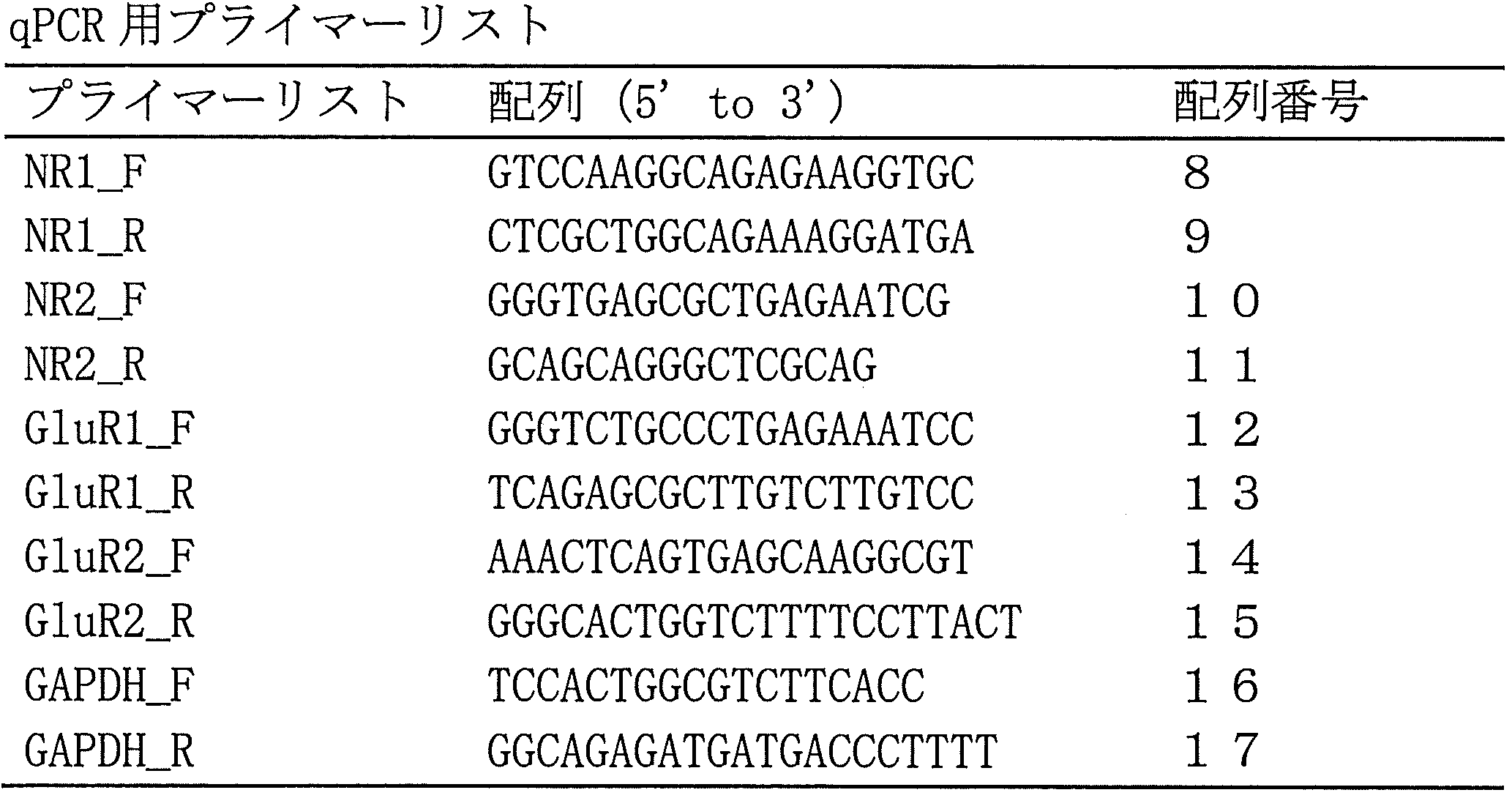
免疫細胞化学
細胞を室温で30分間、4%パラホルムアルデヒドで固定し、PBSで洗浄し、0.2%Triton X-100を含むPBS中に室温で10分間浸透した。続いてBlock Ace(Yukijirushi)で30分間ブロッキングした。一次抗体と4℃で一晩インキュベートした後、細胞をPBSで3回洗浄し、適切な二次抗体とともに室温で1時間インキュベートした。細胞画像をDelta Vision (Applied Precision)又はIN Cell Analyzer 6000 (GE Healthcare)から取得した。細胞数をIN Cell Analyzer 6000及びIN CELL Developer toolbox software 1.9 (GE Healthcare)で定量化した。このアッセイでは、以下の一次抗体を用いた:βIII チューブリン(1:2,000, Covance)、NeuN (1:500, Millipore)、TOC1 (1:1,000)、Nanog (1:200, Abcam)、SSEA4 (1:500, Millipore)。
細胞を室温で30分間、4%パラホルムアルデヒドで固定し、PBSで洗浄し、0.2%Triton X-100を含むPBS中に室温で10分間浸透した。続いてBlock Ace(Yukijirushi)で30分間ブロッキングした。一次抗体と4℃で一晩インキュベートした後、細胞をPBSで3回洗浄し、適切な二次抗体とともに室温で1時間インキュベートした。細胞画像をDelta Vision (Applied Precision)又はIN Cell Analyzer 6000 (GE Healthcare)から取得した。細胞数をIN Cell Analyzer 6000及びIN CELL Developer toolbox software 1.9 (GE Healthcare)で定量化した。このアッセイでは、以下の一次抗体を用いた:βIII チューブリン(1:2,000, Covance)、NeuN (1:500, Millipore)、TOC1 (1:1,000)、Nanog (1:200, Abcam)、SSEA4 (1:500, Millipore)。
ウエスタンブロット分析
細胞を回収し、0.1%SDS、150 mM NaCl、1% NP-40、0.5% デオキシコレート、50 mM Tris-HCl(pH8.0)、プロテアーゼ阻害剤(Roche)、及びホスファターゼ阻害剤(Roche)を含むRIPA緩衝液に溶解した。超音波処理後、試料を13,000 × gで15分間4℃で遠心分離した。上清中のタンパク質濃度を、ビシンコニン酸(BCA)アッセイキット(Pierce)を用いて測定した。2-メルカプトエタノールの有り無しにより(それぞれ還元条件又は非還元条件)、総タンパク質抽出物(レーン当たり20μg)を、10% ポリアクリルアミドゲル上のサイズにより分離し、Immobilon-Pメンブレン(Millipore)に移した。メンブレンを5% スキムミルクでブロックし、適切な抗体とハイブリダイズさせ、ECL Prime検出キット(GE Healthcare)を用いて可視化した。ImageQuant LAS 4000(GE Healthcare)から画像を取得した。以下の一次抗体を用いた:MAP2(1:1,000、CST)、βIIIチューブリン(1:5,000、Covance)、Tau12(1:10,000、Millipore)、TOC1(1:5,000)及びβ-アクチン(1:5,000)、Sigma)。
細胞を回収し、0.1%SDS、150 mM NaCl、1% NP-40、0.5% デオキシコレート、50 mM Tris-HCl(pH8.0)、プロテアーゼ阻害剤(Roche)、及びホスファターゼ阻害剤(Roche)を含むRIPA緩衝液に溶解した。超音波処理後、試料を13,000 × gで15分間4℃で遠心分離した。上清中のタンパク質濃度を、ビシンコニン酸(BCA)アッセイキット(Pierce)を用いて測定した。2-メルカプトエタノールの有り無しにより(それぞれ還元条件又は非還元条件)、総タンパク質抽出物(レーン当たり20μg)を、10% ポリアクリルアミドゲル上のサイズにより分離し、Immobilon-Pメンブレン(Millipore)に移した。メンブレンを5% スキムミルクでブロックし、適切な抗体とハイブリダイズさせ、ECL Prime検出キット(GE Healthcare)を用いて可視化した。ImageQuant LAS 4000(GE Healthcare)から画像を取得した。以下の一次抗体を用いた:MAP2(1:1,000、CST)、βIIIチューブリン(1:5,000、Covance)、Tau12(1:10,000、Millipore)、TOC1(1:5,000)及びβ-アクチン(1:5,000)、Sigma)。
TOC1抗体はオリゴマー中にモノマー又はフィラメントと比較してオリゴマー中にディスプレイされる直線状エピトープを有する(Ward, S.M., et al. TOC1: characterization of a selective oligomeric tau antibody. J Alzheimers Dis 37, 593-602 (2013).)ため、TOC1抗体を使用してオリゴマー形態のタウを評価するためには非変性条件が要求されることは注目に値する。変性条件の使用では、TOC1を用いたこれらの異なるタウ種、オリゴマー、モノマー及び/又はフィラメント、の間の区別はできない。本稿で使用したタウ抗体を表3に示す。

ドットブロット分析
細胞を回収し、プロテアーゼ阻害剤及びホスファターゼ阻害剤を含むTBS中に溶解した。超音波処理し、13,000×gで15分間遠心分離した後、各溶解物サンプル(1.2μg/スポット)をニトロセルロースメンブレン(0.45μm孔径、GE Healthcare)上に充填した。細胞培養培地を回収し、800 x gで3分間遠心分離して残渣を除去した。各試料からの500μlの上清を、濾過膜の分画分子量が10 kDであるVivaspin(GE Healthcare)を用いて50 μlに濃縮した。各濃縮サンプル2 μlをニトロセルロースメンブレンに充填した。メンブレンを5%スキムミルクでブロックし、適切な抗体とハイブリダイズし、Western Lightning Plus-ECL(PerkinElmer社)を用いて可視化した。ImageQuant LAS 4000(GE Healthcare)から画像を取得した。以下の一次抗体を用いた:Tau12 (1:10,000, Millipore)、Tau46 (1:1,000)、TOC1 (1:5,000)。
細胞を回収し、プロテアーゼ阻害剤及びホスファターゼ阻害剤を含むTBS中に溶解した。超音波処理し、13,000×gで15分間遠心分離した後、各溶解物サンプル(1.2μg/スポット)をニトロセルロースメンブレン(0.45μm孔径、GE Healthcare)上に充填した。細胞培養培地を回収し、800 x gで3分間遠心分離して残渣を除去した。各試料からの500μlの上清を、濾過膜の分画分子量が10 kDであるVivaspin(GE Healthcare)を用いて50 μlに濃縮した。各濃縮サンプル2 μlをニトロセルロースメンブレンに充填した。メンブレンを5%スキムミルクでブロックし、適切な抗体とハイブリダイズし、Western Lightning Plus-ECL(PerkinElmer社)を用いて可視化した。ImageQuant LAS 4000(GE Healthcare)から画像を取得した。以下の一次抗体を用いた:Tau12 (1:10,000, Millipore)、Tau46 (1:1,000)、TOC1 (1:5,000)。
細胞生存アッセイ
Ngn2を導入したiPS細胞を単細胞に解離し、1 μg/mlのドキシサイクリンを含む神経培地を用いてマトリゲルでコーティングした96穴のプレート(BD Bioscience)上に5×10⁴細胞/ウェルで播種した。8日目及び21日目に細胞を固定し、染色した。NeuNで染色した生存神経細胞の数をIN Cell Analyzer 6000(GE Healthcare)で定量し、21日目/8日目の神経細胞数として表した。D-2-アミノ-5-ホスホノペンタノエート(AP-5)(SIGMA)及び6-シアノ-7-ニトロキノキサリン-2,3-ジオン(CNQX)(SIGMA)を8日目から21日目まで毎日添加し、生存する神経細胞の数を21日目に評価した。
Ngn2を導入したiPS細胞を単細胞に解離し、1 μg/mlのドキシサイクリンを含む神経培地を用いてマトリゲルでコーティングした96穴のプレート(BD Bioscience)上に5×10⁴細胞/ウェルで播種した。8日目及び21日目に細胞を固定し、染色した。NeuNで染色した生存神経細胞の数をIN Cell Analyzer 6000(GE Healthcare)で定量し、21日目/8日目の神経細胞数として表した。D-2-アミノ-5-ホスホノペンタノエート(AP-5)(SIGMA)及び6-シアノ-7-ニトロキノキサリン-2,3-ジオン(CNQX)(SIGMA)を8日目から21日目まで毎日添加し、生存する神経細胞の数を21日目に評価した。
細胞死アッセイ
Ngn2を導入したiPS細胞を単細胞に解離し、1 μg/mlのドキシサイクリンを含む神経培地を用いてマトリゲルでコーティングした96穴のプレート(BD Bioscience)上に5×10⁴細胞/ウェルで播種した。培養培地を8日目に交換し、ヨウ化プロピジウム(PI)染色を10日目に行った。細胞を1.5μMのPI(ナカライテスク)と共に37℃で30分間インキュベートし、4%パラホルムアルデヒドで固定した。 PI染色された細胞の数を、IN Cell Analyzer 6000により定量した。
Ngn2を導入したiPS細胞を単細胞に解離し、1 μg/mlのドキシサイクリンを含む神経培地を用いてマトリゲルでコーティングした96穴のプレート(BD Bioscience)上に5×10⁴細胞/ウェルで播種した。培養培地を8日目に交換し、ヨウ化プロピジウム(PI)染色を10日目に行った。細胞を1.5μMのPI(ナカライテスク)と共に37℃で30分間インキュベートし、4%パラホルムアルデヒドで固定した。 PI染色された細胞の数を、IN Cell Analyzer 6000により定量した。
オリゴマータウの酵素結合免疫吸着アッセイ(ELISA)
96穴のプレート(Greiner)を、0.05 M炭酸ナトリウム緩衝液中で、3 μg/ml TOC1抗体を用いて4℃で一晩コーティングした。1% BSAを含むTBS-Tで洗浄及びブロッキングした後、100 μlの培養培地を添加し、室温で2時間インキュベートした。組換えオリゴマータウタンパク質を用いて標準曲線を得た。検出のために、プレートを2 μg/mlのアフィニティー精製したウサギポリクローナル抗ヒトタウ抗体と共に、次いで西洋ワサビペルオキシダーゼに連結したヒツジ抗ウサギIgG F(ab)'2フラグメント(1:3,000; GE Healthcare)と共にインキュベートした。室温で30分間のテトラメチルベンジジン溶液(BD Bioscience)でインキュベーション後、VersaMax(Molecular Device)により450 nmでの吸光度を測定した。2匹のウサギの免疫化により、タウのアミノ末端ポリペプチド(最も長いヒトタウアイソフォームに応じた、アミノ酸19~33)に対して、抗ヒトタウ抗体を産生させた。
96穴のプレート(Greiner)を、0.05 M炭酸ナトリウム緩衝液中で、3 μg/ml TOC1抗体を用いて4℃で一晩コーティングした。1% BSAを含むTBS-Tで洗浄及びブロッキングした後、100 μlの培養培地を添加し、室温で2時間インキュベートした。組換えオリゴマータウタンパク質を用いて標準曲線を得た。検出のために、プレートを2 μg/mlのアフィニティー精製したウサギポリクローナル抗ヒトタウ抗体と共に、次いで西洋ワサビペルオキシダーゼに連結したヒツジ抗ウサギIgG F(ab)'2フラグメント(1:3,000; GE Healthcare)と共にインキュベートした。室温で30分間のテトラメチルベンジジン溶液(BD Bioscience)でインキュベーション後、VersaMax(Molecular Device)により450 nmでの吸光度を測定した。2匹のウサギの免疫化により、タウのアミノ末端ポリペプチド(最も長いヒトタウアイソフォームに応じた、アミノ酸19~33)に対して、抗ヒトタウ抗体を産生させた。
カルシウムイメージングアッセイ
iPS細胞を単細胞に解離し、1 μg/mlのドキシサイクリンを含む神経培地を用いてマトリゲルでコーティングした96穴のプレート(BD Bioscience)上に5×10⁴細胞/ウェルで播種した。10日目に、e-FDSS /μCELL(Hamamatsu Photonics)(Shoji, E., et al. Early pathogenesis of Duchenne muscular dystrophy modelled in patient-derived human induced pluripotent stem cells. Sci Rep 5, 12831 (2015).)を用いて、電気刺激された細胞内Ca2+動員を測定した。簡潔には、細胞を5μM Fluo-8/AM(AAT Bioquest)及び0.01% Pluronic F-127(SIGMA)と共に神経培地中でインキュベートした。PBSで洗浄した後、培地をフェノールレッドを含まないNeurobasal medium(Gibco)に交換した。電気刺激(電圧:20 V、パルス幅:3 msec、回数:50 回/刺激、周波数:50 Hz)を96穴のプレートの各ウェルに送達し、蛍光変化(励起及び発光波長は、それぞれ480及び540nm)(刺激前後に記録された蛍光のパーセンテージとして表した)を記録した。
iPS細胞を単細胞に解離し、1 μg/mlのドキシサイクリンを含む神経培地を用いてマトリゲルでコーティングした96穴のプレート(BD Bioscience)上に5×10⁴細胞/ウェルで播種した。10日目に、e-FDSS /μCELL(Hamamatsu Photonics)(Shoji, E., et al. Early pathogenesis of Duchenne muscular dystrophy modelled in patient-derived human induced pluripotent stem cells. Sci Rep 5, 12831 (2015).)を用いて、電気刺激された細胞内Ca2+動員を測定した。簡潔には、細胞を5μM Fluo-8/AM(AAT Bioquest)及び0.01% Pluronic F-127(SIGMA)と共に神経培地中でインキュベートした。PBSで洗浄した後、培地をフェノールレッドを含まないNeurobasal medium(Gibco)に交換した。電気刺激(電圧:20 V、パルス幅:3 msec、回数:50 回/刺激、周波数:50 Hz)を96穴のプレートの各ウェルに送達し、蛍光変化(励起及び発光波長は、それぞれ480及び540nm)(刺激前後に記録された蛍光のパーセンテージとして表した)を記録した。
電気生理学的記録
全細胞パッチクランプ記録は、iPS細胞由来の神経細胞から行った。記録用マイクロピペットを、NaOHでpH7.4に調整した、140 mM KCl、2 mM MgCl2、10 mM HEPES及び1 mM EGTAから構成される細胞内溶液で満たした。実験中細胞を30℃で維持し、125 mM NaCl、2.5 mM KCl、1.25 mM NaH2PO4、26 mM NaHCO3、1 mM MgCl2、2 mM CaCl2及び20 mM グルコースからなる酸化クレブスリンガー溶液で連続的に灌流した。電圧クランプ及び電流クランプ記録は、EPC9増幅器(HEKA)を用いて行い、データを、Patchmasterソフトウェア(HEKA)を用いて分析した。
全細胞パッチクランプ記録は、iPS細胞由来の神経細胞から行った。記録用マイクロピペットを、NaOHでpH7.4に調整した、140 mM KCl、2 mM MgCl2、10 mM HEPES及び1 mM EGTAから構成される細胞内溶液で満たした。実験中細胞を30℃で維持し、125 mM NaCl、2.5 mM KCl、1.25 mM NaH2PO4、26 mM NaHCO3、1 mM MgCl2、2 mM CaCl2及び20 mM グルコースからなる酸化クレブスリンガー溶液で連続的に灌流した。電圧クランプ及び電流クランプ記録は、EPC9増幅器(HEKA)を用いて行い、データを、Patchmasterソフトウェア(HEKA)を用いて分析した。
統計分析
統計的有意性を決定するために、one-way ANOVA及び続いてのTukey post hoc分析又はスチューデントのt検定を用いて結果を分析した。差は、p < 0.05で有意とした。解析はSPSS software(IBM)を用いて行った。全ての棒グラフは平均±SEMを表す。
統計的有意性を決定するために、one-way ANOVA及び続いてのTukey post hoc分析又はスチューデントのt検定を用いて結果を分析した。差は、p < 0.05で有意とした。解析はSPSS software(IBM)を用いて行った。全ての棒グラフは平均±SEMを表す。
[結果]
イントロン10 + 14C→T(Hutton, M., et al. Association of missense and 5'-splice-site mutations in tau with the inherited dementia FTDP-17. Nature 393, 702-705 (1998).)、又はエクソンR406W(Spillantini, M.G., Van Swieten, J.C. & Goedert, M. Tau gene mutations in frontotemporal dementia and parkinsonism linked to chromosome 17 (FTDP-17). Neurogenetics 2, 193-205 (2000).))のいずれかの変異を有するFTLD-Tau患者からiPS細胞を作製した(図1(a)、1(b)、図5(a)、5(b)及び表1)。これらの患者は、前頭側頭型認知症を示した。イントロン10 + 14C→T変異は、CRISPR-Cas9を用いて修正した(図1(c)及び1(d))。tet-on発現系を有するpiggyBacベクターにより、Ngn2をこれらのiPS細胞に導入し、続いて安定した細胞株樹立のための薬剤選択を行った。樹立されたiPS細胞は、ドキシサイクリンを含む神経培地中での培養7日後に、皮質神経細胞に変換した。得られた株の分化傾向に差はなかった。対照、FTLD-Tau1、FTLD-Tau1 corrected及びFTLD-Tau2株における神経細胞の割合は、それぞれ、89.5±1.9%、91.2±0.9%、90.6±1.6%及び87.6±0.2%であった(n = 3)。作製した神経細胞は、神経伝達物質の受容体のmRNAを発現し(図5(c))、電気生理学的分析により、それらの機能的特性が示された(図6(a)-6(h))。以前報告されたように(Hutton, M., et al. Association of missense and 5'-splice-site mutations in tau with the inherited dementia FTDP-17. Nature 393, 702-705 (1998).)、イントロン10の変異を有するFTLD-Tau1神経細胞では、3リピートタウの発現に比べて4リピートタウの発現が増加し、変異の修正により、その割合は修復された(図1(e)及び1(f))。これらのFTLD-Tau及び対照の神経細胞を用いてFTLD-Tauをモデル化した(図2(a))。分化した神経細胞は神経細胞マーカーのMAP2A/Bを示し、これらのレベルはそれぞれの株間で差異がなかった(図2(b))。イントロンMAPT変異、及びエクソン変異を有する両方のFTLD-Tau神経細胞では、抗オリゴマー凝集抗体であるTOC1抗体(Patterson, K.R., et al. Characterization of prefibrillar Tau oligomers in vitro and in Alzheimer disease. J Biol Chem 286, 23063-23076 (2011)., Ward, S.M., et al. TOC1: characterization of a selective oligomeric tau antibody. J Alzheimers Dis 37, 593-602 (2013).)(図2(c)及び図7(a))を用いた免疫細胞化学により、細胞内のミスフォールドされたタウの蓄積が検出された。FTLD-Tau神経細胞の中には、典型的なミスフォールドされたタウの斑点とドットを示すものもあり、遺伝子修正された株を含む対照の神経細胞は、ミスフォールドされたタウの斑点又はドットについて大部分が陰性であった。ドットブロット分析により、TOC1抗体を用いた非変性状態における細胞内のミスフォールドされた形態のタウの蓄積が示された(図2(d)、(e))。また、以前に示されたように(Ward, S.M., et al. TOC1: characterization of a selective oligomeric tau antibody. J Alzheimers Dis 37, 593-602 (2013).)、変性条件におけるタウ種を検出するため、TOC1抗体を用いたウエスタンブロット分析により、ミスフォールドされたタウを分析した。イントロンのMAPT変異又はエクソンの変異を有するいずれかのFTLD-Tau神経細胞は、対照よりも高い分子量のタウ種の蓄積を示した(図2(f))。しかし、イントロンのMAPT変異を有するFTLD-Tau神経細胞とエクソンの変異を有するFTLD-Tau神経細胞の間には、ミスフォールドされたタウの分子量シフトには違いがあり(図2(f))、これはミスフォールドされたタウのコンフォメーションが変性条件において均一ではない可能性を示唆していた。さらに、FTLD-Tau神経細胞は、ドットブロット分析によって検出され(図2(g)及び2(h))、ELISAによって確認される(図2(i))ように、培養培地中へのミスフォールドされたタウの放出を示した。
イントロン10 + 14C→T(Hutton, M., et al. Association of missense and 5'-splice-site mutations in tau with the inherited dementia FTDP-17. Nature 393, 702-705 (1998).)、又はエクソンR406W(Spillantini, M.G., Van Swieten, J.C. & Goedert, M. Tau gene mutations in frontotemporal dementia and parkinsonism linked to chromosome 17 (FTDP-17). Neurogenetics 2, 193-205 (2000).))のいずれかの変異を有するFTLD-Tau患者からiPS細胞を作製した(図1(a)、1(b)、図5(a)、5(b)及び表1)。これらの患者は、前頭側頭型認知症を示した。イントロン10 + 14C→T変異は、CRISPR-Cas9を用いて修正した(図1(c)及び1(d))。tet-on発現系を有するpiggyBacベクターにより、Ngn2をこれらのiPS細胞に導入し、続いて安定した細胞株樹立のための薬剤選択を行った。樹立されたiPS細胞は、ドキシサイクリンを含む神経培地中での培養7日後に、皮質神経細胞に変換した。得られた株の分化傾向に差はなかった。対照、FTLD-Tau1、FTLD-Tau1 corrected及びFTLD-Tau2株における神経細胞の割合は、それぞれ、89.5±1.9%、91.2±0.9%、90.6±1.6%及び87.6±0.2%であった(n = 3)。作製した神経細胞は、神経伝達物質の受容体のmRNAを発現し(図5(c))、電気生理学的分析により、それらの機能的特性が示された(図6(a)-6(h))。以前報告されたように(Hutton, M., et al. Association of missense and 5'-splice-site mutations in tau with the inherited dementia FTDP-17. Nature 393, 702-705 (1998).)、イントロン10の変異を有するFTLD-Tau1神経細胞では、3リピートタウの発現に比べて4リピートタウの発現が増加し、変異の修正により、その割合は修復された(図1(e)及び1(f))。これらのFTLD-Tau及び対照の神経細胞を用いてFTLD-Tauをモデル化した(図2(a))。分化した神経細胞は神経細胞マーカーのMAP2A/Bを示し、これらのレベルはそれぞれの株間で差異がなかった(図2(b))。イントロンMAPT変異、及びエクソン変異を有する両方のFTLD-Tau神経細胞では、抗オリゴマー凝集抗体であるTOC1抗体(Patterson, K.R., et al. Characterization of prefibrillar Tau oligomers in vitro and in Alzheimer disease. J Biol Chem 286, 23063-23076 (2011)., Ward, S.M., et al. TOC1: characterization of a selective oligomeric tau antibody. J Alzheimers Dis 37, 593-602 (2013).)(図2(c)及び図7(a))を用いた免疫細胞化学により、細胞内のミスフォールドされたタウの蓄積が検出された。FTLD-Tau神経細胞の中には、典型的なミスフォールドされたタウの斑点とドットを示すものもあり、遺伝子修正された株を含む対照の神経細胞は、ミスフォールドされたタウの斑点又はドットについて大部分が陰性であった。ドットブロット分析により、TOC1抗体を用いた非変性状態における細胞内のミスフォールドされた形態のタウの蓄積が示された(図2(d)、(e))。また、以前に示されたように(Ward, S.M., et al. TOC1: characterization of a selective oligomeric tau antibody. J Alzheimers Dis 37, 593-602 (2013).)、変性条件におけるタウ種を検出するため、TOC1抗体を用いたウエスタンブロット分析により、ミスフォールドされたタウを分析した。イントロンのMAPT変異又はエクソンの変異を有するいずれかのFTLD-Tau神経細胞は、対照よりも高い分子量のタウ種の蓄積を示した(図2(f))。しかし、イントロンのMAPT変異を有するFTLD-Tau神経細胞とエクソンの変異を有するFTLD-Tau神経細胞の間には、ミスフォールドされたタウの分子量シフトには違いがあり(図2(f))、これはミスフォールドされたタウのコンフォメーションが変性条件において均一ではない可能性を示唆していた。さらに、FTLD-Tau神経細胞は、ドットブロット分析によって検出され(図2(g)及び2(h))、ELISAによって確認される(図2(i))ように、培養培地中へのミスフォールドされたタウの放出を示した。
次に、カルシウムイメージングアッセイにより、FTLD-Tau神経細胞の細胞内Ca2+濃度の上昇を評価した。電気刺激は、細胞内のCa2+レベルを増加させ、FTLD-Tau神経細胞では、対照の神経細胞と比較して増加割合が高く、FTLD-Tau神経細胞の興奮性の増加を示していた(図3(a)及び3(b))。次に、FTLD-Tau神経細胞の変性に対する脆弱性を調べたところ、FTLD-Tau神経細胞は、対照の神経細胞よりも細胞死を受けやすいことを見出した(図3(c)及び3(d)、図7(b)及び7(c))。
神経細胞の活動を制御することを可能にするDREADDを用いて、神経細胞の興奮とミスフォールドされたタウの間の機構的な関連を評価した。薬物選択後に、tol2ベクターを用いてFTLD-Tau iPS細胞に、人工的な変異をもつヒトムスカリン性アセチルコリンM4受容体(M4D)(Alexander, G.M., et al. Remote control of neuronal activity in transgenic mice expressing evolved G protein-coupled receptors. Neuron 63, 27-39 (2009).)から構成されるDREADDを組み込み、M4Dを構成的に発現する神経細胞を作製した(図4(a))。M4Dの排他的なアゴニストである、Giシグナル伝達を介してカルシウム流入を阻害するクロザピン-N-オキシド(CNO)によるM4Dの刺激は、細胞内及び細胞外のミスフォールドされたタウを減少させたが(図4(b)-4(d))、これは神経細胞の活性がタウの病態を調節し得ることを示唆していた。さらに、N-メチル-D-アスパラギン酸(NMDA)受容体の阻害剤であるD-2-アミノ-5-ホスホノペンタノエート(AP-5)及びα-アミノ-3-ヒドロキシ-5-メチル-4-イソキサゾールプロピオン酸(AMPA)受容体の阻害剤である6-シアノ-7-ニトロキノキサリン-2,3-ジオン(CNQX)は、FTLD-Tau神経細胞の生存を増加させた(図4(e)及び4(f))。
以上より、ミスフォールドされたタウの蓄積及び神経細胞死を含むFTLD-Tau神経細胞の細胞表現型は、イントロン及びエクソンのMAPT変異の両方において類似しており、神経細胞の活性に関連するCa2+流入は、タウオパシーにおける神経変性を調節する(図4(g))。
本出願は、米国仮特許出願第62/405,602号(出願日:2016年10月7日)を基礎としており、その内容は本明細書に全て包含されるものである。
本発明の細胞内へのカルシウムイオンの流入阻害薬は、前頭側頭葉変性症の予防及び/又は治療に有用であり、細胞内へのカルシウムイオンの流入を指標とするスクリーニング方法は、前頭側頭葉変性症の予防及び/又は治療剤のスクリーニングに有用である。
Claims (13)
- 細胞内へのカルシウムイオンの流入阻害薬を含有してなる、前頭側頭葉変性症の予防又は治療剤。
- 前記カルシウムイオンの流入阻害が、Gi/oシグナリングを介して生じる、請求項1記載の剤。
- 前記カルシウムイオンの流入阻害薬が、NMDA型グルタミン酸受容体、AMPA型グルタミン酸受容体、グループI 代謝型グルタミン酸受容体又は電位依存性カルシウムチャネルに対するアンタゴニスト又は発現阻害薬である、請求項1記載の剤。
- 前記カルシウムイオンの流入阻害薬が、グループII 代謝型グルタミン酸受容体又はグループIII代謝型グルタミン酸受容体に対するアゴニストである、請求項1記載の剤。
- タウタンパク質のミスフォールディングの阻害剤である、請求項1~請求項4のいずれか1項に記載の剤。
- 前頭側頭葉変性症が前頭側頭葉変性症タウオパチー(FTLD-Tau)である、請求項1~請求項5のいずれか1項に記載の剤。
- 前頭側頭葉変性症タウオパチーがMAPT遺伝子変異に起因するものである、請求項6記載の剤。
- 以下の工程を含む、前頭側頭葉変性症の予防又は治療剤のスクリーニング方法。
(1)被験物質を、細胞と接触させる工程、
(2)該細胞内へのカルシウムイオンの流入を測定する工程、
(3)上記(2)のカルシウムイオンの流入の程度を、被験物質を接触させなかった細胞におけるカルシウムイオンの流入の程度と比較する工程、
(4)カルシウムイオンの流入の程度を低下させた被験物質を、FTLDの病態を伴う神経細胞に添加し、ミスフォールドされたタウタンパク質の蓄積又は細胞外への放出を検出又は測定する工程、並びに
(5)ミスフォールドされたタウタンパク質の蓄積又は細胞外への放出が減少した場合に、該被験物質を前頭側頭葉変性症の予防又は治療剤の候補として選択する工程 - 以下の工程を含む、前頭側頭葉変性症の予防又は治療剤のスクリーニング方法。
(1)被験物質を、FTLDの病態を伴う神経細胞と接触させる工程、
(2)該細胞内へのカルシウムイオンの流入及びタウタンパク質のミスフォールドの程度を測定する工程、
(3)上記(2)のカルシウムイオンの流入及びタウタンパク質のミスフォールドの程度を、被験物質を接触させなかった細胞におけるカルシウムイオンの流入及びタウタンパク質のミスフォールドの程度と比較する工程、並びに
(4)カルシウムイオンの流入及びタウタンパク質のミスフォールドの程度を両方とも低下させた被験物質を、前頭側頭葉変性症の予防又は治療剤の候補として選択する工程 - 以下の工程を含む、前頭側頭葉変性症の予防又は治療剤のスクリーニング方法。
(1)被験物質を、外因性のムスカリン受容体のサブタイプM4を恒常的に発現させた細胞と接触させる工程、
(2)該細胞内のcAMP量を測定する工程、
(3)上記(2)のcAMP量を、被験物質を接触させなかった細胞内のcAMP量と比較する工程、並びに
(4)cAMP量を低下させた被験物質を、前頭側頭葉変性症の予防又は治療剤の候補として選択する工程 - 以下の工程を含む、前頭側頭葉変性症の予防又は治療剤のスクリーニング方法。
(1)被験物質を、外因性のムスカリン受容体のサブタイプM3を恒常的に発現させた細胞と接触させる工程、
(2)該細胞内のイノシトール3リン酸(IP3)量又はジアシルグリセロール(DAG)量を測定する工程、
(3)上記(2)のIP3量又はDAG量を、被験物質を接触させなかった細胞内のIP3量又はDAG量と比較する工程、並びに
(4)IP3量又はDAG量を低下させた被験物質を、前頭側頭葉変性症の予防又は治療剤の候補として選択する工程 - 哺乳動物に対し、細胞内へのカルシウムイオンの流入阻害薬の有効量を投与することを特徴とする、該哺乳動物における前頭側頭葉変性症の予防または治療方法。
- 前頭側頭葉変性症の予防又は治療に使用するための、細胞内へのカルシウムイオンの流入阻害薬。
Priority Applications (1)
| Application Number | Priority Date | Filing Date | Title |
|---|---|---|---|
| JP2018543993A JP7162302B2 (ja) | 2016-10-07 | 2017-10-06 | Ftldの予防又は治療剤及びそのスクリーニング方法 |
Applications Claiming Priority (2)
| Application Number | Priority Date | Filing Date | Title |
|---|---|---|---|
| US201662405602P | 2016-10-07 | 2016-10-07 | |
| US62/405,602 | 2016-10-07 |
Publications (1)
| Publication Number | Publication Date |
|---|---|
| WO2018066701A1 true WO2018066701A1 (ja) | 2018-04-12 |
Family
ID=61831804
Family Applications (1)
| Application Number | Title | Priority Date | Filing Date |
|---|---|---|---|
| PCT/JP2017/036520 Ceased WO2018066701A1 (ja) | 2016-10-07 | 2017-10-06 | Ftldの予防又は治療剤及びそのスクリーニング方法 |
Country Status (2)
| Country | Link |
|---|---|
| JP (1) | JP7162302B2 (ja) |
| WO (1) | WO2018066701A1 (ja) |
Cited By (4)
| Publication number | Priority date | Publication date | Assignee | Title |
|---|---|---|---|---|
| CN112469827A (zh) * | 2018-07-04 | 2021-03-09 | 国立大学法人东海国立大学机构 | 控制Tau的剪接的反义寡核苷酸及其用途 |
| JPWO2020090966A1 (ja) * | 2018-11-01 | 2021-09-30 | 国立研究開発法人国立長寿医療研究センター | タウオパチーの治療薬または予防薬のスクリーニング方法およびタウオパチーの診断方法 |
| CN114599397A (zh) * | 2019-10-25 | 2022-06-07 | 国立大学法人京都大学 | Tau蛋白病的预防或治疗剂 |
| WO2025095099A1 (ja) * | 2023-11-02 | 2025-05-08 | 株式会社ケイファーマ | 前頭側頭葉変性症治療剤及び治療用組成物 |
Citations (1)
| Publication number | Priority date | Publication date | Assignee | Title |
|---|---|---|---|---|
| WO2016076435A1 (ja) * | 2014-11-13 | 2016-05-19 | 国立大学法人京都大学 | 誘導神経細胞を用いたスクリーニング方法 |
-
2017
- 2017-10-06 JP JP2018543993A patent/JP7162302B2/ja active Active
- 2017-10-06 WO PCT/JP2017/036520 patent/WO2018066701A1/ja not_active Ceased
Patent Citations (1)
| Publication number | Priority date | Publication date | Assignee | Title |
|---|---|---|---|---|
| WO2016076435A1 (ja) * | 2014-11-13 | 2016-05-19 | 国立大学法人京都大学 | 誘導神経細胞を用いたスクリーニング方法 |
Non-Patent Citations (2)
| Title |
|---|
| ALEXANDER G M. ET AL.: "Remote Control of Neuronal Activity in Transgenic Mice Expressing Evolved G Protein-Coupled Receptors", NEURON, vol. 63, 2009, pages 27 - 39, XP055185944, ISSN: 0896-6273 * |
| COLLEEN M. ET AL.: "Metabotropic Glutamate Receptors: Physiology, Pharmacology, and Disease", THE ANNUAL REVIEW OF PHARMACOLOGY AND TOXICOLOGY, vol. 50, 2010, pages 295 - 322, XP002686775, ISSN: 0362-1642 * |
Cited By (8)
| Publication number | Priority date | Publication date | Assignee | Title |
|---|---|---|---|---|
| CN112469827A (zh) * | 2018-07-04 | 2021-03-09 | 国立大学法人东海国立大学机构 | 控制Tau的剪接的反义寡核苷酸及其用途 |
| CN112469827B (zh) * | 2018-07-04 | 2024-05-24 | 学校法人爱知医科大学 | 控制Tau的剪接的反义寡核苷酸及其用途 |
| US12570974B2 (en) | 2018-07-04 | 2026-03-10 | Knc Laboratories Co., Ltd. | Oligonucleotides for controlling tau splicing, and uses thereof |
| JPWO2020090966A1 (ja) * | 2018-11-01 | 2021-09-30 | 国立研究開発法人国立長寿医療研究センター | タウオパチーの治療薬または予防薬のスクリーニング方法およびタウオパチーの診断方法 |
| JP7296136B2 (ja) | 2018-11-01 | 2023-06-22 | 国立研究開発法人国立長寿医療研究センター | タウオパチーの治療薬または予防薬のスクリーニング方法およびタウオパチーの診断方法 |
| CN114599397A (zh) * | 2019-10-25 | 2022-06-07 | 国立大学法人京都大学 | Tau蛋白病的预防或治疗剂 |
| CN114599397B (zh) * | 2019-10-25 | 2024-05-28 | 国立大学法人京都大学 | Tau蛋白病的预防或治疗剂 |
| WO2025095099A1 (ja) * | 2023-11-02 | 2025-05-08 | 株式会社ケイファーマ | 前頭側頭葉変性症治療剤及び治療用組成物 |
Also Published As
| Publication number | Publication date |
|---|---|
| JP7162302B2 (ja) | 2022-10-28 |
| JPWO2018066701A1 (ja) | 2019-08-29 |
Similar Documents
| Publication | Publication Date | Title |
|---|---|---|
| JP7162302B2 (ja) | Ftldの予防又は治療剤及びそのスクリーニング方法 | |
| AU2023303022A1 (en) | Cns targeting complexes and uses thereof | |
| Zhang et al. | Generation of a novel mouse model of Parkinson’s disease via targeted knockdown of glutamate transporter GLT-1 in the substantia nigra | |
| JP6870988B2 (ja) | 神経細胞増幅及び中枢神経系障害治療のためのセレブロンの活性化因子の使用方法 | |
| US20240011027A1 (en) | Methods and compositions for restoring stmn2 levels | |
| Tindi et al. | ANKS1B gene product AIDA-1 controls hippocampal synaptic transmission by regulating GluN2B subunit localization | |
| CN106456699A (zh) | 作为抗老化剂抑制抗凋亡的Bcl‑2蛋白的组合物和方法 | |
| Rougier et al. | Cardiac voltage-gated calcium channel macromolecular complexes | |
| CN103874486A (zh) | 用小分子治疗与电压门控钠通道的α亚基(SCNxA)相关的疾病 | |
| US20100311767A1 (en) | Method for the treatment of amyloidoses | |
| US20220133848A1 (en) | Methods and compositions for restoring stmn2 levels | |
| JP2021529169A (ja) | 統合失調症及び他の神経精神障害の治療方法 | |
| JPWO2013054534A1 (ja) | アミロイドβ関連の疾患用医薬及びそのスクリーニング | |
| JP2018531046A (ja) | 核酸ベースのtia−1阻害剤 | |
| JP2018531046A6 (ja) | 核酸ベースのtia−1阻害剤 | |
| CN119095622A (zh) | Nmdar拮抗剂通过提高20s蛋白酶体活性来预防老化和老化相关病况和疾病 | |
| JP2023154067A (ja) | 筋萎縮性側索硬化症の予防及び/又は治療剤 | |
| WO2019115472A1 (en) | Polycomb inhibitors and uses thereof | |
| KR20190088475A (ko) | 운동 뉴런 질병의 치료를 위한 강글리오사이드 대사 억제제 | |
| WO2002087559A1 (fr) | Agent d'ouverture du canal potassique | |
| JP2018515507A (ja) | アルツハイマー病の処置のための神経細胞ストア作動性カルシウム流入経路の活性化方法 | |
| US20260053804A1 (en) | Compositions and Methods for the Restoration of Ciliary Function | |
| US20200188431A1 (en) | Glutamine dehydrogenase inhibitors for use in muscle regeneration | |
| Xie et al. | Long-term 4-AP treatment facilitates functional expression of human Kv1. 5 channel | |
| Shelton | Targeting the Hsp90/Aha1 Complex for the Treatment of Tauopathies |
Legal Events
| Date | Code | Title | Description |
|---|---|---|---|
| 121 | Ep: the epo has been informed by wipo that ep was designated in this application |
Ref document number: 17858537 Country of ref document: EP Kind code of ref document: A1 |
|
| ENP | Entry into the national phase |
Ref document number: 2018543993 Country of ref document: JP Kind code of ref document: A |
|
| NENP | Non-entry into the national phase |
Ref country code: DE |
|
| 122 | Ep: pct application non-entry in european phase |
Ref document number: 17858537 Country of ref document: EP Kind code of ref document: A1 |