WO2017175808A1 - 移植片対宿主病の治療剤又は予防剤、ファイブロサイト浸潤抑制剤、及び涙液減少と杯細胞の減少の抑制剤 - Google Patents
移植片対宿主病の治療剤又は予防剤、ファイブロサイト浸潤抑制剤、及び涙液減少と杯細胞の減少の抑制剤 Download PDFInfo
- Publication number
- WO2017175808A1 WO2017175808A1 PCT/JP2017/014290 JP2017014290W WO2017175808A1 WO 2017175808 A1 WO2017175808 A1 WO 2017175808A1 JP 2017014290 W JP2017014290 W JP 2017014290W WO 2017175808 A1 WO2017175808 A1 WO 2017175808A1
- Authority
- WO
- WIPO (PCT)
- Prior art keywords
- versus
- graft
- host disease
- acceptable salt
- pharmacologically acceptable
- Prior art date
- Legal status (The legal status is an assumption and is not a legal conclusion. Google has not performed a legal analysis and makes no representation as to the accuracy of the status listed.)
- Ceased
Links
Images






Classifications
-
- A—HUMAN NECESSITIES
- A61—MEDICAL OR VETERINARY SCIENCE; HYGIENE
- A61K—PREPARATIONS FOR MEDICAL, DENTAL OR TOILETRY PURPOSES
- A61K31/00—Medicinal preparations containing organic active ingredients
- A61K31/185—Acids; Anhydrides, halides or salts thereof, e.g. sulfur acids, imidic, hydrazonic or hydroximic acids
- A61K31/19—Carboxylic acids, e.g. valproic acid
- A61K31/192—Carboxylic acids, e.g. valproic acid having aromatic groups, e.g. sulindac, 2-aryl-propionic acids, ethacrynic acid
-
- A—HUMAN NECESSITIES
- A61—MEDICAL OR VETERINARY SCIENCE; HYGIENE
- A61P—SPECIFIC THERAPEUTIC ACTIVITY OF CHEMICAL COMPOUNDS OR MEDICINAL PREPARATIONS
- A61P37/00—Drugs for immunological or allergic disorders
- A61P37/02—Immunomodulators
- A61P37/06—Immunosuppressants, e.g. drugs for graft rejection
Definitions
- the present invention relates to a therapeutic or prophylactic agent for graft-versus-host disease, a fibrosite infiltration inhibitor, and an inhibitor of tear reduction and goblet cell reduction.
- graft-versus-host disease that develops in recent years when bone marrow cells, peripheral blood or umbilical cord blood transplants provided by donors attack the recipient's target organ by immune response. GVHD) is a problem.
- Non-patent document 1 describes the use of rebamipide and diquafosol in combination to treat graft-versus-host disease. It is disclosed to improve dry eye, which is one of the symptoms.
- Non-Patent Document 2 discloses that cell senescence in donor immune cells and recipient cells is detrimentally involved in the onset process of cGVHD.
- endoplasmic reticulum stress that plays a detrimental role in the development of chronic inflammation and age-related diseases is known (for example, Non-Patent Documents 3 and 4).
- endoplasmic reticulum stress will be outlined with reference to FIGS. If the protein is synthesized in the endoplasmic reticulum, the protein must be properly folded and the endoplasmic reticulum chaperone assists in protein folding. However, hypoxia, calcium ion deficiency, oxidative damage, viral infection and inflammatory cytokines prevent normal folding of endoplasmic reticulum proteins.
- the failed UPRs were: (1) the pro-inflammatory molecule thioredoxin interacting protein (TXNIP) and activated B cell transcription factor nuclear factor kappa light chain enhancer (NF - ⁇ B), and (2) it is known to induce the expression of apoptotic protein C / EBP homologous protein (CHOP) (Non-Patent Documents 6 to 8).
- TXNIP pro-inflammatory molecule thioredoxin interacting protein
- NF - ⁇ B activated B cell transcription factor nuclear factor kappa light chain enhancer
- Gard AD Kaczmarek A, Krysko O, Vandabeele P, Krysko DV, Agostinis P.
- ER stress-induced information does it aid or impeded disease progression? Trends Mol Med. 2012; 18: 589-598.
- Lerner AG Upton JP, Praven PVK, et al.
- the present invention has been made in view of the above circumstances, and provides a novel therapeutic agent or preventive agent for graft-versus-host disease, an inhibitor of fibrosite infiltration, and an inhibitor of decrease in tears and goblet cells. With the goal.
- the present inventors have found that in graft-versus-host disease, fibrosite infiltration occurs in the lacrimal gland, and that this fibrosite infiltration is suppressed by phenylbutyric acid or a pharmacologically acceptable salt thereof. Furthermore, the inventors have (1) that an organ affected by cGVHD exhibits an increased level of endoplasmic reticulum stress, and (2) that severe relaxation of endoplasmic reticulum stress is caused by cGVHD (particularly systemic cGVHD). The present inventors have found a new hypothesis that it can be a powerful therapeutic means for relieving symptoms, and have completed the present invention. That is, the present invention is as follows.
- a therapeutic or prophylactic agent for graft-versus-host disease comprising phenylbutyric acid or a pharmacologically acceptable salt thereof as an active ingredient.
- ⁇ 6> The therapeutic or prophylactic agent according to any one of ⁇ 1> to ⁇ 5>, wherein the phenylbutyric acid is 4-phenyl-n-butyric acid or a pharmacologically acceptable salt thereof.
- ⁇ 7> A fibrosite infiltration inhibitor comprising phenylbutyric acid or a pharmacologically acceptable salt thereof as an active ingredient.
- ⁇ 8> The fibrosite infiltration inhibitor according to ⁇ 7>, wherein the phenylbutyric acid is 4-phenyl-n-butyric acid or a pharmacologically acceptable salt thereof.
- ⁇ 9> An inhibitor of goblet cell decrease, comprising phenylbutyric acid or a pharmacologically acceptable salt thereof as an active ingredient.
- a tear reduction inhibitor comprising phenylbutyric acid or a pharmacologically acceptable salt thereof as an active ingredient.
- the present invention preferably has the following configuration.
- graft-versus-host disease is graft-versus-host disease associated with fibrosite infiltration.
- phenylbutyric acid is 4-phenyl-n-butyric acid or a pharmacologically acceptable salt thereof.
- a method for inhibiting fibrosite infiltration comprising administering phenylbutyric acid or a pharmacologically acceptable salt thereof.
- phenylbutyric acid is 4-phenyl-n-butyric acid or a pharmacologically acceptable salt thereof.
- a method for inhibiting tear reduction comprising administering phenylbutyric acid or a pharmacologically acceptable salt thereof.
- a novel therapeutic or preventive agent for graft-versus-host disease a fibrosite infiltration inhibitor, and an inhibitor of tear and goblet cell reduction.
- FIG. 1A is a diagram showing the results of qPCR analysis of GRP78 in cGVHD-affected organs and control subjects.
- FIG. 1B is a figure which shows the immunoblot analysis result of an endoplasmic reticulum stress marker and an associated inflammation marker.
- FIG. 1C is a diagram showing a quantitative analysis result corresponding to each protein band.
- FIG. 1D is an electron micrograph of epithelial cells in the lacrimal gland affected by cGVHD and its cognate control lacrimal gland. It is a figure which shows the survival% and weight change of a PBA processing mouse group and a vehicle processing mouse group.
- FIG. 1A is a diagram showing the results of qPCR analysis of GRP78 in cGVHD-affected organs and control subjects.
- FIG. 1B is a figure which shows the immunoblot analysis result of an endoplasmic reticulum stress marker and an associated inflammation marker.
- FIG. 1C is a diagram showing a quantitative analysis result corresponding to each protein
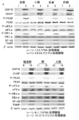
- FIG. 3A is a diagram showing the results of suppressing endoplasmic reticulum stress caused by cGVHD by PBA
- FIG. 3A is a diagram showing the results of protein level test of endoplasmic reticulum stress markers and related inflammatory markers in cGVHD target organs by immunoblot analysis.
- It is a figure which shows the suppression result of endoplasmic reticulum stress resulting from cGVHD by PBA
- FIG. 3B is a figure which shows the result of having quantified the target protein of each organ by densitometry about a PBA processing organ and a vehicle processing organ.
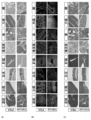
- FIG. 4A is a graph showing reduction results of cGVHD-induced systemic inflammation and fibrosis caused by PBA relieving endoplasmic reticulum stress
- FIG. 4A is a photograph of HE staining of PBA-treated organs and HE of solvent-treated organs. It is a dyeing
- FIG. 4B is a diagram showing immunostaining results of CD45, which is a general leukocyte marker, in PBA-treated tissues and vehicle-treated tissues.
- FIG. 4C is a diagram showing the results of Mallory staining for PBA-treated organs and vehicle-treated organs.
- FIG. 4D is an electron micrograph of the lacrimal gland and small intestine treated with PBA and the lacrimal gland and small intestine treated with PBA by reducing PG-induced endoplasmic reticulum stress to reduce cGVHD-induced systemic inflammation and fibrosis.
- FIG. 4E shows the density of CD45-positive cells in PBA-treated organs and vehicle-treated organs.
- FIG. 4F is a diagram showing the results of PAS (Periodic acid-Schiff stain) staining of PBA-treated small intestine and PBA-treated eyes, and vehicle-treated small intestine and eyes.
- FIG. 4G shows goblet cell density in PBA-treated small intestine and PBA-treated eyes and vehicle-treated small intestine and eyes.
- FIG. 5A shows the results of an ELISA performed 28 days after bone marrow transplantation to measure MCP-1, TNF- ⁇ and IFN- ⁇ , inflammatory markers in serum collected from PBA-treated and vehicle-treated mice.
- FIG. 5B shows the results of immunoblot analysis of CTGF, which is a fibrosis marker.
- FIG. 5C is a diagram showing the results (PBA-treated organ and vehicle-treated organ) of quantifying CTGF in each organ by densitometry.
- FIG. 6A shows the results of immunoblot analysis of endoplasmic reticulum stress markers, activation markers, and fibrosis markers in mouse lacrimal fibroblasts.
- FIG. 6B is a diagram showing the results of quantitative analysis of the corresponding protein bands (PBA-treated fibroblasts and vehicle-treated fibroblasts).
- FIG. 6C is a diagram showing the results of measuring the protein level of MCP-1 produced by PBA-treated fibroblasts and vehicle-treated fibroblasts using ELISA.
- FIG. 6D shows the results of qPCR analysis of IL-6 and TGF- ⁇ in PBA-treated fibroblasts and vehicle-treated fibroblasts. It is a figure which shows the relieving result of the endoplasmic reticulum stress resulting from cGVHD in the macrophage using PBA.
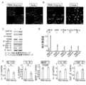
- FIG. 7A shows immunofluorescence images of the PBA-injected lacrimal gland and the vehicle-injected lacrimal gland.
- FIG. 7B shows immunofluorescence results of cultured spleen macrophages derived from PBA-administered mice and vehicle-administered mice.
- FIG. 7C shows the results of immunoblot analysis of each endoplasmic reticulum stress marker in mouse spleen macrophages.
- FIG. 7D shows the corresponding quantitative analysis results for each protein band.
- FIG. 7E shows the results of qPCR analysis of each marker of M1 macrophages and M2 macrophages from the spleen.
- FIG. 5 shows a presumed correlation between cGVHD and endoplasmic reticulum stress. It is a figure which shows the outline
- the therapeutic agent or preventive agent for graft-versus-host disease of the present invention contains phenylbutyric acid or a pharmacologically acceptable salt thereof as an active ingredient.
- the phenylbutyric acid in the present invention is preferably 4-phenyl-n-butyric acid (hereinafter also simply referred to as “PBA”) represented by the following formula.
- fibrosite infiltration means that fibrosite infiltrates from a blood into a certain tissue (for example, lacrimal gland).
- inhibittion of fibrosite infiltration means to suppress the infiltration of fibrosite into the tissue as a result.
- Fibrosite is also recognized by those skilled in the art as being identical to fibroblasts (Fibroblast) (Disease Models & Mechanisms 2011 May; 4 (3) 318-333 (especially page 325)).
- phenylbutyric acid or pharmacologically acceptable salt thereof in the present invention can be prepared by a known method.
- phenylbutyric acid or a pharmacologically acceptable salt thereof can be prepared by the following method.
- the phenylbutyric acid according to the present invention (for example, 4-phenyl-n-butyric acid (CAS registration number: 1821-12-1)) can be obtained as a reagent or an industrial raw material, and can be synthesized according to a conventional method. You can also.
- the pharmacologically acceptable salt of phenylbutyric acid according to the present invention is not limited as long as it can form a salt with a carboxy group.
- alkali metal, alkaline earth metal, amine, basic Examples thereof include salts such as amino acids, and sodium salts, potassium salts, calcium salts, and magnesium salts are particularly preferable.
- the phenylbutyric acid or a pharmacologically acceptable salt thereof of the present invention may be in the form of a derivative (so-called prodrug) such as ester or ether that is easily hydrolyzed in vivo (surface) after administration.
- a derivative such as ester or ether that is easily hydrolyzed in vivo (surface) after administration.
- prodrug such as ester or ether
- 3-phenyl-n-butyric acid CAS registration number: 4593-90-2
- 2-phenyl-n-butyric acid CAS registration number: 90-27-7
- 2-phenylisobutyric acid Needless to say, other structural isomers such as (CAS registration number: 826-55-1) and 3-phenylisobutyric acid (CAS registration number: 77-83-8) are also included.
- the type of graft-versus-host disease in the present invention is not particularly limited, and examples thereof include those whose symptoms are expressed in organs having exocrine glands such as the eye, oral cavity, liver, gastrointestinal tract, skin, and lung. Of these, the therapeutic agent or prophylactic agent of the present invention is preferably used for graft-versus-host disease in the eye.
- the graft-versus-host disease in the eye is not particularly limited, and examples thereof include conjunctival fibrosis associated with dry eye such as dry eye, dry conjunctivitis, and corneal damage.
- conjunctival fibrosis associated with dry eye such as dry eye, dry conjunctivitis, and corneal damage.
- the therapeutic or preventive effect is particularly excellent, it is preferable that the disease of graft-versus-host disease in the eye to be treated or prevented is dry eye.
- graft-versus-host disease in the oral cavity examples include, but are not particularly limited to, xerostomia and opening disorders due to skin hardening.
- the graft-versus-host disease in the liver is not particularly limited, and examples thereof include acute hepatitis.
- graft-versus-host disease in the gastrointestinal tract examples include, but are not limited to, diarrhea, anorexia, vomiting, upper gastrointestinal stenosis, intestinal infarction (obstruction), and the like.
- the graft-versus-host disease in the skin is not particularly limited, and examples thereof include dry skin and scleroderma.
- the graft-versus-host disease in the lung is not particularly limited, and examples thereof include interstitial pneumonia and obstructive pneumonia.
- the organ to be transplanted that causes graft-versus-host disease is not particularly limited, and examples thereof include bone marrow and blood (blood transfusion).
- Graft-versus-host disease in particular, after bone marrow transplantation, bone marrow-derived hematopoietic stem cells differentiate into fibroblasts that cause graft-versus-host disease and infiltrate the tissues of exocrine glands via the blood.
- the therapeutic agent or prophylactic agent of the present invention since the therapeutic agent or prophylactic agent of the present invention has an excellent inhibitory effect on fibrosite infiltration, it exhibits a particularly excellent therapeutic or preventive effect on such graft-versus-host disease after bone marrow transplantation. Therefore, the graft-versus-host disease targeted by the therapeutic agent or prophylactic agent of the present invention is preferably graft-versus-host disease after bone marrow transplantation.
- the graft-versus-host disease may be chronic graft-versus-host disease (cGVHD) or acute graft-versus-host disease (acute GVHD: aGVHD).
- chronic graft-versus-host disease refers to graft-versus-host disease that develops after a certain period of time after transplantation. For example, in the classical classification, grafts that develop after 100 days after transplantation. Refers to host disease.
- Acute graft-versus-host disease refers to graft-versus-host disease that develops within a certain period after transplantation (within a period shorter than the period until chronic graft-versus-host disease develops). Specifically, it refers to graft-versus-host disease that develops within 100 days.
- the therapeutic agent or preventive agent for graft-versus-host disease of the present invention can be suitably used particularly for chronic graft-versus-host disease. The reason is presumed to be effective in suppressing fibrosis.
- the therapeutic agent or prophylactic agent of the present invention can be applied to any mild, moderate, or severe graft-versus-host disease, and is particularly suitably used for the treatment or prevention of severe graft-versus-host disease. be able to.
- the reason is that fibrosis is prominent in severe cases of dry eye.
- Severe graft-versus-host disease usually refers to ocular graft-versus-host disease in which lacrimal gland cells, keratoconjunctival mucin secretory epithelial cells, and meibomian gland cells that are the source of tear secretion are severely impaired. Point to.
- phenylbutyric acid or a pharmacologically acceptable salt thereof in the present invention can particularly suppress fibrosite infiltration. Therefore, it is particularly preferable that the graft-versus-host disease in the present invention is accompanied by fibrosite infiltration.
- the dosage form of the therapeutic agent or prophylactic agent of the present invention is not particularly limited, and can be set as appropriate according to the organ in which the symptoms of graft-versus-host disease develop.
- ointments and intravenous injections including infusions
- intramuscular injections including infusions
- intraperitoneal injections injections such as subcutaneous injections, suppositories, intratumoral direct administration agents, etc.
- injection preparations unit dosage ampoules or multiple doses It may be provided in a volume container.
- the therapeutic agent or preventive agent of the present invention when used as a therapeutic agent or preventive agent for graft-versus-host disease in the eye, it is preferably used as an eye drop, a subconjunctival injection, or an ointment.
- compositions include excipients, extenders, binders, wetting agents, disintegrants, lubricants, pH adjusters, surfactants, dispersants, buffers, preservatives, and solubilizing agents that are usually used in the formulation.
- Preservatives, flavoring agents, soothing agents, stabilizers, tonicity agents, and the like can be used in a conventional manner.
- the therapeutic agent or prophylactic agent of the present invention may have components other than phenylbutyric acid or a pharmacologically acceptable salt thereof for the purpose of formulating the above-mentioned preparation, but it may not be present. More specific examples of components that may be included in the therapeutic agent or prophylactic agent of the present invention include, for example, methylcellulose.
- the administration method of the therapeutic agent or prophylactic agent of the present invention is not particularly limited, and can be appropriately selected depending on the organ in which symptoms occur.
- intravenous injection, intramuscular injection, intraperitoneal injection, subcutaneous injection Administration is possible by such means.
- administration by eye drops or subconjunctival injection is possible.
- the therapeutic agent or prophylactic agent of the present invention can be appropriately selected depending on the patient's age and symptoms.
- the dosage varies depending on the age, administration route, and number of administrations, and can be appropriately selected by those skilled in the art.
- the dose is not particularly limited, but in general, in the case of oral or intravenous administration, phenylbutyric acid or a pharmacologically acceptable salt thereof per kg of body weight per one dose is in the range of about 0.1 ⁇ g to 10 mg. It is.
- phenylbutyric acid or a pharmacologically acceptable salt thereof per kg of body weight per one dose is in the range of about 0.1 ⁇ g to 10 mg. It is.
- 1 to several drops are administered once to several times a day, but the dosage may be adjusted according to symptoms.
- eye ointments apply an appropriate amount one to several times a day.
- the present invention relates to a method for screening a candidate substance for a therapeutic or prophylactic agent for graft-versus-host disease, comprising the steps of administering a test substance to a model animal for graft-versus-host disease excluding humans, A step of measuring the degree of inhibition of fibrosite infiltration or the degree of inhibition of endoplasmic reticulum stress in the administered model animal, and testing as a candidate for a therapeutic or prophylactic agent for graft-versus-host disease based on the measurement results
- a screening method having a step of selecting a substance, or a screening method having a step of evaluating the inhibitory effect of a test substance on the extraction or infiltration of cultured lacrimal gland tissue of fibroblasts stimulated with inflammatory cytokines, etc. .
- the administration step in the present invention is a step of administering the test substance to a model animal for graft-versus-host disease excluding humans.
- test substance is not particularly limited, and may be any substance such as a natural or synthetic organic or inorganic low-molecular or high-molecular substance.
- the model animal model of graft-versus-host disease excluding humans is not particularly limited.
- mammals such as mice, rats, dogs, cats, monkeys, pigs, cows, sheep, and rabbits can be used. it can.
- the model animal for graft-versus-host disease is, for example, a male B10. It can be generated by transplanting bone marrow of D2 (H-2 d ) mice into female BALB / c (H-2 d ) mice. Also, the excessive inflammation and fibrosis observed in the above model mice for graft versus host disease is very similar to the inflammation and fibrosis observed in humans. In addition, systemic inflammation and fibrosis have been observed in the model mice (Yaguchi S, Ogawa Y, Shimmura S, et al. Angiotensin II Type 1 Receptor Antagonist Ligand Liganto Lentirins Attrientates Lactrin Chronic Graft-Versus-Host Disease. PLoS One. 2013; 8: 1-11 .; Ogawa Y, Morikawa S, Okano H, et al. cells in a scleroderma mouse model. eLife. 2016; 5: e09394.).
- the administration method is not particularly limited, and can be appropriately selected depending on, for example, the target organ in which the symptom appears.
- the target organ in which the symptom appears For example, intravenous injection, intramuscular injection, intraperitoneal injection, subcutaneous injection, etc. May be administered.
- administration may be performed by subconjunctival injection or eye drop.
- the measurement step in the present invention is a step of measuring the degree of suppression of fibrosite infiltration in a graft versus host disease model animal to which a test substance has been administered.
- the measurement method is not particularly limited, and a conventionally known method can be used.
- a target tissue such as lacrimal gland
- Exocrine gland tissue and fibrosite markers (eg CD45, collagen type I, CXCR4 etc.) or endoplasmic reticulum stress markers (eg GRP78, CHOP, p-PERK, p-eIF2 ⁇ , p-IRE1 ⁇ ) Etc.) is detected by a method such as immunostaining, and the degree of fibrosite infiltration (eg, detected area) or the degree of inhibition of endoplasmic reticulum stress is measured, thereby suppressing fibrosite invasion or endoplasmic reticulum stress. Can be measured. Based on this measurement result, a candidate substance can be selected in a selection step described later.
- the measurement step in the present invention may or may not measure other than the degree of suppression of fibrosite infiltration or the degree of suppression of endoplasmic reticulum stress.
- Examples of such objects that may be measured include the number of goblet cells in the mucosa, the degree of inflammation in the tissue, a gene that serves as a marker (eg, a gene that serves as a fibrosis marker (eg, prefibrotic mediator binding) Tissue growth factor (CTGF)), mRNA expression levels of genes that serve as inflammatory markers (eg, NF- ⁇ B, TXNIP, MCP-1, TNF- ⁇ , IFN- ⁇ , etc.), etc.
- a marker eg, a gene that serves as a fibrosis marker (eg, prefibrotic mediator binding) Tissue growth factor (CTGF)
- CTGF prefibrotic mediator binding
- mRNA expression levels of genes that serve as inflammatory markers eg, NF- ⁇ B, TXNIP, MCP-1, TNF- ⁇ , I
- CTGF is known to be a fibrosis marker (Dziadzio M, Usinger W, Leak A, et al. N-terminal connective tissue growth factor of the marker of the marker. Journal of Medicine. 2005; 98: 485-492.).
- the selection step is a step of selecting a test substance as a candidate substance for a therapeutic or prophylactic agent for graft-versus-host disease based on the measurement result.
- the selection method is not particularly limited, and a candidate substance may be selected by a conventionally known selection method. For example, based on the result that the test substance suppressed fibrosite infiltration compared to the degree of fibrosite infiltration in the tissue before administration of the test substance of the model animal for graft versus host disease excluding humans, the test substance May be selected as a candidate substance.
- a substance may be selected as a candidate substance (for example, phenylbutyric acid having a therapeutic effect for graft-versus-host disease or a pharmacologically acceptable salt thereof is administered to a model animal for graft-versus-host disease excluding humans. If the measurement results are similar to the measurement results obtained, the test substance can be selected as a candidate substance).
- the test substance when measuring things other than the degree of suppression of fibroblast infiltration (number of goblet cells in mucosa, degree of inflammation in tissue, expression level of mRNA of gene serving as marker, etc.), these measurements
- the result may be combined with the measurement result of the degree of suppression of fibroblast infiltration, and the test substance may be selected as a candidate substance for a therapeutic or prophylactic agent for graft-versus-host disease.
- test substance for a test substance that has been shown to have an inhibitory effect on fibrosite infiltration, after administration of the test substance to a model animal, the number of goblet cells in the mucosa, the degree of inflammation in the tissue, or the mRNA of the gene serving as a marker Expression level, or two or more of them are measured, and each effect (that is, suppression effect of decrease in the number of goblet cells in mucosa, suppression effect of inflammation in tissue, expression level of mRNA of gene serving as marker)
- the test substance may be selected as a candidate substance for a therapeutic or prophylactic agent for graft-versus-host disease after confirming that there is a reduction or promotion effect, or two or more of these effects.
- the screening method of the present invention may or may not have steps other than those described above.
- the steps that the screening method of the present invention may have are not particularly limited.
- a test substance selected as a candidate for a therapeutic or prophylactic agent for graft-versus-host disease is used as a graft pair excluding humans.
- a step of administering to a model animal of host disease and confirming its therapeutic or prophylactic effect can be mentioned.
- the present invention includes a fibrosite infiltration inhibitor containing phenylbutyric acid or a pharmacologically acceptable salt thereof as an active ingredient.
- the fibroblast infiltration suppressor of the present invention is the same as the therapeutic or preventive agent for graft-versus-host disease (similar phenylbutyric acid or a pharmacologically acceptable salt thereof, a similar dosage form, a similar Administration methods, similar components, and the like).
- the present invention includes an inhibitor of goblet cell reduction containing phenylbutyric acid or a pharmacologically acceptable salt thereof as an active ingredient.
- the type of goblet cell is preferably conjunctival goblet cell.
- the inhibitor of goblet cell decrease according to the present invention is the same as the therapeutic or preventive agent for graft-versus-host disease (similar phenylbutyric acid or a pharmacologically acceptable salt thereof, a similar dosage form, The same administration method, the same component, etc.) can be used.
- the present invention includes a tear reduction inhibitor containing phenylbutyric acid or a pharmacologically acceptable salt thereof as an active ingredient.
- tear reduction refers to tear reduction as a symptom due to the graft-versus-host disease (for example, dry eye) described above.
- the tear reduction inhibitor of the present invention is the same as the therapeutic or preventive agent for graft-versus-host disease (similar phenylbutyric acid or a pharmacologically acceptable salt thereof, a similar dosage form, a similar Administration methods, similar components, and the like).
- the present invention includes a method for the treatment or prevention of graft-versus-host disease comprising administering phenylbutyric acid or a pharmacologically acceptable salt thereof to a patient in need thereof.
- the same treatment or prevention agent for graft-versus-host disease as described above can be used.
- the present invention includes a method for inhibiting fibrosite infiltration, which comprises administering phenylbutyric acid or a pharmacologically acceptable salt thereof to a patient in need thereof.
- a method for inhibiting fibrosite infiltration which comprises administering phenylbutyric acid or a pharmacologically acceptable salt thereof to a patient in need thereof.
- the same fibrosite infiltration inhibitor similar phenylbutyric acid or a pharmacologically acceptable salt thereof, a similar dosage form, a similar administration method, a similar component, etc.
- the present invention encompasses a method for inhibiting goblet cell loss comprising administering phenylbutyric acid or a pharmacologically acceptable salt thereof to a patient in need thereof.
- the same inhibitor as a goblet cell decrease (similar phenylbutyric acid or a pharmacologically acceptable salt thereof, a similar dosage form, a similar administration method, a similar component, etc.) Can be used.
- the present invention includes a method for inhibiting tear reduction, comprising administering phenylbutyric acid or a pharmacologically acceptable salt thereof to a patient in need thereof.
- the same tear reduction inhibitor similar phenylbutyric acid or a pharmacologically acceptable salt thereof, a similar dosage form, a similar administration method, a similar component, etc. is used. be able to.
- ⁇ Method> 8 weeks old B10.
- D2 mice and BALB / c mice were purchased from Sankyo Research Institute (Tokyo, Japan).
- Donor is B10.
- Bone marrow transplantation was performed as described above (bone marrow transplantation) and allogeneic bone marrow transplant recipient mice were divided into two groups.
- One group was treated with PBA (10 mg / kg) (Aldrich) by intraperitoneal injection, and the other was treated with solvent vehicle / phosphate buffered saline (PBS) (pH 7.4) by intraperitoneal injection.
- PBA 10 mg / kg
- PBS solvent vehicle / phosphate buffered saline
- Sacrifice was performed 28 days after bone marrow transplantation.
- cGVHD affected organs exorbital lacrimal gland, proximal part of small intestine, dorsal skin, liver, salivary gland, lung, large intestine and eye) were analyzed.
- the paraffin was first removed, followed by antigen recovery using one of the following two antigen recovery methods.
- A In order to stain sections with CD45 antibody (30-F11, manufactured by BD Pharmingen), they were immersed in an antigen recovery solution (Target Retrieval Solution; manufactured by Dako) and then boiled in a microwave oven for 10 minutes.
- B In the case of multiple staining of CD68 (FA-11, manufactured by Abd Serotec) and CHOP (F-168, manufactured by Santa Cruz Biotechnology), sections were collected using an antigen recovery solution (HistoVT One; manufactured by Nacalai Tesque). Washing was followed by heating in a water bath at 90 ° C. for 40 minutes.
- the sections were then blocked with 10% normal goat serum and the reaction between the antigen and primary antibody in the tissue sections was performed overnight at 4 ° C.
- the sections were treated with a secondary antibody labeled with a fluorophore for 45 minutes at room temperature (for example, 1 to 30 ° C.), and mounted with a discoloration-preventing mount medium (Fluorescent Mounting Medium; manufactured by Dako).
- Fluorescence images were taken with an LSM confocal microscope (Carl Zeiss). For CD45 + cell counts, five regions of each tissue section were randomly taken at 200x magnification, followed by determination of the number of CD45 + cells in each image.
- secondary antibodies goat anti-mouse IgG (H + L) secondary antibody, Alexa Fluor 488 conjugate (Molecular Probes) and goat anti-rat IgG (H + L) secondary antibody, Alexa Fluor 568 conjugate (Molecular Probes) It was used.
- rat IgG2b, ⁇ (eB149 / 10H5, eBioscience), rat IgG2a (54447, R & D Systems) and rabbit IgG (Cell Signaling Technology) were used for CD45, CD68 and CHOP, respectively.
- ⁇ eB149 / 10H5, eBioscience
- rat IgG2a 54447, R & D Systems
- rabbit IgG Cell Signaling Technology
- Fibroblasts derived from mouse lacrimal gland were cultured using the established method reported by Yaguchi et al. (Yaguchi S, Ogawa Y, Shimmura S, et al. Presence and Physiological Function of the Rein &#; in Mouse Lacrimal Gland. Investigative Ophthalmology & Visual Science. 2012; 53: 5416-5425). Mouse lacrimal glands were harvested and subsequently cut into small pieces. Tissue fragments were incubated at 37 ° C. in DMEM (Life Technologies Japan) containing 5% fetal bovine serum (FBS) (Sigma) and 5% antibiotics.
- FBS fetal bovine serum
- a 1: 1 mixture of streptomycin sulfate (Meiji Seika Pharma Co., Ltd.) and benzylpenicillin potassium (Meiji Seika Co., Ltd.) was used as the antibiotic.
- Fibroblasts started to grow after being minced 3-4 days later. Fibroblasts were cultured in DMEM containing 5% FBS and 5% antibiotics and used for experiments after 3-5 passages. Trypsin (Becton Dickinson) was used to separate fibroblasts from the culture dish.
- Mouse spleen macrophages were cultured in accordance with the method of Alatter and Besta (Alatter A, Basa S. An effective culture method for the generation of the three quantifiers of Japan. Splenocytes suspended in RPMI containing 5% FBS were seeded in a culture dish and left overnight at 37 ° C. The floating cells were then removed. Desired cells attached to the dish were cultured for 7 days in RPMI containing recombinant mouse M-CSF (5 ng / mL) (Peprotech), 5% FBS and 5% antibiotics. The induced macrophages were then separated from the culture dish using Accutase (Thermo Fisher Scientific) and used for experiments.
- Induced macrophages were placed in 8-well chamber slides (Fibrectine Culture Slide; manufactured by Corning) and fixed with 10% neutral buffered formalin. Next, the mixture was blocked with methanol containing 0.3% hydrogen peroxide at room temperature for 30 minutes, and incubated with primary antibodies CD68 (AbD Serotec) and CHOP (Santa Cruz) at 4 ° C. overnight. The sections were then used with a goat anti-mouse IgG (H + L) secondary antibody, Alexa Fluor 568 conjugate (Molecular Probes) and 4 ′, 6-diamidino-2-phenylindole (DAPI) (Life Technologies). The substrate was treated at room temperature for 45 minutes, and mounted with a mounting medium for preventing discoloration (manufactured by Dako). Fluorescence images were taken with an LSM confocal microscope (Carl Zeiss).
- the obtained sections were placed on a mesh grid, stained with uranyl acetate and lead citrate, and examined with an electron microscope (1230 EXII; manufactured by JOEL). All electron micrographs were acquired using a bioscan camera (Gatan bioscan camera model 792).
- Primers used to analyze mRNA expression of the following genes by using TaqMan real-time polymerase chain reaction (PCR) were purchased from Applied Biosystems.
- GAPDH glyceraldehyde 3-phosphate
- GAP78 glucose regulatory protein 78
- IL-1 ⁇ interleukin-1 ⁇
- IL-6 interleukin-6
- MCP- 1 macrophage chemoattractant protein-1
- TGF transcription growth factor- ⁇
- the target tissue was placed in an Eppendorf tube and pre-cooled RIPA buffer was added to the tube. The tissue was then homogenized using an electric homogenizer. Each sample was left on ice for 1 hour and then centrifuged at 15000 rpm, 4 ° C. for 5 minutes. Thereafter, the supernatant was collected in a fresh tube on ice and used as a cell lysate. An equal volume of 5 ⁇ Laemmli buffer was added to each cell lysate followed by protein denaturation at 100 ° C. for 5 minutes.
- the membrane was washed with 1 ⁇ TBST (3 ⁇ 10 minutes), subjected to secondary antibody for 1 hour at room temperature, then 1 ⁇ TBST (3 ⁇ 10 minutes) and 1 ⁇ TBS (2 ⁇ 10 minutes) ).
- the protein of interest was visualized using one of the following two methods. (1) Colorimetric detection of the target protein was performed using BCIP / NBT substrate (Promega). (2) The signal was developed using enhanced chemiluminescence (ECL) detection reagent (GE Healthcare), and then the target protein was visualized with LAS4000 mini chemiluminescence imaging system (Fuji Film / GE Healthcare). .
- ECL enhanced chemiluminescence
- the concentration analysis of the obtained protein band was performed using image processing software ImageJ.
- the primary antibodies used in this experiment were GRP78 (manufactured by Abcam), phospho-PERK (Thr980; Cell Signaling Technology), PERK (C33E10; Cell Signaling Technology), Phosphor-Ihl ⁇ Fhri-Fihl.
- IRE1 ⁇ 14C10; manufactured by Cell Signaling Technology
- phosphor-eIF2 ⁇ 119A11; manufactured by Cell Signaling Technology
- eIF2 ⁇ manufactured by Cell Signaling Technology, Inc.
- F3E Cell Signaling Technology (manufactured by Abcam), NF- ⁇ B (manufactured by Abcam), HSP47 (SPA-470; Stress Gen Biotechnologies), CTGF (manufactured by Abcam) and ⁇ -actin (AC-15; Abcam) was used.
- an AP-conjugated anti-mouse IgG antibody Promega
- an AP-conjugated anti-rabbit IgG antibody Promega
- an HRP-conjugated anti-mouse antibody Thermo Fisher Scientific
- an HRP-conjugated anti-rabbit antibody Thermo Fisher Scientific
- ELISA Enzyme-linked immunosorbent assay
- FIG. 1B Immunoblot analysis of endoplasmic reticulum stress markers and related inflammatory markers was performed. The results are shown in FIG. 1B.
- lanes 1, 3, 5, and 7 are syngeneic control subjects, and lanes 2, 4, 6, and 8 are cGVHD affected organs.
- FIG. 1C Quantitative analysis corresponding to each protein band was performed. The results are shown in FIG. 1C.
- FIG. 1D is an electron micrograph of epithelial cells in the lacrimal gland affected by cGVHD and its cognate control lacrimal gland. The photograph was taken at a magnification of 2000 times, and the scale bar represents 5 ⁇ m.
- * indicates a site where the endoplasmic reticulum has enlarged due to protein accumulation.
- accumulation of misfolded / unfolded proteins enlarged the endoplasmic reticulum in cGVHD-affected lacrimal epithelium (* in FIG. 1D).
- the endoplasmic reticulum in the control appeared to be intact. From these findings, it was found that endoplasmic reticulum stress increases in organs affected by cGVHD.
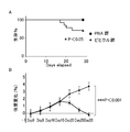
- FIG. 2 is a graph showing% survival (FIG. 2A) and body weight change (FIG. 2B) of the PBA-treated mouse group and the vehicle-treated mouse group.
- the PBA-treated mouse group had a higher survival rate than the vehicle-treated mouse group.
- the vehicle-treated mouse group lost weight compared to the PBA-treated mouse group.
- some of the vehicle-treated mice had (1) hair loss and / or (2) wrinkles.
- FIG. 3A results are shown in FIG. 3A.
- lanes 1, 3, 5, 7, 9, 11, and 13 are PBA-treated organs
- lanes 2, 4, 6, 8, 10, 12, and 14 are vehicle-treated organs.
- endoplasmic reticulum stress in mouse organs caused by cGVHD can be relieved by PBA, and endoplasmic reticulum stress-related pro-inflammatory molecules NF- ⁇ B and TXNIP are significantly suppressed accordingly.
- epithelial-mesenchymal transition is correlated with the fibrosis observed in human eye cGVHD, and TXNIP is known to be associated with epithelial-mesenchymal transition (Ogawa Y, Shimmura S, Kawakita T, Yoshida S). , Kawakami Y, Tsubota K.
- FIG. 4A is an HE (hematoxylin and eosin) stained photograph of a PBA-treated organ and an HE stained photograph of a solvent vehicle-treated organ.
- the image was taken at 200 ⁇ magnification and the scale bar at 200 ⁇ m. Severe inflamed areas are indicated by *.
- an ellipse is attached to the part where the fat tissue is lost.
- the thicknesses of PBA-treated vehicle and vehicle-treated skin are also indicated by arrows.
- the meibomian gland contracted and contracted with an ellipse, and the conjunctival epithelium was indicated by an arrow.
- FIG. 4B is a diagram showing immunostaining results of CD45, which is a general leukocyte marker, in PBA-treated tissues and vehicle-treated tissues.
- CD45 is a general leukocyte marker
- FIG. 4B the image was taken at 200 ⁇ magnification and the scale bar at 20 ⁇ m. Cell cytosol and nucleus were stained red and blue, respectively.
- FIG. 4C is a diagram showing the results of Mallory staining for PBA-treated organs and vehicle-treated organs.
- the photograph is taken at a magnification of 200 times, and the scale bar indicates 200 ⁇ m. Abnormal fibrosis areas are indicated by *.
- FIG. 4D is an electron micrograph of PBA-treated lacrimal gland and small intestine and vehicle-treated lacrimal gland and small intestine.
- photographs of lacrimal gland stromal (left) and small intestinal epithelial cells (right) were taken at a magnification of 2000 ⁇ , and the scale bar was 5 ⁇ m.
- the photo of the lacrimal gland blood vessel (middle) is 15000 times magnification and the scale bar is 500 nm.
- Cap indicates a capillary vessel and M indicates a mitochondria.
- a mark was added to the part where the endoplasmic reticulum was enlarged due to protein accumulation, and the cell debris was shown as a rectangle.
- the image of the vehicle-treated small intestine the area where the microvilli were destroyed was marked with an ellipse, and the damaged tissue was indicated by an arrow.
- FIG. 4E shows the density of CD45-positive cells in PBA-treated organs and vehicle-treated organs.
- FIG. 4F is a diagram showing the results of PAS (Periodic acid-Schiff stain) staining of PBA-treated small intestine and PBA-treated eyes, and vehicle-treated small intestine and eyes. The image was taken at 200 times magnification and the scale bar was 200 ⁇ m. Goblet cells were stained as purple dots.
- PAS Periodic acid-Schiff stain
- FIG. 4G shows goblet cell density in PBA-treated small intestine and PBA-treated eyes and vehicle-treated small intestine and eyes.
- FIGS. 4A and C show that reduction of endoplasmic reticulum stress using PBA can be an effective treatment for cGVHD. Yes.
- FIG. 4A it can be seen that infiltration of inflammatory cells was suppressed in the PBA-treated organ as compared to the vehicle-treated organ.
- FIG. 5A shows the results of ELISA performed 28 days after bone marrow transplantation in order to measure MCP-1, TNF- ⁇ and IFN- ⁇ , which are inflammatory markers in serum collected from PBA-treated mice and vehicle-treated mice. It is.
- the serum of PBA-treated mice had lower protein levels of MCP-1, TNF- ⁇ and IFN- ⁇ , which are inflammatory markers, compared to vehicle-treated mice.
- FIG. 5B shows the results of immunoblot analysis of prefibrotic mediator connective tissue growth factor (CTGF), which is a fibrosis marker.
- CTGF connective tissue growth factor
- CTGF was overexpressed in vehicle-treated organs by immunoblot analysis, but was not observed in PBA-treated organs. This can be said that PBA suppresses cGVHD-induced overexpression of CTGF.
- FIGS. 6A to 6D show the results of reducing cGVHD-induced endoplasmic reticulum stress in lacrimal fibroblasts by PBA relieving endoplasmic reticulum stress.
- FIG. 6A shows the results of immunoblot analysis of endoplasmic reticulum stress markers, activation markers, and fibrosis markers in mouse lacrimal fibroblasts.
- lane 1 shows PBA-treated lacrimal gland-derived fibroblasts
- lane 2 shows vehicle-treated lacrimal gland-derived fibroblasts.
- FIG. 6B is a diagram showing the results of quantitative analysis of the corresponding protein bands (PBA-treated fibroblasts and vehicle-treated fibroblasts).
- FIG. 6C is a diagram showing the results of measuring the protein level of MCP-1 produced by PBA-treated fibroblasts and vehicle-treated fibroblasts using ELISA.
- FIG. 6D shows the results of qPCR analysis of IL-6 and TGF- ⁇ in PBA-treated fibroblasts and vehicle-treated fibroblasts.
- FIG. 7A shows immunofluorescence images of the PBA-injected lacrimal gland and the vehicle-injected lacrimal gland.
- the photograph was taken at 200 ⁇ magnification, and the scale bar represents 20 ⁇ m. Macrophages and CHOP were stained red and green, respectively.
- FIG. 7A it was found that when the mouse was treated with a solvent vehicle, macrophages expressing CHOP migrated to the tissue. This indicates that macrophages subjected to endoplasmic reticulum stress are associated with the progression of cGVHD. On the other hand, such macrophages were not observed in lacrimal glands collected from PBA-treated mice.
- FIG. 7B shows immunofluorescence results of cultured spleen macrophages derived from PBA-treated mice and vehicle-treated mice. The photograph was taken at 200 times magnification and the scale bar shows 20 ⁇ m. Macrophages and CHOP were stained red and green, respectively. The results of immunohistochemistry shown in FIG. 7B revealed that vehicle-treated spleen macrophages express CHOP in contrast to PBA-treated spleen macrophages.
- FIG. 7C shows the results of immunoblot analysis of each endoplasmic reticulum stress marker in mouse spleen macrophages.
- lane 1 shows splenic macrophages derived from PBA-treated mice
- lane 2 shows splenic macrophages derived from vehicle-treated mice.
- FIG. 7D shows the corresponding quantitative analysis results for each protein band.
- endoplasmic reticulum stress markers GRP78, CHOP, phosphorylated PERK, phosphorylated eIF2 ⁇ and phosphorylated IRE1 ⁇ were detected. It can be seen that it is substantially suppressed as compared to the solvent vehicle.
- FIG. 7E shows the results of qPCR analysis of each marker of M1 macrophages and M2 macrophages from the spleen.
- TGF- ⁇ and IL-10 which are M2 macrophage markers
- cGVHD-induced endoplasmic reticulum stress alternatively differentiates macrophages into activated phenotypes, and (2) converts macrophages into activated phenotypes. It is speculated that cGVHD-induced fibrosis by PBA can be mitigated by reducing endoplasmic reticulum stress in macrophages.
- TXNIP has been described to induce extensive inflammation by activating pilin domain 3 (NLRP3), an inflammasome-like receptor family
- NLRP3 pilin domain 3
- TXNIP and cGVHD have not been known so far. As described above, it was revealed that endoplasmic reticulum stress-induced TXNIP expression could correlate with cGVHD-induced disability.
- FIG. 8 is a diagram showing the estimated correlation between cGVHD and endoplasmic reticulum stress.
- an organ affected by cGVHD is considered to be subjected to oxidative stress.
- endoplasmic reticulum stress in epithelial cells, fibroblasts and macrophages can increase.
- inflammation-related molecules such as NF- ⁇ B and TXNIP are expressed and / or activated in an uncontrollable manner, and (2) fibroblasts become dysfunctional, resulting in abnormal collagen bundles.
- macrophages can be differentiated into an M2 phenotype that can be associated with aberrant fibrosis.
- PBA can be an effective drug for treating cGVHD by relieving endoplasmic reticulum stress.
Landscapes
- Health & Medical Sciences (AREA)
- Immunology (AREA)
- Public Health (AREA)
- Bioinformatics & Cheminformatics (AREA)
- Chemical & Material Sciences (AREA)
- Life Sciences & Earth Sciences (AREA)
- Animal Behavior & Ethology (AREA)
- General Health & Medical Sciences (AREA)
- Medicinal Chemistry (AREA)
- Veterinary Medicine (AREA)
- Engineering & Computer Science (AREA)
- Pharmacology & Pharmacy (AREA)
- Epidemiology (AREA)
- Transplantation (AREA)
- Chemical Kinetics & Catalysis (AREA)
- General Chemical & Material Sciences (AREA)
- Nuclear Medicine, Radiotherapy & Molecular Imaging (AREA)
- Organic Chemistry (AREA)
- Acyclic And Carbocyclic Compounds In Medicinal Compositions (AREA)
Abstract
新規な移植片対宿主病の治療剤又は予防剤、ファイブロサイト浸潤抑制剤並びに涙液及び杯細胞の減少の抑制剤を提供する。 移植片対宿主病の治療又は予防方法は、フェニル酪酸またはその薬理学的に許容される塩をそれを必要とする患者に投与することを含む。移植片対宿主病は、骨髄移植後の移植片対宿主病であることが好ましい。また、移植片対宿主病は、眼における移植片対宿主病であることが好ましい。
Description
本発明は、移植片対宿主病の治療剤又は予防剤、ファイブロサイト浸潤抑制剤及び涙液減少と杯細胞の減少の抑制剤に関する。
造血幹細胞の腫瘍化に伴う血液悪性疾患に対して、他者の健康な造血幹細胞を移植して、白血病を根治させる治療法として確立されている医療である造血幹細胞移植が行われる場合がある。他方、近年、ドナーから提供された骨髄細胞、末梢血又は臍帯血の移植片が、免疫応答によってレシピエントの標的臓器を攻撃することによって発症する移植片対宿主病(graft-versus-host disease:GVHD)が問題となっている。
この移植片対宿主病の治療剤として、例えば、眼表面の治療としてレバミピドやジクアホソルが知られており、非特許文献1には、レバミピドとジクアホソルとを併用することで、移植片対宿主病の症状の一つであるドライアイを改善することが開示されている。
特に、慢性移植片対宿主病(chronic graft-versus-host disease:cGVHD)は造血幹細胞移植(骨髄移植)後の重大な合併症であり、一部の患者にとっては致死的であり得る。cGVHDは過去数十年にわたり集中的に研究されてきたが、cGVHDの有効な治療法の開発が遅れている。
cGVHD患者は、多くの場合、シクロスポリン等の免疫抑制剤やステロイドで治療されているが、望ましい結果は得られ難い。それゆえ、臨床現場では、cGVHDを治療する有効な方法が切望されている。
例えば、非特許文献2には、ドナー免疫細胞及びレシピエント細胞における細胞老化がcGVHDの発症プロセスに有害的に関与することが開示されている。
cGVHD患者は、多くの場合、シクロスポリン等の免疫抑制剤やステロイドで治療されているが、望ましい結果は得られ難い。それゆえ、臨床現場では、cGVHDを治療する有効な方法が切望されている。
例えば、非特許文献2には、ドナー免疫細胞及びレシピエント細胞における細胞老化がcGVHDの発症プロセスに有害的に関与することが開示されている。
一方、慢性炎症及び加齢性疾患の発症に有害な役割を果たす「小胞体ストレス」が知られている(例えば、非特許文献3、4)。
以下、図9及び10を参照して小胞体ストレスについて概説する。
タンパク質が小胞体において合成される場合、タンパク質は適切にフォールディングされなければならず、小胞体シャペロンはタンパク質のフォールディングを助ける。
しかし、低酸素、カルシウムイオン欠乏、酸化的傷害、ウイルス感染及び炎症性サイトカインは、小胞体のタンパク質の正常なフォールディングを妨げる。
以下、図9及び10を参照して小胞体ストレスについて概説する。
タンパク質が小胞体において合成される場合、タンパク質は適切にフォールディングされなければならず、小胞体シャペロンはタンパク質のフォールディングを助ける。
しかし、低酸素、カルシウムイオン欠乏、酸化的傷害、ウイルス感染及び炎症性サイトカインは、小胞体のタンパク質の正常なフォールディングを妨げる。
図9に示したように、フォールディングされていないタンパク質及びミスフォールドされたタンパク質が小胞体に蓄積されると小胞体ストレスが起こり、結果的に3つの膜貫通タンパク質(イノシトール要求(IRE)1α、PKR様ERキナーゼ(PERK)及び活性化転写因子(ATF)6α)が活性化されて、フォールディングされていないタンパク質に対する応答(UPR)が開始される。
しかし、UPRが長期化または失敗すると、炎症及びアポトーシス経路が活性化される(非特許文献5)。
その結果、図9及び10に示したように、失敗したUPRは、(1)炎症誘発性分子であるチオレドキシン相互作用タンパク質(TXNIP)及び活性化B細胞の転写因子核因子κ軽鎖エンハンサー(NF-κB)、並びに(2)アポトーシスタンパク質C/EBP相同タンパク質(CHOP)の発現を誘導することが知られている(非特許文献6~8)。
しかし、UPRが長期化または失敗すると、炎症及びアポトーシス経路が活性化される(非特許文献5)。
その結果、図9及び10に示したように、失敗したUPRは、(1)炎症誘発性分子であるチオレドキシン相互作用タンパク質(TXNIP)及び活性化B細胞の転写因子核因子κ軽鎖エンハンサー(NF-κB)、並びに(2)アポトーシスタンパク質C/EBP相同タンパク質(CHOP)の発現を誘導することが知られている(非特許文献6~8)。
しかし、小胞体ストレスとGVHD(特に、cGVHD)との相関はこれまで知られていなかった。
Mio Yamane, Yoko Ogawa,et al., "Long-term Rebamipide and Diquafosol in Two Cases of Immune-Mediated Dry Eye" Optometry and Vision Science, Vol. 92, No. 4S, April 2015
Kawai M, Ogawa Y, Shimmura S, et al. Expression and localization of aging markers in lacrimal gland of chronic graft-versus-host disease. Scientific Reports. 2013;3(2445):1-6.
Brown MK, NirinjiniNaidoo. The endoplasmic reticulum stress response in aging and age-related diseases. Front Physiol. 2012;3(263):1-10.
Garg AD, Kaczmarek A, Krysko O, Vandenabeele P, Krysko DV, Agostinis P. ER stress-induced inflammation: does it aid or impede disease progression? Trends Mol Med. 2012;18:589-598.
Lee AS. The ER chaperone and signaling regulator GRP78/BiP as a monitor of endoplasmic reticulum stress. Methods. 2005;35:373-381.
Anthony TG, Wek RC. TXNIP Switches Tracks toward a Terminal UPR. Cell Metab. 2012;16:135-137.
Lerner AG, Upton JP, Praveen PVK, et al. IRE1a Induces Thioredoxin-Interacting Protein to Activate the NLRP3 Inflammasome and Promote Programmed Cell Death under Irremediable ER Stress. Cell Metab. 2012;16:250-264.
Oslowski CM, Hara T, Murphy BOS, et al. Thioredoxin-Interacting Protein Mediates ER Stress-Induced Beta Cell Death through Initiation of the Inflammasome. Cell Metab. 2012;16:265-273.
本発明は、以上の実情に鑑みてなされたものであり、新規な移植片対宿主病の治療剤又は予防剤、ファイブロサイト浸潤抑制剤並びに涙液及び杯細胞の減少の抑制剤を提供することを目的とする。
本発明者らは、移植片対宿主病において、ファイブロサイト浸潤が涙腺に生じること、及びこのファイブロサイト浸潤をフェニル酪酸またはその薬理学的に許容される塩が抑制することを見出した。
更に、本発明者らは、(1)cGVHDに罹患した臓器が小胞体ストレスレベルの上昇を示すこと、(2)小胞体ストレスの緩和がcGVHD(特に、全身性cGVHD)によって引き起こされる重篤な症状を緩和する強力な治療手段になり得るという新たな仮説を見出し、本発明を完成するに至った。
すなわち、本発明は以下の通りである。
更に、本発明者らは、(1)cGVHDに罹患した臓器が小胞体ストレスレベルの上昇を示すこと、(2)小胞体ストレスの緩和がcGVHD(特に、全身性cGVHD)によって引き起こされる重篤な症状を緩和する強力な治療手段になり得るという新たな仮説を見出し、本発明を完成するに至った。
すなわち、本発明は以下の通りである。
<1>フェニル酪酸またはその薬理学的に許容される塩を有効成分とする、移植片対宿主病の治療又は予防剤。
<2>前記移植片対宿主病は、骨髄移植後の移植片対宿主病である、<1>に記載の治療又は予防剤。
<3>前記移植片対宿主病は、眼における移植片対宿主病である、<1>又は<2>に記載の治療又は予防剤。
<4>前記移植片対宿主病は、ドライアイである、<3>に記載の治療又は予防剤。
<5>前記移植片対宿主病は、ファイブロサイト浸潤を伴う移植片対宿主病である、<1>から<4>のいずれかに記載の治療又は予防剤。
<2>前記移植片対宿主病は、骨髄移植後の移植片対宿主病である、<1>に記載の治療又は予防剤。
<3>前記移植片対宿主病は、眼における移植片対宿主病である、<1>又は<2>に記載の治療又は予防剤。
<4>前記移植片対宿主病は、ドライアイである、<3>に記載の治療又は予防剤。
<5>前記移植片対宿主病は、ファイブロサイト浸潤を伴う移植片対宿主病である、<1>から<4>のいずれかに記載の治療又は予防剤。
<6>フェニル酪酸が4-フェニル-n-酪酸またはその薬理学的に許容される塩である、<1>から<5>のいずれかに記載の治療又は予防剤。
<7>フェニル酪酸またはその薬理学的に許容される塩を有効成分とする、ファイブロサイト浸潤抑制剤。
<8>フェニル酪酸が4-フェニル-n-酪酸またはその薬理学的に許容される塩である、<7>に記載のファイブロサイト浸潤抑制剤。
<7>フェニル酪酸またはその薬理学的に許容される塩を有効成分とする、ファイブロサイト浸潤抑制剤。
<8>フェニル酪酸が4-フェニル-n-酪酸またはその薬理学的に許容される塩である、<7>に記載のファイブロサイト浸潤抑制剤。
<9>フェニル酪酸またはその薬理学的に許容される塩を有効成分とする、杯細胞の減少の抑制剤。
<10>フェニル酪酸が4-フェニル-n-酪酸またはその薬理学的に許容される塩である、<9>に記載の杯細胞の減少の抑制剤。
<11>フェニル酪酸またはその薬理学的に許容される塩を有効成分とする、涙液減少抑制剤。
<12>フェニル酪酸が4-フェニル-n-酪酸またはその薬理学的に許容される塩である、<11>に記載の涙液減少抑制剤。
<10>フェニル酪酸が4-フェニル-n-酪酸またはその薬理学的に許容される塩である、<9>に記載の杯細胞の減少の抑制剤。
<11>フェニル酪酸またはその薬理学的に許容される塩を有効成分とする、涙液減少抑制剤。
<12>フェニル酪酸が4-フェニル-n-酪酸またはその薬理学的に許容される塩である、<11>に記載の涙液減少抑制剤。
本発明は以下の構成も好ましい。
(1) フェニル酪酸またはその薬理学的に許容される塩を投与することを含む、移植片対宿主病の治療又は予防方法。
(1) フェニル酪酸またはその薬理学的に許容される塩を投与することを含む、移植片対宿主病の治療又は予防方法。
(2) 前記移植片対宿主病は、骨髄移植後の移植片対宿主病である、(1)に記載の治療又は予防方法。
(3) 前記移植片対宿主病は、眼における移植片対宿主病である、(1)又は(2)に記載の治療又は予防方法。
(4) 前記移植片対宿主病は、ドライアイである、(3)に記載の治療又は予防方法。
(5) 前記移植片対宿主病は、ファイブロサイト浸潤を伴う移植片対宿主病である、(1)から(4)のいずれかに記載の治療又は予防方法。
(6) フェニル酪酸が4-フェニル-n-酪酸またはその薬理学的に許容される塩である、(1)から(5)のいずれかに記載の治療又は予防方法。
(7) フェニル酪酸またはその薬理学的に許容される塩を投与することを含む、ファイブロサイト浸潤抑制方法。
(8) フェニル酪酸が4-フェニル-n-酪酸またはその薬理学的に許容される塩である、(7)に記載のファイブロサイト浸潤抑制方法。
(9) フェニル酪酸またはその薬理学的に許容される塩を投与することを含む、杯細胞の減少の抑制方法。
(10) フェニル酪酸が4-フェニル-n-酪酸またはその薬理学的に許容される塩である、(9)に記載の杯細胞の減少の抑制方法。
(11) フェニル酪酸またはその薬理学的に許容される塩を投与することを含む、涙液減少抑制方法。
(12) フェニル酪酸が4-フェニル-n-酪酸またはその薬理学的に許容される塩である、(11)に記載の涙液減少抑制方法。
本発明によれば、新規な移植片対宿主病の治療剤又は予防剤、ファイブロサイト浸潤抑制剤並びに涙液及び杯細胞の減少の抑制剤を提供することができる。
以下、本発明の具体的な実施形態について詳細に説明するが、本発明は以下の実施形態に何ら限定されるものではなく、本発明の目的の範囲内において、適宜変更を加えて実施することができる。なお、説明が重複する箇所については、適宜説明を省略する場合があるが、発明の要旨を限定するものではない。
<移植片対宿主病の治療剤又は予防剤>
本発明の移植片対宿主病の治療剤又は予防剤は、フェニル酪酸またはその薬理学的に許容される塩を有効成分として含有する。
本発明の移植片対宿主病の治療剤又は予防剤は、フェニル酪酸またはその薬理学的に許容される塩を有効成分として含有する。
(フェニル酪酸またはその薬理学的に許容される塩)
本発明におけるフェニル酪酸は、以下の式で表される4-フェニル-n-酪酸(以下、単に「PBA」ともいう。)であることが好ましい。
本発明におけるフェニル酪酸は、以下の式で表される4-フェニル-n-酪酸(以下、単に「PBA」ともいう。)であることが好ましい。

本発明において、「ファイブロサイト浸潤」とは、ある組織中(例えば、涙腺)に、血中からファイブロサイトが浸潤することを意味する。また、「ファイブロサイト浸潤抑制」とは、結果として、組織中にファイブロサイトが浸潤する状態となることを抑制することを意味する。
また、ファイブロサイトは、線維芽細胞(フィブロブラスト)と同一であると当業者に認識されている(Disease Models&Mechanisms 2011 May;4(3)318-333(特に325頁))。
また、ファイブロサイトは、線維芽細胞(フィブロブラスト)と同一であると当業者に認識されている(Disease Models&Mechanisms 2011 May;4(3)318-333(特に325頁))。
本発明におけるフェニル酪酸またはその薬理学的に許容される塩は、公知の方法により、準備可能である。例えば、以下のような方法により、フェニル酪酸またはその薬理学的に許容される塩を準備することができる。
本発明にかかるフェニル酪酸(例えば、4-Phenyl-n-butyric acid(CAS登録番号:1821-12-1))は、試薬・工業原料等として入手することもできるし、常法に従って合成することもできる。
本発明にかかるフェニル酪酸の薬理学的に許容される塩とは、カルボキシ基と塩を形成できるものであれば限定されないが、具体的には例えばアルカリ金属、アルカリ土類金属、アミン、塩基性アミノ酸などの塩を挙げることができ、特に、ナトリウム塩、カリウム塩、カルシウム塩、マグネシウム塩がより好ましい。
また本発明のフェニル酪酸またはその薬理学的に許容される塩は、投与後生体内(表面)で容易に加水分解されるエステル・エーテル等の誘導体(いわゆるプロドラッグ)の形態であってもよい。なお本発明のフェニル酪酸として、3-フェニル-n-酪酸(CAS登録番号:4593-90-2)、2-フェニル-n-酪酸(CAS登録番号:90-27-7)、2-フェニルイソ酪酸(CAS登録番号:826-55-1)、3-フェニルイソ酪酸(CAS登録番号:77-83-8)など他の構造異性体も含まれることは言うまでもない。
(移植片対宿主病)
本発明における移植片対宿主病の種類としては、特に限定されず、例えば、その症状が眼、口腔、肝臓、消化管、皮膚、肺等の外分泌腺を備える器官において発現するものが挙げられる。これらのうち、本発明の治療剤又は予防剤は、眼における移植片対宿主病に用いることが好ましい。
本発明における移植片対宿主病の種類としては、特に限定されず、例えば、その症状が眼、口腔、肝臓、消化管、皮膚、肺等の外分泌腺を備える器官において発現するものが挙げられる。これらのうち、本発明の治療剤又は予防剤は、眼における移植片対宿主病に用いることが好ましい。
眼における移植片対宿主病としては、特に限定されないが、例えば、ドライアイ、乾性結膜炎、角膜損傷等、ドライアイに伴う結膜線維化が挙げられる。これらのうち、特に治療又は予防効果に優れることから、治療又は予防対象である眼における移植片対宿主病の疾患は、ドライアイであることが好ましい。
口腔における移植片対宿主病としては、特に限定されないが、例えば、口腔乾燥症、皮膚硬化による開口障害等が挙げられる。
肝臓における移植片対宿主病としては、特に限定されないが、例えば、急性肝炎等が挙げられる。
消化管における移植片対宿主病としては、特に限定されないが、例えば、下痢、食欲不振、嘔吐、上部消化管狭窄や腸梗塞(閉塞)等が挙げられる。
皮膚における移植片対宿主病としては、特に限定されないが、例えば、皮膚乾燥症、強皮症等が挙げられる。
肺における移植片対宿主病としては、特に限定されないが、例えば、間質性肺炎、閉塞性肺炎等が挙げられる。
移植片対宿主病の原因となる移植される臓器としては、特に限定されず、例えば、骨髄、血液(輸血)等が挙げられる。移植片対宿主病は、特に、骨髄移植後に、骨髄由来の造血幹細胞が、移植片対宿主病の原因であるファイブロサイトに分化して、血中を経由して、外分泌腺の組織に浸潤する可能性が指摘されている。しかしながら、本発明の治療剤又は予防剤は、ファイブロサイト浸潤の抑制効果に優れることから、このような骨髄移植後の移植片対宿主病に対して、特に優れた治療又は予防効果を発揮する。このことから、本発明の治療剤又は予防剤の対象の移植片対宿主病は、骨髄移植後の移植片対宿主病であることが好ましい。
移植片対宿主病は、慢性移植片対宿主病(cGVHD)であってもよく、急性移植片対宿主病(acute GVHD:aGVHD)であってもよい。なお、通常、慢性移植片対宿主病とは、移植後、一定期間経過後に発症する移植片対宿主病のことを指し、例えば、古典的な分類では移植後、100日以降に発症する移植片対宿主病を指す。急性移植片対宿主病とは、移植後、一定期間内(慢性移植片対宿主病が発症するまでの期間より短い期間内)に発症する移植片対宿主病を指し、例えば、移植後、古典的には100日以内に発症する移植片対宿主病を指す。現在の分類では時期による分類はなされておらず急性、慢性に典型的で特徴的所見により両者の区別がなされている。本発明の移植片対宿主病の治療剤又は予防剤は、特に、慢性移植片対宿主病に好適に用いることができる。その理由は、線維化抑制に有効であるためと推測される。
本発明の治療剤又は予防剤は、軽度、中度、又は重度のいずれの移植片対宿主病にも適用可能であるが、特に、重度の移植片対宿主病の治療又は予防に好適に用いることができる。その理由は、重症例のドライアイでは線維化が著明であるからである。なお、重度の移植片対宿主病とは、通常、涙液分泌の細胞源となる涙腺細胞、角結膜ムチンの分泌上皮細胞、マイボーム腺細胞が重度の障害された眼移植片対宿主病のことを指す。
上述のとおり、本発明におけるフェニル酪酸またはその薬理学的に許容される塩は、ファイブロサイト浸潤を特に抑制できる。このことから、本発明における移植片対宿主病は、ファイブロサイト浸潤を伴うものであることが特に好ましい。
(剤形)
本発明の治療剤又は予防剤の剤形は、特に限定されず、移植片対宿主病の症状が発現する器官に応じて、適宜設定することができるが、例えば、軟膏剤、静脈内注射剤(点滴を含む)、筋肉内注射剤、腹腔内注射剤、皮下注射剤等の注射剤、坐剤、腫瘍内直接投与剤等に製剤化し、注射用製剤の場合は単位投与量アンプル又は多投与量容器の状態で提供されてよい。特に、本発明の治療剤又は予防剤を、眼における移植片対宿主病の治療剤又は予防剤として用いる場合、点眼剤、結膜下注射剤、軟膏剤として用いることが好ましい。
本発明の治療剤又は予防剤の剤形は、特に限定されず、移植片対宿主病の症状が発現する器官に応じて、適宜設定することができるが、例えば、軟膏剤、静脈内注射剤(点滴を含む)、筋肉内注射剤、腹腔内注射剤、皮下注射剤等の注射剤、坐剤、腫瘍内直接投与剤等に製剤化し、注射用製剤の場合は単位投与量アンプル又は多投与量容器の状態で提供されてよい。特に、本発明の治療剤又は予防剤を、眼における移植片対宿主病の治療剤又は予防剤として用いる場合、点眼剤、結膜下注射剤、軟膏剤として用いることが好ましい。
これらの各種製剤は、製剤上通常用いられる賦形剤、増量剤、結合剤、湿潤剤、崩壊剤、潤滑剤、pH調整剤、界面活性剤、分散剤、緩衝剤、保存剤、溶解補助剤、防腐剤、矯味矯臭剤、無痛化剤、安定化剤、等張化剤等を適宜用い、常法により製造することができる。
本発明の治療剤又は予防剤は、上記製剤化を目的として、フェニル酪酸またはその薬理学的に許容される塩以外の成分を有してもよいが、有さなくてもよい。より具体的な、本発明の治療剤又は予防剤に含んでもよい成分の例示としては、例えば、メチルセルロース等が挙げられる。
(投与方法)
本発明の治療剤又は予防剤の投与方法は、特に限定されず、症状が発現した器官に応じて適宜選択することができるが、例えば、静脈内注射、筋肉内注射、腹腔内注射、皮下注射等により、投与が可能である。特に、眼における移植片対宿主病の場合、点眼、結膜下注射による投与が可能である。
本発明の治療剤又は予防剤は、患者の年齢、症状により適宜投与方法を選択することができる。その投与量は、年齢、投与経路、投与回数により異なり、当業者であれば適宜選択できる。
投与量としては特に制限はないが、一般的には、経口または経静脈投与の場合、一回につき体重1kgあたりフェニル酪酸またはその薬理学的に許容される塩が0.1μg~10mg程度の範囲である。
また点眼剤の場合は、一回につき1~数滴を一日に1~数回投与するが、症状により適宜増減することができる。
さらに眼軟膏の場合は、一日に1~数回、適量を塗布する。
本発明の治療剤又は予防剤の投与方法は、特に限定されず、症状が発現した器官に応じて適宜選択することができるが、例えば、静脈内注射、筋肉内注射、腹腔内注射、皮下注射等により、投与が可能である。特に、眼における移植片対宿主病の場合、点眼、結膜下注射による投与が可能である。
本発明の治療剤又は予防剤は、患者の年齢、症状により適宜投与方法を選択することができる。その投与量は、年齢、投与経路、投与回数により異なり、当業者であれば適宜選択できる。
投与量としては特に制限はないが、一般的には、経口または経静脈投与の場合、一回につき体重1kgあたりフェニル酪酸またはその薬理学的に許容される塩が0.1μg~10mg程度の範囲である。
また点眼剤の場合は、一回につき1~数滴を一日に1~数回投与するが、症状により適宜増減することができる。
さらに眼軟膏の場合は、一日に1~数回、適量を塗布する。
<スクリーニング方法>
本発明は、移植片対宿主病の治療剤又は予防剤の候補物質のスクリーニング方法であって、被験物質を、ヒトを除く、移植片対宿主病のモデル動物に投与する工程と、被験物質が投与された前記モデル動物におけるファイブロサイト浸潤の抑制の程度又は小胞体ストレスの抑制の程度を測定する工程と、測定結果に基づいて、移植片対宿主病の治療剤又は予防剤の候補物質として被験物質を選択する工程と、を有するスクリーニング方法、あるいは炎症性サイトカインで刺激されたファイブロブラストの、摘出あるいは培養涙腺組織への浸潤に対する被験物質の抑制効果を評価する工程を有するスクリーニング方法などを包含する。
本発明は、移植片対宿主病の治療剤又は予防剤の候補物質のスクリーニング方法であって、被験物質を、ヒトを除く、移植片対宿主病のモデル動物に投与する工程と、被験物質が投与された前記モデル動物におけるファイブロサイト浸潤の抑制の程度又は小胞体ストレスの抑制の程度を測定する工程と、測定結果に基づいて、移植片対宿主病の治療剤又は予防剤の候補物質として被験物質を選択する工程と、を有するスクリーニング方法、あるいは炎症性サイトカインで刺激されたファイブロブラストの、摘出あるいは培養涙腺組織への浸潤に対する被験物質の抑制効果を評価する工程を有するスクリーニング方法などを包含する。
(投与工程)
本発明における投与工程は、被験物質を、ヒトを除く、移植片対宿主病のモデル動物に投与する工程である。
本発明における投与工程は、被験物質を、ヒトを除く、移植片対宿主病のモデル動物に投与する工程である。
被験物質は、特に限定されず、天然又は合成の有機又は無機の低分子又は高分子物質等の任意の物質であってよい。
ヒトを除く、移植片対宿主病のモデル動物の種類は、特に限定されないが、例えば、マウス、ラット、イヌ、ネコ、サル、ブタ、ウシ、ヒツジ、ウサギ等の哺乳動物等を使用することができる。
移植片対宿主病のモデル動物は、例えば、マウスの移植片対宿主病のモデル動物を作製する場合、雄のB10.D2(H―2d)マウスの骨髄を、雌のBALB/c(H―2d)マウスに移植することにより、作製することができる。
また、移植片対宿主病の上記モデルマウスで観察される過度の炎症及び線維化は、ヒトで観察される炎症及び線維化に非常に類似している。更に、上記モデルマウスにおいて全身性炎症及び線維化が観察されている(Yaguchi S, Ogawa Y, Shimmura S, et al. Angiotensin II Type 1 Receptor Antagonist Attenuates Lacrimal Gland, Lung, and Liver Fibrosis in a Murine Model of Chronic Graft-Versus-Host Disease. PLoS One. 2013;8:1-11.;Ogawa Y, Morikawa S, Okano H, et al. MHC-compatible bone marrow stromal/stem cells trigger fibrosis by activating host T cells in a scleroderma mouse model. eLife. 2016;5:e09394.)。
また、移植片対宿主病の上記モデルマウスで観察される過度の炎症及び線維化は、ヒトで観察される炎症及び線維化に非常に類似している。更に、上記モデルマウスにおいて全身性炎症及び線維化が観察されている(Yaguchi S, Ogawa Y, Shimmura S, et al. Angiotensin II Type 1 Receptor Antagonist Attenuates Lacrimal Gland, Lung, and Liver Fibrosis in a Murine Model of Chronic Graft-Versus-Host Disease. PLoS One. 2013;8:1-11.;Ogawa Y, Morikawa S, Okano H, et al. MHC-compatible bone marrow stromal/stem cells trigger fibrosis by activating host T cells in a scleroderma mouse model. eLife. 2016;5:e09394.)。
投与方法は、特に限定されず、例えば、目的とする、症状が発現する器官に応じて適宜選択することができるが、例えば、静脈内注射、筋肉内注射、腹腔内注射、皮下注射等の方法で投与してもよい。また、目的とする、症状が発現する器官が、眼である場合、結膜下注射、点眼により投与してもよい。
(測定工程)
本発明における測定工程は、被験物質が投与された移植片対宿主病のモデル動物におけるファイブロサイト浸潤の抑制の程度を測定する工程である。
本発明における測定工程は、被験物質が投与された移植片対宿主病のモデル動物におけるファイブロサイト浸潤の抑制の程度を測定する工程である。
測定方法は、特に限定されず、従来の公知の方法を使用することができ、例えば、被験物質の投与後のヒトを除く移植片対宿主病のモデル動物から、対象となる組織(涙腺等の外分泌腺の組織)を摘出し、ファイブロサイトのマーカー(例えば、CD45、コラーゲン・タイプI、CXCR4等)又は小胞体ストレスのマーカー(例えば、GRP78、CHOP、p-PERK、p-eIF2α、p-IRE1α等)を免疫染色等の方法で検出し、ファイブロサイト浸潤の程度(例えば、検出された面積)又は小胞体ストレスの抑制の程度を測定することで、ファイブロサイト浸潤の抑制又は小胞体ストレスの抑制の程度の程度を測定することができる。この測定結果に基づいて、後述の選択工程において、候補物質を選択することができる。
本発明における測定工程は、ファイブロサイト浸潤の抑制の程度又は小胞体ストレスの抑制の程度以外のものを測定してもよく、測定しなくてもよい。そのような測定してもよい対象としては、例えば、粘膜における杯細胞の数、組織中の炎症の程度、マーカーとなる遺伝子(例えば、繊維化のマーカーとなる遺伝子(例えば、前線維化メディエーター結合組織成長因子(CTGF))、炎症性のマーカーとなる遺伝子(例えば、NF-κB、TXNIP、MCP-1、TNF-α、IFN-γ等)のmRNAの発現量等が挙げられる。これらの測定結果を、ファイブロサイト浸潤の抑制の程度の測定結果と組み合わせて、後述の選択工程において、被験物質を移植片対宿主病の治療剤又は予防剤の候補物質として選択してもよい。
なお、CTGFは線維化のマーカーとなることが知られている(Dziadzio M, Usinger W, Leask A, et al. N-terminal connective tissue growth factor is a marker of the fibrotic phenotype in scleroderma. QJM: An International Journal of Medicine. 2005;98:485-492.)。
なお、CTGFは線維化のマーカーとなることが知られている(Dziadzio M, Usinger W, Leask A, et al. N-terminal connective tissue growth factor is a marker of the fibrotic phenotype in scleroderma. QJM: An International Journal of Medicine. 2005;98:485-492.)。
(選択工程)
選択工程は、上記測定結果に基づいて、移植片対宿主病の治療剤又は予防剤の候補物質として被験物質を選択する工程である。
選択工程は、上記測定結果に基づいて、移植片対宿主病の治療剤又は予防剤の候補物質として被験物質を選択する工程である。
選択方法は、特に限定されず、従来公知の選択方法により候補物質を選択すればよい。例えば、ヒトを除く移植片対宿主病のモデル動物の被験物質の投与前の組織中のファイブロサイト浸潤の程度と比較して、被験物質がファイブロサイト浸潤を抑制したという結果に基づいて、被験物質を候補物質として選択してもよい。また、あらかじめ測定した他の物質(例えば、フェニル酪酸またはその薬理学的に許容される塩)を、ヒトを除く移植片対宿主病のモデル動物に投与したときの測定結果と比較して、被験物質を候補物質として選択してもよい(例えば、移植片対宿主病の治療効果を有するフェニル酪酸またはその薬理学的に許容される塩を、ヒトを除く移植片対宿主病のモデル動物に投与したときの測定結果と比較して、同様の測定結果である場合、その被験物質を候補物質として選択することができる)。
上記の測定工程において、ファイブロサイト浸潤の抑制の程度以外のもの(粘膜における杯細胞の数、組織中の炎症の程度、マーカーとなる遺伝子のmRNAの発現量等)を測定した場合、これらの測定結果を、ファイブロサイト浸潤の抑制の程度の測定結果と組み合わせて、被験物質を移植片対宿主病の治療剤又は予防剤の候補物質として選択してもよい。例えば、ファイブロサイト浸潤の抑制効果が示された被験物質について、該被験物質のモデル動物への投与後に更に、粘膜における杯細胞の数、組織中の炎症の程度、もしくはマーカーとなる遺伝子のmRNAの発現量、又はこれらのうち2つ以上を測定して、それぞれの効果(つまり、粘膜における杯細胞の数の減少の抑制効果、組織中の炎症の抑制効果、マーカーとなる遺伝子のmRNAの発現量の減少もしくは促進効果、又はこれらのうち2つ以上の効果)があることを確認してから、該被験物質を移植片対宿主病の治療剤又は予防剤の候補物質として選択してもよい。
(その他の工程)
本発明のスクリーニング方法は、上記で述べた工程以外の工程を有してもよく、有さなくてもよい。本発明のスクリーニング方法が、有してもよい工程としては、特に限定されないが、例えば、移植片対宿主病の治療剤又は予防剤の候補物質として選択した被験物質を、ヒトを除く移植片対宿主病のモデル動物に投与して、その治療又は予防効果を確認する工程が挙げられる。
本発明のスクリーニング方法は、上記で述べた工程以外の工程を有してもよく、有さなくてもよい。本発明のスクリーニング方法が、有してもよい工程としては、特に限定されないが、例えば、移植片対宿主病の治療剤又は予防剤の候補物質として選択した被験物質を、ヒトを除く移植片対宿主病のモデル動物に投与して、その治療又は予防効果を確認する工程が挙げられる。
<ファイブロサイト浸潤抑制剤>
本発明は、フェニル酪酸またはその薬理学的に許容される塩を有効成分として含有する、ファイブロサイト浸潤抑制剤を包含する。
本発明は、フェニル酪酸またはその薬理学的に許容される塩を有効成分として含有する、ファイブロサイト浸潤抑制剤を包含する。
本発明のファイブロサイト浸潤抑制剤は、上記の移植片対宿主病の治療剤又は予防剤と同様のもの(同様のフェニル酪酸またはその薬理学的に許容される塩、同様の剤型、同様の投与方法、同様の成分等)を用いることができる。
<杯細胞の減少の抑制剤>
本発明は、フェニル酪酸またはその薬理学的に許容される塩を有効成分として含有する、杯細胞の減少の抑制剤を包含する。杯細胞の種類は、結膜杯細胞であることが好ましい。
本発明は、フェニル酪酸またはその薬理学的に許容される塩を有効成分として含有する、杯細胞の減少の抑制剤を包含する。杯細胞の種類は、結膜杯細胞であることが好ましい。
本発明の杯細胞の減少の抑制剤は、上記の移植片対宿主病の治療剤又は予防剤と同様のもの(同様のフェニル酪酸またはその薬理学的に許容される塩、同様の剤型、同様の投与方法、同様の成分等)を用いることができる。
<涙液減少抑制剤>
本発明は、フェニル酪酸またはその薬理学的に許容される塩を有効成分として含有する、涙液減少抑制剤を包含する。
本発明は、フェニル酪酸またはその薬理学的に許容される塩を有効成分として含有する、涙液減少抑制剤を包含する。
本発明において、「涙液減少」とは、例えば、上述した移植片対宿主病(例えば、ドライアイ)による症状としての涙液減少のことを指す。
本発明の涙液減少抑制剤は、上記の移植片対宿主病の治療剤又は予防剤と同様のもの(同様のフェニル酪酸またはその薬理学的に許容される塩、同様の剤型、同様の投与方法、同様の成分等)を用いることができる。
<移植片対宿主病の治療又は予防方法>
本発明は、フェニル酪酸またはその薬理学的に許容される塩をそれを必要とする患者に投与することを含む、移植片対宿主病の治療又は予防方法を包含する。本発明の方法において、上述の移植片対宿主病の治療又は予防剤と同様のもの(同様のフェニル酪酸またはその薬理学的に許容される塩、同様の剤型、同様の投与方法、同様の成分等)を用いることができる。
本発明は、フェニル酪酸またはその薬理学的に許容される塩をそれを必要とする患者に投与することを含む、移植片対宿主病の治療又は予防方法を包含する。本発明の方法において、上述の移植片対宿主病の治療又は予防剤と同様のもの(同様のフェニル酪酸またはその薬理学的に許容される塩、同様の剤型、同様の投与方法、同様の成分等)を用いることができる。
<ファイブロサイト浸潤抑制方法>
本発明は、フェニル酪酸またはその薬理学的に許容される塩をそれを必要とする患者に投与することを含む、ファイブロサイト浸潤抑制方法を包含する。上述の本発明の方法において、ファイブロサイト浸潤抑制剤と同様のもの(同様のフェニル酪酸またはその薬理学的に許容される塩、同様の剤型、同様の投与方法、同様の成分等)を用いることができる。
本発明は、フェニル酪酸またはその薬理学的に許容される塩をそれを必要とする患者に投与することを含む、ファイブロサイト浸潤抑制方法を包含する。上述の本発明の方法において、ファイブロサイト浸潤抑制剤と同様のもの(同様のフェニル酪酸またはその薬理学的に許容される塩、同様の剤型、同様の投与方法、同様の成分等)を用いることができる。
<杯細胞の減少の抑制方法>
本発明は、フェニル酪酸またはその薬理学的に許容される塩をそれを必要とする患者に投与することを含む、杯細胞の減少の抑制方法を包含する。上述の本発明の方法において、杯細胞の減少の抑制剤と同様のもの(同様のフェニル酪酸またはその薬理学的に許容される塩、同様の剤型、同様の投与方法、同様の成分等)を用いることができる。
本発明は、フェニル酪酸またはその薬理学的に許容される塩をそれを必要とする患者に投与することを含む、杯細胞の減少の抑制方法を包含する。上述の本発明の方法において、杯細胞の減少の抑制剤と同様のもの(同様のフェニル酪酸またはその薬理学的に許容される塩、同様の剤型、同様の投与方法、同様の成分等)を用いることができる。
<涙液減少抑制方法>
本発明は、フェニル酪酸またはその薬理学的に許容される塩をそれを必要とする患者に投与することを含む、涙液減少抑制方法を包含する。上述の本発明の方法において、涙液減少抑制剤と同様のもの(同様のフェニル酪酸またはその薬理学的に許容される塩、同様の剤型、同様の投与方法、同様の成分等)を用いることができる。
本発明は、フェニル酪酸またはその薬理学的に許容される塩をそれを必要とする患者に投与することを含む、涙液減少抑制方法を包含する。上述の本発明の方法において、涙液減少抑制剤と同様のもの(同様のフェニル酪酸またはその薬理学的に許容される塩、同様の剤型、同様の投与方法、同様の成分等)を用いることができる。
以下、本発明の実施例を記載する。なお、本発明は以下の実施例の記載に限定されるものではない。
<方法>
(骨髄移植)
8週齢のB10.D2マウス及びBALB/cマウスは三共研究所(東京、日本)から購入した。
骨髄移植をcGVHDのマウスモデルに行った(Zhang Y, McCormick LL, Desai SR, Wu C, Gilliam AC. Murine Sclerodermatous Graft-Versus-Host Disease, a Model for Human Scleroderma: Cutaneous Cytokines, Chemokines, and Immune Cell Activation. The Journal of Immunology. 2002;168:3088-3098.)。
ドナーがB10.D2マウスであり、レシピエントがBALB/cマウスであった場合、それは同種異系骨髄移植となり、cGVHDのマウスモデルを発現した。
対照的に、BALB/cマウスからBALB/cマウスへの骨髄移植は同系骨髄移植であり、cGVHDは移植レシピエントには起こらなかった。
cGVHDを発現しないレシピエントマウスを対照(コントロール)とした。
レシピエントに骨髄移植の前に700cGyを照射し、Gammacel 137 Cs線源(日立メディコ社製)を用いて致死照射を行った。ドナーからの1×10⁶個の骨髄細胞及び2×10⁶個の脾臓細胞を含有する懸濁液を尾静脈を介して各レシピエントマウスに投与した。
ドナー細胞をRPMI 1640(Life Technologies Japan社製)に懸濁した。
(骨髄移植)
8週齢のB10.D2マウス及びBALB/cマウスは三共研究所(東京、日本)から購入した。
骨髄移植をcGVHDのマウスモデルに行った(Zhang Y, McCormick LL, Desai SR, Wu C, Gilliam AC. Murine Sclerodermatous Graft-Versus-Host Disease, a Model for Human Scleroderma: Cutaneous Cytokines, Chemokines, and Immune Cell Activation. The Journal of Immunology. 2002;168:3088-3098.)。
ドナーがB10.D2マウスであり、レシピエントがBALB/cマウスであった場合、それは同種異系骨髄移植となり、cGVHDのマウスモデルを発現した。
対照的に、BALB/cマウスからBALB/cマウスへの骨髄移植は同系骨髄移植であり、cGVHDは移植レシピエントには起こらなかった。
cGVHDを発現しないレシピエントマウスを対照(コントロール)とした。
レシピエントに骨髄移植の前に700cGyを照射し、Gammacel 137 Cs線源(日立メディコ社製)を用いて致死照射を行った。ドナーからの1×10⁶個の骨髄細胞及び2×10⁶個の脾臓細胞を含有する懸濁液を尾静脈を介して各レシピエントマウスに投与した。
ドナー細胞をRPMI 1640(Life Technologies Japan社製)に懸濁した。
(同種異系骨髄移植レシピエントマウスのPBAによる治療)
上記(骨髄移植)に記載のように骨髄移植を実施し、同種異系骨髄移植レシピエントマウスを2群に分けた。
一方の群はPBA(10mg/kg)(Aldrich社製)で腹腔内注射により処理し、他方は腹腔内注射により溶媒ビヒクル・リン酸緩衝食塩水(PBS)(pH7.4)処理した。
骨髄移植後10日目から27日目まで、同種異系骨髄移植レシピエントにPBA又は溶媒ビヒクルを1日1回投与した。
骨髄移植後28日目にサクリファイした。
本願明細書において、cGVHD罹患臓器(眼窩外涙腺、小腸の近位部、背側皮膚、肝臓、唾液腺、肺、大腸及び眼)を分析した。
上記(骨髄移植)に記載のように骨髄移植を実施し、同種異系骨髄移植レシピエントマウスを2群に分けた。
一方の群はPBA(10mg/kg)(Aldrich社製)で腹腔内注射により処理し、他方は腹腔内注射により溶媒ビヒクル・リン酸緩衝食塩水(PBS)(pH7.4)処理した。
骨髄移植後10日目から27日目まで、同種異系骨髄移植レシピエントにPBA又は溶媒ビヒクルを1日1回投与した。
骨髄移植後28日目にサクリファイした。
本願明細書において、cGVHD罹患臓器(眼窩外涙腺、小腸の近位部、背側皮膚、肝臓、唾液腺、肺、大腸及び眼)を分析した。
(組織学的分析及び免疫組織化学)
骨髄移植の3又は4週間後、眼窩外涙腺、小腸の近位部、背側皮膚、肝臓、唾液腺、肺、大腸及び眼を移植レシピエントから採取した。
その後、これらの試料を10%中性緩衝ホルマリンで固定し、パラフィンに包埋した。
パラフィンブロックを7μm厚の切片に切断し、
(1)ヘマトキシリン及びエオシン、
(2)マロリー3色染色(Hopwood J. Fixation and fixtative. In: Bancroft JD, Stevens A, eds. Theory and Practice of Histological Techniques 4th ed Edinburgh: Churchill-Livingstone 1996:23-46.;Anderson G, Gordon K. Tissue processing, microtomy and paraffin sections. In: Bancroft JD, Stevens A, eds. T. Theory and Practice of Histological Techniques 4th ed Edinburgh: Churchill-Livingstone. 1996:47-68.)及び
(3)抗体で染色した。
骨髄移植の3又は4週間後、眼窩外涙腺、小腸の近位部、背側皮膚、肝臓、唾液腺、肺、大腸及び眼を移植レシピエントから採取した。
その後、これらの試料を10%中性緩衝ホルマリンで固定し、パラフィンに包埋した。
パラフィンブロックを7μm厚の切片に切断し、
(1)ヘマトキシリン及びエオシン、
(2)マロリー3色染色(Hopwood J. Fixation and fixtative. In: Bancroft JD, Stevens A, eds. Theory and Practice of Histological Techniques 4th ed Edinburgh: Churchill-Livingstone 1996:23-46.;Anderson G, Gordon K. Tissue processing, microtomy and paraffin sections. In: Bancroft JD, Stevens A, eds. T. Theory and Practice of Histological Techniques 4th ed Edinburgh: Churchill-Livingstone. 1996:47-68.)及び
(3)抗体で染色した。
免疫組織化学的アッセイのために、まず、パラフィンを除去し、続いて以下の2つの抗原回収法のいずれかを用いて抗原を回収した。
(A)CD45抗体(30-F11、BD Pharmingen社製)で切片を染色するために、それらを抗原回収溶液(Target Retrieval Solution; Dako社製)に浸し、次いで電子レンジオーブンで10分間沸騰した。
(B)CD68(FA-11、Abd Serotec社製)及びCHOP(F-168、Santa Cruz Biotechnology社製)の多重染色の場合、切片を抗原回収溶液(HistoVT One;ナカライテスク社製)を用いて洗浄し、続いて90℃で40分間水浴で加熱した。次に、切片を10%正常ヤギ血清でブロックし、組織切片中の抗原と一次抗体との間の反応を4℃で一晩行った。
次いで、切片を室温(例えば、1~30℃)45分間フルオロフォア標識した二次抗体で処理し、退色防止用マウントメディウム(Fluorescent Mounting Medium;Dako社製)でマウントした。
(A)CD45抗体(30-F11、BD Pharmingen社製)で切片を染色するために、それらを抗原回収溶液(Target Retrieval Solution; Dako社製)に浸し、次いで電子レンジオーブンで10分間沸騰した。
(B)CD68(FA-11、Abd Serotec社製)及びCHOP(F-168、Santa Cruz Biotechnology社製)の多重染色の場合、切片を抗原回収溶液(HistoVT One;ナカライテスク社製)を用いて洗浄し、続いて90℃で40分間水浴で加熱した。次に、切片を10%正常ヤギ血清でブロックし、組織切片中の抗原と一次抗体との間の反応を4℃で一晩行った。
次いで、切片を室温(例えば、1~30℃)45分間フルオロフォア標識した二次抗体で処理し、退色防止用マウントメディウム(Fluorescent Mounting Medium;Dako社製)でマウントした。
蛍光画像をLSM共焦点顕微鏡(Carl Zeiss社製)で撮影した。
CD45+細胞の計数に関しては、各組織切片の5つの領域を200倍の倍率で無作為に撮影し、続いて個々の画像におけるCD45+細胞の数を決定した。
二次抗体として、ヤギ抗マウスIgG(H+L)二次抗体、Alexa Fluor 488コンジュゲート(Molecular Probes社製)及びヤギ抗ラットIgG(H+L)二次抗体、Alexa Fluor 568コンジュゲート(Molecular Probes社製)を使用した。
アイソタイプコントロールに関して、ラットIgG2b、κ(eB149/10H5、eBioscience社製)、ラットIgG2a(54447、R&D Systems社製)及びウサギIgG(Cell Signaling Technology社製)をCD45、CD68及びCHOPのためにそれぞれ使用した。
CD45+細胞の計数に関しては、各組織切片の5つの領域を200倍の倍率で無作為に撮影し、続いて個々の画像におけるCD45+細胞の数を決定した。
二次抗体として、ヤギ抗マウスIgG(H+L)二次抗体、Alexa Fluor 488コンジュゲート(Molecular Probes社製)及びヤギ抗ラットIgG(H+L)二次抗体、Alexa Fluor 568コンジュゲート(Molecular Probes社製)を使用した。
アイソタイプコントロールに関して、ラットIgG2b、κ(eB149/10H5、eBioscience社製)、ラットIgG2a(54447、R&D Systems社製)及びウサギIgG(Cell Signaling Technology社製)をCD45、CD68及びCHOPのためにそれぞれ使用した。
(マウス涙腺由来線維芽細胞の培養)
マウス涙腺に由来する線維芽細胞を、Yaguchiらによって報告された確立された方法を用いて培養した(Yaguchi S, Ogawa Y, Shimmura S, et al. Presence and Physiologic Function of the Renin–Angiotensin System in Mouse Lacrimal Gland. Investigative Ophthalmology & Visual Science. 2012;53:5416-5425)。
マウス涙腺を採取し、続いて小片に切断した。組織断片を5%ウシ胎児血清(FBS)(Sigma社製)及び5%の抗生物質を含むDMEM(Life Technologies Japan社製)中37℃でインキュベートした。
抗生物質は、硫酸ストレプトマイシン(明治製菓ファーマ社製)とベンジルペニシリンカリウム(明治製菓社製)の1:1混合物を用いた。
線維芽細胞は、3~4日後に細かく切ったものから増殖を開始した。
線維芽細胞を、5%のFBS及び5%の抗生物質を含むDMEM中で培養し、3~5継代後に実験に使用した。トリプシン(Becton Dickinson社製)を使用して、線維芽細胞を培養皿から分離した。
マウス涙腺に由来する線維芽細胞を、Yaguchiらによって報告された確立された方法を用いて培養した(Yaguchi S, Ogawa Y, Shimmura S, et al. Presence and Physiologic Function of the Renin–Angiotensin System in Mouse Lacrimal Gland. Investigative Ophthalmology & Visual Science. 2012;53:5416-5425)。
マウス涙腺を採取し、続いて小片に切断した。組織断片を5%ウシ胎児血清(FBS)(Sigma社製)及び5%の抗生物質を含むDMEM(Life Technologies Japan社製)中37℃でインキュベートした。
抗生物質は、硫酸ストレプトマイシン(明治製菓ファーマ社製)とベンジルペニシリンカリウム(明治製菓社製)の1:1混合物を用いた。
線維芽細胞は、3~4日後に細かく切ったものから増殖を開始した。
線維芽細胞を、5%のFBS及び5%の抗生物質を含むDMEM中で培養し、3~5継代後に実験に使用した。トリプシン(Becton Dickinson社製)を使用して、線維芽細胞を培養皿から分離した。
(脾臓マクロファージの培養)
Alatery及びBestaの方法に準じてマウス脾臓マクロファージを培養した(Alatery A, Basta S. An efficient culture method for generating large quantities of mature mouse splenic macrophages. J Immunol Methods. 2008;338:47-57)。
5%FBSを含むRPMI中に懸濁した脾細胞を培養皿に播種し、37℃で一晩放置した。次いで、浮遊細胞を除去した。皿に付着した所望の細胞を、組換えマウスM-CSF(5ng/mL)(Peprotech社製)、5%FBS及び5%抗生物質を含むRPMI中で7日間培養した。誘導されたマクロファージは、その後、アキュターゼ(Thermo Fisher Scientific社製)を利用して培養皿から分離し、実験に使用した。
Alatery及びBestaの方法に準じてマウス脾臓マクロファージを培養した(Alatery A, Basta S. An efficient culture method for generating large quantities of mature mouse splenic macrophages. J Immunol Methods. 2008;338:47-57)。
5%FBSを含むRPMI中に懸濁した脾細胞を培養皿に播種し、37℃で一晩放置した。次いで、浮遊細胞を除去した。皿に付着した所望の細胞を、組換えマウスM-CSF(5ng/mL)(Peprotech社製)、5%FBS及び5%抗生物質を含むRPMI中で7日間培養した。誘導されたマクロファージは、その後、アキュターゼ(Thermo Fisher Scientific社製)を利用して培養皿から分離し、実験に使用した。
(培養マクロファージの免疫染色)
誘導したマクロファージを8ウェルチャンバースライド(Fibronectin Culture Slide;Corning社製)に入れ、10%中性緩衝化ホルマリンで固定した。次いで、0.3%過酸化水素を含有するメタノールで室温で30分間ブロックし、一次抗体CD68(AbD Serotec社製)及びCHOP(Santa Cruz社製)と共に4℃で一晩インキュベートした。次いで、切片を、ヤギ抗マウスIgG(H+L)二次抗体、Alexa Fluor 568コンジュゲート(Molecular Probes社製)及び4’、6-ジアミジノ-2-フェニルインドール(DAPI)(Life Technologies社製)を用いて室温で45分間処理し、退色防止用のマウント媒体(Dako社製)でマウントした。
蛍光画像をLSM共焦点顕微鏡(Carl Zeiss社製)で撮影した。
誘導したマクロファージを8ウェルチャンバースライド(Fibronectin Culture Slide;Corning社製)に入れ、10%中性緩衝化ホルマリンで固定した。次いで、0.3%過酸化水素を含有するメタノールで室温で30分間ブロックし、一次抗体CD68(AbD Serotec社製)及びCHOP(Santa Cruz社製)と共に4℃で一晩インキュベートした。次いで、切片を、ヤギ抗マウスIgG(H+L)二次抗体、Alexa Fluor 568コンジュゲート(Molecular Probes社製)及び4’、6-ジアミジノ-2-フェニルインドール(DAPI)(Life Technologies社製)を用いて室温で45分間処理し、退色防止用のマウント媒体(Dako社製)でマウントした。
蛍光画像をLSM共焦点顕微鏡(Carl Zeiss社製)で撮影した。
(電子顕微鏡)
透過電子顕微鏡分析は標準的なプロトコールに従って行った。
マウス涙腺及び小腸から組織を採取し、直ちに0.1Mリン酸緩衝液(pH7.4)中2.5%グルタルアルデヒドで4℃4時間固定し、0.1Mリン酸緩衝液で3回洗浄した。続いて試料を2%オスミウム四酸化物で再び固定し、段階的な一連のエタノール及び100%プロピレンオキシド中で脱水し、エポキシ樹脂に包埋した。処理した組織から1μmの切片を作製し、次いでメチレンブルーで染色した。厚い切片を顕微鏡で観察し、超薄切片の調製に適した部分を見いだした。得られた切片をメッシュグリッド上に置き、ウラニルアセテート及びクエン酸鉛で染色し、電子顕微鏡(1230 EXII;JOEL社製)で調べた。全ての電子顕微鏡写真は、バイオスキャンカメラ(ガタンバイオスキャンカメラモデル792)を用いて取得した。
透過電子顕微鏡分析は標準的なプロトコールに従って行った。
マウス涙腺及び小腸から組織を採取し、直ちに0.1Mリン酸緩衝液(pH7.4)中2.5%グルタルアルデヒドで4℃4時間固定し、0.1Mリン酸緩衝液で3回洗浄した。続いて試料を2%オスミウム四酸化物で再び固定し、段階的な一連のエタノール及び100%プロピレンオキシド中で脱水し、エポキシ樹脂に包埋した。処理した組織から1μmの切片を作製し、次いでメチレンブルーで染色した。厚い切片を顕微鏡で観察し、超薄切片の調製に適した部分を見いだした。得られた切片をメッシュグリッド上に置き、ウラニルアセテート及びクエン酸鉛で染色し、電子顕微鏡(1230 EXII;JOEL社製)で調べた。全ての電子顕微鏡写真は、バイオスキャンカメラ(ガタンバイオスキャンカメラモデル792)を用いて取得した。
(定量的ポリメラーゼ連鎖反応)
miRNeasyミニキット(Qiagen社製)を用いて、眼窩外涙腺、小腸近位部分、背側皮膚、肝臓、培養線維芽細胞及び培養マクロファージから全RNAを抽出し、Rever Tra Ace qPCR RTキット(東洋紡績社製)を用いて対応する相補的DNAを合成した。
TaqManリアルタイムポリメラーゼ連鎖反応(PCR)の使用による下記遺伝子のmRNA発現を分析するために使用されるプライマーは、Applied Biosystems社製から購入した。
ハウスキーピング遺伝子グリセルアルデヒド3-リン酸(GAPDH)、グルコース調節タンパク質78(GRP78)、インターロイキン-1β(IL-1β)、IL-6、IL-10、マクロファージ化学誘引物質タンパク質-1(MCP-1)及び転写増殖因子-β(TGF)。
Step One Plusシステム(Applied Biosystems社製)を用いて定量的リアルタイムPCRを行い、得られたデータを解析するために2-ΔΔCT法を用い、mRNAの発現を測定するための内部標準としてGAPDHを用いた。
miRNeasyミニキット(Qiagen社製)を用いて、眼窩外涙腺、小腸近位部分、背側皮膚、肝臓、培養線維芽細胞及び培養マクロファージから全RNAを抽出し、Rever Tra Ace qPCR RTキット(東洋紡績社製)を用いて対応する相補的DNAを合成した。
TaqManリアルタイムポリメラーゼ連鎖反応(PCR)の使用による下記遺伝子のmRNA発現を分析するために使用されるプライマーは、Applied Biosystems社製から購入した。
ハウスキーピング遺伝子グリセルアルデヒド3-リン酸(GAPDH)、グルコース調節タンパク質78(GRP78)、インターロイキン-1β(IL-1β)、IL-6、IL-10、マクロファージ化学誘引物質タンパク質-1(MCP-1)及び転写増殖因子-β(TGF)。
Step One Plusシステム(Applied Biosystems社製)を用いて定量的リアルタイムPCRを行い、得られたデータを解析するために2-ΔΔCT法を用い、mRNAの発現を測定するための内部標準としてGAPDHを用いた。
(イムノブロッティング分析)
目的の組織をエッペンドルフチューブに入れ、予め冷却したRIPA緩衝液をチューブに添加した。次いで、組織を電気ホモジナイザーを用いてホモジナイズした。
各サンプルを氷上で1時間放置した後、15000rpm、4℃で5分間遠心分離した。その後、上清を氷上で新鮮なチューブに回収し、細胞溶解物として使用した。等量の5×Laemmli緩衝液を各細胞溶解物に添加し、続いて100℃で5分間タンパク質変性させた。
目的の組織をエッペンドルフチューブに入れ、予め冷却したRIPA緩衝液をチューブに添加した。次いで、組織を電気ホモジナイザーを用いてホモジナイズした。
各サンプルを氷上で1時間放置した後、15000rpm、4℃で5分間遠心分離した。その後、上清を氷上で新鮮なチューブに回収し、細胞溶解物として使用した。等量の5×Laemmli緩衝液を各細胞溶解物に添加し、続いて100℃で5分間タンパク質変性させた。
各サンプルからの同量のタンパク質をSDS-PAGEゲルのウェルに充填し、次いで溶解した。タンパク質をゲルから15Vで20分間膜にトランスファーした。
膜を1×TBST(トリス緩衝食塩水及びTween20の混合物)中の5%スキムミルク又は5%BSA(ウシ血清アルブミン)で室温1時間ブロックした。
次いで、膜を一次抗体とともに4℃で一晩インキュベートした。上記一次抗体は、1×TBST中の5%スキムミルク又は5%BSAで1000倍に希釈した。一次抗体とインキュベーション後、膜を1×TBST(3×10分)で洗浄し、室温で1時間二次抗体に供し、次いで1×TBST(3×10分)及び1×TBS(2×10分)で洗浄した。
膜を1×TBST(トリス緩衝食塩水及びTween20の混合物)中の5%スキムミルク又は5%BSA(ウシ血清アルブミン)で室温1時間ブロックした。
次いで、膜を一次抗体とともに4℃で一晩インキュベートした。上記一次抗体は、1×TBST中の5%スキムミルク又は5%BSAで1000倍に希釈した。一次抗体とインキュベーション後、膜を1×TBST(3×10分)で洗浄し、室温で1時間二次抗体に供し、次いで1×TBST(3×10分)及び1×TBS(2×10分)で洗浄した。
関心対象のタンパク質は、以下の2つの方法のいずれかを用いて視覚化した。
(1)BCIP/NBT基質(Promega社製)を用いて、標的タンパク質の比色検出を行った。
(2)シグナルは、増強化学発光(ECL)検出試薬(GE Healthcare社製)を用いて現像し、その後、標的タンパク質をLAS4000ミニ化学発光イメージングシステム(富士フイルム/GE Healthcare社製)で視覚化した。
(1)BCIP/NBT基質(Promega社製)を用いて、標的タンパク質の比色検出を行った。
(2)シグナルは、増強化学発光(ECL)検出試薬(GE Healthcare社製)を用いて現像し、その後、標的タンパク質をLAS4000ミニ化学発光イメージングシステム(富士フイルム/GE Healthcare社製)で視覚化した。
得られたタンパク質バンドの濃度測定分析は、画像処理ソフトウェアImageJを用いて行った。この実験で用いた一次抗体は、GRP78(Abcam社製)、phospho-PERK(Thr980;Cell Signaling Technology社製)、PERK(C33E10;Cell Signaling Technology社製)、phosphor-IRE1α(Thermo Fisher Sceientific社製)、IRE1α(14C10;Cell Signaling Technology社製)、phosphor-eIF2α(119A11;Cell Signaling Technology社製)、eIF2α(Cell Signaling Technology社製)、CHOP(9C8;Thermo Fisher Scientific社製)、TXNIP(D5F3E、Cell Signaling Technology社製)、NF-κB(Abcam社製)、HSP47(SPA-470;Stress Gen Biotechnologies社製)、CTGF(Abcam社製)及びβ-アクチン(AC-15;Abcam社製)を用いた。
二次抗体は、(1)タンパク質バンドを発色させて可視化した場合、AP結合抗マウスIgG抗体(Promega社製)又はAP結合抗ウサギIgG抗体(Promega社製)のいずれかを用い、(2)標的タンパク質をECLで検出するためには、HRP結合抗マウス抗体(Thermo Fisher Scientific社製)又はHRP結合抗ウサギ抗体(Thermo Fisher Scientific社製)を用いた。
(酵素結合免疫吸着アッセイ(ELISA))
血液をPBA処理マウス及びビヒクル処理マウスから採取し、続いて4000rpmで10分間遠心分離した。
得られた血清中のMCP-1レベル、腫瘍壊死因子-α(TNF-α)レベル及びインターフェロン-γ(IFN-γ)レベルを、ELISAセット(Becton Dickinson社製)を用いて測定した。
これらのアッセイは、製造者Becton Dickinson社によって提供されたプロトコールに従って行われた。
血液をPBA処理マウス及びビヒクル処理マウスから採取し、続いて4000rpmで10分間遠心分離した。
得られた血清中のMCP-1レベル、腫瘍壊死因子-α(TNF-α)レベル及びインターフェロン-γ(IFN-γ)レベルを、ELISAセット(Becton Dickinson社製)を用いて測定した。
これらのアッセイは、製造者Becton Dickinson社によって提供されたプロトコールに従って行われた。
(統計分析)
統計的有意性は、マンホイットニーU検定(対応のない2群の差の検討)の使用によって決定した。差は、P<0.05の場合に有意であると考えられる。取得されたデータは、平均値±SD(標準偏差)として示した。
統計的有意性は、マンホイットニーU検定(対応のない2群の差の検討)の使用によって決定した。差は、P<0.05の場合に有意であると考えられる。取得されたデータは、平均値±SD(標準偏差)として示した。
<結果>
[cGVHDに罹患した臓器における小胞体ストレスマーカーの上昇]
小胞体ストレスマーカーを測定することにより、cGVHD罹患臓器において小胞体ストレスが上昇するか否かを調査した。
まず、cGVHD罹患臓器及び対照被験体におけるGRP78のqPCR分析を行った。結果を図1Aに示す。
図1A中、データは、平均値±SD、対照:n=5、cGVHD:n=4~5、*P<0.05として示す。
[cGVHDに罹患した臓器における小胞体ストレスマーカーの上昇]
小胞体ストレスマーカーを測定することにより、cGVHD罹患臓器において小胞体ストレスが上昇するか否かを調査した。
まず、cGVHD罹患臓器及び対照被験体におけるGRP78のqPCR分析を行った。結果を図1Aに示す。
図1A中、データは、平均値±SD、対照:n=5、cGVHD:n=4~5、*P<0.05として示す。
小胞体ストレスマーカー及び関連炎症マーカーのイムノブロット分析を行った。結果を図1Bに示す。
図1B中、レーン1、3、5、7は、同系対照被験体であり、レーン2、4、6、8はcGVHD罹患臓器である。
図1B中、レーン1、3、5、7は、同系対照被験体であり、レーン2、4、6、8はcGVHD罹患臓器である。
各タンパク質バンドに対応する定量分析を行った。結果を図1Cに示す。
図1C中、データは、平均±SD、対照:n=4、cGVHD:n=4、*P<0.05として示す。
図1C中、データは、平均±SD、対照:n=4、cGVHD:n=4、*P<0.05として示す。
図1A~Cに示した結果から明らかなように、リアルタイム定量PCR(qPCR)及びイムノブロット分析から、
(1)小胞体ストレスマーカーであるGRP78、CHOP、p-PERK、p-eIF2α及びp-IRE1αが、cGVHDに罹患したマウス臓器において、コントロール(対照)被検体に比べて増加したことが分かり、
(2)それに応じて、2つの炎症マーカーNF-κB及びTXNIPが、cGVHD罹患臓器において活性化及び/又は増加したことが分かる。
(1)小胞体ストレスマーカーであるGRP78、CHOP、p-PERK、p-eIF2α及びp-IRE1αが、cGVHDに罹患したマウス臓器において、コントロール(対照)被検体に比べて増加したことが分かり、
(2)それに応じて、2つの炎症マーカーNF-κB及びTXNIPが、cGVHD罹患臓器において活性化及び/又は増加したことが分かる。
図1Dは、cGVHDに罹患した涙腺及びその同系対照の涙腺における上皮細胞の電子顕微鏡写真を示す図である。写真は2000倍の倍率で撮影し、スケールバーは5μmを表す。cGVHDに罹患した涙腺上皮写真において、*は、タンパク質の蓄積のために小胞体が肥大した箇所を示す。
図1Dに示した電子顕微鏡写真から明らかなように、cGVHD罹患涙腺上皮において、ミスフォールドした/フォールディングしていないタンパク質の蓄積が、小胞体を肥大させた(図1D中*)。
逆に、コントロール(対照)中の小胞体は無傷のようであった。
これら知見から、cGVHDに罹患した臓器において小胞体ストレスが上昇することが分かった。
図1Dに示した電子顕微鏡写真から明らかなように、cGVHD罹患涙腺上皮において、ミスフォールドした/フォールディングしていないタンパク質の蓄積が、小胞体を肥大させた(図1D中*)。
逆に、コントロール(対照)中の小胞体は無傷のようであった。
これら知見から、cGVHDに罹患した臓器において小胞体ストレスが上昇することが分かった。
[小胞体ストレス誘導性炎症経路のPBAによる失活]
次に、PBAによる小胞体ストレスの低減を介してcGVHDの治療を試験した。
上記<方法>に示すように、同種異系骨髄移植を受けたマウスをPBA又は溶媒ビヒクルで処理した。
図2は、PBA処理マウス群及びビヒクル処理マウス群の生存%(図2A)及び体重変化(図2B)を示す図である。
図2A中、値は、平均値±SE(標準誤差)であり、n=14、*P<0.05として示す。
図2B中、値は、平均値±SEであり、n=10、***P<0.001として示す。
図2Aから明らかなように、PBA処理マウス群は、ビヒクル処理マウス群よりも生存率が高かった。
図2Bから明らかなように、ビヒクル処理マウス群はPBA処理マウス群に比べて体重が減少した。
また、ビヒクル処理マウス群の幾匹かは(1)体毛が抜ける及び/又は(2)臥していた。
次に、PBAによる小胞体ストレスの低減を介してcGVHDの治療を試験した。
上記<方法>に示すように、同種異系骨髄移植を受けたマウスをPBA又は溶媒ビヒクルで処理した。
図2は、PBA処理マウス群及びビヒクル処理マウス群の生存%(図2A)及び体重変化(図2B)を示す図である。
図2A中、値は、平均値±SE(標準誤差)であり、n=14、*P<0.05として示す。
図2B中、値は、平均値±SEであり、n=10、***P<0.001として示す。
図2Aから明らかなように、PBA処理マウス群は、ビヒクル処理マウス群よりも生存率が高かった。
図2Bから明らかなように、ビヒクル処理マウス群はPBA処理マウス群に比べて体重が減少した。
また、ビヒクル処理マウス群の幾匹かは(1)体毛が抜ける及び/又は(2)臥していた。
次に、イムノブロット分析を実施して、cGVHD標的臓器における小胞体ストレスマーカー及び関連炎症マーカーのタンパク質レベルを試験した。結果を図3Aに示す。
図3A中、レーン1、3、5、7、9、11、13はPBA処理臓器であり、レーン2、4、6、8、10、12、14はビヒクル処理臓器である。
図3A中、レーン1、3、5、7、9、11、13はPBA処理臓器であり、レーン2、4、6、8、10、12、14はビヒクル処理臓器である。
続いて、PBA処理臓器及びビヒクル処理臓器について、各臓器の標的タンパク質をデンシトメトリーによって定量した。結果を図3Bに示す。
図3B中、データは平均値±SD、PBA:n=4、ビヒクル:n=4、*P<0.05として示す。
図3B中、データは平均値±SD、PBA:n=4、ビヒクル:n=4、*P<0.05として示す。
図3A及びBに示した結果から明らかなように、イムノブロット分析は、PBA処理マウスから採取した臓器が、ビヒクル処理マウス由来の臓器と比較して、GRP78、CHOP、p-PERK、p-eIF2α及びp-IRE1αのタンパク質レベルが低かった。
図3A及びBに示したように、結果的に、関連する炎症促進分子NF-κB及びTXNIPが、ビヒクル処理された臓器と比較して、PBA処理された臓器において抑制されたことがわかる。
すなわち、cGVHDに起因するマウスの臓器における小胞体ストレスがPBAによって緩和され得、それに応じて小胞体ストレス関連の炎症促進分子NF-κB及びTXNIPが大幅に抑制されることがわかる。
ここで、上皮間葉移行がヒト眼球cGVHDで観察される線維化と相関し、TXNIPが上皮間葉転換と関連していることが知られている(Ogawa Y, Shimmura S, Kawakita T, Yoshida S, Kawakami Y, Tsubota K. Epithelial Mesenchymal Transition in Human Ocular Chronic Graft-Versus-Host Disease. The American Journal of Pathology. 2009;175:2372-2381.;Wei J, Shi Y, Hou Y, et al. Knockdown of thioredoxin-interacting protein ameliorates high glucose-induced epithelial to mesenchymal transition in renal tubular epithelial cells. Cell Signal. 2013;25:2788-2796.)。
したがって、PBAは、TXNIPの発現を低下させることによって上皮間葉転換を排除するため、cGVHD誘発性の全身性線維化を緩和することができると推測される。
図3A及びBに示したように、結果的に、関連する炎症促進分子NF-κB及びTXNIPが、ビヒクル処理された臓器と比較して、PBA処理された臓器において抑制されたことがわかる。
すなわち、cGVHDに起因するマウスの臓器における小胞体ストレスがPBAによって緩和され得、それに応じて小胞体ストレス関連の炎症促進分子NF-κB及びTXNIPが大幅に抑制されることがわかる。
ここで、上皮間葉移行がヒト眼球cGVHDで観察される線維化と相関し、TXNIPが上皮間葉転換と関連していることが知られている(Ogawa Y, Shimmura S, Kawakita T, Yoshida S, Kawakami Y, Tsubota K. Epithelial Mesenchymal Transition in Human Ocular Chronic Graft-Versus-Host Disease. The American Journal of Pathology. 2009;175:2372-2381.;Wei J, Shi Y, Hou Y, et al. Knockdown of thioredoxin-interacting protein ameliorates high glucose-induced epithelial to mesenchymal transition in renal tubular epithelial cells. Cell Signal. 2013;25:2788-2796.)。
したがって、PBAは、TXNIPの発現を低下させることによって上皮間葉転換を排除するため、cGVHD誘発性の全身性線維化を緩和することができると推測される。
[cGVHD標的器官の組織学的観察]
PBAが小胞体ストレスを緩和することによりcGVHD誘発性の全身性炎症及び線維化が軽減された。結果を図4A~Gに示す。
図4Aは、PBA処理臓器のHE(ヘマトキシリン及びエオシン)染色写真と溶媒ビヒクル処理臓器のHE染色写真である。
図4A中、画像は倍率200倍で、スケールバーは200μmで撮影した。
重度の炎症部分は*で示した。ビヒクル処置皮膚の画像では、脂肪組織が失われた箇所に楕円を付した。
PBA処理皮膚及びビヒクル処理皮膚の厚さも矢印で示した。ビヒクルによる眼の写真では、マイボーム腺が縮小し収縮した箇所に楕円を付し、結膜上皮は矢印で示した。
PBAが小胞体ストレスを緩和することによりcGVHD誘発性の全身性炎症及び線維化が軽減された。結果を図4A~Gに示す。
図4Aは、PBA処理臓器のHE(ヘマトキシリン及びエオシン)染色写真と溶媒ビヒクル処理臓器のHE染色写真である。
図4A中、画像は倍率200倍で、スケールバーは200μmで撮影した。
重度の炎症部分は*で示した。ビヒクル処置皮膚の画像では、脂肪組織が失われた箇所に楕円を付した。
PBA処理皮膚及びビヒクル処理皮膚の厚さも矢印で示した。ビヒクルによる眼の写真では、マイボーム腺が縮小し収縮した箇所に楕円を付し、結膜上皮は矢印で示した。
図4Bは、PBA処理組織及びビヒクル処理組織における一般的な白血球マーカーであるCD45の免疫染色結果を示す図である。
図4B中、画像は倍率200倍で、スケールバーは20μmで撮影した。
細胞の細胞質ゾル及び核は、それぞれ赤色及び青色に染色された。
図4B中、画像は倍率200倍で、スケールバーは20μmで撮影した。
細胞の細胞質ゾル及び核は、それぞれ赤色及び青色に染色された。
図4Cは、PBA処理臓器及びビヒクル処理臓器に対するマロリー染色結果を示す図である。
図4C中、写真は200倍の倍率で撮影し、スケールバーは200μmを示す。異常な線維化領域は*で示した。
図4C中、写真は200倍の倍率で撮影し、スケールバーは200μmを示す。異常な線維化領域は*で示した。
図4Dは、PBA処理した涙腺及び小腸並びにビヒクル処理した涙腺及び小腸の電子顕微鏡写真である。
図4D中、涙腺のストローマ(左)と小腸の上皮細胞(右)の写真は2000倍の倍率で撮影し、スケールバーは5μmである。涙腺の血管の写真(中央)は倍率15000倍で、スケールバーは500nmである。
図4D中、Capは毛細血管を示し、Mはミトコンドリアを示す。
ビヒクル処理涙腺の写真では、タンパク質の蓄積により小胞体が肥大した箇所に*を付し、細胞破片が長方形で示した。
ビヒクル処理小腸の画像では、微絨毛が破壊された箇所に楕円を付し、損傷組織は矢印で示した。
図4D中、涙腺のストローマ(左)と小腸の上皮細胞(右)の写真は2000倍の倍率で撮影し、スケールバーは5μmである。涙腺の血管の写真(中央)は倍率15000倍で、スケールバーは500nmである。
図4D中、Capは毛細血管を示し、Mはミトコンドリアを示す。
ビヒクル処理涙腺の写真では、タンパク質の蓄積により小胞体が肥大した箇所に*を付し、細胞破片が長方形で示した。
ビヒクル処理小腸の画像では、微絨毛が破壊された箇所に楕円を付し、損傷組織は矢印で示した。
図4Eは、PBA処理臓器及びビヒクル処理臓器におけるCD45陽性細胞の密度を示す図である。
図4E中、データは、平均値±SDであり、PBA:n=3、ビヒクル:n=3、*P<0.05、**P<0.01、***P<0.001として示す。
図4E中、データは、平均値±SDであり、PBA:n=3、ビヒクル:n=3、*P<0.05、**P<0.01、***P<0.001として示す。
図4Fは、PBA処理小腸及びPBA処理眼並びにビヒクル処理した小腸及び眼に対するPAS(Periodic acid-Schiff stain)染色結果を示す図である。画像は200倍の倍率で撮影し、スケールバーは200μmである。
杯細胞は、紫色の点として染色された。
杯細胞は、紫色の点として染色された。
図4Gは、PBA処理小腸及びPBA処理眼並びにビヒクル処理した小腸及び眼における杯細胞の密度を示す図である。
図4G中、数値は、平均値±SDであり、PBA:n=3(小腸)、ビヒクル:n=3(小腸)、PBA:n=5(眼)、ビヒクル:n=5(眼)***P<0.001として示した。
図4G中、数値は、平均値±SDであり、PBA:n=3(小腸)、ビヒクル:n=3(小腸)、PBA:n=5(眼)、ビヒクル:n=5(眼)***P<0.001として示した。
図4A及びCに示したcGVHD罹患臓器のHE(ヘマトキシリン及びエオシン)染色及びマロリー(3色)染色は、PBAを用いた小胞体ストレスの低減がcGVHDの有効な治療法になり得ることを示している。
図4Aに示したHE写真から分かるように、炎症細胞の浸潤が、PBA処理臓器において、ビヒクル処理臓器と比較して抑制されたことがわかる。
特に、(1)PBAで処理した小腸及び大腸の腸絨毛は、溶媒ビヒクルで処理した小腸及び大腸とは対照的に損なわれていないことが見て取れ、
(2)PBA処理皮膚とは対照的にビヒクル処理皮膚は肥厚し、脂肪組織を失い、コラーゲン束の密度が上昇し、
(3)ビヒクル処理眼のマイボーム腺は、PBA処理マイボーム腺と比較して減少し、収縮し、
(4)PBA処理結膜上皮とは対照的に、ビヒクル処理結膜上皮の菲薄化及び損傷が観察され、PBAの全身注射によってシンブルファロンが予防できることが示唆された。
図4Aに示したHE写真から分かるように、炎症細胞の浸潤が、PBA処理臓器において、ビヒクル処理臓器と比較して抑制されたことがわかる。
特に、(1)PBAで処理した小腸及び大腸の腸絨毛は、溶媒ビヒクルで処理した小腸及び大腸とは対照的に損なわれていないことが見て取れ、
(2)PBA処理皮膚とは対照的にビヒクル処理皮膚は肥厚し、脂肪組織を失い、コラーゲン束の密度が上昇し、
(3)ビヒクル処理眼のマイボーム腺は、PBA処理マイボーム腺と比較して減少し、収縮し、
(4)PBA処理結膜上皮とは対照的に、ビヒクル処理結膜上皮の菲薄化及び損傷が観察され、PBAの全身注射によってシンブルファロンが予防できることが示唆された。
図4C及びEに示した免疫染色及びその後のCD45+細胞の計数から分かるように、PBA処理臓器中の炎症細胞の数は、ビヒクル処理臓器中の炎症細胞の数よりもかなり少なかった。これら知見はまた、PBA処理臓器における免疫細胞の移動及び増殖の抑制を示している。
さらに、全身性線維化は、cGVHDにおける最も重大な問題の1つである(Ogawa Y, Morikawa S, Okano H, et al. MHC-compatible bone marrow stromal/stem cells trigger fibrosis by activating host T cells in a scleroderma mouse model. eLife. 2016;5:e09394.)。
さらに、全身性線維化は、cGVHDにおける最も重大な問題の1つである(Ogawa Y, Morikawa S, Okano H, et al. MHC-compatible bone marrow stromal/stem cells trigger fibrosis by activating host T cells in a scleroderma mouse model. eLife. 2016;5:e09394.)。
図4Cに示したマロリー染色の結果から、cGVHD誘発性の全身性の線維化が、ビヒクル処理臓器と比較して、PBA処理によって実質的に抑制されることが明らかになった。
図4Dに示した涙腺の電子顕微鏡写真の結果から、涙腺を溶媒ビヒクルで処理した場合、(1)フォールディングしていない/ミスフォールドしたタンパク質の蓄積により上皮細胞及び内皮細胞の小胞体が肥大していること、
(2)小胞体には、ストローマに多量の細胞破片が存在すること、
(3)血管内の上皮細胞及び内皮細胞のミトコンドリアが損傷したことがわかる。
これら結果は、PBAがcGVHDに起因する小胞体ストレスから血管を保護し、それによって免疫細胞の組織への移動を抑制することを示唆している。
図4Dに示した涙腺の電子顕微鏡写真の結果から、涙腺を溶媒ビヒクルで処理した場合、(1)フォールディングしていない/ミスフォールドしたタンパク質の蓄積により上皮細胞及び内皮細胞の小胞体が肥大していること、
(2)小胞体には、ストローマに多量の細胞破片が存在すること、
(3)血管内の上皮細胞及び内皮細胞のミトコンドリアが損傷したことがわかる。
これら結果は、PBAがcGVHDに起因する小胞体ストレスから血管を保護し、それによって免疫細胞の組織への移動を抑制することを示唆している。
また、図4Dに示した小腸の電子顕微鏡写真分析の結果から、小腸を溶媒ビヒクルで処理した場合、その小絨毛が破壊され、隣接する組織が損傷していることがわかる。
これらの組織学的所見は、PBA処理涙腺及びPBA処理小腸では観察されなかった。
これらの組織学的所見は、PBA処理涙腺及びPBA処理小腸では観察されなかった。
さらに、図4F及びGに示したPAS(Periodic acid-Schiff stain)染色の結果から、(1)PBA処理小腸及びPBA処理結膜上皮の杯細胞が、ビヒクル注入対応物の杯細胞よりも多いこと、(2)腸粘膜及び結膜粘膜の構造は、PBAの全身注射によって保持されることがわかった。
以上の組織学的観察からわかるように、PBAは、cGVHD関連小胞体ストレスによって誘発される全身性炎症及び線維化を緩和することができる。
以上の組織学的観察からわかるように、PBAは、cGVHD関連小胞体ストレスによって誘発される全身性炎症及び線維化を緩和することができる。
[炎症性メディエーター及び前線維化メディエーターのPBAによる低減]
PBAがcGVHDに起因する全身性炎症を緩和するか否かをより詳細に調査するために、PBA処理マウス及びビヒクル処理マウスから収集した血清中の炎症マーカーMCP-1、TNF-α及びIFN-γのレベルを測定するELISAを行った。
図5AはPBA処理マウス及びビヒクル処理マウスから収集した血清中の炎症マーカーであるMCP-1、TNF-α及びIFN-γを測定するために骨髄移植の28日後にELISAを行った結果を示す図である。
図5A中、データは平均値±SD、PBA:n=4、ビヒクルn=4、*P<0.05として示す。
図5Aに示したように、PBA処理マウスの血清は、炎症マーカーであるMCP-1、TNF-α及びIFN-γのタンパク質レベルが、ビヒクル処理マウスと比較して低かった。
PBAがcGVHDに起因する全身性炎症を緩和するか否かをより詳細に調査するために、PBA処理マウス及びビヒクル処理マウスから収集した血清中の炎症マーカーMCP-1、TNF-α及びIFN-γのレベルを測定するELISAを行った。
図5AはPBA処理マウス及びビヒクル処理マウスから収集した血清中の炎症マーカーであるMCP-1、TNF-α及びIFN-γを測定するために骨髄移植の28日後にELISAを行った結果を示す図である。
図5A中、データは平均値±SD、PBA:n=4、ビヒクルn=4、*P<0.05として示す。
図5Aに示したように、PBA処理マウスの血清は、炎症マーカーであるMCP-1、TNF-α及びIFN-γのタンパク質レベルが、ビヒクル処理マウスと比較して低かった。
さらに、cGVHD誘発全身性線維化の緩和についてより詳細な調査を行った。
図5Bは線維化マーカーである前線維化メディエーター結合組織成長因子(CTGF)のイムノブロット分析を行った結果を示す図である。
図5B中、レーン1、3、5、7はPBA処理臓器を示し、レーン2、4、6、8はビヒクル処理臓器を示す。
図5Bは線維化マーカーである前線維化メディエーター結合組織成長因子(CTGF)のイムノブロット分析を行った結果を示す図である。
図5B中、レーン1、3、5、7はPBA処理臓器を示し、レーン2、4、6、8はビヒクル処理臓器を示す。
図5Cは各臓器のCTGFをデンシトメトリーにより定量した結果(PBA処理臓器及びビヒクル処理臓器)を示す図である。
データは平均値±SD、PBA:n=4、ビヒクルn=4、*P<0.05として示す。
データは平均値±SD、PBA:n=4、ビヒクルn=4、*P<0.05として示す。
図5B及びCに示したように、イムノブロット分析により、CTGFがビヒクル処理臓器で過剰発現したが、PBA処理臓器では観察されなかった。これは、PBAがCTGFのcGVHD誘導性過剰発現を抑制しているといえる。
以上のデータは、PBAがcGVHD誘導性小胞体ストレスを抑制し、結果として全身性炎症及び線維化を緩和できることを示しており、cGVHD誘導性の身体障害を緩和できることを示している。
以上のデータは、PBAがcGVHD誘導性小胞体ストレスを抑制し、結果として全身性炎症及び線維化を緩和できることを示しており、cGVHD誘導性の身体障害を緩和できることを示している。
[小胞体ストレスと機能不全線維芽細胞との相関]
cGVHDに起因する全身性線維化は重大な問題であり、多臓器不全を引き起こす(Ogawa Y, Morikawa S, Okano H, et al. MHC-compatible bone marrow stromal/stem cells trigger fibrosis by activating host T cells in a scleroderma mouse model. eLife. 2016;5:e09394.)。
cGVHD誘発性線維化のメカニズムはまだ解明されていないが、線維化の発生には線維芽細胞が関係している可能性がある(Ogawa Y, Razzaque MS, Kameyama K, et al. Role of Heat Shock Protein 47, a Collagen-Binding Chaperone, in Lacrimal Gland Pathology in Patients with cGVHD. Investigative Ophthalmology & Visual Science. 2007;48:1079-1086.)。
いくつかの生物学的シグナルは線維芽細胞を刺激し、創傷治癒などのいくつかの生物学的プロセスにおいて不可欠な役割を果たし得る。しかし、線維芽細胞が制御されない様式で活性化されると異常なコラーゲン束を生成し、その異常の原因についてはほとんど知られていないものの、重篤な線維化を誘発する(Darby IA, Hewitson TD. Fibroblast Differentiation in Wound Healing and Fibrosis. International of Review of Cytology. 2007;257:143-179.;Kendall R, Feghali-Bostwick CA. Fibroblasts in fibrosis : novel roles and mediators. Front Pharmacol. 2014;5:1-13.)。
cGVHDに起因する全身性線維化は重大な問題であり、多臓器不全を引き起こす(Ogawa Y, Morikawa S, Okano H, et al. MHC-compatible bone marrow stromal/stem cells trigger fibrosis by activating host T cells in a scleroderma mouse model. eLife. 2016;5:e09394.)。
cGVHD誘発性線維化のメカニズムはまだ解明されていないが、線維化の発生には線維芽細胞が関係している可能性がある(Ogawa Y, Razzaque MS, Kameyama K, et al. Role of Heat Shock Protein 47, a Collagen-Binding Chaperone, in Lacrimal Gland Pathology in Patients with cGVHD. Investigative Ophthalmology & Visual Science. 2007;48:1079-1086.)。
いくつかの生物学的シグナルは線維芽細胞を刺激し、創傷治癒などのいくつかの生物学的プロセスにおいて不可欠な役割を果たし得る。しかし、線維芽細胞が制御されない様式で活性化されると異常なコラーゲン束を生成し、その異常の原因についてはほとんど知られていないものの、重篤な線維化を誘発する(Darby IA, Hewitson TD. Fibroblast Differentiation in Wound Healing and Fibrosis. International of Review of Cytology. 2007;257:143-179.;Kendall R, Feghali-Bostwick CA. Fibroblasts in fibrosis : novel roles and mediators. Front Pharmacol. 2014;5:1-13.)。
そこで、cGVHD誘発小胞体ストレスと機能不全線維芽細胞との相関を鋭意調査する観点から、PBA処理マウス涙腺及びビヒクル処理マウス涙腺からの線維芽細胞を培養した。
PBAが小胞体ストレスを緩和することにより涙腺線維芽細胞におけるcGVHD誘発小胞体ストレスが緩和された結果を図6A~Dに示す。
図6Aは、マウスの涙腺線維芽細胞における小胞体ストレスマーカー、活性化マーカー及び線維化マーカーのイムノブロット分析の結果を示す図である。
図6A中、レーン1はPBA処理涙腺由来線維芽細胞を示し、レーン2はビヒクル処理涙腺由来線維芽細胞を示す。
PBAが小胞体ストレスを緩和することにより涙腺線維芽細胞におけるcGVHD誘発小胞体ストレスが緩和された結果を図6A~Dに示す。
図6Aは、マウスの涙腺線維芽細胞における小胞体ストレスマーカー、活性化マーカー及び線維化マーカーのイムノブロット分析の結果を示す図である。
図6A中、レーン1はPBA処理涙腺由来線維芽細胞を示し、レーン2はビヒクル処理涙腺由来線維芽細胞を示す。
図6Bは、対応タンパク質バンドの定量分析(PBA処理線維芽細胞及びビヒクル処理線維芽細胞)の結果を示す図である。
図6B中、データは平均値±SD、PBA:n=4、ビヒクル:n=4、*P<0.05として示す。
図6B中、データは平均値±SD、PBA:n=4、ビヒクル:n=4、*P<0.05として示す。
図6Cは、ELISAを用いて、PBA処理線維芽細胞及びビヒクル処理線維芽細胞によって産生されたMCP-1のタンパク質レベルを測定した結果を示す図である。
図6C中、データは平均値±SD、PBA:n=4、ビヒクル:n=4、*P<0.05として示す。
図6C中、データは平均値±SD、PBA:n=4、ビヒクル:n=4、*P<0.05として示す。
図6Dは、PBA処理線維芽細胞及びビヒクル処理線維芽細胞におけるIL-6及びTGF-βのqPCR分析結果を示す図である。
図6D中、値は平均値±SD、PBA:n=4、ビヒクル:n=4、*P<0.05として示す。
図6D中、値は平均値±SD、PBA:n=4、ビヒクル:n=4、*P<0.05として示す。
図6Aに示した培養線維芽細胞のイムノブロット分析の結果から明らかなように、GRP78、CHOP、リン酸化PERK、リン酸化eIF2α及びリン酸化IRE1αが溶媒ビヒクル処理マウスと比較してマウスをPBAで処理することによって大きく抑制されたことがわかる。
したがって、図6A~Dに示した結果から明らかなように、ビヒクル処理線維芽細胞と比較してPBA処理線維芽細胞では、(1)HSP47、CTGF、線維芽細胞活性化マーカー及び線維化マーカーの各々のタンパク質レベル、(2)MCP-1の産生及び(3)IL-6のmRNAレベルが低減したことがわかる。
すなわち、上記結果は、(1)cGVHDに起因する小胞体ストレスが有害な方法で線維芽細胞を活性化し、それにより広範囲の線維化を誘発する一方、(2)PBAは小胞体ストレスを緩和することによって線維芽細胞の機能不全を防ぐことができることを示している。
したがって、図6A~Dに示した結果から明らかなように、ビヒクル処理線維芽細胞と比較してPBA処理線維芽細胞では、(1)HSP47、CTGF、線維芽細胞活性化マーカー及び線維化マーカーの各々のタンパク質レベル、(2)MCP-1の産生及び(3)IL-6のmRNAレベルが低減したことがわかる。
すなわち、上記結果は、(1)cGVHDに起因する小胞体ストレスが有害な方法で線維芽細胞を活性化し、それにより広範囲の線維化を誘発する一方、(2)PBAは小胞体ストレスを緩和することによって線維芽細胞の機能不全を防ぐことができることを示している。
[小胞体ストレスと、マクロファージのM2マクロファージへの分化との相関]
上述のように、マクロファージは免疫応答及び炎症において重要な役割を果たしており、老化マクロファージがcGVHDの発症プロセスと有害な関連性があることが知られている。
また、(1)小胞体ストレスがM2マクロファージへの分化を促進すること、及び(2)M2マクロファージが線維化関連疾患に関与することが示唆されている(Oh J, Riek AE, Weng S, et al. Endoplasmic Reticulum Stress Controls M2 Macrophage Differentiation and Foam Cell Formation. The Journal of Biological Chemistry.2012;287:11629-11641.;
Xue J, Sharma V, Hsieh MH, et al. Alternatively activated macrophages promote pancreatic fibrosis in chronic pancreatitis. Nature Communications.2015;6:1-11.;
Shivshankar P, Halade GV, Calhoun C, et al. Caveolin-1 deletion exacerbates cardiac interstitial fibrosis by promoting M2 macrophage activation in mice after myocardial infarction. J Mol Cell Cardiol.2014;76:84-93.)。
上述のように、マクロファージは免疫応答及び炎症において重要な役割を果たしており、老化マクロファージがcGVHDの発症プロセスと有害な関連性があることが知られている。
また、(1)小胞体ストレスがM2マクロファージへの分化を促進すること、及び(2)M2マクロファージが線維化関連疾患に関与することが示唆されている(Oh J, Riek AE, Weng S, et al. Endoplasmic Reticulum Stress Controls M2 Macrophage Differentiation and Foam Cell Formation. The Journal of Biological Chemistry.2012;287:11629-11641.;
Xue J, Sharma V, Hsieh MH, et al. Alternatively activated macrophages promote pancreatic fibrosis in chronic pancreatitis. Nature Communications.2015;6:1-11.;
Shivshankar P, Halade GV, Calhoun C, et al. Caveolin-1 deletion exacerbates cardiac interstitial fibrosis by promoting M2 macrophage activation in mice after myocardial infarction. J Mol Cell Cardiol.2014;76:84-93.)。
そこで、cGVHD誘導小胞体ストレスと、機能不全マクロファージ(M2マクロファージへの分化)との関係を鋭意調査した。
まず、マウス涙管内のマクロファージが小胞体ストレスマーカーであるCHOPを発現するか否かを調査するために免疫染色を行った。
まず、マウス涙管内のマクロファージが小胞体ストレスマーカーであるCHOPを発現するか否かを調査するために免疫染色を行った。
PBAを用いることによりマクロファージにおけるcGVHDに起因する小胞体ストレスが緩和された結果を図7A~Eに示す。
図7Aは、PBA注入涙腺及びビヒクル注入涙腺の免疫蛍光画像を示す図である。
図7A中、写真は200倍の倍率で撮影し、スケールバーは20μmを示す。マクロファージ及びCHOPは、それぞれ赤色及び緑色に染色された。
図7Aに示した涙腺の蛍光写真から判断されるように、マウスを溶媒ビヒクルで処理すると、CHOPを発現するマクロファージが組織に移動したことがわかる。
これは、小胞体ストレスを受けたマクロファージがcGVHDの進行に関連していることを示している。
一方、そのようなマクロファージは、PBA処理マウスから採取した涙腺では観察されなかった。
図7Aは、PBA注入涙腺及びビヒクル注入涙腺の免疫蛍光画像を示す図である。
図7A中、写真は200倍の倍率で撮影し、スケールバーは20μmを示す。マクロファージ及びCHOPは、それぞれ赤色及び緑色に染色された。
図7Aに示した涙腺の蛍光写真から判断されるように、マウスを溶媒ビヒクルで処理すると、CHOPを発現するマクロファージが組織に移動したことがわかる。
これは、小胞体ストレスを受けたマクロファージがcGVHDの進行に関連していることを示している。
一方、そのようなマクロファージは、PBA処理マウスから採取した涙腺では観察されなかった。
次に、cGVHD罹患マクロファージにおける小胞体ストレスについてのより深く考察するために、PBA処理マウス由来及びビヒクル処理マウス由来の脾臓マクロファージを培養した。
図7Bは、PBA処理マウス由来及びビヒクル処理マウス由来の培養脾臓マクロファージの免疫蛍光結果を示す図である。写真は200倍の倍率で撮影し、スケールバーは20μmを示す。マクロファージ及びCHOPは、それぞれ赤色及び緑色に染色された。
図7Bに示した免疫組織化学の結果から、ビヒクル処理脾臓マクロファージは、PBA処理脾臓マクロファージとは対照的に、CHOPを発現することが明らかになった。
図7Bは、PBA処理マウス由来及びビヒクル処理マウス由来の培養脾臓マクロファージの免疫蛍光結果を示す図である。写真は200倍の倍率で撮影し、スケールバーは20μmを示す。マクロファージ及びCHOPは、それぞれ赤色及び緑色に染色された。
図7Bに示した免疫組織化学の結果から、ビヒクル処理脾臓マクロファージは、PBA処理脾臓マクロファージとは対照的に、CHOPを発現することが明らかになった。
図7Cは、マウス脾臓マクロファージにおける小胞体ストレス各マーカーのイムノブロット分析結果を示す図である。
図7C中、レーン1はPBA処理マウス由来の脾臓マクロファージを示し、レーン2はビヒクル処理マウス由来の脾臓マクロファージを示す。
図7C中、レーン1はPBA処理マウス由来の脾臓マクロファージを示し、レーン2はビヒクル処理マウス由来の脾臓マクロファージを示す。
図7Dは、各タンパク質バンドの対応する定量分析結果を示す図である。
図7D中、データは平均値±SD、PBA:n=4、ビヒクル:n=4、*P<0.05として示す。
図7C及びDに示したマクロファージの免疫ブロット分析の結果から明らかなように、マクロファージをPBAで処理した場合、小胞体ストレスマーカーであるGRP78、CHOP、リン酸化PERK、リン酸化eIF2α及びリン酸化IRE1αが溶媒ビヒクルと比較して実質的に抑制されていることがわかる。
図7D中、データは平均値±SD、PBA:n=4、ビヒクル:n=4、*P<0.05として示す。
図7C及びDに示したマクロファージの免疫ブロット分析の結果から明らかなように、マクロファージをPBAで処理した場合、小胞体ストレスマーカーであるGRP78、CHOP、リン酸化PERK、リン酸化eIF2α及びリン酸化IRE1αが溶媒ビヒクルと比較して実質的に抑制されていることがわかる。
さらに、脾臓マクロファージにおける(1)M1マクロファージマーカーであるIL-1β、IL-6及びMCP-1の遺伝子発現並びに(2)M2マクロファージマーカーであるTGF-β及びIL-10の遺伝子発現を試験するためにqPCRを行った(Sene A, Khan AA, Cox D, et al. Impaired Cholesterol Efflux in Senescent Macrophages Promotes Age-Related Macular Degeneration. Cell Metab.2013;17:549-561.)。
図7Eは、脾臓からのM1マクロファージ及びM2マクロファージの各マーカーのqPCR分析結果を示す図である。
図7E中、データは平均値±SD、PBA:n=4~5、ビヒクル:n=4~5、*P<0.05として示す。
図7Eに示した結果から明らかなように、M2マクロファージマーカーであるTGF-β及びIL-10のmRNA発現が、ビヒクル処理マクロファージにおいて、PBA処理マクロファージと比較して増加したことがわかる。
一方、マクロファージをPBAで処理した場合、溶媒ビヒクルで処理したマクロファージよりも、M1マクロファージマーカーであるIL-1β及びIL-6の遺伝子発現が高いことがわかる。
以上の結果から、(1)cGVHD誘発性小胞体ストレスがマクロファージを活性化された表現型に二者択一的に分化させること、及び(2)マクロファージが活性化された表現型に変換することによるcGVHD誘導線維化を、PBAがマクロファージにおける小胞体ストレスを低減することにより緩和することができると推測される。
図7E中、データは平均値±SD、PBA:n=4~5、ビヒクル:n=4~5、*P<0.05として示す。
図7Eに示した結果から明らかなように、M2マクロファージマーカーであるTGF-β及びIL-10のmRNA発現が、ビヒクル処理マクロファージにおいて、PBA処理マクロファージと比較して増加したことがわかる。
一方、マクロファージをPBAで処理した場合、溶媒ビヒクルで処理したマクロファージよりも、M1マクロファージマーカーであるIL-1β及びIL-6の遺伝子発現が高いことがわかる。
以上の結果から、(1)cGVHD誘発性小胞体ストレスがマクロファージを活性化された表現型に二者択一的に分化させること、及び(2)マクロファージが活性化された表現型に変換することによるcGVHD誘導線維化を、PBAがマクロファージにおける小胞体ストレスを低減することにより緩和することができると推測される。
<考察>
以上のように、(1)小胞体ストレスの上昇と、(2)cGVHD罹患臓器におけるNF-κB及びTXNIPなどの炎症誘発性分子の活性及び/又は発現の増加との相関が明らかにされた。
上記のように、NF-κBがcGVHD罹患臓器において活性化され、その一部がcGVHDに起因する小胞体ストレスから生じることを示唆している。
上述の非特許文献6~8には、(1)TXNIPがPERK及びIRE1α経路の下流で生成されること、(2)TXNIPが小胞体ストレスを受けた細胞が恒常性を回復するかアポトーシスを受けるかを決定する交差点において重要な役割を果たすこと、(3)TXNIPがインフラマソーム瘤様受容体ファミリーであるピリンドメイン3(NLRP3)を活性化することによって広範な炎症を誘発することが記載されているが、TXNIPとcGVHDとの間の関連性についてはこれまで知られていなかった。
上記のように、小胞体ストレス誘発TXNIPの発現がcGVHDによる身体障害と相関し得ることが明らかにされた。
以上のように、(1)小胞体ストレスの上昇と、(2)cGVHD罹患臓器におけるNF-κB及びTXNIPなどの炎症誘発性分子の活性及び/又は発現の増加との相関が明らかにされた。
上記のように、NF-κBがcGVHD罹患臓器において活性化され、その一部がcGVHDに起因する小胞体ストレスから生じることを示唆している。
上述の非特許文献6~8には、(1)TXNIPがPERK及びIRE1α経路の下流で生成されること、(2)TXNIPが小胞体ストレスを受けた細胞が恒常性を回復するかアポトーシスを受けるかを決定する交差点において重要な役割を果たすこと、(3)TXNIPがインフラマソーム瘤様受容体ファミリーであるピリンドメイン3(NLRP3)を活性化することによって広範な炎症を誘発することが記載されているが、TXNIPとcGVHDとの間の関連性についてはこれまで知られていなかった。
上記のように、小胞体ストレス誘発TXNIPの発現がcGVHDによる身体障害と相関し得ることが明らかにされた。
図8はcGVHDと小胞体ストレスとの間の推定される相関を示す図である。
図8に示したように、cGVHDに罹患した臓器は酸化的ストレスを受けると考えられる。それに応じて、上皮細胞、線維芽細胞及びマクロファージにおける小胞体ストレスが増大し得る。
その結果、(1)NF-κB及びTXNIPなどの炎症関連分子が制御不能な様式で発現及び/又は活性化され、(2)線維芽細胞が機能不全に陥り、その結果、異常なコラーゲン束が生成され、(3)マクロファージは、異常型線維化に関連し得るM2表現型に分化され得る。
さらに、小胞体ストレスは炎症によって増加すると考えられているので、小胞体ストレスと炎症との間の悪循環が、cGVHDに罹患した臓器において形成され得る。
したがって、cGVHDに起因する小胞体ストレスの低減はまた、上記悪循環から脱却する可能性がある。
以上まとめると、PBAは小胞体ストレスを緩和することにより、cGVHDを治療するための有効な薬剤になり得る。
図8に示したように、cGVHDに罹患した臓器は酸化的ストレスを受けると考えられる。それに応じて、上皮細胞、線維芽細胞及びマクロファージにおける小胞体ストレスが増大し得る。
その結果、(1)NF-κB及びTXNIPなどの炎症関連分子が制御不能な様式で発現及び/又は活性化され、(2)線維芽細胞が機能不全に陥り、その結果、異常なコラーゲン束が生成され、(3)マクロファージは、異常型線維化に関連し得るM2表現型に分化され得る。
さらに、小胞体ストレスは炎症によって増加すると考えられているので、小胞体ストレスと炎症との間の悪循環が、cGVHDに罹患した臓器において形成され得る。
したがって、cGVHDに起因する小胞体ストレスの低減はまた、上記悪循環から脱却する可能性がある。
以上まとめると、PBAは小胞体ストレスを緩和することにより、cGVHDを治療するための有効な薬剤になり得る。
Claims (24)
- フェニル酪酸またはその薬理学的に許容される塩を有効成分とする、移植片対宿主病の治療又は予防剤。
- 前記移植片対宿主病は、骨髄移植後の移植片対宿主病である、請求項1に記載の治療又は予防剤。
- 前記移植片対宿主病は、眼における移植片対宿主病である、請求項1又は2に記載の治療又は予防剤。
- 前記移植片対宿主病は、ドライアイである、請求項3に記載の治療又は予防剤。
- 前記移植片対宿主病は、ファイブロサイト浸潤を伴う移植片対宿主病である、請求項1から4のいずれかに記載の治療又は予防剤。
- フェニル酪酸が4-フェニル-n-酪酸またはその薬理学的に許容される塩である、請求項1から5のいずれかに記載の治療又は予防剤。
- フェニル酪酸またはその薬理学的に許容される塩を有効成分とする、ファイブロサイト浸潤抑制剤。
- フェニル酪酸が4-フェニル-n-酪酸またはその薬理学的に許容される塩である、請求項7に記載のファイブロサイト浸潤抑制剤。
- フェニル酪酸またはその薬理学的に許容される塩を有効成分とする、杯細胞の減少の抑制剤。
- フェニル酪酸が4-フェニル-n-酪酸またはその薬理学的に許容される塩である、請求項9に記載の杯細胞の減少の抑制剤。
- フェニル酪酸またはその薬理学的に許容される塩を有効成分とする、涙液減少抑制剤。
- フェニル酪酸が4-フェニル-n-酪酸またはその薬理学的に許容される塩である、請求項11に記載の涙液減少抑制剤。
- フェニル酪酸またはその薬理学的に許容される塩を投与することを含む、移植片対宿主病の治療又は予防方法。
- 前記移植片対宿主病は、骨髄移植後の移植片対宿主病である、請求項13に記載の治療又は予防方法。
- 前記移植片対宿主病は、眼における移植片対宿主病である、請求項13又は14に記載の治療又は予防方法。
- 前記移植片対宿主病は、ドライアイである、請求項15に記載の治療又は予防方法。
- 前記移植片対宿主病は、ファイブロサイト浸潤を伴う移植片対宿主病である、請求項13から16のいずれかに記載の治療又は予防方法。
- フェニル酪酸が4-フェニル-n-酪酸またはその薬理学的に許容される塩である、請求項13から17のいずれかに記載の治療又は予防方法。
- フェニル酪酸またはその薬理学的に許容される塩を投与することを含む、ファイブロサイト浸潤抑制方法。
- フェニル酪酸が4-フェニル-n-酪酸またはその薬理学的に許容される塩である、請求項19に記載のファイブロサイト浸潤抑制方法。
- フェニル酪酸またはその薬理学的に許容される塩を投与することを含む、杯細胞の減少の抑制方法。
- フェニル酪酸が4-フェニル-n-酪酸またはその薬理学的に許容される塩である、請求項21に記載の杯細胞の減少の抑制方法。
- フェニル酪酸またはその薬理学的に許容される塩を投与することを含む、涙液減少抑制方法。
- フェニル酪酸が4-フェニル-n-酪酸またはその薬理学的に許容される塩である、請求項23に記載の涙液減少抑制方法。
Priority Applications (2)
| Application Number | Priority Date | Filing Date | Title |
|---|---|---|---|
| US16/090,933 US20190192458A1 (en) | 2016-04-05 | 2017-04-05 | Therapeutic/prophylactic agent for graft-versus-host disease, fibrocyte invasion inhibitor, and inhibitor against tear reduction and reduction in goblet cells |
| JP2018510646A JP6936496B2 (ja) | 2016-04-05 | 2017-04-05 | 移植片対宿主病の治療剤又は予防剤、ファイブロサイト浸潤抑制剤、及び涙液減少と杯細胞の減少の抑制剤 |
Applications Claiming Priority (2)
| Application Number | Priority Date | Filing Date | Title |
|---|---|---|---|
| US201662318404P | 2016-04-05 | 2016-04-05 | |
| US62/318,404 | 2016-04-05 |
Publications (1)
| Publication Number | Publication Date |
|---|---|
| WO2017175808A1 true WO2017175808A1 (ja) | 2017-10-12 |
Family
ID=60001188
Family Applications (1)
| Application Number | Title | Priority Date | Filing Date |
|---|---|---|---|
| PCT/JP2017/014290 Ceased WO2017175808A1 (ja) | 2016-04-05 | 2017-04-05 | 移植片対宿主病の治療剤又は予防剤、ファイブロサイト浸潤抑制剤、及び涙液減少と杯細胞の減少の抑制剤 |
Country Status (3)
| Country | Link |
|---|---|
| US (1) | US20190192458A1 (ja) |
| JP (1) | JP6936496B2 (ja) |
| WO (1) | WO2017175808A1 (ja) |
Citations (3)
| Publication number | Priority date | Publication date | Assignee | Title |
|---|---|---|---|---|
| JP2006290863A (ja) * | 2005-04-07 | 2006-10-26 | Asan Labs Ltd | 胃腸障害の治療 |
| JP2009051777A (ja) * | 2007-08-28 | 2009-03-12 | Asan Lab Co Cayman Ltd | 痒みを寛解するための方法 |
| JP2009078977A (ja) * | 2007-09-25 | 2009-04-16 | Japan Health Science Foundation | 心筋の小胞体ストレス抑制剤 |
-
2017
- 2017-04-05 WO PCT/JP2017/014290 patent/WO2017175808A1/ja not_active Ceased
- 2017-04-05 JP JP2018510646A patent/JP6936496B2/ja active Active
- 2017-04-05 US US16/090,933 patent/US20190192458A1/en not_active Abandoned
Patent Citations (3)
| Publication number | Priority date | Publication date | Assignee | Title |
|---|---|---|---|---|
| JP2006290863A (ja) * | 2005-04-07 | 2006-10-26 | Asan Labs Ltd | 胃腸障害の治療 |
| JP2009051777A (ja) * | 2007-08-28 | 2009-03-12 | Asan Lab Co Cayman Ltd | 痒みを寛解するための方法 |
| JP2009078977A (ja) * | 2007-09-25 | 2009-04-16 | Japan Health Science Foundation | 心筋の小胞体ストレス抑制剤 |
Non-Patent Citations (5)
| Title |
|---|
| CAO, STEWART SIYAN ET AL.: "The unfolded protein response and chemical chaperones reduce protein misfolding and colitis in mice", GASTROENTEROLOGY, vol. 144, no. 5, 2013, pages 989 - 1000,1000.e1-e6, XP055429742, ISSN: 0016-5085 * |
| KOLB, P.S. ET AL.: "The therapeutic effects of 4-phenylbutyric acid in maintaining proteostasis", THE INTERNATIONAL JOURNAL OF BIOCHEMISTRY AND CELL BIOLOGY, vol. 61, 7 February 2015 (2015-02-07), pages 45 - 52, XP029202710, ISSN: 1357-2725 * |
| LU , HAO ET AL.: "Tauroursodeoxycholic acid and 4-phenyl butyric acid alleviate endoplasmic reticulum stress and improve prognosis of donation after cardiac death liver transplantation in rats", HEPATOBILIARY AND PANCREATIC DISEASES INTERNATIONAL, vol. 13, no. 6, 15 December 2014 (2014-12-15), pages 586 - 593, ISSN: 1499-3872 * |
| MUKAI, SHIN ET AL.: "Reduction of endoplasmic reticulum stress for treatment of ocular chronic graft-versus-host-disease", ARVO 2015 ANNUAL MEETING ABSTRACTS, 2015 * |
| YOKO OGAWA ET AL.: "Zoketsu Kansaibo Ishokugo no GVHD ni yoru Dry Eye no Byotai Kaiseki", OURNAL OF THE EYE, vol. 22, no. 9, 2005, pages 1233 - 1235, ISSN: 0910-1810 * |
Also Published As
| Publication number | Publication date |
|---|---|
| US20190192458A1 (en) | 2019-06-27 |
| JPWO2017175808A1 (ja) | 2019-02-21 |
| JP6936496B2 (ja) | 2021-09-15 |
Similar Documents
| Publication | Publication Date | Title |
|---|---|---|
| US11331328B2 (en) | Compositions and methods for inhibiting antiapoptotic Bcl-2 proteins as anti-aging agents | |
| Wei et al. | Reduced glutathione level promotes epithelial-mesenchymal transition in lens epithelial cells via a wnt/β-catenin–mediated pathway: Relevance for cataract therapy | |
| JP2020164539A (ja) | 腫瘍免疫寛容を破綻させるためのyapの阻害方法 | |
| KR101775262B1 (ko) | 편도 유래 중간엽 줄기세포 또는 이의 조정 배지를 포함하는 피부 염증 질환 예방 또는 치료용 조성물 | |
| JP6573329B2 (ja) | リゾリン脂質受容体を活性化する物質を含有する薬剤送達促進剤 | |
| CN111018999A (zh) | 二聚体免疫融合蛋白、药物组合物和用途 | |
| Khan et al. | Fasciola helminth defense molecule‐1 protects against experimental arthritis by inhibiting osteoclast formation and function without modulating the systemic immune response | |
| Lu et al. | Tetramethylpyrazine improves oxazolone-induced colitis by inhibiting the NF-κB pathway | |
| Yang et al. | Macrophage-derived amphiregulin induces myofibroblast transition in adipogenic lineage precursors near Staphylococcus aureus abscess in bone marrow | |
| Becker et al. | Differential role of the Ca2+ sensor synaptotagmin VII in macrophages and dendritic cells | |
| JP6936496B2 (ja) | 移植片対宿主病の治療剤又は予防剤、ファイブロサイト浸潤抑制剤、及び涙液減少と杯細胞の減少の抑制剤 | |
| Mutlu et al. | Adoptive Transfer of T Cells as a Potential Therapeutic Approach in the Bleomycin‐Injured Mouse Lung | |
| KR102600585B1 (ko) | 홍삼을 유효성분으로 하는 만성폐쇄성폐질환의 예방, 개선 또는 치료를 위한 조성물 | |
| WO2018137701A1 (zh) | 靶向cxcr7的药物组合物和方法 | |
| Li et al. | Rac1 overexpression promotes Treg-derived cytokines to mediate choroidal neovascularization in wet age-related macular degeneration | |
| Liu et al. | Immunoregulatory effects of α-GalCer in a murine model of autoimmune myocarditis | |
| Han et al. | Polydatin attenuates LPS-induced acute lung injury in rats via targeting HSP90AB1 | |
| KR102600598B1 (ko) | 홍삼을 유효성분으로 하는 미세먼지로 인한 만성폐쇄성폐질환의 예방, 개선 또는 치료를 위한 조성물 | |
| US12076341B2 (en) | Use of apoptotic compensatory proliferation signaling vesicles in tissue repair and wound healing | |
| KR102414285B1 (ko) | 플루나리진 또는 이의 약학적으로 허용 가능한 염을 유효성분으로 함유하는 골질환의 예방 또는 치료용 약학적 조성물 | |
| KR20220094660A (ko) | 홍삼을 유효성분으로 하는 담배로 인한 만성폐쇄성폐질환의 예방, 개선 또는 치료를 위한 조성물 | |
| Zhao et al. | Toxoplasma gondii effector MAF1 blocks mouse AIM2 inflammasome activation by inhibiting mtDNA release | |
| Qian et al. | Palmitic acid reprograms neutrophils to compromise vascular integrity and promote breast cancer lung metastasis | |
| KR102142559B1 (ko) | Dpp4 억제제를 유효성분으로 포함하는 심근섬유증의 예방 또는 치료용 조성물 | |
| WO2023219119A1 (ja) | 生理活性物質を含有する細胞外小胞を生産する方法 |
Legal Events
| Date | Code | Title | Description |
|---|---|---|---|
| ENP | Entry into the national phase |
Ref document number: 2018510646 Country of ref document: JP Kind code of ref document: A |
|
| NENP | Non-entry into the national phase |
Ref country code: DE |
|
| 121 | Ep: the epo has been informed by wipo that ep was designated in this application |
Ref document number: 17779186 Country of ref document: EP Kind code of ref document: A1 |
|
| 122 | Ep: pct application non-entry in european phase |
Ref document number: 17779186 Country of ref document: EP Kind code of ref document: A1 |