WO2017131242A1 - Système de transport de substance intracellulaire et utilisation de celui-ci - Google Patents
Système de transport de substance intracellulaire et utilisation de celui-ci Download PDFInfo
- Publication number
- WO2017131242A1 WO2017131242A1 PCT/JP2017/003880 JP2017003880W WO2017131242A1 WO 2017131242 A1 WO2017131242 A1 WO 2017131242A1 JP 2017003880 W JP2017003880 W JP 2017003880W WO 2017131242 A1 WO2017131242 A1 WO 2017131242A1
- Authority
- WO
- WIPO (PCT)
- Prior art keywords
- nanoparticles
- general formula
- group
- represented
- site
- Prior art date
Links
- 0 *c1ccc(C[Tc])cc1C(F)F Chemical compound *c1ccc(C[Tc])cc1C(F)F 0.000 description 3
Images


Classifications
-
- A—HUMAN NECESSITIES
- A61—MEDICAL OR VETERINARY SCIENCE; HYGIENE
- A61K—PREPARATIONS FOR MEDICAL, DENTAL OR TOILETRY PURPOSES
- A61K47/00—Medicinal preparations characterised by the non-active ingredients used, e.g. carriers or inert additives; Targeting or modifying agents chemically bound to the active ingredient
- A61K47/50—Medicinal preparations characterised by the non-active ingredients used, e.g. carriers or inert additives; Targeting or modifying agents chemically bound to the active ingredient the non-active ingredient being chemically bound to the active ingredient, e.g. polymer-drug conjugates
- A61K47/69—Medicinal preparations characterised by the non-active ingredients used, e.g. carriers or inert additives; Targeting or modifying agents chemically bound to the active ingredient the non-active ingredient being chemically bound to the active ingredient, e.g. polymer-drug conjugates the conjugate being characterised by physical or galenical forms, e.g. emulsion, particle, inclusion complex, stent or kit
- A61K47/6921—Medicinal preparations characterised by the non-active ingredients used, e.g. carriers or inert additives; Targeting or modifying agents chemically bound to the active ingredient the non-active ingredient being chemically bound to the active ingredient, e.g. polymer-drug conjugates the conjugate being characterised by physical or galenical forms, e.g. emulsion, particle, inclusion complex, stent or kit the form being a particulate, a powder, an adsorbate, a bead or a sphere
- A61K47/6925—Medicinal preparations characterised by the non-active ingredients used, e.g. carriers or inert additives; Targeting or modifying agents chemically bound to the active ingredient the non-active ingredient being chemically bound to the active ingredient, e.g. polymer-drug conjugates the conjugate being characterised by physical or galenical forms, e.g. emulsion, particle, inclusion complex, stent or kit the form being a particulate, a powder, an adsorbate, a bead or a sphere the form being a microcapsule, nanocapsule, microbubble or nanobubble
-
- A—HUMAN NECESSITIES
- A61—MEDICAL OR VETERINARY SCIENCE; HYGIENE
- A61K—PREPARATIONS FOR MEDICAL, DENTAL OR TOILETRY PURPOSES
- A61K31/00—Medicinal preparations containing organic active ingredients
- A61K31/33—Heterocyclic compounds
- A61K31/395—Heterocyclic compounds having nitrogen as a ring hetero atom, e.g. guanethidine or rifamycins
- A61K31/435—Heterocyclic compounds having nitrogen as a ring hetero atom, e.g. guanethidine or rifamycins having six-membered rings with one nitrogen as the only ring hetero atom
- A61K31/44—Non condensed pyridines; Hydrogenated derivatives thereof
-
- A—HUMAN NECESSITIES
- A61—MEDICAL OR VETERINARY SCIENCE; HYGIENE
- A61K—PREPARATIONS FOR MEDICAL, DENTAL OR TOILETRY PURPOSES
- A61K45/00—Medicinal preparations containing active ingredients not provided for in groups A61K31/00 - A61K41/00
-
- A—HUMAN NECESSITIES
- A61—MEDICAL OR VETERINARY SCIENCE; HYGIENE
- A61K—PREPARATIONS FOR MEDICAL, DENTAL OR TOILETRY PURPOSES
- A61K47/00—Medicinal preparations characterised by the non-active ingredients used, e.g. carriers or inert additives; Targeting or modifying agents chemically bound to the active ingredient
- A61K47/06—Organic compounds, e.g. natural or synthetic hydrocarbons, polyolefins, mineral oil, petrolatum or ozokerite
- A61K47/24—Organic compounds, e.g. natural or synthetic hydrocarbons, polyolefins, mineral oil, petrolatum or ozokerite containing atoms other than carbon, hydrogen, oxygen, halogen, nitrogen or sulfur, e.g. cyclomethicone or phospholipids
-
- A—HUMAN NECESSITIES
- A61—MEDICAL OR VETERINARY SCIENCE; HYGIENE
- A61K—PREPARATIONS FOR MEDICAL, DENTAL OR TOILETRY PURPOSES
- A61K47/00—Medicinal preparations characterised by the non-active ingredients used, e.g. carriers or inert additives; Targeting or modifying agents chemically bound to the active ingredient
- A61K47/30—Macromolecular organic or inorganic compounds, e.g. inorganic polyphosphates
- A61K47/34—Macromolecular compounds obtained otherwise than by reactions only involving carbon-to-carbon unsaturated bonds, e.g. polyesters, polyamino acids, polysiloxanes, polyphosphazines, copolymers of polyalkylene glycol or poloxamers
-
- A—HUMAN NECESSITIES
- A61—MEDICAL OR VETERINARY SCIENCE; HYGIENE
- A61K—PREPARATIONS FOR MEDICAL, DENTAL OR TOILETRY PURPOSES
- A61K47/00—Medicinal preparations characterised by the non-active ingredients used, e.g. carriers or inert additives; Targeting or modifying agents chemically bound to the active ingredient
- A61K47/50—Medicinal preparations characterised by the non-active ingredients used, e.g. carriers or inert additives; Targeting or modifying agents chemically bound to the active ingredient the non-active ingredient being chemically bound to the active ingredient, e.g. polymer-drug conjugates
- A61K47/69—Medicinal preparations characterised by the non-active ingredients used, e.g. carriers or inert additives; Targeting or modifying agents chemically bound to the active ingredient the non-active ingredient being chemically bound to the active ingredient, e.g. polymer-drug conjugates the conjugate being characterised by physical or galenical forms, e.g. emulsion, particle, inclusion complex, stent or kit
- A61K47/6921—Medicinal preparations characterised by the non-active ingredients used, e.g. carriers or inert additives; Targeting or modifying agents chemically bound to the active ingredient the non-active ingredient being chemically bound to the active ingredient, e.g. polymer-drug conjugates the conjugate being characterised by physical or galenical forms, e.g. emulsion, particle, inclusion complex, stent or kit the form being a particulate, a powder, an adsorbate, a bead or a sphere
- A61K47/6923—Medicinal preparations characterised by the non-active ingredients used, e.g. carriers or inert additives; Targeting or modifying agents chemically bound to the active ingredient the non-active ingredient being chemically bound to the active ingredient, e.g. polymer-drug conjugates the conjugate being characterised by physical or galenical forms, e.g. emulsion, particle, inclusion complex, stent or kit the form being a particulate, a powder, an adsorbate, a bead or a sphere the form being an inorganic particle, e.g. ceramic particles, silica particles, ferrite or synsorb
-
- A—HUMAN NECESSITIES
- A61—MEDICAL OR VETERINARY SCIENCE; HYGIENE
- A61K—PREPARATIONS FOR MEDICAL, DENTAL OR TOILETRY PURPOSES
- A61K9/00—Medicinal preparations characterised by special physical form
- A61K9/14—Particulate form, e.g. powders, Processes for size reducing of pure drugs or the resulting products, Pure drug nanoparticles
-
- A—HUMAN NECESSITIES
- A61—MEDICAL OR VETERINARY SCIENCE; HYGIENE
- A61P—SPECIFIC THERAPEUTIC ACTIVITY OF CHEMICAL COMPOUNDS OR MEDICINAL PREPARATIONS
- A61P35/00—Antineoplastic agents
-
- A—HUMAN NECESSITIES
- A61—MEDICAL OR VETERINARY SCIENCE; HYGIENE
- A61P—SPECIFIC THERAPEUTIC ACTIVITY OF CHEMICAL COMPOUNDS OR MEDICINAL PREPARATIONS
- A61P43/00—Drugs for specific purposes, not provided for in groups A61P1/00-A61P41/00
-
- C—CHEMISTRY; METALLURGY
- C12—BIOCHEMISTRY; BEER; SPIRITS; WINE; VINEGAR; MICROBIOLOGY; ENZYMOLOGY; MUTATION OR GENETIC ENGINEERING
- C12N—MICROORGANISMS OR ENZYMES; COMPOSITIONS THEREOF; PROPAGATING, PRESERVING, OR MAINTAINING MICROORGANISMS; MUTATION OR GENETIC ENGINEERING; CULTURE MEDIA
- C12N9/00—Enzymes; Proenzymes; Compositions thereof; Processes for preparing, activating, inhibiting, separating or purifying enzymes
- C12N9/99—Enzyme inactivation by chemical treatment
-
- B—PERFORMING OPERATIONS; TRANSPORTING
- B82—NANOTECHNOLOGY
- B82Y—SPECIFIC USES OR APPLICATIONS OF NANOSTRUCTURES; MEASUREMENT OR ANALYSIS OF NANOSTRUCTURES; MANUFACTURE OR TREATMENT OF NANOSTRUCTURES
- B82Y5/00—Nanobiotechnology or nanomedicine, e.g. protein engineering or drug delivery
Definitions
- the present invention relates to an intracellular substance transfer system and use thereof.
- This application claims the priority of Japanese Patent Application No. 2016-15760 filed on Jan. 29, 2016, the entire description of which is specifically incorporated herein by reference.
- Non-patent Document 1 Non-patent Document 1
- the target intracellular space or endolysosome can be used efficiently regardless of whether it is a low molecule or a high molecule.
- the problem to be solved by the present invention is to provide a versatile system capable of efficiently transporting drugs to endolysosomes and allowing lysosomal enzymes to function at low concentrations. It aims at providing such a system.
- a further object of the present invention is to provide a system for delivering an active substance (for example, an inhibitor) against an enzyme in a cell membrane, such as a kinase, using the above system for efficiently transporting a drug to an endolysosome. To do.
- an object of the present invention is to provide an anticancer agent using such a system.
- Non-patent document 2 describes a complex of a phospholipid and a sugar chain having a sialic acid residue on the surface of metal nanoparticles.
- the complex is administered to the tail blood vessel of a mouse, and the complex initially spreads throughout the body and then collects in a specific organ.
- this document does not discuss the uptake of this complex in highly active cells such as cancer cells.
- a complex comprising a nanoparticle, a lysosomal enzyme inhibitor or kinase inhibitor represented by the following general formula (A) supported on the surface of the nanoparticle, and a phospholipid mimic substance represented by the following general formula (B).
- n1 is an integer of 2 ⁇ 30, n2 is an integer of 2 ⁇ 30, -S- terminal is nanoparticles supported site, R 10 is a suicide substrate site or kinase inhibitory site .
- n3 is an integer in the range of 2 to 30, and the -S-terminal is the nanoparticle-supported site.
- n1 is an integer of 2 to 30, n2 is an integer of 2 to 30, and the —S-terminal is a nanoparticle-supporting site.
- the total of n1 and n2 is n3 or more, and the complex according to [1] or [2].
- the suicide substrate site is At least selected from the group consisting of N-acetyl-D-glucosamine residue, N-acetyl-D-gugalactosamine residue, galactose residue, glucose residue, fucose residue, mannose residue, and sialic acid residue
- the complex according to [4] having one sugar residue and at least one reactive group selected from the group consisting of a difluoromethylaryl group and a trifluoromethylaryl group.
- the suicide substrate site is a complex according to [4], which contains the following functional group.
- R is an N-acetyl-D-glucosamine residue, an N-acetyl-D-gugalactosamine residue, a galactose residue, a glucose residue, a fucose residue, a mannose residue, and a sialic acid residue, (At least one sugar residue selected from the group consisting of LK and LK is a linker.)
- the molar ratio of the lysosomal enzyme inhibitor represented by the general formula (A) and the phospholipid mimetic represented by the general formula (B) is in the range of 1: 100 to 10: 1, [1] to [1] 0
- Any of [2] to [1] 1, wherein the molar ratio of the lysosomal enzyme inhibitor represented by the general formula (A) to the substance represented by the general formula (A ′) is in the range of 1: 100 to 100: 0.
- the metal nanoparticles are at least one kind of particles selected from the group consisting of gold nanoparticles, platinum nanoparticles, silver nanoparticles, and iron magnetic nanoparticles.
- the semiconductor nanoparticles are quantum dots.
- the nanoparticles have a particle diameter in the range of 0.1 to 100 nm.
- An anticancer agent comprising the complex according to any one of [16] as an active ingredient.
- [19] [1] A method for producing a composite according to any one of 16 to 16, The inhibitor cross-linking precursor X represented by the following general formula (C) and the phospholipid pseudo-substance precursor represented by the following general formula (D) and colloidal nanoparticles are mixed, and the inhibitor cross-linking is performed on the surface of the nanoparticles.
- a surface-modified nanoparticle carrying the precursor X and a phospholipid mimetic (1) The resulting surface-modified nanoparticles are mixed with lysosomal enzyme inhibitor precursor Y or kinase inhibitor precursor Z and linked with inhibitor cross-linking precursor X to give a lysosomal enzyme inhibitor represented by the following general formula (A) Or form a kinase inhibitor,
- the lysosomal enzyme inhibitor precursor Y includes a suicide substrate site containing a group having reactivity with the active center of the lysosomal enzyme (hereinafter, reactive group)
- the kinase inhibitor precursor Z comprises a kinase inhibitory site comprising a group reactive with the kinase,
- n1 is an integer of 2 to 30, and n2 is an integer of 2 to 30.
- n3 is an integer in the range of 2 to 30.
- n1 is an integer of 2 ⁇ 30, n2 is an integer of 2 ⁇ 30, -S- terminal is nanoparticles supported site, R 10 is a suicide substrate site or kinase inhibitory site .
- the present invention for example, it is possible to specifically inactivate intracellular lysosomal enzymes or intracellular kinases, and as a result, an antitumor effect exhibiting an antitumor effect at an order of magnitude lower than conventional antitumor drugs.
- a tumor agent can be provided, and an extremely effective anti-tumor drug candidate can be created.
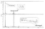
- Example 1 it was confirmed by MALD-TOFMS that the compound was presented on the nanoparticle surface.
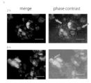
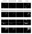
- the intracellular behavior of fluorescent nanoparticles (10 nM) presenting Compound 7 in cancer cells is shown.
- (A) Intracellular dynamics (Scale bar size 50 ⁇ m) after the start of culture of fluorescent nanoparticle breast cancer cells (MCF7) (1 to 8 hours).
- the intracellular behavior of fluorescent nanoparticles (10 nM) presenting Compound 7 in cancer cells is shown.
- (B) Intracellular distribution of QD-anchored inhibitor 7 (fluorescent nanoparticle carrying compound 7) 2 hours (top) and 8 hours (bottom) after the start of culture (Scale bar size 50 ⁇ m).
- Example 2 it was confirmed by MALDI-TOFMS that the compound was presented on the nanoparticle surface.
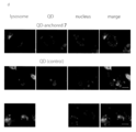
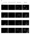
- the intracellular behavior a of the fluorescent nanoparticle (10 nM) presenting Compound 15 is shown.
- Intracellular moving body (Scale bar size 50 ⁇ m) after the start of culture of fluorescent nanoparticle breast cancer cells (MCF-7) (1 to 8 hours).
- the result of the breast cancer cell killing effect of the fluorescent nanoparticle (100 nM QD-anchored 15) which presented the compound 15 is shown.
- A The survival rate of MCF-7 cells after 96 hours of culturing in the presence of fluorescent nanoparticles presenting compound 15 and various test drugs used for comparison was quantified by MTT assay.
- the survival rate of MCF-7 cells after 96 hours of culturing in the presence of fluorescent nanoparticles presenting compound 20 and various test drugs used for comparison was quantified by MTT assay.
- Behavior of fluorescent nanoparticle (10 nM) loaded with compound 7 in cancer cells (HepG2 cells): after initiation of co-culture of phospholipid-coated fluorescent nanoparticles loaded with inhibitor and liver cancer cells (HepG2) (1-8) Intracellular behavior in time) (Scale bar size 50 mm).
- the viability of HepG2 cells after 96 hours of culturing in the presence of the various test agents used in the above was quantified by Cell viability assay (MTT method).
- Hepatocellular carcinoma killing effect of fluorescent nanoparticle (1-100 nM QD-anchored 7) carrying compound 7 after 96 hours in the presence of fluorescent nanoparticle carrying compound 7 and various test drugs used for comparison
- the survival rate of HepG2 cells was quantified by Cell viability assay.
- the survival rate of cancer cells after culturing for 48 hours with HepG2 cells alone and nanoparticle alone was quantified by MTT method.
- the present invention includes a composite comprising a nanoparticle, a lysosomal enzyme inhibitor or kinase inhibitor represented by the following general formula (A) supported on the surface of the nanoparticle, and a phospholipid mimic substance represented by the following general formula (B) About the body.
- n1 is an integer of 2 ⁇ 30, n2 is an integer of 2 ⁇ 30, -S- terminal is nanoparticles supported site, R 10 is a suicide substrate site or kinase inhibitory site .
- n3 is an integer in the range of 2 to 30, and the -S-terminal is the nanoparticle-supported site.
- the lysosomal enzyme inhibitor includes a suicide substrate site containing a group (reactive group) reactive with the active center of the lysosomal enzyme.
- the suicide substrate site contains a sugar residue that can be a substrate for the lysosomal enzyme and a reactive group for the lysosomal enzyme for enzyme inactivation.
- the sugar residue can be appropriately selected according to the substrate specificity of the lysosomal enzyme to be inactivated, for example, N-acetyl-D-glucosamine residue, N-acetyl-D-gugalactosamine residue, galactose It is at least one sugar residue selected from the group consisting of a residue, a glucose residue, a fucose residue, and a mannose residue sialic acid residue.
- the sugar residue is preferably an N-acetyl-D-glucosamine residue and / or a galactose residue in view of the substrate specificity of the lysosomal enzyme.
- a plurality of sugar residues may be used for one composite particle, and in that case, a single or a plurality of types of sugar residues may be present in one composite particle. Further, one or more types of sugar residues may exist in one complex particle, and the same or different single or multiple types of sugar residues may exist in different complex particles that coexist. .
- the reactive group may be any reactive group having reactivity with the reaction center of the enzyme.
- the reactive group having reactivity with the reaction center of the enzyme include a difluoromethylaryl group and a trifluoromethylaryl group. It can be at least one reactive group selected from the group consisting of: The reactivity with the reaction center of the enzyme may be reversible or irreversible.
- Biochemistry, 2005, 44, 11669-11675 can refer to the fact that the difluoromethylaryl group is a reactive group having reactivity with the reaction center of the enzyme.
- the suicide substrate in which the reactive group is a difluoromethylaryl group can be, for example, a group represented by the following general formula (E). In the formula, R is a sugar residue, and F is a fluorine atom.
- the general formula (E) can further be the general formula (E ′) having a linker LK.
- R is a sugar residue
- F is a fluorine atom.
- the linker LK is not particularly limited as long as it is a group capable of cross-linking between an aryl group having a sugar residue and a chain portion.
- it can be a linker containing an alkylene group shown below.
- the left end is the binding site with the aryl group
- the right end is the binding site with the chain site
- the alkylene group in the linker LK can have, for example, a range of 1 to 10 carbon atoms, and an amide group which is a binding site with the leftmost aryl group and / or an imino group which is a binding site with the rightmost chain site Each may be another linking group.
- the suicide substrate site can be, for example, a site containing any functional group represented by the following formula.
- the kinase inhibition site includes a group reactive with a kinase, preferably a group reactive with a kinase in a cell membrane.
- a kinase inhibition site can be, for example, a site represented by the following formula.
- LK represents a linker
- R 1 is an electron-withdrawing group.
- the electron-withdrawing group include a halogen atom (fluorine, chlorine, bromine or iodine), a nitro group, a cyano group, a tosyl group, an acyl group, and the like.
- This kinase inhibition site includes the following sorafenib.
- part can be mentioned as a specific example of the site
- Kinases are a family of enzymes that catalyze the phosphorylation of specific residues in proteins. In general, kinases are divided into three groups. Those that preferentially phosphorylate serine and / or threonine residues, those that preferentially phosphorylate tyrosine residues, and those that phosphorylate both tyrosine and serine / threonine residues. Kinases are considered to be important elements in signal transduction pathways to transmit extracellular signals, including cytokine activation at the receptor, to the nucleus and trigger various biological events.
- inhibitors for tyrosine kinase activity include gefitinib, erlotinib, cetuximab, panitumumab and the like.
- Sorafenib is known as a drug that inhibits tyrosine kinase, Raf kinase (a kind of serine / threonine kinase deeply involved in cell proliferation signal).
- the suicide substrate site possessed by the lysosomal enzyme inhibitor represented by the general formula (A) or the kinase inhibiting site possessed by the kinase inhibitor represented by the general formula (A) is immobilized on the surface of the nanoparticle. It presents a suicide substrate site or a kinase inhibition site on the surface of the nanoparticle.
- “presentation” of a suicide substrate site or kinase inhibition site means immobilization of a suicide substrate site or kinase inhibition site on the surface of the nanoparticle.
- n1 and n2 in the formula are integer in the range of 2 to 30, preferably 5 to 16, and more preferably 4 to 15.
- n2 is an integer in the range of 2 to 30, preferably 5 to 20, and more preferably 7 to 15.
- n3 is an integer in the range of 2 to 30, preferably 5 to 20, and more preferably 7 to 15.
- the amount of the lysosomal enzyme inhibitor or kinase inhibitor represented by the general formula (A) and the phospholipid mimetic represented by the general formula (B) with respect to one nanoparticle depends on the metal element (reaction point) on the surface of the nanoparticle. ) Is preferably 80% or more. It can be determined appropriately in consideration of the function of inhibiting the aggregation of the complex of the present invention and the function of taking it into cells by edesis.
- the supported amount is more preferably an amount that can cover all reaction points on the surface of the nanoparticles.
- the ratio of the lysosomal enzyme inhibitor or kinase inhibitor represented by the general formula (A) to the phospholipid mimetic represented by the general formula (B) is determined by the inactivation of lysosomal enzyme in the cell.
- the molar ratio can be in the range of 1: 100 to 10: 1, preferably Is in the range of 1:10 to 1: 1.
- the complex of the present invention has the following general formula (A).
- the substance represented by ') can be further supported.
- the substance represented by the general formula (A ′) is derived from the precursor X for crosslinking of the enzyme inhibitor represented by the general formula (A) described later.
- n1 is an integer of 2 to 30
- n2 is an integer of 2 to 30
- the —S-terminal is a nanoparticle-supporting site.
- the general formula (A ′) can further carry a physiologically active substance such as a sugar chain or an antibody, if desired.
- the molar ratio of the lysosomal enzyme inhibitor or kinase inhibitor represented by the general formula (A) to the substance represented by the general formula (A ′) can be in the range of 1: 100 to 100: 0. It can be in the range of ⁇ 100: 10.
- the nanoparticles can be metal nanoparticles or semiconductor nanoparticles.
- the material of the metal nanoparticles is not particularly limited, but can be gold, platinum, silver, iron magnetic material.
- the metal nanoparticles are gold nanoparticles, platinum nanoparticles, silver nanoparticles are iron magnetic materials. It can be a nanoparticle.
- the metal nanoparticles are preferably gold nanoparticles, platinum nanoparticles, and silver nanoparticles from the viewpoint of safety with respect to the living body.
- the semiconductor nanoparticles can also be quantum dots.
- a quantum dot is a small lump of about 10 to several nm in which several hundred to several thousand semiconductor atoms are gathered, and is a fluorescent nanoparticle.
- the wavelength (color) of fluorescence emitted varies depending on the particle diameter.
- Nanoparticles can have a particle size in the range of 0.1 to 100 nm, preferably in the range of 1 to 50 nm, more preferably in the range of 5 to 20 nm, and still more preferably in the range of 5 to 15 nm.
- the complex of the present invention can be schematically represented by, for example, the following general formula (10).
- the substituted phenyl group on the terminal side from the linker LK in the formula is an example of a lysosomal enzyme inhibitor, and in the case of a kinase inhibitor, a kinase inhibitor site group exists on the terminal side from LK (described later).
- at least one lysosomal enzyme inhibitor or kinase inhibitor represented by the general formula (A) and at least one phospholipid mimetic represented by the general formula (B) are supported on the nanoparticle NP. ing.
- R is a sugar residue
- F is a fluorine atom
- NP is a nanoparticle.
- LK is a linker between an aryl group having a sugar residue and a chain moiety linked to the nanoparticle NP.
- the linker LK is not particularly limited as long as it is a group capable of cross-linking between an aryl group having a sugar residue and a chain portion.
- it can be a linker containing an alkylene group shown below.
- the left end is the binding site with the aryl group
- the right end is the binding site with the chain site
- the alkylene group in the linker LK can have, for example, a range of 1 to 10 carbon atoms, and an amide group which is a binding site with the leftmost aryl group and / or an imino group which is a binding site with the rightmost chain site
- Each may be another linking group.
- the complex that can be schematically represented by the general formula (10) can be schematically described by the following general formula (20) that embodies LK. R, F, and NP are the same as those in the general formula (10).
- the actual complex schematically represented by the general formula (20) includes at least one lysosomal enzyme inhibitor represented by the general formula (A) and at least one phospholipid mimetic represented by the general formula (B).
- the nanoparticle NP is supported.
- a complex having a kinase inhibitor represented by the general formula (A) can be schematically described by the following general formula (30).
- R 1 is an electron-withdrawing group, and examples of the electron-withdrawing group include a halogen atom (for example, fluorine, chlorine, bromine). , Iodine) or a nitro group, a cyano group, a tosyl group, an acyl group, and the like, F is a fluorine atom, and QD is a colloidal quantum dot which is one kind of nanoparticles.
- LK is a linker between the amide group side and the chain portion linked to QD.
- the linker LK is not particularly limited as long as it is a group capable of crosslinking between the amide group side and the chain portion. For example, it can be a linker containing an alkylene group.
- the complex that can be schematically shown by the general formula (30) can be schematically described by the following formula (40) that embodies LK and R 1 .
- F and QD are the same as those in the general formula (30).
- the complex represented by the formula (40) is actually a nanoparticle comprising at least one or more of the kinase inhibitor represented by the general formula (A) and the phospholipid mimetic represented by the general formula (B). Supported on the QD.
- Method for producing composite> The present invention includes a method for producing the complex of the present invention.
- the inhibitor cross-linking precursor X represented by the following general formula (C) and the phospholipid pseudo-substance precursor represented by the following general formula (D) and colloidal nanoparticles are mixed, and the inhibitor cross-linking is performed on the surface of the nanoparticles.
- n1 is an integer of 2 to 30, and n2 is an integer of 2 to 30.
- n3 is an integer in the range of 2 to 30.
- n1 is an integer of 2 ⁇ 30, n2 is an integer of 2 ⁇ 30, -S- terminal is nanoparticles supported site, R 10 is a suicide substrate site or kinase inhibitory site .
- Inhibitor cross-linking precursor X represented by general formula (C) and phospholipid pseudo-substance precursor represented by general formula (D) and colloidal nanoparticles are mixed to form an inhibitor cross-linking precursor on the surface of the nanoparticles. This is a step of obtaining surface-modified nanoparticles by supporting X and a phospholipid mimetic substance.
- General formula (C) is the same as the lysosomal enzyme inhibitor or kinase inhibitor represented by general formula (A) except that it does not have a suicide substrate site or kinase inhibitory site and the terminal is an SH group.
- n1 is an integer in the range of 2 to 30, preferably 5 to 20, and more preferably 7 to 15.
- n2 is an integer in the range of 2 to 30, preferably 5 to 20, and more preferably 7 to 15.
- General formula (D) is the same as the phospholipid mimetic represented by general formula (B) except that the terminal is an SH group.
- n3 is an integer in the range of 2 to 30, preferably 5 to 20, and more preferably 7 to 15.
- the inhibitor cross-linking precursor X represented by the general formula (C) and the phospholipid pseudo-substance precursor represented by the general formula (D) are both commercially available, and the reference (T. Ohyanagi, et.al., J. Am. Chem. Soc. 2011, 133, 12507-12517).
- the mixing ratio of the inhibitor cross-linking precursor X represented by the general formula (C), the phospholipid mimetic precursor represented by the general formula (D), and the nanoparticles is the inhibitor cross-linking precursor represented by the general formula (C). It can be determined as appropriate in consideration of the desired loading amount of the phospholipid pseudo-substance precursor represented by X and the general formula (D) with respect to the nanoparticles.
- Example 1 The step (1) of Example 1 is shown below for reference.
- the inhibitor crosslinking precursor X represented by the general formula (C) shown in the following scheme n1 is 6, n2 is 9, and n3 of the phospholipid mimetic precursor represented by the general formula (D) is 9 It is.
- the molar ratio AO / PC of aminooxy linker (AO) to phosphorylcholine linker (PC) is 1/2.
- AO / PC is 1/2 in Example 2, 1/4 in Example 3, and 1/16 in Example 5.
- the molar ratio AO / PC is the ratio of the lysosomal enzyme inhibitor or kinase inhibitor represented by the general formula (A) to the phospholipid mimetic represented by the general formula (B) in the complex of the present invention.
- the molar ratio can be in the range of 1: 100 to 10: 1, and preferably in the range of 1:10 to 1: 1, so that the ratio according to this can be achieved.
- Nanoparticles are the same as described in the composite.
- colloidal quantum dots QDs (QD: Quantum Dot)
- QDs QD: Quantum Dot
- Colloidal quantum dots have a protective group on the surface of luminescent semiconductor nanoparticles having a diameter in the range of, for example, 1 to 20 nm.
- the colloidal quantum dots shown in the above scheme have a trialkyl phosphate group on the surface.
- colloidal metal nanoparticles can be used as a raw material.
- Colloidal quantum dots and colloidal metal nanoparticles are commercially available.
- the inhibitor cross-linking precursor X represented by the general formula (C) and the phospholipid mimetic precursor represented by the general formula (D) are thiol (SH) as a functional group bonded to the nanoparticle surface of the colloidal nanoparticle. Has a group.
- the colloidal nanoparticles, the inhibitor crosslinking precursor X, and the phospholipid mimetic precursor are dispersed in, for example, a solvent, and two types of phospholipid mimics are supported on the surface of the nanoparticle.
- a mixed solvent of hexane and water is used as a solvent, and the reaction is performed at room temperature (for example, 15 to 25 ° C.).
- the solvent to be used can be appropriately determined according to the type of colloidal nanoparticles, the inhibitor crosslinking precursor X, and the type of phospholipid mimetic precursor.
- the concentration and reaction time in the solvent of the colloidal nanoparticles, the inhibitor crosslinking precursor X, and the phospholipid mimetic precursor can also be appropriately determined according to the desired surface-modified nanoparticles.
- a general method for supporting the colloidal nanoparticles is known, and can be carried out, for example, with reference to the methods described in the following references A and B.
- Step (2) The surface-modified nanoparticles obtained in the step (1) are mixed with the lysosomal enzyme inhibitor precursor Y or the kinase inhibitor precursor Z and linked with the inhibitor crosslinking precursor X, which is represented by the general formula (A). It is a step of forming a lysosomal enzyme inhibitor or a kinase inhibitor to obtain the complex of the present invention.
- Example 1 The step (2) of Example 1 is shown below for reference.
- the compound 7 synthesized in Example 1 is used as the lysosomal enzyme inhibitor precursor Y.
- the surface-modified nanoparticles obtained in step (1) are mixed with lysosomal enzyme inhibitor precursor Y and linked with inhibitor crosslinking precursor X to form a lysosomal enzyme inhibitor represented by general formula (A).
- the lysosomal enzyme inhibitor precursor Y includes a suicide substrate site containing a group (reactive group) reactive with the active center of the lysosomal enzyme.
- the explanation regarding the complex can be referred to.
- Examples of the lysosomal enzyme inhibitor precursor Y include compounds represented by the following formulas (7), (15), and (20). However, it is not the intention limited to these.
- the compound (7) is used as the lysosomal enzyme inhibitor precursor Y and reacts with the oxyamino group derived from the lysosomal enzyme inhibitor precursor X represented by the general formula (C) on the surface of the surface-modified nanoparticles.
- a lysosomal enzyme inhibitor represented by the general formula (A) is formed.
- the surface-modified nanoparticles obtained in step (1) are mixed with a kinase inhibitor precursor Z and linked with an inhibitor crosslinking precursor X to form a kinase inhibitor represented by the general formula (A).
- the complex of the present invention is obtained.
- Kinase inhibitor precursor Z contains a kinase inhibition site. For the kinase inhibition site, the explanation regarding the complex can be referred to.
- kinase inhibitor precursor Z the compound shown by following formula (32) can be mentioned, for example. However, it is not the intention limited to this.
- the compound (32) is used as the kinase inhibitor precursor Z, which reacts with the oxyamino group derived from the inhibitor crosslinking precursor X represented by the general formula (C) on the surface of the surface-modified nanoparticles.
- a kinase inhibitor represented by the general formula (A) is used as the kinase inhibitor precursor Z, which reacts with the oxyamino group derived from the inhibitor crosslinking precursor X represented by the general formula (C) on the surface of the surface-modified nanoparticles.
- the compound represented by the formula (32) and a similar kinase inhibitor precursor Z can be prepared from known raw material compounds with reference to the synthesis scheme of Example 5A.
- the reaction product can be separated and produced by a known method.
- the substance that did not react with the lysosomal enzyme inhibitor precursor Y or the kinase inhibitor precursor Z forms a complex while being supported on the nanoparticle surface as a substance represented by the general formula (A ′).
- the lysosomal enzyme inhibitor precursor precursor in step (2) taking into account the type of lysosomal enzyme inhibitor Y, the desired loading in the complex of lysosomal enzyme inhibitor represented by formula (A), reaction conditions, etc.
- the amount of Y used is appropriately selected.
- the present invention includes an anticancer agent containing the complex as an active ingredient.
- the anticancer agent of the present invention is a pharmaceutical composition for preventing or / and treating cancer (cancer, malignant tumor) containing the above complex as an active ingredient, and optionally further comprises a pharmaceutically acceptable carrier.
- the cancer is not limited as long as it is caused by cancer cells whose efficiency of incorporation of the complex of the present invention is orders of magnitude higher than that of normal cells.
- breast cancer, prostate cancer, hepatocellular carcinoma, pancreatic cancer, colon Examples include cancer, ovarian cancer, kidney cancer, lung cancer, and brain tumor.
- Breast cancer, hepatocellular carcinoma, pancreatic cancer, and brain tumor are preferable.
- lysosomal enzymes exist in lysosomes, which are one of the organelles, and play a role in degrading intracellular substrates such as complex carbohydrates and lipids. Lysosomal enzymes include a large number of glycolytic enzymes. Highly active cells such as cancer cells (tumor cells) have an order of magnitude higher uptake of the complex of the present invention than normal cells, and as a result, highly active cells have lysosomal enzyme inhibitors. The complex is selectively taken up and inactivates lysosomal enzymes (eg, glycolytic enzymes). Inhibition of lysosomal enzyme activity leads to cell death, so that a drug containing the complex of the present invention as an active ingredient is effective as an anticancer drug.
- lysosomal enzymes eg, glycolytic enzymes
- kinases are a typical example of such enzymes and contribute to phosphorylation of various substances.
- highly active cells such as cancer cells (tumor cells) have an order of magnitude higher uptake of the complex of the present invention than normal cells.
- the complex with the agent is selectively taken up and inactivates the kinase. Since inhibition of kinase activity leads to cell death, a drug containing the complex of the present invention as an active ingredient is effective as an anticancer drug.
- the anticancer agent of the present invention can be formulated by methods known to those skilled in the art using the complex as an active ingredient.
- it can be used parenterally in the form of a sterile solution with water or other pharmaceutically acceptable liquid, or an injection of suspension.
- a pharmacologically acceptable carrier or medium specifically, sterile water or physiological saline, vegetable oil, emulsifier, suspension, surfactant, stabilizer, flavoring agent, excipient, vehicle, preservative
- a pharmaceutical preparation by combining with a binder or the like as appropriate and mixing in a unit dosage form generally required for pharmaceutical practice. The amount of active ingredient in these preparations is such that an appropriate dose within the indicated range can be obtained.
- a sterile composition for injection can be formulated in accordance with normal pharmaceutical practice using a vehicle such as distilled water for injection.
- a vehicle such as distilled water for injection.
- the aqueous solution for injection include isotonic solutions containing physiological saline, glucose and other adjuvants such as D-sorbitol, D-mannose, D-mannitol, and sodium chloride, and suitable solubilizers such as You may use together with alcohol, specifically ethanol, polyalcohol, for example, propylene glycol, polyethyleneglycol, nonionic surfactant, for example, polysorbate 80 (TM), HCO-60.
- alcohol specifically ethanol, polyalcohol, for example, propylene glycol, polyethyleneglycol, nonionic surfactant, for example, polysorbate 80 (TM), HCO-60.
- oily liquid examples include sesame oil and soybean oil, which may be used in combination with benzyl benzoate or benzyl alcohol as a solubilizing agent.
- buffer for example, phosphate buffer, sodium acetate buffer, a soothing agent, for example, procaine hydrochloride, stabilizer, for example, benzyl alcohol, phenol, antioxidant.
- the prepared injection solution is usually filled into a suitable ampoule. Liposomes can also be used to encapsulate the drug for cell delivery.
- Administration is oral or parenteral, preferably parenteral administration. Specific examples include injection, nasal administration, pulmonary administration, and transdermal administration. As an example of the injection form, it can be administered systemically or locally by, for example, intravenous injection, intramuscular injection, intraperitoneal injection, subcutaneous injection, or the like.
- the dose and administration method of the complex of the present invention can be appropriately selected depending on the patient's annual salary, weight, sex, nature or severity of symptoms to be treated, and the like.
- the dosage of the pharmaceutical composition containing the present antibody can be selected, for example, in the range of 0.0001 mg to 1,000 mg per kg of body weight per time. Alternatively, the dose can be selected within the range of 0.01 to 100,000 mg / body per patient, but is not necessarily limited to these values.
- the dose and administration method vary depending on the patient's age, weight, sex, symptom, and the like, but can be appropriately selected by a person concerned.
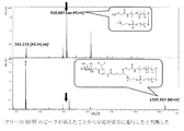
- Step of synthesizing compound 7 (i) AcCl (20 eq.), R. t. 48h, 68%, (ii) 5-Nitro salicylaldehyde (3 eq.), Ag 2 O (1.3 eq.), MeCN, 35 ° C., 53%, (iii) DAST (5 eq.), CH 2 Cl 2 , r. t. 24h, 48%, (iv) Pd / C (10 w%), H 2 gas, r. t. , 0.5 h, quant, (v) 5-oxohexanoic acid (5 eq.), DIC (5 eq.), THF, r. t. 7h, 88%, (vi) NaOMe (0.1 eq.), MeOH, r. t. , 0.5h, quant.
- Compound 7 was synthesized using N-Acetyl-D-glucosamine as a starting material.
- Compound 3 was obtained ⁇ -selectively by a glycosyl reaction of reactive sugar donor 2 and 5-nitrosalicyclicdehydride, and then converted to compound 4 by conversion of the aldehyde group of 3 to a difluoromethyl group by dietylaminosulfur trifluoride (DAST).
- DAST dietylaminosulfur trifluoride
- 5 obtained by reduction of the nitro group of compound 4 and 5-oxohexanoic acid are condensed in the presence of N, N′-diisopropylcarbodiimide (DIC) to derive compound 6, and the following target is obtained by deacetylation.
- DIC N′-diisopropylcarbodiimide
- Compound 7 was presented (immobilized) to fluorescent nanoparticles (quantum dots, Invitrogen Qdot (R) 655) according to the methods described in References 2 and 3.
- aminooxy linker (10 mM, 10 ⁇ l / MeOH), phosphorylcholine linker (100 mM, 4 ⁇ l / MeOH), NaBH 4 (1 ⁇ l, 12 wt% in 14N NaOH) and milli Q (50 ⁇ l), which have been activated by deprotection in advance, are added.
- YM50 After purification by ultrafiltration (YM50), it was subjected to reaction with compound 7.
- the cells were stained with the following method and then observed with a fluorescence microscope. After removing the medium, the cells were washed three times with Opti-MEM, LysoTracker® Green DND-26 (200 ⁇ l, 5 nM / Opti-MEM) was added, and the cells were incubated at 37 ° C. and 5% CO 2 for 30 minutes.
- Lysosomes were stained. After that, Hoechst (2 ⁇ l, 0.1 ng / ⁇ l / Opti-MEM) was added and incubated at 37 ° C. in a 5% CO 2 atmosphere for 15 minutes to stain the nucleus, and then washed with Opti-MEM three times for imaging. Performed (FIG. 2).
- Fluorescent nanoparticles presenting compound 7 were rapidly taken up by breast cancer cells and were present in lysosomes after 1 hour (FIGS. 2a-1h), but after 2 hours, nanoparticles that had already escaped from lysosomes were observed. (FIGS. 2a-2h). In 4 to 8 hours, most of the nanoparticles are dispersed in the cells (FIGS. 2a-4h and 8h). At 4 to 8 hours later, the decrease in the area stained with LysoTracker R Green DND-26 was remarkable. On the other hand, almost all of the fluorescent nanoparticles (QD control) not carrying the compound 7 used for comparison remained trapped in the lysosome after 8 hours (FIGS. 2a-8h, bottom). In addition, lysosomes are hardly detected in the floating dead cells scattered in FIG. 2b (2 and 8 hours after the start of culture).
- the cells were washed 3 times with medium components, 10 ⁇ l of cell culture solution was collected, Cell Counting Kit-8 solution (Dojindo, Kumamoto, Japan) was added, and 37 ° C., 5% CO 2 atmosphere. The cell viability was quantified by incubating for 5 hours and measuring the absorbance at 450 nm.
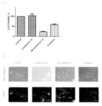
- FIG. 3 shows the results of the breast cancer cell killing effect of fluorescent nanoparticles (100 nM QD-anchored 7) presenting compound 7.
- A The survival rate of MCF7 cells after culturing for 96 hours in the presence of fluorescent nanoparticles presenting Compound 7 and various test drugs used for comparison was quantified by the MTA method.
- the cells were stained with the following method and then observed with a fluorescence microscope. After removing the culture medium, the cells were washed 3 times with Opti-MEM, then Hoechest (200 ⁇ l, 0.001 ng / ⁇ l / Opti-MEM) was added, and the cells were incubated at 37 ° C. in a 5% CO 2 atmosphere for 15 minutes. After staining, imaging was performed by washing with Opti-MEM three times (FIG. 3d).
- the nanoparticle presenting the NAG suicide substrate is released from the lysosome by destroying or permeating the lysosomal membrane by forming a covalent bond with NAG present in the lysosome.
- Step of synthesizing compound 15 (i) Ac 2 O (10 eq.), R. t. 48h, (ii) 30% HBr / AcOH (5.0 eq.), R. t. , 0.5 h, (iii) 5-Nitrosalicylidedeide (3 eq.), Ag 2 O (1.3 eq.), MeCN, r. t. , Three step yield 96%, (iv) DAST (3 eq.), CH 2 Cl 2 , r. t. , 18h, 72%, (v) Pd / C (10 w%), H 2 gas, r. t.
- Compound 15 which is a precursor of the suicide substrate site was synthesized with reference to Document 1 using Galactose as a starting material.
- Compound 11 was selectively obtained by a glycosylation reaction between reactive sugar donor 10 and 5-nitrosalicyclicdehydride, and then converted to compound 12 by conversion of the aldehyde group of 11 to a difluoromethyl group with dietylaminosulfur trifluoride (DAST).
- DAST dietylaminosulfur trifluoride
- 13 obtained by reduction of the nitro group of Compound 12 and 5-oxohexanoic acid are condensed in the presence of 1-Ethyl-3- (3-dimethylaminopropyl) carbohydrate Hydrochloride (EDC) to induce Compound 14 to be removed.
- EDC 1-Ethyl-3- (3-dimethylaminopropyl) carbohydrate Hydrochloride
- Fluorescent nanoparticles according to the method described compound 15 in references 2 and 3 presented to (quantum dots, Invitrogen Qdot (R) 655) .
- FIG. 5 shows that the fluorescent nanoparticle presenting the compound 15 is taken into the cell.
- the cells were washed 3 times with the medium components, and 10 ⁇ l of Cell Counting Kit-8 (Dojindo, Kumamoto, Japan) was added to the cell culture solution, and 1.5 ° C in a 5% CO 2 atmosphere at 37 ° C. Cell viability was quantified by incubating for an hour and measuring absorbance at 450 nm.
- Cell Counting Kit-8 (Dojindo, Kumamoto, Japan) was added to the cell culture solution, and 1.5 ° C in a 5% CO 2 atmosphere at 37 ° C. Cell viability was quantified by incubating for an hour and measuring absorbance at 450 nm.
- FIG. 6 shows the results of the breast cancer cell killing effect of the fluorescent nanoparticles (100 nM QD-anchored 15) presenting compound 15.
- A The survival rate of MCF-7 cells after 96 hours of culturing in the presence of fluorescent nanoparticles presenting compound 15 and various test drugs used for comparison was quantified by MTT assay.
- Fluorescent nanoparticles (100 nM QD-anchored 15) presenting compound 15 significantly inhibited the growth of breast cancer cell MCF-7, and after 100 hours of culture, the viability was about 20% and 100 mM cisplatin (100 nM QD-anchored 15 It exhibited a strong anticancer effect far exceeding the 1000-fold dose) (FIG. 6a).
- dead cells that changed shape and floated were observed only in an experimental system seeded with 100 nM QD-anchored 15 and 100 ⁇ M cisplatin (FIG. 6 b).
- Step of synthesizing compound 20 (i) 1) AcCl, r. t. 48h, 2) 5-Nitrosalicylaldehyde (1.5 eq.), DIEA (1.5 eq.), MeCN, r. t. , Tow step yield 77%, (ii) DAST (2.5 eq.), CH 2 Cl 2 , r. t. , 15h, 77%, (iii) 1) Zn (40 eq.), AcOH, r. t. , 0.5 h, 2) 5-oxohexanoic acid (1.5 eq.), EDC (1.5 eq.), HOBt (1.5 eq.), MeCN, r. t. 20h, 75%, (iv) 1) NaOMe (cat.), MeOH, r. t. , 8h,. 2) NaOH (2.0 eq.), H 2 O, 0 ° C., 1H
- Compound 20 was synthesized using Sialic acid as a starting material.
- Compound 16 is converted into a sugar reaction donor by halogenation of compound 16, and compound 17 is selectively obtained by glycosylation with 5-nitrosalicylidedehyde, and then conversion of 17 aldehyde groups to difluoromethyl groups by dietylamino sulfur trifluoride (DAST).
- DAST dietylamino sulfur trifluoride
- DAST dietylamino sulfur trifluoride
- EDC 1-Ethyl-3- (3-dimethylaminopropyl) carbohydrate Hydrochloride
- the target compound 20 shown below was efficiently prepared by demethylation reaction.
- Compound 20 was presented to fluorescent nanoparticles (quantum dots, Invitrogen Qdot R655) according to the methods described in References 1 and 2.
- aminooxy linker (10 mM, 10 ⁇ l / MeOH), phosphorylcholine linker (100 mM, 4 ⁇ l / MeOH), NaBH 4 (1 ⁇ l, 12 wt% in 14 N NaOH), and milli Q (50 ⁇ l), which had been activated by deprotection in advance.
- YM50 After purification by ultrafiltration (YM50), reaction with compound 20 was performed.
- FIG. 8 shows that the fluorescent nanoparticle presenting the compound 20 is taken into the cell.
- the cells were washed 3 times with the medium components, and 10 ⁇ l of Cell Counting Kit-8 (Dojindo, Kumamoto, Japan) was added to the cell culture solution, and 1.5 ° C in a 5% CO 2 atmosphere at 37 ° C. Cell viability was quantified by incubating for an hour and measuring absorbance at 450 nm.
- Cell Counting Kit-8 (Dojindo, Kumamoto, Japan) was added to the cell culture solution, and 1.5 ° C in a 5% CO 2 atmosphere at 37 ° C. Cell viability was quantified by incubating for an hour and measuring absorbance at 450 nm.
- FIG. 9 Breast cancer cell killing effect of fluorescent nanoparticle (100 nM QD-anchored 15) presenting compound 20: MCF-7 after 96 hours culture in the presence of fluorescent nanoparticle presenting compound 20 and various test agents used for comparison Cell viability was quantified by MTT assay. A significant difference in growth inhibition of breast cancer cell MCF-7 by the fluorescent nanoparticle (100 nM QD-anchored 20) presenting compound 20 could not be confirmed. Compound 20 exhibited suicide substrate activity in this system, albeit weakly.
- Example 4 Effect of nanoparticle carrying lysosomal enzyme inhibitor on hepatocellular carcinoma
- the phospholipid-coated fluorescent nanoparticles (red) have already partially escaped from the lysosome 1 hour after the start of co-culture with cancer cells. After 8 hours, most of them do not localize in lysosomes and appear to move freely in the cytoplasm. Phospholipid-coated nanoparticles that do not carry inhibitors remain localized in lysosomes without moving through the cytoplasm regardless of the type of cancer cell (RS Tan, et al., ACS Chem. Biol. 2015, 10, 2073-2086) has already been reported.
- Compounds 7, 15, and 20 are irreversible inhibitors of hexosaminidase, galactosidase, and sialidase, respectively, and are expected to induce conformational changes around the active center of the target enzyme during the inhibition reaction. .
- all of these glycolytic enzymes distributed in lysosomes are thought to contribute strongly to the stabilization of lysosomal membranes, and significant changes in the structure and physical properties of lysosomal membranes due to inhibition of these enzymes (membrane damage) As a result of this, the nanoparticle seems to have leaked into the cytoplasm.
- the fluorescent nanoparticles (100 nM) carrying compounds 7 and 15 significantly inhibited the growth of the liver cancer cell HepG2, and 15% and 96% of the living cells at 96 hours after co-culture, respectively, It was about 30%. Since no significant anti-tumor activity was observed for 5-FU (100 nM) used as a control drug in any of the experiments, fluorescent nanoparticles (100 nM) carrying compounds 7 and 15 were treated with 5-FU (100 nM). It can be judged that a powerful anticancer effect far surpassing that of) was exhibited (FIGS. 11A and 11B).
- the fluorescent nanoparticle (100 nM) carrying the compound 20 only exhibited a significantly weaker antitumor effect than the above two drugs under the same conditions. This is because 1) the expression level of sialidase in lysosome is considerably lower than that of galactosidase and hexosaminidase. 2) Since ⁇ -sialoside bond is more unstable under acidic conditions than other glycoside bonds, 3) Sialic acid (Neu5Ac) is synthesized by aldol reaction with ManNAc derived from GlcNAc as an intermediate in the hexosamine synthesis pathway as an intermediate.
- HepG2 cells (5 ⁇ 10 4 / 100ml / well) to 37 °C, QD-anchored 7 for each medium 90ml after incubation for 24 hours at 5% CO 2 atmosphere of or QD-anchored 15 (1-0.05mM , 10 ml / milliQ), QD control (1-0.05 mM, 10 ml / milliQ), 5-FU (1-0.01 mM, 10 ml / milliQ), respectively, to give final concentrations of 100, 50, 25, 10, and It adjusted so that it might become 5 nM.
- fluorescent nanoparticles (QD-anchored 7 and 15) carrying compounds 7 and 15 both inhibited the growth of liver cancer cell HepG2 in a concentration-dependent manner, and 96 hours after co-culture.
- IC 50 > 100 nM as a comparative control drug
- QD-anchored 7: IC 50 10.7 nM
- the IC 50 for each glucosidase in vitro of compounds 7, 15 and 20 is on the order of mM (references 1 to 3), so the results shown in FIG.
- the “specific (local) concentration effect of a molecular irreversible inhibitor into lysosomes” proves to be extremely effective.
- references References related to the synthesis method of compounds (glycolytic enzyme inhibitors by irreversible reaction mechanism) M.M. Ichikawa, et al. Bioorg. Med. Chem. Lett. 2001, 11, 1769-1773. 2. M.M. Kurogochi, et al. , J .; Biol. Chem. 2004, 279, 44704-44712. 3. H. Hinou, et al. Biochemistry 2005, 44, 11669-11675. References related to the preparation and properties of phospholipid monolayer-coated quantum dots (biological membrane mimicking fluorescent nanoparticles) 4. T. T. et al. Ohyanagi, et al. , J .; Am. Chem. Soc. 2011, 133, 12057-12517. 5). R. S. Tan, et al. , ACS Chem. Biol. 2015, 10, 2073-2086.
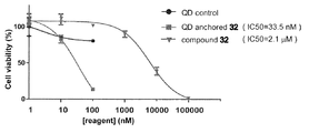
- Example 5 Synthesis of phospholipid monolayer-coated fluorescent nanoparticles loaded with a kinase inhibitor (a novel sorafenib derivative) and antitumor effect against hepatocellular carcinoma HepG2 A: Synthesis of a novel sorafenib derivative (32) Sorafenib, a therapeutic agent for hepatocellular carcinoma and renal cell carcinoma, is mostly composed of serum albumin and a1-acidic protein in the blood of cancer patients due to its hydrophobic structure. It is clear that the uptake efficiency into cancer tissues (cancer cells) is extremely low.
- VEGFR2 co-crystal of sorafenib and kinase
- Compound 32 is a novel sorafenib derivative that can be supported on phospholipid monolayer-coated nanoparticles.
- the sorafenib basic structure 30 of Compound 32 is chlorinated at the meta position of Picolinic acid 25 with reference to the previous report (Hamock, BD et al., Bioorganic & Med. Chem. Lett. 2013, 23, 3732-3737).
- Compound 26 was prepared by the condensation reaction with 4-aminophenol by the condensation reaction with Compound 29 in the presence of bis (trichloromethyl) carbonate 28 as Compound 27 by the condensation reaction with 4-aminophenol.
- a functional linker precursor 24 derived from a commercially available starting material 21 was synthesized for the purpose of modification in the picolinic acid region of sorafenib.
- sorafenib derivative 30 and compound 24 are condensed in the presence of 1H-Benzotriazole-1-yloxytris (pyrrolidyne-1-yl) phosphonium / hexafluorophosphate (PyBOP) to finally give an isopropylidene ketal group.
- the desired compound 32 was efficiently synthesized by deprotection.
- the cells were washed three times with Opti-MEM, Lyso Tracker® Green DND-26 (200 ml, 5 nM / Opti-MEM) was added, and the cells were incubated at 37 ° C. and 5% CO 2 for 30 minutes. The lysosomes were stained. Thereafter, Hoechest (2 ml, 0.1 ng / ml / Opti-MEM) was added, and the cells were further incubated for 15 minutes at 37 ° C. in an atmosphere of 5% CO 2 to stain the nucleus, and then washed three times with Opti-MEM for imaging. Performed (FIG. 14).
- the phospholipid-coated fluorescent nanoparticle (red) carrying the sorafenib derivative 32 remained in the lysosome even 24 hours after the start of co-culture with HepG2 cells. It was shown that. This result suggests that the kinase group targeted by sorafenib is not distributed in the lysosome but delocalized in the cancer cytoplasm, so the active substance (sorafenib derivative) is linked to the nanoparticle from the endolysosome. This suggests that the improvement of the effect of sorafenib as an anticancer agent cannot be expected if it remains immobilized.
- 2-D Antitumor effect of fluorescent nanoparticle carrying the sorafenib derivative 32 (hepatocellular carcinoma killing action)
- Cell viability assay of by dead cells Quantification: HepG2 cells (5 ⁇ 10 4 / 100ml / well) to 37 °C, 5% CO QD ( 1000 ⁇ 10nM 2 atmosphere against media respectively after incubation for 24 hours 90 ml, 10 ml / milli Q), QD-anchored 32 (1000 to 10 nM, 10 ml / milli Q), and sorafenib derivative 32 (10 ⁇ 10 7 to 10 nM, 10 ml / DMSO) added to a final concentration of 100 to 1 nM (however, the compound 32 was adjusted to be 1 ⁇ 10 6 to 1 nM) because it was estimated that the IC 50 value was at the mM level.
- the present invention is useful in the field related to anticancer agents.
Abstract
Priority Applications (3)
| Application Number | Priority Date | Filing Date | Title |
|---|---|---|---|
| JP2017563899A JP7018191B2 (ja) | 2016-01-29 | 2017-01-27 | 細胞内物質移送システムおよびその利用 |
| EP17744471.8A EP3409772B1 (fr) | 2016-01-29 | 2017-01-27 | Système de transport de substance intracellulaire et utilisation de celui-ci |
| US16/073,677 US11007276B2 (en) | 2016-01-29 | 2017-01-27 | Intracellular substance transport system and use thereof |
Applications Claiming Priority (2)
| Application Number | Priority Date | Filing Date | Title |
|---|---|---|---|
| JP2016-015760 | 2016-01-29 | ||
| JP2016015760 | 2016-01-29 |
Publications (1)
| Publication Number | Publication Date |
|---|---|
| WO2017131242A1 true WO2017131242A1 (fr) | 2017-08-03 |
Family
ID=59398225
Family Applications (1)
| Application Number | Title | Priority Date | Filing Date |
|---|---|---|---|
| PCT/JP2017/003880 WO2017131242A1 (fr) | 2016-01-29 | 2017-01-27 | Système de transport de substance intracellulaire et utilisation de celui-ci |
Country Status (4)
| Country | Link |
|---|---|
| US (1) | US11007276B2 (fr) |
| EP (1) | EP3409772B1 (fr) |
| JP (1) | JP7018191B2 (fr) |
| WO (1) | WO2017131242A1 (fr) |
Cited By (2)
| Publication number | Priority date | Publication date | Assignee | Title |
|---|---|---|---|---|
| WO2019208820A1 (fr) * | 2018-04-27 | 2019-10-31 | 国立大学法人北海道大学 | Système de transport de substances intracellulaires et utilisation associée |
| WO2021054420A1 (fr) * | 2019-09-20 | 2021-03-25 | 国立大学法人北海道大学 | Particules de présentation de chaînes glucidiques et procédé de production associé |
Families Citing this family (1)
| Publication number | Priority date | Publication date | Assignee | Title |
|---|---|---|---|---|
| CN113230415B (zh) * | 2021-05-18 | 2022-04-12 | 南华大学 | 岩藻糖与环糊精修饰多肽靶向动脉粥样硬化相关巨噬细胞纳米载体系统及其制备方法和应用 |
Citations (6)
| Publication number | Priority date | Publication date | Assignee | Title |
|---|---|---|---|---|
| JP2007254452A (ja) * | 2006-03-24 | 2007-10-04 | Kyushu Univ | 有機化合物 |
| JP2009534309A (ja) | 2006-03-31 | 2009-09-24 | マサチューセッツ インスティテュート オブ テクノロジー | 治療剤の標的化送達のためのシステム |
| JP2011528275A (ja) | 2008-07-17 | 2011-11-17 | ミセル テクノロジーズ,インク. | 薬物送達医療デバイス |
| WO2014197937A1 (fr) * | 2013-06-13 | 2014-12-18 | University Of South Australia | Procédés pour détecter le cancer de la prostate |
| JP2015520194A (ja) | 2012-06-07 | 2015-07-16 | プレジデント・アンド・フェロウズ・オブ・ハーバード・カレッジ | 薬物標的指向化のためのナノ療法 |
| JP2015520197A (ja) | 2012-06-20 | 2015-07-16 | フランク・グー | 粘膜付着性ナノ粒子送達系 |
Family Cites Families (4)
| Publication number | Priority date | Publication date | Assignee | Title |
|---|---|---|---|---|
| EP1140840B1 (fr) | 1999-01-13 | 2006-03-22 | Bayer Pharmaceuticals Corp. | Diphenylurees a substituants -g(v)-carboxyaryles, inhibitrices de kinase raf |
| EP2104420A2 (fr) | 2006-10-30 | 2009-09-30 | George Mason Intellectual Properties, Inc. | Procede de conservation et de fixation de tissus |
| DK2379114T3 (da) * | 2008-12-16 | 2014-09-29 | Univ Santiago Compostela | Konjugater omfattende nanopartikler overtrukket med platinholdige forbindelser |
| JP2014214139A (ja) | 2013-04-26 | 2014-11-17 | ポーラ化成工業株式会社 | Nadh産生促進剤 |
-
2017
- 2017-01-27 WO PCT/JP2017/003880 patent/WO2017131242A1/fr active Application Filing
- 2017-01-27 US US16/073,677 patent/US11007276B2/en active Active
- 2017-01-27 JP JP2017563899A patent/JP7018191B2/ja active Active
- 2017-01-27 EP EP17744471.8A patent/EP3409772B1/fr active Active
Patent Citations (6)
| Publication number | Priority date | Publication date | Assignee | Title |
|---|---|---|---|---|
| JP2007254452A (ja) * | 2006-03-24 | 2007-10-04 | Kyushu Univ | 有機化合物 |
| JP2009534309A (ja) | 2006-03-31 | 2009-09-24 | マサチューセッツ インスティテュート オブ テクノロジー | 治療剤の標的化送達のためのシステム |
| JP2011528275A (ja) | 2008-07-17 | 2011-11-17 | ミセル テクノロジーズ,インク. | 薬物送達医療デバイス |
| JP2015520194A (ja) | 2012-06-07 | 2015-07-16 | プレジデント・アンド・フェロウズ・オブ・ハーバード・カレッジ | 薬物標的指向化のためのナノ療法 |
| JP2015520197A (ja) | 2012-06-20 | 2015-07-16 | フランク・グー | 粘膜付着性ナノ粒子送達系 |
| WO2014197937A1 (fr) * | 2013-06-13 | 2014-12-18 | University Of South Australia | Procédés pour détecter le cancer de la prostate |
Non-Patent Citations (20)
| Title |
|---|
| A. T. OHYANAGI, J. AM. CHEM. SOC., vol. 133, 2011, pages 12507 - 12517 |
| B. R. S.TAN, ACS CHEM. BIOL., vol. 10, 2015, pages 2073 - 2086 |
| BIOCHEMISTRY, vol. 44, 2005, pages 11669 - 11675 |
| BIOORG. MED. CHEM. LETT., vol. 11, 2001, pages 1769 - 1773 |
| H. HINOU ET AL., BIOCHEMISTRY, vol. 44, 2005, pages 11669 - 11675 |
| HAMMOCK, B. D., BIOORGANIC & MED. CHEM. LETT., vol. 23, 2013, pages 3732 - 3737 |
| KANIA, R. S. ET AL., PROC. NATL. ACAD. SCI. USA, vol. 109, 2012, pages 18281 - 18289 |
| LIN, L. ET AL., CANCER RES., vol. 66, 2006, pages 11851 - 11858 |
| M. ICHIKAWA ET AL., BIOORG. MED. CHEM. LETT., vol. 11, 2001, pages 1769 - 1773 |
| M. KUROGOCHI ET AL., J. BIOL. CHEM., vol. 279, 2004, pages 44704 - 44712 |
| R. S. TAN ET AL., ACS CHEM. BIOL., vol. 10, 2015, pages 2073 - 2086 |
| R. S.TAN, ACS CHEM. BIOL., vol. 10, 2015, pages 2073 - 2086 |
| See also references of EP3409772A4 |
| SHIN-ICHIRO NISHIMURA ET AL., ANGEW. CHEM. INT. ED., vol. 51, 2012, pages 3386 - 3390 |
| SMITH, M. A.; HOUGTON, P., CLIN. CANCER RES., vol. 19, 2013, pages 2828 - 2833 |
| T. OHYANAGI ET AL., J. AM. CHEM. SOC., vol. 133, 2011, pages 12057 - 12517 |
| T. OHYANAGI ET AL., J. AM. CHEM. SOC., vol. 133, 2011, pages 12507 - 12517 |
| T. OHYANAGI, J. AM. CHEM. SOC., vol. 133, 2011, pages 12507 - 12517 |
| TAN, ROGER S.: "Rapid Endolysosomal Escape and Controlled Intracellular Trafficking of Cell Surface Mimetic Quantum-Dots-Anchored Peptides and Glycopeptides", ACS CHEM. BIOL., vol. 10, 24 June 2015 (2015-06-24), pages 2073 - 2086, XP055538410 * |
| WILHELM, S. M. ET AL., CANCER RES., vol. 64, 2004, pages 2099 - 7109 |
Cited By (2)
| Publication number | Priority date | Publication date | Assignee | Title |
|---|---|---|---|---|
| WO2019208820A1 (fr) * | 2018-04-27 | 2019-10-31 | 国立大学法人北海道大学 | Système de transport de substances intracellulaires et utilisation associée |
| WO2021054420A1 (fr) * | 2019-09-20 | 2021-03-25 | 国立大学法人北海道大学 | Particules de présentation de chaînes glucidiques et procédé de production associé |
Also Published As
| Publication number | Publication date |
|---|---|
| US11007276B2 (en) | 2021-05-18 |
| US20200138973A1 (en) | 2020-05-07 |
| JPWO2017131242A1 (ja) | 2018-11-22 |
| EP3409772A4 (fr) | 2019-10-09 |
| EP3409772B1 (fr) | 2023-06-28 |
| EP3409772A1 (fr) | 2018-12-05 |
| JP7018191B2 (ja) | 2022-02-10 |
Similar Documents
| Publication | Publication Date | Title |
|---|---|---|
| Sung et al. | Delivery of nitric oxide with a nanocarrier promotes tumour vessel normalization and potentiates anti-cancer therapies | |
| Malik et al. | AS1411-conjugated gold nanospheres and their potential for breast cancer therapy | |
| Lin et al. | Targeting the delivery of glycan-based paclitaxel prodrugs to cancer cells via glucose transporters | |
| CN103221070B (zh) | 用于狭窄病变和溶解血栓疗法的切变控制释放 | |
| Yu et al. | Dually enzyme-and acid-triggered self-immolative ketal glycoside nanoparticles for effective cancer prodrug monotherapy | |
| Zhang et al. | Multifunctional gold nanoparticle-based fluorescence resonance energy-transfer probe for target drug delivery and cell fluorescence imaging | |
| Lei et al. | Nature-inspired smart DNA nanodoctor for activatable in vivo cancer imaging and in situ drug release based on recognition-triggered assembly of split aptamer | |
| Chen et al. | Smart hypoxia-responsive transformable and charge-reversible nanoparticles for the deep penetration and tumor microenvironment modulation of pancreatic cancer | |
| Xiang et al. | Restoration and enhancement of immunogenic cell death of cisplatin by coadministration with digoxin and conjugation to HPMA copolymer | |
| WO2017131242A1 (fr) | Système de transport de substance intracellulaire et utilisation de celui-ci | |
| KR102053065B1 (ko) | 히알루론산 및 독소루비신을 이용한 pH 감응성 항암 엑소좀 조성물 | |
| CN103764604A (zh) | 茋类似物和治疗癌症的方法 | |
| Petrov et al. | New small-molecule glycoconjugates of docetaxel and GalNAc for targeted delivery to hepatocellular carcinoma | |
| Li et al. | Metal-phenolic networks with ferroptosis to deliver NIR-responsive CO for synergistic therapy | |
| Tang et al. | Oxaliplatin-based platinum (IV) prodrug bearing toll-like receptor 7 agonist for enhanced immunochemotherapy | |
| Cheng et al. | Carrier‐Free Nanoassembly of Curcumin–Erlotinib Conjugate for Cancer Targeted Therapy | |
| Chen et al. | Penetrating micelle for reversing immunosuppression and drug resistance in pancreatic cancer treatment | |
| Li et al. | Liver‑targeted delivery of liposome‑encapsulated curcumol using galactosylated‑stearate | |
| Jia et al. | Micromixer based preparation of functionalized liposomes and targeting drug delivery | |
| Xiao et al. | Synthesis of novel tetravalent galactosylated DTPA-DSPE and study on hepatocyte-targeting efficiency in vitro and in vivo | |
| Mehata et al. | Chitosan-g-estrone nanoparticles of palbociclib vanished hypoxic breast tumor after targeted delivery: development and ultrasound/photoacoustic imaging | |
| Zeng et al. | A mitochondria-targeting ROS-activated nanoprodrug for self-augmented antitumor oxidation therapy | |
| Huan et al. | In vivo anti-tumor activity of a new doxorubicin conjugate via α-linolenic acid | |
| Dou et al. | Orthogonally engineered albumin with attenuated macrophage phagocytosis for the targeted visualization and phototherapy of liver cancer | |
| Yao et al. | A Self‐Assembly Combined Nano‐Prodrug to Overcome Gemcitabine Chemo‐Resistance of Pancreatic Tumors |
Legal Events
| Date | Code | Title | Description |
|---|---|---|---|
| 121 | Ep: the epo has been informed by wipo that ep was designated in this application |
Ref document number: 17744471 Country of ref document: EP Kind code of ref document: A1 |
|
| WWE | Wipo information: entry into national phase |
Ref document number: 2017563899 Country of ref document: JP |
|
| NENP | Non-entry into the national phase |
Ref country code: DE |
|
| WWE | Wipo information: entry into national phase |
Ref document number: 2017744471 Country of ref document: EP |
|
| ENP | Entry into the national phase |
Ref document number: 2017744471 Country of ref document: EP Effective date: 20180829 |