WO2017130613A1 - 観察装置 - Google Patents
観察装置 Download PDFInfo
- Publication number
- WO2017130613A1 WO2017130613A1 PCT/JP2016/088266 JP2016088266W WO2017130613A1 WO 2017130613 A1 WO2017130613 A1 WO 2017130613A1 JP 2016088266 W JP2016088266 W JP 2016088266W WO 2017130613 A1 WO2017130613 A1 WO 2017130613A1
- Authority
- WO
- WIPO (PCT)
- Prior art keywords
- bacteria
- information
- fine particles
- image
- observation apparatus
- Prior art date
- Legal status (The legal status is an assumption and is not a legal conclusion. Google has not performed a legal analysis and makes no representation as to the accuracy of the status listed.)
- Ceased
Links
Images




Classifications
-
- G—PHYSICS
- G01—MEASURING; TESTING
- G01N—INVESTIGATING OR ANALYSING MATERIALS BY DETERMINING THEIR CHEMICAL OR PHYSICAL PROPERTIES
- G01N15/00—Investigating characteristics of particles; Investigating permeability, pore-volume or surface-area of porous materials
- G01N15/02—Investigating particle size or size distribution
- G01N15/0205—Investigating particle size or size distribution by optical means
- G01N15/0227—Investigating particle size or size distribution by optical means using imaging; using holography
-
- C—CHEMISTRY; METALLURGY
- C12—BIOCHEMISTRY; BEER; SPIRITS; WINE; VINEGAR; MICROBIOLOGY; ENZYMOLOGY; MUTATION OR GENETIC ENGINEERING
- C12M—APPARATUS FOR ENZYMOLOGY OR MICROBIOLOGY; APPARATUS FOR CULTURING MICROORGANISMS FOR PRODUCING BIOMASS, FOR GROWING CELLS OR FOR OBTAINING FERMENTATION OR METABOLIC PRODUCTS, i.e. BIOREACTORS OR FERMENTERS
- C12M1/00—Apparatus for enzymology or microbiology
- C12M1/34—Measuring or testing with condition measuring or sensing means, e.g. colony counters
-
- C—CHEMISTRY; METALLURGY
- C12—BIOCHEMISTRY; BEER; SPIRITS; WINE; VINEGAR; MICROBIOLOGY; ENZYMOLOGY; MUTATION OR GENETIC ENGINEERING
- C12M—APPARATUS FOR ENZYMOLOGY OR MICROBIOLOGY; APPARATUS FOR CULTURING MICROORGANISMS FOR PRODUCING BIOMASS, FOR GROWING CELLS OR FOR OBTAINING FERMENTATION OR METABOLIC PRODUCTS, i.e. BIOREACTORS OR FERMENTERS
- C12M1/00—Apparatus for enzymology or microbiology
- C12M1/36—Apparatus for enzymology or microbiology including condition or time responsive control, e.g. automatically controlled fermentors
- C12M1/38—Temperature-responsive control
-
- C—CHEMISTRY; METALLURGY
- C12—BIOCHEMISTRY; BEER; SPIRITS; WINE; VINEGAR; MICROBIOLOGY; ENZYMOLOGY; MUTATION OR GENETIC ENGINEERING
- C12Q—MEASURING OR TESTING PROCESSES INVOLVING ENZYMES, NUCLEIC ACIDS OR MICROORGANISMS; COMPOSITIONS OR TEST PAPERS THEREFOR; PROCESSES OF PREPARING SUCH COMPOSITIONS; CONDITION-RESPONSIVE CONTROL IN MICROBIOLOGICAL OR ENZYMOLOGICAL PROCESSES
- C12Q1/00—Measuring or testing processes involving enzymes, nucleic acids or microorganisms; Compositions therefor; Processes of preparing such compositions
- C12Q1/02—Measuring or testing processes involving enzymes, nucleic acids or microorganisms; Compositions therefor; Processes of preparing such compositions involving viable microorganisms
- C12Q1/025—Measuring or testing processes involving enzymes, nucleic acids or microorganisms; Compositions therefor; Processes of preparing such compositions involving viable microorganisms for testing or evaluating the effect of chemical or biological compounds, e.g. drugs, cosmetics
-
- G—PHYSICS
- G01—MEASURING; TESTING
- G01N—INVESTIGATING OR ANALYSING MATERIALS BY DETERMINING THEIR CHEMICAL OR PHYSICAL PROPERTIES
- G01N15/00—Investigating characteristics of particles; Investigating permeability, pore-volume or surface-area of porous materials
- G01N15/10—Investigating individual particles
- G01N15/14—Optical investigation techniques, e.g. flow cytometry
- G01N15/1429—Signal processing
- G01N15/1433—Signal processing using image recognition
-
- G—PHYSICS
- G02—OPTICS
- G02B—OPTICAL ELEMENTS, SYSTEMS OR APPARATUS
- G02B21/00—Microscopes
- G02B21/36—Microscopes arranged for photographic purposes or projection purposes or digital imaging or video purposes including associated control and data processing arrangements
-
- G—PHYSICS
- G02—OPTICS
- G02B—OPTICAL ELEMENTS, SYSTEMS OR APPARATUS
- G02B21/00—Microscopes
- G02B21/36—Microscopes arranged for photographic purposes or projection purposes or digital imaging or video purposes including associated control and data processing arrangements
- G02B21/365—Control or image processing arrangements for digital or video microscopes
-
- G—PHYSICS
- G06—COMPUTING OR CALCULATING; COUNTING
- G06T—IMAGE DATA PROCESSING OR GENERATION, IN GENERAL
- G06T7/00—Image analysis
- G06T7/0002—Inspection of images, e.g. flaw detection
- G06T7/0012—Biomedical image inspection
- G06T7/0014—Biomedical image inspection using an image reference approach
- G06T7/0016—Biomedical image inspection using an image reference approach involving temporal comparison
-
- G—PHYSICS
- G06—COMPUTING OR CALCULATING; COUNTING
- G06T—IMAGE DATA PROCESSING OR GENERATION, IN GENERAL
- G06T7/00—Image analysis
- G06T7/10—Segmentation; Edge detection
- G06T7/11—Region-based segmentation
-
- G—PHYSICS
- G06—COMPUTING OR CALCULATING; COUNTING
- G06T—IMAGE DATA PROCESSING OR GENERATION, IN GENERAL
- G06T7/00—Image analysis
- G06T7/10—Segmentation; Edge detection
- G06T7/194—Segmentation; Edge detection involving foreground-background segmentation
-
- G—PHYSICS
- G01—MEASURING; TESTING
- G01N—INVESTIGATING OR ANALYSING MATERIALS BY DETERMINING THEIR CHEMICAL OR PHYSICAL PROPERTIES
- G01N15/00—Investigating characteristics of particles; Investigating permeability, pore-volume or surface-area of porous materials
- G01N15/01—Investigating characteristics of particles; Investigating permeability, pore-volume or surface-area of porous materials specially adapted for biological cells, e.g. blood cells
-
- G—PHYSICS
- G01—MEASURING; TESTING
- G01N—INVESTIGATING OR ANALYSING MATERIALS BY DETERMINING THEIR CHEMICAL OR PHYSICAL PROPERTIES
- G01N15/00—Investigating characteristics of particles; Investigating permeability, pore-volume or surface-area of porous materials
- G01N2015/0003—Determining electric mobility, velocity profile, average speed or velocity of a plurality of particles
-
- G—PHYSICS
- G01—MEASURING; TESTING
- G01N—INVESTIGATING OR ANALYSING MATERIALS BY DETERMINING THEIR CHEMICAL OR PHYSICAL PROPERTIES
- G01N15/00—Investigating characteristics of particles; Investigating permeability, pore-volume or surface-area of porous materials
- G01N2015/0042—Investigating dispersion of solids
- G01N2015/0053—Investigating dispersion of solids in liquids, e.g. trouble
-
- G—PHYSICS
- G01—MEASURING; TESTING
- G01N—INVESTIGATING OR ANALYSING MATERIALS BY DETERMINING THEIR CHEMICAL OR PHYSICAL PROPERTIES
- G01N15/00—Investigating characteristics of particles; Investigating permeability, pore-volume or surface-area of porous materials
- G01N15/10—Investigating individual particles
- G01N15/14—Optical investigation techniques, e.g. flow cytometry
- G01N2015/1486—Counting the particles
-
- G—PHYSICS
- G02—OPTICS
- G02B—OPTICAL ELEMENTS, SYSTEMS OR APPARATUS
- G02B21/00—Microscopes
- G02B21/0004—Microscopes specially adapted for specific applications
- G02B21/0088—Inverse microscopes
-
- G—PHYSICS
- G06—COMPUTING OR CALCULATING; COUNTING
- G06T—IMAGE DATA PROCESSING OR GENERATION, IN GENERAL
- G06T2207/00—Indexing scheme for image analysis or image enhancement
- G06T2207/20—Special algorithmic details
- G06T2207/20021—Dividing image into blocks, subimages or windows
-
- G—PHYSICS
- G06—COMPUTING OR CALCULATING; COUNTING
- G06T—IMAGE DATA PROCESSING OR GENERATION, IN GENERAL
- G06T2207/00—Indexing scheme for image analysis or image enhancement
- G06T2207/30—Subject of image; Context of image processing
- G06T2207/30004—Biomedical image processing
- G06T2207/30024—Cell structures in vitro; Tissue sections in vitro
Definitions
- the present invention relates to an observation apparatus.
- Methods for measuring the state of cells and bacteria in a liquid mainly include a light scattering method for analyzing light scattering and an image imaging method for imaging with a microscope or the like.
- the light scattering method can easily measure the size and number of relatively high concentration particles.
- the light scattering method is a method for measuring the scattering intensity of light incident on a sample liquid in which cells and bacteria are suspended. The scattering intensity has a correlation with the volume and number of cells / bacteria, and is generally used as a method for examining drug activity.
- the liquid containing bacteria is irradiated with light, the incident light is scattered by the bacteria, and the attenuation of the transmitted light amount is measured. Thereby, the growth state of bacteria is measured. Further, in recent years, image imaging methods that measure not only the volume and number of cells but also the shape of cells by microscopic observation have been actively used. Examination of drug activity against cells is considered indispensable to elucidate the mechanism of canceration and the nature of hepatocytes, which are regarded as important as a tool for realizing regenerative medicine.
- a method for observing the state of cells by image imaging optical microscope observation, fluorescence microscope observation, and the like are known.
- the most common and simple method is an optical microscope.
- the cells are transparent and it may be difficult to recognize the shape of the cells.
- As a method for observing cells with an optical microscope there is a method of observing transparent cells by an observation method that captures a change in refractive index called a phase difference observation method.
- a fluorescence microscope can recognize cells with higher sensitivity than a normal optical microscope by staining cells with a fluorescent substance. Since the fluorescence microscope needs to stain cells with a fluorescent substance, there are cases where the cells to be observed cannot be observed as they are.
- molecules or proteins introduced as fluorescent substances may affect the activity of biomolecules. In examining drug activity, it is necessary to observe the intact state of the cells.
- sensitivity testing devices that test the effects of antibacterial agents on bacteria, that is, the growth of bacteria is suppressed by antibacterial agents in the test of drug activity against bacteria.
- the number of bacteria is counted using a light scattering method.
- an image imaging method as a method for counting the number of bacteria. Observe the growth process of one bacterium by microscopic observation.
- Some bacteria have chemotaxis (for example, Escherichia coli), and it is also possible to capture the change in chemotaxis caused by an antibacterial agent by an image imaging method.
- a moving image is generally acquired. For example, an apparatus for detecting an effect on drug motility with respect to pulsating cardiomyocytes has been proposed (see Patent Document 1).
- Cells and bacteria may move out of the focal plane and may be present in front of or behind the objective lens. It is also possible to evaluate the motility of cells / bacteria by determining the presence in front or behind.
- the present invention provides a technique for recognizing the presence of cells / bacteria out of the focal plane even when the shape cannot be recognized due to defocusing, and for quantitative evaluation.
- an observation apparatus of the present application includes an optical system used for measuring fine particles present in a sample liquid in a sample container, A driving mechanism for driving at least one of the sample container and a part of the optical system in order to three-dimensionally search the bottom surface of the sample container; A control unit for controlling the optical system or the drive mechanism; An image processing unit that divides the image of the fine particles in the sample container at the first time and the second time into a focused region and an unfocused region, and acquires information about the fine particles; A display unit that displays information about the fine particles as information representing a temporal change between the first time and the second time; It has.
- the present invention in an observation image of cells / bacteria, from an image obtained by a single imaging, even an image that is not in focus is analyzed as information, and the positional relationship of the cells / bacteria in the optical axis direction is detected. It becomes possible to evaluate the motility of cells and bacteria.
- FIG. 3A It is the schematic diagram which showed the whole system of the observation apparatus. It is a figure explaining the various process parts of a computer. It is the flowchart which showed the test
- FIG. 6A It is the image after the external shape extraction of FIG. 6A according to the second embodiment. It is the image after the external shape extraction by the process different from FIG. 6B of FIG. 6A concerning 2nd Example. It is the image after the external shape extraction by the process different from FIG. 6B and FIG. 6C of FIG. 6A concerning 2nd Example. It is an example of the screen displayed on the display of the computer which concerns on 2nd Example. It is another example of the screen displayed on the display of the computer which concerns on 2nd Example.
- the present application includes a plurality of means for solving the above-described problems.
- an optical system such as an objective lens for measuring cells / bacteria existing on the bottom surface of the sample container and a digital image thereof are included. It consists of an image sensor that converts it into a signal, and a personal computer that receives and analyzes the digital signal of the image.
- the image processing device is configured to extract the boundary line of the image focused on the cells and bacteria present on the bottom of the sample container.
- An observation apparatus including a display unit for displaying information analyzed on a detection area and a background area on a monitor or the like is provided.
- an XYZ rectangular coordinate system is set.
- a predetermined direction in the horizontal plane is defined as the X direction
- a direction orthogonal to the X direction in the horizontal plane is defined as the Y direction
- a direction orthogonal to each of the X direction and the Y direction is defined as the Z direction.
- FIG. 1 is a schematic view of a bacteria observation apparatus of this example.
- the bacteria observation apparatus includes an illumination 101, a sample container 102, a pedestal 103, an XY stage 104, an objective lens 105, an objective lens actuator 106, an image sensor 107, and a computer 108 as main components.
- the illumination 101 is preferably Koehler illumination that is optically designed to illuminate the bottom surface of the sample container 102 uniformly.
- the sample container 102 has a storage unit that can hold at least one sample liquid.
- the pedestal 103 can hold the sample container 102 and has a structure in which light enters from the top surface and light exits from the bottom surface.
- the XY stage 104 can move the base 103 on which the sample container 102 is placed in the X direction and the Y direction.
- the XY stage 104 includes a heater (not shown) that controls the temperature of the sample container 102.
- the sample container 102 may be surrounded by a transparent glass heater.
- the entire optical system may be surrounded by a heat insulating material, and the temperature inside may be adjusted with a heater.
- the objective lens actuator 106 is an actuator that moves the objective lens 105 in the Z direction, and can scan the focal position of the objective lens 105 in the depth direction of the sample container 102.
- the image sensor 107 is installed at a position where an image at the focal position of the objective lens 105 is formed.
- An imaging lens may be installed between the image sensor 107 and the objective lens 105.
- the image sensor 107 has a structure in which an image formed on the image sensor is converted into a digital signal and transferred to the computer 108.
- the computer 108 includes at least a processor such as a CPU (Central Processing Unit), a storage unit such as a memory, and a storage device such as a hard disk. Further, the computer 108 includes an input device (such as a mouse and a keyboard) that receives input from the user, and a display device (such as a display) that displays measurement results.
- a processor such as a CPU (Central Processing Unit)
- a storage unit such as a memory
- a storage device such as a hard disk.
- the computer 108 includes an input device (such as a mouse and a keyboard) that receives input from the user, and a display device (such as a display) that displays measurement results.
- a display device such as a display
- the bottom surface of the sample container 102 is desirably thin and smooth.
- the sample container 102 may be a microtiter plate having a large number of sample holders.
- the sample container 102 is fixed to the base 103.
- Light having a uniform intensity is irradiated from the illumination 101 to the bottom surface of the sample container 102.
- an imaging system optical system such as a bright field, a dark field, and a phase difference is desirable.
- the bacteria image existing near the bottom surface of the illuminated sample container 102 is collected by the objective lens 105.
- the bacteria image collected by the objective lens 105 is formed on the image sensor 107.
- the objective lens actuator 106 is moved to adjust the Z height position of the objective lens 105.
- an autofocus function that takes a process of monitoring the contrast of an image captured by the image sensor 107 may be used.
- the image captured by the image sensor 107 is transmitted to the computer 108 as digital data.
- the transmitted image is stored in a storage device of the computer 108.
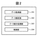
- FIG. 2 is a diagram for explaining various processing units of the computer 108.
- the computer 108 controls an optical system such as the illumination 101, a driving mechanism (the objective lens actuator 106 and the XY stage 104), and a heater, and acquires information about bacteria at a plurality of different times.
- the computer 108 obtains information representing temporal changes in information about bacteria from the acquired information.
- the computer 108 includes a data acquisition unit 201, a data analysis unit 202, a data display processing unit 203, and a control unit 204.
- the data acquisition unit 201 is a module that acquires an image formed on the image sensor 107.
- the data acquisition unit 201 reproduces a two-dimensional image of digital data transmitted from the image sensor 107.
- the data analysis unit 202 is a module that uses the image generated by the data acquisition unit 201 as input information and outputs the analysis result of the measurement data. For example, the data analysis unit 202 analyzes image information for a plurality of different times, and at least one quantitative value of the number of bacteria, the shape of the bacteria, the size of the bacteria, the amount of suspended bacteria, and the proportion of bacteria in the focal plane May be calculated. As an example, the data analysis unit 202 may calculate the number of bacteria by counting the number of portions of the image information that have a predetermined luminance value or more. The shape of the bacteria, the size of the bacteria, and the proportion of the bacteria in the focal plane may also be obtained from the size of the portion of the image information that is equal to or greater than a predetermined luminance value.
- the data analysis unit 202 may obtain information on the motility of the bacteria from the information on the floating bacteria. Further, the data analysis unit 202 may obtain information on temporal changes in bacterial motility from information representing the relationship between airborne bacteria information and time. For example, when the number of bacteria at a position away from the bottom surface of the sample container decreases, it can be determined that the drug has affected the chemotactic bacteria, and the response of the drug to the bacteria can be detected.
- the data analysis unit 202 may include statistical software that can inspect changes in the state of bacteria from temporal changes in information about bacteria.
- the data display processing unit 203 is a module that displays the analysis result from the data analysis unit 202 on the display of the computer 108.
- the data display processing unit 203 displays information on bacteria on a plurality of focal planes on a display in a format in which temporal changes can be compared.
- the data display processing unit 203 may display the analysis result of the temporal change in the information about the bacteria on the display. For example, the data display processing unit 203 may display a determination result regarding the motility of bacteria and information regarding the response of the drug to the bacteria on the display.
- the control unit 204 is a module that controls each component of the bacteria observation apparatus.
- the control unit 204 can manage each component of the bacteria observation apparatus and manage the start of measurement and the end of measurement.
- the control unit 204 can control the driving of the objective lens actuator 106 and the driving of the XY stage 104 when acquiring measurement data.
- processing unit described above may be realized as a function of a program executed on the computer 108.
- the processing unit may be realized by storing a program code corresponding to each process in a memory and executing a program code by a processor.
- a part of the processing unit may be configured by hardware such as a dedicated circuit board.
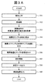
- FIG. 3A is a flowchart showing a method for testing a bacterial specimen.
- a specimen which is a bacterium separated and cultured from blood or the like is obtained (301). Since the amount of bacteria is usually small, pre-culture is performed (302).
- a mixed solution (sample solution) of a drug for examining the response to bacteria and a culture solution is prepared (303).
- sample solution mixed with drug 304.
- a control which is a sample solution containing only the culture solution containing no drug is also prepared.
- Dispense the sample solution containing bacteria into the sample container (305).
- the sample container is set in the observation apparatus of this embodiment (306).
- Measurement with an observation apparatus is started (307). Thereafter, measurement data is acquired while performing temperature control and measurement (308). The acquired measurement data is analyzed by the computer 108. After the analysis process, the test result of how the drug has affected the bacteria is output on the computer 108 (309).
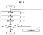
- FIG. 3B is a flowchart showing the contents of step 308 in FIG. 3A.
- the data acquisition unit 201 acquires measurement data measured by the image sensor 107 (311).
- the control unit 204 may drive the objective lens actuator 106 to perform autofocus having a function of aligning the focal position of the objective lens 105 with the bottom surface of the sample container 102.
- the data analysis unit 202 analyzes the two-dimensional plane data acquired by the data acquisition unit 201 (312). As an example, the data analysis unit 202 may convert two-dimensional plane data into image information. As another example, the data analysis unit 202 analyzes the image information and obtains at least one quantitative value of the number of bacteria, the shape of the bacteria, the size of the bacteria, the amount of airborne bacteria, and the proportion of bacteria in the focal plane. It may be calculated. Further, the data analysis unit 202 may measure the motility of bacteria from the calculated quantitative value. Further, the data analysis unit 202 may inspect the change in the state of the bacteria from the temporal change in the information about the bacteria.
- the data analysis unit 202 outputs the analysis result (313).
- the data display processing unit 203 may display the analysis result from the data analysis unit 202 on the display of the computer 108.
- the data analysis unit 202 analyzes the two-dimensional plane data or the image for a plurality of different times after the drug is put into the sample solution.
- the data analysis unit 202 can obtain information on the response of the drug to the bacteria from information representing temporal changes in information on the bacteria. For example, the data analysis unit 202 outputs the analysis result of the measurement data after 1 hour (first time) after putting the medicine and the analysis result of the measurement data after 5 hours (second time).
- the data display processing unit 203 displays the analysis result of the measurement data at the first time and the analysis result of the measurement data at the second time on the display of the computer 108. can do.
- the data acquisition unit 201 acquires two-dimensional plane data at a plurality of focal planes before and after the drug is added to the sample solution, and the data analysis unit 202 acquires information on the bacteria before and after the drug is added. You may obtain
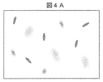
- FIG. 4A is an example of the acquired image.
- the bacteria image is not defocused, and the bacteria are positioned at the bottom surface of the sample container 102. If it is far away from it, the bacteria image will be defocused.
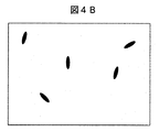
- FIG. 4B is a processed image divided into a bacterial region and a background region by performing the outer shape extraction of FIG. 4A and binarizing the image. The contour extraction is performed based on the contrast of the image. For example, a Sobel filter is generally used for contour extraction.
- FIG. 4C is the image of FIG. 4A fractionated as the background region in FIG. 4B.
- the area fractionated as the background area includes information on bacteria away from the bottom surface position of the sample container 102.
- a defocused bacterial image is included in the background area, the luminance variation in the background area increases and the standard deviation (S.D.) increases.
- the defocused bacterial image is not included in the background area, only the signal noise is present, the luminance variation in the background area is reduced, and the luminance standard deviation (S.D.) is reduced.
- the standard deviation of the luminance of the background area is correlated with the amount of floating bacteria away from the bottom surface of the sample container 102.
- FIG. 5 is an example of a screen displayed on the computer display by the data display processing unit 203.
- an experimental condition display unit 501 for displaying experimental conditions indicating the state of a solution containing bacteria
- a measurement condition display unit 502 for indicating an incident light quantity and an image measurement interval, which are conditions for acquiring an image
- a first A first area 503 displaying a bacterial image of time (for example, 1 hour after the suspension of bacteria) and a second area displaying a bacterial image of the second time (for example, 5 hours after the suspension of bacteria) 504.
- a bacteria number display unit 505 indicating the number of bacteria on the bottom surface of the sample container 102 in the image and a standard deviation of the luminance of the background region indicating the amount of floating bacteria are included. It has a floating bacteria amount display section 506 on which (SD) is displayed.
- an amount correlated with the amount of bacteria floating in the culture solution is displayed.
- information on the number of bacteria at different times is displayed side by side in a format that allows comparison of changes in the number of bacteria over time. Therefore, the response of the drug to the bacteria over time can be observed.
- information on bacteria at two different times (the number of bacteria and the amount of suspended bacteria) is displayed, but information on bacteria at three or more different times is displayed on the screen in a format that can be compared. May be.
- the bacterial observation apparatus of the above embodiment focuses on the bottom surface of the sample container 102 and the optical system that measures fine particles (bacteria, cells, etc.) present on the focal plane of the objective lens 105 in the sample liquid in the sample container 102.
- a drive mechanism objective lens actuator 105 and XY stage 1004.
- the measurement result (observation image) obtained by the image sensor 107 is converted into digital data and transferred to the computer 108.
- the computer 108 converts the digital data into image information and displays the image information on a display.
- the computer 108 displays the measurement results at a plurality of different times on the display.
- the computer 108 has a process of dividing the image into two parts, that is, a bacterial region and a background region by performing image processing on the image information. From the area of bacteria, quantitative values such as the number, size, and shape of bacteria are calculated. From the background area, the amount of airborne bacteria is calculated. The computer 108 may measure the proliferation power and motility of the bacteria from the quantitative value, and inspect the state change of the microparticles from the time change of the motility.
- the shape of the fine particles (cells or bacteria) near the bottom surface of the sample container can be accurately measured, and the spatial distribution information that the defocused image is floating in the sample liquid. Can be measured. It is possible to obtain information on the mobility of fine particles from a single still image without acquiring a plurality of images and shooting a moving image. Moreover, after making a chemical
- medical agent for example, antibacterial agent
- the degree of bacterial chemotaxis can be examined by measuring the extent of bacterial distribution in the liquid.
- the data analysis unit 202 obtains information related to the response of the drug to bacteria from the information before and after the drug is put.
- FIG. 6A is an example of the acquired image.
- the focal position of the objective lens 105 is aligned with the bottom surface of the sample container 102 and the bacteria are close to the bottom surface of the sample container 102, the bacteria image is not defocused, and the bacteria are positioned at the bottom surface of the sample container 102. If it is far away from it, the bacteria image will be defocused.
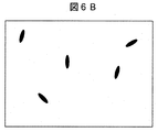
- FIG. 6B is a processed image that is divided into a bacterial region and a background region by extracting the outer shape of FIG. 6A and binarizing the image.
- the contour extraction is performed based on the contrast of the image.
- a Sobel filter is generally used for contour extraction.
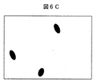
- FIG. 6C is a post-processing image obtained by extracting the outer shape of the image of FIG. 6A divided as the background area of FIG. 6B with a binarization parameter different from the image processing performed in FIG. 6B.
- a sharpening process may be performed in addition to changing the binarization parameter.
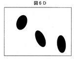
- 6D is a post-processing image obtained by performing outline extraction of the image of FIG. 6A that is fractionated as the background region of FIG. 6B and the background region of FIG.
- a sharpening process may be performed in addition to changing the binarization parameter.
- the binarization parameters were changed in three stages, and the outline of bacteria was extracted for each defocus amount. Since the defocus amount is correlated with the distance from the bottom surface of the sample container 102, the amount of bacteria at the distance from the bottom surface of the sample container 102 can be calculated. When calculating the amount of bacteria with the distance from the bottom surface with higher resolution, it is possible to change the binarization parameter in three steps performed in this embodiment by changing the binarization parameter in more steps. . In addition, since the defocused image does not reflect an accurate shape, it is desirable to correct the outer shape of the bacteria for each defocus amount.
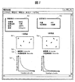
- FIG. 7 is an example of a screen displayed on the computer display by the data display processing unit 203.
- an experimental condition display unit 701 for displaying an experimental condition indicating the state of a solution containing bacteria
- a measurement condition display unit 702 for indicating an incident light amount and an image measurement interval, which are conditions for acquiring an image
- a first A first area 703 that displays a bacterial image of time (1 hour after the suspension of bacteria)
- a second area 704 that displays a bacterial image of the second time (5 hours after the suspension of bacteria)
- a bacteria number display unit 705 indicating the number of bacteria on the bottom surface of the sample container 102 in the image
- a floating bacteria amount display unit 706 displaying the number of floating bacteria.
- a bacterial number distribution diagram 707 that displays the bacterial number distribution at a distance from the bottom surface of the sample container 102 of floating bacteria.
- an amount correlated with the amount of bacteria floating in the culture solution is displayed.
- information on the number of bacteria at different times is displayed side by side in a format that allows comparison of changes in the number of bacteria over time. Therefore, the response of the drug to the bacteria over time can be observed.
- FIG. 8 shows information representing the relationship between the spatial distribution information of bacteria and time.
- FIG. 8 is a graph plotting a temporal change in the distribution of the number of bacteria with respect to the height from the bottom surface of the sample container.
- the vertical axis is the number of bacteria
- the horizontal axis is the height from the bottom (position in the Z direction)
- the depth axis is the incubation time (for example, the elapsed time since the sample solution was prepared, etc. ).
- the data analysis unit 202 may output information as shown in FIG. If the bottom surface of the sample container is 0 ⁇ m, the number of bacteria at the position of 0 ⁇ m increases with time.
- the number of bacteria at 200 ⁇ m from the bottom of the sample container decreases with time.
- the bacteria tend to grow or be constant at each height from the bottom surface, here 200 ⁇ m.
- the decrease in the number of bacteria at a location 200 ⁇ m from the bottom of the sample container means that the bacteria in the sample liquid that were originally moving lose their chemotaxis under the influence of the drug (for example, an antibacterial agent), and the activity of the bacteria itself decreases. It shows that.
- the effect of the antibacterial agent can be examined by analyzing the time-dependent change of the distribution of the number of bacteria in the sample solution in the height direction depending on the culture time. Therefore, the data analysis unit 202 may output the determination result of the response of the drug to the bacteria from the information representing the relationship between the spatial distribution information of the bacteria and the time.
- the bacterial observation apparatus of the above embodiment focuses on the bottom surface of the sample container 102 and the optical system that measures fine particles (bacteria, cells, etc.) present on the focal plane of the objective lens 105 in the sample liquid in the sample container 102.
- a drive mechanism objective lens actuator 105 and XY stage 1004.
- the measurement result (observation image) obtained by the image sensor 107 is converted into digital data and transferred to the computer 108.
- the computer 108 converts the digital data into image information and displays the image information on a display.
- the computer 108 displays the measurement results at a plurality of different times on the display.
- the computer 108 has a process of dividing the image into two parts, that is, a bacterial region and a background region by performing image processing on the image information. From the area of bacteria, quantitative values such as the number, size, and shape of bacteria are calculated. From the background area, the amount of airborne bacteria is calculated. The computer 108 may measure the proliferation power and motility of the bacteria from the quantitative value, and inspect the state change of the microparticles from the time change of the motility. Alternatively, the change in specific gravity or surface state of the fine particles may be evaluated from the natural sedimentation of the fine particles.
- the shape of the fine particles (cells or bacteria) near the bottom surface of the sample container can be accurately measured, and the spatial distribution information that the defocused image is floating in the sample liquid. Can be measured. It is possible to obtain information on the mobility of fine particles from a single still image without acquiring a plurality of images and shooting a moving image. Moreover, after making a chemical
- medical agent for example, antibacterial agent
- the degree of bacterial chemotaxis can be examined by measuring the extent of bacterial distribution in the liquid.
Landscapes
- Engineering & Computer Science (AREA)
- Chemical & Material Sciences (AREA)
- Health & Medical Sciences (AREA)
- Physics & Mathematics (AREA)
- Life Sciences & Earth Sciences (AREA)
- General Physics & Mathematics (AREA)
- Analytical Chemistry (AREA)
- Bioinformatics & Cheminformatics (AREA)
- General Health & Medical Sciences (AREA)
- Organic Chemistry (AREA)
- Biochemistry (AREA)
- Zoology (AREA)
- Wood Science & Technology (AREA)
- Computer Vision & Pattern Recognition (AREA)
- Biotechnology (AREA)
- Multimedia (AREA)
- Immunology (AREA)
- Dispersion Chemistry (AREA)
- Pathology (AREA)
- Theoretical Computer Science (AREA)
- Microbiology (AREA)
- Medicinal Chemistry (AREA)
- Genetics & Genomics (AREA)
- General Engineering & Computer Science (AREA)
- Optics & Photonics (AREA)
- Proteomics, Peptides & Aminoacids (AREA)
- Sustainable Development (AREA)
- Biomedical Technology (AREA)
- Signal Processing (AREA)
- Quality & Reliability (AREA)
- Medical Informatics (AREA)
- Nuclear Medicine, Radiotherapy & Molecular Imaging (AREA)
- Molecular Biology (AREA)
- Radiology & Medical Imaging (AREA)
- Biophysics (AREA)
- Toxicology (AREA)
- Investigating Or Analysing Materials By Optical Means (AREA)
- Apparatus Associated With Microorganisms And Enzymes (AREA)
- Measuring Or Testing Involving Enzymes Or Micro-Organisms (AREA)
- Image Analysis (AREA)
Abstract
Description
前記サンプル容器の底面を3次元的に探索するために前記サンプル容器及び前記光学系の一部の少なくともいずれか一方を駆動させる駆動機構と、
前記光学系または前記駆動機構を制御する制御部と、
第1の時間及び第2の時間における前記サンプル容器内の微粒子の画像を焦点が合った領域と焦点が合っていない領域に分け、前記微粒子に関する情報を取得する画像処理部と、
前記微粒子に関する情報を、前記第1の時間及び第2の時間の間の時間的な変化を表す情報として表示する表示部と、
を備えている。
各実施例について、以下の項目について説明する。
1.観察装置の測定方法の概略
2.検査方法と結果出力
以下の説明においては、XYZ直交座標系を設定する。水平面内の所定方向をX方向、水平面内においてX方向と直交する方向をY方向、X方向及びY方向のそれぞれに直交する方向(すなわち鉛直方向)をZ方向とする。
図1に本実施例の細菌観察装置の概略図を示す。細菌観察装置は、主要な構成要素として、照明101と、サンプル容器102と、台座103と、XYステージ104と、対物レンズ105と、対物レンズアクチュエータ106と、撮像素子107と、コンピュータ108を備える。
図3Aは、細菌検体の検査方法を示したフローチャートである。まず、血液などから分離培養された細菌である検体を入手する(301)。通常細菌量が少ないため、前培養を行う(302)。次に、細菌への応答を検査する薬剤と培養液との混合液(サンプル溶液)を準備する(303)。細菌を薬剤が混ざったサンプル溶液に懸濁する(304)。このとき、細菌への薬剤応答は最短でも数時間を要するため、薬剤が入っていない培養液のみのサンプル溶液であるコントロールも用意しておく。細菌入りのサンプル溶液をサンプル容器に分注する(305)。次に、サンプル容器を本実施例の観察装置にセットする(306)。観察装置での測定を開始する(307)。その後、温度コントロール及び計測を行いながら、測定データを取得する(308)。取得した測定データは、コンピュータ108によって解析処理が行われる。解析処理後に薬剤が細菌にどのような影響を与えたかの検査結果をコンピュータ108上に出力する(309)。
第1実施例と同様である。
図3A及び図3Bを用いて上述した通り、データ解析部202が、薬剤を入れる前及び後についての情報から、薬剤の細菌への応答に関する情報を求める。
102 …サンプル容器
103 …台座
104 …XYステージ
105 …対物レンズ
106 …対物レンズアクチュエータ
107 …撮像素子
108 …コンピュータ
201 …データ取得部
202 …データ解析部
203 …データ表示処理部
204 …制御部
501 …実験条件表示部
502 …測定条件表示部
503 …画像表示部1
504 …画像表示部2
505 …細菌数表示部
506 …風有細菌量表示部
701 …実験条件表示部
702 …測定条件表示部
703 …画像表示部1
704 …画像表示部2
705 …細菌数表示部
706 …風有細菌量表示部
707 …細菌数分布表示部
Claims (11)
- サンプル容器内のサンプル液中に存在する微粒子の測定に用いる光学系と、
前記サンプル容器の底面を3次元的に探索するために前記サンプル容器及び前記光学系の一部の少なくともいずれか一方を駆動させる駆動機構と、
前記光学系または前記駆動機構を制御する制御部と、
第1の時間及び第2の時間における前記サンプル容器内の微粒子の画像を焦点が合った領域と焦点が合っていない領域に分け、前記微粒子に関する情報を取得する画像処理部と、
前記微粒子に関する情報を、前記第1の時間及び第2の時間の間の時間的な変化を表す情報として表示する表示部と、
を備えることを特徴とする観察装置。 - 請求項1に記載の観察装置において、
前記画像処理部は、コントラストにより外形抽出を行うことで焦点が合った領域と焦点が合っていない領域に分けることを特徴とする観察装置。 - 請求項1に記載の観察装置において、
前記時間的な変化を表す情報は、前記サンプル液中に浮遊する微粒子量の情報と、時間との関係を表す情報であることを特徴とする観察装置。 - 請求項3に記載の観察装置において、
前記サンプル液中に浮遊する微粒子量の情報は、焦点があっていない領域の輝度のばらつきを指標とし、算出することを特徴とする観察装置。 - 請求項1に記載の観察装置において、
前記焦点が合っていない領域は、さらにデフォーカス量によって、複数の領域に分画し、
分画された領域毎に画像処理を行い、分画された領域毎に前記微粒子の量を算出し、前記微粒子のサンプル液中の空間分布を求めることを特徴とする観察装置。 - 請求項5に記載の観察装置において、
デフォーカス量によって分画された領域毎にデフォーカス補正を行い、前期微粒子のサンプル液中の空間分布を求めることを特徴とする観察装置。 - 請求項5に記載の観察装置において、
前記表示装置は、前記第1の時間での前記微粒子の空間的分布の情報と、前記第2の時間での前記微粒子の空間的分布の情報とを表示することを特徴とする観察装置。 - 請求項1に記載の観察装置において、
前記微粒子に関する情報は、前記微粒子の数、前記微粒子の大きさ、前記微粒子の形状、及び、前記焦点面における前記微粒子が占める割合の少なくとも1つであることを特徴とする観察装置。 - 請求項1に記載の観察装置において、
前記微粒子は細菌であり、サンプル液を温調する機構を備え、前記時間的な変化を表す情報は、前記細菌のサンプル液中に浮遊する量の情報と、時間との関係を表す情報であることを特徴とする観察装置。 - 請求項9に記載の観察装置において、
前記制御装置は、前記時間的な変化を表す情報から、前記細菌の運動性に関する情報を求めることを特徴とする観察装置。 - 請求項9に記載の観察装置において、
前記制御装置は、前記サンプル液に薬剤を入れる前後についての前記時間的な変化を表す情報から、前記薬剤の前記細菌への応答に関する情報を求めることを特徴とする観察装置。
Priority Applications (3)
| Application Number | Priority Date | Filing Date | Title |
|---|---|---|---|
| JP2017563744A JP6571210B2 (ja) | 2016-01-27 | 2016-12-22 | 観察装置 |
| US16/073,009 US10983039B2 (en) | 2016-01-27 | 2016-12-22 | Observation device |
| DE112016006056.4T DE112016006056T5 (de) | 2016-01-27 | 2016-12-22 | Betrachtungsvorrichtung |
Applications Claiming Priority (2)
| Application Number | Priority Date | Filing Date | Title |
|---|---|---|---|
| JP2016012930 | 2016-01-27 | ||
| JP2016-012930 | 2016-01-27 |
Publications (1)
| Publication Number | Publication Date |
|---|---|
| WO2017130613A1 true WO2017130613A1 (ja) | 2017-08-03 |
Family
ID=59397924
Family Applications (1)
| Application Number | Title | Priority Date | Filing Date |
|---|---|---|---|
| PCT/JP2016/088266 Ceased WO2017130613A1 (ja) | 2016-01-27 | 2016-12-22 | 観察装置 |
Country Status (4)
| Country | Link |
|---|---|
| US (1) | US10983039B2 (ja) |
| JP (1) | JP6571210B2 (ja) |
| DE (1) | DE112016006056T5 (ja) |
| WO (1) | WO2017130613A1 (ja) |
Cited By (1)
| Publication number | Priority date | Publication date | Assignee | Title |
|---|---|---|---|---|
| JP2019198293A (ja) * | 2018-05-18 | 2019-11-21 | 株式会社ニコン | 細菌の薬剤感受性の判定方法及び細菌の薬剤感受性の判定装置 |
Families Citing this family (2)
| Publication number | Priority date | Publication date | Assignee | Title |
|---|---|---|---|---|
| JP2022012314A (ja) * | 2020-07-01 | 2022-01-17 | ネッパジーン株式会社 | 細胞回収装置 |
| US11906435B2 (en) | 2020-09-11 | 2024-02-20 | Atonarp Inc. | System including auto-alignment |
Citations (5)
| Publication number | Priority date | Publication date | Assignee | Title |
|---|---|---|---|---|
| JP2007527540A (ja) * | 2004-03-05 | 2007-09-27 | ザ・リサーチ・ファンデーション・オブ・ステート・ユニバーシティ・オブ・ニューヨーク | 細胞の体積変化を測定するための方法及び装置 |
| WO2009107321A1 (ja) * | 2008-02-28 | 2009-09-03 | 株式会社ニコン | 顕微鏡装置および細胞培養装置 |
| JP2010011814A (ja) * | 2008-07-04 | 2010-01-21 | Japan Science & Technology Agency | 培養細胞観察用チャンバー及びその使用 |
| JP2011525009A (ja) * | 2008-05-30 | 2011-09-08 | ジーイー・ヘルスケア・バイオサイエンス・コーポレイション | 1以上のデフォーカス画像又は低コントラスト対雑音比画像を検出し除去するシステム及び方法 |
| JP2014209085A (ja) * | 2013-03-22 | 2014-11-06 | オリンパス株式会社 | 位相分布計測方法、及び、位相分布計測装置 |
Family Cites Families (15)
| Publication number | Priority date | Publication date | Assignee | Title |
|---|---|---|---|---|
| US5932872A (en) * | 1994-07-01 | 1999-08-03 | Jeffrey H. Price | Autofocus system for scanning microscopy having a volume image formation |
| US7518652B2 (en) * | 2000-05-03 | 2009-04-14 | Aperio Technologies, Inc. | Method and apparatus for pre-focus in a linear array based slide scanner |
| JP2005514617A (ja) * | 2001-07-02 | 2005-05-19 | ザ、ボード、オブ、トラスティーズ、オブ、ザ、リーランド、スタンフォード、ジュニア、ユニバーシティ | 細胞の表現型決定および操作のためのマイクロアレイ |
| DK2184346T3 (en) * | 2001-09-06 | 2017-06-26 | Rapid Micro Biosystems Inc | Rapid detection of replicating cells |
| US20040241832A1 (en) * | 2003-06-02 | 2004-12-02 | Olympus Corporation | Cell culture detection apparatus, cell culture observation apparatus, and cell culture observation method |
| US8848199B2 (en) * | 2007-07-10 | 2014-09-30 | Massachusetts Institute Of Technology | Tomographic phase microscopy |
| JP2009089628A (ja) | 2007-10-05 | 2009-04-30 | Nikon Corp | 識別装置および識別方法 |
| JP6078943B2 (ja) | 2011-02-28 | 2017-02-15 | ソニー株式会社 | 画像処理装置および方法、並びに、プログラム |
| US9904842B2 (en) | 2012-12-19 | 2018-02-27 | Koninklijke Philips N.V. | System and method for classification of particles in a fluid sample |
| EP2990849B1 (en) * | 2013-04-26 | 2020-09-02 | Hamamatsu Photonics K.K. | Image acquisition device and method and system for creating focus map for specimen |
| CN105579828B (zh) * | 2013-05-28 | 2020-02-14 | 克莫麦特公司 | 图像式细胞仪 |
| WO2015157246A2 (en) * | 2014-04-07 | 2015-10-15 | Massachusetts Institute Of Technology | Use of microparticle additives to simultaneously enable artifact-free image registration, auto-focusing, and chromatic aberration correction in microscopy |
| FR3028616A1 (fr) * | 2014-11-13 | 2016-05-20 | Commissariat Energie Atomique | Procede d'analyse comprenant la determination d'une position d'une particule biologique. |
| FR3031180A1 (fr) * | 2014-12-30 | 2016-07-01 | Commissariat Energie Atomique | Methode et dispositf de controle de la position d'une particule transparente par pile d'images holographiques |
| JP6559555B2 (ja) * | 2015-12-02 | 2019-08-14 | 株式会社日立エルジーデータストレージ | 光計測方法および装置 |
-
2016
- 2016-12-22 US US16/073,009 patent/US10983039B2/en active Active
- 2016-12-22 DE DE112016006056.4T patent/DE112016006056T5/de active Pending
- 2016-12-22 WO PCT/JP2016/088266 patent/WO2017130613A1/ja not_active Ceased
- 2016-12-22 JP JP2017563744A patent/JP6571210B2/ja active Active
Patent Citations (5)
| Publication number | Priority date | Publication date | Assignee | Title |
|---|---|---|---|---|
| JP2007527540A (ja) * | 2004-03-05 | 2007-09-27 | ザ・リサーチ・ファンデーション・オブ・ステート・ユニバーシティ・オブ・ニューヨーク | 細胞の体積変化を測定するための方法及び装置 |
| WO2009107321A1 (ja) * | 2008-02-28 | 2009-09-03 | 株式会社ニコン | 顕微鏡装置および細胞培養装置 |
| JP2011525009A (ja) * | 2008-05-30 | 2011-09-08 | ジーイー・ヘルスケア・バイオサイエンス・コーポレイション | 1以上のデフォーカス画像又は低コントラスト対雑音比画像を検出し除去するシステム及び方法 |
| JP2010011814A (ja) * | 2008-07-04 | 2010-01-21 | Japan Science & Technology Agency | 培養細胞観察用チャンバー及びその使用 |
| JP2014209085A (ja) * | 2013-03-22 | 2014-11-06 | オリンパス株式会社 | 位相分布計測方法、及び、位相分布計測装置 |
Cited By (2)
| Publication number | Priority date | Publication date | Assignee | Title |
|---|---|---|---|---|
| JP2019198293A (ja) * | 2018-05-18 | 2019-11-21 | 株式会社ニコン | 細菌の薬剤感受性の判定方法及び細菌の薬剤感受性の判定装置 |
| JP7230342B2 (ja) | 2018-05-18 | 2023-03-01 | 株式会社ニコン | 細菌の薬剤感受性の判定方法及び細菌の薬剤感受性の判定装置 |
Also Published As
| Publication number | Publication date |
|---|---|
| US10983039B2 (en) | 2021-04-20 |
| US20190033192A1 (en) | 2019-01-31 |
| JPWO2017130613A1 (ja) | 2018-10-18 |
| DE112016006056T5 (de) | 2018-09-06 |
| JP6571210B2 (ja) | 2019-09-04 |
Similar Documents
| Publication | Publication Date | Title |
|---|---|---|
| Charwat et al. | Potential and limitations of microscopy and Raman spectroscopy for live-cell analysis of 3D cell cultures | |
| DK2778231T3 (en) | PROCEDURE FOR TEMPERATURE OF STEM CELLS, PROCEDURE FOR REMOVAL OF CELL AREA IN STANDING TENDING AGAINST DIFFERENTIALIZATION, AND DEVICE FOR TEMPERATURE OF STEM CELLS | |
| RU2010134422A (ru) | Способ и устройство для анализа частиц в жидком образце | |
| TW201741664A (zh) | 具有放大功能的測試設備 | |
| US20200393661A1 (en) | Sample observation device and sample observation method | |
| JP6571210B2 (ja) | 観察装置 | |
| US12136211B2 (en) | Fertilized egg development stage determination method, recording medium, imaging method and imaging apparatus | |
| US11169079B2 (en) | Captured image evaluation apparatus, captured image evaluation method, and captured image evaluation program | |
| JP2011004638A (ja) | 受精卵観察の画像処理方法、画像処理プログラム及び画像処理装置 | |
| WO2016166847A1 (ja) | 観察装置 | |
| EP2817610A1 (en) | A digital imaging system for biopsy inspection | |
| US20180313839A1 (en) | Cancer Test System and Method for Assessing Cancer Test | |
| US12085704B2 (en) | Microscopic image capturing method and microscopic image capturing device | |
| JP7382289B2 (ja) | 画像処理方法、プログラムおよび記録媒体 | |
| CN113340894B (zh) | 一种非透明微粒的检测方法 | |
| KR100988471B1 (ko) | 실시간 세포 분석 장치 및 이를 이용한 세포의 대사 측정방법 | |
| Tyagi et al. | i-scope: a compact automated fluorescence microscope for cell counting applications in low resource settings | |
| RU242429U1 (ru) | Анализатор размеров и концентрации наночастиц методом анализа траекторий наночастиц | |
| WO2019065144A1 (ja) | 観察装置、観察方法および観察プログラム | |
| Alanis et al. | Optical detection of Trypanosoma cruzi in blood samples for diagnosis purpose | |
| WO2025037488A1 (ja) | 観察装置、観察方法、及びプログラム | |
| JP6348976B2 (ja) | 検体評価方法 | |
| Alanis et al. | Detection of motile micro-organisms in biological samples by means of a fully automated image processing system | |
| Daly et al. | Confocal fluorometer for diffusion tracking in 3D engineered tissue constructs | |
| JP2006158207A (ja) | 微生物検出方法及び微生物計測装置 |
Legal Events
| Date | Code | Title | Description |
|---|---|---|---|
| 121 | Ep: the epo has been informed by wipo that ep was designated in this application |
Ref document number: 16888197 Country of ref document: EP Kind code of ref document: A1 |
|
| ENP | Entry into the national phase |
Ref document number: 2017563744 Country of ref document: JP Kind code of ref document: A |
|
| WWE | Wipo information: entry into national phase |
Ref document number: 112016006056 Country of ref document: DE |
|
| 122 | Ep: pct application non-entry in european phase |
Ref document number: 16888197 Country of ref document: EP Kind code of ref document: A1 |