WO2016195394A1 - 효모 세포벽 만노단백질 유래의 당사슬을 이용한 만노스-6-인산 당사슬이 부가된 당단백질의 제조방법 - Google Patents
효모 세포벽 만노단백질 유래의 당사슬을 이용한 만노스-6-인산 당사슬이 부가된 당단백질의 제조방법 Download PDFInfo
- Publication number
- WO2016195394A1 WO2016195394A1 PCT/KR2016/005853 KR2016005853W WO2016195394A1 WO 2016195394 A1 WO2016195394 A1 WO 2016195394A1 KR 2016005853 W KR2016005853 W KR 2016005853W WO 2016195394 A1 WO2016195394 A1 WO 2016195394A1
- Authority
- WO
- WIPO (PCT)
- Prior art keywords
- mannose
- protein
- peg
- phosphate
- oligosaccharide
- Prior art date
- Legal status (The legal status is an assumption and is not a legal conclusion. Google has not performed a legal analysis and makes no representation as to the accuracy of the status listed.)
- Ceased
Links
Images




Classifications
-
- C—CHEMISTRY; METALLURGY
- C07—ORGANIC CHEMISTRY
- C07K—PEPTIDES
- C07K1/00—General methods for the preparation of peptides, i.e. processes for the organic chemical preparation of peptides or proteins of any length
- C07K1/107—General methods for the preparation of peptides, i.e. processes for the organic chemical preparation of peptides or proteins of any length by chemical modification of precursor peptides
- C07K1/1072—General methods for the preparation of peptides, i.e. processes for the organic chemical preparation of peptides or proteins of any length by chemical modification of precursor peptides by covalent attachment of residues or functional groups
- C07K1/1077—General methods for the preparation of peptides, i.e. processes for the organic chemical preparation of peptides or proteins of any length by chemical modification of precursor peptides by covalent attachment of residues or functional groups by covalent attachment of residues other than amino acids or peptide residues, e.g. sugars, polyols, fatty acids
-
- C—CHEMISTRY; METALLURGY
- C07—ORGANIC CHEMISTRY
- C07K—PEPTIDES
- C07K1/00—General methods for the preparation of peptides, i.e. processes for the organic chemical preparation of peptides or proteins of any length
- C07K1/107—General methods for the preparation of peptides, i.e. processes for the organic chemical preparation of peptides or proteins of any length by chemical modification of precursor peptides
-
- C—CHEMISTRY; METALLURGY
- C07—ORGANIC CHEMISTRY
- C07K—PEPTIDES
- C07K1/00—General methods for the preparation of peptides, i.e. processes for the organic chemical preparation of peptides or proteins of any length
- C07K1/14—Extraction; Separation; Purification
-
- C—CHEMISTRY; METALLURGY
- C12—BIOCHEMISTRY; BEER; SPIRITS; WINE; VINEGAR; MICROBIOLOGY; ENZYMOLOGY; MUTATION OR GENETIC ENGINEERING
- C12N—MICROORGANISMS OR ENZYMES; COMPOSITIONS THEREOF; PROPAGATING, PRESERVING, OR MAINTAINING MICROORGANISMS; MUTATION OR GENETIC ENGINEERING; CULTURE MEDIA
- C12N15/00—Mutation or genetic engineering; DNA or RNA concerning genetic engineering, vectors, e.g. plasmids, or their isolation, preparation or purification; Use of hosts therefor
- C12N15/09—Recombinant DNA-technology
- C12N15/63—Introduction of foreign genetic material using vectors; Vectors; Use of hosts therefor; Regulation of expression
- C12N15/79—Vectors or expression systems specially adapted for eukaryotic hosts
- C12N15/80—Vectors or expression systems specially adapted for eukaryotic hosts for fungi
- C12N15/81—Vectors or expression systems specially adapted for eukaryotic hosts for fungi for yeasts
Definitions
- the present invention provides a method for producing a glycoprotein to which mannose-6-phosphate oligosaccharide is added, and a step of treating an enzyme that releases pronase or N-sugar chain to cell wall mannose protein, mannose-6-phosphate oligosaccharide. It relates to a manufacturing method of.
- glycoproteins added by glycosylation are currently leading the market with over 60% of the global market for recombinant protein pharmaceuticals.
- sugar chains are emerging as a major factor in determining the quality of drugs because they play an important role in the therapeutic efficacy, glycemic sustainability, targeting and immune response of glycoprotein drugs.
- the addition reaction in which sugar chain is largely N - O bond or a - combination glycosylation and two types of (N -linked or O -linked glycosylation) the of the N - a sugar chain which is added via the binding glycosylation N - oligosaccharide ( N- glycan) and synthesis begins in the endoplasmic reticulum.
- the initial biosynthesis process of the N -sugar chain is preserved in almost the same process, from yeast as eukaryotic microorganisms to mammals as higher animals.
- the oligosaccharides that have passed to the Golgi are subjected to various oligosaccharide modification processes specific to each species, resulting in completely different forms of oligosaccharides in yeasts, insects, plants and animals.
- glycoproteins In animal cells, many glycoproteins have a complex type N -glycoside (Neu5Ac 2 Gal 2 GlcNAc 2 Man 3 GlcNAc 2 ) with GlcNAc, galactose and sialic acid added to the Trimannosyl core sugar chain (Man 3 GlcNAc 2 ).
- GlcNAc Trimannosyl core sugar chain
- Man 3 GlcNAc 2 Trimannosyl core sugar chain
- glycoproteins directed to lysosomes with digestion and self-decomposition functions that decompose and recycle unwanted substances may also add mannose-6-phosphate to the high-mannose oligosaccharides.
- GlcNAc-PT N-acetylglucosamine-1-phosphotransferase
- GlcNAc-PT N-acetylglucosamine-1-phosphotransferase
- an uncovering enzyme N- acetylglucosamine-1-phosphodiester a- N- acetylglucosaminidase acts to remove only the outer GlcNAc residue to form mannose-6-phosphate.
- mannose-6-phosphate is recognized by the cation-dependent mannose-6-phosphate receptor (CD-MPR) and transferred to the lysosome through the endosome.
- CD-MPR cation-dependent mannose-6-phosphate receptor
- glycoproteins that fail to bind CD-MPR are secreted out of the cell.
- secretion-recapture pathway that transports these secreted glycoproteins back to the lysosomes via endocytosis and is primarily a cation-independent mannose receptor.
- 6-phosphate receptor (CI-MPR) is known to function.
- lysosomal storage disease is a disease caused by the accumulation of metabolites due to lack of the activity of the degrading enzymes present in the lysosome over the innate gene. Patients born with this condition are most likely to die at a young age without treatment.
- the present inventors earnestly endeavored to develop a recombinant enzyme therapeutic with a large amount of mannose-6-phosphate oligosaccharide, resulting in mannose-6-phosphate oligosaccharide from the cell wall mannose protein obtained from yeast, and a crosslinker in the oligosaccharide thus obtained. )
- One object of the present invention is to obtain a mannose-6-phosphate oligosaccharide from cell wall mannose protein isolated from yeast; And (b) using a crosslinker (crosslinker) to provide a method for producing a glycoprotein to which the mannose-6-phosphate oligosaccharide added mannose-6-phosphate oligosaccharide added.
- Another object of the present invention is to provide a method for preparing mannose-6-phosphate oligosaccharide, comprising the step of treating an enzyme that releases pronase or N-sugar chain to cell wall mannose protein.
- mannose-6-phosphate oligosaccharide easily obtainable from the cell wall of the redesigned yeast oligosaccharide pathway to the target protein, a target protein that can be introduced into the lysosome in the cell can be prepared.
- the high mannose-6-phosphate content allows the production of enzyme therapeutics with excellent therapeutic efficacy at low cost.
- mannose-6-phosphate oligosaccharides can be used to deliver useful substances into the cytoplasm or nucleus rather than the lysosome by combining with peptides, lipids or nanomaterials for inclusion escape.
- 1 is a schematic diagram showing a method of attaching mannose-6-phosphate oligosaccharide having asparagine at a terminal obtained from a cell wall mannose protein of a redesigned oligosaccharide biosynthetic pathway to a target protein.
- the target protein is treated with NHS-PEG 4 -azide to prepare a protein having an azide reactor attached to an amine group of lysine and arginine residues so that it can be combined with mannose-6-phosphate oligosaccharide having a DBCO reactor.
- the number of mannose-6-phosphate oligosaccharides attached to the target protein can be attached to the desired number by controlling the molar ratio when the NHS-PEG 4 -azide is first bound to the target protein.
- mannose-6-phosphate oligosaccharide can be attached by introducing an aldehyde group into the sugar chain using a periodate oxidation reaction.
- an aldehyde group may be introduced by selectively oxidizing by lowering the periodate concentration.
- an aldehyde group may be introduced into a residue of galactose, mannose, GlcNAc, or the like by using galactose oxidase or a similar enzyme.
- FIG. 2 is a schematic diagram of pColdII-sfGFP-C vector for sfGFP-C expression.
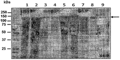
- Figure 3 is the result of analysis of the fraction obtained during the expression and purification of sfGFP-C by SDS-PAGE (sodium dodecyl sulfate polyacrylamide gel electrophoresis).
- 1 Cell lysate before inducing sfGFP-C expression
- 2 Insoluble cell lysate after induction of expression
- 3 Soluble cell lysate after expression is induced
- 4 Elution by flowing cell lysate supernatant over column Flow through, eluate obtained by flowing wash buffer over column, 6-8: eluate obtained by flowing elution buffer over column, arrow: sfGFP-C protein band.
- P is a control group analyzed under reducing conditions using a sample buffer of SDS-PAGE
- N is a control group under non-reducing conditions, and reduced after treatment with DTT, 2-ME, or TCEP.
- Figure 5 shows the results of binding the maleimide-PEG 6 -sugar chain to the cysteine residue of the sfGFP-C protein by SDS-PAGE.
- 1 unbound sfGFP-C protein control group
- 2-6 maleimide-PEG 6 -sugar chain and sfGFP-C protein in a molar ratio of 1: 1, 2: 1, 4: 1, 8: 1, and 16: 1, respectively.
- FIG. 6 is a result of blotting analysis of mannose-6-phosphate oligosaccharide bound to sfGFP-C protein using maleimide using Dom9, a lectin that binds mannose-6-phosphate.
- 1 and 2 represent the reactions in which the maleimide-PEG 6 -sugar chain and the sfGFP-C protein were combined at molar ratios of 8: 1 and 16: 1, respectively.
- Figure 7 shows the results of binding the oligosaccharide and the sfGFP-C protein using NHS-PEG 6 -maleimide by IEF (isoelectric focusing).
- 1 Unbound sfGFP-C protein control
- 2-4 Reactions in which the oligosaccharide and the sfGFP-C protein were bound at molar ratios of 1: 1, 2: 1, and 4: 1, respectively.
- DBCO-PEG 4 - is the analysis of the result of the combination of oligosaccharides and Azide-PEG 4 -sfGFP-C protein by SDS-PAGE.
- Azide-PEG 4 -sfGFP-C protein is prepared by the reaction of NHS-PEG 4 -Azide to sfGFP-C protein in the form of azide linked to amine groups of lysine and arginine residues.
- 1 unbound Azide-PEG 4 -sfGFP-C protein control
- 2-5 DBCO-PEG 4 -chain
- Azide-PEG 4 -sfGFP-C protein were respectively 1: 1, 2: 1, 4: 1, And 8: 1 combined.
- 10 is an isoelectric focusing (IEF) analysis of the results of binding the sugar chain and the sfGFP-C protein using a coupling reaction between DBCO and azide.
- IEF isoelectric focusing
- FIG. 11 shows the results of analyzing the oligosaccharide bound to the sfGFP-C protein by MALID-TOF.
- Titanium dioxide (TiO 2 ) No purification process (A) or after purification process (B) shows the results of the sugar chains isolated. The presence of phosphoric acid was confirmed by further treatment with alkaline phosphatase (C). Symbols representing the sugar chain in the drawing are the same as in FIG. 1.
- FIG. 12 shows the results of binding the DBCO-PEG 4 -sugar chain to Azide-PEG 3 -fetuin (A) or Azide-PEG 3 -RNase B (B) protein by SDS-PAGE.
- the Azide-PEG 3 -fetuin protein was prepared by binding Aminooxy-PEG 3 -Azide to fetuin oxidized with 1 mM periodate, and the Azide-PEG 3 -RNase B protein was Aminooxy- PEG 3- Azide was prepared by binding to RNase B oxidized with periodate at a concentration of 10 mM.
- 1 glycoprotein fetuin (A) and RNase B (B) control without azide group
- 2 glycoprotein reactant reacted with Aminooxy-PEG 3 -azide
- 3 DBCO-PEG 4 -sugar chain and Azide-PEG
- FIG. 13 shows the results of analysis of Dom9 blotting of fetuin (A) and RNase B (B) to which mannose-6-phosphate oligosaccharide is bound through DBC0 and azide reactions.
- A fetuin
- B RNase B
- FIG. 16 shows the results obtained by combining Azide-PEG 3 -RNase B and DBCO-PEG 4 -sugar chain obtained through the GOase H1 oxidation reaction with SDS-PAGE (A) and Dom9 blotting (B).
- 1 RNase B protein control
- 2 RNase B protein reacted with Aminooxy-PEG 3 -azide
- 3 reactant with 1: 1 binding of DBCO-PEG 4 -sugar chain and Azide-PEG 3 -glycoprotein
- 4 It represents a reaction in which DBCO-PEG 4 -sugar chain and Azide-PEG 3 -glycoprotein are respectively 4: 1 bound.
- Fig. 17 is a schematic diagram showing a method of attaching mannose-6-phosphate oligosaccharide obtained from yeast cell wall mannose protein to a target protein.
- Yeast cell wall mannoproteins are denatured by PNGase enzyme treatment to obtain oligosaccharides, followed by uncapping to remove outer mannose and trimming of free mannose at the end through ⁇ -mannosidase treatment.
- Trimming and only the phosphate-attached oligosaccharides are separated using titanium dioxide (TiO 2 ), it is possible to obtain mannose-6-phosphate oligosaccharides with high lysosomal targeting ability.
- the target protein can be treated with DBCO-PEG 4 -NHS to prepare a protein having a DBCO reactor attached to an amine group of lysine and arginine residues so that it can be combined with mannose-6-phosphate oligosaccharide having an azide reactor. have. That is, the mannose-6-phosphate oligosaccharide can be bound to the target protein using click chemistry, which connects DBCO and azide.
- FIG. 18 shows the results of combining Azide-PEG 3 -sugar chain and DBCO-PEG 4 -sfGFP-C protein prepared by reacting Aminooxy-PEG 3 -azide with mannose-6-phosphate oligosaccharide, with SDS-PAGE (A) and Dom9 blots. Analysis by Ting (B).
- the present invention in one aspect comprising the steps of (a) obtaining mannose-6-phosphate oligosaccharide from cell wall mannose protein isolated from yeast; And (b) binding the mannose-6-phosphate oligosaccharide to the target protein using a crosslinker, to provide a method for producing a glycoprotein to which mannose-6-phosphate oligosaccharide is added.
- the glycoprotein to which the mannose-6-phosphate oligosaccharide is added may be a glycoprotein obtained by binding a mannose-6-phosphate oligosaccharide to a target protein using a crosslinking agent.
- the glycoprotein Since the glycoprotein has mannose-6-phosphate oligosaccharide, the glycoprotein can move to intracellular lysosomes through the mannose-6-phosphate receptor. That is, the cation-independent mannose-6-phosphate receptor (CI-MPR), a mannose-6-phosphate receptor of the "secretory-reintroduction pathway,” recognizes the mannose-6-phosphate oligosaccharide and binds the glycoprotein to the glycoprotein by inclusion. As a result, the lysosomal targeting effect can be expected as an enzyme therapeutic. Furthermore, the mannose-6-phosphate oligosaccharide may be combined with peptides, lipids, or nanomaterials for escaping the inclusions and used to deliver useful substances into the cytoplasm or nucleus rather than the lysosome.
- C-MPR cation-independent mannose-6-phosphate receptor
- the lysosomal targeting effect can be expected as an enzyme therapeutic.
- mannose-6-phosphate oligosaccharide in order to produce a glycoprotein to which mannose-6-phosphate oligosaccharide is added, first, mannose-6-phosphate oligosaccharide is obtained from mannose protein separated from the cell wall, and the oligosaccharide obtained above is used as a target protein using a crosslinking agent.
- a method of preparing glycoproteins to which mannose-6-phosphate oligosaccharides have been added has been developed.
- target protein refers to a protein to which mannose-6-phosphate oligosaccharide is attached.
- the type thereof is not limited, and may be appropriately selected by those skilled in the art according to the intended purpose. . That is, for example, the target protein may be changed depending on the purpose of binding mannose-6-phosphate oligosaccharide, such as for the purpose of targeting lysosomes, or after being encapsulated and delivered into the cytoplasm or nucleus. It is not limited to the example of.
- the target protein may include, but is not limited to, pathogen protein, lysosomal protein, growth factor, cytokine, chemokine, antibody, or antigen thereof- It may be an antigen-binding fragment, or a fusion protein, and the lysosomal protein may be a degrading enzyme associated with lysosomal storage disease.
- the lysosomal storage disease is specifically Fabry's disease, mucopolysaccharidosis I, Farber disease, Gaucher disease, GM1-gangliosidosis, Tei Tay-Sachs disease, Sandhoff disease, GM2 activator disease, Krabbe disease, metachromatic leukodystrophy, Niemann-Peak disease Pick disease, Scheie disease, Hunter disease, Sanfilippo disease, Morquio disease, Maroteaux-Lamy disease, Hyaluro Hyaluronidase deficiency, aspartylglucosaminuria, fucosidosis, mannosidosis, Schindler disease, sialidosis type 1 1), Pompe disease, Pycnodysostosis, ceroid lipofuscinosis, cholesterol ester storage disease, Wolman disease, Multiple sulfatase deficiency, galactosity Kilomicron-bearing diseases with galactosialidosis, mucolipidosis, cystinosis, sialic acid
- the target protein is a degrading enzyme associated with lysosomal storage disease
- the target protein may be, for example, acid alpha glucosidase, alpha galactosidase, alpha- L-iduronidase, beta-Dgalactosidase, beta-glucosidase, beta-hexosaminidase ), Beta-D-mannosidase, alpha-L-fucosidase, arylsulfatase B, arylsulfatase A ), Alpha-N-acetylgalactosaminidase, aspartylglucosaminidase, induronate-2-sulfatase, alpha-glucosami Need-N-acetyltransferase, beta-D-glucologidaa (beta-D-glucoronidase), hyaluronidase, alpha-L-mannosidase, alpha-neuraminidas
- the target protein may include cysteine, lysine or arginine residues linked with a crosslinking agent, and may be a glycoprotein including oligosaccharide, but is not limited thereto.
- Step (a) is a step of obtaining mannose-6-phosphate oligosaccharide from the isolated cell wall mannose protein, wherein the oligosaccharide is linked with the target protein via a crosslinking agent using an aldehyde group of the oligosaccharide reducing terminal residue or an asparagine linked to the oligosaccharide terminal.
- a crosslinking agent using an aldehyde group of the oligosaccharide reducing terminal residue or an asparagine linked to the oligosaccharide terminal.
- step (a) may be to obtain an mannose-6-phosphate oligosaccharide by treating the isolated cell wall mannose protein with an enzyme.
- the enzyme may be, for example, an enzyme that releases pronase or an N-sugar chain, but is not limited thereto.
- asparagine when treated with pronase, asparagine can separate the oligosaccharide located at the terminal, and when treated with an enzyme that liberates the N-sugar chain, mannose-6-phosphate oligosaccharide without asparagine can be separated.
- N-linked glycan refers to the form in which the sugar chain is connected to the NH 2 group of asparagine
- enzyme which liberates the N-sugar chain specifically refers to the GlcNAc-Asn bond of the N-sugar chain. It may be an enzyme that cleaves or an enzyme that cleaves GlcNAc-GlcNAc bonds.
- the enzyme that liberates the N-sugar chain may be peptide-N-glycosidase (PNGase), or endoglycosidase (ENdoglycosidase, ENGase), but is not limited thereto.
- the enzyme used in the step (a) can be appropriately selected by those skilled in the art according to the purpose of separating the mannose-6-phosphate oligosaccharide, such as the separation conditions of the oligosaccharide, the type of protein to be combined with the oligosaccharide, and thus the crosslinking agent and The binding reaction can vary.
- step (a) may be to include a step of obtaining the mannose-6-phosphate oligosaccharide by removing the outer mannose from the separated sugar chain, but is not limited thereto.
- the cell wall mannoprotein (CWM) is first separated to obtain mannose-6-phosphate oligosaccharide, specifically yeast, and more specifically yeast engineered to add excessive mannose phosphate compared to natural yeast, more specifically, the yeast-specific sugar chain in the biosynthesis pathway are removed mannose phosphate is engineered to add an excess of yeast, further more specifically at least one trait selected from the group consisting of OCH1 deletion, MNN1 defect, YlMPO1 overexpression and MNN4 overexpression import mannose as a phosphate is operated to add an excess of yeast, and even more specifically OCH1 deletion, and may be a yeast having a MNN1 defect, most specifically in the yeast having a yeast-specific sugar chain biosynthesis removed OCH1 deletion and MNN1 defect It may be a yeast overexpressing YlMPO1 or MNN4 , but is not limited thereto.
- OH1 deletion and MNN1 defects in the present invention means that the function of the protein that the gene coding lost to be used as a deletion in the same sense of the gene, "YlMPO1 over-expression” is a foreign gene YlMPO1 overexpression " MNN4 overexpression” means that the expression level of MNN4 is higher than that of natural yeast.
- the yeast may be specifically Saccharomyces cerevisiae , but is not limited thereto.
- OCH1 gene and MNN1 is a defect ⁇ och1 mnn1 saccharide as (Saccharomyces in my process serenity busy cerevisiae )
- YEp352-YlMpo1 vector was introduced into the transformed yeast och1 ⁇ mnn1 / YEp352-YlMpo1 strain (Accession No. KCTC12674BP) to prepare a cell wall mannose protein from the hot citrate buffer (hot citrate buffer) .
- Step (a) may include the step of treating proteolytic enzymes to separate the oligosaccharides of the asparagine-linked form by decomposing the protein to individual amino acid levels in the isolated cell wall mannoprotein.
- the protease is specifically papain, bromelain, pancreatin, trypsin, pronase, subtilisin, subtilisin carlsberg, alcalase, pescalase (pescalase) and maxazyme may be one or more enzymes selected from the group, more specifically pronase (pronase), but is not limited thereto.
- the enzyme In order to separate the asparagine-free mannose-6-phosphate oligosaccharide from the isolated cell wall mannose protein, the enzyme is released by denaturing the cell-wall mannose protein and then treated with an enzyme that liberates the N-sugar chain, thereby obtaining a carbon column. It can be used by purification.
- the oligosaccharide separated by treating the enzyme that liberates the N-sugar chain has no asparagine at its end, but since the reducing end of some oligosaccharides is present as an aldehyde group under equilibrium conditions, the aldehyde group is bound to the target protein by binding the aldehyde group with a crosslinking agent. It is possible.
- the concentration of oligosaccharide in which the reducing end is present as an aldehyde group may be maintained at a constant level.
- an enzyme that liberates an N-sugar chain after denaturation of an isolated cell wall mannoprotein a method of providing a PNGase F enzyme (New England Biolabs, Ipswich, MA, USA) by a manufacturer is provided. Therefore, the N-sugar chain was liberated by treatment and reaction, and only the oligosaccharide could be selectively prepared by purification using a carbon column.
- the oligosaccharide obtained by treating the pronase or N-glycose free enzyme may contain mannose-1-phosphate-6-0-mannose linkages, which may be effective for targeting to intracellular lysosomes in the prepared oligosaccharides.
- a process of removing the outer mannose may be further included. That is, the method may further include preparing a glycoprotein having an oligosaccharide added with mannose-6-phosphate (phosphate-6-O-mannose). Through this process, a P 2 -Man 8 GlcNAc 2 oligosaccharide having or without an asparagine residue at its terminal can be prepared.
- the process may be a weak acid hydrolysis or using an uncapping enzyme, more specifically the weak acid is 0.1 to 5 M of formic acid (formic acid), the capping uncapping enzyme (cellulose) ⁇ -mannosidase or Cellulosimicrobium cellulose of the genus Cellulomonas It may be CcGH92_5 derived from cellulan s), but this is merely an example of weak acid hydrolysis or capping removal, but is not limited thereto.
- the weak acid is 0.1 to 5 M of formic acid (formic acid)
- the capping uncapping enzyme cellulose
- Cellulosimicrobium cellulose of the genus Cellulomonas It may be CcGH92_5 derived from cellulan s), but this is merely an example of weak acid hydrolysis or capping removal, but is not limited thereto.
- the step of removing the outer mannose with the mannosidase which can remove the free mannose of the non-reducing end can be carried out in the sugar chain. This step may be performed by simultaneously or sequentially performing the above two treatments, but is not limited thereto.
- the mannosidase which can remove the free mannose of the non-reducing end can be treated to the sugar chain.
- the mannosidase may be specifically ⁇ -mannosidase, but is not limited thereto.
- the capsule (uncapping) the outer mannose by treating 0.5 M formic acid on the oligosaccharide having asparagine at the purified terminal after the pronase treatment, or the purified oligosaccharide after PNGase treatment, ⁇ After reacting with -1,2-mannosidase, purification was performed to obtain mannose-6-phosphate oligosaccharide.
- the step (a) may further include a step for selectively separating the mannose-6-phosphate oligosaccharide.
- a material having affinity for mannose-6-phosphate oligosaccharide may be used, and for example, titanium dioxide (TiO 2 ) may be used, but is not limited thereto.
- titanium dioxide examples include, but are not limited to, a separation and purification process using a column including titanium dioxide (TiO 2 ) beads, titanium dioxide particles, or titanium dioxide.
- oligosaccharide added with mannose-6-phosphate was separated and purified using titanium dioxide (TiO 2 ) beads, and analyzed using a MALDI-TOF mass spectrometer. Were removed and only mannose-6-phosphate oligosaccharides were separated (FIG. 11B).
- Step (b) combines the mannose-6-phosphate oligosaccharide obtained in step (a) with the target protein using a crosslinker to finally prepare a glycoprotein to which mannose-6-phosphate oligosaccharide is added. to be.
- the crosslinker may have a reactor capable of being connected to an asparagine group or an aldehyde group at one end thereof.
- the crosslinking agent at the other end of maleimide maleimide
- DBCO Dibenzocyclooctyne
- DTME Dithio-bis-maleimidoethane
- BM PEG 2 (1,8-Bis-maleimidodiethyleneglycol)
- vinyl sulfone vinylsulfone Consisting of azide, iodoacetyl nitrophenylazide, isocyanate, pyridyldisulfide, hydrazide and hydroxyphenyl azide It may include any one or more selected from the group.
- the target protein to which mannose-6-phosphate oligosaccharide is to be added is a glycoprotein
- an oxidation reaction is induced by chemical or enzymatic method, an aldehyde group is introduced into the oligosaccharide of the target protein, and a reactor is attached thereto.
- Crosslinking reaction can be performed.
- a periodate oxidation reaction may be used, and in the case of a complex oligosaccharide added with sialic acid, aldehyde groups may be introduced by selectively oxidizing by lowering the periodate concentration.
- an aldehyde group may be selectively introduced into residues such as galactose, mannose, and GlcNAc using galactose oxidase or a similar enzyme.
- mannose-6-phosphate oligosaccharides can be added using a crosslinking agent having a group reacting thereto.
- the above-described methods are related to one example of the oxidation reaction, and are not limited thereto as long as an aldehyde group can be introduced into the sugar chain of the glycoprotein.
- the crosslinking agent may be any one or more nonionic hydrophilic polymers selected from the group consisting of polyethylene glycol (PEG), pluronic, polyvinylpyrolidone, and polyoxazolin; Or poly-L-lactic acid, poly-glycolic acid, poly-D-lactic-co-glycolic acid, poly- L-lactic-co-glycolic acid, poly-D, L-lactic-co-glycolic acid, poly-L-lactic-co-glycolic acid At least one biodegradable polyester-based polymer selected from the group consisting of caprolactone (polycaprolactone), poly-valerolactone, poly-hydroxybutyrate, and poly-hydroxyvalerate; It may be included, but is not limited thereto.
- the crosslinking agent is a more specific example, a PEG polymer having NHS and maleimide groups at each terminal, a PEG polymer having DBCO and NHS groups at each terminal, a PEG polymer having NHS and azide groups at each terminal, or each It may be a PEG polymer having oxyamine and azide groups at its ends, but is not limited thereto.
- crosslinking agent in the present invention can bind the mannose-6-phosphate oligosaccharide and the target protein described above, any type of crosslinking agent may be used, which may be appropriately selected by those skilled in the art.
- NHS-PEG 6 -maleimide was used as a crosslinking agent to bind the mannose-6-phosphate oligosaccharide having asparagine at the terminal and the sfGFP-C protein (FIG. 1B).
- NHS N-hydroxysuccinimide
- NHS-PEG 6 -maleimide can bind with amine group (-NH 2 ) of asparagine in mannose-6-phosphate oligosaccharide where asparagine is located, and maleimide group is the target protein
- the sfGFP-C protein can bind to the thiol group (-SH) possessed by cysteine, and as a result, a glycoprotein to which mannose-6-phosphate oligosaccharide is added can be prepared (FIGS. 5 and 6).
- two crosslinking agents DBCO-PEG 4 -NHS and NHS-PEG 4 -Azide, are used to bind sfGFP-C protein with mannose-6-phosphate oligosaccharide having asparagine at the terminal.
- the NHS group of DBCO-PEG 4 -NHS acts to bind the amine group of asparagine in the mannose-6-phosphate oligosaccharide where asparagine is located, as in the case of using the NHS-PEG 6 -maleimide as a crosslinking agent.
- DBCO can be combined with the target protein using copper-free click chemistry combined with azide.
- the target protein sfGFP-C protein is reacted with NHS-PEG 4 -Azide in an appropriate molar ratio.
- Azide-PEG 4 -sfGFP-C in which Azide-PEG 4 is linked to an amine group of lysine and an arginine residue of sfGFP-C protein, was prepared, and then the reaction of DBCO-PEG 4 -sugar chain, DBCO, and azide was produced.
- Glycoprotein can be prepared, and consequently, a glycoprotein to which mannose-6-phosphate oligosaccharide is added can be prepared (FIGS. 8 and 9).
- the NHS-PEG 6 -maleimide and DBCO-PEG 4 - may be obtained through MALDI-TOF mass spectrometry (FIG. 11) and IEF (Isoelectric focusing) analysis (FIGS. 7 and 10). It was confirmed that mannose-6-phosphate oligosaccharide was successfully attached to the sfGFP-C protein using NHS as a crosslinking agent.
- asparagine-free mannose-6-phosphate oligosaccharide and the target protein were combined using two crosslinking agents, Aminooxy-PEG 3 -Azide and DBCO-PEG 4 -NHS as crosslinking agents (FIG. 17).
- the Aminooxy-PEG 3 -Azide oxy amine (oxyamine) group and serves to bond the aldehyde group of the sugar chain asparagine is the reducing end is not Azide-PEG 3 - can be prepared a sugar chain.
- azide can be combined with the target protein using copper-free click chemistry combined with DBCO.
- the target protein is reacted with DBCO-PEG 4 -NHS in an appropriate molar ratio to give the desired number of DBCO-PEG 4 .
- Azide-PEG 4 -proteins linked to the amine groups of the lysine and arginine residues of the protein of interest can be prepared, which can then be combined through the coupling reaction of the produced Azide-PEG 3 -sugar chain with DBCO and azide.
- Glycoproteins with added mannose-6-phosphate oligosaccharides can be prepared (FIG. 17).
- the sfGFP-C, NHS-PEG 6 -maleimide, DBCO-PEG 4 -NHS, and Aminooxy-PEG 3 -Azide used above are all exemplary, and if the target protein can be combined with mannose-6-phosphate oligosaccharide, As described, one of ordinary skill in the art can appropriately select according to the purpose, and a crosslinking agent may be used without limitation as long as the target protein and the mannose-6-phosphate oligosaccharide derived from the yeast cell wall can be combined.
- the present invention provides a method for preparing mannose-6-phosphate oligosaccharide from the isolated cell wall mannose protein.
- mannose-6-phosphate oligosaccharide The method for producing mannose-6-phosphate oligosaccharide is as described above.
- the production method is a step of treating the isolated cell wall mannoprotein by pronase (pronase) to separate the oligosaccharides located at the end of the asparagine, or by processing an enzyme that releases the N- sugar chain to the cell wall mannoprotein to obtain the oligosaccharide It may be to include, and may further include the step of removing the outer mannose in the sugar chains, but is not limited thereto.
- the process of removing the outer mannose may be to use a weak acid hydrolysis or uncapping enzyme, and further comprising the step of treating the mannosidase in the sugar chain together with the process of removing the outer mannose. It may be, but is not limited thereto.
- the manufacturing method may further include the step of selectively separating the mannose-6-phosphate oligosaccharide, which may be performed using titanium dioxide (TiO 2 ), but is not limited thereto.
- the present inventors obtained cell wall mannoproteins (CWMs) from strains that overexpress YlMPO1 in the och1mnn1 ⁇ strain in which the yeast-specific oligosaccharide biosynthesis pathway was removed ( och1mnn1 ⁇ / YlMpo1p, KCTC12674BP), and the strain was subjected to the following procedure. Was made.
- OCH1 loxP for the gene defect - URA3 - the deletion cassette using the loxp Och1_pUG72_F (SEQ ID NO: 1) and Och1_pUG72_R (SEQ ID NO: 2) primer was prepared by amplification from the vector pUG72.
- Och1_CF SEQ ID NO: 3
- Och1_CR SEQ ID NO: 4
- the loxP- kanMX-loxp cassette for MNN1 gene deletion was then prepared using pUG6 as a template, Mnn1_pUG6_F (SEQ ID NO: 5) and Mnn1_pUG6_R (SEQ ID NO: 6) primers, and transformed into the och1 ⁇ strain prepared above.
- Transformed strains were screened in YPD medium supplemented with 1 M sorbitol and 200 mg / L G418, and the MNN1 gene deletion was identified by Mnn1_CF (SEQ ID NO: 7) and Mnn1_CR (SEQ ID NO: 7), which were based on chromosomal DNA. 8) was confirmed by using a polymerase chain reaction using primers.
- markers entered for deletion of genes were removed by expressing Cre protein in SC-Leu medium transformed with pSH68 vector and added 2% galactose instead of 2% glucose as reported in the above papers. It was. Finally, pSH68 for Cre protein expression was removed by continuous culture in YPD medium, the final mnn1 ⁇ och1 ⁇ strain was selected by checking the growth in the appropriate selection medium.
- SEQ ID NO: 1, 2, 5, 6 are part of the lower case in a side sequence and the homology of the OCH1 gene and MNN1 Im
- the promoter of GAPDH (0.8 kb) and terminator (0.2 kb) of S. cerevisiae were identified as ScGAPDHp-F1 (SEQ ID NO: 9) / ScGAPDHp-R1 (SEQ ID NO: 10) and ScGAPDHt-F1 (SEQ ID NO: 11) from the chromosomal DNA of L3262 strain, respectively.
- ) / ScGAPDHt-R1 (SEQ ID NO: 12) primers were used to amplify by polymerase chain reaction.
- the amplified DNA fragments were inserted into pDrive vectors (QIAGEN, Germany), respectively, to prepare pDrive-ScGAPDHp and pDrive-ScGAPDHt vectors.
- the ScGAPDH promoter was cleaved from pDrive-ScGAPDHp using the recombinases EcoRI and KpnI , and inserted into the YEp352 vector digested with the same recombinant enzyme to produce the YEp352-GAPDHp vector.
- the ScGAPDH terminator was cut from pDrive-ScGAPDHt using KpnI and PstI enzymes, and inserted into the YEp352-GAPDHp vector digested with the same recombinant enzyme to prepare a YEp352-GAP vector.
- the YlMPO1 gene (1.935-kb) was amplified from the chromosomal DNA of Y.
- lipolytica SMS397A by polymerase chain reaction using YlMPO1_F1 (SEQ ID NO: 13) and YlMPO1_R1 (SEQ ID NO: 14) primers.
- YlMPO1_F1 SEQ ID NO: 13
- YlMPO1_R1 SEQ ID NO: 14
- a short cut and process the KpnI restriction enzymes and then inserted into a Kpn I position of the YEp352-GAP vector to prepare a vector YEp352-YlMpo1.
- the Examples 1-1 and the OCH1 gene is the MNN1 defect the och1 mnn1 ⁇ ⁇ prepared in Example 1-2 in a strain YEp352-YlMpo1 vector is introduced transformant yeast produced from och1 mnn1 ⁇ ⁇ / YEp352-YlMpo1 (YlMpo1p ) Was produced.
- the YEp352-YlMpo1 vectors och1 ⁇ mnn1 ⁇ transformant introduced in yeast was named och1 ⁇ mnn1 ⁇ / YEp352-YlMpo1 , along which the Budapest Treaty provisions on the International Recognition of the deposit of a patent procedure microorganism, September 2014 As of May 4, it was deposited with the accession no. KCTC12674BP to the Gene Bank (Korea Collection for Type Cultures, KCTC) in the Korea Research Institute of Bioscience and Biotechnology (KRIBB).
- Yeast cell wall mannoprotein was extracted according to the previously reported method using hot citrate buffer (20 mM sodium citrate buffer, pH 7.0) (Park et al, 2011, Appl Environ Microbiol 77: 1187-1195). .
- Example 2-2 Pronase treatment and purification of asparagine-sugar chains
- Example 2-1 10 mg of the cell wall mannoprotein prepared in Example 2-1. was treated with 1 mg of pronase (pronase, Sigma-Aldrich, St. Louis, MO, USA) and reacted at 55 ° C. for 48 hours. This breaks down the protein to the level of the individual amino acids and produces oligosaccharides (asparagine-sugar chains) in the form of asparagine. The resulting asparagine-sugar chain was purified using a carbon column (Graphite Carbon Cartridge, GRACE, Deerfield, IL).
- Example 2-1 And the oligosaccharide prepared through 2-2. Has a structure of Man 8 GlcNAc 2 , Man-P-Man 8 GlcNAc 2, or (Man-P) 2 -Man 8 GlcNAc 2 , which is the outer side for targeting to intracellular lysosomes. Mannose must be uncapped and trimmed. To this end, weak acid hydrolysis and ⁇ -1,2-mannosidase treatment were performed sequentially.
- the concentration of the asparagine-sugar chain thus purified was quantified using fluorescamine, which fluoresces by binding to an amine group in the asparagine residue (Ullrich O et al., Proc. Natl. Acad. Sci. USA 1999). ). Fluorescamin itself has no fluorescence but binds to the primary amine group to form a fluorescence complex. Leucine was used as a standard solution at 0-20 nmol, and 0.4 ml of 0.5 M sodium borate (pH 8.0) was added to the dried asparagine-sugar chain, and 0.1 ml of 1 mM fluorescamin was added with vigorous stirring. Concentration was quantified by measuring fluorescence at excitation 390 nm and emission 470 nm.
- sfGFP-C A vector for the expression of the sfGFP-C protein having a cysteine residue introduced at the C-terminus was constructed.
- sfGFP-C was amplified by a conventional polymerase chain reaction (PCR) using primers SEQ ID NOs: 15 and 16.
- sfGFP-C_BamHI_F 5'- GCGGATCCATGAGCAAAGGTGAAGAACTGTTTAC-3 '(SEQ ID NO: 15)
- sfGFP-C_HidIII_R 5'- GCAAGCTTTTAGCAAGAACCACCTTTTTCGAAC-3 '(SEQ ID NO: 16)
- the amplified DNA was digested with BamH I and Hind III restriction enzyme and cloned into pColdII vector using a conventional genetic recombination method to prepare a pColdII-sfGFP-C vector (FIG. 2).
- the amino acid sequence of the expressed sfGFP-C and the nucleotide sequence for its expression are shown in SEQ ID NOs: 17 and 18.
- the pColdII-sfGFP-C vector was transformed into the E. coli BL21 (DE3) strain for expression to express the protein.
- the transformants were incubated at 37 ° C. in LB medium containing ampicillin (100 mg / ml), and the next day, the cultures were inoculated in TB (Terrific Broth) medium to perform the main culture at 37 ° C.
- TB Terrific Broth
- the cell precipitate obtained through the culture of Example 3-2. was resuspended in lysis buffer (50 mM NaH 2 PO 4 , pH 8.0, 300 mM NaCl, 1 mM PMSF, 0.1 mg / ml lysozyme) and the cells were sonicated. Was dissolved. The cell lysate was centrifuged at 6,000 X g for 20 minutes at 4 ° C., and then the cell lysate in which the insoluble portion was removed was purified from purification buffer (50 mM NaH 2 PO 4 , pH 8.0, 300 mM NaCl, 20 mM imidazole).
- purification buffer 50 mM NaH 2 PO 4 , pH 8.0, 300 mM NaCl, 20 mM imidazole.
- Ni-NTA column (Qiagen, USA) previously equilibrated with) was loaded and purified. After washing with purification buffer corresponding to 20 times the column volume, sfGFP-C protein was eluted with eluent (50 mM NaH 2 PO 4 , pH 8.0, 300 mM NaCl, 250 mM imidazole). The fraction eluted at each step was analyzed by SDS-PAGE (FIG. 3).
- eluent 50 mM NaH 2 PO 4 , pH 8.0, 300 mM NaCl, 250 mM imidazole
- the NHS group of NHS-PEG 6 -Maleimide (Pierce, Rockford, IL, USA) can bind to the amine group of the asparagine-sugar chain obtained in Example 2, thereby becoming an oligosaccharide having a maleimide reactor to form a cysteine residue. Can be bound to the desired protein with (FIG. 1B).
- the reaction between the purified asparagine-sugar chain and NHS-PEG 6 -maleimide was carried out through Example 2.
- NHS-PEG 6 -maleimide was prepared in 200 mM dissolved in DMSO (dimethyl sulfoxide). 100 ⁇ l of 4 mM asparagine-sugar chain was added with 30 ⁇ l of 200 mM NHS-PEG 6 -maleimide and 20 ⁇ l of binding buffer (phosphate buffered saline (PBS with 1 mM EDTA, pH 7.2)). This was 150 ⁇ l, and the reaction was carried out at room temperature for 3 hours.
- PBS phosphate buffered saline
- NHS-PEG 6 -maleimide remaining unreacted after reaction was dialyzed in binding buffer using a dialysis membrane (Septra Por7, Spectrum labs, Collinso Dominguez, CA, USA) with a molecular weight cut off of 1,000 Da. Removed.
- Reduction reaction for removal of disulfide bonds was carried out so that the cysteine of sfGFP-C protein reacted well with maleimide.
- DTT dithiothreitol
- 2-ME 2-Mercaptoethanol
- TCEP tris (2-carboxyethyl) phosphine
- Reductants were all purchased from Sigma-Aldrich (St. Louis, MO, USA).
- the final concentration of DTT and TCEP was 5 mM
- 2-ME was 5%
- the mixture was treated at room temperature for 30 minutes.
- SDS-PAGE sample buffer containing no reducing substance (10% SDS, 20% glycerol, 0.2 M tris pH 6.8, 0.05% bromophenolblue) was added and analyzed by SDS-PAGE under non-reducing conditions.
- reaction with maleimide-PEG 6 -sugar chain was performed using sfGFP-C protein which removed disulfide bond by TCEP.
- Example 4-3 Maleimide-PEG 6 -Binding of Sugar Chains to sfGFP-C Proteins
- a reaction was performed in which the maleimide-PEG 6 -sugar chain obtained in Example 4-1 was combined with the sfGFP-C protein obtained through the reduction reaction of Example 4-2. Wherein the maleimide group is bonded to the SH group of the cysteine residue of sfGFP-C.
- 1 ⁇ g of sfGFP-C protein and maleimide-PEG 6 -sugar chain were added for 1 hour at room temperature at a molar ratio of 1: 1, 2: 1, 4: 1, 8: 1, and 16: 1. After the reaction, the binding was confirmed by SDS-PAGE (FIG. 5).
- Mannose-6-Dom9 a protein that binds to the phosphate sugar chain attached with a three-Flag tag to the C- terminal type (Dom9-3xFlag, SEQ ID NO: 19) as Pichia Codon-optimized DNA synthesis for expression in pastoris yeast was submitted to DNA 2.0 (Menro Park, CA, USA) (SEQ ID NO: 20) and cloned into the pD912 vector provided by DNA 2.0 (pD912_Dom9-3xFlag). The synthetic DNA was purchased. The pD912_Dom9-3xFlag vector thus prepared was introduced into the P. pastoris GS115 strain by transformation.
- P. pastoris GS115 colonies were incubated overnight at 30 ° C. in 10 ml of YPD (1% yeast extract, 2% Bacto peptone, 2% glucose) medium. The next day it was incubated in 50 ml of YPD medium to OD 0.3 and then incubated at 30 ° C. for 4 to 5 hours. After incubation, the cell precipitate was obtained by centrifugation at 6,000 X g for 5 minutes, suspended in 40 ml of cold distilled water, and then centrifuged at 6,000 X g for 5 minutes to obtain a cell precipitate. Then, the cells were suspended in 20 ml of cold 1 M sorbitol, centrifuged at 6,000 X g for 5 minutes, and then suspended in cell precipitate with 1 ml of cold 1 M sorbitol. It was.
- YPD 1% yeast extract, 2% Bacto peptone, 2% glucose
- the colonies were inoculated in YPD-zeocin medium and incubated overnight at 30 ° C., followed by 1.2 L of BMMY (1% yeast extract, 2% peptone, 100 mM potassium phosphate (pH 6.0), 1.34% YNB, 0.00004% biotin, 0.5% MeOH) was added to the 1/100 of the final volume in the medium and the main culture was carried out for 72 hours at 30 °C. In this culture, gene expression was induced by adding an additional 0.5% MeOH every 24 hours. After 72 hours, the culture supernatant was obtained by centrifugation at 6,000 X g at 4 ° C.
- BMMY 1% yeast extract, 2% peptone, 100 mM potassium phosphate (pH 6.0), 1.34% YNB, 0.00004% biotin, 0.5% MeOH
- Example 4-5 Dom9-3xFlag blotting analysis
- SfGFP-C bound with maleimide-PEG 6 -sugar chain, was separated via 12% SDS-PAGE and transferred to PVDF membrane using an electrical method (60 min at 80 V).
- the PVDF membrane was treated with TBST (20 mM Tris, 150 mM sodium chloride, 0.5% Tween 20) blocking buffer containing 5% skim milk for 1 hour.
- TBST 20 mM Tris, 150 mM sodium chloride, 0.5% Tween 20
- Flag2-Dom9 (4 mg / ml) obtained by expressing and purifying as in Example 4-4. was diluted at a ratio of 1 / 1,000 and treated for 2 hours, followed by a 10-minute interval using TBST buffer. Wash three times.
- the PVDF membrane was treated with horseradish peroxidase (HRP) -bound anti-Flag antibody (Sigma-Aldrich) at a ratio of 1: 10,000 for 1 hour, and then washed three times at 10 minute intervals using TBST. Then, the protein bound with mannose-6-phosphate oligosaccharide was detected using an ECL solution (GE Healthcare) (FIG. 6).
- HRP horseradish peroxidase
- ECL solution GE Healthcare
- Example 4-6 Isoelectric focusing (IEF) analysis
- Example 4-3 In order to check whether mannose-6-phosphate oligosaccharide is bound to the protein prepared through the reaction, IEF analysis, an electrophoretic technique for separating proteins according to isoelectric point (pI), was performed. IEF gels were Novex® pH 3-10 IEF gels (Invitrogen, Waltham, Massachusetts, USA). Because this method can separate proteins according to their intrinsic pi, proteins with negatively charged mannose-6-phosphate oligosaccharides move to lower pI values than unattached proteins.
- IEF gels were Novex® pH 3-10 IEF gels (Invitrogen, Waltham, Massachusetts, USA). Because this method can separate proteins according to their intrinsic pi, proteins with negatively charged mannose-6-phosphate oligosaccharides move to lower pI values than unattached proteins.
- Example 4-3 10 ⁇ l of each sample was reacted with Example 4-3. And 10 ⁇ l of 2 ⁇ IEF sample buffer (Invitrogen) was prepared by mixing 1: 1. Then, the IEF gel was placed in a chamber and filled with a cathode buffer inside the chamber and an anode buffer outside (Invitrogen), and then separated at 100 V for 1 hour, 200 V for 1 hour, and 500 V for 30 minutes. The separated gel was treated with fixed buffer (12% trichloroacetic acid) for 30 minutes and washed twice with distilled water. Then stained with Coomassie Brilliant Blue G-250 (Bio-Rad, Philadelphia, PA, USA) and washed with destaining buffer (10% EtOH, 7.5% acetic acid) to confirm the results.
- IEF sample buffer Invitrogen
- the sfGFP-C protein showed a pI value of about 6, and the protein bound to oligosaccharide through NHS-PEG 6 -Maleimide lowered the pI value to 5 or less, confirming that mannose-6-phosphate oligosaccharide was attached.
- Figure 7 the protein bound to oligosaccharide through NHS-PEG 6 -Maleimide lowered the pI value to 5 or less, confirming that mannose-6-phosphate oligosaccharide was attached.
- Example 5-1 Reaction of DBCO-PEG 4 -NHS with Asparagine-sugar Chain
- NHS is asparagine obtained by using a reaction which combines the amine group DBCO-PEG 4 -NHS In Example 2 to prepare a sugar chain-reacting the oligosaccharides DBCO PEG-4.
- DBCO-PEG 4 -NHS was dissolved in DMSO to prepare 200 mM.
- 150 ⁇ l of the final reaction solution prepared by adding 30 ⁇ l of 200 mM DBCO-PEG 4 -NHS and 20 ⁇ l of binding buffer (PBS, pH7.2) at room temperature for 3 hours.
- PBS binding buffer
- DBCO-PEG 4 -NHS remaining unreacted after the reaction was removed by dialysis in binding buffer using a dialysis membrane (Septra Por7) having a molecular weight cut off of 1,000 Da.
- the sfGFP-C proteins are a number of PEG-Azide 4 was produced in the Azide-PEG 4 -sfGFP-C associated with the groups of lysine residues with al amine.
- NHS-PEG 4 -Azide was dissolved in DMSO to prepare 500 mM. 100 ⁇ l of the final reaction solution prepared by adding 4 ⁇ l of 500 mM NHS-PEG 4 -Azide and 86 ⁇ l of binding buffer (PBS pH 7.2) to 10 ⁇ l of sfGFP-C (5 ⁇ g / ⁇ l) protein at room temperature for 3 hours. I was. NHS-PEG 4 -Azide remaining unreacted after the reaction was removed by dialysis in binding buffer using a dialysis membrane (Sigma-Aldrich) having a molecular weight cutoff of 12,400 Da.
- a dialysis membrane Sigma-Aldrich
- Example 5-3 Combination of DBCO-PEG 4 -Chase and Azide-PEG 4 -sfGFP-C
- 3 ⁇ g of Azide-PEG 4 -sfGFP-C protein was added to 20 ⁇ l of the final reaction solution, and the DBCO-PEG 4 -sugar chain was added at 37 ° C. in a molar ratio of 1: 1, 2: 1,4: 1, or 8: 1.
- the reaction was carried out for 4 hours. After the reaction, the binding of oligosaccharide and sfGFP-C was analyzed by SDS-PAGE (FIG. 8).
- Example 5-4 Dom9-3xFlag blotting analysis
- the band was not detected in the Azide-PEG 4 -sfGFP-C protein that did not bind, whereas the bands were detected in all the proteins bound to DBCO-PEG 4 -sugar chain, so that the mannose-6-phosphate oligosaccharide was bound. I could confirm that. Two bands were detected each, and the small band was estimated to be the band of 50-70 kDa observed in the SDS-PAGE analysis, and the large band (120-200 kDa) was additionally observed.
- Example 5-5 Isoelectric focusing (IEF) analysis
- Example 5-3 In order to confirm whether mannose-6-phosphate oligosaccharide is bound to the protein prepared through the reaction, an IEF analysis, which is an electrophoretic technique for separating proteins according to isoelectric point (pI), was performed in the same manner as in Example 4-6.
- IEF analysis which is an electrophoretic technique for separating proteins according to isoelectric point (pI)
- the Azide-PEG 4 -sfGFP-C control group showed a pI value of about 5, whereas the protein bound to DBCO-PEG 4 -sugar chain had a lower pI value of 4 or less, resulting in mannose-6-phosphate oligosaccharides attached. It could be confirmed (Fig. 10).
- the sugar chain is released from the sugar-strained sfGFP-C and analyzed by MALDI-TOF (matrix-assisted laser desorption / ionization time-of-flight) mass spectrometry (Mannose-6). -It was confirmed that the phosphate sugar chain was properly attached.
- MALDI-TOF matrix-assisted laser desorption / ionization time-of-flight
- N -sugar chain was liberated from the protein prepared in Example 5-3 according to the method provided by the manufacturer using PNGase F (New England Biolabs, Ipswich, Mass., USA).
- the free oligosaccharide was purified using a carbon column and dried as in Example 2-2.
- ⁇ l was taken from a solution of 150 ⁇ l of acetic acid in 350 ⁇ l of DMSO, and dissolved in a 1.5 ml tube containing 6 mg of AA. Take 100 ⁇ l of this solution and add 6 mg of NaCNBH 2 (sodium cyanoborohydride) was added to a tube containing about 1 to 2 minutes at 65 °C to make a fluorescent labeling solution. 5 ⁇ l of this fluorescent label solution was placed in a dried oligosaccharide sample and reacted at 37 ° C. for 16 hours, and purified using a cyano SPE column (Agilent Technologies, Santa Clara, CA, USA).
- NaCNBH 2 sodium cyanoborohydride
- Example 6-3 Sugar chain analysis using MALDI-TOF mass spectrometer
- a matrix solution was prepared by dissolving 5 mg / ml of DHB (2,5-dihydroxybenzoic acid) and ATT (6-Aza-2-thiothymine) in 25% ACN containing 0.075% TFA.
- 0.5 ⁇ l of sugar chain to be analyzed was placed on the target MSP 96 polished steel chip for analysis, and then 0.5 ⁇ l of the prepared matrix solution was dried, and the chip was analyzed in a negative mode with a MALDI-TOF mass spectrometer (FIG. 11A). ).
- mannose-6-phosphate one or two are the sugar chain structure (P-Man 5 -6 GlcNAc 2 , P 2 -Man 6 GlcNAc 2) in Gorman North type (high-mannose type) with attached sugar chain (Man 5 -6 GlcNAc 2 ) could be observed. From this, it was found that the oligosaccharides derived from yeast cell wall proteins were successfully attached to the sfGFP-C protein through the coupling reaction between DBCO and azide.
- Mannose-6-phosphate oligosaccharides it is preferable to selectively separate only mannose-6-phosphate oligosaccharides in order to effectively introduce the target protein into the lysosomes in the cell.
- the cell wall protein-derived oligosaccharides of the yeast were analyzed through Example 6, high mannose oligosaccharides were detected together with mannose-6-phosphate oligosaccharides to selectively obtain only mannose-6-phosphate oligosaccharides to obtain titanium dioxide (TiO 2 ). A separate purification procedure was introduced.
- Example 2 As the oligosaccharide obtained in Example 2 has not only mannose-6-phosphate oligosaccharide but also high mannose-type oligosaccharide having no phosphate group, a purification process using titanium dioxide was introduced thereto.
- the sugar chain after the cap removal and a-1,2-mannosidase treatment is dried through Examples 2-1., 2-2. And 2-3.
- 50 mg TiO 2 beads (GL Sciences, Tokyo, Japan) were dissolved in 100 ⁇ l ACN, 50 ⁇ l of which was added to the dried sugar chain and 1 ml loading buffer (1 M glycolic acid, 80% ACN, 5% TFA). ) was added and reacted at room temperature for 30 minutes. Thereafter, the precipitated beads were centrifuged at 1000 X g for 1 minute, and the remaining supernatant was once again bound to fresh TiO 2 bead.
- the beads attached to the oligosaccharide were reacted and washed with 100 ⁇ l washing solution 1 (80% ACN, 1% TFA) for 1 minute, centrifuged at 1000 X g for 1 minute, and the supernatant was removed.
- 100 ⁇ l of Wash Solution 2 (20% ACN, 0.1% TFA), react for 1 minute, wash, and centrifuge under the same conditions to obtain the precipitated beads.
- Example 7-2 Sugar chain analysis using MALDI-TOF mass spectrometer
- the target protein to which the mannose-6-phosphate oligosaccharide is attached can be obtained by reacting the oligosaccharide (DBCO-PEG 4 -sugar chain) having the DBCO reactor prepared in Example 5 with copper-free click chemistry (FIG. 1E).
- Fetuin and RNase B were used as model glycoproteins, and the oligosaccharides attached through the periodate oxidation reaction were converted into forms having highly reactive aldehyde groups to facilitate crosslinking.
- complex oligosaccharides containing sialic acid were attached to oxidize with low concentration (1 mM) of periodate, but in case of RNaseB, high mannose oligosaccharides were attached, and high concentration (10 mM) of periodine was attached. I had to use a date.
- fetuin 5 mg is prepared in 950 ⁇ l of 0.1 M sodium acetate (pH 5.5) buffer, followed by 50 ⁇ l of 20 mM sodium periodate (Sigma-Aldrich) solution to the final 1 mM periodate concentration. After the light was blocked, the oxidation reaction was performed for 30 minutes at room temperature.
- RNaseB protein 5 mg was prepared in 500 ⁇ l of 0.1 M sodium acetate (pH 5.5) buffer, and then 500 ⁇ l of 20 mM sodium periodate (Sigma-Aldrich) was added to the final 10 mM periodate concentration. Blocking was performed for 30 minutes at room temperature. Sodium periodate remaining unreacted after the reaction was removed by dialysis in 0.1 M potassium acetate (pH 5.0) buffer using a dialysis membrane (Septra Por7) having a molecular weight cut off of 1,000 Da. It was.
- Example 8-2 Reaction of Aminooxy-PEG 3 -Azide with sugar chain of glycoprotein
- Example 8-1 Aminooxy-PEG 3 -Azide was reacted with a phenolase and an RNase B protein to which an oligosaccharide attached with an aldehyde group obtained through a periodate oxidation reaction was reacted to bind a desired azide reactor to a target glycoprotein.
- Azide-PEG 3 -Fetuin and Azide-PEG 3 -RNase B were prepared.
- Aminooxy-PEG 3 -Azide (Baseclick) was dissolved in DMSO to a concentration of 100 mM to prepare a solution. 1 ml of a solution (molar ratio, 1: 5) in which 4 ⁇ l of 100 mM Aminooxy-PEG 3 -Azide was added to 996 ⁇ l of 100 mM fetuin to which an aldehyde group was prepared in Example 8-1 was introduced at 37 ° C. for 12 hours. Reacted.
- Example 8-3 Combination of DBCO-PEG 4 -sugar Chain and Azide-PEG 3 -Glycoprotein
- Example 8-4 Analysis of mannose-6-phosphate oligosaccharide binding using Dom9-3xFlag blotting
- Example 8-3 mannose-6-phosphate oligosaccharide attached fetuin prepared by reacting DBCO-PEG 4 -sugar chain with a molar ratio of 1: 1 and 4: 1 was prepared by Alexa Fluor 488 carboxylic acid succinimidyl ester (Thermoscientipic, Waltham, MA USA) was used to label fluorescence. First, Alexa Fluor 488 carboxylic acid succinimidyl ester fluorescent substance was dissolved in DMSO to prepare a stock solution of 7.7 mM.
- Alexa Fluor 488 carboxylic acid succinimidyl ester 50 ⁇ l of 1 M sodium bicarbonate (pH 9.0) was added to 400 ⁇ l of Pettuin prepared to the final 50 mM, and 20 ⁇ l of 7.7 mM Alexa Fluor 488 carboxylic acid succinimidyl ester prepared in advance to room temperature. The reaction was carried out for 1 hour. Alexa Fluor 488 carboxylic acid succinimidyl ester, which remained unreacted after the reaction, was removed by dialysis in binding buffer (PBS) using a dialysis membrane (Septra Por7) having a molecular weight cut off of 1,000 Da.
- PBS binding buffer
- Example 8 an aldehyde group was introduced into the sugar chain of the glycoprotein using a periodate oxidation reaction, and a crosslinking agent was reacted to induce a coupling reaction with the mannose-6-phosphate oligosaccharide.
- sialic acid is present in the sugar chain
- low concentration (1 mM) of periodate can induce the oxidation reaction.
- high concentration of periodate is required, so that many oxidation reactions occur in the sugar chain. Not only that, but also the Met residues of the protein are oxidized.
- RNase B protein was treated with high concentration (10 mM) periodate, it was observed that the size was greatly increased (FIGS.
- a gene expressing a protein of the amino acid sequence reported in the previous paper was synthesized using a gene synthesis service of ThermoFisher scientific (Waltham, MA USA).
- the gene expressing GOase H1 thus synthesized was amplified by polymerase chain reaction (PCR) using primers SEQ ID NOs: 21 and 22.
- GOase H1_EcoRI_F 5 '-GCGAATTCGCAAGCGCACCGATTGGTAGCGC-3' (SEQ ID NO: 21)
- GOase H1_HidIII_R 5 '-CGAAGCTTCTGGGTAACACGAATGGTGCTTGC-3' (SEQ ID NO: 22)
- the amplified DNA was digested with EcoR I and Hind III restriction enzyme and cloned into pColdTF vector using a conventional gene recombination method to prepare pColdTF-GOase H1 vector.
- pColdTF-GOase H1 vector was transformed into the E. coli BL21 (DE3) strain for expression, and the protein was expressed in the same manner as in Example 3-2, and then purified in the same manner as in Example 3-3. After purification, the fractions eluted in each step were analyzed by SDS-PAGE (FIG. 16).
- Example 9-2 Oxidation of RNase B and Combination of Aminooxy-PEG 3 -Azide
- Oxidation reaction of RNAse B protein was performed using GOase H1 purified in Example 10-1. First, 100 ⁇ l of GOase H1 at 10 mg / ml and 4 ⁇ l of catalase (Sigma-Aldrich) at 0.3 mg / ml were added to a solution containing 5 mg of RNase B protein. The reaction was carried out at 30 ° C. for 22 hours.
- Azide-PEG 3 -RNase B to which Azide-PEG 3 was added was prepared by reacting Aminooxy-PEG 3 -Azide with the oxidized RNase B protein obtained through the GOase H1 reaction. For this purpose, 100 ⁇ l of Aminooxy-PEG 3 -Azide was added to 985 ⁇ l of oxidized RNase B protein and reacted at 37 ° C. for 12 hours. After the reaction, a dialysis membrane (Septra Por7) having a molecular weight cut off of 1,000 Da was used to remove Aminooxy-PEG 3 -Azide remaining in the binding buffer (PBS).
- PBS binding buffer
- Example 9-3 Binding of DBCO-PEG 4 -Chase and Azide-PEG 3 -RNase B
- Azide-PEG 3 -RNase B and DBCO-PEG 4 -sugar chains prepared through the above examples were coupled through a copper-free click chemistry reaction between azide and DBCO.
- 3 ⁇ g of Azide-PEG 3 -RNase B (200 ⁇ M) and DBCO-PEG 4 -sugar chain were reacted with 20 ⁇ l of the final reaction solution at 37 ° C. for 4 hours at a molar ratio of 1: 1 and 4: 1.
- the size change of RNase B by oligosaccharide binding was analyzed by SDS-PAGE (FIG. 16A).
- the band was not detected in the unreacted Azide-PEG 3 -RNase B protein, whereas in the case of the proteins that reacted with DBCO-PEG 4 -sugar chain, the band was detected and the mannose-6-phosphate oligosaccharide was bound. I could confirm that.
- mannose-6-phosphate oligosaccharide without asparagine obtained from the cell wall mannose protein of yeast was bound to the target protein using two crosslinking agents (Aminooxy-PEG 3 -Azide and DBCO-PEG 4 -NHS). (See the conceptual diagram of FIG. 17).
- Example 10-1 Purification of Mannose-6-phosphate oligosaccharides using PNGase Enzyme
- N-sugar chain was liberated by treating and reacting PNGase F (New England Biolabs, Ipswich, MA, USA) to 10 mg of the cell wall mannoprotein prepared in the same manner as in Example 2-1, according to the method provided by the manufacturer.
- the oligosaccharide thus obtained was purified in the same manner as in Example 2-2 using a carbon column. After purification, the vacuum dried oligosaccharide was confirmed to have a structure of Man 8 GlcNAc 2 , Man-P-Man 8 GlcNAc 2, and (Man-P) 2 -Man 8 GlcNAc 2 using a MALDI-TOF mass spectrometer. Therefore, weak acid hydrolysis and ⁇ -1,2-mannosidase treatment were sequentially performed in the same manner as in Example 2-3 to form mannose-6-phosphate oligosaccharide, and these were purified using the method of Example 7.
- Azide-PEG 3 -sugar chain was prepared using Aminooxy-PEG 3 -Azide that can react with the aldehyde group formed at the reducing end of the mannose-6-phosphate oligosaccharide prepared in Example 10-1.
- the oligosaccharide and Aminooxy-PEG 3 -Azide prepared by the method of Example 10-1 were added to 150 ⁇ l of the final reaction solution containing 0.1 M potassium acetate (pH 5.0) buffer, respectively, with final concentrations of 1.3 mM and 6.5 mM, respectively. It was made to react (molar ratio of sugar chain and Aminooxy-PEG 3 -Azide 1: 5) to reacted at 37 degreeC for 12 hours. Aminooxy-PEG 3 -Azide remaining unreacted after the reaction was removed by dialysis in PBS buffer using a dialysis membrane (Septra Por7) having a molecular weight cut off of 1,000 Da.
- DBCO-PEG 4 -NHS was reacted with the sfGFP-C protein prepared in Example 3 to prepare DBCO-PEG4-sfGFP-C, in which the DBCO-PEG 4 group was bound to the amine groups of the lysine and arginine residues of the sfGFP-C protein. It was.
- Example 10-4 Binding of Azide-PEG 3 -Chains and DBCO-PEG 4 -sfGFP-C
- Azide-PEG 3 -sugar chain prepared in Example 10-2 and DBCO-PEG 4 -sfGFP-C protein prepared in Example 10-3 were coupled through a click chemistry binding reaction of azide with DBCO.
- Example 10-5 Confirmation of binding of mannose-6-phosphate oligosaccharides using Dom9-3xFlag blotting
- Example 10-4 Whether the mannose-6-phosphate oligosaccharide was properly bound to the sfGFP-C protein through the binding reaction of Example 10-4 was confirmed by Dom9-3xFlag blotting analysis in the same manner as in Example 4-5 (FIG. 18B).
Landscapes
- Chemical & Material Sciences (AREA)
- Life Sciences & Earth Sciences (AREA)
- Organic Chemistry (AREA)
- Health & Medical Sciences (AREA)
- Genetics & Genomics (AREA)
- Molecular Biology (AREA)
- General Health & Medical Sciences (AREA)
- Engineering & Computer Science (AREA)
- Biophysics (AREA)
- Biochemistry (AREA)
- Biomedical Technology (AREA)
- Mycology (AREA)
- Bioinformatics & Cheminformatics (AREA)
- Proteomics, Peptides & Aminoacids (AREA)
- Wood Science & Technology (AREA)
- Zoology (AREA)
- General Engineering & Computer Science (AREA)
- Biotechnology (AREA)
- Medicinal Chemistry (AREA)
- Analytical Chemistry (AREA)
- Plant Pathology (AREA)
- Physics & Mathematics (AREA)
- Microbiology (AREA)
- Chemical Kinetics & Catalysis (AREA)
- General Chemical & Material Sciences (AREA)
- Preparation Of Compounds By Using Micro-Organisms (AREA)
- Peptides Or Proteins (AREA)
Abstract
본 발명은 만노스-6-인산 당사슬의 제조방법 및 만노스-6-인산 당사슬이 부가된 당단백질의 제조방법에 관한 것이다.
Description
본 발명은 만노스-6-인산 당사슬이 부가된 당단백질의 제조방법, 및 세포벽 만노 단백질에 프로나제 (pronase) 또는 N-당사슬을 유리하는 효소를 처리하는 단계를 포함하는, 만노스-6-인산 당사슬의 제조방법에 관한 것이다.
당질화를 통해서 당사슬이 부가되는 당단백질들은 현재 전세계 재조합 단백질 의약품 시장의 60 % 이상을 차지하며 시장을 주도하고 있다. 특히, 당사슬은 당단백질 의약품의 치료 효능, 체내 지속성, 타겟팅 및 면역반응 등에서 중요한 역할을 하기에 의약품의 품질을 결정하는 주요 인자로 부각되고 있다. 당사슬이 부가되는 반응은 크게 N-결합 또는 O-결합 당질화 (N-linked or O-linked glycosylation)의 두 가지 형태가 있으며, 이 중 N-결합 당질화를 통해서 부가되는 당사슬을 N-당사슬 (N-glycan)이라 부르며 소포체에서 합성이 시작된다.
상기 N-당사슬의 초기 생합성 과정은 진핵 미생물인 효모에서부터 고등동물인 포유류에 이르기까지 거의 동일한 과정으로 보존되어 있다. 그러나, 골지체로 넘어간 당사슬은 각 종에 특이적으로 다양한 당사슬 수식 과정을 거치게 되며, 그 결과로 효모, 곤충, 식물 및 동물 등에서 완전히 다른 형태의 당사슬들이 만들어지게 된다.
동물 세포에서는 많은 당단백질들이 Trimannosyl core 당사슬 (Man3GlcNAc2)에 GlcNAc, 갈락토즈 및 시알산이 부가된 복합형 (complex type) N-당사슬 (Neu5Ac2Gal2GlcNAc2Man3GlcNAc2)을 가지고 있으나, 당단백질의 종류에 따라서는 소포체에서 넘어온 형태인 Man8GlcNAc2 구조를 갖는 고만노스형 (high-mannose type) 당사슬을 가지고 있는 경우가 있다. 또한, 불필요한 물질들을 분해하여 재활용하도록 하는 소화와 자기 분해 기능을 가진 라이소좀으로 가는 당단백질들은 고만노스형 당사슬에 만노스-6-인산 (mannose-6-phosphate)이 부가되기도 한다.
이 경우 골지체에서 GlcNAc-PT (N-acetylglucosamine-1-phosphotransferase)가 라이소좀으로 이동할 당단백질들의 3차 구조를 인식하여 GlcNAc-인산을 당사슬의 만노스 잔기에 부가한다. 그리고, 여기에 uncovering 효소 (N-acetylglucosamine-1-phosphodiester a-N-acetylglucosaminidase)가 작용하여 바깥쪽 GlcNAc 잔기만을 제거하는 2단계 과정으로 만노스-6-인산이 형성된다. 이렇게 형성된 만노스-6-인산을 양이온 의존적 만노스-6-인산 수용체 (Cation-dependent mannose-6-phosphate receptor, CD-MPR)가 인식하여 이를 내포 (endosome)를 거쳐 라이소좀으로 이동시킨다. 그러나, 이때 CD-MPR과 결합하지 못한 당단백질들은 세포 밖으로 분비되게 된다. 이렇게 분비된 당단백질들을 다시 내포 반응(endocytosis)를 통해서 라이소좀으로 이동시키는 "분비-재도입 (secretion-recapture) 경로"가 존재하며 주로 양이온 비의존적 만노스-6-인산 수용체 (cation-independent mannose-6-phosphate receptor, CI-MPR)가 그 기능을 한다고 알려져 있다.
한편, 라이소좀 저장질환 (lysosomal storage disease)은 선천적인 유전자 이상으로 라이소좀에 존재하는 분해효소의 활성이 결핍되어 대사물질이 축적되어 발생하는 질환들이다. 이러한 질환을 선천적으로 갖고 태어난 환자들은 치료를 받지 못하면 대부분 젊은 나이에 죽음을 맞이하게 된다.
하지만, 상기 환자들에게 만노스-6-인산 당사슬을 가진 재조합 효소를 투여하면, 상기에서 설명한 "분비-재도입 경로"의 만노스-6-인산 수용체인 CI-MPR이 이를 인식하고 내포 작용에 의해서 라이소좀으로 이동시켜서 축적된 대사물질을 대신 분해해 줄 수 있다. 따라서, 효소 치료제에서는 만노스-6-인산 당사슬이 많이 부가되도록 하는 것이 중요하다.
본 발명자들은 만노스-6-인산 당사슬이 다량 부착된 재조합 효소 치료제를 개발하기 위해 예의 노력한 결과, 효모로부터 수득한 세포벽 만노단백질로부터 만노스-6-인산 당사슬을 수득하고, 이렇게 수득된 당사슬에 가교제 (crosslinker)를 부착하여 목적 단백질에 만노스-6-인산 당사슬을 결합시키는 방법을 개발하였고, 상기 방법을 이용하여 제조된 단백질을 라이소좀 저장 질환의 치료에 사용할 수 있음을 확인하여 본 발명을 완성하였다.
본 발명의 하나의 목적은 (a) 효모로부터 분리된 세포벽 만노단백질로부터 만노스-6-인산 당사슬을 수득하는 단계; 및 (b) 가교제 (crosslinker)를 사용하여 상기 만노스-6-인산 당사슬과 목적 단백질을 결합시키는 단계를 포함하는, 만노스-6-인산 당사슬이 부가된 당단백질의 제조방법을 제공하는 것이다.
본 발명의 다른 하나의 목적은 세포벽 만노 단백질에 프로나제 (pronase) 또는 N-당사슬을 유리하는 효소를 처리하는 단계를 포함하는, 만노스-6-인산 당사슬의 제조방법을 제공하는 것이다.
본 발명을 통해 당사슬 생합성 경로가 재설계된 효모의 세포벽으로부터 손쉽게 얻을 수 있는 만노스-6-인산 당사슬을 목적 단백질에 부착함으로써, 세포 내 라이소좀으로 도입될 수 있는 목적 단백질을 제조할 수 있다. 또한, 이를 통해 만노스-6-인산 함량이 높아 치료 효능이 우수한 효소 치료제들을 저렴하게 생산할 수 있다. 나아가, 만노스-6-인산 당사슬을 내포 탈출을 위한 펩타이드, 지질 또는 나노물질 등과 결합시켜 라이소좀이 아닌 세포질 또는 핵 내로 유용물질을 전달하는데 활용할 수 있다.
도 1은 당사슬 생합성 경로가 재설계된 효모의 세포벽 만노단백질로부터 얻은 말단에 아스파라진이 위치하는 만노스-6-인산 당사슬을 목적 단백질에 부착하는 방법을 나타내는 모식도이다. (A) 효모의 세포벽 유래 만노단백질 (mannoprotein)에 프로나제 (pronase)를 처리하여 아스파라진이 연결된 당사슬을 얻은 후, 바깥쪽 만노스를 제거하는 캡 제거 (uncapping) 과정과 α-만노시다제 처리를 통해서 말단의 자유로운 만노즈를 트리밍 (trimming)하고 인산이 부착된 당사슬들만을 이산화티타늄 (TiO2)을 이용하여 분리하면, 라이소좀 타겟팅 능력이 높은 만노스-6-인산 당사슬이 아스파라진 잔기에 연결된 형태를 얻을 수 있다. (B) 아스파라진이 연결된 만노스-6-인산 당사슬을 NHS-PEG6-말레이미드와 반응시키면 NHS가 아스파라진 잔기의 아민기와 결합한 형태가 만들어진다. 그리고 이를 C-말단에 시스테인 잔기를 넣어준 sfGFP-C (superfolder green fluorescent protein)와 반응시키면 말레이미드 그룹이 시스테인 잔기의 SH 그룹과 결합하여 만노스-6-인산 당사슬이 sfGFP와 결합된 형태를 얻을 수 있다. (C) DBCO-PEG4-NHS를 아스파라진이 연결된 만노스-6-인산 당사슬에 처리하면, NHS 그룹이 아민기와 결합하여 DBCO 반응기를 가진 만노스-6-인산 당사슬 (DBCO-PEG4-M-6-P glycan)이 만들어진다. (D) 목적 단백질에 NHS-PEG4-아자이드를 처리하여 라이신과 알지닌 잔기들의 아민 그룹에 아자이드 반응기가 부착된 단백질을 준비하여 이를 DBCO 반응기를 가진 만노스-6-인산 당사슬과 결합되도록 할 수 있다. 즉, DBCO와 아자이드가 연결되는 click chemistry반응을 통해서 만노스-6-인산 당사슬을 목적 단백질의 라이신 및 알지닌 잔기들에 결합시킬 수 있다. 이때 목적 단백질에 부착되는 만노스-6-인산 당사슬의 개수는 처음 NHS-PEG4-아자이드를 목적 단백질에 결합시킬 때 몰비를 조절 함으로서 원하는 개수로 부착이 가능하다. (E) 목적 단백질이 당단백질이라면 페리오데이트 (periodate) 산화 반응을 이용하여 당사슬에 알데하이드 기를 도입하여 만노스-6-인산 당사슬을 부착시킬 수 있다. 이때, 시알산이 부가된 복합형 당사슬이라면 페리오데이트 농도를 낮춤으로서 선택적으로 산화시켜서 알데하이드 기를 도입할 수 있다. 또 다른 방법으로는 갈락토스 옥시데이즈 (galactose oxidase) 또는 유사 효소 등을 이용하여 갈락토스, 만노즈, GlcNAc 등의 잔기에 알데하이드 기를 도입할 수도 있다. 상기의 방법들을 이용하여 알데하이드 기를 형성한 후에는 Aminooxy-PEG4-Azide를 반응시켜서 아자이드 기를 도입하고 여기에 DBCO 반응기를 가진 만노스-6-인산 당사슬을 반응시키는 click chemistry를 이용하여 만노스-6-인산 당사슬이 부가된 당단백질을 제조할 수 있다.
도면에서 당사슬을 나타내는 기호는 미국 Consortium for Functional Glycomics (htpp://www.functionalglycomics.org/)의 양식을 따랐으며, 인산은 P로 표시하였다 (사각형: GlcNAc, 원: 만노스, 인산: P).
도 2는 sfGFP-C 발현을 위한 pColdII-sfGFP-C 벡터의 모식도이다.
도 3은 sfGFP-C의 발현 및 정제 과정 중에서 수득된 분획을 SDS-PAGE (sodium dodecyl sulfate polyacrylamide gel electrophoresis)로 분석한 결과이다. 1: sfGFP-C 발현을 유도 하기 전 세포 용해물, 2: 발현이 유도된 후 불용성 세포 용해물, 3: 발현이 유도된 후 가용성 세포 용해물, 4: 컬럼에 세포 용해물 상층액을 흘려 용출된 용액 (flow through), 5: 컬럼에 세척 완충액을 흘려 얻은 용출액, 6-8: 컬럼에 용출 완충액을 흘려 얻은 용출액, 화살표: sfGFP-C 단백질 밴드.
도 4는 산화 과정에 의해서 생성된 sfGFP-C의 이황화 결합을 제거하기 위한 환원실험 결과이다. 이황화 결합을 환원시키기 위해 DTT, 2-ME, 또는 TCEP를 처리하여 비교 확인하였다. P는 SDS-PAGE의 표본 완충액을 이용하여 환원 조건에서 분석한 대조군, N은 비환원 조건의 대조군, 그리고 DTT, 2-ME, 또는 TCEP를 처리하여 환원 시킨 후 분석한 결과이다.
도 5는 말레이미드-PEG6-당사슬을 sfGFP-C 단백질의 시스테인 잔기에 결합시킨 결과를 SDS-PAGE로 분석한 것이다. 1: 결합시키지 않은 sfGFP-C 단백질 대조군, 2-6: Maleimide-PEG6-당사슬과 sfGFP-C 단백질을 각각 1 : 1, 2 : 1, 4 : 1, 8 : 1, 및 16 : 1의 몰비로 결합시킨 반응물을 나타낸다.
도 6은 말레이미드를 이용하여 sfGFP-C 단백질에 결합된 만노스-6-인산 당사슬을 만노스-6-인산에 결합하는 렉틴인 Dom9을 이용하여 블로팅 (blotting)으로 분석한 결과이다. 1과 2는 말레이미드-PEG6-당사슬과 sfGFP-C 단백질을 각각 8 : 1과 16 : 1의 몰비로 결합시킨 반응물을 나타낸다.
도 7은 NHS-PEG6-말레이미드를 이용해 당사슬과 sfGFP-C 단백질을 결합시킨 결과를 IEF (isoelectric focusing)로 분석한 것이다. 1: 결합시키지 않은 sfGFP-C 단백질 대조군, 2-4: 당사슬과 sfGFP-C 단백질을 각각 1 : 1, 2 : 1, 및 4 : 1의 몰비로 결합시킨 반응물을 나타낸다.
도 8은 DBCO-PEG4-당사슬과 Azide-PEG4-sfGFP-C 단백질을 결합시킨 결과를 SDS-PAGE로 분석한 것이다. 여기서 Azide-PEG4-sfGFP-C 단백질은 NHS-PEG4-Azide를 sfGFP-C 단백질에 반응시켜서 라이신과 알지닌 잔기의 아민기에 아자이드가 연결된 형태로 제조한 것이다. 1: 결합시키지 않은 Azide-PEG4-sfGFP-C 단백질 대조군, 2-5: DBCO-PEG4-당사슬과 Azide-PEG4-sfGFP-C 단백질을 각각 1 : 1, 2 : 1, 4 : 1, 및 8 : 1로 결합시킨 반응물을 나타낸다.
도 9는 DBCO-아자이드 반응을 통해서 만노스-6-인산 당사슬이 Azide-PEG4-sfGFP-C 단백질에 결합된 것을 Dom9 블로팅으로 확인한 결과이다. 1: Azide-PEG4-sfGFP-C 단백질 대조군, 2-5: DBCO-PEG4-당사슬과 Azide-PEG4-sfGFP-C 단백질을 각각 1 : 1, 2 : 1, 4 : 1, 및 8 : 1로 결합시킨 반응물을 나타낸다.
도 10은 DBCO와 아자이드의 커플링 반응을 이용해 당사슬과 sfGFP-C 단백질을 결합시킨 결과를 isoelectric focusing (IEF)으로 분석한 것이다. 1: 결합시키지 않은 sfGFP-C 단백질 대조군, 2-4: 당사슬과 sfGFP-C 단백질을 각각 1 : 1, 2 : 1, 및 4 : 1의 몰비로 결합시킨 반응물을 나타낸다.
도 11은 sfGFP-C 단백질에 결합된 당사슬을 MALID-TOF로 분석한 결과이다. 이산화티타늄 (TiO2) 정제 과정이 없거나 (A), 정제 과정을 거쳐서 (B) 분리한 당사슬의 분석결과를 보여준다. 인산이 부착되어 있는 것은 알칼리성 인산가수 분해효소를 추가로 처리하여 확인하였다 (C). 도면의 당사슬을 나타내는 기호는 도 1과 같다.
도 12는 DBCO-PEG4-당사슬을 Azide-PEG3-fetuin (A) 또는 Azide-PEG3-RNase B (B) 단백질에 결합시킨 결과를 SDS-PAGE로 분석한 것이다. 여기서 Azide-PEG3-fetuin 단백질은 Aminooxy-PEG3-Azide를 1 mM 농도의 페리오데이트 (periodate)로 산화시킨 페투인 (fetuin)에 결합시켜서 제작하였고, Azide-PEG3-RNase B 단백질은 Aminooxy-PEG3-Azide를 10 mM 농도의 페리오데이트로 산화시킨 RNase B에 결합시켜서 제작하였다. 1: 아자이드 기를 포함하지 않은 당단백질 페투인 (A)과 RNase B (B) 대조군, 2: Aminooxy-PEG3-azide를 반응시킨 당단백질 반응물, 3: DBCO-PEG4-당사슬과 Azide-PEG3-당단백질을 각각 1 : 1로 결합시킨 반응물, 4: DBCO-PEG4-당사슬과 Azide-PEG3-당단백질을 각각 4 : 1로 결합시킨 반응물을 나타낸다.
도 13은 DBC0와 아자이드 반응을 통해서 만노스-6-인산 당사슬이 결합된 페투인 (A)과 RNase B (B)를 Dom9 블로팅으로 분석한 결과이다. 1: Azide-PEG3-당단백질, 2: DBCO-PEG4-당사슬과 Azide-PEG3-당단백질을 각각 1 : 1로 결합시킨 반응물, 3: DBCO-PEG4-당사슬과 Azide-PEG3-당단백질을 각각 4 : 1로 결합시킨 반응물을 나타낸다.
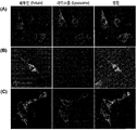
도 14는 DBC0와 아자이드의 copper-free click chemistry 반응을 통해서 제작된 만노스-6-인산 당사슬이 결합된 페투인이 세포 내 라이소좀으로 타겟팅 되는 것을 확인한 결과이다. 만노스-6-인산 당사슬이 결합하지 않은 페투인 (A)과 DBCO-PEG4-당사슬과 Azide-PEG3-Fetuin단백질을 각각 1 : 1 (B) 및 4 : 1 (C)로 결합시킨 반응물을 녹색 형광물질로 표지하고, Fabry fibroblast 세포에 처리하였다. 여기서 빨간색 형광은 라이소좀을 나타낸다.
도 15는 재조합 발현 및 정제 과정에서 수득한 GOase H1가 포함된 분획들을 SDS-PAGE로 분석한 결과이다. 1: 발현이 유도된 후 가용성 세포 용해물, 2: 컬럼에 세포 용해물 상층액을 흘려 용출된 용액 (flow through), 3: 컬럼에 세척 완충액을 흘려 얻은 용출액, 4-9: 컬럼에 용출 완충액을 흘려 얻은 용출액, 화살표: GOase H1단백질 밴드.
도 16은 GOase H1 oxidation 반응을 통해 얻은 Azide-PEG3-RNase B와 DBCO-PEG4-당사슬을 결합시킨 후 SDS-PAGE (A)와 Dom9 블로팅 (B)으로 분석한 결과이다. 1: RNase B 단백질 대조군, 2: Aminooxy-PEG3-azide를 반응시킨 RNase B 단백질, 3: DBCO-PEG4-당사슬과 Azide-PEG3-당단백질을 각각 1:1로 결합시킨 반응물, 4: DBCO-PEG4-당사슬과 Azide-PEG3-당단백질을 각각 4:1로 결합시킨 반응물을 나타낸다.
도 17은 효모의 세포벽 만노단백질로부터 얻은 만노스-6-인산 당사슬을 목적 단백질에 부착하는 방법을 나타내는 모식도이다. (A) 효모의 세포벽 만노단백질을 변성한 후에 PNGase 효소를 처리하여 당사슬을 얻은 후, 바깥쪽 만노스를 제거하는 캡 제거 (uncapping) 과정과 α-만노시다제 처리를 통해서 말단의 자유로운 만노즈를 트리밍 (trimming)하고 인산이 부착된 당사슬들만을 이산화티타늄 (TiO2)을 이용하여 분리하면, 라이소좀 타겟팅 능력이 높은 만노스-6-인산 당사슬을 얻을 수 있다. (B) 상기의 만노스-6-인산 당사슬을 Aminooxy-PEG3-azide와 반응시키면 옥시아민 (oxyamine) 기가 당사슬의 환원말단에 결합해서 아자이드 기를 갖는 만노스-6-인산 당사슬 (Azide-PEG3-glycan)이 만들어진다. (C) 목적 단백질에 DBCO-PEG4-NHS를 처리하여 라이신과 알지닌 잔기들의 아민 그룹에 DBCO 반응기가 부착된 단백질을 준비하여 이를 아자이드 반응기를 가진 만노스-6-인산 당사슬과 결합되도록 할 수 있다. 즉 DBCO와 아자이드가 연결되는 click chemistry를 이용하여 만노스-6-인산 당사슬을 목적 단백질에 결합시킬 수 있다.
도 18은 Aminooxy-PEG3-azide와 만노스-6-인산 당사슬을 반응시켜서 준비한 Azide-PEG3-당사슬과 DBCO-PEG4-sfGFP-C 단백질을 결합시킨 결과를 SDS-PAGE (A)와 Dom9 블로팅 (B)으로 분석한 것이다. 1: sfGFP-C 단백질 대조군, 2: DBCO-PEG4-NHS를 반응시킨 sfGFP-C 단백질, 3: Azide-PEG3-당사슬과 DBCO-PEG4-sfGFP-C 단백질을 1 : 1로 반응시킨 결과물, 4: Azide-PEG3-당사슬과 DBCO-PEG4-sfGFP-C 단백질을 4 : 1로 반응시킨 결과물을 나타낸다.
상기 목적을 달성하기 위하여, 본 발명은 하나의 양태로서 (a) 효모로부터 분리된 세포벽 만노단백질로부터 만노스-6-인산 당사슬을 수득하는 단계; 및 (b) 가교제 (crosslinker)를 사용하여 상기 만노스-6-인산 당사슬과 목적 단백질을 결합시키는 단계를 포함하는, 만노스-6-인산 당사슬이 부가된 당단백질의 제조방법을 제공한다.
본 발명의 목적상 상기 만노스-6-인산 당사슬이 부가된 당단백질은 가교제를 사용하여 목적 단백질에 만노스-6-인산 당사슬을 결합시킨 당단백질일 수 있다.
상기 당단백질은 만노스-6-인산 당사슬을 가지고 있으므로 만노스-6-인산 수용체를 통해 세포 내 라이소좀으로 이동할 수 있다. 즉, “분비-재도입 경로”의 만노스-6-인산 수용체인 CI-MPR (cation-independent mannose-6-phosphate receptor)이 만노스-6-인산 당사슬을 인식하고 내포 작용에 의해서 당단백질을 라이소좀으로 이동시킬 수 있으므로, 효소 치료제로서 라이소좀 표적화 효과를 기대할 수 있다. 나아가, 상기 만노스-6-인산 당사슬을 내포 탈출을 위한 펩타이드, 지질 또는 나노물질 등과 결합시켜 라이소좀이 아닌 세포질 또는 핵 내로 유용물질을 전달하는데 활용할 수 있다.
이에, 본 발명에서는 만노스-6-인산 당사슬이 부가된 당단백질을 생산하기 위하여, 먼저 만노스-6-인산 당사슬을 세포벽으로부터 분리한 만노단백질로부터 수득하고, 상기 수득한 당사슬을 가교제를 사용하여 목적 단백질과 결합시킴으로써 만노스-6-인산 당사슬이 부가된 당단백질을 제조하는 방법을 개발하였다.
본 발명에서 용어, “목적 단백질”은 만노스-6-인산 당사슬을 부착하고자 하는 단백질을 말한다. 본 발명의 목적상 상기 목적 단백질은 본 발명의 제조방법에 의해 만노스-6-인산 당사슬을 결합하여 사용할 수 있는 것이라면 그 종류가 제한되지 않고, 사용하고자 하는 목적에 따라 당업자에 의해 적절히 선택될 수 있다. 즉, 예를 들어 라이소좀 표적화를 목적으로 하거나, 내포된 후 내포탈출하여 세포질 또는 핵 내로 전달하고자 하는 등 만노스-6-인산 당사슬을 결합시키고자 하는 목적에 따라 목적 단백질이 달라질 수 있으며, 본 발명의 예에 제한되지 않는다.
상기 목적 단백질은, 이에 제한되지 않지만 예를 들어 병원체 단백질 (pathogen protein), 라이소좀 단백질 (lysosomal protein), 성장 인자 (growth factor), 사이토카인 (cytokine), 케모카인 (chemokine), 항체 또는 이의 항원-결합 단편 (antigen-binding fragment), 또는 융합 단백질 (fusion protein)일 수 있고, 상기 라이소좀 단백질 (lysosomal protein)은 라이소좀 저장질환 (lysosomal storage disease)과 관련된 분해효소일 수 있다.
상기 라이소좀 저장질환은 구체적으로 파브리병 (Fabry's disease), 점액다당류증 I (mucopolysaccharidosis I), 파버 질병 (Farber disease), 고셔병(Gaucher disease), GM1-강글리오시드증 (GM1-gangliosidosis), 테이-샥스병 (Tay-Sachs disease), 샌드호프병 (Sandhoff disease), GM2 활성제 질병 (GM2 activator disease), 크라베병 (Krabbe disease), 이염성백질이영양증 (metachromatic leukodystrophy), 니만-피크병 (Niemann-Pick disease), 샤이에 질병 (Scheie disease), 헌터 질병 (Hunter disease), 산필립포 질병 (Sanfilippo disease), 모르키오병 (Morquio disease), 마로토-라미 질병 (Maroteaux-Lamy disease), 히알루로니다아제 결핍증 (hyaluronidase deficiency), 아스파르틸글루코스아민뇨증 (aspartylglucosaminuria), 푸코시드축적증 (fucosidosis), 만노시도시스 (mannosidosis), 쉰들러 병 (Schindler disease), 사이알산축적증 유형 1 (sialidosis type 1), 폼피병 (Pompe disease), 피크노디소토시스 (Pycnodysostosis), 세로이드 리포푸신증 (ceroid lipofuscinosis), 콜레스테롤 에스테르 축적 질병 (cholesterol ester storage disease), 월만병 (Wolman disease), 다종 술파타아제 결손증 (Multiple sulfatase deficiency), 갈락토시알리도시스 (galactosialidosis), 뮤코리피드증 (mucolipidosis), 시스틴축적증 (cystinosis), 시알산 축적 질병 (sialic acid storage disorder), 마리네스코-쉐글렌 증후군 (Marinesco-Sjogren syndrome)을 갖는 킬로미크론 보유 질병 (chylomicron retention disease), 헤르만스키-푸드락 증후군 (Hermansky-Pudlak syndrome), 체디아크-히가시 증후군 (Chediak-Higashi syndrome), 다논병 (Danon disease), 또는 겔레오피직 이형성증 (Geleophysic dysplasia)일 수 있으나, 이에 제한되는 것은 아니다.
목적 단백질이 라이소좀 저장질환 (lysosomal storage disease)과 관련된 분해효소인 경우, 상기 목적 단백질은, 예를 들어 산성 알파 글루코시다아제 (acid alpha glucosidase), 알파 갈락토시다아제 (alpha galactosidase), 알파-L-이두로니다아제 (alpha-L-iduronidase), 베타-D-갈락토시다아제 (beta-Dgalactosidase), 베타-갈락토시다아제 (beta-glucosidase), 베타-헥소사미니다아제 (beta-hexosaminidase), 베타-D-만노시다제 (beta-D-mannosidase), 알파-L-푸코시다아제 (alpha-L-fucosidase), 아릴설파타아제 B (arylsulfatase B), 아릴설파타아제 A (arylsulfatase A), 알파-N-아세틸갈락토사미니다아제 (alpha-Nacetylgalactosaminidase), 아스파르틸글루코사미니다아제 (aspartylglucosaminidase), 인두로네이트-2-술파타아제 (iduronate-2-sulfatase), 알파-글루코사미니드-N-아세틸전달효소 (alpha-glucosaminide-Nacetyltransferase), 베타-D-글루코로니다아제 (beta-D-glucoronidase), 히알루로니다아제 (hyaluronidase), 알파-L-만노시다제 (alpha-L-mannosidase), 알파-뉴라미니다아제 (alpha-neuraminidase), 인산전달효소 (phosphotransferase), 산성 리파아제 (acid lipase), 산성 세라미다아제 (acid ceramidase), 스핑고마이엘리나제 (sphingomyelinase), 티오에스테라제 (thioesterase), 카텝신 K (cathepsin K), 또는 지단백질지방분해효소 (lipoprotein lipase)일 수 있으나, 이에 제한되지 않는다.
또한, 상기 목적 단백질은 가교제와 연결되는 시스테인, 라이신 또는 알지닌 잔기를 포함하는 것일 수 있으며, 당사슬이 포함된 당단백질일 수도 있으나 이에 제한되지 않는다.
이하, 만노스-6-인산 당사슬이 부가된 당단백질의 제조방법을 구체적으로 설명한다.
(a) 단계는, 분리된 세포벽 만노단백질로부터 만노스-6-인산 당사슬을 수득하는 단계로서, 상기 당사슬은 당사슬 환원 말단 잔기의 알데하이드 기 또는 당사슬 말단에 연결된 아스파라진을 이용하여 가교제를 통해 목적 단백질과 결합할 수 있다.
구체적으로, (a) 단계는 분리된 세포벽 만노단백질에 효소를 처리하여 만노스-6-인산 당사슬을 수득하는 것일 수 있다. 상기 효소는 예컨대, 프로나제 (pronase) 또는 N-당사슬을 유리하는 효소일 수 있으나, 이에 제한되는 것은 아니다. 이때, 프로나제 (pronase)를 처리하는 경우 아스파라진이 말단에 위치한 당사슬을 분리할 수 있으며, N-당사슬을 유리하는 효소를 처리하는 경우 아스파라진이 없는 만노스-6-인산 당사슬을 분리할 수 있다. 본 발명에서 용어 "N-당사슬 (N-linked glycan)"은 당사슬이 아스파라진의 NH2 기와 연결된 형태를 말하며, "N-당사슬을 유리하는 효소"는 구체적으로 N-당사슬의 GlcNAc-Asn 결합을 절단하는 효소 또는 GlcNAc-GlcNAc 결합을 절단하는 효소일 수 있다. 예를 들어, 상기 N-당사슬을 유리하는 효소는 펩티드-N-글리코시다제(peptide-N-glycosidase, PNGase), 또는 엔도글리코시다제 (endoglycosidase, ENGase)일 수 있으나, 이에 제한되는 것은 아니다.
따라서, 상기 (a) 단계에서 사용되는 효소는 당사슬의 분리 조건, 당사슬과 결합하고자 하는 단백질의 종류 등 만노스-6-인산 당사슬을 분리하기 위한 목적에 따라 당업자가 적절히 선택할 수 있으며, 이에 따라 가교제 및 결합 반응이 달라질 수 있다.
또한, 상기 (a) 단계는 상기 분리된 당사슬에서 바깥쪽 만노스를 제거하여 만노스-6-인산 당사슬을 수득하는 단계를 포함하는 것일 수 있으나, 이에 제한되는 것은 아니다.
상기 세포벽 만노단백질 (cell wall mannoprotein, CWM)은 만노스-6-인산 당사슬을 수득하기 위하여 먼저 분리하는 것으로, 구체적으로는 효모, 더욱 구체적으로는 천연 효모에 비하여 만노스인산이 과량 부가되도록 조작한 효모, 보다 구체적으로는 효모 특이적 당사슬 생합성 경로가 제거되어 만노스인산이 과량 부가되도록 조작한 효모, 보다 더 구체적으로는 OCH1 결손, MNN1 결손, YlMPO1 과발현 및 MNN4 과발현으로 이루어진 군에서 선택되는 1 이상의 유전형질을 가져 만노스인산이 과량 부가되도록 조작한 효모, 더욱 더 구체적으로는 OCH1 결손 및 MNN1 결손을 가지는 효모일 수 있고, 가장 구체적으로는 효모 특이적인 당사슬 생합성 경로가 제거된 OCH1 결손 및 MNN1 결손을 가지는 효모에서 YlMPO1 또는 MNN4 을 과발현하는 효모일 수 있으나, 이에 제한되지 않는다.
본 발명에서 용어 "OCH1 결손" 및 "MNN1 결손"은 상기 유전자들의 결실과 동일한 의미로 사용되는 것으로 상기 유전자가 코딩하는 단백질의 기능이 상실된 것을 의미하며, "YlMPO1 과발현"은 외래 유전자인 YlMPO1이 과발현 된 것을 의미하고, "MNN4 과발현"은 MNN4의 발현 수준이 천연 효모에 비하여 높은 상태를 의미한다.
상기 효모는 구체적으로는 사카로마이세스 세레비지에 (Saccharomyces
cerevisiae)일 수 있으나, 이에 제한되는 것은 아니다.
본 발명의 구체적인 일 실시예에서는 OCH1 및 MNN1 유전자가 결손된 och1
△mnn1 사카로마이세스 세레비지에 (Saccharomyces
cerevisiae) 균주에 YEp352-YlMpo1 벡터를 도입하여 형질전환 효모 och1
△
mnn1/ YEp352-YlMpo1 균주 (기탁번호 제KCTC12674BP)를 제작하고 이로부터 핫 구연산 완충액 (hot citrate buffer)을 이용하여 세포벽 만노단백질을 추출하였다.
(a) 단계에는, 상기 분리된 세포벽 만노단백질에서 단백질을 개별 아미노산 수준으로 분해하여 아스파라진이 연결된 형태의 당사슬을 분리하기 위해, 단백질 분해 효소를 처리하는 단계가 포함될 수 있다.
상기 단백질 분해 효소는 구체적으로 파파인, 브롤멜레인, 판크레아틴, 트립신, 프로나제 (pronase), 서브틸리신 (subtilisin), 서브틸리신 칼스버그 (subtilisin carlsberg), 알칼라아제 (alcalase), 페스칼라제 (pescalase), 및 맥사자임 (maxazyme)으로 이루어진 군에서 선택된 1 종 이상의 효소일 수 있고, 더욱 구체적으로는 프로나제 (pronase)일 수 있으나, 이에 제한되는 것은 아니다.
본 발명의 구체적인 일 실시예에서는 분리된 세포벽 만노단백질에 프로나제를 처리하여 55 ℃에서 48 시간 반응시키고 이를 탄소 컬럼을 이용해 정제함으로써 말단에 아스파라진이 위치하는 당사슬을 제조하였다.
상기 분리된 세포벽 만노단백질로부터 아스파라진이 없는 만노스-6-인산 당사슬을 분리하기 위해서는 세포벽 만노단백질을 변성 (denaturation)시킨 후에 N-당사슬을 유리하는 효소를 처리하여 당사슬을 유리하고, 이를 탄소 컬럼을 이용해 정제하여 사용할 수 있다. 이와 같이 N-당사슬을 유리하는 효소를 처리하여 분리된 당사슬은 말단에 아스파라진이 없으나, 평형 조건에서 일부 당사슬의 환원 말단이 알데하이드 기로 존재하게 되므로, 상기 알데하이드 기를 가교제와 결합시킴으로써 목적 단백질과 결합이 가능하다. 또한, 평형 조건에 의해 환원 말단이 알데하이드 기로 존재하는 당사슬의 농도는 일정 수준으로 유지될 수 있다.
본 발명의 구체적인 일 실시예에서는 분리된 세포벽 만노단백질을 변성시킨 후에 N-당사슬을 유리하는 효소의 일 예로서, PNGase F 효소 (New England Biolabs, Ipswich, MA, USA)를 제조사에서 제공하는 방법을 따라서 처리하고 반응하여 N-당사슬을 유리하였으며, 이를 탄소 컬럼을 이용해 정제함으로써 당사슬만을 선택적으로 제조할 수 있었다.
상기 프로나제 또는 N-당사슬을 유리하는 효소를 처리하여 수득한 당사슬은 만노스-1-인산-6-0-만노스 결합을 포함할 수 있어서, 상기 제조된 당사슬에서 세포 내 라이소좀으로 표적화에 효과적일 수 있도록, 바깥쪽 만노스를 제거하는 과정을 추가로 포함될 수 있다. 즉, 만노스-6-인산 (인산-6-O-만노스)이 부가된 당사슬을 가지는 당단백질을 제조하는 과정을 추가로 포함할 수 있다. 이와 같은 과정을 통하여 말단에 아스파라진 잔기를 갖거나, 갖지 않는 P2-Man8GlcNAc2 당사슬을 제조할 수 있다.
구체적으로 이와 같은 과정은 약산 가수분해 또는 캡핑 제거 (uncapping) 효소를 이용하는 것일 수 있으며, 더욱 구체적으로 상기 약산은 0.1 내지 5 M의 포름산 (formic acid)이고, 상기 캡핑 제거 (uncapping) 효소는 셀룰로모나스 (Cellulomonas) 속 세균의 α-만노시다제 (α-mannosidase) 또는 셀룰로시마이크로비움 셀루란스 (Cellulosimicrobium
cellulans) 유래의 CcGH92_5일 수 있으나, 이는 약산 가수분해 또는 캡핑 제거의 일 예일 뿐, 이에 제한되는 것은 아니다.
나아가, 상기 바깥쪽 만노스를 제거하는 과정과 함께 비환원 말단의 자유로운 만노스들을 제거할 수 있는 만노시다제를 당사슬에 처리하는 단계를 수행할 수 있다. 이러한 단계는 상기 두 처리 과정을 동시 또는 순차적으로 진행하여 수행할 수 있으나, 이에 제한되지 않는다.
구체적으로, 약산 가수분해 또는 캡핑 제거 (uncapping) 효소를 이용하여 바깥쪽 만노스를 제거하는 과정과 함께, 비환원 말단의 자유로운 만노스들을 제거할 수 있는 만노시다제를 당사슬에 처리할 수 있다. 상기 만노시다제는 구체적으로 α-만노시다제일 수 있으나, 이에 제한되지 않는다. 이와 같은 과정을 통하여 비환원 말단의 자유로운 만노스들이 제거된 P2-Man6GlcNAc2 당사슬을 제조할 수 있다.
본 발명의 구체적인 일 실시예에서는 상기 프로나제 처리 후 정제된 말단에 아스파라진이 위치하는 당사슬, 또는 PNGase 처리 후 정제된 당사슬에 0.5 M 포름산을 처리하여 바깥쪽 만노스를 캡 제거 (uncapping)하고, α-1,2-만노시다제와 반응시킨 뒤 정제 과정을 거쳐서 만노스-6-인산 당사슬을 수득하였다.
나아가, 상기 (a) 단계를 통해 수득한 당사슬에는 고만노즈형 당사슬이 존재할 수 있으므로, 상기 (a) 단계에 만노스-6-인산 당사슬을 선택적으로 분리하기 위한 단계를 추가로 포함시킬 수 있다.
이를 위해 만노스-6-인산 당사슬에 친화성을 가지는 물질을 이용할 수 있으며, 그 예로, 이산화티타늄 (TiO2)을 이용할 수 있으나, 이에 제한되지 않는다.
상기 이산화티타늄을 이용하는 예로서, 이산화티타늄 (TiO2) 비드, 이산화티타늄 파티클 또는 이산화티타늄을 포함하는 컬럼을 이용한 분리 정제 과정을 들 수 있으나, 이에 제한되는 것은 아니다.
본 발명의 구체적인 일 실시예에서는 이산화티타늄 (TiO2) 비드를 이용하여 만노스-6-인산이 부가된 당사슬들만을 분리 정제하였으며, 이를 MALDI-TOF 질량 분석기를 이용하여 분석한 결과, 고만노스형 당사슬들은 제거되고 만노스-6-인산 당사슬들만이 분리된 것을 확인하였다 (도 11B).
(b) 단계는 가교제 (crosslinker)를 사용하여 상기 (a) 단계에서 수득한 만노스-6-인산 당사슬을 목적 단백질과 결합시켜, 최종적으로 만노스-6-인산 당사슬이 부가된 당단백질을 제조하는 단계이다.
상기 가교제 (crosslinker)는 이의 한쪽 말단에 아스파라진 기 또는 알데하이드 기와 연결될 수 있는 반응기를 가지는 것일 수 있다. 상기 아스파라진 기 또는 알데하이드 기와 연결될 수 있는 반응기는 예를 들어, 엔하이드록시석신이미드 (NHS, N-hydroxysuccinimide), 옥시아민 (oxyamine), 아이소티오시아네이트 (isothiocyannate), 아이소시아네이트 (isocyanate), 설포닐클로라이드 (sulfonyl chloride), 알데히드 (aldehyde), 카보디이미드 (carbodiimide), 아실아자이드 (acyl azide), 언하이드라이드 (anhydride), 플루오로벤젠 (fluorobenzene), 카보네이트 (carbonate), 이미도에스터 (imidoester), 에폭사이드 (epoxide) 및 플루오로페닐에스터 (fluorophenyl ester)로 이루어진 군에서 선택되는 어느 하나 이상을 포함하는 것일 수 있으나, 이에 제한되는 것은 아니다.
또한, 상기 가교제는 다른 한쪽 말단에 말레이미드 (maleimide), DBCO (Dibenzocyclooctyne), DTME (Dithio-bis-maleimidoethane), BM(PEG)2(1,8-Bis-maleimidodiethyleneglycol), 비닐설폰 (vinylsulfone), 아자이드 (azide), 이오도아세틸 니트로페닐아자이드 (iodoacetyl nitrophenylazide), 아이소시아네이트 (isocyanate), 피리딜다이설파이드 (pyridyldisulfide), 하이드라자이드 (hydrazide) 및 하이드록시페닐 아자이드 (hydroxyphenyl azide)로 이루어진 군에서 선택되는 어느 하나 이상을 포함하는 것일 수 있다.
만노스-6-인산 당사슬을 부가하고자 하는 목적 단백질이 당단백질이라면 화학적 또는 효소적인 방법으로 산화 반응을 유도하여, 상기 목적 단백질의 당사슬에 알데하이드 (aldehyde)기를 도입하고, 여기에 결합 하는 반응기를 이용하여 가교 반응을 할 수 있다.
예를 들어, 화학적인 방법으로는 페리오데이트 (periodate) 산화 반응을 이용할 수 있으며, 이때 시알산이 부가된 복합형 당사슬이라면 페리오데이트 농도를 낮춤으로써 선택적으로 산화시켜서 알데하이드 기를 도입할 수 있다. 또 다른 방법으로는 갈락토스 옥시데이즈 (galactose oxidase) 또는 유사 효소 등을 이용하여 갈락토스, 만노즈, GlcNAc 등의 잔기에 알데하이드 기를 선택적으로 도입할 수도 있다. 상기의 방법들을 이용하여 알데하이드 기를 형성한 후에는 여기에 반응하는 그룹을 가진 가교제를 이용하여 만노스-6-인산 당사슬을 부가할 수 있다. 그러나 상술한 방법들은 산화 반응의 일 예시에 관한 것으로, 당단백질의 당사슬에 알데하이드 기를 도입할 수 있는 한, 이에 제한되는 것은 아니다.
나아가, 상기 가교제는 PEG (polyethylene glycol), 플루로닉 (pluronic), 폴리비닐피롤리돈 (polyvinylpyrolidone) 및 폴리옥사졸린 (polyoxazolin)으로 이루어진 군에서 선택된 어느 하나 이상의 비이온성 친수성 고분자; 또는 폴리 L-락트산 (poly-L-lactic acid), 폴리-글리콜산 (poly-glycolic acid), 폴리-D-락트산-co-글리콜산 (poly-D-lactic-co-glycolic acid), 폴리-L-락트산-co-글리콜산 (poly-L-lactic-co-glycolic acid), 폴리-D,L-락트산-co-글리콜산 (poly-D,L-lactic-co-glycolic acid), 폴리-카프로락톤 (polycaprolactone), 폴리-발레로락톤 (polyvalerolactone), 폴리-하이드록시 부티레이트 (polyhydroxybutyrate) 및 폴리-하이드록시 발러레이트 (polyhydroxyvalerate)로 이루어진 군에서 선택되는 어느 하나 이상의 생분해성 폴리에스테르계 고분자;를 포함하는 것일 수 있으나, 이에 제한되는 것은 아니다.
또한, 상기 가교제는 보다 구체적인 예로서, 각 말단에 NHS 및 말레이미드 기를 가지는 PEG 중합체, 각 말단에 DBCO 및 NHS 기를 가지는 PEG 중합체, 각 말단에 NHS 및 아자이드 (azide) 기를 가지는 PEG 중합체, 또는 각 말단에 옥시아민 (oxyamine) 및 아자이드 기를 가지는 PEG 중합체일 수 있으나, 이에 제한되지 않는다.
본 발명에서 상기 가교제는 상기 기술된, 만노스-6-인산 당사슬과 목적 단백질을 결합시킬 수 있다면, 어떠한 형태의 가교제를 사용하더라도 무방하며, 이는 당업자에 의해 적절히 선택될 수 있다.
본 발명의 구체적인 일 실시예에서는 가교제로 NHS-PEG6-말레이미드를 이용하여 말단에 아스파라진이 위치하는 만노스-6-인산 당사슬과 sfGFP-C 단백질을 결합시켰다 (도 1B). NHS-PEG6-말레이미드의 NHS (N-hydroxysuccinimide)기는 아스파라진이 위치하는 만노스-6-인산 당사슬에서 아스파라진이 가지는 아민기 (-NH2)와 결합할 수 있으며, 말레이미드 기는 목적 단백질인 sfGFP-C 단백질에서 시스테인이 가지는 티올기 (-SH)와 결합할 수 있어 결과적으로 만노스-6-인산 당사슬이 부가된 당단백질을 제조할 수 있다 (도 5 및 도 6).
본 발명의 다른 구체적인 일 실시예에서는 DBCO-PEG4-NHS와 NHS-PEG4-Azide의 두 개의 가교제를 이용하여 말단에 아스파라진이 위치하는 만노스-6-인산 당사슬과 sfGFP-C 단백질을 결합시켰다 (도 1C). 이때, DBCO-PEG4-NHS의 NHS기는 상기 NHS-PEG6-말레이미드를 가교제로 사용한 경우와 마찬가지로 아스파라진이 위치하는 만노스-6-인산 당사슬에서 아스파라진이 가지는 아민기와 결합하는 역할을 하여 DBCO-PEG4-당사슬을 제작할 수 있다. 한편, DBCO의 경우 아자이드와 결합되는 copper-free click chemistry를 이용하여 목적 단백질과 결합할 수 있으므로, 먼저 목적 단백질인 sfGFP-C 단백질을 NHS-PEG4-Azide와 적당한 몰 비로 반응시켜 원하는 숫자의 Azide-PEG4가 sfGFP-C 단백질의 라이신과 알지닌 잔기의 아민기에 연결된 Azide-PEG4-sfGFP-C를 제작한 뒤, 이를 상기 제작한 DBCO-PEG4-당사슬과 DBCO와 아자이드의 결합 반응을 통해 결합시킬 수 있고, 결과적으로 만노스-6-인산 당사슬이 부가된 당단백질을 제조할 수 있다 (도 8 및 도 9).
본 발명의 또 다른 구체적인 일 실시예에서는 MALDI-TOF 질량 분석 (도 11) 및 IEF (Isoelectric focusing) 분석 (도 7 및 도 10)을 통해, 상기 NHS-PEG6-말레이미드 및 DBCO-PEG4-NHS를 가교제로 하여 sfGFP-C 단백질에 만노스-6-인산 당사슬이 성공적으로 부착되었음을 확인하였다.
본 발명의 또 다른 구체적인 일 실시예에서는 가교제로 Aminooxy-PEG3-Azide와 DBCO-PEG4-NHS의 두 개의 가교제를 이용하여 아스파라진이 없는 만노스-6-인산 당사슬과 목적 단백질을 결합시켰다 (도 17). 이때, Aminooxy-PEG3-Azide의 옥시아민 (oxyamine) 기는 아스파라진이 없는 당사슬 환원 말단의 알데하이드 기와 결합하는 역할을 하여 Azide-PEG3-당사슬을 제작할 수 있다. 한편, 아자이드의 경우 DBCO와 결합되는 copper-free click chemistry를 이용하여 목적단백질과 결합할 수 있으므로, 먼저 목적 단백질을 DBCO-PEG4-NHS와 적당한 몰 비로 반응시켜 원하는 숫자의 DBCO-PEG4가 목적 단백질의 라이신과 알지닌 잔기의 아민기에 연결된 Azide-PEG4-단백질을 제작한 뒤, 이를 상기 제작한 Azide-PEG3-당사슬과 DBCO와 아자이드의 결합 반응을 통해 결합시킬 수 있고, 결과적으로 만노스-6-인산 당사슬이 부가된 당단백질을 제조할 수 있다 (도 17).
상기 사용된 sfGFP-C, NHS-PEG6-말레이미드, DBCO-PEG4-NHS, 및 Aminooxy-PEG3-Azide는 모두 예시적인 것으로, 목적 단백질은 만노스-6-인산 당사슬과 결합할 수 있다면 상기 설명한 바와 같이 그 목적에 따라 당업자가 적절하게 선택할 수 있으며, 가교제 역시 목적 단백질과 효모 세포벽 만노단백질 유래 만노스-6-인산 당사슬을 결합시킬 수 있다면 제한 없이 사용될 수 있다.
본 발명은 다른 하나의 양태로서, 분리된 세포벽 만노단백질로부터, 만노스-6-인산 당사슬을 제조하는 방법을 제공한다.
만노스-6-인산 당사슬의 제조방법에 대하여는 상기 설명한 바와 같다.
상기 제조방법은 분리된 세포벽 만노단백질에 프로나제 (pronase)를 처리하여 아스파라진이 말단에 위치한 당사슬을 분리하는 단계, 또는 세포벽 만노단백질에 N-당사슬을 유리하는 효소를 처리하여 당사슬을 수득하는 단계를 포함하는 것일 수 있고, 상기 당사슬들에서 바깥쪽 만노스를 제거하는 단계를 추가로 더 포함하는 것일 수 있으나, 이에 제한되지 않는다.
또한, 상기 바깥쪽 만노스를 제거하는 과정은 약산 가수분해 또는 캡핑제거 (uncapping) 효소를 이용하는 것일 수 있고, 상기 바깥쪽 만노스를 제거하는 과정과 함께 만노시다제를 당사슬에 처리하는 단계를 추가로 포함하는 것일 수 있으나, 이에 제한되지 않는다.
상기 제조방법은 만노스-6-인산 당사슬을 선택적으로 분리하는 단계를 추가로 포함하는 것일 수 있고, 이는 이산화티타늄 (TiO2)을 이용하여 수행되는 것일 수 있으나, 이에 제한되지 않는다.
이하 실시예를 통하여 본 발명을 더욱 상세하게 설명하기로 한다. 이들 실시예는 단지 본 발명을 예시하기 위한 것으로 본 발명의 범위가 이들 실시예에 의해 제한되는 것으로 해석되지는 않는다.
실시예 1. 세포벽 만노단백질의 수득을 위한 효모 균주의 제작
본 발명자들은 효모 특이적인 당사슬 생합성 경로가 제거된 och1mnn1
△ 균주에서 YlMPO1을 과발현하는 균주 (och1mnn1
△/YlMpo1p, KCTC12674BP)로부터 세포벽 만노단백질 (cell wall mannoproteins; CWMs)을 얻었고, 상기 균주는 하기와 같은 과정으로 제작되었다.
실시예 1-1. och1
△
mnn1 균주의 제작
L3262 균주 (MATαura3
-
52leu2
-
3,112his4
-
34)로부터 Cre/loxP 시스템을 이용한 다중 유전자 결손 방법 (U. Gueldener et al, 2002, Nucleic Acids Res 30: e23; J.H. Hegemann, S.B. Heick, 2011, Methods
Mol
Biol 765: 189-206)을 이용하여 OCH1과 MNN1 유전자들이 결손된 S.
cerevisiae och1
△
mnn1
△ 균주를 제작하였다.
우선, OCH1 유전자를 결손하기 위한 loxP
-
URA3
-
loxp 결손 카세트를 Och1_pUG72_F (서열번호 1)와 Och1_pUG72_R (서열번호 2) 프라이머를 이용하여 pUG72 벡터로부터 증폭하여 제작하였다. 1 M 소르비톨 (sorbitol)이 첨가된 SC-Ura 배지에서 형질전환된 균주들을 선별한 후, Och1_CF (서열번호 3)과 Och1_CR (서열번호 4) 프라이머들을 이용한 염색체 DNA를 주형으로 한 중합효소 연쇄반응을 이용하여 OCH1 유전자 결손을 확인하였다. 이어서 MNN1 유전자 결손을 위한 loxP
-kanMX-loxp 카세트를 pUG6를 주형으로 하고, Mnn1_pUG6_F (서열번호 5)와 Mnn1_pUG6_R (서열번호 6) 프라이머들을 이용하여 제작하였으며, 상기 제작된 och1
△ 균주에 형질전환하였다. 1 M 소르비톨 (sorbitol)과 200 mg/ℓ G418이 첨가된 YPD 배지에서 형질전환된 균주를 선별하였으며, MNN1 유전자 결손의 확인은 염색체 DNA를 주형으로 한, Mnn1_CF (서열번호 7)와 Mnn1_CR (서열번호 8) 프라이머들을 이용한 중합효소 연쇄반응을 이용하여 확인하였다. 그리고, 유전자들의 결손을 위해서 들어간 선별용 마커들은 상기의 논문들에 보고된 바와 같이, pSH68 벡터를 형질전환하고 2 % 포도당 대신에 2 % 갈락토스가 첨가된 SC-Leu 배지에서 Cre 단백질을 발현시켜서 제거하였다. 마지막으로 Cre 단백질 발현을 위한 pSH68은 YPD 배지에서 연속 배양을 통해서 제거하였으며, 적절한 선별 배지에서의 성장을 확인하여 최종 mnn1
△
och1
△ 균주를 선별하였다.
상기 기술된 사용된 프라이머의 정보는 하기 표 1에 나타내었다.
| 프라이머 | 서열 |
| Och1_pUG72_F | 5'-atgtctaggaagttgtcccacctgatcgctacaaggaaatcaaaaTACGCTGCAGGTCGACAACC-3'(서열번호 1) |
| Och1_pUG72_R | 5'-ttatttatgacctgcatttttatcagcatcttctttccagctcccACTAGTGGATCTGATATCACC-3' (서열번호 2) |
| Och1_CF | 5'-AATGGGGAGCGCTGATTCTC-3' (서열번호 3) |
| Och1_CR | 5'-TCTACGGAAGGACGTTGAGA-3' (서열번호 4) |
| Mnn1_pUG6_F | 5'-aacgtaatcttgcggtatttaacgctagtttaagaaagtgttactgtgtaTACGCTGCAGGTCGACAACC-3' (서열번호 5) |
| Mnn1_pUG6_R | 5'-gttcacaaaggctagtaccataaacagttagaaaaaacactggttaatgcACTAGTGGATCTGATATCACC-3'(서열번호 6) |
| Mnn1_CF | 5'-ATCATTGCGAGGTCTCAATTGG-3' (서열번호 7) |
| Mnn1_CR | 5'-GATTAGAAAAACTCATCGAGCATCAAATG-3' (서열번호 8) |
* 서열번호 1, 2, 5, 6의 소문자는 OCH1과 MNN1 유전자의 측면서열과 상동성이 있는 부분임
실시예 1-2. YlMpo1 발현 벡터의 제작
S. cerevisiae의 GAPDH의 프로모터 (0.8 kb) 및 터미네이터 (0.2 kb)는 L3262 균주의 염색체 DNA로부터 각각 ScGAPDHp-F1 (서열번호 9)/ScGAPDHp-R1 (서열번호 10)과 ScGAPDHt-F1 (서열번호 11)/ScGAPDHt-R1 (서열번호 12) 프라이머들을 이용하여 중합효소 연쇄반응을 통해서 증폭하였다. 증폭된 DNA 절편은 pDrive 벡터 (QIAGEN, Germany)에 각각 삽입하여 pDrive-ScGAPDHp와 pDrive-ScGAPDHt 벡터들을 제작하였다. 재조합효소 EcoRI과 KpnI을 사용하여 pDrive-ScGAPDHp로부터 ScGAPDH 프로모터를 절단하였으며, 같은 재조합 효소로 절단된 YEp352 벡터에 삽입하여 YEp352-GAPDHp 벡터를 제작하였다. 다음으로 KpnI과 PstI 효소를 사용하여 pDrive-ScGAPDHt로부터 ScGAPDH 터미네이터를 절단하여 같은 재조합 효소로 절단된 YEp352-GAPDHp 벡터에 삽입하여 YEp352-GAP 벡터를 제작하였다. YlMPO1 유전자 (1.935-kb)는 Y.
lipolytica SMS397A의 염색체 DNA로부터 YlMPO1_F1 (서열번호 13)과 YlMPO1_R1 (서열번호 14) 프라이머를 이용한 중합효소 연쇄반응으로 증폭하였다. 여기에 KpnI 제한 효소를 처리하여 절단한 단편을 YEp352-GAP 벡터의 KpnI 위치에 삽입하여 YEp352-YlMpo1 벡터를 제작하였다.
| 프라이머 | 서열 |
| ScGAPDHp-F1 | 5'-AGTCGAATTCATACTAGCGTTGAATGTTAGCG-3' (서열번호 9) |
| ScGAPDHp-R1 | 5'-AGTCGGTACCTTTGTTTGTTTATGTGTGTTTATTC-3' (서열번호 10) |
| ScGAPDHt-F1 | 5'-AGTCGGTACCGGATCCTCTAGAGTGAATTTACTTTAAATCTTGCAT-3' (서열번호 11) |
| ScGAPDHt-R1 | 5'-AGTCCTGCAGATCCACAATGTATCAGGTATCT-3' (서열번호 12) |
| YlMPO1_F1 | 5'-AGTCGGTACCATGGTGCTGCACCCGTTTC-3' (서열번호 13) |
| YlMPO1_R1 | 5'-AGTCGGTACCCTACTCAAACTCCTCGCGAATCCA-3' (서열번호 14) |
실시예 1-3. och1
△
mnn1
△/YlMPo1p 균주의 제작
상기 실시예 1-1에서 제작한 OCH1과 MNN1 유전자가 결손된 och1
△
mnn1
△ 균주에 실시예 1-2에서 제작한 YEp352-YlMpo1 벡터가 도입된 형질전환 효모 och1
△mnn1△/YEp352-YlMpo1 (YlMpo1p)을 제작하였다.
상기 YEp352-YlMpo1 벡터가 och1
△
mnn1
△ 효모에 도입된 형질전환체는 och1
△mnn1△/YEp352-YlMpo1으로 명명되었고, 이를 특허 절차상 미생물 기탁의 국제적 승인에 관한 부다페스트 조약 규정에 따라, 2014 년 9 월 4 일자로 한국생명공학연구원 (Korea Research Institute of Bioscience and Biotechnology, KRIBB) 내 유전자 은행 (Korea Collection for Type Cultures, KCTC)에 기탁번호 제KCTC12674BP로 기탁하였다.
실시예 2. 아스파라진 잔기가 연결된 만노스-6-인산 당사슬의 준비
상기 실시예 1.에서 제작한 효모 특이적인 당사슬 생합성 경로가 제거된 och1△mnn1△에서 YlMPO1을 과발현하는 균주 (och1
△
mnn1
△/YlMpo1p, KCTC12674BP)로부터 세포벽 만노단백질 (cell wall mannoproteins; CWMs)을 얻고, 여기에 프로나제 (pronase)를 처리하여 아스파라진이 연결된 형태로 당사슬을 분리하였다. 이로부터 바깥쪽 만노스를 제거하는 캡 제거 (uncapping) 과정과 α-1,2-만노시다제를 처리하는 트리밍 (trimming) 과정을 거쳐서 라이소좀 표적화에 효율적인 만노스-6-인산 당사슬을 아스파라진이 연결된 형태로 얻었다 (도 1A).
실시예 2-1. 세포벽 만노단백질의 준비
효모의 세포벽 만노단백질은 기존에 보고된 핫 구연산 완충액 (hot citrate buffer: 20 mM sodium citrate buffer, pH 7.0)을 이용한 방법을 따라서 추출하였다 (Park et al, 2011, Appl Environ Microbiol 77: 1187-1195).
실시예 2-2. 프로나제 (Pronase) 처리 및 아스파라진-당사슬의 정제
상기 실시예 2-1.을 통하여 준비된 세포벽 만노단백질 10 mg에 프로나제 (pronase, Sigma-Aldrich, St. Louis, MO, USA) 1 mg를 처리하여 55 ℃에서 48 시간 반응하였다. 이를 통하여 단백질은 개별 아미노산 수준으로 분해되며, 아스파라진이 연결된 형태의 당사슬 (아스파라진-당사슬)들이 생성된다. 이렇게 생성된 아스파라진-당사슬을 분리하기 위해 탄소 컬럼 (Graphite Carbon Cartridge, GRACE, Deerfield, IL)을 이용해 정제하였다. 0.1 % TFA (trifluoro acid)를 포함한 80 % ACN (Acetonitrile)과 증류수로 차례로 세척하고 시료를 로딩한 뒤, 다시 증류수로 세척하였다. 0.075 % TFA를 포함하는 25 % ACN으로 당사슬을 용출하였고 이를 진공 건조시켰다.
실시예 2-3. 만노스-6-인산 당사슬 형성 및 정제
상기 실시예 2-1. 및 2-2.를 통해서 준비된 당사슬은 Man8GlcNAc2, Man-P-Man8GlcNAc2 또는 (Man-P)2-Man8GlcNAc2의 구조를 갖는데, 세포 내 라이소좀으로 표적화를 위해서는 바깥쪽 만노스가 캡 제거 (uncapping)되고 트리밍 (trimming) 과정을 거쳐야 한다. 이를 위해서 약산 가수분해와 α-1,2-만노시다제 처리를 차례로 수행하였다.
먼저 건조된 아스파라진-당사슬에 0.5 M 포름산 (formic acid) 100 ㎕ 를 처리하고 80 ℃에서 1 시간 동안 반응시켜 바깥쪽 만노스를 캡 제거 (uncapping)하였다. 그리고 여기에 α-1,2-만노시다제를 최종 농도 1 ㎍이 되게 하여 37 ℃에서 16 시간 이상 반응시켰다. 그 다음 상기 실시예 2-2.에서 사용한 탄소 컬럼을 이용한 방법으로 아스파라진-당사슬을 정제하였다.
이렇게 정제된 아스파라진-당사슬의 농도는 아스파라진 잔기에 있는 아민기에 결합하여 형광을 내는 플루오레스카민 (fluorescamine)을 이용하여 정량하였다 (Ullrich O et al., Proc. Natl. Acad. Sci. USA 1999). 플루오레스카민 자체는 형광이 없으나 1차 아민기와 결합하여 형광을 나타내는 복합체를 형성한다. 류신을 표준용액으로 하여 0-20 nmol로 사용하였고, 건조된 아스파라진-당사슬에 0.5 M 소듐 보레이트 (sodium borate, pH 8.0) 0.4 ㎖를 넣고 강하게 교반하면서 1 mM 플루오레스카민 0.1 ㎖를 첨가하였다. 여기 (excitation) 390 nm, 방출 (emission) 470 nm에서 형광을 측정하여 농도를 정량하였다.
실시예 3. sfGFP-C 단백질 발현 및 정제
실시예 3-1. sfGFP-C 발현 벡터 제작
C-말단에 시스테인 잔기를 도입한 sfGFP-C 단백질의 발현을 위한 벡터를 제작하였다. sfGFP-C는 서열번호 15과 16의 프라이머를 사용하여 통상의 중합효소연쇄반응 (polymerase chain reaction; PCR)으로 증폭하였다.
sfGFP-C_BamHI_F: 5'- GCGGATCCATGAGCAAAGGTGAAGAACTGTTTAC-3' (서열번호 15)
sfGFP-C_HidIII_R: 5'- GCAAGCTTTTAGCAAGAACCACCTTTTTCGAAC-3' (서열번호 16)
증폭한 DNA는 BamHI과 HindⅢ 제한효소로 절단하고, 통상의 유전자 재조합 방법을 이용하여 pColdII 벡터에 클로닝하여, pColdII-sfGFP-C벡터를 제작하였다 (도 2). 발현된 sfGFP-C의 아미노산 서열과 이의 발현을 위한 염기 서열은 서열번호 17과 18로 나타내었다.
실시예 3-2. sfGFP-C 단백질의 발현
pColdII-sfGFP-C 벡터를 발현용 대장균 BL21(DE3) 균주에 형질전환으로 도입하여 단백질을 발현하였다. 형질전환체는 암피실린 (100 ㎎/㎖)이 포함된 LB 배지로 37 ℃에서 배양하고, 다음날 이를 TB (Terrific Broth) 배지에 접종하여 37 ℃에서 본 배양을 수행하였다. 600 nm에서 흡광도가 0.8에 도달했을 때, 1 mM IPTG (isopropyl b-D-1-thiogalactopyranoside)를 첨가하여 sfGFP-C의 발현을 유도한 후 15 ℃에서 24 시간을 더 배양하였고, 이후 원심분리를 통해서 배양한 세포들을 수확하였다.
실시예 3-3. sfGFP-C 단백질의 정제
상기 실시예 3-2.의 배양을 통해서 얻은 세포 침전물을 용해 완충액 (50 mM NaH2PO4, pH 8.0, 300 mM NaCl, 1 mM PMSF, 0.1 mg/ml lysozyme)에 재현탁시키고 초음파 분해로 세포를 용해시켰다. 상기 세포 용해물을 4 ℃에서 20 분 동안 6,000 X g 조건으로 원심 분리한 후, 불용성 부분이 제거된 세포 용해물을 정제 완충액 (50 mM NaH2PO4, pH 8.0, 300 mM NaCl, 20 mM imidazole)으로 미리 평형화해 둔 Ni-NTA 컬럼 (Qiagen, USA)에 로딩 (loading)하여 정제하였다. 컬럼 부피의 20 배에 해당하는 정제 완충액으로 세척한 후, 용출액 (50 mM NaH2PO4, pH 8.0, 300 mM NaCl, 250 mM imidazole)으로 sfGFP-C 단백질을 용출하였다. 상기 각 단계에서 용출된 분획을 SDS-PAGE로 분석하였다 (도 3).
실시예 4. NHS-PEG6-Maleimide를 이용한 아스파라진-당사슬과 sfGFP-C 단백질의 결합
NHS-PEG6-Maleimide (Pierce, Rockford, IL, USA)의 NHS기는 실시예 2.를 통하여 얻은 아스파라진-당사슬의 아민기에 결합할 수 있으며, 이를 통해서 말레이미드 반응기를 가진 당사슬 형태가 되어서 시스테인 잔기를 가진 목적 단백질에 결합시킬 수 있다 (도 1B).
실시예 4-1. 아스파라진-당사슬과 NHS-PEG6-말레이미드와의 반응
실시예 2.를 통하여 분리 정제된 아스파라진-당사슬과 NHS-PEG6-말레이미드와의 반응을 수행하였다. 구체적으로, NHS-PEG6-말레이미드를 DMSO (dimethyl sulfoxide)에 녹여 200 mM이 되도록 준비하였다. 100 ㎕의 4 mM 아스파라진-당사슬에 30 ㎕의 200 mM NHS-PEG6-말레이미드와 20 ㎕의 결합 완충액 (1 mM EDTA가 포함된 PBS (phosphate buffered saline), pH 7.2)을 넣어서 최종 반응액이 150 ㎕ 되게 한 후, 상온에서 3 시간 동안 반응시켰다. 반응 후 반응하지 못하고 남아 있는 NHS-PEG6-말레이미드는 1,000 Da의 분자량 컷 오프 (cut off)를 가지는 투석 멤브레인 (Septra Por7, Spectrum labs, Rancho Dominguez, CA, USA)을 이용하여 결합 완충액에서 투석으로 제거하였다.
실시예 4-2. sfGFP-C 단백질의 환원
sfGFP-C 단백질의 시스테인이 말레이미드와 잘 반응하도록 이황화 결합의 제거를 위한 환원 반응을 수행하였다. 어떠한 환원물질이 이황화 결합을 잘 제거하는지 알아보기 위해서 DTT (dithiothreitol), 2-ME (2-Mercaptoethanol), 및 TCEP (tris(2-carboxyethyl)phosphine)를 각각 sfGFP-C와 반응 시킨 후 비교 하였다 (환원물질들은 모두 Sigma-Aldrich [St. Louis, MO, USA]에서 구입하였다). 최종 반응액 100 ㎕에 DTT와 TCEP는 최종농도가 5 mM이 되고, 2-ME는 5 %가 되도록 하고 상온에서 30 분 동안 처리하였다. 그리고 환원물질이 들어가지 않은 SDS-PAGE 표본 완충액 (10 % SDS, 20 % glycerol, 0.2 M tris pH 6.8, 0.05 % bromophenolblue)을 넣은 후 비환원 조건에서 SDS-PAGE를 수행하여 분석하였다.
그 결과 DTT와 2-ME에 의한 단백질의 이황화 결합은 확실하게 제거되지 않아 sfGFP-C 단백질 밴드가 여러 개로 나타난 반면에, TCEP에 의해서는 이황화 결합이 확실하게 제거 되어 하나의 밴드로 나타나는 것을 확인하였다 (도 4).
따라서 TCEP로 이황화 결합을 제거한 sfGFP-C 단백질을 이용하여 말레이미드-PEG6-당사슬과의 반응을 진행하였다.
실시예 4-3. 말레이미드-PEG6-당사슬과 sfGFP-C 단백질의 결합
실시예 4-1을 통해서 얻어진 말레이미드-PEG6-당사슬을 실시예 4-2.의 환원반응을 통해서 얻어진 sfGFP-C 단백질과 결합시키는 반응을 수행하였다. 여기서 말레이미드기는 sfGFP-C의 시스테인 잔기의 SH기와 결합된다. 최종 반응액 20 ㎕에 1 ㎍의 sfGFP-C 단백질과 말레이미드-PEG6-당사슬을 각각 1 : 1, 2 : 1, 4 : 1, 8 : 1, 및 16 : 1의 몰비로 상온에서 3 시간 동안 반응시킨 후, SDS-PAGE로 결합 여부를 확인하였다 (도 5).
그 결과, 결합 반응을 시키지 않은 sfGFP-C 대조군에 비해서 말레이미드-PEG6-당사슬과 반응한 경우 처리한 몰비에 따라서 단백질의 크기가 증가 하는 것을 확인하였고, 이로부터 당사슬이 부착된 것을 알 수 있었다.
실시예 4-4. 만노스-6-인산 당사슬에 결합하는 Dom9-3xFlag 단백질의 준비
만노스-6-인산 당사슬에 결합하는 Dom9 단백질을 C-말단에 3 개의 Flag-tag이 연결된 형태 (Dom9-3xFlag, 서열번호 19)로 Pichia
pastoris 효모에서 발현하기 위한 코돈이 최적화된 DNA의 합성을 DNA 2.0 (Menro Park, CA, USA)에 의뢰하였으며 (서열번호 20), DNA 2.0에서 제공하는 pD912 벡터에 클로닝 되어 있는 상태 (pD912_Dom9-3xFlag)로 상기 합성 DNA를 구입하였다. 이렇게 제작된 pD912_Dom9-3xFlag 벡터를 P.
pastoris GS115 균주에 형질전환 방법으로 도입하였다.
구체적으로, P.
pastoris GS115 콜로니를 10 ㎖의 YPD (1 % yeast extract, 2 % Bacto peptone, 2 % glucose) 배지에서 하룻밤 동안 30 ℃에서 배양하였다. 다음 날 이를 YPD 배지 50 ㎖에 OD 0.3까지 본 배양을 한 후 30 ℃에서 4 내지 5 시간 배양하였다. 배양 후 5 분 동안 6,000 X g에서 원심 분리하여 세포 침전물을 얻고, 이를 40 ㎖의 차가운 증류수에 현탁시킨 후 다시 5 분 동안 6,000 X g에서 원심 분리하여 세포 침전물을 얻었다. 그리고 이를 20 ㎖의 차가운 1 M 소르비톨 (sorbitol)에서 현탁시키고 5 분 동안 6,000 X g에서 원심 분리한 후 다시 1 ㎖ 차가운 1 M 소르비톨로 세포 침전물을 현탁시켜 100 ㎕씩 1.5 ㎖ 튜브에 담아 균주를 준비하였다.
상기 균주에 pD912_Dom9-3xFlag 플라스미드 5 ㎍을 넣고 얼음에서 10 분 동안 방치한 후 전기천공 큐벳 (electroporation cuvette)으로 옮겨 전기천공법 (voltage: 1500 V, capacitance 50 uF, resistance 200 Ohms, cuvette 2 mm)으로 형질전환한 다음, 100 ㎍/㎖의 제오신 (zeocin)이 들어 있는 YPD 접시 (YPD-zeocin plate)에 도말하였다. 2 내지 3 일 후 콜로니가 형성되면 크기가 큰 하나의 콜로니를 선택하여 다시 YPD-zeocin 접시에 도말하여 다시 하나의 콜로니를 얻었다.
상기 콜로니를 YPD-zeocin 배지에 접종하여 30 ℃에서 하룻밤 배양한 후, 이를 1.2 ℓ의 BMMY (1 % yeast extract, 2 % peptone, 100 mM potassium phosphate (pH 6.0), 1.34 % YNB, 0.00004 % biotin, 0.5 % MeOH) 배지에 최종 부피의 1/100이 되도록 넣은 후 30 ℃에서 72 시간 동안 본 배양을 수행하였다. 본 배양 시 24 시간마다 최종 0.5 %에 해당하는 MeOH을 추가로 넣어서 유전자 발현을 유도하였다. 72 시간 후 20 분 동안 4 ℃에서 6,000 X g의 조건으로 원심 분리하여 배양 상등액을 얻었고, 이를 Lab scale TFF system (Millipore, Billerica, Massachusetts, USA) 농축기를 사용해 농축하였다. 배양한 세포 상층액 (1.2 ℓ)을 20 ㎖까지 농축한 후, 200 ㎖의 정제 완충액 (50 mM NaH2PO4, pH 8.0, 300 mM NaCl)을 넣은 후 다시 20 ㎖까지 농축하였다. 이렇게 농축된 시료는 상기 실시예 3-3의 Ni-NTA 컬럼을 이용한 방법으로 정제하였다.
실시예 4-5. Dom9-3xFlag 블로팅 (blotting) 분석
sfGFP-C 단백질에 만노스-6-인산 당사슬이 제대로 결합되었는지 확인하기 위해서 만노스-6-인산에 결합하는 수용체의 도메인인 Dom9 단백질을 이용한 블로팅 (blotting) 분석을 수행하였다.
말레이미드-PEG6-당사슬과 결합 반응을 한 sfGFP-C를 12 % SDS-PAGE를 통해서 분리하고, 이를 전기적인 방법 (80 V에서 60 분)을 이용하여 PVDF 막에 옮겼다. 상기 PVDF 막에 5 % 탈지유 (skim milk)를 함유한 TBST (20 mM Tris, 150 mM 염화나트륨, 0.5% Tween 20) 블로킹 완충액을 1 시간 동안 처리하였다. 그리고 일차로 실시예 4-4.에서와 같이 발현하고 정제하여 얻은 Flag2-Dom9 (4 mg/㎖)을 1/1,000의 비율로 희석하여 2 시간 동안 처리한 후, TBST 완충액을 이용해 10 분 간격으로 3 회 세척하였다. 상기 PVDF 막에 이차로 horseradish peroxidase (HRP)가 결합된 항-Flag 항체 (Sigma-Aldrich)를 1 : 10,000의 비율로 1 시간 동안 처리한 후, 다시 TBST를 이용해 10 분 간격으로 3 회 세척하였다. 이후 ECL 용액 (GE Healthcare)을 이용해 만노스-6-인산 당사슬이 결합된 단백질을 검출하였다 (도 6).
그 결과, Dom9-3xFlag이 결합하는 단백질 밴드가 검출되었으나, 그 크기가 예상한 분자량 (25-30 kDa)보다 훨씬 더 큰 분자량 (150-250 kDa)에서 검출되었다. 이러한 결과는 만노스-6-인산 당사슬이 결합한 단백질들이 주로 응집 (aggregation)되어 나타나는 것으로 추정된다.
실시예 4-6. Isoelectric focusing (IEF) 분석
상기 실시예 4-3. 반응을 통해서 준비된 단백질에 만노스-6-인산 당사슬이 결합되어 있는지를 확인하기 위해 등전점 (pI)에 따라서 단백질을 분리하는 전기영동 기법인 IEF 분석을 수행하였다. IEF 겔은 Novex® pH 3-10 IEF 겔 (Invitrogen, Waltham, Massachusetts, USA)을 사용하였다. 이 방법은 단백질의 고유 pI에 따라 단백질을 분리할 수 있기 때문에 음전하를 띈 만노스-6-인산 당사슬이 부착된 단백질은 부착되지 않은 단백질보다 낮은 pI 값으로 이동한다.
상기 실시예 4-3.을 통해 반응이 끝난 각각의 시료 10 ㎕를 2X IEF 표본 완충액 (Invitrogen) 10 ㎕와 1 : 1로 섞어 준비 하였다. 그 다음 IEF 겔을 챔버 (chamber)에 넣고 챔버 안쪽에는 cathode Buffer, 바깥쪽에는 anode Buffer (Invitrogen)로 채운 후 100 V에서 1 시간, 200 V에서 1 시간, 500 V에서 30 분간 분리 하였다. 분리가 끝난 겔은 고정 완충액 (12 % trichloroacetic acid)에 30 분 동안 처리한 후 증류수로 두 번 세척하였다. 그 다음 Coomassie Brilliant Blue G-250 (Bio-Rad, Philadelphia, PA, USA)로 염색한 후 탈색 완충액 (destaining buffer, 10 % EtOH, 7.5 % acetic acid)으로 세척하고 결과를 확인하였다.
그 결과 sfGFP-C 단백질은 pI 값이 약 6 정도로 나타났으며, NHS-PEG6-Maleimide를 통해 당사슬과 결합된 단백질의 경우 pI 값이 5 이하로 낮아져서 만노스-6-인산 당사슬이 부착된 것을 확인할 수 있었다 (도 7).
실시예 5. 두 개의 가교제 (DBCO-PEG4-NHS와 NHS-PEG4-Azide)의 커플링 (coupling) 반응을 이용한 만노스-6-인산 당사슬의 단백질 부착
만노스-6-인산 당사슬을 단백질에 부착하기 위한 다른 방법으로 DBCO와 아자이드가 결합되는 copper-free click chemistry를 이용하였다 (도 1C와 1D). 즉, 아스파라진-당사슬에 DBCO-PEG4-NHS (Click Chemistry Tools, San Diego, CA, USA)를 결합시키고, 목적 단백질은 NHS-PEG4-Azide (Pierce, Rockford, IL, USA)를 결합시킨 후에, 이 둘을 반응시켜서 다수의 만노스-6-인산 당사슬이 부착된 목적 단백질을 얻을 수 있다.
실시예 5-1. DBCO-PEG4-NHS와 아스파라진-당사슬의 반응
NHS가 아민기와 결합하는 반응을 이용해서 DBCO-PEG4-NHS를 실시예 2에서 얻은 아스파라진-당사슬에 반응시켜 DBCO-PEG4-당사슬을 제작하였다.
먼저 DBCO-PEG4-NHS를 DMSO에 녹여 200 mM이 되도록 준비하였다. 4 mM의 아스파라진-당사슬 용액 100 ㎕에 30 ㎕의 200 mM DBCO-PEG4-NHS와 20 ㎕의 결합 완충액 (PBS, pH7.2)을 넣어서 만든 최종 반응액 150 ㎕를 상온에서 3 시간 동안 반응시켰다. 반응 후 반응하지 못하고 남아 있는 DBCO-PEG4-NHS는 1,000 Da의 분자량 컷 오프 (cut off)를 가지는 투석 멤브레인 (Septra Por7)을 이용하여 결합 완충액에서 투석으로 제거하였다.
실시예 5-2. NHS-PEG4-Azide와 sfGFP-C 단백질의 반응
NHS-PEG4-Azide를 sfGFP-C 단백질에 반응시켜서 다수의 Azide-PEG4가 라이신과 알지닌 잔기의 아민기에 연결된 Azide-PEG4-sfGFP-C를 제작하였다.
먼저 NHS-PEG4-Azide를 DMSO에 녹여 500 mM 되게 준비하였다. sfGFP-C (5 ㎍/㎕) 단백질 10 ㎕에 4 ㎕의 500 mM NHS-PEG4-Azide와 86 ㎕의 결합 완충액 (PBS pH 7.2)을 넣어서 만든 최종 반응액 100 ㎕를 상온에서 3 시간 동안 반응시켰다. 반응 후 반응하지 못하고 남아 있는 NHS-PEG4-Azide는 12,400 Da의 분자량 컷 오프를 가지는 투석 멤브레인 (Sigma-Aldrich)을 이용하여 결합 완충액에서 투석으로 제거하였다.
실시예 5-3. DBCO-PEG4-당사슬과 Azide-PEG4-sfGFP-C의 결합
상기 실시예 5-1.과 5-2.에서 각각 제작한 DBCO-PEG4-당사슬과 Azide-PEG4-sfGFP-C를 DBCO와 아자이드의 결합 반응을 통해서 결합시켰다. 최종 반응액 20 ㎕에 3 ㎍의 Azide-PEG4-sfGFP-C 단백질을 넣고, DBCO-PEG4-당사슬을 1 : 1, 2 : 1, 4 : 1, 또는 8 : 1의 몰비로 37 ℃에서 4 시간 반응시켰다. 반응이 끝난 후 당사슬과 sfGFP-C의 결합은 SDS-PAGE로 분석하였다 (도 8).
결합 반응을 하지 않은 Azide-PEG4-sfGFP-C 대조군의 분자량 크기 (25-37 kDa)와 비교하였을 때, DBCO-PEG4-당사슬과 반응한 단백질들은 크기가 50 kDa 이상으로 크게 증가하여서 다수의 당사슬들이 부착된 것을 확인할 수 있었다.
실시예 5-4. Dom9-3xFlag 블로팅 분석
Azide-PEG4-sfGFP-C 단백질에 만노스-6-인산 당사슬이 제대로 결합되었는지를 확인하기 위해서 실시예 4-5.에서와 같은 방법으로 Dom9-3xFlag 단백질을 이용한 블로팅 분석을 수행하였다 (도 9).
분석 결과 결합 반응을 하지 않은 Azide-PEG4-sfGFP-C 단백질에서는 밴드가 검출되지 않은 반면, DBCO-PEG4-당사슬과 결합한 단백질들에서는 모두 밴드가 검출되어 만노스-6-인산 당사슬이 결합되어 있는 것을 확인할 수 있었다. 각각 두 개의 밴드가 검출 되었으며, 작은 크기의 밴드는 SDS-PAGE 분석에서 관찰되었던 50-70 kDa의 밴드로 추정되며, 큰 크기 (120-200 kDa)의 밴드가 추가로 관찰되었다.
실시예 5-5. Isoelectric focusing (IEF) 분석
상기 실시예 5-3. 반응을 통해서 준비된 단백질에 만노스-6-인산 당사슬이 결합되어 있는지를 확인하기 위해 등전점 (pI)에 따라서 단백질을 분리하는 전기영동 기법인 IEF 분석을 실시예 4-6.과 같은 방법으로 수행하였다.
그 결과 Azide-PEG4-sfGFP-C 대조군의 경우 5 정도의 pI 값을 보인 반면, DBCO-PEG4-당사슬과 결합한 단백질의 경우에는 pI 값이 4 이하로 낮아져서 만노스-6-인산 당사슬이 부착된 것을 확인할 수 있었다 (도 10).
실시예 6. 당사슬 분석을 통한 효모의 세포벽 단백질 유래 당사슬 부착 확인
DBCO와 아자이드의 결합을 이용하여 당사슬이 부착된 sfGFP-C로부터 당사슬을 유리하고 MALDI-TOF (matrix-assisted laser desorption/ionization time-of-flight) 질량 분석기 (mass spectrometry)로 분석하여 만노스-6-인산 당사슬이 제대로 부착된 것을 확인하였다.
실시예 6-1. 당사슬의 분리 및 정제
실시예 5-3.에서 제작된 단백질로부터 PNGase F (New England Biolabs, Ipswich, MA, USA)를 이용하여 제조사에서 제공하는 방법을 따라 N-당사슬을 유리하였다. 유리된 당사슬은 실시예 2-2.에서와 같이 탄소 컬럼을 이용하여 정제한 후 건조 하였다.
실시예 6-2. 당사슬의 형광 표지
질량 분석의 민감도를 높이기 위해서 상기 실시예 6-1.에서 정제한 당사슬에 AA (2-aminobenzoic acid)로 형광을 표지 하였다.
구체적으로, DMSO 350 ㎕에 아세트산 (acetic acid) 150 ㎕를 섞은 용액으로부터 100 ㎕를 취해서 6 ㎎의 AA가 들어있는 1.5 ㎖ 튜브에 넣어 용해시켰다. 이 용액 중 100 ㎕를 취해서 6 ㎎의 NaCNBH2
(sodium cyanoborohydride)가 들어있는 튜브에 넣고 65 ℃에서 약 1 내지 2분 가량 반응하여 형광 표지 용액을 만들었다. 이 형광 표지 용액 5 ㎕를 건조된 당사슬 시료에 넣고 37 ℃에서 16 시간 반응시켰고 시아노 SPE 컬럼 (Agilent Technologies, Santa Clara, CA, USA)을 이용하여 정제하였다.
실시예 6-3. MALDI-TOF 질량 분석기를 이용한 당사슬 분석
실시예 6-1.에서 준비된 AA로 표지된 당사슬의 질량을 MALID-TOF 질량 분석기를 이용하여 분석하였다.
먼저, 5 ㎎/㎖의 DHB (2,5-dihydroxybenzoic acid)와 ATT (6-Aza-2-thiothymine)를 0.075 % TFA가 포함된 25 % ACN으로 녹여서 매트릭스 용액을 준비하였다. 분석할 당사슬 0.5 ㎕를 분석용 타겟 MSP 96 연마 스틸 칩 (polished steel chip)에 올린 후 준비 된 매트릭스 용액 0.5 ㎕를 올려 건조시킨 후 상기 칩을 MALDI-TOF 질량 분석기로 negative mode에서 분석하였다 (도 11A).
분석 결과 만노스-6-인산이 하나 또는 두 개가 부착된 당사슬 구조(P-Man5
-6GlcNAc2, P2-Man6GlcNAc2)들과 함께 고만노스형(high-mannose type) 당사슬(Man5-6GlcNAc2)들을 관찰 할 수 있었다. 이로부터 DBCO와 아자이드의 결합 반응을 통해서 효모의 세포벽 단백질 유래 당사슬들을 sfGFP-C 단백질에 성공적으로 부착하였음을 알 수 있었다.
실시예 7. 이산화티타늄 정제를 통한 만노스-6-인산 당사슬의 분리
세포 내 라이소좀으로 목적 단백질을 효과적으로 도입시키기 위해서는 만노스-6-인산 당사슬만을 선택적으로 분리하는 것이 바람직하다. 실시예 6.을 통하여 효모의 세포벽 단백질 유래 당사슬을 분석하였을 때 만노스-6-인산 당사슬과 함께 고만노스형 당사슬이 같이 검출되어 만노스-6-인산 당사슬만을 선택적으로 얻기 위해서 이산화티타늄 (TiO2)을 이용한 분리 정제 과정을 도입하였다.
실시예 7-1. 당사슬의 분리 및 정제
실시예 2를 통하여 얻은 당사슬은 만노스-6-인산 당사슬 뿐만 아니라 인산기를 갖지 않는 고만노즈형 당사슬도 존재하므로 여기에 이산화티타늄을 이용한 정제 과정을 도입하였다.
이를 간략히 기술하면, 실시예 2-1., 2-2.와 2-3.을 통해서 캡 제거 및 a-1,2-만노시다제 처리 과정을 거친 당사슬을 건조해 놓는다. 그리고 50 mg TiO2 비드 (GL Sciences, Tokyo, Japan)를 100 ㎕ ACN으로 녹이고, 이 중 50 ㎕ 를 상기의 건조된 당사슬에 넣고 1 ㎖ 로딩 버퍼 (1 M glycolic acid, 80% ACN, 5% TFA) 를 첨가하여 상온에서 30 분 동안 반응하였다. 그 후 1000 X g, 1 분 조건으로 원심 분리하여 침전된 비드를 얻고 나머지 상층액을 다시 한번 새 TiO2 bead에 결합시켰다. 이렇게 당사슬이 부착된 비드를 100 ㎕ 세척용액 1 (80% ACN, 1% TFA)로 1분간 반응하여 세척하고, 1000 X g, 1 분 조건으로 원심 분리한 후 상층액을 제거하였다. 추가로 세척용액 2 (20% ACN, 0.1% TFA) 100 ㎕를 넣고 1 분간 반응하여 세척한 후 같은 조건으로 원심 분리하여 침천 된 비드를 얻었고 여기에 암모니아수 200 ㎕를 넣고 20 분 상온에서 반응한 후 원심 분리하여 만노스-6-인산이 부가된 당사슬들만을 분리 정제 하였다.
실시예 7-2. MALDI-TOF 질량 분석기를 이용한 당사슬 분석
실시예 7-1의 이산화티타늄을 이용하여 만노스-6-인산 당사슬들이 제대로 분리 정제되었는지 확인하기 위해 실시예 6과 같은 방법으로 PNGase F 를 이용하여 N-당사슬을 유리하고 AA로 표지 한 후, MALID-TOF 질량 분석기를 이용하여 분석하였다 (도 11B). 그 결과 고만노스형 당사슬들은 제거되고, 만노스-6-인산 당사슬들만이 분리 된 것을 확인할 수 있었다. 또한 만노스-6-인산의 구조의 인산이 실제 존재하는지 확인하기 위해 알칼리성 인산가수 분해효소 (AP)를 처리하여 확인한 결과 인산이 제거된 당사슬 구조를 관찰 할 수 있었다(도 11C). 이러한 실험을 통하여 이산화티타늄을 이용하게 되면 만노스-6-인산 당사슬만을 선택적으로 분리 정제 할 수 있는 것을 확인하였다.
실시예 8. 만노스-6-인산 당사슬을 당단백질의 당사슬 부위에 결합
만노스-6-인산 당사슬을 당단백질의 당사슬 부위에 부착 하기 위해서 페리오데이트 (periodate) 산화 반응을 이용하여 목적 당단백질의 당사슬 부위에 알데하이드 기를 도입하고, 여기에 Aminooxy-PEG3-Azide (Baseclick)을 반응 시킨 후, 이를 실시예 5에서 제작한 DBCO 반응기를 가진 당사슬 (DBCO-PEG4-당사슬)과 copper-free click chemistry로 반응시켜서 만노스-6-인산 당사슬이 부착된 목적 단백질을 얻을 수 있다 (도 1E).
실시예 8-1. 페리오데이트 산화 (Periodate oxidation) 반응을 이용한 당단백질의 당사슬에 알데하이드 기 도입
모델 당단백질로는 페투인 (fetuin)과 RNase B를 사용하였으며 페리오데이트 산화 반응을 통해서 부착되어 있는 당사슬을 반응성이 높은 알데하이드 기를 갖는 형태로 전환시켜서 가교제가 결합하기 용이하게 만들어 주었다. 페투인의 경우에는 시알산을 갖는 복합형 당사슬이 부착되어 있어서 낮은 농도 (1 mM)의 페리오데이트로 산화가 가능했으나 RNaseB의 경우에는 고만노즈형 당사슬이 부착되어 있어서 높은 농도 (10 mM)의 페리오데이트를 사용해야 했다.
페투인의 경우 5 mg을 0.1 M 소듐 아세테이트 (sodium acetate, pH 5.5) 완충액 950 ㎕에 준비한 후에 20 mM 소듐 페리오데이트 (sodium periodate, Sigma-Aldrich) 용액 50 ㎕를 더해서 최종 1 mM 페리오데이트 농도가 되게 한 후 빛을 차단하고 상온에서 30 분 동안 산화 반응을 진행하였다. RNaseB 단백질의 경우 5 mg을 0.1 M 소듐 아세테이트 (pH 5.5) 완충액 500 ㎕에 준비 한 후에 20 mM 소듐 페리오데이트 (Sigma-Aldrich)를 500 ㎕를 더해서 최종 10 mM 페리오데이트 농도가 되게 한 후 마찬가지로 빛을 차단하고 상온에서 30 분 동안 산화 반응을 진행하였다. 반응이 끝난 후 반응하지 못하고 남아 있는 소듐 페리오데이트는 1,000 Da의 분자량 컷 오프 (cut off)를 가지는 투석 멤브레인 (Septra Por7)을 이용하여 0.1 M 포타슘 아세테이트 (potassium acetate, pH 5.0) 완충액에서 투석하여 제거하였다.
실시예 8-2. Aminooxy-PEG3-Azide를 당단백질의 당사슬에 결합시키는 반응
실시예 8-1에서 페리오데이트 산화 반응을 통해 얻은 알데하이드 기를 갖는 당사슬이 부착되어 있는 페투인과 RNase B 단백질에 Aminooxy-PEG3-Azide를 반응시켜서 원하는 숫자의 아자이드 반응기가 목적 당단백질에 결합된 Azide-PEG3-Fetuin 및 Azide-PEG3-RNase B를 제작하였다.
먼저 Aminooxy-PEG3-Azide (Baseclick)를 DMSO에 100 mM의 농도가 되도록 녹여서 용액을 준비하였다. 실시예 8-1에서 제작된 알데하이드 기가 도입된 100 mM의 페투인 996 ㎕에 4 ㎕의 100 mM Aminooxy-PEG3-Azide를 넣은 1 ml의 용액 (몰비, 1 : 5)을 37 ℃에서 12 시간 반응시켰다. 마찬가지로, 알데하이드 기가 도입된 330 mM의 RNase B 단백질 987 ㎕에 13 ㎕의 100 mM Aminooxy-PEG3-Azide를 넣어서 만든 1 ml의 용액 (몰비, 1 : 5)을 37 ℃에서 12 시간 반응시켰다. 반응 후 반응하지 못하고 남아 있는 Aminooxy-PEG3-Azide는 1,000 Da의 분자량 컷 오프 (cut off)를 가지는 투석 멤브레인 (Septra Por7)을 이용하여 결합 완충액 (PBS)에서 투석으로 제거하여 Azide-PEG3-Fetuin 및 Azide-PEG3-RNase B를 제작하였다.
실시예 8-3. DBCO-PEG4-당사슬과 Azide-PEG3-당단백질의 결합
실시예 5-1에서 제작된 DBCO-PEG4-당사슬을 실시예 8-2에서 제작한 Azide-PEG3-Fetuin 및 Azide-PEG3-RNase B에 DBCO와 아자이드의 copper-free click chemistry 반응을 통해서 결합시켰다.
반응액 20 ㎕에 60 μM의 Azide-PEG3-Fetuin과 200 μM의 Azide-PEG3-RNase B를 준비하였다. 그리고 각각의 단백질에 대해서 DBCO-PEG4-당사슬을 1 : 1 및 4 : 1의 몰 비로 넣어서 37 ℃에서 4 시간 동안 반응시켰다. 반응이 끝난 후 만노스-6-인산 당사슬이 결합 되어서 분자량이 증가한 페투인과 RNase B는 SDS-PAGE로 분석하였다 (도 12). 페투인의 경우 만노스-6-인산 당사슬이 결합되지 않은 Azide-PEG3-Fetuin 대조군에 비해서 DBCO-PEG4-당사슬과 반응한 페투인 단백질의 크기가 증가하는 것을 통해서 당사슬이 부착된 것을 확인할 수 있었다 (도 12A). RNase B의 경우 결합 반응을 하지 않은 Azide-PEG3-RNase B 대조군 분자량 크기 (16 kDa)와 비교하였을 때 DBCO-PEG4-당사슬과 반응한 RNase B 단백질의 크기가 75 kDa 이상으로 크게 증가한 것을 확인할 수 있었다 (도 12B).
실시예 8-4. Dom9-3xFlag 블로팅을 이용한 만노스-6-인산 당사슬 결합 분석
실시예 8-3의 결합 반응을 통해 당단백질인 페투인과 RNase B에 만노스-6-인산 당사슬이 제대로 결합 되었는지를 확인하기 위해서 실시예 4-5와 동일한 방법으로 Dom9-3xFlag 단백질을 이용한 블로팅 분석을 수행하였다 (도 13).
분석 결과 페투인의 경우, 결합 반응을 하지 않은 Azide-PEG3-fetuin에서는 Dom9-3xFlag 분석에서 밴드가 검출되지 않은 반면, DBCO-PEG4-당사슬과 1 : 1 및 4 : 1의 몰 비로 반응한 페투인들에서는 모두 원래의 크기보다 조금 증가한 위치에서 밴드가 관찰되었다 (도 13A).
또한, RNaseB의 경우, 결합 반응을 하지 않은 대조군에서는 밴드가 관찰되지 않은 반면, DBCO-PEG4-당사슬과 결합 반응을 한 단백질들에서는 모두 밴드가 검출되었다 (도 13B).
이러한 결과들은 페투인과 RNAase B에 효과적으로 만노스-6-인산 당사슬이 부착되었음을 확인해준다.
실시예 8-5. 만노스-6-인산 당사슬이 결합된 당단백질의 라이소좀 타겟팅
실시예 8-3을 통해서 제작된 만노스-6-인산 당사슬이 부가된 페투인과 RNase B 단백질들이 세포 내 라이소좀으로 효과적으로 타겟팅 되는지를 확인하기 위한 실험을 수행하였다.
상기 실시예 8-3에서 DBCO-PEG4-당사슬과 1 : 1 및 4 : 1의 몰 비로 반응하여 제작된 만노스-6-인산 당사슬이 부착된 페투인을 Alexa Fluor 488 carboxylic acid succinimidyl ester (Thermoscientipic, Waltham, MA USA)를 사용하여 형광을 표지 하였다. 먼저 Alexa Fluor 488 carboxylic acid succinimidyl ester 형광물질을 DMSO에 녹여 7.7 mM의 저장용액 (stock solution)을 준비하였다. 최종 50 mM이 되도록 준비한 페투인 400 ㎕ 용액에 1 M 소듐 바이카보네이트 (sodium bicarbonate, pH 9.0) 용액 50 ㎕를 넣고, 여기에 미리 준비했던 7.7 mM Alexa Fluor 488 carboxylic acid succinimidyl ester 20 ㎕를 넣어서 상온에서 1 시간 동안 반응하였다. 반응 후 반응하지 못하고 남아 있는 Alexa Fluor 488 carboxylic acid succinimidyl ester는 1,000 Da의 분자량 컷 오프 (cut off)를 가지는 투석 멤브레인 (Septra Por7)을 이용하여 결합 완충액 (PBS)에서 투석으로 제거하였다.
상기의 과정을 통해서 준비한 페투인 4 mg을 1 x 10⁴ 개의 Fabry fibroblast 세포 (Coriell Institute, Camden, NJ, USA)에 처리한 후 37 ℃ CO2 인큐베이터에서 1시간 동안 방치하였다. 반응 후 세포를 완충액 (PBS)으로 3 번 세척한 후 lysotracker deep red (Thermoscientipic, Waltham, MA USA)를 최종농도 50-70 pM로 세포에 처리하여 37 ℃ CO2 인큐베이터에서 1시간 반응 시켜 라이소좀을 염색하였다. 그 후 세포를 완충액 (PBS)으로 3번 세척한 후 2 % 파라포름알데히드 (paraformaldehyde, Sigma-Aldrich, St. Louis, MO, USA)를 처리하여 상온에서 20분 동안 반응 시켜 고정시킨 후 마찬가지로 완충액 (PBS)으로 3 번 세척하고 DAPI (4',6-Diamidino-2-Phenylindole, Thermoscientipic, Waltham, MA USA)를 완충액 (PBS)에 1/300으로 희석하여 세포에 처리한 후 상온에서 15 분 반응 시켰다. 마지막으로 완충액 (PBS)으로 3 번 세척한 후 컨포칼 형광 현미경을 이용하여 형광 이미지를 관찰 하였다 (도 14). 그 결과 DBCO-PEG4-당사슬을 1 : 1, 4 : 1 몰비로 결합 반응한 페투인들 모두 세포 내 라이소좀으로 타겟팅 되는 것을 확인할 수 있었다.
실시예 9. 산화 효소를 이용한 RNase B의 당사슬 산화를 이용한 만노스-6-인산 당사슬의 결합 반응
상기 실시예 8에서는 페리오데이트 산화 반응을 이용해서 당단백질의 당사슬에 알데하이드 기를 도입하고 여기에 가교제를 반응시켜서 만노스-6-인산 당사슬과의 결합 반응을 유도하였다. 당사슬에 시알산이 있는 경우에는 낮은 농도 (1 mM)의 페리오데이트를 처리해도 산화 반응을 유도할 수 있으나 시알산이 부가되지 않은 당사슬들의 경우에는 고농도의 페리오데이트 처리가 필요하여 당사슬에 다수의 산화 반응이 일어날 뿐만 아니라 단백질의 Met 잔기들이 산화될 우려가 있다. 본 실시예에서도 RNase B 단백질에 고농도 (10 mM)의 페리오데이트를 처리한 경우 크기가 크게 증가한 것을 관찰하였다 (도 12B와 13B). 따라서 보다 선택적으로 당사슬을 산화시키기 위해서 선행 논문 (Rannes et al, 2011, J Am
Chem
Soc)에 보고된 당사슬 내 만노스 잔기를 산화시킬 수 있는 갈락토스 옥시데이즈 효소 (Galactose oxidase mutant H1, GOase H1)를 이용하여 알데하이드 기를 도입하는 실험을 수행하였다.
실시예 9-1. GOase H1 효소의 재조합 발현 및 정제
GOase H1 발현 벡터를 제작하기 위해서 선행 논문에 보고된 아미노산 서열의 단백질을 발현하는 유전자를 ThermoFisher scientific (Waltham, MA USA)의 유전자 합성 서비스를 이용하여 합성하였다. 이렇게 합성된 GOase H1을 발현하는 유전자는 서열번호 21과 22의 프라이머를 사용하여 중합효소연쇄반응 (polymerase chain reaction; PCR)으로 증폭하였다.
GOase H1_EcoRI_F: 5' - GCGAATTCGCAAGCGCACCGATTGGTAGCGC - 3' (서열번호 21)
GOase H1_HidIII_R: 5' - CGAAGCTTCTGGGTAACACGAATGGTGCTTGC - 3' (서열번호 22)
증폭한 DNA는 EcoRI과 HindⅢ 제한효소로 절단하고, 통상의 유전자 재조합 방법을 이용하여 pColdTF 벡터에 클로닝하여, pColdTF-GOase H1 벡터를 제작하였다. pColdTF-GOase H1 벡터를 발현용 대장균 BL21 (DE3) 균주에 형질전환으로 도입하여 실시예 3-2와 동일한 방법으로 단백질을 발현한 후 마찬가지로 실시예 3-3 과 같은 방법으로 정제 하였다. 정제 후 상기 각 단계에서 용출된 분획을 SDS-PAGE로 분석하였다 (도 16).
실시예 9-2. RNase B의 oxidation 및 Aminooxy-PEG3-Azide의 결합
실시예 10-1을 통해 정제한 GOase H1을 이용하여 RNAse B 단백질의 oxidation 반응을 수행하였다. 먼저, 5 mg의 RNase B 단백질이 들어 있는 용액에 10 mg/ml 농도의 GOase H1 100 ㎕와 0.3 mg/ml 농도의 catalase (Sigma-Aldrich) 4 ㎕를 넣어서 최종 반응액의 부피를 1,000 ㎕를 맞추고 30 ℃에서 22 시간 동안 반응시켰다.
상기의 GOase H1 반응을 통해서 얻은 산화된 RNase B 단백질에 Aminooxy-PEG3-Azide를 반응시켜서 Azide-PEG3가 부가된 Azide-PEG3-RNase B를 제작하였다. 이를 위하여 산화된 RNase B 단백질 985 ㎕에 100 mM의 Aminooxy-PEG3-Azide 13 ㎕를 넣고 37 ℃에서 12시간 반응시켰다. 반응 후 1,000 Da의 분자량 컷 오프 (cut off)를 가지는 투석 멤브레인 (Septra Por7)을 이용하여 결합 완충액 (PBS)에서 투석으로 반응하지 못하고 남아 있는 Aminooxy-PEG3-Azide를 제거하였다.
실시예 9-3. DBCO-PEG4-당사슬과 Azide-PEG3-RNase B의 결합
상기 실시예들을 통해서 제작한 Azide-PEG3-RNase B와 DBCO-PEG4-당사슬을 azide와 DBCO의 copper-free click chemistry 반응을 통해서 결합시켰다. 3 ㎍의 Azide-PEG3-RNase B (200 μM) 와 DBCO-PEG4-당사슬을 1 : 1, 4 : 1의 몰비로 최종 반응액 20 ㎕ 로 37 ℃에서 4 시간 반응시켰다. 반응 후 당사슬 결합에 의한 RNase B의 크기 변화는 SDS-PAGE로 분석하였다 (도 16A). 결합 반응을 하지 않은 Azide- PEG3-RNase B 와 비교한 결과 DBCO-PEG4-당사슬과 반응한 단백질들은 크기가 약간 증가한 위치에서 단백질 밴드가 검출 되는 것으로부터 당사슬이 부착된 것을 확인할 수 있었다. 또한 RNase B 단백질에 만노스-6-인산 당사슬이 제대로 결합 되었는지를 실시예 4-5 에서와 같은 Dom9-3xFlag 단백질을 이용한 블로팅 방법으로 확인하였다 (도 16B). 분석 결과 반응을 하지 않은 Azide-PEG3-RNase B 단백질에서는 밴드가 검출 되지 않은 반면에 DBCO-PEG4-당사슬과 결합 반응을 한 단백질들의 경우 밴드가 검출되어 만노스-6-인산 당사슬이 결합되어 있는 것을 확인할 수 있었다.
실시예 10. 아스파라진이 없는 만노스-6-인산 당사슬을 단백질에 결합
본 실시예에서는 효모의 세포벽 만노단백질로부터 얻은 아스파라진이 없는 상태의 만노스-6-인산 당사슬을 두 개의 가교제 (Aminooxy-PEG3-Azide와 DBCO-PEG4-NHS)를 이용하여 목적 단백질을 결합시켰다 (도 17의 개념도 참조).
실시예 10-1. PNGase 효소를 이용한 만노스-6-인산 당사슬의 정제
상기 실시예 2-1과 동일한 방법으로 준비된 세포벽 만노단백질 10 mg에 PNGase F (New England Biolabs, Ipswich, MA, USA)를 제조사에서 제공하는 방법을 따라서 처리하고 반응하여 N-당사슬을 유리하였다. 이렇게 유래된 당사슬은 탄소 컬럼을 이용해서 실시예 2-2와 동일한 방법으로 정제하였다. 정제 후 진공 건조한 당사슬은 Man8GlcNAc2, Man-P-Man8GlcNAc2 및 (Man-P)2-Man8GlcNAc2의 구조를 갖는 것을 MALDI-TOF 질량분석기를 이용하여 확인할 수 있었다. 따라서 실시예 2-3과 동일한 방법으로 약산 가수분해와 α-1,2-만노시다제 처리를 차례로 수행하여 만노스-6-인산 당사슬을 형성하였으며, 실시예 7의 방법을 이용하여 이들을 정제하였다.
실시예 10-2. 만노스-6-인산 당사슬과 Aminooxy-PEG3-Azide 의 반응
상기 실시예 10-1을 통하여 준비한 만노스-6-인산 당사슬의 환원 말단에 형성되는 알데하이드 기와 반응할 수 있는 Aminooxy-PEG3-Azide를 이용하여 Azide-PEG3-당사슬을 제작하였다.
먼저, 실시예 10-1의 방법을 통해서 준비한 당사슬과 Aminooxy-PEG3-Azide를 0.1 M 포타슘 아세테이트 (potassium acetate, pH 5.0) 완충액으로 되어 있는 최종 반응액 150 ㎕에 각각 최종 농도 1.3 mM과 6.5 mM의 농도 (당사슬과 Aminooxy-PEG3-Azide의 몰비 1 : 5)가 되게 하여 37 ℃에서 12 시간 반응시켰다. 반응 후 반응하지 못하고 남아 있는 Aminooxy-PEG3-Azide는 1,000 Da의 분자량 컷 오프 (cut off)를 가지는 투석 멤브레인 (Septra Por7)을 이용하여 PBS 완충액에서 투석으로 제거하였다.
실시예 10-3. sfGFP-C 단백질과 DBCO-PEG4-NHS 와의 반응
실시예 3을 통해서 준비한 sfGFP-C 단백질에 DBCO-PEG4-NHS를 반응시켜 DBCO-PEG4 그룹이 sfGFP-C 단백질의 라이신과 알지닌 잔기의 아민기에 결합된 DBCO-PEG4-sfGFP-C를 제작하였다.
sfGFP-C (40 ㎍/㎕) 단백질 10 ㎕에 4 ㎕의 20 mM DBCO-PEG4-NHS을 넣고 결합 완충액 (PBS, pH7.2)을 86 ㎕ 넣어 최종 반응액을 100 ㎕로 맞추어 상온에서 3 시간 반응시켰다 (sfGFP-C와 DBCO-PEG4-NHS의 몰비 1 : 5). 반응 후, 반응하지 못하고 남아 있는 DBCO-PEG4-NHS 는 12,400 Da의 분자량 컷 오프를 가지는 투석 멤브레인 (Sigma-Aldrich)을 이용하여 PBS 완충액에서 투석으로 제거하였다.
실시예 10-4. Azide-PEG3-당사슬과 DBCO-PEG4-sfGFP-C의 결합
실시예 10-2에서 제작한 Azide-PEG3-당사슬과 실시예 10-3에서 제작한 DBCO-PEG4-sfGFP-C 단백질을 DBCO와 아자이드의 click chemistry 결합 반응을 통해서 결합시켰다.
최종 반응액 20 ㎕ 에 120 μM의 DBCO-PEG4-sfGFP-C 단백질에 Azide-PEG3-당사슬을 1 : 1, 1 : 4의 몰비로 넣어서 37 ℃에서 4 시간 반응시켰다. 반응이 끝난 후 당사슬이 결합하여 분자량이 증가한 sfGFP-C의 결합은 SDS-PAGE로 분석하였다 (도 18A). 분석 결과, 결합 반응을 하지 않은 대조군 DBCO-PEG4-sfGFP-C 단백질 크기 (25-37 kDa)와 비교하였을 때, Azide-PEG3-당사슬과 반응한 단백질들은 75 kDa 이상으로 크게 증가한 것을 확인하였고 이를 통해 다수의 당사슬들이 sfGFP-C 단백질에 부착된 것을 확인할 수 있었다.
실시예 10-5. Dom9-3xFlag 블로팅을 이용한 만노스-6-인산 당사슬의 결합 확인
실시예 10-4의 결합 반응을 통해 만노스-6-인산 당사슬이 sfGFP-C 단백질에 제대로 결합 되었는지를 실시예 4-5와 동일한 방법으로 Dom9-3xFlag 블로팅 분석을 통해 확인하였다 (도 18B).
분석 결과, 결합 반응을 하지 않은 DBCO-PEG4-sfGFP-C에서는 Dom9-3xFlag 블로팅 분석에서 밴드가 검출되지 않은 반면, 만노스-6-인산 당사슬과 1 : 1, 4 : 1의 몰비로 결합 반응한 sfGFP-C 단백질에서는 모두 밴드가 검출 되었다. SDS-PAGE 분석 결과와 마찬가지로 작은 크기의 밴드 (37 kDa)와 큰 크기의 밴드 (150 kDa)가 관찰 되었다. 따라서 이러한 결과들은 sfGFP-C 단백질에 만노스-6-인산 당사슬이 효과적으로 부착 되었음을 보여주고 있다.
이상의 설명으로부터, 본 발명이 속하는 기술분야의 당업자는 본 발명이 그 기술적 사상이나 필수적 특징을 변경하지 않고서 다른 구체적인 형태로 실시될 수 있다는 것을 이해할 수 있을 것이다. 이와 관련하여, 이상에서 기술한 실시예들은 모든 면에서 예시적인 것이며 한정적인 것이 아닌 것으로 이해해야만 한다. 본 발명의 범위는 상기 상세한 설명보다는 후술하는 특허 청구범위의 의미 및 범위 그리고 그 등가 개념으로부터 도출되는 모든 변경 또는 변형된 형태가 본 발명의 범위에 포함되는 것으로 해석되어야 한다.

Claims (28)
- (a) 효모로부터 분리된 세포벽 만노단백질로부터 만노스-6-인산 당사슬을 수득하는 단계; 및(b) 가교제 (crosslinker)를 사용하여 상기 만노스-6-인산 당사슬과 목적 단백질을 결합시키는 단계를 포함하는, 만노스-6-인산 당사슬이 부가된 당단백질의 제조방법.
- 제1항에 있어서, 상기 (a) 단계는, 분리된 세포벽 만노단백질에 프로나제 (pronase)를 처리하여 아스파라진이 말단에 위치한 당사슬을 분리하는 단계를 포함하는 것인, 제조방법.
- 제1항에 있어서, 상기 (a) 단계는, 분리된 세포벽 만노단백질에 N-당사슬을 유리하는 효소를 처리하여 당사슬을 분리하는 단계를 포함하는 것인, 제조방법.
- 제2항 또는 제3항에 있어서, 분리된 당사슬에서 바깥쪽 만노스를 제거하는 단계를 추가로 포함하는 것인, 제조방법.
- 제4항에 있어서, 상기 바깥쪽 만노스를 제거하는 과정은 약산 가수분해 또는 캡핑 제거 (uncapping) 효소를 이용하는 것인, 제조방법.
- 제4항에 있어서, 만노시다제를 상기 당사슬에 처리하는 단계를 추가로 포함하는 것인, 제조방법.
- 제1항에 있어서, 상기 (a) 단계는 만노스-6-인산 당사슬을 선택적으로 분리하는 단계를 추가로 포함하는 것인, 제조방법.
- 제7항에 있어서, 상기 분리는 이산화티타늄 (TiO2)을 이용하여 수행되는 것인, 제조방법.
- 제1항에 있어서, 상기 가교제 (crosslinker)는 이의 한쪽 말단에 아스파라진 기 또는 알데하이드 기와 연결될 수 있는 반응기를 가지는 것인, 제조방법.
- 제9항에 있어서, 상기 가교제는 한쪽 말단에 엔하이드록시석신이미드 (NHS, N-hydroxysuccinimide), 옥시아민 (oxyamine), 아이소티오시아네이트 (isothiocyannate), 아이소시아네이트 (isocyanate), 설포닐클로라이드 (sulfonyl chloride), 알데히드 (aldehyde), 카보디이미드 (carbodiimide), 아실아자이드 (acyl azide), 언하이드라이드 (anhydride), 플루오로벤젠 (fluorobenzene), 카보네이트 (carbonate), 이미도에스터 (imidoester), 에폭사이드 (epoxide) 및 플루오로페닐에스터 (fluorophenyl ester)로 이루어진 군에서 선택되는 어느 하나 이상을 포함하는 것인, 제조방법.
- 제9항에 있어서, 상기 가교제는 다른 한쪽 말단에 말레이미드 (maleimide), DBCO (Dibenzocyclooctyne), DTME (Dithio-bis-maleimidoethane), BM(PEG)2(1,8-Bis-maleimidodiethyleneglycol), 비닐설폰 (vinylsulfone), 아자이드 (azide), 이오도아세틸 니트로페닐아자이드 (iodoacetyl nitrophenylazide), 아이소시아네이트 (isocyanate), 피리딜다이설파이드 (pyridyldisulfide), 하이드라자이드 (hydrazide) 및 하이드록시페닐 아자이드 (hydroxyphenyl azide)로 이루어진 군에서 선택되는 어느 하나 이상을 포함하는 것인, 제조방법.
- 제10항 또는 제11항에 있어서, 상기 가교제는 PEG (polyethylene glycol), 플루로닉 (pluronic), 폴리비닐피롤리돈 (polyvinylpyrolidone) 및 폴리옥사졸린 (polyoxazolin)으로 이루어진 군에서 선택된 어느 하나 이상의 비이온성 친수성 고분자; 또는 폴리 L-락트산 (poly-L-lactic acid), 폴리-글리콜산 (poly-glycolic acid), 폴리-D-락트산-co-글리콜산 (poly-D-lactic-co-glycolic acid), 폴리-L-락트산-co-글리콜산 (poly-L-lactic-co-glycolic acid), 폴리-D,L-락트산-co-글리콜산 (poly-D,L-lactic-co-glycolic acid), 폴리-카프로락톤 (polycaprolactone), 폴리-발레로락톤 (polyvalerolactone), 폴리-하이드록시 부티레이트 (polyhydroxybutyrate) 및 폴리-하이드록시 발러레이트 (polyhydroxyvalerate)로 이루어진 군에서 선택되는 어느 하나 이상의 생분해성 폴리에스테르계 고분자;를 추가로 포함하는 것인, 제조방법.
- 제1항에 있어서, 상기 가교제는 각 말단에 NHS 및 말레이미드 기를 가지는 PEG 중합체, 각 말단에 DBCO 및 NHS 기를 가지는 PEG 중합체, 각 말단에 NHS 및 아자이드 (azide) 기를 가지는 PEG 중합체, 또는 각 말단에 옥시아민 (oxyamine) 및 아자이드 (azide) 기를 가지는 PEG 중합체인, 제조방법.
- 제1항에 있어서, 상기 목적 단백질은 병원체 단백질 (pathogen protein), 라이소좀 단백질 (lysosomal protein), 성장 인자 (growth factor), 사이토카인 (cytokine), 케모카인 (chemokine), 항체 또는 이의 항원-결합 단편 (antigen-binding fragment), 또는 융합 단백질 (fusion protein)인 것인, 제조방법.
- 제14항에 있어서, 상기 라이소좀 단백질 (lysosomal protein)은 라이소좀 저장질환 (lysosomal storage disease) 관련된 분해효소인 것인, 제조방법.
- 제1항에 있어서, 목적 단백질은 상기 가교제와 연결될 수 있는 시스테인, 라이신 또는 알지닌 잔기를 포함하는 것인, 제조방법.
- 제1항에 있어서, 상기 목적 단백질은 당단백질인 것인, 제조방법.
- 제17항에 있어서, 상기 목적 단백질에 산화 반응을 유도하여, 목적 단백질의 당사슬 부위에 알데하이드 기를 도입하는 단계를 포함하는 것인, 제조방법.
- 제1항에 있어서, 상기 세포벽 만노단백질은 천연 효모에 비하여 만노스 인산이 과량 부가되도록 조작한 효모로부터 분리한 것인, 제조방법.
- 제19항에 있어서, 상기 만노스 인산이 과량 부가되도록 조작한 효모는 효모 특이적 당사슬 생합성 경로가 제거된 것인, 제조방법.
- 제19항에 있어서, 상기 만노스 인산이 과량 부가되도록 조작한 효모는 OCH1 결손, MNN1 결손, YlMPO1 과발현, 및 MNN4 과발현으로 이루어진 군에서 선택되는 1 이상의 유전형질을 가지는 것인, 제조방법.
- 분리된 세포벽 만노단백질에 프로나제 (pronase)를 처리하는 단계를 포함하는, 말단에 아스파라진이 위치한 만노스-6-인산 당사슬의 제조방법.
- 분리된 세포벽 만노단백질에 N-당사슬을 유리하는 효소를 처리하는 단계를 포함하는, 만노스-6-인산 당사슬의 제조방법.
- 제22항 또는 제23항에 있어서, 분리된 당사슬에서 바깥쪽 만노스를 제거하는 단계를 추가로 더 포함하는 것인, 제조방법.
- 제24항에 있어서, 상기 바깥쪽 만노스를 제거하는 과정은 약산 가수분해 또는 캡핑제거 (uncapping) 효소를 이용하는 것인, 제조방법.
- 제25항에 있어서, 만노시다제를 상기 당사슬에 처리하는 단계를 추가로 포함하는 것인, 제조방법.
- 제22항 또는 제23항에 있어서, 만노스-6-인산 당사슬을 선택적으로 분리하는 단계를 추가로 포함하는 것인, 제조방법.
- 제27항에 있어서, 상기 분리는 이산화티타늄 (TiO2)을 이용하여 수행되는 것인, 제조방법.
Applications Claiming Priority (2)
| Application Number | Priority Date | Filing Date | Title |
|---|---|---|---|
| KR10-2015-0078235 | 2015-06-02 | ||
| KR20150078235 | 2015-06-02 |
Publications (1)
| Publication Number | Publication Date |
|---|---|
| WO2016195394A1 true WO2016195394A1 (ko) | 2016-12-08 |
Family
ID=57441900
Family Applications (1)
| Application Number | Title | Priority Date | Filing Date |
|---|---|---|---|
| PCT/KR2016/005853 Ceased WO2016195394A1 (ko) | 2015-06-02 | 2016-06-02 | 효모 세포벽 만노단백질 유래의 당사슬을 이용한 만노스-6-인산 당사슬이 부가된 당단백질의 제조방법 |
Country Status (2)
| Country | Link |
|---|---|
| KR (1) | KR101847536B1 (ko) |
| WO (1) | WO2016195394A1 (ko) |
Cited By (2)
| Publication number | Priority date | Publication date | Assignee | Title |
|---|---|---|---|---|
| WO2022125611A1 (en) * | 2020-12-09 | 2022-06-16 | The Regents Of The University Of California | Compositions and methods for mucosal drug delivery |
| WO2024030521A1 (en) * | 2022-08-03 | 2024-02-08 | Virginia Commonwealth Univerfsity | Click chemistry hydrogel with minimal swelling as a local delivery vehicle |
Families Citing this family (2)
| Publication number | Priority date | Publication date | Assignee | Title |
|---|---|---|---|---|
| KR102578025B1 (ko) | 2020-12-07 | 2023-09-15 | (주)아이엠지티 | 초음파 감작제가 결합된 면역관문억제제 및 이의 용도 |
| WO2026019282A1 (ko) * | 2024-07-18 | 2026-01-22 | 한국생명공학연구원 | 라이소좀 타겟팅 키메라, 이의 제조 방법 및 이를 포함하는 약학 조성물 |
Citations (5)
| Publication number | Priority date | Publication date | Assignee | Title |
|---|---|---|---|---|
| KR20040026663A (ko) * | 2001-06-14 | 2004-03-31 | 내셔날 인스티튜트 오브 어드밴스드 인더스트리얼 사이언스 앤드 테크놀로지 | 당단백질 및 그의 제조 방법 |
| JP2008195757A (ja) * | 2007-02-08 | 2008-08-28 | Tokyoto Igaku Kenkyu Kiko | マンノース6−リン酸−ポリエチレングリコール結合体 |
| US20090022702A1 (en) * | 2001-01-18 | 2009-01-22 | Genzyme Corporation | Methods for introducing mannose 6-phosphate and other oligosacharides onto glycoproteins |
| KR20140093602A (ko) * | 2010-09-29 | 2014-07-28 | 옥시레인 유케이 리미티드 | 만노스-1-포스포-6-만노스 결합의 캡핑제거 및 인산화 n-글리칸의 탈만노실이 가능한 만노시다제 및 당단백질의 활용을 통한 포유류 세포의 촉진방법 |
| KR20150042936A (ko) * | 2013-10-14 | 2015-04-22 | 한국생명공학연구원 | YlMpo1 단백질을 이용한 만노스인산화 반응 |
Family Cites Families (1)
| Publication number | Priority date | Publication date | Assignee | Title |
|---|---|---|---|---|
| AU2014228231B2 (en) * | 2013-03-15 | 2019-02-21 | Amicus Therapeutics, Inc. | Chemical crosslinkers |
-
2016
- 2016-06-02 KR KR1020160068937A patent/KR101847536B1/ko active Active
- 2016-06-02 WO PCT/KR2016/005853 patent/WO2016195394A1/ko not_active Ceased
Patent Citations (5)
| Publication number | Priority date | Publication date | Assignee | Title |
|---|---|---|---|---|
| US20090022702A1 (en) * | 2001-01-18 | 2009-01-22 | Genzyme Corporation | Methods for introducing mannose 6-phosphate and other oligosacharides onto glycoproteins |
| KR20040026663A (ko) * | 2001-06-14 | 2004-03-31 | 내셔날 인스티튜트 오브 어드밴스드 인더스트리얼 사이언스 앤드 테크놀로지 | 당단백질 및 그의 제조 방법 |
| JP2008195757A (ja) * | 2007-02-08 | 2008-08-28 | Tokyoto Igaku Kenkyu Kiko | マンノース6−リン酸−ポリエチレングリコール結合体 |
| KR20140093602A (ko) * | 2010-09-29 | 2014-07-28 | 옥시레인 유케이 리미티드 | 만노스-1-포스포-6-만노스 결합의 캡핑제거 및 인산화 n-글리칸의 탈만노실이 가능한 만노시다제 및 당단백질의 활용을 통한 포유류 세포의 촉진방법 |
| KR20150042936A (ko) * | 2013-10-14 | 2015-04-22 | 한국생명공학연구원 | YlMpo1 단백질을 이용한 만노스인산화 반응 |
Cited By (2)
| Publication number | Priority date | Publication date | Assignee | Title |
|---|---|---|---|---|
| WO2022125611A1 (en) * | 2020-12-09 | 2022-06-16 | The Regents Of The University Of California | Compositions and methods for mucosal drug delivery |
| WO2024030521A1 (en) * | 2022-08-03 | 2024-02-08 | Virginia Commonwealth Univerfsity | Click chemistry hydrogel with minimal swelling as a local delivery vehicle |
Also Published As
| Publication number | Publication date |
|---|---|
| KR20160142254A (ko) | 2016-12-12 |
| KR101847536B1 (ko) | 2018-04-11 |
Similar Documents
| Publication | Publication Date | Title |
|---|---|---|
| WO2020022791A1 (ko) | 신규 히알루론산 가수분해 효소 변이체 및 이를 포함하는 약제학적 조성물 | |
| WO2010123290A2 (ko) | 체내 지속성을 유지함으로 체내 반감기가 증가된 단백질 또는 펩티드 융합체, 및 이를 이용하여 체내 반감기를 증가시키는 방법 | |
| WO2017131496A1 (ko) | 치료학적 효소류의 결합체 | |
| WO2016195394A1 (ko) | 효모 세포벽 만노단백질 유래의 당사슬을 이용한 만노스-6-인산 당사슬이 부가된 당단백질의 제조방법 | |
| WO2015167278A1 (en) | A protein secretory factor with high secretory efficiency and an expression vector comprising the same | |
| WO2019125059A1 (ko) | 신규한 구조를 갖는 치료학적 효소 융합단백질 및 이의 용도 | |
| WO2017116205A1 (ko) | 글루카곤, glp-1 및 gip 수용체 모두에 활성을 갖는 삼중 활성체의 지속형 결합체 | |
| WO2016028036A1 (en) | Advanced macromolecule transduction domain (amtd) sequences for improvement of cell-permeability, polynucleotides encoding the same, method to identify the unique features of amtds comprising the same, method to develop the amtd sequences comprising the same | |
| WO2018117613A1 (ko) | 뇌 표적 지속성 단백질 결합체 | |
| WO2011043584A9 (ko) | 형질전환 식물 유래의 고병원성 조류독감 바이러스 단백질 백신 및 그 제조방법 | |
| WO2012176981A1 (en) | Enhanced heterologous protein production in kluyveromyces marxianus | |
| CN101522045A (zh) | 用于动物饲料的木聚糖酶 | |
| WO2022031093A1 (ko) | 재조합 히알루로니다제의 생산 방법 | |
| WO2019190293A1 (ko) | 뇌 표적 지속성 단백질 결합체, 이의 제조 방법, 및 이를 포함하는 조성물 | |
| WO2015108398A1 (ko) | 지속형 인슐린 및 그 용도 | |
| WO2020130749A1 (ko) | 글루카곤, glp-1 및 gip 수용체 모두에 활성을 갖는 삼중 활성체 및 인슐린을 포함하는 약학 조성물 | |
| WO2017116207A1 (ko) | Fgf21 아날로그, fgf21 결합체, 및 이의 용도 | |
| WO2015199387A2 (ko) | 가용성 단백질 발현량 및 활성이 증대된 헬리코박터 파일로리 유래 α-1,3 푸코실 전달효소의 유전자와 단백질 및 α-1,3 푸코실올리고당 생산에의 응용 | |
| WO2022065913A1 (ko) | 요산산화효소-알부민 접합체, 그 제조방법 및 용도 | |
| WO2019066570A1 (ko) | 지속형 단쇄 인슐린 아날로그 및 이의 결합체 | |
| WO2017026779A1 (en) | Improved cell-permeable cre (icp-cre) recombinant protein and use thereof | |
| WO2023121426A1 (ko) | 레바우디오사이드의 생산 | |
| WO2015199386A1 (ko) | 가용성 단백질 발현량이 증대된 헬리코박터 파일로리 유래 α-1,2 푸코실 전달효소의 유전자와 단백질 및 α-1,2 푸코실올리고당 생산에의 응용 | |
| WO2016122058A1 (ko) | 세포성점균을 이용한 인간 페닐알라닌 수산화효소의 활성분석 방법 | |
| WO2022139538A1 (ko) | 신규한 글루카곤, glp-1 및 gip 수용체 모두에 활성을 갖는 삼중활성체 및 이의 용도 |
Legal Events
| Date | Code | Title | Description |
|---|---|---|---|
| 121 | Ep: the epo has been informed by wipo that ep was designated in this application |
Ref document number: 16803749 Country of ref document: EP Kind code of ref document: A1 |
|
| NENP | Non-entry into the national phase |
Ref country code: DE |
|
| 122 | Ep: pct application non-entry in european phase |
Ref document number: 16803749 Country of ref document: EP Kind code of ref document: A1 |