WO2016175436A1 - 가돌리늄 복합체, 이를 포함하는 mri-ct 이중영상 조영제 및 가돌리늄 복합체의 제조 방법 - Google Patents
가돌리늄 복합체, 이를 포함하는 mri-ct 이중영상 조영제 및 가돌리늄 복합체의 제조 방법 Download PDFInfo
- Publication number
- WO2016175436A1 WO2016175436A1 PCT/KR2016/001642 KR2016001642W WO2016175436A1 WO 2016175436 A1 WO2016175436 A1 WO 2016175436A1 KR 2016001642 W KR2016001642 W KR 2016001642W WO 2016175436 A1 WO2016175436 A1 WO 2016175436A1
- Authority
- WO
- WIPO (PCT)
- Prior art keywords
- gadolinium
- oxide particles
- gadolinium oxide
- bis
- preparing
- Prior art date
- Legal status (The legal status is an assumption and is not a legal conclusion. Google has not performed a legal analysis and makes no representation as to the accuracy of the status listed.)
- Ceased
Links
Images






Classifications
-
- A—HUMAN NECESSITIES
- A61—MEDICAL OR VETERINARY SCIENCE; HYGIENE
- A61K—PREPARATIONS FOR MEDICAL, DENTAL OR TOILETRY PURPOSES
- A61K49/00—Preparations for testing in vivo
- A61K49/06—Nuclear magnetic resonance [NMR] contrast preparations; Magnetic resonance imaging [MRI] contrast preparations
-
- A—HUMAN NECESSITIES
- A61—MEDICAL OR VETERINARY SCIENCE; HYGIENE
- A61K—PREPARATIONS FOR MEDICAL, DENTAL OR TOILETRY PURPOSES
- A61K49/00—Preparations for testing in vivo
- A61K49/06—Nuclear magnetic resonance [NMR] contrast preparations; Magnetic resonance imaging [MRI] contrast preparations
- A61K49/08—Nuclear magnetic resonance [NMR] contrast preparations; Magnetic resonance imaging [MRI] contrast preparations characterised by the carrier
-
- A—HUMAN NECESSITIES
- A61—MEDICAL OR VETERINARY SCIENCE; HYGIENE
- A61K—PREPARATIONS FOR MEDICAL, DENTAL OR TOILETRY PURPOSES
- A61K49/00—Preparations for testing in vivo
- A61K49/06—Nuclear magnetic resonance [NMR] contrast preparations; Magnetic resonance imaging [MRI] contrast preparations
- A61K49/08—Nuclear magnetic resonance [NMR] contrast preparations; Magnetic resonance imaging [MRI] contrast preparations characterised by the carrier
- A61K49/10—Organic compounds
Definitions
- the present invention relates to a gadolinium complex, an MRI-CT dual imaging contrast agent and a gadolinium complex comprising the same, and more specifically, a water-soluble and high biocompatibility gadolinium complex, MRI-CT dual imaging contrast agent and gadolinium complex comprising the same It relates to a method for producing.
- Magnetic Resonance Imaging (hereinafter referred to as MRI) is a large magnetic tube that generates a magnetic field, and then enters the human body and generates high frequency to resonate the protons in the body region. It is a technology to reconstruct and image through computer.
- the MRI has a high resolution and high contrast compared to other imaging methods and has a merit of providing deep organ images and 3D information in real time, and thus is widely used for diagnosis.
- MRI can image the unique tissue signal differences in various techniques in vivo, but when contrast medium is used, the self-relaxation time of the water molecules in the tissue can be changed to increase the contrast of the tissue to increase the visibility and thus make the diagnosis more accurate. can do.
- CT and MRI use different types of contrast media because they differ in how they acquire images.
- multi-image contrast agents capable of acquiring two or more diagnostic images from the same diagnosis site have been actively conducted.
- Professor Jang Yong-min of Kyungpook National University had thousands of gadolinium ligands on the surface of gold nanoparticles.
- MRI / CT contrast agents have been developed (Ji-Ae Park, et al, Gold nanoparticles functionalized by gadolinium-DTPA conjugate of cysteine as a multimodal bioimaging agent, BMCL, 2010, 20, 2287-2291).
- One object of the present invention is to provide a gadolinium complex which is water soluble and highly biocompatible.
- Another object of the present invention is to provide a method for producing a gadolinium composite.
- the gadolinium complex for one object of the present invention includes an iodide compound having at least one of a carboxyl group, a hydroxyl group and an amine group as a functional group, disposed on the surface of the gadolinium oxide particles.
- the iodide compound may be a compound having a structure in which the functional group is bonded to a benzene ring in which at least one hydrogen atom is substituted with iodine.
- the iodide compound is 5-amino-2,4,6-triiodoisophthalic acid, 3-[[6- (3-carboxy- 2,4,6-triiodoanilino) -6-oxohexanol] amino] -2,4,6-triiodobenzoic acid (3-[[6- (3-carboxy-2,4,6- triiodoanilino) -6-oxohexanoyl] amino] -2,4,6-triiodobenzoic acid), 3,5-diacetamido-2,4,6-triiodobenzoic acid (3,5-diacetamido-2,4, 6-triiodobenzoic acid) and / or 5,5 '-(2-hydroxypropane-1,3-diyl) bis (acetylazanediyl) bis (N1, N3-bis (2,3-hydroxypropyl)- 2,4,6-triiodoiso
- the functional group of the iodide compound may be disposed by chemical bonding with the surface of the gadolinium oxide particles.
- Dual image contrast agent for MRI-CT for another object of the present invention comprises a gadolinium complex is disposed on the surface of the gadolinium oxide particles, an iodide compound having at least one of a carboxyl group, a hydroxy group and an amine group as a functional group.
- a method for preparing a gadolinium composite comprising preparing a gadolinium oxide particle and mixing an iodide compound having at least one of a carboxyl group and a hydroxy group as a functional group to the gadolinium oxide particle, thereby providing the gadolinium oxide. Forming a surface layer comprising the iodide compound in the particles.
- the gadolinium oxide particles may be prepared by mixing a second solution containing sodium hydroxide with a first solution containing a gadolinium oxide precursor.
- each of the solution containing the iodide compound, the first solution and the second solution may use ethylene glycol as a solvent.
- the step of preparing the gadolinium oxide particles comprises the step of heat treatment at a temperature condition of 150 to 220 °C in a state where the first and second solutions are mixed, the step of forming the surface layer is the oxidation It may comprise the step of heat treatment at a temperature condition of 90 to 130 °C in a mixed state of the gadolinium particles and the solution containing the iodide compound.
- an MRI-CT dual-image contrast agent and a gadolinium complex including the same, a water-soluble and highly biocompatible gadolinium complex can be provided.
- These gadolinium complexes are not toxic and can be used as contrast agents in the human body, and have the advantage of obtaining both MRI and CT images with a single dose.
- 1 is a schematic diagram for explaining the nanostructure of the gadolinium composite according to the present invention.
- 2 to 5 are views for explaining the bonding structure of the core particles and the surface layer in the gadolinium composite of various components according to the present invention.
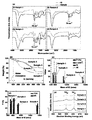
- FIG. 6 is a view for explaining the analysis results of Samples 1 to 4 according to the present invention.
- FIG. 7 is a view for explaining the magnetic properties of the samples 1 to 4 according to the present invention.
- FIG. 8 is a view for explaining the relaxation characteristics and X-ray absorption ability of Samples 1 to 4.
- FIG. 8 is a view for explaining the relaxation characteristics and X-ray absorption ability of Samples 1 to 4.
- FIG. 10 is a view for explaining the result when the sample 1 is applied to the rat.
- 1 is a schematic diagram for explaining the nanostructure of the gadolinium composite according to the present invention.
- the gadolinium complex 100 includes gadolinium oxide particles 110 and a surface layer 120.
- the iodide compound constituting the surface layer 120 in FIG. 1 is denoted by reference numeral "122", and in the iodide compound, in particular, the portion bonded with the gadolinium oxide particles 110 is referred to as a "functional group". It is shown by the number "124".
- the gadolinium oxide particle 110 is a nanoparticle made of gadolinium oxide (Gd 2 O 3 ).
- the gadolinium oxide particles 110 may be prepared by mixing heat treatment by mixing gadolinium chloride with sodium hydroxide.
- each of gadolinium chloride and sodium hydroxide may be mixed with each other in a state of being dissolved in ethylene glycol, and a first solution containing gadolinium chloride and ethylene glycol and a second solution containing sodium hydroxide and ethylene glycol are mixed.
- the gadolinium oxide particles 110 may be manufactured by heat treatment at 150 to 210 ° C.
- Triethylene glycol is mentioned as an example of ethylene glycol.
- the surface layer 120 is a layer made of the iodide compound 122 and is formed to cover at least a part of the surface of the gadolinium oxide particles 110.
- the surface layer 120 may cover the entire surface of the gadolinium oxide particles 110, or may partially cover only part of the surface.
- the iodide compound 122 is spaced apart from the upper surface of the gadolinium oxide particle 110 for convenience of description, but the functional group 124 of the iodide compound 122 and the gadolinium oxide particle 110 are separated from each other.
- the gadolinium complex 100 may be manufactured by preparing a third solution including the iodide compound 122, mixing the mixture with the gadolinium oxide particles 110, and performing a heat treatment.
- the solvent of the third solution may also be ethylene glycol, and the temperature at which the third solution is mixed and heat treated may be 90 to 130 ° C.
- the iodide compound 122 binds to the gadolinium oxide particles 110 through the functional group 124, which is a carboxylic group (-COOH), a hydroxyl group (-OH) or an amine group ( amine group, -NH 2 ).
- An oxygen atom or a nitrogen atom of the functional group 124 may be combined with the gadolinium oxide particle 110 so that the iodide compound 122 may be a ligand of the gadolinium oxide particle 110.
- the iodide compound 122 may be a compound having a structure in which at least one hydrogen atom is substituted with iodine as a skeleton, and a functional group 124 is bonded to the benzene ring.
- a functional group 124 is bonded to the benzene ring.
- 5-amino-2,4,6-triiodoisophthalic acid (5-Amino-2,4,6-triiodoisophthalic acid)
- 3-[[6- (3-carboxy-2 , 4,6-triiodoanilino) -6-oxohexanol] amino] -2,4,6-triiodobenzoic acid 3-[[6- (3-carboxy-2,4,6-triiodoanilino ) -6-oxohexanoyl] amino] -2,4,6-triiodobenzoic acid
- 2 to 5 are views for explaining the bonding structure of the core particles and the surface layer in the gadolinium composite of various components according to the present invention.
- FIG. 2 is a diagram illustrating a gadolinium complex including 5-amino-2,4,6-triiodoisophthalic acid as an iodide compound.
- the iodide compound includes a carboxyl group as a functional group and an oxygen of a carboxyl group. Atoms combine with the surface of the gadolinium oxide particles 110 to form a gadolinium complex.
- an iodide compound includes a carboxyl group and an amine group as a functional group, and oxygen atoms of the carboxyl group may be combined with the surface of the gadolinium oxide particle 110 to form a gadolinium complex. . Unlike FIG. 3, the gadolinium oxide particles 110 may be combined with the amine group of the iodide compound to form the gadolinium complex 100.
- FIG. 4 is a view for explaining a gadolinium complex including 3,5-diacetamido-2,4,6-triiodobenzoic acid as an iodide compound.
- the iodide compound is also a functional group. It includes a carboxyl group and oxygen atoms of the carboxyl group is combined with the surface of the gadolinium oxide particles 110 to form a gadolinium complex.
- FIG. 5 shows 5,5 '-(2-hydroxypropane-1,3-diyl) bis (acetylazanediyl) bis (N1, N3-bis (2,3-hydroxypropyl) -2, as an iodide compound.
- 5 is a diagram for explaining a gadolinium complex including 4,6-triiodoisophthalamide.
- an iodide compound includes two or more hydroxy groups as a functional group, and oxygen atoms of the hydroxyl group are gadolinium oxide particles.
- Figure 5 shows that the iodide compound contains eight hydroxy groups, of which four oxygen atoms of the four hydroxy groups are combined with the gadolinium oxide particles (110). Although illustrated, the remaining four hydroxyl groups may also form a ligand by combining with the gadolinium oxide particles 110.
- the gadolinium complex 100 described with reference to FIGS. 1 to 5 includes the inorganic gadolinium oxide particles 110 as the core, the surface layer 120 containing the iodide compound 122 is formed on the surface thereof, so that the water-soluble and biocompatibility is improved. Can be improved.
- the gadolinium complex 100 may be used as an MRI-CT dual image contrast agent because it shows contrast characteristics for CT as well as MRI.
- the MRI-CT dual imaging contrast medium has little toxicity in vivo and can be easily used to safely diagnose the in vivo condition of humans. Synthesis method of gadolinium complex 100 according to the present invention, the actual manufacturing possibility, the availability as a dual image contrast agent, etc. will be described in detail through the following experiment.
- a first solution was prepared by mixing 5 mmol of gadolinium precursor GdCl 3? xH 2 O in 25 mL of triethylene glycol, and (ii) 15 mmol of sodium hydroxide in 10 mL of triethylene glycol ( NaOH) was mixed to prepare a second solution, and (iii) a third solution was prepared by mixing 5 mmol of 5-amino-2,4,6-triidoisophthalic acid with 10 mL of triethylene glycol.
- the preparation of the first solution a heating process was performed at 60 ° C. with magnetic stirring under atmospheric pressure until the gadolinium precursor was completely dissolved in triethylene glycol. Subsequently, the second solution was mixed with the first solution, and the mixed solution of the first and second solutions was magnetically stirred at 180 ° C. for 4 hours.
- Example 1 a gadolinium complex according to Example 1 of the present invention was prepared.
- Example 2 3-[[6- (3-carboxy-2,4,6-triiodoanilino) -6-oxohexanol] amino] -2,4,6-triiodobenzoic acid
- a gadolinium complex (Sample 2) according to Example 2 of the present invention was prepared in the same manner as in the preparation of Sample 1, except that Adipiodone (trade name, Sinografin) was used.
- FT-IR Fastier transform infrared
- TGA thermogravimetric analysis
- surface layer properties P (weight percent of iodide compound) value Comparative analysis and X-ray photoelectron spectroscopy (XPS) analysis were performed, and the results are shown in FIG. 6.
- the surface layer properties are analyzed by the weight percent (i) of the iodide compound, the grafting density (i) of the iodide compound, the number of iodide compounds per gadolinium complex (N) and the number of iodine atoms per gadolinium complex (NI) It was.
- FT-IR analysis was measured at 400 to 4,000 cm -1 using the Galaxy 7020A (trade name, Mattson Instruments, Inc.) as an analytical instrument.
- TGA analysis was measured in the temperature range from room temperature up to 700 °C using SDT-Q 600 (trade name, TA Instruments).
- Flash 2000 (trade name, ThermoFisher) as an EA device and Quantera SXM (trade name, ULVAC-PHI) for XPS measurements.
- FIG. 6 is a view for explaining the analysis results of Samples 1 to 4 according to the present invention.
- Fig. 6 shows FT-IR analysis results, (b) shows TGA analysis results, (c) shows surface layer characterization results, and (d) shows P values from TGA and elemental analyzer (EA).
- E is the result of X-ray photoelectron spectrometer (XPS) analysis.
- FIG. 6A four graphs relate to Samples 1 to 4, respectively, and show graphs (red lines) and graphs of pre-iodinated compounds (blue lines) of Samples 1 to 4, respectively.
- all of the samples 1 to 4 have absorption peaks showing a tendency almost similar to that of the iodide compound, and it can be confirmed that the iodide compound is coated on the surface of the gadolinium oxide particles.
- each of the samples 1 to 4 represents a specific value, and when the values calculated in each of the samples 1 to 4 are compared, the larger the weight of the iodide compound is, the weight percentage of the iodide compound (P) It can be seen that the grafting density (?) Of the iodide compound, the number of iodide compounds per gadolinium complex (N), and the number of iodine atoms (ni) per gadolinium complex have small values. This may be because the molecules of the iodide compound having a high mass are coated with the gadolinium oxide particles, so that the mass iodide compound takes up a larger volume on the surface of the gadolinium oxide particles.
- each of samples 1 to 4 represents a specific value distinguished.
- the EA data shows a relatively large value compared to the P value estimated by the TGA result because the moisture content has not been removed, but except for this, it shows a value consistent with the EA analysis result and the TGA value. Able to know.
- FIG. 7 is a view for explaining the magnetic properties of the samples 1 to 4 according to the present invention.
- (a) shows the M-H graph and (b) shows the M-T graph.
- the MH graphs at temperatures 5K and 300K, respectively, show that coercivity and remanence are substantially zero in both samples 1 to 4 (hysteresis). ) Does not occur).
- Relaxation times T 1 and T 2 and map images R 1 and R 2 were measured using a GE 1.5T Sigma Advantage (trade name, GE Healthcare), a 1.5 Tesla MRI scanner with knee coils. .
- Samples 1 and 5 aqueous solutions having concentrations of 1 mM, 0.5 mM, 0.25 mM, 0.125 mM and 0.0625 mM were prepared using distilled water, and 5 samples having different concentrations for each sample using the same samples 2 to 4 respectively. Aqueous solutions were prepared. Using these, relaxation times T 1 and T 2 and map images R 1 and R 2 were measured.
- Water proton relaxivity r 1 and r 2 were estimated from the slope of the plots of 1 / T 1 and 1 / T 2 with respect to gadolinium concentration, and the measurement of T 1 was performed using reverse recovery method. The measurement of T 2 was carried out using CPMG (Carr Purcell Meiboon Gill pulse sequence). The result is shown in FIG.
- FIG. 8 is a view for explaining the relaxation characteristics and X-ray absorption ability of Samples 1 to 4.
- FIG. 8 is a view for explaining the relaxation characteristics and X-ray absorption ability of Samples 1 to 4.
- (a) is a graph of 1 / T 1 and 1 / T 2
- (b) is a map image R 1 and R 2
- (c) is a graph of r 1 and r 2
- (d ) Is a graph showing X-ray absorption.
- the values of r 1 and r 2 in the solution state are estimated from the slope and 1 / T1 and 1 / T2, and the estimated values of r 1 and r 2 are Gd-DTPA (Gd ( III) -diethylenetriamine pentaacetic acid) or Gd-DOTA (Gd (III) -1,4,7,10-tetraazacyclododecane-1,4,7,10-tetraacetic acid)
- Gd-DTPA Gd ( III) -diethylenetriamine pentaacetic acid
- Gd-DOTA Gd (III) -1,4,7,10-tetraazacyclododecane-1,4,7,10-tetraacetic acid
- the value of r 1 must be increased and the value of r 1 / r 2 should be close to one. According to the results shown in (a) and (b) of FIG. 8, it can be seen that the T 1 MRI contrast agent having better relaxometric properties than the commercially available contrast agent.
- the values of r 1 and r 2 are different from each other in the samples 1 to 4 according to the size of the iodide compound.
- the different values of r 1 and r 2 in each of Samples 1 to 4 can be inferred to be due to iodide compounds based on the use of the same gadolinium oxide particles.
- Water accessibility to gadolinium oxide particles depends on the size of the ligand. In general, more water molecules allow larger size ligands to access the gadolinium oxide particles, reducing the values of r 1 and r 2 , which can be seen in Samples 1-4.
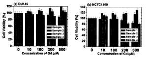
- FIG. 9 is a diagram for explaining in vitro cytotoxicity of each of the samples 1 to 4,
- Figure 9 (a) shows the viability of DU1445, (b) shows the viability of NCTC1469.
- a contrast agent prepared by applying 0.1 mmol / kg of sample 1 to mice (hereinafter referred to as ICR mice) provided by the Institute of Cancer Research (ICR) in the United States was prepared by intravenous injection and obtained by MRI.
- a contrast agent prepared by applying 0.56 mmol / kg of sample 1 was prepared by intravenous injection, and CT images were obtained. The result is shown in FIG.
- FIG. 10 is a view for explaining the result when the sample 1 is applied to the rat.
- (a) is between a T 1 MRI image of the (liver, as represented by L), (b) the height (kidneys, as represented by K), and T 1 of the aorta (indicated by aorta, A) It is an MRI image, and (c) is a CT image of a bladder (denoted by B).
- Sample 4 was used to prepare a contrast agent, which was then administered to rats, followed by MRI and CT imaging to acquire images.
- Omniscan was used as a commercial MRI contrast agent and a commercially available Ultravist was used as a CT contrast agent. The result is shown in FIG.
- (a-i) is an MRI image of Sample 4
- (a-ii) is an MRI image of Omniscan
- (b-i) is a CT image of Sample 4
- (b-ii) is a CT image of Ultravist.
Landscapes
- Health & Medical Sciences (AREA)
- Nuclear Medicine, Radiotherapy & Molecular Imaging (AREA)
- Radiology & Medical Imaging (AREA)
- Epidemiology (AREA)
- Life Sciences & Earth Sciences (AREA)
- Animal Behavior & Ethology (AREA)
- General Health & Medical Sciences (AREA)
- Public Health (AREA)
- Veterinary Medicine (AREA)
- Chemical & Material Sciences (AREA)
- Medicinal Chemistry (AREA)
- Magnetic Resonance Imaging Apparatus (AREA)
- Medicines Containing Antibodies Or Antigens For Use As Internal Diagnostic Agents (AREA)
Abstract
가돌리늄 복합체, 이를 포함하는 MRI-CT 이중영상 조영제 및 가돌리늄 복합체의 제조 방법에서, 가돌리늄 복합체는 산화가돌리늄 입자의 표면에 배치된, 카르복실기, 하이드록시기 및 아민기 중 적어도 어느 하나를 작용기로 갖는 요오드화 화합물을 포함한다.
Description
본 발명은 가돌리늄 복합체, 이를 포함하는 MRI-CT 이중영상 조영제 및 가돌리늄 복합체의 제조 방법에 관한 것으로, 보다 구체적으로는 수용성이면서 생체적합성이 높은 가돌리늄 복합체, 이를 포함하는 MRI-CT 이중영상 조영제 및 가돌리늄 복합체의 제조 방법에 관한 것이다.
방사선 분야의 대표적인 첨단 영상 장치로서, X-선을 사용하는 컴퓨터 단층촬영(Computed Tomography, 이하, CT라 함)은 뼈처럼 밀도 있는 구조물을 영상화하는데 효과적인 영상장치이나 부드러운 조직을 영상화하는 데는 선명도가 크게 감소하기 때문에 조직과 주변부위를 구분하는데 한계가 있다. 이러한 CT의 한계를 극복하기 위하여 CT용 조영제(contrast agent)에 대한 많은 연구가 진행되고 있고, 현재에는 Ultravist(상품명, 바이엘, 미국)와 같은 요오드를 기반으로 하는 조영제가 가장 광범위하게 사용되고 있다.
자기공명영상(Magnetic Resonance Imaging, 이하, MRI라 함)이란 자장을 발생하는 커다란 자석통 속에 인체를 들어가게 한 후 고주파를 발생시켜 신체부위에 있는 양성자를 공명시켜 각 조직에서 나오는 신호의 차이를 측정하여 컴퓨터를 통해 재구성하여, 영상화하는 기술이다. 이러한 MRI는 다른 영상법에 비해 해상도 및 대조도가 높고 심부장기영상과 3차원 정보 제공이 실시간으로 가능한 장점을 가지고 있어, 진단용으로 널리 이용되고 있다. MRI는 고유한 조직 신호 차이를 다양한 기법으로 생체 영상화 할 수 있지만, 조영제를 사용하는 경우에 조직 내 물 분자의 자기이완시간을 변화시켜 조직의 대조도를 더욱 증가시켜 시인성을 높일 수 있으므로 보다 정확하게 진단할 수 있다.
CT와 MRI는 서로 영상을 획득하는 방식이 다르기 때문에 서로 다른 종류의 조영제를 이용한다. 최근에는, 동일한 진단부위에서의 2 개 이상의 진단영상 획득할 수 있는 다중영상 조영제에 대한 연구가 활발하게 이루어지고 있으며 경북대학교 장용민 교수 연구팀에서는 2010년에 금 나노입자 표면에 수천개의 가돌리늄(gadolinium) 리간드를 결합시킨 형태의 MRI/CT 조영제를 개발한 바 있다(Ji-Ae Park, et al, Gold nanoparticles functionalized by gadolinium-DTPA conjugate of cysteine as a multimodal bioimaging agent, BMCL, 2010, 20, 2287-2291).
그러나 본 발명자는 이와 다른 구조와 성분을 갖는 아주 작은 가돌리늄 복합체를 합성하여 MRI/CT 조영제를 발명하였다.
본 발명의 일 목적은 수용성이고 생체적합성이 높은 가돌리늄 복합체를 제공하는 것이다.
본 발명의 다른 목적은 가돌리늄 복합체를 포함하는 MRI-CT 이중영상 조영제를 제공하는 것이다.
본 발명의 또 다른 목적은 가돌리늄 복합체의 제조 방법을 제공하는 것이다.
본 발명의 일 목적을 위한 가돌리늄 복합체는 산화가돌리늄 입자의 표면에 배치된, 카르복실기, 하이드록시기 및 아민기 중 적어도 어느 하나를 작용기로 갖는 요오드화 화합물을 포함한다.
일 실시예에서, 상기 요오드화 화합물은 적어도 1개 이상의 수소 원자가 요오드로 치환된 벤젠고리에 상기 작용기가 결합된 구조를 갖는 화합물일 수 있다.
일 실시예에서, 상기 요오드화 화합물은 5-아미노-2,4,6-트리아이오도이소프탈산(5-Amino-2,4,6-triiodoisophthalic acid), 3-[[6-(3-카복시-2,4,6-트리아이오도아닐리노)-6-옥소헥사놀] 아미노]-2,4,6-트리아이오도벤조산(3-[[6-(3-carboxy-2,4,6-triiodoanilino)-6-oxohexanoyl]amino]-2,4, 6-triiodobenzoic acid), 3,5-디아세트아미도-2,4,6-트리아이오도벤조산(3,5-diacetamido-2,4,6-triiodobenzoic acid) 및/또는 5,5'-(2-하이드록시프로판-1,3-디일)비스(아세틸아잔디일)비스(N1,N3-비스(2,3-하이드록시프로필)-2,4,6-트리아이오도이소프탈아미드)[5,5'-(2-hydroxypropane-1,3-diyl) bis(acetylazanediyl)bis(N1,N3-bis(2,3-dihydroxypropyl)-2,4,6-triiodoisophthalamide)]일 수 있다.
일 실시예에서, 상기 요오드화 화합물의 상기 작용기가 산화가돌리늄 입자의 표면과 화학 결합하여 배치될 수 있다.
본 발명의 다른 목적을 위한 MRI-CT를 위한 이중영상 조영제는 산화가돌리늄 입자의 표면에, 카르복실기, 하이드록시기 및 아민기 중 적어도 어느 하나를 작용기로 갖는 요오드화 화합물이 배치된 가돌리늄 복합체를 포함한다.
본 발명의 또 다른 목적을 위한 가돌리늄 복합체의 제조 방법은, 산화가돌리늄 입자를 제조하는 단계 및 상기 산화가돌리늄 입자에 카르복실기 및 하이드록시기 중 적어도 어느 하나를 작용기로 갖는 요오드화 화합물을 혼합하여, 상기 산화가돌리늄 입자에 상기 요오드화 화합물을 포함하는 표면층을 형성하는 단계를 포함한다.
일 실시예에서, 상기 산화가돌리늄 입자는 산화가돌리늄 전구체을 포함하는 제1 용액에, 수산화나트륨을 포함하는 제2 용액을 혼합하여 제조할 수 있다.
일 실시예에서, 상기 요오드화 화합물을 포함하는 용액, 상기 제1 용액 및 상기 제2 용액 각각은 용매로서 에틸렌계 글리콜을 이용할 수 있다.
일 실시예에서, 상기 산화가돌리늄 입자를 제조하는 단계는 상기 제1 및 제2 용액들이 혼합된 상태에서 150 내지 220℃의 온도 조건으로 열처리하는 단계를 포함하고, 상기 표면층을 형성하는 단계는 상기 산화가돌리늄 입자와 상기 요오드화 화합물을 포함하는 용액이 혼합된 상태에서 90 내지 130℃의 온도 조건으로 열처리하는 단계를 포함할 수 있다.
본 발명의 가돌리늄 복합체, 이를 포함하는 MRI-CT 이중영상 조영제 및 가돌리늄 복합체의 제조 방법에 따르면, 수용성이고 생체적합성이 높은 가돌리늄 복합체를 제공할 수 있다. 이러한 가돌리늄 복합체는 독성이 없어 인체에 조영제로 이용할 수 있으며, 한 번의 투여로 MRI와 CT 이미지를 모두 얻을 수 있는 장점이 있다.
도 1은 본 발명에 따른 가돌리늄 복합체의 나노 구조를 설명하기 위한 모식도이다.
도 2 내지 도 5는 본 발명에 따른 다양한 성분의 가돌리늄 복합체에서 코어 입자와 표면층의 결합 구조를 설명하기 위한 도면들이다.
도 6은 본 발명에 따른 샘플 1 내지 4의 분석 결과를 설명하기 위한 도면이다.
도 7은 본 발명에 따른 샘플 1 내지 4의 자기적 특성을 설명하기 위한 도면이다.
도 8은 샘플 1 내지 4의 릴랙소메트릭 특성 및 X-선 흡수능을 설명하기 위한 도면이다.
도 9는 샘플 1 내지 4 각각의 생체 외 세포독성을 설명하기 위한 도면이다.
도 10은 샘플 1을 쥐에 적용한 경우의 결과를 설명하기 위한 도면이다.
도 11은 샘플 4와 시판하는 조영제의 조영 특성을 설명하기 위한 도면이다.
이하, 첨부한 도면을 참조하여 본 발명의 실시예에 대해 상세히 설명한다. 본 발명은 다양한 변경을 가할 수 있고 여러 가지 형태를 가질 수 있는 바, 특정 실시예들을 도면에 예시하고 본문에 상세하게 설명하고자 한다. 그러나 이는 본 발명을 특정한 개시 형태에 대해 한정하려는 것이 아니며, 본 발명의 사상 및 기술 범위에 포함되는 모든 변경, 균등물 내지 대체물을 포함하는 것으로 이해되어야 한다. 각 도면을 설명하면서 유사한 참조부호를 유사한 구성요소에 대해 사용하였다.
다르게 정의되지 않는 한, 기술적이거나 과학적인 용어를 포함해서 여기서 사용되는 모든 용어들은 본 발명이 속하는 기술 분야에서 통상의 지식을 가진 자에 의해 일반적으로 이해되는 것과 동일한 의미를 가지고 있다. 일반적으로 사용되는 사전에 정의되어 있는 것과 같은 용어들은 관련 기술의 문맥 상 가지는 의미와 일치하는 의미를 가지는 것으로 해석되어야 하며, 본 출원에서 명백하게 정의하지 않는 한, 이상적이거나 과도하게 형식적인 의미로 해석되지 않는다.
도 1은 본 발명에 따른 가돌리늄 복합체의 나노 구조를 설명하기 위한 모식도이다.
도 1을 참조하면, 가돌리늄 복합체(100)는 산화가돌리늄 입자(110) 및 표면층(120)을 포함한다. 설명의 편의를 위해서, 도 1에서 표면층(120)을 구성하는 요오드화 화합물은 도면번호 "122"로 표시하고, 요오드화 화합물에서 특히 산화가돌리늄 입자(110)와 결합된 부분을 "작용기"라고 지칭하며 도면번호 "124"로 도시한다.
산화가돌리늄 입자(110)는 산화가돌리늄(gadolinium oxide, Gd2O3)으로 이루어진 나노 입자이다. 산화가돌리늄 입자(110)는 염화가돌리늄을 수산화나트륨과 혼합하여 열처리함으로써 제조할 수 있다. 이때, 염화가돌리늄 및 수산화나트륨 각각은 에틸렌계 글리콜에 용해된 상태로 서로 혼합될 수 있으며, 염화가돌리늄 및 에틸렌계 글리콜을 포함하는 제1 용액과 수산화나트륨과 에틸린계 글리콜을 포함하는 제2 용액이 혼합된 상태에서 150 내지 210℃에서 열처리함으로써 산화가돌리늄 입자(110)가 제조될 수 있다. 에틸렌계 글리콜의 예로서는, 트리에틸렌 글리콜을 들 수 있다.
표면층(120)은 요오드화 화합물(122)로 이루어진 층으로서, 산화가돌리늄 입자(110) 표면의 적어도 일부를 커버하도록 형성된다. 일례로, 표면층(120)은 산화가돌리늄 입자(110)의 표면을 전체적으로 피복할 수도 있고, 일부만 부분적으로 피복할 수도 있다. 도 1에서는 설명의 편의를 위해서 요오드화 화합물(122)이 산화가돌리늄 입자(110) 표면의 상부에 배치된 것으로 이격시켜 도시하였으나, 요오드화 화합물(122)의 작용기(124)와 산화가돌리늄 입자(110)의 표면에는 화학 결합, 물리적 결합 및/또는 전기적 인력이 존재할 수 있으며, 표면층(120)은 리간드 역할을 하는 요오드화 화합물(122)의 그룹일 수 있다.
요오드화 화합물(122)을 포함하는 제3 용액을 제조하여, 산화가돌리늄 입자(110)에 혼합하여 열처리함으로써 가돌리늄 복합체(100)를 제조할 수 있다. 이때, 상기 제3 용액의 용매 또한 에틸렌계 글리콜일 수 있고, 제3 용액을 혼합하여 열처리하는 온도는 90 내지 130℃일 수 있다.
요오드화 화합물(122)은 작용기(124)를 통해서 산화가돌리늄 입자(110)와 결합하는데, 작용기(124)는 카르복실기(carboxylic group, -COOH), 하이드록시기(hydroxyl group, -OH) 또는 아민기(amine group, -NH2)일 수 있다. 작용기(124)의 산소 원자 또는 질소 원자가 산화가돌리늄 입자(110)와 결합하여 요오드화 화합물(122)이 산화가돌리늄 입자(110)의 리간드가 될 수 있다.
요오드화 화합물(122)은 골격으로서 적어도 1개의 수소 원자가 요오드로 치환된 벤젠 고리를 포함하고, 상기 벤젠 고리에 작용기(124)가 결합된 구조의 화합물일 수 있다. 요오드화 화합물(122)의 예로서는, 5-아미노-2,4,6-트리아이오도이소프탈산(5-Amino-2,4,6-triiodoisophthalic acid), 3-[[6-(3-카복시-2,4,6-트리아이오도아닐리노)-6-옥소헥사놀]아미노]-2,4,6-트리아이오도벤조산(3-[[6-(3-carboxy-2,4,6-triiodoanilino)-6-oxohexanoyl]amino]-2,4,6-triiodobenzoic acid), 3,5-디아세트아미도-2,4,6-트리아이오도벤조산(3,5-diacetamido-2,4,6-triiodobenzoic acid), 5,5'-(2-하이드록시프로판-1,3-디일)비스(아세틸아잔디일)비스(N1,N3-비스(2,3-하이드록시프로필)-2,4,6-트리아이오도이소프탈아미드)[5,5'-(2-hydroxypropane-1,3-diyl)bis(acetylazanediyl)bis(N1,N3-bis(2,3-dihydroxypropyl)-2,4,6-triiodoisophthalamide)] 등을 들 수 있다. 이들은 각각 단독으로 또는 2 이상을 조합하여 이용할 수 있다. 요오드화 화합물(122)과 산화가돌리늄 입자(110)의 결합 구조에 대해서는 도 2 내지 도 5를 참조하여 후술하여 설명하기로 한다.
도 2 내지 도 5는 본 발명에 따른 다양한 성분의 가돌리늄 복합체에서 코어 입자와 표면층의 결합 구조를 설명하기 위한 도면들이다.
도 2는 요오드화 화합물로서 5-아미노-2,4,6-트리아이오도이소프탈산을 포함하는 가돌리늄 복합체를 설명하기 위한 도면으로, 도 2를 참조하면 요오드화 화합물은 작용기로서 카르복시기를 포함하며 카르복시기의 산소원자들이 산화가돌리늄 입자(110)의 표면과 결합하여 가돌리늄 복합체를 구성한다.
도 3은 요오드화 화합물로서 3-[[6-(3-카복시-2,4,6-트리아이오도아닐리노)-6-옥소헥사놀]아미노]-2,4,6-트리아이오도벤조산을 포함하는 가돌리늄 복합체를 설명하기 위한 도면으로, 도 3을 참조하면 요오드화 화합물은 작용기로서 카르복시기 및 아민기를 포함하며 카르복시기의 산소원자들이 산화가돌리늄 입자(110)의 표면과 결합하여 가돌리늄 복합체를 구성할 수 있다. 도 3과 달리, 산화가돌리늄 입자(110)는 요오드화 화합물의 아민기와 결합하여 가돌리늄 복합체(100)를 구성할 수 있다.
도 4는 요오드화 화합물로서 3,5-디아세트아미도-2,4,6-트리아이오도벤조산을 포함하는 가돌리늄 복합체를 설명하기 위한 도면으로, 도 4를 참조하면 이 경우 또한 요오드화 화합물은 작용기로서 카르복시기를 포함하며 카르복시기의 산소원자들이 산화가돌리늄 입자(110)의 표면과 결합하여 가돌리늄 복합체를 구성한다.
도 5는 요오드화 화합물로서 5,5'-(2-하이드록시프로판-1,3-디일)비스(아세틸아잔디일)비스(N1,N3-비스(2,3-하이드록시프로필)-2,4,6-트리아이오도이소프탈아미드를 포함하는 가돌리늄 복합체를 설명하기 위한 도면으로, 도 5를 참조하면 요오드화 화합물은 작용기로서 2개 이상의 하이드록시기를 포함하며 하이드록시기의 산소원자들이 산화가돌리늄 입자(110)의 표면과 결합하여 가돌리늄 복합체를 구성한다. 도 5에서는 요오드화 화합물이 8개의 하이드록시기를 포함하고 있고 이중 4개의 하이드록시기들의 4개의 산소원자들이 산화가돌리늄 입자(110)와 결합된 것을 도시하였으나 나머지 4개의 하이드록시기 또한 산화가돌리늄 입자(110)와 결합하여 리간드를 이룰 수 있다.
도 1 내지 도 5에서 설명한 가돌리늄 복합체(100)는 비록 코어로서 무기물인 산화가돌리늄 입자(110)를 포함하지만 요오드화 화합물(122)을 포함하는 표면층(120)이 그 표면에 형성됨으로써 수용성 및 생체적합성을 향상시킬 수 있다. 또한, 가돌리늄 복합체(100)는 MRI 뿐만 아니라 CT에 대해서도 조영 특성을 나타내므로 MRI-CT 이중영상 조영제로 이용될 수 있다. 이 MRI-CT 이중영상 조영제는 생체 내에서의 독성이 거의 없어 안전하게 인간의 생체 내 상태를 진단하는데 용이하게 이용할 수 있다. 본 발명에 따른 가돌리늄 복합체(100)의 합성 방법, 실제 제조 가능성, 이중영상 조영제로서의 이용 가능성 등은 하기 실험을 통해서 구체적으로 설명하기로 한다.
샘플 1의 제조
(i) 트리에틸렌 글리콜(triethylene glycol) 25 mL에 5 mmol의 가돌리늄 전구체인 GdCl3ㅇxH2O를 혼합하여 제1 용액을 제조하였고, (ii) 트리에틸렌 글리콜 10 mL에 15 mmol의 수산화나트륨(NaOH)을 혼합하여 제2 용액을 준비하였으며, (iii) 10 mL의 트리에틸렌 글리콜에 5-아미노-2,4,6-트리아이도이소프탈산 5 mmol을 혼합하여 제3 용액을 준비하였다.
상기 제1 용액의 제조에서는, 상기 가돌리늄 전구체가 트리에틸렌 글리콜에 완전히 녹을 때까지 대기압 조건 하에서 자석 교반으로 60℃에서 가열 공정을 수행하였다. 이어서, 상기 제2 용액을 상기 제1 용액에 혼합하고, 상기 제1 및 제2 용액들의 혼합 용액을 180℃에서 4 시간동안 자석 교반하였다.
산화가돌리늄 입자에 표면층의 형성을 위해서, 온도를 60℃로 낮추고 여기에 상기 제3 용액을 천천히 첨가하였다. 다시 반응 온도를 110℃로 승온시키고 12 시간동안 자석 교반하였다. 상온으로 냉각시키고 증류수를 이용한 세척 공정을 거쳐 본 발명의 실시예 1에 따른 가돌리늄 복합체(샘플 1)를 제조하였다.
샘플 2 내지 4의 제조
제3 용액의 제조에 3-[[6-(3-카복시-2,4,6-트리아이오도아닐리노)-6-옥소헥사놀]아미노]-2,4,6-트리아이오도벤조산으로서 Adipiodone (상품명, Sinografin사)을 이용한 것을 제외하고는 샘플 1의 제조와 실질적으로 동일한 공정을 통해서 본 발명의 실시예 2에 따른 가돌리늄 복합체(샘플 2)를 제조하였다.
또한, 제3 용액의 제조에 3,5-디아세트아미도-2,4,6-트리아이오도벤조산으로서 Hypaque (상품명, GE healthcare사)을 이용한 것을 제외하고는 샘플 1의 제조와 실질적으로 동일한 공정을 통해서 본 발명의 실시예 3에 따른 가돌리늄 복합체(샘플 3)를 제조하였다.
또한, 제3 용액의 제조에 5,5'-(2-하이드록시프로판-1,3-디일)비스(아세틸아잔디일)비스(N1,N3-비스(2,3-하이드록시프로필)-2,4,6-트리아이오도이소프탈아미드)로서 Visipaque (상품명, GE healthcare사)을 이용한 것을 제외하고는 샘플 1의 제조와 실질적으로 동일한 공정을 통해서 본 발명의 실시예 4에 따른 가돌리늄 복합체(샘플 4)를 제조하였다.
샘플 1 내지 4의 분석
상기와 같이 준비한 샘플 1 내지 4 각각에 대해서, FT-IR(Fourier transform infrared, 퓨리에 변환 적외선) 흡수 분석, TGA(열중량분석, thermogravimetric analysis) 곡선, 표면층 특성, P(요오드화 화합물의 중량%)값의 비교 분석 및 XPS(X선 광전자 분광법) 분석을 수행하였고, 그 결과를 도 6에 나타낸다. 이때, 표면층 특성은 요오드화 화합물의 중량%(P), 요오드화 화합물의 그래프팅 밀도(grafting density, σ), 가돌리늄 복합체당 요오드화 화합물의 수(N) 및 가돌리늄 복합체 당 요오드 원자의 수(NI)로 분석하였다.
FT-IR 분석은 분석 장비로서, Galaxy 7020A(상품명, Mattson Instruments, Inc.사)을 이용하여 400 내지 4,000 cm-1에서 측정하였다.
TGA 분석은 SDT-Q 600(상품명, TA Instruments사)을 이용하여 상온에서부터 700℃ 이하의 온도 범위에서 측정하였다.
표면층 특성 분석은, EA 장치로서 Flash 2000(상품명, ThermoFisher사)을 이용하고 XPS 측정을 위해 Quantera SXM(상품명, ULVAC-PHI사)을 이용하여 수행하였다.
도 6은 본 발명에 따른 샘플 1 내지 4의 분석 결과를 설명하기 위한 도면이다.
도 6에서, (a)가 FT-IR 분석 결과를 나타내고, (b)가 TGA 분석 결과이며, (c)가 표면층 특성 분석 결과이며, (d)가 TGA와 EA(elemental analyzer)로부터 P값의 비교 분석 결과이며, (e)가 XPS(X-ray photoelectron spectrometer) 분석 결과이다.
도 6의 (a)에서, 4개의 그래프들은 각각 샘플 1 내지 샘플 4에 관한 것이며, 샘플 1 내지 샘플 4 각각의 그래프(빨강선)와 프리-요오드화 화합물의 그래프(파랑선)을 나타낸다. 여기서, 샘플 1 내지 4 모두에서 요오드화 화합물의 흡수 피크와 거의 유사한 경향을 나타내는 흡수 피크들이 나타남을 알 수 있고, 이를 통해서 요오드화 화합물이 산화가돌리늄 입자의 표면에 코팅된 것을 확인할 수 있다.
또한, 샘플 1 내지 샘플 3 각각에서는, 프리-요오드화 화합물의 경우에 비해서 가돌리늄 복합체의 파장이 단파장으로 약 74 내지 75 cm-
1만큼 시프트한 것을 확인할 수 있는데, 이는 산화가돌리늄 입자와 요오드화 화합물의 카르복실기의 결합이 존재함을 알 수 있다. 샘플 4에서는, 산화가돌리늄 입자와 요오드화 화합물의 하이드록시기의 결합이 존재함을 예측할 수 있다.
도 6의 (b)를 참조하면, 샘플 1 내지 4 각각에서 산화가돌리늄 입자가 요오드화 화합물에 의해 충분히 코팅되었음을 알 수 있다.
도 6의 (c)를 참조하면, 샘플 1 내지 4 각각이 구분되는 특정한 값을 나타내고, 샘플 1 내지 4 각각에서 계산된 값들을 비교하면, 요오드화 화합물의 중량이 클수록 요오드화 화합물의 중량%(P), 요오드화 화합물의 그래프팅 밀도(grafting density, σ), 가돌리늄 복합체당 요오드화 화합물의 수(N) 및 가돌리늄 복합체 당 요오드 원자의 수(NI)는 작은 값을 갖는 것을 알 수 있다. 이는, 질량이 큰 요오드화 화합물의 분자가 산화가돌리늄 입자가 코팅됨으로써, 질량이 큰 요오드화 화합물이 산화가돌리늄 입자의 표면에서 더 큰 부피를 차지하기 때문인 것으로 볼 수 있다.
도 6의 (d)를 참조하면, 샘플 1 내지 4 각각이 구분되는 특정한 값을 나타내는 것을 알 수 있다. EA 데이터에서는 수분의 함량이 제거되지 않았기 때문에 TGA의 결과에 의해 추정된 P값에 비해 상대적으로 큰 값을 나타내지만, 이를 제외하고는 EA 분석 결과와 TGA에 의한 값과 일관성이 있는 값을 나타내는 것을 알 수 있다.
도 6의 (e)를 참조하면, 전자 결합 에너지(electron binding enegery, EBE) 스케일에서, 예를 들어 약 618.8 eV(3d5/2)와 630.2 eV(3d3/2)에서 특정 피크가 나타나며, 이는 샘플 1 내지 4 각각에서 모두 나타나는 것을 알 수 있다. 즉, 샘플 1 내지 4 모두에서 산화가돌리늄 입자의 표면에 요오드화 화합물이 코팅되어 있는 상태임을 알 수 있다.
샘플 1 내지 4의 자기적 특성 평가
샘플 1 내지 4 각각에 대해서, 온도 5K 또는 300K에서의 -50000 내지 50000 범위에서의 어플라이드 자기장(applied filed, H, 단위: Oe)의 값에 따른 질량-보정된 자화 특성(mass-corrected magnetization, M, 단위: emu/g)을 측정하였다. 또한, H=0 Oe에서의 0K 내지 300K의 온도 범위 내에서 온도(T)에 따른, 자기장이 없는 냉각된 M(zero-field-cooled M)을 측정하였다. 그 결과를 도 7에 나타낸다.
도 7은 본 발명에 따른 샘플 1 내지 4의 자기적 특성을 설명하기 위한 도면이다. 도 7에서, (a)가 M-H 그래프를 나타내고, (b)가 M-T 그래프를 나타낸다.
도 7의 (a)를 참조하면, 온도 5K 및 300K 각각에서의 M-H 그래프는 샘플 1 내지 4 모두에서 보자력(coercivity)과 잔류자기(remanence)가 실질적으로 0인 것을 알 수 있다(이력현상(hysteresis)이 일어나지 않음).
(b)를 참조하면, 5K 이하에서 이력현상의 결핍과 자기전이(magnetic transition)의 부재는 샘플 1 내지 4 모두가 가돌리늄 산화물과 같은 상자성(paramagnetic)임을 보여준다.
MR 및 CT에 대한 기능성 평가
완화시간(relaxation time) T1 및 T2와 맵이미지 R1 및 R2를 니코일(knee coil)을 갖는 1.5 테슬라 MRI 스캐너인 GE 1.5T Sigma Advantage(상품명, GE Healthcare사)를 이용하여 측정하였다. 샘플 1과 증류수를 이용하여 1 mM, 0.5 mM, 0.25 mM, 0.125 mM 및 0.0625 mM의 농도를 갖는 5개의 수용액을 준비하고, 동일하게 샘플 2 내지 4 각각을 이용하여 각 샘플마다 농도가 서로 다른 5개의 수용액들을 준비하였다. 이들을 이용하여 완화시간 T1 및 T2와 맵이미지 R1 및 R2를 측정하였다. 물 양성자 완화(water proton relaxivity) r1 및 r2를 가돌리늄 농도에 대한 1/T1 및 1/T2의 플롯의 기울기로부터 추산하였으며 T1의 측정은 역전회복법(reverse recovery method)을 이용하였다. T2의 측정은 CPMG(Carr Purcell Meiboon Gill pulse sequence)를 이용하였다. 그 결과를 도 8에 나타낸다.
도 8은 샘플 1 내지 4의 릴랙소메트릭 특성 및 X-선 흡수능을 설명하기 위한 도면이다.
도 8에서, (a)는 1/T1 및 1/T2의 그래프이고, (b)는 맵이미지 R1 및 R2를 나타내며, (c)는 r1 및 r2의 그래프이며, (d)는 X선 흡수를 나타낸 그래프이다.
도 8의 (a)를 참조하면, 용액 상태에서 r1 및 r2의 값은 기울기와 1/T1 및 1/T2로부터 추산되는데, 추산되는 r1 및 r2의 값은 Gd-DTPA(Gd(III)-diethylenetriamine pentaacetic acid)나 Gd-DOTA(Gd(III)-1,4,7,10-tetraazacyclododecane-1,4,7,10-tetraacetic acid)와 같은 킬레이트 형태의 Gd 입자에 비해서 큰 값을 갖는다. 또한, r1/r2 또한 1에 가까운 값을 갖는다.
도 8의 (b)를 참조하면, 샘플 1 내지 4 모두에 있어서, 맵이미지 R1 및 R2에 있어서 용액 상태에서 명백한 투여 의존적(dose-dependent) 대비의 향상을 보여준다. 특히, 샘플 1 내지 4에서, 용액의 농도가 증가할수록 보다 선명한 이미지를 나타내는 것을 알 수 있다.
T1 MRI 조영제로 적합하기 위해서는, r1의 값이 증가하고 r1/r2의 값이 1에 가까운 값을 가져야 한다. 도 8의 (a) 및 (b)에 나타난 결과들에 따르면, 시판하고 있는 조영제에 비해서 더 나은 릴랙소메트릭(relaxometric) 특성을 갖는 T1 MRI 조영제임을 알 수 있다.
도 8의 (c)를 참조하면, 샘플 1 내지 4 각각에서 요오드화 화합물의 크기에 따른 r1 및 r2의 값이 다르게 나타나는 것을 알 수 있다. 샘플 1 내지 4 각각에서 서로 다른 r1 및 r2의 값이 나타나는 것은, 동일한 산화가돌리늄 입자를 이용했음에 기초하면, 요오드화 화합물에 의한 것으로 유추할 수 있다. 산화가돌리늄 입자에 대한 물 접근성(water accessibility)는 리간드의 크기에 의존한다. 일반적으로 물 분자가 더 많을수록 더 큰 크기의 리간드가 산화가돌리늄 입자에 접근할 수 있어, r1 및 r2의 값을 감소시키는데 이러한 경향이 샘플 1 내지 4에서도 나타난 것으로 볼 수 있다.
도 8의 (d)를 참조하면, 70 kV의 X선 소스 전압에서 X선 흡수에 있어서, 시판하는 Omniscan 및 Ultravist 조영제의 X선 흡수능과 샘플 1 내지 4 각각의 흡수능을 비교하면 샘플 1 내지 4의 X선 흡수능이 다른 것들에 비해서 현저하게 높은 것을 알 수 있다. 이때의 레퍼런스는 물의 0.0 HU(Hounsfield units)로 하였다. 뿐만 아니라, 100 mM의 농도에서 촬영한 팬텀 영상(phantom image)을 참조하면, 샘플 1 내지 4 각각의 이미지가 시판하는 조영제들의 영상에 비해서 더 밝게 나타남을 알 수 있다. 즉, 샘플 1 내지 4는 시판하는 조영제들에 비해서 더 높은 X선 흡수능을 갖는 X선 조영제로서 이용될 수 있음을 알 수 있다.
상기 기능성 평가 결과, 즉, 도 8의 분석 결과를 참조하면, 본 발명에 따른 샘플 1 내지 4 모두 이 모든 조건을 적절히 만족하는 것을 알 수 있고, MRI 및 CT를 위한 이중영상 조영제로 이용하게 적합함을 알 수 있다.
생체 외 세포독성 평가 결과
샘플 1의 농도를 10, 100, 200 및 500 μM로 한 용액을 준비하였고, 샘플 2 내지 4 각각에 대해서 동일한 용액을 준비하였다. 인체 전립선암(Human prostate cancer) 세포인 DU1445와 일반 쥐의 간세포(hepatocyte)인 NCTC1469을 테스트 셀로 준비하고, 준비된 용액들을 이용하여 48 시간동안의 세포 생존 능력을 측정하였다. 그 결과를 도 9에 나타낸다.
도 9는 샘플 1 내지 4 각각의 생체 외 세포독성을 설명하기 위한 도면이고, 도 9의 (a)는 DU1445의 생존능력을 나타내고, (b)는 NCTC1469의 생존 능력을 나타낸다.
도 9의 (a) 및 (b)를 참조하면, 샘플 1 내지 4가 가해진 경우에도 세포의 생존 능력이 좋은 것을 알 수 있다. 즉, 샘플 1 내지 4는 생체 적합성을 가지고 있음을 알 수 있다.
이중영상 이미지 평가-1
미국의 ICR(Institute of Cancer Research)에서 제공받은 쥐(이하, ICR 마우스라 함)에 샘플 1을 0.1 mmol/kg 적용한 조영제를 제조하고 이를 정맥 주사하여 MRI 촬영하여 그 이미지를 얻었다. 또한, 샘플 1을 0.56 mmol/kg 적용한 조영제를 제조하고 이를 정맥 주사하여 CT 촬영하여 그 이미지를 얻었다. 그 결과를 도 10에 나타낸다.
도 10은 샘플 1을 쥐에 적용한 경우의 결과를 설명하기 위한 도면이다.
도 10에서, (a)는 간(liver, L로 표시함)의 T1 MRI 이미지이고, (b)는 신장(kidneys, K로 표시함) 및 대동맥(aorta, A로 표시함)의 T1 MRI 이미지이며, (c)는 방광(bladder, B로 표시함)의 CT 이미지이다.
도 10의 (a) 및 (b)를 참조하면, 조영제를 투여한 후에, ICR 마우스의 간, 신장 및 대동맥에서 좀 더 밝게 나타나는 부분이 발생하는 것을 알 수 있다. 그러나 간에서는 투여 후 90분 후에는 다시 원래 상태로 돌아왔으며, 신장과 대동맥에서는 주사후 15분 후에 다시 원래 상태로 돌아온 것을 알 수 있다.
뿐만 아니라, 도 10의 (c)를 참조하면, 조영제를 투여한 후에 ICR 마우스의 방광에서 좀 더 밝게 나타나는 부분이 발생하는 것을 알 수 있다. 이때에는 투여 후에 210분이 경과하더라도 X선 흡수가 계속하여 일어나고 있음을 알 수 있다.
이중영상 이미지 평가-2
샘플 4를 이용하여 조영제를 제조하고, 이를 쥐에 투여한 후에 MRI 및 CT 촬영을 하여 이미지를 획득하였다. 이때, 비교를 위해 시판하는 MRI 조영제로서 Omniscan을 이용하였으며 시판하는 Ultravist를 CT 조영제로서 이용하였다. 그 결과를 도 11에 나타낸다.
도 11은 샘플 4와 시판하는 조영제의 조영 특성을 설명하기 위한 도면이다.
도 11에서, (a-i)은 샘플 4의 MRI 이미지이고, (a-ii)는 Omniscan의 MRI 이미지이며, (b-i)는 샘플 4의 CT 이미지이고, (b-ii)는 Ultravist의 CT이미지이다.
도 11의 (a-i) 및 (a-ii)를 참조하면, 투여한 후 12시간이 경과한 후의 이미지를 비교하면 조영제를 투입함으로써 쥐의 간에서 좀 더 밟게 나타나는 부분이 발생하는 것을 알 수 있다. 다만, (a-i)와 (a-ii)를 비교하면, (a-i)의 경우가 (a-ii)에 비해서 더 밝게 나타나는 것을 알 수 있다.
또한, 도 11의 (b-i) 및 (b-ii)를 참조하면, 마찬가지로 투여한 후 3분이 경과한 후의 이미지를 비교하면 조영제를 투입함으로써 쥐의 간에서 좀 더 밟게 나타나는 부분이 발생하는 것을 알 수 있다. 다만, (b-i)와 (b-ii)를 비교하면, (b-i)의 경우가 (b-ii)에 비해서 더 밝게 나타나는 것을 알 수 있다.
상기에서는 본 발명의 바람직한 실시예를 참조하여 설명하였지만, 해당 기술 분야의 숙련된 당업자는 하기의 특허 청구 범위에 기재된 본 발명의 사상 및 영역으로부터 벗어나지 않는 범위 내에서 본 발명을 다양하게 수정 및 변경시킬 수 있음을 이해할 수 있을 것이다.
Claims (9)
- 산화가돌리늄 입자의 표면에, 카르복실기, 하이드록시기 및 아민기 중 적어도 어느 하나를 작용기로 갖는 요오드화 화합물이 배치된,가돌리늄 복합체.
- 제1항에 있어서,요오드화 화합물은적어도 1개 이상의 수소 원자가 요오드로 치환된 벤젠고리에 상기 작용기가 결합된 구조를 갖는 화합물인 것을 특징으로 하는,가돌리늄 복합체.
- 제1항에 있어서,요오드화 화합물은5-아미노-2,4,6-트리아이오도이소프탈산(5-Amino-2,4,6-triiodoisophthalic acid), 3-[[6-(3-카복시-2,4,6-트리아이오도아닐리노)-6-옥소헥사놀] 아미노]-2,4,6-트리아이오도벤조산(3-[[6-(3-carboxy-2,4,6-triiodoanilino)-6-oxohexanoyl]amino]-2,4, 6-triiodobenzoic acid), 3,5-디아세트아미도-2,4,6-트리아이오도벤조산(3,5-diacetamido-2,4,6-triiodobenzoic acid) 및 5,5'-(2-하이드록시프로판-1,3-디일)비스(아세틸아잔디일)비스(N1,N3-비스(2,3-하이드록시프로필)-2,4,6-트리아이오도이소프탈아미드)[5,5'-(2-hydroxypropane-1,3-diyl) bis(acetylazanediyl)bis(N1,N3-bis(2,3-dihydroxypropyl)-2,4,6-triiodoisophthalamide)]으로 이루어진 군으로부터 선택된 적어도 어느 하나를 포함하는 것을 특징으로 하는,가돌리늄 복합체.
- 제1항에 있어서,상기 작용기가 산화가돌리늄 입자의 표면과 화학 결합하여 배치된 것을 특징으로 하는,가돌리늄 복합체.
- 산화가돌리늄 입자의 표면에, 카르복실기, 하이드록시기 및 아민기 중 적어도 어느 하나를 작용기로 갖는 요오드화 화합물이 배치된 가돌리늄 복합체를 포함하는,MRI-CT 이중영상 조영제.
- 산화가돌리늄 입자를 제조하는 단계; 및상기 산화가돌리늄 입자에 카르복실기 및 하이드록시기 중 적어도 어느 하나를 작용기로 갖는 요오드화 화합물을 혼합하여, 상기 산화가돌리늄 입자에 상기 요오드화 화합물을 포함하는 표면층을 형성하는 단계를 포함하는,가돌리늄 복합체의 제조 방법.
- 제6항에 있어서,상기 산화가돌리늄 입자는산화가돌리늄 전구체을 포함하는 제1 용액에, 수산화나트륨을 포함하는 제2 용액을 혼합하여 제조하는 것을 특징으로 하는,가돌리늄 복합체의 제조 방법.
- 제7항에 있어서,상기 요오드화 화합물을 포함하는 용액, 상기 제1 용액 및 상기 제2 용액 각각은 용매로서 에틸렌계 글리콜을 이용하는 것을 특징으로 하는,가돌리늄 복합체의 제조 방법.
- 제7항에 있어서,상기 산화가돌리늄 입자를 제조하는 단계는 상기 제1 및 제2 용액들이 혼합된 상태에서 150 내지 220℃의 온도 조건으로 열처리하는 단계를 포함하고,상기 표면층을 형성하는 단계는 상기 산화가돌리늄 입자와 상기 요오드화 화합물을 포함하는 용액이 혼합된 상태에서 90 내지 130℃의 온도 조건으로 열처리하는 단계를 포함하는 것을 특징으로 하는,가돌리늄 복합체의 제조 방법.
Applications Claiming Priority (2)
| Application Number | Priority Date | Filing Date | Title |
|---|---|---|---|
| KR1020150061094A KR101668189B1 (ko) | 2015-04-30 | 2015-04-30 | 가돌리늄 복합체, 이를 포함하는 mri-ct 이중영상 조영제 및 가돌리늄 복합체의 제조 방법 |
| KR10-2015-0061094 | 2015-04-30 |
Publications (1)
| Publication Number | Publication Date |
|---|---|
| WO2016175436A1 true WO2016175436A1 (ko) | 2016-11-03 |
Family
ID=57199318
Family Applications (1)
| Application Number | Title | Priority Date | Filing Date |
|---|---|---|---|
| PCT/KR2016/001642 Ceased WO2016175436A1 (ko) | 2015-04-30 | 2016-02-18 | 가돌리늄 복합체, 이를 포함하는 mri-ct 이중영상 조영제 및 가돌리늄 복합체의 제조 방법 |
Country Status (2)
| Country | Link |
|---|---|
| KR (1) | KR101668189B1 (ko) |
| WO (1) | WO2016175436A1 (ko) |
Cited By (1)
| Publication number | Priority date | Publication date | Assignee | Title |
|---|---|---|---|---|
| WO2022076537A1 (en) * | 2020-10-06 | 2022-04-14 | The Texas A&M University System | X-ray and mri visible shape memory polymer biopsy sealing device |
Families Citing this family (1)
| Publication number | Priority date | Publication date | Assignee | Title |
|---|---|---|---|---|
| KR20220139491A (ko) | 2021-04-07 | 2022-10-17 | 동국대학교 산학협력단 | 암세포의 pH 변화상에서의 온-오프 스위칭 및 CT-MRI 이중 확인이 가능한 가돌리늄 복합체-무기 폴리머의 코어쉘 입자를 함유하는 조영제 조성물 |
Citations (4)
| Publication number | Priority date | Publication date | Assignee | Title |
|---|---|---|---|---|
| KR100624670B1 (ko) * | 2004-10-28 | 2006-09-18 | 동국정밀화학(주) | 가돌리늄-디에틸렌트리아민펜타아세트산 아미노알콜염 및이의 제조방법 |
| KR101084435B1 (ko) * | 2008-07-11 | 2011-11-21 | 연세대학교 산학협력단 | 금속 나노 복합체, 이의 제조 방법 및 용도 |
| KR101336071B1 (ko) * | 2012-01-04 | 2013-12-05 | 한국원자력의학원 | 암 진단용 mri/ct 이중 조영제 및 그 제조방법 |
| JP5406459B2 (ja) * | 2008-02-20 | 2014-02-05 | 光司 山下 | ガドリニウム化合物及びmri用造影剤 |
-
2015
- 2015-04-30 KR KR1020150061094A patent/KR101668189B1/ko active Active
-
2016
- 2016-02-18 WO PCT/KR2016/001642 patent/WO2016175436A1/ko not_active Ceased
Patent Citations (4)
| Publication number | Priority date | Publication date | Assignee | Title |
|---|---|---|---|---|
| KR100624670B1 (ko) * | 2004-10-28 | 2006-09-18 | 동국정밀화학(주) | 가돌리늄-디에틸렌트리아민펜타아세트산 아미노알콜염 및이의 제조방법 |
| JP5406459B2 (ja) * | 2008-02-20 | 2014-02-05 | 光司 山下 | ガドリニウム化合物及びmri用造影剤 |
| KR101084435B1 (ko) * | 2008-07-11 | 2011-11-21 | 연세대학교 산학협력단 | 금속 나노 복합체, 이의 제조 방법 및 용도 |
| KR101336071B1 (ko) * | 2012-01-04 | 2013-12-05 | 한국원자력의학원 | 암 진단용 mri/ct 이중 조영제 및 그 제조방법 |
Non-Patent Citations (3)
| Title |
|---|
| AHMAD, MD . WASI ET AL.: "Potential Dual Imaging Nanoparticle: Gd203 Nanoparticle", SCIENTIFIC REPORTS, vol. 5, no. 8549, 24 February 2015 (2015-02-24), pages 1 - 11, XP055327232 * |
| KIM, CHO RONG ET AL.: "Ligand-size Dependent Water Proton Relaxivities in Ultrasmall Gadolinium Oxide Nanoparticles and in Vivo T 1 MR Images in a 1.5 T MR Field", PHYSICAL CHEMISTRY CHEMICAL PHYSICS, vol. 16, no. 37, 2014, pages 19866 - 19873, XP055327572 * |
| LUSIC, HRVOJE ET AL.: "X-ray-computed Tomography Contrast Agents", CHEMICAL REVIEWS, vol. 113, 2013, pages 1641 - 1666, XP055327221 * |
Cited By (1)
| Publication number | Priority date | Publication date | Assignee | Title |
|---|---|---|---|---|
| WO2022076537A1 (en) * | 2020-10-06 | 2022-04-14 | The Texas A&M University System | X-ray and mri visible shape memory polymer biopsy sealing device |
Also Published As
| Publication number | Publication date |
|---|---|
| KR101668189B1 (ko) | 2016-10-20 |
Similar Documents
| Publication | Publication Date | Title |
|---|---|---|
| Bai et al. | Time‐dependent T1–T2 switchable magnetic resonance imaging realized by c (RGDyK) modified ultrasmall Fe3O4 nanoprobes | |
| Ittrich et al. | In vivo magnetic resonance imaging of iron oxide–labeled, arterially‐injected mesenchymal stem cells in kidneys of rats with acute ischemic kidney injury: Detection and monitoring at 3T | |
| CN100571787C (zh) | 仲氢标记剂及其在磁共振成像中的应用 | |
| AU657789B2 (en) | Fullerene compositions for magnetic resonance spectroscopy and imaging | |
| JP2004508858A5 (ko) | ||
| Zhao et al. | A magnetofluorescent boron-doped carbon dots as a metal-free bimodal probe | |
| Lee et al. | d‐Glucuronic Acid Coated Gd (IO3) 3· 2H2O Nanomaterial as a Potential T1 MRI‐CT Dual Contrast Agent | |
| Tseng et al. | A noninvasive method to determine the fate of Fe3O4 nanoparticles following intravenous injection using scanning SQUID biosusceptometry | |
| RU2003103092A (ru) | Способ исследования образца методом магнитного резонанса с применением магнитно-резонансного агента визуализации, поляризованного по ядерному спину | |
| Zhang et al. | Facile preparation of multifunctional uniform magnetic microspheres for T1-T2 dual modal magnetic resonance and optical imaging | |
| Zhang et al. | Free‐base porphyrins as CEST MRI contrast agents with highly upfield shifted labile protons | |
| Yi et al. | Hybrid lanthanide nanoparticles as a new class of binary contrast agents for in vivo T 1/T 2 dual-weighted MRI and synergistic tumor diagnosis | |
| JP2001512144A (ja) | 組織の潅流の造影剤増強磁気共鳴撮像法 | |
| WO2020197046A1 (ko) | 극미세 산화철 나노입자 기반 자기공명영상 t1 조영제 | |
| Pudakalakatti et al. | Hyperpolarized MRI with silicon micro and nanoparticles: Principles and applications | |
| Gong et al. | A dual ligand targeted nanoprobe with high MRI sensitivity for diagnosis of breast cancer | |
| De Sousa et al. | In vivo MRI assessment of a novel GdIII‐based contrast agent designed for high magnetic field applications | |
| WO2016175436A1 (ko) | 가돌리늄 복합체, 이를 포함하는 mri-ct 이중영상 조영제 및 가돌리늄 복합체의 제조 방법 | |
| Ma et al. | Three-dimensional angiography fused with CT/MRI for multimodal imaging of nanoparticles based on Ba 4 Yb 3 F 17: Lu 3+, Gd 3+ | |
| Tan et al. | Triphenylmethyl-based carbon biradical nanoparticles for magnetic resonance imaging | |
| Wan et al. | Binuclear gadolinium (III) complex based on DTPA and 1, 3-bis (4-aminophenyl) adamantane as a high-relaxivity MRI contrast agent | |
| KR101389062B1 (ko) | 폴리머-실리카 코팅된 금-가돌리늄 나노입자 및 이를 포함하는 ct/mri 이중 모드 조영제 | |
| Park et al. | Extremely small pseudoparamagnetic iron oxide nanoparticle as a novel blood pool T1 magnetic resonance contrast agent for 3 T whole-heart coronary angiography in canines: comparison with gadoterate meglumine | |
| Liu et al. | Biocompatible KMnF 3 nanoparticular contrast agent with proper plasma retention time for in vivo magnetic resonance imaging | |
| Zhong et al. | Flow reactor-based batch production of direct radiolabelable superparamagnetic iron oxide for PET/MRI dual-modality contrast agent |
Legal Events
| Date | Code | Title | Description |
|---|---|---|---|
| 121 | Ep: the epo has been informed by wipo that ep was designated in this application |
Ref document number: 16786623 Country of ref document: EP Kind code of ref document: A1 |
|
| NENP | Non-entry into the national phase |
Ref country code: DE |
|
| 122 | Ep: pct application non-entry in european phase |
Ref document number: 16786623 Country of ref document: EP Kind code of ref document: A1 |