WO2016143717A1 - コントロールスライド及びコントロール実験の方法 - Google Patents
コントロールスライド及びコントロール実験の方法 Download PDFInfo
- Publication number
- WO2016143717A1 WO2016143717A1 PCT/JP2016/056894 JP2016056894W WO2016143717A1 WO 2016143717 A1 WO2016143717 A1 WO 2016143717A1 JP 2016056894 W JP2016056894 W JP 2016056894W WO 2016143717 A1 WO2016143717 A1 WO 2016143717A1
- Authority
- WO
- WIPO (PCT)
- Prior art keywords
- experiment
- binding substance
- control slide
- control
- antibody
- Prior art date
- Legal status (The legal status is an assumption and is not a legal conclusion. Google has not performed a legal analysis and makes no representation as to the accuracy of the status listed.)
- Ceased
Links
Images
Classifications
-
- C—CHEMISTRY; METALLURGY
- C03—GLASS; MINERAL OR SLAG WOOL
- C03C—CHEMICAL COMPOSITION OF GLASSES, GLAZES OR VITREOUS ENAMELS; SURFACE TREATMENT OF GLASS; SURFACE TREATMENT OF FIBRES OR FILAMENTS MADE FROM GLASS, MINERALS OR SLAGS; JOINING GLASS TO GLASS OR OTHER MATERIALS
- C03C15/00—Surface treatment of glass, not in the form of fibres or filaments, by etching
-
- C—CHEMISTRY; METALLURGY
- C03—GLASS; MINERAL OR SLAG WOOL
- C03C—CHEMICAL COMPOSITION OF GLASSES, GLAZES OR VITREOUS ENAMELS; SURFACE TREATMENT OF GLASS; SURFACE TREATMENT OF FIBRES OR FILAMENTS MADE FROM GLASS, MINERALS OR SLAGS; JOINING GLASS TO GLASS OR OTHER MATERIALS
- C03C19/00—Surface treatment of glass, not in the form of fibres or filaments, by mechanical means
-
- G—PHYSICS
- G01—MEASURING; TESTING
- G01N—INVESTIGATING OR ANALYSING MATERIALS BY DETERMINING THEIR CHEMICAL OR PHYSICAL PROPERTIES
- G01N33/00—Investigating or analysing materials by specific methods not covered by groups G01N1/00 - G01N31/00
- G01N33/48—Biological material, e.g. blood, urine; Haemocytometers
-
- G—PHYSICS
- G01—MEASURING; TESTING
- G01N—INVESTIGATING OR ANALYSING MATERIALS BY DETERMINING THEIR CHEMICAL OR PHYSICAL PROPERTIES
- G01N33/00—Investigating or analysing materials by specific methods not covered by groups G01N1/00 - G01N31/00
- G01N33/48—Biological material, e.g. blood, urine; Haemocytometers
- G01N33/50—Chemical analysis of biological material, e.g. blood, urine; Testing involving biospecific ligand binding methods; Immunological testing
- G01N33/53—Immunoassay; Biospecific binding assay; Materials therefor
Definitions
- the present invention relates to a control slide for an immunostaining experiment and a method for the control experiment.
- Tissue immunostaining is an essential technique for definitive diagnosis. Since no mistakes are allowed in diagnosis by immunostaining, it is necessary to carry out positive and negative control experiments (control experiments) at the time of staining. In Western countries, it is common to conduct a control experiment at the time of dyeing as described above, and those without a result of the control experiment are not trusted.
- a control experiment in an immunostaining experiment is performed by using a control slide to which a tissue section or a cultured cell, which is known to contain or not contain the test substance, is attached.
- the control slide can be obtained by the experimenter himself / herself during the immunostaining experiment or by purchasing a commercially available one.
- one control slide is generally put into each staining batch for staining.
- Patent Document 1 a control slide that is a paraffin section specimen of prostate cancer-derived cultured cells PC3 is used in a control experiment of a prostate cancer malignancy determination kit.
- control slide can be prepared unless a new cell line is established. could not. Further, since conventional control slides use tissues and cultured cells, there is a problem that it is very difficult to maintain reproducibility by lot. In addition, commercially available control slides are expensive because they use tissue sections or cultured cells. On the other hand, when trying to make a control slide by itself, it takes time and labor for obtaining a control tissue and for culturing cultured cells.
- the present invention for solving the above problems is a control slide for an immunostaining experiment in which a test substance in a test sample is detected using a labeled antibody, Immobilizing the test sample with a supporting part to which a binding substance capable of forming a complex directly or indirectly with the labeled antibody and / or a non-binding substance capable of forming a complex with the labeled antibody is bound.
- a control slide comprising a substrate having a sample fixing portion to be operated.
- the binding substance is bound to the carrier
- the positive control experiment can be easily performed simultaneously with the immunostaining experiment.
- the negative control experiment can be easily performed simultaneously with the immunostaining experiment.
- control slide of the present invention there is no need to prepare tissues or cultured cells that are known in advance to contain a test substance. Therefore, even if it is difficult to obtain such tissues and cultured cells, the control experiment can be performed according to the control slide of the present invention.
- control slide of the present invention can obtain the result of the control experiment by staining the supporting part to which the binding substance and / or the non-binding substance are bound, the tissue or cultured cells were used. Compared to conventional control slides, the difference between lots can be reduced. That is, according to the control slide of the present invention, a control experiment with excellent reproducibility can be performed.
- the binding substance and / or the non-binding substance is a protein or a nucleic acid. According to this form of the present invention, a control experiment can be easily performed.
- the control slide of the present invention is preferably applied to a control experiment of an immunostaining experiment in which the test sample is a tissue section.
- the test sample is a tissue section.
- tissue sections or cultured cells that have been confirmed to be positive or negative as controls.
- the control slide of the present invention it is possible to easily perform a control experiment of an immunostaining experiment of a tissue section without using a tissue section or cultured cells that are expensive and vary in properties depending on the lot.
- the carrier is made of rough glass.
- Rough glass has a large surface area per unit area. Therefore, this form of the present invention can bind a large number of binding substances and / or non-binding substances to the carrier, and can provide a control slide with excellent sensitivity.
- the rough glass is blasted glass or etched glass. According to this, the control slide which has the support part of rough surface glass which is cheap cheaply can be provided.
- the binding substance is the test substance.
- a two-step antigen-antibody reaction is carried out: a reaction between a test substance and a primary antibody and a reaction between the primary antibody and a labeled secondary antibody.
- the control slide of the present invention in such a form, it can be confirmed that all the antigen-antibody reactions performed in the immunostaining experiment are operating normally.
- the immunostaining experiment is based on an indirect method, and the binding substance is an isotype control antibody derived from the same animal species as the primary antibody used in the immunostaining experiment.
- the control slide of the present invention having such a form is versatile because it can be used for all immunostaining experiments using a primary antibody derived from the same animal species as the isotype control antibody bound to the carrier.
- the present invention is a method of a control experiment for an immunostaining experiment in which a test substance in a test sample is detected using a labeled antibody, A binding substance that can form a complex directly or indirectly with the labeled antibody and / or a non-binding substance that cannot form a complex with the labeled antibody, which is bound to the carrier part of the substrate;
- the antibody solution used in the immunostaining experiment is contacted under the same conditions as in the immunostaining experiment. According to the method of the present invention, a control experiment of an immunostaining experiment can be easily performed.
- the present invention is a control slide for an immunostaining experiment in which a test substance in a test sample is detected using a labeled antibody, and a supporting part is provided by subjecting a part of a substrate to blasting or etching,
- a control characterized in that a binding substance capable of forming a complex directly or indirectly with the labeled antibody and / or a non-binding substance capable of not forming a complex with the labeled antibody are bound to the carrier.
- control slide of the present invention is excellent in versatility.
- highly reproducible control experiment results can be obtained.
- a control slide that is less expensive than a conventional control slide can be provided.
- an immunostaining experiment on a test sample is also referred to as a main experiment.
- control slide of the present invention can be used for any ordinary immunostaining experiment using tissue sections or cultured cells as test samples.
- control slide of the present invention for a control experiment in an immunostaining experiment using a tissue section.
- control slide of the present invention can be applied without particular limitation as long as it is a normal immunostaining experiment using protein or nucleic acid as a test substance.
- the type of labeling of the labeled antibody is not limited, and a label usually used in immunostaining such as a radioisotope, a gold colloid, a fluorescent dye, an enzyme, and biotin can be applied.
- a label usually used in immunostaining such as a radioisotope, a gold colloid, a fluorescent dye, an enzyme, and biotin can be applied.
- the primary antibody that directly reacts with the antigen is labeled, and the direct method in which the antigen-antibody reaction is performed only once, and the first antigen-antibody reaction using the unlabeled primary antibody, the primary antibody itself
- the present invention can be applied to any of the indirect methods in which another antibody (secondary antibody) having as an antigen is further reacted to carry out two or more antigen-antibody reactions.
- the substance to be bound to the supporting part in the control slide of the present invention is a binding substance that can form a complex directly or indirectly with the labeled antibody, the non-binding property that cannot form a complex with the labeled antibody It may be a substance.
- a positive control experiment of all antigen-antibody reactions conducted in the immunostaining experiment can be performed by using a binding substance as a test substance (Table 1 (B)). ).
- the peptide corresponding to the primary antibody recognition site in the protein may be bound to the carrier as a binding substance.
- an isotype control antibody derived from the same animal species as the antibody used in the immunostaining experiment may be used as the binding substance.
- An antigen-antibody reaction using an antibody as an antigen in an immunostaining experiment, and a subsequent positive control experiment of an antigen-antibody reaction can be performed (Table 1 (D)).
- the non-binding substance When performing a direct immunostaining experiment, the non-binding substance should be a substance that does not cause an antigen-antibody reaction with the antibody used in this experiment, so that the result of the immunostaining experiment is not a false positive that detected the substance.
- the negative control experiment shown can be performed (Table 2 (A)).
- the non-binding substance is preferably the same molecular species as the test substance.
- the test substance is a protein
- the non-binding substance is also preferably a protein.
- the non-binding substance when the test substance is a protein, the non-binding substance is preferably a homologue of the protein. By setting it as such embodiment, it can show that the result obtained by this experiment is not a false positive which recognized the homolog with high homology with the protein which is a test substance.
- the non-binding substance is preferably the same molecular species as the test substance.
- the test substance is a protein
- the non-binding substance is also preferably a protein.
- the non-binding substance is preferably a homologue of the protein.
- the non-binding substance is an antibody derived from a different animal species than the antibody used in this experiment, so that the result of the immunostaining experiment of the immunostaining experiment detects the antibody.
- Negative control experiments can be performed to show that they were not false positives (Table 2 (C)).
- This form of the present invention is effective when the immunostaining experiment is multiple staining using a plurality of primary antibodies derived from different animal species.
- the antibody which is a non-binding substance is preferably an isotype control antibody (Table 2 (D)). This form of the present invention is excellent in versatility.
- the binding substance and the non-binding substance that bind to the support portion of the control slide of the present invention may be a biological substance extracted from tissues, cultured cells, E. coli, or the like, or a chemically synthesized substance.
- the binding substance and non-binding substance are proteins, endogenous proteins extracted from cultured cells or tissues, exogenous proteins extracted after forced expression using expression vectors in cultured cells, Recombinant protein by a protein expression system using E. coli, or further chemically synthesized protein may be used.
- the control slide of the present invention includes a substrate having a support portion. Although it does not specifically limit as a raw material of the board
- the substrate When the substrate is made of glass, it may be smooth glass or rough glass, but the support is preferably made of rough glass.
- the rough glass can be easily applied by blasting or etching.
- the amount of the binding substance or the non-binding substance that can be bonded can be increased by using the support glass as the rough glass.
- the average roughness (Ra) in the case of rough glass is preferably 0.01 to 20.0 ⁇ m, more preferably 0.05 to 10.0 ⁇ m, and 0.1 to 1 ⁇ m. More preferably, the thickness is more preferably 0.2 to 0.3 ⁇ m, and most preferably 0.226 to 0.252 ⁇ m.
- the maximum depth of protrusion (Rz) in the case of rough glass is preferably 0.1 to 150.0 ⁇ m, more preferably 0.5 to 100.0 ⁇ m, and 1.0 It is more preferably from ⁇ 10.0 ⁇ m, further preferably from 2.0 to 3.0 ⁇ m, most preferably from 2.19 to 2.70 ⁇ m.
- the protrusion interval (Sm) in the case of rough glass is preferably 10 to 2000 ⁇ m, more preferably 30 to 1000 ⁇ m, still more preferably 50 to 500 ⁇ m, and more preferably 100 to 200 ⁇ m. More preferably, it is most preferably 165 to 179 ⁇ m.
- regulated by JISB0601 shall be used for average roughness (Ra), maximum depth (Rz), and protrusion space
- the method for binding the binding substance or the non-binding substance to the support is not particularly limited, and a known method can be used. Methods known to those skilled in the art, such as the silane coupling method and the silicon bio method, are applied to the method for supporting a binding substance or non-binding substance, which is a biological substance such as protein or nucleic acid, on the supporting part of the glass substrate. can do.
- a binding substance or a non-binding substance that is a protein can be bound to the supporting part of the glass substrate by the following method. That is, the support is dipped in a 1% (3-glycidyloxypropyl) trimethoxysilane solution dissolved in 0.5% acetic acid for 16 hours, and then dried by blowing dry compressed air.
- a solution in which a protein which is a binding substance or non-binding substance is dissolved in a 50 mM sodium carbonate buffer (pH 8.0) containing 0.5 M NaCl, preferably at a concentration of 0.1 to 1 mg / ml is prepared. .
- the protein solution is dropped onto the supporting part treated with the (3-glycidyloxypropyl) trimethoxysilane solution and allowed to stand for 16 hours.
- the carrier was washed with 20 mM Tris-HCl buffer (pH 7.4) containing 0.05% Tween 20 and 150 mM NaCl, and the carrier was further treated with TBS containing 1 mg / ml bovine serum albumin. Soak at room temperature for 1 hour.
- the protein which is a binding substance or a non-binding substance can be bound to the support part by washing the support part with water and drying it by blowing dry compressed air.
- activation treatment such as microwave treatment, warm bath treatment, and autoclave treatment can also be performed.
- the number of supporting parts in the substrate may be one, it is preferably 2 or more.
- the number of the supporting parts is two or more, it is preferable to bind both the binding substance and the non-binding substance to the supporting part.
- the number of supporting parts is two or more, it is preferable to bind different amounts of binding substances or non-binding substances to the respective supporting parts. If a signal having an intensity proportional to the amount of the bound or unbound substance bound is obtained, it can be confirmed that the signal is derived from the bound or unbound substance bound to the support.
- the immunostaining experiment is multiple staining, multiple test substances to be subjected to multiple staining, multiple primary antibodies used for multiple staining, and the same animal species as the primary antibody used for multiple staining
- the control slide of the present invention has a sample fixing part for fixing the test sample on the substrate. Since the control slide of the present invention has a supporting part for the control experiment and a sample fixing part for the experiment on the same substrate, the control experiment and the experiment can be performed under the same conditions, and the reliability High experimental results can be obtained.
- the experiment using the control slide of the present invention can be performed by the same technique as a normal immunostaining experiment. That is, staining can be performed by incubation with an antibody solution and washing operation. At this time, the antigen-antibody reaction and washing operation can be performed by impregnating the entire control slide with the antibody solution and the buffer solution.
- control slide 10 of the present invention a control experiment can be performed under the same conditions as the antigen-antibody reaction in this experiment, such as incubation time and temperature, so that a highly reliable experimental result can be obtained. .
- the present invention also provides a label characterized by bringing an antibody solution used in an immunostaining experiment into contact with a binding substance and / or a non-binding substance bound to the support portion of the substrate under the same conditions as in the immunostaining experiment.
- the present invention also relates to a control experiment method for an immunostaining experiment in which a test substance in a test sample is detected using an antibody.
- the control experiment method of the present invention can be realized by using the above-described control slide of the present invention.
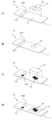
- the control slide 10 uses a slide glass 11 as a substrate. Further, the control slide 10 has two supporting portions, a supporting portion 121 and a supporting portion 122. A binding substance is bound to the carrier 121 and a non-binding substance is bound to the carrier 122. And the control slide 10 has the sample fixing
- the paraffin-embedded tissue section (tissue section 14, paraffin 141) is fixed to the sample fixing portion 13 on the slide glass 11 according to a conventional method (FIG. 1A).
- control slide 10 when using cultured cells as a test sample, the control slide 10 can be placed on a cytospin, and the cultured cells can be fixed to the sample fixing unit 13 by centrifugation.
- the paraffin 141 is removed according to a conventional method to remove the paraffin 141 (FIG. 1B). Thereafter, in the same manner as in normal tissue immunostaining, the control slide 10 is heat-treated by a microwave treatment, a warm bath treatment, an autoclave treatment or the like in order to activate the antigen.
- the staining operation can be carried out by the same procedure as in a normal immunostaining experiment. That is, staining can be performed by impregnating the control slide 10 with an antibody solution and washing with a buffer solution.
- the antigen-antibody reaction and washing may be performed by forming a water-repellent circle around the sample fixing part and the supporting part using a water-repellent pen, and dropping the antibody solution and the buffer solution thereto.
- the labeled antibody is an enzyme label
- the sample fixing part and the supporting part are reacted with the substrate solution to cause color development.
- an encapsulant is dropped onto the tissue section 14 and the cover glass 15 is placed to enclose the tissue section 14 (FIGS. 1C and 1D). Thereafter, if necessary, the end of the cover glass 15 is sealed with a nail polish and then observed with a microscope.
- the result of each carrier is a positive control of the staining result of the tissue section 14 and It can be used as a negative control (FIG. 1D).
- test example using the control slide of the present invention using a slide glass as a substrate will be described.
- mouse IgG was used as the binding substance
- goat IgG was used as the non-binding substance.
- mice IgG Mouse serum was centrifuged at 40,000 ⁇ g for 20 minutes at 4 ° C. to obtain a supernatant. 10 ml of the supernatant was passed at a flow rate of 1 ml / min through 5 ml of HiTrap Protein G HP (GE Healthcare) equilibrated with 20 mM sodium phosphate buffer (pH 7.4) containing 150 mM NaCl, and then 20 ml of equilibration buffer. Washed with liquid.
- the IgG bound to the column was eluted with 0.1M glycine-hydrochloric acid buffer (pH 3.0), and immediately neutralized by adding 1/10 volume of 1M Tris-hydrochloric acid buffer (pH 8.6) to 40 mg of mouse IgG. Got.
- Mouse IgG which is a binding substance, in the following procedure with the blasting part, the etching part in the slide glass, and the untreated part not subjected to these treatments as the supporting part, A non-binding substance, goat IgG, was bound.
- the processed slide glass was immersed in a 1% (3-glycidyloxypropyl) trimethoxysilane solution dissolved in 0.5% acetic acid for 16 hours, and then dried by blowing dry compressed air. 1 mg / ml and 0.1 mg / ml solutions of mouse IgG and goat IgG prepared using 50 mM sodium carbonate buffer (pH 8.0) containing 0.5 M NaCl as a solvent were prepared.
- mice IgG solution and goat IgG solution described above and 1 ⁇ l of a 20% aqueous sucrose solution were dropped on the blasted portion, the etched portion, and the untreated portion of the slide glass, and left for 16 hours. After standing, it was washed with 20 mM Tris-HCl buffer (pH 7.4) (TBS) containing 0.05% Tween 20 and 150 mM NaCl, and immersed in TBS containing 1 mg / ml bovine serum albumin at room temperature for 1 hour. After soaking, it was rinsed with water and dried by blowing dry compressed air.
- TBS Tris-HCl buffer
- the support portion bound to mouse IgG was stained regardless of whether the concentration of mouse IgG was 1 mg / ml or 0.1 mg / ml.
- the supporting parts bound with goat IgG was stained.
- bonded the non-binding substance goat IgG has shown that it is a negative control which shows that the peroxidase labeled antibody has not cross-reacted with goat IgG.
- the strongest color development was observed in the blasted carrying portion, and the color development in the untreated carrying portion was the weakest. This result suggests that the amount of the binding substance that can be bound per unit area can be increased by making the supporting portion rough glass, and as a result, the signal from the labeled antibody can be increased.
- the present invention can be applied to a control experiment of an immunostaining experiment.
- Control slide 11 Slide glass (substrate) 121 Supporting part 122 to which a binding substance is bound 122 Supporting part to which a non-binding substance is bound 13
- Sample fixing part 14 Tissue section (test sample) 141 Paraffin 15 Cover glass
Landscapes
- Life Sciences & Earth Sciences (AREA)
- Chemical & Material Sciences (AREA)
- Engineering & Computer Science (AREA)
- Health & Medical Sciences (AREA)
- Immunology (AREA)
- Urology & Nephrology (AREA)
- Molecular Biology (AREA)
- Hematology (AREA)
- Biomedical Technology (AREA)
- Biochemistry (AREA)
- General Health & Medical Sciences (AREA)
- Pathology (AREA)
- General Physics & Mathematics (AREA)
- Materials Engineering (AREA)
- Organic Chemistry (AREA)
- Geochemistry & Mineralogy (AREA)
- General Chemical & Material Sciences (AREA)
- Food Science & Technology (AREA)
- Medicinal Chemistry (AREA)
- Physics & Mathematics (AREA)
- Analytical Chemistry (AREA)
- Chemical Kinetics & Catalysis (AREA)
- Microbiology (AREA)
- Cell Biology (AREA)
- Biotechnology (AREA)
- Mechanical Engineering (AREA)
- Investigating Or Analysing Biological Materials (AREA)
- Surface Treatment Of Glass (AREA)
- Sampling And Sample Adjustment (AREA)
Abstract
新規のコントロールスライドを提供することを課題とする。 標識抗体を用いて被験試料14中の被験物質を検出する免疫染色実験のためのコントロールスライド10であって、前記標識抗体と直接若しくは間接的に複合体を形成し得る結合性物質及び/又は前記標識抗体と複合体を形成し得ない非結合性物質が結合している担持部12と、前記被験試料を固定する試料固定部13を有する基板11を備えることを特徴とする、コントロールスライド10。
Description
本発明は、免疫染色実験のためのコントロールスライド及びコントロール実験の方法に関する。
組織の免疫染色技術は確定診断に不可欠な技術である。免疫染色による診断には間違いが許されないため、染色時に陽性と陰性の対照実験(コントロール実験)を行う必要がある。
欧米先進国では上述した染色時にコントロール実験を行うことが一般化しており、コントロール実験の結果が無いものは信用されない。
欧米先進国では上述した染色時にコントロール実験を行うことが一般化しており、コントロール実験の結果が無いものは信用されない。
免疫染色実験におけるコントロール実験は、被験物質が含まれている、又は含まれていないことが予めわかっている組織切片や培養細胞を貼付としたコントロールスライドによって行うことが理想的である。コントロールスライドは、免疫染色実験に際して実験者自らが作製したり、市販のものを購入したりすることで入手することができる。しかしながら、後述する問題点により、一般的に染色のバッチ単位に1つコントロールスライドを入れて染色している。
例えば、特許文献1では、前立腺がんの悪性度判定用キットのコントロール実験に、前立腺がん由来培養細胞PC3のパラフィン切片標本であるコントロールスライドを用いている。
従来、免疫染色実験の対象とする被験物質が発現していることが予めわかっている組織や培養細胞が無い場合には、新たに細胞株を樹立するなどしない限りはコントロールスライドを用意することができなかった。
また、従来のコントロールスライドは組織や培養細胞を用いているため、ロットによる再現性を維持することが非常に困難であるという問題がある。
また、市販のコントロールスライドは組織切片か培養細胞を用いているため高価であるという問題がある。一方、コントロールスライドを自作しようとすると、コントロールとなる組織を入手する手間や、培養細胞を培養する手間などがかかり、実験者にとって大きな負担となる。
また、従来のコントロールスライドは組織や培養細胞を用いているため、ロットによる再現性を維持することが非常に困難であるという問題がある。
また、市販のコントロールスライドは組織切片か培養細胞を用いているため高価であるという問題がある。一方、コントロールスライドを自作しようとすると、コントロールとなる組織を入手する手間や、培養細胞を培養する手間などがかかり、実験者にとって大きな負担となる。
このような状況に鑑みて、新規のコントロールスライドを提供することを本発明の解決しようとする課題とする。
上記課題を解決する本発明は、標識抗体を用いて被験試料中の被験物質を検出する免疫染色実験のためのコントロールスライドであって、
前記標識抗体と直接若しくは間接的に複合体を形成し得る結合性物質及び/又は前記標識抗体と複合体を形成し得ない非結合性物質が結合している担持部と、前記被験試料を固定する試料固定部を有する基板を備えることを特徴とする、コントロールスライドである。
結合性物質が担持部に結合している形態の本発明のコントロールスライドによれば、免疫染色実験と同時にそのポジティブコントロール実験を簡便に行うことができる。
また非結合性物質が担持部に結合している形態の本発明のコントロールスライドによれば、免疫染色実験と同時にそのネガティブコントロール実験を簡便に行うことができる。
前記標識抗体と直接若しくは間接的に複合体を形成し得る結合性物質及び/又は前記標識抗体と複合体を形成し得ない非結合性物質が結合している担持部と、前記被験試料を固定する試料固定部を有する基板を備えることを特徴とする、コントロールスライドである。
結合性物質が担持部に結合している形態の本発明のコントロールスライドによれば、免疫染色実験と同時にそのポジティブコントロール実験を簡便に行うことができる。
また非結合性物質が担持部に結合している形態の本発明のコントロールスライドによれば、免疫染色実験と同時にそのネガティブコントロール実験を簡便に行うことができる。
本発明のコントロールスライドによれば、被験物質を含むことが予めわかっている組織や培養細胞を用意する必要が無い。そのため、そのような組織や培養細胞などの入手が困難な場合であっても、本発明のコントロールスライドによればコントロール実験を行うことができる。
また、本発明のコントロールスライドは、結合性物質及び/又は非結合性物質が結合している担持部を染色することでコントロール実験の結果を得ることができるため、組織や培養細胞を用いていた従来のコントロールスライドに比して、ロットによる差を低減することができる。すなわち、本発明のコントロールスライドによれば、再現性に優れたコントロール実験を行うことができる。
本発明の好ましい形態では、前記結合性物質及び/又は前記非結合性物質がタンパク質又は核酸である。
このような形態の本発明によれば、容易にコントロール実験を行うことができる。
このような形態の本発明によれば、容易にコントロール実験を行うことができる。
本発明のコントロールスライドは、被験試料が組織切片である免疫染色実験のコントロール実験に適用することが好ましい。
従来、組織切片の免疫染色実験では、陽性又は陰性であることが確認されている組織切片や培養細胞をコントロールとして使用することが一般的であった。本発明のコントロールスライドによれば、高価でロットによる性質のバラツキがある組織切片や培養細胞を用いずとも、簡便に組織切片の免疫染色実験のコントロール実験を行うことができる。
従来、組織切片の免疫染色実験では、陽性又は陰性であることが確認されている組織切片や培養細胞をコントロールとして使用することが一般的であった。本発明のコントロールスライドによれば、高価でロットによる性質のバラツキがある組織切片や培養細胞を用いずとも、簡便に組織切片の免疫染色実験のコントロール実験を行うことができる。
本発明の好ましい形態では、前記担持部が粗面ガラスによって構成されている。
粗面ガラスは単位エリア当たりの表面積が大きい。そのため、このような形態の本発明は、担持部により多くの結合性物質及び/又は非結合性物質を結合することができ、感度に優れたコントロールスライドを提供することができる。
粗面ガラスは単位エリア当たりの表面積が大きい。そのため、このような形態の本発明は、担持部により多くの結合性物質及び/又は非結合性物質を結合することができ、感度に優れたコントロールスライドを提供することができる。
本発明の好ましい形態では、前記粗面ガラスがブラスト処理ガラス又はエッチング処理ガラスである。
これによると、簡単に安価な粗面ガラスの担持部を有するコントロールスライドを提供することができる。
これによると、簡単に安価な粗面ガラスの担持部を有するコントロールスライドを提供することができる。
本発明の好ましい形態では、前記結合性物質が、前記被験物質である。
一般的に免疫染色実験では被験物質と一次抗体との反応と、一次抗体と標識された二次抗体との反応の2段階の抗原抗体反応を行う。このような形態の本発明のコントロールスライドによれば、免疫染色実験で行われる前述の抗原抗体反応が全て正常に動作していることを裏付けることができる。
一般的に免疫染色実験では被験物質と一次抗体との反応と、一次抗体と標識された二次抗体との反応の2段階の抗原抗体反応を行う。このような形態の本発明のコントロールスライドによれば、免疫染色実験で行われる前述の抗原抗体反応が全て正常に動作していることを裏付けることができる。
本発明の好ましい形態では、前記免疫染色実験が間接法によるものであって、前記結合性物質が、前記免疫染色実験において用いられる一次抗体と同一の動物種由来のアイソタイプコントロール抗体である。
このような形態の本発明のコントロールスライドは、担持部に結合しているアイソタイプコントロール抗体と同一の動物種由来の一次抗体を用いる全ての免疫染色実験に用いることができるため、汎用性がある。
このような形態の本発明のコントロールスライドは、担持部に結合しているアイソタイプコントロール抗体と同一の動物種由来の一次抗体を用いる全ての免疫染色実験に用いることができるため、汎用性がある。
また、本発明は、標識抗体を用いて被験試料中の被験物質を検出する免疫染色実験のためのコントロール実験の方法であって、
基板の担持部に結合している、前記標識抗体と直接若しくは間接的に複合体を形成し得る結合性物質及び/又は前記標識抗体と複合体を形成し得ない非結合性物質に、
前記免疫染色実験で用いる抗体溶液を、前記免疫染色実験と同一条件で接触させることを特徴とする、方法にもある。
本発明の方法によれば、簡便に免疫染色実験のコントロール実験を行うことができる。
基板の担持部に結合している、前記標識抗体と直接若しくは間接的に複合体を形成し得る結合性物質及び/又は前記標識抗体と複合体を形成し得ない非結合性物質に、
前記免疫染色実験で用いる抗体溶液を、前記免疫染色実験と同一条件で接触させることを特徴とする、方法にもある。
本発明の方法によれば、簡便に免疫染色実験のコントロール実験を行うことができる。
また、本発明は、標識抗体を用いて被験試料中の被験物質を検出する免疫染色実験のためのコントロールスライドであって、基板の一部にブラスト処理又はエッチング処理を施して担持部を設け、その担持部に前記標識抗体と直接若しくは間接的に複合体を形成し得る結合性物質及び/又は前記標識抗体と複合体を形成し得ない非結合性物質を結合させることを特徴とする、コントロールスライドの製造方法にもある。
本発明の製造方法によれば、容易に本発明のコントロールスライドを製造することができる。
本発明の製造方法によれば、容易に本発明のコントロールスライドを製造することができる。
本発明のコントロールスライドは汎用性に優れる。また、本発明によれば再現性の高いコントロール実験の結果を得ることができる。
また、本発明の一形態では、従来のコントロールスライドよりも安価なコントロールスライドを提供することができる。
また、本発明の一形態では、従来のコントロールスライドよりも安価なコントロールスライドを提供することができる。
本明細書においては被験試料についての免疫染色実験を本実験とも言う。
本発明のコントロールスライドは、組織切片や培養細胞などを被験試料とする通常の免疫染色実験の何れにも用いることができる。
従来、組織切片を用いた免疫染色実験では、高価なコントロールスライドをコントロール実験に用いる必要があった。そのため、組織切片を用いた免疫染色実験におけるコントロール実験に本発明のコントロールスライドを使用することが特に好ましい。
本発明のコントロールスライドはタンパク質や核酸などを被験物質とする通常の免疫染色実験であれば特に制限なく適用することができる。
また、標識抗体の標識の種類も限定されず、放射性同位元素、金コロイド、蛍光色素、酵素、ビオチンなど通常免疫染色において用いられる標識を適用することができる。
免疫染色実験が、抗原に直接反応する一次抗体を標識し、抗原抗体反応を1度しか行わない直接法と、標識していない一次抗体を用いて1度目の抗原抗体反応を行い、一次抗体自体を抗原とする別の抗体(二次抗体)をさらに反応させ2回以上の抗原抗体反応を行う間接法の何れであっても、本発明を適用することができる。
本発明のコントロールスライドにおける担持部に結合させる物質は、標識抗体と直接若しくは間接的に複合体を形成し得る結合性物質であっても、前記標識抗体と複合体を形成し得ない非結合性物質であってもよい。
結合性物質が担持部に結合している形態の本発明のコントロールスライドによれば、免疫染色実験のポジティブコントロール実験を簡便に行うことができる(表1)。

直接法の免疫染色実験を行う場合に、結合性物質を被験物質とすることで、標識抗体と被験物質との抗原抗体反応のポジティブコントロール実験を行うことができる。(表1(A))。
また、間接法の免疫染色実験を行う場合に、結合性物質を被験物質とすることで、免疫染色実験で行われる全ての抗原抗体反応のポジティブコントロール実験を行うことができる(表1(B))。
なお、被験物質がタンパク質である場合、該タンパク質における一次抗体の認識部位にあたるペプチドを結合性物質として担持部に結合しても良い。
また、間接法の免疫染色実験を行う場合に、結合性物質を標識抗体以外の本実験で用いる抗体とすることで、免疫染色実験における当該抗体を抗原とする抗原抗体反応、さらにはそれに続く抗原抗体反応のポジティブコントロール実験を行うことができる(表1(C))。
なお、標識していない一次抗体と標識抗体の2種類の抗体を用いる通常の間接法の免疫染色実験においては、結合性物質を一次抗体とすることで、一次抗体と標識抗体との抗原抗体反応のポジティブコントロール実験を行うことができる。
なお、標識していない一次抗体と標識抗体の2種類の抗体を用いる通常の間接法の免疫染色実験においては、結合性物質を一次抗体とすることで、一次抗体と標識抗体との抗原抗体反応のポジティブコントロール実験を行うことができる。
また、間接法の免疫染色実験を行う場合に、該免疫染色実験で用いる抗体と同一の動物種由来のアイソタイプコントロール抗体を結合性物質としても良い。免疫染色実験における抗体を抗原とする抗原抗体反応、さらにはそれに続く抗原抗体反応のポジティブコントロール実験を行うことができる(表1(D))。
このような形態とすることによって上記表1(C)の形態と同一の効果が得られる他、当該アイソタイプコントロール抗体と同一の動物種由来の抗体を用いる全ての免疫染色実験に適用することができ、汎用性に優れる。
このような形態とすることによって上記表1(C)の形態と同一の効果が得られる他、当該アイソタイプコントロール抗体と同一の動物種由来の抗体を用いる全ての免疫染色実験に適用することができ、汎用性に優れる。
一方、非結合性物質が担持部に結合している形態の本発明のコントロールスライドによれば、免疫染色実験のネガティブコントロール実験を簡便に行うことができる(表2)。
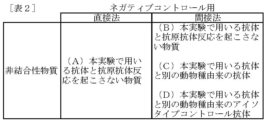
直接法の免疫染色実験を行う場合に、非結合性物質を本実験で用いる抗体と抗原抗体反応を起こさない物質とすることで、免疫染色実験の結果が当該物質を検出した偽陽性でないことを示すネガティブコントロール実験を行うことができる(表2(A))。
この場合、非結合性物質は被験物質と同一種類の分子種とすることが好ましい。具体的には、被験物質がタンパク質である場合には、非結合性物質もタンパク質であることが好ましい。
本発明のさらに好ましい実施の形態では、被験物質がタンパク質である場合には、非結合性物質は該タンパク質のホモログであることが好ましい。このような実施の形態とすることによって、本実験で得られた結果が、被験物質であるタンパク質と相同性の高いホモログを認識した偽陽性ではないことを示すことができる。
この場合、非結合性物質は被験物質と同一種類の分子種とすることが好ましい。具体的には、被験物質がタンパク質である場合には、非結合性物質もタンパク質であることが好ましい。
本発明のさらに好ましい実施の形態では、被験物質がタンパク質である場合には、非結合性物質は該タンパク質のホモログであることが好ましい。このような実施の形態とすることによって、本実験で得られた結果が、被験物質であるタンパク質と相同性の高いホモログを認識した偽陽性ではないことを示すことができる。
また、間接法の免疫染色実験を行う場合に、非結合性物質を本実験で用いる抗体と抗原抗体反応を起こさない物質とすることで、免疫染色実験の結果が当該物質を検出した偽陽性でないことを示すネガティブコントロール実験を行うことができる(表2(B))。
この場合、非結合性物質は被験物質と同一種類の分子種とすることが好ましい。具体的には、被験物質がタンパク質である場合には、非結合性物質もタンパク質であることが好ましい。
本発明のさらに好ましい実施の形態では、被験物質がタンパク質である場合には、非結合性物質は該タンパク質のホモログであることが好ましい。このような実施の形態とすることによって、本実験で得られた結果が、被験物質であるタンパク質と相同性の高いホモログを認識した偽陽性ではないことを示すことができる。
この場合、非結合性物質は被験物質と同一種類の分子種とすることが好ましい。具体的には、被験物質がタンパク質である場合には、非結合性物質もタンパク質であることが好ましい。
本発明のさらに好ましい実施の形態では、被験物質がタンパク質である場合には、非結合性物質は該タンパク質のホモログであることが好ましい。このような実施の形態とすることによって、本実験で得られた結果が、被験物質であるタンパク質と相同性の高いホモログを認識した偽陽性ではないことを示すことができる。
また、間接法の免疫染色実験を行う場合に、非結合性物質を本実験で用いる抗体と別の動物種由来の抗体とすることで、免疫染色実験の免疫染色実験の結果が当該抗体を検出した偽陽性でないことを示すネガティブコントロール実験を行うことができる(表2(C))。このような形態の本発明は、免疫染色実験が異なる動物種由来の複数の一次抗体を用いる多重染色である場合に有効である。
また、この場合、非結合性物質である当該抗体はアイソタイプコントロール抗体とすることが好ましい(表2(D))。このような形態の本発明は汎用性に優れる。
また、この場合、非結合性物質である当該抗体はアイソタイプコントロール抗体とすることが好ましい(表2(D))。このような形態の本発明は汎用性に優れる。
本発明のコントロールスライドの担持部に結合する結合性物質及び非結合性物質としては、組織、培養細胞、又は大腸菌などから抽出した生体由来の物質でも良いし、化学合成した物質でも良い。
結合性物質及び非結合性物質がタンパク質である場合には、培養細胞や組織などから抽出した内在性のタンパク質、培養細胞中で発現ベクターを用いて強制発現させた後に抽出した外来性のタンパク質、大腸菌を用いたタンパク質発現系によるリコンビナントタンパク質、さらには化学合成されたタンパク質の何れを用いても良い。
結合性物質及び非結合性物質がタンパク質である場合には、培養細胞や組織などから抽出した内在性のタンパク質、培養細胞中で発現ベクターを用いて強制発現させた後に抽出した外来性のタンパク質、大腸菌を用いたタンパク質発現系によるリコンビナントタンパク質、さらには化学合成されたタンパク質の何れを用いても良い。
本発明のコントロールスライドは、担持部を有する基板を備える。
担持部を含む基板の素材としては特に限定されないが、ガラスが好ましい。ガラスは透明性に優れるとともに薬品に対して安定しているため、標識抗体の呈色や蛍光を容易に観察することができる。
担持部を含む基板の素材としては特に限定されないが、ガラスが好ましい。ガラスは透明性に優れるとともに薬品に対して安定しているため、標識抗体の呈色や蛍光を容易に観察することができる。
基板をガラス製とする場合には、滑面ガラスであっても粗面ガラスであっても構わないが、担持部は粗面ガラスとすることが好ましい。粗面ガラスは、ブラスト処理やエッチング処理で容易に施すことができる。
このように担持部を粗面ガラスとすることによって、結合できる結合性物質又は非結合性物質の量を増やすことができる。
このように担持部を粗面ガラスとすることによって、結合できる結合性物質又は非結合性物質の量を増やすことができる。
粗面ガラスとする場合の平均の粗さ(Ra)は、0.01~20.0μmであることが好ましく、0.05~10.0μmであることがより好ましく、0.1~1μmであることがさらに好ましく、0.2~0.3μmであることがさらに好ましく、0.226~0.252μmであることが最も好ましい。
また、粗面ガラスとする場合の突出の最大の深さ(Rz)は、0.1~150.0μmであることが好ましく、0.5~100.0μmであることがより好ましく、1.0~10.0μmであることがさらに好ましく、2.0~3.0μmであることがさらに好ましく、2.19~2.70μmであることが最も好ましい。
また、粗面ガラスとする場合の突出の間隔(Sm)は、10~2000μmであることが好ましく、30~1000μmであることがより好ましく、50~500μmであることがさらに好ましく、100~200μmであることがさらに好ましく、165~179μmであることが最も好ましい。
なお、平均の粗さ(Ra)、最大の深さ(Rz)、突出の間隔(Sm)は、JIS B0601で規定された定義を用いるものとする。
結合性物質又は非結合性物質を担持部に結合させる方法は特に限定されず、公知の方法を用いることができる。
ガラス製の基板の担持部にタンパク質や核酸などの生体物質である結合性物質又は非結合性物質を担持させる方法については、シランカップリング法、シリコンバイオ法など、当業者に公知の方法を適用することができる。
ガラス製の基板の担持部にタンパク質や核酸などの生体物質である結合性物質又は非結合性物質を担持させる方法については、シランカップリング法、シリコンバイオ法など、当業者に公知の方法を適用することができる。
具体的には、以下の方法によりガラス製の基板の担持部にタンパク質である結合性物質又は非結合性物質を結合させることができる。
すなわち、担持部を0.5%酢酸に溶解した1%(3-グリシジルオキシプロピル)トリメトキシシラン溶液に16時間浸漬した後、乾燥した圧搾空気を吹き付けて乾燥する。
また、0.5M NaClを含む50mM 炭酸ナトリウム緩衝液(pH8.0)に、好ましくは0.1~1mg/mlの濃度で結合性物質又は非結合性物質であるタンパク質を溶解した溶液を調製する。
そして、(3-グリシジルオキシプロピル)トリメトキシシラン溶液により処理された担持部に、前記タンパク質溶液を滴下し、16時間静置する。
16時間に静置の後、0.05% Tween20と150mM NaClを含む20mM Tris-HCl緩衝液(pH7.4)で担持部を洗浄し、さらに担持部を1mg/mlのウシ血清アルブミンを含むTBSに室温で1時間浸漬する。
その後、担持部を水で洗い流して乾燥した圧搾空気を吹き付けて乾燥することによって、結合性物質又は非結合性物質であるタンパク質を担持部に結合することができる。
すなわち、担持部を0.5%酢酸に溶解した1%(3-グリシジルオキシプロピル)トリメトキシシラン溶液に16時間浸漬した後、乾燥した圧搾空気を吹き付けて乾燥する。
また、0.5M NaClを含む50mM 炭酸ナトリウム緩衝液(pH8.0)に、好ましくは0.1~1mg/mlの濃度で結合性物質又は非結合性物質であるタンパク質を溶解した溶液を調製する。
そして、(3-グリシジルオキシプロピル)トリメトキシシラン溶液により処理された担持部に、前記タンパク質溶液を滴下し、16時間静置する。
16時間に静置の後、0.05% Tween20と150mM NaClを含む20mM Tris-HCl緩衝液(pH7.4)で担持部を洗浄し、さらに担持部を1mg/mlのウシ血清アルブミンを含むTBSに室温で1時間浸漬する。
その後、担持部を水で洗い流して乾燥した圧搾空気を吹き付けて乾燥することによって、結合性物質又は非結合性物質であるタンパク質を担持部に結合することができる。
担持部に結合性物質又は非結合性物質を結合した後に、マイクロウエーブ処理、温浴処理、そしてオートクレーブ処理などの賦活化処理を行うこともできる。
基板における担持部の数は1つであっても良いが、好ましくは2以上である。
担持部の数を2以上とする場合には、結合性物質と非結合性物質の両方を担持部に結合することが好ましい。このような形態とすることによって、一つのコントロールスライドによってポジティブコントロール実験とネガティブコントロール実験を同時に行うことができる。
担持部の数を2以上とする場合には、結合性物質と非結合性物質の両方を担持部に結合することが好ましい。このような形態とすることによって、一つのコントロールスライドによってポジティブコントロール実験とネガティブコントロール実験を同時に行うことができる。
また、担持部の数を2以上とする場合には、異なる量の結合性物質又は非結合性物質をそれぞれの担持部に結合させることが好ましい。
結合した結合性物質又は非結合性物質の量に比例した強度のシグナルが得られれば、該シグナルが担持部に結合した結合性物質又は非結合性物質に由来することを確認することができる。
結合した結合性物質又は非結合性物質の量に比例した強度のシグナルが得られれば、該シグナルが担持部に結合した結合性物質又は非結合性物質に由来することを確認することができる。
また、本実験である免疫染色実験が多重染色である場合には、多重染色の対象である複数の被験物質、多重染色で用いる複数の一次抗体、多重染色で用いる一次抗体と同一の動物種由来の複数のアイソタイプコントロール抗体などを結合するために、担持部は複数設けることが好ましい。
本発明のコントロールスライドは被験試料を固定するための試料固定部を基板に有する。
本発明のコントロールスライドは、コントロール実験のための担持部と、本実験のための試料固定部を同一の基板に有するため、コントロール実験と本実験を同一条件のもと行うことができ、信頼性の高い実験結果を得ることができる。
本発明のコントロールスライドは、コントロール実験のための担持部と、本実験のための試料固定部を同一の基板に有するため、コントロール実験と本実験を同一条件のもと行うことができ、信頼性の高い実験結果を得ることができる。
本発明のコントロールスライドを用いた実験は、通常の免疫染色実験と同様の手法により行うことができる。すなわち、抗体溶液とのインキュベーションと洗浄操作を行うことにより染色を行うことができる。この時、コントロールスライド全体を抗体溶液及び緩衝液に含浸することで、抗原抗体反応及び洗浄操作を行うことができる。
このように、本発明のコントロールスライド10によれば、インキュベーション時間、温度など本実験における抗原抗体反応と同様の条件でコントロール実験を行うことができるため、信頼性の高い実験結果を得ることができる。
また、本発明は、基板の担持部に結合した結合性物質及び/又は非結合性物質に、免疫染色実験で用いる抗体溶液を、免疫染色実験と同一条件で接触させることを特徴とする、標識抗体を用いて被験試料中の被験物質を検出する免疫染色実験のためのコントロール実験の方法にも関する。
本発明のコントロール実験の方法は、上述の本発明のコントロールスライドを用いることで実現することができる。
本発明のコントロール実験の方法は、上述の本発明のコントロールスライドを用いることで実現することができる。
<1>本発明のコントロールスライドの実施例
図1を参照しながら、本発明のコントロールスライド10の一実施形態を説明する。
図1を参照しながら、本発明のコントロールスライド10の一実施形態を説明する。
コントロールスライド10は、スライドガラス11を基板とする。
また、コントロールスライド10は担持部121と担持部122の2つの担持部を有している。担持部121には結合性物質、担持部122には非結合性物質を結合している。
そして、コントロールスライド10は、被験試料を固定するための試料固定部13を有している。
また、コントロールスライド10は担持部121と担持部122の2つの担持部を有している。担持部121には結合性物質、担持部122には非結合性物質を結合している。
そして、コントロールスライド10は、被験試料を固定するための試料固定部13を有している。
次に、被験試料としてパラフィン包埋組織切片を用いる場合の本発明のコントロールスライド10の使用方法について説明する。
パラフィン包埋組織切片(組織切片14、パラフィン141)は常法に従ってスライドガラス11上の試料固定部13に固定する(図1A)。
パラフィン包埋組織切片(組織切片14、パラフィン141)は常法に従ってスライドガラス11上の試料固定部13に固定する(図1A)。
なお、被験試料として培養細胞を用いる場合には、コントロールスライド10をサイトスピンに設置し、遠心によって培養細胞を試料固定部13に固定することができる。
パラフィン包埋組織切片を固定した後には常法に従い、脱パラフィン処理を行い、パラフィン141を除去する(図1B)。その後、通常の組織免疫染色と同様、抗原を賦活化させるために、コントロールスライド10をマイクロウエーブ処理、温浴処理、そしてオートクレーブ処理などで加熱処理する。
その後は通常の免疫染色実験と同様の手順によって染色操作を行うことができる。
すなわち、コントロールスライド10の抗体溶液への含浸と緩衝液による洗浄により染色を行うことができる。
また、撥水ペンを用いて試料固定部と担持部の周りに撥水サークルを形成し、そこへ抗体溶液及び緩衝液を滴下することによって、抗原抗体反応及び洗浄を行ってもよい。
標識抗体が酵素標識である場合には、標識抗体による抗原抗体反応に続く洗浄の後に、試料固定部と担持部を基質溶液と反応させ発色させる。
すなわち、コントロールスライド10の抗体溶液への含浸と緩衝液による洗浄により染色を行うことができる。
また、撥水ペンを用いて試料固定部と担持部の周りに撥水サークルを形成し、そこへ抗体溶液及び緩衝液を滴下することによって、抗原抗体反応及び洗浄を行ってもよい。
標識抗体が酵素標識である場合には、標識抗体による抗原抗体反応に続く洗浄の後に、試料固定部と担持部を基質溶液と反応させ発色させる。
その後、組織切片14上に封入剤を滴下し、カバーガラス15を載せて組織切片14を封入する(図1C、D)。
その後、必要に応じてマニキュア液によりカバーガラス15の端をシールした後に顕微鏡観察を行う。
その後、必要に応じてマニキュア液によりカバーガラス15の端をシールした後に顕微鏡観察を行う。
結合性物質を結合させた担持部121が染色され、非結合性物質を結合させた担持部122が染色されていなければ、それぞれの担持部の結果は、組織切片14の染色結果のポジティブコントロール及びネガティブコントロールとすることができる(図1D)。
<2>試験例
以下、スライドガラスを基板とする本発明のコントロールスライドを用いた試験例について説明する。本試験例においては、結合性物質としてマウスIgG、非結合性物質としてヤギIgGを用いた。
以下、スライドガラスを基板とする本発明のコントロールスライドを用いた試験例について説明する。本試験例においては、結合性物質としてマウスIgG、非結合性物質としてヤギIgGを用いた。
(1)マウスIgGの調製
マウス血清を4℃、40,000×gで20分間遠心分離し、上清を得た。この上清10mlを150mM NaClを含む20mM リン酸ナトリウム緩衝液(pH7.4)で平衡化した5mlのHiTrap Protein G HP(GEヘルスケア)に1ml/minの流速で通し、次いで20mlの平衡化緩衝液で洗浄した。0.1M グリシン-塩酸緩衝液(pH3.0)でカラムに結合したIgGを溶出し、直ちに1M Tris-塩酸緩衝液(pH8.6)を1/10容量加えて中和し、40mgのマウスIgGを得た。
マウス血清を4℃、40,000×gで20分間遠心分離し、上清を得た。この上清10mlを150mM NaClを含む20mM リン酸ナトリウム緩衝液(pH7.4)で平衡化した5mlのHiTrap Protein G HP(GEヘルスケア)に1ml/minの流速で通し、次いで20mlの平衡化緩衝液で洗浄した。0.1M グリシン-塩酸緩衝液(pH3.0)でカラムに結合したIgGを溶出し、直ちに1M Tris-塩酸緩衝液(pH8.6)を1/10容量加えて中和し、40mgのマウスIgGを得た。
(2)ヤギIgGの調製
ヤギ血清を4℃、40,000×gで20分間遠心分離し、上清を得た。実施例1と同様の手順でHiTrap Protein G HP(GEヘルスケア)を用いて60mgのIgGを得た。
ヤギ血清を4℃、40,000×gで20分間遠心分離し、上清を得た。実施例1と同様の手順でHiTrap Protein G HP(GEヘルスケア)を用いて60mgのIgGを得た。
(3)スライドガラスの加工
26mm×76mmのスライドガラスの一部をブラスト処理したものを作製した。ブラスト処理部分の隣に約5mmの幅でガラスエッチング剤(グラスファンタジー、有限会社フロステック)を塗布し、室温で30分間静置した。静置後水でエッチング剤を洗い流し、乾燥した圧搾空気を吹き付けて乾燥してエッチング処理部分を作製した。
26mm×76mmのスライドガラスの一部をブラスト処理したものを作製した。ブラスト処理部分の隣に約5mmの幅でガラスエッチング剤(グラスファンタジー、有限会社フロステック)を塗布し、室温で30分間静置した。静置後水でエッチング剤を洗い流し、乾燥した圧搾空気を吹き付けて乾燥してエッチング処理部分を作製した。
(4)IgGの担持部への結合
スライドガラスにおけるブラスト処理部分、エッチング処理部分そして、これらの処理がなされていない無処理部分を担持部として、以下の手順で結合性物質であるマウスIgGと、非結合性物質であるヤギIgGを結合した。
加工したスライドガラスを0.5%酢酸に溶解した1%(3-グリシジルオキシプロピル)トリメトキシシラン溶液に16時間浸漬し、その後、乾燥した圧搾空気を吹き付けて乾燥した。0.5M NaClを含む50mM 炭酸ナトリウム緩衝液(pH8.0)を溶媒として調製したマウスIgGとヤギIgGの1mg/mlと0.1mg/mlの溶液を調製した。スライドガラスのブラスト処理部分、エッチング処理部分、及び無処理部分に、上述のマウスIgG溶液及びヤギIgG溶液、並びに20%スクロース水溶液 1μlを滴下して16時間静置した。静置後、0.05% Tween20と150mM NaClを含む20mM Tris-HCl緩衝液(pH7.4)(TBS)で洗浄し、1mg/mlのウシ血清アルブミンを含むTBSに室温で1時間浸漬した。浸漬後水で洗い流し、乾燥した圧搾空気を吹き付けて乾燥した。
スライドガラスにおけるブラスト処理部分、エッチング処理部分そして、これらの処理がなされていない無処理部分を担持部として、以下の手順で結合性物質であるマウスIgGと、非結合性物質であるヤギIgGを結合した。
加工したスライドガラスを0.5%酢酸に溶解した1%(3-グリシジルオキシプロピル)トリメトキシシラン溶液に16時間浸漬し、その後、乾燥した圧搾空気を吹き付けて乾燥した。0.5M NaClを含む50mM 炭酸ナトリウム緩衝液(pH8.0)を溶媒として調製したマウスIgGとヤギIgGの1mg/mlと0.1mg/mlの溶液を調製した。スライドガラスのブラスト処理部分、エッチング処理部分、及び無処理部分に、上述のマウスIgG溶液及びヤギIgG溶液、並びに20%スクロース水溶液 1μlを滴下して16時間静置した。静置後、0.05% Tween20と150mM NaClを含む20mM Tris-HCl緩衝液(pH7.4)(TBS)で洗浄し、1mg/mlのウシ血清アルブミンを含むTBSに室温で1時間浸漬した。浸漬後水で洗い流し、乾燥した圧搾空気を吹き付けて乾燥した。
(5)染色
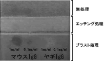
賦活化処理を行った後にTBSで洗浄し、上述のマウスIgG及びヤギIgG溶液、並びに20%スクロース水溶液を滴下した部分に、1mg/mlのウシ血清アルブミンを含むTBSで500倍に希釈したペルオキシダーゼ標識ヤギ抗マウスIgG(Jackson社)を滴下し、乾燥しないように蓋ができるプラスティック箱に入れて25℃で1時間静置して反応させた。反応後、TBSでペルオキシダーゼ標識抗体を洗浄除去し、ペルオキシダーゼ染色DABキット(ナカライテスク)を用いて5分間染色した。結果を図2に示す。
賦活化処理を行った後にTBSで洗浄し、上述のマウスIgG及びヤギIgG溶液、並びに20%スクロース水溶液を滴下した部分に、1mg/mlのウシ血清アルブミンを含むTBSで500倍に希釈したペルオキシダーゼ標識ヤギ抗マウスIgG(Jackson社)を滴下し、乾燥しないように蓋ができるプラスティック箱に入れて25℃で1時間静置して反応させた。反応後、TBSでペルオキシダーゼ標識抗体を洗浄除去し、ペルオキシダーゼ染色DABキット(ナカライテスク)を用いて5分間染色した。結果を図2に示す。
図2に示すように、マウスIgGを結合した担持部はマウスIgGの濃度が1mg/ml、0.1mg/mlの何れであっても染色された。一方、ヤギIgGを結合した担持部は何れも染色されなかった。
結合性物質であるマウスIgGを結合した担持部での観察結果は、ペルオキシダーゼ標識抗体が正常に動作していることを示すポジティブコントロールとなっていることを示している。
また、非結合性物質であるヤギIgGを結合した担持部での観察結果は、ペルオキシダーゼ標識抗体がヤギIgGに交差反応をしていないことを示すネガティブコントロールとなっていることを示している。
結合性物質であるマウスIgGを結合した担持部での観察結果は、ペルオキシダーゼ標識抗体が正常に動作していることを示すポジティブコントロールとなっていることを示している。
また、非結合性物質であるヤギIgGを結合した担持部での観察結果は、ペルオキシダーゼ標識抗体がヤギIgGに交差反応をしていないことを示すネガティブコントロールとなっていることを示している。
また、図2に示すように、ブラスト処理されている担持部における発色が最も強く観察され、無処理の担持部における発色が最も弱かった。
この結果は、担持部を粗面ガラスとすることによって、単位エリア当たりに結合できる結合性物質の量を増大させ、結果として標識抗体からのシグナルを増大させることができることを示唆している。
この結果は、担持部を粗面ガラスとすることによって、単位エリア当たりに結合できる結合性物質の量を増大させ、結果として標識抗体からのシグナルを増大させることができることを示唆している。
本発明は、免疫染色実験のコントロール実験に応用することができる。
10 コントロールスライド
11 スライドガラス(基板)
121 結合性物質を結合させた担持部
122 非結合性物質を結合させた担持部
13 試料固定部
14 組織切片(被験試料)
141 パラフィン
15 カバーガラス
11 スライドガラス(基板)
121 結合性物質を結合させた担持部
122 非結合性物質を結合させた担持部
13 試料固定部
14 組織切片(被験試料)
141 パラフィン
15 カバーガラス
Claims (9)
- 標識抗体を用いて被験試料中の被験物質を検出する免疫染色実験のためのコントロールスライドであって、
前記標識抗体と直接若しくは間接的に複合体を形成し得る結合性物質及び/又は前記標識抗体と複合体を形成し得ない非結合性物質が結合している担持部と、前記被験試料を固定する試料固定部を有する基板を備えることを特徴とする、コントロールスライド。 - 前記結合性物質及び/又は前記非結合性物質が、タンパク質又は核酸であることを特徴とする、請求項1に記載のコントロールスライド。
- 前記被験試料が組織切片であることを特徴とする、請求項1又は2に記載のコントロールスライド。
- 前記担持部が粗面ガラスによって構成されていることを特徴とする、請求項1~3の何れか一項に記載のコントロールスライド。
- 前記粗面ガラスがブラスト処理ガラス又はエッチング処理ガラスであることを特徴とする、請求項4に記載のコントロールスライド。
- 前記結合性物質が、前記被験物質であることを特徴とする、請求項1~5の何れか一項に記載のコントロールスライド。
- 前記免疫染色実験が間接法によるものであって、前記結合性物質が、前記免疫染色実験において用いられる一次抗体と同一の動物種由来のアイソタイプコントロール抗体であることを特徴とする、請求項1~5の何れか一項に記載のコントロールスライド。
- 標識抗体を用いて被験試料中の被験物質を検出する免疫染色実験のためのコントロール実験の方法であって、
基板の担持部に結合している、前記標識抗体と直接若しくは間接的に複合体を形成し得る結合性物質及び/又は前記標識抗体と複合体を形成し得ない非結合性物質に、
前記免疫染色実験で用いる抗体溶液を、前記免疫染色実験と同一条件で接触させることを特徴とする、方法。 - 標識抗体を用いて被験試料中の被験物質を検出する免疫染色実験のためのコントロールスライドであって、
基板の一部にブラスト処理又はエッチング処理を施して担持部を設け、その担持部に前記標識抗体と直接若しくは間接的に複合体を形成し得る結合性物質及び/又は前記標識抗体と複合体を形成し得ない非結合性物質を結合させることを特徴とする、コントロールスライドの製造方法。
Applications Claiming Priority (2)
| Application Number | Priority Date | Filing Date | Title |
|---|---|---|---|
| JP2015044853A JP6634566B2 (ja) | 2015-03-06 | 2015-03-06 | コントロールスライド及びコントロール実験の方法 |
| JP2015-044853 | 2015-03-06 |
Publications (1)
| Publication Number | Publication Date |
|---|---|
| WO2016143717A1 true WO2016143717A1 (ja) | 2016-09-15 |
Family
ID=56875965
Family Applications (1)
| Application Number | Title | Priority Date | Filing Date |
|---|---|---|---|
| PCT/JP2016/056894 Ceased WO2016143717A1 (ja) | 2015-03-06 | 2016-03-04 | コントロールスライド及びコントロール実験の方法 |
Country Status (2)
| Country | Link |
|---|---|
| JP (1) | JP6634566B2 (ja) |
| WO (1) | WO2016143717A1 (ja) |
Families Citing this family (1)
| Publication number | Priority date | Publication date | Assignee | Title |
|---|---|---|---|---|
| JP2020098097A (ja) * | 2017-03-31 | 2020-06-25 | 松浪硝子工業株式会社 | スライドグラス及びスライドグラスの製造のための溶液 |
Citations (3)
| Publication number | Priority date | Publication date | Assignee | Title |
|---|---|---|---|---|
| JP2002211954A (ja) * | 2000-11-07 | 2002-07-31 | Toyo Kohan Co Ltd | 粗面化スライドグラス及びそれを用いて生体物質を解析する方法 |
| JP2002533695A (ja) * | 1998-12-23 | 2002-10-08 | アメリカン・レジストリー・オブ・パソロジー | スライド上で生物学的試料を効率的に処理する装置および方法 |
| JP2013039065A (ja) * | 2011-08-12 | 2013-02-28 | Nagasaki Univ | 自己炎症疾患又は自己免疫疾患関連遺伝子及びその利用 |
Family Cites Families (3)
| Publication number | Priority date | Publication date | Assignee | Title |
|---|---|---|---|---|
| US4541987A (en) * | 1983-09-29 | 1985-09-17 | Helena Laboratories Corporation | Test pad for detecting occult blood |
| DK2184346T3 (en) * | 2001-09-06 | 2017-06-26 | Rapid Micro Biosystems Inc | Rapid detection of replicating cells |
| US20120282285A1 (en) * | 2009-07-17 | 2012-11-08 | Rigshospitalet | Masp isoforms as inhibitors of complement activation |
-
2015
- 2015-03-06 JP JP2015044853A patent/JP6634566B2/ja active Active
-
2016
- 2016-03-04 WO PCT/JP2016/056894 patent/WO2016143717A1/ja not_active Ceased
Patent Citations (3)
| Publication number | Priority date | Publication date | Assignee | Title |
|---|---|---|---|---|
| JP2002533695A (ja) * | 1998-12-23 | 2002-10-08 | アメリカン・レジストリー・オブ・パソロジー | スライド上で生物学的試料を効率的に処理する装置および方法 |
| JP2002211954A (ja) * | 2000-11-07 | 2002-07-31 | Toyo Kohan Co Ltd | 粗面化スライドグラス及びそれを用いて生体物質を解析する方法 |
| JP2013039065A (ja) * | 2011-08-12 | 2013-02-28 | Nagasaki Univ | 自己炎症疾患又は自己免疫疾患関連遺伝子及びその利用 |
Also Published As
| Publication number | Publication date |
|---|---|
| JP6634566B2 (ja) | 2020-01-22 |
| JP2016164515A (ja) | 2016-09-08 |
Similar Documents
| Publication | Publication Date | Title |
|---|---|---|
| Jackson et al. | Immunohistochemical techniques | |
| RU2538022C2 (ru) | Способ и аппарат для процесса восстановления антигена | |
| CN102435728B (zh) | 一种用于免疫组化过程质量检控的阳性参照物的制备方法 | |
| JP5669757B2 (ja) | 精巣癌診断および予後診断におけるrbm3タンパク質 | |
| Zhu et al. | A simple integrated system for rapid analysis of sialic‐acid‐containing N‐glycopeptides from human serum | |
| CN110073218A (zh) | 用于对肿瘤样品中的细胞外基质生物标志物评分的方法和系统 | |
| KR102190207B1 (ko) | 표적 마커 검출용 조합물 | |
| US20180328930A1 (en) | Prostate carcinoma determination method | |
| KR20200128689A (ko) | Magea4 검출 방법 | |
| CN111351924B (zh) | 一种基于酶诱导磷酸根离子激活的近红外荧光免疫分析试剂盒及检测方法 | |
| WO2016143717A1 (ja) | コントロールスライド及びコントロール実験の方法 | |
| WO2016147514A1 (ja) | 幹細胞を検出するための方法及びキット | |
| KR20090131745A (ko) | 특정 렉틴 친화 크로마토그래피를 이용하여 혈액 유래당단백질을 동정하는 방법 | |
| Goč et al. | Evaluation of Molecular Species of Prostate‐Specific Antigen Complexed with Immunoglobulin M in Prostate Cancer and Benign Prostatic Hyperplasia | |
| JP2005326165A (ja) | タンパク質相互作用解析のための抗タグ抗体チップ | |
| CN103743903A (zh) | 肝癌术后复发预测标志物Capn4检测方法及试剂盒 | |
| CN116789835B (zh) | 抗Trop2蛋白的单克隆抗体及其应用 | |
| CN111273027B (zh) | 一种用于检测肝癌血清糖链的spdp修饰凝集素芯片及其制备与应用 | |
| CN106191295B (zh) | Sharpin基因在制备诊断人皮肤肿瘤试剂中的用途 | |
| US20180284114A1 (en) | Methods for processing biopolymeric arrays | |
| Hengstschläger et al. | An antibody-based approach for the in situ evaluation of mouse contribution in human stem cell-derived xenografts | |
| CN104880424B (zh) | 一种用于检测amacr的双功能纳米探针、试剂盒及方法 | |
| Nikitina et al. | A new immuno-PCR format for serological diagnosis of colon cancer | |
| JP4887505B2 (ja) | 癌の転移性及び浸潤能の検査方法 | |
| Du et al. | On-site rapid detection of ancient leather using a dual recognition strategy |
Legal Events
| Date | Code | Title | Description |
|---|---|---|---|
| 121 | Ep: the epo has been informed by wipo that ep was designated in this application |
Ref document number: 16761687 Country of ref document: EP Kind code of ref document: A1 |
|
| NENP | Non-entry into the national phase |
Ref country code: DE |
|
| 122 | Ep: pct application non-entry in european phase |
Ref document number: 16761687 Country of ref document: EP Kind code of ref document: A1 |