WO2016114341A1 - 多能性細胞識別化合物 - Google Patents
多能性細胞識別化合物 Download PDFInfo
- Publication number
- WO2016114341A1 WO2016114341A1 PCT/JP2016/050946 JP2016050946W WO2016114341A1 WO 2016114341 A1 WO2016114341 A1 WO 2016114341A1 JP 2016050946 W JP2016050946 W JP 2016050946W WO 2016114341 A1 WO2016114341 A1 WO 2016114341A1
- Authority
- WO
- WIPO (PCT)
- Prior art keywords
- cells
- cell
- group
- stemfit
- medium
- Prior art date
- Legal status (The legal status is an assumption and is not a legal conclusion. Google has not performed a legal analysis and makes no representation as to the accuracy of the status listed.)
- Ceased
Links
Images







Classifications
-
- C—CHEMISTRY; METALLURGY
- C12—BIOCHEMISTRY; BEER; SPIRITS; WINE; VINEGAR; MICROBIOLOGY; ENZYMOLOGY; MUTATION OR GENETIC ENGINEERING
- C12N—MICROORGANISMS OR ENZYMES; COMPOSITIONS THEREOF; PROPAGATING, PRESERVING, OR MAINTAINING MICROORGANISMS; MUTATION OR GENETIC ENGINEERING; CULTURE MEDIA
- C12N5/00—Undifferentiated human, animal or plant cells, e.g. cell lines; Tissues; Cultivation or maintenance thereof; Culture media therefor
- C12N5/10—Cells modified by introduction of foreign genetic material
- C12N5/12—Fused cells, e.g. hybridomas
- C12N5/16—Animal cells
-
- C—CHEMISTRY; METALLURGY
- C12—BIOCHEMISTRY; BEER; SPIRITS; WINE; VINEGAR; MICROBIOLOGY; ENZYMOLOGY; MUTATION OR GENETIC ENGINEERING
- C12N—MICROORGANISMS OR ENZYMES; COMPOSITIONS THEREOF; PROPAGATING, PRESERVING, OR MAINTAINING MICROORGANISMS; MUTATION OR GENETIC ENGINEERING; CULTURE MEDIA
- C12N5/00—Undifferentiated human, animal or plant cells, e.g. cell lines; Tissues; Cultivation or maintenance thereof; Culture media therefor
- C12N5/0081—Purging biological preparations of unwanted cells
-
- C—CHEMISTRY; METALLURGY
- C07—ORGANIC CHEMISTRY
- C07C—ACYCLIC OR CARBOCYCLIC COMPOUNDS
- C07C229/00—Compounds containing amino and carboxyl groups bound to the same carbon skeleton
- C07C229/02—Compounds containing amino and carboxyl groups bound to the same carbon skeleton having amino and carboxyl groups bound to acyclic carbon atoms of the same carbon skeleton
- C07C229/34—Compounds containing amino and carboxyl groups bound to the same carbon skeleton having amino and carboxyl groups bound to acyclic carbon atoms of the same carbon skeleton the carbon skeleton containing six-membered aromatic rings
- C07C229/36—Compounds containing amino and carboxyl groups bound to the same carbon skeleton having amino and carboxyl groups bound to acyclic carbon atoms of the same carbon skeleton the carbon skeleton containing six-membered aromatic rings with at least one amino group and one carboxyl group bound to the same carbon atom of the carbon skeleton
-
- C—CHEMISTRY; METALLURGY
- C07—ORGANIC CHEMISTRY
- C07K—PEPTIDES
- C07K14/00—Peptides having more than 20 amino acids; Gastrins; Somatostatins; Melanotropins; Derivatives thereof
- C07K14/001—Peptides having more than 20 amino acids; Gastrins; Somatostatins; Melanotropins; Derivatives thereof by chemical synthesis
-
- C—CHEMISTRY; METALLURGY
- C07—ORGANIC CHEMISTRY
- C07K—PEPTIDES
- C07K7/00—Peptides having 5 to 20 amino acids in a fully defined sequence; Derivatives thereof
- C07K7/04—Linear peptides containing only normal peptide links
- C07K7/06—Linear peptides containing only normal peptide links having 5 to 11 amino acids
-
- C—CHEMISTRY; METALLURGY
- C08—ORGANIC MACROMOLECULAR COMPOUNDS; THEIR PREPARATION OR CHEMICAL WORKING-UP; COMPOSITIONS BASED THEREON
- C08G—MACROMOLECULAR COMPOUNDS OBTAINED OTHERWISE THAN BY REACTIONS ONLY INVOLVING UNSATURATED CARBON-TO-CARBON BONDS
- C08G69/00—Macromolecular compounds obtained by reactions forming a carboxylic amide link in the main chain of the macromolecule
- C08G69/02—Polyamides derived from amino-carboxylic acids or from polyamines and polycarboxylic acids
- C08G69/08—Polyamides derived from amino-carboxylic acids or from polyamines and polycarboxylic acids derived from amino-carboxylic acids
- C08G69/10—Alpha-amino-carboxylic acids
-
- C—CHEMISTRY; METALLURGY
- C12—BIOCHEMISTRY; BEER; SPIRITS; WINE; VINEGAR; MICROBIOLOGY; ENZYMOLOGY; MUTATION OR GENETIC ENGINEERING
- C12N—MICROORGANISMS OR ENZYMES; COMPOSITIONS THEREOF; PROPAGATING, PRESERVING, OR MAINTAINING MICROORGANISMS; MUTATION OR GENETIC ENGINEERING; CULTURE MEDIA
- C12N5/00—Undifferentiated human, animal or plant cells, e.g. cell lines; Tissues; Cultivation or maintenance thereof; Culture media therefor
- C12N5/06—Animal cells or tissues; Human cells or tissues
- C12N5/0602—Vertebrate cells
- C12N5/0652—Cells of skeletal and connective tissues; Mesenchyme
- C12N5/0657—Cardiomyocytes; Heart cells
-
- C—CHEMISTRY; METALLURGY
- C12—BIOCHEMISTRY; BEER; SPIRITS; WINE; VINEGAR; MICROBIOLOGY; ENZYMOLOGY; MUTATION OR GENETIC ENGINEERING
- C12N—MICROORGANISMS OR ENZYMES; COMPOSITIONS THEREOF; PROPAGATING, PRESERVING, OR MAINTAINING MICROORGANISMS; MUTATION OR GENETIC ENGINEERING; CULTURE MEDIA
- C12N5/00—Undifferentiated human, animal or plant cells, e.g. cell lines; Tissues; Cultivation or maintenance thereof; Culture media therefor
- C12N5/06—Animal cells or tissues; Human cells or tissues
- C12N5/0602—Vertebrate cells
- C12N5/0696—Artificially induced pluripotent stem cells, e.g. iPS
-
- C—CHEMISTRY; METALLURGY
- C12—BIOCHEMISTRY; BEER; SPIRITS; WINE; VINEGAR; MICROBIOLOGY; ENZYMOLOGY; MUTATION OR GENETIC ENGINEERING
- C12N—MICROORGANISMS OR ENZYMES; COMPOSITIONS THEREOF; PROPAGATING, PRESERVING, OR MAINTAINING MICROORGANISMS; MUTATION OR GENETIC ENGINEERING; CULTURE MEDIA
- C12N5/00—Undifferentiated human, animal or plant cells, e.g. cell lines; Tissues; Cultivation or maintenance thereof; Culture media therefor
- C12N5/10—Cells modified by introduction of foreign genetic material
-
- G—PHYSICS
- G01—MEASURING; TESTING
- G01N—INVESTIGATING OR ANALYSING MATERIALS BY DETERMINING THEIR CHEMICAL OR PHYSICAL PROPERTIES
- G01N33/00—Investigating or analysing materials by specific methods not covered by groups G01N1/00 - G01N31/00
- G01N33/48—Biological material, e.g. blood, urine; Haemocytometers
- G01N33/50—Chemical analysis of biological material, e.g. blood, urine; Testing involving biospecific ligand binding methods; Immunological testing
- G01N33/5005—Chemical analysis of biological material, e.g. blood, urine; Testing involving biospecific ligand binding methods; Immunological testing involving human or animal cells
- G01N33/5008—Chemical analysis of biological material, e.g. blood, urine; Testing involving biospecific ligand binding methods; Immunological testing involving human or animal cells for testing or evaluating the effect of chemical or biological compounds, e.g. drugs, cosmetics
- G01N33/5044—Chemical analysis of biological material, e.g. blood, urine; Testing involving biospecific ligand binding methods; Immunological testing involving human or animal cells for testing or evaluating the effect of chemical or biological compounds, e.g. drugs, cosmetics involving specific cell types
- G01N33/5073—Stem cells
-
- C—CHEMISTRY; METALLURGY
- C12—BIOCHEMISTRY; BEER; SPIRITS; WINE; VINEGAR; MICROBIOLOGY; ENZYMOLOGY; MUTATION OR GENETIC ENGINEERING
- C12N—MICROORGANISMS OR ENZYMES; COMPOSITIONS THEREOF; PROPAGATING, PRESERVING, OR MAINTAINING MICROORGANISMS; MUTATION OR GENETIC ENGINEERING; CULTURE MEDIA
- C12N2501/00—Active agents used in cell culture processes, e.g. differentation
- C12N2501/70—Enzymes
- C12N2501/72—Transferases [EC 2.]
- C12N2501/727—Kinases (EC 2.7.)
-
- C—CHEMISTRY; METALLURGY
- C12—BIOCHEMISTRY; BEER; SPIRITS; WINE; VINEGAR; MICROBIOLOGY; ENZYMOLOGY; MUTATION OR GENETIC ENGINEERING
- C12N—MICROORGANISMS OR ENZYMES; COMPOSITIONS THEREOF; PROPAGATING, PRESERVING, OR MAINTAINING MICROORGANISMS; MUTATION OR GENETIC ENGINEERING; CULTURE MEDIA
- C12N2506/00—Differentiation of animal cells from one lineage to another; Differentiation of pluripotent cells
- C12N2506/45—Differentiation of animal cells from one lineage to another; Differentiation of pluripotent cells from artificially induced pluripotent stem cells
-
- C—CHEMISTRY; METALLURGY
- C12—BIOCHEMISTRY; BEER; SPIRITS; WINE; VINEGAR; MICROBIOLOGY; ENZYMOLOGY; MUTATION OR GENETIC ENGINEERING
- C12N—MICROORGANISMS OR ENZYMES; COMPOSITIONS THEREOF; PROPAGATING, PRESERVING, OR MAINTAINING MICROORGANISMS; MUTATION OR GENETIC ENGINEERING; CULTURE MEDIA
- C12N2510/00—Genetically modified cells
-
- C—CHEMISTRY; METALLURGY
- C12—BIOCHEMISTRY; BEER; SPIRITS; WINE; VINEGAR; MICROBIOLOGY; ENZYMOLOGY; MUTATION OR GENETIC ENGINEERING
- C12N—MICROORGANISMS OR ENZYMES; COMPOSITIONS THEREOF; PROPAGATING, PRESERVING, OR MAINTAINING MICROORGANISMS; MUTATION OR GENETIC ENGINEERING; CULTURE MEDIA
- C12N2533/00—Supports or coatings for cell culture, characterised by material
- C12N2533/50—Proteins
- C12N2533/52—Fibronectin; Laminin
Definitions
- the present invention relates to a compound used for selectively removing pluripotent cells from a cell sample containing pluripotent stem cells and cells differentiated from the pluripotent stem cells.
- Pluripotent stem cells such as human embryonic stem (ES) cells (Non-patent document 1) and induced pluripotent stem (iPS) cells (Non-patent document 2) proliferate indefinitely and differentiate into various cell types. Therefore, it is expected as a material for regenerative medicine.
- ES human embryonic stem
- iPS induced pluripotent stem
- Non-patent document 2 proliferate indefinitely and differentiate into various cell types. Therefore, it is expected as a material for regenerative medicine.
- safety concerns have been raised by the occurrence of teratomas in animal models when transplanted with cell samples containing small amounts of undifferentiated cells. It is therefore necessary to remove such undifferentiated cells, ie cells that retain pluripotency.
- Non-patent Document 3 For such detection of human pluripotent stem cells, antibodies against SSEA-4 (stage-specific embryonic antigen 4) have been widely used (Non-patent Document 3).
- SSEA-4 stage-specific embryonic antigen 4
- the inventors of the present invention have conducted intensive research on compounds that selectively cause pluripotent cells to die, and as a result, the polyphenylalanine derivative can selectively kill only pluripotent stem cells. As a result, the present invention has been completed.
- the present invention has the following features.
- a method for removing pluripotent cells comprising a step of contacting a compound represented by a target cell population.
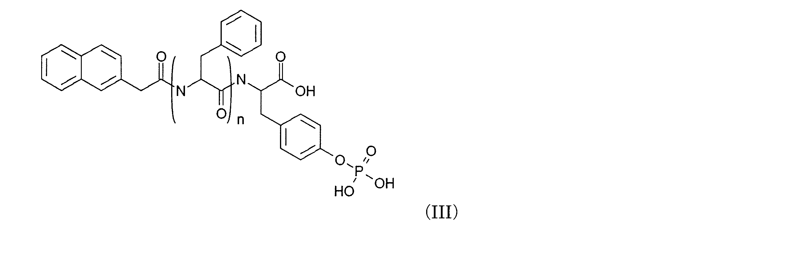
- the method according to [1] wherein the compound is a compound represented by the formula (I), wherein R 1 is a naphthyl group and m is 1.
- a kit for removing pluripotent cells containing the represented compound [6] The kit according to [5], wherein the compound is a compound represented by formula (I), wherein R 1 is a naphthyl group and m is 1. [7] The kit according to [6], wherein the compound is a compound represented by formula (I), wherein n is 3.
- the polyphenylalanine derivative since the polyphenylalanine derivative has an action of killing pluripotent cells, it can be applied to removing pluripotent cells contained from the target cell group.
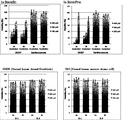
- Figure 1 shows that D-3 was contacted with 201B7 or cardiomyocyte for 1 hour (1h) or 2 hours (2h) and then cultured in Stemfit (upper left figure) and StemPro (upper right figure) for 1 day. It is a graph which shows the survival rate of a cell.
- the normal cell line NHDF Normal human dermal fibroblasts
- HS-5 normal human marrow stroma cell
- the vertical axis represents the cell viability calculated from the result of the same experiment conducted without contacting D-3 as 100%.
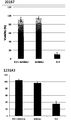
- FIG. 2 is a graph showing the survival rate of cells cultured for 1 day in Stemfit (upper figure) and DMEM (lower figure) after contacting D-3 with 1231A3 for 1 hour (1 h) or 2 hours (2 h).
- the vertical axis represents the cell viability calculated from the result of the same experiment conducted without contacting D-3 as 100%.
- Figure 3 shows that after contacting phosphatase inhibitor and D-3 (inhibitor ⁇ + D-3), phosphatase inhibitor (inhibitor), and D-3 for 1 hour (1h) to 201B7 (top) or 1231A3 (bottom) It is a graph which shows the survival rate of the cell cultured for 1 day.
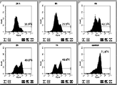
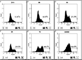
- FIG. 4 shows a histogram after staining each cell with TRA-1-60 antibody (upper figure) or SSEA-4 antibody (lower figure).
- the figure on the left shows the result of cells cultured for 1 day after contact with D-3 (201B7d16) for 1 hour (1h) after inducing differentiation of 201B7, and the center figure shows the cells after differentiation induction of 201B7 (201B7d16). ) And the right figure shows the result of 201B7.
- the numbers indicate the content of TRA-1-60 positive cells (upper figure) or SSEA-4 positive cells (lower figure).
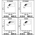
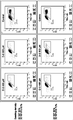
- FIG 5 shows a dot plot of the results of FACS measurement after staining each cell with TRA-1-60 antibody.
- the upper figure is the result of cells cultured for 23 hours after contacting naturally differentiated 201B7 (201B7 is cultured with Stemfit without bFGF for 16 days) with D-3, and the lower figure is the result of spontaneously differentiated 201B7 (201B7d16) It is.
- the figure on the right side of the lower figure shows the result of staining the cells after inducing differentiation of 201B7 with IgG as isotype control.
- the numbers indicate the content of TRA-1-60 positive cells (upper right diagram, upper left diagram, lower left diagram) or false positive cell content (lower right diagram).
- Figure 6 shows cells mixed with 201B7 and 201B7 after induction of differentiation (201B7d16) after contact with D-3, 24 hours (24h), 8 hours (8h), 4 hours (4h), 2 hours (2h) ), Or cells cultured for 1 hour (1 h), or cells mixed with 201B7 and 201B7d16 (control) are stained with TRA-1-60 antibody, and a histogram of the results of FACS measurement is shown. In the figure, the numbers indicate the content of TRA-1-60 positive cells.
- Fig. 7 shows 24 hours (24h), 8 hours (8h), 4 hours (4h), and 2 hours (2h) after contacting cells mixed with 1231A3 and 1231A3 after induction of differentiation (1231A3d18) with D-3.
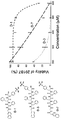
- FIG. 9 is a graph showing the results of evaluating the anti-iPS cell effect of a diphenylalanine-based phosphor D peptide.
- D-1 has two phenylalanines and D3 has three phenylalanines.
- D-7 further has glycine as a linker between naphthyl group and triphenylalanine as compared to D-3.
- FIG. 10 is a graph showing the mortality when three iPS cell lines (201B7, 1231A, and 1383D7) were treated with different concentrations of D-3 for 1 to 4 hours. The top two panels compare the toxicity to 201B7 with different cell concentrations and different colony sizes.
- FIG. 9 is a graph showing the results of evaluating the anti-iPS cell effect of a diphenylalanine-based phosphor D peptide.
- D-1 has two phenylalanines and D3 has three phenylalanines.
- D-7 further has glycine as a linker between naph
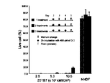
- FIG. 11 is a graph showing that 201B7 can be removed at a stage close to confluence by iterative processing using D-3.
- the graph shows the survival rate of 201B7 after each day treatment with D-3 compared to a control with daily media change.
- 201B7 was seeded at different concentrations to form small colonies, large colonies, and near confluent stages.
- NHDF cells were used as a control and treated with the same D-3 every day.
- FIG. 12 shows normal skin fibroblasts (NHDF), human primary hepatocytes, human primary renal proximal tubular epithelial cells, spontaneously differentiated 201B7, 201B7-derived neurons, and 201B7, where D-3 is a non-iPS sputum cell It is a graph which shows that there is almost no toxicity with respect to the derived cardiac muscle cell.
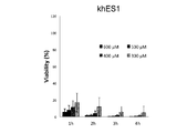
- FIG. 13 shows that D-3 is also toxic to human ES cells (khES1).
- FIG. 14 is a graph comparing the toxicity of D-3 and commercially available anti-iPS cell substance rBC2LCN-PE23 to 201B7, showing that D-3 is more potent than rBC2LCN-PE23.
- FIG. 15 shows that the toxicity of D-3 is related to the amount of P-ALP mRNA introduced into the cells (ALPP in Fig. 15 represents the same as P-ALP).
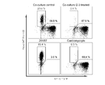
- FIG. 16 shows that iPS cells mixed in differentiated cell groups were successfully removed by using D-3. 201B7-derived cardiomyocytes expressing 201B7 and MHY6-EGFP were analyzed by flow cytometry after culturing with 400 ⁇ D-3 for 1 hour or without such culturing. Untreated 201B7 and untreated cardiomyocytes were used as controls.
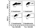
- FIG. 17 shows that a small amount of differentiated cells was successfully purified from a cell group containing a large amount of iPS cells and a small amount of differentiated cells by repeated treatment with D-3.
- FIG. 18 shows that the remaining iPS cells were successfully removed from the 201B7 differentiation-resistant clones that had been naturally differentiated for 15 days using D-3.
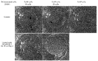
- FIG. 19 shows that by using D-3, the remaining iPS cells were successfully removed from the co-culture of iPS cells and differentiated cells.
- staining indicating the presence of iPS cells is visible
- the lower left, lower right showing samples that have been treated with D-3, as well as the upper without iPS cells.
- the presence of iPS cells cannot be confirmed.
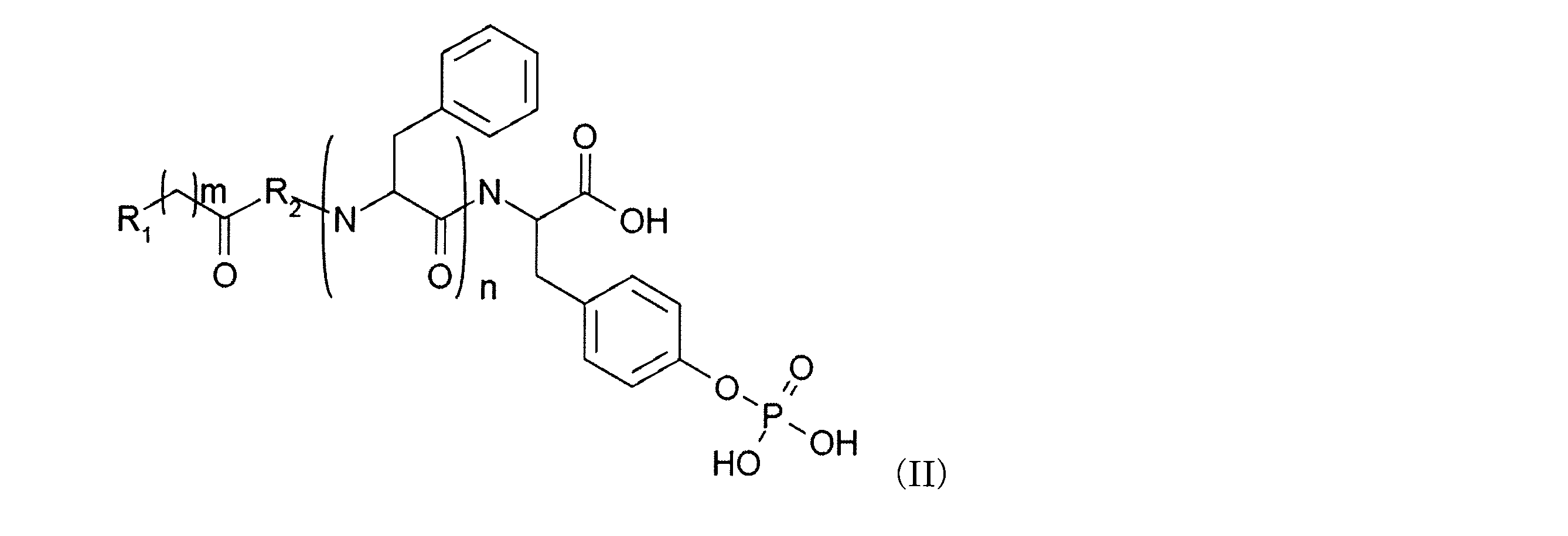
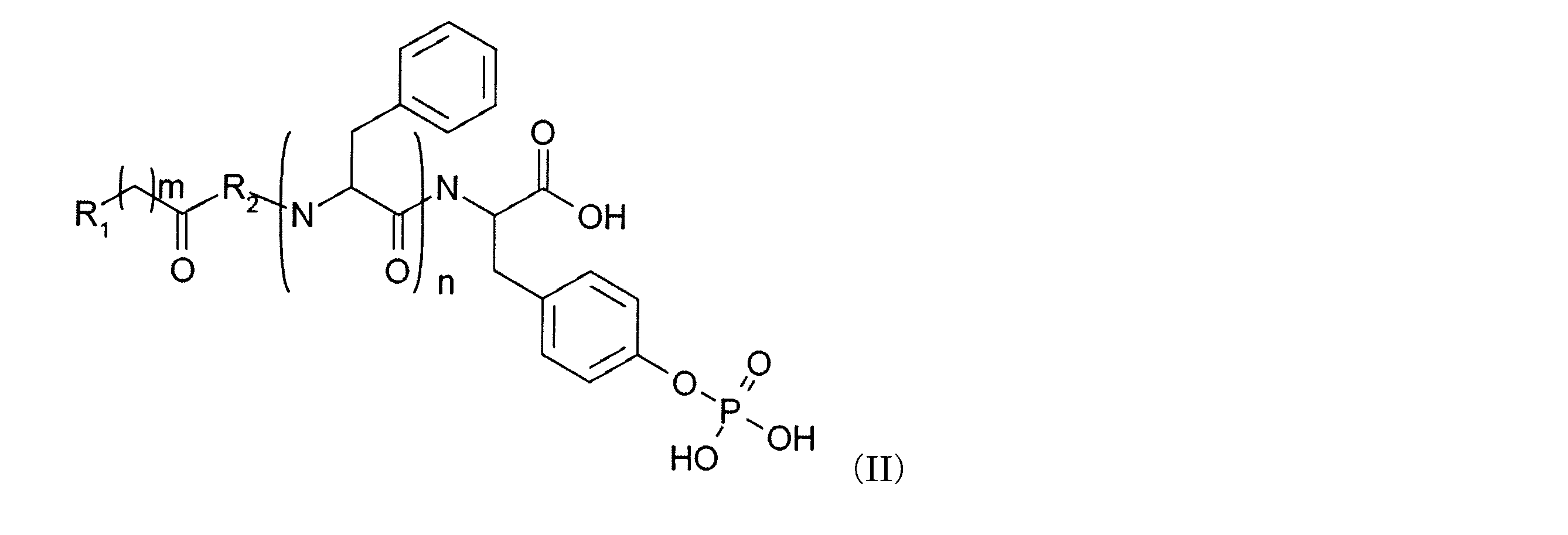
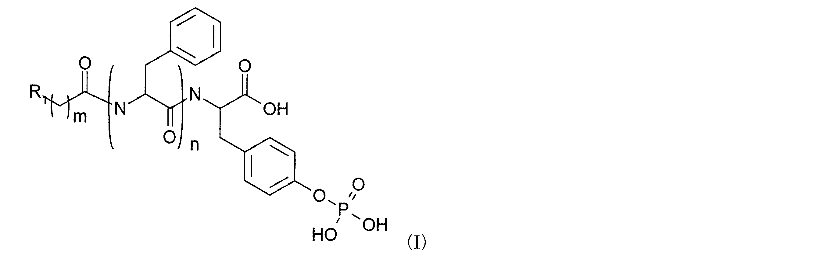
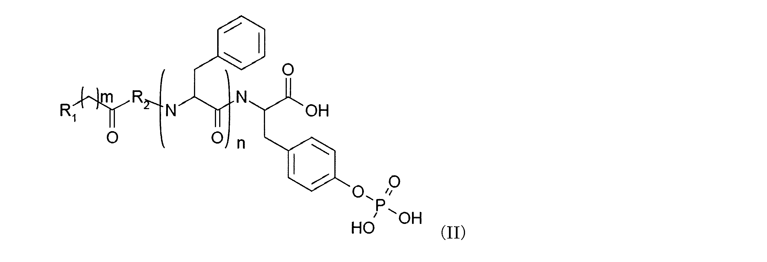
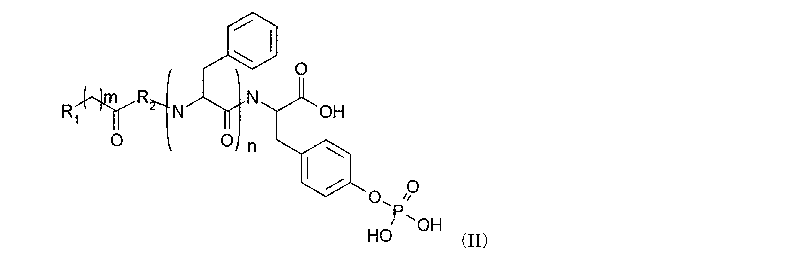
- the present invention is directed to formula (I): (Wherein R 1 is an aryl group or a heteroaryl group, m is an integer of 1 to 6, and n is an integer of 2 or more) or formula (II): Wherein R 1 is an aryl group or a heteroaryl group, R 2 is an alanine residue or a glycine residue, m is an integer of 1 to 6, and n is an integer of 2 or more.
- a method for removing pluripotent cells, or a kit for removing pluripotent cells containing the polyphenylalanine derivative, comprising the step of contacting the represented polyphenylalanine derivative with a target cell population.
- the aryl group means a monovalent 6-carbon monocyclic, 10-carbon bicyclic, or 14-carbon tricyclic aromatic ring system.
- Examples of the aryl group include, but are not limited to, a phenyl group, a naphthyl group, and an anthracenyl group.
- a heteroaryl group is a monovalent aromatic 5- to 8-membered monocyclic system or 8- to 12-membered bicyclic system having one or more heteroatoms (such as O, N, S, or Se). Or an 11-14 membered tricyclic system.
- the heteroaryl group include a pyridyl group, a furyl group, an imidazolyl group, a benzimidazolyl group, a pyrimidinyl group, a thienyl group, a quinolinyl group, an indolyl group, and a thiazolyl group.
- an aryl group as used herein includes both substituted and unsubstituted moieties.
- substituted refers to one or more substituents, which can be the same or different, each replacing a hydrogen atom.
- substituents include halogen groups (eg, F, Cl, Br, I), hydroxyl groups, amino groups, cyano groups, nitro groups, mercapto groups, alkoxycarbonyl groups, acyloxy groups, amide groups, carboxy groups, alkanes.
- Cycloalkyl groups, cycloalkenyl groups, heterocycloalkyl groups, heterocycloalkenyl groups, aryl groups, and heteroaryl groups can be fused together.
- the substituent can be a protecting group during the synthesis process.
- protecting group refers to a group or moiety used to protect or mask functionality that causes unwanted reactions during process steps. The protecting group prevents the reaction in the process but can then be removed to reveal the original functionality. Examples of protecting groups include trimethylsilyl group (TES), tert-butyloxycarbonyl group (tBoc), benzyloxycarbonyl group (CBZ), 9-fluorenylmethyloxycarbonyl group (Fmoc). Not exclusively.
- a preferred polyphenylalanine derivative is represented by the formula (III) in which R 1 in the formula (I) is a naphthyl group and m is 1. It is a compound represented by these.
- a more preferable polyphenylalanine derivative is a compound in which n in the formula (III) is 3.
- the alanine residue, glycine residue, phenylalanine residue and O-phospho-D-tyrosine contained in the polyphenylalanine derivative of the present invention may be D-form or L-form and are not easily decomposed. In view of the above, the D form is preferred.
- the polyphenylalanine derivative of the present invention can be synthesized by appropriately using an amino acid or a carboxylic acid by a known solid phase peptide synthesis method.
- pluripotent cells have the property of being capable of differentiating into all cells present in a living body, and are alkaline phosphatase, Oct-3 / 4, Nanog, SSEA-3, SSEA-4, TRA-1 A cell that is positive for at least one marker gene of the group consisting of -60 and TRA-1-81.
- Pluripotent cells include pluripotent stem cells.
- the target cell population is not particularly limited as long as it is a cell population that may contain pluripotent cells, and examples thereof include a cell population induced to differentiate from pluripotent stem cells.
- a pluripotent stem cell has pluripotency that can be differentiated into all cells present in a living body, and also has a proliferation ability.
- pluripotent stem cells include embryonic stem (ES) cells, embryonic stem cells derived from cloned embryos obtained by nuclear transfer (“ntES cells”), germ stem cells (“GS cells”), embryonic germ cells (“EG cells”), and induced pluripotent stem (iPS) cells, but are not limited to these.
- Examples of preferred pluripotent stem cells include ES cells, ntES cells, and iPS cells.
- Embryonic stem cells ES cells are stem cells established from the inner cell mass of early embryos (for example, blastocysts) of mammals such as humans and mice, and have pluripotency and proliferation ability by self-replication. .
- ES cells are embryonic stem cells derived from the inner cell mass of blastocysts, which are embryos after the 8-cell stage and morula stage of fertilized eggs, the ability to differentiate into any cell that constitutes an adult, so-called differentiation It has pluripotency and proliferation ability by self-replication.
- ES cells were discovered in mice in 1981 (M. J. Evans and M. H. Kaufman (1981), Nature 292: 154-156), and then ES cell lines were established in primates such as humans and monkeys. (J. A. Thomson et al. (1998), Science 282: 1145-1147; J. A. Thomson et al. (1995), Proc. Natl. Acad. Sci. USA, 92: 7844-7848; J. A. Thomson et al. (1996), Biol. Reprod., 55: 254-259; and J. A. Thomson and V. S. Marshall (1998), Curr. Top. Dev. Biol., 38: 133-165).
- ES cells can be established by taking an inner cell mass from a blastocyst of a fertilized egg of a target animal and culturing the inner cell mass on a fibroblast that is a feeder cell. Further, maintenance of cells by subculture can be performed using a medium supplemented with substances such as leukemia inhibitory factor (LIF) and basic fibroblast growth factor (bFGF).
- LIF leukemia inhibitory factor
- bFGF basic fibroblast growth factor
- human ES cells use DMEM / F-12 medium supplemented with 0.1 mM 2-mercaptoethanol, 0.1 mM non-essential amino acid, 2 mM L-glutamic acid, 20% KSR and 4 ng / ml bFGF at 37 ° C. It can be maintained in a 5% CO2 wet atmosphere.
- ES cells need to be passaged every 3 to 4 days. At this time, passage is performed, for example, 0.25% trypsin and 0.1 mg / ml in PBS containing 1 mM CaCl 2 and 20% KSR. It can be performed using collagenase IV.
- ES cells can be selected using the expression of gene markers such as alkaline phosphatase, Oct-3 / 4, Nanog as an index.
- gene markers such as alkaline phosphatase, Oct-3 / 4, Nanog as an index.
- the expression of gene markers such as OCT-3 / 4, NANOG, etc. can be detected by Real-Time PCR, or cell surface antigens SSEA-3, SSEA-4, TRA-1 -60, TRA-1-81 can be detected by immunostaining (Klimanskaya I, et al. (2006), Nature. 444: 481-485).
- Germ stem cells are testis-derived pluripotent stem cells that are the origin of spermatogenesis. Like ES cells, these cells can be induced to differentiate into various lineages, and have the property of producing chimeric mice when transplanted into mouse blastocysts, for example (M. Kanatsu-Shinohara et al. (2003)). Biol. Reprod., 69: 612-616; K. Shinohara et al. (2004), Cell, 119: 1001-1012). Germ stem cells can self-replicate in a medium containing glial cell line-derived neurotrophic factor (GDNF), and can be obtained by repeated passage under the same culture conditions as ES cells. (Masatake Takebayashi et al. (2008), Experimental Medicine, Vol. 26, No. 5 (extra number), pages 41-46, Yodosha (Tokyo, Japan)).
- GDNF glial cell line-derived neurotrophic factor
- Embryonic germ cells are cells that are established from embryonic primordial germ cells and have the same pluripotency as ES cells. In the presence of substances such as LIF, bFGF, and stem cell factor Can be established by culturing primordial germ cells in (Y. Matsui et al. (1992), Cell, 70: 841-847; JL Resnick et al. (1992), Nature, 359: 550-551).
- iPS Artificial pluripotent stem cells introduce a specific nuclear reprogramming substance into a somatic cell in the form of DNA or protein, or contain the nuclear reprogramming substance by the drug. It is an artificial stem cell derived from a somatic cell that has almost the same characteristics as ES cells, such as pluripotency of differentiation and proliferation ability by self-replication, which can be produced by increasing the expression of sex mRNA and protein ( K. Takahashi and S. Yamanaka (2006) Cell, 126: 663-676; K. Takahashi et al. (2007) Cell, 131: 861-872; J. Yu et al. (2007) Science, 318: 1917- 1920; M.
- the nuclear reprogramming substance is not particularly limited as long as it is a gene specifically expressed in ES cells, a gene that plays an important role in maintaining undifferentiation of ES cells, or a gene product thereof.
- Nucleotide sequences of mouse and human cDNA of each nuclear reprogramming substance and amino acid sequence information of the protein encoded by the cDNA can be obtained by referring to NCBI accession numbers described in WO 2007/069666.
- mouse and human cDNA and amino acid sequence information of L-Myc, Lin28, Lin28b, Esrrb, Esrrg, and Glis1 can be obtained by referring to the following NCBI accession numbers, respectively.
- a person skilled in the art can prepare a desired nuclear reprogramming substance by a conventional method based on the cDNA sequence or amino acid sequence information.
- nuclear reprogramming substances may be introduced into somatic cells in the form of proteins, for example, by lipofection, binding to cell membrane permeable peptides, microinjection, or in the form of DNA, for example, You may introduce
- retrovirus vectors As viral vectors, retrovirus vectors, lentiviral vectors (above, Cell, 126, pp.663-676, 2006; Cell, 131, pp.861-872, 2007; Science, 318, pp.1917-1920, 2007 ), Adenovirus vectors (Science, 322, 945-949, 2008), adeno-associated virus vectors, Sendai virus vectors (Proc Jpn Acad Ser B Phys Biol Sci. 85, 348-62, 2009) and the like.
- artificial chromosome vectors include human artificial chromosomes (HAC), yeast artificial chromosomes (YAC), and bacterial artificial chromosomes (BAC, PAC).
- a plasmid for mammalian cells can be used (Science, 322: 949-953, 2008).
- Vectors can contain regulatory sequences such as promoters, enhancers, ribosome binding sequences, terminators, polyadenylation sites, and the like.
- the promoter used include EF1 ⁇ promoter, CAG promoter, SR ⁇ promoter, SV40 promoter, LTR promoter, CMV (cytomegalovirus) promoter, RSV (rous sarcoma virus) promoter, MoMuLV (Moloney murine leukemia virus) LTR, HSV- A TK (herpes simplex virus thymidine kinase) promoter or the like is used.
- EF1 ⁇ promoter CAG promoter, MoMuLV LTR, CMV promoter, SR ⁇ promoter and the like are preferable.
- selectable marker sequences such as drug resistance genes (for example, kanamycin resistance gene, ampicillin resistance gene, puromycin resistance gene), thymidine kinase gene, diphtheria toxin gene; green fluorescent protein (GFP), ⁇ -glucuronidase ( GUS), FLAG and other reporter gene sequences;
- the above vector contains a LoxP sequence before and after the gene or promoter encoding the nuclear reprogramming substance and the gene encoding the nuclear reprogramming substance that binds to it. You may have.
- transposons include, for example, piggyBac, a transposon derived from lepidopterous insects (Kaji, KaK. Et al., (2009), Nature, 458: 771-775; Woltjen et al., (2009), Nature, 458: 766-770; WO 2010/012077).
- vectors can be replicated without chromosomal integration and exist in episomes so that they can be the origin of lymphotropic herpesvirus (lymphotrophic ⁇ ⁇ ⁇ ⁇ herpes virus), BK virus or bovine papillomavirus (Bovine papillomavirus) It may contain a sequence related to the replication. Examples include EBNA-1 and oriP sequences, or Large T and SV40ori sequences (WO 2009/115295, WO 2009/157201, and WO 2009/149233).
- an expression vector for polycistronic expression may be used.
- the gene coding sequence may be linked by an IRES or foot-and-mouth disease virus (FMDV) 2A coding region (Science, 322: 949-953, 2008 and WO 2009/092042 2009/152529).
- HDAC histone deacetylase
- VPA valproic acid
- MC1293, M344 small molecule inhibitors
- siRNA and shRNA against HDAC eg, HDAC1 siRNA Smartpool (Millipore), HuSH 29mer shRNA Constructs against nucleic acid expression inhibitors such as HDAC1 (OriGene) etc.
- DNA methyltransferase inhibitors eg 5'-azacytidine
- G9a histone Methyltransferase inhibitors eg, small molecule inhibitors such as BIX-01294 (Cell Stem Cell, 2: 525-528 (2008)), siRNA and shRNA against G9a (eg, G9a siRNA (human) (Santa Cruz Biotechnology), etc.) ) Nucleic acid expression inhibitors etc.
- L -channel calcium agonist eg Bayk8644
- p53 inhibitors eg siRNA and shRNA against p53
- Wnt Signaling activator eg soluble Wnt3a
- growth factors such as LIF or bFGF, ALK5 inhibitor (eg SB431542) (Nat.
- Drugs used to increase the expression of the nuclear reprogramming endogenous protein include 6-bromoindirubin-3′-oxime, indirubin-5-nitro-3′-oxime, valproic acid, 2- (3- ( 6-methylpyridin-2-yl) -1H-pyrazol-4-yl) -1,5-naphthyridine, 1- (4-methylphenyl) -2- (4,5,6,7-tetrahydro-2-imino -3 (2H) -benzothiazolyl) ethanone, HBr (pifthrin-alpha), prostaglandin J2, prostaglandin E2, etc. are exemplified (WO 2010/068955).
- a medium for inducing iPS cells for example, (1) DMEM, DMEM / F12 or DME medium containing 10 to 15% FBS (in addition to these mediums, LIF, penicillin / streptomycin, puromycin, L-glutamine) , (2) ES cell medium containing bFGF or SCF, for example, mouse ES cell medium (for example, TX-WES medium, Thrombo X) ) Or primate ES cell culture medium (for example, primate (human & monkey) ES cell culture medium (Reprocell, Kyoto, Japan), mTeSR-1), and the like.
- a somatic cell and a nuclear reprogramming substance are brought into contact with each other in a DMEM or DMEM / F12 medium containing 10% FBS in the presence of 5% CO 2 at 37 ° C.
- Culture for ⁇ 7 days then re-spread cells onto feeder cells (eg, mitomycin C-treated STO cells, SNL cells, etc.), and bFGF-containing primate ES cells approximately 10 days after contact between somatic cells and nuclear reprogramming substance
- the cells can be cultured in a working medium to generate ES cell-like colonies about 30 to about 45 days or more after the contact.
- the cells may be cultured under conditions of an oxygen concentration as low as 5-10%.
- somatic cells can be used on feeder cells (eg, mitomycin C-treated STO cells, SNL cells, etc.) in 10% FBS-containing DMEM medium (including LIF, penicillin / streptomycin, puromycin, L-glutamine, non-essential amino acids, ⁇ -mercaptoethanol, etc. can be appropriately contained), and ES-like colonies can be formed after about 25 to about 30 days or more from the culture.
- feeder cells eg, mitomycin C-treated STO cells, SNL cells, etc.
- FBS-containing DMEM medium including LIF, penicillin / streptomycin, puromycin, L-glutamine, non-essential amino acids, ⁇ -mercaptoethanol, etc.
- the medium is replaced with a fresh medium once a day from the second day after the start of the culture.
- the number of somatic cells used for nuclear reprogramming is not limited, but ranges from about 5 ⁇ 10 3 to about 5 ⁇ 10 6 cells per 100 cm 2 of culture dish.
- a marker gene-expressing cell When a gene containing a drug resistance gene is used as a marker gene, a marker gene-expressing cell can be selected by culturing in a medium (selective medium) containing the corresponding drug.
- the marker gene is a fluorescent protein gene
- the marker gene-expressing cells can be obtained by observing with a fluorescence microscope, by adding a luminescent substrate in the case of a luminescent enzyme gene, and by adding a chromogenic substrate in the case of a chromogenic enzyme gene. Can be detected.
- a “somatic cell” may be any cell other than a germ cell derived from a mammal (eg, human, mouse, monkey, pig, rat, etc.), for example, keratinized epithelial cell (Eg, keratinized epidermal cells), mucosal epithelial cells (eg, epithelial cells of the tongue surface), exocrine glandular epithelial cells (eg, mammary cells), hormone-secreting cells (eg, adrenal medullary cells), cells for metabolism and storage (Eg, hepatocytes), luminal epithelial cells that make up the interface (eg, type I alveolar cells), luminal epithelial cells (eg, vascular endothelial cells) in the inner chain, and ciliated cells that are capable of transporting (Eg, airway epithelial cells), extracellular matrix secreting cells (eg, fibroblasts), contractile cells (eg, smooth muscle cells), blood and immune system cells (e
- the degree of cell differentiation and the age of the animal from which the cells are collected there is no particular limitation on the degree of cell differentiation and the age of the animal from which the cells are collected. This can be applied to both undifferentiated progenitor cells (including somatic stem cells) and finally differentiated mature cells. It can be used as the source of somatic cells in the invention.
- undifferentiated progenitor cells include tissue stem cells (somatic stem cells) such as neural stem cells, hematopoietic stem cells, mesenchymal stem cells, and dental pulp stem cells.
- the mammal individual from which somatic cells are collected is not particularly limited, but is preferably a human.
- E Cloned embryo-derived ES cells obtained by nuclear transfer nt ES cells are cloned embryo-derived ES cells produced by nuclear transfer technology and have almost the same characteristics as ES cells derived from fertilized eggs (T. Wakayama et al. (2001), Science, 292: 740-743; S. Wakayama et al. (2005), Biol. Reprod., 72: 932-936; J. Byrne et al. (2007) , Nature, 450: 497-502).
- an ES cell established from an inner cell mass of a clonal embryo-derived blastocyst obtained by substituting the nucleus of an unfertilized egg with the nucleus of a somatic cell is an nt ES (nuclear transfer ES) cell.
- nt ES nuclear transfer ES
- a combination of nuclear transfer technology JB Cibelli et al. (1998), Nat. Biotechnol., 16: 642-646) and ES cell production technology (above) is used ( Wakayama Seika et al. (2008), Experimental Medicine, Vol. 26, No. 5 (extra number), pages 47-52).
- reprogramming can be performed by injecting a somatic cell nucleus into an enucleated unfertilized egg of a mammal and culturing it for several hours.
- (F) Fusion stem cell A stem cell that has the same pluripotency as a fused ES cell by fusing a somatic cell and an egg or ES cell, and also has a gene unique to the somatic cell (Tada M et al. al. Curr Biol. 11: 1553-8, 2001; Cowan CA et al. Science. 2005 Aug 26; 309 (5739): 1369-73).
- differentiation induction from pluripotent stem cells is not only induced to differentiate into specific organ cells and their precursor cells, but also includes a wide range of cell types such as endoderm cells, mesoderm cells, and ectoderm cells. Including inducing differentiation into a cell population comprising.
- organ cells include cells contained in skin, blood vessels, cornea, kidney, heart, liver, umbilical cord, intestine, nerve, lung, placenta, pancreas, brain, extremities, and retina. It is not limited. Any differentiation induction method obvious to those skilled in the art can be used.
- Examples include the method for inducing differentiation into neural stem cells described in JP-A-2002-291469 or WO2011 / 019092, the method for inducing differentiation into pancreatic stem-like cells described in JP-A-2004-121165, and JP 2003-505006 And the method for inducing differentiation into hematopoietic cells described in the above, and the method for inducing differentiation into cardiomyocytes described in WO2014 / 185358. Furthermore, examples of the differentiation induction method by formation of embryoid bodies include the methods described in JP-T-2003-523766.
- the polyphenylalanine derivative of the present invention can be removed by adding to a culture solution of a target cell population to induce cell death of pluripotent cells contained in the target cell population.
- the contact time between the target cell population and the polyphenylalanine derivative is not particularly limited, and examples thereof include 1 hour, 2 hours, 3 hours, 4 hours, 5 hours, 6 hours, 12 hours, and 24 hours.
- the concentration of the polyphenylalanine derivative in the culture solution is not particularly limited, but examples thereof include 100 ⁇ M to 1 mM, more preferably 200 ⁇ M, 300 ⁇ M, or 400 ⁇ M.
- Human iPS cells (MYH6-EIP4-201B7) were prepared by introducing an EGFP cassette (MYH6-EIP4) operably linked to the MYH6 promoter into the 201B7.
- the MYH6-EIP4-introduced iPS cell line was prepared by introducing a vector operably linked to the EGFP cassette downstream of MYH (myosin heavy chain) -promoter into 201B7 using the PiggyBac transposon system.
- ⁇ Cardiomyocyte cell line> The myocardial cell line was treated with CTK solution (ReproCELL) for 2 minutes after removing MYH6-EIP4-201B7, then treated with Accumax (Innovative Cell Technologies) for 5 minutes, and then dissociated into single cells by pipetting. .
- Cells are collected by centrifugation and transferred to a low adhesion 6 well dish (Corning), 2 mM L-Glutamine, 150 ⁇ g / mL Transferrin, 50 ⁇ g / mL Ascorbic Acid (sigma), 4 ⁇ 10 ⁇ 4 M monothioglycerol (MTG) and 2 ng EBs were formed by culturing in 1.5 ml / well STEMPRO 34 (Invitrogen) supplemented with / mL BMP4 (R & D) at 37 ° C. under 5% oxygen condition.
- STEMPRO 34 Invitrogen
- the obtained EB was washed with IMDM (Invitrogen), and 1% L-Glutamine, 150 ⁇ g / mL Transferrin, 50 ⁇ g / mL Ascorbic Acid, 4 ⁇ 10 ⁇ 4 M MTG, 10 ng / mL VEGF and 1 ⁇ M IWP-3 were added.
- STEMPRO 34 was added to the dish and cultured at 37 ° C. under 5% oxygen for 4 days.
- the medium was changed to STEMPRO 34 supplemented with 1% L-Glutamine, 150 ⁇ g / mL Transferrin, 50 ⁇ g / mL Ascorbic Acid, 4 ⁇ 10 ⁇ 4 M MTG, 10 ng / mL VEGF and 5 ng / mL bFGF for 4 days, 37
- the cells were cultured at 5 ° C. and 5% oxygen. At this time, the medium was replaced once every two days with the same conditions. Subsequently, the cells were cultured for 8 days while changing to a medium with the same conditions once every two days at normal oxygen concentration, and EGFP-positive cells were isolated using FACS using EGFP as a substitute for MYH-6 as an index. Used as a cardiomyocyte cell line.
- D-1 and D-3 were prepared by a solid-phase peptide synthesis method described in Ottinger, E.A., et al., Biochemistry, 32, 1993. Specifically, 2-chlorotrityl chloride resin, N-Fmoc-protected amino acids, and 2-Naphthaleneacetic acid were converted to N, N-diisopropylethylamine (DIPEA) and O-benzotriazole-N, N, N ', N'-tetramethyl-uronium- It was synthesized by activation with hexafluoro-phosphate (HBTU).
- DIPEA N-diisopropylethylamine
- HBTU hexafluoro-phosphate
- O-phospho-D-tyrosine is synthesized by reacting D-tyrosine and phosphorus pentoxide overnight in the presence of phosphoric acid according to the method described in Alewood, P. F., et al., Synthesis, 30-31 1983. did. N-Fmoc-O-phospho-D-tyrosine dissolves O-phospho-D-tyrosine and Fmoc-OSu in a 1: 1 mixture of water and CH3CN, and adds Et3N to adjust the pH to 8 ⁇ Obtained by adjusting to 9 and reacting for 30 minutes (Ottinger, EA, et al., Biochemistry, 32, 1993).
- 201B7 cells were seeded on Stemfit (Ajinomoto) containing Rock inhibitor on a 24 well plate coated with laminin to give 2 ⁇ 10 4 / well (24 well plate) (day 0). After 24 hours, the medium was replaced with only Stemfit, and the culture was continued for 24 hours. Alternatively, 1 ⁇ 10 4 / well (24 well plate) cardiomyocyte cell line was seeded and cultured in StemPro (Life Technologies) for 1 day. Subsequently, 0 ⁇ M, 200 ⁇ M, 300 ⁇ M, or 400 ⁇ M of D-3 was added to the culture solution, and cultured for 1 hour or 2 hours.
- the medium was replaced with a medium containing no D-3, and after culturing for 1 day, cell death was evaluated by WST-1 assay, and the cell viability was calculated as compared with the case where 0 ⁇ M D-3 was added. As a result, it was confirmed that 201B7 caused cell death when 300 ⁇ M or more of D-3 was added to the culture medium and cultured for 1 hour or longer (FIG. 1). On the other hand, the cardiomyocyte cell line was not found to be cytotoxic by D-3. These results suggest that D-3 causes cell death specifically in pluripotent stem cells.
- D-3 0 ⁇ M, 200 ⁇ M, 300 ⁇ M, or 400 ⁇ M D-3 was added to NHDF (normal human dermal fibroblasts) or HS5 (normal human medullary matrix cells) and cultured for 1 hour or 2 hours. The medium was replaced with a medium containing no D-3, and after culturing for 1 day, cell death was evaluated by the WST-1 assay. As a result, no cytotoxicity due to D-3 was found for any of the cells.
- NHDF normal human dermal fibroblasts
- HS5 normal human medullary matrix cells
- 1231A3 cells were seeded at 2 ⁇ 10 4 / well (24 well plate) on Stemfit (Ajinomoto) containing Rock inhibitor on a 24 well plate coated with laminin (day 0). After 24 hours, the cells were cultured in Stemfit or DMEM (Life Technologies) for 1 day. 0 ⁇ M, 200 ⁇ M, 300 ⁇ M or 400 ⁇ M D-1 or 0 ⁇ M, 200 ⁇ M, 300 ⁇ M or 400 ⁇ M D-3 was added to the culture and cultured for 1 hour or 2 hours. Subsequently, the medium was replaced with a medium that did not contain D-1 and D-3.
- the WST-1 assay was used to compare with cells without addition of D-1 or D-3 (0 ⁇ M). Each cell viability was calculated. As a result, it was confirmed that D-3 was not affected by the medium and exhibited toxicity to pluripotent stem cells (FIG. 2).
- 201B7 or 1231A3 cells were seeded on Stemfit (Ajinomoto) containing Rock inhibitor on a laminin-coated 24 well plate so as to be 2 ⁇ 10 4 / well (24 well plate) (day 0). After 24 hours, the medium was replaced with only Stemfit, and the culture was continued for 24 hours. Subsequently, 0 ⁇ M or 400 ⁇ M of D-3 and a phosphatase inhibitor (a mixture of 5 mM L-phenylalanine (Wakken Pharmaceutical Co., Ltd.) and 2 mM L-homoarginine (Wakken Pharmaceutical Co., Ltd.)) were added to the culture solution, Incubated for 1 or 2 hours.
- 201B7 was cultured in Stemfit without bFGF for 16 days, and the cells were naturally differentiated. Subsequently, the cells were seeded at 2 ⁇ 10 4 / well (24 well plate) on Stemfit (Ajinomoto) containing Rock inhibitor and not containing bFGF on a laminin-coated 24 well plate (day 0). ) After 24 hours, the medium was replaced with only Stemfit without the addition of bFGF, and the culture was continued for 24 hours. Subsequently, 0 ⁇ M or 400 ⁇ M D-3 was added to the culture and cultured for 1 hour.
- TRA-1-60 BD Pharmingen
- SSEA4 BD Pharmingen
- Viable cells that were not stained with 7-Amino-Actinomycin D (7-AAD) were evaluated for the content of TRA-1-60 or SSEA4-positive cells using FACS.
- a positive control that did not differentiate 201B7 was used.
- TRA-1-60 or SSEA4-positive cells were removed or reduced by adding D-3 and culturing (FIG. 4).
- 201B7 was cultured for 16 days in Stemfit without addition of bFGF, and the cells were naturally differentiated. Subsequently, the cells were seeded at 2 ⁇ 10 4 / well (24 well plate) on Stemfit (Ajinomoto) containing Rock inhibitor and not containing bFGF on a laminin-coated 24 well plate (day 0). ) After 24 hours, the medium was replaced with only Stemfit without the addition of bFGF, and the culture was continued for 24 hours. Subsequently, 0 ⁇ M (control) or 400 ⁇ M D-3 was added to the culture and cultured for 1 hour.
- the medium was replaced with a medium containing no D-3, and after culturing for 23 hours, the cells were separated and collected, and stained with an antibody against TRA-1-60. Viable cells that were not stained with 7-ADD were evaluated for the content of TRA-1-60 positive cells using FACS. At this time, a few non-specific positive cells were detected by staining with IgM, ⁇ isotype (BD Pharmingen) as a negative control. As a result, as described above, it was confirmed that TRA-1-60 positive cells were removed or reduced by adding D-3 and culturing (FIG. 5).
- 201B7 was cultured in Stemfit without bFGF for 16 days, and the cells were naturally differentiated. 0.7 ⁇ 10 5 201B7 and 0.7 ⁇ 10 5 spontaneously differentiated 201B7 were mixed and seeded on a laminin-coated 6-well plate containing Rock inhibitor and no bFGF added (day 0). After 24 hours, the medium was replaced with only Stemfit without the addition of bFGF, and the culture was continued for 24 hours. Subsequently, the cells were cultured for 1 hour in Stemfit not containing bFGF supplemented with 400 ⁇ M D-3. The medium was replaced with a medium containing no D-3, and further cultured for 1 hour, 2 hours, 4 hours, 8 hours, or 24 hours.
- the cultured cells were separately collected and stained with an antibody against TRA-1-60. Viable cells that were not stained with 7-ADD were evaluated for the content of TRA-1-60 positive cells using FACS. As positive controls, cells immediately after mixing of cells induced to differentiate by culturing 201B7 and 201B7 in Stemfit not containing bFGF for 16 days were used. As a result, it was confirmed that cell damage occurred 1 hour after contact with D-3, and cell death of pluripotent stem cells occurred over time (FIG. 6).
- 1231A3 was cultured for 16 days in Stemfit without the addition of bFGF, and the cells were naturally differentiated.
- 0.5 ⁇ 10 5 of 1231A3 and 0.7 ⁇ 10 5 of spontaneously differentiated 201B7 were mixed and seeded on a laminin-coated 6-well plate containing Rock inhibitor without stem bFGF (Ajinomoto) (day 0).
- the medium was replaced with only Stemfit without the addition of bFGF, and the culture was continued for 24 hours. Subsequently, the cells were cultured for 1 hour in Stemfit not containing bFGF supplemented with 400 ⁇ M D-3.
- the medium was replaced with a medium containing no D-3, and further cultured for 1 hour, 2 hours, 4 hours, 8 hours, or 24 hours.
- the cultured cells were separately collected and stained with an antibody against TRA-1-60.
- Viable cells that were not stained with 7-ADD were evaluated for the content of TRA-1-60 positive cells using FACS.
- As positive controls cells immediately after mixing of cells induced to differentiate by culturing 1231A3 and 1231A3 in Stemfit not containing bFGF for 18 days were used. As a result, it was confirmed that cytotoxicity occurred after contact with D-3 and cell death of pluripotent stem cells occurred over time (Fig. 7).
- 201B7 was cultured in Stemfit without addition of bFGF for 16 days to naturally differentiate the cells.
- 201B7 and spontaneously differentiated 201B7 were mixed at 2: 8 or 3: 7, and seeded on Stemfit (Ajinomoto) containing Rock inhibitor ⁇ and without bFGF on 6-well plates coated with laminin (day 0).
- the medium was replaced with only Stemfit without the addition of bFGF and culture was continued for 24 hours.
- the cells were cultured for 1 hour in Stemfit without bFGF supplemented with 0 ⁇ M, 300 ⁇ M or 400 ⁇ M D-3.
- the medium was replaced with a medium containing no D-3, and further cultured for 23 hours.
- the cultured cells were separately collected and stained with an antibody against TRA-1-60. Viable cells that were not stained with 7-ADD were evaluated for the content of TRA-1-60 positive cells using FACS. As a result, it was confirmed that cell death occurred selectively on TRA-1-60 positive cells by contact with D-3 (FIG. 8).
- 201B7 was seeded on Stemfit containing Rock inhibitor on a laminin-coated 24-well plate so that the concentration was 2.6 ⁇ 10 4 cell / cm 2 . After 24 hours, the medium was replaced with only Stemfit and the culture was continued. After another 24 hours, the medium was replaced with Stemfit containing D-1, D-3, and D-7 molecules at different concentrations, or Stemfit as a control. After 2 hours of culture, the medium was replaced with Stemfit only. The next day, cells were harvested with 100 ⁇ L Accumax. Next, 300 ⁇ L of Stemfit was added to stop the Accumax reaction. The number of viable cells in 100 ⁇ L of each sample was measured using flow cytometry. Prior to analysis to discriminate dead cells, 7-AAD was added to each sample. As a result, it was shown that 201B7 can be killed not only by D-3 but also by using D-1 and D-7 (FIG. 9).
- Example 8 Three types of iPS cell lines (201B7, 1231A, 1383D7) were seeded on Stemfit containing Rock inhibitor on a laminin-coated 24-well plate so as to be 2.6 ⁇ 10 4 cell / cm 2 . 201B7 was also seeded at 5.3 ⁇ 10 4 / cm 2 to prepare a sample in which a large colony was generated. Human iPS cells (1383D7) were used after culturing those provided by Dr. Asuka Morimi, Kyoto University. After 24 hours, the medium was replaced with only Stemfit and the culture was continued. After another 24 hours, the medium was replaced with Stemfit containing different concentrations of D-3 or Stemfit as a control.
- 201B7 cells should be 2.6 x 10 4 cell / cm 2 , 5.3 x 10 4 cell / cm 2 , 10.5 x 10 4 cell / cm 2 on Stemfit containing Rock inhibitor on a laminin-coated 24 well plate. Sowing (day 0). After 24 hours, the medium was replaced with only Stemfit and the culture was continued. After another 24 hours, the medium was replaced with Stemfit containing D-3 at a concentration of 400 ⁇ M. After 2 hours of culture, the medium was replaced with Stemfit only. Each cell was treated with D3 once (only day 2), twice (day 2 and day 3), and three times (day 2-4).
- NHDF Normal skin fibroblasts
- ATCC American Type Culture Collection
- Human primary hepatocytes and hepatocyte medium were purchased from Cosmo Bio.
- Human primary renal proximal tubular epithelial cells and REGMTM BulletKit medium were purchased from Lonza.
- Spontaneous differentiation 201B7 was prepared by culturing 201B7 in Stemfit without bFGF for 2 weeks.
- 201B7-derived neurons were prepared according to the method described in the aforementioned paper Nakagawa, et al. Sci Rep. 4: 3594, 2014.
- 201B7-derived cardiomyocytes were prepared by the method described above. All cells were maintained at 37 ° C., 5% CO 2 .
- Each cell was seed
- the medium was replaced with a medium containing different concentrations of D-3 molecules or only as a control medium.
- the medium was replaced with medium alone.
- cells were harvested with 100 ⁇ L Accumax.
- 300 ⁇ L of Stemfit was added to stop the Accumax reaction.
- the number of viable cells in 100 ⁇ L of each sample was measured using flow cytometry. Prior to analysis to discriminate dead cells, 7-AAD was added to each sample. As a result, it was shown that D-3 is hardly toxic to various cells that are not iPS cells (FIG. 12).
- khES1 was cultured and used from Dr. Yoshinori Yoshida of Kyoto University.
- the toxicity of D-3 to khES1 was examined in the same manner as in Example 8 except that khES1 was seeded so as to give 5.3 ⁇ 10 4 cell / cm 2 .
- D-3 is also toxic to human ES cells (khES1) (FIG. 13).
- the iPS cell line (201B7) was seeded on Stemfit containing Rock inhibitor on a laminin-coated 24-well plate so as to be 5.3 ⁇ 10 4 cell / cm 2 . After 24 hours, the medium was replaced with only Stemfit and the culture was continued. After another 24 hours, the medium was replaced with Stemfit containing 400 ⁇ M D-3, Stemfit containing 10 ⁇ g / mL rBC2LCN-PE23, or Stemfit as a control. After 1 hour or 2 hours of culture for D-3 and after 2 hours or 24 hours of culture for rBC2LCN-PE23, the medium was replaced with Stemfit alone. The next day, cells were harvested with 100 ⁇ L Accumax.
- D-3 has higher efficacy than rBC2LCN-PE23, which is a commercially available anti-iPS cell substance (FIG. 14).
- D-3 not only has a high iPS cell removal effect, but also enables a reduction in the treatment time for removal.
- D-3 which is a short peptide molecule, is excellent in that it can be easily synthesized economically with a large volume.
- 293FT was purchased from invitrogen. 293FT was seeded at 5.3 ⁇ 10 4 cell / cm 2 in DMEM medium (10% FBS, 1% non essential amino acids) on a 24-well plate. Twenty-four hours later, 293FT was transfected with different concentrations of wild-type placental alkaline phosphatase mRNA (P-ALP mRNA) using Stemfect reagent according to standard protocols. After 4 hours, the medium was changed to culture medium only. As a control, 293FT without D-3 treatment was used in which only the medium was exchanged at the same timing as that treated. After 24 hours, the medium was replaced with one containing D-3 at different concentrations, and the control was replaced with medium alone.
- P-ALP mRNA wild-type placental alkaline phosphatase mRNA
- 201B7-derived cardiomyocytes expressing 201B7 and MHY6-EGFP have a cell number ratio of 1: 9, and a total of 5.3 ⁇ 10 4 cells / cm 2 on a laminin-coated 24-well plate. Seed Stemfit containing After 24 hours, the medium was replaced with Stemfit containing a suspension of 201B7-derived cardiomyocytes expressing MHY6-EGFP. After another 24 hours, the medium was replaced with Stemfit containing D-3 at a concentration of 400 ⁇ M, and the control was replaced with Stemfit-only medium. After 1 hour of culture, the medium was replaced with Stemfit only. The next day, cells were harvested with 100 ⁇ L Accumax.
- 201B7-derived cardiomyocytes expressing 201B7 and MHY6-EGFP have a cell number ratio of 7: 3, and a total of 5.3 ⁇ 10 4 cells / cm 2 on a rock inhibitor-coated 24-well plate.
- Seed Stemfit containing (day 0). After 24 hours, the medium was replaced with Stemfit containing a suspension of 201B7-derived cardiomyocytes expressing MHY6-EGFP (day 1). After another 24 hours, the medium was replaced with Stemfit containing D-3 at a concentration of 400 ⁇ M, and the control was replaced with Stemfit-only medium. After 2 hours of culture, the medium was replaced with Stemfit only.
- Example 16 Spontaneous differentiation of 201B7 was used and cultured in Stemfit without bFGF to induce spontaneous differentiation.
- cells were seeded on Stemfit containing Rock inhibitor on a laminin-coated 24 well plate so that the concentration was 5.3 ⁇ 10 4 cell / cm 2 (day 0).
- the medium was changed to Stemfit.
- the medium was replaced with Stemfit containing 400 ⁇ M D-3 or Stemfit as a control.
- the medium was replaced with Stemfit only. Each cell was treated once (day 2 only), twice (day 2 and day 3), and three times (day 2-4).
- Example 17 5 x 10 4 naturally differentiated 201B7 and 50, 20 and 0 201B7 were mixed and seeded on Stemfit (Ajinomoto) containing Rock inhibitor on a laminin-coated 24-well plate (day 0). After 24 hours, the medium was changed to Stemfit (day 1). After another 24 hours, the medium was replaced with Stemfit containing D-3 at 400 ⁇ M or Stemfit as a control (day 2). After 2 hours of culture, the medium was replaced with Stemfit only. The cells were then cultured for an additional 7 days on Stemfit. By ALP staining, 201B7 colony was stained reddish purple. As a result, it was shown that the remaining iPS cells could be removed from the co-culture of iPS cells and differentiated cells by using D-3 (FIG. 19).
Landscapes
- Health & Medical Sciences (AREA)
- Life Sciences & Earth Sciences (AREA)
- Chemical & Material Sciences (AREA)
- Engineering & Computer Science (AREA)
- Organic Chemistry (AREA)
- Biomedical Technology (AREA)
- Genetics & Genomics (AREA)
- Bioinformatics & Cheminformatics (AREA)
- Biotechnology (AREA)
- Zoology (AREA)
- Wood Science & Technology (AREA)
- Biochemistry (AREA)
- General Health & Medical Sciences (AREA)
- Cell Biology (AREA)
- Microbiology (AREA)
- Molecular Biology (AREA)
- General Engineering & Computer Science (AREA)
- Medicinal Chemistry (AREA)
- Immunology (AREA)
- Chemical Kinetics & Catalysis (AREA)
- Urology & Nephrology (AREA)
- Hematology (AREA)
- Developmental Biology & Embryology (AREA)
- Biophysics (AREA)
- Proteomics, Peptides & Aminoacids (AREA)
- Toxicology (AREA)
- Tropical Medicine & Parasitology (AREA)
- Food Science & Technology (AREA)
- Physics & Mathematics (AREA)
- Analytical Chemistry (AREA)
- General Physics & Mathematics (AREA)
- Pathology (AREA)
- Gastroenterology & Hepatology (AREA)
- General Chemical & Material Sciences (AREA)
- Polymers & Plastics (AREA)
- Transplantation (AREA)
- Cardiology (AREA)
- Rheumatology (AREA)
- Micro-Organisms Or Cultivation Processes Thereof (AREA)
- Peptides Or Proteins (AREA)
Abstract
Description
[1]式(I):

[2]前記化合物が、式(I)で表される化合物であって、式中、R1がナフチル基であり、mが1である、[1]に記載の方法。
[3]前記化合物が、式(I)で表される化合物であって、式中、nが3である、[2]に記載の方法。
[4]前記対象細胞集団が、多能性幹細胞から分化誘導された細胞集団である[1]から[3]のいずれか1項に記載の方法。
[5]式(I):

[6]前記化合物が、式(I)で表される化合物であって、式中、R1がナフチル基であり、mが1である、[5]に記載のキット。
[7]前記化合物が、式(I)で表される化合物であって、式中、nが3である、[6]に記載のキット。
保護基はその工程で反応を防止するが、その後取り除いて本来の官能性を現すことができる。保護基の例には、トリメチルシリル基(TES)、tert-ブチルオキシカルボニル基(tBoc)、ベンジルオキシカルボニル基(CBZ)、9-フルオレニルメチルオキシカルボニル基(Fmoc)が含まれるが、これらに限らない。
ES細胞は、ヒトやマウスなどの哺乳動物の初期胚(例えば胚盤胞)の内部細胞塊から樹立された、多能性と自己複製による増殖能とを有する幹細胞である。
生殖幹細胞は、精巣由来の多能性幹細胞であり、精子形成のための起源となる細胞である。この細胞は、ES細胞と同様に、種々の系列の細胞に分化誘導可能であり、例えばマウス胚盤胞に移植するとキメラマウスを作出できる性質をもつ(M. Kanatsu-Shinohara et al. (2003) Biol. Reprod., 69:612-616;K. Shinohara et al. (2004), Cell, 119:1001-1012)。生殖幹細胞は、神経膠細胞系由来神経栄養因子(GDNF)を含む培地で自己複製可能であるし、またES細胞と同様の培養条件下で継代を繰り返すことによって、生殖幹細胞を得ることができる(竹林正則ら(2008),実験医学,26巻,5号(増刊),41~46頁,羊土社(東京、日本))。
胚性生殖細胞は、胎生期の始原生殖細胞から樹立される、ES細胞と同様な多能性をもつ細胞であり、LIF、bFGF、幹細胞因子などの物質の存在下で始原生殖細胞を培養することによって樹立しうる(Y. Matsui et al. (1992), Cell, 70:841-847;J.L. Resnick et al. (1992), Nature, 359:550-551)。
人工多能性幹(iPS)細胞は、ある特定の核初期化物質を、DNAまたはタンパク質の形態で体細胞に導入することまたは薬剤によって当該核初期化物質の内在性のmRNAおよびタンパク質の発現を上昇させることによって作製することができる、ES細胞とほぼ同等の特性、例えば分化多能性と自己複製による増殖能、を有する体細胞由来の人工の幹細胞である(K. Takahashi and S. Yamanaka (2006) Cell, 126: 663-676;K. Takahashi et al. (2007) Cell, 131: 861-872;J. Yu et al. (2007) Science, 318: 1917-1920;M. Nakagawa et al. (2008) Nat. Biotechnol., 26: 101-106;WO 2007/069666;およびWO 2010/068955)。核初期化物質は、ES細胞に特異的に発現している遺伝子またはES細胞の未分化維持に重要な役割を果たす遺伝子もしくはそれらの遺伝子産物であればよく、特に限定されないが、例えば、Oct3/4、Klf4、Klf1、Klf2、Klf5、Sox2、Sox1、Sox3、Sox15、Sox17、Sox18、c-Myc、L-Myc、N-Myc、TERT、SV40 Large T antigen、HPV16 E6、HPV16 E7、Bmil、Lin28、Lin28b、Nanog、Esrrb、Esrrg、およびGlis1が例示される。これらの初期化物質は、iPS細胞樹立の際には、組み合わされて使用されてもよい。例えば、上記初期化物質を、少なくとも1つ、2つもしくは3つ含む組み合わせであり、好ましくは4つを含む組み合わせである。
遺伝子名 マウス ヒト
L-Myc NM_008506 NM_001033081
Lin28 NM_145833 NM_024674
Lin28b NM_001031772 NM_001004317
Esrrb NM_011934 NM_004452
Esrrg NM_011935 NM_001438
Glis1 NM_147221 NM_147193
nt ES細胞は、核移植技術によって作製されたクローン胚由来のES細胞であり、受精卵由来のES細胞とほぼ同じ特性を有している(T. Wakayama et al. (2001), Science, 292:740-743;S. Wakayama et al. (2005), Biol. Reprod., 72:932-936;J. Byrne et al. (2007), Nature, 450:497-502)。すなわち、未受精卵の核を体細胞の核と置換することによって得られたクローン胚由来の胚盤胞の内部細胞塊から樹立されたES細胞がnt ES(nuclear transfer ES)細胞である。nt ES細胞の作製のためには、核移植技術(J.B. Cibelli et al. (1998), Nat. Biotechnol., 16:642-646)とES細胞作製技術(上記)との組み合わせが利用される(若山清香ら(2008),実験医学,26巻,5号(増刊),47~52頁)。核移植においては、哺乳動物の除核した未受精卵に、体細胞の核を注入し、数時間培養することで再プログラム化することができる。
体細胞と卵子もしくはES細胞とを融合させることにより、融合させたES細胞と同様な多能性を有し、さらに体細胞に特有の遺伝子も有する幹細胞である(Tada M et al. Curr Biol. 11:1553-8, 2001;Cowan CA et al. Science. 2005 Aug 26;309(5739):1369-73)。
ヒトiPS細胞(201B7)は、京都大学の山中教授(Takahashi K, et al. Cell. 131:861-72, 2007)より供与されたものを、Nakagawa, et al. Sci Rep. 4:3594, 2014に記載の方法で培養した。
心筋細胞株は、MYH6-EIP4-201B7をCTK solution(ReproCELL)で2分処理後、溶液を除去し、続いてAccumax(Innovative Cell Technologies)で5分処理後、ピペッティングによりシングルセルへと解離した。遠心分離により細胞を回収し、低接着6 wellディッシュ(Corning)へ移し、2mM L-Glutamine、150μg/mL Transferrin、50μg/mL Ascorbic Acid(sigma)、4×10⁻⁴M monothioglycerol (MTG)および2ng/mL BMP4(R&D)を添加した1.5 ml/well STEMPRO 34(Invitrogen)中、37℃、5%酸素条件下にて培養して、EBを形成させた。翌日、1% L-Glutamine、150μg/mL Transferrin、50μg/mL Ascorbic Acid、4×10⁻⁴M MTG 、18ng/mL BMP4、10ng/mL bFGFおよび12ng/mL Activin Aを添加したSTEMPRO 34を等量、EBの培養を行っている6 wellプレート中に加え、37℃、5%酸素条件にて3日間培養した。得られたEBをIMDM(Invitrogen)にて洗浄し、1%L-Glutamine、150μg/mL Transferrin、50μg/mL Ascorbic Acid、4×10⁻⁴M MTG 、10ng/mL VEGFおよび1μM IWP-3を添加したSTEMPRO 34をディッシュに加えて、37℃、5%酸素条件下で、4日間培養した。培地を、1% L-Glutamine、150μg/mL Transferrin、50μg/mL Ascorbic Acid、4×10⁻⁴M MTG 、10ng/mL VEGFおよび5ng/mL bFGFを添加したSTEMPRO 34に交換し、4日間、37℃、5%酸素条件下で培養した。この時、2 日に1度同じ条件の培地に交換した。続いて、通常の酸素濃度にて、2 日に1度同じ条件の培地に交換しながら8日間培養し、MYH-6を代替するEGFPを指標として、FACSを用いてEGFP陽性細胞を単離して、心筋細胞株として用いた。
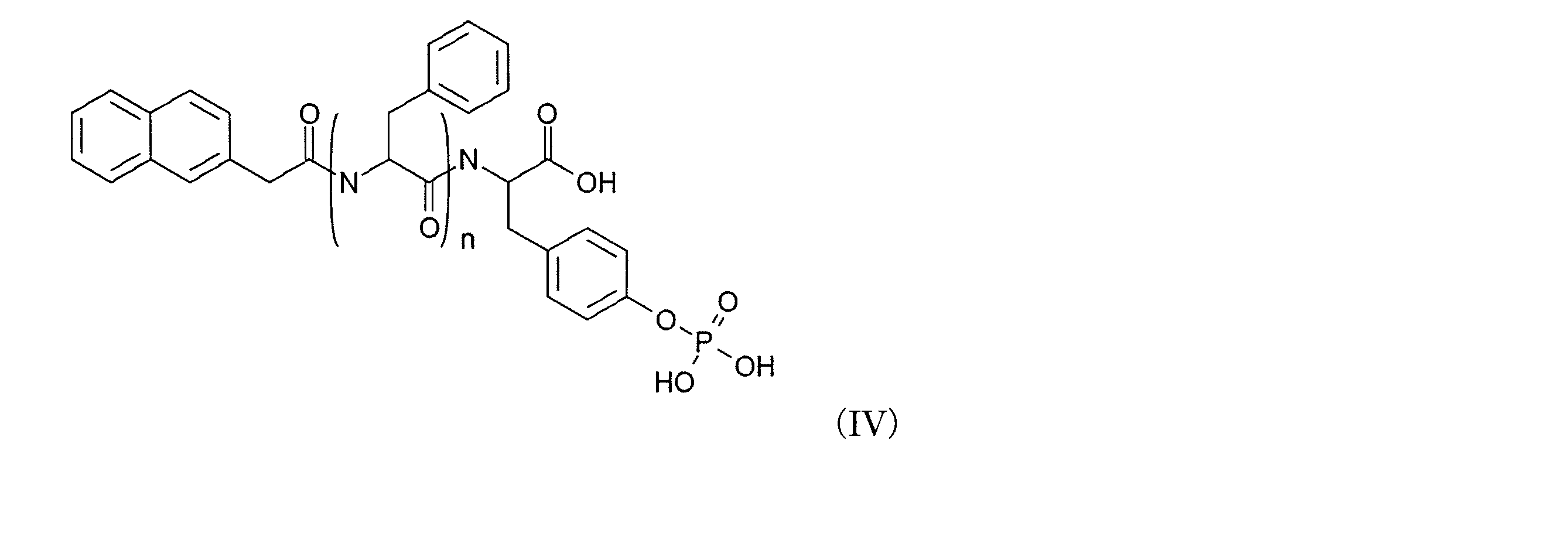
化合物D-1は下記の式(IV)(式中、n=2)の構造式、および化合物D-3は下記の式(IV)(式中、n=3)の構造式を有するものを使用した。
以上より、D-3は、多能性幹細胞に特異的に細胞死を引き起こすことが示唆された。同様に、NHDF (正常ヒト真皮線維芽細胞) またはHS5 (正常ヒト髄基質細胞)へ0μM、200μM、300μMまたは400μMのD-3を培養液に添加し、1時間または2時間培養した。D-3を含まない培地へ交換し、1日間培養後、WST-1アッセイにより細胞死を評価し、0μMのD-3を添加した場合と比較して細胞生存率を算出した。その結果、いずれの細胞に対してもD-3による細胞障害性は見いだせなかった。
201B7は、ラミニン被覆した24wellプレート上の、Rock inhibitor を含有するStemfitに、2.6 x 10⁴ cell/cm2となるように播種した。24時間後、培地を、Stemfitのみのものに取り換え、培養を続けた。さらに24時間後、培地を、D-1、D-3、D-7の各分子を異なる濃度で含むStemfit、あるいはコントロールとしてのStemfitに取り換えた。2時間の培養後に、培地をStemfitのみのものに取り換えた。翌日、細胞を100 μL のAccumaxを用いて採取した。次いで、300 μLの Stemfitを加え、Accumax反応を停止した。各サンプルの100 μL中の生細胞の数をフローサイトメトリーを用いて測定した。死細胞を判別するための分析の前に、各サンプルに7-AADを加えた。その結果、D-3のみならず、D-1及びD-7を用いた場合でも、201B7を細胞死させることができることが示された(図9)。
ラミニン被覆した24wellプレート上の、Rock inhibitor を含有するStemfitに、3種のiPS細胞株(201B7、1231A、1383D7)は2.6 x 10⁴ cell/cm2 となるように播種した。201B7については、他に5.3x10⁴/cm2となるように播種し、大きなコロニーを生成させたサンプルも作製した。なお、ヒトiPS細胞(1383D7)は、京都大学 森実 飛鳥博士より供与されたものを培養して用いた。24時間後、培地を、Stemfitのみのものに取り換え、培養を続けた。さらに24時間後、培地を、D-3を異なる濃度で含むStemfit、あるいはコントロールとしてのStemfitに取り換えた。1~4時間の培養後に、培地をStemfitのみのものに取り換えた。翌日、細胞を100 μL のAccumaxを用いて採取した。次いで、300 μLの Stemfitを加え、Accumax反応を停止した。各サンプルの100uL中の生細胞の数を、フローサイトメトリーを用いて測定した。死細胞を判別するための分析の前に、各サンプルに7-AADを加えた。その結果、D-3を加えることで、201B7、1231A、1383D7のいずれのiPS細胞株も、細胞死させることが可能であることが示された(図10)。また、同一の細胞株については、高濃度での播種により大きなコロニーを形成した201B7よりも、低濃度で播種した201B7に対し、D-3による細胞死の効率がより高いことがわかった。これは、高濃度播種の場合はD-3と細胞が接触しない領域が生じるため、D-3と細胞との接触面積による影響が強いと考えられる。
201B7細胞は、ラミニン被覆した24wellプレート上の、Rock inhibitor を含有するStemfitに、2.6 x 10⁴ cell/cm2、5.3 x 104 cell/cm2、10.5 x 104 cell/cm2となるように播種した(day 0) 。24時間後、培地を、Stemfitのみのものに取り換え、培養を続けた。さらに24時間後、培地を、D-3を400 μMの濃度で含むStemfitに取り換えた。2時間の培養後に、培地をStemfitのみのものに取り換えた。それぞれの細胞は、D3により一度(day 2のみ)、二度(day 2 及び day 3)、三度(day 2-4)にわたって処理した。次いで、day5に、細胞を100 μL のAccumaxを用いて採取し、300 μLの Stemfitを加え、Accumax反応を停止した。各サンプルの100μL中の生細胞の数を、フローサイトメトリーを用いて測定した。死細胞を判別するための分析の前に、各サンプルに7-AADを加えた。NHDFは、24wellプレート上の、FGM fibroblast培地に、5.3 x 10⁴cell/cm2となるように播種した。細胞は、 day 2-4に、FGM fibroblast培地中の400 μMの D-3で 2時間インキュベーションすることにより処理した。次いで、day5に、細胞を上記201B7細胞と同様の方法により採取し、Accumax反応を停止し、各サンプルの100μL中の生細胞の数を、フローサイトメトリーを用いて同様に測定した。死細胞を判別するための分析の前に、各サンプルに7-AADを加えた。その結果、D-3による繰り返し処理により、コンフルエントに近い段階における201B7であっても高精度に除去できることがわかった(図11)。
正常皮膚繊維芽細胞(NHDF)は、ATCC(American Type Culture Collection)から購入した。ヒト初代肝細胞及び肝細胞培地はコスモバイオから購入した。ヒト初代腎臓近位尿細管上皮細胞及びREGMTM BulletKit培地は、Lonzaから購入した。自然分化201B7は、201B7をbFGF を添加しないStemfitで2週間培養することにより調製した。201B7由来の神経細胞は、前出の論文Nakagawa, et al. Sci Rep. 4:3594, 2014に記載の方法に従って調製した。201B7由来の心筋細胞は、先に示した方法により調製した。すべての細胞は、37 oC, 5% CO2で維持した。各細胞は、24wellプレート上のそれぞれの培地に、5.3 x 10⁴ cell/cm2となるように播種した。24時間後、培地を、D-3分子を異なる濃度で含む培地、あるいはコントロールとしての培地のみに取り換えた。1~4時間の培養後に、培地のみのものに取り換えた。翌日、細胞を100μL のAccumaxを用いて採取した。次いで、300 μLの Stemfitを加え、Accumax反応を停止した。各サンプルの100 μL中の生細胞の数を、フローサイトメトリーを用いて測定した。死細胞を判別するための分析の前に、各サンプルに7-AADを加えた。その結果、D-3がiPS 細胞ではない各種の細胞に対しては、毒性がほとんどないことが示された(図12)。
khES1は、京都大学の吉田善紀博士より供与されたものを培養して用いた。khES1を、5.3 x 10⁴ cell/cm2となるように播種した以外は、実施例8に示す方法と同様にして、D-3のkhES1に対する毒性を調べた。その結果、D-3がヒトES細胞(khES1)に対しても毒性であることが示された(図13)。
ラミニン被覆した24wellプレート上の、Rock inhibitor を含有するStemfitに、iPS細胞株(201B7)は5.3 x 10⁴ cell/cm2 となるように播種した。24時間後、培地を、Stemfitのみのものに取り換え、培養を続けた。さらに24時間後、培地を、D-3を400 μMで含むStemfit、rBC2LCN-PE23 を 10μg/mLで含むStemfit、あるいはコントロールとしてのStemfitに取り換えた。D-3については1時間または2時間の培養後に、rBC2LCN-PE23については2時間または24時間の培養後に、培地をStemfitのみのものに取り換えた。翌日、細胞を100μL のAccumaxを用いて採取した。次いで、300 μLの Stemfitを加え、Accumax反応を停止した。各サンプルの100μL中の生細胞の数を、フローサイトメトリーを用いて測定した。死細胞を判別するための分析の前に、各サンプルに7-AADを加えた。その結果、D-3が、市販の抗iPS細胞物質である rBC2LCN-PE23よりも効能が高いことが示された(図14)。D-3は、iPS細胞の除去効果が高いだけでなく、除去のための処理時間の短縮も可能とする。短いペプチド分子であるD-3は、経済的に、大容量で容易に合成できる点においても優れている。
293FT は、invitrogenから購入した。24 wellプレート上のDMEM 培地 (10% FBS, 1% non essential amino acids)に、293FTを5.3 x 10⁴cell/cm2となるように播種した。24時間後、標準的なプロトコルに従って、Stemfect試薬を用い、異なる濃度の野生型胎盤型アルカリフォスファタ-ゼのmRNA (P-ALP mRNA)で293FT をトランスフェクトした。4時間後、培地を培養培地のみに交換した。コントロールとしては、293FT をD-3処理をすることなく、処理したものと同じタイミングで培地交換のみを行ったものを用いた。24時間後、培地を、D-3を異なる濃度で含むものに取り換え、コントロールについては培地だけのものに取り換えた。1時間の培養後に、培地をStemfitのみのものに取り換えた。 翌日、細胞を100μL のAccumaxを用いて採取した。次いで、300 μLの Stemfitを加え、Accumax反応を停止した。各サンプルの100μL中の生細胞の数をフローサイトメトリーを用いて測定した。死細胞を判別するための分析の前に、各サンプルに7-AADを加えた。その結果、D-3の毒性が、P-ALP mRNA のトランスフェクション量と関連することが示された(図15)。
201B7及びMHY6-EGFP を発現する201B7由来の心筋細胞は、細胞数比が1:9となり、合計で、5.3 x 10⁴cell/cm2となるように、ラミニン被覆した24wellプレート上の、Rock inhibitor を含有するStemfitに播種した。24時間後、培地を、MHY6-EGFP を発現する201B7由来の心筋細胞の懸濁液を含有するStemfitに交換した。さらに24時間後、培地を、D-3 を 400 μMの濃度で含むStemfitと交換し、コントロールについてはStemfitのみの培地と交換した。1時間の培養後に、培地をStemfitのみのものに取り換えた。 翌日、細胞を100μL のAccumaxを用いて採取した。次いで、400 μLの Stemfitを加え、Accumax反応を停止した。各サンプルの100μL中の生細胞の数を、フローサイトメトリーを用いて測定した。死細胞を判別するための分析の前に、各サンプルに7-AADを加えた。201B7を判別するために、Alexa 647 fluorophoreを備えるTra-1-60抗体を用いた。また、分化後20日もしくは21日の心筋細胞はEGFP陽性細胞を選択することにより選別した。その結果、D-3を用いることにより、分化細胞群からiPS細胞を除去することができることが示された(図16)。
201B7及びMHY6-EGFP を発現する201B7由来の心筋細胞は、細胞数比が7:3となり、合計で、5.3 x 10⁴ cell/cm2となるように、ラミニン被覆した24wellプレート上の、Rock inhibitor を含有するStemfitに播種した(day 0)。24時間後、培地を、MHY6-EGFP を発現する201B7由来の心筋細胞の懸濁液を含有するStemfitに交換した (day 1) 。 さらに24時間後、培地を、D-3 を 400 μMの濃度で含むStemfitと交換し、コントロールについてはStemfitのみの培地と交換した。2時間の培養後に、培地をStemfitのみのものに取り換えた。細胞は一度(day 2のみ)、二度(day 2 及び day 3)、または三度(day 2-4)にわたってD-3で処理した。翌日、細胞を100μL のAccumaxを用いて採取した。次いで、300 μLの Stemfitを加え、Accumax反応を停止した。各サンプルの100μL中の生細胞の数を、フローサイトメトリーを用いて測定した。死細胞を判別するための分析の前に、各サンプルに7-AADを加えた。201B7を判別するために、Alexa 647 fluorophoreを備えるTra-1-60抗体を用いた。また、分化後20日もしくは21日の心筋細胞はEGFP陽性細胞を選択することにより選別した。その結果、D-3により、多量のiPS細胞と少量の分化細胞を含む細胞群から、少量の分化細胞を精製することに成功したことが示された(図17)。
201B7の自然分化に対する抵抗性クローンを用いて、bFGFを含まないStemfitで培養して、自然分化を誘導した。day 15に、5.3 x 10⁴ cell/cm2となるように、ラミニン被覆した24wellプレート上の、Rock inhibitor を含有するStemfitに細胞を播種した(day 0)。24時間後、培地をStemfitに交換した。さらに24時間後、培地を、D-3を400 μMで含むStemfit、あるいはコントロールとしてのStemfitに取り換えた。2時間の培養後に、培地をStemfitのみのものに取り換えた。それぞれの細胞は、一度(day 2のみ)、二度(day 2 及び day 3)、三度(day 2-4)にわたって処理した。次いで、day5に、細胞を100μL のAccumaxを用いて採取し、300 μLの Stemfitを加え、Accumax反応を停止した。各サンプルの100μL中の生細胞の数をフローサイトメトリーを用いて測定した。死細胞を判別するための分析の前に、各サンプルに7-AADを加えた。201B7を判別するために、Alexa 647 fluorophoreを備えるTra-1-60抗体を用いた。その結果、D-3 により、15日間自然分化を誘導した場合の分化抵抗性(differentiation deficient)を示す201B7由来iPS細胞由来細胞集団から、残存するiPS細胞を除去することができたことが示された(図18)。
5 x 10⁴個の自然分化させた201B7と、50個、20個、0個の201B7を混合し、ラミニン被覆した24 wellプレート上の、Rock inhibitor を含有するStemfit(味の素)に播種した(day 0) 。24時間後、培地をStemfitに交換した (day 1)。さらに24時間後、培地を、D-3を400 μMで含むStemfit、あるいはコントロールとしてのStemfitに取り換えた (day 2)。2時間の培養後に、培地をStemfitのみのものに取り換えた。そして、細胞をStemfitでさらに7日間培養した。ALP染色により、 201B7 コロニーを赤紫色に染色した。 その結果、D-3を用いることで、iPS細胞と分化細胞との共培養から、残存したiPS細胞を除去することができたことが示された(図19)。
Claims (7)
- 式(I):

(式中、R1はアリール基またはヘテロアリール基であり、mは1から6の整数であり、nは2以上の整数である)、または、式(II):
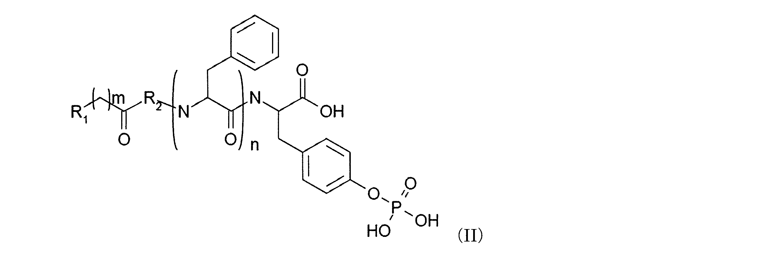
(式中、R1はアリール基またはヘテロアリール基であり、R2は、アラニン残基またはグリシン残基であり、mは1から6の整数であり、nは2以上の整数である)で表される化合物と対象細胞集団とを接触させる工程を含む、多能性細胞を除去する方法。 - 前記化合物が、式(I)で表される化合物であって、式中、R1がカルボニル基であり、R2が、ナフチル基であり、mは1である、請求項1に記載の方法。
- 前記化合物が、式(I)で表される化合物であって、式中、nは3である、請求項2に記載の方法。
- 前記対象細胞集団が、多能性幹細胞から分化誘導された細胞集団である請求項1から請求項3のいずれか1項に記載の方法。
- 式(I):
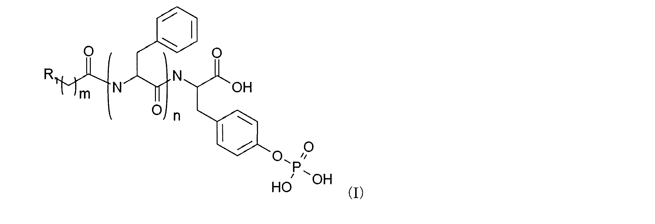
(式中、R1はアリール基またはヘテロアリール基であり、mは1から6の整数であり、nは2以上の整数である)、または、式(II):
- 前記化合物が、式(I)で表される化合物であって、式中、R1がナフチル基であり、mが1である、請求項5に記載のキット。
- 前記化合物が、式(I)で表される化合物であって、式中、nが3である、請求項6に記載のキット。
Priority Applications (3)
| Application Number | Priority Date | Filing Date | Title |
|---|---|---|---|
| EP16737412.3A EP3246395B1 (en) | 2015-01-14 | 2016-01-14 | Compound for identifying pluripotent cells |
| JP2016569500A JP6708559B2 (ja) | 2015-01-14 | 2016-01-14 | 多能性細胞識別化合物 |
| US15/543,488 US10689624B2 (en) | 2015-01-14 | 2016-01-14 | Compound for identifying pluripotent cells |
Applications Claiming Priority (2)
| Application Number | Priority Date | Filing Date | Title |
|---|---|---|---|
| JP2015005107 | 2015-01-14 | ||
| JP2015-005107 | 2015-01-14 |
Publications (1)
| Publication Number | Publication Date |
|---|---|
| WO2016114341A1 true WO2016114341A1 (ja) | 2016-07-21 |
Family
ID=56405882
Family Applications (1)
| Application Number | Title | Priority Date | Filing Date |
|---|---|---|---|
| PCT/JP2016/050946 Ceased WO2016114341A1 (ja) | 2015-01-14 | 2016-01-14 | 多能性細胞識別化合物 |
Country Status (4)
| Country | Link |
|---|---|
| US (1) | US10689624B2 (ja) |
| EP (1) | EP3246395B1 (ja) |
| JP (1) | JP6708559B2 (ja) |
| WO (1) | WO2016114341A1 (ja) |
Cited By (2)
| Publication number | Priority date | Publication date | Assignee | Title |
|---|---|---|---|---|
| WO2018122932A1 (ja) * | 2016-12-26 | 2018-07-05 | オリンパス株式会社 | 細胞の選別方法、純化された細胞集団の製造方法、及び発光イメージングシステム |
| WO2019013294A1 (ja) | 2017-07-12 | 2019-01-17 | 国立大学法人京都大学 | 高発現性mRNAスイッチ |
Families Citing this family (1)
| Publication number | Priority date | Publication date | Assignee | Title |
|---|---|---|---|---|
| CN106412680B (zh) * | 2015-07-31 | 2021-01-22 | 中兴通讯股份有限公司 | 多屏控制方法及装置 |
Citations (2)
| Publication number | Priority date | Publication date | Assignee | Title |
|---|---|---|---|---|
| JP2013009742A (ja) * | 2011-06-28 | 2013-01-17 | Nagoya City Univ | 多能性幹細胞の増殖を抑制するための組成物及び方法 |
| WO2013186946A1 (ja) * | 2012-06-11 | 2013-12-19 | 国立大学法人北海道大学 | 多能性幹細胞の選別方法 |
Family Cites Families (2)
| Publication number | Priority date | Publication date | Assignee | Title |
|---|---|---|---|---|
| JP5357928B2 (ja) | 2011-06-16 | 2013-12-04 | 東芝テック株式会社 | ラベルプリンタ |
| US11155576B2 (en) | 2014-04-09 | 2021-10-26 | Brandeis University | Synthetic peptides, enzymatic formation of pericellular hydrogels/nanofibrils, and methods of use |
-
2016
- 2016-01-14 US US15/543,488 patent/US10689624B2/en active Active
- 2016-01-14 WO PCT/JP2016/050946 patent/WO2016114341A1/ja not_active Ceased
- 2016-01-14 EP EP16737412.3A patent/EP3246395B1/en not_active Not-in-force
- 2016-01-14 JP JP2016569500A patent/JP6708559B2/ja active Active
Patent Citations (2)
| Publication number | Priority date | Publication date | Assignee | Title |
|---|---|---|---|---|
| JP2013009742A (ja) * | 2011-06-28 | 2013-01-17 | Nagoya City Univ | 多能性幹細胞の増殖を抑制するための組成物及び方法 |
| WO2013186946A1 (ja) * | 2012-06-11 | 2013-12-19 | 国立大学法人北海道大学 | 多能性幹細胞の選別方法 |
Non-Patent Citations (3)
| Title |
|---|
| LI, X. ET AL.: "Supramolecular Nanofibers and Hydrogels of Nucleopeptides", ANGEW. CHEM. INT. ED., vol. 50, 2011, pages 9365 - 9369, XP055466086 * |
| MURAMATSU, T. ET AL.: "Carbohydrate markers of ES cells", TRENDS IN GLYCOSCIENCE AND GLYCOTECHNOLOGY, vol. 21, no. 120, 2009, pages 197 - 206, XP055466524 * |
| See also references of EP3246395A4 * |
Cited By (2)
| Publication number | Priority date | Publication date | Assignee | Title |
|---|---|---|---|---|
| WO2018122932A1 (ja) * | 2016-12-26 | 2018-07-05 | オリンパス株式会社 | 細胞の選別方法、純化された細胞集団の製造方法、及び発光イメージングシステム |
| WO2019013294A1 (ja) | 2017-07-12 | 2019-01-17 | 国立大学法人京都大学 | 高発現性mRNAスイッチ |
Also Published As
| Publication number | Publication date |
|---|---|
| US10689624B2 (en) | 2020-06-23 |
| EP3246395B1 (en) | 2021-11-10 |
| EP3246395A1 (en) | 2017-11-22 |
| US20180002673A1 (en) | 2018-01-04 |
| JPWO2016114341A1 (ja) | 2017-11-30 |
| JP6708559B2 (ja) | 2020-06-10 |
| EP3246395A4 (en) | 2018-08-01 |
Similar Documents
| Publication | Publication Date | Title |
|---|---|---|
| JP5896360B2 (ja) | ヒト多能性幹細胞から中間中胚葉細胞への分化誘導方法 | |
| JP6143268B2 (ja) | ヒト多能性幹細胞から中間中胚葉細胞への分化誘導方法 | |
| JP6246125B2 (ja) | 多能性細胞を選別するための方法 | |
| KR102320537B1 (ko) | 신규 도파민 생산 신경 전구 세포의 유도 방법 | |
| JP2019058192A (ja) | 神経分化誘導用の多能性幹細胞 | |
| JP6612736B2 (ja) | 心筋細胞の選別方法 | |
| JP6090937B2 (ja) | 新規ドーパミン産生神経前駆細胞マーカー | |
| JP6373253B2 (ja) | 新規心筋細胞マーカー | |
| US10620109B2 (en) | Method for selecting skeletal muscle progenitor cell | |
| JPWO2016108288A1 (ja) | 骨格筋前駆細胞の製造方法 | |
| JP6646311B2 (ja) | 多能性幹細胞から中胚葉前駆細胞および血液血管前駆細胞への分化誘導法 | |
| JP6708559B2 (ja) | 多能性細胞識別化合物 | |
| JP6960159B6 (ja) | 組織細胞の選別方法 | |
| JP5842289B2 (ja) | 効率的な内皮細胞の誘導方法 | |
| JP5888769B2 (ja) | 相同組換えにより遺伝子改変された多能性幹細胞の簡便な検出法 | |
| WO2017094878A1 (ja) | 細胞内在性タンパク質の識別方法 | |
| JP7072756B2 (ja) | 多能性幹細胞から中胚葉前駆細胞および血液血管前駆細胞への分化誘導法 | |
| WO2022102742A1 (ja) | 骨格筋系譜細胞および骨格筋幹細胞の高効率純化用細胞表面マーカーおよびその利用 | |
| WO2022138593A1 (ja) | 骨格筋細胞の製造方法 |
Legal Events
| Date | Code | Title | Description |
|---|---|---|---|
| 121 | Ep: the epo has been informed by wipo that ep was designated in this application |
Ref document number: 16737412 Country of ref document: EP Kind code of ref document: A1 |
|
| ENP | Entry into the national phase |
Ref document number: 2016569500 Country of ref document: JP Kind code of ref document: A |
|
| WWE | Wipo information: entry into national phase |
Ref document number: 15543488 Country of ref document: US |
|
| NENP | Non-entry into the national phase |
Ref country code: DE |
|
| REEP | Request for entry into the european phase |
Ref document number: 2016737412 Country of ref document: EP |