WO2016114242A1 - 表面外胚葉系細胞から誘導された角膜上皮様細胞 - Google Patents
表面外胚葉系細胞から誘導された角膜上皮様細胞 Download PDFInfo
- Publication number
- WO2016114242A1 WO2016114242A1 PCT/JP2016/050619 JP2016050619W WO2016114242A1 WO 2016114242 A1 WO2016114242 A1 WO 2016114242A1 JP 2016050619 W JP2016050619 W JP 2016050619W WO 2016114242 A1 WO2016114242 A1 WO 2016114242A1
- Authority
- WO
- WIPO (PCT)
- Prior art keywords
- cells
- cell
- corneal
- corneal epithelial
- derived
- Prior art date
- Legal status (The legal status is an assumption and is not a legal conclusion. Google has not performed a legal analysis and makes no representation as to the accuracy of the status listed.)
- Ceased
Links
Images




Classifications
-
- C—CHEMISTRY; METALLURGY
- C12—BIOCHEMISTRY; BEER; SPIRITS; WINE; VINEGAR; MICROBIOLOGY; ENZYMOLOGY; MUTATION OR GENETIC ENGINEERING
- C12N—MICROORGANISMS OR ENZYMES; COMPOSITIONS THEREOF; PROPAGATING, PRESERVING, OR MAINTAINING MICROORGANISMS; MUTATION OR GENETIC ENGINEERING; CULTURE MEDIA
- C12N5/00—Undifferentiated human, animal or plant cells, e.g. cell lines; Tissues; Cultivation or maintenance thereof; Culture media therefor
- C12N5/06—Animal cells or tissues; Human cells or tissues
- C12N5/0602—Vertebrate cells
- C12N5/0618—Cells of the nervous system
- C12N5/0621—Eye cells, e.g. cornea, iris pigmented cells
-
- C—CHEMISTRY; METALLURGY
- C12—BIOCHEMISTRY; BEER; SPIRITS; WINE; VINEGAR; MICROBIOLOGY; ENZYMOLOGY; MUTATION OR GENETIC ENGINEERING
- C12N—MICROORGANISMS OR ENZYMES; COMPOSITIONS THEREOF; PROPAGATING, PRESERVING, OR MAINTAINING MICROORGANISMS; MUTATION OR GENETIC ENGINEERING; CULTURE MEDIA
- C12N5/00—Undifferentiated human, animal or plant cells, e.g. cell lines; Tissues; Cultivation or maintenance thereof; Culture media therefor
- C12N5/06—Animal cells or tissues; Human cells or tissues
- C12N5/0602—Vertebrate cells
-
- C—CHEMISTRY; METALLURGY
- C07—ORGANIC CHEMISTRY
- C07K—PEPTIDES
- C07K14/00—Peptides having more than 20 amino acids; Gastrins; Somatostatins; Melanotropins; Derivatives thereof
- C07K14/435—Peptides having more than 20 amino acids; Gastrins; Somatostatins; Melanotropins; Derivatives thereof from animals; from humans
- C07K14/46—Peptides having more than 20 amino acids; Gastrins; Somatostatins; Melanotropins; Derivatives thereof from animals; from humans from vertebrates
- C07K14/47—Peptides having more than 20 amino acids; Gastrins; Somatostatins; Melanotropins; Derivatives thereof from animals; from humans from vertebrates from mammals
- C07K14/4701—Peptides having more than 20 amino acids; Gastrins; Somatostatins; Melanotropins; Derivatives thereof from animals; from humans from vertebrates from mammals not used
- C07K14/4702—Regulators; Modulating activity
-
- C—CHEMISTRY; METALLURGY
- C12—BIOCHEMISTRY; BEER; SPIRITS; WINE; VINEGAR; MICROBIOLOGY; ENZYMOLOGY; MUTATION OR GENETIC ENGINEERING
- C12N—MICROORGANISMS OR ENZYMES; COMPOSITIONS THEREOF; PROPAGATING, PRESERVING, OR MAINTAINING MICROORGANISMS; MUTATION OR GENETIC ENGINEERING; CULTURE MEDIA
- C12N15/00—Mutation or genetic engineering; DNA or RNA concerning genetic engineering, vectors, e.g. plasmids, or their isolation, preparation or purification; Use of hosts therefor
- C12N15/09—Recombinant DNA-technology
- C12N15/63—Introduction of foreign genetic material using vectors; Vectors; Use of hosts therefor; Regulation of expression
- C12N15/79—Vectors or expression systems specially adapted for eukaryotic hosts
- C12N15/85—Vectors or expression systems specially adapted for eukaryotic hosts for animal cells
- C12N15/86—Viral vectors
-
- C—CHEMISTRY; METALLURGY
- C12—BIOCHEMISTRY; BEER; SPIRITS; WINE; VINEGAR; MICROBIOLOGY; ENZYMOLOGY; MUTATION OR GENETIC ENGINEERING
- C12N—MICROORGANISMS OR ENZYMES; COMPOSITIONS THEREOF; PROPAGATING, PRESERVING, OR MAINTAINING MICROORGANISMS; MUTATION OR GENETIC ENGINEERING; CULTURE MEDIA
- C12N5/00—Undifferentiated human, animal or plant cells, e.g. cell lines; Tissues; Cultivation or maintenance thereof; Culture media therefor
- C12N5/10—Cells modified by introduction of foreign genetic material
-
- C—CHEMISTRY; METALLURGY
- C12—BIOCHEMISTRY; BEER; SPIRITS; WINE; VINEGAR; MICROBIOLOGY; ENZYMOLOGY; MUTATION OR GENETIC ENGINEERING
- C12N—MICROORGANISMS OR ENZYMES; COMPOSITIONS THEREOF; PROPAGATING, PRESERVING, OR MAINTAINING MICROORGANISMS; MUTATION OR GENETIC ENGINEERING; CULTURE MEDIA
- C12N2501/00—Active agents used in cell culture processes, e.g. differentation
- C12N2501/10—Growth factors
- C12N2501/11—Epidermal growth factor [EGF]
-
- C—CHEMISTRY; METALLURGY
- C12—BIOCHEMISTRY; BEER; SPIRITS; WINE; VINEGAR; MICROBIOLOGY; ENZYMOLOGY; MUTATION OR GENETIC ENGINEERING
- C12N—MICROORGANISMS OR ENZYMES; COMPOSITIONS THEREOF; PROPAGATING, PRESERVING, OR MAINTAINING MICROORGANISMS; MUTATION OR GENETIC ENGINEERING; CULTURE MEDIA
- C12N2501/00—Active agents used in cell culture processes, e.g. differentation
- C12N2501/60—Transcription factors
-
- C—CHEMISTRY; METALLURGY
- C12—BIOCHEMISTRY; BEER; SPIRITS; WINE; VINEGAR; MICROBIOLOGY; ENZYMOLOGY; MUTATION OR GENETIC ENGINEERING
- C12N—MICROORGANISMS OR ENZYMES; COMPOSITIONS THEREOF; PROPAGATING, PRESERVING, OR MAINTAINING MICROORGANISMS; MUTATION OR GENETIC ENGINEERING; CULTURE MEDIA
- C12N2501/00—Active agents used in cell culture processes, e.g. differentation
- C12N2501/60—Transcription factors
- C12N2501/603—Oct-3/4
-
- C—CHEMISTRY; METALLURGY
- C12—BIOCHEMISTRY; BEER; SPIRITS; WINE; VINEGAR; MICROBIOLOGY; ENZYMOLOGY; MUTATION OR GENETIC ENGINEERING
- C12N—MICROORGANISMS OR ENZYMES; COMPOSITIONS THEREOF; PROPAGATING, PRESERVING, OR MAINTAINING MICROORGANISMS; MUTATION OR GENETIC ENGINEERING; CULTURE MEDIA
- C12N2501/00—Active agents used in cell culture processes, e.g. differentation
- C12N2501/60—Transcription factors
- C12N2501/604—Klf-4
-
- C—CHEMISTRY; METALLURGY
- C12—BIOCHEMISTRY; BEER; SPIRITS; WINE; VINEGAR; MICROBIOLOGY; ENZYMOLOGY; MUTATION OR GENETIC ENGINEERING
- C12N—MICROORGANISMS OR ENZYMES; COMPOSITIONS THEREOF; PROPAGATING, PRESERVING, OR MAINTAINING MICROORGANISMS; MUTATION OR GENETIC ENGINEERING; CULTURE MEDIA
- C12N2506/00—Differentiation of animal cells from one lineage to another; Differentiation of pluripotent cells
- C12N2506/09—Differentiation of animal cells from one lineage to another; Differentiation of pluripotent cells from epidermal cells, from skin cells, from oral mucosa cells
- C12N2506/097—Differentiation of animal cells from one lineage to another; Differentiation of pluripotent cells from epidermal cells, from skin cells, from oral mucosa cells from oral mucosa cells
-
- C—CHEMISTRY; METALLURGY
- C12—BIOCHEMISTRY; BEER; SPIRITS; WINE; VINEGAR; MICROBIOLOGY; ENZYMOLOGY; MUTATION OR GENETIC ENGINEERING
- C12N—MICROORGANISMS OR ENZYMES; COMPOSITIONS THEREOF; PROPAGATING, PRESERVING, OR MAINTAINING MICROORGANISMS; MUTATION OR GENETIC ENGINEERING; CULTURE MEDIA
- C12N2510/00—Genetically modified cells
-
- C—CHEMISTRY; METALLURGY
- C12—BIOCHEMISTRY; BEER; SPIRITS; WINE; VINEGAR; MICROBIOLOGY; ENZYMOLOGY; MUTATION OR GENETIC ENGINEERING
- C12N—MICROORGANISMS OR ENZYMES; COMPOSITIONS THEREOF; PROPAGATING, PRESERVING, OR MAINTAINING MICROORGANISMS; MUTATION OR GENETIC ENGINEERING; CULTURE MEDIA
- C12N2740/00—Reverse transcribing RNA viruses
- C12N2740/00011—Details
- C12N2740/10011—Retroviridae
- C12N2740/15011—Lentivirus, not HIV, e.g. FIV, SIV
- C12N2740/15041—Use of virus, viral particle or viral elements as a vector
- C12N2740/15043—Use of virus, viral particle or viral elements as a vector viral genome or elements thereof as genetic vector
Definitions
- the present invention relates to a method for inducing keratin 12 positive corneal epithelial cells from cells derived from surface ectoderm. More specifically, a method for inducing K12-positive corneal epithelium-like cells by introducing PAX6, KLF4, and OCT4 into cells derived from surface ectoderm such as oral mucosal epithelial cells, and corneal epithelium induced by the method Related to like cells.
- a treatment method using oral mucosal epithelial cells of the patient himself has been developed.
- a cultured corneal epithelial cell sheet is prepared from oral mucosal epithelial cells and transplanted to the affected eye (Patent Documents 1 to 3, Non-patent Document 1), but the oral mucosal epithelium does not differentiate into a complete corneal epithelium.
- the ability to cause angiogenesis is different, after transplantation of the oral mucosal epithelial sheet, in some cases, invasion of blood vessels may occur in the cornea, which is essentially avascular.
- stem cells undifferentiated cells
- ES cells embryonic stem cells
- iPS cells induced pluripotent stem cells
- iPS cells remain undifferentiated cells even after differentiation induction, and there is a risk of tumorigenesis after transplantation.
- Another problem is that it takes several months or more to establish iPS cells and induce differentiation.
- Non-patent Document 2 a method for inducing nerve cells (Non-patent Document 2) and cardiomyocytes (Non-patent Document 3) from dermal fibroblasts by direct reprogramming that directly induces somatic cells of interest without going through iPS cells. It has been known. According to this method, there is no risk of tumor formation due to residual undifferentiated cells, and target somatic cells can be obtained easily and in a short time.
- An object of the present invention is to induce cells having functions close to those of original corneal epithelial cells by direct reprogramming without going through the state of pluripotent cells.
- OCT4 is added to oral mucosal epithelial cells that are developmentally and functionally similar to corneal epithelial cells. It has been found that corneal epithelium-like cells expressing keratin epithelium-specific keratin 12 can be induced without introducing the pluripotent cell state.
- the present invention provides the following (1) to (12): (1) A method for producing corneal epithelial cells, characterized by inducing keratin 12-positive corneal epithelial cells by introducing PAX6, KLF4, and OCT4 into cells derived from surface ectoderm; (2) The method according to (1) above, wherein the corneal epithelial-like cell is induced without going through the state of pluripotent cells; (3) The method according to (1) or (2) above, wherein the cells derived from the surface ectoderm are oral mucosal epithelial cells; (4) The method according to any one of (1) to (3) above, wherein the corneal epithelial-like cell is further keratin 3 positive; (5) The method according to (4) above, wherein both two PAX6 isoforms Pa and Pb are introduced; (6) The method according to any one of (1) to (5) above, wherein PAX6, KLF4, and OCT4 are introduced with one viral vector; (7) A method for producing a corneal epithelial
- the corneal epithelial cell according to (8) which is further keratin 3 positive;
- a corneal epithelial-like cell sheet formed by overlaying the cells according to any one of (8) to (11) above.
- keratin 12-specific cells specific for corneal epithelium can be derived from ectoderm-derived cells such as oral mucosal epithelial cells. Furthermore, using this keratin 12-positive cultured oral mucosal epithelial cell as a cell source, it becomes possible to produce a corneal epithelial-like cell sheet having properties closer to those of corneal epithelial cells than conventional cultured oral mucosal epithelial sheets.
- corneal epithelial-like cells are directly induced from somatic cells without involving the state of pluripotent cells, so there is less risk of tumorigenesis compared to induction from pluripotent stem cells, and it is simple and short.
- the desired cells can be obtained with As a result, a better clinical outcome can be obtained, which also contributes to improving the patient's QOL.
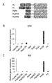
- FIG. 1 shows the results of confirming the expression of K12 (A-C) and K3 (D-F) mRNA by real-time PCR.
- K12 was a combination of PbOK
- K3 was a combination of PaOK or PaK, or Pa could induce significant expression of mRNA.
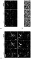
- FIG. 2 shows the results of confirming the expression of K12 and K3 at the protein level by immunostaining. Similar to the results of real-time PCR, K12 and K3 were a combination of PbOK, and K3 was a combination of PaOK or Pa.
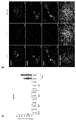
- FIG. 3 shows the results of confirming the expression of NANOG, which is a pluripotency marker, by real-time PCR and immunostaining.
- FIG. 4 shows the results of confirming the expression of pluripotency markers SSEA4 and TRA-1-60 by real-time PCR and immunostaining. In any cell, the expression of SSEA4 and TRA-1-60 was not observed.
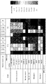
- FIG. 5 shows the results of confirming the expression of K12 and K3 by real-time PCR in various cells into which Pa, Pb, PaOK, and PbOK were introduced.
- Fig. 6 shows that all genes (PAX6, OCT4, KLF4) are loaded into one lentiviral vector using All-in-one vector and introduced into 293T cells, confirming that most of each protein is expressed independently.
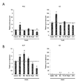
- FIG. 7 shows the results of luciferase reporter assay of 1K to 6K upstream of K12 (A) or K3 (B) (graphs are 1K, 2K, 3K, 4K, 5K, 6K from the left, respectively).
- FIG. 8 shows K12 (A) after adding various small molecules (Control, BIO, BIX, RG108, BayK, VPA) that are known to increase or maintain iPS cell establishment efficiency.
- FIG. 8 shows real-time PCR expression analysis of K12 (B) and K3 (C) in cells infected with OCT4 and KLF4 by deleting two PAX6 DNA binding domains (PAI and RED) in various combinations. Results are shown.
- ectoderm-derived cells Human embryos form three germ layers at the stage of development: endoderm, mesoderm, and ectoderm.
- the endoderm is the mucosal epithelium, liver and pancreas of the stomach and small intestine, the mesoderm is muscle, bone, blood vessels and blood, subcutaneous tissue, heart, kidney, etc.
- the ectoderm is the nerve, eyes (corneal epithelium), epidermis Etc.
- the ectoderm is further divided into nerve ectoderm that becomes brain and nerve tissue, and surface ectoderm that mainly becomes epithelial tissue on the surface of the skin, and the latter is referred to as “surface ectoderm-derived cells”.
- Examples of the “surface ectoderm-derived cell” include stratified epithelial cells covering the body surface such as epidermis, oral mucosa epithelium, corneal epithelium and the like.
- the oral mucosal epithelium is functionally close to the corneal epithelium and is easy to obtain, so it is preferable as a source of corneal epithelial-like cell induction.
- PAX6 “Paired box protein)” is a transcription factor expressed during embryogenesis and is also called aniridia type II protein (AN2) or oculorhombin. PAX6 is an important regulatory gene in eye and brain development, and the protein encoded by PAX6 has two different binding sites that bind to DNA and function as regulators of gene transcription. PAX6 mutations are known to cause various eye abnormalities. “PAX6” has two isoforms Pa and Pb. Among the DNA binding sites, those that do not contain 5a sequence in PAIRED domain are Pa, and those that are contained are Pb.
- KLF4 KLF4 (Kruppel-like factor 4) is a member of the KLF transcription factor family and regulates proliferation, differentiation, apoptosis, and somatic reprogramin.
- OCT4 octamer-binding transcription factor 4
- OCT4 is a transcription factor having a POU (Pit-Oct-Unc) domain and plays an important role in early embryogenesis and maintenance of pluripotency of ES cells.
- OCT4 is used as an undifferentiation marker (pluripotency marker) because it changes the epigenomic state and is closely involved in self-renewal of undifferentiated embryonic stem cells. It is also used as a marker for germ cell canceration.
- Corneal epithelial-like cells From the surface, the cornea has a three-layer structure of a corneal epithelial layer, a corneal stroma layer, and a corneal endothelium layer.
- Corneal epithelial cells are the cells constituting the outermost layer of the cornea and are composed of 4 to 5 corneal epithelial cell layers.
- Corneal epithelial cells are derived from the epidermal ectoderm, but the stroma and endothelium of the cornea are derived from neural crests, and it is thought that individual stem cells exist.
- the “corneal epithelial cell” according to the present invention is a cell having functions and characteristics similar to those of the corneal epithelial cell, and is characterized by the expression of keratin 12 which is a corneal epithelial differentiation marker.
- Corneal epithelial cell-specific markers Keratin 12 (Cytokeratin 12: K12) and keratin 3 (Cytokeratin 3: K3) are both corneal epithelial cell-specific markers.
- Pluripotency marker In the present invention, it is a gene / protein that serves as an index of pluripotency of human cells.
- pluripotent (stem cell) markers include OCT4 (supra), NANOG, TRA-1-60, and SSEA-4.
- NANOG is a transcriptional regulator involved in the proliferation and self-renewal of inner cell mass and ES cells. Suppresses differentiation of ES cells into extraembryonic endoderm and trophectoderm and maintains pluripotency. In addition, NANOG acts on SMAD1 and is thought to block the mesodermal differentiation of ES cells by BMP signals by inhibiting the binding of coactivators that activate transcription of SMAD1.
- the anti-TRA-1-60 antibody recognizes an antigen expressed on the surface of human EC cells, EG cells, ES cells, and iPS cells.
- SSEA-4 stage specific embryonic antigen 4
- SSEA-4 stage specific embryonic antigen 4
- SSEA-4 is a glycosphingolipid localized on the cell surface.
- expression of SSEA-4 is observed in EC cells, EG cells, and ES cells, but the expression decreases with differentiation, and SSEA-1 expression increases instead. For this reason, SSEA-4 is used as a human pluripotent (stem cell) marker.
- keratin 12 (K12) -positive corneal epithelium-like cells are induced by introducing PAX6, OCT4, and KLF4 into cells derived from surface ectoderm.
- the gene introduction method is not particularly limited, and can be performed using a known vector.
- the vector is not particularly limited as long as it can introduce the gene into mammalian cells.
- adenovirus vectors adeno-associated virus vectors, retrovirus vectors, lentivirus vectors, Sendai virus, and other virus vectors can be used.
- adenoviral vectors can be expressed in large quantities, but they are highly toxic.
- Retroviral vectors can be expected to be stable for a long time, but they are not suitable for non-dividing cells, and adeno-associated viral vectors cannot incorporate large genes. There is.
- lentiviral vectors have few problems described above, and stable expression can be expected for a long time.
- PAX6, OCT4, and KLF4 may be introduced as separate vectors, or may be introduced as one vector. As demonstrated in the examples described below, even when genes are placed in series in one vector, each protein is expressed independently, and as in the case of separate introduction, K12-positive corneal epithelium. Like cells can be obtained.
- the cells are directly induced from the cells derived from the surface ectoderm to corneal epithelial cells without going through the pluripotent state. Therefore, there is little risk of tumorigenesis due to the remaining undifferentiated cells, such as induction from pluripotent stem cells, and the desired corneal epithelial-like cells can be obtained easily and in a short time.
- Surface ectoderm-derived cells include epidermis, oral mucosal epithelium, corneal epithelial-like cells, etc., but oral mucosal epithelial cells that are similar in function are preferable, and patients who transplant the obtained corneal epithelial-like cells themselves Oral mucosal epithelial cells are more preferred.
- the cell culture is not particularly limited as long as it is a medium that can be used for the culture of epithelial cells.
- a commercially available medium for epithelial cells may be used, or a basic medium may be prepared by adding various nutrient sources necessary for the maintenance and growth of cells and components necessary for differentiation induction.
- Basic medium includes DMEM medium, Keratinocyte-SFM medium, BME medium, BGJb medium, CMRL 1066 medium, Glasgow MEM medium, Improved MEM Zinc Option medium, IMDM medium, Medium 199 medium, Eagle MEM medium, ⁇ MEM medium, Dulbecco MEM Any medium that can be used for culturing animal cells, such as a medium, ham medium, RPMI 1640 medium, Fischer's medium, McCoy's medium, Williams E medium, and mixed medium thereof, can be used.
- Nutrient sources include glycerol, glucose, fructose, sucrose, lactose, honey, starch, dextrin and other carbon sources, fatty acids, fats and oils, lecithin, alcohols and other hydrocarbons, ammonium sulfate, ammonium nitrate, ammonium chloride, urea Nitrogen sources such as sodium nitrate, salt, potassium salt, phosphate, magnesium salt, calcium salt, iron salt, manganese salt and other inorganic salts, monopotassium phosphate, dipotassium phosphate, magnesium sulfate, sodium chloride, sulfuric acid Ferrous iron, sodium molybdate, sodium tungstate and manganese sulfate, various vitamins, amino acids and the like can be included.
- Nitrogen sources such as sodium nitrate, salt, potassium salt, phosphate, magnesium salt, calcium salt, iron salt, manganese salt and other inorganic salts, monopotassium phosphate, dipotassium phosphate,
- serum substitutes include albumin (eg, lipid-rich albumin), transferrin, fatty acid, insulin, collagen precursor, trace element, ⁇ -mercaptoethanol or 3 ′ thiolglycerol, growth factor (epidermal growth factor; EGF, keratinocyte) growth factor; KGF, etc.), commercially available Knockout Serum Replacement (KSR), bovine pituitary extract (BPE), Chemically-defined Lipid concentrated (Gibco), Glutamax (Gibco), B27 supplement (Gibco), Y -27632 (Wako Pure Chemical Industries) is listed.
- the pH of the medium obtained by blending these components is in the range of 5.5 to 9.0, preferably 6.0 to 8.0, more preferably 6.5 to 7.5.
- Culturing is carried out at 36 ° C. to 38 ° C., preferably 36.5 ° C. to 37.5 ° C. under conditions of 1% to 25% O 2 and 1% to 15% CO 2 .
- the corneal epithelial-like cell preferably expresses keratin 3 (K3) in addition to K12.
- K3 keratin 3
- the expression of K12 and K3 at the gene level can be easily confirmed by real-time PCR, and the expression at the protein level can be easily confirmed by immunostaining using antibodies specific for K12 and K3.
- Corneal epithelial-like cells derived from surface ectoderm cells The present invention also provides K12 positive corneal epithelial-like cells derived from cells derived from surface ectoderm. It is preferred that the cell is further K3 positive.
- K12 and K3 are both corneal epithelial cell-specific markers, and cells expressing K12 and K3 have the same functions and properties as the original corneal epithelial cells. Therefore, the corneal epithelial-like cell of the present invention can be used as a cell preparation in research and regenerative medicine.
- the cell preparation may contain scaffold materials and components that assist in maintenance / proliferation of cells and administration to the affected area, and other pharmaceutically acceptable carriers.
- Components necessary for cell maintenance / proliferation include media components such as carbon sources, nitrogen sources, vitamins, minerals, salts, various cytokines, and extracellular matrix preparations such as Matrigel TM .
- scaffold materials and components that assist administration to the affected area include biodegradable polymers; for example, collagen, polylactic acid, hyaluronic acid, cellulose, and derivatives thereof, and a complex composed of two or more thereof, an aqueous solution for injection;
- biodegradable polymers for example, collagen, polylactic acid, hyaluronic acid, cellulose, and derivatives thereof, and a complex composed of two or more thereof, an aqueous solution for injection
- examples include physiological saline, medium, physiological buffer such as PBS, and isotonic solutions containing glucose and other adjuvants (eg D-sorbitol, D-mannose, D-mannitol, sodium chloride), etc.
- Adjuvants such as alcohol, specifically ethanol, polyalcohol such as propylene glycol, polyethylene glycol, nonionic surfactants such as polysorbate 80, HCO-50, etc. may be used in combination.
- organic solvents polyvinyl alcohol, polyvinyl pyrrolidone, carboxyvinyl polymer, sodium carboxymethylcellulose, sodium polyacrylate, sodium alginate, water-soluble dextran, sodium carboxymethyl starch, pectin, methylcellulose as necessary , Ethyl cellulose, xanthan gum, gum arabic, casein, agar, polyethylene glycol, diglycerin, glycerin, propylene glycol, petrolatum, paraffin, stearyl alcohol, stearic acid, mannitol, sorbitol, lactose, surfactants acceptable as pharmaceutical additives, It may contain a buffer, an emulsifier, a suspension, a soothing agent, a stabilizer and the like.
- a purified antibody is dissolved in a solvent such as physiological saline, buffer solution, glucose solution, etc., and an adsorption inhibitor such as Tween 80, Tween 20, gelatin or the like is added thereto.
- a solvent such as physiological saline, buffer solution, glucose solution, etc.
- an adsorption inhibitor such as Tween 80, Tween 20, gelatin or the like is added thereto.
- Tween 80, Tween 20, gelatin or the like is added thereto.
- Examples of the diseases that can be the subject of the cell preparation of the present invention include Stevens-Johnson syndrome, pemphigoid, heat / chemical trauma, aniridia, peripheral corneal ulcer, limbal tumor, and colloidal corneal dystrophy. Is mentioned.
- Corneal epithelium-like cell sheet A corneal epithelium-like cell sheet can be prepared by stratifying K12-positive corneal epithelial-like cells obtained by the method of the present invention.
- the present invention also provides such a corneal epithelial-like cell sheet and a method for producing the same.
- corneal epithelial-like cells induced by the method of the present invention are cultured on a porous membrane, and the medium is always supplied from the lower layer through the porous membrane, so that epithelial cells are layered and cultured.
- An epithelial cell sheet can be prepared (Japanese Patent Laid-Open No. 2005-130838).
- the obtained cell sheet is excellent in transparency and mechanical strength, and has the same characteristics and functions as the original corneal epithelial cells. Therefore, it is more suitable for clinical application.
- Example 1 Induction of corneal epithelial-like cells from oral mucosal epithelial cells
- Materials and methods (cell) Immortalized human oral epithelial cells (OKF6 / TERT-1 cells) were used.
- the cells were cultured in Keratinocyte-SFM supplemented with 25 ⁇ g / mL bovine pituitary extract (BPE), 0.2 ng / ml epidermal growth factor (EGF), and 0.3 mM CaCl 2 at 37 ° C. with 5% CO 2 .
- BPE bovine pituitary extract
- EGF epidermal growth factor
- the pLenti7.3 related vector was introduced into 293FT cells using the ViraPower Lentiviral Expression System (Life technologies) to produce lentivirus.
- CSV-CMV-MCS-IRES2-Venus plasmid related vector was introduced into 293T cells with pCMV-VZV-G-RSV-Rev, pCAG-HIV-gp (RIKEN Bio Resource Center) using FuGene HD (Roche), lentivirus Produced.
- pCMV-VZV-G-RSV-Rev pCAG-HIV-gp
- FuGene HD FuGene HD
- Were infected with these lentiviruses to cells seeded the day before at 1.875x10 4 cell / 48well plates (polybrene also added), the following day the medium replacement, the cells were further cultured for 2 days.
- Example 2 Gene transfer into various cells Materials and methods (cell lines) Surface ectoderm cells: OKF6 / TERT-1, OKF6 / TERT-2, N / TERT-1, N / TERT-2, HOK Neuroectodermal: ARPE-19, SH-SY5Y Neural crest: HCEC Mesodermal: HUVEC, NHDF-Ad, 293T Endoderm: MKN1, HepG2 iPS cells: 201B7
- PAX6 (Pa, Pb), KLF4 and OCT4 were introduced into each of the above cell lines, and the expression of K12 and K3 in the obtained cells was confirmed by real-time PCR.
- Example 3 Gene transfer by All-in-one vector Method Three genes (PAX6, OCT4, KLF4) were linked to one lentiviral vector by the All-in-one vector, and a combination of PaOK or PbOK was connected by 2A sequences to prepare two types of vectors. This vector was transduced into oral mucosal epithelial cells in the same manner as in Example 1, and the expression of mRNA and protein was confirmed by electrophoresis, real-time PCR and immunostaining.
- Example 4 Regulation of expression of K12 and K3
- Six types of reporters were produced by binding secreted luciferase to the sequence of 1K-6K upstream of K12 or K3, and together with reporter, PaPbOK was transferred to oral mucosal epithelial cells in various combinations. It was a reduction. As a result, it was confirmed that Pa directly controls the expression upstream of K3 (FIG. 7).
- small molecules related to iPS cell establishment efficiency and maintenance were added, and expression of K12 and K3 was confirmed by real-time PCR.
- the small molecules added were BIO related to Wnt signaling, BIX related to epigenomic modification, DNA methyltransferase inhibitor RG108, calcium channel agonist BayK, and valproic acid (VPA), a histone deacetylase (HDAC) inhibitor.
- BIO basic structural protein
- BIX histone deacetylase
- PAI and RED PAX6 DNA binding domains
- keratin 12 positive cells close to the original corneal epithelial cells can be induced from cells derived from surface ectoderm such as oral mucosal epithelial cells.
- the method of the present invention has a lower risk of tumorigenesis than the induction from pluripotent stem cells, and the target cells can be obtained easily and in a short time.
- the cell sheet in which the cells are layered is excellent in transparency and mechanical strength, and is therefore useful for the treatment of corneal epithelial diseases such as corneal transplantation.
Landscapes
- Health & Medical Sciences (AREA)
- Life Sciences & Earth Sciences (AREA)
- Engineering & Computer Science (AREA)
- Genetics & Genomics (AREA)
- Chemical & Material Sciences (AREA)
- Biomedical Technology (AREA)
- Organic Chemistry (AREA)
- Zoology (AREA)
- Biotechnology (AREA)
- Bioinformatics & Cheminformatics (AREA)
- Wood Science & Technology (AREA)
- General Health & Medical Sciences (AREA)
- General Engineering & Computer Science (AREA)
- Biochemistry (AREA)
- Microbiology (AREA)
- Cell Biology (AREA)
- Biophysics (AREA)
- Molecular Biology (AREA)
- Plant Pathology (AREA)
- Physics & Mathematics (AREA)
- Virology (AREA)
- Toxicology (AREA)
- Gastroenterology & Hepatology (AREA)
- Medicinal Chemistry (AREA)
- Proteomics, Peptides & Aminoacids (AREA)
- Ophthalmology & Optometry (AREA)
- Neurology (AREA)
- Neurosurgery (AREA)
- Micro-Organisms Or Cultivation Processes Thereof (AREA)
- Medicines Containing Material From Animals Or Micro-Organisms (AREA)
- Materials For Medical Uses (AREA)
Abstract
Description
本出願は,日本特許出願2015-004389(2015年1月13日出願)に基づく優先権を主張しており、この内容は本明細書に参照として取り込まれる。
本発明は表面外胚葉由来の細胞からケラチン12陽性の角膜上皮様細胞を誘導する方法に関する。より詳細には、口腔粘膜上皮細胞等の表面外胚葉由来の細胞にPAX6、KLF4、及びOCT4を導入することにより、K12陽性の角膜上皮様細胞を誘導する方法及び前記方法で誘導された角膜上皮様細胞に関する。
(1)表面外胚葉由来の細胞にPAX6、KLF4、及びOCT4を導入することにより、ケラチン12陽性の角膜上皮様細胞を誘導することを特徴とする、角膜上皮細胞の製造方法;
(2)多能性細胞の状態を介することなく角膜上皮様細胞が誘導される、上記(1)に記載の方法;
(3)表面外胚葉由来の細胞が口腔粘膜上皮細胞である、上記(1)又は(2)に記載の方法;
(4)角膜上皮様細胞がさらにケラチン3陽性である、上記(1)~(3)のいずれかに記載の方法;
(5)2つのPAX6アイソフォームPa及びPbの両方を導入する、上記(4)に記載の方法;
(6)PAX6、KLF4、及びOCT4を1つのウイルスベクターで導入する、上記(1)~(5)のいずれかに記載の方法;
(7)上記(1)~(6)のいずれかに記載の方法で角膜上皮様細胞を製造し、前記細胞を重層化することを特徴とする、角膜上皮様細胞シートの製造方法;
(8)表面外胚葉由来の細胞から直接誘導されたケラチン12陽性の角膜上皮様細胞。
(9)さらにケラチン3陽性である、上記(8)に記載の角膜上皮様細胞;
(10)表面外胚葉由来の細胞が口腔粘膜上皮細胞である、上記(8)又は(9)に記載の角膜上皮様細胞;
(11)表面外胚葉由来の細胞にPAX6、KLF4、及びOCT4を導入して得られる、上記(10)に記載の角膜上皮様細胞;
(12)上記(8)~(11)のいずれかに記載の細胞を重層してなる角膜上皮様細胞シート。
表面外胚葉由来の細胞:
ヒトの胚は、発生の段階で3つの胚葉、すなわち内胚葉、中胚葉、外胚葉を形成する。内胚葉は、胃や小腸の粘膜上皮、肝臓、膵臓等に、中胚葉は筋肉、骨、血管や血液、皮下組織、心臓、腎臓等になる、外胚葉は神経、目(角膜上皮)、表皮等を形成する。外胚葉はさらに脳や神経組織となる神経外胚葉、主に皮膚の表面の上皮組織となる表面外胚葉に分かれ、後者を「表面外胚葉由来の細胞」と称する。
「表面外胚葉由来の細胞」としては、表皮、口腔粘膜上皮、角膜上皮などの体表を覆っている重層上皮細胞が挙げられる。特に、口腔粘膜上皮は機能的にも角膜上皮に近く、取得も容易であることから、角膜上皮様細胞誘導のソースとして好ましい。
「PAX6(Paired box protein)」 は、胚発生期に発現する転写因子で、aniridia type II protein (AN2) あるいは oculorhombin とも称される。PAX6は眼と脳の発生における重要な制御遺伝子であり、PAX6にコードされるタンパクはDNAに結合し遺伝子転写のレギュレータとして機能する2つの異なる結合部位を有する。PAX6の変異は眼の様々な異常を引き起こすことが知られている。「PAX6」には、2つのアイソフォームPaとPbが存在する。DNA結合部位のうちPAIRED domainの中に5a sequenceが含まれないものがPaで、含まれるものがPbである。
「KLF4(Kruppel-like factor 4)」はKLF転写因子ファミリーのメンバーであり、増殖、分化、アポトーシス、体細胞リプログラミンを制御する。
「OCT4 (octamer-binding transcription factor 4)」は、POU(Pit-Oct-Unc)ドメインを有する転写因子で、初期胚発生やES細胞の多能性維持に重要な役割を果たす。OCT4は、エピゲノム状態を変化させ、未分化胚性幹細胞の自己複製に密接に関与するため、未分化マーカー(多能性マーカー)として利用されている。また、生殖細胞の癌化に対するマーカーとしても用いられている。
角膜は、表面から、角膜上皮層、角膜実質層、角膜内皮層の3層構造をしている。角膜上皮細胞は、この角膜の一番外側の層を構成する細胞で、4~5層の角膜上皮細胞層から構成されている。角膜上皮細胞は表皮外胚葉に由来するが、角膜の実質と内皮は神経堤由来であり、それぞれ個別の幹細胞が存在すると考えられている。本発明にかかる「角膜上皮様細胞」は、この角膜上皮細胞に似た機能と特性を有する細胞で、角膜上皮分化マーカーであるケラチン12の発現によって特徴づけられる。
ケラチン12(Cytokeratin 12:K12)及びケラチン3(Cytokeratin 3:K3)は、いずれも角膜上皮細胞特異的マーカーである。
本発明では細胞のヒト細胞の多能性の指標となる遺伝子・タンパクである。多能性(幹細胞)マーカーとしては、OCT4(前掲)、NANOG、TRA-1-60、SSEA-4を挙げることができる。
NANOGは内部細胞塊やES細胞の増殖、自己複製に関与する転写調節因子である。ES細胞の胚体外内胚葉や栄養外胚葉への分化を抑制し、多能性を維持する。また、NANOGはSMAD1に作用し、SMAD1の転写を活性化するコアクチベーターの結合を阻害することにより、BMPシグナルによるES細胞の中胚葉分化を阻止すると考えられている。
抗TRA-1-60抗体は、ヒトEC細胞、EG細胞、ES細胞、iPS細胞の表面に発現する抗原を認識する。
SSEA-4(stage specific embryonic antigen 4)は細胞表面に局在するスフィンゴ糖脂質の一種である。ヒトではEC細胞、EG細胞及びES細胞でSSEA-4の発現が認められるが、分化とともにその発現は低下し、代わりにSSEA-1の発現が上昇する。このため、SSEA-4はヒトの多能性(幹細胞)マーカーとして用いられる。
本発明では、表面外胚葉由来の細胞にPAX6、OCT4、及びKLF4を導入することで、ケラチン12(K12)陽性の角膜上皮様細胞を誘導する。
本発明は、表面外胚葉由来の細胞から誘導されたK12陽性の角膜上皮様細胞も提供する。前記細胞はさらにK3陽性であることが好ましい。
本発明の方法で得られたK12陽性の角膜上皮様細胞を重層化することにより角膜上皮様細胞シートを作製することができる。本発明はそのような角膜上皮様細胞シートとその製造方法も提供する。
1.材料・方法:
(細胞)
不死化ヒト口腔上皮細胞(OKF6/TERT-1細胞)を用いた。
25μg/mL bovine pituitary extract (BPE), 0.2ng/ml epidermal growth factor (EGF), 0.3mM CaCl2を加えたKeratinocyte-SFM中で5% CO2 37℃で培養した。
PAX6の各アイソフォームPaおよびPb、その変異体、PAX6-OCT4-KLF4を2Aで連結したall-in-one vectorはpLenti7.3/V5-DEST Vector (Life technologies)に挿入した。OCT4およびKLF4はCSV-CMV-MCS-IRES2-Venus plasmid (RIKEN Bio Resource Center)に挿入した。
以下、PAX6の各アイソフォームをPa及びPb、KLF4をK、及びOCT4をOと記載することもある。
pLenti7.3関連ベクターは293FT細胞にViraPower Lentiviral Expression System (Life technologies)を用いて導入、レンチウイルスを産生した。CSV-CMV-MCS-IRES2-Venus plasmid関連ベクターは293T細胞にpCMV-VZV-G-RSV-Rev, pCAG-HIV-gp (RIKEN Bio Resource Center)と共にFuGene HD(Roche)を用いて導入、レンチウイルスを産生した。
これらのレンチウイルスを1.875x10⁴cell/48wellプレートで前日に播種した細胞に感染させ(polybreneも添加)、翌日培地交換、さらに2日間培養した。
RNAをRNeasyPlus Micro Kitで抽出し、SuperScript III First-Strand Synthesis Systemを用いてcDNA化した。cDNAはTaqman Gene Expression Assayにて定量した。
100%冷メタノールもしくは4% paraformaldehydeで固定した培養細胞を5% normal donkey serumで、1時間常温でblockおよびpermeabilizeした。そのうえでヒツジ抗K12抗体、マウス抗K3/76抗体、ウサギ抗NANOG抗体、マウス抗SSEA4抗体、マウス抗TRA-1-60抗体を加え4℃でオーバーナイトでインキュベートした。さらにマウス、ウサギ、ヒツジの2次抗体と共にインキュベートした。最後にHoechst 33342を加えて画像を取得した。
(1)角膜特異的マーカーの発現
リアルタイムPCRの結果、K12はPbOKの組み合わせで、K3はPaOK又はPaKの組み合わせ、若しくはPaで効率よくmRNAの発現を誘導できた(図1)。
免疫染色の結果、mRNAの発現と同様に、K12はPbOKの組み合わせで、K3はPaOKの組み合わせ、若しくはPaにより、蛋白質レベルでもK12及びK3の発現が確認された(図2)。
多能性マーカーであるNANOGの発現をリアルタイムPCR及び免疫染色で確認したところ、single cell levelではわずかにmRNAの上昇が認められ、部分的なreprogrammingの可能性はあるものの、蛋白質レベルではNANOGの発現は見られなかった(図3)。
多能性マーカーであるSSEA4、TRA-1-60の発現を免疫染色で確認したところ、いずれも発現は見られなかった(図4)。
以上より、多能性幹細胞の状態を経由して、K3、12発現が誘導されたわけではないことが確認された。
1.材料・方法
(細胞株)
表面外胚葉系細胞:OKF6/TERT-1, OKF6/TERT-2, N/TERT-1, N/TERT-2, HOK
神経外胚葉:ARPE-19, SH-SY5Y
神経堤:HCEC
中胚葉:HUVEC, NHDF-Ad, 293T
内胚葉:MKN1, HepG2
iPS細胞:201B7
上記した各細胞株に実施例1と同様にして様々な組み合わせでPAX6(Pa,Pb)、KLF4及びOCT4を導入し、得られた細胞におけるK12及びK3の発現をリアルタイムPCRで確認した。
結果を図5にまとめて示す。K12/K3は主に表面外胚葉由来の細胞において効率的に誘導することが可能であったが、内胚葉、中胚葉由来細胞やiPS細胞からはほとんど誘導されなかった。角膜上皮と発生起源が同じ表面外胚葉由来細胞では誘導効率が高かった。
1.方法
All-in-one vectorにより3つの遺伝子(PAX6、OCT4、KLF4)を一つのレンチウイルスベクターにPaOKあるいはPbOKの組み合わせを2A配列で連結し、2種類のベクターを作製した。このベクターを実施例1と同様の方法で口腔粘膜上皮細胞にトランスダクションを行い、電気泳動、リアルタイムPCRと免疫染色でmRNA及び蛋白質の発現を確認した。
電気泳動の結果、各蛋白質が分離して発現していることが確認された。また、リアルタイムPCRと免疫染色の結果から、All-in-one vectorを用いて十分なPAX6、OCT4、KLF4の発現が得られ、また、それによりK12とK3の誘導が可能なことが確認された(図6)。
K12もしくはK3の上流1K~6Kの配列に分泌型ルシフェラーゼを結合したreporterを各6種作製し、reporterと共に、PaPbOKを様々な組み合わせで口腔粘膜上皮細胞にトランスダクションした。その結果、PaはK3の上流で直接発現を制御することが確認された(図7)。
Claims (12)
- 表面外胚葉由来の細胞にPAX6、KLF4、及びOCT4を導入することにより、ケラチン12陽性の角膜上皮様細胞を誘導することを特徴とする、角膜上皮細胞の製造方法。
- 多能性細胞の状態を介することなく角膜上皮様細胞が誘導される、請求項1に記載の方法。
- 表面外胚葉由来の細胞が口腔粘膜上皮細胞である、請求項1又は2に記載の方法。
- 角膜上皮様細胞がさらにケラチン3陽性である、請求項1~3のいずれか1項に記載の方法。
- 2つのPAX6アイソフォームPa及びPbの両方を導入する、請求項4に記載の方法。
- PAX6、KLF4、及びOCT4を1つのウイルスベクターで導入する、請求項1~5のいずれか1項に記載の方法。
- 請求項1~6のいずれか1項に記載の方法で角膜上皮様細胞を製造し、前記細胞を重層化することを特徴とする、角膜上皮様細胞シートの製造方法。
- 表面外胚葉由来の細胞から直接誘導されたケラチン12陽性の角膜上皮様細胞。
- さらにケラチン3陽性である、請求項8に記載の角膜上皮様細胞。
- 表面外胚葉由来の細胞が口腔粘膜上皮細胞である、請求項8又は9に記載の角膜上皮様細胞。
- 表面外胚葉由来の細胞にPAX6、KLF4、及びOCT4を導入して得られる、請求項10に記載の角膜上皮様細胞。
- 請求項8~11のいずれか1項に記載の細胞を重層してなる角膜上皮様細胞シート。
Priority Applications (4)
| Application Number | Priority Date | Filing Date | Title |
|---|---|---|---|
| ES16737313T ES2751641T3 (es) | 2015-01-13 | 2016-01-12 | Células epitelioides corneales derivadas de células del ectodermo superficial |
| US15/543,021 US10457913B2 (en) | 2015-01-13 | 2016-01-12 | Corneal epithelioid cells derived from surface ectodermal cells |
| EP16737313.3A EP3246397B1 (en) | 2015-01-13 | 2016-01-12 | Corneal epithelioid cells derived from surface ectodermal cells |
| JP2016569347A JP6446067B2 (ja) | 2015-01-13 | 2016-01-12 | 表面外胚葉系細胞から誘導された角膜上皮様細胞 |
Applications Claiming Priority (2)
| Application Number | Priority Date | Filing Date | Title |
|---|---|---|---|
| JP2015004389 | 2015-01-13 | ||
| JP2015-004389 | 2015-01-13 |
Publications (1)
| Publication Number | Publication Date |
|---|---|
| WO2016114242A1 true WO2016114242A1 (ja) | 2016-07-21 |
Family
ID=56405788
Family Applications (1)
| Application Number | Title | Priority Date | Filing Date |
|---|---|---|---|
| PCT/JP2016/050619 Ceased WO2016114242A1 (ja) | 2015-01-13 | 2016-01-12 | 表面外胚葉系細胞から誘導された角膜上皮様細胞 |
Country Status (5)
| Country | Link |
|---|---|
| US (1) | US10457913B2 (ja) |
| EP (1) | EP3246397B1 (ja) |
| JP (1) | JP6446067B2 (ja) |
| ES (1) | ES2751641T3 (ja) |
| WO (1) | WO2016114242A1 (ja) |
Cited By (1)
| Publication number | Priority date | Publication date | Assignee | Title |
|---|---|---|---|---|
| WO2018235786A1 (ja) | 2017-06-19 | 2018-12-27 | 国立大学法人大阪大学 | 角膜内皮細胞マーカー及びその利用 |
Families Citing this family (1)
| Publication number | Priority date | Publication date | Assignee | Title |
|---|---|---|---|---|
| CN109694855B (zh) * | 2018-12-03 | 2020-12-29 | 北京仁源欣生生物科技有限公司 | 利用转基因技术提高人表皮干细胞诱导为角膜上皮细胞效率的方法 |
Family Cites Families (5)
| Publication number | Priority date | Publication date | Assignee | Title |
|---|---|---|---|---|
| WO2003009783A1 (en) | 2001-07-25 | 2003-02-06 | Japan Tissue Engineering Co., Ltd. | Laminates for treating cornea and process for producing the same |
| ES2372814T3 (es) | 2001-11-19 | 2012-01-26 | Arblast Co., Ltd. | Lámina semejante a la ectocórnea y método para construir la misma. |
| WO2006003818A1 (ja) | 2004-06-30 | 2006-01-12 | Alblast Co., Ltd. | 角膜上皮シート及びその作製方法 |
| JPWO2010134619A1 (ja) * | 2009-05-18 | 2012-11-12 | 国立大学法人東北大学 | 人工多能性幹細胞からの上皮系前駆細胞・幹細胞群及び角膜上皮細胞群の分化誘導方法 |
| US9574171B2 (en) * | 2010-12-02 | 2017-02-21 | Technion Research & Development Foundation Ltd. | Methods of generating corneal cells and cell populations comprising same |
-
2016
- 2016-01-12 WO PCT/JP2016/050619 patent/WO2016114242A1/ja not_active Ceased
- 2016-01-12 JP JP2016569347A patent/JP6446067B2/ja active Active
- 2016-01-12 US US15/543,021 patent/US10457913B2/en active Active
- 2016-01-12 ES ES16737313T patent/ES2751641T3/es active Active
- 2016-01-12 EP EP16737313.3A patent/EP3246397B1/en active Active
Non-Patent Citations (6)
| Title |
|---|
| NORIYUKI TSUMAKI: "Direct induction of chondrogenic cells from mouse dermal fibroblast culture", JOURNAL OF CLINICAL AND EXPERIMENTAL MEDICINE, vol. 239, no. 14, 2011, pages 1332 - 1337, XP008182186, ISSN: 0039-2359 * |
| See also references of EP3246397A4 * |
| SHALOM-FEUERSTEIN, R. ET AL.: "Skin and corneal cellular therapy using embryonic stem cells: how far are we?", EXPERT REVIEW OF DERMATOLOGY, vol. 3, no. 3, 2008, pages 357 - 366, XP001539385, ISSN: 1746-9872, DOI: doi:10.1586/17469872.3.3.357 * |
| SHIRAISHI, A. ET AL.: "Identification of the Cornea-Specific Keratin 12 Promoter by In Vivo Particle-Mediated Gene Transfer", IOVS, vol. 39, no. 13, 1998, pages 2554 - 2561, XP001021154, ISSN: 1552-5783 * |
| YANG, K. ET AL.: "Corneal epithelial-like transdifferentiation of hair follicle stem cells is mediated by pax6 and beta-catenin/Lef-1", CELL BIOLOGY INTERNATIONAL, vol. 33, 2009, pages 861 - 866, XP026437905, ISSN: 1065-6995 * |
| YU , D. ET AL.: "Differentiation of mouse induced pluripotent stem cells into corneal epithelial- like cells", CELL BIOLOGY INTERNATIONAL, vol. 37, 2013, pages 87 - 94, XP002730463, ISSN: 1065-6995, DOI: doi:10.1002/cbin.10007 * |
Cited By (1)
| Publication number | Priority date | Publication date | Assignee | Title |
|---|---|---|---|---|
| WO2018235786A1 (ja) | 2017-06-19 | 2018-12-27 | 国立大学法人大阪大学 | 角膜内皮細胞マーカー及びその利用 |
Also Published As
| Publication number | Publication date |
|---|---|
| JPWO2016114242A1 (ja) | 2017-09-28 |
| US10457913B2 (en) | 2019-10-29 |
| EP3246397A1 (en) | 2017-11-22 |
| ES2751641T3 (es) | 2020-04-01 |
| JP6446067B2 (ja) | 2018-12-26 |
| US20180148685A1 (en) | 2018-05-31 |
| EP3246397B1 (en) | 2019-09-18 |
| EP3246397A4 (en) | 2018-06-06 |
Similar Documents
| Publication | Publication Date | Title |
|---|---|---|
| US12247219B2 (en) | Method for inducing differentiation of corneal epithelial cells from pluripotent stem cells | |
| Maruotti et al. | A simple and scalable process for the differentiation of retinal pigment epithelium from human pluripotent stem cells | |
| ES2688019T3 (es) | Métodos y composiciones para la producción acelerada de células epiteliales pigmentadas retinianas a partir de células pluripotentes | |
| JPWO2010134619A1 (ja) | 人工多能性幹細胞からの上皮系前駆細胞・幹細胞群及び角膜上皮細胞群の分化誘導方法 | |
| JP2009526517A (ja) | インビトロ及びインビボにおいて細胞を細胞新生させる方法 | |
| CN107636149A (zh) | 上皮细胞的离体增殖 | |
| Gu et al. | Optimizing the method for generation of integration-free induced pluripotent stem cells from human peripheral blood | |
| WO2011019957A1 (en) | Method for formation of induced pluripotent stem cells | |
| WO2010140698A1 (ja) | 多能性幹細胞からの神経堤細胞群の分化誘導方法 | |
| JP7700673B2 (ja) | 神経堤細胞または角膜上皮細胞の純化方法 | |
| WO2018143312A1 (ja) | 多能性幹細胞の分化制御方法 | |
| US20190264171A1 (en) | Differentiation of pluripotent stem cells into corneal cells | |
| JP2025156357A (ja) | 細胞をリプログラミングするための方法 | |
| WO2017097007A1 (zh) | 分化培养基及其在制备神经干细胞中的用途 | |
| Guo et al. | bFGF and Activin A function to promote survival and proliferation of single iPS cells in conditioned half-exchange mTeSR1 medium | |
| JP6446067B2 (ja) | 表面外胚葉系細胞から誘導された角膜上皮様細胞 | |
| Zhang et al. | Human bone marrow mesenchymal stem cells support the derivation and propagation of human induced pluripotent stem cells in culture | |
| CN111836884A (zh) | 源自干细胞的泪腺组织的制作方法 | |
| KR20240144127A (ko) | 다능성 줄기세포로부터 표피각질형성세포로의 분화 유도 방법 | |
| CN114127264A (zh) | 包含环植物鞘氨醇-1-磷酸酯或其药学上可接受的盐作为有效成分的干细胞增殖促进用组合物 | |
| Hariharan | In vitro nephrogenesis from human pluripotent stem cells | |
| Asal | Generation of Lacrimal Gland from Induced Pluripotent Stem Cells | |
| Montserrat Pulido et al. | Generation of feeder-free pig induced pluripotent stem cells without Pou5f1 | |
| HK1240614A1 (en) | Method for inducing differentiation of corneal epithelial cells from pluripotent stem cells |
Legal Events
| Date | Code | Title | Description |
|---|---|---|---|
| 121 | Ep: the epo has been informed by wipo that ep was designated in this application |
Ref document number: 16737313 Country of ref document: EP Kind code of ref document: A1 |
|
| ENP | Entry into the national phase |
Ref document number: 2016569347 Country of ref document: JP Kind code of ref document: A |
|
| WWE | Wipo information: entry into national phase |
Ref document number: 15543021 Country of ref document: US |
|
| NENP | Non-entry into the national phase |
Ref country code: DE |
|
| REEP | Request for entry into the european phase |
Ref document number: 2016737313 Country of ref document: EP |