WO2015147291A1 - 免疫クロマト分析装置及び免疫クロマト分析方法 - Google Patents
免疫クロマト分析装置及び免疫クロマト分析方法 Download PDFInfo
- Publication number
- WO2015147291A1 WO2015147291A1 PCT/JP2015/059739 JP2015059739W WO2015147291A1 WO 2015147291 A1 WO2015147291 A1 WO 2015147291A1 JP 2015059739 W JP2015059739 W JP 2015059739W WO 2015147291 A1 WO2015147291 A1 WO 2015147291A1
- Authority
- WO
- WIPO (PCT)
- Prior art keywords
- substance
- detection
- incubation
- detected
- labeling substance
- Prior art date
- Legal status (The legal status is an assumption and is not a legal conclusion. Google has not performed a legal analysis and makes no representation as to the accuracy of the status listed.)
- Ceased
Links
Images
Classifications
-
- G—PHYSICS
- G01—MEASURING; TESTING
- G01N—INVESTIGATING OR ANALYSING MATERIALS BY DETERMINING THEIR CHEMICAL OR PHYSICAL PROPERTIES
- G01N33/00—Investigating or analysing materials by specific methods not covered by groups G01N1/00 - G01N31/00
- G01N33/48—Biological material, e.g. blood, urine; Haemocytometers
- G01N33/50—Chemical analysis of biological material, e.g. blood, urine; Testing involving biospecific ligand binding methods; Immunological testing
- G01N33/53—Immunoassay; Biospecific binding assay; Materials therefor
- G01N33/543—Immunoassay; Biospecific binding assay; Materials therefor with an insoluble carrier for immobilising immunochemicals
- G01N33/54366—Apparatus specially adapted for solid-phase testing
Definitions
- the present invention relates to an immunochromatography analyzer and an immunochromatography analysis method.
- immunochromatographic immunoassays that do not require pretreatment of specimens have been used as simple in-vitro diagnostic kits or portable diagnostic devices that detect antigens in sample solutions using the specific reactivity of antibodies.
- pathogen testing kits such as viruses and bacteria are familiar immunochromatographic analyzers widely used in general hospitals and clinics.
- the simplest structure of a conventional immunochromatography analyzer is a structure in which a sample addition unit, a labeling substance holding unit, a chromatographic medium carrying a detection unit, and an absorption unit are connected to each other.
- Patent Document 1 a method (Patent Document 1) is proposed in which a specimen is developed with a surfactant solution having a steroid skeleton, and then the labeling substance is amplified.
- Patent Document 2 a method (Patent Document 2) has been proposed in which a solution containing a test substance and a labeling substance modified with a first antibody against the test substance is developed at a specific rate on a porous carrier.
- the conventional immunochromatography analyzer cannot increase the detection sensitivity of the detected substance to the required level.
- an object of the present invention is to provide an immunochromatography analyzer and an immunochromatography analysis method capable of significantly increasing the detection sensitivity of a substance to be detected.
- the present invention is as follows. 1. A sample addition unit for adding a specimen, a labeling substance holding part for holding a labeling substance for recognizing the substance to be detected contained in the specimen, an incubation part for delaying elution of the labeling substance, and detecting the substance to be detected An immunochromatographic analyzer including a chromatographic medium portion on which a detection unit is supported, and an absorption unit that absorbs liquid that has passed through the detection unit. 2. 2. The immunochromatographic analyzer according to item 1, wherein the incubation unit contains a surfactant. 3. 2. The immunochromatographic analyzer according to item 1 above, wherein the incubation part contains a molecular chaperone. 4). 3.
- An immunochromatographic analysis method comprising sequentially performing the following steps (1) to (5) using the immunochromatographic analyzer according to any one of items 1 to 5.
- the step of adding the sample to the sample addition unit (2) The step of recognizing the substance to be detected contained in the sample by the labeling substance held in the labeling substance holding unit (3) The labeling substance in the incubation unit (4) A step of developing the analyte and the labeled substance as a mobile phase in a chromatographic medium part (5) A step of detecting a substance to be detected in the developed mobile phase by a detection part.
- the immunochromatography analyzer of the present invention includes a sample addition unit for adding a sample, a labeling substance holding unit for holding a labeling substance that recognizes a target substance contained in the sample, and an incubation unit for delaying elution of the labeling substance And a chromatographic medium part carrying a detection part for detecting the substance to be detected, and an absorption part for absorbing the liquid that has passed through the detection part.
- the incubation part contains a surfactant
- the reaction between the detected substance and the detected substance and the solubilization (denaturation) of the detected substance are promoted, and adverse effects caused by substances other than the detected substance are prevented. It is possible to further increase the detection sensitivity at the detection unit.
- the detection substance can be refolded into a natural structure to increase the reactivity of the detection substance in the detection part, thereby further increasing the detection sensitivity. it can.
- the incubation part contains a cyclic oligosaccharide together with a surfactant
- coloring due to the aggregation of the surfactant does not occur, there is an effect that the solubilization of the substance to be detected is further increased and the detection sensitivity is increased.
- the average particle diameter of the labeling substance is 20 to 200 nm, a higher sensitivity increasing effect due to the delay of elution can be obtained.
- the elution of the labeling substance is delayed by the incubation part, so that it is included in the specimen in the incubation part.
- a time for the labeling substance to wait for the arrival of the detected substance is generated, and this greatly increases the opportunity for the binding between the labeled substance and the detected substance, thereby significantly increasing the detection sensitivity of the detected substance.
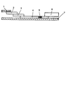
- FIG. 1 is a diagram for explaining the structure of an immunochromatographic analyzer.
- the immunochromatography analyzer of the present invention includes a sample addition unit for adding a sample, a labeling substance holding unit for holding a labeling substance that recognizes a target substance contained in the sample, and an incubation unit for delaying elution of the labeling substance And a chromatographic medium part carrying a detection part for detecting the substance to be detected, and an absorption part for absorbing the liquid that has passed through the detection part.
- the immunochromatographic analyzer of the present invention comprises a sample addition part (also called a sample pad) (1), a labeling substance holding part (also called a conjugate pad) (2), and an incubation part (also called an incubation pad). ) (3), a chromatographic medium part (4), a detection part (5), an absorption part (6) and a backing sheet (7).
- sample addition part also called a sample pad
- labeling substance holding part also called a conjugate pad
- an incubation part also called an incubation pad
- the sample addition part (1) is a part where the specimen (sample) is dropped in the immunochromatography analyzer.
- the sample may be any material as long as it is a material used in a normal immunochromatography analyzer.
- As the sample adding portion glass fiber or cellulose fiber for absorbing and holding the sample is usually used.
- the labeling substance holding part (2) contains a labeling substance (marker substance) to be described later, which is bound in advance to an antibody that binds to the substance to be detected. When the substance to be detected moves inside the labeling substance holding part, it binds to an antibody or the like and is labeled.
- the labeling substance holding part is made of, for example, a glass fiber nonwoven fabric or cellulose fiber.
- the labeling substance labels substances that specifically bind to the substance to be detected, such as antibodies, antigens, aptamers, peptides, and lectins.
- an enzyme or the like is used for labeling the detection reagent in the immunochromatography method.
- an insoluble carrier since it is suitable for visually determining the presence of the substance to be detected, it is preferable to use an insoluble carrier as the labeling substance.
- a labeled detection reagent can be prepared by sensitizing the detection reagent to an insoluble carrier.
- Insoluble carriers as labeling substances include colloidal metal particles such as gold, silver or platinum, colloidal metal oxide particles such as iron oxide, colloidal nonmetal particles such as sulfur, and latex particles composed of synthetic polymers. Or others can be used. In particular, colloidal gold is preferable because it is easy to detect.
- the average particle diameter of these particles is preferably 20 to 200 nm, and more preferably 30 to 150 nm. If the average particle size is within this range, the reaction between the detection target substance and the labeled detection substance due to the delay in elution, compared to the case where latex particles having an average particle size larger than the above range are used. The sensitivity is significantly improved, and highly sensitive detection is possible.
- the average particle diameter was measured with a transmission electron microscope (TEM: manufactured by JEOL Ltd., JEM-2010), and the projected area circle equivalent diameter of 100 particles was measured randomly using a photograph taken. It can be calculated from the average value.
- the incubation part (3) has a function of delaying elution of the labeling substance.
- Incubation part (3) consists of glass fiber nonwoven fabric, rayon nonwoven fabric, cellulose fiber, etc., for example.
- the incubation part (3) preferably contains a surfactant.
- the presence of the surfactant promotes the reaction between the detected substance and the detected substance and solubilization of the detected substance, can prevent adverse effects caused by substances other than the detected substance, and can further increase the detection sensitivity at the detection unit. .
- the surfactant may be appropriately selected in consideration of the type of specimen, labeling substance, detection method, and the like.
- cationic surfactants such as alkyltrimethylammonium salt, dialkyldimethylammonium salt, alkyldimethylbenzylammonium salt, amine salt system; rosinate such as potassium rosinate and sodium rosinate, potassium oleate, potassium laurate, Anionic interfaces such as sodium or potassium salts of fatty acids such as sodium laurate, sodium stearate and potassium stearate, sulfates of fatty alcohols such as sodium lauryl sulfate, and alkylallyl sulfonic acids such as sodium dodecylbenzene sulfonate Activating agents; Nonionic surfactants such as alkyl esters of polyethylene glycol, alkyl ethers, alkylphenyl ethers, octyl glucosides, decanoyl-N-methylglucamide
- preferred surfactants are nonionic surfactants, and polyethylene glycol alkyl esters, alkyl ethers, alkylphenyl ethers, octyl glucoside, decanoyl-N-methylglucamide are optimally used.
- the addition amount of the surfactant is, for example, preferably 0.05 to 2 mg / cm 2 , more preferably 0.1 to 2 mg / cm 2 with respect to the incubation part (3). It is because solubilization is further accelerated and detection sensitivity is further enhanced by being in the above range.
- the incubation part (3) preferably contains a molecular chaperone. Due to the presence of the molecular chaperone, the reactivity of the detection substance in the detection part can be increased, and the detection sensitivity can be further increased.
- a molecular chaperone has an activity of restoring (refolding) a three-dimensional structure of an antigen such as a denatured protein to a normal form, or an activity of suppressing irreversible aggregation of the denatured protein. Therefore, in detecting a sample containing a denatured antigen, when a detection substance that more strongly recognizes a normal type protein is used in the detection unit, detection sensitivity can be improved by refolding while suppressing protein aggregation.
- the molecular chaperone may be a natural molecule or an artificial molecule, and examples thereof include saccharides having molecular chaperone activity, nonionic surfactants and heat shock proteins (heat shock proteins). Of these, cyclic saccharides and nonionic surfactants are preferred.
- the addition amount of the molecular chaperone is, for example, 5 to 20% by mass, preferably 7 to 15% by mass, and more preferably 8 to 12% by mass with respect to the incubation part (3).
- the incubation part (3) for example, 0.05 to 5 mg / cm 2 is preferable, and more preferably 0.1 to 2 mg / cm 2 . This is because the detection sensitivity can be further increased by being in the above range.
- the incubation part (3) preferably contains a cyclic oligosaccharide together with a surfactant. Due to the presence of the cyclic oligosaccharide, errors in sensitivity (color intensity) between immunochromatographic devices can be reduced, the incubation part (3) can be dried well during production, and coloring due to aggregation of the surfactant can also occur. Therefore, the effect of the surfactant can be increased and the detection sensitivity of the substance to be detected can be increased.
- cyclic oligosaccharide examples include oligosaccharides having a structure in which about 5 to 10 monosaccharides are connected in a ring. Specifically, ⁇ -cyclodextrin, ⁇ -cyclodextrin, ⁇ -cyclodextrin, ⁇ -cycloawaodoline, ⁇ -cycloawaodoline and their hydroxy groups (OH groups) are methylated and monochlorotriazinylated. Derivatives modified by hydroxypropylation, acetylation, triacetylation, sulfobutylation, glycosylation or maltose addition, and the like. A cyclic oligosaccharide may be used alone or in combination of two or more.
- the amount of the cyclic oligosaccharide added is, for example, 0.1 to 4 mg / cm 2 , preferably 0.1 to 3 mg / cm 2 , more preferably 0.2 to 2 mg / cm 2 with respect to the incubation part (3). is there. This is because the effect of the surfactant can be further increased and the detection sensitivity of the substance to be detected can be further increased by being in the above range.
- the cyclic oligosaccharide is converted into a micelle form of the surfactant by the action of the hydrophilic group present on the surface of the cyclic oligosaccharide and the hydrophobic group present inside. It is presumed that aggregation is suppressed and the effects as described above are produced.
- the chromatographic medium part (4) is a development part of the chromatograph.
- the chromatographic medium portion is an inactive film made of a microporous material that exhibits capillary action. Examples of those that are not reactive with a detection reagent, an immobilization reagent, or a substance to be detected used in a chromatograph include a membrane made of nitrocellulose (hereinafter sometimes referred to as “nitrocellulose membrane”).
- Nitrocellulose-based membranes only need to contain nitrocellulose as a main component, and membranes based on nitrocellulose, such as pure products or nitrocellulose-mixed products, should be used. However, there are no problems with other materials. .
- Examples of other materials include cellulose membranes, nylon membranes, and porous plastic cloth (polyethylene, polypropylene).
- the nitrocellulose membrane may further contain a substance that promotes capillary action.
- a substance that lowers the surface tension of the film surface and brings about hydrophilicity is preferable.
- a substance having an amphipathic action such as saccharides, amino acid derivatives, fatty acid esters, various synthetic surfactants or alcohols, which has no effect on the movement of a substance to be detected on an immunochromatograph and is a marker substance
- substances that do not affect the color development are preferred.
- Nitrocellulose membrane is porous and exhibits capillary action.
- the index of the capillary phenomenon can be confirmed by measuring the water absorption rate (water absorption time: capillary flow time).
- the water absorption rate affects detection sensitivity and inspection time.
- the form and size of the nitrocellulose membrane as a chromatographic medium used in the immunochromatographic analysis method of the present invention are not particularly limited, and may be appropriate in terms of actual operation and observation of reaction results.
- the support made of plastic or the like on the back surface of the chromatographic medium on which the reaction sites are formed on the surface.
- the properties of the support are not particularly limited, but when the measurement result is observed by visual judgment, the support preferably has a color that is not similar to the color caused by the labeling substance. Usually, it is preferably colorless or white.
- the detection part (5) is formed on the chromatographic medium part (4), that is, a reaction site in which a substance that specifically binds to the substance to be detected, for example, an antibody is immobilized as an immobilization reagent at an arbitrary position. Is formed.
- the immobilization reagent is immobilized on the chromatographic medium by directly immobilizing the immobilization reagent on the chromatographic medium by physical or chemical means, and by physically or chemically binding the immobilization reagent to the fine particles.
- a direct immobilization method physical adsorption may be used, or covalent bonding may be used. In the case of a nitrocellulose membrane, physical adsorption can be performed. In the covalent bond, cyanogen bromide, glutaraldehyde, carbodiimide or the like is generally used to activate the chromatographic medium, but any method can be used.
- an indirect immobilization method there is a method in which an immobilization reagent is bound to insoluble fine particles and then immobilization to a chromatographic medium.
- the particle size of the insoluble fine particles can be selected such that it is captured by the chromatographic medium but cannot move, and is preferably fine particles having an average particle size of about 5 ⁇ m or less.
- organic polymers such as organic polymer latex particles obtained by emulsion polymerization methods such as polystyrene, styrene-butadiene copolymer, styrene-methacrylic acid copolymer, polyglycidyl methacrylate or acrolein-ethylene glycol dimethacrylate copolymer.
- Examples include fine particles of substances, fine particles such as gelatin, bentonite, agarose or cross-linked dextran, inorganic oxides such as silica, silica-alumina or alumina, or inorganic particles obtained by introducing a functional group to an inorganic oxide by silane coupling treatment, etc. .
- direct immobilization is preferable from the viewpoint of ease of sensitivity adjustment.
- Various methods can be used to immobilize the immobilization reagent on the chromatographic medium.
- reaction site is not particularly limited, but it may be fixed as a circular spot, a line extending perpendicularly to the development direction of the chromatographic medium, a numeral, a character, or a symbol such as +,-.
- the chromatographic medium can be subjected to a blocking treatment by a known method as necessary in order to prevent the accuracy of the analysis from being reduced due to nonspecific adsorption.
- a protein such as bovine serum albumin, skim milk, casein or gelatin is preferably used for the blocking treatment.
- a protein such as bovine serum albumin, skim milk, casein or gelatin is preferably used for the blocking treatment.
- one or a combination of two or more surfactants such as Tween 20, Triton X-100, or SDS may be washed.
- the absorption part (6) is installed at the end of the chromatographic medium in order to absorb a liquid such as a specimen or a developing solution that has passed through the detection part (5).
- the absorption part can be made of glass fiber, for example. When the absorption part is made of glass fiber, liquid return of the sample liquid can be greatly reduced.
- the backing sheet (7) is a base material. By applying an adhesive on one side or sticking an adhesive tape, one side has adhesiveness, on which the sample addition part (1), labeling substance holding part (2), incubation part (3) Part or all of the chromatographic medium (4), the detection unit (5), and the absorption unit (6) are provided in close contact with each other.
- the backing sheet (7) is not particularly limited as long as the backing sheet (7) becomes impermeable and impermeable to the sample solution by the adhesive.
- the immunochromatographic analysis method of the present invention includes the following steps (1) to (5), and a substance to be detected contained in a specimen is detected using the above immunochromatographic analyzer.
- (1) The step of adding the sample to the sample addition unit (2)
- the labeling substance in the incubation unit (4) A step of developing the analyte and the labeled substance as a mobile phase in a chromatographic medium part (5)
- a step of detecting a substance to be detected in the developed mobile phase by a detection part Each step will be described below.
- Step of adding sample to sample adding portion first, the sample diluent is adjusted to a concentration that allows the sample to move smoothly through the immunochromatographic medium without reducing measurement accuracy. It is preferable to prepare or dilute the sample-containing solution by adjusting or diluting. Secondly, a predetermined amount (usually 0.05 to 2 ml) of the specimen-containing liquid is dropped onto the sample addition part (1). When the specimen-containing liquid is dropped, the specimen-containing liquid starts to move in the sample addition part (1).
- the sample diluent can also be used as a developing solution.
- water is used as a solvent, and a buffer solution, a salt, and a nonionic surfactant, and further, for example, an antigen-antibody reaction is promoted.
- PVP high molecular compounds
- a sample and developing solution mixed in advance can be supplied and dropped onto the sample addition unit, or developed, or after the sample is first supplied and dropped onto the sample addition unit.
- the developing solution may be supplied and dropped on the sample addition unit to be developed.
- Samples containing the substance to be detected include, for example, biological samples, that is, whole blood, serum, plasma, urine, saliva, sputum, nasal cavity or throat wipe, cerebrospinal fluid, amniotic fluid, nipple discharge, tears, sweat, skin As well as extracts from milk, eggs, wheat, beans, beef, pork, chicken, and foods containing them, and the like.
- the labeling substance labels substances that specifically bind to the substance to be detected, such as antibodies, antigens, aptamers, peptides, and lectins.
- an enzyme or the like is used for labeling the detection reagent in the immunochromatography method.
- an insoluble carrier since it is suitable for visually determining the presence of the substance to be detected, it is preferable to use an insoluble carrier as the labeling substance.
- a labeled detection reagent can be prepared by sensitizing the detection reagent to an insoluble carrier.
- Insoluble carriers as labeling substances include colloidal metal particles such as gold, silver or platinum, colloidal metal oxide particles such as iron oxide, colloidal nonmetal particles such as sulfur, and latex particles composed of synthetic polymers. Or others can be used. In particular, colloidal gold is preferable because it is easy to detect.
- the average particle diameter of these particles is preferably 20 to 200 nm, and more preferably 30 to 150 nm. If the average particle size is within this range, the reaction between the detection target substance and the labeled detection substance due to the delay in elution, compared to the case where latex particles having an average particle size larger than the above range are used. The sensitivity is significantly improved, and highly sensitive detection is possible.
- the average particle diameter was measured with a transmission electron microscope (TEM: manufactured by JEOL Ltd., JEM-2010), and the projected area circle equivalent diameter of 100 particles was measured randomly using a photograph taken. It can be calculated from the average value.
- TEM transmission electron microscope
- the insoluble carrier is a labeling substance suitable for visually determining the presence of the substance to be detected, and is preferably colored to facilitate visual determination.
- the colloidal metal particles and the colloidal metal oxide particles themselves exhibit a specific natural color corresponding to the particle diameter, and the color can be used as a label.
- colloidal metal particles and colloidal metal oxide particles that can be used as labeling substances include colloidal gold particles, colloidal silver particles, colloidal platinum particles, colloidal iron oxide particles, and colloidal aluminum hydroxide particles. Etc.
- the colloidal gold particles and the colloidal silver particles are preferable in that the colloidal gold particles are red and the colloidal silver particles are yellow in an appropriate particle size.
- colloidal metal particles for example, when colloidal gold particles are used, commercially available ones may be used.
- colloidal gold particles can be prepared by a conventional method, for example, a method of reducing chloroauric acid with sodium citrate.
- the detection reagent As a method for sensitizing the detection reagent to the colloidal metal particles, known methods such as physical adsorption and chemical bonding can be used.
- the antibody in the detection reagent in which antibody is sensitized to colloidal gold particles, the antibody is unbound by adding bovine serum albumin solution after adding the antibody to a colloidally dispersed solution and physically adsorbing the antibody. Prepare by blocking the particle surface.
- the substance to be detected to be detected by the method of the present invention is not particularly limited as long as a substance that specifically binds to the substance is present, and examples thereof include proteins, peptides, nucleic acids, sugars (especially sugar moieties of glycoproteins, Glycosides of glycolipids) and complex carbohydrates.
- “specifically binds” means to bind based on the affinity of a biomolecule.
- binding based on affinity include binding between an antigen and an antibody, binding between a sugar and a lectin, binding between a hormone and a receptor, binding between an enzyme and an inhibitor, complementary nucleic acids and nucleic acids and nucleic acids. Examples include binding to a binding protein.
- examples of the substance that specifically binds to the substance to be detected include polyclonal antibodies and monoclonal antibodies.
- examples of the substance that specifically binds to the substance to be detected include lectin protein.
- the substance to be detected include carcinoembryonic antigen (CEA), HER2 protein, prostate specific antigen (PSA), CA19-9, ⁇ -fetoprotein (AFP), immunosuppressive acidic protein (IPA), CA15- 3, CA125, estrogen receptor, progesterone receptor, fecal occult blood, troponin I, troponin T, CK-MB, CRP, human chorionic gonadotropin (hCG), luteinizing hormone (LH), follicle stimulating hormone (FSH), syphilis antibody, Influenza virus, pneumococci, Legionella, human hemoglobin, chlamydia antigen, group A ⁇ -streptococcal antigen, HBs antibody, HBs antigen, rotavirus, adenovirus, albumin, glycated albumin, etc. Absent.
- Step (3) Step of delaying elution of labeling substance in incubation part Step (3) is performed after the substance to be detected is recognized by the labeling substance in step (2) and delaying elution of the labeling substance from the labeling substance holding part Have a role to play.
- the sample-containing liquid added in the sample addition part reaches the labeling substance holding part as described above, but here, before the substance to be detected and the labeling substance sufficiently react, the labeling substance from the labeling substance holding part Tends to elute (flow out).
- the labeling substance waits for the detection substance contained in the specimen to reach the incubation part, and this greatly increases the opportunity for binding between the labeling substance and the detection substance.
- the detection sensitivity of the substance to be detected can be significantly increased.
- the incubation part (3) exists between the upper labeling substance holding part (2) and the lower chromatographic medium part (4), and its end overlaps in the sample development direction.
- the present invention is not limited to this form, and the entire upper surface and / or the entire lower surface of the incubation unit (3) is perpendicular to the sample development direction, and the labeling substance holding unit (2) and the chromatograph are arranged. Each may overlap with the graph medium part (4).
- the incubation part (3) is arranged so that the labeling substance holding part (2) does not directly contact the chromatographic medium part (4), and further from the downstream end of the labeling substance holding part (2) in the developing direction. It is more preferable that the downstream end of the incubation part (3) in the developing direction protrudes.
- the length of the incubation part (3) in the sample development direction is preferably 5 to 20 mm, and more preferably 8 to 16 mm. This is because, within this range, elution of the labeling substance is appropriately delayed, non-specific false positives do not occur, and the detection sensitivity can be further increased.
- step (4) Step of developing specimen and labeling substance as mobile phase in chromatographic medium part
- step (4) after the substance to be detected is recognized by the labeling substance and passing through the incubation part in step (3), the specimen and labeling are performed. In this step, the substance is passed over the chromatographic medium section (4) as a mobile phase.
- step (5) A step of detecting a substance to be detected in the developed mobile phase by the detection unit
- the substance to be detected in the specimen that has passed through the chromatographic medium as a mobile phase is, for example, a specific antigen / antibody
- the labeling reagent dissolved in the moisture of the sample does not cause a specific binding reaction even when it passes through the detection part on the chromatographic medium, so that the detection part is not colored.
- the immunochromatography analyzer and the immunochromatography analysis method of the present invention it becomes possible to remarkably increase the detection sensitivity of a substance to be detected contained in a specimen.
- the substance to be detected in the present invention may or may not be solubilized.
- the type of antibody can be appropriately selected.
- the antigen when the substance to be detected is pneumococci as an antigen, the antigen has a normal three-dimensional structure that is not solubilized in vivo, but may be solubilized in vitro.
- the detection sensitivity can be further improved by including a molecular chaperone in the incubation part. Street.
- the antibodies in the labeling substance holding part and the detection part it is possible to select antibodies having different properties such as a three-dimensional structure recognition ability, and it becomes possible to greatly expand the combinations of the types of antibodies in the labeling substance holding part and the detection part .
- the antibody in the detection part can be selected to be able to bind strongly or specifically to the solubilized antigen. As described above, the detection sensitivity is increased.
- the antibody in the labeling substance holding part and the detection part can be selected from antibodies having different properties such as a three-dimensional structure recognition ability, and the labeling substance holding part and the detection part
- the combination of antibody types in can be greatly expanded.
- sample addition part The nonwoven fabric (Millipore company_made: 300 mm x 30 mm) which consists of glass fiber was used as a sample addition part.
- the labeling substance solution was prepared by the above procedure.
- the labeling substance holding part was produced by drying with a dryer.
- chromatographic medium part and detection part As a membrane, a sheet made of nitrocellulose (manufactured by Millipore, trade name: HF120, 300 mm ⁇ 25 mm) was used.
- a sample addition part, a labeling substance holding part, an incubation part, a chromatographic medium part carrying a detection part, a developed sample and a labeling substance are mounted on a base material composed of a backing sheet.
- Glass fiber non-woven fabrics were bonded together as an absorbing part for absorption. And it cut
- the lengths of the labeling substance holding part and the incubation part in the sample development direction are the lengths shown in Tables 1 to 3, respectively. It arranged so that it might protrude 5 mm downstream. Further, the incubation part was arranged and bonded so that the labeling substance holding part and the chromatographic medium part were not in direct contact.
- the collected sputum was diluted 20-fold with the above sample diluent, and this was used as a negative sample sample. Moreover, what added the commercially available inactivated pneumococcus so that a pneumococcal density
- concentration might be set to 5.0 * 10 ⁇ 4 > CFU / mL to the negative specimen sample was made into the positive specimen sample.
- the measured value by the densitometer showed the average value of the measured values of the test repeated five times in each example and comparative example, and the peeling range from the average value was calculated as the error range of the measured value between the analyzers. Densitometer measurements and nonspecific reactions were also measured in the same manner. The evaluation method measured visually is described below. +: Red color development can be confirmed ++: Red color development can be confirmed strongly +++: Red color development can be confirmed very strongly ⁇ : Red color development can be confirmed weakly-: Red color development cannot be confirmed Example 1 In -19, the presence or absence of poor bonding to the backing sheet of the incubation part was evaluated. Specifically, 20 immunochromatographic analyzers manufactured by cutting to a width of 5 mm with a cutting machine were randomly extracted, and the number of peeled off incubation portions was visually confirmed. The results are shown in Tables 1 to 3.
- Example 1 In Example 1, Example 1 was repeated except that the incubation part was not provided and the length of the labeling substance holding part in the sample development direction was set to 12 mm. The results are shown in Table 1.
Landscapes
- Health & Medical Sciences (AREA)
- Immunology (AREA)
- Life Sciences & Earth Sciences (AREA)
- Engineering & Computer Science (AREA)
- Molecular Biology (AREA)
- Biomedical Technology (AREA)
- Chemical & Material Sciences (AREA)
- Hematology (AREA)
- Urology & Nephrology (AREA)
- Biotechnology (AREA)
- Microbiology (AREA)
- Cell Biology (AREA)
- Food Science & Technology (AREA)
- Medicinal Chemistry (AREA)
- Physics & Mathematics (AREA)
- Analytical Chemistry (AREA)
- Biochemistry (AREA)
- General Health & Medical Sciences (AREA)
- General Physics & Mathematics (AREA)
- Pathology (AREA)
- Investigating Or Analysing Biological Materials (AREA)
Abstract
被検出物質の検出感度を著しく高めることのできる、免疫クロマト分析装置を提供することを課題とし、検体を添加する試料添加部(1)と、検体に含まれる被検出物質を認識する標識物質を保持する標識物質保持部(2)と、標識物質の溶出を遅延させるインキュベーション部(3)と、被検出物質を検出する検出部(5)が担持されたクロマトグラフ媒体部(4)と、検出部(5)を通過した液体を吸収する吸収部(6)とを含む免疫クロマト分析装置を提供する。
Description
本発明は、免疫クロマト分析装置及び免疫クロマト分析方法に関する。
近年、検体の前処理を行う必要の無い、免疫クロマトグラフィーによるイムノアッセイは、抗体の持つ特異的反応性を利用して、試料液中の抗原を検出する簡便な体外診断キットもしくは携帯用診断装置として重要性が高まっている。特に、ウィルスや細菌等の病原体検査キットは、一般の病院やクリニックでも汎用されている身近な免疫クロマト分析装置である。
従来の免疫クロマト分析装置の最も簡単な構造としては、試料添加部、標識物質保持部、検出部が担持されたクロマトグラフ媒体、及び吸収部が相互に繋がった構造である。
このような免疫クロマト分析装置に対し、現在ではインフルエンザウィルスのような微量な抗原を検出するために、高感度化が求められている。この要求を満たすために、例えば検体を、ステロイド骨格を持つ界面活性剤溶液で展開し、その後標識物質を増幅する方法(特許文献1)が提案されている。また、被験物質と該被験物質に対する第一の抗体で修飾した標識物質とを含む溶液を多孔性担体上で特定の速度で展開する方法(特許文献2)が提案されている。
しかし、従来の免疫クロマト分析装置では、要求されるレベルにまで被検出物質の検出感度を高めることはできなかった。
したがって、本発明の目的は、被検出物質の検出感度を著しく高めることのできる、免疫クロマト分析装置及び免疫クロマト分析方法を提供することにある。
本発明は、以下の通りである。
1.検体を添加する試料添加部と、前記検体に含まれる被検出物質を認識する標識物質を保持する標識物質保持部と、前記標識物質の溶出を遅延させるインキュベーション部と、前記被検出物質を検出する検出部が担持されたクロマトグラフ媒体部と、前記検出部を通過した液体を吸収する吸収部とを含む免疫クロマト分析装置。
2.前記インキュベーション部が、界面活性剤を含有することを特徴とする前項1に記載の免疫クロマト分析装置。
3.前記インキュベーション部が、分子シャペロンを含有することを特徴とする前項1に記載の免疫クロマト分析装置。
4.前記インキュベーション部が、環状オリゴ糖を含有することを特徴とする前項2に記載の免疫クロマト分析装置。
5.前記標識物質の平均粒径が、20~200nmであることを特徴とする前項1~4のいずれか1項に記載の免疫クロマト分析装置。
6.前項1~5のいずれか1項に記載の免疫クロマト分析装置を用い、下記工程(1)~(5)を順次実施することを特徴とする、免疫クロマト分析方法。
(1)前記検体を試料添加部に添加する工程
(2)前記標識物質保持部に保持されている標識物質により前記検体に含まれる被検出物質を認識させる工程
(3)前記インキュベーション部において標識物質の溶出を遅延させる工程
(4)前記検体及び標識物質を移動相としてクロマトグラフ媒体部に展開させる工程
(5)展開された移動相中の被検出物質を検出部で検出する工程。
1.検体を添加する試料添加部と、前記検体に含まれる被検出物質を認識する標識物質を保持する標識物質保持部と、前記標識物質の溶出を遅延させるインキュベーション部と、前記被検出物質を検出する検出部が担持されたクロマトグラフ媒体部と、前記検出部を通過した液体を吸収する吸収部とを含む免疫クロマト分析装置。
2.前記インキュベーション部が、界面活性剤を含有することを特徴とする前項1に記載の免疫クロマト分析装置。
3.前記インキュベーション部が、分子シャペロンを含有することを特徴とする前項1に記載の免疫クロマト分析装置。
4.前記インキュベーション部が、環状オリゴ糖を含有することを特徴とする前項2に記載の免疫クロマト分析装置。
5.前記標識物質の平均粒径が、20~200nmであることを特徴とする前項1~4のいずれか1項に記載の免疫クロマト分析装置。
6.前項1~5のいずれか1項に記載の免疫クロマト分析装置を用い、下記工程(1)~(5)を順次実施することを特徴とする、免疫クロマト分析方法。
(1)前記検体を試料添加部に添加する工程
(2)前記標識物質保持部に保持されている標識物質により前記検体に含まれる被検出物質を認識させる工程
(3)前記インキュベーション部において標識物質の溶出を遅延させる工程
(4)前記検体及び標識物質を移動相としてクロマトグラフ媒体部に展開させる工程
(5)展開された移動相中の被検出物質を検出部で検出する工程。
本発明の免疫クロマト分析装置は、検体を添加する試料添加部と、前記検体に含まれる被検出物質を認識する標識物質を保持する標識物質保持部と、前記標識物質の溶出を遅延させるインキュベーション部と、前記被検出物質を検出する検出部が担持されたクロマトグラフ媒体部と、前記検出部を通過した液体を吸収する吸収部とを備えてなるものである。
前記インキュベーション部によって標識物質の溶出が遅延されることから、インキュベーション部内で検体に含まれる被検出物質の到達を標識物質が待機する時間が生じ、これにより、標識物質と被検出物質との結合の機会が大幅に増加し、被検出物質の検出感度を著しく高めることを可能にしている。
また、前記インキュベーション部が、界面活性剤を含有する形態によれば、被検出物質と検出物質の反応や被検出物質の可溶化(変性)が促進され、被検出物質以外の物質による悪影響を防止でき、検出部での検出感度をさらに高めることができる。
また、前記インキュベーション部が、分子シャペロンを含有する形態によれば、被検出物質を天然の構造にリフォールディングさせ被検出物質の検出部における反応性を高めることができ、検出感度をさらに高めることができる。
また、前記インキュベーション部が、界面活性剤と共に環状オリゴ糖を含有する形態によれば、免疫クロマト装置間の感度(発色強度)の誤差を低減できるとともに、インキュベーション部を良好に乾燥し、バッキングシートからの剥がれを防止できる。また界面活性剤の凝集に起因する着色も生じないことから、被検出物質の可溶化をより高め検出感度を高めるという効果を奏する。
また、前記標識物質の平均粒径が、20~200nmである形態によれば、溶出の遅延による、より高い感度増大効果が得られる。
また、前記免疫クロマト分析装置を用い、前記工程(1)~(5)を順次実施する方法によれば、前記インキュベーション部によって標識物質の溶出が遅延されることから、インキュベーション部内で検体に含まれる被検出物質の到達を標識物質が待機する時間が生じ、これにより、標識物質と被検出物質との結合の機会が大幅に増加し、被検出物質の検出感度を著しく高めることが可能となる。
以下に、本発明を実施するための形態を説明する。
本発明の免疫クロマト分析装置は、検体を添加する試料添加部と、前記検体に含まれる被検出物質を認識する標識物質を保持する標識物質保持部と、前記標識物質の溶出を遅延させるインキュベーション部と、前記被検出物質を検出する検出部が担持されたクロマトグラフ媒体部と、前記検出部を通過した液体を吸収する吸収部とを備えてなる。
以下、図面を参照しながら本発明の免疫クロマト分析装置の一実施形態について説明する。
以下、図面を参照しながら本発明の免疫クロマト分析装置の一実施形態について説明する。
本発明の免疫クロマト分析装置は図1に示すように、試料添加部(サンプルパッドともいう)(1)、標識物質保持部(コンジュゲートパッドともいう)(2)、インキュベーション部(インキュベーションパッドともいう)(3)、クロマトグラフ媒体部(4)、検出部(5)、吸収部(6)及びバッキングシート(7)から構成されている。それらの各部位の構造、仕様及び態様は以下のとおりである。
試料添加部(1)は、免疫クロマト分析装置において、検体(サンプル)を滴下する部位である。サンプルは、通常の免疫クロマト分析装置に使用される素材であればどのようなものでもよい。試料添加部は試料を吸収保持するグラスファイバーまたはセルロースファイバーが通常使用される。
標識物質保持部(2)は、あらかじめ被検出物質と結合する抗体等に結合した、後述する標識物質(マーカー物質)が含有されている。標識物質保持部内を被検出物質が移動する際に抗体等と結合し、標識化される。標識物質保持部は、例えば、グラスファイバー不織布またはセルロースファイバー等からなっている。
標識物質は、被検出物質と特異的に結合する物質、例えば抗体、抗原、アプタマー、ペプチド及びレクチン等を標識化する。免疫クロマトグラフ法における検出試薬の標識には、一般に酵素等も使用されるが、被検出物質の存在を目視で判定するのに適していることから、標識物質としては不溶性担体を用いることが好ましい。検出試薬を不溶性担体に感作することにより標識化した検出試薬を調製することができる。
標識物質としての不溶性担体には、金、銀もしくは白金のようなコロイド状金属粒子、酸化鉄のようなコロイド状金属酸化物粒子、硫黄などのコロイド状非金属粒子及び合成高分子よりなるラテックス粒子、またはその他を用いることができる。特に金コロイドが、検出が簡便で好ましい。
これらの粒子の平均粒径は、20~200nmであることが好ましく、30~150nmであることがさらに好ましい。このような平均粒径の範囲内であれば、平均粒径が前記範囲よりも大きいラテックス粒子などをもちいた場合に比べ、溶出の遅延に起因する被検出物質と標識された検出物質との反応性がより顕著に向上し高感度な検出を可能にする。平均粒径は、透過型電子顕微鏡(TEM:日本電子(株)製、JEM-2010)により、撮影した投影写真を用いて無造作に100個の粒子を粒子の投影面積円相当径を計測し、その平均値から算出することができる。
インキュベーション部(3)は、標識物質の溶出を遅延させる機能を有する。インキュベーション部(3)は、例えば、グラスファイバー不織布、レーヨン不織布及びセルロースファイバー等からなっている。
インキュベーション部(3)は、界面活性剤を含有することが好ましい。界面活性剤の存在により、被検出物質と検出物質の反応や被検出物質の可溶化が促進され、被検出物質以外の物質による悪影響を防止でき、検出部での検出感度をさらに高めることができる。
とりわけ、可溶化した被検出物質に対してより強く反応する検出物質を検出部で用いる場合、可溶化していない被検出物質を多く含む試料を用いても、インキュベーション部に含有する界面活性剤により可溶化が促進され検出感度がより高められる。
界面活性剤としては、検体の種類、標識物質、検出方法等を勘案して適宜選択すればよい。例えば、アルキルトリメチルアンモニウム塩、ジアルキルジメチルアンモニウム塩、アルキルジメチルベンジルアンモニウム塩、アミン塩系等のカチオン性界面活性剤;ロジン酸カリウム、ロジン酸ナトリウム等のロジン酸塩、オレイン酸カリウム、ラウリン酸カリウム、ラウリン酸ナトリウム、ステアリン酸ナトリウム、ステアリン酸カリウム等の脂肪酸のナトリウム塩又はカリウム塩、ラウリル硫酸ナトリウム等の脂肪族アルコールの硫酸エステル塩、ドデシルベンゼンスルホン酸ナトリウム等のアルキルアリルスルホン酸等のアニオン性界面活性剤;ポリエチレングリコールのアルキルエステル、アルキルエーテル、アルキルフェニルエーテル、オクチルグルコシド、デカノイル‐N-メチルグルカミド等の非イオン性界面活性剤;アルキルアミノ脂肪酸塩、アルキルベタイン、アルキルアミンオキシド等の両性界面活性剤等が挙げられる。
中でも好ましい界面活性剤は、非イオン性界面活性剤であり、ポリエチレングリコールのアルキルエステル、アルキルエーテル、アルキルフェニルエーテル、オクチルグルコシド、デカノイル‐N-メチルグルカミドが最適に用いられる。
界面活性剤の添加量は、インキュベーション部(3)に対し、例えば、0.05~2mg/cm2が好ましく、より好ましくは0.1~2mg/cm2である。前記範囲であることによって可溶化がより促進され検出感度が更に高められるからである。
また、インキュベーション部(3)は、分子シャペロンを含有することが好ましい。分子シャペロンの存在により、被検出物質の検出部における反応性を高めることができ、検出感度をさらに高めることができる。
具体的には、分子シャペロンは変性したタンパク質などの抗原の立体構造を正常型に復元(リフォールディング)させる活性、または、変性したタンパク質の不可逆的な凝集を抑制する活性を有する。そのため、変性した抗原を含む試料の検出において、検出部に正常型のタンパク質をより強く認識する検出物質を用いる場合に、タンパク質の凝集を抑制しつつリフォールディングさせることで検出感度が高められる。
したがって、例えば、下記で説明する検出部(5)に可溶化していないタンパク質と結合し易い抗体を用いた場合、より多くの被検出物質が抗体と結合することになり、感度が高まる。
分子シャペロンとしては天然の分子でも人工の分子でも用いられ、例えば、分子シャペロン活性を有する糖類、非イオン性界面活性剤及びヒートショックプロテイン(熱ショック性タンパク質)等が挙げられる。中でも環状糖類、非イオン性界面活性剤が好ましい。
分子シャペロンの添加量は、インキュベーション部(3)に対し、例えば、5~20質量%、好ましくは7~15質量%、さらに好ましくは8~12質量%である。または、インキュベーション部(3)に対し、例えば、0.05~5mg/cm2が好ましく、より好ましくは0.1~2mg/cm2である。前記範囲であることによって検出感度をより高められるからである。
また、インキュベーション部(3)は、界面活性剤と共に環状オリゴ糖を含有することが好ましい。環状オリゴ糖の存在により、免疫クロマト装置間の感度(発色強度)の誤差を低減できるとともに、製造時、インキュベーション部(3)を良好に乾燥し、かつ界面活性剤の凝集に起因する着色も生じないことから、界面活性剤の効果を高め被検出物質の検出感度を高めることができる。
環状オリゴ糖としては、単糖が5~10個程度環状につながった構造を有するオリゴ糖が挙げられる。具体的には、α-シクロデキストリン、β-シクロデキストリン、γ-シクロデキストリン、α-シクロアワオドリン、β-シクロアワオドリン及びこれらのヒドロキシ基(OH基)をメチル化、モノクロロトリアジノ化、ヒドロキシプロピル化、アセチル化、トリアセチル化、スルホブチル化、グリコシル化またはマルトース付加等により修飾した誘導体等が挙げられる。環状オリゴ糖は、1種単独でも2種以上併用してもよい。
環状オリゴ糖の添加量は、インキュベーション部(3)に対し、例えば、0.1~4mg/cm2、好ましくは0.1~3mg/cm2、さらに好ましくは0.2~2mg/cm2である。前記範囲であることによって界面活性剤の効果をより高め被検出物質の検出感度を更に高められるからである。
環状オリゴ糖は、インキュベーション部(3)が界面活性剤を含有する場合に、環状オリゴ糖の表面に存在する親水性基及び内部に存在する疎水性基の作用により、界面活性剤のミセル化や凝集を抑制し、上記のような効果を奏するものと推測される。
クロマトグラフ媒体部(4)は、クロマトグラフの展開部位である。クロマトグラフ媒体部は、毛管現象を示す微細多孔性物質からなる不活性の膜である。クロマトグラフで使用される検出試薬、固定化試薬または被検出物質などと反応性を有しないものとして、例えば、ニトロセルロース製のメンブレン(以下「ニトロセルロースメンブレン」という場合がある)等が挙げられる。
ニトロセルロース製のメンブレンとしては、ニトロセルロースが主体で含まれていればよく、純品またはニトロセルロース混合品などニトロセルロースを主材とするメンブレンを使用すると良いが、その他の材料でも何ら問題はない。
その他の材料としては、例えば、セルロース類メンブレン、ナイロンメンブレン及び多孔質プラスチック布類(ポリエチレン、ポリプロピレン)などが挙げられる。
前記ニトロセルロースメンブレンは、さらに毛細管現象を促進させる物質を含有させることもできる。該物質としては、膜面の表面張力を低下させ、親水性をもたらす物質が好ましい。
例えば、糖類、アミノ酸の誘導体、脂肪酸エステル、各種合成界面活性剤またはアルコール等の両親媒性の作用を有する物質であって、免疫クロマトグラフ上での被検出物質の移動に影響がなく、マーカー物質(例えば金コロイドなど)の発色に影響を及ぼさない物質が好ましい。
ニトロセルロースメンブレンは、多孔性であって、毛細管現象を示す。この毛細管現象の指標は、吸水速度(吸水時間:capillary flow time)を測ることで確認できる。吸水速度は、検出感度と検査時間に影響する。
本発明の免疫クロマト分析方法で用いるクロマトグラフ媒体としてのニトロセルロースメンブレンの形態及び大きさは特に制限されるものではなく、実際の操作の点及び反応結果の観察の点において適切であればよい。
さらに操作をより簡便にするためには、反応部位が表面に形成されているクロマトグラフ媒体の裏面に、プラスチックなどよりなる支持体を設けることが好ましい。この支持体の性状は特に制限されるものではないが、目視判定によって測定結果の観察を行う場合には、支持体は、標識物質によりもたらされる色彩と類似しない色彩を有するものであることが好ましく、通常、無色又は白色であることが好ましい。
検出部(5)は、前記クロマトグラフ媒体部(4)上に形成され、すなわち、被検出物質と特異的に結合する物質、例えば抗体が固定化試薬として任意の位置に固定化された反応部位が形成される。
固定化試薬をクロマトグラフ媒体に固定化する方法としては、固定化試薬をクロマトグラフ媒体に物理的又は化学的手段により直接固定化する方法と、固定化試薬を微粒子に物理的又は化学的に結合し、この微粒子をクロマトグラフ媒体に捕捉して固定化する間接固定化方法がある。
直接的に固定化する方法としては、物理吸着を利用してもよいし、共有結合によってもよい。ニトロセルロースメンブレンの場合、物理吸着を行うことができる。共有結合ではクロマトグラフ媒体の活性化には一般的に臭化シアン、グルタルアルデヒドまたはカルボジイミド等が用いられるが、いずれの方法も用いることができる。
間接的に固定化する方法としては、不溶性微粒子に固定化試薬を結合した後に、クロマトグラフ媒体に固定化する方法がある。不溶性微粒子の粒径はクロマトグラフ媒体に捕捉されるが移動することのできないサイズのものを選択することができ、好ましくは平均粒径5μm程度以下の微粒子である。
これらの粒子としては抗原抗体反応に使用されるものが種々知られており、本発明でもこれら公知の粒子を使用することができる。例えば、ポリスチレン、スチレン-ブタジエン共重合体、スチレン-メタクリル酸共重合体、ポリグリシジルメタクリレートもしくはアクロレイン-エチレングリコールジメタクリレート共重合体などの乳化重合法によって得られる有機高分子ラテックス粒子などの有機高分子物質の微粒子、ゼラチン、ベントナイト、アガロースもしくは架橋デキストランなどの微粒子、シリカ、シリカ-アルミナもしくはアルミナなどの無機酸化物または無機酸化物にシランカップリング処理などで官能基を導入した無機粒子等が挙げられる。
本発明においては、感度調整の容易さ等から直接固定化の方が好ましい。また、クロマトグラフ媒体への固定化試薬の固定化には、いろいろな方法が使用できる。
例えば、マイクロシリンジ、調節ポンプ付きペンまたはインキ噴射印刷等、種々の技術が使用可能である。反応部位の形態としては特に限定されないが、円形のスポット、クロマトグラフ媒体の展開方向に対し垂直にのびるライン、数字、文字または+、-などの記号等として固定化することもできる。
固定化試薬を固定化した後、非特異的な吸着により分析の精度が低下することを防止するため、必要に応じて、クロマトグラフ媒体に、公知の方法でブロッキング処理を行うことができる。
一般にブロッキング処理はウシ血清アルブミン、スキムミルク、カゼインまたはゼラチン等の蛋白質が好適に用いられる。かかるブロッキング処理後に、必要に応じて、例えば、Tween20、TritonX-100またはSDS等の界面活性剤を1つ又は2つ以上組み合わせて洗浄してもよい。
吸収部(6)は、クロマトグラフ媒体の末端に、検出部(5)を通過した検体や展開液等の液体を吸収させるために設置される。本発明の免疫クロマト分析装置において、吸収部は例えば、グラスファイバーからなることができる。吸収部がグラスファイバーからなることによって、試料液の液戻りを大幅に低減することができる。
バッキングシート(7)は、基材である。片面に粘着剤を塗布したり、粘着テープを貼り付けることにより、片面が粘着性を有し、該粘着面上に試料添加部(1)、標識物質保持部(2)、インキュベーション部(3)、クロマトグラフ媒体(4)、検出部(5)、及び吸収部(6)の一部または全部が密着して設けられている。バッキングシート(7)は、粘着剤によって試料液に対して不透過性、非透湿性となるようなものであれば、基材としては、特に限定されない。
本発明の免疫クロマト分析方法は以下の工程(1)~(5)を含み、上記の免疫クロマト分析装置を用いて検体に含まれる被検出物質を検出する。
(1)前記検体を試料添加部に添加する工程
(2)前記標識物質保持部に保持されている標識物質により前記検体に含まれる被検出物質を認識させる工程
(3)前記インキュベーション部において標識物質の溶出を遅延させる工程
(4)前記検体及び標識物質を移動相としてクロマトグラフ媒体部に展開させる工程
(5)展開された移動相中の被検出物質を検出部で検出する工程。
各工程について以下に説明する。
(1)前記検体を試料添加部に添加する工程
(2)前記標識物質保持部に保持されている標識物質により前記検体に含まれる被検出物質を認識させる工程
(3)前記インキュベーション部において標識物質の溶出を遅延させる工程
(4)前記検体及び標識物質を移動相としてクロマトグラフ媒体部に展開させる工程
(5)展開された移動相中の被検出物質を検出部で検出する工程。
各工程について以下に説明する。
(1)検体を試料添加部に添加する工程
工程(1)では、第1に、検体を、測定精度を低下させることなく、免疫クロマトグラフ媒体中をスムーズに移動する程度の濃度に検体希釈液で調整または希釈して検体含有液とするのが好ましい。第2に、該検体含有液を試料添加部(1)上に、所定量(通常、0.05~2ml)滴下する。検体含有液が滴下されると、検体含有液は試料添加部(1)中で移動を開始する。
工程(1)では、第1に、検体を、測定精度を低下させることなく、免疫クロマトグラフ媒体中をスムーズに移動する程度の濃度に検体希釈液で調整または希釈して検体含有液とするのが好ましい。第2に、該検体含有液を試料添加部(1)上に、所定量(通常、0.05~2ml)滴下する。検体含有液が滴下されると、検体含有液は試料添加部(1)中で移動を開始する。
検体希釈液は、また展開液としても使用することができるものであるが、通常、溶媒として水を用い、これに緩衝液、塩、及び非イオン界面活性剤、さらに、例えば抗原抗体反応の促進あるいは非特異的反応を抑制するための蛋白質、高分子化合物(PVP等)、イオン性界面活性剤又はポリアニオン、あるいは、抗菌剤、キレート剤等々の1種もしくは2種以上を加えてもよい。
展開液として用いる場合には、検体と展開液を予め混合したものを、試料添加部上に供給・滴下して展開させることもできるし、先に検体を試料添加部上に供給・滴下した後、展開液を試料添加部上に供給・滴下して展開させてもよい。
被検出物質を含む検体としては、例えば、生体試料、即ち、全血、血清、血漿、尿、唾液、喀痰、鼻腔又は咽頭拭い液、髄液、羊水、乳頭分泌液、涙、汗、皮膚からの浸出液、組織や細胞及び便からの抽出液等の他、牛乳、卵、小麦、豆、牛肉、豚肉、鶏肉などやそれらを含む食品等の抽出液等が挙げられる。
(2)標識物質保持部に保持されている標識物質により検体に含まれる被検出物質を認識させる工程
工程(2)は、工程(1)において試料添加部に添加された検体含有液を、標識物質保持部(2)へと移動させ、標識物質保持部に保持されている標識物質により検体中の被検出物質を認識させる工程である。
工程(2)は、工程(1)において試料添加部に添加された検体含有液を、標識物質保持部(2)へと移動させ、標識物質保持部に保持されている標識物質により検体中の被検出物質を認識させる工程である。
標識物質は、被検出物質と特異的に結合する物質、例えば抗体、抗原、アプタマー、ペプチド及びレクチン等を標識化する。免疫クロマトグラフ法における検出試薬の標識には、一般に酵素等も使用されるが、被検出物質の存在を目視で判定するのに適していることから、標識物質としては不溶性担体を用いることが好ましい。検出試薬を不溶性担体に感作することにより標識化した検出試薬を調製することができる。
標識物質としての不溶性担体には、金、銀もしくは白金のようなコロイド状金属粒子、酸化鉄のようなコロイド状金属酸化物粒子、硫黄などのコロイド状非金属粒子及び合成高分子よりなるラテックス粒子、またはその他を用いることができる。特に金コロイドが、検出が簡便で好ましい。
これらの粒子の平均粒径は、20~200nmであることが好ましく、30~150nmであることがさらに好ましい。このような平均粒径の範囲内であれば、平均粒径が前記範囲よりも大きいラテックス粒子などをもちいた場合に比べ、溶出の遅延に起因する被検出物質と標識された検出物質との反応性がより顕著に向上し高感度な検出を可能にする。
平均粒径は、透過型電子顕微鏡(TEM:日本電子(株)製、JEM-2010)により、撮影した投影写真を用いて無造作に100個の粒子を粒子の投影面積円相当径を計測し、その平均値から算出することができる。
不溶性担体は、被検出物質の存在を視覚的に判定するのに適した標識物質であり、目視による判定を容易にするためには有色であることが好ましい。コロイド状金属粒子及びコロイド状金属酸化物粒子は、それ自体が粒径に応じた特定の自然色を呈するものであり、その色彩を標識として利用することができる。
標識物質として用いることのできるコロイド状金属粒子及びコロイド状金属酸化物粒子には、例えば、コロイド状金粒子、コロイド状銀粒子、コロイド状白金粒子、コロイド状酸化鉄粒子、コロイド状水酸化アルミニウム粒子などが挙げられる。特に、コロイド状金粒子とコロイド状銀粒子が適当な粒径において、コロイド状金粒子は赤色、コロイド状銀粒子は黄色を示す点で好ましい。
コロイド状金属粒子として、例えばコロイド状金粒子を用いる場合には、市販のものを用いてもよい。あるいは、常法、例えば塩化金酸をクエン酸ナトリウムで還元する方法によりコロイド状金粒子を調製することができる。
検出試薬をコロイド状金属粒子に感作する方法としては、物理吸着や化学結合などの公知の方法が使用できる。例えば、コロイド状金粒子に抗体を感作した検出試薬は、金粒子がコロイド状に分散した溶液に抗体を加えて物理吸着させた後、牛血清アルブミン溶液を添加して抗体が未結合である粒子表面をブロッキングすることにより調製する。
本発明の方法により検出される被検出物質としては、それに特異的に結合する物質が存在するものであれば特に限定されず、例えば、蛋白質、ペプチド、核酸、糖(特に糖タンパク質の糖部分、糖脂質の糖部分等)、複合糖質等が挙げられる。
本発明において「特異的に結合する」とは、生体分子が持つ親和力に基づいて結合することを意味する。このような親和力に基づく結合としては、例えば、抗原と抗体との結合、糖とレクチンとの結合、ホルモンと受容体との結合、酵素と阻害剤との結合、相補的核酸同士及び核酸と核酸結合蛋白質との結合などが挙げられる。
従って、被検出物質が抗原性を有する場合、被検出物質に特異的に結合する物質としては、例えば、ポリクローナル抗体又はモノクローナル抗体が挙げられる。また、被検出物質が糖の場合、被検出物質に特異的に結合する物質としては例えば、レクチンタンパク質が挙げられる。
具体的な被検出物質としては、例えば、癌胎児性抗原(CEA)、HER2タンパク、前立腺特異抗原(PSA)、CA19-9、α-フェトプロテイン(AFP)、免疫抑制酸性タンパク(IPA)、CA15-3、CA125、エストロゲンレセプター、プロゲステロンレセプター、便潜血、トロポニンI、トロポニンT、CK-MB、CRP、ヒト絨毛性ゴナドトロピン(hCG)、黄体形成ホルモン(LH)、卵胞刺激ホルモン(FSH)、梅毒抗体、インフルエンザウイルス、肺炎球菌、レジオネラ菌、ヒトヘモグロビン、クラミジア抗原、A群β溶連菌抗原、HBs抗体、HBs抗原、ロタウイルス、アデノウイルス、アルブミンまたは糖化アルブミン等が挙げられるが、これらに限定されるものではない。
(3)インキュベーション部において標識物質の溶出を遅延させる工程
工程(3)は、工程(2)において被検出物質が標識物質に認識された後に実施され、標識物質保持部から標識物質の溶出を遅延させる役割を有する。試料添加部で添加された検体含有液は、前記のように標識物質保持部に到達するが、ここでは被検出物質と標識物質とが十分反応するよりも前に、標識物質保持部から標識物質が溶出して(流れ出て)しまう傾向にある。
工程(3)は、工程(2)において被検出物質が標識物質に認識された後に実施され、標識物質保持部から標識物質の溶出を遅延させる役割を有する。試料添加部で添加された検体含有液は、前記のように標識物質保持部に到達するが、ここでは被検出物質と標識物質とが十分反応するよりも前に、標識物質保持部から標識物質が溶出して(流れ出て)しまう傾向にある。
そこで、インキュベーション部を新たに設けることによって、インキュベーション部内で検体に含まれる被検出物質の到達を標識物質が待機する時間が生じ、これにより、標識物質と被検出物質との結合の機会が大幅に増加し、被検出物質の検出感度を著しく高めることができる。
また、インキュベーション部に界面活性剤を添加する形態では、被検出物質の可溶化が促進され、被検出物質以外の物質による悪影響を防止でき、検出部での検出感度をさらに高めることができること、また、分子シャペロンを添加する形態では、被検出物質の検出部における反応性が高められること、また、界面活性剤と共に環状オリゴ糖を添加する形態では、感度ムラを防止できるとともに、製造時、インキュベーション部(3)を良好に乾燥でき、かつ着色も防止されること、は上述の通りである。
なお図1の形態では、インキュベーション部(3)は、上部の標識物質保持部(2)と下部のクロマトグラフ媒体部(4)との間に存在し、試料展開方向においてその端部が重なって配置されているが本発明はこの形態に限定されず、インキュベーション部(3)の上面の全体及び/または下面の全体が、試料展開方向に対し垂直方向において、標識物質保持部(2)及びクロマトグラフ媒体部(4)にそれぞれ重なっていてもよい。
好ましくは、標識物質保持部(2)がクロマトグラフ媒体部(4)に直接接触しない様にインキュベーション部(3)を配置し、更に標識物質保持部(2)の展開方向の下流の端部よりインキュベーション部(3)の展開方向の下流の端部が突出することがより好ましい。
インキュベーション部(3)の試料展開方向の長さは、5~20mmであることが好ましく、8~16mmであることがより好ましい。前記範囲であることによって適切に標識物質の溶出が遅延され非特異的な偽陽性も生じず検出感度をより高めることができるからである。
(4)検体及び標識物質を移動相としてクロマトグラフ媒体部に展開させる工程
工程(4)は、工程(3)において被検出物質が標識物質に認識されかつインキュベーション部を通過した後、検体及び標識物質を、クロマトグラフ媒体部(4)上を移動相として通過させる工程である。
工程(4)は、工程(3)において被検出物質が標識物質に認識されかつインキュベーション部を通過した後、検体及び標識物質を、クロマトグラフ媒体部(4)上を移動相として通過させる工程である。
(5)展開された移動相中の被検出物質を検出部で検出する工程
工程(5)は、クロマトグラフ媒体上を移動相として通過した検体中の被検出物質が、例えば抗原・抗体の特異的結合反応により、検出部に保持、即ち、担持固定されている抗体と標識試薬とによってサンドイッチ状に挟まれるように特異的に反応結合して、検出部が着色する工程である。
工程(5)は、クロマトグラフ媒体上を移動相として通過した検体中の被検出物質が、例えば抗原・抗体の特異的結合反応により、検出部に保持、即ち、担持固定されている抗体と標識試薬とによってサンドイッチ状に挟まれるように特異的に反応結合して、検出部が着色する工程である。
被検出物質が存在しない場合には、試料の水分に溶解した標識試薬は、クロマトグラフ媒体上の検出部を通過しても特異的結合反応が起こらないので、検出部が着色しない。
最後に、検体含有液の水分は、吸収部(6)へと移動する。
このように、本発明の免疫クロマト分析装置及び免疫クロマト分析方法によれば、検体に含まれる被検出物質の検出感度を著しく高めることが可能となる。なお本発明における被検出物質は、可溶化されていてもいなくてもよい。それぞれの被検出物質の形態によって、例えば抗体の種類を適宜選択することができる。
具体的には、被検出物質が抗原として肺炎球菌である場合、抗原は生体内では可溶化していない正常型の立体構造であるが生体外では可溶化されている場合がある。この場合、検出部における抗体は、可溶化していない抗原に強くまたは特異的に結合できるものを選択しても、インキュベーション部に分子シャペロンを含有させることによって、検出感度がより高まることは上述の通りである。
標識物質保持部、検出部における抗体は立体構造認識能などの各々別の性質を持つ抗体を選択できることとなり、標識物質保持部及び検出部における抗体の種類の組み合わせを大幅に広げることが可能となる。
これとは別に、抗原が可溶化していない場合、検出部における抗体は、可溶化した抗原に強くまたは特異的に結合できるものを選択しても、インキュベーション部に界面活性剤を含有させることによって、検出感度が高まることは上述の通りである。
なお、本発明では、上記のように構成したことから、標識物質保持部、検出部における抗体は立体構造認識能などの各々別の性質を持つ抗体を選択できることとなり、標識物質保持部及び検出部における抗体の種類の組み合わせを大幅に広げることが可能となる。
以下、本発明を実施例及び比較例によりさらに説明するが、本発明は下記実施例に制限されるものではない。
[実施例1~19]
(1)試料添加部の作製
試料添加部としてグラスファイバーからなる不織布(ミリポア社製:300mm×30mm)を用いた。
(1)試料添加部の作製
試料添加部としてグラスファイバーからなる不織布(ミリポア社製:300mm×30mm)を用いた。
(2)標識物質保持部の作製
金コロイド懸濁液(田中貴金属工業社製:LC80、30または150nm)0.5mlに、リン酸緩衝液(pH7.4)で0.05mg/mlの濃度になるように希釈したマウス由来抗インフルエンザAモノクローナル抗体(第二抗体)を0.1ml加え、室温で10分間静置した。
次いで、1質量%の牛血清アルブミン(BSA)を含むリン酸緩衝液(pH7.4)を0.1ml加え、更に室温で10分間静置した。その後、十分撹拌した後、8000×gで15分間遠心分離を行い、上清を除去した後、1質量%のBSAを含むリン酸緩衝液(pH7.4)を0.1ml加えた。以上の手順で標識物質溶液を作製した。
金コロイド懸濁液(田中貴金属工業社製:LC80、30または150nm)0.5mlに、リン酸緩衝液(pH7.4)で0.05mg/mlの濃度になるように希釈したマウス由来抗インフルエンザAモノクローナル抗体(第二抗体)を0.1ml加え、室温で10分間静置した。
次いで、1質量%の牛血清アルブミン(BSA)を含むリン酸緩衝液(pH7.4)を0.1ml加え、更に室温で10分間静置した。その後、十分撹拌した後、8000×gで15分間遠心分離を行い、上清を除去した後、1質量%のBSAを含むリン酸緩衝液(pH7.4)を0.1ml加えた。以上の手順で標識物質溶液を作製した。
上記作製した標識物質溶液300μLに300μLの10質量%トレハロース水溶液と1.8mLの蒸留水を加えたものを15mm×300mmのグラスファイバーパッド(ミリポア社製)に均一になるように添加した後、真空乾燥機にて乾燥させ、標識物質保持部を作製した。
(3)インキュベーション部の作製
インキュベーション部として、12mm×300mmグラスファイバーパッド(ミリポア社製)を用いた。
界面活性剤を加える場合は、表1~3に記載の含有量となるようにグラスファイバーパッドに表1~3に記載の界面活性剤10質量%溶液を加えた後に乾燥し保持させた(実施例5~17)。
環状オリゴ糖を更に加える場合は、インキュベーション部に対し表1~3に記載の含有量となるように前記界面活性剤を含む5質量%のα-シクロデキストリン(東京化成 catNo.C0776)水溶液またはγ-シクロデキストリン(東京化成 catNo.C0869)水溶液を加えた後に乾燥し保持させた(実施例5~17)。実施例5~15ではα-シクロデキストリン、実施例16~17ではγ-シクロデキストリンを使用した。
分子シャペロンを加える場合は、表3に記載の含有量となるように、インキュベーション部に対し、10質量%の分子シャペロン(Hsp70,Human,Recombinant(フナコシ社製:SPR-103B))溶液を加えた後に乾燥し保持させた(実施例18~19)。
インキュベーション部として、12mm×300mmグラスファイバーパッド(ミリポア社製)を用いた。
界面活性剤を加える場合は、表1~3に記載の含有量となるようにグラスファイバーパッドに表1~3に記載の界面活性剤10質量%溶液を加えた後に乾燥し保持させた(実施例5~17)。
環状オリゴ糖を更に加える場合は、インキュベーション部に対し表1~3に記載の含有量となるように前記界面活性剤を含む5質量%のα-シクロデキストリン(東京化成 catNo.C0776)水溶液またはγ-シクロデキストリン(東京化成 catNo.C0869)水溶液を加えた後に乾燥し保持させた(実施例5~17)。実施例5~15ではα-シクロデキストリン、実施例16~17ではγ-シクロデキストリンを使用した。
分子シャペロンを加える場合は、表3に記載の含有量となるように、インキュベーション部に対し、10質量%の分子シャペロン(Hsp70,Human,Recombinant(フナコシ社製:SPR-103B))溶液を加えた後に乾燥し保持させた(実施例18~19)。
(4)クロマトグラフ媒体部及び検出部の作製
メンブレンとしてニトロセルロースからなるシート(ミリポア社製、商品名:HF120、300mm×25mm)を用いた。
メンブレンとしてニトロセルロースからなるシート(ミリポア社製、商品名:HF120、300mm×25mm)を用いた。
次に、5質量%のイソプロピルアルコールを含むリン酸緩衝液(pH7.4)で1.0mg/mlの濃度になるようにウサギ由来抗肺炎球菌ポリクローナル抗体(第一抗体)を希釈した溶液150μLを、乾燥されたメンブレン上の検出部位(検出ライン)に1mmの幅で塗布し、金ナノ粒子標識試薬の展開の有無や展開速度を確認するために検出部位の下流に、金ナノ粒子標識物質や被検出物質である動物肉タンパク質などと広く親和性を有するヤギ由来抗血清をリン酸緩衝液(pH7.4)で希釈した液をコントロール部位(コントロールライン)に塗布した。その後、50℃で30分間乾燥させ、室温で一晩乾燥させ、クロマトグラフ媒体部及び検出部を作製した。
(5)免疫クロマト分析装置の作成
次に、バッキングシートから成る基材に、試料添加部、標識物質保持部、インキュベーション部、検出部が担持されたクロマトグラフ媒体部、展開した試料や標識物質を吸収するための吸収部としてグラスファイバー製の不織布を順次貼り合わせた。そして、裁断機で幅が5mmとなるように裁断し、免疫クロマト分析装置とした。
次に、バッキングシートから成る基材に、試料添加部、標識物質保持部、インキュベーション部、検出部が担持されたクロマトグラフ媒体部、展開した試料や標識物質を吸収するための吸収部としてグラスファイバー製の不織布を順次貼り合わせた。そして、裁断機で幅が5mmとなるように裁断し、免疫クロマト分析装置とした。
なお、標識物質保持部及びインキュベーション部の試料展開方向の長さをそれぞれ表1~3に記載の長さとし、標識物質保持部の展開方向下流の端部からインキュベーション部の展開方向下流の端部が下流へ5mm突出する様に配置した。また、インキュベーション部は、標識物質保持部とクロマトグラフ媒体部とが直接接触しない様に配置し貼り合わせた。
(6)検体希釈液
表1~3に記載の含有量の非イオン界面活性剤(日油株式会社製、商品名:MN811とナカライテスク社製、商品名NP-40の1:1混合物)、80mMの塩化カリウム、20mMのグアニジン塩酸塩、0.4重量%のポリビニルピロリドン(平均分子量36万)を含む50mMのHEPES緩衝液(pH7.5)を調製し、鼻汁・痰・咽頭ぬぐい液等の検体を希釈処理するための試薬とした。
表1~3に記載の含有量の非イオン界面活性剤(日油株式会社製、商品名:MN811とナカライテスク社製、商品名NP-40の1:1混合物)、80mMの塩化カリウム、20mMのグアニジン塩酸塩、0.4重量%のポリビニルピロリドン(平均分子量36万)を含む50mMのHEPES緩衝液(pH7.5)を調製し、鼻汁・痰・咽頭ぬぐい液等の検体を希釈処理するための試薬とした。
(7)測定
上記作製した免疫クロマト分析装置を用いて、以下の方法で試料中の肺炎球菌の存在の有無を測定した。即ち、吸引トラップの片方の管を吸引ポンプに、もう片方の管を肺炎球菌に感染していない人の喉の奥部まで挿入し、吸引ポンプを陰圧にして喀痰を採取した。
上記作製した免疫クロマト分析装置を用いて、以下の方法で試料中の肺炎球菌の存在の有無を測定した。即ち、吸引トラップの片方の管を吸引ポンプに、もう片方の管を肺炎球菌に感染していない人の喉の奥部まで挿入し、吸引ポンプを陰圧にして喀痰を採取した。
採取した喀痰を上記検体希釈液で20倍に希釈し、これを陰性検体試料とした。また、陰性検体試料に、肺炎球菌濃度が5.0×10⁴CFU/mLとなるように市販の不活化肺炎球菌を加えたものを陽性検体試料とした。
陰性検体試料、陽性検体試料とも150μLを免疫クロマト分析装置の試料添加部上に載せて展開させ、10分後に、検出部の発色の度合いを目視及びデンシトメーター(浜松ホトニクス社製商品名イムノクロマトリーダーC1066-10)により測定した。
デンシトメーターによる測定値は、各実施例及び比較例において繰り返し5回の試験の測定値の平均値を示し、その平均値からの剥離範囲を分析装置間の測定値の誤差範囲として算出した。デンシトメーターでの測定また、非特異的反応についても同様に測定した。
目視で測定した評価方法を以下に記す。
+:赤色の発色を確認できる
++:赤色の発色を強く確認できる
+++:赤色の発色を非常に強く確認できる
±:赤色の発色を弱く確認できる
-:赤色の発色を確認できない
また、実施例1~19において、インキュベーション部のバッキングシートへの張り合わせ不良の有無を評価した。具体的には、裁断機で5mm幅に裁断し製造された20本の免疫クロマト分析装置を無作為に抽出し、インキュベーション部の剥がれが生じた本数を目視で確認した。
結果を表1~3に示す。
目視で測定した評価方法を以下に記す。
+:赤色の発色を確認できる
++:赤色の発色を強く確認できる
+++:赤色の発色を非常に強く確認できる
±:赤色の発色を弱く確認できる
-:赤色の発色を確認できない
また、実施例1~19において、インキュベーション部のバッキングシートへの張り合わせ不良の有無を評価した。具体的には、裁断機で5mm幅に裁断し製造された20本の免疫クロマト分析装置を無作為に抽出し、インキュベーション部の剥がれが生じた本数を目視で確認した。
結果を表1~3に示す。
[比較例1]
実施例1において、インキュベーション部を設けず、標識物質保持部の試料展開方向の長さを12mmとしたこと以外は、実施例1を繰り返した。結果を表1に示す。
実施例1において、インキュベーション部を設けず、標識物質保持部の試料展開方向の長さを12mmとしたこと以外は、実施例1を繰り返した。結果を表1に示す。

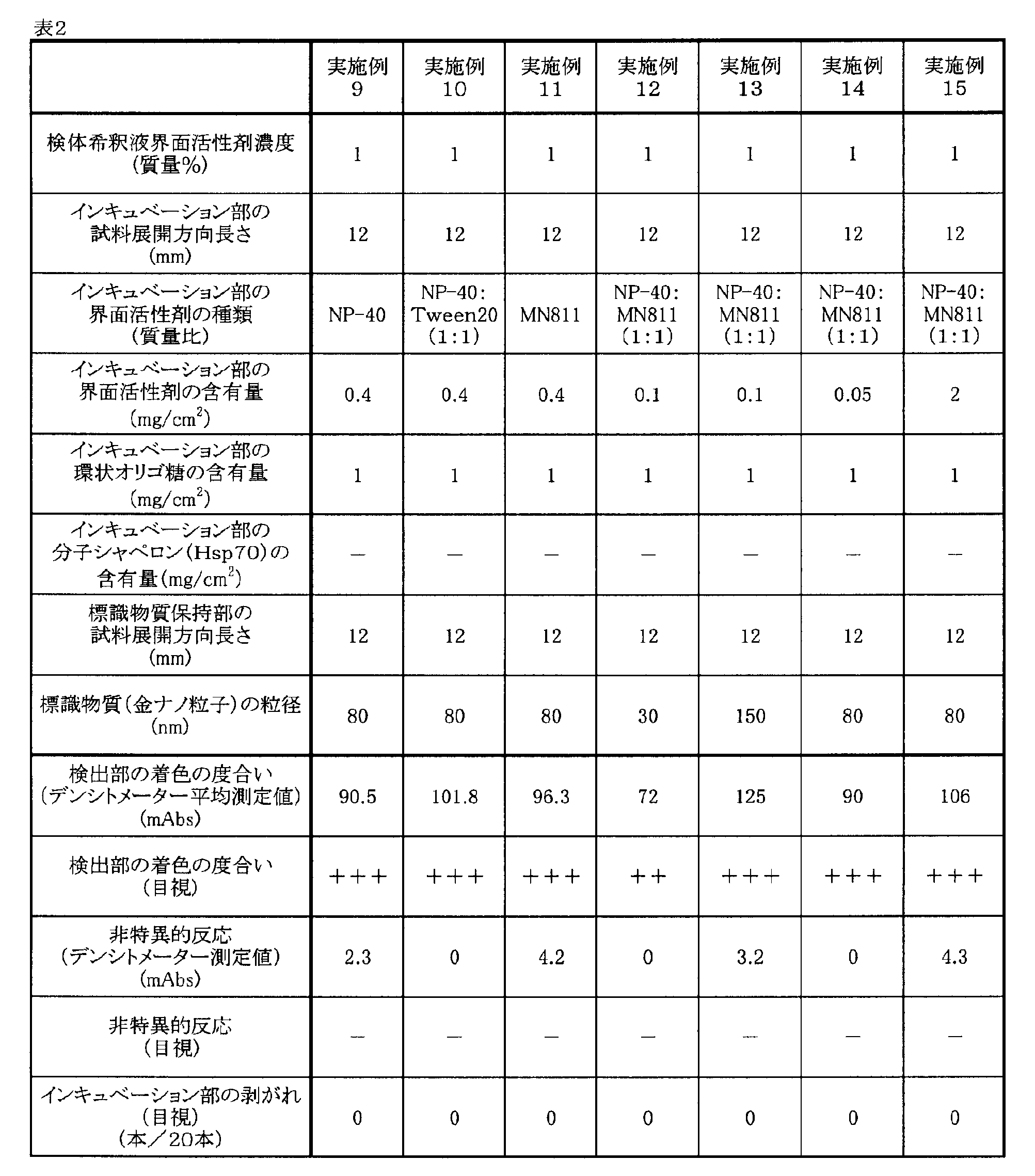
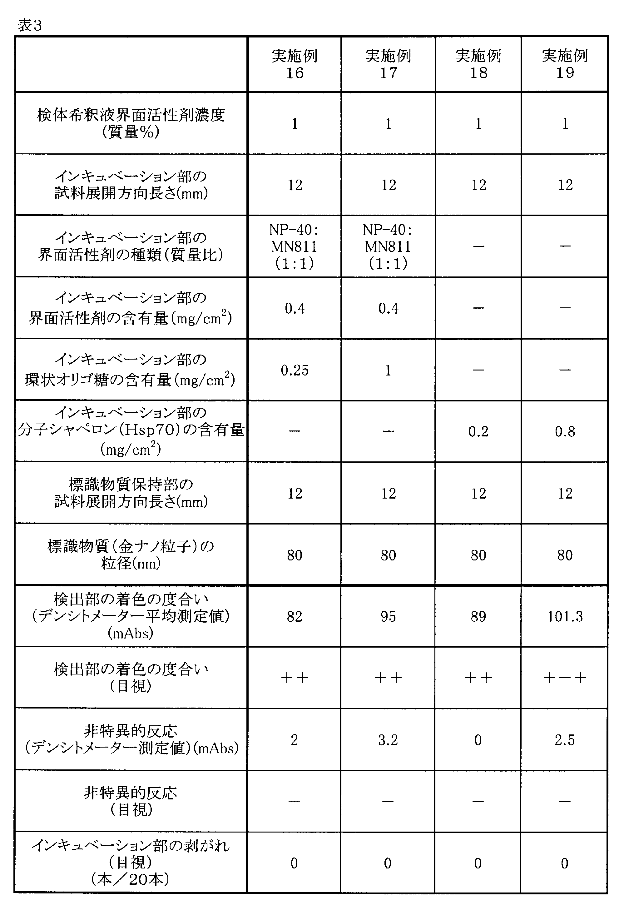
表1~3の結果から、各実施例の免疫クロマト分析装置は、インキュベーション部を設けない比較例1に比べて、被検出物質の検出感度を著しく高め得ることが判明した。とくにインキュベーション部に界面活性剤を含有する実施例5~17、及びインキュベーション部に分子シャペロンを含有する実施例18~19では、さらに感度が向上していることが分かる。
また、環状オリゴ糖を添加した実施例6~17のインキュベーション部は、界面活性剤が均一に分散し良好な凍結乾燥状態が得られ、変色も起こらなかった。これに対し、界面活性剤を添加し環状オリゴ糖を添加しなかった上記実施例5のインキュベーション部は、凍結乾燥後に濡れ(ベタツキ感)があり、やや黄色に着色し、裁断後の免疫クロマト分析装置のインキュベーション部の剥がれ不具合が生じていた。
また、同仕様の免疫クロマト装置間の発色強度のムラを繰り返し5回試験の測定値の平均値からの剥離範囲を誤差として算出することにより調べたところ、実施例5の装置では、10mAbsを超える誤差(発色強度のムラ)が観察された。実施例6~17の装置では、何れも3~5mAbsの誤差範囲と発色強度ムラの少ない結果であった。
本発明を特定の態様を用いて詳細に説明したが、本発明の意図と範囲を離れることなく様々な変更および変形が可能であることは、当業者にとって明らかである。なお本出願は、2014年3月27日付で出願された日本特許出願(特願2014-066127)に基づいており、その全体が引用により援用される。
1.試料添加部
2.標識物質保持部
3.インキュベーション部
4.クロマトグラフ媒体部
5.検出部
6.吸収部
7.バッキングシート
2.標識物質保持部
3.インキュベーション部
4.クロマトグラフ媒体部
5.検出部
6.吸収部
7.バッキングシート
Claims (6)
- 検体を添加する試料添加部と、前記検体に含まれる被検出物質を認識する標識物質を保持する標識物質保持部と、前記標識物質の溶出を遅延させるインキュベーション部と、前記被検出物質を検出する検出部が担持されたクロマトグラフ媒体部と、前記検出部を通過した液体を吸収する吸収部とを含む免疫クロマト分析装置。
- 前記インキュベーション部が、界面活性剤を含有することを特徴とする請求項1に記載の免疫クロマト分析装置。
- 前記インキュベーション部が、分子シャペロンを含有することを特徴とする請求項1に記載の免疫クロマト分析装置。
- 前記インキュベーション部が、環状オリゴ糖を含有することを特徴とする請求項2に記載の免疫クロマト分析装置。
- 前記標識物質の平均粒径が、20~200nmであることを特徴とする請求項1~4のいずれか1項に記載の免疫クロマト分析装置。
- 請求項1~5のいずれか1項に記載の免疫クロマト分析装置を用い、下記工程(1)~(5)を順次実施することを特徴とする、免疫クロマト分析方法。
(1)前記検体を試料添加部に添加する工程
(2)前記標識物質保持部に保持されている標識物質により前記検体に含まれる被検出物
質を認識させる工程
(3)前記インキュベーション部において標識物質の溶出を遅延させる工程
(4)前記検体及び標識物質を移動相としてクロマトグラフ媒体部に展開させる工程
(5)展開された移動相中の被検出物質を検出部で検出する工程。
Priority Applications (1)
| Application Number | Priority Date | Filing Date | Title |
|---|---|---|---|
| JP2016510565A JP6533216B2 (ja) | 2014-03-27 | 2015-03-27 | 免疫クロマト分析装置及び免疫クロマト分析方法 |
Applications Claiming Priority (2)
| Application Number | Priority Date | Filing Date | Title |
|---|---|---|---|
| JP2014-066127 | 2014-03-27 | ||
| JP2014066127 | 2014-03-27 |
Publications (1)
| Publication Number | Publication Date |
|---|---|
| WO2015147291A1 true WO2015147291A1 (ja) | 2015-10-01 |
Family
ID=54195795
Family Applications (1)
| Application Number | Title | Priority Date | Filing Date |
|---|---|---|---|
| PCT/JP2015/059739 Ceased WO2015147291A1 (ja) | 2014-03-27 | 2015-03-27 | 免疫クロマト分析装置及び免疫クロマト分析方法 |
Country Status (2)
| Country | Link |
|---|---|
| JP (1) | JP6533216B2 (ja) |
| WO (1) | WO2015147291A1 (ja) |
Cited By (2)
| Publication number | Priority date | Publication date | Assignee | Title |
|---|---|---|---|---|
| EP3425398A4 (en) * | 2016-03-04 | 2019-01-09 | Tanaka Kikinzoku Kogyo K.K. | IMMUNCHROMATOGRAPHIEVORRICHTUNG |
| CN114280312A (zh) * | 2020-09-27 | 2022-04-05 | 河北特温特生物科技发展有限公司 | 一种用于免疫荧光层析检测的全血分离膜及其制备方法和应用 |
Citations (3)
| Publication number | Priority date | Publication date | Assignee | Title |
|---|---|---|---|---|
| JPH0545360A (ja) * | 1991-08-12 | 1993-02-23 | Konica Corp | 免疫学的測定装置 |
| JPH11153601A (ja) * | 1997-09-18 | 1999-06-08 | Matsushita Electric Ind Co Ltd | 免疫クロマトグラフィー装置 |
| JP2005509852A (ja) * | 2001-11-14 | 2005-04-14 | プリオン ディベロプメンタル ラボラトリーズ, インコーポレイテッド | 迅速なプリオン検出アッセイ |
-
2015
- 2015-03-27 JP JP2016510565A patent/JP6533216B2/ja active Active
- 2015-03-27 WO PCT/JP2015/059739 patent/WO2015147291A1/ja not_active Ceased
Patent Citations (3)
| Publication number | Priority date | Publication date | Assignee | Title |
|---|---|---|---|---|
| JPH0545360A (ja) * | 1991-08-12 | 1993-02-23 | Konica Corp | 免疫学的測定装置 |
| JPH11153601A (ja) * | 1997-09-18 | 1999-06-08 | Matsushita Electric Ind Co Ltd | 免疫クロマトグラフィー装置 |
| JP2005509852A (ja) * | 2001-11-14 | 2005-04-14 | プリオン ディベロプメンタル ラボラトリーズ, インコーポレイテッド | 迅速なプリオン検出アッセイ |
Cited By (4)
| Publication number | Priority date | Publication date | Assignee | Title |
|---|---|---|---|---|
| EP3425398A4 (en) * | 2016-03-04 | 2019-01-09 | Tanaka Kikinzoku Kogyo K.K. | IMMUNCHROMATOGRAPHIEVORRICHTUNG |
| US11808670B2 (en) | 2016-03-04 | 2023-11-07 | Tanaka Kikinzoku Kogyo K.K. | Immunochromatographic device |
| CN114280312A (zh) * | 2020-09-27 | 2022-04-05 | 河北特温特生物科技发展有限公司 | 一种用于免疫荧光层析检测的全血分离膜及其制备方法和应用 |
| CN114280312B (zh) * | 2020-09-27 | 2023-09-15 | 河北特温特生物科技发展有限公司 | 一种用于免疫荧光层析检测的全血分离膜及其制备方法和应用 |
Also Published As
| Publication number | Publication date |
|---|---|
| JPWO2015147291A1 (ja) | 2017-04-13 |
| JP6533216B2 (ja) | 2019-06-19 |
Similar Documents
| Publication | Publication Date | Title |
|---|---|---|
| JP5792859B1 (ja) | 免疫クロマト分析方法 | |
| JP6226624B2 (ja) | 溶血性レンサ球菌診断イムノクロマト試薬、キット及び検出方法 | |
| JP6008670B2 (ja) | イムノクロマトグラフ試験ストリップ用メンブレン、試験ストリップ及び検査方法 | |
| WO2012018057A1 (ja) | イムノクロマトグラフィー用試薬組成物およびそれを用いた測定方法 | |
| TWI639002B (zh) | Chromatographic medium | |
| CN107709994B (zh) | 层析分析装置及层析分析方法 | |
| JP6192374B2 (ja) | 担体及び水溶性多糖類を含むイムノクロマト用展開液 | |
| JP6533216B2 (ja) | 免疫クロマト分析装置及び免疫クロマト分析方法 | |
| JP5827703B2 (ja) | クロマトグラフ方法、クロマトグラフ用キット及びクロマトグラフ用不溶性担体の製造方法 | |
| JP5825190B2 (ja) | アナライトを検出または定量するためのラテラルフロー型クロマト法用テストストリップ | |
| JP6595215B2 (ja) | 免疫クロマト分析装置およびその製造方法並びに免疫クロマト分析方法 | |
| JP2008197038A (ja) | イムノクロマトグラフィー法 | |
| JP7533226B2 (ja) | イムノクロマト試験片およびそれを用いた測定方法 | |
| JP5265423B2 (ja) | クロマトグラフ方法 | |
| WO2024122616A1 (ja) | イムノクロマト法検査デバイスの試料展開部の形成方法およびイムノクロマト法検査デバイス |
Legal Events
| Date | Code | Title | Description |
|---|---|---|---|
| 121 | Ep: the epo has been informed by wipo that ep was designated in this application |
Ref document number: 15768473 Country of ref document: EP Kind code of ref document: A1 |
|
| DPE1 | Request for preliminary examination filed after expiration of 19th month from priority date (pct application filed from 20040101) | ||
| ENP | Entry into the national phase |
Ref document number: 2016510565 Country of ref document: JP Kind code of ref document: A |
|
| NENP | Non-entry into the national phase |
Ref country code: DE |
|
| 122 | Ep: pct application non-entry in european phase |
Ref document number: 15768473 Country of ref document: EP Kind code of ref document: A1 |