WO2015115331A1 - 抗トランスサイレチンヒト化抗体 - Google Patents
抗トランスサイレチンヒト化抗体 Download PDFInfo
- Publication number
- WO2015115331A1 WO2015115331A1 PCT/JP2015/051856 JP2015051856W WO2015115331A1 WO 2015115331 A1 WO2015115331 A1 WO 2015115331A1 JP 2015051856 W JP2015051856 W JP 2015051856W WO 2015115331 A1 WO2015115331 A1 WO 2015115331A1
- Authority
- WO
- WIPO (PCT)
- Prior art keywords
- ttr
- antibody
- amino acid
- acid sequence
- polypeptide
- Prior art date
Links
Images


Classifications
-
- C—CHEMISTRY; METALLURGY
- C07—ORGANIC CHEMISTRY
- C07K—PEPTIDES
- C07K16/00—Immunoglobulins [IGs], e.g. monoclonal or polyclonal antibodies
- C07K16/18—Immunoglobulins [IGs], e.g. monoclonal or polyclonal antibodies against material from animals or humans
-
- A—HUMAN NECESSITIES
- A61—MEDICAL OR VETERINARY SCIENCE; HYGIENE
- A61K—PREPARATIONS FOR MEDICAL, DENTAL OR TOILETRY PURPOSES
- A61K39/00—Medicinal preparations containing antigens or antibodies
- A61K39/0005—Vertebrate antigens
- A61K39/0007—Nervous system antigens; Prions
-
- A—HUMAN NECESSITIES
- A61—MEDICAL OR VETERINARY SCIENCE; HYGIENE
- A61K—PREPARATIONS FOR MEDICAL, DENTAL OR TOILETRY PURPOSES
- A61K39/00—Medicinal preparations containing antigens or antibodies
- A61K39/395—Antibodies; Immunoglobulins; Immune serum, e.g. antilymphocytic serum
-
- A—HUMAN NECESSITIES
- A61—MEDICAL OR VETERINARY SCIENCE; HYGIENE
- A61P—SPECIFIC THERAPEUTIC ACTIVITY OF CHEMICAL COMPOUNDS OR MEDICINAL PREPARATIONS
- A61P25/00—Drugs for disorders of the nervous system
-
- A—HUMAN NECESSITIES
- A61—MEDICAL OR VETERINARY SCIENCE; HYGIENE
- A61P—SPECIFIC THERAPEUTIC ACTIVITY OF CHEMICAL COMPOUNDS OR MEDICINAL PREPARATIONS
- A61P25/00—Drugs for disorders of the nervous system
- A61P25/28—Drugs for disorders of the nervous system for treating neurodegenerative disorders of the central nervous system, e.g. nootropic agents, cognition enhancers, drugs for treating Alzheimer's disease or other forms of dementia
-
- A—HUMAN NECESSITIES
- A61—MEDICAL OR VETERINARY SCIENCE; HYGIENE
- A61P—SPECIFIC THERAPEUTIC ACTIVITY OF CHEMICAL COMPOUNDS OR MEDICINAL PREPARATIONS
- A61P43/00—Drugs for specific purposes, not provided for in groups A61P1/00-A61P41/00
-
- A—HUMAN NECESSITIES
- A61—MEDICAL OR VETERINARY SCIENCE; HYGIENE
- A61P—SPECIFIC THERAPEUTIC ACTIVITY OF CHEMICAL COMPOUNDS OR MEDICINAL PREPARATIONS
- A61P9/00—Drugs for disorders of the cardiovascular system
-
- G—PHYSICS
- G01—MEASURING; TESTING
- G01N—INVESTIGATING OR ANALYSING MATERIALS BY DETERMINING THEIR CHEMICAL OR PHYSICAL PROPERTIES
- G01N33/00—Investigating or analysing materials by specific methods not covered by groups G01N1/00 - G01N31/00
- G01N33/48—Biological material, e.g. blood, urine; Haemocytometers
- G01N33/50—Chemical analysis of biological material, e.g. blood, urine; Testing involving biospecific ligand binding methods; Immunological testing
- G01N33/68—Chemical analysis of biological material, e.g. blood, urine; Testing involving biospecific ligand binding methods; Immunological testing involving proteins, peptides or amino acids
- G01N33/6893—Chemical analysis of biological material, e.g. blood, urine; Testing involving biospecific ligand binding methods; Immunological testing involving proteins, peptides or amino acids related to diseases not provided for elsewhere
- G01N33/6896—Neurological disorders, e.g. Alzheimer's disease
-
- A—HUMAN NECESSITIES
- A61—MEDICAL OR VETERINARY SCIENCE; HYGIENE
- A61K—PREPARATIONS FOR MEDICAL, DENTAL OR TOILETRY PURPOSES
- A61K39/00—Medicinal preparations containing antigens or antibodies
- A61K2039/505—Medicinal preparations containing antigens or antibodies comprising antibodies
-
- C—CHEMISTRY; METALLURGY
- C07—ORGANIC CHEMISTRY
- C07K—PEPTIDES
- C07K14/00—Peptides having more than 20 amino acids; Gastrins; Somatostatins; Melanotropins; Derivatives thereof
-
- C—CHEMISTRY; METALLURGY
- C07—ORGANIC CHEMISTRY
- C07K—PEPTIDES
- C07K2317/00—Immunoglobulins specific features
- C07K2317/20—Immunoglobulins specific features characterized by taxonomic origin
- C07K2317/24—Immunoglobulins specific features characterized by taxonomic origin containing regions, domains or residues from different species, e.g. chimeric, humanized or veneered
-
- C—CHEMISTRY; METALLURGY
- C07—ORGANIC CHEMISTRY
- C07K—PEPTIDES
- C07K2317/00—Immunoglobulins specific features
- C07K2317/30—Immunoglobulins specific features characterized by aspects of specificity or valency
- C07K2317/34—Identification of a linear epitope shorter than 20 amino acid residues or of a conformational epitope defined by amino acid residues
-
- C—CHEMISTRY; METALLURGY
- C07—ORGANIC CHEMISTRY
- C07K—PEPTIDES
- C07K2317/00—Immunoglobulins specific features
- C07K2317/50—Immunoglobulins specific features characterized by immunoglobulin fragments
- C07K2317/51—Complete heavy chain or Fd fragment, i.e. VH + CH1
-
- C—CHEMISTRY; METALLURGY
- C07—ORGANIC CHEMISTRY
- C07K—PEPTIDES
- C07K2317/00—Immunoglobulins specific features
- C07K2317/50—Immunoglobulins specific features characterized by immunoglobulin fragments
- C07K2317/515—Complete light chain, i.e. VL + CL
-
- C—CHEMISTRY; METALLURGY
- C07—ORGANIC CHEMISTRY
- C07K—PEPTIDES
- C07K2317/00—Immunoglobulins specific features
- C07K2317/50—Immunoglobulins specific features characterized by immunoglobulin fragments
- C07K2317/56—Immunoglobulins specific features characterized by immunoglobulin fragments variable (Fv) region, i.e. VH and/or VL
- C07K2317/565—Complementarity determining region [CDR]
-
- C—CHEMISTRY; METALLURGY
- C07—ORGANIC CHEMISTRY
- C07K—PEPTIDES
- C07K2317/00—Immunoglobulins specific features
- C07K2317/60—Immunoglobulins specific features characterized by non-natural combinations of immunoglobulin fragments
- C07K2317/62—Immunoglobulins specific features characterized by non-natural combinations of immunoglobulin fragments comprising only variable region components
- C07K2317/622—Single chain antibody (scFv)
-
- C—CHEMISTRY; METALLURGY
- C07—ORGANIC CHEMISTRY
- C07K—PEPTIDES
- C07K2317/00—Immunoglobulins specific features
- C07K2317/70—Immunoglobulins specific features characterized by effect upon binding to a cell or to an antigen
- C07K2317/76—Antagonist effect on antigen, e.g. neutralization or inhibition of binding
-
- C—CHEMISTRY; METALLURGY
- C07—ORGANIC CHEMISTRY
- C07K—PEPTIDES
- C07K2317/00—Immunoglobulins specific features
- C07K2317/90—Immunoglobulins specific features characterized by (pharmaco)kinetic aspects or by stability of the immunoglobulin
- C07K2317/92—Affinity (KD), association rate (Ka), dissociation rate (Kd) or EC50 value
-
- G—PHYSICS
- G01—MEASURING; TESTING
- G01N—INVESTIGATING OR ANALYSING MATERIALS BY DETERMINING THEIR CHEMICAL OR PHYSICAL PROPERTIES
- G01N2333/00—Assays involving biological materials from specific organisms or of a specific nature
- G01N2333/435—Assays involving biological materials from specific organisms or of a specific nature from animals; from humans
- G01N2333/46—Assays involving biological materials from specific organisms or of a specific nature from animals; from humans from vertebrates
- G01N2333/47—Assays involving proteins of known structure or function as defined in the subgroups
-
- G—PHYSICS
- G01—MEASURING; TESTING
- G01N—INVESTIGATING OR ANALYSING MATERIALS BY DETERMINING THEIR CHEMICAL OR PHYSICAL PROPERTIES
- G01N2800/00—Detection or diagnosis of diseases
- G01N2800/28—Neurological disorders
-
- G—PHYSICS
- G01—MEASURING; TESTING
- G01N—INVESTIGATING OR ANALYSING MATERIALS BY DETERMINING THEIR CHEMICAL OR PHYSICAL PROPERTIES
- G01N2800/00—Detection or diagnosis of diseases
- G01N2800/70—Mechanisms involved in disease identification
- G01N2800/7047—Fibrils-Filaments-Plaque formation
Definitions
- the present invention provides an antibody that effectively suppresses the formation of amyloid fibrils and deposition of tissue of transthyretin (TTR: transthyretin) and a therapeutic method using the antibody.
- TTR tissue of transthyretin
- This antibody therapy is based on a new therapeutic strategy that does not act on normal TTR and suppresses only amyloid formation of abnormal TTR, and is expected to be a novel therapeutic method with excellent safety.
- Amyloidosis is a group of diseases that cause dysfunction due to deposition of a protein that forms a fiber structure in organs throughout the body, and includes various diseases such as Alzheimer-type dementia and prion disease (Non-patent Document 1). .
- Familial amyloid polyneuropathy (Familial Amyloidotic Polyneuropathy: FAP) is an autosomal dominant hereditary systemic amyloidosis caused by point mutations and deletions of genes such as TTR, apolipoprotein A1, and gelsolin (non-patented) Reference 2). Among these, FAP is caused by the genetic variation of TTR. Mutant TTR forms amyloid fibrils that usually become dysfunctional by depositing in almost any systemic tissue, such as peripheral nerves, heart, kidney, gastrointestinal tract, eye, brain, and meninges after middle age. Are known. The prognosis of the patient is very poor, and it is an intractable disease that will die in about 10 years from the onset.
- V30M Val30Met mutation
- methionine the Val30Met mutation in which the 30th valine of TTR is mutated to methionine is the most common, and there are many patients in Portugal, Sweden, and Japan. Because more than 6,000 FAP patients have been confirmed in Portugal, and there are many areas where FAP has not yet been investigated, and it is expected that global discovery of FAP patients will continue in the future. It is believed that there are well over 10,000 patients worldwide. Recent studies have shown that the clinical appearance of FAP (onset age, deposited organ specificity, etc.) is greatly influenced by the type of mutation in the TTR gene (Non-patent Document 3).
- the L55P mutation has a fulminant clinical appearance that develops in teens, while the V122I mutation develops after the age of 60.
- the V30M mutation has both a disease type that develops at a young age and a disease type that develops at an old age.
- the D18G mutation deposits in the brain and meninges, causing central neuropathy, while the V30M mutation deposits in systemic tissues, causing peripheral neuropathy and myocardial damage (Non-Patent Documents) 3, 4).
- TTR is a protein composed of 127 amino acids and having a molecular weight of 14 kDa, and has a structure in which eight ⁇ -strands present inside form two antiparallel ⁇ -sheets (Non-patent Document 5). Although it is produced in the liver, it is also produced in the ventricular choroid plexus, retinal pigment epithelial cells of the retina, spleen and the like.
- TTR usually forms a stable tetramer with a molecular weight of 55 kDa in the blood, mainly in blood and cerebrospinal fluid, vitamin A-retinol binding protein complex, and transporter of thyroid hormone T4 Is functioning as Although its blood concentration is as high as 200-400 ⁇ g / mL, its half-life is as short as 2 days (Non-patent Documents 2 to 6). It is known that there are two homologous T4 binding sites in the center of the TTR tetramer, and the tetramer structure is stabilized by the binding of T4 (Non-patent Document 3).
- Non-Patent Document 2 As other functions, various reports such as an insulin secretion promoting action, a cranial nerve protecting action, and an action relating to lipid metabolism have been made (Non-Patent Document 2). On the other hand, in mice where the TTR gene was knocked out, although blood retinol and thyroid hormone concentrations were reduced, no significant changes were observed in phenotypes such as survival rate or fertility (Non-patent Document 7). It remains unclear whether is directly essential in the maintenance of actual life activities.
- Non-patent Document 3 For TTR amyloid formation, dissociation from tetramer to monomer and structural change of the monomer are very important steps (Non-patent Document 3). Above all, it has been clarified that dissociation from the tetramer to the monomer is the rate-limiting step of the reaction. On the other hand, in the process of TTR forming amyloid and depositing in tissues and damaging organs throughout the body, the molecular form that exhibits toxicity to tissues has not been completely clarified. There are reports that low molecular weight oligomers such as monomers and dimers have cytotoxicity, while TTR amyloid of 100 kDa or more does not have cytotoxicity (Non-patent Document 5). The elucidation of the relationship with is awaited.
- Treatment strategies for FAP caused by TTR gene abnormalities are mainly classified into the following four types.
- Non-patent Document 2 Since most TTR in blood is produced in the liver (Non-patent Document 2), the most common treatment method among them is liver transplantation classified as (1). Although the progression of the disease state can be delayed by performing liver transplantation, immunosuppressants must be used throughout life, and the burden on donors and patients is also great. Furthermore, since deposition in several organs including the eyes and heart continues, there are many cases in which the symptoms of these organs are exacerbated (Non-patent Document 8). Longed for.
- Non-patent Document 10 a new epitope (cryptic epitope, Cryptic Epitope) is exposed on the surface of the molecule with the structural change of TTR.
- an antibody that specifically recognizes the Cryptic Epitope of TTR specifically binds to TTR amyloid (or TTR with a structural change that constitutes TTR amyloid), and inhibits or eliminates the formation of TTR amyloid. There is a possibility of promoting this. In other words, it is suggested that an antibody that specifically recognizes TTR Cryptic Epitope may be a novel therapeutic agent for FAP.
- BIOCODEX Anti-TTR antibodies based on these concepts has been reported by BIOCODEX.
- the company produced AD7F6, a mouse monoclonal antibody specific for amyloidogenic TTR, using TTR knockout mice, and demonstrated that it suppresses TTR tissue deposition using Tg mice (ATTRAPV30M), a FAP pathological model.
- Patent Document 1 The BIOCODEX patent claims the amino acid sequence of a mouse antibody and is difficult to administer to humans. In addition, regarding this antibody, whether or not there is reactivity with a V30M mutant having a tetrameric structure is not explicitly stated.
- the V30M mutant present in the blood has a tetrameric structure, but it is thought that a part of the monomer undergoes a structural change after dissociating into a monomer to form amyloid. Yes. Therefore, it is more effective and safer to be an antibody that does not react with the V30M mutant having a tetrameric structure, but reacts only with the amyloidated (or intermediate stage of forming amyloid) V30M mutant. This is considered a necessary requirement for realizing antibody therapy. Regarding the reactivity of this antibody, since only the serum of the V30M carrier is used as a clinical sample, the reactivity with the tissue depositing amyloid present in the patient's body is unknown.
- Non-Patent Document 10 mouse monoclonal antibodies mAb 39-44 and mAb 56-61 specific to TTR that have undergone structural changes have been prepared and that these react with amyloid of V30M mutants derived from living organisms. However, it is specified that they did not have an inhibitory effect on amyloid formation, and only mentions the possibility of being available for the diagnosis of FAP.
- TTR amyloidosis the monomer TTR undergoes a structural change after dissociation of some tetrameric TTR into monomers, and forms amyloid, while the tetramer that functions normally. Body TTR also thought to exist.
- the present inventors examined an antibody that specifically binds to TTR that has undergone a structural change and has an activity of inhibiting TTR fibrosis.
- the present invention was completed by earnestly examining humanized antibodies having the above-mentioned activities.
- the present invention (1) a humanized antibody having an activity of inhibiting fibrosis of transthyretin (TTR); (2) The humanized antibody according to (1), which specifically recognizes TTR that has undergone structural change; (3) The humanized antibody according to (1) or (2), which specifically binds to TTR amyloid; (4) The humanized antibody according to any one of (1) to (3), which binds to TTR amyloid derived from two or more types of mutant TTR; (5) The humanized antibody according to (4), wherein the mutant TTR is a TTR having a mutation selected from the group consisting of D18G, V30M, E54K, L55P, Y114C, Y116S, and V122I; (6) The humanized antibody according to any one of (1) to (5), which promotes removal of TTR amyloid; (7) The humanized antibody according to any one of (1) to (6), which promotes macrophage phagocytosis against TTR amyloid; (8) The humanized antibody according to any one of (1) to (7)
- a polypeptide comprising the amino acid sequence shown in SEQ ID NOs: 1 to 3.
- B The amino acid sequence shown in SEQ ID NOs: 1 to 3, consisting of an amino acid sequence in which one or several amino acids are substituted, deleted, inserted, and / or added, and determination of complementation of H chain to TTR A polypeptide that becomes a region.
- C A polypeptide comprising the amino acid sequence shown in SEQ ID NOs: 4 to 6.
- the amino acid sequence shown in SEQ ID NOs: 4 to 6 consists of an amino acid sequence in which one or several amino acids are substituted, deleted, inserted, and / or added, and determination of complementation of the L chain to TTR
- (F) a polypeptide comprising an amino acid sequence in which one or several amino acids are substituted, deleted, inserted, and / or added in the amino acid sequence shown in SEQ ID NO: 7, and serving as a heavy chain variable region for TTR .
- (G) A polypeptide having the amino acid sequence represented by SEQ ID NO: 8.
- (H) a polypeptide comprising an amino acid sequence in which one or several amino acids are substituted, deleted, inserted, and / or added in the amino acid sequence shown in SEQ ID NO: 8, and serving as a light chain variable region for TTR ;
- (H) a polypeptide comprising an amino acid sequence in which one or several amino acids are substituted, deleted, inserted, and / or added in the amino acid sequence shown in SEQ ID NO: 8, and serving as a light chain variable region for TTR ;
- the H chain variable region fragment comprising the H chain complementarity determining region according to (15) or the H chain variable region fragment according to (17), and the L chain complementarity determining region according to (16) A single-chain variable region fragment of an antibody against TTR, which is obtained by linking an L-chain variable region fragment containing or an L-chain
- the present inventors have succeeded in creating a monoclonal antibody that specifically recognizes TTR that has undergone structural changes, and opening the way to the development of antibody drugs that enable the treatment of FAP.
- the antibody of the present invention effectively suppresses the formation of amyloid fibrils and tissue deposition of TTR, and does not react with normal TTR functioning in blood, so that it can be an antibody drug with excellent safety. There is expected.
- (1) suppresses the formation and tissue deposition of TTR amyloid
- (2) also acts on TTR amyloid deposited in the tissue to promote clearance, that is, once accumulated amyloid Two effects can be expected to be reduced. Such an effect cannot be achieved by the conventional technology or the previously developed product, and the present antibody therapy as a novel therapeutic strategy for TTR amyloidosis is expected.
- the antibody of the present invention not only can be expected to provide a new therapeutic method other than liver transplantation for FAP, but also has potential as a therapeutic agent for senile systemic amyloidosis (SSA).
- SSA senile systemic amyloidosis
- TTR amyloidosis in addition to FAP caused by mutations in the TTR gene, SSA is known which develops when wild-type TTR mainly causes amyloid deposition in the heart. It is also positioned as Alzheimer's disease in the heart. Amyloid deposits are also found in the lungs, blood vessel walls, and renal medulla. Patients are often asymptomatic or have cardiac symptoms (slowly progressive heart failure) and may have carpal tunnel syndrome.
- This antibody preparation is expected to be applied not only to FAP but also to amyloidogenic diseases for various organs caused by TTR, and is expected to be able to contribute to the treatment of many of these diseases for which there is currently no therapeutic method .
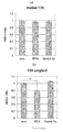
- the map of (a) mouse T24 antibody H chain expression vector pKMA010-T24-mCg1 and (b) mouse T24 antibody L chain expression vector pKMA009-T24-mCk is shown.
- the result of epitope analysis is shown.
- the result of the reaction specificity analysis using the surface plasmon resonance method is shown.
- (A) shows the results with RT24 antibody, (b) with Dako polyclonal antibody, and (c) with negative control antibody.
- the result of the reactivity analysis to patient serum is shown.
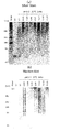
- the result of the reactivity analysis to a patient tissue is shown (paraffin section).
- the result of the reactivity analysis to a patient tissue is shown (frozen section).
- the result of the reactivity analysis to TTR fiber is shown.
- (A) shows the result of silver staining, and (b) shows the result of Western blotting.
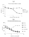
- the result of a TTR fibrosis inhibition test is shown.
- (A) shows the results of RT24 antibody, and (b) shows the results of RT24-a antibody and RT24-b antibody, respectively.
- the result of a macrophage phagocytic test is shown.
- (A) shows the results with untreated purified V30M, and (b) shows the results with TTR fibers.
- the result of a drug efficacy evaluation test using V30M Tg rats is shown.
- positions 115 to 124 of TTR are not exposed on the molecular surface in normal (tetrameric) TTR, but are exposed on the surface when TTR is fibrotic. Attention was paid to TTR115-124 as an antigen for antibody production. After immunization of the antigen to a TTR knockout mouse, a mouse antibody was produced, and the amino acid sequence of the mouse antibody was humanized to produce a humanized antibody.
- the method for preparing TTR115-124 may be chemical synthesis, or may be purified from E. coli. In the latter case, the base sequence is amplified using primer DNA designed to amplify the base sequence corresponding to 115-124 using the base sequence of human TTR as a template. Then, it is incorporated into an expression vector and introduced into a host (such as E. coli). After expressing TTR115-124 using a host, it is purified and used as an antigen.
- TTR115-124 obtained by chemical synthesis or expression method may be immunized to mice after binding to carrier protein (KLH, BSA, etc.). Examples of the method for binding the carrier protein include a method using Immunogen EDC Kit with mcKLH and BSA (Thermo).
- One molecule of TTR115-124 may be bound to a carrier protein, or two or more molecules may be bound.
- the site where TTR115-124 is bound to the carrier protein may be any of the N-terminal, C-terminal,
- TTR115-124 is immunized to TTR knockout mice.
- the TTR knockout mouse may be prepared by the method described in Non-Patent Document 13.
- the number of times of immunization with TTR115-124 is preferably 2 times or more, and each immunization is preferably about 3 weeks.
- the number and duration of immunizations may be appropriately changed while confirming the degree of increase in the antibody titer against TTR.
- antibody-producing cells are collected from the spleen of the mouse, and antibody-producing cells and myeloma cells are fused by a conventional hybridoma method to produce a hybridoma.
- TTR115-124 is directly or indirectly immobilized on an ELISA plate.
- TTR115-124 is immobilized on a plate, it may be on the N-terminal side or C-terminal side of TTR115-124.
- mouse antibody is added to the plate and allowed to react. A method of adding an enzyme-labeled anti-mouse antibody and a substrate in this order to the plate and detecting the substrate is exemplified. Using such an ELISA test as an index, antibodies against TTR115-124 are selected.
- a step of selecting an antibody that specifically binds to TTR amyloid may be provided.
- the method to verify the binding property to TTR amyloid is illustrated.
- the TTR peptide is allowed to stand for a period of formation of fibrosis under acidic conditions to form TTR amyloid.
- the period during which acidic conditions and fibrosis are formed may be appropriately changed depending on the type of TTR peptide.
- the presence or absence of TTR fibrosis can be confirmed by thioflavin T assay.
- the acidic condition is preferably pH 5.0 or less, more preferably pH 3.0 to pH 4.0.
- the period of forming fibrosis is preferably overnight.
- TTR amyloid acid-treated TTR (TTR amyloid) or untreated TTR (normal TTR) is bound to an ELISA plate, and then a mouse antibody is reacted. You may select the antibody with the high binding property with respect to TTR amyloid compared with normal type TTR.
- a method for producing a humanized antibody from a mouse antibody will be exemplified.
- reverse transcription is performed to prepare cDNA.
- the cDNA is used as a template to amplify the VH region and VL region, and the region is introduced into an appropriate plasmid, and then the nucleotide sequence of the VH region or VL region is analyzed. Then, a portion (CDR1-3) corresponding to the CDR in the VH region or VL region is specified.
- a human antibody suitable for CDR transplantation is selected.
- an amino acid sequence in which the amino acid sequence of CDR1-3 in the mouse antibody is inserted into the amino acid sequence of the VH region or VL region in the human antibody is designed.
- the amino acid sequence is converted into a base sequence and chemically synthesized.
- An expression vector is prepared by inserting the nucleotide sequence into an expression vector containing a nucleotide sequence encoding the H chain or L chain constant region.
- the expression vector is introduced into a suitable host (animal cell), and the humanized antibody is expressed using the host.
- a modified humanized antibody in the humanized antibody, an amino acid sequence in which the amino acid sequence was modified for the framework part of the VH region or VL region was designed, and a base sequence based on the modified amino acid sequence was chemically synthesized.
- a modified humanized antibody was produced by the same method as described above.
- a human TTR variant in which one amino acid is altered in the amino acid sequence at positions 115 to 124 of human TTR is prepared.
- a modified gene is prepared by a conventional site-directed mutagenesis method, inserted into an expression vector, and then expressed and purified using an appropriate host (preferably E. coli). Examples of such variants include Y114C, S115A, Y116A, S117A, T118A, T119A, A120S, V121A, V122A, V122I, T123A, and N124A.
- the modified body is electrophoresed on SDS-PAGE using a conventional Western blotting method, and then reacted with the antibody to be analyzed to detect the reactivity between the modified body and the antibody.
- the altered portion may be considered as the epitope portion of the antibody.
- the antibody of the present invention had an epitope portion at positions 118-122.
- mice and humanized antibodies have specific reactivity to structurally altered TTR, reactivity with fibrotic TTR, immunostaining for TTR patient-derived tissues, inhibitory activity against mutant TTR fibrosis, TTR It is possible to evaluate the effect of promoting macrophage phagocytosis on amyloid and evaluating the drug efficacy using FAP model animals.
- a method using surface plasmon resonance is exemplified as a method for analyzing specific reactivity to TTR that has undergone structural change.
- a wild type TTR tetramer, a mutant TTR (such as V30M) tetramer, or a mutant TTR amyloid is generated.
- Matsubara et al. Non-patent Document 14
- the above tetrameric TTR peptide is adjusted to 3 mg / mL, then mixed with an equal amount of 200 mM acetate buffer and 100 mM NaCl solution, and reacted at 37 ° C. for 16 hours. Is exemplified.
- fluorescence intensity may be measured by Thioflavin T assay (excitation wavelength: 440 nm, fluorescence wavelength: 480 nm) to confirm that fibers are formed.
- the binding property between TTR and the antibody is expressed as a response unit (RU).
- the antibody with an RU against the mutant TTR amyloid that is significantly higher than the RU against the wild-type TTR tetramer or the mutant TTR tetramer is considered to specifically recognize the structurally altered TTR. .
- an antibody that specifically recognizes a TTR having a molecular structure higher than that of a tetramer can be used as an antibody that specifically recognizes a TTR that has undergone a structural change. I do not care.
- a surfactant is mixed with the solution containing TTR and the antibody to be evaluated to a final concentration of 0.01 to 1%, and the mixture is allowed to stand at the temperature and time at which TTR forms fibers. Then, the fluorescence intensity is measured with Thioflavin T assay (excitation wavelength: 440 nm, fluorescence wavelength: 480 nm) to evaluate the degree of TTR fibrosis.
- the surfactant include benzalkonium chloride, sodium deoxycholate, Zwittergent 3-16, and NP-40. Deoxycholic acid is particularly preferable.
- a final concentration of 0.01-1% is exemplified, but 0.1% is particularly preferred.
- the time and temperature are exemplified at 37 ° C. for 3 to 4 days, but may be appropriately changed depending on the combination of time and temperature.
- This analysis method is an excellent evaluation system that suppresses denaturation of the antibody to be evaluated because analysis can be performed at a pH near neutral.
- a method for analyzing macrophage phagocytosis on TTR amyloid is illustrated.
- Human iPS cells are prepared from skin tissue derived from an FAP patient according to a standard method, and iPS cells are differentiated into macrophages according to a standard method. 5 ⁇ 10 4 macrophages differentiated from TTR fibers are mixed, an evaluation antibody is added, and cultured for a certain period (eg, 3 days). The remaining amount of TTR after culturing is quantified by ELISA to evaluate phagocytic ability by macrophages.
- Wild-type TTR and mutant TTR are treated under acidic conditions for the period of TTR fibrosis to produce TTR fibers. Fibrosis time can be appropriately selected depending on the type of pH and TTR. If a broad band is confirmed at a position higher than 60 kDa after electrophoresis of the acid-treated sample by Native PAGE and silver staining, it may be used as an indicator that TTR is fibrillated. Then, using a conventional Western blot method, TTR amyloid is electrophoresed on SDS-PAGE, and the antibody to be analyzed is reacted and detected. An antibody having a higher reactivity to TTR amyloid compared to TTR not treated with acid (non-fibrotic TTR) may be used as an antibody having a binding action to TTR amyloid.
- Non-patent Document 15 a transgenic rat into which a human TTR gene in which position 30 is mutated from valine to methionine is introduced in the amino acid sequence of TTR
- a fixed amount of an evaluation antibody for example, 10 mg / kg
- a certain period of time eg for 6 months
- a certain frequency eg once a week
- necropsy is performed to collect the large intestine, and formalin fixation is performed.
- the fixed large intestine tissue is embedded in a paraffin block to prepare a tissue section.
- Tissue sections are immunostained using Polyclonal Rabbit Anti-Human Prealbumin (Dako) and HRP-labeled Goat anti-Rabbit IgG (Dako), and the degree of TTR deposition in the muscle layer of the large intestine is quantified and compared between groups .
- the humanized antibody of the present invention has an activity to inhibit TTR fibrosis, a specific binding activity to structurally altered TTR, an effect of promoting phagocytosis of TTR amyloid by macrophages, a binding action to TTR amyloid, an effect on FAP model animals Have. Further, as a result of analyzing the epitope of the antibody of the present invention, it was present in TTR118-122. Therefore, the present invention includes the following humanized antibodies. (1) An antibody having an activity of inhibiting TTR fibrosis. (2) An antibody that specifically recognizes TTR that has undergone structural change and does not recognize tetrameric functional TTR. (3) An antibody that specifically binds to TTR amyloid. (4) An antibody that promotes removal of TTR amyloid.
- An antibody that promotes macrophage phagocytosis against TTR amyloid (6)
- the above-mentioned antibodies (1) to (7) may have one characteristic as described in (1) to (7), and the characteristics described in the other (1) to (7). You may also have
- the amino acid sequence and base sequence of CDR1-3 on the VH region or VL region are as shown in the table below.
- the present invention includes humanized antibodies having the following characteristics in amino acid sequences.
- a human comprising the H chain complementarity determining region comprising the following polypeptide (a) or (b) and the L chain complementarity determining region comprising the polypeptide (c) or (d): Antibody.
- a polypeptide comprising the amino acid sequence shown in SEQ ID NOs: 1 to 3.
- B The amino acid sequence shown in SEQ ID NOs: 1 to 3, consisting of an amino acid sequence in which one or several amino acids are substituted, deleted, inserted, and / or added, and determination of complementation of H chain to TTR A polypeptide that becomes a region.
- C A polypeptide comprising the amino acid sequence shown in SEQ ID NOs: 4 to 6.
- the amino acid sequence shown in SEQ ID NOs: 4 to 6 consists of an amino acid sequence in which one or several amino acids are substituted, deleted, inserted, and / or added, and determination of complementation of the L chain to TTR A polypeptide that becomes a region.
- the humanized antibody may have the characteristics described in (1) to (7).
- a plurality of types of humanized antibodies were produced in which the amino acid sequences in the framework regions of the VH region and VL region were mutated.
- An example of such a humanized antibody is a VH region or a VL region encoded by the following amino acid sequence or base sequence.
- a humanized antibody comprising an H chain variable region comprising the following polypeptide (e) or (f) and an L chain variable region comprising the polypeptide (g) or (h).
- E A polypeptide consisting of the amino acid sequence shown in SEQ ID NO: 7.
- F a polypeptide comprising an amino acid sequence in which one or several amino acids are substituted, deleted, inserted, and / or added in the amino acid sequence shown in SEQ ID NO: 7, and serving as a heavy chain variable region for TTR .
- G A polypeptide having the amino acid sequence represented by SEQ ID NO: 8.
- (H) a polypeptide comprising an amino acid sequence in which one or several amino acids are substituted, deleted, inserted, and / or added in the amino acid sequence shown in SEQ ID NO: 8, and serving as a light chain variable region for TTR .
- the humanized antibody may have the characteristics described in (1) to (8).
- the present invention includes H chain variable region fragments containing the following CDRs of the H chain.
- a heavy chain variable region fragment comprising a heavy chain complementarity determining region comprising the following polypeptide (a) or (b): (a) a polypeptide comprising the amino acid sequence shown in SEQ ID NOs: 1 to 3; (B) The amino acid sequence shown in SEQ ID NOs: 1 to 3, consisting of an amino acid sequence in which one or several amino acids are substituted, deleted, inserted, and / or added, and determination of complementation of H chain to TTR A polypeptide that becomes a region.
- the present invention includes L chain variable region fragments containing the following light chain CDRs.
- An L chain variable region fragment comprising an L chain complementarity determining region comprising the following polypeptide (c) or (d):
- D) The amino acid sequence shown in SEQ ID NOs: 4 to 6 consists of an amino acid sequence in which one or several amino acids are substituted, deleted, inserted, and / or added, and determination of complementation of the L chain to TTR A polypeptide that becomes a region.
- the present invention includes the following heavy chain variable region fragments.
- (12) A heavy chain variable region fragment comprising the following polypeptide (e) or (f):
- the present invention includes the following L chain variable region fragments.
- An L chain variable region fragment comprising the following polypeptide (g) or (h):
- the present invention includes the following single-chain variable region fragments.
- (14) The H chain variable region fragment comprising the H chain complementarity determining region according to (10) or the H chain variable region fragment according to (12), and the L chain complementarity determining region according to (11)
- a single-chain variable region fragment of an antibody against TTR which is obtained by linking an L-chain variable region fragment comprising or an L-chain variable region fragment according to (13).
- the H-chain variable region fragment and the L-chain variable region fragment are linked, they are usually linked by an appropriate peptide linker or the like.
- this peptide linker for example, any single chain peptide consisting of 10 to 25 amino acid residues is used.
- the present invention includes an antibody obtained by linking a human-derived constant region to the following H chain variable region fragment and / or L chain variable region fragment or a fragment thereof.
- the H chain variable region fragment comprising the H chain complementarity determining region described in (10) or the H chain variable region fragment described in (12) and / or the L chain complement described in (11)
- a human-derived antibody against TTR or a fragment thereof obtained by linking a human-derived constant region to an L-chain variable region fragment containing a sex-determining region or the L-chain variable region fragment described in (13).
- the antibody or fragment thereof linked to the above human constant region is Fab, Fab ′, F (ab ′) 2 , scAb having at least a part of Fc, or scFvFc, or a complete antibody. Also good. ScAb is a scFv with a part of the constant region of the L chain or H chain (C domain), scFvFc is a scFv with a part of the constant region of the H chain (Fc region) It is.
- the antibody includes proteins structurally related to the antibody, and means an immunoglobulin.
- the antibody of the present invention may be any class of IgA, IgD, IgE, IgG, or IgM. In other words, it may be a monomer or a multimer such as a dimer, trimer, tetramer, or pentamer.
- the above “one or several amino acids are substituted, deleted, inserted and / or added” means substitution or deletion by a known mutant protein production method such as site-directed mutagenesis. Means that as many amino acids as can be inserted, inserted and / or added are substituted, deleted, inserted and / or added. Therefore, for example, the polypeptide of (b) above is a mutant peptide of the polypeptide of (a) above, and the “mutation” mentioned here is a mutation introduced mainly by a known mutant protein production method. Meaning, it may be a product obtained by isolating and purifying a similar mutant polypeptide existing in nature (eg, human).
- mutant refers to a human-derived structure or a human that does not cause an immune reaction when the antibody of the present invention or a fragment thereof is used as a pharmaceutical composition (when administered to a human) as described later. It is not particularly limited when it is performed within the range and used as a detection instrument or diagnostic agent (when it is not administered to humans). In addition, when the antibody of the present invention or a fragment thereof is administered to humans, it is preferable to carry out mutation within a range that maintains the higher order structure of the CDR that recognizes the antigen.
- the antibody and fragment thereof of the present invention may contain an additional polypeptide.
- additional polypeptide examples include a case where the protein of the present invention is epitope-tagged by His, Myc, Flag, or the like.
- a modifying agent may be bound to the antibody of the present invention and fragments thereof in order to improve stability and antibody titer. That is, the antibodies and fragments thereof of the present invention may be modified antibodies.
- this modifier include sugar chains and polymers. When sugar chain modification is performed, the sugar chain may have some physiological activity, but when simple polymer modification such as polyethylene glycol (PEG) is performed, it does not exhibit physiological activity itself. . Furthermore, PEGylation may suppress absorption in the liver or improve stability in blood. That is, as the modifier, a simple polymer such as PEG is preferable.
- the modification of the antibody and fragment thereof of the present invention with a modifying agent is performed within the range where humans do not cause an immune reaction when used as a therapeutic agent, as in the preparation of the mutant peptide described above.
- a kit it is not particularly limited.
- the antibody of the present invention or a fragment thereof is administered to a human, it is preferably modified within the range that maintains the higher order structure of the CDR that recognizes the antigen.
- the present invention includes a gene encoding the above-mentioned antibody 1 or a fragment thereof.
- a gene having the following base sequence as an open reading frame (ORF) region and a modified gene obtained by modifying a part of the base sequence are included.
- Base sequence including SEQ ID NO: 1 to 3 and / or SEQ ID NO: 4 to 6 Base sequence including SEQ ID NO: 7 and / or SEQ ID NO: 8
- the above gene encodes the antibody of the present invention or a fragment thereof, it can be introduced into an appropriate host (eg, bacteria, yeast) to express the antibody of the present invention or a fragment thereof.
- an appropriate host eg, bacteria, yeast
- the above gene may contain a sequence such as an untranslated region (UTR) sequence or a vector sequence (including an expression vector sequence) in addition to the base sequence encoding the antibody 1 or a fragment thereof.
- a sequence such as an untranslated region (UTR) sequence or a vector sequence (including an expression vector sequence) in addition to the base sequence encoding the antibody 1 or a fragment thereof.
- the gene of the present invention can be amplified as desired by constructing the gene of the present invention by connecting the sequence shown in SEQ ID NO: 7 or 8 to a vector sequence and amplifying the gene in a suitable host. Moreover, you may use the partial arrangement
- the gene of the present invention may be used as a gene therapy agent in diseases involving TTR amyloid.
- This gene therapy agent is designed so that the antibody or fragment thereof of the present invention is expressed in the body after administration, so that the antibody or fragment thereof of the present invention is formed in the body after ingesting the therapeutic agent, and is the same as the inhibitor. You may give the effect of.
- Recombinant expression vector of the present invention includes a recombinant expression vector comprising the gene encoding the above-mentioned 2 genes, that is, the above-mentioned 1 antibody or a fragment thereof.
- a recombinant expression vector into which cDNA having the base sequence shown in SEQ ID NO: 7 or 8 has been inserted can be mentioned.
- a plasmid, phage, cosmid or the like can be used, but it is not particularly limited.
- the specific type of the vector is not particularly limited, and a vector that can be expressed in the host cell may be appropriately selected. That is, according to the type of the host cell, a promoter sequence may be appropriately selected in order to reliably express the gene, and this and the gene according to the present invention incorporated into various plasmids may be used as an expression vector.
- the gene of the present invention In order to confirm whether or not the gene of the present invention has been introduced into the host cell, and whether or not it has been reliably expressed in the host cell, various markers may be used.
- a gene that is deleted in the host cell is used as a marker, and a plasmid containing this marker and the gene of the present invention is introduced into the host cell as an expression vector.
- the introduction of the gene of the present invention can be confirmed from the expression of the marker gene.
- the antibody or fragment thereof according to the present invention and a marker protein may be expressed as a fusion protein.
- the green fluorescent protein GFP Green Fluorescent protein
- the antibody or fragment thereof according to the present invention May be expressed as a GFP fusion protein.
- the host cell is not particularly limited, and various conventionally known cells can be suitably used. Specifically, including human or mouse-derived cells, nematodes (Caenorhabditis elegans), Xenopus laevis oocytes, cultured cells of various mammals (rats, rabbits, pigs, monkeys, etc.), Alternatively, animal cells such as cultured cells of insects such as Drosophila melanogaster, Bombyx mori, bacteria such as Escherichia coli, yeast (Saccharomyces cerevisiae), fission yeast (Schizosaccharomyces pombe), etc. It is not particularly limited.
- the method for introducing the recombinant expression vector into the host cell is not particularly limited, and a conventionally known method such as an electroporation method, a calcium phosphate method, a liposome method, a DEAE dextran method can be preferably used. it can.
- the transformant of the present invention is a transformant into which the above-mentioned 2 genes, that is, the gene encoding the antibody 1 or a fragment thereof is introduced.
- gene introduced means that the gene is introduced into a target cell (host cell) so as to be expressed by a known genetic engineering technique (gene manipulation technique).
- the “transformant” means not only cells / tissues / organs but also animals.
- the target animal is not particularly limited, and examples thereof include mammals such as cows, pigs, sheep, goats, rabbits, dogs, cats, guinea pigs, hamsters, mice and rats.
- rodents such as mice and rats are widely used as experimental animals and pathological model animals. Among them, many inbred strains have been created for mice, and techniques such as culturing fertilized eggs, in vitro fertilization, etc. Is preferable as an experimental animal / pathological model animal.
- the above antibody 1 or fragment thereof can also be produced by the transformant of the present invention produced using the recombinant expression vector of the present invention.
- the antibody of the present invention specifically recognizes TTR (for example, TTR amyloid) that has undergone structural change, inhibits TTR fibrosis, and has a preventive effect on FAP. Demonstrate. Therefore, the present invention provides a device for detecting a structural change of TTR, a diagnostic agent for TTR amyloidosis (particularly FAP), a drug that inhibits TTR fibrosis, and a pharmaceutical composition for preventing and / or treating TTR amyloidosis (particularly FAP). Including things.
- the present invention includes an instrument (TTR amyloid detection instrument) for detecting a structural change in TTR, comprising the antibody or fragment thereof of (1).
- the detection instrument of the present invention include an antibody chip or an antibody column in which an antibody or a fragment thereof that specifically binds to a structurally changed TTR is immobilized on a substrate (carrier).
- the detection instrument of the present invention may be used for detecting structurally changed TTR (for example, TTR amyloid) contained in a sample such as blood or urine.
- TTR amyloid for example, TTR amyloid
- it may be used for diagnostic use or therapeutic use for determining a disease involving TTR (TTR amyloid) whose structure has changed and for evaluating the therapeutic effect.
- the present invention includes a carrier (TTR amyloid removal carrier) used for the purpose of removing TTR amyloid, comprising the antibody or fragment thereof of (1).
- the removal carrier may be prepared by binding an antibody or the like to a carrier usually used in chromatography by a general method. Examples of the removal carrier include collecting blood from a patient with amyloidosis caused by TTR amyloid, and removing the TTR amyloid in the blood through a column filled with the removal carrier.
- the present invention includes a reagent (TTR amyloid detection reagent) for detecting TTR amyloid, comprising the antibody 1 or a fragment thereof. That is, when applying a labeled immunoassay such as a radioimmunoassay, an enzyme immunoassay, or a fluorescent immunoassay using these antibodies or fragments thereof, TTR in a test sample can be quickly and accurately qualitatively or quantitatively analyzed.
- a labeled immunoassay such as a radioimmunoassay, an enzyme immunoassay, or a fluorescent immunoassay using these antibodies or fragments thereof
- TTR in a test sample can be quickly and accurately qualitatively or quantitatively analyzed.
- the antibody or fragment thereof is used after being labeled with, for example, a radioactive substance, an enzyme and / or a fluorescent substance.
- these antibodies and fragments thereof react specifically with TTR amyloid and exhibit an immune response.
- labeled immunoassays are characterized by being able to analyze a large number of test samples at once, requiring less time and labor for analysis, and being highly accurate in analysis.
- the present invention includes a diagnostic agent for TTR amyloidosis comprising the antibody or fragment thereof described above.
- the method for diagnosing a disease with the diagnostic agent of the present invention measures the amount of TTR amyloid in a test sample (blood, body fluid, tissue, etc.) and diagnoses the disease according to the measurement result.
- the disease includes a disease caused by TTR amyloid. Examples of such diseases include senile systemic amyloidosis (SSA) and familial amyloidosis (FAP).
- the present invention includes an agent (fibrosis inhibitor) that inhibits fibrosis of TTR, which comprises the antibody 1 or a fragment thereof.
- Fibrosis inhibitors include pharmaceuticals such as one or more excipients, one or more binders, one or more disintegrants, one or more lubricants, one or more buffers, etc. Acceptable additives may be included.
- the present invention includes a pharmaceutical composition for preventing and / or treating TTR amyloidosis, comprising the antibody 1 or a fragment thereof.
- the pharmaceutical composition is pharmaceutically acceptable, such as one or more excipients, one or more binders, one or more disintegrants, one or more lubricants, one or more buffers, etc. Additive may be included.
- TTR peptide conjugation A peptide in which cysteine was added to the N-terminal or C-terminal of human TTR115-124 peptide (SEQ ID NO: 9) was prepared by chemical synthesis (outsourced to SIGMA-ALDRICH).
- TTR115-124 peptide with cysteine added to the N-terminus is referred to as TTR02 peptide
- TTR115-124 peptide with cysteine added to the C-terminus is referred to as TTR03 peptide.
- KLH or BSA was conjugated to N-terminal or C-terminal cysteine of TTR02 / TTR03 peptide using Immunogen EDC Kit with mcKLH and BSA (Thermo).
- TTR02 and TTR03 peptides synthesized in Example 1 were biotinylated using Biotin-PE-maleimide (Dojindo Laboratories).
- the biotinylated TTR02, 03 peptide was purified by gel filtration under PBS (SIGMA) using Superdex peptide (GE Healthcare) to obtain a biotinylated TTR peptide.
- spleen cells were excised from the mouse, and cell fusion was performed with mouse myeloma cell P3U1 by the PEG method.
- the fused cells were suspended in HAT medium and seeded in 96-well plate. Only hybridomas were selected by continuing the culture under HAT medium.
- the antibody contained in the culture supernatant was evaluated for the binding activity to human TTR by the following method.
- Anti-TTR antibody binding activity test The antigen binding activity of the antibodies produced by the obtained hybridomas was evaluated by ELISA.
- the BTR-conjugated TTR02, 03 peptide prepared in Example 1 was diluted to 2 ⁇ g / mL with PBS (SIGMA), placed in a Maxisorp Plate (Nunc) at 100 ⁇ L per well, and incubated for 1 hour at room temperature. Solidified. The plate was blocked by adding 300 ⁇ L of 1% BSA-PBS per well to the immobilized plate and incubating at room temperature for 1 hour. 100 ⁇ L of the obtained hybridoma culture supernatant was added to each well of the plate and incubated at 37 ° C.
- Hybridoma T24 expressing an antibody having binding activity to TTR peptide was cloned by a limiting dilution method.
- Total RNA was extracted using TRIzol (Invitrogen) using 1 ⁇ 10 7 cells of the hybridoma after cloning as a starting material.
- First-strand cDNA was prepared using total RNA as a template and random primer (Invitrogen) and Ready-To-Go You-Prime First-Strand Beads (GE Healthcare). Next, using this cDNA as a template, it was designed with reference to the sequence classification of the V region and J region by Kabat et al.
- VH and VL gene fragments were amplified with Ex-Taq (Takara) using a primer for the leader region and a primer for the J region.
- the amplified VH region and VL region DNA was TA cloned into pCR2.1-TOPO (Invitrogen) to obtain the gene sequence of the anti-TTR antibody.
- the DNA base sequence of the cloned T24 antibody was determined using Big Dye Terminator v3.1 Cycle sequencing kit (Applied Biosystems) and Genetic analyzer ABI Prism 3100 (Applied biosystems).
- T24 antibody expression vector was constructed based on the sequence obtained in Example 5.
- T24VH-Fw primer SEQ ID NO: 10
- T24VH-Rv primer SEQ ID NO: 11
- primestar GXL Takara
- the VH region was amplified by PCR.
- the VL region was amplified using T24VL-Fw primer (SEQ ID NO: 12) and T24VL-Rv primer (SEQ ID NO: 13) using the T24VL insertion vector as a template, and purified by QIAquick PCR Purification Kit (QIAGEN).
- VH region amplified fragment was introduced into pKMA010-mCg1 vector treated with XhoI and NruI, and VL region amplified fragment was introduced into pKMA009-mCk vector treated with XhoI and BamHI using In-fusion Enzyme (Clontech).
- Vectors H chain expression vector pKMA010-T24-mCg1 and L chain expression vector pKMA009-T24-mCk) expressing T24 antibody having a constant region (hereinafter referred to as mouse T24 antibody) were constructed (FIG. 1).
- the pKMA010-T24-mCg1 vector has a mouse T24 antibody H chain sequence inserted downstream of the CAG promoter, and has a Dhfr gene as a drug resistance gene.
- the pKMA009-T24-mCk vector is a vector in which the sequence of the mouse T24 antibody L chain is inserted downstream of the CAG promoter.
- the pKMA010-T24-mCg1 vector and the pKMA009-T24-mCk vector are collectively called mouse T24 antibody expression vectors.
- the anti-TTR antibody obtained was humanized using CDR Grafting method to produce RT24 antibody.
- CDRs 1 to 3 in the VH region or VL region of the T24 antibody were determined by the Kabat numbering method.
- the amino acid sequences and base sequences of the CDR1-3 are shown in SEQ ID NOs: 1-6.
- An amino acid sequence was designed by grafting the amino acid sequence of CDR1-3 of T24 to the amino acid sequence of the VH region or VL region of a human antibody.
- an amino acid sequence in which several amino acids were modified in the framework region of the designed VH region or VL region was also designed. Base sequences encoding these amino acid sequences were chemically synthesized (outsourced to Takara Bio Inc.).
- RT24 antibody A vector expressing an RT24 antibody having a human constant region (hereinafter referred to as human RT24 antibody) was constructed by the following method 0.
- the chemically synthesized plasmid containing RT24 was digested with HindIII and BamHI, and the region containing sequences encoding the VH region and VL region of the RT24 antibody was excised.
- the excised RT24 VH sequence was similarly digested with HindIII and BamHI pUC-hC ⁇ (the human C ⁇ 1 gene was inserted into the expression vector pUC19.
- pUC-RT24-hC ⁇ 1 was treated with SalI and introduced into the same SalI-treated expression vector pCAGG-S1 (Sal) .dhfr.neo (Patent Document 2), and the human RT24 antibody H chain expression vector pCAGGS1.dhfr.neo- RT24-hCg1 was constructed.
- pUC-RT24-hC ⁇ was treated with SalI and introduced into the same SalI-treated expression vector pCAGG-S1 (Sal) (Patent Document 2) to construct a human RT24 antibody L chain expression vector pKMA009-RT24-hCk.
- the pCAGGS1.dhfr.neo-RT24-hCg1 vector has a human RT24 antibody VH region sequence and a human C ⁇ 1 gene inserted downstream of the CAG promoter, and has a Dhfr gene and a neomycin resistance gene as drug resistance genes.
- the pKMA009-RT24-hCk vector is a vector in which the sequence of the human RT24 antibody VL region and the human C ⁇ gene are inserted downstream of the CAG promoter.
- the pCAGGS1.dhfr.neo-RT24-hCg1 vector and the pKMA009-RT24-hCk vector are collectively referred to as human RT24 antibody expression vectors.
- a vector expressing an RT24 antibody having a mouse constant region (hereinafter referred to as chimeric RT24 antibody) was constructed.
- VH region pCAGGS1.dhfr.neo-RT24-hCg1 was used as a template, pCAG-Fw primer (SEQ ID NO: 14), and T24VH-Rv primer, and PCR was performed using primerstar GXL (Takara). The region was amplified.
- pKMA009-RT24-hCk was treated with XbaI and BamHI, and a fragment containing the RT24 VL region was excised.
- the VH region amplified fragment was introduced into pKMA010-mCg1 vector treated with XbaI and NruI, and the VL region fragment was introduced into pKMA009-mCk vector treated with XbaI and BamHI using In-fusion-Enzyme (Clontech), respectively.
- Antibody H chain expression vector pKMA010-RT24-mCg1 and chimeric RT24 antibody L chain expression vector pKMA009-RT24-mCk were constructed.
- the pKMA010-RT24-mCg1 vector has a human RT24 antibody VH region sequence and a mouse C ⁇ 1 gene inserted downstream of the CAG promoter, and has a Dhfr gene as a drug resistance gene.
- the pKMA009-RT24-mCk vector is a vector in which the sequence of the human RT24 antibody VL region and the mouse C ⁇ gene are inserted downstream of the CAG promoter.
- the pKMA010-RT24-mCg1 vector and the pKMA009-RT24-mCk vector are collectively referred to as a chimeric RT24 antibody expression vector.
- Mouse T24 antibody expression vector, chimeric RT24 antibody expression vector and human RT24 antibody expression vector were introduced into Freestyle293F cells (Invitrogen) using Neofection, and cultured with shaking at 125 rpm in a 37 ° C 8% CO 2 environment Then, mouse T24 antibody, chimeric RT24 antibody, or human RT24 antibody was secreted and expressed. On day 5 of culture, the supernatant was collected and chromatographically purified using HiTraprProteinA FF (GE Healthcare).
- the eluted fraction containing mouse T24 antibody, chimeric RT24 antibody or human RT24 antibody was dialyzed against PBS (SIGMA) to obtain a purified antibody product of mouse T24 antibody, chimeric RT24 antibody or human RT24 antibody.
- TTR-F2 SEQ ID NO: 15 and TTR-R: SEQ ID NO: 16
- Ex-PCR was performed using Taq (Takara). After PCR cloning of the PCR product into pCR2.1-TOPO, the nucleotide sequence of the human TTR gene was confirmed by sequence analysis.
- pCR2.1-TOPO into which the TTR gene was inserted was treated with BamHI and HindIII, and a region containing the sequence encoding the TTR gene was cut out.
- the excised sequence was introduced into BamHI and HindIII-treated pQE-30 (QIAGEN) to construct a wild type human TTR expression vector.
- RT24 antibody reactivity was analyzed using the alanine substitution mutant of the TTR115-124 peptide region used to create this antibody.
- the TTR mutant expression vector constructed in Example 9 was transformed into E. coli strain M15 and cultured at 37 ° C. with 20 mL of LB / Ampicilin (50 ⁇ g / mL) / Kanamycin (25 ⁇ g / mL).
- OD600nm 0.5
- IPTG was added to a final concentration of 10 mM and cultured overnight. The culture solution was centrifuged, and the precipitate fraction was solubilized with Bugbuster (Merck).
- a purified recombinant TTR product was prepared with reference to the method of Matsubara et al. (Non-patent Document 14).
- the expression vector expressing the wild type TTR constructed in Examples 8 and 9, or D18G, V30M, E54K, L55P, Y114C, Y116S, V122I TTR mutant was transformed into E. coli strain M15, and 20 mL of LB / Ampicilin (50 ⁇ g / mL) / Kanamycin (25 ⁇ g / mL) at 37 ° C.
- OD600nm 0.5
- IPTG was added to a final concentration of 10 mM and cultured overnight.
- the cells were collected from the culture solution by centrifugation and suspended in Buffer A (50 mM PB + 0.3 M NaCl + 10 mM Imidazole + 20 mM 2-Mercaptoethanol). The suspension was sonicated for 15 minutes, further centrifuged, and the supernatant was collected. The supernatant was subjected to His-tag purification using Ni-NTA Agarose (QIAGEN), and the eluted fraction containing the recombinant TTR was dialyzed against 20 mM NaHCO 3 .
- Buffer A 50 mM PB + 0.3 M NaCl + 10 mM Imidazole + 20 mM 2-Mercaptoethanol.
- the suspension was sonicated for 15 minutes, further centrifuged, and the supernatant was collected. The supernatant was subjected to His-tag purification using Ni-NTA Agarose (QIAGEN), and the eluted fraction containing the recombinant TTR was
- the dialyzed recombinant TTR was purified by gel filtration using Superdex75 (GE Healthcare) under 10 mM PB (pH 7.5), and the tetrameric TTR fraction was used as a purified recombinant TTR product.
- reaction specificity analysis of RT24 antibody In order to analyze the reactivity of RT24 antibody to TTR tetramer , reaction specificity analysis using surface plasmon resonance method was performed.
- the purified V30M TTR product prepared in Example 11 was diluted with 10 mM PB (pH 7.5) to 3 mg / mL, and mixed with an equal amount of 200 mM acetate buffer + 100 mM NaCl (pH 3.0).
- V30M TTR fibers were prepared by adjusting to a concentration of 1.5 mg / mL and reacting in a constant temperature bath at 37 ° C. for 16 hours. About TTR after reaction, it confirmed that the fiber was formed by ThioflavinT assay.
- ThioflavinT assay is diluted with 50 mM Glycine-NaOH Buffer (pH 9.5) so that ThioflavinT is 20 ⁇ M and TTR is 30-60 ⁇ g / mL, and fluorescence intensity is measured by spectrofluorometer FP-6500 (JASCO). (Excitation wavelength 440 nm, fluorescence wavelength 480 nm).
- the RT24 antibody does not react with WT TTR and V30M tetramer TTR and specifically recognizes V30M T fibers (a).
- the Dako polyclonal antibody reacted strongly with WT TTR and V30M TTR, weakly reactive to V30M TTR fibers (b), and the negative control antibody did not react with any TTR (c).
- T24 antibody was reactive to sera from FAP patients. It is a desirable property as an antibody for FAP treatment that the administered antibody does not recognize human TTR in patient serum.
- BTR conjugates of TTR02 and TTR03 peptides prepared in Example 1, healthy human serum, and serum from a FAP patient having a V30M TTR mutation were treated with pH 3.0 in the same manner as in Example 12 at 2 ⁇ g / mL.
- Serum-derived wild-type TTR fibers and TTR amyloid extracted from the kidneys of FAP patients at about 4 ⁇ g / mL were added to the Maxisorp plate (Nunc) at 100 ⁇ L per well to solidify the antigen.
- the plate was blocked by adding 300 ⁇ L of 1% BSA-PBS per well to the immobilized plate and incubating at room temperature for 1 hour.
- T24 antibody was serially diluted with 1% BSA-PBS, 100 ⁇ L was added to each well of the plate, and incubated at 37 ° C. After 1 hour, the wells were washed with PBST, and 100 ⁇ L of detection antibody anti-mouse IgG (H + L) / HRP (Zymed) was added to each well of the plate and incubated at 37 ° C. After 1 hour, the plate was washed with PBST and developed by adding 100 ⁇ L of TMB (SIGMA) to each well of the plate. After 30 minutes, the reaction was stopped with 1N sulfuric acid, and the color value (OD450 nm) was measured with a microplate reader (Molecular Devices).
- the T24 antibody shows a clear concentration-dependent reactivity to TTR peptide, FTR patient-derived TTR amyloid, and serum-derived wild-type TTR amyloidated by acid treatment.
- sera from healthy subjects and FAP patients did not show clear reactivity.
- T24 antibody / chimeric RT24 antibody / negative control antibody as a primary antibody was diluted to 10 ⁇ g / mL with 0.5% BSA-PBS, respectively, and immersed at 4 ° C. overnight.
- HRP-labeled Rabbit anti-mouse IgG (Dako) as a secondary antibody was diluted 100-fold with 0.5% BSA-PBS and immersed for 1 hour at room temperature. Washed with PBS and developed with DAB. Hematoxylin staining was also performed.
- the T24 antibody and the RT24 antibody specifically detected TTR deposited on the heart of FAP patients in both paraffin sections (FIG. 5a) and frozen sections (FIG. 5b). It was confirmed to recognize.
- Example 12 Analysis of reactivity of RT24 antibody to TTR fiber Seven types of mutant TTR purified products of D18G, V30M, E54K, L55P, Y114C, Y116S, and V122I prepared in Example 11 and wild type TTR purified product are shown in Example 12.
- Various recombinant TTR fibers were prepared by allowing to stand overnight at 37 ° C. in a pH 3.0 environment by the indicated method. Recombinant TTR was electrophoresed on 8-16% SDS-PAGE in 1.5 ⁇ g increments (2), and one was silver-stained using Silver stain KANTO III (Kanto Chemical). The remaining one was transferred to Immobilon-P (Millipore).
- the RT24 antibody recognizes various TTR fibers but does not recognize a wild type TTR purified product that is not fibrotic.
- V30M TTR fibrosis inhibition activity measurement system Recombinant V30M TTR is diluted with PBS (-) to 375 ⁇ g / mL, and the final concentration of four surfactants is 0.1%, 0.01%, 0.001% Mixed.
- Benzalkonium chloride Yamazen
- Sodium deoxycholate Nacalai
- Zwittergent3-16 Carbiochem
- NP-40 NP-40
- TTR fibrosis was promoted when using any surfactant at a concentration of 0.1%.
- any surfactant at a concentration of 0.1%.
- TTR fibrosis progresses most rapidly when sodium deoxycholate was used. Subsequently, the optimum concentration of sodium deoxycholate was examined.
- Recombinant V30M TTR was diluted with PBS ( ⁇ ) to 375 ⁇ g / mL, and mixed so that the concentration of sodium deoxycholate was 1%, 0.5%, 0.2%, 0.1%, 0.01%. Allowed to stand at 37 ° C, and after 4 days, 7 days later, thioflavin T assay was performed to evaluate the degree of TTR fibrosis
- RT24 antibody V30M TTR fibrosis inhibition test V30M TTR purified product and RT24 antibody or negative control antibody were mixed so that the molar ratio was 10 ⁇ M: 0.01-2 ⁇ M (TTR: 550 ⁇ g / mL, antibody: 1.5-300 ⁇ g) / mL), in a PBS + 0.1% sodium deoxycholate environment at 37 ° C. for 3 days.
- ThioflavinT assay excitation wavelength: 440 nm, fluorescence wavelength: 480 nm was performed to measure the fluorescence intensity.
- RT24 antibody has an activity to inhibit fibrosis of V30M TTR in an antibody concentration-dependent manner. Furthermore, when RT24-a and RT24-b, which were modified with several amino acids in the framework region based on the RT24 antibody, were also subjected to the same fibrosis inhibition test, they exhibited V30M TTR fibrosis inhibitory activity similar to RT24. Had. Since no system that can evaluate the TTR fibrosis inhibitory activity of an anti-TTR antibody has been reported so far, the RT24 antibody can be said to be the first antibody having an activity to inhibit fibrosis of V30M TTR.
- Macrophage phagocytic test A macrophage phagocytic test was performed to determine whether the RT24 antibody promotes the ability of macrophages to engulf TTR fibers. This study mimics the process by which macrophages remove TTR deposited in TTR patient tissue, and RT24 antibody is deposited in human tissue if RT24 antibody promotes macrophage phagocytic activity. It is expected to have an activity that promotes the removal of.
- Human iPS cells were prepared from skin tissue derived from an FAP patient according to the method described in Non-Patent Document 17, and further differentiated into macrophages (iPS-MP). 1-2 ⁇ 10 6 iPS-MPs were pre-cultured in a 10 cm dish for 24 hours in the presence of 50 ng / mL hGM-SCF and 25 pg / mL M-CSF. After washing with iPS-MP in PBS, incubate for 10 minutes at 37 ° C in a medium containing 20 ⁇ g / mL mitomycin C to stop cell growth and add to a 96-well plate at 5 ⁇ 10 4 cells / 100 ⁇ L / well It was.
- iPS-MP macrophages
- V30M TTR 50 ⁇ L of V30M TTR that had been untreated or acid-treated for 24 hours was diluted with the medium to 3.2 ⁇ g / mL and then added in 50 ⁇ L aliquots. Furthermore, PBS / RT24 antibody / negative control antibody was diluted to 40 ⁇ g / mL, and 50 ⁇ L was added. After culturing at 37 ° C. and 5% CO 2 for 3 days, the culture supernatant was recovered.
- the residual amount of TTR after culturing was quantified by the ELISA method described below, and the phagocytic ability by macrophages was evaluated.
- 5 ⁇ L of the culture supernatant was added to a 96-well plate, 100 ⁇ L of a coating solution (25 mM sodium carbonate buffer) was further added, and the mixture was allowed to stand at 4 ° C. overnight. After washing with PBST, 250 ⁇ L of blocking solution (0.5% gelatin dissolved in coating solution) was added and incubated at room temperature for 1 hour.
- V30M Tg rat Using 3-month-old V30M Tg rat (non-patent document 15, transgenic rat introduced with a human TTR gene in which the 30th position is mutated from valine to methionine in the amino acid sequence of TTR) Then, mouse T24 antibody 10 mg / kg or PBS was administered 26 times once a week for 6 months of 3 to 9 months, 4 mice in each group. After administration, the autopsy was performed to collect the large intestine, and formalin fixation was performed. The fixed large intestine tissue was embedded in a paraffin block to prepare a tissue section.
- Tissue deposits were immunostained using Polyclonal Rabbit Anti-Human Prealbumin (Dako) as the primary antibody and HRP-labeled Goat anti-Rabbit IgG (Dako) as the secondary antibody to quantify the degree of TTR deposition in the muscle layer of the large intestine. And compared between groups.
- Dako Polyclonal Rabbit Anti-Human Prealbumin
- Dako HRP-labeled Goat anti-Rabbit IgG
- TTR deposition was significantly suppressed in the T24 antibody administration group as compared to the PBS administration group.
- the humanized anti-transthyretin antibody of the present invention has an activity (TTR fibrosis inhibitory activity, macrophage phagocytic activity promoting activity, etc.) and / or specificity (specifically recognizes TTR and TTR fibers that have undergone structural changes). Because of its superiority, it is useful as an effective drug for various diseases involving TTR structural changes and fibrosis.
Landscapes
- Health & Medical Sciences (AREA)
- Life Sciences & Earth Sciences (AREA)
- Chemical & Material Sciences (AREA)
- Engineering & Computer Science (AREA)
- Medicinal Chemistry (AREA)
- Biomedical Technology (AREA)
- General Health & Medical Sciences (AREA)
- Immunology (AREA)
- Organic Chemistry (AREA)
- Molecular Biology (AREA)
- Neurosurgery (AREA)
- Neurology (AREA)
- Biochemistry (AREA)
- Proteomics, Peptides & Aminoacids (AREA)
- Urology & Nephrology (AREA)
- Hematology (AREA)
- Animal Behavior & Ethology (AREA)
- Public Health (AREA)
- Pharmacology & Pharmacy (AREA)
- Veterinary Medicine (AREA)
- Bioinformatics & Cheminformatics (AREA)
- Microbiology (AREA)
- General Chemical & Material Sciences (AREA)
- Nuclear Medicine, Radiotherapy & Molecular Imaging (AREA)
- Chemical Kinetics & Catalysis (AREA)
- Pathology (AREA)
- General Physics & Mathematics (AREA)
- Genetics & Genomics (AREA)
- Biotechnology (AREA)
- Biophysics (AREA)
- Food Science & Technology (AREA)
- Physics & Mathematics (AREA)
- Analytical Chemistry (AREA)
- Cell Biology (AREA)
- Mycology (AREA)
- Epidemiology (AREA)
- Heart & Thoracic Surgery (AREA)
- Cardiology (AREA)
- Hospice & Palliative Care (AREA)
- Psychiatry (AREA)
Abstract
Description
(1)変異型TTRの産生レベルを抑制する
(2)変異型TTRを含むTTR四量体構造を安定化させる
(3)四量体から解離したTTRのアミロイド化を阻止する
(4)組織に沈着したTTRアミロイドを除去する
(1)トランスサイレチン(TTR)の線維化を阻害する活性を持つヒト化抗体;
(2)構造変化を起こしたTTRを特異的に認識する、(1)に記載のヒト化抗体;
(3)TTRアミロイドと特異的に結合する、(1)又は(2)に記載のヒト化抗体;
(4)2種類以上の変異TTRに由来するTTRアミロイドと結合する、(1)から(3)のいずれか一項に記載のヒト化抗体;
(5)変異TTRが、D18G、V30M、E54K、L55P、Y114C、Y116SおよびV122Iからなる群から選択される変異を有するTTRである、(4)に記載のヒト化抗体;
(6)TTRアミロイドの除去を促進する、(1)から(5)のいずれか一項に記載のヒト化抗体;
(7)TTRアミロイドに対するマクロファージ貪食能を促進する、(1)から(6)のいずれか一項に記載のヒト化抗体;
(8)エピトープがTTRの118位から122位を含む配列である、(1)から(7)いずれか一項に記載のヒト化抗体;
(9)エピトープがTTRの118位から122位である、(8)に記載のヒト化抗体;
(10)TTRアミロイドーシスに対する治療効果及び/又は予防効果を有する、(1)から(9)のいずれか一項に記載のヒト化抗体;
(11)TTRアミロイドーシスが家族性アミロイドポリニューロパチー(FAP)である、(10)に記載のヒト化抗体;
(12)TTRアミロイドーシスが老人性全身性アミロイドーシス(SSA)である、(10)に記載のヒト化抗体;
(13)以下の(a)又は(b)のポリペプチドからなるH鎖の相補性決定領域と、(c)又は(d)のポリペプチドからなるL鎖の相補性決定領域とを含む、(1)から(12)のいずれか一項に記載のヒト化抗体。
(a)配列番号1~3に示されるアミノ酸配列からなるポリペプチド。
(b)配列番号1~3に示されるアミノ酸配列において、1又は数個のアミノ酸が置換、欠失、挿入、及び/又は付加されたアミノ酸配列からなり、かつ、TTRに対するH鎖の相補性決定領域となるポリペプチド。
(c)配列番号4~6に示されるアミノ酸配列からなるポリペプチド。
(d)配列番号4~6に示されるアミノ酸配列において、1又は数個のアミノ酸が置換、欠失、挿入、及び/又は付加されたアミノ酸配列からなり、かつ、TTRに対するL鎖の相補性決定領域となるポリペプチド;
(14)以下の(e)又は(f)のポリペプチドからなるH鎖可変領域と、(g)又は(h)のポリペプチドからなるL鎖可変領域とを含む、(1)から(13)のいずれか一項に記載のヒト化抗体
(e)配列番号7に示されるアミノ酸配列からなるポリペプチド。
(f)配列番号7に示されるアミノ酸配列において、1又は数個のアミノ酸が置換、欠失、挿入、及び/又は付加されたアミノ酸配列からなり、かつ、TTRに対するH鎖可変領域となるポリペプチド。
(g)配列番号8に示されるアミノ酸配列からなるポリペプチド。
(h)配列番号8に示されるアミノ酸配列において、1又は数個のアミノ酸が置換、欠失、挿入、及び/又は付加されたアミノ酸配列からなり、かつ、TTRに対するL鎖可変領域となるポリペプチド;
(15)以下の(a)又は(b)のポリペプチドからなるH鎖の相補性決定領域を含むH鎖可変領域断片
(a)配列番号1~3に示されるアミノ酸配列からなるポリペプチド
(b)配列番号1~3に示されるアミノ酸配列において、1又は数個のアミノ酸が置換、欠失、挿入、及び/又は付加されたアミノ酸配列からなり、かつ、TTRに対するH鎖の相補性決定領域となるポリペプチド;
(16)以下の(c)又は(d)のポリペプチドからなるL鎖の相補性決定領域を含むL鎖可変領域断片
(c)配列番号4~6に示されるアミノ酸配列からなるポリペプチド;
(d)配列番号4~6に示されるアミノ酸配列において、1又は数個のアミノ酸が置換、欠失、挿入、及び/又は付加されたアミノ酸配列からなり、かつ、TTRに対するL鎖の相補性決定領域となるポリペプチド;
(17)以下の(e)又は(f)のポリペプチドからなる、H鎖可変領域断片
(e)配列番号7に示されるアミノ酸配列からなるポリペプチド。
(f)配列番号7に示されるアミノ酸配列において、1又は数個のアミノ酸が置換、欠失、挿入、及び/又は付加されたアミノ酸配列からなり、かつ、TTRに対するH鎖可変領域となるポリペプチド;
(18)以下の(g)又は(h)のポリペプチドからなる、L鎖可変領域断片;
(g)配列番号8に示されるアミノ酸配列からなるポリペプチド;
(h)配列番号8に示されるアミノ酸配列において、1又は数個のアミノ酸が置換、欠失、挿入、及び/又は付加されたアミノ酸配列からなり、かつ、TTRに対するL鎖可変領域となるポリペプチド;
(19)(15)に記載のH鎖の相補性決定領域を含むH鎖可変領域断片または(17)に記載のH鎖可変領域断片と、(16)に記載のL鎖の相補性決定領域を含むL鎖可変領域断片または(18)に記載のL鎖可変領域断片とを連結してなる、TTRに対する抗体の1本鎖可変領域断片;
(20)(15)に記載のH鎖の相補性決定領域を含むH鎖可変領域断片または(17)に記載のH鎖可変領域断片、及び/又は、(16)に記載のL鎖の相補性決定領域を含むL鎖可変領域断片または(18)に記載のL鎖可変領域断片に、ヒト由来の定常領域を連結してなる、(1)から(12)に記載のヒト化抗体又はその断片;
(21)(1)から(20)のいずれか一項に記載の抗体又はその断片をコードする遺伝子;
(22)(21)に記載の遺伝子を含む組換え発現ベクター;
(23)(21)に記載の遺伝子又は(22)に記載の発現ベクターが導入された形質転換体;
(24)(1)から(20)のいずれか一項に記載の抗体又はその断片を含む、TTRアミロイドを検出する器具;
(25)(1)から(20)のいずれか一項に記載の抗体又はその断片を含む、TTRアミロイドを除去する担体;
(26)(1)から(20)のいずれか一項に記載の抗体又はその断片を含む、TTRアミロイドを検出する試薬;
(27)(1)から(20)のいずれか一項に記載の抗体又はその断片を含む、TTRアミロイドーシス診断薬;
(28)TTRアミロイドーシスが家族性アミロイドポリニューロパチー(FAP)である、(27)に記載の診断薬;
(29)TTRアミロイドーシスが老人性全身性アミロイドーシス(SSA)である、(27)に記載の診断薬;
(30)(1)から(20)のいずれか一項に記載の抗体又はその断片を含む、TTRの線維化阻害剤;
(31)(1)から(20)のいずれか一項に記載の抗体又はその断片を含む、TTRアミロイドーシスを予防及び/又は治療するための医薬組成物;
(32)TTRアミロイドーシスが家族性アミロイドポリニューロパチー(FAP)である、(31)に記載の医薬組成物;
(33)TTRアミロイドーシスが老人性全身性アミロイドーシス(SSA)である、(31)に記載の医薬組成物
に関する。
1.本発明の抗体およびその断片
本発明では、TTRの115-124位は正常型(四量体)TTRでは分子表面に出ていないが、TTRが線維化した場合に表面に露出してくることに着目し、TTR115-124を抗体作製の抗原として選択し、当該抗原をTTRノックアウトマウスへ免疫した後にマウス抗体を作製し、マウス抗体のアミノ酸配列をヒト化することでヒト化抗体を作製した。
(1)TTRの線維化を阻害する活性を持つ抗体。
(2)構造変化を起こしたTTRを特異的に認識し、かつ四量体の機能性TTRを認識しない抗体。
(3)TTRアミロイドと特異的に結合する抗体。
(4)TTRアミロイドの除去を促進する抗体。
(5)TTRアミロイドに対するマクロファージ貪食能を促進する抗体。
(6)TTRアミロイドーシスに対する治療効果及び/又は予防効果を有する抗体。
(7)TTR118-122をエピトープとするヒト化抗体。
上記の(1)から(7)の抗体は、(1)から(7)に記載される通り、一つの特徴を有してもよく、他の(1)から(7)に記載される特徴をあわせ持っていてもかまわない。

(8)以下の(a)又は(b)のポリペプチドからなるH鎖の相補性決定領域と、(c)又は(d)のポリペプチドからなるL鎖の相補性決定領域とを含む、ヒト化抗体。
(a)配列番号1~3に示されるアミノ酸配列からなるポリペプチド。
(b)配列番号1~3に示されるアミノ酸配列において、1又は数個のアミノ酸が置換、欠失、挿入、及び/又は付加されたアミノ酸配列からなり、かつ、TTRに対するH鎖の相補性決定領域となるポリペプチド。
(c)配列番号4~6に示されるアミノ酸配列からなるポリペプチド。
(d)配列番号4~6に示されるアミノ酸配列において、1又は数個のアミノ酸が置換、欠失、挿入、及び/又は付加されたアミノ酸配列からなり、かつ、TTRに対するL鎖の相補性決定領域となるポリペプチド。
また、当該ヒト化抗体は、(1)から(7)に記載される特徴を併せ持っていても構わない。

(9)以下の(e)又は(f)のポリペプチドからなるH鎖可変領域と、(g)又は(h)のポリペプチドからなるL鎖可変領域とを含む、ヒト化抗体。
(e)配列番号7に示されるアミノ酸配列からなるポリペプチド。
(f)配列番号7に示されるアミノ酸配列において、1又は数個のアミノ酸が置換、欠失、挿入、及び/又は付加されたアミノ酸配列からなり、かつ、TTRに対するH鎖可変領域となるポリペプチド。
(g)配列番号8に示されるアミノ酸配列からなるポリペプチド。
(h)配列番号8に示されるアミノ酸配列において、1又は数個のアミノ酸が置換、欠失、挿入、及び/又は付加されたアミノ酸配列からなり、かつ、TTRに対するL鎖可変領域となるポリペプチド。
当該ヒト化抗体は、(1)から(8)に記載される特徴を併せ持っていても構わない。
(10)以下の(a)又は(b)のポリペプチドからなるH鎖の相補性決定領域を含むH鎖可変領域断片
(a)配列番号1~3に示されるアミノ酸配列からなるポリペプチド。
(b)配列番号1~3に示されるアミノ酸配列において、1又は数個のアミノ酸が置換、欠失、挿入、及び/又は付加されたアミノ酸配列からなり、かつ、TTRに対するH鎖の相補性決定領域となるポリペプチド。
(11)以下の(c)又は(d)のポリペプチドからなるL鎖の相補性決定領域を含むL鎖可変領域断片。
(c)配列番号4~6に示されるアミノ酸配列からなるポリペプチド。
(d)配列番号4~6に示されるアミノ酸配列において、1又は数個のアミノ酸が置換、欠失、挿入、及び/又は付加されたアミノ酸配列からなり、かつ、TTRに対するL鎖の相補性決定領域となるポリペプチド。
(12)以下の(e)又は(f)のポリペプチドからなる、H鎖可変領域断片。
(e)配列番号7に示されるアミノ酸配列からなるポリペプチド。
(f)配列番号7に示されるアミノ酸配列において、1又は数個のアミノ酸が置換、欠失、挿入、及び/又は付加されたアミノ酸配列からなり、かつ、TTRに対するH鎖可変領域となるポリペプチド。
(13)以下の(g)又は(h)のポリペプチドからなる、L鎖可変領域断片。
(g)配列番号8に示されるアミノ酸配列からなるポリペプチド。
(h)配列番号8に示されるアミノ酸配列において、1又は数個のアミノ酸が置換、欠失、挿入、及び/又は付加されたアミノ酸配列からなり、かつ、TTRに対するL鎖可変領域となるポリペプチド。
(14)(10)に記載のH鎖の相補性決定領域を含むH鎖可変領域断片または(12)に記載のH鎖可変領域断片と、(11)に記載のL鎖の相補性決定領域を含むL鎖可変領域断片または(13)に記載のL鎖可変領域断片とを連結してなる、TTRに対する抗体の1本鎖可変領域断片。
(15)(10)に記載のH鎖の相補性決定領域を含むH鎖可変領域断片または(12)に記載のH鎖可変領域断片、及び/又は、(11)に記載のL鎖の相補性決定領域を含むL鎖可変領域断片または(13)に記載のL鎖可変領域断片に、ヒト由来の定常領域を連結してなる、TTRに対するヒト由来の抗体またはその断片。
本発明には、上記1の抗体またはその断片をコードする遺伝子が含まれる。例えば、下記の塩基配列をオープンリーディングフレーム(ORF)領域として有する遺伝子、およびその塩基配列の一部を改変した改変遺伝子などが含まれる。
(1)配列番号1~3、及び/又は、配列番号4~6を含む塩基配列
(2)配列番号7、及び/又は、配列番号8を含む塩基配列
本発明には、前記2の遺伝子、すなわち、上記1の抗体またはその断片をコードする遺伝子を含む組換え発現ベクターが含まれる。例えば、配列番号7又は8に示される塩基配列を有するcDNAが挿入された組換え発現ベクターが挙げられる。組換え発現ベクターの作製には、プラスミド、ファージ、又はコスミドなどを用いることができるが特に限定されるものではない。
本発明の抗体は、構造変化を起こしたTTR(例えば、TTRアミロイド)を特異的に認識し、TTRの線維化を阻害し、FAPに対する予防効果を発揮する。そのため、本発明は、TTRの構造変化を検出する器具、TTRアミロイドーシス(特にFAP)診断薬、TTRの線維化を阻害する薬剤、TTRアミロイドーシス(特にFAP)に対する予防及び/又は治療するための医薬組成物を含む。
市販のキットあるいは試薬を用いた部分については、特に断りの無い限り添付のプロトコールにしたがって実験を行った。
ヒトTTR115-124ペプチド(配列番号9)に対してN末端あるいはC末端にシステインを付加したペプチドを化学合成によって作製した(SIGMA-ALDRICH社へ外注)。以下、N末端にシステインを付加させたTTR115-124ペプチドをTTR02ペプチド、C末端にシステインを付加させたTTR115-124ペプチドをTTR03ペプチドと呼ぶ。Immunogen EDC Kit with mcKLH and BSA(Thermo)を用いて、KLHあるいはBSAをTTR02/TTR03ペプチドのN末端又はC末端のシステインへコンジュゲートさせた。
Biotin-PE-maleimide(同仁化学)を用いて、実施例1で合成した2種のTTR02、TTR03ペプチドをビオチン化した。ビオチン化後のTTR02、03ペプチドを、Superdex peptide (GE Healthcare)を用いてPBS(SIGMA)下でゲルろ過精製を行い、ビオチン化TTRペプチドとした。
TTRノックアウトマウス(非特許文献13、熊本大学より分与)に、KLHコンジュゲートTTR02ペプチド100μg/mLとFreund's Complete Adjuvant(DIFCO)を1:1で混合し、200μLでマウスを免疫した。1回目の免疫から2週間以上間隔を空け、KLHコンジュゲートしたTTR02ペプチドとFreund's InComplete Adjuvant(DIFCO)を混合し、マウスを免疫した。免疫後、1~2週間おきに採血を実施し、抗体価が十分に上昇するまで上述と同じ条件でマウスを免疫した。抗体価はELISA法によって確認した。
取得したハイブリドーマが産生した抗体の抗原結合活性をELISA法で評価した。実施例1で作製したBSAコンジュゲートしたTTR02、03ペプチドをPBS(SIGMA)で2μg/mLに希釈し、Maxisorp Plate(Nunc)に各ウェル当り100μL入れ、1時間室温でインキュベートすることによってTTRペプチドを固相化した。固相化したプレートに1%BSA-PBSを各ウェル当り300μL入れ、1時間室温でインキュベートすることによってプレートのブロッキングを行った。取得したハイブリドーマの培養上清をプレートの各ウェルに100μLずつ加え、37℃でインキュベーションした。1時間後、PBSTでウェルを洗浄し、1%BSA-PBSで5000倍希釈した検出抗体anti-mouse IgG(H+L)/HRP(Zymed)をプレートの各ウェルに100μLずつ加え、37℃でインキュベーションした。1時間後、PBSTで洗浄し、TMB(SIGMA)をプレートの各ウェルに100μLずつ加えることによって発色させた。30分後に1N 硫酸で反応を停止させ、マイクロプレートリーダー(Molecular Devices)で発色値(O.D.450nm/650nm)を測定した。
TTRペプチドへの結合活性を有する抗体を発現するハイブリドーマT24について、限界希釈法によってハイブリドーマのクローニングを行った。クローニング後のハイブリドーマ1×10⁷cellsを出発材料とし、TRIzol(Invitrogen)を用いてtotal RNAを抽出した。total RNAを鋳型として、random primer (Invitrogen)、Ready-To-Go You-Prime First-Strand Beads(GE Healthcare)を用いて、First-strand cDNAを調製した。次に、このcDNAを鋳型として用い、KabatらによるV領域及びJ領域の配列の分類(Sequences of Proteins of Immunological Interest 4th ed., Public Health Service, NIH, Washington DC, 1987)を参照してデザインしたリーダー領域に対するプライマーとJ領域に対するプライマーを用い、Ex-Taq(Takara)でVHおよびVL遺伝子断片を増幅した。増幅したVH領域やVL領域のDNAはpCR2.1-TOPO(Invitrogen)にTAクローニングを行い、抗TTR抗体の遺伝子配列を取得した。クローニングしたT24抗体のDNA塩基配列をBig Dye Terminator v3.1 Cycle sequencing kit (Applied Biosystems)およびGenetic analyzer ABI Prism 3100 (Applied biosystems)を用いて決定した。
実施例5で取得した配列を元に、T24抗体発現ベクターを構築した。VH領域に関しては、実施例5で構築したT24VH挿入ベクターを鋳型として、T24VH-Fwプライマー(配列番号10)およびT24VH-Rvプライマー(配列番号11)を用いて、primestar GXL (Takara)を利用してPCR法によりVH領域を増幅した。VL領域に関しては、同様にT24VL挿入ベクターを鋳型として、T24VL-Fwプライマー(配列番号12)およびT24VL-Rvプライマー(配列番号13)を用いて増幅し、QIAquick PCR Purification Kit (QIAGEN)で精製した。VH領域増幅断片については、XhoI, NruI処理したpKMA010-mCg1ベクターに、VL領域増幅断片については、XhoI,BamHI処理したpKMA009-mCkベクターにそれぞれIn-fusion Enzyme (Clontech)を用いて導入し、マウス定常領域を持つT24抗体(以下マウスT24抗体)を発現するベクター(H鎖発現ベクターpKMA010-T24-mCg1及びL鎖発現ベクターpKMA009-T24-mCk)を構築した(図1)。pKMA010-T24-mCg1ベクターはCAGプロモーターの下流にマウスT24抗体H鎖の配列が挿入されており、薬剤耐性遺伝子としてDhfr遺伝子を有している。pKMA009-T24-mCkベクターは、CAGプロモーターの下流にマウスT24抗体L鎖の配列が挿入されたベクターである。pKMA010-T24-mCg1ベクター及びpKMA009-T24-mCkベクターを総じてマウスT24抗体発現ベクターと呼ぶ。
取得した抗TTR抗体をCDR Grafting法を用いてヒト化し、RT24抗体を作製した。T24抗体のVH領域又はVL領域におけるCDR1~3はKabatのナンバリング法により決定した。当該CDR1~3のアミノ酸配列および塩基配列を配列番号1~6に示す。ヒト抗体のVH領域又はVL領域のアミノ酸配列に対して、T24のCDR1~3のアミノ酸配列を移植したアミノ酸配列を設計した。また、設計後のVH領域又はVL領域のフレームワーク領域において、数個のアミノ酸を改変させたアミノ酸配列もあわせて設計した。それらのアミノ酸配列をコードする塩基配列を化学合成した(タカラバイオ社へ外注)。それらの抗体の一つをRT24抗体とし、そのVH領域又はVL領域のアミノ酸配列および塩基配列を配列番号7、8に示す。ヒト定常領域を持つRT24抗体(以下ヒトRT24抗体)を発現するベクターを以下0の方法で構築した。化学合成したRT24の含まれたプラスミドをHindIIIおよびBamHIで消化し、RT24抗体のVH領域およびVL領域をコードする配列が含まれた領域を切り出した。切り出したRT24抗体のVH配列を、同じくHindIIIおよびBamHI消化処理したpUC-hCγ(発現ベクターpUC19にヒトCγ1遺伝子が挿入されたもの。ヒトCγ1遺伝子の上流には、制限酵素サイトSalI、HindIII及びBamHIサイトが、下流には制限酵素サイトSalIが挿入されている)に導入し、pUC-RT24-hCγ1を得た。同様に、切り出したRT24抗体のVL配列を、HindIII、BamHI処理したpUC-hCκ(発現ベクターpUC19にヒトCκ遺伝子が挿入されたもの。ヒトCκ遺伝子の上流には、制限酵素サイトSalI、HindIII及びBamHIサイトが、下流には制限酵素サイトSalIが挿入されている) に導入し、pUC-RT24-hCκを得た。pUC-RT24-hCγ1をSalIで処理し、同様にSalI処理した発現ベクターpCAGG-S1(Sal).dhfr.neo (特許文献2)に導入し、ヒトRT24抗体H鎖発現ベクターpCAGGS1.dhfr.neo-RT24-hCg1を構築した。pUC-RT24-hCκをSalIで処理し、同様にSalI処理した発現ベクターpCAGG-S1(Sal) (特許文献2)に導入し、ヒトRT24抗体L鎖発現ベクターpKMA009-RT24-hCkを構築した。pCAGGS1.dhfr.neo-RT24-hCg1ベクターはCAGプロモーターの下流にヒトRT24抗体VH領域の配列及びヒトCγ1遺伝子が挿入されており、薬剤耐性遺伝子としてDhfr遺伝子およびネオマイシン耐性遺伝子を有している。また、pKMA009-RT24-hCkベクターは、CAGプロモーターの下流にヒトRT24抗体VL領域の配列及びヒトCκ遺伝子が挿入されたベクターである。pCAGGS1.dhfr.neo-RT24-hCg1ベクター、及びpKMA009-RT24-hCkベクターを総じてヒトRT24抗体発現ベクターと呼ぶ。
ヒトTTR発現ベクターを構築するにあたり、ヒトTTR遺伝子のクローニングを行った。Human liver Marathon-Ready cDNA(Clontech)を鋳型に、成熟型TTRの5´末端又は3´末端に設定したプライマー(TTR-F2:配列番号15、およびTTR-R:配列番号16)、及びEx-Taq(Takara)を用いてPCRを行った。PCR産物をpCR2.1-TOPOにTAクローニングした後、シークエンス解析によってヒトTTR遺伝子の塩基配列を確認した。配列が正しいことを確認した後、TTR遺伝子が挿入されたpCR2.1-TOPOをBamHIおよびHindIII処理して、TTR遺伝子をコードする配列が含まれた領域を切り出した。切り出した配列をBamHIおよびHindIII処理したpQE-30(QIAGEN)に導入し、野生型ヒトTTR発現ベクターを構築した。
実施例8で構築した野生型ヒトTTR発現ベクターを鋳型に、site-directed mutagenesis法を用いてアミノ酸の点変異導入を行った。アミノ酸の点変異導入は、D18G、V30M、E54K、L55P、Y114C、S115A、Y116S、S117A、T118A、T119A、A120S、V121I、V122A、V122I、T123A、N124Aの16種類についてそれぞれ実施した。上記16種類のTTR変異体をコードする配列をpQE-30にそれぞれ導入した。
RT24抗体のエピトープを更に詳しく解析するために、本抗体の創出に用いたTTR115-124ぺプチド領域のアラニン置換変異体を用いて、RT24抗体の反応性解析を行った。実施例9で構築したTTR変異体発現ベクターを大腸菌株M15に形質転換し、20mLのLB / Ampicilin(50μg/mL) / Kanamycin(25μg/mL) で、37℃で培養した。O.D.600nm=0.5になった時点でIPTGを終濃度10mMになるように添加し、一晩培養した。培養液を遠心し、沈殿画分をBugbuster(Merck)によって、可溶化した。可溶化した菌体液を8-16%SDS-PAGEゲルに泳動し、ゲルからImmobilon-P(Millipore)へ転写した。転写した膜に2%Skimmilk-PBSTを加え、室温で1時間振とうすることで膜のブロッキングを行った。キメラRT24抗体を2%Skimmilk-PBSTで1μg/mLの濃度に希釈し、10mLを膜へ添加して室温で1時間振とうした。PBSTで洗浄し、検出抗体HRP標識anti-mouse IgG(H+L)(AMERICAN QUALEX INTERNATIONAL)を2%Skimmilk-PBSTで5000倍希釈した後に膜へ添加し、室温で1時間振とうした。PBSTで洗浄した後、Ez West Blue(ATTO)で発色させた。
Matsubaraらの方法(非特許文献14)を参考に、組換えTTR精製品の調製を行った。実施例8および9で構築した野生型TTR、又はD18G、V30M、E54K、L55P、Y114C、Y116S、V122I TTR変異体を発現する発現ベクターを大腸菌株M15に形質転換し、20mLのLB / Ampicilin(50μg/mL) / Kanamycin(25μg/mL) で、37℃で培養した。O.D.600nm=0.5になった時点でIPTGを終濃度10mMになるように添加し、一晩培養した。培養液から菌体を遠心で回収し、BufferA(50mM PB + 0.3M NaCl + 10mM Imidazole + 20mM 2-Mercaptoethanol)に懸濁した。懸濁液を15分間ソニケーションし、更に遠心を行い、上清を回収した。上清をNi-NTA Agarose(QIAGEN)を用いてHis-tag精製を行い、組換えTTRの含まれた溶出画分を20mM NaHCO3に対して透析した。透析後の組換えTTRを、10 mM PB (pH7.5)下でSuperdex75(GE Healthcare)を用いてゲルろ過精製し、4量体TTR画分を組換えTTR精製品とした。
RT24抗体のTTR 4量体に対する反応性を解析するため、表面プラズモン共鳴法を用いた反応特異性解析を行った。実施例11で調製したV30M TTR精製品を3 mg/mLになるように10mM PB (pH7.5)で希釈し、等量の200 mM 酢酸バッファー + 100 mM NaCl (pH3.0)と混合して1.5mg/mLの濃度になるように調製し、37℃恒温槽で16時間反応させることでV30M TTR線維を調製した。反応後のTTRについては、ThioflavinT assayによって線維が形成されていることを確認した。ThioflavinT assayは、ThioflavinTが20μM、TTRが30~60μg/mLになるように、50mM Glycine-NaOH Buffer(pH9.5)で希釈し、分光蛍光光度計FP-6500(JASCO) によって蛍光強度を測定することにより行った(励起波長 440 nm、蛍光波長 480 nm)。
T24抗体がFAP患者由来の血清に対して反応性を示すかどうかについて解析を行った。投与した抗体が患者血清中のヒトTTRを認識しないことが、FAP治療用の抗体として望ましい特性である。実施例1で調製したTTR02およびTTR03ペプチドのBSAコンジュゲート、健常人の血清、及びV30M TTR変異を有するFAP患者由来の血清を2μg/mLで、実施例12と同様にpH3.0処理を行った血清由来の野生型TTRの線維、及びFAP患者の腎臓から抽出したTTRアミロイドを約4μg/mLで、Maxisorp plate(Nunc)に各ウェル当り100μL添加し、抗原の固相化を行った。固相化したプレートに1%BSA-PBSを各ウェル当り300μL入れ、1時間室温でインキュベートすることによってプレートのブロッキングを行った。T24抗体を1%BSA-PBSで段階希釈し、プレートの各ウェルに100μLずつ加え、37℃でインキュベーションした。1時間後、PBSTでウェルを洗浄し、検出抗体anti-mouse IgG(H+L)/HRP(Zymed)をプレートの各ウェルに100 μLずつ加え、37℃でインキュベーションした。1時間後、PBSTで洗浄し、TMB(SIGMA)をプレートの各ウェルに100μLずつ加えることによって発色させた。30分後、1N 硫酸で反応を停止させ、マイクロプレートリーダー(Molecular Devices)で発色値(O.D.450nm)を測定した。
V30M TTR保有FAP患者の心臓を採取し、ホルマリン固定を行った。固定した心臓組織をパラフィンブロックに包埋し、組織切片を作製した。組織切片を4μmの厚さにスライスしてスライドグラスに接着させた後、脱パラフィン処理を行った。PBSで洗浄した後、0.1% 過よう酸二水和物に10分間浸透し、更にPBSで洗浄した。0.5% BSA-PBSで50倍希釈したRabbit serum (Dako)に室温で1時間浸漬し、ブロッキング処理を行った。PBSで洗浄した後、一次抗体としてT24抗体/キメラRT24抗体/陰性コントロール抗体をそれぞれ0.5%BSA-PBSで10μg/mLに希釈し、4℃で一晩浸漬した。二次抗体としてHRP標識Rabbit anti-mouse IgG(Dako)を0.5%BSA-PBSで100倍希釈し、室温で1時間浸漬した。PBSで洗浄し、DABを用いて発色させた。ヘマトキシリン染色もあわせて行った。陽性コントロールでは、一次抗体にPolyclonal Rabbit Anti-Human Prealbumin(Dako)を、二次抗体にHRP標識Goat anti-Rabbit IgG(Dako)を用いて同様な処理を行った。また、ホルマリン固定によりTTRが変性し、立体構造が変化してしまう可能性を考慮し、V30M TTR保有FAP患者の心臓の凍結組織切片についても、同様に免疫染色を行った。さらに、アミロイド線維の存在を確認するために、Congo red染色も行った。Congo redはアミロイド線維に定着して短波長シフトを起こすことが知られている。
実施例11で調製したD18G、V30M、E54K、L55P、Y114C、Y116S、V122Iの7種類の変異TTR精製品、および野生型TTR精製品を、実施例12に示した方法でpH3.0環境下、37℃で一晩静置し、各種組換えTTR線維を調製した。組換えTTRを1.5μgずつ8-16%SDS-PAGEで泳動し(2枚)、1枚はSilver stain KANTO III(関東化学)を用いて銀染色を行った。残りの1枚はImmobilon-P(Millipore)に転写した。転写した膜に2%Skimmilk-PBSTを加え、室温で1時間振とうすることで膜のブロッキングを行った。キメラRT24抗体を2%Skimmilk-PBSTで1μg/mLの濃度に希釈し、10mLを膜へ添加して室温で1時間振とうした。PBSTで洗浄し、検出抗体HRP標識anti-mouse IgG(H+L)(AMERICAN QUALEX INTERNATIONAL)を2%Skimmilk-PBSTで5000倍希釈し、室温で1時間振とうした。PBSTで洗浄した後、Ez West Blue(ATTO)で発色させた。
組換えV30M TTRを375μg/mLになるようにPBS(-)で希釈し、4種類の界面活性剤を終濃度が0.1%、0.01%、0.001%になるように混合した。使用する界面活性剤として、(1)塩化ベンザルコニウム(山善)、(2)デオキシコール酸ナトリウム(ナカライ)、(3)Zwittergent3-16(Carbiochem)、(4)NP-40(Wako)を用いた。37℃で4日間静置し、ThioflavinT assay(励起波長 440 nm, 蛍光波長 480 nm)で蛍光強度を測定し、TTR線維化の程度を評価した。
V30M TTR精製品、及びRT24抗体又は陰性コントロール抗体のモル比が 10μM:0.01~2μMになるように両者を混合し(TTR: 550μg/mL、抗体: 1.5~300μg/mL)、PBS+0.1%デオキシコール酸ナトリウム環境下で37℃ 3日間静置した。静置後のサンプルを用いてThioflavinT assay(励起波長 440 nm, 蛍光波長 480 nm)を行い、蛍光強度を測定した。
マクロファージがTTR線維を貪食する際、RT24抗体がその能力を促進するかどうかについて、マクロファージ貪食能試験を行った。本試験は、TTR患者組織内に沈着しているTTRをマクロファージが除去する過程を模倣しており、RT24抗体の添加によってマクロファージの貪食活性が促進されれば、RT24抗体はヒト組織内でTTR沈着の除去を促進する活性を有していると期待される。
3ヶ月齢のV30M Tgラット(非特許文献15、TTRのアミノ酸配列において30位をバリンからメチオニンに変異させたヒトTTRの遺伝子を導入したトランスジェニックラット)を用いて、マウスT24抗体 10mg/kg あるいはPBSを各群4匹ずつ、3~9ヶ月齢の6ヶ月間に亘り、週1回ずつ26回投与した。投与後に剖検して大腸を採取し、ホルマリン固定を行った。固定した大腸組織をパラフィンブロックに包埋し、組織切片を作製した。組織切片を一次抗体にPolyclonal Rabbit Anti-Human Prealbumin(Dako)を、二次抗体にHRP標識Goat anti-Rabbit IgG(Dako)を用いて免疫染色し、大腸の筋層におけるTTR沈着の程度を数値化して群間で比較した。
Claims (33)
- トランスサイレチン(TTR)の線維化を阻害する活性を持つヒト化抗体。
- 構造変化を起こしたTTRを特異的に認識する、請求項1に記載のヒト化抗体。
- TTRアミロイドと特異的に結合する、請求項1又は請求項2に記載のヒト化抗体
- 2種類以上の変異TTRに由来するTTRアミロイドと結合する、請求項1から請求項3のいずれか一項に記載のヒト化抗体。
- 変異TTRが、D18G、V30M、E54K、L55P、Y114C、Y116SおよびV122Iからなる群から選択される変異を有するTTRである、請求項4に記載のヒト化抗体。
- TTRアミロイドの除去を促進する、請求項1から請求項5のいずれか一項に記載のヒト化抗体。
- TTRアミロイドに対するマクロファージ貪食能を促進する、請求項1から請求項6のいずれか一項に記載のヒト化抗体。
- エピトープがTTRの118位から122位を含む配列である、請求項1から請求項7いずれか一項に記載のヒト化抗体。
- エピトープがTTRの118位から122位である、請求項8に記載のヒト化抗体。
- TTRアミロイドーシスに対する治療効果及び/又は予防効果を有する、請求項1から請求項9のいずれか一項に記載のヒト化抗体。
- TTRアミロイドーシスが家族性アミロイドポリニューロパチー(FAP)である、請求項10に記載のヒト化抗体。
- TTRアミロイドーシスが老人性全身性アミロイドーシス(SSA)である、請求項10に記載のヒト化抗体
- 以下の(a)又は(b)のポリペプチドからなるH鎖の相補性決定領域と、(c)又は(d)のポリペプチドからなるL鎖の相補性決定領域とを含む、請求項1から請求項12のいずれか一項に記載のヒト化抗体。
(a)配列番号1~3に示されるアミノ酸配列からなるポリペプチド。
(b)配列番号1~3に示されるアミノ酸配列において、1又は数個のアミノ酸が置換、欠失、挿入、及び/又は付加されたアミノ酸配列からなり、かつ、TTRに対するH鎖の相補性決定領域となるポリペプチド。
(c)配列番号4~6に示されるアミノ酸配列からなるポリペプチド。
(d)配列番号4~6に示されるアミノ酸配列において、1又は数個のアミノ酸が置換、欠失、挿入、及び/又は付加されたアミノ酸配列からなり、かつ、TTRに対するL鎖の相補性決定領域となるポリペプチド。 - 以下の(e)又は(f)のポリペプチドからなるH鎖可変領域と、(g)又は(h)のポリペプチドからなるL鎖可変領域とを含む、請求項1から請求項13のいずれか一項に記載のヒト化抗体。
(e)配列番号7に示されるアミノ酸配列からなるポリペプチド。
(f)配列番号7に示されるアミノ酸配列において、1又は数個のアミノ酸が置換、欠失、挿入、及び/又は付加されたアミノ酸配列からなり、かつ、TTRに対するH鎖可変領域となるポリペプチド。
(g)配列番号8に示されるアミノ酸配列からなるポリペプチド。
(h)配列番号8に示されるアミノ酸配列において、1又は数個のアミノ酸が置換、欠失、挿入、及び/又は付加されたアミノ酸配列からなり、かつ、TTRに対するL鎖可変領域となるポリペプチド。 - 以下の(a)又は(b)のポリペプチドからなるH鎖の相補性決定領域を含むH鎖可変領域断片
(a)配列番号1~3に示されるアミノ酸配列からなるポリペプチド。
(b)配列番号1~3に示されるアミノ酸配列において、1又は数個のアミノ酸が置換、欠失、挿入、及び/又は付加されたアミノ酸配列からなり、かつ、TTRに対するH鎖の相補性決定領域となるポリペプチド。 - 以下の(c)又は(d)のポリペプチドからなるL鎖の相補性決定領域を含むL鎖可変領域断片。
(c)配列番号4~6に示されるアミノ酸配列からなるポリペプチド。
(d)配列番号4~6に示されるアミノ酸配列において、1又は数個のアミノ酸が置換、欠失、挿入、及び/又は付加されたアミノ酸配列からなり、かつ、TTRに対するL鎖の相補性決定領域となるポリペプチド。 - 以下の(e)又は(f)のポリペプチドからなる、H鎖可変領域断片。
(e)配列番号7に示されるアミノ酸配列からなるポリペプチド。
(f)配列番号7に示されるアミノ酸配列において、1又は数個のアミノ酸が置換、欠失、挿入、及び/又は付加されたアミノ酸配列からなり、かつ、TTRに対するH鎖可変領域となるポリペプチド。 - 以下の(g)又は(h)のポリペプチドからなる、L鎖可変領域断片。
(g)配列番号8に示されるアミノ酸配列からなるポリペプチド。
(h)配列番号8に示されるアミノ酸配列において、1又は数個のアミノ酸が置換、欠失、挿入、及び/又は付加されたアミノ酸配列からなり、かつ、TTRに対するL鎖可変領域となるポリペプチド。 - 請求項15に記載のH鎖の相補性決定領域を含むH鎖可変領域断片または請求項17に記載のH鎖可変領域断片と、請求項16に記載のL鎖の相補性決定領域を含むL鎖可変領域断片または請求項18に記載のL鎖可変領域断片とを連結してなる、TTRに対する抗体の1本鎖可変領域断片。
- 請求項15に記載のH鎖の相補性決定領域を含むH鎖可変領域断片または請求項17に記載のH鎖可変領域断片、及び/又は、請求項16に記載のL鎖の相補性決定領域を含むL鎖可変領域断片または請求項18に記載のL鎖可変領域断片に、ヒト由来の定常領域を連結してなる、請求項1から請求項12に記載のヒト化抗体又はその断片。
- 請求項1から請求項20のいずれか一項に記載の抗体又はその断片をコードする遺伝子。
- 請求項21に記載の遺伝子を含む組換え発現ベクター。
- 請求項21に記載の遺伝子又は請求項22に記載の発現ベクターが導入された形質転換体。
- 請求項1から請求項20のいずれか一項に記載の抗体又はその断片を含む、TTRアミロイドを検出する器具。
- 請求項1から請求項20のいずれか一項に記載の抗体又はその断片を含む、TTRアミロイドを除去する担体。
- 請求項1から請求項20のいずれか一項に記載の抗体又はその断片を含む、TTRアミロイドを検出する試薬。
- 請求項1から請求項20のいずれか一項に記載の抗体又はその断片を含む、TTRアミロイドーシス診断薬。
- TTRアミロイドーシスが家族性アミロイドポリニューロパチー(FAP)である、請求項27に記載の診断薬。
- TTRアミロイドーシスが老人性全身性アミロイドーシス(SSA)である、請求項27に記載の診断薬。
- 請求項1から請求項20のいずれか一項に記載の抗体又はその断片を含む、TTRの線維化阻害剤。
- 請求項1から請求項20のいずれか一項に記載の抗体又はその断片を含む、TTRアミロイドーシスを予防及び/又は治療するための医薬組成物。
- TTRアミロイドーシスが家族性アミロイドポリニューロパチー(FAP)である、請求項31に記載の医薬組成物。
- TTRアミロイドーシスが老人性全身性アミロイドーシス(SSA)である、請求項31に記載の医薬組成物。
Priority Applications (5)
| Application Number | Priority Date | Filing Date | Title |
|---|---|---|---|
| US15/114,600 US10604562B2 (en) | 2014-01-29 | 2015-01-23 | Anti-transthyretin humanized antibody |
| EP15743230.3A EP3101131B1 (en) | 2014-01-29 | 2015-01-23 | Anti-transthyretin humanized antibody |
| JP2015559912A JP6818268B2 (ja) | 2014-01-29 | 2015-01-23 | 抗トランスサイレチンヒト化抗体 |
| KR1020167023087A KR102538555B1 (ko) | 2014-01-29 | 2015-01-23 | 항-트랜스티레틴 인간화 항체 |
| CN201580017195.3A CN106574258B (zh) | 2014-01-29 | 2015-01-23 | 抗-运甲状腺素蛋白人源化抗体 |
Applications Claiming Priority (2)
| Application Number | Priority Date | Filing Date | Title |
|---|---|---|---|
| JP2014014911 | 2014-01-29 | ||
| JP2014-014911 | 2014-01-29 |
Publications (1)
| Publication Number | Publication Date |
|---|---|
| WO2015115331A1 true WO2015115331A1 (ja) | 2015-08-06 |
Family
ID=53756905
Family Applications (1)
| Application Number | Title | Priority Date | Filing Date |
|---|---|---|---|
| PCT/JP2015/051856 WO2015115331A1 (ja) | 2014-01-29 | 2015-01-23 | 抗トランスサイレチンヒト化抗体 |
Country Status (7)
| Country | Link |
|---|---|
| US (1) | US10604562B2 (ja) |
| EP (1) | EP3101131B1 (ja) |
| JP (1) | JP6818268B2 (ja) |
| KR (1) | KR102538555B1 (ja) |
| CN (1) | CN106574258B (ja) |
| PT (1) | PT3101131T (ja) |
| WO (1) | WO2015115331A1 (ja) |
Cited By (11)
| Publication number | Priority date | Publication date | Assignee | Title |
|---|---|---|---|---|
| WO2020085457A1 (ja) | 2018-10-25 | 2020-04-30 | Kmバイオロジクス株式会社 | 改変CMV gBタンパク質及びこれを含むCMVワクチン |
| JPWO2019039332A1 (ja) * | 2017-08-23 | 2020-07-27 | 富士フイルム株式会社 | 金属箔、金属箔の製造方法、二次電池用負極および二次電池用正極 |
| JP2020536532A (ja) * | 2017-10-06 | 2020-12-17 | プロセナ バイオサイエンシーズ リミテッド | 抗トランスサイレチン抗体 |
| WO2021228987A1 (en) | 2020-05-12 | 2021-11-18 | Neurimmune Ag | Combination therapy for ttr amyloidosis |
| US11180545B2 (en) | 2013-12-20 | 2021-11-23 | Neurimmune Holding Ag | Antibody-based therapy of transthyretin (TTR) amyloidosis and human-derived antibodies therefor |
| WO2023099788A1 (en) | 2021-12-03 | 2023-06-08 | Neurimmune Ag | Novel potency assay for antibody-based drugs and useful means therefor |
| US11873332B2 (en) | 2017-11-29 | 2024-01-16 | Novo Nordisk A/S | Lyophilized formulation of a monoclonal antibody against transthyretin |
| US11891618B2 (en) | 2019-06-04 | 2024-02-06 | Regeneron Pharmaceuticals, Inc. | Mouse comprising a humanized TTR locus with a beta-slip mutation and methods of use |
| US11912759B2 (en) | 2015-01-28 | 2024-02-27 | Novo Nordisk A/S | Anti-transthyretin antibodies |
| WO2024121156A1 (en) | 2022-12-05 | 2024-06-13 | Neurimmune Ag | Cyclic compounds and their use in assays for detecting antibodies |
| US12010979B2 (en) | 2017-09-29 | 2024-06-18 | Regeneron Pharmaceuticals, Inc. | Non-human animals comprising a humanized TTR locus and methods of use |
Families Citing this family (3)
| Publication number | Priority date | Publication date | Assignee | Title |
|---|---|---|---|---|
| US9879080B2 (en) | 2015-01-28 | 2018-01-30 | Prothena Biosciences Limited | Anti-transthyretin antibodies |
| US10633433B2 (en) | 2015-01-28 | 2020-04-28 | Prothena Biosciences Limited | Anti-transthyretin antibodies |
| CU20200022A7 (es) * | 2017-10-06 | 2021-01-12 | Prothena Biosciences Ltd | Métodos para detectar transtiretina |
Citations (3)
| Publication number | Priority date | Publication date | Assignee | Title |
|---|---|---|---|---|
| WO2003004647A1 (fr) | 2001-07-06 | 2003-01-16 | Juridical Foundation The Chemo-Sero-Therapeutic Research Institute | Ecarine genetiquement modifiee et procede d'elaboration |
| WO2010030203A1 (en) | 2008-09-09 | 2010-03-18 | Biocodex - Incubação De Empresas De Ciências Da Vida, S.A. | Monoclonal antibody to human amyloidogenic and modified forms of transthyretin and its use in the detection and treatment of fap and pathologies presenting modified ttr |
| JP2010195710A (ja) | 2009-02-25 | 2010-09-09 | Kumamoto Univ | アミロイド線維形成抑制剤及びその利用 |
Family Cites Families (7)
| Publication number | Priority date | Publication date | Assignee | Title |
|---|---|---|---|---|
| US20040151721A1 (en) * | 2001-10-19 | 2004-08-05 | O'keefe Theresa | Humanized anti-CCR2 antibodies and methods of use therefor |
| US7601351B1 (en) | 2002-06-26 | 2009-10-13 | Human Genome Sciences, Inc. | Antibodies against protective antigen |
| KR101557173B1 (ko) | 2007-10-25 | 2015-10-05 | 더 스크립스 리서치 인스티튜트 | 박테리아에서의 쿼럼 센싱의 항체 매개성 파괴 |
| US8791243B2 (en) | 2007-12-28 | 2014-07-29 | Onclave Therapeutics Limited | Treatment and prophylaxis of amyloidosis |
| PE20150091A1 (es) * | 2012-02-24 | 2015-02-16 | Stem Centrx Inc | Anticuerpos anti-sez6 y metodos de empleo |
| US9534048B2 (en) | 2012-08-24 | 2017-01-03 | University Health Network | Antibodies to TTR and methods of use |
| WO2014124334A2 (en) * | 2013-02-08 | 2014-08-14 | Misfolding Diagnostics, Inc. | Transthyretin antibodies and uses thereof |
-
2015
- 2015-01-23 US US15/114,600 patent/US10604562B2/en active Active
- 2015-01-23 JP JP2015559912A patent/JP6818268B2/ja active Active
- 2015-01-23 WO PCT/JP2015/051856 patent/WO2015115331A1/ja active Application Filing
- 2015-01-23 KR KR1020167023087A patent/KR102538555B1/ko active IP Right Grant
- 2015-01-23 PT PT157432303T patent/PT3101131T/pt unknown
- 2015-01-23 CN CN201580017195.3A patent/CN106574258B/zh active Active
- 2015-01-23 EP EP15743230.3A patent/EP3101131B1/en active Active
Patent Citations (3)
| Publication number | Priority date | Publication date | Assignee | Title |
|---|---|---|---|---|
| WO2003004647A1 (fr) | 2001-07-06 | 2003-01-16 | Juridical Foundation The Chemo-Sero-Therapeutic Research Institute | Ecarine genetiquement modifiee et procede d'elaboration |
| WO2010030203A1 (en) | 2008-09-09 | 2010-03-18 | Biocodex - Incubação De Empresas De Ciências Da Vida, S.A. | Monoclonal antibody to human amyloidogenic and modified forms of transthyretin and its use in the detection and treatment of fap and pathologies presenting modified ttr |
| JP2010195710A (ja) | 2009-02-25 | 2010-09-09 | Kumamoto Univ | アミロイド線維形成抑制剤及びその利用 |
Non-Patent Citations (22)
| Title |
|---|
| AKIHIKO HOSOI: "Kazokusei Amyloid Polyneuropathy no Kotai Ryoho", REIMEI, vol. 21, 2012, pages 47 - 51, XP008184732 * |
| ANDO, Y.; JONO, H.: "Pathogenesis and therapy for transthyretin related amyloidosis.", RINSHO BYORI, vol. 56, 2008, pages 114 - 120 |
| ARAKI, S.; ANDO, Y.: "Transthyreti.n-related familial amyloidotic polyneuropathy - Progress in Kumamoto, Japan (1967-2012", PROC JPN ACAD, SER B PHYS BIOL SCI, vol. 86, 2010, pages 694 - 706 |
| BERGSTROEM, J.; ENGSTROEM, U.; YAMASHITA, T.; ANDO, Y.; WESTERMARK, P.: "Surface exposed epitopes and structural heterogeneity of in vivo formed transthyretin amyloid fibrils", BIOCHEM BIOPHYS RES COMMUN, vol. 348, 2006, pages 532 - 539 |
| EPISKOPOU, V.; MAEDA, S.; NISHIGUCHI, S.; SHIMADA, K.; GAITANARIS, G.A.; GOTTESMAN, M.E.; ROBERTSON, E.J.: "Disruption of the transthyretin gene results in mice with depressed levels of plasma retinol and thyroid hormone", PROC NATL ACAD SCI U S A, vol. 90, 1993, pages 2375 - 2379, XP001093658, DOI: doi:10.1073/pnas.90.6.2375 |
| EPISKOPOU, V.; MAEDA, S.; NISHIGUCHI, S.; SHIMADA, K.; GAITANARIS, G.A.; GOTTESMAN, M.E.; ROBERTSON, E.J.: "Disruption of the transthyretin gene results in mice with depressed levels of plasma retinol and thyroid hormone", PROC NATL ACAD SCI USA, vol. 90, 1993, pages 2375 - 2379, XP001093658, DOI: doi:10.1073/pnas.90.6.2375 |
| GLENNER, G.G.: "Amyloid deposits and amyloidosis: the beta-fibrilloses (second of two parts).", N ENGL J MED, vol. 302, 1980, pages 1333 - 1343 |
| GOLDSTEINS, G.; PERSSON, H.; ANDERSSON, K.; OLOFSSON, A.; DACKLIN, I.; EDVINSSON, A.; SARAIVA, M.J.; LUNDGREN, E.: "Exposure of cryptic epitopes on transthyretin only in amyloid and in amyloidogenic mutants.", PROC NATL ACAD SCI U S A, vol. 96, 1999, pages 3108 - 3113, XP002987251, DOI: doi:10.1073/pnas.96.6.3108 |
| HIROYOSHI OBA: "Jinko Kotai Library ni yoru Kotai Iyaku Kaihatsu", KOTAI IYAKU NO SAIZENSEN, 2007, pages 157 - 169, XP008184908 * |
| HOU, X.; AGUILAR, M.I.; SMALL, D.H.: "Transthyretin and familial amyloidotic polyneuropathy. Recent progress in understanding the molecular mechanism of neurodegeneration.", FEBS J, vol. 274, 2007, pages 1637 - 1650 |
| KABAT ET AL.: "Sequences of Proteins of Immunological Interest", 1987, NIH |
| MATSUBARA, K.; MIZUGUCHI, M.; KAWANO, K.: "Expression of a synthetic gene encoding human transthyretin in Escherichia coli", PROTEIN EXPR PURIF, vol. 30, 2003, pages 55 - 61, XP004432804, DOI: doi:10.1016/S1046-5928(03)00069-X |
| MITSUHARU UEDA ET AL.: "Saishin no Shinkei Nanbyo Chiryo to Kongo no Tenbo 6. Amyloidosis", MEDICINE AND DRUG JOURNAL, vol. 48, no. 5, 2012, pages 1307 - 1314, XP008184689 * |
| SAID, G.; GRIPPON, S.; KIRKPATRICK, P., TAFAMIDIS.: NAT REV DRUG DISCOV, vol. 11, 2012, pages 185 - 186 |
| See also references of EP3101131A4 |
| SENJU, S.; HARUTA, M.; MATSUMURA, K.; MATSUNAGA, Y.; FUKUSHIMA, S.; IKEDA, T.; TAKAMATSU, K.; IRIE, A.; NISHIMURA, Y.: "Generation of dendritic cells and macrophages from human induced pluripotent stem cells aiming at cell therapy", GENE THER, vol. 18, 2011, pages 874 - 883, XP055027827, DOI: doi:10.1038/gt.2011.22 |
| TERAZAKI, H.; ANDO, Y.; FERNANDES, R.; YAMAMURA, K.; MAEDA, S.; SARAIVA, M.J.: "Immunization in familial amyloidotic polyneuropathy: counteracting deposition by immunization with a Y78F TTR mutant.", LAB INVEST, vol. 86, 2006, pages 23 - 31, XP002531715, DOI: doi:10.1038/LABINVEST.3700365 |
| UEDA, M.; ANDO, Y.; HAKAMATA, Y.; NAKAMURA, M.; YAMASHITA, T.; OBAYASHI, K.; HIMENO, S.; INOUE, S.; SATO, Y.; KANEKO, T.: "A transgenic rat with the human ATTR V30M: a novel tool for analyses of ATTR metabolisms", BIOCHEM BIOPHYS RES COMMUN, vol. 352, 2007, pages 299 - 304, XP029000099, DOI: doi:10.1016/j.bbrc.2006.11.045 |
| YOSHIKI SEKIJIMA: "Molecular mechanism of TTR amyloid deposition and its control", IGAKU-NO-AYUMI, vol. 229, 2009, pages 349 - 356 |
| YUKIO ANDO: "Kazokusei Amyloid Polyneuropathy (FAP) no Kan Ishoku to Aratana Chiryo no Kaihatsu", ANNUAL REVIEW SHINKEI, vol. 2011, 2011, pages 310 - 317, XP008184906 * |
| YUKIO ANDO: "Liver transplantation and other treatments for familial amyloidotic polyneuropathy (FAP", IGAKU-NO-AYUMI, vol. 229, 2009, pages 363 - 368 |
| YUKO MOTOZAKI; SHOJI YAMADA: "Molecular epidemiology of familial amyloidotic polyneuropathy (FAP", IGAKU-NO-AYUMI, vol. 229, 2009, pages 357 - 362 |
Cited By (12)
| Publication number | Priority date | Publication date | Assignee | Title |
|---|---|---|---|---|
| US11180545B2 (en) | 2013-12-20 | 2021-11-23 | Neurimmune Holding Ag | Antibody-based therapy of transthyretin (TTR) amyloidosis and human-derived antibodies therefor |
| US11912759B2 (en) | 2015-01-28 | 2024-02-27 | Novo Nordisk A/S | Anti-transthyretin antibodies |
| JPWO2019039332A1 (ja) * | 2017-08-23 | 2020-07-27 | 富士フイルム株式会社 | 金属箔、金属箔の製造方法、二次電池用負極および二次電池用正極 |
| US12010979B2 (en) | 2017-09-29 | 2024-06-18 | Regeneron Pharmaceuticals, Inc. | Non-human animals comprising a humanized TTR locus and methods of use |
| JP2020536532A (ja) * | 2017-10-06 | 2020-12-17 | プロセナ バイオサイエンシーズ リミテッド | 抗トランスサイレチン抗体 |
| JP7357609B2 (ja) | 2017-10-06 | 2023-10-06 | ノボ ノルディスク エー/エス | 抗トランスサイレチン抗体 |
| US11873332B2 (en) | 2017-11-29 | 2024-01-16 | Novo Nordisk A/S | Lyophilized formulation of a monoclonal antibody against transthyretin |
| WO2020085457A1 (ja) | 2018-10-25 | 2020-04-30 | Kmバイオロジクス株式会社 | 改変CMV gBタンパク質及びこれを含むCMVワクチン |
| US11891618B2 (en) | 2019-06-04 | 2024-02-06 | Regeneron Pharmaceuticals, Inc. | Mouse comprising a humanized TTR locus with a beta-slip mutation and methods of use |
| WO2021228987A1 (en) | 2020-05-12 | 2021-11-18 | Neurimmune Ag | Combination therapy for ttr amyloidosis |
| WO2023099788A1 (en) | 2021-12-03 | 2023-06-08 | Neurimmune Ag | Novel potency assay for antibody-based drugs and useful means therefor |
| WO2024121156A1 (en) | 2022-12-05 | 2024-06-13 | Neurimmune Ag | Cyclic compounds and their use in assays for detecting antibodies |
Also Published As
| Publication number | Publication date |
|---|---|
| JP6818268B2 (ja) | 2021-01-20 |
| JPWO2015115331A1 (ja) | 2017-03-23 |
| KR102538555B1 (ko) | 2023-05-30 |
| CN106574258B (zh) | 2021-03-30 |
| US20160347832A1 (en) | 2016-12-01 |
| US10604562B2 (en) | 2020-03-31 |
| CN106574258A (zh) | 2017-04-19 |
| EP3101131B1 (en) | 2020-08-19 |
| EP3101131A1 (en) | 2016-12-07 |
| PT3101131T (pt) | 2020-11-05 |
| EP3101131A4 (en) | 2017-06-28 |
| KR20160113206A (ko) | 2016-09-28 |
Similar Documents
| Publication | Publication Date | Title |
|---|---|---|
| WO2015115331A1 (ja) | 抗トランスサイレチンヒト化抗体 | |
| JP6759408B2 (ja) | Ttrの線維化を阻害する活性を測定する方法 | |
| KR101439828B1 (ko) | 아밀로이드 베타 단백질에 대한 모노클로날 항체 및 이의 용도 | |
| JP6124591B2 (ja) | タウオリゴマーに結合する抗体 | |
| JP4729717B2 (ja) | GM1ガングリオシド結合型アミロイドβタンパク質を認識する抗体、及び該抗体をコードするDNA | |
| US8795664B2 (en) | Monoclonal antibodies targeting amyloid beta oligomers | |
| KR20210143858A (ko) | 피로글루타메이트 아밀로이드-β에 대한 항체 및 이의 용도 | |
| WO2023238869A1 (ja) | 筋萎縮性側索硬化症、パーキンソン病、ハンチントン病、脊髄小脳失調症、老化に関連する変性疾患若しくは神経疾患、脳老化、あるいは脳老化を伴う疾患の予防剤又は治療剤 | |
| KR101592854B1 (ko) | Anks1a 단백질을 포함하는 뇌수종 진단용 바이오마커 조성물 | |
| EA045911B1 (ru) | АНТИТЕЛА К ПИРОГЛУТАМАТ-β-АМИЛОИДУ И ИХ ПРИМЕНЕНИЕ | |
| JPWO2011046097A1 (ja) | T細胞非依存性抗原に対するIgG型抗体の製造方法、T細胞非依存性抗原に対するIgG型抗体を産生する遺伝子組換えカイコおよびその製造方法 |
Legal Events
| Date | Code | Title | Description |
|---|---|---|---|
| 121 | Ep: the epo has been informed by wipo that ep was designated in this application |
Ref document number: 15743230 Country of ref document: EP Kind code of ref document: A1 |
|
| ENP | Entry into the national phase |
Ref document number: 2015559912 Country of ref document: JP Kind code of ref document: A |
|
| WWE | Wipo information: entry into national phase |
Ref document number: 15114600 Country of ref document: US |
|
| NENP | Non-entry into the national phase |
Ref country code: DE |
|
| REEP | Request for entry into the european phase |
Ref document number: 2015743230 Country of ref document: EP |
|
| WWE | Wipo information: entry into national phase |
Ref document number: 2015743230 Country of ref document: EP |
|
| ENP | Entry into the national phase |
Ref document number: 20167023087 Country of ref document: KR Kind code of ref document: A |