WO2015108102A1 - Atp可視化動物およびその用途 - Google Patents
Atp可視化動物およびその用途 Download PDFInfo
- Publication number
- WO2015108102A1 WO2015108102A1 PCT/JP2015/050932 JP2015050932W WO2015108102A1 WO 2015108102 A1 WO2015108102 A1 WO 2015108102A1 JP 2015050932 W JP2015050932 W JP 2015050932W WO 2015108102 A1 WO2015108102 A1 WO 2015108102A1
- Authority
- WO
- WIPO (PCT)
- Prior art keywords
- atp
- human mammal
- protein
- fusion protein
- fluorescent
- Prior art date
- Legal status (The legal status is an assumption and is not a legal conclusion. Google has not performed a legal analysis and makes no representation as to the accuracy of the status listed.)
- Ceased
Links
Images






Classifications
-
- A—HUMAN NECESSITIES
- A61—MEDICAL OR VETERINARY SCIENCE; HYGIENE
- A61K—PREPARATIONS FOR MEDICAL, DENTAL OR TOILETRY PURPOSES
- A61K49/00—Preparations for testing in vivo
- A61K49/0004—Screening or testing of compounds for diagnosis of disorders, assessment of conditions, e.g. renal clearance, gastric emptying, testing for diabetes, allergy, rheuma, pancreas functions
- A61K49/0008—Screening agents using (non-human) animal models or transgenic animal models or chimeric hosts, e.g. Alzheimer disease animal model, transgenic model for heart failure
-
- A—HUMAN NECESSITIES
- A01—AGRICULTURE; FORESTRY; ANIMAL HUSBANDRY; HUNTING; TRAPPING; FISHING
- A01K—ANIMAL HUSBANDRY; AVICULTURE; APICULTURE; PISCICULTURE; FISHING; REARING OR BREEDING ANIMALS, NOT OTHERWISE PROVIDED FOR; NEW BREEDS OF ANIMALS
- A01K67/00—Rearing or breeding animals, not otherwise provided for; New or modified breeds of animals
- A01K67/027—New or modified breeds of vertebrates
- A01K67/0275—Genetically modified vertebrates, e.g. transgenic
-
- C—CHEMISTRY; METALLURGY
- C12—BIOCHEMISTRY; BEER; SPIRITS; WINE; VINEGAR; MICROBIOLOGY; ENZYMOLOGY; MUTATION OR GENETIC ENGINEERING
- C12N—MICROORGANISMS OR ENZYMES; COMPOSITIONS THEREOF; PROPAGATING, PRESERVING, OR MAINTAINING MICROORGANISMS; MUTATION OR GENETIC ENGINEERING; CULTURE MEDIA
- C12N15/00—Mutation or genetic engineering; DNA or RNA concerning genetic engineering, vectors, e.g. plasmids, or their isolation, preparation or purification; Use of hosts therefor
- C12N15/09—Recombinant DNA-technology
- C12N15/63—Introduction of foreign genetic material using vectors; Vectors; Use of hosts therefor; Regulation of expression
- C12N15/79—Vectors or expression systems specially adapted for eukaryotic hosts
- C12N15/85—Vectors or expression systems specially adapted for eukaryotic hosts for animal cells
- C12N15/8509—Vectors or expression systems specially adapted for eukaryotic hosts for animal cells for producing genetically modified animals, e.g. transgenic
-
- C—CHEMISTRY; METALLURGY
- C12—BIOCHEMISTRY; BEER; SPIRITS; WINE; VINEGAR; MICROBIOLOGY; ENZYMOLOGY; MUTATION OR GENETIC ENGINEERING
- C12N—MICROORGANISMS OR ENZYMES; COMPOSITIONS THEREOF; PROPAGATING, PRESERVING, OR MAINTAINING MICROORGANISMS; MUTATION OR GENETIC ENGINEERING; CULTURE MEDIA
- C12N9/00—Enzymes; Proenzymes; Compositions thereof; Processes for preparing, activating, inhibiting, separating or purifying enzymes
- C12N9/14—Hydrolases (3)
-
- C—CHEMISTRY; METALLURGY
- C12—BIOCHEMISTRY; BEER; SPIRITS; WINE; VINEGAR; MICROBIOLOGY; ENZYMOLOGY; MUTATION OR GENETIC ENGINEERING
- C12Y—ENZYMES
- C12Y306/00—Hydrolases acting on acid anhydrides (3.6)
- C12Y306/03—Hydrolases acting on acid anhydrides (3.6) acting on acid anhydrides; catalysing transmembrane movement of substances (3.6.3)
-
- A—HUMAN NECESSITIES
- A01—AGRICULTURE; FORESTRY; ANIMAL HUSBANDRY; HUNTING; TRAPPING; FISHING
- A01K—ANIMAL HUSBANDRY; AVICULTURE; APICULTURE; PISCICULTURE; FISHING; REARING OR BREEDING ANIMALS, NOT OTHERWISE PROVIDED FOR; NEW BREEDS OF ANIMALS
- A01K2217/00—Genetically modified animals
- A01K2217/07—Animals genetically altered by homologous recombination
- A01K2217/072—Animals genetically altered by homologous recombination maintaining or altering function, i.e. knock in
-
- A—HUMAN NECESSITIES
- A01—AGRICULTURE; FORESTRY; ANIMAL HUSBANDRY; HUNTING; TRAPPING; FISHING
- A01K—ANIMAL HUSBANDRY; AVICULTURE; APICULTURE; PISCICULTURE; FISHING; REARING OR BREEDING ANIMALS, NOT OTHERWISE PROVIDED FOR; NEW BREEDS OF ANIMALS
- A01K2217/00—Genetically modified animals
- A01K2217/15—Animals comprising multiple alterations of the genome, by transgenesis or homologous recombination, e.g. obtained by cross-breeding
-
- A—HUMAN NECESSITIES
- A01—AGRICULTURE; FORESTRY; ANIMAL HUSBANDRY; HUNTING; TRAPPING; FISHING
- A01K—ANIMAL HUSBANDRY; AVICULTURE; APICULTURE; PISCICULTURE; FISHING; REARING OR BREEDING ANIMALS, NOT OTHERWISE PROVIDED FOR; NEW BREEDS OF ANIMALS
- A01K2217/00—Genetically modified animals
- A01K2217/20—Animal model comprising regulated expression system
-
- A—HUMAN NECESSITIES
- A01—AGRICULTURE; FORESTRY; ANIMAL HUSBANDRY; HUNTING; TRAPPING; FISHING
- A01K—ANIMAL HUSBANDRY; AVICULTURE; APICULTURE; PISCICULTURE; FISHING; REARING OR BREEDING ANIMALS, NOT OTHERWISE PROVIDED FOR; NEW BREEDS OF ANIMALS
- A01K2227/00—Animals characterised by species
- A01K2227/10—Mammal
- A01K2227/105—Murine
-
- A—HUMAN NECESSITIES
- A01—AGRICULTURE; FORESTRY; ANIMAL HUSBANDRY; HUNTING; TRAPPING; FISHING
- A01K—ANIMAL HUSBANDRY; AVICULTURE; APICULTURE; PISCICULTURE; FISHING; REARING OR BREEDING ANIMALS, NOT OTHERWISE PROVIDED FOR; NEW BREEDS OF ANIMALS
- A01K2267/00—Animals characterised by purpose
- A01K2267/03—Animal model, e.g. for test or diseases
- A01K2267/035—Animal model for multifactorial diseases
- A01K2267/0356—Animal model for processes and diseases of the central nervous system, e.g. stress, learning, schizophrenia, pain, epilepsy
-
- A—HUMAN NECESSITIES
- A01—AGRICULTURE; FORESTRY; ANIMAL HUSBANDRY; HUNTING; TRAPPING; FISHING
- A01K—ANIMAL HUSBANDRY; AVICULTURE; APICULTURE; PISCICULTURE; FISHING; REARING OR BREEDING ANIMALS, NOT OTHERWISE PROVIDED FOR; NEW BREEDS OF ANIMALS
- A01K2267/00—Animals characterised by purpose
- A01K2267/03—Animal model, e.g. for test or diseases
- A01K2267/035—Animal model for multifactorial diseases
- A01K2267/0375—Animal model for cardiovascular diseases
-
- A—HUMAN NECESSITIES
- A01—AGRICULTURE; FORESTRY; ANIMAL HUSBANDRY; HUNTING; TRAPPING; FISHING
- A01K—ANIMAL HUSBANDRY; AVICULTURE; APICULTURE; PISCICULTURE; FISHING; REARING OR BREEDING ANIMALS, NOT OTHERWISE PROVIDED FOR; NEW BREEDS OF ANIMALS
- A01K2267/00—Animals characterised by purpose
- A01K2267/03—Animal model, e.g. for test or diseases
- A01K2267/0393—Animal model comprising a reporter system for screening tests
-
- C—CHEMISTRY; METALLURGY
- C07—ORGANIC CHEMISTRY
- C07K—PEPTIDES
- C07K2319/00—Fusion polypeptide
- C07K2319/60—Fusion polypeptide containing spectroscopic/fluorescent detection, e.g. green fluorescent protein [GFP]
-
- C—CHEMISTRY; METALLURGY
- C12—BIOCHEMISTRY; BEER; SPIRITS; WINE; VINEGAR; MICROBIOLOGY; ENZYMOLOGY; MUTATION OR GENETIC ENGINEERING
- C12N—MICROORGANISMS OR ENZYMES; COMPOSITIONS THEREOF; PROPAGATING, PRESERVING, OR MAINTAINING MICROORGANISMS; MUTATION OR GENETIC ENGINEERING; CULTURE MEDIA
- C12N15/00—Mutation or genetic engineering; DNA or RNA concerning genetic engineering, vectors, e.g. plasmids, or their isolation, preparation or purification; Use of hosts therefor
- C12N15/09—Recombinant DNA-technology
- C12N15/87—Introduction of foreign genetic material using processes not otherwise provided for, e.g. co-transformation
- C12N15/90—Stable introduction of foreign DNA into chromosome
- C12N15/902—Stable introduction of foreign DNA into chromosome using homologous recombination
- C12N15/907—Stable introduction of foreign DNA into chromosome using homologous recombination in mammalian cells
-
- C—CHEMISTRY; METALLURGY
- C12—BIOCHEMISTRY; BEER; SPIRITS; WINE; VINEGAR; MICROBIOLOGY; ENZYMOLOGY; MUTATION OR GENETIC ENGINEERING
- C12N—MICROORGANISMS OR ENZYMES; COMPOSITIONS THEREOF; PROPAGATING, PRESERVING, OR MAINTAINING MICROORGANISMS; MUTATION OR GENETIC ENGINEERING; CULTURE MEDIA
- C12N15/00—Mutation or genetic engineering; DNA or RNA concerning genetic engineering, vectors, e.g. plasmids, or their isolation, preparation or purification; Use of hosts therefor
- C12N15/09—Recombinant DNA-technology
- C12N15/63—Introduction of foreign genetic material using vectors; Vectors; Use of hosts therefor; Regulation of expression
- C12N15/79—Vectors or expression systems specially adapted for eukaryotic hosts
- C12N15/85—Vectors or expression systems specially adapted for eukaryotic hosts for animal cells
- C12N15/8509—Vectors or expression systems specially adapted for eukaryotic hosts for animal cells for producing genetically modified animals, e.g. transgenic
- C12N2015/8527—Vectors or expression systems specially adapted for eukaryotic hosts for animal cells for producing genetically modified animals, e.g. transgenic for producing animal models, e.g. for tests or diseases
-
- C—CHEMISTRY; METALLURGY
- C12—BIOCHEMISTRY; BEER; SPIRITS; WINE; VINEGAR; MICROBIOLOGY; ENZYMOLOGY; MUTATION OR GENETIC ENGINEERING
- C12N—MICROORGANISMS OR ENZYMES; COMPOSITIONS THEREOF; PROPAGATING, PRESERVING, OR MAINTAINING MICROORGANISMS; MUTATION OR GENETIC ENGINEERING; CULTURE MEDIA
- C12N2830/00—Vector systems having a special element relevant for transcription
- C12N2830/15—Vector systems having a special element relevant for transcription chimeric enhancer/promoter combination
Definitions
- the present invention relates to an ATP-visualized animal capable of observing ATP distribution and fluctuations in a mammal and its use.
- Adenosine triphosphate (hereinafter simply referred to as “ATP”) is called an energy currency in the living body and is used for various biological reactions. Therefore, monitoring the ATP concentration is important for grasping the state of the living body.
- the most commonly used method for measuring ATP so far is to obtain a cell, then weigh it or count the number of cells, then crush the cell, and use the extract as the main component of luciferase Is a luciferase method in which the amount of ATP is measured by the amount of luminescence.
- the number of cells cannot be measured accurately, the sensitivity is high but the error value is large, the amount of luminescence changes depending on the time after mixing the reagents, and it cannot be measured accurately.
- luminescence of luciferase is the main principle, it is not possible to perform detailed classification (cell cytoplasm, organelles such as mitochondria) in the cell, which is highly dependent on oxygen concentration, pH, etc. There is a problem that change cannot be measured.
- cyanine fluorescent protein is added to the CBS domain of inosine monophosphate dehydrogenase 2 (IMPDH2), a protein capable of reacting with adenosine nucleic acids such as ATP, ADP and AMP.
- a measurement method using a fluorescence resonance energy transfer (FRET) method in which yellow fluorescent protein (YFP) is bound (Non-Patent Document 1).
- FRET fluorescence resonance energy transfer
- YFP yellow fluorescent protein
- Patent Document 1 ATP using a fluorescently labeled fusion protein in which two types of fluorescent substances that can be donors and acceptors in FRET are respectively bound to ⁇ protein, which is an ATP-sensitive peptide of ATP synthase F0F1 ATPase derived from Bacillus subtilis.
- a measurement method is disclosed. However, it has been difficult to quantitatively measure the amount of ATP, its distribution, and changes over time in one cell in a living body.
- the present invention provides a means for quantitatively measuring the amount of ATP, its distribution and changes over time in one cell in a non-human mammal living body, and quantifying and preventing various diseases by measuring biological characteristics using this means.
- An object of the present invention is to provide a transgenic animal useful for a screening method for a substance to be treated.
- the present inventor has intensively studied to solve the above problems.
- a mouse that expresses a fusion protein in which two types of fluorescent proteins that can be donors and acceptors in fluorescence resonance energy transfer (FRET) are bound to the amino terminal side and carboxy terminal side of the ⁇ subunit of ATP synthase, respectively.
- FRET fluorescence resonance energy transfer
- the present invention is as follows. (1) A non-human mammal that expresses a fusion protein obtained by binding two types of fluorescent proteins that can be donors and acceptors in fluorescence resonance energy transfer to the amino terminal side and carboxy terminal side of the ⁇ subunit of ATP synthase. . (2) The non-human mammal according to (1), wherein the ⁇ protein subunit of ATP synthase comprises the amino acid sequence set forth in SEQ ID NO: 1 or 2.
- the combination of fluorescent proteins that can be donors and acceptors is cyan fluorescent protein (CFP), yellow fluorescent protein (YFP), blue fluorescent protein (BFP), green fluorescent protein (GFP),
- CFP cyan fluorescent protein
- YFP yellow fluorescent protein
- BFP blue fluorescent protein
- GFP green fluorescent protein
- the non-human mammal according to (1) or (2) which is a combination capable of producing FRET, such as red fluorescent protein (RFP) and wedge metal orange protein (KO).
- FRET red fluorescent protein
- KO wedge metal orange protein
- the DNA encoding the fusion protein has a lox-type sequence, and the fusion protein is expressed by removing a region in which Cre recombinase is sandwiched between the lox-type sequences, (1) to (4) The non-human mammal according to any one of the above.
- a non-human mammal obtained by crossing the non-human mammal according to any one of (1) to (6) with a disease model animal.
- the non-human mammal according to (7), wherein the disease model animal is an autism model animal or a heart disease model animal.
- the non-human mammal according to any one of (1) to (8) which is a mouse.
- a test substance is administered to the non-human mammal according to any one of (1) to (9) or a cell, tissue or organ obtained from the non-human mammal, and the ATP concentration is measured based on the fluorescence intensity.
- a method for measuring ATP is a method for measuring ATP.
- a drug screening or drug efficacy evaluation method comprising measuring ATP by the method according to (10) and evaluating the drug efficacy of a test substance based on the measurement result.
- the present invention it has become possible to quantitatively measure the amount of ATP, its distribution and changes over time in one cell in a non-human mammal living body.
- the amount of ATP can be measured as a living cell without disrupting the cells, and the amount of ATP can be accurately measured within an error value of 4%. Furthermore, the measurement is independent of the oxygen concentration. It is also possible to quantitatively measure the amount of ATP in intracellular organelles. In addition, the distribution of ATP in non-human mammal embryos during development can also be examined in detail.
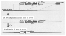
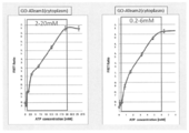
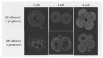
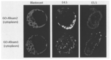
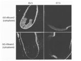
- FIG. 1 Schematic diagram of GO-ATeam expression construct. A graph plotting ATP concentration and FRET ratio. Fluorescence micrograph showing the ATP distribution of each GO-ATeam mouse embryo (1 cell, 2 cell and 4 cell stage). Fluorescence micrograph showing the ATP distribution of each GO-ATeam mouse embryo (8 cell stage, 16 cell stage and morula). Fluorescence micrograph showing the ATP distribution of each GO-ATeam mouse embryo (blastocyst, E4.5 and E5.5). Fluorescence micrograph showing the ATP distribution of each GO-ATeam mouse embryo (E6.5 and E7.5). Fluorescence micrograph showing the distribution of ATP in each GO-ATeam mouse (newborn) organ. Fluorescence micrograph showing ATP distribution in each GO-ATeam mouse (12 months) organ.
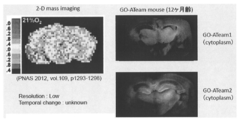
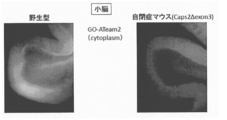
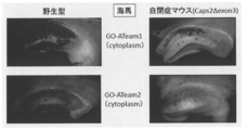
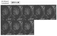
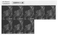
- Fluorescence micrograph (right) showing ATP distribution in the brain of each GO-ATeam mouse (12 months). The left shows 2D mass imaging as a reference. Fluorescence micrograph showing the ATP distribution in the cerebellum of mice obtained by crossing GO-ATeam 2 mice with autism model mice (Caps2 ⁇ exon3) and wild-type mice. Fluorescence micrograph showing the ATP distribution of the hippocampus of mice and wild type mice obtained by crossing GO-ATeam 1 or 2 mice with autism model mice (Caps2 ⁇ exon3). Fluorescence micrograph showing the ATP distribution of the time course of the heart under normal conditions in GO-ATeam 2 mice. Fluorescence micrograph showing the ATP distribution of the heart over time in hypoxia in GO-ATeam 2 mice.
- Fluorescence micrograph showing the ATP distribution of the heart over time in the chronic myocardial infarction state of GO-ATeam 2 mice.
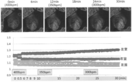
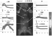
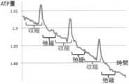
- the graph which shows the time-dependent change of the amount of ATP in each ventricle and the atrium when the heart rate is lowered by administering a drug ( ⁇ blocker) to GO-ATeam 2 visualized mouse.
- the vertical axis of the graph represents the relative value of the ATP amount.
- the vertical axis of the graph indicates the relative value of the ATP amount
- the time on the horizontal axis is in units of 10 seconds, and 100 is 1000 seconds.
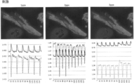
- the center is a fluorescence micrograph showing the ATP distribution in the viscera of a GO-ATeam 2 visualization mouse in control (0 sec).
- the graph which shows the time-dependent change of the amount of ATP in the muscle contraction-relaxation cycle that occurs when electrical stimulation is applied to cardiomyocytes isolated from adult GO-ATeam 2 visualized mice.
- the vertical axis of the graph represents the relative value of the ATP amount.
- the horizontal axis indicates time.
- the vertical axis of the graph indicates the relative value of the ATP amount with respect to the solid line, and the cell area with respect to the dotted line.
- the horizontal axis indicates time. The time is in units of 30 milliseconds and 13 is 390 milliseconds. 1 pps indicates 1 pulse / second electrical stimulation.
- Above is a fluorescence micrograph showing ATP distribution of isolated cardiomyocytes.
- the graph which shows the result of having investigated the ATP dynamics in the anterior tibial muscle when electrically stimulating the sciatic nerve of GO-ATeam 2 visualization mouse and causing muscle contraction.
- the vertical axis of the graph indicates the relative value of the ATP amount, and the horizontal axis indicates time (seconds). Electrical stimulation was applied for 30 seconds between the two arrows.
- the non-human mammal of the present invention has two types of fluorescent proteins that can serve as donors and acceptors in FRET on the amino terminal side and the carboxy terminal side of the ⁇ subunit, which is an ATP-sensitive peptide of ATP synthase F0F1 ATPase derived from Bacillus subtilis. It is a non-human mammal that expresses a fusion protein (hereinafter also referred to as a fluorescent fusion protein) that is bound to each other.
- the non-human mammal of the present invention can be any non-human mammal such as a mouse, rat, rabbit, goat, pig, dog, cat, guinea pig, hamster, sheep, cow, marmoset, and the like.
- ATP synthase is classified into F type and V type, and F type is further classified into F 1 site and F 0 site.
- the F 1 site is composed of subunits such as ⁇ , ⁇ , ⁇ , ⁇ , and ⁇ . Since the ⁇ subunit of ATP synthase has a high affinity for ATP, it has no affinity for nucleotides other than ATP, such as ADP, dATP, GTP, etc., and when it binds to ATP, it causes a structural change. It is effective for.
- the ⁇ subunit used in the present invention may be derived from any biological species, but considering the ease of handling, it is derived from microorganisms such as Bacillus sp. PS3 (GenBank accession No. AB044942 (sequence No. 1)) and the ⁇ subunit shown in Bacillus subtilis origin (GenBank accession No. Z28592 (SEQ ID NO: 2)) can be preferably used.
- ⁇ subunits obtained from bacterial species other than these bacterial species can also be used. Affinities to ATP are different if the biological species derived are different (J. Biological Chemistry 2003; 278, 36013-36016., FEBS Letters 2005; 579, 6875-6878). Therefore, by using different ⁇ subunits, from those that respond to low concentrations of ATP to those that respond to high concentrations of ATP, such as ATP inside cells, ATP measurement reagents that are sensitive to various concentration regions Can be created.
- the ⁇ subunit includes two domains, an N-terminal domain and a C-terminal domain.
- the N-terminal domain consists of about 85 amino acids from the N-terminal side of the ⁇ subunit and consists of about 10 ⁇ -strands.
- the C-terminal domain is composed of amino acids up to about 45 from the C-terminal side of the ⁇ subunit, and consists of two ⁇ helices.
- the C-terminal domain shows a folded crystal structure when the ⁇ subunit alone is used, and an extended structure when the ⁇ subunit forms a complex with a ⁇ subunit that is another subunit of ATP synthase. Indicates.
- the C-terminal domain of the ⁇ subunit undergoes a structural change depending on the presence or absence of ATP. It is shown that a folded structure is obtained. From these, it is considered that the ⁇ subunit has a C-terminal domain extended or easily moved in the absence of ATP, and is folded and compact when ATP is bound.
- the ⁇ subunit need not be full length, and may be a partial fragment.
- the partial fragment is not particularly limited as long as it is a part of an amino acid sequence constituting a site capable of causing a structural change at least specifically in ATP in the ⁇ protein.
- any structure may be used as long as it includes two domains, the N-terminal domain and the C-terminal domain described above, which can cause a structural change due to the presence of ATP.
- the ⁇ subunit may be a natural subunit as long as it contains an amino acid sequence that constitutes a site capable of causing a structural change specifically in the ATP.
- amino acid sequence of the natural ⁇ subunit 1
- several, for example, 1 to 5 amino acids may be substituted, deleted, inserted or added.
- the amino acid sequence may be partially substituted.
- a hydrophobic amino acid residue (Val20 / Val42 / Phe67 / Leu78 in SEQ ID NO: 1 and Val42 / Phe67 / in SEQ ID NO: 2 is required for interaction with the ⁇ subunit, which is another subunit. Leu78) can be replaced with hydrophilic amino acid residues.
- SEQ ID NO: 3 a mutation of R122K / R126K may be introduced in the wild type.
- the fluorescent fusion protein in the present invention is a fusion protein of two types of fluorescent proteins and the ⁇ subunit.
- the two types of fluorescent proteins to be combined are not particularly limited as long as they are fluorescent proteins composed of combinations that can be donors and acceptors in FRET.
- FRET means that the light energy absorbed by the donor moves to the acceptor when two kinds of fluorescent materials called donor and acceptor approach within a certain distance.
- FRET detection is usually performed by irradiating the excitation wavelength of the donor and measuring the fluorescence intensity of the donor or acceptor.
- FRET detection is usually performed by irradiating the excitation wavelength of the donor and measuring the fluorescence intensity of the donor or acceptor.
- the fluorescent protein a commonly used protein can be used.
- green fluorescent protein (GFP) represented by green fluorescent protein derived from Aequorea jellyfish, yellow fluorescent protein (YFP) and cyan, in which various mutations are introduced into GFP and the color of fluorescence is changed Examples thereof include fluorescent protein (CFP), blue fluorescent protein (BFP), red fluorescent protein (RFP) which is a red fluorescent protein derived from koji, and wedge orange orange protein (KO).
- fluorescent proteins described in Table 1 of Nature Methods vol. 9, No. 10, 1005-1012 (2012) can also be used.
- a fluorescent protein In order for a fluorescent protein to exhibit the function of FRET, it can be used as a combination of donor and acceptor, for example, CFP and YFP, BFP and GFP, or GFP and RFP. Any of them may be the amino terminal side or the carboxy terminal side of the ⁇ subunit.
- Commercially available fluorescent proteins can be used. For example, CFP from Invitrogen as CFP, YFP from Invitrogen as YFP, Phi-Yellow from Evrogen as EFP, Tag-GFP from Evrogen as EGFP from Clontech as GFP, Clontech's DsRed2-monomer and Evrogen's HcRed-Tandem can be used.
- the non-human mammal of the present invention can be obtained by integrating the DNA encoding the fluorescent fusion protein on the chromosome of the non-human mammal and expressing the fluorescent fusion protein.
- the DNA encoding the fluorescent fusion protein is preferably expressed under the control of a promoter that can function in cells of a non-human mammal.
- the promoter include ⁇ -actin promoter, CMV promoter, CAG (CAGGS) promoter and the like, but CAG (CAGGS) promoter is particularly preferable in terms of expression efficiency.
- the promoter may be a time-specific promoter or a tissue-specific promoter.
- the DNA construct encoding the fluorescent fusion protein may contain a selection marker.
- Selectable marker refers to a genetic element that provides a selectable phenotype to a cell into which the selectable marker has been introduced, and generally the resistance of the gene product to an agent that inhibits or kills the cell. It is a gene to be given. Specific examples include Neo gene, Hyg gene, hisD gene, Gpt gene, and Ble gene. Drugs useful for selecting the presence of a selectable marker include, for example, G418 for the Neo gene, hygromycin for the Hyg gene, histidinol for the hisD gene, and 6- for the Gpt gene. For thioxanthine and the Ble gene, bleomycin is mentioned.
- the DNA construct encoding the fluorescent fusion protein preferably further includes a sequence homologous to the target site on the chromosome when the endogenous gene on the chromosome is replaced by homologous recombination.
- the target site on the chromosome is preferably the Rosa26 locus.
- the mouse ROSA26 locus was discovered by Friedrich and Soriano in 1991 in a gene trap experiment using retrovirally infected embryonic stem (ES) cells (Friedrich, G. and P. Soriano, Genes & Development, 1991, 5, 1513-1523).
- ES embryonic stem
- Targeting the ROSA26 locus with mouse ES cells has been widely used to construct transgenic mouse models (Kisseberth et al., Developmental Biology, 1999, 214, 128-138; MaoX. Et al., Proc. Natl. Acad. Sci. USA, 1999, 96, 5037-5042; Soriano, 1999, above; Awatramani et al., Nature Genetics, 2001, 29, 257-259; Mao X.
- the DNA construct encoding the fluorescent fusion protein may further contain a recognition sequence typified by a lox sequence that is recognized by a recombinant protein such as CreCrecombinase or Flp recombinase.
- a recombinant protein such as CreCrecombinase or Flp recombinase.
- CreCrecombinase a recombinant protein
- Flp recombinase recombinase
- FRT sequences recognized by FLP can be used.
- the transgenic non-human mammal of the present invention can be obtained, for example, by a method including the following steps (a) to (g).
- a step of selecting cells (c) a step of introducing the selected ES cells into a fertilized egg (d) a step of transplanting the fertilized egg into the oviduct or uterus of a pseudopregnant female non-human mammal (e) the non-human mammal Selecting a germline chimera from the offspring bred and born (f) crossing the germline chimera and selecting an individual having a chromosome into which a DNA encoding a fluorescent fusion protein is inserted from the born puppies (G) The individuals obtained in (f) are m
- ES cells are obtained by culturing embryos prior to transplantation in vitro under appropriate conditions (Nature 292: 154-156 (1981); Nature 309: 255-258 (1984); Proc. Natl. Acad. Sci. USA 83: 9065-9069 (1986); and Nature 322: 445-448 (1986)).
- Targeting vectors are obtained from ES cells by DNA transfection using various methods known in the art, including electroporation, calcium phosphate coprecipitation, protoplast or spheroplast fusion, lipofection and DEAE dextran-mediated transfection. Can be efficiently introduced into.
- viral infections such as adenoviruses, retroviruses, lentiviruses can also be used to introduce genes into non-human animals.
- Developing non-human embryos are cultured in vitro to the blastocyst stage. During this time, the blastomere is infected with the virus (Proc. Natl. Acad. Sci. USA 73: 1260-1264 (1976)).
- Transfection is accomplished easily and efficiently by culturing blastomeres on a monolayer of virus producing cells (EMBO J. 6: 383-388 (1987)). Alternatively, infection may be performed at a later stage. Virus or virus-producing cells may be injected into the blastocoel (Nature 298: 623-628 (1982)).
- transgene into the germ line by in utero virus infection of the embryo in the mid-gestation. It is also possible to microinject mitomycin C-treated cells producing virus particles or virus into the perivitelline space of fertilized eggs or early embryos.
- the ES cells into which the target gene has been introduced in this way are then introduced into the blastocoel of the embryo at the blastocyst stage, and then incorporated into the embryo and incorporated into the germ line of the chimeric animal.
- the transgene provides a means for selection
- the transfected ES cells are subjected to various selection protocols to introduce the transgene.
- ES cells incorporating can be concentrated.
- PCR can be used to screen for ES cells incorporating the transgene. Thereby, introduction efficiency can be raised.
- the implanted embryos are transplanted into pseudopregnant female fallopian tubes / uterus and finally transgenic animals are obtained.
- the founder can be mated, inbred, outbred or crossed to produce a particular animal population. For example, when combined with a disease model non-human mammal, it is useful for disease evaluation and therapeutic drug screening.
- Disease model non-human mammals include autism model animals (e.g. Caps2 ⁇ exon3, Sadakata, T. et al. PNAS 2012: www.pnas.org/content/early/2012/11/28/1210055109.full.pdf ) And heart disease model animals.
- Examples of the heart disease model animal include model animals that have caused hypoxia or myocardial infarction.
- examples include, but are not limited to, diabetes model animals such as db / db mice, cancer model animals such as p53 / k-ras mutant mice, and infectious disease model animals.
- the non-human mammal that expresses the fluorescent fusion protein of the present invention and the tissues and cells obtained from the animal can be used for measuring the concentration and distribution of ATP in the living body, as well as changes over time.
- ATP present in the living body changes the structure of the ⁇ subunit of ATP synthase, which causes a FRET reaction to emit fluorescence. Therefore, the presence of ATP can be monitored using a fluorescence microscope or the like. .
- the non-human mammal of the present invention and the tissues and cells obtained from the animal can be used for screening for cancer drug candidates.
- screening for a drug candidate / concentration that kills cancer cells specifically is performed by accurately changing and measuring the ATP concentration. Is possible. It can also be used for drug safety testing.
- the effects of drugs on the liver such as toxicity and burden on the liver, can be accurately examined by observing changes in the amount of ATP using liver cells.
- the non-human mammal of the present invention and tissues and cells obtained from the animal can be used for analysis and evaluation of various disease states, drug evaluation and screening. For example, it can be used for screening for therapeutic agents for diseases that change the energy metabolic state of each cell and for evaluating drug efficacy. In addition, by using heart tissues and cells, it can be used for screening for therapeutic agents for heart diseases and for evaluating drug efficacy.
- the ATP concentration becomes excessive by administering a drug candidate substance to the non-human mammal of the present invention that has been given so as to exhibit autism and evaluating whether the substance has an action of lowering the ATP concentration.
- screening of candidate substances for therapeutic agents for diseases that cause metabolic abnormalities can be performed. Conversely, by evaluating whether a substance has an effect of increasing ATP concentration, it is possible to screen for candidate substances for therapeutic agents for diseases in which ATP concentration decreases to cause metabolic abnormalities.
- Compounds as therapeutic agent candidate substances used for evaluation or screening include, for example, synthetic or natural low or high molecular compounds, metal-containing compounds, carbohydrates, peptides, proteins, peptidomimetics, glycoproteins, lipoproteins, nucleic acids An antibody or the like can be used. Libraries that contain a large number of natural and synthetic compounds can also be used.
- abnormalities in movement / function can be regarded as abnormal changes in ATP content and distribution over time.
- mammals or tissues and cells obtained therefrom new diagnostic methods and therapeutic methods can be developed. It is also possible to reproduce the movement of an organ or the like from the information on the distribution and change of the ATP amount (for example, reproduction of walking from the ATP distribution and change of leg muscles during walking) with a machine or the like.
- the ability to recover from exercise can be examined using muscle cells and tissues, it can be used not only for application to muscle diseases, but also for development of exercise / sport equipment and food / muscle augmentation agents. .
- ATP can be quantified using cells obtained from the non-human mammal of the present invention.
- the following can be considered as measurement conditions.
- any substance containing ATP may be used, and examples thereof include a solution containing ATP, a biological sample such as a cell, and one obtained by extracting ATP from a biological sample.
- a lysis buffer For example, the thing of pH which can emit the fluorescence of a fluorescent substance on optimal conditions can be selected.
- YFP is used as a fluorescent substance, it is preferable to use a buffer solution having a pH of 7 or higher because YFP fluorescence decreases on the acidic side.
- the reaction temperature may be any temperature at which the fluorescent protein probe can cause a structural change, and can be measured, for example, at 37 ⁇ 1 ° C. or 25 ⁇ 1 ° C.
- the reaction time can also be appropriately determined, but it is preferably 1 minute or longer, and the reaction can be performed for a time that does not deactivate the protein.
- Additives can be appropriately added to the reaction. For example, 0.05% surfactant (TritonX100) or 1 mg / mL bovine serum albumin (BSA) can be added for the purpose of stabilization.
- a chelating agent such as EDTA or EGTA may be added.
- the fluorescence spectrum for the test substance after the reaction is completed, using a fluorometer, irradiating the excitation light of the donor, measuring the fluorescence intensity at a wavelength where the fluorescence intensity of each fluorescent substance alone peaks, It can be measured by calculating the ratio.
- an ATP solution having a known concentration is prepared, and a fluorescence intensity ratio is similarly obtained to prepare a calibration curve. By using this calibration curve, the ATP concentration of the specimen can be obtained.
- a targeting vector was prepared so that each GO-ATeam cDNA (GO-ATeam1 and GO-ATeam2) was inserted into the mouse ROSA26 region for ATP-visualized mice (FIG. 1).
- These GO-ATeams were constructed in such a way that a STOP sequence sandwiched between LoxPs was inserted so that the GO-ATeam could be controlled from the CAGGS promoter and expressed after the site and time of Cre expression.
- IiMOG-GA_pBS-APS vector After digesting pBS-APS vector with AscI, PmeI, ISwaI sequence inserted from both sides of MCS side of pBlueScript with PmeI, T4 DNA polymerase treatment was performed, and STOP sequence of ROSA26-CAG-STOP vector 149bp, After LoxP, the stuffer sequence of the pEF-BOS vector (Nucleic Acids Res. 1990 Sep 11; 18 (17): 5322.), G-CSF pA, was bound, and ROSA26 3 'arm 49bp (ch6 114904549-114904597) The inserted vector was used (hereinafter referred to as IiMOG-GA_pBS-APS vector).
- GO-ATeam1 and GO-ATeam2 were each excised with NheI-HindIII (provided by Dr. Hiroomi Imamura) in pcDNA vector, and HindIII site was treated with T4 DNA polymerase.
- the IiMOG-GA_pBS-APS vector was digested with NheI-XbaI, and the XbaI site was treated with T4 DNA polymerase. These were combined to obtain GO-ATeam1_IiMOG-GA and GO-ATeam2_IiMOG-GA.
- This GO-ATeam1_IiMOG-GA and GO-ATeam2_IiMOG-GA were digested with SwaI, and then a cDNA-containing fragment was obtained.
- the target vector was obtained by ligation (Invitrogen).
- GO-Ateam 1 and GO-Ateam 2 are as follows.
- Ateam A denosine 5'- T riphosphate indicator based on E psilon subunit for A nalytical M easurement
- G FP- and O FP-based ATeam GO-Ateam 1: Construct with GFP (cp173-mEGFP) inserted at the N-terminus and OFP (mKO ⁇ ) at the C-terminus of the ⁇ subunit (SEQ ID NO: 2) of Bacillus subtilis F0F1-ATP synthase: with signal It is expressed in the cytoplasm because there is no GO-Ateam 2: GO-Ateam 1 ⁇ subunit R122K / R126K mutant (SEQ ID NO: 3): It is expressed in the cytoplasm because it has no signal.
- Chimeric mice were obtained by fusing mulberry embryos obtained from ICR mice and ES cells obtained from the above by the aggregation method, and then transplanting them into the uterus of ICR mouse subjected to pseudopregnancy treatment. As described above, mice with GO-ATeam1 and GO-ATeam2 expressed in the cytoplasm were created.
- MEF cells were obtained from embryonic day 13 embryos of GO-ATeam1 and GO-ATeam2 visualized mice obtained above and cultured. Using the fourth passage, seeding was carried out in a glass coat dish. After washing MEF cells with Permilization buffer (140 mM KCl, 6 mM NaCl, 0.1 mM EGTA, 10 mM HEPES pH 7.4), ⁇ -Hemolycin (Sigma cat # H9395) was diluted with Permilization buffer to a final concentration of 50 ⁇ g / ml. At 37 ° C. for 30 minutes.
- Permilization buffer 140 mM KCl, 6 mM NaCl, 0.1 mM EGTA, 10 mM HEPES pH 7.4
- ⁇ -Hemolycin Sigma cat # H9395
- GO-ATeam1 and GO-ATeam2 at each ATP concentration are emitted by acclimating to the calibration buffer (140 mM KCl, 6 mM NaCl, 0.5 mM MgCl 2 , 10 mM HEPES pH 7.4) diluted with MgATP between 0 mM and 30 mM.
- Fluorescence images were acquired with a confocal microscope.
- the confocal microscope used DM405 / 488 with 488 nm laser as an excitation DM and dispersed with dichroic mirror LM01-552, and then obtained GFP fluorescence and KusabiraOrange fluorescence with BA505-540 and BA575-675 filters.
- the FRET ratio value was obtained by calculating the FRET Ratio value with Metamorph.
- a graph was created by plotting each ATP concentration and the FRET ratio value taken at that time. The results are shown in FIG.
- Fertilized eggs were obtained by mating GO-ATeam1 and GO-ATeam2 visualized mice (female) with wild-type mice. Fertilized eggs were collected in DMEM (Sigma) / 10% FCS and cultured at 37 ° C. and 5% CO 2 in M2 medium (sigma) until the third day after fertilization. For observation, an image was obtained in the same manner as above under conditions of 37 ° C. and 5% CO 2 in M2 medium (no phenol red) with a confocal microscope. From day 4 after fertilization, DMEM (sigma) / rat serum 50% was used instead of M2 medium. The FRET ratio value was also obtained in the same manner as above.
- each organ of GO-ATeam 2 visualization mouse is placed in a mixture of 95% nitrogen and 5% carbon dioxide to create hypoxia and hypoxia treatment, and the time course of ATP in each organ is examined. Compared with normal state. The results are shown in FIG. From this result, it was found that the quality of cell activity in all organs can be measured as the change in the amount of ATP.
- cardiomyocytes were isolated from the hearts of GO-ATeam 2 visualized mice, and the changes in ATP over time when contracting movements were induced by applying electrical pulses using the isolated cardiomyocytes were examined.
- the results are shown in FIG. From this result, it became clear that ATP dynamics in mature cardiomyocytes can be measured.
- the amount of ATP increases rapidly, which means that ADP is released from myosin into the cytoplasm, and phosphate is passed from creatine phosphate to ADP.
- the amount of ATP decreases, indicating that ATP has begun to bind to myosin.
- FIG. 20 shows the results of examining the ATP dynamics in the anterior tibial muscle when the sciatic nerve of GO-ATeam 2 visualized mouse was electrically stimulated to cause muscle contraction. After anesthetizing the mouse, the sciatic nerve was dissected out, electrodes were attached, and the anterior tibial muscle was observed by removing the fur and observed with a fluorescence stereomicroscope system. Electrical stimulation was for 30 seconds, after which ATP recovery was observed. From this result, it was possible to investigate in detail the recovery degree and recovery time of ATP amount with respect to exercise load, and it was found that the ATP visualization mouse can be used for development of exercise / sport equipment, food / muscle augmentation agent, etc. .
- the mouse of the present invention is useful in fields such as medicine, pharmaceutical development, exercise science, and basic research.
Landscapes
- Life Sciences & Earth Sciences (AREA)
- Health & Medical Sciences (AREA)
- Chemical & Material Sciences (AREA)
- Engineering & Computer Science (AREA)
- Zoology (AREA)
- Genetics & Genomics (AREA)
- Organic Chemistry (AREA)
- General Health & Medical Sciences (AREA)
- Bioinformatics & Cheminformatics (AREA)
- Wood Science & Technology (AREA)
- General Engineering & Computer Science (AREA)
- Biomedical Technology (AREA)
- Biotechnology (AREA)
- Veterinary Medicine (AREA)
- Biochemistry (AREA)
- Animal Behavior & Ethology (AREA)
- Environmental Sciences (AREA)
- Molecular Biology (AREA)
- Microbiology (AREA)
- Biodiversity & Conservation Biology (AREA)
- Epidemiology (AREA)
- Animal Husbandry (AREA)
- Chemical Kinetics & Catalysis (AREA)
- Public Health (AREA)
- Medicinal Chemistry (AREA)
- Diabetes (AREA)
- Endocrinology (AREA)
- Gastroenterology & Hepatology (AREA)
- Pathology (AREA)
- Rheumatology (AREA)
- Toxicology (AREA)
- Urology & Nephrology (AREA)
- Plant Pathology (AREA)
- Biophysics (AREA)
- Physics & Mathematics (AREA)
- Measuring Or Testing Involving Enzymes Or Micro-Organisms (AREA)
- Investigating, Analyzing Materials By Fluorescence Or Luminescence (AREA)
- Investigating Or Analysing Biological Materials (AREA)
- Micro-Organisms Or Cultivation Processes Thereof (AREA)
Abstract
Description
(1)ATP合成酵素のεサブユニットのアミノ末端側およびカルボキシ末端側に、蛍光共鳴エネルギー移動におけるドナー及びアクセプターとなりうる2種類の蛍光タンパク質をそれぞれ結合させてなる融合タンパク質を発現する非ヒト哺乳動物。
(2)ATP合成酵素のεタンパク質サブユニットが配列番号1または2に記載のアミノ酸配列を含む、(1)に記載の非ヒト哺乳動物。
(3)ドナー及びアクセプターとなりうる蛍光タンパク質の組み合わせが、シアンフルオレセントプロテイン(CFP)、イエローフルオレセントプロテイン(YFP)、ブルーフルオレセントプロテイン(BFP)、グリーンフルオレセントプロテイン(GFP)、レッドフルオレセントプロテイン(RFP)及びクサビラオレンジプロテイン(KO)などFRETを生じることのできる組み合わせである、(1)または(2)の非ヒト哺乳動物。
(4)前記融合タンパク質をコードするDNAが染色体上のROSA26遺伝子座に挿入された、(1)~(3)のいずれかに記載の非ヒト哺乳動物。
(5)前記融合タンパク質をコードするDNAはlox系配列を有し、Cre recombinaseがlox系配列で挟まれた領域を除去することにより、前記融合タンパク質が発現する、(1)~(4)のいずれかに記載の非ヒト哺乳動物。
(6)前記融合タンパク質をコードするDNAはCAGプロモーターの制御下におかれた、(1)~(5)のいずれかに記載の非ヒト哺乳動物。
(7)(1)~(6)のいずれかに記載の非ヒト哺乳動物と、疾患モデル動物との掛け合わせによって得られる、非ヒト哺乳動物。
(8)疾患モデル動物が、自閉症モデル動物または心疾患モデル動物である、(7)に記載の非ヒト哺乳動物。
(9)マウスである、(1)~(8)のいずれかに記載の非ヒト哺乳動物。
(10)(1)~(9)のいずれかに記載の非ヒト哺乳動物又は該非ヒト哺乳動物から得られる細胞、組織もしくは器官に被験物質を投与し、蛍光強度に基づいてATP濃度を測定することを特徴とする、ATPの測定方法。
(11)(10)に記載の方法によってATPを測定し、該測定結果に基づいて被験物質の薬効を評価することを特徴とする、薬剤のスクリーニングまたは薬効評価方法。
本発明の非ヒト哺乳動物は、例えば、マウス、ラット、ウサギ、ヤギ、ブタ、イヌ、ネコ、モルモット、ハムスター、ヒツジ、ウシ、マーモセットなどの任意の非ヒト哺乳動物であり得る。
εサブユニットは、上記ATP特異的に構造変化を生じうる部位を構成するアミノ酸配列を含むのであれば、天然型サブユニットであってもよいし、該天然型εサブユニットのアミノ酸配列において、1または数個、例えば、1~5個のアミノ酸が、置換、欠失、挿入又は付加されていてもよい。例えば、εサブユニットとATPとの反応によるεタンパク質の構造変化を阻害しうる物質がεサブユニットに結合するのを防ぐために、アミノ酸の配列を一部置換してもよい。例えば、εサブユニットにおいて、他のサブユニットであるγサブユニットとの相互作用に必要な疎水的なアミノ酸残基(配列番号1ではVal20/Val42/Phe67/Leu78、配列番号2ではVal42/Phe67/Leu78)を、親水的なアミノ酸残基に置換することができる。また、配列番号3で示すように、野生型においてR122K/R126Kの変異を導入してもよい。
結合される2種類の蛍光タンパク質は、FRETにおけるドナー及びアクセプターとなりうる組み合わせからなる蛍光タンパク質であればよく、特に限定されない。ここにFRETとは、ドナーとアクセプターと呼ばれる2種類の蛍光物質が一定の距離以内に近づいた時に、ドナーが吸収した光エネルギーがアクセプターに移動することをいう。FRETの検出は、通常ドナーの励起波長を照射し、ドナー又はアクセプターの蛍光強度を測定することにより行う。ドナーの蛍光強度を測定する場合、2種類の蛍光タンパク質が近い位置にあるとアクセプターに光が吸収されるためにドナーの蛍光強度は低くなり、距離が離れると蛍光強度が高くなる。アクセプターの蛍光強度を測定する場合はその逆で、距離が近いと蛍光強度が強くなり、離れると強度が低くなる。
蛍光タンパク質は、一般的に使用されているものを用いることができる。具体的には、オワンクラゲ由来の緑色蛍光タンパク質に代表されるグリーンフルオレセントプロテイン(GFP)、GFPに種々の変異を導入し、蛍光の色を変化させたイエローフルオレセントプロテイン(YFP)やシアンフルオレセントプロテイン(CFP)、ブルーフルオレセントプロテイン(BFP)、あるいは珊瑚由来の赤色蛍光タンパク質であるレッドフルオレセントプロテイン(RFP)、クサビラオレンジプロテイン(KO)等が挙げられる。他にも、Nature Methods vol. 9, No. 10, 1005-1012 (2012)のTable 1に記載された蛍光タンパク質も使用できる。
蛍光タンパク質がFRETの機能を発揮するために、ドナー及びアクセプターの組み合わせとして、例えば、CFP及びYFP、BFP及びGFP、あるいはGFP及びRFPにより使用することができる。いずれがεサブユニットのアミノ末端側、カルボキシ末端側でもよい。これらの蛍光タンパク質は、市販のものを使用することができる。例えば、CFPとしてはInvitrogen社のCFPが、YFPとしてはInvitrogen社のYFPの他にEvrogen社のPhi-Yellowが、GFPとしてはClontech社のEGFPの他にEvrogen社のTag-GFPが、RFPとしてはClontech社のDsRed2-monomer、Evrogen社のHcRed-Tandemが使用可能である。
前記蛍光融合タンパク質をコードするDNAは非ヒト哺乳動物の細胞で機能し得るプロモーターの制御下で発現されることが好ましい。プロモーターとしては、β-アクチンプロモーター、CMVプロモーター、CAG(CAGGS)プロモーター等が例示されるが、発現効率の点ではCAG(CAGGS)プロモーターが特に好ましい。なお、プロモーターは時期特異的プロモーターや、組織特異的プロモーターであってもよい。
(a)蛍光融合タンパク質をコードするDNA構築物を含むターゲッティングベクターを非ヒト哺乳動物の胚性幹(ES)細胞に導入する工程
(b)蛍光融合タンパク質をコードするDNAが挿入された染色体を有するES細胞を選択する工程
(c)選択したES細胞を受精卵に導入する工程
(d)該受精卵を偽妊娠雌性非ヒト哺乳動物の卵管あるいは子宮に移植する工程
(e)該非ヒト哺乳動物を飼育し、産まれた仔から、生殖系列キメラを選択する工程
(f)該生殖系列キメラを交配させ、産まれた仔から、蛍光融合タンパク質をコードするDNAが挿入された染色体を有する個体を選択する工程
(g)(f)で得られた個体同士を交配させ、産まれた仔から、両方の相同染色体において、蛍光融合タンパク質をコードするDNAが挿入されたホモ接合体を選択する工程
ES細胞は、適切な条件下において、インビトロで移植前の胚を培養することによって得られる(Nature 292:154-156(1981);Nature 309:255-258(1984);Proc.Natl.Acad.Sci.USA 83:9065-9069(1986);およびNature 322:445-448(1986))。
また、db/dbマウスなどの糖尿病モデル動物や、p53/k-ras変異マウスなどの癌モデル動物や感染症モデル動物が例示されるが、これらには限定されない。
また、筋肉の細胞や組織を用いて運動からの回復能を調べることができるので、筋疾患への応用だけでなく、運動・スポーツ器機開発や食品・筋肉増強剤開発などにも利用可能である。
測定条件として、以下が考えられる。被験物質としては、ATPを含むものであれば良く、例えばATPを含む溶液、細胞などの生体試料、生体試料からATPを抽出したものなどが挙げられる。溶解緩衝液としては特に限定されないが、例えば蛍光物質の蛍光を最適な条件で発しうるpHのものを選択することができる。具体的には、YFPを蛍光物質として用いる場合は、YFPの蛍光は酸性側では減少するため、pH7以上の緩衝液を用いることが好ましい。反応温度は、蛍光タンパク質プローブが構造変化を起こしうる温度であればよく、例えば37±1℃や、25±1℃において測定することができる。反応時間についても適宜決定することができるが、好ましくは1分以上であり、タンパク質が失活しない程度の時間反応させることができる。反応には、適宜添加物を加えることができる。例えば0.05%の界面活性剤(TritonX100)もしくは1mg/mLウシ血清アルブミン(BSA)などを安定化の目的で加えることができる。被験試料に混在するマグネシウムの影響を避けるためには、例えばEDTAやEGTAなどのキレート剤を添加してもよい。
マウスROSA26 5' arm 1080bp (Ch6 113076036-113077116)にSplice acceptor, bgh pAの後にCAGGS, LoxP, pA, Neo (逆方向), MC1 (逆方向), STOP配列( SV40pA含む), LoxP, bgh pAを結合し、その後にROSA26 3' arm 4267bp (ch6 114900586-114904926)を結合し、その後にPGK promoter, DT-A, bgh pAをつなげた配列を持つpBlueScript(星野克明博士より供与)を利用した(以下ROSA26-CAG-STOPベクター)。
IiMOG-GA_pBS-APSベクターをNheI-XbaIで消化し、XbaIサイトはT4 DNA polymerase処理を行った。
これらを結合し、GO-ATeam1_ IiMOG-GAおよびGO-ATeam2_ IiMOG-GAを得た。
このGO-ATeam1_ IiMOG-GAおよびGO-ATeam2_ IiMOG-GAをSwaI消化した後、cDNAを含む断片を得て、AscI-NruI消化した後に、T4 DNA polymerase処理を行ったROSA26-CAG-STOPベクターとGeneArt(Invitrogen社)にて結合させてターゲティングベクターを得た。
Ateam (Adenosine 5'-Triphosphate indicator based on Epsilon subunit for Analytical Measurement)
GO-Ateam (GFP- and OFP-based ATeam)
GO-Ateam 1: Bacillus subtilis F0F1-ATP synthaseのεサブユニット(配列番号2)のN末側にGFP (cp173-mEGFP)を、C末側にOFP (mKOκ)を挿入したコンストラクト:シグナルを有さないので細胞質で発現する。
GO-Ateam 2: GO-Ateam 1のεsubunitにおいてR122K/R126Kとしたmutant(配列番号3):
シグナルを有さないので細胞質で発現する。
参考文献
Imamura et al., Pro N.A.S. 2009, 106, 15651-15656
Nakano et al., ACS Chem. Biol. 2011, 6, 709-715
Waldeck-Weiermair et al., PLoS ONE 7(9): e45917 2012
WO2008/117792
上記ターゲティングベクターをAsiSIで直線化した後、定法によりES細胞(C57/BL6×129hybrid)へ導入した後、遺伝子組換えが正確に起こったES細胞だけをネオマイシン耐性遺伝子, PCR法、Southern Blotting法により選択して、Creが発現した細胞のみでGO-ATeamが発現するようにしたES細胞を得た。常時GO-ATeamが発現するES細胞は上記ES細胞へCMV-Creベクターを導入することにより得た。
以上により、GO-ATeam1, GO-ATeam2が細胞質で発現するマウスを作出した。
上記で得られたGO-ATeam1及びGO-ATeam2可視化マウスの胎生13日胚からMEF細胞を取得し、培養した。
継代4代目を用いてglass coat dishへ播種した。MEF細胞をPermilization buffer (140mM KCl, 6mM NaCl, 0.1mM EGTA, 10mM HEPES pH7.4)でwashした後、α-Hemolycin(Sigma cat#H9395)をPermilization bufferで希釈して終濃度50μg/mlの中で37℃ 30分処理を行った。その後、MgATPを0mMから30mMの間で希釈したCalibration buffer (140mM KCl, 6mMNaCl, 0.5mM MgCl2, 10mM HEPES pH7.4)に馴染ませて、各ATP濃度時のGO-ATeam1及びGO-ATeam2が発する蛍光画像を共焦点顕微鏡にて取得した。共焦点顕微鏡は488nmのレーザーをexcitation DMとしてDM405/488を使用し、ダイクロイックミラーLM01-552で分光した後にBA505-540とBA575-675のフィルターにてGFP蛍光とKusabiraOrange蛍光を得た。得られた蛍光画像はMetamorphにてFRET Ratio値を計算することでFRET ratio値を得た。各ATP濃度と、その時に取るFRET ratio値をプロットすることでグラフを作成した。結果を図2に示す。
GO-ATeam1及びGO-ATeam2可視化マウス(メス)と野生型マウスを交配して受精卵を得た。受精卵はDMEM(Sigma)/10%FCS内にて回収し、受精後3日目まではM2 medium (sigma)内で37℃ 5%CO2にて培養した。観察は共焦点顕微鏡にてM2 medium (フェノールレッドなし)内で37℃ 5%CO2の条件下で上記と同じ方法にて画像を取得した。受精後4日目以降はM2 mediumの代わりにDMEM(sigma)/rat serum 50%を使用した。FRET ratio値も上記と同様に得た。
結果を図3~6に示す。
各時期でのATP量を計測できた。またcytosol内でATP濃度分布に違いが見られた。1細胞期において核周囲でのATP量は他の部位と比較して低い。4細胞期においては、割球の接合部付近でのATP量は他の部位と比較して低い。また、5.5日胚と比較して、6.5日胚から胚性外胚葉でのATP量が上昇している事が分かる。また、7.5日胚ではnodeにおけるATP量が亢進している。
マウス胚で計測したATP量とルシフェラーゼ法を用いて計測したATP量に相関性があった。
マウス新生児・成体を気化麻酔下で開胸し、蛍光写真を得た。マウス脳は脱頭後に急性スライスを用いて約200nmのスライスを作成後に蛍光写真を得た。撮影は実体蛍光顕微鏡(ライカ社 M165 FC)を用いて、フィルターGFP3(励起波長ST470/40nm, 吸収波長 ET525/50nm), GFP/OFP(励起波長ET470/40, 吸収波長ET585/40)を利用して蛍光写真を得た。得られた蛍光画像はMetamorphにてFRET Ratio値を計算することでFRET ratio値を得た。結果を図7,8に示す。マウス成体内の臓器でATP量を測定するとルシフェラーゼ法での測定でATP量が高いと報告されている筋肉や心臓でATP値が高い事が分かった。
さらに、マウス成体(12ヶ月)脳の蛍光写真も得た(図9)。マウス脳内のATP量の計測を質量イメージングとGO-Ateam knock-in mouseとで比較したところ、GO-Ateam knock-in mouseを用いた方法は質量イメージング法よりも高解像度に経時的な変化を観察可能であることがわかった。
以上より、各臓器や脳の各部位でのATP量を計測できた。これまでLuciferase法やイメージ質量分析法で測定された値に近い値が得られた。また解像度は数百個の細胞毎で観察・計測されていたものが1細胞・subcelluarレベルでの解析が可能となった。
図13の低酸素処理(人工呼吸器にて呼吸させている時に人工呼吸器を停める事により実施)時には通常と比較して心房でのATP量の増減が消失する。また心室では増減が低下することがわかった。
図14の心筋梗塞(冠状動脈を結紮した後、5日後)を起こした心臓では梗塞部位の心室でのATPの低下が観察される。同時に梗塞部位以外での心室ではATP量が亢進している部位、通常より低下している部位が観察される。
また、図19では、1秒間に1回から3回までの異なる電気刺激(1pps(pulse/second), 2pps, 3pps)をかける事により筋収縮運動を起こした時の単離心筋細胞の面積(点線)とATP量(実線)の変化を示す。1pps, 2ppsでは電気パルスに併せて変化するが、3ppsでは電気パルスについていけずにいる。このように、パルスの強さによって単離心筋細胞の面積とATP量は異なった変化を示し、単離心筋細胞を用いてATP動態を測定することで心エネルギーに対するドラッグスクリーニングなどにも応用可能であることがわかった。
Claims (11)
- ATP合成酵素のεサブユニットのアミノ末端側およびカルボキシ末端側に、蛍光共鳴エネルギー移動におけるドナー及びアクセプターとなりうる2種類の蛍光タンパク質をそれぞれ結合させてなる融合タンパク質を発現する非ヒト哺乳動物。
- ATP合成酵素のεタンパク質サブユニットが配列番号1、2または3に記載のアミノ酸配列を含む、請求項1に記載の非ヒト哺乳動物。
- ドナー及びアクセプターとなりうる蛍光タンパク質の組み合わせが、シアンフルオレセントプロテイン(CFP)、イエローフルオレセントプロテイン(YFP)、ブルーフルオレセントプロテイン(BFP)、グリーンフルオレセントプロテイン(GFP)、レッドフルオレセントプロテイン(RFP)及びクサビラオレンジプロテイン(KO)から選ばれる組み合わせである、請求項1または2に記載の非ヒト哺乳動物。
- 前記融合タンパク質をコードするDNAが染色体上のROSA26遺伝子座に挿入された、請求項1~3のいずれか一項に記載の非ヒト哺乳動物。
- 前記融合タンパク質をコードするDNAはlox系配列を有し、Cre recombinaseがlox系配列で挟まれた領域を除去することにより、前記融合タンパク質が発現する、請求項1~4のいずれか一項に記載の非ヒト哺乳動物。
- 前記融合タンパク質をコードするDNAはCAGプロモーターの制御下におかれた、請求項1~5のいずれか一項に記載の非ヒト哺乳動物。
- 請求項1~6のいずれか一項に記載の非ヒト哺乳動物と、疾患モデル動物との掛け合わせによって得られる、非ヒト哺乳動物。
- 疾患モデル動物が、自閉症モデル動物または心疾患モデル動物である、請求項7に記載の非ヒト哺乳動物。
- マウスである、請求項1~8のいずれか一項に記載の非ヒト哺乳動物。
- 請求項1~9のいずれか一項に記載の非ヒト哺乳動物又は該非ヒト哺乳動物から得られる細胞、組織もしくは器官に被験物質を投与し、蛍光強度に基づいてATP濃度を測定することを特徴とする、ATPの測定方法。
- 請求項10に記載の方法によってATPを測定し、該測定結果に基づいて被験物質の薬効を評価することを特徴とする、薬剤のスクリーニングまたは薬効評価方法。
Priority Applications (2)
| Application Number | Priority Date | Filing Date | Title |
|---|---|---|---|
| JP2015557864A JP6593595B2 (ja) | 2014-01-15 | 2015-01-15 | Atp可視化動物およびその用途 |
| US15/111,730 US11160879B2 (en) | 2014-01-15 | 2015-01-15 | Transgenic animal for visualization of ATP and use thereof |
Applications Claiming Priority (2)
| Application Number | Priority Date | Filing Date | Title |
|---|---|---|---|
| JP2014-005429 | 2014-01-15 | ||
| JP2014005429 | 2014-01-15 |
Publications (1)
| Publication Number | Publication Date |
|---|---|
| WO2015108102A1 true WO2015108102A1 (ja) | 2015-07-23 |
Family
ID=53542988
Family Applications (1)
| Application Number | Title | Priority Date | Filing Date |
|---|---|---|---|
| PCT/JP2015/050932 Ceased WO2015108102A1 (ja) | 2014-01-15 | 2015-01-15 | Atp可視化動物およびその用途 |
Country Status (3)
| Country | Link |
|---|---|
| US (1) | US11160879B2 (ja) |
| JP (1) | JP6593595B2 (ja) |
| WO (1) | WO2015108102A1 (ja) |
Cited By (6)
| Publication number | Priority date | Publication date | Assignee | Title |
|---|---|---|---|---|
| WO2017094885A1 (ja) * | 2015-12-04 | 2017-06-08 | 国立大学法人東京大学 | リガンド蛍光センサータンパク質とその使用 |
| WO2019173604A1 (en) * | 2018-03-09 | 2019-09-12 | Essen Instruments, Inc. D/B/A Essen Bioscience, Inc. | Methods and compositions for live cell analysis of intracellular atp |
| WO2020091301A1 (ko) * | 2018-10-31 | 2020-05-07 | 고려대학교 산학협력단 | 리포칼린-2의 선택적 과발현을 통한 염증형성 형질전환 동물모델 및 그의 제조방법 |
| KR20200049552A (ko) * | 2018-10-31 | 2020-05-08 | 고려대학교 산학협력단 | 리포칼린-2의 선택적 과발현을 통한 염증형성 형질전환 동물모델 및 그의 제조방법 |
| WO2021117874A1 (ja) | 2019-12-13 | 2021-06-17 | 中外製薬株式会社 | 細胞外プリン受容体リガンドを検出するシステムおよび当該システムを導入した非ヒト動物 |
| WO2025154823A1 (ja) * | 2024-01-18 | 2025-07-24 | 国立研究開発法人国立循環器病研究センター | 筋細胞の収縮におけるエネルギー効率を測定する方法 |
Citations (1)
| Publication number | Priority date | Publication date | Assignee | Title |
|---|---|---|---|---|
| WO2008117792A1 (ja) * | 2007-03-27 | 2008-10-02 | The New Industry Research Organization | アデノシン三リン酸測定のための蛍光標識融合タンパク質 |
Family Cites Families (7)
| Publication number | Priority date | Publication date | Assignee | Title |
|---|---|---|---|---|
| WO1999053017A2 (en) * | 1998-04-15 | 1999-10-21 | Fred Hutchinson Cancer Research Center | Methods and vector constructs for making transgenic non-human animals which ubiquitously express a heterologous gene |
| WO2001033957A1 (en) * | 1999-11-05 | 2001-05-17 | Kyowa Hakko Kogyo Co., Ltd. | Transgenic non-human mammals for monitoring change in calcium ion concentration in cells |
| JP2002000121A (ja) * | 2000-06-23 | 2002-01-08 | Rikogaku Shinkokai | テロメレース遺伝子のプロモーターが導入されたトランスジェニックマウス及びその利用 |
| US6586251B2 (en) | 2000-10-31 | 2003-07-01 | Regeneron Pharmaceuticals, Inc. | Methods of modifying eukaryotic cells |
| AU2006230242B2 (en) | 2005-03-28 | 2013-05-30 | Pericor Therapeutics, Inc. | Methods, compositions, and formulations for preventing or reducing adverse effects in a patient |
| JP2007252370A (ja) * | 2006-02-22 | 2007-10-04 | Institute Of Physical & Chemical Research | Il−4初期発現系トランスジェニック非ヒト動物およびその利用 |
| GB201122047D0 (en) | 2011-12-21 | 2012-02-01 | Kymab Ltd | Transgenic animals |
-
2015
- 2015-01-15 US US15/111,730 patent/US11160879B2/en active Active
- 2015-01-15 WO PCT/JP2015/050932 patent/WO2015108102A1/ja not_active Ceased
- 2015-01-15 JP JP2015557864A patent/JP6593595B2/ja active Active
Patent Citations (1)
| Publication number | Priority date | Publication date | Assignee | Title |
|---|---|---|---|---|
| WO2008117792A1 (ja) * | 2007-03-27 | 2008-10-02 | The New Industry Research Organization | アデノシン三リン酸測定のための蛍光標識融合タンパク質 |
Non-Patent Citations (3)
| Title |
|---|
| AOKI, K. ET AL.: "Fluorescence resonance energy transfer imaging of cell signaling from in vitro to in vivo: basis of biosensor construction, live imaging, and image processing.", DEV. GROWTH DIFFER., vol. 55, no. 4, 2013, pages 515 - 522, XP055213297 * |
| THUNEMANN, M. ET AL.: "Transgenic mice for cGMP imaging.", CIRCULATION RESEARCH, vol. 113, no. 4, 2013, pages 365 - 371, XP055212985 * |
| ZHANG, J. ET AL.: "In vivo assessment of artery smooth muscle [Ca2+]i and MLCK activation in FRET-based biosensor mice.", AM. J. PHYSIOL. HEART CIRC. PHYSIOL., vol. 299, no. 3, 1 September 2010 (2010-09-01), pages H946 - H956, XP055212993 * |
Cited By (13)
| Publication number | Priority date | Publication date | Assignee | Title |
|---|---|---|---|---|
| JP2021141892A (ja) * | 2015-12-04 | 2021-09-24 | 国立大学法人 東京大学 | リガンド蛍光センサータンパク質とその使用 |
| JPWO2017094885A1 (ja) * | 2015-12-04 | 2018-09-20 | 国立大学法人 東京大学 | リガンド蛍光センサータンパク質とその使用 |
| WO2017094885A1 (ja) * | 2015-12-04 | 2017-06-08 | 国立大学法人東京大学 | リガンド蛍光センサータンパク質とその使用 |
| JP7098196B2 (ja) | 2015-12-04 | 2022-07-11 | 国立大学法人 東京大学 | リガンド蛍光センサータンパク質とその使用 |
| WO2019173604A1 (en) * | 2018-03-09 | 2019-09-12 | Essen Instruments, Inc. D/B/A Essen Bioscience, Inc. | Methods and compositions for live cell analysis of intracellular atp |
| US12158470B2 (en) | 2018-03-09 | 2024-12-03 | Sartorius Bioanalytical Instruments, Inc. | Methods and compositions for live cell analysis of intracellular ATP |
| CN111183219A (zh) * | 2018-03-09 | 2020-05-19 | 埃森仪器公司Dba埃森生物科学公司 | 用于对细胞内atp进行活细胞分析的方法和组合物 |
| CN111183219B (zh) * | 2018-03-09 | 2024-02-20 | 赛多利斯生物分析仪器有限公司 | 用于对细胞内atp进行活细胞分析的方法和组合物 |
| WO2020091301A1 (ko) * | 2018-10-31 | 2020-05-07 | 고려대학교 산학협력단 | 리포칼린-2의 선택적 과발현을 통한 염증형성 형질전환 동물모델 및 그의 제조방법 |
| KR102156089B1 (ko) * | 2018-10-31 | 2020-09-15 | 고려대학교 산학협력단 | 리포칼린-2의 선택적 과발현을 통한 염증형성 형질전환 동물모델 및 그의 제조방법 |
| KR20200049552A (ko) * | 2018-10-31 | 2020-05-08 | 고려대학교 산학협력단 | 리포칼린-2의 선택적 과발현을 통한 염증형성 형질전환 동물모델 및 그의 제조방법 |
| WO2021117874A1 (ja) | 2019-12-13 | 2021-06-17 | 中外製薬株式会社 | 細胞外プリン受容体リガンドを検出するシステムおよび当該システムを導入した非ヒト動物 |
| WO2025154823A1 (ja) * | 2024-01-18 | 2025-07-24 | 国立研究開発法人国立循環器病研究センター | 筋細胞の収縮におけるエネルギー効率を測定する方法 |
Also Published As
| Publication number | Publication date |
|---|---|
| JP6593595B2 (ja) | 2019-10-23 |
| US20180110880A1 (en) | 2018-04-26 |
| JPWO2015108102A1 (ja) | 2017-03-23 |
| US11160879B2 (en) | 2021-11-02 |
Similar Documents
| Publication | Publication Date | Title |
|---|---|---|
| Muzumdar et al. | A global double‐fluorescent Cre reporter mouse | |
| JP6593595B2 (ja) | Atp可視化動物およびその用途 | |
| Tang et al. | Cell type–specific manipulation with GFP-dependent Cre recombinase | |
| Ivanova et al. | In vivo genetic ablation by Cre‐mediated expression of diphtheria toxin fragment A | |
| Ivanova et al. | Characterization of transgenic mouse lines expressing Cre recombinase in the retina | |
| US20130298263A1 (en) | Nucleic acid construct for expression of oxidative stress indicator and use thereof | |
| EP1870472B1 (en) | Model animal of Alzheimer's disease and use thereof | |
| KR20160035585A (ko) | Lincrna-결핍 비인간 동물 | |
| Kaneko-Goto et al. | Goofy coordinates the acuity of olfactory signaling | |
| Igarashi et al. | A novel reporter rat strain that conditionally expresses the bright red fluorescent protein tdTomato | |
| Tsou et al. | Important roles of ring finger protein 112 in embryonic vascular development and brain functions | |
| Kaneko et al. | Inhibitory neuron‐specific Cre‐dependent red fluorescent labeling using VGAT BAC‐based transgenic mouse lines with identified transgene integration sites | |
| Redolfi et al. | A new transgenic mouse line for imaging mitochondrial calcium signals | |
| JP6407507B2 (ja) | 神経変性疾患関連タンパク質を神経細胞内で可視化したモデル動物 | |
| KR102296075B1 (ko) | epcam 유전자 변이 제브라피쉬 및 이의 용도 | |
| Yoshimura et al. | Advanced genetic and viral methods for labelling and manipulation of oxytocin and vasopressin neurones in rats | |
| Imayoshi et al. | Light-induced silencing of neural activity in Rosa26 knock-in and BAC transgenic mice conditionally expressing the microbial halorhodopsin eNpHR3 | |
| JP4851041B2 (ja) | 細胞内のカルシウムイオンの濃度変化をモニターするトランスジェニック非ヒト哺乳動物 | |
| JPWO2018012497A1 (ja) | 疾患モデル動物および疾患治療剤 | |
| JPWO2001033957A1 (ja) | 細胞内のカルシウムイオンの濃度変化をモニターするトランスジェニック非ヒト哺乳動物 | |
| Balu et al. | Behavioral and physiological characterization of PKC-dependent phosphorylation in the Grin2a∆ PKC mouse | |
| JP6124442B2 (ja) | Dnaカセット、ベクター、形質転換細胞、形質転換動物、機能性遺伝子発現動物及び解析装置 | |
| US9974290B2 (en) | Animal model and method for studying gene-gene interactions | |
| JP5605718B2 (ja) | アルツハイマー病モデル動物およびその用途 | |
| EP1386968A1 (en) | Gene expression controlling unit and utilization thereof |
Legal Events
| Date | Code | Title | Description |
|---|---|---|---|
| 121 | Ep: the epo has been informed by wipo that ep was designated in this application |
Ref document number: 15737295 Country of ref document: EP Kind code of ref document: A1 |
|
| DPE1 | Request for preliminary examination filed after expiration of 19th month from priority date (pct application filed from 20040101) | ||
| ENP | Entry into the national phase |
Ref document number: 2015557864 Country of ref document: JP Kind code of ref document: A |
|
| NENP | Non-entry into the national phase |
Ref country code: DE |
|
| 122 | Ep: pct application non-entry in european phase |
Ref document number: 15737295 Country of ref document: EP Kind code of ref document: A1 |
|
| WWE | Wipo information: entry into national phase |
Ref document number: 15111730 Country of ref document: US |