WO2015046869A1 - 생물학적 활성을 가진 인간 gcsf 재조합 단백질의 수용성 발현 및 정제방법 - Google Patents
생물학적 활성을 가진 인간 gcsf 재조합 단백질의 수용성 발현 및 정제방법 Download PDFInfo
- Publication number
- WO2015046869A1 WO2015046869A1 PCT/KR2014/008876 KR2014008876W WO2015046869A1 WO 2015046869 A1 WO2015046869 A1 WO 2015046869A1 KR 2014008876 W KR2014008876 W KR 2014008876W WO 2015046869 A1 WO2015046869 A1 WO 2015046869A1
- Authority
- WO
- WIPO (PCT)
- Prior art keywords
- hgcsf
- recombinant
- protein
- gene
- tag
- Prior art date
- Legal status (The legal status is an assumption and is not a legal conclusion. Google has not performed a legal analysis and makes no representation as to the accuracy of the status listed.)
- Ceased
Links
Images

Classifications
-
- C—CHEMISTRY; METALLURGY
- C12—BIOCHEMISTRY; BEER; SPIRITS; WINE; VINEGAR; MICROBIOLOGY; ENZYMOLOGY; MUTATION OR GENETIC ENGINEERING
- C12N—MICROORGANISMS OR ENZYMES; COMPOSITIONS THEREOF; PROPAGATING, PRESERVING, OR MAINTAINING MICROORGANISMS; MUTATION OR GENETIC ENGINEERING; CULTURE MEDIA
- C12N15/00—Mutation or genetic engineering; DNA or RNA concerning genetic engineering, vectors, e.g. plasmids, or their isolation, preparation or purification; Use of hosts therefor
- C12N15/09—Recombinant DNA-technology
- C12N15/11—DNA or RNA fragments; Modified forms thereof; Non-coding nucleic acids having a biological activity
-
- C—CHEMISTRY; METALLURGY
- C07—ORGANIC CHEMISTRY
- C07K—PEPTIDES
- C07K14/00—Peptides having more than 20 amino acids; Gastrins; Somatostatins; Melanotropins; Derivatives thereof
- C07K14/435—Peptides having more than 20 amino acids; Gastrins; Somatostatins; Melanotropins; Derivatives thereof from animals; from humans
- C07K14/52—Cytokines; Lymphokines; Interferons
- C07K14/53—Colony-stimulating factor [CSF]
- C07K14/535—Granulocyte CSF; Granulocyte-macrophage CSF
-
- C—CHEMISTRY; METALLURGY
- C07—ORGANIC CHEMISTRY
- C07K—PEPTIDES
- C07K2/00—Peptides of undefined number of amino acids; Derivatives thereof
-
- C—CHEMISTRY; METALLURGY
- C12—BIOCHEMISTRY; BEER; SPIRITS; WINE; VINEGAR; MICROBIOLOGY; ENZYMOLOGY; MUTATION OR GENETIC ENGINEERING
- C12N—MICROORGANISMS OR ENZYMES; COMPOSITIONS THEREOF; PROPAGATING, PRESERVING, OR MAINTAINING MICROORGANISMS; MUTATION OR GENETIC ENGINEERING; CULTURE MEDIA
- C12N15/00—Mutation or genetic engineering; DNA or RNA concerning genetic engineering, vectors, e.g. plasmids, or their isolation, preparation or purification; Use of hosts therefor
- C12N15/09—Recombinant DNA-technology
- C12N15/63—Introduction of foreign genetic material using vectors; Vectors; Use of hosts therefor; Regulation of expression
- C12N15/70—Vectors or expression systems specially adapted for E. coli
-
- C—CHEMISTRY; METALLURGY
- C07—ORGANIC CHEMISTRY
- C07K—PEPTIDES
- C07K2319/00—Fusion polypeptide
- C07K2319/20—Fusion polypeptide containing a tag with affinity for a non-protein ligand
- C07K2319/21—Fusion polypeptide containing a tag with affinity for a non-protein ligand containing a His-tag
Definitions
- the present invention relates to a water-soluble expression and purification method of human granulocyte colony-stimulating factor (hGCSF) recombinant protein having biological activity using an N-terminal tag.
- hGCSF human granulocyte colony-stimulating factor
- Granulocyte colony-stimulating factor also known as pluripoietin, acts on the hematopoietic process by regulating the production, differentiation and function of granulocytes, which make up 70% of white blood cells.
- Growth factors are expressed in macrophages, fibroblasts and endothelial cells by several inflammatory stimuli such as interleukin-1 ⁇ , tumor necrosis factor-alpha, and lipopolysaccharide. Usually secreted.
- human GCSF hGCSF
- hGCSF human GCSF
- hGCSF has been approved for the treatment of neutropenia resulting from an extremely low number of neutrophils in the blood. Cancer patients usually suffer from such leukopenia after radiation or chemotherapy.
- GCSF also has neuroprotective effects.
- the protein has also been applied as a therapeutic agent in mouse models of various neurodegenerative diseases such as Lou Gehrig's disease.
- hGCSF was produced from cancer cell lines that continuously secreted hGCSF.
- the hormone When expressed in methylotrophic yeast Pichia Pastoris , the hormone was secreted in water-soluble form, but it was agglomerated significantly and had to be solubilized using high denaturants such as guanidine hydrochloride or urea. After that, the denaturant had to be removed and refolded to be purified into a biologically activated form.
- E. coli Escherichia coli
- proteins overexpressed in the cytoplasm are aggregated and produced in inclusion bodies. In many cases, the overall yield of proteins with biological activity obtained from inclusion bodies was low. Instead, the protein was secreted into the periplasm of E. coli , but the yield was also low.
- Korean Patent Publication No. 2012-0057416 discloses a polynucleotide and its use for the production of human VIII-CSF protein, but a water-soluble expression of hGCSF of the present invention and a tagging system such as hPDIb'a 'domain for isolating it There is no mention of.
- An object of the present invention is to provide a recombinant expression vector comprising a solubility enhancing tag and a human granulocyte colony-stimulating factor (hGCSF) gene, and a recombinant microorganism transformed with the recombinant expression vector.
- hGCSF human granulocyte colony-stimulating factor
- Another object of the present invention is to provide a production method of the hGCSF recombinant protein (rhGCSF) and hGCSF recombinant protein (rhGCSF) produced by the production method.
- the present invention provides a recombinant expression vector comprising a solubility enhancing tag and a human granulocyte colony-stimulating factor (hGCSF) gene.
- hGCSF human granulocyte colony-stimulating factor
- the present invention also provides a recombinant microorganism transformed with the recombinant expression vector.
- the present invention also provides a production method of the hGCSF recombinant protein (rhGCSF) and hGCSF recombinant protein (rhGCSF) produced by the production method.
- the present invention relates to a water-soluble expression and purification method of a human GCSF recombinant protein using an N-terminal tag, a human GCSF protein having increased solubility and stability in Escherichia coli through tagging the b'a 'domain of PDI at the N-terminus. It is about a method of separating and purifying.
- the human GCSF protein thus obtained is expected to play an important role as a therapeutic agent for neutropenia caused by the extremely low number of neutrophils in the blood.
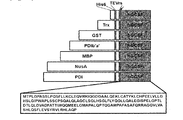
- Figure 1 shows a schematic diagram of the production process and domain structure of the PDIb'a'-hGCSF expression vector.
- A PDIb'a'-hGCSF vector map. Fusion protein expression in E. coli is regulated by the IPTG-induced T7 promoter and the selection marker is ampicillin.
- B The structural schematic diagram of seven fusion proteins (His6-, Trx-, GST-, PDIb'a'-, MBP-, PDI-, and NusA-hGCSF) is shown.
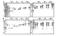
- FIG. 2 shows the results of analysis of E. coli expression of hGCSF fused with seven tags using SDS-PAGE gel. Protein expression was induced with 1 mM IPTG at (A) 18 ° C and (B) 30 ° C. After sonication, 20 ug of total protein was loaded on 10% Tris-tricine gel. Arrows indicate fused hGCSF proteins with each tag. M, molecular weight size marker; C, total cell protein before IPTG induction as control; T, total cellular protein after IPTG induction; P, protein of pellets after sonication; S, protein of the supernatant after sonication.
- M molecular weight size marker
- C total cell protein before IPTG induction as control
- T total cellular protein after IPTG induction
- P protein of pellets after sonication
- S protein of the supernatant after sonication.
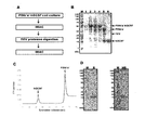
- FIG. 3 is for the separation of hGCSF from PDIb'a'-hGCSF.
- A The schematic of the purification process is shown.
- B PDIb'a'-hGCSF expressed from E. coli was analyzed by SDS-PAGE. lane 5 indicates that hPDIb'a 'was almost completely truncated.
- lane 1 total cells before IPTG induction as negative control; lane 2, total cells treated with IPTG; lane 3, aqueous fraction after cell sonication; lane 4, PDIb'a'-hGCSF fusion protein (59.1 kDa) isolated using His-trap HP column; lane 5, hPDIb'a 'digested with TEV protease tags: hPDIb'a' (35.6 kDa) and hGCSF (18.8 kDa); lane 6, final isolated hGCSF.
- C The chromatogram of using a His-trap HP column for hGCSF separation after cleavage reaction is shown.
- D Silver stained 10% SDS gel electrophoresis is shown under reduced and non-reducing conditions to confirm final purity of isolated hGCSF.
- M molecular weight marker
- R reduction
- NR non-reducing
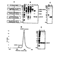
- hGCSF was purified from E. coli by a combination of MBP affinity and IMAC chromatography. 10 ug of total protein was loaded on 10% Tris-tricine gel.
- M molecular weight marker
- lane 1 total cell protein before IPTG induction as control
- lane 2 total cellular protein after IPTG induction
- lane 4 MBP-hGCSF fusion protein (63.3 kDa) isolated using MBP affinity column
- lane 5 His6-MBP tags digested with TEV protease: His6-MBP (40.3 kDa) and hGCSF (18.8 kDa); lane 6, final purified hGCSF (18.8 kDa).
- C 10% SDS gel electrophoresis silver stained under reducing conditions to confirm final purity of purified hGCSF.
- M molecular weight marker
- lane 1 final purified hGCSF.
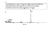
- Figure 5 shows the MALDI-TOF MS analysis of hGCSF.
- A Trypsin digest peptide map of hGCSF (aa).
- B MALDI-TOF MS results of hGCSF isolated under reducing conditions.
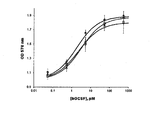
- FIG. 6 shows the biological activity of purified hGCSF against M-NFS 60 cells. Different concentrations of isolated hGCSF were incubated with myeloid leukemia M-NFS 60 cells for 72 hours. ?: Commercial hGCSF,?: HGCSF purified from MBP-hGCSF,?: HGCSF purified from PDIb'a'-hGCSF.
- the inventors have developed a method for expressing water soluble hGCSF protein with biological activity in the cytoplasm of E. coli .
- the present invention provides a recombinant expression vector comprising a solubility enhancing tag and a human granulocyte colony-stimulating factor (hGCSF) gene.
- hGCSF human granulocyte colony-stimulating factor
- any of the water-soluble enhancing tag can be used without limitation as long as it can express the hGCSF recombinant protein in water-soluble form.
- the water-soluble enhancement tag may be hexahistidine (His6), thioredoxin (Trx), glutathione S-transferase (GST), maltose binding protein (MBP). ), N-utilization substance Protein A (NusA), human protein disulfide isomerase (hPDI) and the b'a 'domain of hPDI hPDIb'a ').
- the hGCSF gene is characterized in that the nucleic acid sequence represented by SEQ ID NO: 1, the Trx gene is a nucleic acid sequence represented by SEQ ID NO: 2, the GST gene is a nucleic acid sequence represented by SEQ ID NO: 3,
- the NusA gene is a nucleic acid sequence represented by SEQ ID NO: 4, and the hPDIb'a 'gene is characterized in that the nucleic acid sequence represented by SEQ ID NO: 5.
- the hPDIb'a 'gene is a portion of the hPDI gene is removed, hPDI gene is shown in SEQ ID NO: 6.
- hGCSF amino acid sequence is shown by SEQ ID NO: 7. More specifically, the recombinant expression vector is characterized by having a cleavage map of FIG.
- vector refers to a DNA molecule that is a self-replicating molecule used to carry a clone gene (or another piece of clone DNA).
- an "expression vector” refers to a recombinant DNA molecule comprising a coding sequence of interest and a suitable nucleic acid sequence necessary to express a coding sequence operably linked in a particular host organism.
- the expression vector may preferably comprise one or more selectable markers.
- the marker is typically a nucleic acid sequence having properties that can be selected by a chemical method, which corresponds to all genes that can distinguish transformed cells from non-transformed cells. Examples include, but are not limited to, antibiotic resistance genes such as ampicilin, kanamycin, G418, bleomycin, hygromycin, and chloramphenicol, but are not limited thereto. It can select suitably.
- the gateway vector system used in the present invention is composed of an entry vector and an object vector.
- the entry vector is a vector having attL1 and attL2 at the sock end of the target gene
- the object vector is a vector including attR1 and attR2.
- AttR1 and attR2 of the entry vector cause the LR reaction by the recombination enzyme with attL1 and attL2 of the entry vector, and the target gene included in the entry vector is transferred to the object vector in the process.
- attR1 and attR2 are replaced with attB1 and attB2 sequences.
- the present invention also provides a recombinant microorganism transformed with the recombinant expression vector.
- the microorganism is characterized in that E. coli, and more specifically, E. coli BL21 (DE3).
- the present invention also provides a recombinant microorganism in which a gene construct including a water soluble enhancement tag and a hGCSF gene is inserted into a chromosome of a host cell. It will be apparent to those skilled in the art that the present invention may have the same effect as when the recombinant vector is introduced into the host cell even when the gene is inserted into the genomic chromosome of the host cell.
- the method for inserting the gene on the chromosome of the host cell in the present invention can be used a commonly known genetic engineering method, for example, retrovirus vector, adenovirus vector, adeno-associated virus vector, herpes simplex virus vector , Poxvirus vectors, lentiviral vectors or non-viral vectors.
- retrovirus vector for example, retrovirus vector, adenovirus vector, adeno-associated virus vector, herpes simplex virus vector , Poxvirus vectors, lentiviral vectors or non-viral vectors.
- the present invention comprises the steps of culturing the recombinant microorganism to express the water-soluble enhancing tag-hGCSF recombinant protein; Removing the water soluble enhancement tag from the expressed water soluble enhancement tag-hGCSF recombinant protein; And it provides a hGCSF recombinant protein (rhGCSF) production method comprising the step of recovering the hGCSF recombinant protein from which the water-soluble enhancement tag is removed.
- rhGCSF hGCSF recombinant protein
- tobacco tobacco virus tobacco Etch Virus (TEV) protease
- TEV tobacco Etch Virus
- the protease is a factor Xa protease, which can be commonly used in the art, Thrombin, Enterokinase, Ubiquitin-specific protease, Furin, Genease I or Proteinase K It may be, but is not limited thereto.
- the present invention also provides hGCSF recombinant protein (rhGCSF) produced by the above production method.
- rhGCSF hGCSF recombinant protein
- hGCSF gene (NCBI Reference Sequence: NP_757373) was codon optimized, attB1 site (5'-GGGGACAAGTTTGTACAAAAAAGCAGGCTTC-3 '; SEQ ID NO: 9), TEV protease recognition site (TEVrs, ENLYFQ ⁇ G) and attB2 (5'-ACCCAGCTTTCTTGTACAAAGTGGTCCCC-3 '; SEQ ID NO: 10).
- the DNA was synthesized and subcloned into plasmid pUC57 (Genscript, New Jersey, USA).
- the plasmid containing the hGCSF gene was recombined with pDONOR207 (Invitrogen, Carlsbad, CA, USA). Then, to construct an expression vector containing hGCSF and each tag, pENTR-hGCSF and seven target vectors pDEST-HGWA, pDEST-HXGWA, pDEST-HGGWA, pDEST-HMGWA, pDEST-HNGWA, pDEST-PDI and LR recombination cloning was performed between pDEST-PDIb'a '. The sequence of the expression plasmids was confirmed by DNA sequencing (Macrogen, Daejeon, Korea) and transformed with E. coli BL21 (DE3).
- hPDIb'a'-hGCSF 3x5 mL HisTrap HP column (GE Healthcare, Piscataway, NJ, USA) was used in the first step to isolate the fusion protein. After equilibrating the column with binding buffer, wash buffer (50 mM Tris-HCl, pH 8.0, 500 mM NaCl, 100 mM imidazole, 5% glycerol) to apply lysate solution to the column and remove all nonspecific binding (v / v)).
- wash buffer 50 mM Tris-HCl, pH 8.0, 500 mM NaCl, 100 mM imidazole, 5% glycerol
- hPDIb'a'-hGCSF was eluted with a buffer containing 500 mM imidazole (50 mM Tris-HCl, pH 8.0, 500 mM NaCl, 500 mM imidazole, 5% glycerol (v / v)) and dialyzed membrane (Viskase Corpration, Illinois, USA) was exchanged for free NaCl buffer (50 mM Tris-HCl, pH 8.0, 5% glycerol (v / v)).
- Protein TEV protease was cut at a ratio of 1:20 with TEV protease at 18 ° C. for 12 hours.
- the cleavage solution was then used in the buffer exchange procedure, using HisTrap binding buffer (50 mM Tris-HCl, pH 8.0, 500 mM NaCl, 5% glycerol (v / v)) via a 10 kDa dialysis membrane (Viskase Corpration, Illinois, USA). Was added.
- HisTrap binding buffer 50 mM Tris-HCl, pH 8.0, 500 mM NaCl, 5% glycerol (v / v)
- binding buffer 50 mM Tris-HCl, pH 8.0, 500 mM NaCl, 5% glycerol (v / v)
- hGCSF Unlike other proteins in solution, hGCSF has low affinity with Ni resin, 50 mM imidazole (50 mM Tris-HCl, pH 8.0, 500 mM NaCl, 5% glycerol, 50 mM imidazole ( v / v)) easily fell off the HisTrap column by washing. Based on the chromatograms, hGCSF monomers were identified via 10% Tris-tricine SDS-PAGE analysis under reducing and non-reducing conditions.
- MBP-hGCSF fusion protein 500 mL of MBP-hGCSF clones were incubated with 1 mM IPTG for 12 hours at 18 ° C. Cultured cells were harvested by centrifugation at 3,500 rpm for 30 minutes. The collected pellets were then resuspended in 100 mL MBP binding buffer (50 mM Tris-HCl, pH 8.0, 0.5 mM EDTA, 200 mM NaCl, 5% glycerol (v / v)) and the solution became completely clear. It was sonicated until cold and centrifuged for 20 minutes at 15,000 rpm.
- MBP binding buffer 50 mM Tris-HCl, pH 8.0, 0.5 mM EDTA, 200 mM NaCl, 5% glycerol (v / v)
- MBP-hGCSF was a buffer containing maltose (50 mM Tris-HCl, pH 8.0, 0.5 mM EDTA, 200 mM NaCl, 5% glycerol, 10 mM maltose monohydrate (v / v)). Based on the chromatograms, MBP-hGCSF was collected and confirmed by 10% Tris-tricine SDS-PAGE analysis.
- the eluted samples were diluted until the final concentration of NaCl was 50 mM, and the TEV treatment and cleaved hGCSF purification proceeded in the same way as for cutting hGCSF from hPDIb'a'-hGCSF.
- the hGCSF collected in the washing step was subjected to PBS buffer (137 mM NaCl, 2.7 mM KCl, 8 mM Na 2 HPO 4 , 1.4 mM KH 2 PO 4).
- Coomassie blue R-250 (AMRESCO, Solon, OH, USA) was treated to identify the protein bands and the presence of the protein was visualized on a 10% Tris-tricine gel. Expression, solubility and purity were quantified by ImageJ program.
- a polyacrylamide gel was placed in a Fixative Enhancer Solution for 20 minutes and then washed with distilled water to increase the sensitivity and to ensure the difference in staining.
- a silver complex solution, a reduction moderator solution, and an image development reagent were added to perform dyeing and color reaction, respectively.
- FIG. 3A To separate hGCSF from PDIb'a'-hGCSF, two IMACs and a final GPC were performed (FIG. 3A). Since the N-terminal tags used in the present invention all contain His6 tags, IMAC was possible. Cells containing the PDIb'a'-hGCSF plasmid were induced with IPTG and collected ( Figure 3B, lane 2). Cells were lysed and centrifuged to collect supernatants (FIG. 3B, lane 3). The supernatant was then loaded onto a Ni column and the bound protein was eluted after the wash procedure ( Figure 3B, lane 4). Most of the other proteins were removed in the process, but there were still some impurity bands.
- TEV protease cleavage Although other protein bands were present. Most of the PDIb'a'-hGCSF was cleaved by TEV protease ( Figure 3B, lane 5). To remove the PDIb'a 'tag, uncut PDIb'a'-hGCSF and TEV protease, a second His-trap HP column was used. The cleaved hGCSF was weakly bound to the Ni column and eluted at 50 mM imidazole. To remove residual impurities, samples were loaded onto a Superdex gel filtration column (FIG. 3C). hGCSF was completely separated and pure hGCSF was obtained (FIG. 3B, lane 6).
- FIG. 4A shows the overall procedure for purifying hGCSF from MBP-hGCSF.
- the first step of MBP chromatography is to separate about 80% of the MBP-hGCSF fusion protein from the protein mixture ( Figure 4B, lane 4).
- TEV protease was cleaved by treatment with MBP-hGCSF at low concentration (TEV: target protein (1:20)), and only hGCSF was isolated (Fig. 4B, lane 5).
- hGCSF monomer was obtained by washing the mixture containing hGCSF multimer (including about 92% of hGCSF monomer) with 50 mM imidazole through Ni NTA chromatography.
- Triton X-114 (Sigma-Aldrich, St. Louis, MO, USA) was used at a final concentration of 1% to remove entotoxin from the separated hGCSF solution and treated at 4 ° C. for 30 minutes. Triton X-114 was collected after reacting the samples at room temperature and removed by centrifugation at 12,000 rpm for 10 minutes.
- LAL Endpoint Chromogenic Limulus Amebocyte Lysate
- hGCSF was precipitated with trichloroacetic acid (TCA) and IA buffer (0.5 M Tris-HCl, pH 8.0, 5% glycerol (v / v)). , 50 mM IAA dissolved in 100 mM NaCl, 1 mM EDTA, 1% SDS) and darkened for 1 hour. After performing SDS-PAGE, hGCSF related bands were cut from the gel and cut overnight at 37 ° C. with kemotrypsin (Promega, Madison, Wis., USA).
- ⁇ -Cyano-4-hydroxycinnamic acid (CHCA) was used as the matrix and the MALDI-TOF MS procedure was performed using Voyager-DE TM STR (Applied Biosystems, South San Francisco). , CA, USA) and 4700 MALDI TOF / TOF TM Analyzer (AB Sciex, Framingham, Mass., USA). Peptide mass was analyzed by Data Explorer TM software (Applied Biosystems, South San Francisco, CA, USA).
- the M-NFS60 cell line was provided by Dr. Kyung-Woon Kim, RPMI-1640 medium (GIBCO Invitrogen, CA, USA), 10% fetal bovine serum (FBS), 1X penicillin streptomycin (GIBCO Invitrogen) , CA, USA) and 0.05 mM ⁇ -mercaptoethanol. Cells were maintained at 5% CO 2 , 37 ° C.
- the EC 50 of commercially available hGCSF was 1.62 ⁇ 0.19 pM
- the EC 50 of hGCSF isolated from PDIb'a 'tag was 2.78 ⁇ 0.27 pM
- the EC 50 of hGCSF purified from MBP tag was 3.49 ⁇ 0.54 pM ( Hill coefficients are 0.73 ⁇ 0.21, 0.71 ⁇ 0.29, and 0.67 ⁇ 0.21, respectively.
Landscapes
- Health & Medical Sciences (AREA)
- Chemical & Material Sciences (AREA)
- Life Sciences & Earth Sciences (AREA)
- Organic Chemistry (AREA)
- Genetics & Genomics (AREA)
- Molecular Biology (AREA)
- Zoology (AREA)
- Engineering & Computer Science (AREA)
- Biochemistry (AREA)
- Biophysics (AREA)
- General Health & Medical Sciences (AREA)
- Biomedical Technology (AREA)
- Medicinal Chemistry (AREA)
- Proteomics, Peptides & Aminoacids (AREA)
- Biotechnology (AREA)
- Wood Science & Technology (AREA)
- Bioinformatics & Cheminformatics (AREA)
- General Engineering & Computer Science (AREA)
- Toxicology (AREA)
- Gastroenterology & Hepatology (AREA)
- Immunology (AREA)
- Plant Pathology (AREA)
- Physics & Mathematics (AREA)
- Microbiology (AREA)
- Preparation Of Compounds By Using Micro-Organisms (AREA)
- Peptides Or Proteins (AREA)
Abstract
본 발명은 N-말단 태그를 이용한 생물학적 활성을 가진 인간 GCSF 재조합 단백질의 수용성 발현 및 정제방법에 관한 것으로서, 수용성 향상 태그(solubility enhancing tag) 및 인간 과립구 생성 촉진인자(human granulocyte colony-stimulating factor; hGCSF) 유전자를 포함하는 재조합 발현벡터를 제공한다. 또한 상기 재조합 발현벡터로 형질전환된 재조합 미생물을 제공하며, 상기 hGCSF 재조합 단백질(rhGCSF)의 생산방법 및 상기 생산방법에 의해 생산된 hGCSF 재조합 단백질(rhGCSF)을 제공한다.
Description
본 발명은 N-말단 태그를 이용한 생물학적 활성을 가진 인간 과립구 생성 촉진인자(human granulocyte colony-stimulating factor; hGCSF) 재조합 단백질의 수용성 발현 및 정제방법에 관한 것이다.
과립구 생성 촉진인자(granulocyte colony-stimulating factor; GCSF)는 플루리포이에틴(pluripoietin)으로도 알려졌으며, 백혈구의 70%를 구성하는 과립구의 생산, 분화 및 기능을 조절함으로써 조혈과정에 작용한다. 성장인자는 인터루킨-1β, 종양 괴사 인자-알파(tumor necrosis factor-alpha), 지질다당류(lipopolysaccharide)와 같은 여러 염증 자극에 의해 대식세포(macrophages), 섬유아세포(fibroblasts) 및 내피세포(endothelials)에서 대부분 분비된다. 임상적으로, 인간 GCSF(hGCSF)는 혈액 내 호중구(neutrophils)의 수가 극히 낮아 발생하는 호중구감소증(neutropenia)의 치료를 위해 승인되었다. 보통 암 환자들은 방사선치료 또는 화학치료 후에 이와 같은 백혈구 감소증(leukopenia)으로 인해 고통 받는다. 또한, GCSF는 신경보호효과를 나타내기도 한다. 따라서, 상기 단백질은 루게릭병과 같은 다양한 신경퇴행성 질환의 마우스 모델에 치료제로서 적용되기도 하였다.
초기에, hGCSF는 hGCSF를 계속적으로 분비하는 암세포주로부터 생산되었다. 메탄올자화(methylotrophic) 효모인 피히아 파스토리스(Pichia Pastoris)에서 발현시, 상기 호르몬은 수용성 형태로 분비되었으나 상당히 응집되어서 구아니딘 하이드로클로라이드 또는 요소(urea)와 같은 고농도의 변성제를 이용하여 수용화시켜야 했고, 그 후에 생물학적으로 활성화된 형태로 정제하기 위해서 변성제를 제거하여 재접힘시켜야 했다. 또한, 대장균(Escherichia coli; E. coli)과 같은 박테리아를 이용한 발현과 관련된 많은 연구들에서 세포질(cytoplasm) 내에 과발현된 단백질들이 응집되어 봉입체 형태(inclusion bodies)로 생산되는 것을 확인하였다. 많은 경우, 봉입체로부터 얻은 생물학적 활성을 가진 단백질의 전체적인 수율은 낮았다. 대신에, 단백질이 E. coli의 주변 세포질(periplasm)로 분비되었으나 수율은 역시 낮았다.
한편 한국공개특허 제2012-0057416호는 인간 G-CSF 단백질 생산을 위한 폴리뉴클레오티드 및 이의 용도에 관해 개시하고 있지만, 본원발명의 hGCSF의 수용성 발현 및 이를 분리하기 위한 hPDIb'a' 도메인과 같은 태깅 시스템에 대한 언급은 없다.
본 발명의 목적은 수용성 향상 태그(solubility enhancing tag) 및 인간 과립구 생성 촉진인자(human granulocyte colony-stimulating factor; hGCSF) 유전자를 포함하는 재조합 발현벡터, 상기 재조합 발현벡터로 형질전환된 재조합 미생물을 제공하는데 있다.
본 발명의 다른 목적은 상기 hGCSF 재조합 단백질(rhGCSF)의 생산방법 및 상기 생산방법에 의해 생산된 hGCSF 재조합 단백질(rhGCSF)을 제공하는데 있다.
상기 목적을 달성하기 위하여, 본 발명은 수용성 향상 태그(solubility enhancing tag) 및 인간 과립구 생성 촉진인자(human granulocyte colony-stimulating factor; hGCSF) 유전자를 포함하는 재조합 발현벡터를 제공한다.
또한, 본 발명은 상기 재조합 발현벡터로 형질전환된 재조합 미생물을 제공한다.
또한, 본 발명은 상기 hGCSF 재조합 단백질(rhGCSF)의 생산방법 및 상기 생산방법에 의해 생산된 hGCSF 재조합 단백질(rhGCSF)을 제공한다.
본 발명은 N-말단 태그를 이용한 인간 GCSF 재조합 단백질의 수용성 발현 및 정제방법에 관한 것으로서, N-말단에 PDI의 b'a' 도메인 태깅을 통해 대장균 내에서 용해도 및 안정도가 증가된 인간 GCSF 단백질을 분리, 정제하는 방법에 대한 것이다. 이렇게 얻어진 인간 GCSF 단백질은 혈액 내 호중구(neutrophils)의 수가 극히 낮아 발생하는 호중구감소증(neutropenia)의 치료제로서 중요한 역할을 할 것으로 기대된다.
도 1은 PDIb'a'-hGCSF 발현벡터의 제작 과정 및 도메인 구조의 모식도를 나타낸다. (A) PDIb'a'-hGCSF 벡터 맵. E. coli에서의 융합 단백질 발현은 IPTG-유도성 T7 프로모터에 의해 조절되고, 선별 마커는 앰피실린(ampicillin)이다. (B) 7개의 융합 단백질들(His6-, Trx-, GST-, PDIb'a'-, MBP-, PDI- 및 NusA-hGCSF)의 구조 모식도를 나타낸다.
도 2는 SDS-PAGE 젤을 사용하여 7개의 태그들로 융합된 hGCSF의 E. coli 발현에 대해 분석한 결과이다. 단백질 발현은 (A) 18℃ 및 (B) 30℃에서 1 mM IPTG로 유도되었다. 초음파 처리 후에, 20ug의 총 단백질은 10% 트리스-트리신 젤(Tris-tricine gel)에 로딩되었다. 화살표는 각각의 태그를 가진 융합된 hGCSF 단백질을 나타낸다. M, 분자량 사이즈 마커; C, 대조군으로서 IPTG 유도 전의 총 세포 단백질; T, IPTG 유도 후의 총 세포 단백질; P, 초음파 처리 후 펠렛의 단백질; S, 초음파 처리 후 상등액의 단백질.
도 3은 PDIb'a'-hGCSF로부터 hGCSF의 분리에 대한 것이다. (A) 정제과정의 모식도를 나타낸다. (B) 대장균으로부터 발현된 PDIb'a'-hGCSF는 SDS-PAGE에 의해 분석되었다. lane 5는 hPDIb'a'가 거의 완전하게 절단되었다는 것을 나타낸다. M, 분자량 마커; lane 1, 음성대조구로서 IPTG 유도 전 총 세포; lane 2, IPTG로 처리한 총 세포; lane 3, 세포 초음파 처리 후 수용성 분획; lane 4, His-trap HP 컬럼을 이용하여 분리한 PDIb'a'-hGCSF 융합 단백질(59.1 kDa); lane 5, TEV 단백질 분해효소로 절단된 hPDIb'a' 태그: hPDIb'a' (35.6 kDa) 및 hGCSF (18.8 kDa); lane 6, 최종 분리된 hGCSF. (C) 절단 반응 후의 hGCSF 분리를 위한 His-trap HP 컬럼 사용의 크로마토그램을 나타낸다. hPDIb'a'와 함께 절단된 hGCSF는 다른 크기로 뚜렷하게 분리된다. (D) 분리된 hGCSF의 최종 순도를 확인하기 위하여 환원 및 비환원 조건에서 실버 염색된 10% SDS 젤 전기영동을 나타낸다. M, 분자량 마커; R, 환원; NR, 비환원
도 4는 MBP-hGCSF로부터 hGCSF의 분리에 대한 것이다. (A) hGCSF 정제 과정의 모식도를 나타낸다. (B) MBP 친화도 및 IMAC 크로마토그래피의 조합에 의해 E. coli로부터 hGCSF를 정제하였다. 10ug의 총 단백질은 10% 트리스-트리신 젤(Tris-tricine gel)에 로딩되었다. M, 분자량 마커; lane 1, 대조군으로서 IPTG 유도 전 총 세포 단백질; lane 2, IPTG 유도 후 총 세포 단백질; lane 3, 세포 초음파 처리 후 수용성 분획; lane 4, MBP 친화도 컬럼을 사용하여 분리한 MBP-hGCSF 융합 단백질(63.3 kDa); lane 5, TEV 단백질 분해효소에 절단된 His6-MBP 태그 : His6-MBP (40.3 kDa) 및 hGCSF (18.8 kDa); lane 6, 최종 정제된 hGCSF (18.8 kDa). (C) 정제된 hGCSF의 최종 순도를 확인하기 위하여, 환원 조건하에서 실버 염색된 10% SDS 젤 전기영동 사진이다. M, 분자량 마커; lane 1, 최종 정제된 hGCSF. (D) 젤 여과에 의한 hGCSF 단량체의 정제에 대한 결과이다. 2차 정제 단계 후 hGCSF의 Superdex 200 16/60 크로마토그램을 나타낸다. (E) 정제된 hGCSF의 실버 염색 결과를 나타낸다. M, 분자량 마커; lane 1 및 2, hGCSF는 10 mM DTT 유무에 따라 환원되었다.
도 5는 hGCSF의 MALDI-TOF MS 분석결과를 나타낸다. (A) hGCSF (aa)의 트립신 분해 펩타이드 지도. (B) 환원 조건에서 분리된 hGCSF의 MALDI-TOF MS 결과이다.
도 6는 정제된 hGCSF의 M-NFS 60 세포에 대한 생물학적 활성을 나타낸다. 여러 농도의 분리된 hGCSF가 골수성 백혈병 M-NFS 60 세포와 함께 72시간 동안 배양되었다. ●: commercial hGCSF, △: MBP-hGCSF로부터 정제된 hGCSF, □: PDIb'a' -hGCSF로부터 정제된 hGCSF.
본 발명자들은 E. coli의 세포질에서 생물학적 활성을 가진 수용성 hGCSF 단백질을 발현시키기 위한 방법을 개발하였다. 7개의 N-말단 융합 태그들, 헥사히스티딘(hexahistidine; His6), 티오레독신(thioredoxin; Trx), 글루타치온 S-트랜스퍼라제(glutathione S-transferase; GST), 말토스 결합 단백질(Maltose Binding Protein, MBP), N-이용 기질 단백질 A(N-utilization substance Protein A; NusA), 인간 단백질 이황화물 이성질화효소(human protein disulfide isomerase; hPDI) 및 hPDI의 b'a' 도메인(b'a' domain of hPDI; hPDIb'a')을 hGCSF의 수용성 과발현을 위해 시험하였다. 이 중 4개의 태그 단백질, MBP, NusA, hPDI 및 hPDIb'a'가 30℃에서 hGCSF 용해도를 상당히 증가시킨다는 것을 확인하였으며, 골수성 백혈병 M-NFS 60 세포를 이용하여 hGCSF의 활성을 측정하고 본 발명을 완성하였다.
본 발명은 수용성 향상 태그(solubility enhancing tag) 및 인간 과립구 생성 촉진인자(human granulocyte colony-stimulating factor; hGCSF) 유전자를 포함하는 재조합 발현벡터를 제공한다.
본 발명에 있어서, 상기 수용성 향상 태그는 hGCSF 재조합 단백질을 수용성 형태로 발현시킬 수 있는 것이면 어느 것이든 제한 없이 이용가능하다. 예를 들어, 상기 수용성 향상 태그는 헥사히스티딘(hexahistidine; His6), 티오레독신(thioredoxin; Trx), 글루타치온 S-트랜스퍼라제(glutathione S-transferase; GST), 말토스 결합 단백질(Maltose Binding Protein, MBP), N-이용 기질 단백질 A(N-utilization substance Protein A; NusA), 인간 단백질 이황화물 이성질화효소(human protein disulfide isomerase; hPDI) 및 hPDI의 b'a' 도메인(b'a' domain of hPDI; hPDIb'a') 중 어느 하나일 수 있다.
상세하게는, 상기 hGCSF 유전자는 서열번호 1로 표시되는 핵산서열인 것을 특징으로 하고, 상기 Trx 유전자는 서열번호 2로 표시되는 핵산서열이고, 상기 GST 유전자는 서열번호 3으로 표시되는 핵산서열이고, 상기 NusA 유전자는 서열번호 4로 표시되는 핵산서열이고, 상기 hPDIb'a' 유전자는 서열번호 5로 표시되는 핵산서열인 것을 특징으로 한다. 한편, 상기 hPDIb'a' 유전자는 hPDI 유전자의 일부가 제거된 것으로서, hPDI 유전자는 서열번호 6으로 표시하였다. 또한 hGCSF 아미노산 서열은 서열번호 7로 표시하였다. 보다 상세하게는 상기 재조합 발현벡터는 도 1a의 개열지도를 갖는 것을 특징으로 한다.
본 발명에 있어서, “벡터”는 클론유전자(또는 클론 DNA의 다른 조각)를 운반하는데 사용되는 스스로 복제되는 DNA분자를 의미한다.
본 발명에서 있어서, “발현 벡터”는 목적한 코딩 서열과, 특정 숙주 생물에서 작동 가능하게 연결된 코딩 서열을 발현하는데 필수적인 적정 핵산 서열을 포함하는 재조합 DNA 분자를 의미한다. 발현 벡터는 바람직하게는 하나 이상의 선택성 마커를 포함할 수 있다. 상기 마커는 통상적으로 화학적인 방법으로 선택될 수 있는 특성을 갖는 핵산 서열로, 형질 전환된 세포를 비 형질전환 세포로부터 구별할 수 있는 모든 유전자가 이에 해당된다. 그 예로는 앰피실린(ampicilin), 카나마이신(kanamycin), G418, 블레오마이신(Bleomycin), 하이그로마이신(hygromycin), 클로람페니콜(chloramphenicol) 과 같은 항생제 내성 유전자가 있으나, 이에 한정되는 것은 아니며, 당업자에 의해 적절히 선택 가능하다.
본 발명에서 사용한 Gateway Vector system은 엔트리벡터와 목적벡터로 구성되며, 상기 엔트리벡터는 목적유전자의 양말단에 attL1, attL2를 갖는 벡터이며, 목적벡터는 attR1, attR2를 포함하는 벡터이다. 상기 엔트리벡터와 상기 목적벡터는 목적벡터의 attR1 및 attR2가 엔트리벡터의 attL1 및 attL2와 재조합효소에 의해 LR반응을 일으키며, 이 과정에서 엔트리벡터에 포함되어 있던 목적유전자가 목적벡터로 전달되게 되고, attR1과 attR2 는 attB1과 attB2 서열로 치환되어진다.
또한, 본 발명은 상기 재조합 발현벡터로 형질전환된 재조합 미생물을 제공한다. 상세하게는 상기 미생물은 대장균인 것을 특징으로 하고, 보다 상세하게는 대장균 BL21(DE3)인 것을 특징으로 한다.
또한, 본 발명은 수용성 향상 태그 및 hGCSF 유전자가 포함된 유전자 구조체(gene construct)가 숙주세포의 염색체 내에 삽입되어 있는 재조합 미생물을 제공한다. 본 발명이 속하는 기술분야의 당업자에게 있어 상기 유전자를 숙주세포의 게놈 염색체에 삽입하여서도 상기와 같이 재조합 벡터를 숙주세포에 도입한 경우와 동일한 효과를 가질 것은 자명하다 할 것이다.
본 발명에서 상기 유전자를 숙주세포의 염색체상에 삽입하는 방법으로는 통상적으로 알려진 유전자조작방법을 사용할 수 있으며, 일 예로는 레트로바이러스 벡터, 아데노바이러스 벡터, 아데노-연관 바이러스 벡터, 헤르페스 심플렉스 바이러스 벡터, 폭스바이러스 벡터, 렌티바이러스 벡터 또는 비바이러스성 벡터를 이용하는 방법을 들 수 있다.
또한, 본 발명은 상기 재조합 미생물을 배양하여 수용성 향상 태그-hGCSF 재조합 단백질을 발현시키는 단계; 상기 발현된 수용성 향상 태그-hGCSF 재조합 단백질에서 수용성 향상 태그를 제거하는 단계; 및 상기 수용성 향상 태그가 제거된 hGCSF 재조합 단백질을 회수하는 단계를 포함하는 hGCSF 재조합 단백질(rhGCSF) 생산방법을 제공한다.
한편, 본 발명의 실시예에서는 담배 식각 바이러스(Tobacco Etch Virus; TEV) 단백질 분해효소(protease)를 사용했지만, 상기 단백질 분해효소(protease)는 당업계에서 통상적으로 사용될 수 있는 Factor Xa 단백질 분해효소, 트롬빈(Thrombin), 엔테로키나제(Enterokinase), 유비퀴틴-특이적 단백질 분해효소(Ubiquitin-specific protease), 푸린(Furin), 유전자 분해효소 I(Genease I) 또는 단백질 분해효소 K(Proteinase K)등을 적용할 수 있으나, 이에 한정되는 것은 아니다.
또한, 본 발명은 상기 생산방법에 의해 생산된 hGCSF 재조합 단백질(rhGCSF)을 제공한다.
본 발명에서 사용한 유전공학적 기술과 관련된 사항은 샘브룩 등의 문헌(Sambrook, et al. Molecular Cloning, A Laboratory Manual, Cold Spring Harbor laboratory Press, Cold Spring Harbor, N. Y.(2001)) 및 프레드릭 등의 문헌 (Frederick M. Ausubel et al., Current protocols in molecular biology volume 1,2,3, John Wiley & Sons, Inc.(1994)) 등을 참조할 수 있다.
이하, 하기 실시예를 통해 본 발명을 보다 상세하게 설명한다. 다만, 이러한 실시예에 의해 본 발명이 한정되는 것은 아니다.
<실시예 1> 발현벡터의 제작 및 발현
hGCSF 유전자(NCBI Reference Sequence: NP_757373)는 코돈 최적화되었고, attB1 사이트 (5'- GGGGACAAGTTTGTACAAAAAAGCAGGCTTC-3'; 서열번호 9), TEV 단백질 분해효소 인식 사이트(TEV protease recognition site; TEVrs, ENLYFQˇG) 및 attB2 (5'-ACCCAGCTTTCTTGTACAAAGTGGTCCCC-3'; 서열번호 10)에 삽입되었다. 상기 DNA는 합성되었고, 플라스미드 pUC57 (Genscript, New Jersey, USA)에 서브클론되었다. 엔트리벡터 pENTR-hGCSF를 제작하기 위해서, hGCSF 유전자를 포함하는 플라스미드는 pDONOR207 (Invitrogen, Carlsbad, CA, USA)로 재조합되었다. 그 후, hGCSF와 각 태그들을 포함하는 발현벡터를 제작하기 위해서, pENTR-hGCSF와 7개의 목적 벡터인 pDEST-HGWA, pDEST-HXGWA, pDEST-HGGWA, pDEST-HMGWA, pDEST-HNGWA, pDEST-PDI 및 pDEST-PDIb'a' 사이에 LR 재조합 클로닝을 시행하였다. 발현 플라스미드들의 서열은 DNA 서열분석에 의해 확인하였고(Macrogen, Daejeon, Korea), E.coli BL21 (DE3)로 형질전환되었다.
E. coli 세포질에서 hGCSF를 수용성 형태로 발현시키기 위해서, hGCSF 서열의 N-말단에 7개의 태그 유전자(His6, Trx, GST, PDIb'a', MBP, PDI 및 NusA)를 융합하는 과정은 도 1에 나타냈다. TEVrs는 정제 과정 중에 분리하기 위해서 각 태그들과 hGCSF 사이에 삽입되었다.
세포들은 50ug/ml 앰피실린이 포함된 2mL LB 배지에서 배양하였고, 37℃, 200rpm에서 배양하였다. OD600이 약 0.4-0.6에 이르렀을 때, hGCSF 융합 단백질의 발현을 유도하기 위해서, 1mM 이소프로필-β-D-티오갈락토시드(isopropyl-β-D-thiogalactoside; IPTG)를 첨가하였다. 30℃에서 5시간 동안과 18℃에서 12시간 동안 배양한 후에 각각 세포를 수확하였다(도 2 및 표 1). 모든 태그들에 있어서 두 발현 온도에서 hGCSF의 발현 수준은 33 - 68% 사이였고, 18℃에서 더 높은 발현 수준을 나타냈다. 용해도는 태그와 발현 온도에 따라서 상이했다. MBP, NusA, PDI 및 PDIb'a'의 4개의 태그 단백질은 30℃에서 용해도를 상당히 증가시켰다(도 2A 및 표 1). 또한, 발현 온도를 18℃로 낮추면 Trx-hGCSF 및 GST-hGCSF의 용해도를 증가시켰다(도 2B 및 표 1). His6-hGCSF는 두 발현 온도 모두에서 불용성이었다. 용해도 및 태그 단백질의 크기에 기초하여, 본 발명자들은 이후 연구를 위해서 2개의 융합단백질 PDIb'a'-hGCSF 및 MBP-hGCSF을 선택하였다.
표 1 여러 N-말단 태그를 가진 hGCSF의 발현 수준 및 용해도
| 태그 | 태그 크기 (kDa) | 융합 단백질 크기 (kDa) | 발현(%) | 용해도(%) | |||
| 18℃ | 30℃ | 18℃ | 30℃ | ||||
| hGCSF (18.8 kDa) | His6 | 0.8 | 23.5 | 43.8 | 33.6 | - | - |
| Trx | 11.8 | 35.3 | 61.4 | 48.8 | 98.3 | 5.0 | |
| GST | 25.7 | 49.2 | 41.3 | 40.0 | 78.4 | 3.2 | |
| PDIb'a' | 35.6 | 59.1 | 66.3 | 42.2 | 96.0 | 73.5 | |
| MBP | 40.3 | 63.8 | 61.4 | 58.4 | 96.5 | 88.1 | |
| PDI | 55.1 | 78.7 | 55.6 | 43.8 | 98.1 | 89.3 | |
| NusA | 54.9 | 78.4 | 68.0 | 44.8 | 97.5 | 89.5 | |
<실시예 2> 재조합 hGCSF의 분리 및 정제
1. hPDIb'a'-hGCSF로부터 hGCSF의 분리 및 정제
hPDIb'a'-hGCSF의 발현에 있어서, 3x5 mL HisTrap HP column (GE Healthcare, Piscataway, NJ, USA)을 상기 융합 단백질을 분리하기 위한 첫 단계에서 사용하였다. 결합 완충액에 의해 컬럼을 평형화한 후에, 컬럼에 용해물(lysate) 용액을 적용하고 비특이적 결합을 모두 제거하기 위해서 세척 완충액(50mM Tris-HCl, pH 8.0, 500 mM NaCl, 100 mM imidazole, 5% glycerol (v/v))을 흘려보냈다. hPDIb'a'-hGCSF는 500mM 이미다졸(imidazole)이 포함된 완충액(50mM Tris-HCl, pH 8.0, 500 mM NaCl, 500 mM imidazole, 5% glycerol (v/v))을 사용하여 용출되었고, 투석막(Viskase Corpration, Illinois, USA)을 통해 유리 NaCl 완충액(50mM Tris-HCl, pH 8.0, 5% glycerol (v/v))으로 교환되었다. 단백질 : TEV 단백질 분해효소(protease)를 1:20의 비율로 하여 TEV 단백질 분해효소(protease)로 18℃에서 12시간 동안 절단하였다. 그 후, 절단 용액은 완충액 교환 과정에 사용되었는데, 10kDa 투석막(Viskase Corpration, Illinois, USA)을 통해 HisTrap 결합 완충액 (50mM Tris-HCl, pH 8.0, 500 mM NaCl, 5% glycerol (v/v))에 첨가되었다. 결합 완충액(50 mM Tris-HCl, pH 8.0, 500 mM NaCl, 5% glycerol (v/v))으로 완전히 채워진 미리-팩킹된 HisTrap HP column (GE Healthcare, Piscataway, NJ, USA)을 사용하여, 모든 처리가 완료된 샘플을 2x5 mL IMAC에 로드하였다. 용액 내의 다른 단백질들과는 다르게, hGCSF는 Ni 레진(resin)과의 친화도가 낮고, 50 mM 이미다졸(imidazole)(50 mM Tris-HCl, pH 8.0, 500 mM NaCl, 5% glycerol, 50 mM imidazole (v/v)) 세척에 의해 쉽게 HisTrap 컬럼에서 떨어졌다. 크로마토그램에 기초하여, hGCSF 단량체를 환원 및 비환원 조건하에서 10% 트리스-트리신(Tris-tricine) SDS-PAGE 분석을 통해 확인하였다.
2. MBP-hGCSF로부터 hGCSF의 분리 및 정제
MBP-hGCSF 융합 단백질을 과발현시키기 위해서 18℃에서 12시간 동안 1 mM IPTG로 유도하여 MBP-hGCSF 클론을 500mL 전-배양하였다. 3,500 rpm에서 30분 동안 원심분리하여 배양된 세포를 수확하였다. 그 후, 모인 펠렛(pellet)을 100mL MBP 결합 완충액(50 mM Tris-HCl, pH 8.0, 0.5 mM EDTA, 200 mM NaCl, 5% glycerol (v/v))에 재현탁시켰고, 용액이 완전히 투명해질 때까지 차갑게 하여 초음파처리하였고, 15,000 rpm에서 20분 동안 원심분리하였다. 원심분리 및 여과한 후의 용해물을 2x5 mL MBP Trap HP (GE Healthcare, Piscataway, NJ, USA)에 MBP 결합 완충액으로 평형화된 AKTA Prime Plus machine (GE life science, NF, USA)을 사용하여 적용하였다. 비특이적인 결합 단백질은 결합 완충액으로 씻어냈고, MBP-hGCSF는 말토스를 포함하는 완충액(50 mM Tris-HCl, pH 8.0, 0.5 mM EDTA, 200 mM NaCl, 5% glycerol, 10 mM maltose monohydrate (v/v))으로 용출하였다. 크로마토그램을 기초로, MBP-hGCSF를 모았고, 10% 트리스-트리신(Tris-tricine) SDS-PAGE 분석에 의해 확인하였다. 용출된 샘플은 NaCl의 최종 농도가 50mM이 될 때까지 희석하였고, TEV 처리 과정 및 절단된 hGCSF 정제 과정은 hPDIb'a'-hGCSF로부터 hGCSF를 절단하는 방법과 동일하게 진행되었다. hGCSF 완충액을 PBS 완충액으로 교환할 뿐만 아니라 다량체로부터 hGCSF 단량체로 분리하기 위해서, 세척단계에서 모인 hGCSF를 PBS 완충액 (137 mM NaCl, 2.7 mM KCl, 8 mM Na2HPO4, 1.4 mM KH2PO4, 5% glycerol (v/v)) 상의 gel filtration superdex 200 16/60 column (GE Healthcare, Piscataway, NJ, USA)에 로딩하였다. 크로마토그램에 기초하여, hGCSF 단량체를 환원 및 비환원 조건하에서 10% 트리스-트리신(Tris-tricine) SDS-PAGE 분석을 통해 확인하였다.
3. SDS-PAGE 분석 및 실버 염색
단백질 밴드를 확인하기 위해 coomassie blue R-250 (AMRESCO, Solon, OH, USA) 처리하고, 10% 트리스-트리신(Tris-tricine) 젤 상에서 단백질의 존재가 가시화되었다. 발현, 용해도 및 순도는 ImageJ program에 의해 정량화하였다.
실버 염색을 위해, 폴리아크릴아미드 젤(polyacrylamide gel)을 Fixative Enhancer Solution에 20분 동안 넣은 후, 민감도를 증가시키고 염색 차이를 확실히 하기 위해서 증류수로 씻어냈다. silver complex solution, reduction moderator solution 및 image development reagent (Bio-Rad Laboratories, Hercules, CA, USA)을 각각 첨가하여 염색 및 발색 반응을 진행했고, 5% 아세트산 용액으로 반응을 중단시켰다.
4. 결과
(1) PDIb'a'-hGCSF로부터 hGCSF 정제
PDIb'a'-hGCSF로부터 hGCSF를 분리하기 위해서, 두 번의 IMAC와 최종 GPC를 수행하였다(도 3A). 본 발명에 사용된 N-말단 태그들에는 모두 His6 태그를 포함하고 있으므로 IMAC가 가능하였다. PDIb'a'-hGCSF 플라스미드를 포함하는 세포는 IPTG로 유도되었고, 수집되었다(도 3B, 레인 2). 세포는 용해되었고, 상등액을 모으기 위해서 원심분리되었다(도 3B, 레인 3). 그 후, 상등액을 Ni 컬럼에 로딩하였고, 세척 과정 후에 결합된 단백질을 용출하였다(도 3B, 레인 4). 상기 과정에서 다른 대부분의 단백질은 제거되었으나, 여전히 약간의 불순물 밴드들이 있었다. 다른 단백질 밴드들이 존재함에도 본 발명자들은 TEV 단백질 분해효소 절단을 수행하였다. 대부분의 PDIb'a'-hGCSF는 TEV 단백질 분해효소에 의해 절단되었다(도 3B, 레인 5). PDIb'a' 태그, 미절단 PDIb'a'-hGCSF 및 TEV 단백질 분해효소를 제거하기 위해서, 2차 His-trap HP column을 사용하였다. 절단된 hGCSF는 Ni 컬럼에 약하게 결합하였고, 50mM 이미다졸(imidazole)에 용출하였다. 잔여 불순물을 제거하기 위해서, 샘플들을 Superdex gel filtration column에 로딩하였다(도 3C). hGCSF는 완전히 분리되었고, 순수 hGCSF를 얻을 수 있었다(도 3B, 레인 6). 순도를 측정하기 위해서, 환원 조건에서 정제된 hGCSF의 실버 염색을 수행하였고, 결과는 hGCSF가 순수하게 정제되었다는 것을 나타냈다(도 3D). 1L의 세포 배양에서 약 11.3 mg의 순수한 hGCSF를 얻었다(표 2).
(2) MBP-hGCSF로부터 hGCSF 정제
도 4A는 MBP-hGCSF로부터 hGCSF를 정제하기 위한 전체적인 과정을 나타낸다. hGCSF 정제 과정에 있어서, MBP 크로마토그래피의 첫 단계는 단백질 혼합물로부터 약 80%의 MBP-hGCSF 융합단백질을 분리하는 것이다(도 4B, 레인 4). 그 후, TEV 단백질분해효소를 낮은 농도(TEV : 표적 단백질 (1:20))로 MBP-hGCSF에 처리하여 절단하였고, hGCSF만을 분리하였다(도 4B, 레인 5). TEV 절단 후에, hGCSF 다량체가 섞여있는 혼합물(hGCSF 단량체 약 92% 포함)을 Ni NTA 크로마토그래피를 통해 50mM 이미다졸(imidazole)로 세척하는 단계를 거쳐 hGCSF 단량체를 얻었다. Superdex 200으로 hGCSF 단량체를 분리한 결과, 330-370 mL의 유지 용량(retention volume)으로 용출된 대부분의 단백질은 hGCSF 단량체였다(도 4B, 레인 6 및 도 4D). hGCSF 단량체는 환원 및 비환원 조건에서 SDS-PAGE 분석에 의해 확인되었는데, 최종 산물에서 잔여 이량체의 약 2% 정도가 발현되었다(도 4E). 실버 염색 결과, hGCSF의 순도는 약 99.8%로 확인되었다(도 4C). 1L의 세포 배양에서 약 10.2mg의 순수한 hGCSF를 얻었다(표 2).
표 2
| 정제 과정 | PDIb'a'-hGCSF로부터 hGCSF 정제 | MBP-hGCSF로부터 hGCSF 정제 | ||||||
| 총 단백질 (mg) | 순도(%) | hGCSF(mg) | 수율(%) | 중량 (mg) | 순도(%) | hGCSF(mg) | 수율(%) | |
| Cell weight | 1500 | - | - | 1500 | - | - | ||
| Supernatant | 140 | 69.1 | 30.8 | - | 118.8 | 75.9 | 26.6 | - |
| 1stChromatography(IMAC/MBP) | 71.5 | 73.3 | 16.7 | 54 | 79.8 | 88 | 20.7 | 77.8 |
| 2ndChromatography | 11.4 | 99 | 11.3 | 36.7 | 10.3 | 99 | 10.2 | 38.3 |
<실시예 3> hGCSF 엔도톡신(endotoxin) 분석
분리된 hGCSF 용액으로부터 엔토톡신을 제거하기 위해 Triton X-114 (Sigma-Aldrich, St. Louis, MO, USA)를 최종 농도 1%로 사용하였고, 4℃에서 30분 동안 처리하였다. Triton X-114는 상온에서 샘플을 반응시킨 후 수집되었고, 12,000rpm에서 10분 동안 원심분리에 의해 제거하였다.
표적 용액 내의 잔여 엔도톡신을 측정하기 위해서, Endpoint Chromogenic Limulus Amebocyte Lysate (LAL) 시험 (Lonza, Basel, Switzerland)을 수행하였다. 기질을 첨가하기 전에, LAL 및 hGCSF 샘플을 혼합하였고, 37℃에서 10분 동안 반응하였다. 그 후, 중단 시약(25% v/v glacial acetic acid)을 혼합물에 첨가하였다. 방출된 pNA는 405-410 nm에서 분광광도계로 측정하였다.
Triton X-114 처리 후, 용액 내 잔여 엔도톡신의 양은 0.1 EU/㎍ 보다 낮았다. 이는 상업적으로 판매하는 hGCSF의 엔도톡신과 비교하여(< 1 EU/㎍), E.coli로부터 분리된 단백질로서는 만족스러운 수치이다.
<실시예 4> MALDI-TOF
MS 분석에 의한 재조합 hGCSF의 확인
디설파이드 결합을 확인하고 hGCSF를 동정하기 위해서, MALDI-TOF MS 분석이 수행되었다. 10mM DTT 첨가 유무에 따라 환원 및 비환원된 조건 하에서, 트리클로로아세트산(trichloroacetic acid; TCA)에 의해 hGCSF가 침전되었고, IA 완충액(0.5 M Tris-HCl, pH 8.0, 5% glycerol (v/v), 100 mM NaCl, 1 mM EDTA, 1% SDS)에 녹인 50 mM IAA로 1시간 동안 어둡게 하여 처리되었다. SDS-PAGE 수행 후, hGCSF 관련 밴드를 젤로부터 잘라냈고, 케포트립신(chemotrypsin; Promega, Madison, WI, USA)으로 37℃에서 밤새도록 절단하였다. α-시아노-4-하이드록시시나믹산(α-Cyano-4-hydroxycinnamic acid; CHCA)을 매트릭스(matrix)로서 사용하였고, MALDI-TOF MS 과정은 Voyager-DE™ STR (Applied Biosystems, South San Francisco, CA, USA) 및 4700 MALDI TOF/TOF™ Analyzer (AB Sciex, Framingham, MA, USA)을 이용하여 수행되었다. 펩타이드 질량은 Data Explorer TM software (Applied Biosystems, South San Francisco, CA, USA)에 의해 분석되었다.
분리된 hGCSF를 확인하기 위해서, MALDI-TOF MS 분석을 환원 조건에서 수행하였다. 분리된 hGCSF의 MALDI-TOF MS 분석 결과는 대부분의 트립신 분해 펩타이드 질량이 hGCSF와 관련된 것으로 관측되었다(도 5). 환원조건에서, hGCSF의 5개 시스테인 잔기 중 C19가 속해있는 T2 펩타이드 단편이 이오도아세트아미드(iodoacetamide)와 결합하여 유리된 것을 확인하였다 (도 5B).
<실시예 5> 정제된 hGCSF의 생물학적 활성 측정
1. M-NFS60 세포 배양
M-NFS60 세포주는 김경운 박사에게서 제공받았으며, RPMI-1640 medium (GIBCO Invitrogen, CA, USA), 10% 우태아혈청(fetal bovine serum; FBS), 1X 페니실린(penicillin) 스트렙토마이신(streptomycin) (GIBCO Invitrogen, CA, USA) 및 0.05 mM β-머캡토에탄올(β-mercaptoethanol)이 포함된 배지에서 배양하였다. 세포들은 5% CO2, 37℃에서 유지되었다.
2. 색도 분석(Colorimetric assay)
M-NFS60 세포 상에서 정제된 hGCSF의 활성분석은 WST-1 (2-(4-Iodophenyl)-3-(4-nitrophenyl)-5-(2,4-disulfophenyl)-2H-tetrazolium, monosodium salt) 분석 (Roche, IN, USA)을 기초로 하였다. 배양된 세포들을 배양 배지가 첨가된 96-웰 플레이트에 웰당 3x10⁴의 밀도로 접종하였다. M-NFS60 세포에 있어 hGCSF의 영향을 조사하기 위해서, 각각 다른 농도(0.001, 0.005, 0.01, 0.05, 0.1, 0.5, 1, 5, 10 ng/mL)의 hGCSF를 각 웰당 최종 부피 100ul로 하여 동시에 첨가하였다. 72시간 배양 후, 10ul의 Cell Proliferation Reagent WST-1을 첨가하였고, 어둡게 하여 37℃에서 1시간 동안 반응시켰다. 최종적으로 450nm 파장에서의 광학 밀도를 ELISA plate reader로 측정하였다.
3. 결과
분리된 hGCSF의 생물학적 활성을 확인하기 위해서, 골수성 백혈병 M-NFS 60 세포의 증식을 WST-1 assay로 측정한 결과, PDIb'a' 혹은 MBP 태그로부터 분리된 hGCSF는 상업적으로 판매하는 hGCSF의 생물학적 활성과 유사하게 나타났다 (도 6). 활성은 1 pM 에서부터 기하급수적으로 증가하였고, 최대값은 80 pM 부근이었다. 상업적으로 시판되는 hGCSF의 EC50은 1.62 ± 0.19 pM이었고, PDIb'a' 태그로부터 분리된 hGCSF의 EC50은 2.78 ± 0.27 pM이었으며, MBP 태그로부터 정제된 hGCSF의 EC50은 3.49 ± 0.54 pM이었다(Hill 계수는 각각 0.73 ± 0.21 , 0.71 ± 0.29, and 0.67 ± 0.21임).
Claims (13)
- 수용성 향상 태그(solubility enhancing tag) 및 인간 과립구 생성 촉진인자(human granulocyte colony-stimulating factor; hGCSF) 유전자를 포함하는 재조합 발현벡터.
- 제 1 항에 있어서, 상기 수용성 향상 태그는 티오레독신(Thioredoxin, Trx), 글루타치온 S-트랜스퍼라제(Glutatione S-transferase; GST), N-이용 기질 단백질 A(N-utilization substance Protein A; NusA) 및 인간 단백질 이황화물 이성질화효소 b'a' 도메인(human protein disulfide isomerase b'a' domain; hPDIb'a')로 이루어진 그룹에서 선택된 어느 하나인 것을 특징으로 하는 재조합 발현벡터.
- 제 1 항에 있어서, 상기 hGCSF 유전자는 서열번호 1로 표시되는 핵산서열인 것을 특징으로 하는 재조합 발현벡터.
- 제 2 항에 있어서, 상기 Trx 유전자는 서열번호 2로 표시되는 핵산서열이고, 상기 GST 유전자는 서열번호 3으로 표시되는 핵산서열이고, 상기 NusA 유전자는 서열번호 4로 표시되는 핵산서열이고, 상기 hPDIb'a' 유전자는 서열번호 5로 표시되는 핵산서열인 것을 특징으로 하는 재조합 발현벡터.
- 제 1 항에 있어서, 상기 재조합 발현벡터는 도 1a의 개열지도를 갖는 것을 특징으로 하는 재조합 발현벡터.
- 제 1 항에 따른 재조합 발현벡터로 형질전환된 재조합 미생물.
- 제 6 항에 있어서, 상기 미생물은 대장균인 것을 특징으로 하는 재조합 미생물.
- 제 7 항에 있어서, 상기 대장균은 BL21(DE3)인 것을 특징으로 하는 재조합 미생물.
- 수용성 향상 태그 및 hGCSF 유전자가 포함된 유전자 구조체(gene construct)가 숙주세포의 염색체 내에 삽입되어 있는 재조합 미생물.
- 제 9 항에 있어서, 상기 수용성 향상 태그는 티오레독신(Thioredoxin, Trx), 글루타치온 S-트랜스퍼라제(Glutatione S-transferase; GST), N-이용 기질 단백질 A(N-utilization substance Protein A; NusA) 및 인간 단백질 이황화물 이성질화효소 b'a' 도메인(human protein disulfide isomerase b'a' domain; hPDIb'a')로 이루어진 그룹에서 선택된 어느 하나인 것을 특징으로 하는 재조합 미생물.
- 제 6 항 또는 제 9 항에 따른 재조합 미생물을 배양하여 수용성 향상 태그-hGCSF 재조합 단백질을 발현시키는 단계;상기 발현된 수용성 향상 태그-hGCSF 재조합 단백질에서 수용성 향상 태그를 제거하는 단계; 및상기 수용성 향상 태그가 제거된 hGCSF 재조합 단백질을 회수하는 단계를 포함하는 hGCSF 재조합 단백질(rhGCSF) 생산방법.
- 제 11 항에 있어서, 상기 수용성 향상 태그는 티오레독신(Thioredoxin, Trx), 글루타치온 S-트랜스퍼라제(Glutatione S-transferase; GST), N-이용 기질 단백질 A(N-utilization substance Protein A; NusA) 및 인간 단백질 이황화물 이성질화효소 b'a' 도메인(human protein disulfide isomerase b'a' domain; hPDIb'a')로 이루어진 그룹에서 선택된 어느 하나인 것을 특징으로 hGCSF 재조합 단백질(rhGCSF) 생산방법.
- 제 11 항에 따른 생산방법에 의해 생산된 hGCSF 재조합 단백질(rhGCSF).
Priority Applications (1)
| Application Number | Priority Date | Filing Date | Title |
|---|---|---|---|
| US15/025,283 US9926358B2 (en) | 2013-09-26 | 2014-09-24 | Water-soluble expression and purification method of human GCSF recombinant protein having biological activities |
Applications Claiming Priority (2)
| Application Number | Priority Date | Filing Date | Title |
|---|---|---|---|
| KR1020130114432A KR101557196B1 (ko) | 2013-09-26 | 2013-09-26 | 생물학적 활성을 가진 인간 gcsf 재조합 단백질의 수용성 발현 및 정제방법 |
| KR10-2013-0114432 | 2013-09-26 |
Publications (1)
| Publication Number | Publication Date |
|---|---|
| WO2015046869A1 true WO2015046869A1 (ko) | 2015-04-02 |
Family
ID=52743910
Family Applications (1)
| Application Number | Title | Priority Date | Filing Date |
|---|---|---|---|
| PCT/KR2014/008876 Ceased WO2015046869A1 (ko) | 2013-09-26 | 2014-09-24 | 생물학적 활성을 가진 인간 gcsf 재조합 단백질의 수용성 발현 및 정제방법 |
Country Status (3)
| Country | Link |
|---|---|
| US (1) | US9926358B2 (ko) |
| KR (1) | KR101557196B1 (ko) |
| WO (1) | WO2015046869A1 (ko) |
Families Citing this family (4)
| Publication number | Priority date | Publication date | Assignee | Title |
|---|---|---|---|---|
| KR101723306B1 (ko) | 2015-10-22 | 2017-04-04 | 울산대학교 산학협력단 | 생물학적 활성을 가진 인간 혈관내피성장인자 재조합 단백질의 수용성 과발현 및 정제방법 |
| CN109517074A (zh) * | 2018-12-04 | 2019-03-26 | 湖北大学 | 一种融合标签促进Kuma030蛋白酶可溶性表达及非亲和层析快速纯化的应用 |
| CN111378047B (zh) * | 2018-12-28 | 2022-12-16 | 复旦大学 | 一种提高蛋白表达的融合标签蛋白及其应用 |
| KR20240047641A (ko) | 2022-10-05 | 2024-04-12 | 주식회사 제이알랩스 | 수용성 재조합 인간 혈청 알부민의 생산을 증가시키는 방법 및 조성물 |
Citations (2)
| Publication number | Priority date | Publication date | Assignee | Title |
|---|---|---|---|---|
| KR20080039879A (ko) * | 2005-10-24 | 2008-05-07 | 주식회사 벡손 | Pdi를 융합파트너로 이용한 수용성 및 활성형 재조합단백질의 제조방법 |
| WO2009139784A1 (en) * | 2008-05-13 | 2009-11-19 | Nora Therapeutics, Inc. | Human g-csf analogs and methods of making and using thereof |
Family Cites Families (4)
| Publication number | Priority date | Publication date | Assignee | Title |
|---|---|---|---|---|
| US20110171168A1 (en) | 2008-05-13 | 2011-07-14 | Nora Therapeutics, Inc. | Human g-csf analogs and methods of making and using thereof |
| EP2443135A1 (en) | 2009-06-16 | 2012-04-25 | Lupin Ltd. | Process for purification of recombinant human granulocyte colony stimulating factor |
| KR101341061B1 (ko) | 2010-11-26 | 2013-12-11 | 학교법인 동의학원 | 인간 g-csf 단백질 생산을 위한 폴리뉴클레오티드 및 이의 용도 |
| US20120234314A1 (en) | 2011-03-16 | 2012-09-20 | Solopower, Inc. | Roll-to-roll reactor for processing flexible continuous workpiece |
-
2013
- 2013-09-26 KR KR1020130114432A patent/KR101557196B1/ko not_active Expired - Fee Related
-
2014
- 2014-09-24 US US15/025,283 patent/US9926358B2/en active Active
- 2014-09-24 WO PCT/KR2014/008876 patent/WO2015046869A1/ko not_active Ceased
Patent Citations (2)
| Publication number | Priority date | Publication date | Assignee | Title |
|---|---|---|---|---|
| KR20080039879A (ko) * | 2005-10-24 | 2008-05-07 | 주식회사 벡손 | Pdi를 융합파트너로 이용한 수용성 및 활성형 재조합단백질의 제조방법 |
| WO2009139784A1 (en) * | 2008-05-13 | 2009-11-19 | Nora Therapeutics, Inc. | Human g-csf analogs and methods of making and using thereof |
Non-Patent Citations (3)
| Title |
|---|
| DAS, K. M. P. ET AL.: "Cloning, Soluble Expression and Purification of High Yield Recombinant hGMCSF in Escherichia coli.", INT. J. MOL. SCI., vol. 12, no. 3, March 2011 (2011-03-01), pages 2064 - 2076 * |
| KIM, C. W. ET AL.: "N-terminal domains of native multidomain proteins have the potential to assist de novo folding of their downstream domains in vivo by acting as solubility enhancers", PROTEIN SCIENCE, vol. 16, no. 4, March 2007 (2007-03-01), pages 635 - 643 * |
| SARKAR, C. A ET AL.: "Rational cytokine design for increased lifetime and enhanced potency using pH-activated ''histidine switching", NATURE BIOTECHNOLOGY, vol. 20, no. 9, 2002, pages 908 - 913, XP002657852, DOI: doi:10.1038/nbt725 * |
Also Published As
| Publication number | Publication date |
|---|---|
| US9926358B2 (en) | 2018-03-27 |
| KR101557196B1 (ko) | 2015-10-06 |
| KR20150034885A (ko) | 2015-04-06 |
| US20160215034A1 (en) | 2016-07-28 |
Similar Documents
| Publication | Publication Date | Title |
|---|---|---|
| McNulty et al. | Mistranslational errors associated with the rare arginine codon CGG in Escherichia coli | |
| WO2015046869A1 (ko) | 생물학적 활성을 가진 인간 gcsf 재조합 단백질의 수용성 발현 및 정제방법 | |
| EP3630968B1 (en) | Enzymes for glycan analysis | |
| Zhang et al. | Characterization of a species-specific insulinase-like protease in Cryptosporidium parvum | |
| WO2009073975A1 (en) | Production of recombinant interferon proteins | |
| KR102060881B1 (ko) | 재조합 인간 혈청 알부민의 수용성 과발현 및 정제 방법 | |
| KR101949556B1 (ko) | 폴리펩티드의 제조 방법 | |
| KR102006101B1 (ko) | 인간 섬유아세포 성장인자 21 재조합 단백질의 수용성 과발현 방법 | |
| CN101970469B (zh) | 外源蛋白或肽的制备方法 | |
| KR101508052B1 (ko) | 생물학적 활성을 가진 인간 Dkk2 재조합 단백질의 수용성 발현 및 정제방법 | |
| CN104120163B (zh) | 一种聚乙二醇修饰的蛋白质药物的修饰位点的测定方法 | |
| JP6500397B2 (ja) | N型糖鎖が付加している部位又はその割合の測定方法 | |
| WO2016153150A1 (ko) | 다중 이황화 결합 펩티드의 재조합 발현 시스템 개발 | |
| Farci et al. | On the S-layer of Thermus thermophilus and the assembling of its main protein SlpA | |
| KR101434734B1 (ko) | 인간 PDIb´a´도메인을 이용한 인간 FGF2 재조합 단백질의 대장균 내 발현 및 정제방법 | |
| Wu et al. | Identification and functional characterization of a novel cytidine deaminase in a gastropod abalone, Haliotis diversicolor supertexta | |
| KR101517297B1 (ko) | 생물학적 활성을 가진 인간 성장호르몬 재조합 단백질의 수용성 발현 및 정제방법 | |
| Yan et al. | A novel pro-apoptosis protein PNAS-4 from Xenopus laevis: cloning, expression, purification, and polyclonal antibody production | |
| JPH0265781A (ja) | ヒト膵蔵ホスホリパーゼa↓2の製造法 | |
| Ahmad et al. | Baseer Ahmad1, 2, Zaheer Abbas1, Wei Xubiao1, Kashif Hussain2, Atif Rehman2, Si Dayong1, Asghar Abbas2, Christina Hölzel3 and Zhang Rijun1 | |
| CN101298611A (zh) | 衣原体蛋白酶体样活性因子的活性片段及其表达方法 | |
| KR20140026781A (ko) | 인간 PDIb´a´태그를 이용한 생물학적 활성을 가진 인간 LIF 재조합 단백질의 대장균 내 발현 및 정제방법 | |
| CN120309741A (zh) | 融合蛋白THSGp及其在制备抗肿瘤药物中的应用 | |
| Takamoto et al. | MPSA Short Communication Listing | |
| CN121045359A (zh) | 人白蛋白结合肽4f1及人白蛋白纯化的方法 |
Legal Events
| Date | Code | Title | Description |
|---|---|---|---|
| 121 | Ep: the epo has been informed by wipo that ep was designated in this application |
Ref document number: 14847161 Country of ref document: EP Kind code of ref document: A1 |
|
| WWE | Wipo information: entry into national phase |
Ref document number: 15025283 Country of ref document: US |
|
| NENP | Non-entry into the national phase |
Ref country code: DE |
|
| 122 | Ep: pct application non-entry in european phase |
Ref document number: 14847161 Country of ref document: EP Kind code of ref document: A1 |