WO2014189203A1 - 인지질을 이용한 암의 진단 방법 - Google Patents
인지질을 이용한 암의 진단 방법 Download PDFInfo
- Publication number
- WO2014189203A1 WO2014189203A1 PCT/KR2014/002773 KR2014002773W WO2014189203A1 WO 2014189203 A1 WO2014189203 A1 WO 2014189203A1 KR 2014002773 W KR2014002773 W KR 2014002773W WO 2014189203 A1 WO2014189203 A1 WO 2014189203A1
- Authority
- WO
- WIPO (PCT)
- Prior art keywords
- tissue
- peak intensity
- cancer
- mode
- malignant
- Prior art date
- Legal status (The legal status is an assumption and is not a legal conclusion. Google has not performed a legal analysis and makes no representation as to the accuracy of the status listed.)
- Ceased
Links
Images

Classifications
-
- G—PHYSICS
- G01—MEASURING; TESTING
- G01N—INVESTIGATING OR ANALYSING MATERIALS BY DETERMINING THEIR CHEMICAL OR PHYSICAL PROPERTIES
- G01N33/00—Investigating or analysing materials by specific methods not covered by groups G01N1/00 - G01N31/00
- G01N33/48—Biological material, e.g. blood, urine; Haemocytometers
- G01N33/50—Chemical analysis of biological material, e.g. blood, urine; Testing involving biospecific ligand binding methods; Immunological testing
- G01N33/53—Immunoassay; Biospecific binding assay; Materials therefor
- G01N33/575—Immunoassay; Biospecific binding assay; Materials therefor for cancer
-
- G—PHYSICS
- G01—MEASURING; TESTING
- G01N—INVESTIGATING OR ANALYSING MATERIALS BY DETERMINING THEIR CHEMICAL OR PHYSICAL PROPERTIES
- G01N33/00—Investigating or analysing materials by specific methods not covered by groups G01N1/00 - G01N31/00
- G01N33/48—Biological material, e.g. blood, urine; Haemocytometers
- G01N33/50—Chemical analysis of biological material, e.g. blood, urine; Testing involving biospecific ligand binding methods; Immunological testing
- G01N33/53—Immunoassay; Biospecific binding assay; Materials therefor
- G01N33/575—Immunoassay; Biospecific binding assay; Materials therefor for cancer
- G01N33/5758—Immunoassay; Biospecific binding assay; Materials therefor for cancer involving compounds serving as markers for tumours, cancers or neoplasias, e.g. cellular determinants, receptors, heat shock/stress proteins, A-protein, oligosaccharides or metabolites
- G01N33/5759—Immunoassay; Biospecific binding assay; Materials therefor for cancer involving compounds serving as markers for tumours, cancers or neoplasias, e.g. cellular determinants, receptors, heat shock/stress proteins, A-protein, oligosaccharides or metabolites involving compounds localised on the membrane of tumour or cancer cells
-
- C—CHEMISTRY; METALLURGY
- C12—BIOCHEMISTRY; BEER; SPIRITS; WINE; VINEGAR; MICROBIOLOGY; ENZYMOLOGY; MUTATION OR GENETIC ENGINEERING
- C12Q—MEASURING OR TESTING PROCESSES INVOLVING ENZYMES, NUCLEIC ACIDS OR MICROORGANISMS; COMPOSITIONS OR TEST PAPERS THEREFOR; PROCESSES OF PREPARING SUCH COMPOSITIONS; CONDITION-RESPONSIVE CONTROL IN MICROBIOLOGICAL OR ENZYMOLOGICAL PROCESSES
- C12Q1/00—Measuring or testing processes involving enzymes, nucleic acids or microorganisms; Compositions therefor; Processes of preparing such compositions
- C12Q1/68—Measuring or testing processes involving enzymes, nucleic acids or microorganisms; Compositions therefor; Processes of preparing such compositions involving nucleic acids
-
- G—PHYSICS
- G01—MEASURING; TESTING
- G01N—INVESTIGATING OR ANALYSING MATERIALS BY DETERMINING THEIR CHEMICAL OR PHYSICAL PROPERTIES
- G01N33/00—Investigating or analysing materials by specific methods not covered by groups G01N1/00 - G01N31/00
- G01N33/48—Biological material, e.g. blood, urine; Haemocytometers
-
- C—CHEMISTRY; METALLURGY
- C12—BIOCHEMISTRY; BEER; SPIRITS; WINE; VINEGAR; MICROBIOLOGY; ENZYMOLOGY; MUTATION OR GENETIC ENGINEERING
- C12Q—MEASURING OR TESTING PROCESSES INVOLVING ENZYMES, NUCLEIC ACIDS OR MICROORGANISMS; COMPOSITIONS OR TEST PAPERS THEREFOR; PROCESSES OF PREPARING SUCH COMPOSITIONS; CONDITION-RESPONSIVE CONTROL IN MICROBIOLOGICAL OR ENZYMOLOGICAL PROCESSES
- C12Q2537/00—Reactions characterised by the reaction format or use of a specific feature
- C12Q2537/10—Reactions characterised by the reaction format or use of a specific feature the purpose or use of
- C12Q2537/164—Methylation detection other then bisulfite or methylation sensitive restriction endonucleases
-
- G—PHYSICS
- G01—MEASURING; TESTING
- G01N—INVESTIGATING OR ANALYSING MATERIALS BY DETERMINING THEIR CHEMICAL OR PHYSICAL PROPERTIES
- G01N2560/00—Chemical aspects of mass spectrometric analysis of biological material
-
- G—PHYSICS
- G01—MEASURING; TESTING
- G01N—INVESTIGATING OR ANALYSING MATERIALS BY DETERMINING THEIR CHEMICAL OR PHYSICAL PROPERTIES
- G01N2570/00—Omics, e.g. proteomics, glycomics or lipidomics; Methods of analysis focusing on the entire complement of classes of biological molecules or subsets thereof, i.e. focusing on proteomes, glycomes or lipidomes
Definitions
- the present invention uses m-z 798.0-799.0 or phosphatidylcholine in positive ion mode using matrix-assisted laser desorption / ionization mass spectrometry (MALDI MS) in tissue isolated from a subject.
- MALDI MS matrix-assisted laser desorption / ionization mass spectrometry
- Tissue-directed matrix-assisted laser desorption / ionization (MALDI) mass spectrometry imaging was developed by Richard M. Caprioli in 1999 as an imaging mass spectrometry using Maldi ionization.
- a matrix is applied to the surface of the tissue to be analyzed and Maldi ionization is applied to obtain mass spectrometry data directly from the tissue.
- Tissue-directed mass spectrometry is a method for obtaining mass spectrometric information on the distribution of ionized proteins, peptides, lipids, or small molecules in biopsy tissue directly from tissue without label-free material. to be. This method allows a highly sensitive proteomic analysis to distinguish between normal and tumor tissues (J Proteeome Res 2010; 9: 4123-4130).
- the present inventors measured phospholipid differences between cancer and surrounding normal tissues by using mass spectrometry on frozen surgical tissues such as lung cancer, breast cancer, colon cancer, prostate cancer, and thyroid cancer that were surgically resected at the National Cancer Center.
- the standardized peak intensity value of phosphatidylcholine 34: 1 [M + K] + (m / z: 798.6) increases and lysophosphatidylserine 18: 1 [M + H] + (m / z: 524.4) and the standardized peak intensity values of lysophosphatidylcholine 18: 3 [M + H] + (m / z: 518.4) decrease (P ⁇ 10 ⁇ 7 )
- the expression difference is a phenomenon generally observed in Korean incidence of cancer, and thus, the present invention was completed by confirming that it is useful for diagnosis.
- thyroid cancer is the most incidence cancer in Korea, and among these, the tissue type of papillary thyroid carcinoma is most frequently observed (Cancer Res Treat 2012; 44: 11-24, Thyroid 2013 ). Preoperative histological diagnosis is made on biopsy specimens. According to the National Cancer Institute Thyroid FNA State of the Science Conference, the thyroid gland is benign, atypia, follicular neoplasm, suspicious for malignancy, and malignant ( Cancer 2008; 114: 71-73, Tyroid 2009; 19: 1159-1165. Malignant tumors include the papillary thyroid cancer, whose tissues involve structural changes in the subtle focal nuclear.
- Nodule which is termed malignant tumor suspect, is usually excised, of which 60-75% of the majority is determined to be papillary carcinoma (Tyroid 2009; 19: 1159-1165). Therefore, there is a difficulty in diagnosing papillary thyroid cancer using a small tissue sample, and thus a more sensitive and specific diagnostic method for thyroid cancer is urgently needed (Minerva Chir 2010; 65: 83-93).
- the object of the present invention is to provide a convenient object of the present invention.
- step 3 to provide a mass spectrometry method of phospholipids for providing information of cancer diagnosis comprising the step of determining the malignancy by comparing the peak intensity value of step 2) with the control.
- step 3 provides a mass spectrometry method of phospholipids for providing cancer diagnosis information comprising the step of determining the malignancy by comparing the peak intensity value of step 2) with the control.
- step 3) provides a mass spectrometry method of phospholipids for providing information of cancer diagnosis comprising the step of determining whether the tissue is malignant using the result value of step 2).
- step 3 provides a method for mass spectrometry of phospholipids to provide cancer diagnosis information comprising determining whether the tissue is malignant using the result value of step 2):
- the present invention uses m-z 798.0-799.0 or phosphatidylcholine 34: 1 [M + K] in positive ion mode using matrix-assisted laser desorption / ionization mass spectrometry (MALDI MS).
- MALDI MS matrix-assisted laser desorption / ionization mass spectrometry
- the cancer biopsy which is difficult to accurately diagnose by conventional pathological methods by measuring and applying standardized peak intensity values for each of -519.0 or lysophosphatidylcholine 18: 3 [M + H] + (m / z: 518.4) It is possible to determine whether or not malignant tumors for the tissue, provide clinical treatment guidelines, can be usefully used as a diagnostic method for malignant tumors.
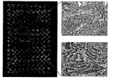
- FIG. 1 is a diagram showing a cryosection ITO slide and an enlarged H & E staining slide for malignant tumor specimens.
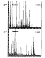
- MALDI MS Maldi mass spectrometry
- FIG. 3 is a diagram showing a Principal component analysis plot of papillary thyroid carcinoma; Each sphere represents a single sample, green represents papillary thyroid cancer and red represents a normal sample, and the lipid MALDI profiles of the samples are very similar and are located close to each other.
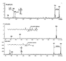
- FIG. 4 is a diagram showing molecular structure identification using MALDI LIFT (MS / MS) analysis.
- the present invention is a.
- step 3 provides a mass spectrometry method of phospholipids for providing cancer diagnosis information comprising the step of determining the malignancy by comparing the peak intensity value of step 2) with the control.
- the tissue is preferably, but not limited to, thyroid, colon, breast, lung, pancreas, and prostate derived tissue.
- the tissue is a frozen section of the biopsy tissue, but is not limited thereto.
- the peak intensity value is m / z 798.0-799.0 in positive ion mode or phosphatidylcholine 34: 1 [M + K] + (m / z: 798.6) compared to the normal control. Increases, and decreases the peak intensity value of m / z 524.0-525.0 in cationic mode or lysophosphatidylserine 18: 1 [M + H] + (m / z: 524.4), and m / z in cationic mode
- step 3) provides a mass spectrometry method of phospholipids for providing information of cancer diagnosis comprising the step of determining whether the tissue is malignant using the result value of step 2).
- the tissue is preferably, but not limited to, thyroid, colon, breast, lung, pancreas, and prostate derived tissue.
- the tissue is a frozen section of the biopsy tissue, but is not limited thereto.
- the tissue is preferably, but not limited to, a lysate of a cell aspirate sample, or a proximal fluid such as pleural effusion and ascites.
- the cancer is preferably selected from the group including thyroid cancer, colon cancer, breast cancer, lung cancer, pancreatic cancer, and prostate cancer, but is not limited thereto.
- step 3 it is preferable to compare the result value in step 3) with the result value derived from the normal control, but is not limited thereto.
- step 3 if the result value is 43.4 or more, it is determined as malignant tumor;
- pancreatic tissue if the result is 54.4 or more, it is determined as malignant tumor.
- prostate tissue In the case of prostate tissue, if the result value is 80.6 or more, it is preferable to determine it as a malignant tumor, but not limited thereto.
- the prostate tissue may be reset based on the median value of the malignant tumor tissue and the normal tissue in the training set.
- step 3 provides a mass spectrometry method for phospholipids to provide cancer diagnosis information comprising determining whether the tissue is malignant using the result value of step 2) as follows:
- cryosections were obtained for cryosections of 38 biopsy tissues collected from patients with thyroid cancer of the National Cancer Center (FIG. 1). Maldi mass spectrometry was performed on the prepared samples, and the resulting data was analyzed using FlexImaging software and ClinProTools program (FIG. 2).
- the compound covariate predictor algorithm was calculated based on the peak intensity values of the datasets for the three phospholipids (Equation 1). It was confirmed that 43.4, which is the median peak intensity value of thyroid cancer tissue, was used as a cutoff to determine whether a malignant tumor was present.
- cryosections were obtained for a total of 60 biopsy tissues taken from cancer patients composed of colorectal cancer, breast cancer, lung cancer, pancreatic cancer and prostate cancer, and mass spectrometry was performed in the same manner as mass spectrometry using thyroid cancer tissue described above.
- the normalized peak intensity values of phospholipid phosphatidylcholine 34: 1 [M + K] + were significantly increased in the tissues of the cancer patients, similar to those of the thyroid cancer tissues.
- phosphatidylcholine 34 1 [M + K] + (m / z: 798.6), lysophosphatidylserine, using lipid maldi mass spectrometry, for biopsy tissues that are difficult to accurately diagnose by conventional pathological methods Normalized peak intensity values for (lysophosphatidylserine) 18: 1 [M + H] + (m / z: 524.4), and lysophosphatidylcholine 18: 3 [M + H] + (m / z: 518.4) It can be useful to determine whether the tissue is malignant by measuring and applying it.
- Biopsy tissue was collected from 38 patients who underwent thyroidectomy or partial resection at the National Cancer Center Thyroid Cancer Center from 2012 to 2013. Cryosections were obtained from the thyroid biopsy tissue to a thickness of 10 um and cryosectioned.
- MALDI mass-assisted laser desorption / ionization
- MS mass spectrometry
- one of the frozen sections is subjected to hematoxylin / eosin (H & E) staining, corresponding to epithelial cells. Only spots are marked for selection (right side of FIG. 1).
- Samples were prepared in the same manner as in Example ⁇ 1-1> from patients being treated for colon cancer, breast cancer, lung cancer, pancreatic cancer, and prostate cancer, respectively.
- a lysate of biopsy tissue or a proximal fluid such as pleural fluid and ascites is used, but is not limited thereto.
- 13, 26, 11, 4 and 6, i.e., 60 samples were respectively obtained for tumor tissues including colon cancer, breast cancer, lung cancer, pancreatic cancer and prostate cancer.
- ClinProTools were as follows: resolution, 800; Top hat baseline with 10% minimal baseline width as baseline subtraction; Reference and recalibration in between the peaks, the maximum peak moves by weight 2,000 ppm (recalibratation with a maximal peak shift of 2,000 ppm between reference and peak masses); % Match to calibrant peaks, 10%; Signal-to-noise ratio cutoff, 5; Peak area for peak calculation with zero level integration.
- Table 1 ⁇ b> Standardized peak intensity values and corresponding phospholipids in normal and malignant thyroid tissue ⁇ / b> m / v P FDR Normal tissue Thyroid Cancer Malignant Tumor Tissue Ratio 1 Phospholipids p798.6 0.0001 0.0005 7.5 14.5 1.9 PC (34: 1) K + p524.4 0.0002 0.0012 2.2 3.5 0.4 LPS (18: 1) p518.4 0.0003 0.0015 1.7 1.2 0.7 LPC (18: 3)
- PC phosphatidylcholine
- LPS lysophosphatidylserine
- LPC Lysophosphatidylcholine
- compound covariate predictor based on a neural algorithm that discriminates malignant tumor tissue from normal tissue using peak intensity values of the three phospholipids based on the t-value in the dataset. predictor) algorithm (Equation 1) was calculated.
- Example ⁇ 1-1> The mass fraction of the thyroid cancer obtained in Example ⁇ 1-1> was performed according to ⁇ Example 2>. Based on the results, each m / z 798.6; Phosphatidylcholine 34: 1 [M + K] + , m / z 524.4; The standardized peak intensity values of lysophosphatidylserine 18: 1 [M + H] + , m / z 518.4, and lysophosphatidylcholine 18: 3 [M + H] + , respectively, are shown in Equation 1 of ⁇ Example 2>. It was calculated by substituting in parentheses, and determined whether or not it had a malignant tumor (Equation 1).
- the prediction rate in 38 samples is 100% accurate (sensitivity, 100%; specificity, 100%) and the calculated result is medium A median value of 43.4 or more was determined as a malignancy.
- the calculated result is less than the median 43.4 may be a benign tumor or normal.
- Example ⁇ 1-2> Malditic mass spectrometry was performed on the frozen sections of each tissue selected from colorectal cancer, breast cancer, lung cancer, pancreatic cancer and prostate cancer obtained in Example ⁇ 1-2> in the same manner as in ⁇ Example 2>.
- m / z 798.6 significantly changed in thyroid cancer tissue as shown in Table 2 below;
- Three phospholipid expression patterns consisting of lysophosphatidylcholine 18: 3 [M + H] + showed similar expression patterns in malignant tumor tissues of the group consisting of colon cancer, breast cancer, lung cancer, pancreatic cancer and prostate cancer (Table 2). ).
- tumor tissue total 60 samples including colon cancer, breast cancer, lung cancer, pancreatic cancer and prostate cancer tissue
- normal tissue total 60 samples including colon cancer, breast cancer, lung cancer, pancreatic cancer and prostate cancer tissue
- Example ⁇ 1-2> 13 samples were prepared by the method of Example ⁇ 1-2>, and Maldi mass spectrometry was performed in the same manner as in ⁇ Example 2>.
- the training set (training set) according to the ⁇ Example 2> according to the aspect of the mass spectrometry results showed an accuracy of 100% (13/13).
- the reference cutoff value for determining the malignancy can be specified as 97.9, the median value. In general, more than 97.9 cases were determined as malignant tumors, and the accuracy thereof was 84.6%. In addition, if the calculated result is less than the median value of 97.9 may be a benign tumor or normal.
- Example ⁇ 1-2> 26 samples were prepared by the method of Example ⁇ 1-2>, and Maldi mass spectrometry was performed in the same manner as in ⁇ Example 2>.
- Maldi mass spectrometry was performed in the same manner as in ⁇ Example 2>.
- analyzed by a comparative sample and compared the aspect of the Maldi mass spectrometry according to the ⁇ Example 2> showed an accuracy of 84.6% (22/26).
- the reference cutoff value for determining the malignancy can be designated as -85.5, which is the median value.
- -85.5 is the median value.
- more than -85.5 was diagnosed as malignant tumor and its accuracy was 69.2%.
- the calculated result is less than the median value of -85.5 may be benign tumor or normal.
- Example ⁇ 1-2> 11 samples were prepared by the method of Example ⁇ 1-2>, and Maldi mass spectrometry was performed in the same manner as in ⁇ Example 2>.
- Maldi mass spectrometry results showed an accuracy of 90.9% (10/11).
- the reference cutoff value for determining the malignancy can be designated as the median 64.7.
- the accuracy was 90.9%.
- the calculated result is less than the median value of 64.7 may be benign tumor or normal.
- Example ⁇ 1-2> In the case of prostate cancer tissue, six samples were prepared by the method of Example ⁇ 1-2>, and Maldi mass spectrometry was performed in the same manner as in ⁇ Example 2>. When analyzed by a comparative sample according to the ⁇ Example 2> compared the aspect of the mass spectrometry results showed the accuracy of 100% (6/6).
- Example ⁇ 1-2> In the case of pancreatic cancer tissue, four samples were prepared by the method of Example ⁇ 1-2>, and Maldi mass spectrometry was performed in the same manner as in ⁇ Example 2>. When analyzed by a comparative sample according to the ⁇ Example 2> compared the aspect of the mass spectrometry results showed the accuracy of 100% (4/4).
- the reference cutoff value for determining the malignancy can be designated as the median 54.4.
- the median 54.4 As a rule, more than 54.4 were considered as malignant tumors and the accuracy thereof was 75.0%.
- the calculated result is less than the median value of 54.4 may be a benign tumor or normal.
- Example ⁇ 2-1> it is applicable not only to the diagnosis of thyroid cancer but also to the diagnosis of other colon cancer, breast cancer, lung cancer, pancreatic cancer, or prostate cancer.
- the analysis of the new biopsy tissues in the future is carried out in the same manner, but the existing positive / malignant biopsy tissues are always analyzed simultaneously with the comparative sample, and then the standardized peak intensities are substituted into the above formulas and the calculation is performed. This can significantly discriminate whether cancer is malignant.
Landscapes
- Health & Medical Sciences (AREA)
- Life Sciences & Earth Sciences (AREA)
- Engineering & Computer Science (AREA)
- Chemical & Material Sciences (AREA)
- Immunology (AREA)
- Molecular Biology (AREA)
- Biomedical Technology (AREA)
- Hematology (AREA)
- Urology & Nephrology (AREA)
- Physics & Mathematics (AREA)
- Analytical Chemistry (AREA)
- General Health & Medical Sciences (AREA)
- Biochemistry (AREA)
- Medicinal Chemistry (AREA)
- Biotechnology (AREA)
- Food Science & Technology (AREA)
- General Physics & Mathematics (AREA)
- Pathology (AREA)
- Microbiology (AREA)
- Organic Chemistry (AREA)
- Zoology (AREA)
- Cell Biology (AREA)
- Proteomics, Peptides & Aminoacids (AREA)
- Wood Science & Technology (AREA)
- Biophysics (AREA)
- Bioinformatics & Cheminformatics (AREA)
- General Engineering & Computer Science (AREA)
- Genetics & Genomics (AREA)
- Other Investigation Or Analysis Of Materials By Electrical Means (AREA)
- Investigating Or Analysing Biological Materials (AREA)
Abstract
본 발명은 인지질을 이용한 암의 진단 방법에 관한 것으로, 구체적으로 기존의 병리학적 방법으로 정확한 진단이 어려운 경우가 있는 생검조직에 대해 지질 말디 질량분석법(lipid MALDI MS)을 이용하여 양이온 모드(positive ion mode)의 m/z 798.0-799.0 또는 포스파티딜콜린(phosphatidylcholine) 34:1 [M+K]+(m/z: 798.6), 양이온 모드의 m/z 524.0-525.0 또는 리소포스파티딜세린(lysophosphatidylserine) 18:1 [M+H]+(m/z: 524.4), 및 양이온 모드의 m/z 518.0-519.0 또는 리소포스파티딜콜린(lysophosphatidylcholine) 18:3 [M+H]+(m/z: 518.4) 각각에 대한 표준화된 피크 강도 값을 측정 및 응용하여 조직의 악성여부를 판단하는데 유용하게 사용될 수 있다.
Description
본 발명은 피검체로부터 분리된 조직에서 말디 질량분석(matrix-assisted laser desorption/ionization mass spectrometry, MALDI MS)을 이용하여 양이온 모드(positive ion mode)의 m/z 798.0-799.0 또는 포스파티딜콜린(phosphatidylcholine) 34:1 [M+K]+(m/z: 798.6), 양이온 모드의 m/z 524.0-525.0 또는 리소포스파티딜세린(lysophosphatidylserine) 18:1 [M+H]+(m/z: 524.4), 및 양이온 모드의 m/z 518.0-519.0 또는 리소포스파티딜콜린(lysophosphatidylcholine) 18:3 [M+H]+(m/z: 518.4) 각각에 대한 표준화된 피크 강도 값을 측정 및 응용하여 조직의 악성여부를 판단하는 암 진단의 정보를 제공하기 위한 인지질의 질량분석 방법에 관한 것이다.
조직-직접적 말디 질량분석 이미징(histology-directed matrix-assisted laser desorption/ionization(MALDI) mass spectrometry imaging)은 말디 이온화법을 이용한 이미징 질량분석 방법으로 1999년에 Richard M. Caprioli에 의해서 개발되었다. 분석하고자 하는 조직 표면에 매트릭스(matrix)를 도포하여 말디 이온화법을 적용하여, 조직으로부터 직접질량 분석 데이터를 얻는다. 조직-직접적 말디 질량분석법은 생검조직에서 이온화된 단백질, 펩타이드, 지질, 또는 소형 분자(small molecules)의 분포에 대한 질량분석 정보를 표지 물질을 사용하지 않고(label-free) 조직으로부터 직접 획득하는 방법이다. 이 방법은 정상 조직과 종양 조직을 구분할 수 있는 민감도가 높은 단백질체학적 분석을 가능하도록 한다(J Proteeome Res 2010; 9: 4123-4130).
말디 질량분석이 인체조직에 직접 이용되기 시작한 기술의 역사는 오래되지 않았으나, 시나피닉 산(sinapinic acid)을 매트릭스로 하여 동결조직(frozen tissue)을 분석하는 경우 암의 진단과 병리학적 분류에 유용한 정보를 제공할 수 있다는 것이 보고되어 있다(Lancet 2003; 362(9382): 433-449, Cancer Res 2005; 65(17): 7674-7681, Mol Cell Proteomics 2006; 5: 1975-1983). 말디 질량분석을 이용하여 정상 조직과 폐암 조직의 지질 프로파일을 비교한 결과, 분명한 차이가 존재함이 보고되었다(Lung Cancer 2012; 76: 197-203). 최근 Ishikawa 등(PLoS One 2012; 7: e48873)이 갑상선암 특이적 지질 말디 질량분석 프로파일을 보고하였으나, 적은 수의 표본에 대한 결과로 많은 수의 환자에 임상적으로 적용가능하지 않다.
이에 본 발명자들은 국립암센터에서 수술적으로 절제된 폐암, 유방암, 대장암, 전립선암, 갑상선 암 등의 동결 수술 조직에 대한 말디 질량분석을 이용하여 암과 주변정상 조직 간의 인지질 차이를 측정한 결과, 포스파티딜콜린(phosphatidylcholine) 34:1 [M+K]+(m/z: 798.6)의 표준화된 피크 강도 값은 증가하고, 리소포스파티딜세린(lysophosphatidylserine) 18:1 [M+H]+(m/z: 524.4) 및 리소포스파티딜콜린(lysophosphatidylcholine) 18:3 [M+H]+(m/z: 518.4))의 표준화된 피크 강도 값은 감소함을 확인함(P<10-7)으로써, 이와 같은 인지질의 발현 차이는 대한민국 호발암에서 전반적으로 관찰되는 현상으로 진단에 유용함을 확인함으로써 본 발명을 완성하였다.
그 중 갑상선암을 예로 들면, 갑상선암은 대한민국에서 가장 발병률이 높은 암이고, 이 중 유두갑상선암(papillary thyroid carcinoma)의 조직 형태가 가장 빈번하게 관찰된다(Cancer Res Treat 2012; 44: 11-24, Thyroid 2013). 수술 전 조직학적 진단은 생체검사(biopsy) 표본을 대상으로 이루어진다. National Cancer Institute Thyroid FNA State of the Science Conference에 따르면, 갑상선 부위는 양성(benign), 이형(atypia), 소낭의 종양(follicular neoplasm), 악성종양 의심(suspicious for malignancy), 및 악성종양(malignant)(Cancer 2008; 114: 71-73, Tyroid 2009; 19: 1159-1165)으로 분류된다. 악성종양 의심에 상기 유두갑상선암이 포함되며, 이의 조직은 미묘한 핵 중심부(focal nuclear)의 구조적인 변화를 수반한다. 악성종양 의심이라고 명명되는 소결(nodule)은 대개 절제되고, 이 중 60 내지 75%의 대부분은 유두암(papillary carcinoma)으로 판정된다(Tyroid 2009; 19: 1159-1165). 따라서, 작은 조직 표본을 사용하여 유두갑상선암을 진단하는 데에 어려움이 존재하여, 갑상선암에 대한 더욱 민감도 및 특이도가 높은 진단 방법이 시급히 필요한 상황이다(Minerva Chir 2010; 65: 83-93).
본 발명의 목적은
1) 악성종양이 의심되는 실험군 및 대조군에서 각각 조직을 분리하는 단계;
2) 상기 단계 1)에서 분리한 실험군 및 대조군 조직에서 말디 질량분석(matrix-assisted laser desorption/ionization mass spectrometry, MALDI MS)을 이용하여 양이온 모드(positive ion mode)의 m/z 798.0-799.0 또는 포스파티딜콜린(phosphatidylcholine) 34:1 [M+K]+(m/z: 798.6), 양이온 모드의 m/z 524.0-525.0 또는 리소포스파티딜세린(lysophosphatidylserine) 18:1 [M+H]+(m/z: 524.4), 및 양이온 모드의 m/z 518.0-519.0 또는 리소포스파티딜콜린(lysophosphatidylcholine) 18:3 [M+H]+(m/z: 518.4) 각각의 표준화된 피크 강도 값을 측정하는 단계; 및
3) 상기 단계 2)의 피크 강도 값을 대조군과 비교하여 악성여부를 판단하는 단계를 포함하는 암 진단의 정보를 제공하기 위한 인지질의 질량분석 방법을 제공하는 것이다.
상기 목적을 달성하기 위하여, 본 발명은
1) 악성종양이 의심되는 실험군 및 대조군에서 각각 조직을 분리하는 단계;
2) 상기 단계 1)에서 분리한 실험군 및 대조군 조직에서 말디 질량분석(matrix-assisted laser desorption/ionization mass spectrometry, MALDI MS)을 이용하여 양이온 모드(positive ion mode)의 m/z 798.0-799.0 또는 포스파티딜콜린(phosphatidylcholine) 34:1 [M+K]+(m/z: 798.6), 양이온 모드의 m/z 524.0-525.0 또는 리소포스파티딜세린(lysophosphatidylserine) 18:1 [M+H]+(m/z: 524.4), 및 양이온 모드의 m/z 518.0-519.0 또는 리소포스파티딜콜린(lysophosphatidylcholine) 18:3 [M+H]+(m/z: 518.4) 각각의 표준화된 피크 강도 값을 측정하는 단계; 및
3) 상기 단계 2)의 피크 강도 값을 대조군과 비교하여 악성여부를 판단하는 단계를 포함하는 암 진단의 정보를 제공하기 위한 인지질의 질량분석 방법을 제공한다.
또한, 본 발명은
1) 피검체로부터 분리된 조직에서 말디 질량분석(matrix-assisted laser desorption/ionization mass spectrometry, MALDI MS)을 이용하여 양이온 모드(positive ion mode)의 m/z 798.0-799.0 또는 포스파티딜콜린(phosphatidylcholine) 34:1 [M+K]+(m/z: 798.6), 양이온 모드의 m/z 524.0-525.0 또는 리소포스파티딜세린(lysophosphatidylserine) 18:1 [M+H]+(m/z: 524.4), 및 양이온 모드의 m/z 518.0-519.0 또는 리소포스파티딜콜린(lysophosphatidylcholine) 18:3 [M+H]+(m/z: 518.4) 각각의 표준화된 피크 강도 값을 측정하는 단계;
2) 상기 단계 1)의 양이온 모드(positive ion mode)의 m/z 798.0-799.0 또는 포스파티딜콜린(phosphatidylcholine) 34:1 [M+K]+(m/z: 798.6), 양이온 모드의 m/z 524.0-525.0 또는 리소포스파티딜세린(lysophosphatidylserine) 18:1 [M+H]+(m/z: 524.4), 및 양이온 모드의 m/z 518.0-519.0 또는 리소포스파티딜콜린(lysophosphatidylcholine) 18:3 [M+H]+(m/z: 518.4) 각각에 대하여 측정한 피크 강도 값을 하기 [수학식 1]에 대입하여 결과값을 산출하는 단계; 및
[수학식 1]

(m/z 798.6): 양이온 모드(positive ion mode)의 m/z 798.0-799.0 또는 포스파티딜콜린 34:1 [M+K]+의 표준화된 피크 강도 값;
(m/z 524.4): 양이온 모드의 m/z 524.0-525.0 또는 리소포스파티딜세린 18:1 [M+H]+의 표준화된 피크 강도 값; 및
(m/z 518.4): 양이온 모드의 m/z 518.0-519.0 또는 리소포스파티딜콜린 18:3 [M+H]+의 표준화된 피크 강도 값.
3) 상기 단계 2)의 결과값을 이용하여 조직의 악성여부를 판단하는 단계를 포함하는 암 진단의 정보를 제공하기 위한 인지질의 질량분석 방법을 제공한다.
또한, 본 발명은
1) 피검체로부터 분리된 조직에서 말디 질량분석(matrix-assisted laser desorption/ionization mass spectrometry, MALDI MS)을 이용하여 양이온 모드(positive ion mode)의 m/z 798.0-799.0 또는 포스파티딜콜린(phosphatidylcholine) 34:1 [M+K]+(m/z: 798.6), 양이온 모드의 m/z 524.0-525.0 또는 리소포스파티딜세린(lysophosphatidylserine) 18:1 [M+H]+(m/z: 524.4), 및 양이온 모드의 m/z 518.0-519.0 또는 리소포스파티딜콜린(lysophosphatidylcholine) 18:3 [M+H]+(m/z: 518.4) 각각의 표준화된 피크 강도 값을 측정하는 단계;
2) 상기 단계 1)의 양이온 모드의 m/z 798.0-799.0 또는 포스파티딜콜린(phosphatidylcholine) 34:1 [M+K]+(m/z: 798.6), 양이온 모드의 m/z 524.0-525.0 또는 리소포스파티딜세린(lysophosphatidylserine) 18:1 [M+H]+(m/z: 524.4), 및 양이온 모드의 m/z 518.0-519.0 또는 리소포스파티딜콜린(lysophosphatidylcholine) 18:3 [M+H]+(m/z: 518.4) 각각에 대하여 측정한 피크 강도 값을 하기 [수학식 1]에 대입하여 결과값을 산출하는 단계; 및
[수학식 1]

(m/z 798.6): 양이온 모드(positive ion mode)의 m/z 798.0-799.0 또는 포스파티딜콜린 34:1 [M+K]+의 표준화된 피크 강도 값;
(m/z 524.4): 양이온 모드의 m/z 524.0-525.0 또는 리소포스파티딜세린 18:1 [M+H]+의 표준화된 피크 강도 값; 및
(m/z 518.4): 양이온 모드의 m/z 518.0-519.0 또는 리소포스파티딜콜린 18:3 [M+H]+의 표준화된 피크 강도 값.
3) 상기 단계 2)의 결과값을 이용하여 조직의 악성여부를 판단하는 단계를 포함하는 암 진단의 정보를 제공하기 위한 인지질의 질량분석 방법을 제공한다:
i) 갑상선 조직의 경우, 결과값이 43.4 이상이면 악성종양으로 판정하고;
ii) 대장 조직의 경우, 결과값이 97.9 이상이면 악성종양으로 판정하고;
iii) 유방 조직의 경우, 결과값이 -85.5 이상이면 악성종양으로 판정하고;
iv) 폐 조직의 경우, 결과값이 64.7 이상이면 악성종양으로 판정하고;
v) 췌장 조직의 경우, 결과값이 54.4 이상이면 악성종양으로 판정하고; 및
vi) 전립선 조직의 경우, 결과값이 80.6 이상이면 악성종양으로 판정함.
본 발명은 말디 질량분석(matrix-assisted laser desorption/ionization mass spectrometry, MALDI MS)을 이용하여 양이온 모드(positive ion mode)의 m/z 798.0-799.0 또는 포스파티딜콜린(phosphatidylcholine) 34:1 [M+K]+(m/z: 798.6), 양이온 모드의 m/z 524.0-525.0 또는 리소포스파티딜세린(lysophosphatidylserine) 18:1 [M+H]+(m/z: 524.4), 및 양이온 모드의 m/z 518.0-519.0 또는 리소포스파티딜콜린(lysophosphatidylcholine) 18:3 [M+H]+(m/z: 518.4) 각각에 대한 표준화된 피크 강도 값을 측정 및 응용하여 기존의 병리학적 방법으로 정확한 진단이 어려운 상기 암 생검조직에 대하여 악성종양 여부를 판정 가능하도록 하고, 임상적 치료지침을 제공하여, 악성종양 여부 진단법으로 유용하게 이용될 수 있다.
도 1은 악성 종양 표본에 대한 크리오섹션(cryosection) ITO 슬라이드 및 확대된 H&E 염색 슬라이드를 나타내는 도이다.
도 2는 평균 말디 질량분석(MALDI MS) 스펙트럼을 나타내는 도이다; 적색은 갑상선암조직 및 녹색은 정상조직을 나타낸다.
도 3은 유두갑상선암(papillary thyroid carcinoma)의 주성분 분석 플랏(Principal component analysis plot)을 나타내는 도이다; 각각의 구는 단일 샘플을 나타내고, 녹색은 유두갑상선암 및 적색은 정상 샘플을 의미하며, 샘플들의 지질 MALDI 프로파일은 매우 유사하여 서로 근접하게 위치한다.
도 4는 말디 리프트(MALDI LIFT(MS/MS)) 분석을 이용한 분자구조 동정을 나타내는 도이다.
이하, 본 발명을 상세하게 설명한다.
본 발명은
1) 악성종양이 의심되는 실험군 및 대조군에서 각각 조직을 분리하는 단계;
2) 상기 단계 1)에서 분리한 실험군 및 대조군 조직에서 말디 질량분석(matrix-assisted laser desorption/ionization mass spectrometry, MALDI MS)을 이용하여 양이온 모드(positive ion mode)의 m/z 798.0-799.0 또는 포스파티딜콜린(phosphatidylcholine) 34:1 [M+K]+(m/z: 798.6), 양이온 모드의 m/z 524.0-525.0 또는 리소포스파티딜세린(lysophosphatidylserine) 18:1 [M+H]+(m/z: 524.4), 및 양이온 모드의 m/z 518.0-519.0 또는 리소포스파티딜콜린(lysophosphatidylcholine) 18:3 [M+H]+(m/z: 518.4) 각각의 표준화된 피크 강도 값을 측정하는 단계; 및
3) 상기 단계 2)의 피크 강도 값을 대조군과 비교하여 악성여부를 판단하는 단계를 포함하는 암 진단의 정보를 제공하기 위한 인지질의 질량분석 방법을 제공한다.
구체적으로, 상기 조직은 갑상선, 대장, 유방, 폐, 췌장, 및 전립선 유래 조직인 것이 바람직하나 이에 한정하지 않는다.
상기 조직은 생검조직의 동결절편을 이용하는 것이 바람직하나 이에 한정하지 않는다.
상기 피크 강도 값은 정상 대조군과 비교하여 양이온 모드(positive ion mode)의 m/z 798.0-799.0 또는 포스파티딜콜린(phosphatidylcholine) 34:1 [M+K]+(m/z: 798.6)의 피크 강도 값은 증가하고, 양이온 모드의 m/z 524.0-525.0 또는 리소포스파티딜세린(lysophosphatidylserine) 18:1 [M+H]+(m/z: 524.4)의 피크 강도 값은 감소하며, 및 양이온 모드의 m/z 518.0-519.0 또는 리소포스파티딜콜린(lysophosphatidylcholine) 18:3 [M+H]+(m/z: 518.4)의 피크 강도 값은 감소한다.
또한, 본 발명은
1) 피검체로부터 분리된 조직에서 말디 질량분석(matrix-assisted laser desorption/ionization mass spectrometry, MALDI MS)을 이용하여 양이온 모드(positive ion mode)의 m/z 798.0-799.0 또는 포스파티딜콜린(phosphatidylcholine) 34:1 [M+K]+(m/z: 798.6), 양이온 모드의 m/z 524.0-525.0 또는 리소포스파티딜세린(lysophosphatidylserine) 18:1 [M+H]+(m/z: 524.4), 및 양이온 모드의 m/z 518.0-519.0 또는 리소포스파티딜콜린(lysophosphatidylcholine) 18:3 [M+H]+(m/z: 518.4) 각각의 표준화된 피크 강도 값을 측정하는 단계;
2) 상기 단계 1)의 양이온 모드의 m/z 798.0-799.0 또는 포스파티딜콜린(phosphatidylcholine) 34:1 [M+K]+(m/z: 798.6), 양이온 모드의 m/z 524.0-525.0 또는 리소포스파티딜세린(lysophosphatidylserine) 18:1 [M+H]+(m/z: 524.4), 및 양이온 모드의 m/z 518.0-519.0 또는 리소포스파티딜콜린(lysophosphatidylcholine) 18:3 [M+H]+(m/z: 518.4) 각각에 대하여 측정한 피크 강도 값을 하기 [수학식 1]에 대입하여 결과값을 산출하는 단계; 및
[수학식 1]

(m/z 798.6): 양이온 모드(positive ion mode)의 m/z 798.0-799.0 또는 포스파티딜콜린 34:1 [M+K]+의 표준화된 피크 강도 값;
(m/z 524.4): 양이온 모드의 m/z 524.0-525.0 또는 리소포스파티딜세린 18:1 [M+H]+의 표준화된 피크 강도 값; 및
(m/z 518.4): 양이온 모드의 m/z 518.0-519.0 또는 리소포스파티딜콜린 18:3 [M+H]+의 표준화된 피크 강도 값.
3) 상기 단계 2)의 결과값을 이용하여 조직의 악성여부를 판단하는 단계를 포함하는 암 진단의 정보를 제공하기 위한 인지질의 질량분석 방법을 제공한다.
구체적으로, 상기 조직은 갑상선, 대장, 유방, 폐, 췌장, 및 전립선 유래 조직인 것이 바람직하나 이에 한정하지 않는다.
상기 조직은 생검조직의 동결절편을 이용하는 것이 바람직하나 이에 한정하지 않는다.
상기 조직은 세포흡인검체의 용해물(lysate), 또는 흉수 및 복수와 같은 근위 체액(proximal fluid)을 대상으로 하는 것이 바람직하나 이에 한정하지 않는다.
또한, 상기 암은 갑상선암, 대장암, 유방암, 폐암, 췌장암, 및 전립선암을 포함한 군으로부터 선택되는 것이 바람직하나 이에 한정하지 않는다.
상기 방법에 있어서, 단계 3)에서 결과값은 정상 대조군 유래 결과값과 비교하는 것이 바람직하나 이에 한정하지 않는다.
또한, 상기 단계 3)에서 갑상선 조직의 경우, 결과값이 43.4 이상이면 악성종양으로 판정하고;
대장 조직의 경우, 결과값이 97.9 이상이면 악성종양으로 판정하고;
유방 조직의 경우, 결과값이 -85.5 이상이면 악성종양으로 판정하고;
폐 조직의 경우, 결과값이 64.7 이상이면 악성종양으로 판정하고;
췌장 조직의 경우, 결과값이 54.4 이상이면 악성종양으로 판정하고; 및
전립선 조직의 경우, 결과값이 80.6 이상이면 악성종양으로 판정하는 것이 바람직하나 이에 한정하지 않고, 비교 검체(training set)에서의 악성종양 조직과 정상 조직의 중간값을 기준으로 재설정이 가능하다.
또한, 본 발명은
1) 피검체로부터 분리된 조직에서 말디 질량분석(matrix-assisted laser desorption/ionization mass spectrometry, MALDI MS)을 이용하여 양이온 모드(positive ion mode)의 m/z 798.0-799.0 또는 포스파티딜콜린(phosphatidylcholine) 34:1 [M+K]+(m/z: 798.6), 양이온 모드의 m/z 524.0-525.0 또는 리소포스파티딜세린(lysophosphatidylserine) 18:1 [M+H]+(m/z: 524.4), 및 양이온 모드의 m/z 518.0-519.0 또는 리소포스파티딜콜린(lysophosphatidylcholine) 18:3 [M+H]+(m/z: 518.4) 각각의 표준화된 피크 강도 값을 측정하는 단계;
2) 상기 단계 1)의 양이온 모드의 m/z 798.0-799.0 또는 포스파티딜콜린(phosphatidylcholine) 34:1 [M+K]+(m/z: 798.6), 양이온 모드의 m/z 524.0-525.0 또는 리소포스파티딜세린(lysophosphatidylserine) 18:1 [M+H]+(m/z: 524.4), 및 양이온 모드의 m/z 518.0-519.0 또는 리소포스파티딜콜린(lysophosphatidylcholine) 18:3 [M+H]+(m/z: 518.4) 각각에 대하여 측정한 피크 강도 값을 하기 [수학식 1]에 대입하여 결과값을 산출하는 단계; 및
[수학식 1]

(m/z 798.6): 양이온 모드(positive ion mode)의 m/z 798.0-799.0 또는 포스파티딜콜린 34:1 [M+K]+의 표준화된 피크 강도 값;
(m/z 524.4): 양이온 모드의 m/z 524.0-525.0 또는 리소포스파티딜세린 18:1 [M+H]+의 표준화된 피크 강도 값; 및
(m/z 518.4): 양이온 모드의 m/z 518.0-519.0 또는 리소포스파티딜콜린 18:3 [M+H]+의 표준화된 피크 강도 값.
3) 상기 단계 2)의 결과값을 이용하여 하기와 같이 조직의 악성여부를 판단하는 단계를 포함하는 암 진단의 정보를 제공하기 위한 인지질의 질량분석 방법을 제공한다:
i) 갑상선 조직의 경우, 결과값이 43.4 이상이면 악성종양으로 판정하고;
ii) 대장 조직의 경우, 결과값이 97.9 이상이면 악성종양으로 판정하고;
iii) 유방 조직의 경우, 결과값이 -85.5 이상이면 악성종양으로 판정하고;
iv) 폐 조직의 경우, 결과값이 64.7 이상이면 악성종양으로 판정하고;
v) 췌장 조직의 경우, 결과값이 54.4 이상이면 악성종양으로 판정하고; 및
vi) 전립선 조직의 경우, 결과값이 80.6 이상이면 악성종양으로 판정함.
구체적으로, 국립암센터 갑상선암 환자로부터 채취한 생검조직 38개에 대하여 동결절편을 얻어 크리오섹션(cryosection)하였다(도 1). 준비된 시료로 말디 질량분석을 수행하였고, 그 결과로 얻은 데이터를 FlexImaging 소프트웨어 및 ClinProTools 프로그램을 이용하여 분석하였다(도 2). 정상 대조군의 결과와 비교하여 갑상선암 환자에서 인지질 포스파티딜콜린(phosphatidylcholine) 34:1 [M+K]+(m/z: 798.6)의 표준화된 피크 강도 값은 유의적으로 증가하였고, 리소포스파티딜세린(lysophosphatidylserine) 18:1 [M+H]+(m/z: 524.4) 및 리소포스파티딜콜린(lysophosphatidylcholine) 18:3 [M+H]+(m/z: 518.4)의 표준화된 피크 강도 값은 유의적으로 감소하였다(표 1). 상기 세 인지질에 대한 데이터세트(dataset)의 피크 강도 값을 기반으로 하여 화합물 공변량 예측변수 알고리즘을 산출하였다(수학식 1). 갑상선암 조직의 피크 강도 값의 중간값인 43.4를 컷오프(cutoff)로 하여 악성종양 여부를 판단할 수 있음을 확인하였다.
또한, 대장암, 유방암, 폐암, 췌장암 및 전립선암으로 구성된 암 환자로부터 채취한 생검조직 총 60개에 대하여 동결절편을 얻어, 상기 기재된 갑상선암 조직을 이용한 말디 질량분석과 동일하게 말디 질량분석을 수행하였다. 그 결과, 상기 암 환자의 조직에서도 갑상선암 조직의 결과와 유사하게, 인지질 포스파티딜콜린(phosphatidylcholine) 34:1 [M+K]+(m/z: 798.6)의 표준화된 피크 강도 값은 유의적으로 증가하였고, 리소포스파티딜세린(lysophosphatidylserine) 18:1 [M+H]+(m/z: 524.4) 및 리소포스파티딜콜린(lysophosphatidylcholine) 18:3 [M+H]+(m/z: 518.4)의 표준화된 피크 강도 값은 유의적으로 감소하였다(표 2). 화합물 공변량 예측변수 알고리즘의 악성종양 여부 판별 컷오프를 대장암의 경우 97.9로, 유방암의 경우 -85.5로, 폐암의 경우 64.7로, 췌장암의 경우 54.4로, 전립선암의 경우 80.6으로 할 수 있음을 확인하였다.
따라서, 기존의 병리학적 방법으로 정확한 진단이 어려운 경우가 있는 생검조직에 대해 지질 말디 질량분석법을 이용하여 포스파티딜콜린(phosphatidylcholine) 34:1 [M+K]+(m/z: 798.6), 리소포스파티딜세린(lysophosphatidylserine) 18:1 [M+H]+(m/z: 524.4), 및 리소포스파티딜콜린(lysophosphatidylcholine) 18:3 [M+H]+(m/z: 518.4) 각각에 대한 표준화된 피크 강도 값을 측정 및 응용하여 조직의 악성여부를 판단하는데 유용하게 사용될 수 있다.
이하, 본 발명을 실시예 및 실험예에 의해 상세히 설명한다.
단, 하기 실시예 및 실험예는 본 발명을 구체적으로 설명하는 것일 뿐, 본 발명의 내용이 하기 실시예 및 실험예에 의해 이에 한정되지 않는다.
<실시예 1> 조직의 준비
<1-1> 갑상선암 조직의 준비
국립암센터 갑상선암 센터에서 2012년 내지 2013년 동안 갑상선 전절제술 또는 부분절제술을 시행 받은 환자 38명으로부터 생검조직을 채취하였다. 갑상선 생검조직으로부터 10 um 두께로 동결절편을 얻어 크리오섹션(cryosection)하였다. 말디(matrix-assisted laser desorption/ionization, MALDI) 질량분석(mass spectrometry, MS)을 시행하기 위한 준비 단계로, 동결절편 중 하나는 H&E(hematoxylin/eosin) 염색을 수행하여, 상피 세포에 해당하는 부위(spot)만 선택하도록 표시하였다(도 1의 우측). 나머지 동결절편은 크리오섹션 ITO(indium tin oxide) 슬라이드로 준비하기 위하여, 상피세포 부위에 해당하는 곳에 Chip-1000(Shimadzu, Kyoto, Japan) 기계를 이용하여 7 mg의 2,5-디히드록시벤조산(2,5-dihydroxy benzoic acid, DHB)/알파-시아노-4-하이드록신나믹산(a-cyano-4-hydroxy cinnamic acid, CHCA)(1 ml의 70% 메탄올 및 0.1% TFA/1% 피페리딘(piperidine))을 매트릭스(matrix)로 하여 100 pl을 도포하였다(도 1의 좌측). 이때, Chip-1000 대신 마이크로피펫(micropipette)으로 250 nl씩 두 번 상기 매트릭스를 도포할 수도 있다. 동결절편을 ITO 슬라이드에 녹이면서 붙인(thaw-mount) 후, 진공건조기에서 건조하였다.
<1-2> 대장암, 유방암, 폐암, 췌장암 및 전립선암 조직의 준비
각각 대장암, 유방암, 폐암, 췌장암 및 전립선암으로 치료받고 있는 환자로부터 실시예 <1-1>과 동일한 방법으로 시료를 준비하였다. 이때, 생검조직의 용해물(lysate), 또는 흉수 및 복수와 같은 근위 체액(proximal fluid)을 사용하였으나, 이에 한정하지 않는다. 구체적으로, 대장암, 유방암, 폐암, 췌장암 및 전립선암을 포함한 종양 조직에 대하여 각각 13, 26, 11, 4 및 6개, 즉 전체 60개를 시료를 수득하였다.
<실시예 2> 말디 질량분석(MALDI MS)을 이용한 인지질 발현 양상 확인
말디 질량분석은 UltrafleXtreme MALDI time-of-light 질량 분석기(Bruker Daltonics, Bremen, Germany)로 스캔(scan)하였다. 이때 m/z 범위는 500 내지 1,100 Da으로 하고, 3000 consecutive laser shots(빈도: 1,000 Hz)을 주고 그 값을 평균으로 산출하였다.
FlexImaging 소프트웨어(version 2.1, Bruker Daltonics)를 사용하여 다시 한번 악성종양 표본의 상피세포 유래 인지질 구성에 대한 질량분석 데이터를 추출하였다(도 2). 이때 H&E 슬라이드를 참조하여 상피세포의 적색으로 표시된 해당 부분(도 1)에서의 질량 패턴만 ClinProTools(버전 2.2, Bruker Daltonics)에서 분석처리되도록 하였다. ClinProTools를 사용할 경우의 변수(parameter)는 하기와 같았다: 해상도(resolution), 800; 바탕선 제거시 최소 10% 길이로 바탕선 이동한 탑 햇 바탕선(top hat baseline with 10% minimal baseline width as baseline subtraction); 참조 및 피크 무게 사이에 2,000 ppm만큼 최대 피크 이동으로 재보정(recalibratation with a maximal peak shift of 2,000 ppm between reference and peak masses); 보정된 피크과 일치하는 퍼센트(% match to calibrant peaks), 10%; 신호-대-소음 비율 컷오프(signal-to-noise ratio cutoff), 5; 영점 수준으로 통합된 피크 계산을 위한 피크 지역(peak area for peak calculation with zero level integration).
갑상선암 조직 환자 1명의 시료(sample)로부터 하나의 스펙트럼(spectrum)만을 얻었고, 전체 38 개의 시료에 대한 피크 강도(intensity) 값의 평균값으로 데이터를 표준화(normalization)하였다. 표준화한 피크 강도 값의 분석 결과, 악성 갑상선암 조직에서 공통적으로 인지질 포스파티딜콜린(phosphatidylcholine, PC) 34:1 [M+K]+(m/z: 798.6)이 통계적으로 유의하게 증가하였고, 리소포스파티딜세린(lysophosphatidyl serine, LPS) 18:1 [M+H]+(m/z: 524.4) 및 리소포스파티딜콜린(lysophosphatidyl choline, LPC) 18:3 [M+H]+(m/z: 518.4)이 유의하게 감소하는 것을 확인하였다(표 1 및 도 3).
표 1 갑상선 정상 및 악성종양 조직에서 변화한 표준화한 피크 강도 값 및 해당 인지질
| m/v | P | FDR | 정상 조직 | 갑상선암 악성종양 조직 | 비율1 | 인지질 |
| p798.6 | 0.0001 | 0.0005 | 7.5 | 14.5 | 1.9 | PC (34:1) K+ |
| p524.4 | 0.0002 | 0.0012 | 2.2 | 3.5 | 0.4 | LPS (18:1) |
| p518.4 | 0.0003 | 0.0015 | 1.7 | 1.2 | 0.7 | LPC (18:3) |
1비율, 정상 조직에 대한 갑상선암 종양 조직(38개 시료)의 해당 지질 발현 비율
PC, 포스파티딜콜린; LPS, 리소포스파티딜세린; LPC, 리소포스파티딜콜린
또한, 데이터세트(dataset)에서 t-값을 근간으로 하여, 상기 3개의 인지질의 피크 강도 값을 이용한 악성종양 조직과 정상 조직을 감별하는 직선 알고리즘(near algorithm) 기반의 화합물 공변량 예측변수(compound covariate predictor) 알고리즘(수학식 1)을 산출하였다.
[수학식 1]

(m/z 798.6): 양이온 모드(positive ion mode)의 m/z 798.0-799.0 또는 포스파티딜콜린 34:1 [M+K]+의 표준화된 피크 강도 값;
(m/z 524.4): 양이온 모드의 m/z 524.0-525.0 또는 리소포스파티딜세린 18:1 [M+H]+의 표준화된 피크 강도 값; 및
(m/z 518.4): 양이온 모드의 m/z 518.0-519.0 또는 리소포스파티딜콜린 18:3 [M+H]+의 표준화된 피크 강도 값.
구체적으로, 각 시료에서 말디 질량분석으로 측정한 m/z 798.6; 포스파티딜콜린 34:1 [M+K]+, m/z 524.4; 리소포스파티딜세린 18:1 [M+H]+, 및 m/z 518.4, 리소포스파티딜콜린 18:3 [M+H]+의 표준화된 피크 강도 값을 각각 [수학식 1]의 상응하는 위치에 대입하여, 계산한 결과값의 크기에 따라 악성종양 여부를 예측할 수 있다. 그 값이 클수록 악성종양일 확률이 높아지는 것을 확인하였다.
<실험예 1> 갑상선암 조직의 말디 질량분석
상기 실시예 <1-1>에서 수득한 갑상선암의 동결절편을 <실시예 2>에 따라 말디 질량분석을 수행하였다. 그 결과를 바탕으로 각 m/z 798.6; 포스파티딜콜린 34:1 [M+K]+, m/z 524.4; 리소포스파티딜세린 18:1 [M+H]+, m/z 518.4, 및 리소포스파티딜콜린 18:3 [M+H]+의 표준화된 피크 강도 값을 각각 상기 <실시예 2>의 [수학식 1]의 괄호 안에 대입하여 산출하고 악성종양 여부를 판정하였다(수학식 1).
그 결과, 대비교(paired comparison)로 시행하는 경우, 38개 시료에서의 예측률은 정확도가 100% (민감도(sensitivity), 100%; 특이도(specificity), 100%)이며, 산출 결과값이 중간값(median value)인 43.4 이상이면 악성종양으로 판정하였다. 또한, 상기 산출 결과값이 중간값인 43.4 미만이면 양성종양이거나 정상일 수 있다.
또한, 상기 <실시예 2>의 [수학식 1]에 대입한 결과가 43.4보다 낮으면 양성질환으로 판정하는 컷오프를 일률적으로 적용하는 경우, 정확도는 84.2%(민감도, 87.5%; 특이도, 80%)였다. 이때 양성질환 20개 시료의 평균값은 22.0(범위, -4.5 내지 74.2)이고, 악성질환 16개 시료의 평균값은 133.9(범위, 28.2-294.8)였다.
<실험예 2> 대장암, 유방암, 폐암, 췌장암 및 전립선암 조직 분석
상기 실시예 <1-2>에서 수득한 대장암, 유방암, 폐암, 췌장암 및 전립선암으로부터 선택된 각 조직의 동결절편을 <실시예 2>와 동일한 방법으로 말디 질량분석을 수행하였다.
그 결과, 하기 표 2에 나타낸 바와 같이 갑상선암 조직에서 유의적으로 변화한 m/z 798.6; 포스파티딜콜린 34:1 [M+K]+, m/z 524.4; 리소포스파티딜세린 18:1 [M+H]+, 및 m/z 518.4; 리소포스파티딜콜린 18:3 [M+H]+로 이루어진 3개의 인지질 발현 양상이, 대장암, 유방암, 폐암, 췌장암 및 전립선암으로 이루어진 군의 악성종양 조직에서도 유사한 발현 양상을 나타냄을 확인하였다(표 2).
표 2 대장암, 유방암, 폐암, 췌장암, 전립선암 종양 조직에서 변화한 표준화한 피크 강도 값 데이터에서 t-값을 기반으로 분석한 결과
| m/z | P | FDR | 비율1 |
| p798.6 | <1e-07 | <1e-07 | 1.8 |
| p524.4 | <1e-07 | <1e-07 | 0.6 |
| p518.4 | <1e-07 | <1e-07 | 0.7 |
1비율, 정상 조직에 대한 종양 조직(대장암, 유방암, 폐암, 췌장암 및 전립선암 조직 포함 총 60개 시료)의 해당 지질 발현 비율.
<2-1> 대장암 조직의 말디 질량분석
대장암 조직의 경우, 13개의 시료를 상기 실시예 <1-2>의 방법으로 준비하고, 상기 <실시예 2>와 동일한 방법으로 말디 질량분석을 수행하였다. 비교 검체(training set)로 분석하여 상기 <실시예 2>에 따라 말디 질량분석 결과의 양상을 비교할 때 100%(13/13)의 정확도를 보였다.
상기 <실시예 2>의 [수학식 1]을 대장암 조직의 데이터에 적용한 결과, 악성종양 여부를 판단하는 기준 컷오프 값을 중간값인 97.9로 지정할 수 있음을 확인하였다. 일률적으로 97.9 이상인 경우 악성종양으로 판정하였고, 이의 정확도는 84.6%였다. 또한, 상기 산출 결과값이 중간값인 97.9 미만이면 양성종양이거나 정상일 수 있다.
<2-2> 유방암 조직의 말디 질량분석
유방암 조직의 경우, 26개의 시료를 상기 실시예 <1-2>의 방법으로 준비하고, 상기 <실시예 2>와 동일한 방법으로 말디 질량분석을 수행하였다. 비교 검체로 분석하여 상기 <실시예 2>에 따라 말디 질량분석 결과의 양상을 비교할 때 84.6%(22/26)의 정확도를 보였다.
상기 <실시예 2>의 [수학식 1]을 유방암 조직의 결과에 적용한 결과 악성종양 여부를 판단하는 기준 컷오프 값을 중간값인 -85.5로 지정할 수 있음을 확인하였다. 일률적으로 -85.5 이상인 경우 악성종양으로 판정하였고, 이의 정확도는 69.2%였다. 또한, 상기 산출 결과값이 중간값인 -85.5 미만이면 양성종양이거나 정상일 수 있다.
<2-3> 폐암 조직의 말디 질량분석
폐암 조직의 경우, 11개의 시료를 상기 실시예 <1-2>의 방법으로 준비하고, 상기 <실시예 2>와 동일한 방법으로 말디 질량분석을 수행하였다. 비교 검체로 분석하여 상기 <실시예 2>에 따라 말디 질량분석 결과의 양상을 비교할 때 90.9%(10/11)의 정확도를 보였다.
상기 <실시예 2>의 [수학식 1]을 폐암 조직의 결과에 적용한 결과 악성종양 여부를 판단하는 기준 컷오프 값을 중간값인 64.7로 지정할 수 있음을 확인하였다. 일률적으로 64.7 이상인 경우 악성종양으로 판정하였고, 이의 정확도는 90.9%였다. 또한, 상기 산출 결과값이 중간값인 64.7 미만이면 양성종양이거나 정상일 수 있다.
<2-4> 전립선암 조직의 말디 질량분석
전립선암 조직의 경우, 6개의 시료를 상기 실시예 <1-2>의 방법으로 준비하고, 상기 <실시예 2>와 동일한 방법으로 말디 질량분석을 수행하였다. 비교 검체로 분석하여 상기 <실시예 2>에 따라 말디 질량분석 결과의 양상을 비교할 때 100%(6/6)의 정확도를 보였다.
상기 <실시예 2>의 [수학식 1]을 전립선암 조직의 결과에 적용한 결과 악성종양 여부를 판단하는 기준 컷오프 값을 중간값인 80.6으로 지정할 수 있음을 확인하였다. 일률적으로 80.6 이상인 경우 악성종양으로 판정하였고, 이의 정확도는 83.3%였다. 또한, 상기 산출 결과값이 중간값인 80.6 미만이면 양성종양이거나 정상일 수 있다.
<2-5> 췌장암 조직의 말디 질량분석
췌장암 조직의 경우, 4개의 시료를 상기 실시예 <1-2>의 방법으로 준비하고, 상기 <실시예 2>와 동일한 방법으로 말디 질량분석을 수행하였다. 비교 검체로 분석하여 상기 <실시예 2>에 따라 말디 질량분석 결과의 양상을 비교할 때 100%(4/4)의 정확도를 보였다.
상기 <실시예 2>의 [수학식 1]을 췌장암 조직의 결과에 적용한 결과 악성종양 여부를 판단하는 기준 컷오프 값을 중간값인 54.4로 지정할 수 있음을 확인하였다. 일률적으로 54.4 이상인 경우 악성종양으로 판정하였고, 이의 정확도는 75.0%였다. 또한, 상기 산출 결과값이 중간값인 54.4 미만이면 양성종양이거나 정상일 수 있다.
따라서 상기 실시예 <2-1>과 같이 갑상선암의 진단뿐 아니라, 기타 대장암, 유방암, 폐암, 췌장암 또는 전립선암의 진단에도 적용 가능하다.
따라서, 향후 신환의 생검조직에 대한 분석도 동일한 방법으로 진행하되, 이때 기존 양성/악성 생검조직은 비교 검체로 항상 동시에 분석을 수행하여 이들과 표준화된 피크 강도를 상기 식에 대입 후, 계산을 수행하여 암의 악성 여부를 유의적으로 감별할 수 있다.
Claims (12)
1) 악성종양이 의심되는 실험군 및 대조군에서 각각 조직을 분리하는 단계;
2) 상기 단계 1)에서 분리한 실험군 및 대조군 조직에서 말디 질량분석(matrix-assisted laser desorption/ionization mass spectrometry, MALDI MS)을 이용하여 양이온 모드(positive ion mode)의 m/z 798.0-799.0 또는 포스파티딜콜린(phosphatidylcholine) 34:1 [M+K]+(m/z: 798.6), 양이온 모드의 m/z 524.0-525.0 또는 리소포스파티딜세린(lysophosphatidylserine) 18:1 [M+H]+(m/z: 524.4), 및 양이온 모드의 m/z 518.0-519.0 또는 리소포스파티딜콜린(lysophosphatidylcholine) 18:3 [M+H]+(m/z: 518.4) 각각의 표준화된 피크 강도 값을 측정하는 단계; 및
3) 상기 단계 2)의 피크 강도 값을 대조군과 비교하여 악성여부를 판단하는 단계를 포함하는 암 진단의 정보를 제공하기 위한 인지질의 질량분석 방법.
제 1항에 있어서, 상기 조직은 갑상선, 대장, 유방, 폐, 췌장, 및 전립선 유래 조직인 것을 특징으로 하는 방법.
제 1항에 있어서, 상기 조직은 생검조직의 동결절편인 것을 특징으로 하는 방법.
제 1항에 있어서, 상기 피크 강도 값은 정상 대조군과 비교하여 양이온 모드(positive ion mode)의 m/z 798.0-799.0 또는 포스파티딜콜린(phosphatidylcholine) 34:1 [M+K]+(m/z: 798.6)의 피크 강도 값은 증가하고, 양이온 모드의 m/z 524.0-525.0 또는 리소포스파티딜세린(lysophosphatidylserine) 18:1 [M+H]+(m/z: 524.4)의 피크 강도 값은 감소하며, 및 양이온 모드의 m/z 518.0-519.0 또는 리소포스파티딜콜린(lysophosphatidylcholine) 18:3 [M+H]+(m/z: 518.4)의 피크 강도 값은 감소하는 것을 특징으로 하는 방법.
1) 피검체로부터 분리된 조직에서 말디 질량분석(matrix-assisted laser desorption/ionization mass spectrometry, MALDI MS)을 이용하여 양이온 모드(positive ion mode)의 m/z 798.0-799.0 또는 포스파티딜콜린(phosphatidylcholine) 34:1 [M+K]+(m/z: 798.6), 양이온 모드의 m/z 524.0-525.0 또는 리소포스파티딜세린(lysophosphatidylserine) 18:1 [M+H]+(m/z: 524.4), 및 양이온 모드의 m/z 518.0-519.0 또는 리소포스파티딜콜린(lysophosphatidylcholine) 18:3 [M+H]+(m/z: 518.4) 각각의 표준화된 피크 강도 값을 측정하는 단계;
2) 상기 단계 1)의 양이온 모드의 m/z 798.0-799.0 또는 포스파티딜콜린(phosphatidylcholine) 34:1 [M+K]+(m/z: 798.6), 양이온 모드의 m/z 524.0-525.0 또는 리소포스파티딜세린(lysophosphatidylserine) 18:1 [M+H]+(m/z: 524.4), 및 양이온 모드의 m/z 518.0-519.0 또는 리소포스파티딜콜린(lysophosphatidylcholine) 18:3 [M+H]+(m/z: 518.4) 각각에 대하여 측정한 피크 강도 값을 하기 [수학식 1]에 대입하여 결과값을 산출하는 단계; 및
[수학식 1]

(m/z 798.6): 양이온 모드(positive ion mode)의 m/z 798.0-799.0 또는 포스파티딜콜린 34:1 [M+K]+의 표준화된 피크 강도 값;
(m/z 524.4): 양이온 모드의 m/z 524.0-525.0 또는 리소포스파티딜세린 18:1 [M+H]+의 표준화된 피크 강도 값; 및
(m/z 518.4): 양이온 모드의 m/z 518.0-519.0 또는 리소포스파티딜콜린 18:3 [M+H]+의 표준화된 피크 강도 값.
3) 상기 단계 2)의 결과값을 이용하여 조직의 악성여부를 판단하는 단계를 포함하는 암 진단의 정보를 제공하기 위한 인지질의 질량분석 방법.
제 5항에 있어서, 상기 조직은 갑상선, 대장, 유방, 폐, 췌장, 및 전립선 유래 조직인 것을 특징으로 하는 방법.
제 5항에 있어서, 상기 조직은 생검조직의 동결절편인 것을 특징으로 하는 방법.
제 5항에 있어서, 상기 조직은 세포흡인검체의 용해물(lysate), 또는 흉수 또는 복수를 포함하는 근위 체액(proximal fluid)을 대상으로 하는 것을 특징으로 하는 방법.
제 5항에 있어서, 상기 암은 갑상선암, 대장암, 유방암, 폐암, 췌장암, 및 전립선암을 포함한 군으로부터 선택되는 것을 특징으로 하는 방법.
제 5항에 있어서, 상기 단계 3)에서 결과값은 정상 대조군 유래 결과값과 비교하는 것을 특징으로 하는 방법.
제 5항에 있어서, 상기 단계 3)에서
갑상선 조직의 경우, 결과값이 43.4 이상이면 악성종양으로 판정하고;
대장 조직의 경우, 결과값이 97.9 이상이면 악성종양으로 판정하고;
유방 조직의 경우, 결과값이 -85.5 이상이면 악성종양으로 판정하고;
폐 조직의 경우, 결과값이 64.7 이상이면 악성종양으로 판정하고;
췌장 조직의 경우, 결과값이 54.4 이상이면 악성종양으로 판정하고; 및
전립선 조직의 경우, 결과값이 80.6 이상이면 악성종양으로 판정하는 것을 특징으로 하는 방법.
1) 피검체로부터 분리된 조직에서 말디 질량분석(matrix-assisted laser desorption/ionization mass spectrometry, MALDI MS)을 이용하여 양이온 모드(positive ion mode)의 m/z 798.0-799.0 또는 포스파티딜콜린(phosphatidylcholine) 34:1 [M+K]+(m/z: 798.6), 양이온 모드의 m/z 524.0-525.0 또는 리소포스파티딜세린(lysophosphatidylserine) 18:1 [M+H]+(m/z: 524.4), 및 양이온 모드의 m/z 518.0-519.0 또는 리소포스파티딜콜린(lysophosphatidylcholine) 18:3 [M+H]+(m/z: 518.4) 각각의 표준화된 피크 강도 값을 측정하는 단계;
2) 상기 단계 1)에서 분리한 실험군 및 대조군 조직에서 말디 질량분석(matrix-assisted laser desorption/ionization mass spectrometry, MALDI MS)을 이용하여 양이온 모드(positive ion mode)의 m/z 798.0-799.0 또는 포스파티딜콜린(phosphatidylcholine) 34:1 [M+K]+(m/z: 798.6), 양이온 모드의 m/z 524.0-525.0 또는 리소포스파티딜세린(lysophosphatidylserine) 18:1 [M+H]+(m/z: 524.4), 및 양이온 모드의 m/z 518.0-519.0 또는 리소포스파티딜콜린(lysophosphatidylcholine) 18:3 [M+H]+(m/z: 518.4) 각각의 표준화된 피크 강도 값을 측정하는 단계; 및
[수학식 1]

(m/z 798.6): 양이온 모드(positive ion mode)의 m/z 798.0-799.0 또는 포스파티딜콜린 34:1 [M+K]+의 표준화된 피크 강도 값;
(m/z 524.4): 양이온 모드의 m/z 524.0-525.0 또는 리소포스파티딜세린 18:1 [M+H]+의 표준화된 피크 강도 값; 및
(m/z 518.4): 양이온 모드의 m/z 518.0-519.0 또는 리소포스파티딜콜린 18:3 [M+H]+의 표준화된 피크 강도 값.
3) 상기 단계 2)의 결과값을 이용하여 하기와 같이 조직의 악성여부를 판단하는 단계를 포함하는 암 진단의 정보를 제공하기 위한 인지질의 질량분석 방법:
i) 갑상선 조직의 경우, 결과값이 43.4 이상이면 악성종양으로 판정하고;
ii) 대장 조직의 경우, 결과값이 97.9 이상이면 악성종양으로 판정하고;
iii) 유방 조직의 경우, 결과값이 -85.5 이상이면 악성종양으로 판정하고;
iv) 폐 조직의 경우, 결과값이 64.7 이상이면 악성종양으로 판정하고;
v) 췌장 조직의 경우, 결과값이 54.4 이상이면 악성종양으로 판정하고; 및
vi) 전립선 조직의 경우, 결과값이 80.6 이상이면 악성종양으로 판정함.
Applications Claiming Priority (2)
| Application Number | Priority Date | Filing Date | Title |
|---|---|---|---|
| KR1020130056543A KR101389382B1 (ko) | 2013-05-20 | 2013-05-20 | 인지질을 이용한 암의 진단 방법 |
| KR10-2013-0056543 | 2013-05-20 |
Publications (1)
| Publication Number | Publication Date |
|---|---|
| WO2014189203A1 true WO2014189203A1 (ko) | 2014-11-27 |
Family
ID=50658749
Family Applications (1)
| Application Number | Title | Priority Date | Filing Date |
|---|---|---|---|
| PCT/KR2014/002773 Ceased WO2014189203A1 (ko) | 2013-05-20 | 2014-04-01 | 인지질을 이용한 암의 진단 방법 |
Country Status (2)
| Country | Link |
|---|---|
| KR (1) | KR101389382B1 (ko) |
| WO (1) | WO2014189203A1 (ko) |
Cited By (1)
| Publication number | Priority date | Publication date | Assignee | Title |
|---|---|---|---|---|
| WO2024248604A3 (en) * | 2023-05-29 | 2025-03-06 | National Taiwan University | Method of establishing cancer screening module, using method and platform thereof |
Citations (4)
| Publication number | Priority date | Publication date | Assignee | Title |
|---|---|---|---|---|
| JP2009270996A (ja) * | 2008-05-09 | 2009-11-19 | Sumitomo Bakelite Co Ltd | N結合型糖鎖を利用した膵臓癌の診断方法 |
| WO2012153964A2 (ko) * | 2011-05-09 | 2012-11-15 | 국립암센터 | 지질 프로파일을 이용한 비소세포성 폐암의 조직학적 분류를 위한 정보를 제공하는 방법 |
| KR20120126194A (ko) * | 2011-05-11 | 2012-11-21 | 국립암센터 | 말디 질량분석 패턴으로 원발부위 불명 선암의 조직기원을 예측하는 방법 |
| KR101223270B1 (ko) * | 2010-08-12 | 2013-01-17 | 국립암센터 | 대장암 진단용 저질량 이온 결정 방법, 이를 이용하여 대장암 진단을 위한 정보를 제공하는 방법, 및 이를 위한 연산 장치 |
-
2013
- 2013-05-20 KR KR1020130056543A patent/KR101389382B1/ko active Active
-
2014
- 2014-04-01 WO PCT/KR2014/002773 patent/WO2014189203A1/ko not_active Ceased
Patent Citations (4)
| Publication number | Priority date | Publication date | Assignee | Title |
|---|---|---|---|---|
| JP2009270996A (ja) * | 2008-05-09 | 2009-11-19 | Sumitomo Bakelite Co Ltd | N結合型糖鎖を利用した膵臓癌の診断方法 |
| KR101223270B1 (ko) * | 2010-08-12 | 2013-01-17 | 국립암센터 | 대장암 진단용 저질량 이온 결정 방법, 이를 이용하여 대장암 진단을 위한 정보를 제공하는 방법, 및 이를 위한 연산 장치 |
| WO2012153964A2 (ko) * | 2011-05-09 | 2012-11-15 | 국립암센터 | 지질 프로파일을 이용한 비소세포성 폐암의 조직학적 분류를 위한 정보를 제공하는 방법 |
| KR20120126194A (ko) * | 2011-05-11 | 2012-11-21 | 국립암센터 | 말디 질량분석 패턴으로 원발부위 불명 선암의 조직기원을 예측하는 방법 |
Non-Patent Citations (2)
| Title |
|---|
| LEE, G-K. ET AL.: "Lipid MALDI profile classifies non-small cell lung cancers according to the histologic type.", LUNG CANCER., vol. 76, 2012, pages 197 - 203 * |
| RYU, J. ET AL.: "Lipid MALDI MS profiling accurately distinguishes papillary thyroid carcinoma from normal tissue.", J PROTEOMICS BIOINFORM., vol. 6, no. 4, 2013, pages 65 - 71 * |
Cited By (1)
| Publication number | Priority date | Publication date | Assignee | Title |
|---|---|---|---|---|
| WO2024248604A3 (en) * | 2023-05-29 | 2025-03-06 | National Taiwan University | Method of establishing cancer screening module, using method and platform thereof |
Also Published As
| Publication number | Publication date |
|---|---|
| KR101389382B1 (ko) | 2014-04-29 |
Similar Documents
| Publication | Publication Date | Title |
|---|---|---|
| JP7488903B2 (ja) | 結腸直腸癌又は腺腫を検出するためのバイオマーカー及びその方法 | |
| Smith et al. | Molecular signatures of medullary thyroid carcinoma by matrix-assisted laser desorption/ionisation mass spectrometry imaging | |
| Barbosa et al. | Proteomics: methodologies and applications to the study of human diseases | |
| Ocak et al. | Mass spectrometry–based proteomic profiling of lung cancer | |
| WO2011072130A1 (en) | Methods for diagnosing or monitoring for recurrence of prostate cancer | |
| US20080046224A1 (en) | Apparatus and method for predicting disease | |
| KR20180041556A (ko) | 유방암의 조기 진단을 위한 지질 마커 | |
| Huang et al. | Liquid chromatography–mass spectrometry based serum peptidomic approach for renal clear cell carcinoma diagnosis | |
| WO2012021042A2 (ko) | 대장암 진단용 저질량 이온 결정 방법 및 이를 이용한 대장암 진단 방법 | |
| Lv et al. | Exploratory study on application of MALDI‑TOF‑MS to detect serum and urine peptides related to small cell lung carcinoma | |
| Wu et al. | Identification of carboxyl terminal peptide of Fibrinogen as a potential serum biomarker for gastric cancer | |
| JP2011519413A (ja) | 改良された前立腺癌診断学のための質量顕微鏡法 | |
| KR101389382B1 (ko) | 인지질을 이용한 암의 진단 방법 | |
| KR20240025222A (ko) | 질량분석법 기반의 간세포암 조기 진단용 바이오마커 패널 | |
| Buszewska‐Forajta et al. | Tissue sample preparations for preclinical research determined by molecular imaging mass spectrometry using matrix‐assisted laser desorption/ionization | |
| WO2012153964A2 (ko) | 지질 프로파일을 이용한 비소세포성 폐암의 조직학적 분류를 위한 정보를 제공하는 방법 | |
| CN111273023B (zh) | 一种肺腺癌肿瘤标记物蛋白及其应用 | |
| US20170154759A1 (en) | Mass spectrometry imaging of benign melanocytic nevi and malignant melanomas | |
| KR101314097B1 (ko) | 말디 질량분석 패턴으로 원발부위 불명 선암의 조직기원을 예측하는 방법 | |
| WO2014163421A1 (ko) | 인간 혈청 당쇄 지도 및 이를 이용하여 혈청 시료를 검증하는 방법 | |
| WO2009075864A1 (en) | Methods for detecting colon carcinoma | |
| Park et al. | Lipid profiles for intrahepatic cholangiocarcinoma identified using matrix-assisted laser desorption/ionization mass spectrometry | |
| WO2021172944A1 (ko) | 소변의 라만 신호를 이용한 암 진단 방법 | |
| CN106353498A (zh) | 辅助诊断乳腺癌的脂质化合物及其相关试剂盒 | |
| Li et al. | Proteomic profiling of serum from stage I lung squamous cell carcinoma patients |
Legal Events
| Date | Code | Title | Description |
|---|---|---|---|
| 121 | Ep: the epo has been informed by wipo that ep was designated in this application |
Ref document number: 14800239 Country of ref document: EP Kind code of ref document: A1 |
|
| NENP | Non-entry into the national phase |
Ref country code: DE |
|
| 122 | Ep: pct application non-entry in european phase |
Ref document number: 14800239 Country of ref document: EP Kind code of ref document: A1 |