WO2014188888A1 - Méthode de culture cellulaire, support de culture particulaire, et agrégat cellulaire comprenant des particules - Google Patents
Méthode de culture cellulaire, support de culture particulaire, et agrégat cellulaire comprenant des particules Download PDFInfo
- Publication number
- WO2014188888A1 WO2014188888A1 PCT/JP2014/062495 JP2014062495W WO2014188888A1 WO 2014188888 A1 WO2014188888 A1 WO 2014188888A1 JP 2014062495 W JP2014062495 W JP 2014062495W WO 2014188888 A1 WO2014188888 A1 WO 2014188888A1
- Authority
- WO
- WIPO (PCT)
- Prior art keywords
- cell
- cells
- particles
- culture
- particle
- Prior art date
Links
- 238000000034 method Methods 0.000 title abstract description 34
- 238000012258 culturing Methods 0.000 title abstract description 11
- 210000004027 cell Anatomy 0.000 claims abstract description 313
- 239000002245 particle Substances 0.000 claims abstract description 261
- 230000035699 permeability Effects 0.000 claims abstract description 17
- 210000003494 hepatocyte Anatomy 0.000 claims description 57
- 238000004113 cell culture Methods 0.000 claims description 25
- 238000010899 nucleation Methods 0.000 claims description 25
- 239000000126 substance Substances 0.000 claims description 18
- 230000021164 cell adhesion Effects 0.000 claims description 17
- 210000000130 stem cell Anatomy 0.000 claims description 7
- 238000005192 partition Methods 0.000 claims description 4
- 230000002440 hepatic effect Effects 0.000 claims 3
- 206010073071 hepatocellular carcinoma Diseases 0.000 claims 2
- 201000007270 liver cancer Diseases 0.000 claims 1
- 208000014018 liver neoplasm Diseases 0.000 claims 1
- 108010010803 Gelatin Proteins 0.000 abstract description 38
- 239000008273 gelatin Substances 0.000 abstract description 38
- 229920000159 gelatin Polymers 0.000 abstract description 38
- 235000019322 gelatine Nutrition 0.000 abstract description 38
- 235000011852 gelatine desserts Nutrition 0.000 abstract description 38
- 239000003814 drug Substances 0.000 abstract description 33
- 229940079593 drug Drugs 0.000 abstract description 31
- 239000003153 chemical reaction reagent Substances 0.000 abstract description 13
- 230000006870 function Effects 0.000 abstract description 9
- QVGXLLKOCUKJST-UHFFFAOYSA-N atomic oxygen Chemical compound [O] QVGXLLKOCUKJST-UHFFFAOYSA-N 0.000 abstract description 8
- 235000015097 nutrients Nutrition 0.000 abstract description 8
- 239000001301 oxygen Substances 0.000 abstract description 8
- 229910052760 oxygen Inorganic materials 0.000 abstract description 8
- 210000004748 cultured cell Anatomy 0.000 abstract description 5
- 230000004956 cell adhesive effect Effects 0.000 abstract description 4
- 230000003915 cell function Effects 0.000 abstract description 4
- 239000001963 growth medium Substances 0.000 abstract description 4
- 238000001727 in vivo Methods 0.000 abstract 1
- 238000009331 sowing Methods 0.000 abstract 1
- 230000000694 effects Effects 0.000 description 67
- 239000002609 medium Substances 0.000 description 59
- 241000700159 Rattus Species 0.000 description 29
- 238000012360 testing method Methods 0.000 description 25
- 102000004328 Cytochrome P-450 CYP3A Human genes 0.000 description 19
- 108010081668 Cytochrome P-450 CYP3A Proteins 0.000 description 19
- 102000004190 Enzymes Human genes 0.000 description 14
- 108090000790 Enzymes Proteins 0.000 description 14
- 210000000013 bile duct Anatomy 0.000 description 14
- 229940088598 enzyme Drugs 0.000 description 14
- 230000006698 induction Effects 0.000 description 13
- CURLTUGMZLYLDI-UHFFFAOYSA-N Carbon dioxide Chemical compound O=C=O CURLTUGMZLYLDI-UHFFFAOYSA-N 0.000 description 12
- 239000002061 nanopillar Substances 0.000 description 12
- 102000007469 Actins Human genes 0.000 description 11
- 108010085238 Actins Proteins 0.000 description 11
- 102000002004 Cytochrome P-450 Enzyme System Human genes 0.000 description 11
- 108010015742 Cytochrome P-450 Enzyme System Proteins 0.000 description 11
- 230000002503 metabolic effect Effects 0.000 description 11
- KPKZJLCSROULON-QKGLWVMZSA-N Phalloidin Chemical compound N1C(=O)[C@@H]([C@@H](O)C)NC(=O)[C@H](C)NC(=O)[C@H](C[C@@](C)(O)CO)NC(=O)[C@H](C2)NC(=O)[C@H](C)NC(=O)[C@@H]3C[C@H](O)CN3C(=O)[C@@H]1CSC1=C2C2=CC=CC=C2N1 KPKZJLCSROULON-QKGLWVMZSA-N 0.000 description 10
- 241000894007 species Species 0.000 description 10
- 108091003079 Bovine Serum Albumin Proteins 0.000 description 9
- 230000015572 biosynthetic process Effects 0.000 description 9
- 238000001514 detection method Methods 0.000 description 9
- JUJBNYBVVQSIOU-UHFFFAOYSA-M sodium;4-[2-(4-iodophenyl)-3-(4-nitrophenyl)tetrazol-2-ium-5-yl]benzene-1,3-disulfonate Chemical compound [Na+].C1=CC([N+](=O)[O-])=CC=C1N1[N+](C=2C=CC(I)=CC=2)=NC(C=2C(=CC(=CC=2)S([O-])(=O)=O)S([O-])(=O)=O)=N1 JUJBNYBVVQSIOU-UHFFFAOYSA-M 0.000 description 9
- 239000000725 suspension Substances 0.000 description 9
- 230000029142 excretion Effects 0.000 description 8
- 108090000623 proteins and genes Proteins 0.000 description 8
- 102000004169 proteins and genes Human genes 0.000 description 8
- 210000000170 cell membrane Anatomy 0.000 description 7
- 230000008859 change Effects 0.000 description 7
- 238000010586 diagram Methods 0.000 description 7
- 239000012091 fetal bovine serum Substances 0.000 description 7
- 239000000017 hydrogel Substances 0.000 description 7
- 238000004519 manufacturing process Methods 0.000 description 7
- 238000005259 measurement Methods 0.000 description 7
- 238000004264 monolayer culture Methods 0.000 description 7
- 238000011160 research Methods 0.000 description 7
- CSCPPACGZOOCGX-UHFFFAOYSA-N Acetone Chemical compound CC(C)=O CSCPPACGZOOCGX-UHFFFAOYSA-N 0.000 description 6
- LFQSCWFLJHTTHZ-UHFFFAOYSA-N Ethanol Chemical compound CCO LFQSCWFLJHTTHZ-UHFFFAOYSA-N 0.000 description 6
- 238000003556 assay Methods 0.000 description 6
- 229910002092 carbon dioxide Inorganic materials 0.000 description 6
- 239000001569 carbon dioxide Substances 0.000 description 6
- 230000017455 cell-cell adhesion Effects 0.000 description 6
- 238000012136 culture method Methods 0.000 description 6
- UREBDLICKHMUKA-CXSFZGCWSA-N dexamethasone Chemical compound C1CC2=CC(=O)C=C[C@]2(C)[C@]2(F)[C@@H]1[C@@H]1C[C@@H](C)[C@@](C(=O)CO)(O)[C@@]1(C)C[C@@H]2O UREBDLICKHMUKA-CXSFZGCWSA-N 0.000 description 6
- 229960003957 dexamethasone Drugs 0.000 description 6
- 230000001965 increasing effect Effects 0.000 description 6
- 239000002207 metabolite Substances 0.000 description 6
- 230000002829 reductive effect Effects 0.000 description 6
- 239000000243 solution Substances 0.000 description 6
- 108010009711 Phalloidine Proteins 0.000 description 5
- 238000004458 analytical method Methods 0.000 description 5
- 239000007789 gas Substances 0.000 description 5
- 108010082117 matrigel Proteins 0.000 description 5
- 238000001000 micrograph Methods 0.000 description 5
- PYWVYCXTNDRMGF-UHFFFAOYSA-N rhodamine B Chemical compound [Cl-].C=12C=CC(=[N+](CC)CC)C=C2OC2=CC(N(CC)CC)=CC=C2C=1C1=CC=CC=C1C(O)=O PYWVYCXTNDRMGF-UHFFFAOYSA-N 0.000 description 5
- 238000003756 stirring Methods 0.000 description 5
- 230000036962 time dependent Effects 0.000 description 5
- 206010013710 Drug interaction Diseases 0.000 description 4
- 238000006243 chemical reaction Methods 0.000 description 4
- 238000007876 drug discovery Methods 0.000 description 4
- 238000007877 drug screening Methods 0.000 description 4
- 239000010419 fine particle Substances 0.000 description 4
- 230000005732 intercellular adhesion Effects 0.000 description 4
- 238000002955 isolation Methods 0.000 description 4
- 239000000463 material Substances 0.000 description 4
- 239000012528 membrane Substances 0.000 description 4
- 230000008569 process Effects 0.000 description 4
- 230000008961 swelling Effects 0.000 description 4
- XLYOFNOQVPJJNP-UHFFFAOYSA-N water Substances O XLYOFNOQVPJJNP-UHFFFAOYSA-N 0.000 description 4
- 108010035532 Collagen Proteins 0.000 description 3
- 102000008186 Collagen Human genes 0.000 description 3
- 101710088194 Dehydrogenase Proteins 0.000 description 3
- 108010037362 Extracellular Matrix Proteins Proteins 0.000 description 3
- 102000010834 Extracellular Matrix Proteins Human genes 0.000 description 3
- 108010085895 Laminin Proteins 0.000 description 3
- 102000007547 Laminin Human genes 0.000 description 3
- 108010066419 Multidrug Resistance-Associated Protein 2 Proteins 0.000 description 3
- 239000004793 Polystyrene Substances 0.000 description 3
- 241000135309 Processus Species 0.000 description 3
- 230000008827 biological function Effects 0.000 description 3
- 239000000872 buffer Substances 0.000 description 3
- 229920001436 collagen Polymers 0.000 description 3
- 238000009826 distribution Methods 0.000 description 3
- 239000000839 emulsion Substances 0.000 description 3
- 238000005516 engineering process Methods 0.000 description 3
- 210000002744 extracellular matrix Anatomy 0.000 description 3
- 239000007850 fluorescent dye Substances 0.000 description 3
- 238000010438 heat treatment Methods 0.000 description 3
- 238000000338 in vitro Methods 0.000 description 3
- 230000001939 inductive effect Effects 0.000 description 3
- 239000012533 medium component Substances 0.000 description 3
- 230000002438 mitochondrial effect Effects 0.000 description 3
- 239000004006 olive oil Substances 0.000 description 3
- 235000008390 olive oil Nutrition 0.000 description 3
- 239000008363 phosphate buffer Substances 0.000 description 3
- 229920000642 polymer Polymers 0.000 description 3
- 229920002223 polystyrene Polymers 0.000 description 3
- 239000002356 single layer Substances 0.000 description 3
- 238000010186 staining Methods 0.000 description 3
- 239000000758 substrate Substances 0.000 description 3
- 210000001519 tissue Anatomy 0.000 description 3
- 238000005406 washing Methods 0.000 description 3
- 229920001817 Agar Polymers 0.000 description 2
- 229920000936 Agarose Polymers 0.000 description 2
- 108010067306 Fibronectins Proteins 0.000 description 2
- 102000016359 Fibronectins Human genes 0.000 description 2
- SXRSQZLOMIGNAQ-UHFFFAOYSA-N Glutaraldehyde Chemical compound O=CCCCC=O SXRSQZLOMIGNAQ-UHFFFAOYSA-N 0.000 description 2
- 239000012981 Hank's balanced salt solution Substances 0.000 description 2
- 239000000853 adhesive Substances 0.000 description 2
- 230000001070 adhesive effect Effects 0.000 description 2
- 239000008272 agar Substances 0.000 description 2
- 239000000919 ceramic Substances 0.000 description 2
- 238000010382 chemical cross-linking Methods 0.000 description 2
- 238000001816 cooling Methods 0.000 description 2
- 238000004132 cross linking Methods 0.000 description 2
- 230000004069 differentiation Effects 0.000 description 2
- 238000010790 dilution Methods 0.000 description 2
- 239000012895 dilution Substances 0.000 description 2
- 238000002474 experimental method Methods 0.000 description 2
- 239000012894 fetal calf serum Substances 0.000 description 2
- 238000002073 fluorescence micrograph Methods 0.000 description 2
- 239000007863 gel particle Substances 0.000 description 2
- 231100000304 hepatotoxicity Toxicity 0.000 description 2
- NOESYZHRGYRDHS-UHFFFAOYSA-N insulin Chemical compound N1C(=O)C(NC(=O)C(CCC(N)=O)NC(=O)C(CCC(O)=O)NC(=O)C(C(C)C)NC(=O)C(NC(=O)CN)C(C)CC)CSSCC(C(NC(CO)C(=O)NC(CC(C)C)C(=O)NC(CC=2C=CC(O)=CC=2)C(=O)NC(CCC(N)=O)C(=O)NC(CC(C)C)C(=O)NC(CCC(O)=O)C(=O)NC(CC(N)=O)C(=O)NC(CC=2C=CC(O)=CC=2)C(=O)NC(CSSCC(NC(=O)C(C(C)C)NC(=O)C(CC(C)C)NC(=O)C(CC=2C=CC(O)=CC=2)NC(=O)C(CC(C)C)NC(=O)C(C)NC(=O)C(CCC(O)=O)NC(=O)C(C(C)C)NC(=O)C(CC(C)C)NC(=O)C(CC=2NC=NC=2)NC(=O)C(CO)NC(=O)CNC2=O)C(=O)NCC(=O)NC(CCC(O)=O)C(=O)NC(CCCNC(N)=N)C(=O)NCC(=O)NC(CC=3C=CC=CC=3)C(=O)NC(CC=3C=CC=CC=3)C(=O)NC(CC=3C=CC(O)=CC=3)C(=O)NC(C(C)O)C(=O)N3C(CCC3)C(=O)NC(CCCCN)C(=O)NC(C)C(O)=O)C(=O)NC(CC(N)=O)C(O)=O)=O)NC(=O)C(C(C)CC)NC(=O)C(CO)NC(=O)C(C(C)O)NC(=O)C1CSSCC2NC(=O)C(CC(C)C)NC(=O)C(NC(=O)C(CCC(N)=O)NC(=O)C(CC(N)=O)NC(=O)C(NC(=O)C(N)CC=1C=CC=CC=1)C(C)C)CC1=CN=CN1 NOESYZHRGYRDHS-UHFFFAOYSA-N 0.000 description 2
- 230000008611 intercellular interaction Effects 0.000 description 2
- 230000001788 irregular Effects 0.000 description 2
- 210000004185 liver Anatomy 0.000 description 2
- 210000005229 liver cell Anatomy 0.000 description 2
- 230000007056 liver toxicity Effects 0.000 description 2
- 238000004020 luminiscence type Methods 0.000 description 2
- 238000004949 mass spectrometry Methods 0.000 description 2
- 239000000203 mixture Substances 0.000 description 2
- 238000012986 modification Methods 0.000 description 2
- 230000004048 modification Effects 0.000 description 2
- 229920000729 poly(L-lysine) polymer Polymers 0.000 description 2
- 238000012545 processing Methods 0.000 description 2
- 239000002994 raw material Substances 0.000 description 2
- 230000001172 regenerating effect Effects 0.000 description 2
- 230000000241 respiratory effect Effects 0.000 description 2
- 239000000523 sample Substances 0.000 description 2
- 238000001878 scanning electron micrograph Methods 0.000 description 2
- 238000010998 test method Methods 0.000 description 2
- 239000002699 waste material Substances 0.000 description 2
- 102100028161 ATP-binding cassette sub-family C member 2 Human genes 0.000 description 1
- 108010039627 Aprotinin Proteins 0.000 description 1
- ZOXJGFHDIHLPTG-UHFFFAOYSA-N Boron Chemical compound [B] ZOXJGFHDIHLPTG-UHFFFAOYSA-N 0.000 description 1
- 102000012422 Collagen Type I Human genes 0.000 description 1
- 108010022452 Collagen Type I Proteins 0.000 description 1
- 102000029816 Collagenase Human genes 0.000 description 1
- 108060005980 Collagenase Proteins 0.000 description 1
- IGXWBGJHJZYPQS-SSDOTTSWSA-N D-Luciferin Chemical compound OC(=O)[C@H]1CSC(C=2SC3=CC=C(O)C=C3N=2)=N1 IGXWBGJHJZYPQS-SSDOTTSWSA-N 0.000 description 1
- CYCGRDQQIOGCKX-UHFFFAOYSA-N Dehydro-luciferin Natural products OC(=O)C1=CSC(C=2SC3=CC(O)=CC=C3N=2)=N1 CYCGRDQQIOGCKX-UHFFFAOYSA-N 0.000 description 1
- 241000196324 Embryophyta Species 0.000 description 1
- IAYPIBMASNFSPL-UHFFFAOYSA-N Ethylene oxide Chemical compound C1CO1 IAYPIBMASNFSPL-UHFFFAOYSA-N 0.000 description 1
- BJGNCJDXODQBOB-UHFFFAOYSA-N Fivefly Luciferin Natural products OC(=O)C1CSC(C=2SC3=CC(O)=CC=C3N=2)=N1 BJGNCJDXODQBOB-UHFFFAOYSA-N 0.000 description 1
- 229920002683 Glycosaminoglycan Polymers 0.000 description 1
- 101000666657 Homo sapiens Rho-related GTP-binding protein RhoQ Proteins 0.000 description 1
- 102000004877 Insulin Human genes 0.000 description 1
- 108090001061 Insulin Proteins 0.000 description 1
- 239000012839 Krebs-Henseleit buffer Substances 0.000 description 1
- DDWFXDSYGUXRAY-UHFFFAOYSA-N Luciferin Natural products CCc1c(C)c(CC2NC(=O)C(=C2C=C)C)[nH]c1Cc3[nH]c4C(=C5/NC(CC(=O)O)C(C)C5CC(=O)O)CC(=O)c4c3C DDWFXDSYGUXRAY-UHFFFAOYSA-N 0.000 description 1
- 241001465754 Metazoa Species 0.000 description 1
- 102000004316 Oxidoreductases Human genes 0.000 description 1
- 108090000854 Oxidoreductases Proteins 0.000 description 1
- 229930040373 Paraformaldehyde Natural products 0.000 description 1
- 239000004372 Polyvinyl alcohol Substances 0.000 description 1
- 206010036790 Productive cough Diseases 0.000 description 1
- GOOHAUXETOMSMM-UHFFFAOYSA-N Propylene oxide Chemical compound CC1CO1 GOOHAUXETOMSMM-UHFFFAOYSA-N 0.000 description 1
- 108010067787 Proteoglycans Proteins 0.000 description 1
- 102000016611 Proteoglycans Human genes 0.000 description 1
- 102100038339 Rho-related GTP-binding protein RhoQ Human genes 0.000 description 1
- 229920002125 Sokalan® Polymers 0.000 description 1
- 229920002472 Starch Polymers 0.000 description 1
- 102000046299 Transforming Growth Factor beta1 Human genes 0.000 description 1
- 101800002279 Transforming growth factor beta-1 Proteins 0.000 description 1
- 108010031318 Vitronectin Proteins 0.000 description 1
- 102100035140 Vitronectin Human genes 0.000 description 1
- 238000002835 absorbance Methods 0.000 description 1
- 238000009825 accumulation Methods 0.000 description 1
- 210000000677 aggregate cell Anatomy 0.000 description 1
- 230000002776 aggregation Effects 0.000 description 1
- 238000004220 aggregation Methods 0.000 description 1
- 239000000783 alginic acid Substances 0.000 description 1
- 235000010443 alginic acid Nutrition 0.000 description 1
- 229920000615 alginic acid Polymers 0.000 description 1
- 229960001126 alginic acid Drugs 0.000 description 1
- 150000004781 alginic acids Chemical class 0.000 description 1
- 229960004405 aprotinin Drugs 0.000 description 1
- 239000007864 aqueous solution Substances 0.000 description 1
- 210000004369 blood Anatomy 0.000 description 1
- 239000008280 blood Substances 0.000 description 1
- 210000001185 bone marrow Anatomy 0.000 description 1
- 229910052796 boron Inorganic materials 0.000 description 1
- OQUUTERJWTYTHP-UHFFFAOYSA-N butanedioate;1h-tetrazol-1-ium Chemical compound [NH2+]1C=NN=N1.[NH2+]1C=NN=N1.[O-]C(=O)CCC([O-])=O OQUUTERJWTYTHP-UHFFFAOYSA-N 0.000 description 1
- 238000012832 cell culture technique Methods 0.000 description 1
- 239000013592 cell lysate Substances 0.000 description 1
- 230000012292 cell migration Effects 0.000 description 1
- 239000006285 cell suspension Substances 0.000 description 1
- 239000001913 cellulose Substances 0.000 description 1
- 229920002678 cellulose Polymers 0.000 description 1
- 238000005119 centrifugation Methods 0.000 description 1
- 229960002424 collagenase Drugs 0.000 description 1
- 230000000052 comparative effect Effects 0.000 description 1
- 239000000306 component Substances 0.000 description 1
- 230000008094 contradictory effect Effects 0.000 description 1
- 238000007796 conventional method Methods 0.000 description 1
- 239000003431 cross linking reagent Substances 0.000 description 1
- 239000012228 culture supernatant Substances 0.000 description 1
- 230000003247 decreasing effect Effects 0.000 description 1
- 238000005238 degreasing Methods 0.000 description 1
- 230000018044 dehydration Effects 0.000 description 1
- 238000006297 dehydration reaction Methods 0.000 description 1
- 238000011161 development Methods 0.000 description 1
- 239000002359 drug metabolite Substances 0.000 description 1
- 238000003255 drug test Methods 0.000 description 1
- 238000003028 enzyme activity measurement method Methods 0.000 description 1
- 230000001747 exhibiting effect Effects 0.000 description 1
- 238000007667 floating Methods 0.000 description 1
- 238000001215 fluorescent labelling Methods 0.000 description 1
- 239000011521 glass Substances 0.000 description 1
- -1 glucosaminoglycan Polymers 0.000 description 1
- 239000008187 granular material Substances 0.000 description 1
- 210000005260 human cell Anatomy 0.000 description 1
- 229910052588 hydroxylapatite Inorganic materials 0.000 description 1
- 230000002401 inhibitory effect Effects 0.000 description 1
- ZPNFWUPYTFPOJU-LPYSRVMUSA-N iniprol Chemical compound C([C@H]1C(=O)NCC(=O)NCC(=O)N[C@H]2CSSC[C@H]3C(=O)N[C@@H](CCCCN)C(=O)N[C@@H](C)C(=O)N[C@@H](CCCNC(N)=N)C(=O)N[C@H](C(N[C@H](C(=O)N[C@@H](CCCNC(N)=N)C(=O)N[C@@H](CC=4C=CC(O)=CC=4)C(=O)N[C@@H](CC=4C=CC=CC=4)C(=O)N[C@@H](CC=4C=CC(O)=CC=4)C(=O)N[C@@H](CC(N)=O)C(=O)N[C@@H](C)C(=O)N[C@@H](CCCCN)C(=O)N[C@@H](C)C(=O)NCC(=O)N[C@@H](CC(C)C)C(=O)N[C@@H](CSSC[C@H](NC(=O)[C@H](CC(O)=O)NC(=O)[C@H](CCC(O)=O)NC(=O)[C@H](C)NC(=O)[C@H](CO)NC(=O)[C@H](CCCCN)NC(=O)[C@H](CC=4C=CC=CC=4)NC(=O)[C@H](CC(N)=O)NC(=O)[C@H](CC(N)=O)NC(=O)[C@H](CCCNC(N)=N)NC(=O)[C@H](CCCCN)NC(=O)[C@H](C)NC(=O)[C@H](CCCNC(N)=N)NC2=O)C(=O)N[C@@H](CCSC)C(=O)N[C@@H](CCCNC(N)=N)C(=O)N[C@@H]([C@@H](C)O)C(=O)N[C@@H](CSSC[C@H](NC(=O)[C@H](CC=2C=CC=CC=2)NC(=O)[C@H](CC(O)=O)NC(=O)[C@H]2N(CCC2)C(=O)[C@@H](N)CCCNC(N)=N)C(=O)N[C@@H](CC(C)C)C(=O)N[C@@H](CCC(O)=O)C(=O)N2[C@@H](CCC2)C(=O)N2[C@@H](CCC2)C(=O)N[C@@H](CC=2C=CC(O)=CC=2)C(=O)N[C@@H]([C@@H](C)O)C(=O)NCC(=O)N2[C@@H](CCC2)C(=O)N3)C(=O)NCC(=O)NCC(=O)N[C@@H](C)C(O)=O)C(=O)N[C@@H](CCC(N)=O)C(=O)N[C@H](C(=O)N[C@@H](CC=2C=CC=CC=2)C(=O)N[C@H](C(=O)N1)C(C)C)[C@@H](C)O)[C@@H](C)CC)=O)[C@@H](C)CC)C1=CC=C(O)C=C1 ZPNFWUPYTFPOJU-LPYSRVMUSA-N 0.000 description 1
- 229940125396 insulin Drugs 0.000 description 1
- 210000005228 liver tissue Anatomy 0.000 description 1
- 238000002844 melting Methods 0.000 description 1
- 230000008018 melting Effects 0.000 description 1
- 230000004060 metabolic process Effects 0.000 description 1
- 229910052751 metal Inorganic materials 0.000 description 1
- 239000002184 metal Substances 0.000 description 1
- 150000002739 metals Chemical class 0.000 description 1
- 239000004005 microsphere Substances 0.000 description 1
- 238000013508 migration Methods 0.000 description 1
- 230000008811 mitochondrial respiratory chain Effects 0.000 description 1
- 238000002156 mixing Methods 0.000 description 1
- 239000003068 molecular probe Substances 0.000 description 1
- 238000000465 moulding Methods 0.000 description 1
- 239000003921 oil Substances 0.000 description 1
- 235000019198 oils Nutrition 0.000 description 1
- 229910000489 osmium tetroxide Inorganic materials 0.000 description 1
- 239000012285 osmium tetroxide Substances 0.000 description 1
- 229920002866 paraformaldehyde Polymers 0.000 description 1
- 230000036961 partial effect Effects 0.000 description 1
- 238000000059 patterning Methods 0.000 description 1
- 229920001277 pectin Polymers 0.000 description 1
- 239000001814 pectin Substances 0.000 description 1
- 235000010987 pectin Nutrition 0.000 description 1
- 230000035515 penetration Effects 0.000 description 1
- XYJRXVWERLGGKC-UHFFFAOYSA-D pentacalcium;hydroxide;triphosphate Chemical compound [OH-].[Ca+2].[Ca+2].[Ca+2].[Ca+2].[Ca+2].[O-]P([O-])([O-])=O.[O-]P([O-])([O-])=O.[O-]P([O-])([O-])=O XYJRXVWERLGGKC-UHFFFAOYSA-D 0.000 description 1
- 238000002135 phase contrast microscopy Methods 0.000 description 1
- 229920000747 poly(lactic acid) Polymers 0.000 description 1
- 229920002401 polyacrylamide Polymers 0.000 description 1
- 239000004584 polyacrylic acid Substances 0.000 description 1
- 239000004626 polylactic acid Substances 0.000 description 1
- 229920002451 polyvinyl alcohol Polymers 0.000 description 1
- 235000019422 polyvinyl alcohol Nutrition 0.000 description 1
- 229920000036 polyvinylpyrrolidone Polymers 0.000 description 1
- 239000001267 polyvinylpyrrolidone Substances 0.000 description 1
- 235000013855 polyvinylpyrrolidone Nutrition 0.000 description 1
- 239000000843 powder Substances 0.000 description 1
- 239000000047 product Substances 0.000 description 1
- 230000002062 proliferating effect Effects 0.000 description 1
- 230000035755 proliferation Effects 0.000 description 1
- 239000008213 purified water Substances 0.000 description 1
- 238000011002 quantification Methods 0.000 description 1
- 238000010992 reflux Methods 0.000 description 1
- 239000011347 resin Substances 0.000 description 1
- 229920005989 resin Polymers 0.000 description 1
- JQXXHWHPUNPDRT-WLSIYKJHSA-N rifampicin Chemical compound O([C@](C1=O)(C)O/C=C/[C@@H]([C@H]([C@@H](OC(C)=O)[C@H](C)[C@H](O)[C@H](C)[C@@H](O)[C@@H](C)\C=C\C=C(C)/C(=O)NC=2C(O)=C3C([O-])=C4C)C)OC)C4=C1C3=C(O)C=2\C=N\N1CC[NH+](C)CC1 JQXXHWHPUNPDRT-WLSIYKJHSA-N 0.000 description 1
- 229960001225 rifampicin Drugs 0.000 description 1
- 239000012488 sample solution Substances 0.000 description 1
- 230000035945 sensitivity Effects 0.000 description 1
- 210000002966 serum Anatomy 0.000 description 1
- 210000003802 sputum Anatomy 0.000 description 1
- 208000024794 sputum Diseases 0.000 description 1
- 235000019698 starch Nutrition 0.000 description 1
- 239000008107 starch Substances 0.000 description 1
- 210000003518 stress fiber Anatomy 0.000 description 1
- 239000012085 test solution Substances 0.000 description 1
- 229950003937 tolonium Drugs 0.000 description 1
- HNONEKILPDHFOL-UHFFFAOYSA-M tolonium chloride Chemical compound [Cl-].C1=C(C)C(N)=CC2=[S+]C3=CC(N(C)C)=CC=C3N=C21 HNONEKILPDHFOL-UHFFFAOYSA-M 0.000 description 1
- 230000001988 toxicity Effects 0.000 description 1
- 231100000419 toxicity Toxicity 0.000 description 1
- 231100001265 toxicological assessment Toxicity 0.000 description 1
- 238000001291 vacuum drying Methods 0.000 description 1
- 238000010792 warming Methods 0.000 description 1
Images


Classifications
-
- C—CHEMISTRY; METALLURGY
- C12—BIOCHEMISTRY; BEER; SPIRITS; WINE; VINEGAR; MICROBIOLOGY; ENZYMOLOGY; MUTATION OR GENETIC ENGINEERING
- C12M—APPARATUS FOR ENZYMOLOGY OR MICROBIOLOGY; APPARATUS FOR CULTURING MICROORGANISMS FOR PRODUCING BIOMASS, FOR GROWING CELLS OR FOR OBTAINING FERMENTATION OR METABOLIC PRODUCTS, i.e. BIOREACTORS OR FERMENTERS
- C12M25/00—Means for supporting, enclosing or fixing the microorganisms, e.g. immunocoatings
- C12M25/16—Particles; Beads; Granular material; Encapsulation
-
- C—CHEMISTRY; METALLURGY
- C12—BIOCHEMISTRY; BEER; SPIRITS; WINE; VINEGAR; MICROBIOLOGY; ENZYMOLOGY; MUTATION OR GENETIC ENGINEERING
- C12N—MICROORGANISMS OR ENZYMES; COMPOSITIONS THEREOF; PROPAGATING, PRESERVING, OR MAINTAINING MICROORGANISMS; MUTATION OR GENETIC ENGINEERING; CULTURE MEDIA
- C12N5/00—Undifferentiated human, animal or plant cells, e.g. cell lines; Tissues; Cultivation or maintenance thereof; Culture media therefor
- C12N5/06—Animal cells or tissues; Human cells or tissues
- C12N5/0602—Vertebrate cells
- C12N5/067—Hepatocytes
- C12N5/0671—Three-dimensional culture, tissue culture or organ culture; Encapsulated cells
-
- C—CHEMISTRY; METALLURGY
- C12—BIOCHEMISTRY; BEER; SPIRITS; WINE; VINEGAR; MICROBIOLOGY; ENZYMOLOGY; MUTATION OR GENETIC ENGINEERING
- C12N—MICROORGANISMS OR ENZYMES; COMPOSITIONS THEREOF; PROPAGATING, PRESERVING, OR MAINTAINING MICROORGANISMS; MUTATION OR GENETIC ENGINEERING; CULTURE MEDIA
- C12N2533/00—Supports or coatings for cell culture, characterised by material
- C12N2533/50—Proteins
- C12N2533/54—Collagen; Gelatin
Definitions
- the present invention relates to a particulate culture carrier, and more particularly to a cell culture technique using a particle-containing cell condensate in a culture in which cells are adhered to a culture vessel.
- Patent Document 1 describes a particle-containing cell aggregate composed of hydrogel particles and cells and a method for producing the same.
- Patent Document 2 describes a gelatinizable cell culture substrate using gelatin and boron as essential raw materials and a method for using the same, and an appropriate particle size is 5 to 100 ⁇ m in diameter, more preferably. It is described that the diameter is 10 to 70 ⁇ m.
- Patent Document 3 which is another background art, gelatin spheres having a predetermined size (10 to 50 ⁇ m, etc.) are arranged on a flow path surface partitioned by micro-sized pillars, and cells are three-dimensionally cultured. Are listed.
- Patent Document 4 which is another background art, a method of recovering a three-dimensional tissue by culturing cells in a culture substrate composed of fine particle-containing polymer gel particles and an aggregate of the fine particle-containing polymer gel particles Is described.
- Patent Document 5 describes an example in which cells are supported and cultured on the surface of a microcarrier made of gelatin hydrogel having a particle diameter of 0.1 to 500 ⁇ m.
- Patent Document 1 reports a method of incorporating a polymer hydrogel into cell aggregates as a method for improving substance permeability into cell aggregates in which cells are gathered three-dimensionally. Since hydrogel particles are permeable to nutrients and oxygen, it is considered that the lack of nutrients and oxygen in the internal cells and the problem of waste accumulation are improved in the three-dimensional aggregate in which the particles are dispersed. . Patent Document 1 reports a method of preparing an aggregate having a diameter of about 1 mm incorporating particles by mixing proliferative cells and hydrogel particles in a non-adhesive culture vessel and then seeding them.
- the object of the present invention is to improve the permeability of nutrients, oxygen, drugs, reagents, and the like from a culture medium while adhering a three-dimensional aggregate having a cell function close to that of a living body to a culture vessel.
- the object is to provide a cell culture method, a particulate culture carrier, and a particle-containing cell aggregate.
- cells that form a cell aggregate including particles by seeding isolated cells in a culture container and administering particles having cell adhesion and substance permeability are provided.
- a culture method is provided.
- a particulate culture carrier is provided.
- the present invention provides a particle-containing cell aggregate formed by seeding cells in a culture vessel and administering particles having cell adhesion and substance permeability on the cells. To do.
- a culture method that can maintain a cultured cell having a function similar to that of a living body for a long period of time and can be used for a cell test in a test tube.
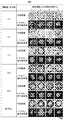
- Example 1 It is a figure which concerns on Example 1 and shows the example which measured the metabolic enzyme cytochrome P450 (molecular species CYP3A) activity of the rat hepatocyte aggregate which administered the particle
- FIG. 2 is a diagram showing each culture condition of the measurement example in FIGS. 1A to 1C according to Example 1.
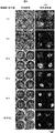
- Example 1 It is a figure which concerns on Example 1 and shows the example of a time-dependent change of a cell aggregate formation when the particle
- Example 3 It is a figure which concerns on Example 3 and shows the example of a time-dependent change by microscopic observation when a rat hepatocyte is seed
- FIG. 4 is a diagram showing an example in which rat hepatocytes were cultured and mitochondrial dehydrogenase activity was measured by WST-1 assay according to Example 4.
- Example 5 It is a figure which concerns on Example 5 and shows the example of the time-dependent change of cell aggregate formation when administering a particle
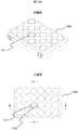
- FIG. 10 is a partial enlarged view of a multiwell plate for cell culture used in each example according to Example 6.
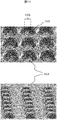
- Example 6 It is a figure which concerns on Example 6 and shows the example of the scanning electron micrograph of the culture surface by which the assembly area
- the present invention relates to a cell culture method for forming a cell aggregate including particles by seeding isolated cells in a culture vessel and administering particles having cell adhesion and substance permeability, a particulate culture carrier, and It is an invention of particle-containing cell aggregates.
- the particle having cell adhesion and substance permeability is a micro-scaffold particle that is a cell scaffold particle, and the administration of the particle is performed after the seeded cells settle on the culture surface. More specifically, after seeding the isolated cells in the culture vessel, the cells start to adhere to the culture surface, and then the cells move and the adjacent cells adhere to each other to start the formation of cell aggregates. By carrying out the process until it is completed, adhesion of the cells to the culture container, adhesion between the cells and the particles can be ensured.
- the cell culture method using microscaffold particles which is a preferred embodiment of the present invention is a culture method using fine particles obtained by forming chemical crosslinks between gelatin molecules by heat treatment or the like.
- a chemical crosslinking agent such as glutaraldehyde can be used.
- the concentration of the gelatin aqueous solution when preparing the gelatin particles is preferably 10% by weight, but is not limited thereto.
- One method of making gelatin particles is to prepare an emulsion by stirring an aqueous gelatin solution with an oil such as olive oil, cooling the emulsion, degreasing and dehydrating with cold acetone, etc. A procedure for obtaining fine particles is mentioned. By sifting at this stage, it is possible to divide the particle size. The dried particles are heat treated to crosslink the molecules.
- a microreactor for example, a microprocess server, manufactured by Hitachi Plant Technology
- a microprocess server manufactured by Hitachi Plant Technology
- the micro-scaffold particles may be any particles made of any material as long as they have two properties: cell adhesion on the particle surface and water-soluble substance permeability into the particles.
- it may be made of a cell adhesive substance, or the surface may be covered with a substance to which cells adhere.
- the cell adhesive substance include collagen, fibronectin, laminin, glycosaminoglycan, antibodies that bind to proteins and sugar chains on the cell membrane surface, sugar chains that bind to the cell membrane surface, and poly-L-lysine. However, it is not limited to these.
- hydrogels containing a large amount of water, porous ceramics, and porous metals, but are not limited thereto.
- Hydrogels include polyacrylic acid, polyacrylamide, polyvinylpyrrolidone, polyvinyl alcohol, alginic acid, starch, polylactic acid, agarose, pectin, cellulose, glucosaminoglycan, collagen, gelatin, fibronectin, vitronectin, laminin, proteoglycan, etc.
- quality ceramics include, but are not limited to, hydroxyapatite.
- Particle-containing cell aggregates such as microscaffold particles of the present invention are prepared by seeding cells exhibiting adhesiveness to a culture surface in a culture vessel having an adhesion region on the culture surface, and after the cells settle on the vessel. Is a cell aggregate formed by administration. When cells adhere to the raw material gelatin, cell aggregates including particles are formed in a state where the cells adhere to the culture surface.
- the particle administration time, particle size, and number of administration it is possible to obtain cell aggregates without detaching the cell aggregates from the culture equipment and without inhibiting cell-cell adhesion in the aggregates. is there.
- the cell function to be maintained may differ, so that it is desirable to adjust the appropriate particle administration time, particle diameter, and number of administration according to the purpose.
- the present invention including particles in a state in which cell aggregates are adhered to a culture container is a culture method for obtaining useful substances by floating culture by attaching cells to the surface of a conventional microcarrier. This is different from the technique of forming a cell aggregate including a culture carrier in a container.
- the present invention which includes particles in a state where cell aggregates are adhered to a culture vessel, can perform drug treatment and cell activity measurement in the same manner as in the usual test using monolayer cultured cells. .
- the particle inclusion cell aggregate which maintained activity can be easily used for cell tests such as drug screening in drug discovery research and examination of differentiation induction conditions in regenerative medicine research.
- drugs and reagents can easily penetrate into internal cells through substance-permeable microscaffold particles, the problem of drug and reagent permeability that is concerned with cell aggregates that do not contain particles is solved. Can do.
- Example 1 is an example relating to a particulate culture carrier for culturing hepatocytes, a particle-containing cell aggregate, and a cell culture method.
- the hepatocytes used in this example are mature hepatocytes that do not proliferate, and their function tends to decrease during the culture period. For this reason, generally, the period during which hepatocytes can be donated to drug tests and the like is from the start of culture to about one week.
- gelatin particles are administered to rat hepatocytes and cultured to extend the period during which the metabolic enzyme cytochrome P450 (CYP3A) activity can be measured to 10 days or more.
- CYP3A metabolic enzyme cytochrome P450
- FIG. 1A, FIG. 1B, and FIG. 1C show that in this example, four types of gelatin particles having different diameters are administered to rat hepatocytes, enzyme activity is measured over time, and the enzyme activity maintaining effect varies depending on the particle size. This is an example.
- the culture conditions 1 to 7 shown in these figures are as shown in 1 to 7 of FIG. 1D.
- FIG. 2 shows changes over time in the formation of cell aggregates when particles of different diameters were administered to rat hepatocytes under conditions 1 to 5 and cultured for 11 days.
- the enzyme activity measurement in FIGS. 1A, 1B, and 1C was performed. It is an example of the microscope observation image of the sample to be used.
- FIG. 3 shows an example of a microscopic observation image of gelatin particle suspensions having different diameters swollen in a medium.
- the rat hepatocytes of this example were prepared from Fisher 344 rats or SD rats male, 5-8 weeks old (both Charles River Japan) by the two-stage collagenase reflux method.
- Cell concentration 5 ⁇ 10 5 cells / ml in Williams E medium (8.6 nM insulin (Sigma Aldrich), 255 nM dexamethasone (Nacalai Tesque), 50 ng / ml EGF, 5 KIV / ml aprotinin) containing 10% fetal calf serum
- the isolated hepatocytes were suspended in and seeded at a density of 1 ⁇ 10 5 cells / cm 2 per culture area of the culture vessel.
- the culture plate was placed in an incubator at 37 ° C. and a carbon dioxide gas concentration of 5% to culture the cells.
- the culture vessel used in this example has a large number of hole structures with a diameter of 200 ⁇ m on the culture surface, and a fine 2 ⁇ m diameter and 1 ⁇ m height at the center of the bottom surface of each hole.
- a 96-well nanopillar culture plate with processed protrusions was used.
- the former describes a culture sheet on which nanopillars capable of controlling cell adhesion and migration are formed.
- the latter describes the conditions under which hepatocytes can easily form cell aggregates on the nanopillar sheet, and describes that the function of the three-dimensional aggregate is closer to that of a living body than the two-dimensional culture method.
- the material of the culture plate is polystyrene, and the culture surface having holes and fine protrusions is hydrophilized by surface modification and has cell adhesion.
- the nanopillar portion which is a fine protrusion, has an effect of capturing cells.
- BD biocoat
- Non-Patent Document 1 In vitro Proliferation and condrogenic differentiation of rat bone marrow stem cells cultured with gelatin hygrogel microspheres for TGF- ⁇ 1 release. Ogawa T. et al. Journal of Biomaterias Science, 21, 609-621 (2010)
- Dry gelatin powder (isoelectric point 5) was weighed, purified water was added to a concentration of 10%, and the mixture was swollen and dissolved at 37 degrees with stirring to prepare an aqueous gelatin solution.
- Olive oil was stirred while warming in a 40 ° water bath, and the gelatin solution was added thereto, and emulsified by stirring with a homogenizer (manufactured by Polytron).
- the emulsion was cooled with stirring to gelatinize the gelatin. After cooling acetone was added and further stirred, particles were collected by centrifugation. The particles were passed through a sieve while washing with cold acetone, and the particles that passed through the sieve were collected, and dried gelatin particles were obtained by vacuum drying.
- Thermal dehydration crosslinking was performed by heating at 140 ° C. for 48 hours under reduced pressure using a vacuum oven. Sterilized with ethylene oxide gas and stored in sterile dry particles.
- the average particle size and standard deviation of the particle size of the gelatin particles were obtained by swelling the particles after thermal crosslinking with water and measuring the size under a microscope.
- four types of gelatin particles having different sizes were used, as shown in FIG. 1D, with a diameter of 4.5 ⁇ 1.7 ⁇ m, 10.2 ⁇ 4.3 ⁇ m, 16.8 ⁇ 6.7 ⁇ m, 23.7. ⁇ 10.3 ⁇ m was used.
- FIG. 3 shows microscopic images of four types of gelatin particles swollen in the medium.
- the particle concentration of the particle suspension was adjusted to 3 mg / ml by dry particle weight.
- the medium in which the cells are cultured is changed from Williams E medium containing 10% fetal bovine serum to serum-free Williams E medium in which the particles are suspended, whereby the particles are administered to the cultured cells. did.
- Matrigel (BD) was replaced with serum-free Williams E medium containing 0.25 mg / ml of total protein, and thereafter culture medium was replaced with serum-free Williams E medium every 24 hours. .
- monolayer culture as a conventional method was performed. Isolated hepatocytes were suspended at a cell concentration of 5 ⁇ 10 5 cells / ml and seeded on the aforementioned culture plate at a density of 1 ⁇ 10 5 cells / cm 2 per culture area. The culture plate was placed in an incubator at 37 ° C. and a carbon dioxide gas concentration of 5% to culture the cells. Monolayer culture was replaced with serum-free Williams E medium 24 hours after cell seeding, and thereafter replaced with serum-free Williams E medium every 24 hours.
- the activity of the hepatocyte metabolic enzyme cytochrome P450 was measured using P450-Glo TM Assay (Luciferin-IPA, Promega). The measurement procedure is as follows.
- the medium in which hepatocytes were cultured was removed, replaced with 60 ⁇ l of Luciferin-IPA solution (final concentration 3 ⁇ M) diluted to 1/1000 with the medium, and reacted for 1 hour in an incubator at 37 ° C. and 5% carbon dioxide concentration. .
- 50 ⁇ l of the culture supernatant of each well was dispensed into a white 96-well plate, 50 ⁇ l of luciferin detection reagent (Promega) was added, and the mixture was allowed to react at room temperature for 20 minutes while protected from light.
- the white plate after the reaction was placed in a plate reader (SH-8000 Lab, Corona Electric), and luminescence was measured at a gate time of 1 second.
- FIG. 1A, FIG. 1B, and FIG. 1C show the average values and standard deviations of 8 wells for each culture condition.
- the activity of the cells not administered with the particles was high, and therefore the effect of the particles was not observed in the early stage of the culture.
- the activity of the cells of the aggregate administered with particles having a diameter of 4.5 ⁇ 1.7 ⁇ m and the activity of the cells of the aggregate not administered with particles were high.
- the activity of the aggregate cells administered with particles of 10.2 ⁇ 4.3 ⁇ m, 16.8 ⁇ 6.7 ⁇ m, and 23.7 ⁇ 10.3 ⁇ m was different. Compared to the above conditions, the activity was low in particles having a diameter of 4.5 ⁇ 1.7 ⁇ m, no particles, and monolayer sandwich culture, and the activity was below the detection limit in the most common monolayer culture.
- FIG. 2 shows a micrograph of the cultured hepatocytes observed with a phase-contrast inverted microscope, and photographing an arbitrary cell aggregate near the center of the well of the culture plate.
- cells administered with particles having a diameter of 4.5 ⁇ 1.7 ⁇ m promoted adhesion between cells more than cells not administered with particles on the third and fifth days of culture. There was a tendency to agglomerate. Thereafter, the shape of the aggregates on day 7 and 11 of the culture was similar to the cells to which particles were not administered, and the aggregates adhered to the area where the fine protrusions gathered at the center of the hole of the culture plate.
- the diameter of hepatocytes is about 15-30 ⁇ m
- particles with a diameter of 4.5 ⁇ 1.7 ⁇ m are about 1/10 to 1/5 the size of the cells. It did not have a big impact. Since the size of the agglomerates on day 11 is not significantly different from that of no particles, the particle dosage is 3 mg / ml dry particle weight for any particle, but the diameter is 4.5 ⁇ 1. In the case of 7 ⁇ m particles, the total amount of particles taken into the aggregates was considered to be small.
- particles having a diameter of 10.2 ⁇ 4.3 ⁇ m having a particle size of 1/5 or more of the cells formed aggregates including the particles.
- a slightly planar irregular aggregate was formed. Assuming that the diameter of the hepatocytes is about 15 to 30 ⁇ m, it was considered that the particles having a diameter of 23.7 ⁇ 10.3 ⁇ m have the same particle size as that of the cells to about twice the size of the cells.
- cytochrome P450 molecular species CYP3A
- Example 2 when gelatin particles having different diameters were administered to rat hepatocytes and cultured, depending on the particle size, the particles included in the cell aggregates were distributed only inside the aggregates, or only inside the aggregates. An example in the case where it is also present in the portion in contact with the medium will be described.
- FIG. 4 is an example of a micrograph showing a cross section of the gelatin particle-containing cell aggregate in Example 2.
- the schematic diagram at the bottom of the photo shows the outline of the cells that can be identified in the upper photo with lines, and the location of the included particles with the US marks.
- the culture vessel has a large number of hole structures with a diameter of 200 ⁇ m on the culture surface described in JP-A-2011-004612 described above, and fine protrusions with a diameter of 2 ⁇ m and a height of 1 ⁇ m are processed at the center of the bottom of each hole.
- the nanopillar culture sheet was used by adhering to the well bottom of the 2-well slide chamber.
- the culture sheet is made of polystyrene, and the culture surface having holes and fine protrusions is hydrophilized by surface modification and has cell adhesion.
- the nanopillar portion which is a fine protrusion, has an effect of capturing cells.
- Isolated hepatocytes were suspended in Williams E medium containing 10% fetal bovine serum at a cell concentration of 5 ⁇ 10 5 cells / ml and seeded at a density of 1 ⁇ 10 5 cells / cm 2 per culture area of the culture vessel.
- the slide chamber was placed in an incubator at 37 ° C. and a carbon dioxide gas concentration of 5%, and the cells were cultured.
- particles with a diameter of 4.5 ⁇ 1.7 ⁇ m were suspended in Williams E medium containing 10% fetal calf serum. Since the particles were fine and could not be counted with a cell counter, a suspension having the same concentration as the particles having a diameter of 16.8 ⁇ 6.7 ⁇ m was prepared by dry particle weight.
- the particles were administered to the cells by replacing them with Williams E medium containing 10% fetal bovine serum in which the particles were suspended. 24 hours after cell seeding, the medium was replaced with serum-free Williams E medium. After 48 hours, Matrigel (BD) was replaced with serum-free Williams E medium containing 0.25 mg / ml of total protein, and then serum-free Williams E every 24 hours. The medium was changed to E medium and the culture was continued for 5 days.
- Williams E medium containing 10% fetal bovine serum in which the particles were suspended.
- serum-free Williams E medium After 48 hours, Matrigel (BD) was replaced with serum-free Williams E medium containing 0.25 mg / ml of total protein, and then serum-free Williams E every 24 hours. The medium was changed to E medium and the culture was continued for 5 days.
- the cells cultured for 5 days on the nanopillar culture sheet were pre-fixed with 0.1 M phosphate buffer (pH 7.4) containing 2.5% glutaraldehyde and post-fixed with phosphate buffer containing 1% osmium tetroxide.
- the nanopillar sheet to which the cells were adhered was embedded in low melting point agar (Sea plaquer agarose, Lonza) with a concentration of 1.5%, dehydrated in an ethanol dilution series, and polystyrene was dissolved with propylene oxide.
- the Epon-Alardite mixed resin was infiltrated into the agar in which the cells were embedded, and polymerized by heating.
- a quasi-ultra thin section having a thickness of 400 mm was prepared from a resin-embedded sample.
- the sections were adhered to a slide glass, stained with 1% toluidine blue, and observed with an upright light microscope.
- Example 3 describes an example showing that the effect of maintaining the activity of rat liver cell metabolic enzyme cytochrome P450 (CYP3A) varies depending not only on the particle diameter but also on the dose of the particles.
- cytochrome P450 CYP3A
- FIG. 5A and FIG. 5B are diagrams showing examples in which the basal activity and drug-inducing activity of CYP3A were measured in this example.
- FIG. 5A shows the ratio of administering particles to the number of cells, and the time for administering the particles.
- 5B shows an example in which rat liver cells were cultured for 10 days and the basal activity of the metabolic enzyme cytochrome P450 (molecular species CYP3A) was measured.
- FIG. 5B shows the ratio of the number of particles administered to the number of cells and the time for administering the particles.
- rat hepatocytes were seeded using 96-well nanopillar culture equipment. Particles having a diameter of 16.8 ⁇ 6.7 ⁇ m were swollen in Williams E medium containing 10% fetal bovine serum, and after 2.5 hours or 22 hours after cell seeding, the cell number: particle number was 1: 1.
- the particle suspension was administered onto the cells in a ratio of 4: 1, 10: 1, 20: 1, 50: 1. 24 hours after cell seeding, the serum-free Williams E medium was replaced. After 48 hours, Matrigel (BD) was replaced with serum-free Williams E medium containing 0.25 mg / ml of total protein, and serum-free every 24 hours thereafter. The medium was changed to Williams E medium and cultured for 10 days.
- cytochrome P450 molecular species CYP3A
- FIG. 6A shows an example in which changes in cell morphology (time-dependent change) over time after seeding rat hepatocytes in a low-adhesion culture vessel are shown by phase contrast microscopy and F-actin distribution in this example.
- 6A is a photomicrograph of the cell morphology observed with an inverted microscope over time after cell seeding, with the top row being a phase contrast microscope image and the bottom row being a fluorescence microscope image in which F-actin is stained with rhodamine-labeled phalloidin. is there.
- the phase contrast image shows the whole image, and the outline of each cell is shown by the F-actin fluorescent staining image.
- FIG. 6B is an example in which the major axis of 60 cells arbitrarily selected in the photomicrograph after cell seeding was measured, the average value of the major axis and the standard deviation were obtained, and the change with time was shown.
- the procedure for rhodamine-labeled phalloidin staining in FIG. 6A is as follows. The cells fixed with 4% paraformaldehyde were washed with 0.1 M phosphate buffer (pH 7.4). To prevent non-specific staining, treatment with protein block (DAKO) for 30 minutes, followed by treatment with rhodamine-labeled phalloidin (Invitrogen) diluted 1:40 with PBS for 30 minutes, 0.075% Brij35j (Sigma Aldrich) ) Washed with PBS containing sputum.
- DAKO protein block
- rhodamine-labeled phalloidin Invitrogen
- PBS 0.075% Brij35j
- the cells 0.5 hours after seeding were isolated and had a round shape and an average diameter of 20 ⁇ m or less. After 1.0 hour, the two-dimensional image of the cell shown in the photograph was slightly larger, and the average diameter slightly exceeded 20 ⁇ m.
- This example describes another example showing that the effect of maintaining the activity of rat hepatocytes varies depending on the dose of particles.
- the WST-1 test is a method for measuring the activity of mitochondrial respiratory chain enzyme (succinate tetrazolium reductase) in living cells, and the higher the measured value, the higher the respiratory activity of spheroids.
- rat hepatocytes were seeded using 96-well nanopillar culture equipment. Particles having a diameter of 16.8 ⁇ 6.7 ⁇ m were swollen in Williams E medium containing 10% fetal bovine serum, and after 2.5 hours or 22 hours after cell seeding, the cell number: particle number was 1: 4, Cell suspension of the particle suspension to be 1: 2, 1: 1, 2: 1, 4: 1, 8: 1, 10: 1, 20: 1, 40: 1, 50: 1, 80: 1 Was administered on top of. 24 hours after cell seeding, the serum-free Williams E medium was replaced. After 48 hours, Matrigel (BD) was replaced with serum-free Williams E medium containing 0.25 mg / ml of total protein, and serum-free every 24 hours thereafter. The medium was changed to Williams E medium and cultured for 5 days.
- BD Matrigel
- Premix WST-1 (Takara Bio) was used. After culturing hepatocytes for 5 days, the entire medium was removed from each well of the 96-well plate containing the cells, and washed twice with Hank's solution. After removing the washing solution, 65 ⁇ l of Hank's solution containing 10% (v / v) concentration of WST-1 was added and reacted in an incubator maintained at 37 ° C. and 5% carbon dioxide concentration for 1 hour. The 96 wells after the reaction were set in a plate reader, and the absorbance at two wavelengths (440 nm, 650 nm) was measured at a half width of 5 nm. A value obtained by subtracting the value of 650 nm, which is the reference wavelength, from the value of 440 nm was obtained.
- FIG. 7 shows that rat hepatocytes were cultured for 5 days while changing the ratio of administration of particles to the number of cells and the administration time of particles, and the activity of mitochondrial dehydrogenase was measured by WST-1 assay.
- the results of calculating the average value and standard deviation of 3 wells for the group were shown.
- the number of cells: number of particles is 1:80, that is, the effect of particles is recognized even when the number of particles is more than 1/80 with respect to the number of cells.
- Administering more particles than 4: 1 significantly increased the WST-1 measurement, and further increasing the dose beyond 1: 1 further increased the value.
- the above results indicate that the intercellular particles improve the nutrient and oxygen supply from the medium to maintain the cell activity, and increase the permeability of the detection reagent WST-1 into the cells. This is considered to be the reason that the detection efficiency of respiratory activity is improved.
- Example 1 the description of the isolation and culture of rat hepatocytes already described in Example 1, the method for producing gelatin particles, and the procedure for swelling the particles in the medium are omitted.
- This example shows an example of the cell aggregate formation process and the shape of the formed cell aggregate when the particles are administered in different amounts. Furthermore, an example will be described that shows that the efficiency of forming a capillary bile duct that is a liver tissue-specific structure varies depending on the dose of particles.
- FIG. 8 is a diagram showing an example of changes over time in the formation of cell aggregates when particles are administered at a different ratio to the number of cells and cultured for 5 days.
- the phase contrast image shows the whole image of the particles and cells
- the F-actin fluorescent staining image shows the distribution of cells. That is, to a rat hepatocyte, particles having a diameter of 16.8 ⁇ 6.7 ⁇ m were administered so that the cell number: particle number was 1: 1, 4: 1, 10: 1, 20: 1, 50: 1.
- the process of cell aggregate formation when cultured for 5 days is shown.
- the upper row of each column is a phase contrast microscope image, and the lower row is a fluorescence microscope image of F-actin stained with rhodamine-labeled phalloidin.
- F-actin forms a membrane lining structure and cells have a polygonal three-dimensional structure. By observing the distribution of F-actin, it is possible to verify whether the tissue is close to living cells. So far, in the cell aggregate formed in the nanopillar culture vessel, F-actin has formed a membrane lining structure close to that of a living body (see WO2010 / 079962).
- the aggregate formed with the cell number: particle number 50: 1 or without the particles had F-actin forming a membrane backing structure close to that of a living body.
- the particles are administered at a cell number: particle number of 20: 1, 10: 1, 4: 1, or 1: 1, the cell surface that adheres to the gelatin particles is linear or dotted. An image showing strong fluorescence was observed, suggesting the possibility of adhesion points or stress fibers. These are structures formed when cells adhere to the extracellular matrix. Therefore, it was considered that the cells formed an adhesion structure on the surface of the gelatin particles, similar to the adhesion with the extracellular matrix.
- the cell number: particle number is preferably 50: 1 or less in order to form a membrane backing structure close to a living body.
- FIG. 9 is a diagram showing an example of fluorescent reagent uptake and excretion into the capillary bile duct when particles are administered at different ratios to the number of cells and cultured for 5 days. Specifically, this is an example in which the excretion ability of the capillary bile duct formed on the intercellular adhesion surface of hepatocytes is shown on the 5th day of culture using a fluorescent labeling reagent carbodichlorofluorescein-diacetate (CDFDA, Molecular Probe).
- CDFDA fluorescent labeling reagent carbodichlorofluorescein-diacetate
- CDFDA administered into the medium is taken up into cells and metabolized to become carbodichlorofluorescein (CDF), which is excreted into the capillaries through the transporter multidrug resistance-associated protein 2 (MRP2) of the cell membrane facing the capillaries.
- CDF carbodichlorofluorescein
- MRP2 transporter multidrug resistance-associated protein 2
- the procedure of the CDFDA process of this embodiment is as follows.
- the medium of hepatocytes cultured in a 24-well nanopillar culture plate was removed and washed twice with Krebs-Henseleit buffer (KH buffer).
- KH buffer Krebs-Henseleit buffer
- 400 ⁇ l of KH buffer containing 5 ⁇ M CDFDA was added per well, placed in a 37 ° incubator for 20 minutes, washed twice with KH buffer, and observed with an inverted fluorescence microscope.
- capillary bile duct Since the capillary bile duct is formed on the cell-cell adhesion surface, cell-cell interaction is required. In addition, it is known that proteins present inside the cell membrane on the intercellular adhesion surface play an important role in the expression of transporter MRP2, which excretes drugs from the cell into the capillary bile duct. For this reason, it was thought that the capillary bile duct was difficult to regenerate when there were many particles because the particles that existed between cells excessively inhibited adhesion between cells.
- the number of cells: number of particles was 50: 1 or less.
- the cell number: particle number is desirably 50: 1 to 4: 1, and more desirably 50: 1 to 10: 1. This is because, in order to improve both the uptake and excretion of drugs, not only intercellular adhesion is promoted, but a surface in contact with the medium through particles must be formed. It was.
- Example 4 the inclusion of a large number of particles showed the possibility of improving the supply of medium components to the inside of cell aggregates.
- the presence of excessive particles caused liver It became clear that the excretion function peculiar to a tissue fell. Although it is a contradictory condition in particle administration, it is desirable to obtain an appropriate particle amount according to the purpose of use of the cell aggregate.
- Example 3 the isolation and culture of rat hepatocytes already explained in Example 1, the method for producing gelatin particles, the procedure for swelling particles in the medium, and the procedure for rhodamine-labeled phalloidin staining shown in Example 3 are explained. Omitted.
- a culture container provided with a cell adhesion region for capturing cell aggregates by molding a plurality of protrusions locally on the culture surface with low cell adhesion used in each of the examples described above.
- An example will be described.
- FIG. 10A shows a perspective view and an upper surface of the cell culture multiwell plate 100 at the upper and lower stages.
- the culture surface of the bottom surface 102 of the well 101 indicated by AA and BB is subjected to fine three-dimensional processing.
- FIG. 10B shows an enlarged view of the AA and BB portions, an enlarged view of the CC and DD portions, and an end surface of the line EE in FIG. 10A at the upper, middle, and lower portions.
- the fine three-dimensional processing applied to the culture surface is enlargement of the CC and DD portions and the hole 103 shown on the end surface of the EE line, and the collective region 105 of the protrusion 104 is formed at the center of the hole 103. It is a figure which shows the example currently shape
- molded shows the example currently shape
- FIG. 11 is an example of a scanning electron micrograph of the culture surface in which the aggregation region 105 of the protrusions 104 is molded at the center of the hole 103 described above.
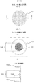
- FIG. 12 shows that cells 106 are seeded in the holes 103 on the culture surface shown in FIG. 10B and FIG. 11 and gelatin particles 107 are administered when the cells 106 adhere to the bottom surface, so that cell aggregates containing the particles can be obtained.
- molded in the center of the hole 103 is shown.
- a local cell adhesion region that captures cells on the culture surface is not limited to the protrusions of this example, extracellular matrix components such as collagen and laminin, antibodies that bind to the cell membrane surface, It can also be formed by optionally patterning a cell-adhesive substance such as a sugar chain that binds to the cell membrane surface or poly-L-lysine.
- the culture container of this example described above holds the particle-containing cell aggregates in the culture container by forming a cell adhesion region capable of capturing the cell aggregates on the low adhesion culture surface. Further, preferably, by providing a partition on the culture surface, the number of cells and the number of particles forming the aggregate are controlled to a desired number, and a particle-containing cell aggregate having a desired size is formed. Such particle-containing cell aggregates adhered to the culture container of this Example can be used as a material for cell tests.
- Example 1 an example of a human cell test method involving particles will be described.
- Example 3 in cell aggregates containing particles, the activity of the metabolic enzyme cytochrome P450 (molecular species CYP3A) is maintained for a longer period than aggregates not containing particles and conventional monolayer culture methods. Showed the effect.
- cytochrome P450 molecular species CYP3A
- cytochrome P450 is an enzyme that metabolizes drugs taken into hepatocytes from the blood, in drug discovery research, metabolism in pharmacokinetics, drug interaction by CYP enzyme induction, liver toxicity by CYP metabolites, etc. It has become.
- cytochrome P450 since there are species differences in enzyme substrate properties and metabolic activities, not only animal experiments but also human tests are required, and test methods using isolated human hepatocytes have been developed. Therefore, by using cell aggregates containing the above-mentioned microscaffold particles for cell tests, it is possible to carry out tests using isolated human hepatocytes that tend to lose their activity stably for a longer period of time than before. It becomes possible.
- the cryopreserved human hepatocytes are thawed, suspended in Williams E medium containing 10% fetal bovine serum at a cell concentration of 5 ⁇ 10 5 cells / ml, and 1 ⁇ 10 5 cells / cm 2 per culture area of the culture vessel. Seeded at a density of The culture plate was placed in an incubator at 37 ° C. and a carbon dioxide gas concentration of 5% to culture the cells. Cell aggregates containing the particles are obtained by swelling particles having a diameter of 16.8 ⁇ 6.7 ⁇ m in a medium and administering the particle suspension onto the cells so that the number of particles is larger than the number of cells. Formed.
- the drug metabolized by CYP was administered to the medium, and the cells and medium were collected after 2 and 7 days.
- the cell lysate and the medium were deproteinized, and the collected sample solution was analyzed by mass spectrometry to identify drug metabolites in the cell or in the medium.
- cryopreserved human hepatocytes were cultured, and the particles were administered to form cell aggregates containing the particles.
- the activity of CYP enzyme was measured using a detection reagent that specifically reacts with each molecular species of CYP.
- the induction rate was calculated by dividing the induced activity value by the basal activity value, with the CYP enzyme activity of cells not treated with the subject drug as the basal activity and the CYP enzyme activity of cells treated with the subject drug as the inductive activity.
- CYP enzyme activity for example, the P450-Glo TM Assay series (Promega) shown in Example 1 and Example 3 is used to determine the CYP induction activity by the test drug by quantifying luminescence. Can be measured.
- CYP-inducing activity can be measured by quantifying a metabolite by mass spectrometry using a known substance that is metabolized specifically for each molecular species of CYP.
- a method for estimating the degree of drug interaction by the drug to be tested can be cited.
- liver toxicity analysis using CYP metabolites will be described. Similar to the metabolite analysis example, cryopreserved human hepatocytes were cultured, and the particles were administered to form cell aggregates containing the particles. The toxicity of the drug was evaluated by changing the concentration of the test drug and administering it to the medium, and measuring the change in the number of cells over time. For example, the WST-1 method or the ATP quantification method shown in Example 4 can be used for measuring the number of cells. As mentioned above, although the some test example was given as description of Example 7, it is not restricted to this.
- the cell culture method according to the present invention described in detail above is a culture method characterized in that cells are adhered to the culture surface of a culture vessel, and has been performed by a conventional monolayer culture method so far. Cell tests can be applied in the same procedure.
- the particulate culture alone and the particle-containing cell aggregate according to the present invention are three-dimensional cell aggregates having a structure closer to that of a living body than monolayer culture, and supply nutrients and oxygen in the medium via the particles.
Landscapes
- Health & Medical Sciences (AREA)
- Engineering & Computer Science (AREA)
- Life Sciences & Earth Sciences (AREA)
- Biomedical Technology (AREA)
- Zoology (AREA)
- Chemical & Material Sciences (AREA)
- Biotechnology (AREA)
- Organic Chemistry (AREA)
- Wood Science & Technology (AREA)
- Bioinformatics & Cheminformatics (AREA)
- Genetics & Genomics (AREA)
- General Engineering & Computer Science (AREA)
- Biochemistry (AREA)
- General Health & Medical Sciences (AREA)
- Microbiology (AREA)
- Immunology (AREA)
- Sustainable Development (AREA)
- Gastroenterology & Hepatology (AREA)
- Cell Biology (AREA)
- Micro-Organisms Or Cultivation Processes Thereof (AREA)
- Apparatus Associated With Microorganisms And Enzymes (AREA)
Abstract
La présente invention concerne une méthode de culture capable d'amener un agrégat tridimensionnel, dont la fonction cellulaire est similaire à la fonction in vivo, à adhérer à un récipient de culture tout en améliorant la perméabilité des nutriments, de l'oxygène, des médicaments et des réactifs en provenance du milieu de culture et en maintenant la fonction cellulaire. En ensemençant le récipient de culture avec des cellules isolées et, après dépôt desdites cellules sur la surface de culture, et en administrant des particules de micro-échafaudage qui adhèrent aux cellules et qui sont perméables aux substances, il est possible d'amener les cellules à adhérer au récipient de culture tout en formant des agrégats cellulaires comprenant lesdites particules. Par exemple, en administrant des particules de gélatine dans lesquelles la taille du support de culture destiné à être inclus dans l'agrégat cellulaire représente au moins 1/5ème du diamètre des cellules pour un rapport nombre de cellules sur nombre de particules d'au moins 80:1 en termes de nombre de cellules cultivées par unité de surface de culture, des agrégats cellulaires comprenant lesdites particules peuvent être formés.
Priority Applications (2)
| Application Number | Priority Date | Filing Date | Title |
|---|---|---|---|
| EP14800833.7A EP3000869A1 (fr) | 2013-05-23 | 2014-05-09 | Méthode de culture cellulaire, support de culture particulaire, et agrégat cellulaire comprenant des particules |
| US14/891,155 US20160168525A1 (en) | 2013-05-23 | 2014-05-09 | Cell Culturing Method, Particulate Culture Carrier, and Particle-Encompassing Cell Aggregate |
Applications Claiming Priority (2)
| Application Number | Priority Date | Filing Date | Title |
|---|---|---|---|
| JP2013-109154 | 2013-05-23 | ||
| JP2013109154A JP2014226097A (ja) | 2013-05-23 | 2013-05-23 | 細胞培養方法、粒子状培養担体、及び粒子包含細胞凝集体 |
Publications (1)
| Publication Number | Publication Date |
|---|---|
| WO2014188888A1 true WO2014188888A1 (fr) | 2014-11-27 |
Family
ID=51933451
Family Applications (1)
| Application Number | Title | Priority Date | Filing Date |
|---|---|---|---|
| PCT/JP2014/062495 WO2014188888A1 (fr) | 2013-05-23 | 2014-05-09 | Méthode de culture cellulaire, support de culture particulaire, et agrégat cellulaire comprenant des particules |
Country Status (4)
| Country | Link |
|---|---|
| US (1) | US20160168525A1 (fr) |
| EP (1) | EP3000869A1 (fr) |
| JP (1) | JP2014226097A (fr) |
| WO (1) | WO2014188888A1 (fr) |
Cited By (2)
| Publication number | Priority date | Publication date | Assignee | Title |
|---|---|---|---|---|
| JP2018000081A (ja) * | 2016-06-30 | 2018-01-11 | 株式会社新菱 | ガス供給機能付き容器、細胞培養容器、運搬用容器、保存用容器、細胞培養方法、内容物運搬方法および内容物保存方法 |
| EP3395830A4 (fr) * | 2015-12-25 | 2018-12-26 | Konica Minolta, Inc. | Particules de gélatine, procédé de production de particules de gélatine, cellules encapsulant des particules de gélatine, et procédé de production de cellules encapsulant des particules de gélatine |
Families Citing this family (1)
| Publication number | Priority date | Publication date | Assignee | Title |
|---|---|---|---|---|
| JP2019058156A (ja) * | 2017-09-28 | 2019-04-18 | オリンパス株式会社 | 画像処理装置および細胞観察システム |
Citations (9)
| Publication number | Priority date | Publication date | Assignee | Title |
|---|---|---|---|---|
| JPH0833486A (ja) * | 1994-07-25 | 1996-02-06 | Res Dev Corp Of Japan | 細胞培養担体と、細胞培養方法 |
| JP2005027532A (ja) | 2003-07-09 | 2005-02-03 | Fuji Xerox Co Ltd | 細胞培養基材及びその製造方法、並びに細胞培養方法 |
| US20050054101A1 (en) | 2003-07-17 | 2005-03-10 | Global Cell Solutions Llc | Automated cell culture system and process |
| WO2010079602A1 (fr) | 2009-01-08 | 2010-07-15 | 株式会社日立製作所 | Procédé pour la culture d'hépatocytes animaux |
| JP2010524442A (ja) * | 2007-04-20 | 2010-07-22 | フラウンホファー ゲセルシャフト ツール フェールデルンク ダー アンゲヴァンテン フォルシュンク エー.ファオ. | ナノ粒子を含む改善された三次元生体適合性骨格構造 |
| JP2011004612A (ja) | 2009-06-23 | 2011-01-13 | Hitachi Ltd | 培養基材、培養シート、及び細胞培養方法 |
| WO2011059112A1 (fr) | 2009-11-13 | 2011-05-19 | 株式会社日立ハイテクノロジーズ | Agrégat de cellules contenant des particules |
| JP2011130720A (ja) | 2009-12-25 | 2011-07-07 | Medgel Corp | 細胞培養基材およびその使用方法 |
| US20110256574A1 (en) | 2008-08-08 | 2011-10-20 | Agency For Science, Technology And Research | Microfluidic Continuous Flow Device |
Family Cites Families (3)
| Publication number | Priority date | Publication date | Assignee | Title |
|---|---|---|---|---|
| JPH078273A (ja) * | 1993-06-14 | 1995-01-13 | Kurabo Ind Ltd | 接着性動物細胞の無血清培養法 |
| JP2004275122A (ja) * | 2003-03-18 | 2004-10-07 | Kitakyushu Foundation For The Advancement Of Industry Science & Technology | 培養床、培養床コーティング剤、培養床製造方法並びにこれらを用いた細胞培養方法及び培養細胞 |
| WO2007037407A1 (fr) * | 2005-09-30 | 2007-04-05 | 3-D Matrix Japan, Ltd. | Procédé de culture de cellules et culture de cellules |
-
2013
- 2013-05-23 JP JP2013109154A patent/JP2014226097A/ja active Pending
-
2014
- 2014-05-09 US US14/891,155 patent/US20160168525A1/en not_active Abandoned
- 2014-05-09 WO PCT/JP2014/062495 patent/WO2014188888A1/fr active Application Filing
- 2014-05-09 EP EP14800833.7A patent/EP3000869A1/fr not_active Withdrawn
Patent Citations (9)
| Publication number | Priority date | Publication date | Assignee | Title |
|---|---|---|---|---|
| JPH0833486A (ja) * | 1994-07-25 | 1996-02-06 | Res Dev Corp Of Japan | 細胞培養担体と、細胞培養方法 |
| JP2005027532A (ja) | 2003-07-09 | 2005-02-03 | Fuji Xerox Co Ltd | 細胞培養基材及びその製造方法、並びに細胞培養方法 |
| US20050054101A1 (en) | 2003-07-17 | 2005-03-10 | Global Cell Solutions Llc | Automated cell culture system and process |
| JP2010524442A (ja) * | 2007-04-20 | 2010-07-22 | フラウンホファー ゲセルシャフト ツール フェールデルンク ダー アンゲヴァンテン フォルシュンク エー.ファオ. | ナノ粒子を含む改善された三次元生体適合性骨格構造 |
| US20110256574A1 (en) | 2008-08-08 | 2011-10-20 | Agency For Science, Technology And Research | Microfluidic Continuous Flow Device |
| WO2010079602A1 (fr) | 2009-01-08 | 2010-07-15 | 株式会社日立製作所 | Procédé pour la culture d'hépatocytes animaux |
| JP2011004612A (ja) | 2009-06-23 | 2011-01-13 | Hitachi Ltd | 培養基材、培養シート、及び細胞培養方法 |
| WO2011059112A1 (fr) | 2009-11-13 | 2011-05-19 | 株式会社日立ハイテクノロジーズ | Agrégat de cellules contenant des particules |
| JP2011130720A (ja) | 2009-12-25 | 2011-07-07 | Medgel Corp | 細胞培養基材およびその使用方法 |
Non-Patent Citations (1)
| Title |
|---|
| OGAWA T ET AL., JOURNAL OF BIOMATERIALS SCIENCE, vol. 21, 2010, pages 609 - 621 |
Cited By (2)
| Publication number | Priority date | Publication date | Assignee | Title |
|---|---|---|---|---|
| EP3395830A4 (fr) * | 2015-12-25 | 2018-12-26 | Konica Minolta, Inc. | Particules de gélatine, procédé de production de particules de gélatine, cellules encapsulant des particules de gélatine, et procédé de production de cellules encapsulant des particules de gélatine |
| JP2018000081A (ja) * | 2016-06-30 | 2018-01-11 | 株式会社新菱 | ガス供給機能付き容器、細胞培養容器、運搬用容器、保存用容器、細胞培養方法、内容物運搬方法および内容物保存方法 |
Also Published As
| Publication number | Publication date |
|---|---|
| JP2014226097A (ja) | 2014-12-08 |
| EP3000869A1 (fr) | 2016-03-30 |
| US20160168525A1 (en) | 2016-06-16 |
Similar Documents
| Publication | Publication Date | Title |
|---|---|---|
| JP2022095914A (ja) | 操作した肝臓組織、そのアレイ、およびそれを製造する方法 | |
| KR101954743B1 (ko) | 성체 간 전구 세포 제조 방법 | |
| Yamada et al. | Cell-sized condensed collagen microparticles for preparing microengineered composite spheroids of primary hepatocytes | |
| Pampaloni et al. | Three-dimensional tissue models for drug discovery and toxicology | |
| Pampaloni et al. | Three-dimensional cell cultures in toxicology | |
| Cheng et al. | Three-dimensional polymer scaffolds for high throughput cell-based assay systems | |
| JP5818001B2 (ja) | 肝細胞の培養方法 | |
| US20180002672A1 (en) | Methods to generate gastrointestinal epithelial tissue constructs | |
| US20140154735A1 (en) | Tumour cell and tissue culture | |
| WO2006122147A2 (fr) | Encapsulation d'alginate poly-l-lysine utilisee comme technologie dans la differenciation controlee des cellules souches embryonnaires | |
| Bazou | Biochemical properties of encapsulated high-density 3-D HepG2 aggregates formed in an ultrasound trap for application in hepatotoxicity studies: Biochemical responses of encapsulated 3-D HepG2 aggregates | |
| Tronser et al. | Miniaturized platform for high-throughput screening of stem cells | |
| Zhang et al. | Long-term mechanical loading is required for the formation of 3D bioprinted functional osteocyte bone organoids | |
| Wang et al. | Paper supported long-term 3D liver co-culture model for the assessment of hepatotoxic drugs | |
| WO2014188888A1 (fr) | Méthode de culture cellulaire, support de culture particulaire, et agrégat cellulaire comprenant des particules | |
| US20160123960A1 (en) | Method for preparing three-dimensional, organotypic cell cultures and uses thereof | |
| Agarwal et al. | Inexpensive and versatile paper-based platform for 3D culture of liver cells and related bioassays | |
| Aalders et al. | Liquid marble technology to create cost-effective 3D cardiospheres as a platform for in vitro drug testing and disease modelling | |
| Cao et al. | An Automation Workflow for High‐Throughput Manufacturing and Analysis of Scaffold‐Supported 3D Tissue Arrays | |
| KR20180049000A (ko) | 세포 배양 | |
| Kintzios et al. | Differential effect of the shape of calcium alginate matrices on the physiology of immobilized neuroblastoma N2a and Vero cells: A comparative study | |
| Liang et al. | Chemically defined stem cell microniche engineering by microfluidics compatible with iPSCs’ growth in 3D culture | |
| Liu et al. | Progress of 3D organoid technology for preclinical investigations: towards human in vitro models | |
| Ryoo et al. | Advances in high throughput cell culture technologies for therapeutic screening and biological discovery applications | |
| Wu et al. | In Situ Self-Assembling Liver Spheroids with Synthetic Nanoscaffolds for Preclinical Drug Screening Applications |
Legal Events
| Date | Code | Title | Description |
|---|---|---|---|
| 121 | Ep: the epo has been informed by wipo that ep was designated in this application |
Ref document number: 14800833 Country of ref document: EP Kind code of ref document: A1 |
|
| WWE | Wipo information: entry into national phase |
Ref document number: 14891155 Country of ref document: US |
|
| REEP | Request for entry into the european phase |
Ref document number: 2014800833 Country of ref document: EP |
|
| WWE | Wipo information: entry into national phase |
Ref document number: 2014800833 Country of ref document: EP |
|
| NENP | Non-entry into the national phase |
Ref country code: DE |