WO2014051013A1 - 核医学診断装置および医用データ処理装置 - Google Patents
核医学診断装置および医用データ処理装置 Download PDFInfo
- Publication number
- WO2014051013A1 WO2014051013A1 PCT/JP2013/076161 JP2013076161W WO2014051013A1 WO 2014051013 A1 WO2014051013 A1 WO 2014051013A1 JP 2013076161 W JP2013076161 W JP 2013076161W WO 2014051013 A1 WO2014051013 A1 WO 2014051013A1
- Authority
- WO
- WIPO (PCT)
- Prior art keywords
- diameter
- pet
- subject
- radiation
- nuclear medicine
- Prior art date
- Legal status (The legal status is an assumption and is not a legal conclusion. Google has not performed a legal analysis and makes no representation as to the accuracy of the status listed.)
- Ceased
Links
Images
Classifications
-
- G—PHYSICS
- G01—MEASURING; TESTING
- G01T—MEASUREMENT OF NUCLEAR OR X-RADIATION
- G01T1/00—Measuring X-radiation, gamma radiation, corpuscular radiation, or cosmic radiation
- G01T1/29—Measurement performed on radiation beams, e.g. position or section of the beam; Measurement of spatial distribution of radiation
- G01T1/2914—Measurement of spatial distribution of radiation
- G01T1/2985—In depth localisation, e.g. using positron emitters; Tomographic imaging (longitudinal and transverse section imaging; apparatus for radiation diagnosis sequentially in different planes, steroscopic radiation diagnosis)
-
- A—HUMAN NECESSITIES
- A61—MEDICAL OR VETERINARY SCIENCE; HYGIENE
- A61B—DIAGNOSIS; SURGERY; IDENTIFICATION
- A61B6/00—Apparatus or devices for radiation diagnosis; Apparatus or devices for radiation diagnosis combined with radiation therapy equipment
- A61B6/02—Arrangements for diagnosis sequentially in different planes; Stereoscopic radiation diagnosis
- A61B6/03—Computed tomography [CT]
- A61B6/032—Transmission computed tomography [CT]
-
- A—HUMAN NECESSITIES
- A61—MEDICAL OR VETERINARY SCIENCE; HYGIENE
- A61B—DIAGNOSIS; SURGERY; IDENTIFICATION
- A61B6/00—Apparatus or devices for radiation diagnosis; Apparatus or devices for radiation diagnosis combined with radiation therapy equipment
- A61B6/02—Arrangements for diagnosis sequentially in different planes; Stereoscopic radiation diagnosis
- A61B6/03—Computed tomography [CT]
- A61B6/037—Emission tomography
-
- A—HUMAN NECESSITIES
- A61—MEDICAL OR VETERINARY SCIENCE; HYGIENE
- A61B—DIAGNOSIS; SURGERY; IDENTIFICATION
- A61B6/00—Apparatus or devices for radiation diagnosis; Apparatus or devices for radiation diagnosis combined with radiation therapy equipment
- A61B6/52—Devices using data or image processing specially adapted for radiation diagnosis
- A61B6/5205—Devices using data or image processing specially adapted for radiation diagnosis involving processing of raw data to produce diagnostic data
-
- A—HUMAN NECESSITIES
- A61—MEDICAL OR VETERINARY SCIENCE; HYGIENE
- A61B—DIAGNOSIS; SURGERY; IDENTIFICATION
- A61B6/00—Apparatus or devices for radiation diagnosis; Apparatus or devices for radiation diagnosis combined with radiation therapy equipment
- A61B6/42—Arrangements for detecting radiation specially adapted for radiation diagnosis
- A61B6/4208—Arrangements for detecting radiation specially adapted for radiation diagnosis characterised by using a particular type of detector
- A61B6/4258—Arrangements for detecting radiation specially adapted for radiation diagnosis characterised by using a particular type of detector for detecting non x-ray radiation, e.g. gamma radiation
-
- A—HUMAN NECESSITIES
- A61—MEDICAL OR VETERINARY SCIENCE; HYGIENE
- A61B—DIAGNOSIS; SURGERY; IDENTIFICATION
- A61B6/00—Apparatus or devices for radiation diagnosis; Apparatus or devices for radiation diagnosis combined with radiation therapy equipment
- A61B6/44—Constructional features of apparatus for radiation diagnosis
- A61B6/4417—Constructional features of apparatus for radiation diagnosis related to combined acquisition of different diagnostic modalities
-
- G—PHYSICS
- G06—COMPUTING OR CALCULATING; COUNTING
- G06T—IMAGE DATA PROCESSING OR GENERATION, IN GENERAL
- G06T11/00—2D [Two Dimensional] image generation
- G06T11/003—Reconstruction from projections, e.g. tomography
- G06T11/005—Specific pre-processing for tomographic reconstruction, e.g. calibration, source positioning, rebinning, scatter correction, retrospective gating
Definitions
- Embodiments of the present invention generally relate to a nuclear medicine diagnostic apparatus and a medical data processing apparatus that image a subject using a radiation detector such as a gamma camera and a positron emission tomography scanner.
- a radiation detector such as a gamma camera and a positron emission tomography scanner.
- PET imaging a radiopharmaceutical is introduced into a subject to be imaged through injection, inhalation, or ingestion. After administration of the radiopharmaceutical, the radiopharmaceutical is concentrated in a specific part of the human body due to the physical and biomolecular properties of the radiopharmaceutical. The actual spatial distribution of the radiopharmaceutical, the strength of the area where the radiopharmaceutical accumulates, and the kinetics in the process from administration to final elimination are all factors that may have clinical significance. During this process, positron emitters (radionuclides) attached to radiopharmaceuticals emit positrons according to isotope physical properties such as half-life and branching ratio.
- the radionuclide emits a positron, and when the emitted positron collides with the electron, an annihilation event occurs and the positron and the electron disappear. Usually, annihilation events produce two gamma rays (511 keV) that travel substantially 180 degrees apart.
- the expected position of the original collapse can be obtained.
- This process only identifies potential interaction lines, but accumulates many of these lines and estimates the original distribution (radionuclide distribution) through a tomographic reconstruction process be able to.
- a time-of-flight (TOF) calculation can be used to determine the event's expected position along the line. More information may be added. Constraints on the scanner's timing resolution determine the positioning accuracy along this line.
- the field of view of imaging (field-of-view: hereinafter referred to as FOV) has a central circular area in the cross section whose diameter is smaller than the inner diameter of the scanner, and the same axis as the PET scanner.
- the transverse FOV which is the diameter of the circular area in the cross section, is usually one of two possible values, for example 256 mm for brain scans and about 576-700 mm for whole body scans.
- the same fixed coincidence decision window (ranging from 4 to 6 ns) is used.
- a conventional CT (X-ray Computed Tomography) system supports multiple FOVs.
- An object is to provide a nuclear medicine diagnostic apparatus and a medical data processing apparatus that determine a coincidence determination determination window based on an FOV set for a PET scanner.
- the nuclear medicine diagnostic apparatus is arranged around the top plate in a cylindrical shape having a predetermined axial length and a predetermined ring diameter along the long axis direction of the top plate, and is generated in the subject.
- a scanner having a plurality of radiation detectors for detecting the emitted radiation, and a control unit for calculating a coincidence time width for identifying the generation point of the radiation based on a diameter of an imaging field for the subject.
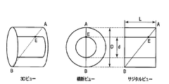
- FIG. 1 is a diagram showing various views in the field of view of PET according to the present embodiment.
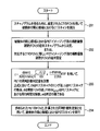
- FIG. 2 is a flowchart illustrating an example of a procedure for calculating a PET coincidence determination window according to the present embodiment.
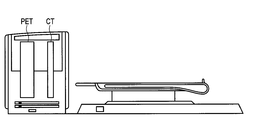
- FIG. 3 is a diagram illustrating an appearance of a PET-CT scanner system according to the present embodiment.
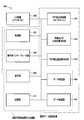
- FIG. 4 is a diagram showing components in the PET-CT scanner system of FIG. 3 according to the present embodiment.
- the embodiments described herein relate to a new method for determining a coincidence decision window based on an FOV set for a PET scanner.
- each FOV of PET has a corresponding optimal coincidence decision window size.
- the coincidence determination window (coincidence time width) is determined by the maximum flight time difference for the longest diagonal line of response (LOR) with the radiation point at the end of the FOV, taking into account the measurement uncertainty in the flight time difference. Desired. Therefore, the optimal coincidence decision window size depends on the transverse FOV and axial length of the PET scanner and also on the time-of-flight resolution of the PET scanner.
- the present embodiment is not limited to a PET / CT system. That is, this embodiment can also be applied to a single unit of a nuclear medicine diagnosis apparatus including a PET / MR and a PET apparatus.
- a method for determining a coincidence determination window for imaging a region of interest of a subject using a positron emission tomography (PET) scanner comprising: Determining the diameter of the transverse field of view (FOV) to image the region of interest of the specimen; and (2) the determined diameter, the PET scanner ring diameter, the PET scanner axial length, and the PET scanner flight. Calculating a coincidence determination window based on the temporal resolution.
- PET positron emission tomography
- the determining step includes determining the diameter of the transverse FOV from a computed tomography (CT) scanogram in the region of interest of the subject.
- CT computed tomography
- the calculating step comprises:
- D is the ring diameter of the PET scanner
- L is the axial length of the PET scanner
- d is the diameter of the transverse FOV
- FWHM ⁇ TOF is the time-of-flight resolution (TOF time resolution) of the PET scanner
- N is a predetermined number (2 ⁇ n ⁇ 3).
- the axial length L corresponds to the length of a plurality of PET detectors along the long axis direction of the top plate in the PET scanner.
- the ring diameter D corresponds to the diameter of a ring formed by a plurality of PET detectors arranged along the circumference of the top plate in the PET scanner. D corresponds to the diameter of the imaging field in the PET scan.
- FWHM is a full width at half maximum. For example, the FWHM corresponds to the width of the flight time difference connecting two points indicating the half value of the maximum value ( ⁇ TOF max ) in the distribution of the flight time difference ( ⁇ TOF) generated based on the output from the PET detector described later.
- the method further includes using a PET scanner to perform a PET scan of the region of interest of the subject using the calculated coincidence determination window.
- a method for determining a PET coincidence determination window for imaging a region of interest of a subject using a positron emission tomography (PET) -computed tomography (CT) combined scanner includes: (1) setting the CT field of view (FOV) based on the size of the region of interest of the subject; and (2) setting the diameter of the transverse FOV for PET imaging. (3) PET simultaneous based on the determined diameter, the PET scanner ring diameter, the PET scanner axial length, and the PET scanner time-of-flight resolution. Calculating a counting decision window.
- FOV positron emission tomography
- CT positron emission tomography
- a positron emission tomography (PET) -computed tomography (CT) combined scanning device is provided, which is based on (1) the size of a region of interest of a subject.
- a CT scanner configured to perform a CT scan of a region of interest of a subject using a set CT field of view (FOV), and (2) a subject using a PET FOV having a cross-sectional diameter.
- a PET scanner configured to perform a PET scan of the region of interest, the diameter being determined by a CT scan of the region of interest, wherein the PET scanner is the determined diameter, the ring diameter of the PET scanner, the axis of the PET scanner
- a controller configured to calculate a coincidence determination window based on the length and time-of-flight resolution of the PET scanner.
- a positron emission tomography (PET) -computed tomography (CT) combined scanning device is provided, which is based on (1) the size of a region of interest of a subject.
- a CT scanner configured to perform a CT scan of a region of interest of a subject using a set CT field of view (FOV), and (2) a subject using a PET FOV having a cross-sectional diameter.
- a PET scanner configured to perform a PET scan of the region of interest.
- the PET scanner sets the PET FOV diameter in the cross section to be equal to the set CT FOV.
- the PET scanner includes a controller configured to calculate a coincidence determination window based on the set diameter, the PET scanner ring diameter, the PET scanner axial length, and the time-of-flight resolution of the PET scanner.
- FIG. 1 shows an imaging FOV directed to a clinical whole body PET scanner.
- ⁇ TOF maximum time-of-flight difference
- c is the speed of light (300 mm / ns).
- the coincidence determination timing window ⁇ ie, the threshold for filtering coincidence events by the difference in arrival times of two detected photons, considers both the maximum TOF difference and timing uncertainty. It is necessary to The flight time difference ⁇ TOF is Gaussian and the standard deviation ⁇ ⁇ TOF is
- the coincidence decision window for a cylindrical FOV is
- the calculated coincidence decision window detects 99.7% of the true LOR in the cylinder.
- the confidence interval is 95%. A value of n between 2 and 3 is recommended.
- the coincidence determination window is a function of FOV (d) (in the PET scanner), and the PET detector diameter (ring diameter: D), length (axis length: L), and timing resolution (FWHM). ⁇ TOF ) is clearly calculated.
- the embodiment in which the coincidence determination window is calculated based on the FOV and the characteristics of the PET detector has advantages over conventional scanners. For example, the amount of random coincidence for each FOV is reduced by the “optimal” coincidence decision window calculated above instead of a fixed coincidence decision window, while true coincidence is not affected.
- the amount in random reduction for various sized FOVs using the NEMA-NU2 count rate phantom is listed.
- FIG. 2 illustrates a method for performing a PET scan using a PET scanner in a PET-CT scanner, according to one embodiment.
- a CT scan in the region of interest of the subject is performed using the set CT FOV.
- Table 1 any one of a plurality of FOVs in CT may be used depending on the region of interest of the subject to be imaged.
- the diameter d of the transverse FOV for PET imaging is obtained. This diameter may be determined from the CT scanogram obtained in step 201. Alternatively, the diameter may be set to be the same as the CT FOV value used in step 201.
- step 203 the following equation:
- D is the PET scanner ring diameter
- L is the PET scanner axial length
- d is the transverse FOV diameter
- FWHM ⁇ TOF is the time-of-flight resolution in the PET scanner
- n is a predetermined number It is.
- step 204 the PET scan in the region of interest of the subject is performed using the PET FOV obtained in step 202 and the coincidence determination window calculated in step 203.
- the system includes a CT device 406 configured to perform a CT scan and a PET detector device (PET scanner) 401 configured to perform a PET scan.
- PET scanner PET detector device
- a PET detector device (PET scanner) 401 is arranged around the top plate in a cylindrical shape having a predetermined axial length and a predetermined ring diameter along the long axis direction of the top plate, and is generated in the subject.
- the PET / CT system 400 having a plurality of radiation detectors for detecting the detected radiation is based on a command received from the control unit 407 and the like, and a patient pallet inside the PET detector device 401 and the CT device (CT scanner) 406
- a movable patient pallet (top plate) 402 including a pallet (top plate) positioning unit configured to perform positioning of the (top plate) is included.
- the control unit 407 controls the entire function of the PET / CT system 400 including adjustment of the position of the patient pallet (top plate) via the pallet positioning unit.
- the pallet positioning unit includes a mechanism configured to move the patient pallet at least in the longitudinal direction.
- the control unit 407 controls the PET detector positioning unit 403 that performs positioning of one or more PET detector portions around the patient on the pallet.
- control unit 407 is configured to calculate the coincidence determination determination window based on the obtained diameter, the ring diameter of the PET scanner, the axial length of the PET scanner, and the time-of-flight resolution of the PET scanner. ing.
- the control unit 407 calculates the coincidence time width for specifying the radiation generation point based on the diameter of the imaging field of view for the subject, the axial length, the ring diameter, and the time resolution of the flight time of the radiation. .
- the control unit 407 outputs the calculated coincidence time width to the data processing unit described later.
- the control unit 407 controls the PET scanner 401 in order to execute the PET scan on the subject using the calculated coincidence time width.
- control unit 407 may determine the diameter of the imaging field in the PET scan based on a scanogram generated by X-ray computed tomography on the subject. Further, the control unit 407 may set the diameter of the imaging field in the PET scan to be equal to the diameter of the imaging field in the X-ray computed tomography.
- the data collection unit 404 acquires PET event data from the PET detector device 401 during the PET scan, and sends the event data to the data processing unit in order to reconstruct the PET image. Further, the PET event data may be stored in the storage unit 410 before being processed by the data processing unit 405.
- the operator interface unit 408 is configured to receive, for example, an operator command for starting a CT scan or a PET scan, or an operator command for setting a region of interest of a CT image, and / or relates to a scan. It is configured to receive parameters.
- the PET and CT images of the patient and the operation parameters related to the scan are displayed on the display unit 409.
- control unit 407 and the data processing unit 405 may be individual logic gates, application specific integrated circuits (ASICs), field programmable gate arrays (FPGAs), or other complex programmable logic devices (CPLDs).
- a CPU that can be implemented as:
- An FPGA or CPLD implementation may be coded in VHDL, Verilog, or any other hardware description language and the code may be stored directly in electronic memory internal to the FPGA or CPLD, or It may be stored in another electronic memory.
- the storage unit 410 may be non-volatile like ROM, EPROM, EEPROM, or flash memory.
- the storage unit 410 may be volatile like static RAM or dynamic RAM, and a processor such as a microcontroller or microprocessor not only manages the electronic memory but also exchanges between the FPGA or CPLD and the memory unit. It may be provided for management.
- the storage unit 410 stores radiation detection data (PET event data) generated along with a scan for detecting radiation generated in the subject placed on the top board.
- PET event data radiation detection data
- the CPU in the control unit 407 or the data processing unit 405 may execute a computer program including a set of computer-readable instructions for executing the functions described in this specification.
- This program may be stored in the above-mentioned non-transitory electronic memory and / or hard disk drive, CD, DVD, flash drive, or any other known storage medium.
- the computer readable instructions include processors such as Intel Xenon processor in the United States or Opteron processor from AMD in the United States, as well as Microsoft Vista, UNIX (registered trademark), Solaris, LINUX (registered trademark), Apple, MAC - May be provided as a utility application, background daemon, or operating system component, or a combination thereof, operating with an operating system such as the OS and other operating systems known to those skilled in the art May be.
- processors such as Intel Xenon processor in the United States or Opteron processor from AMD in the United States, as well as Microsoft Vista, UNIX (registered trademark), Solaris, LINUX (registered trademark), Apple, MAC - May be provided as a utility application, background daemon, or operating system component, or a combination thereof, operating with an operating system such as the OS and other operating systems known to those skilled in the art May be.
- the data processing unit 405 generates a distribution image of the radionuclide administered into the subject based on the radiation detection data and the coincidence counting time width.
- the processed signal is stored in the storage unit 410 and / or displayed on the display unit 409.
- the storage unit 410 may be a hard disk drive, CD-ROM drive, DVD drive, flash drive, RAM, ROM, or any other electronic storage device known in the art.
- Display unit 409 may be implemented as an LCD display, CRT display, plasma display, OLED, LED, or any other display known in the art.
- storage part 410 and the display part 409 performed by this specification is only an illustration, and does not limit the scope of the present invention at all.
- the apparatus has constituent elements within an alternate long and short dash line in the configuration diagram of FIG.
- each process for determining the coincidence time width corresponds to each process in the flowchart of FIG.
- each process for determining the coincidence time width corresponds to each process in the flowchart of FIG.
- the present invention is not limited to the above-described embodiment as it is, and can be embodied by modifying constituent elements without departing from the scope of the invention in the implementation stage.
- various inventions can be formed by appropriately combining a plurality of components disclosed in the embodiment. For example, some components may be deleted from all the components shown in the embodiment.
- constituent elements over different embodiments may be appropriately combined.
- PET / CT system 400 ... PET detector device (PET scanner) 402 ... Top plate and top plate positioning unit 403 ... PET detector positioning unit 404 ... Data collection unit 405 ... Data processing unit 406 ... CT apparatus (CT scanner), 407 ... control unit, 408 ... operator interface unit, 409 ... display unit, 410 ... storage unit
Landscapes
- Health & Medical Sciences (AREA)
- Life Sciences & Earth Sciences (AREA)
- Engineering & Computer Science (AREA)
- Medical Informatics (AREA)
- Physics & Mathematics (AREA)
- Molecular Biology (AREA)
- High Energy & Nuclear Physics (AREA)
- Heart & Thoracic Surgery (AREA)
- General Health & Medical Sciences (AREA)
- Pathology (AREA)
- Radiology & Medical Imaging (AREA)
- Biomedical Technology (AREA)
- Nuclear Medicine, Radiotherapy & Molecular Imaging (AREA)
- Biophysics (AREA)
- Surgery (AREA)
- Animal Behavior & Ethology (AREA)
- Optics & Photonics (AREA)
- Public Health (AREA)
- Veterinary Medicine (AREA)
- General Physics & Mathematics (AREA)
- Spectroscopy & Molecular Physics (AREA)
- Computer Vision & Pattern Recognition (AREA)
- Pulmonology (AREA)
- Theoretical Computer Science (AREA)
- Nuclear Medicine (AREA)
- Apparatus For Radiation Diagnosis (AREA)
- Measurement Of Radiation (AREA)
Priority Applications (2)
| Application Number | Priority Date | Filing Date | Title |
|---|---|---|---|
| EP13840867.9A EP2902806B1 (en) | 2012-09-28 | 2013-09-26 | Nuclear medicine diagnostic device and medical data processing device |
| CN201380003710.3A CN104024885B (zh) | 2012-09-28 | 2013-09-26 | 核医学诊断装置及医用数据处理装置 |
Applications Claiming Priority (4)
| Application Number | Priority Date | Filing Date | Title |
|---|---|---|---|
| US13/630,787 | 2012-09-28 | ||
| US13/630,787 US8809792B2 (en) | 2012-09-28 | 2012-09-28 | Field-of-view-dependent coincidence window for positron emission tomography |
| JP2013-196195 | 2013-09-20 | ||
| JP2013196195A JP2014071113A (ja) | 2012-09-28 | 2013-09-20 | 核医学診断装置および医用データ処理装置 |
Publications (1)
| Publication Number | Publication Date |
|---|---|
| WO2014051013A1 true WO2014051013A1 (ja) | 2014-04-03 |
Family
ID=50385986
Family Applications (1)
| Application Number | Title | Priority Date | Filing Date |
|---|---|---|---|
| PCT/JP2013/076161 Ceased WO2014051013A1 (ja) | 2012-09-28 | 2013-09-26 | 核医学診断装置および医用データ処理装置 |
Country Status (5)
| Country | Link |
|---|---|
| US (1) | US8809792B2 (enExample) |
| EP (1) | EP2902806B1 (enExample) |
| JP (1) | JP2014071113A (enExample) |
| CN (1) | CN104024885B (enExample) |
| WO (1) | WO2014051013A1 (enExample) |
Families Citing this family (5)
| Publication number | Priority date | Publication date | Assignee | Title |
|---|---|---|---|---|
| EP3077850B1 (en) * | 2013-12-04 | 2020-01-15 | Koninklijke Philips N.V. | Reconstruction apparatus for reconstructing a pet image |
| CN104352246A (zh) * | 2014-12-02 | 2015-02-18 | 东南大学 | 基于可视化的锥束ct感兴趣区域的扫描方法 |
| CN110215227B (zh) * | 2019-06-05 | 2022-10-14 | 上海联影医疗科技股份有限公司 | 时间窗设置方法、装置、计算机设备和存储介质 |
| WO2021066808A1 (en) * | 2019-10-01 | 2021-04-08 | Siemens Medical Solutions Usa, Inc. | Model-based injected dose optimization for long axial fov pet imaging |
| US12138096B2 (en) * | 2020-01-10 | 2024-11-12 | Siemens Medical Solutions Usa, Inc. | Partial scan and reconstruction for a positron emission tomography system |
Citations (3)
| Publication number | Priority date | Publication date | Assignee | Title |
|---|---|---|---|---|
| WO2011125181A1 (ja) * | 2010-04-06 | 2011-10-13 | 独立行政法人放射線医学総合研究所 | Pet装置における同時計数判定方法及び装置 |
| JP2012103179A (ja) * | 2010-11-12 | 2012-05-31 | Hitachi Ltd | 放射線検出装置及びその方法 |
| JP2012145419A (ja) * | 2011-01-11 | 2012-08-02 | Natl Inst Of Radiological Sciences | Pet装置及びpet−mri装置 |
Family Cites Families (10)
| Publication number | Priority date | Publication date | Assignee | Title |
|---|---|---|---|---|
| US6763082B2 (en) * | 2002-02-27 | 2004-07-13 | Kabushiki Kaisha Toshiba | X-ray computer tomography apparatus |
| FR2866713B1 (fr) * | 2004-02-24 | 2006-03-24 | Commissariat Energie Atomique | Circuit electronique de diagnostic de spectrometrie et chaine de comptage associee |
| US7557350B2 (en) * | 2005-01-28 | 2009-07-07 | Koninklijke Philips Electronics N.V. | Timing calibration using radioactive source |
| US7402807B2 (en) * | 2005-11-02 | 2008-07-22 | Siemens Medical Solutions Usa, Inc. | Method for reducing an electronic time coincidence window in positron emission tomography |
| CN101401009B (zh) * | 2006-03-10 | 2012-07-25 | 株式会社岛津制作所 | 核医学诊断装置以及用于此的诊断系统 |
| WO2009054070A1 (ja) * | 2007-10-26 | 2009-04-30 | Shimadzu Corporation | 放射線検出器 |
| US8063376B2 (en) * | 2008-08-15 | 2011-11-22 | Koninklijke Philips Electronics N.V. | Large bore PET and hybrid PET/CT scanners and radiation therapy planning using same |
| JP5677751B2 (ja) * | 2010-02-24 | 2015-02-25 | 株式会社東芝 | X線ct装置及び画像処理プログラム |
| US8604440B2 (en) * | 2010-03-09 | 2013-12-10 | The University Of Chicago | Use of flat panel microchannel photomultipliers in sampling calorimeters with timing |
| US8921801B2 (en) * | 2011-01-07 | 2014-12-30 | Brookhaven Science Associates, Llc | Detection system for high-resolution gamma radiation spectroscopy with neutron time-of-flight filtering |
-
2012
- 2012-09-28 US US13/630,787 patent/US8809792B2/en active Active
-
2013
- 2013-09-20 JP JP2013196195A patent/JP2014071113A/ja active Pending
- 2013-09-26 CN CN201380003710.3A patent/CN104024885B/zh active Active
- 2013-09-26 EP EP13840867.9A patent/EP2902806B1/en active Active
- 2013-09-26 WO PCT/JP2013/076161 patent/WO2014051013A1/ja not_active Ceased
Patent Citations (3)
| Publication number | Priority date | Publication date | Assignee | Title |
|---|---|---|---|---|
| WO2011125181A1 (ja) * | 2010-04-06 | 2011-10-13 | 独立行政法人放射線医学総合研究所 | Pet装置における同時計数判定方法及び装置 |
| JP2012103179A (ja) * | 2010-11-12 | 2012-05-31 | Hitachi Ltd | 放射線検出装置及びその方法 |
| JP2012145419A (ja) * | 2011-01-11 | 2012-08-02 | Natl Inst Of Radiological Sciences | Pet装置及びpet−mri装置 |
Also Published As
| Publication number | Publication date |
|---|---|
| CN104024885A (zh) | 2014-09-03 |
| EP2902806B1 (en) | 2021-01-27 |
| JP2014071113A (ja) | 2014-04-21 |
| US8809792B2 (en) | 2014-08-19 |
| EP2902806A1 (en) | 2015-08-05 |
| CN104024885B (zh) | 2017-02-08 |
| US20140095106A1 (en) | 2014-04-03 |
| EP2902806A4 (en) | 2016-05-25 |
Similar Documents
| Publication | Publication Date | Title |
|---|---|---|
| JP6188418B2 (ja) | 偶発イベント削減方法、偶発イベント削減装置及び非一時的コンピュータ可読記憶媒体 | |
| US8502154B2 (en) | Method and system for organ specific PET imaging | |
| EP3229689B1 (en) | Outside-fov activity estimation using surview and prior patient data in positron emission tomography | |
| US10743830B2 (en) | Method and apparatus for scatter correction in position emission tomography (PET) imaging by performing a short PET scan in an extended region to estimate scatter coming from outside of the field of view (FOV) | |
| US10049465B2 (en) | Systems and methods for multi-modality imaging component alignment | |
| JP6400265B2 (ja) | PET(PositronEmissionTomography)スキャナ | |
| WO2014051013A1 (ja) | 核医学診断装置および医用データ処理装置 | |
| Piccinelli et al. | Advances in single-photon emission computed tomography hardware and software | |
| JP6670643B2 (ja) | Pet装置 | |
| JP6400266B2 (ja) | PET(PositronEmissionTomography)スキャナ | |
| JP2014052353A (ja) | 画像処理方法及び核医学診断装置 | |
| Germano et al. | Technical aspects of cardiac PET imaging and recent advances | |
| CN112998736A (zh) | 一种扫描装置的时间校正系统及其时间校正方法 | |
| Belcari et al. | PET/CT and PET/MR tomographs: Image acquisition and processing | |
| US11982779B2 (en) | Method and apparatus for guided pairing of multi-coincidences for time of flight positron emission tomography | |
| EP4075168A1 (en) | Nuclear medicine diagnosis device and nuclear medicine image data generation method | |
| Suda | Benefits of time-of-flight positron emission tomography computed tomography with 13N-ammonia | |
| JP2010085147A (ja) | 放射線撮像装置及び画像情報作成方法 |
Legal Events
| Date | Code | Title | Description |
|---|---|---|---|
| 121 | Ep: the epo has been informed by wipo that ep was designated in this application |
Ref document number: 13840867 Country of ref document: EP Kind code of ref document: A1 |
|
| WWE | Wipo information: entry into national phase |
Ref document number: 2013840867 Country of ref document: EP |
|
| NENP | Non-entry into the national phase |
Ref country code: DE |