WO2013151122A1 - Procédé et kit pour la détection immunologique de complexe mycobacterium tuberculosis - Google Patents
Procédé et kit pour la détection immunologique de complexe mycobacterium tuberculosis Download PDFInfo
- Publication number
- WO2013151122A1 WO2013151122A1 PCT/JP2013/060301 JP2013060301W WO2013151122A1 WO 2013151122 A1 WO2013151122 A1 WO 2013151122A1 JP 2013060301 W JP2013060301 W JP 2013060301W WO 2013151122 A1 WO2013151122 A1 WO 2013151122A1
- Authority
- WO
- WIPO (PCT)
- Prior art keywords
- mycobacterium tuberculosis
- biological sample
- heat treatment
- detecting
- group
- Prior art date
Links
Images

Classifications
-
- G—PHYSICS
- G01—MEASURING; TESTING
- G01N—INVESTIGATING OR ANALYSING MATERIALS BY DETERMINING THEIR CHEMICAL OR PHYSICAL PROPERTIES
- G01N33/00—Investigating or analysing materials by specific methods not covered by groups G01N1/00 - G01N31/00
- G01N33/48—Biological material, e.g. blood, urine; Haemocytometers
- G01N33/50—Chemical analysis of biological material, e.g. blood, urine; Testing involving biospecific ligand binding methods; Immunological testing
- G01N33/68—Chemical analysis of biological material, e.g. blood, urine; Testing involving biospecific ligand binding methods; Immunological testing involving proteins, peptides or amino acids
-
- G—PHYSICS
- G01—MEASURING; TESTING
- G01N—INVESTIGATING OR ANALYSING MATERIALS BY DETERMINING THEIR CHEMICAL OR PHYSICAL PROPERTIES
- G01N33/00—Investigating or analysing materials by specific methods not covered by groups G01N1/00 - G01N31/00
- G01N33/48—Biological material, e.g. blood, urine; Haemocytometers
- G01N33/50—Chemical analysis of biological material, e.g. blood, urine; Testing involving biospecific ligand binding methods; Immunological testing
- G01N33/53—Immunoassay; Biospecific binding assay; Materials therefor
- G01N33/569—Immunoassay; Biospecific binding assay; Materials therefor for microorganisms, e.g. protozoa, bacteria, viruses
- G01N33/56911—Bacteria
- G01N33/5695—Mycobacteria
-
- G—PHYSICS
- G01—MEASURING; TESTING
- G01N—INVESTIGATING OR ANALYSING MATERIALS BY DETERMINING THEIR CHEMICAL OR PHYSICAL PROPERTIES
- G01N33/00—Investigating or analysing materials by specific methods not covered by groups G01N1/00 - G01N31/00
- G01N33/48—Biological material, e.g. blood, urine; Haemocytometers
- G01N33/50—Chemical analysis of biological material, e.g. blood, urine; Testing involving biospecific ligand binding methods; Immunological testing
- G01N33/53—Immunoassay; Biospecific binding assay; Materials therefor
- G01N33/543—Immunoassay; Biospecific binding assay; Materials therefor with an insoluble carrier for immobilising immunochemicals
-
- G—PHYSICS
- G01—MEASURING; TESTING
- G01N—INVESTIGATING OR ANALYSING MATERIALS BY DETERMINING THEIR CHEMICAL OR PHYSICAL PROPERTIES
- G01N33/00—Investigating or analysing materials by specific methods not covered by groups G01N1/00 - G01N31/00
- G01N33/48—Biological material, e.g. blood, urine; Haemocytometers
- G01N33/50—Chemical analysis of biological material, e.g. blood, urine; Testing involving biospecific ligand binding methods; Immunological testing
- G01N33/53—Immunoassay; Biospecific binding assay; Materials therefor
- G01N33/569—Immunoassay; Biospecific binding assay; Materials therefor for microorganisms, e.g. protozoa, bacteria, viruses
- G01N33/56911—Bacteria
Definitions
- a biological sample containing a Mycobacterium tuberculosis group is subjected to a heat treatment at a predetermined temperature, and a culture operation is performed by immunologically detecting a Mycobacterium tuberculosis group-specific secreted protein secreted outside the cells.
- the present invention relates to a highly safe immunological detection method and kit for detection of Mycobacterium tuberculosis that can be easily and quickly performed directly from a biological sample without performing it.
- Mycobacteria that are pathogenic to humans include Mycobacterium tuberculosis (Mycobacterium tuberculosis), Mycobacterium bovis (Mycobacterium bovis), Mycobacterium Microcochi (Mycobacterium microti), Mycobacterium africanum (Mycobacterium africanum), are known, and non-tuberculous mycobacteria are Mycobacterium avium, Mycobacterium Known are Mycobacterium kansassi, Mycobacterium marinum, and the like.
- the Mycobacterium tuberculosis group that infects humans is mainly Mycobacterium tuberculosis, and rarely Mycobacterium bovis infection.
- infection with Mycobacterium avium has increased among nontuberculous mycobacteria.
- the clinical symptoms of both are similar, and the identification of the causative microorganism is important in determining the treatment strategy.
- differential detection is also important for the purpose of preventing secondary infection to third parties for public health.
- a detection method using culture has been performed as a method for detecting the Mycobacterium tuberculosis group.
- the culture method is performed by inoculating a biological sample into either a liquid medium or a solid medium and detecting the grown cells.
- the culture period of the biological sample is shortened, but there is a high risk of secondary infection during work, and highly safe facilities and devices are required.
- a long period of about two months is required until the detection result becomes clear.
- Culture of Mycobacterium tuberculosis must be carried out with great care in order to prevent secondary infection using a highly safe apparatus and a highly safe apparatus.
- a method for identifying the Mycobacterium tuberculosis group from the culture after culturing a biological sample a method for detecting the Mycobacterium tuberculosis group-specific protein secreted in the medium by immunological measurement is known as a simple method. It has been.
- a biological sample collected from a tuberculosis patient is inoculated into a solid medium or a liquid medium, and after culture for several days to several weeks, it is specific for a group of tuberculosis bacteria secreted into the medium.
- a method for detecting the presence of Mycobacterium tuberculosis by immunologically detecting MPB64, which is a secreted protein.
- MPB64 which is a secreted protein
- a tuberculosis group in a biological sample is cultured, and the culture is used as a sample. Therefore, detection and identification take approximately the same time as the number of culture days, and much labor and cost are required.
- MPB64 is produced by Mycobacterium bovis BCG (M. bovis BCG) and is a secretory protein of Mycobacterium tuberculosis group secreted outside the cell body.
- MPT64 is produced by Mycobacterium tuberculosis and secreted from the Mycobacterium tuberculosis group, but it is known that MPB64 and MPT64 are the same substance.
- MPT64 which is a secreted protein specific for Mycobacterium tuberculosis, can be detected by an immunoassay without culturing the Mycobacterium tuberculosis in a biological sample. According to this method, the presence of M. tuberculosis group is detected by detecting MPT64 contained in the biological sample by treating sputum or the like as the biological sample.
- MPB64 in a biological sample is specifically detected using an antibody against a specific epitope of MPB64, so that diagnosis of infection by M. tuberculosis can be performed quickly and safely with higher accuracy than before.
- a method of performing is disclosed. According to this method, as a sample to be applied, a culture obtained by culturing a biological sample using a small amount of liquid medium only for a time period during which the Mycobacterium tuberculosis group cells do not substantially grow can be applied.
- An object of the present invention is to solve the above-mentioned problems by providing a method for detecting Mycobacterium tuberculosis that is simple, rapid, and more reliable.
- the present inventors secreted tuberculosis group-specific secreted proteins such as MPB64 to the outside of the cells by subjecting the biological sample containing the tuberculosis group to heat treatment.
- the inventors have found that the above problem can be solved by detecting the secreted protein, and have completed the present invention.
- detection of Mycobacterium tuberculosis group comprising subjecting Mycobacterium tuberculosis group-specific protein secreted outside the bacterial body by subjecting a biological sample containing Mycobacterium tuberculosis group to heat treatment.
- a method is provided.
- a tuberculosis group detection kit for carrying out the method a treatment container for heat-treating a biological sample containing the tuberculosis group, and a tuberculosis group secreted by the heat treatment.
- a kit for detecting Mycobacterium tuberculosis comprising at least an immunological measurement apparatus for detecting a specific secreted protein.
- the kit may further contain a sample treatment agent and / or a solvent.
- the Mycobacterium tuberculosis group can be detected quickly and easily.
- a method for secreting Mycobacterium tuberculosis group-specific protein outside the microbial cells comprising subjecting a biological sample containing Mycobacterium tuberculosis group to a heat treatment.
- FIG. 1 is a graph showing the relationship between the heat treatment temperature and the effect of promoting the secretion of MPB64 to the outside of cells when cells of M. bovis BCG Tokyo strain are used.
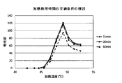
- FIG. 2 is a graph showing the relationship between the heat treatment time and the effect of promoting the secretion of MPB64 to the outside of the cells when the cells of M. bovis BCG Tokyo strain are used.
- the biological sample used in the present invention is not particularly limited as long as the Mycobacterium tuberculosis group can exist, and examples include sputum, pleural effusion, bronchial secretion, gastric fluid, blood, spinal fluid, urine, and feces.
- sputum is used because of the high bacterial concentration.
- bronchial lavage fluid collected at the time of examination in the respiratory organs, tissue pieces collected from the bronchi or lungs can be used as biological samples. These biological samples may be used alone or in combination of two or more.
- the heat treatment of the biological sample is preferably performed by treating the biological sample containing the highly infectious M. tuberculosis group, and placing the biological sample in a sealable container and heating the entire container. Furthermore, from the viewpoint of ensuring the safety of the operator, it is preferable to perform the heat treatment operation in a safety cabinet.
- the container used for the heat treatment is not particularly limited as long as it can keep a sealed state and can withstand a predetermined heating temperature, and can be appropriately selected according to the biological sample to be treated.
- a dropping nozzle with a filter may be attached to the opening of the container so that the treated biological sample can be easily discharged from the container when subjected to immunological measurement.
- the use of a dropping nozzle with a filter is preferable because solid substances such as denatured components contained in the biological sample after treatment can be removed, and furthermore, the biological sample can be easily dropped onto the immunological measurement apparatus.
- the biological sample may be subjected to a heat treatment after being subjected to a treatment such as dissolution with a sample treatment agent according to its properties.
- a treatment such as dissolution with a sample treatment agent according to its properties.
- a cocoon when used as a sample, it may be dissolved with an alkaline substance, a reducing substance, a protease, a sugar, a surfactant, a protein denaturant, etc. that lowers the viscosity of the cocoon in order to improve the efficiency of the heat treatment.
- a treatment using an alkaline substance sodium hydroxide and a reducing substance N-acetyl-L-cysteine in combination is simple and preferably used.
- the biological sample can be dispersed or dissolved in a solvent and subjected to heat treatment.
- a solvent such as phosphate buffered saline or for acid-fast bacteria isolation culture such as Middlebrook 7H9.
- a liquid medium can be used.
- a biological sample dissolved with the sample treatment agent may be redispersed in a solvent.
- the heating temperature of the biological sample may be any temperature as long as the protein can be secreted from the Mycobacterium tuberculosis group into the biological sample or solvent, and is preferably in the range of 40 ° C to 60 ° C, more preferably 40 ° C to It is in the range of 55 ° C.
- the temperature at which secretion is particularly effectively promoted is in the range of 45 ° C to 50 ° C.
- the treatment time of the biological sample may be sufficient as long as it is maintained at the treatment temperature and secreted by a detectable amount of the Mycobacterium tuberculosis group-specific secreted protein outside the cells.
- the treatment time is preferably 15 minutes or more, more preferably 15 minutes or more and 2.5 hours or less, even more preferably 15 minutes or more and 1.5 hours or less, and most preferably 30 minutes or more and 1 hour or less.
- the sample subjected to the heat treatment may be subjected to inactivation treatment for Mycobacterium tuberculosis by raising the heating temperature as it is.
- inactivation treatment for Mycobacterium tuberculosis by raising the heating temperature as it is.
- limiting in the temperature which inactivates a tuberculosis microbe group It is preferable to process at 100 degreeC.
- the heating device used for the heat treatment is not particularly limited as long as it is a device capable of keeping the above temperature constant, and a thermostatic bath, a heater block, an incubator, and the like can be used.
- a small heating device is particularly preferred because all tuberculosis detection operations can be completed within the safety cabinet.
- the present invention it is possible to heat-treat the biological sample and secrete the Mycobacterium tuberculosis group-specific protein to the outside of the bacterial body. Therefore, by using the treated biological sample, the presence of the Mycobacterium tuberculosis group can be immunized. It becomes easy to detect by physics measurement etc.
- the Mycobacterium tuberculosis group-specific protein secreted outside the cells is not particularly limited as long as it is secreted outside the cells.
- proteins secreted by the Mycobacterium tuberculosis group outside the cells include many proteins such as MPT64, MPB64, MPB70, ESAT-6, and CFP-10.
- a protein that is suitable for use as an indicator of the presence of the Mycobacterium tuberculosis group in the immunological measurement used in the present invention is a protein that is secreted in large quantities outside the cells in a short time.
- MPB64 or MPT64 is secreted or released in large quantities outside the cells without culturing, that is, before the cells grow.
- MPB64 that is, MPT64
- the presence or absence of the Mycobacterium tuberculosis group can be determined by immunologically measuring MPB64 secreted outside the cells, that is, MPT64.
- the immunoassay in this detection method is not particularly limited, but sandwich immunoassay using the first antibody and the second antibody against the Mycobacterium tuberculosis group-specific secreted protein, especially ELISA (Enzyme -linked (Immunosorbent assay) or immunochromatographic measurement is preferred.
- the first antibody and the second antibody may be the same antibody or different antibodies as long as a tuberculosis group-specific secreted protein can be detected.
- the obtained bacterial solution was centrifuged to recover the bacterial cells.
- the middle brook 7H9 liquid medium described above was added to the collected cells and resuspended, and the cells were further collected by centrifugation. This operation was repeated three times, and the bacterial cells were washed to remove the Mycobacterium tuberculosis group secreted protein attached to the bacterial cells.
- Capilia registered trademark
- TB manufactured by Towns
- Example 1 (Examination of optimum conditions of heat treatment temperature) The turbidity solution equivalent to McFarland No. 1 prepared in Reference Example 1 was prepared to an OD of 0.1 at an absorbance of 530 nm, and the test solution (corresponding to the 7th power of 10) Prepared. Further, the test bacterial solution was diluted with a liquid medium to prepare a 10-fold diluted bacterial solution (corresponding to the sixth power of 10) and a 100-fold diluted bacterial solution (corresponding to the fifth power of 10). 200 ⁇ l of each prepared bacterial solution was dispensed into a plastic tube (nucleic acid amplification tube) and heat-treated with a heat block for a nucleic acid amplification device capable of adjusting the temperature.
- a plastic tube nucleic acid amplification tube
- a temperature of 5 ° C. was set from 35 ° C. to 75 ° C., and the heat treatment was performed for 30 minutes.
- a sample at 35 ° C. which is a general culture temperature, was used as a control.
- 100 ⁇ l was collected from each sample and subjected to Capilia (registered trademark) TB (manufactured by Towns) in the same manner as in Reference Example 1 to detect the presence of MPB64.
- the absorbance of the determination part was measured with an immunochromatographic reader (manufactured by Otsuka Electronics).
- the results are shown in FIG.
- the vertical axis in FIG. 1 indicates the measured value of color intensity (absorbance) in Capilia (registered trademark) TB (towns), and the horizontal axis indicates the heating temperature.
- the results of the test bacterial solution (corresponding to 10 7 cells as the number of bacteria) are shown as a solid line, 10-fold diluted bacterial solution (equivalent to 10 6 cells as the number of bacteria) and 100-fold diluted bacteria
- the results of the liquid are shown by a broken line and a long broken line, respectively. From this result, it was confirmed that the absorbance increased from 40 ° C. to 60 ° C.
- Example 2 (Examination of optimum conditions of heat treatment temperature)
- the turbidity solution equivalent to McFarland No. 1 prepared in Reference Example 1 was prepared to an OD of 0.1 at an absorbance of 530 nm, and the test solution (corresponding to the 7th power of 10) Prepared.
- Each 200 ⁇ l of the prepared bacterial solution was dispensed into a plastic tube (nucleic acid amplification tube), and heat treatment was performed in a heat block for a nucleic acid amplification device capable of adjusting the temperature.
- the heating temperature was 35 ° C., 13 temperatures at intervals of 2 ° C. from 40 ° C. to 64 ° C., and two temperatures of 70 ° C. and 80 ° C.
- MPB64 secretion is not performed in a temperature range of 60 ° C. or higher. Therefore, from this result, by subjecting the bacterial sample to heat treatment at 40 ° C to 60 ° C, especially 45 ° C to 50 ° C, MPB64 secretion was significantly promoted outside the bacterial cell, and immunology It was confirmed that the detection rate by the automatic measurement method increased remarkably.
- Example 3 (Examination of optimum conditions for heat treatment time)
- the turbidity solution equivalent to McFarland No. 1 prepared in Reference Example 1 was prepared to an OD of 0.1 at an absorbance of 530 nm, and the test solution (corresponding to the 7th power of 10) Prepared.
- Each 200 ⁇ l of the prepared bacterial solution was dispensed into a plastic tube, and heat treatment was carried out in a heat block for a nucleic acid amplifier capable of adjusting the temperature.
- the heating operation was carried out at intervals of 5 ° C. from 35 ° C. to 75 ° C.
- the treatment time was set to 0 minutes, 15 minutes, 30 minutes and 60 minutes.
- the vertical axis in FIG. 2 indicates the measured value of color intensity (absorbance) in Capilia (registered trademark) TB (manufactured by Towns), and the horizontal axis indicates the heating temperature.
- the sample with a treatment time of 15 minutes is indicated by a broken line
- the sample with a treatment time of 30 minutes and a sample of 60 minutes are indicated by a solid line and a long broken line. From this result, a significant increase was observed from 45 ° C. to 50 ° C. in all samples with a treatment time of 15 minutes, 30 minutes and 60 minutes, and no difference in treatment time was observed. There was no significant difference from the other samples even at a heating time of 60 minutes. Therefore, the effect of promoting the secretion of MPB64 to the outside of the cells by a heat treatment time of at least 15 minutes was confirmed.
- Example 4 (Examination of optimum conditions for heat treatment time) The turbidity solution equivalent to McFarland No. 1 prepared in Reference Example 1 was prepared to an OD of 0.01 at an absorbance of 530 nm, and the test solution (corresponding to the 6th power of 10) Prepared. Each 200 ⁇ l of the prepared bacterial solution was dispensed into a plastic tube, and heat treatment was performed at 50 ° C. using a heat block for a nucleic acid amplifier capable of adjusting the temperature. The treatment time was set to 0 minutes, 15 minutes, 30 minutes, 45 minutes, 60 minutes, 75 minutes, 120 minutes and 150 minutes. As a control, the test bacterial solution was left at room temperature without heat treatment.
- Example 2 After the treatment, 100 ⁇ l was collected from each sample and applied to Capilia (registered trademark) TB (manufactured by Towns) in the same manner as in Example 1, and the color intensity of the accumulated labeling substance was observed with the naked eye.
- the color intensity is 5 (colorless), ⁇ (weak coloration), + (clear coloration), ++ (more clear coloration), and ++ (significant coloration). Judged in stages. The determination results are shown in Table 2.
- Example 5 (Study of solvent) Using a phosphate buffer solution containing Tween 80 or a Middlebrook 7H9 liquid medium as a solvent for treating a biological sample, a turbidity bacterial solution equivalent to McFarland No. 1 prepared in Reference Example 1, A bacterial solution containing 7th power, 10th power and 10th power was prepared and used as a test sample. 200 ⁇ l of each test sample was dispensed into a plastic tube. Then, after heat treatment at 50 ° C. for 30 minutes in a heat block, 100 ⁇ l was collected from the sample and subjected to determination using Capilia (registered trademark) TB (manufactured by Towns) in the same manner as in Example 1.
- Capilia registered trademark
- the determination was performed by visually observing the color intensity of the labeling substance accumulated in the determination unit.
- the color intensity is 5 (colorless), ⁇ (weak coloration), + (clear coloration), ++ (more clear coloration), and ++ (significant coloration). Judged in stages.
- the determination results are shown in Table 3.
- Example 6 (Detection from a sputum sample) Add the turbidity solution equivalent to McFarland No. 1 prepared in Reference Example 1 to the cocoon that has been confirmed to be negative for M. tuberculosis infection, and the number of bacteria is 10 Samples were prepared and dispensed into plastic tubes. To this sputum sample, N-acetyl-L-cysteine and an aqueous sodium hydroxide solution were added in equal amounts, and allowed to stand at room temperature for 15 minutes to dissolve. Then, after heat treatment at 50 ° C.
- Example 2 100 ⁇ l was collected from the sample and subjected to determination using Capilia (registered trademark) TB (manufactured by Towns) in the same manner as in Example 1.
- Capilia registered trademark
- TB manufactured by Towns
- the determination was performed by visually observing the color intensity of the labeling substance accumulated in the determination unit. The color intensity is 5 (colorless), ⁇ (weak coloration), + (clear coloration), ++ (more clear coloration), and ++ (significant coloration). Judged in stages. The determination results are shown in Table 4.
- the present invention performs a culture operation by subjecting a biological sample containing Mycobacterium tuberculosis group to a heat treatment at a predetermined temperature, and immunologically measuring the Mycobacterium tuberculosis group-specific secreted protein secreted outside the cells.
- the present invention provides a method and kit for detecting Mycobacterium tuberculosis that can be easily and quickly performed directly from a biological sample, and is useful not only for detection of Mycobacterium tuberculosis but also for appropriate diagnosis and treatment of tuberculosis. is there.
Landscapes
- Health & Medical Sciences (AREA)
- Life Sciences & Earth Sciences (AREA)
- Immunology (AREA)
- Engineering & Computer Science (AREA)
- Urology & Nephrology (AREA)
- Hematology (AREA)
- Biomedical Technology (AREA)
- Chemical & Material Sciences (AREA)
- Molecular Biology (AREA)
- Medicinal Chemistry (AREA)
- Biochemistry (AREA)
- Cell Biology (AREA)
- Pathology (AREA)
- Biotechnology (AREA)
- Food Science & Technology (AREA)
- General Physics & Mathematics (AREA)
- Physics & Mathematics (AREA)
- Analytical Chemistry (AREA)
- Microbiology (AREA)
- General Health & Medical Sciences (AREA)
- Virology (AREA)
- Tropical Medicine & Parasitology (AREA)
- Proteomics, Peptides & Aminoacids (AREA)
- Measuring Or Testing Involving Enzymes Or Micro-Organisms (AREA)
- Investigating Or Analysing Biological Materials (AREA)
- Sampling And Sample Adjustment (AREA)
Abstract
Priority Applications (12)
| Application Number | Priority Date | Filing Date | Title |
|---|---|---|---|
| ES13772467T ES2751357T3 (es) | 2012-04-05 | 2013-04-04 | Procedimiento de detección inmunológica del complejo Mycobacterium tuberculosis |
| JP2013539828A JP5612778B2 (ja) | 2012-04-05 | 2013-04-04 | 結核菌群の免疫学的検出方法及び検出用キット |
| BR112014024574-6A BR112014024574B1 (pt) | 2012-04-05 | 2013-04-04 | método e kit para detecção imunológica do complexo mycobacterium tuberculosis |
| IN8246DEN2014 IN2014DN08246A (fr) | 2012-04-05 | 2013-04-04 | |
| US14/390,706 US10823730B2 (en) | 2012-04-05 | 2013-04-04 | Method and kit for immunological detection of Mycobacterium tuberculosis |
| EP13772467.0A EP2835646B1 (fr) | 2012-04-05 | 2013-04-04 | Procédé de détection immunologique du complexe mycobacterium tuberculosis |
| CN201380018968.0A CN104246505B (zh) | 2012-04-05 | 2013-04-04 | 结核菌群的免疫学检测方法和试剂盒 |
| EP19150248.3A EP3499237B1 (fr) | 2012-04-05 | 2013-04-04 | Procédé de sécrétion extracellulaire d'une protéine spécifique du complexe mycobacterium tuberculosis |
| EP16166865.2A EP3093667B1 (fr) | 2012-04-05 | 2013-04-04 | Procede et kit pour la detection immunologique de complexe mycobacerium tuberculosis |
| RU2014144283A RU2657581C2 (ru) | 2012-04-05 | 2013-04-04 | Способ и набор для иммунологической детекции комплекса mycobacterium tuberculosis |
| US15/141,384 US10883988B2 (en) | 2012-04-05 | 2016-04-28 | Method and kit for immunological detection of Mycobacterium tuberculosis complex |
| US15/980,920 US10830769B2 (en) | 2012-04-05 | 2018-05-16 | Method and kit for immunological detection of Mycobacterium tuberculosis |
Applications Claiming Priority (2)
| Application Number | Priority Date | Filing Date | Title |
|---|---|---|---|
| JP2012-086566 | 2012-04-05 | ||
| JP2012086566 | 2012-04-05 |
Related Child Applications (3)
| Application Number | Title | Priority Date | Filing Date |
|---|---|---|---|
| US14/390,706 A-371-Of-International US10823730B2 (en) | 2012-04-05 | 2013-04-04 | Method and kit for immunological detection of Mycobacterium tuberculosis |
| US15/141,384 Division US10883988B2 (en) | 2012-04-05 | 2016-04-28 | Method and kit for immunological detection of Mycobacterium tuberculosis complex |
| US15/980,920 Continuation US10830769B2 (en) | 2012-04-05 | 2018-05-16 | Method and kit for immunological detection of Mycobacterium tuberculosis |
Publications (1)
| Publication Number | Publication Date |
|---|---|
| WO2013151122A1 true WO2013151122A1 (fr) | 2013-10-10 |
Family
ID=49300600
Family Applications (1)
| Application Number | Title | Priority Date | Filing Date |
|---|---|---|---|
| PCT/JP2013/060301 WO2013151122A1 (fr) | 2012-04-05 | 2013-04-04 | Procédé et kit pour la détection immunologique de complexe mycobacterium tuberculosis |
Country Status (9)
| Country | Link |
|---|---|
| US (3) | US10823730B2 (fr) |
| EP (3) | EP3093667B1 (fr) |
| JP (2) | JP5612778B2 (fr) |
| CN (3) | CN105403700B (fr) |
| BR (1) | BR112014024574B1 (fr) |
| ES (3) | ES2751357T3 (fr) |
| IN (1) | IN2014DN08246A (fr) |
| RU (1) | RU2657581C2 (fr) |
| WO (1) | WO2013151122A1 (fr) |
Cited By (3)
| Publication number | Priority date | Publication date | Assignee | Title |
|---|---|---|---|---|
| WO2015164617A1 (fr) * | 2014-04-24 | 2015-10-29 | Somalogic, Inc. | Biomarqueurs de la tuberculose dans l'urine et leurs utilisations |
| RU2594063C1 (ru) * | 2015-04-27 | 2016-08-10 | Федеральное государственное бюджетное научное учреждение "Центральный научно-исследовательский институт туберкулёза" | Способ диагностики микобактерий туберкулёза |
| PL423175A1 (pl) * | 2017-10-16 | 2019-04-23 | Politechnika Warszawska | Sposób detekcji bakterii gruźlicy |
Families Citing this family (4)
| Publication number | Priority date | Publication date | Assignee | Title |
|---|---|---|---|---|
| JP5612778B2 (ja) * | 2012-04-05 | 2014-10-22 | 株式会社ビーエル | 結核菌群の免疫学的検出方法及び検出用キット |
| ES2862224T3 (es) * | 2015-11-02 | 2021-10-07 | Biofire Diagnostics Llc | Preparación de muestras para tipos de muestra difíciles |
| JP6989428B2 (ja) * | 2018-03-23 | 2022-01-05 | アークレイ株式会社 | 前処理剤、前処理キット、抗原検査キット及び抗原検査方法 |
| JP7352490B2 (ja) | 2020-02-27 | 2023-09-28 | 京セラ株式会社 | 配線基板 |
Citations (7)
| Publication number | Priority date | Publication date | Assignee | Title |
|---|---|---|---|---|
| JPH07110332A (ja) * | 1993-10-12 | 1995-04-25 | Tetsuo Tomiyama | 結核菌の検査試薬及び検出方法 |
| JPH11108931A (ja) | 1997-09-30 | 1999-04-23 | Towns:Kk | 結核菌検出法および結核菌検出用キット |
| JP2002062299A (ja) | 2000-08-24 | 2002-02-28 | Kyowa Medex Co Ltd | 結核菌の検出方法及び試薬 |
| JP2004166564A (ja) * | 2002-11-19 | 2004-06-17 | Bl:Kk | モノクローナル抗体および結核症診断法 |
| JP2006184295A (ja) * | 2006-03-31 | 2006-07-13 | Towns:Kk | 結核菌検出用キット |
| JP2006234627A (ja) * | 2005-02-25 | 2006-09-07 | Chang Gung Univ | 体液中の結核菌抗原を検出する方法 |
| WO2009084481A1 (fr) | 2007-12-28 | 2009-07-09 | Bl Co., Ltd. | Test d'immunodétection du complexe mycobacterium tuberculosis |
Family Cites Families (4)
| Publication number | Priority date | Publication date | Assignee | Title |
|---|---|---|---|---|
| DE3700049A1 (de) * | 1987-01-02 | 1988-07-14 | Biotest Serum Institut Gmbh | Verfahren zum immunologischen nachweis von mykobakterien im sputum sowie monoklonale antikoerper zur durchfuehrung des verfahrens |
| SE9600949D0 (sv) | 1996-03-12 | 1996-03-12 | Stefan Svenson | Method of diagnosing a mycobacterial disease and immunoassay kit |
| CA2634968A1 (fr) * | 2005-12-26 | 2007-07-05 | Bl Co., Ltd. | Procede destine a la detection d'une souche virulente du virus grippal a |
| JP5612778B2 (ja) | 2012-04-05 | 2014-10-22 | 株式会社ビーエル | 結核菌群の免疫学的検出方法及び検出用キット |
-
2013
- 2013-04-04 JP JP2013539828A patent/JP5612778B2/ja active Active
- 2013-04-04 US US14/390,706 patent/US10823730B2/en active Active
- 2013-04-04 CN CN201610009079.3A patent/CN105403700B/zh active Active
- 2013-04-04 ES ES13772467T patent/ES2751357T3/es active Active
- 2013-04-04 ES ES16166865T patent/ES2746565T3/es active Active
- 2013-04-04 CN CN201610245191.7A patent/CN105784999B/zh active Active
- 2013-04-04 BR BR112014024574-6A patent/BR112014024574B1/pt active IP Right Grant
- 2013-04-04 WO PCT/JP2013/060301 patent/WO2013151122A1/fr active Application Filing
- 2013-04-04 EP EP16166865.2A patent/EP3093667B1/fr active Active
- 2013-04-04 ES ES19150248T patent/ES2887019T3/es active Active
- 2013-04-04 CN CN201380018968.0A patent/CN104246505B/zh active Active
- 2013-04-04 RU RU2014144283A patent/RU2657581C2/ru active
- 2013-04-04 EP EP19150248.3A patent/EP3499237B1/fr active Active
- 2013-04-04 EP EP13772467.0A patent/EP2835646B1/fr active Active
- 2013-04-04 IN IN8246DEN2014 patent/IN2014DN08246A/en unknown
-
2014
- 2014-09-04 JP JP2014179856A patent/JP6216299B2/ja active Active
-
2016
- 2016-04-28 US US15/141,384 patent/US10883988B2/en active Active
-
2018
- 2018-05-16 US US15/980,920 patent/US10830769B2/en active Active
Patent Citations (7)
| Publication number | Priority date | Publication date | Assignee | Title |
|---|---|---|---|---|
| JPH07110332A (ja) * | 1993-10-12 | 1995-04-25 | Tetsuo Tomiyama | 結核菌の検査試薬及び検出方法 |
| JPH11108931A (ja) | 1997-09-30 | 1999-04-23 | Towns:Kk | 結核菌検出法および結核菌検出用キット |
| JP2002062299A (ja) | 2000-08-24 | 2002-02-28 | Kyowa Medex Co Ltd | 結核菌の検出方法及び試薬 |
| JP2004166564A (ja) * | 2002-11-19 | 2004-06-17 | Bl:Kk | モノクローナル抗体および結核症診断法 |
| JP2006234627A (ja) * | 2005-02-25 | 2006-09-07 | Chang Gung Univ | 体液中の結核菌抗原を検出する方法 |
| JP2006184295A (ja) * | 2006-03-31 | 2006-07-13 | Towns:Kk | 結核菌検出用キット |
| WO2009084481A1 (fr) | 2007-12-28 | 2009-07-09 | Bl Co., Ltd. | Test d'immunodétection du complexe mycobacterium tuberculosis |
Cited By (3)
| Publication number | Priority date | Publication date | Assignee | Title |
|---|---|---|---|---|
| WO2015164617A1 (fr) * | 2014-04-24 | 2015-10-29 | Somalogic, Inc. | Biomarqueurs de la tuberculose dans l'urine et leurs utilisations |
| RU2594063C1 (ru) * | 2015-04-27 | 2016-08-10 | Федеральное государственное бюджетное научное учреждение "Центральный научно-исследовательский институт туберкулёза" | Способ диагностики микобактерий туберкулёза |
| PL423175A1 (pl) * | 2017-10-16 | 2019-04-23 | Politechnika Warszawska | Sposób detekcji bakterii gruźlicy |
Also Published As
| Publication number | Publication date |
|---|---|
| JPWO2013151122A1 (ja) | 2015-12-17 |
| ES2887019T3 (es) | 2021-12-21 |
| US10823730B2 (en) | 2020-11-03 |
| RU2657581C2 (ru) | 2018-06-14 |
| CN105403700A (zh) | 2016-03-16 |
| CN104246505A (zh) | 2014-12-24 |
| EP3499237A1 (fr) | 2019-06-19 |
| CN105784999A (zh) | 2016-07-20 |
| EP2835646A4 (fr) | 2015-12-02 |
| ES2751357T3 (es) | 2020-03-31 |
| EP3093667A1 (fr) | 2016-11-16 |
| US10830769B2 (en) | 2020-11-10 |
| US20160238599A1 (en) | 2016-08-18 |
| ES2746565T3 (es) | 2020-03-06 |
| RU2014144283A (ru) | 2016-05-27 |
| US10883988B2 (en) | 2021-01-05 |
| US20180259519A1 (en) | 2018-09-13 |
| JP2014232117A (ja) | 2014-12-11 |
| BR112014024574B1 (pt) | 2020-10-27 |
| US20150064729A1 (en) | 2015-03-05 |
| EP2835646A1 (fr) | 2015-02-11 |
| JP5612778B2 (ja) | 2014-10-22 |
| CN105403700B (zh) | 2018-03-27 |
| EP3093667B1 (fr) | 2019-07-10 |
| IN2014DN08246A (fr) | 2015-07-10 |
| JP6216299B2 (ja) | 2017-10-18 |
| CN105784999B (zh) | 2018-05-22 |
| EP3499237B1 (fr) | 2021-06-09 |
| CN104246505B (zh) | 2016-08-24 |
| EP2835646B1 (fr) | 2019-07-17 |
Similar Documents
| Publication | Publication Date | Title |
|---|---|---|
| JP6216299B2 (ja) | 結核菌群の免疫学的検出方法及び検出用キット | |
| Kanade et al. | Utility of MPT 64 antigen detection assay for rapid characterization of mycobacteria in a resource constrained setting | |
| Peres et al. | Comparison of two concentrations of NALC-NaOH for decontamination of sputum for mycobacterial culture | |
| Srwar et al. | Rapid detection of Mycobacterium tuberculosis and Rifampicin Resistance in extra pulmonary samples using Gene Xpert MTB/RIF assay | |
| Gil-Setas et al. | Evaluation of the MB/BacT system compared with Middlebrook 7H11 and Lowenstein-Jensen media for detection and recovery of mycobacteria from clinical specimens | |
| Gomathi et al. | Capilia test for identification of Mycobacterium tuberculosis in MGIT™-positive cultures | |
| Stich et al. | Evaluation of an automated system for non-radiometric detection of Mycobacterium avium paratuberculosis in bovine feces | |
| Ramos et al. | Capilia™ TB-Neo assay: a new tool for rapid distinction between tuberculous and non-tuberculous mycobacteria | |
| Chinedum et al. | Comparative assessment of five laboratory techniques in the diagnosis of pulmonary tuberculosis in Abuja | |
| Khan et al. | GeneXpert assay for rapid detection of Mycobacterium tuberculosis complex in respiratory specimens from a high TB endemic area of Pakistan | |
| CN101560542B (zh) | 微菌落分子信标培养结核菌诊断试剂盒及制备方法和应用 | |
| Addise et al. | effect of 1.5% sodium hydroxide final concentration on recovery rate of Mycobacterial Species and decontamination of other Bacterial and Fungal contaminants on sputum | |
| Gawish et al. | Gene Xpert/RIF Assay: A New Era in Rapid Detection of Pulmonary Tuberculosis | |
| Usharani et al. | Molecular epidemiology of female genital tuberculosis leading to infertility | |
| Al-Wazni et al. | Association the level of some immunological parameters with development of bacterial skin infection in atopic dermatitis patients | |
| Verma | NONTUBERCULOUS MYCOBACTERIA (NTM) | |
| Kumar et al. | Rapid detection of Mycobacterium tuberculosis and Rifampicin Resistance in extra pulmonary samples using GeneXpert MTB/RIF assay | |
| KR101716239B1 (ko) | 마이코박테리아의 성장 증진용 조성물 및 방법 | |
| Bostănaru et al. | MPT64 antigen simple and rapid test for identification and discrimination of Mycobacterium tuberculosis complex from nontuberculous mycobacteria. | |
| Brittle | The optimisation of laboratory cultivation in childhood mycobacterial disease in South Africa | |
| Pirali et al. | A Rapid System for the Identification of Mycobacteria | |
| O’Sullivan | 10 Monitoring Therapy by Bacterial Load | |
| Gutiérrez Fernández et al. | Comparative evaluation of three culture methods for the isolation of mycobacteria from clinical samples |
Legal Events
| Date | Code | Title | Description |
|---|---|---|---|
| ENP | Entry into the national phase |
Ref document number: 2013539828 Country of ref document: JP Kind code of ref document: A |
|
| 121 | Ep: the epo has been informed by wipo that ep was designated in this application |
Ref document number: 13772467 Country of ref document: EP Kind code of ref document: A1 |
|
| WWE | Wipo information: entry into national phase |
Ref document number: 14390706 Country of ref document: US |
|
| NENP | Non-entry into the national phase |
Ref country code: DE |
|
| REG | Reference to national code |
Ref country code: BR Ref legal event code: B01A Ref document number: 112014024574 Country of ref document: BR |
|
| WWE | Wipo information: entry into national phase |
Ref document number: 2013772467 Country of ref document: EP |
|
| ENP | Entry into the national phase |
Ref document number: 2014144283 Country of ref document: RU Kind code of ref document: A |
|
| ENP | Entry into the national phase |
Ref document number: 112014024574 Country of ref document: BR Kind code of ref document: A2 Effective date: 20141001 |