WO2013054797A1 - Dispositif de commande de foyer, système d'endoscope et procédé de commande de foyer - Google Patents
Dispositif de commande de foyer, système d'endoscope et procédé de commande de foyer Download PDFInfo
- Publication number
- WO2013054797A1 WO2013054797A1 PCT/JP2012/076158 JP2012076158W WO2013054797A1 WO 2013054797 A1 WO2013054797 A1 WO 2013054797A1 JP 2012076158 W JP2012076158 W JP 2012076158W WO 2013054797 A1 WO2013054797 A1 WO 2013054797A1
- Authority
- WO
- WIPO (PCT)
- Prior art keywords
- magnification
- image
- unit
- variation
- timing
- Prior art date
Links
Images
Classifications
-
- G—PHYSICS
- G02—OPTICS
- G02B—OPTICAL ELEMENTS, SYSTEMS OR APPARATUS
- G02B7/00—Mountings, adjusting means, or light-tight connections, for optical elements
- G02B7/28—Systems for automatic generation of focusing signals
- G02B7/36—Systems for automatic generation of focusing signals using image sharpness techniques, e.g. image processing techniques for generating autofocus signals
-
- H—ELECTRICITY
- H04—ELECTRIC COMMUNICATION TECHNIQUE
- H04N—PICTORIAL COMMUNICATION, e.g. TELEVISION
- H04N23/00—Cameras or camera modules comprising electronic image sensors; Control thereof
- H04N23/60—Control of cameras or camera modules
- H04N23/67—Focus control based on electronic image sensor signals
- H04N23/673—Focus control based on electronic image sensor signals based on contrast or high frequency components of image signals, e.g. hill climbing method
-
- A—HUMAN NECESSITIES
- A61—MEDICAL OR VETERINARY SCIENCE; HYGIENE
- A61B—DIAGNOSIS; SURGERY; IDENTIFICATION
- A61B1/00—Instruments for performing medical examinations of the interior of cavities or tubes of the body by visual or photographical inspection, e.g. endoscopes; Illuminating arrangements therefor
- A61B1/00064—Constructional details of the endoscope body
- A61B1/00071—Insertion part of the endoscope body
- A61B1/0008—Insertion part of the endoscope body characterised by distal tip features
- A61B1/00096—Optical elements
-
- A—HUMAN NECESSITIES
- A61—MEDICAL OR VETERINARY SCIENCE; HYGIENE
- A61B—DIAGNOSIS; SURGERY; IDENTIFICATION
- A61B1/00—Instruments for performing medical examinations of the interior of cavities or tubes of the body by visual or photographical inspection, e.g. endoscopes; Illuminating arrangements therefor
- A61B1/00163—Optical arrangements
- A61B1/00188—Optical arrangements with focusing or zooming features
-
- A—HUMAN NECESSITIES
- A61—MEDICAL OR VETERINARY SCIENCE; HYGIENE
- A61B—DIAGNOSIS; SURGERY; IDENTIFICATION
- A61B1/00—Instruments for performing medical examinations of the interior of cavities or tubes of the body by visual or photographical inspection, e.g. endoscopes; Illuminating arrangements therefor
- A61B1/04—Instruments for performing medical examinations of the interior of cavities or tubes of the body by visual or photographical inspection, e.g. endoscopes; Illuminating arrangements therefor combined with photographic or television appliances
- A61B1/045—Control thereof
-
- G—PHYSICS
- G02—OPTICS
- G02B—OPTICAL ELEMENTS, SYSTEMS OR APPARATUS
- G02B23/00—Telescopes, e.g. binoculars; Periscopes; Instruments for viewing the inside of hollow bodies; Viewfinders; Optical aiming or sighting devices
- G02B23/24—Instruments or systems for viewing the inside of hollow bodies, e.g. fibrescopes
- G02B23/2407—Optical details
- G02B23/2423—Optical details of the distal end
- G02B23/243—Objectives for endoscopes
- G02B23/2438—Zoom objectives
-
- G—PHYSICS
- G02—OPTICS
- G02B—OPTICAL ELEMENTS, SYSTEMS OR APPARATUS
- G02B23/00—Telescopes, e.g. binoculars; Periscopes; Instruments for viewing the inside of hollow bodies; Viewfinders; Optical aiming or sighting devices
- G02B23/24—Instruments or systems for viewing the inside of hollow bodies, e.g. fibrescopes
- G02B23/2407—Optical details
- G02B23/2461—Illumination
- G02B23/2469—Illumination using optical fibres
-
- G—PHYSICS
- G02—OPTICS
- G02B—OPTICAL ELEMENTS, SYSTEMS OR APPARATUS
- G02B23/00—Telescopes, e.g. binoculars; Periscopes; Instruments for viewing the inside of hollow bodies; Viewfinders; Optical aiming or sighting devices
- G02B23/24—Instruments or systems for viewing the inside of hollow bodies, e.g. fibrescopes
- G02B23/2476—Non-optical details, e.g. housings, mountings, supports
- G02B23/2484—Arrangements in relation to a camera or imaging device
-
- H—ELECTRICITY
- H04—ELECTRIC COMMUNICATION TECHNIQUE
- H04N—PICTORIAL COMMUNICATION, e.g. TELEVISION
- H04N23/00—Cameras or camera modules comprising electronic image sensors; Control thereof
- H04N23/50—Constructional details
- H04N23/555—Constructional details for picking-up images in sites, inaccessible due to their dimensions or hazardous conditions, e.g. endoscopes or borescopes
-
- H—ELECTRICITY
- H04—ELECTRIC COMMUNICATION TECHNIQUE
- H04N—PICTORIAL COMMUNICATION, e.g. TELEVISION
- H04N23/00—Cameras or camera modules comprising electronic image sensors; Control thereof
- H04N23/56—Cameras or camera modules comprising electronic image sensors; Control thereof provided with illuminating means
Definitions
- the present invention relates to a focusing control device, an endoscope system, a focusing control method, and the like.
- an endoscope apparatus that irradiates illumination light to tissue in a body cavity and performs diagnosis and treatment using an image signal generated from the reflected light is widely used. Since the endoscope optical system is generally designed to have a wide depth of field and pan focus, an image in focus from the far point to the near point of the subject can be obtained during normal observation.
- the frequency at which focusing is not achieved (hereinafter referred to as not being in focus) is high. Since the user observes the subject in the focused endoscope image, the user needs to manually perform the focusing operation, which is a heavy burden.
- AF autofocus
- the first group drive lens shown in FIG. 19A controls the in-focus object position by driving the zoom lens, so changing the in-focus object position changes the magnification at the same time.
- the two-group driving lens shown in FIG. 19B since the zoom lens and the focusing lens can be driven, there is an advantage that the magnification and the in-focus object position can be controlled independently.
- the configuration of the second group driving lens is more complicated than that of the first group driving lens. Therefore, mounting is more difficult than in the case of a single-unit driving lens, and there is a problem that the diameter of the endoscope becomes large.
- the magnification is also changed at the same time, so there is a problem that the contrast value (high frequency component of the image) can not be calculated stably in contrast AF.
- contrast AF the lens is driven so that the high frequency component extracted from the image by the filtering process has a maximum value on the premise that the high frequency component of the image is maximized at the time of focusing.
- the magnification also changes with the AF operation by the first group driving lens, the frequency characteristic itself of the image also changes, and it becomes difficult to search for the maximum value of the contrast value.
- a focusing control device for suppressing an adverse effect due to a magnification change generated at the time of obtaining an AF evaluation value when performing AF with a first group driving lens Etc. can be provided.
- the imaging control unit controls the drive of an imaging optical system whose in-focus object position is changed along with a change in imaging magnification, and an imaging magnification that is different by imaging via the imaging optical system
- the focusing control unit includes: an image acquisition unit that acquires a plurality of captured images; and a magnification fluctuation detection unit that detects a magnification fluctuation that is a fluctuation of at least one of the imaging magnification and the size of an object in the image.
- the imaging optical system calculates an AF evaluation value representing a focus state of the imaging optical system based on the image and the magnification variation, and drives the imaging optical system based on the calculated AF evaluation value.
- the present invention relates to a focusing control device that performs focusing control of a system.
- a magnification fluctuation which is a fluctuation in at least one of an imaging magnification and a size of an object in an image is detected and detected
- the AF evaluation value is calculated based on the fluctuation.
- the focusing control unit controls the drive of the imaging optical system in which the in-focus object position is changed along with the imaging magnification change.
- An image acquisition unit that acquires a plurality of images captured at an imaging magnification; and a magnification variation detection unit that detects a magnification variation that is a variation of at least one of the imaging magnification and the size of an object in the image;
- the focus control unit calculates an AF evaluation value representing a focus state of the imaging optical system based on the image and the magnification change, and drives the imaging optical system based on the calculated AF evaluation value.
- the present invention relates to an endoscope system that performs focusing control of the imaging optical system.
- Another aspect of the present invention is a focusing control method of controlling driving of an imaging optical system in which a focusing object position is changed along with a change in imaging magnification, and imaging is performed by the imaging optical system.
- a plurality of images captured at different imaging magnifications are acquired, a magnification fluctuation that is a fluctuation of at least one of the imaging magnification and the size of the subject in the image is detected, and the acquired image and the detected magnification fluctuation are detected
- Focusing control for performing focusing control of the imaging optical system by calculating an AF evaluation value representing a focus state of the imaging optical system and driving the imaging optical system based on the calculated AF evaluation value Relate to the way.
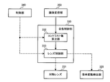
- FIG. 1 is a configuration example of a focusing control device according to a first embodiment and an endoscope system including the same.
- FIG. 2 is a configuration example of an imaging device.
- FIG. 3 is a structural example of the focusing control part of 1st Embodiment.
- FIG. 4 is a view for explaining a method of changing filter frequency characteristics according to magnification change.
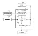
- FIG. 5 is a configuration example of a focusing control device according to a second embodiment and an endoscope system including the same.
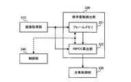
- FIG. 6 is a configuration example of a magnification variation detection unit according to the second embodiment.
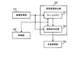
- FIG. 7 is another configuration example of the magnification variation detection unit of the second embodiment.
- FIG. 8 is a configuration example of a focusing control unit of the second embodiment.
- FIGS. 9 (A) and 9 (B) are diagrams for explaining a method of setting an evaluation area according to a change in magnification.
- FIG. 10 shows another configuration example of the focusing control unit of the second embodiment.
- 11 (A) and 11 (B) are diagrams for explaining a method of setting an initial evaluation area.
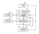
- FIG. 12 is a configuration example of a focusing control device according to a third embodiment and an endoscope system including the same.
- FIG. 13 is a configuration example of a magnification variation detection unit according to the third embodiment.
- 14 (A) to 14 (C) show an example of variation of an endoscopic image accompanying magnification change
- FIGS. 14 (D) and 14 (E) show examples of a scaled image acquired according to magnification change.
- FIGS. 15A to 15E show an example of an endoscopic image and a scaled image when the variation in magnification is within the range of the allowable scaling ratio.
- FIGS. 16A to 16E show examples of an endoscopic image and a scaled image when the magnification variation is smaller than the permissible scaling ratio.
- FIGS. 17A to 17E show an example of an endoscopic image and a scaled image when the magnification variation is larger than the allowable scaling ratio.
- FIG. 18 is a configuration example of a focusing control unit of the third embodiment.
- FIG. 19A shows an example of the first group drive lens
- FIG. 19B shows an example of the second group drive lens.
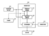
- FIG. 20 shows a configuration example of a focusing control device according to a fourth embodiment and an endoscope system including the same.
- FIG. 21 shows an example of the configuration of the image scaling unit according to the fourth embodiment.
- FIG. 22 is a structural example of the display magnification adjustment part of 4th Embodiment.
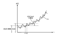
- FIG. 23 shows an example of detected magnification variation.
- FIG. 24 shows an example of the variation due to the high frequency magnification variation when the low frequency magnification variation is used as a reference.
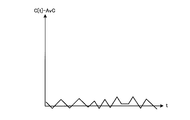
- FIG. 25 shows an example in which the influence of high frequency components is reduced from the detected magnification variation.
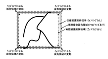
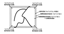
- FIG. 26 is a view for explaining the fluctuation of the angle of view due to the wobbling.
- FIG. 27 shows a configuration example of an image adjustment unit of the fourth embodiment.
- FIG. 28A shows an example in which the enlargement process is performed as the image scaling process, and FIG.
- FIG. 29 is an explanatory diagram of a target angle of view area according to a modification of the fourth embodiment.
- FIG. 30 is a view for explaining the process of a modification of the fourth embodiment;
- the method of the present embodiment will be described.
- the in-focus object position is changed by moving the zoom lens. That is, the movement of the in-focus object position is accompanied by a change in imaging magnification. Therefore, when performing AF (Auto Focus) using the first group driving lens, the calculation of an AF evaluation value (for example, contrast value) used for AF becomes a problem.
- AF Auto Focus
- the in-focus object position is the relative position (object point) of the object with respect to the reference position when the system including the object, the imaging optical system, the image plane and the like is in the in-focus state. Specifically, when an image plane is set at a given position and an imaging optical system is in a given state, an image formed on the image plane by the imaging optical system is in focus. Represents the position of the object in question. In the focusing control device (or endoscope system) or the like according to the present embodiment, it is assumed that the image plane coincides with the surface of the imaging device included in the imaging unit, and therefore the surface of the imaging device is fixed The in-focus object position can be determined by determining the state of the optical system.
- contrast AF it is assumed that the relationship between a plurality of AF evaluation values is determined in AF so that the in-focus object position is changed to calculate the contrast value at each position and the maximum value of the calculated contrast value is determined.
- the imaging magnification also changes when the in-focus object position is changed. Therefore, as compared with the image used to calculate the AF evaluation value at a certain timing, the magnification of the subject changes in the image at another timing, and the AF evaluation value can not be calculated stably.
- the imaging magnification is increased (enlarged) during the AF operation
- high frequency components included in the image are shifted to the low frequency side (edges are rounded), and AF before and after enlargement is performed.
- the calculation conditions of the evaluation value are different. Therefore, the magnitude of the AF evaluation value can not be determined properly, which causes trouble in the AF operation.
- the applicant proposes a method of calculating an AF evaluation value that compensates for magnification fluctuation (magnification fluctuation information). Specifically, three methods are proposed, and in the first embodiment, the frequency characteristic of the filter used when calculating the AF evaluation value is changed based on the change in magnification. In the second embodiment, the size of an evaluation area representing the range of pixels for which the AF evaluation value is to be calculated is changed based on the magnification variation. In the third embodiment, scaling processing is performed on the image itself based on magnification variation, and the AF evaluation value is calculated using the scaled image after scaling processing.
- magnification fluctuation is acquired based on control information from the imaging optical system (for example, a lens control signal related to the zoom lens position).
- magnification variation is acquired based on the subject size on the captured image.
- the magnification fluctuation acquired by the control information is information indicating the fluctuation of the imaging magnification
- the magnification fluctuation acquired by the subject size in the image is in addition to the fluctuation of the imaging magnification
- the imaging optical system Is information indicating a change in relative distance between the subject and the subject.
- the combination of the filter processing and the lens control signal in the first embodiment is described as a combination of the AF evaluation value calculation method and the magnification variation detection method, and in the second embodiment, the evaluation area and the subject size
- the combination of is described, it is not limited to this.
- the combination of the three AF evaluation value calculation methods and the two magnification variation detection methods is arbitrary.
- both the magnification variation based on the lens control signal and the magnification variation based on the subject size may be acquired, and it is particularly useful when using a scaling process as an AF evaluation value calculation method. Details will be described in the third embodiment and its modification.
- the magnification (angle of view) of the display image presented to the user (doctor) also changes frequently.
- the frequent change of the angle of view of the display image is likely to be stressful for the doctor, which may be a factor to prevent an appropriate diagnosis. Therefore, a display image that is easy for the user to observe may be generated and displayed by performing appropriate scaling processing on the captured image.
- the ratio of the imaging magnification or the subject size between two adjacent timings is the magnification fluctuation Z at many places
- the cumulative magnification A which is the direct product of Z, is between two given timings (adjacent).
- the magnification variation Z and the cumulative magnification A are different in calculation method, they represent variations in imaging magnification etc. between two different timings, and have essentially the same meaning. Therefore, in the present embodiment, the cumulative magnification A is also broadly included in the magnification fluctuation.
- magnification change Z the ratio between adjacent two timings is described as magnification change Z and the direct product of Z is described in different terms as A, but it is difficult to distinguish (or distinguish)
- magnification variation may refer to either Z or A.
- the endoscope apparatus includes a light source unit 100, an insertion unit 200, a signal processing unit 300, a display unit 400, and an external I / F unit 500.
- the light source unit 100 includes a white light source 110 and a condenser lens 120.
- the white light source 110 emits white light.
- the condenser lens 120 condenses the light emitted from the white light source 110 on a light guide fiber 210 described later.
- the insertion portion 200 is formed to be elongated and bendable, for example, to allow insertion into a body cavity.
- the insertion unit 200 includes a light guide fiber 210, an illumination lens 220, and an imaging unit 230.
- the light guide fiber 210 guides the light collected by the light source unit 100 to the tip of the insertion unit 200.
- the illumination lens 220 diffuses the light guided to the tip by the light guide fiber 210 and illuminates the observation target.
- the imaging unit 230 includes an objective lens 231, an imaging device 232, and an A / D conversion unit 233.
- the objective lens 231 condenses the reflected light returning from the observation target on the imaging element 232.
- the objective lens 231 has a function of simultaneously changing the magnification and the in-focus object position.
- the imaging element 232 outputs an analog signal based on the detected reflected light to the A / D converter 233.
- the A / D conversion unit 233 converts an analog signal output from the imaging device 232 into a digital signal based on a control signal output from the control unit 340 described later, and outputs the digital signal to the signal processing unit 300 as a RAW image.
- the imaging device 232 has a primary color Bayer array, and the image acquired by the imaging device 232 is a primary color Bayer image.
- the primary color Bayer image is an image in which each pixel has a signal of either RGB in a checkerboard shape.
- the signal processing unit 300 includes an image acquisition unit 310, a magnification variation detection unit 320, a focusing control unit 330, and a control unit 340.
- the RAW image output from the imaging unit 230 is output to the image acquisition unit 310.
- the image acquisition unit 310 is connected to the focusing control unit 330 and the display unit 400.
- the magnification variation detection unit 320 is connected to the focusing control unit 330.
- the focusing control unit 330 is connected to the objective lens 231, and controls the magnification and the in-focus object position based on the control of the objective lens 231 by the lens control signal.
- the lens control signal is also output to the magnification variation detection unit 320.
- the control unit 340 is bidirectionally connected to the imaging unit 230, the magnification variation detection unit 320, the focusing control unit 330, the image acquisition unit 310, the display unit 400, and the external I / F unit 500, These are mutually controlled by the control signal.
- the control signal includes an AF trigger signal that indicates the start and end of an autofocus (hereinafter referred to as AF) function that automatically focuses on the subject.
- AF autofocus
- the image acquisition unit 310 performs existing image processing such as white balance processing and demosaicing processing on the RAW image output from the imaging unit 230, and acquires an endoscopic image.
- the acquired endoscopic image is output to the focusing control unit 330 and the display unit 400.
- the endoscopic image is a RGB color image.
- the times t and t-1 are times when the control signal is output to the magnification variation detection unit 320
- the time t-1 is a time when the control signal is output to the magnification variation detection unit 320 immediately before time t. It is time.
- the focus control unit 330 detects the start of the AF operation according to the control signal
- the focusing control unit 330 detects the start of the AF operation based on the endoscope image output from the image acquisition unit 310 and the magnification variation output from the magnification variation detection unit 320.
- Control is performed to control the in-focus object position.
- the imaging magnification is also changed at the same time as changing the in-focus object position.
- only the in-focus object position control is described here for the sake of explanation.
- the focusing control unit 330 includes a filter selection unit 331, a contrast value calculation unit 332, and a lens control unit 333.
- the endoscopic image output from the image acquisition unit 310 is output to the contrast value calculation unit 332.
- the scaling factor variation output from the scaling factor variation detection unit 320 is output to the filter selection unit 331.
- the filter selection unit 331 is connected to the contrast value calculation unit 332.
- the contrast value calculation unit 332 is connected to the lens control unit 333.
- the lens control unit 333 is connected to the objective lens 231, and controls this by outputting a lens control signal.
- the lens control signal is also output to the magnification variation detection unit 320.
- the filter selection unit 331 selects a filter to be used for calculating a contrast value to be described later from a predetermined filter group based on the magnification change output from the magnification change detection unit 320.
- the filter is a known high pass filter. Further, in order to simplify the description, three filters are included in the filter group.
- the cumulative magnification is calculated by the following equation (1).
- t represents the current time
- At represents the cumulative magnification at the current time
- s represents the time when the AF operation started.
- i is an index indicating time
- Z is a value of magnification fluctuation (magnification fluctuation value). That is, the cumulative magnification is a direct product of the value of magnification fluctuation from the start of the AF operation to the present time.
- High pass filter selection based on the cumulative magnification will be described with reference to FIG.
- FIG. 4 shows the frequency characteristics of the high pass filter.
- the cumulative magnification is greater than 1.0, that is, if the magnification is higher than at the start of the AF operation, then a high-pass filter that passes a lower frequency signal than that at the start of the AF operation is selected. If the cumulative magnification is smaller than 1.0, that is, if the magnification is lower than at the start of the AF operation, then a high-pass filter which passes a higher frequency signal than the start of the AF operation is selected. Further, although the case where the filter group is configured by three filters has been described in the present embodiment, a larger number of filters may be used. In this case, the higher the degree of the cumulative magnification, the more the low frequency or high frequency signal is passed. Further, for a predetermined high pass filter, the frequency characteristic may be changed based on the cumulative magnification. In this case, a filter having a frequency characteristic of the following equation (2) is designed.
- Gt (u) is the frequency characteristic of the filter at time t.
- u is the spatial frequency.
- Go (u) is a frequency characteristic of a predetermined filter.
- the high pass filter was used as a filter in this embodiment, you may use a band pass filter. Also in this case, as in the case of the high-pass filter, select a filter that has a frequency characteristic that passes the low frequency signal when the magnification is higher than at the start of the AF operation and the high frequency signal when the magnification is lower than the start of the AF operation .
- the contrast value calculation unit 332 performs a filtering process by the filter selected by the filter selection unit 331 on the endoscopic image output from the image acquisition unit 310 to calculate a contrast value.
- the contrast value is the sum of pixel values of the filtering-processed endoscopic image in a rectangular area of a predetermined size located at the center of the endoscopic image.
- the contrast value is calculated by performing the filtering process only on the G signal of the endoscopic image. This is because, in the body cavity which is the main subject in the endoscopic image, the local change in pixel value is the largest in the G signal, and is suitable for contrast value calculation.
- the calculated contrast value is output to the lens control unit 333. Further, in the present embodiment, the contrast value is calculated only from the G signal, but filtering processing may be performed for all the RGB signals for each channel, and the sum may be calculated by summing up the respective RGB signals.
- the lens control unit 333 outputs a lens control signal for controlling the objective lens 231 based on the contrast value output from the contrast value calculation unit 332.
- a specific focus control method based on the contrast value is known as an AF technique, and thus the detailed technical description is omitted.
- the lens control signal is output to the objective lens 231 and the magnification variation detection unit 320.
- the display unit 400 outputs the endoscopic image output from the image acquisition unit 310 onto an image display device such as an endoscope monitor.
- the external I / F unit 500 is an interface for performing input from the user to the focusing control device, a power switch for turning the power on / off, a shutter button for starting a photographing operation, and a photographing mode And other mode switching buttons for switching various modes, and an AF button for starting an autofocus operation for automatically focusing on an object.
- an endoscopic image is acquired through an endoscope imaging optical system in which a magnification variation and an in-focus object position variation are interlocked.
- the magnification variation of the endoscope imaging optical system is detected from the control signal of the endoscope imaging optical system.
- the frequency characteristic of the high pass filter is selected based on the detected change in magnification.
- a contrast value is calculated by filtering the endoscopic image with the high pass filter.
- the AF function is realized by controlling the imaging optical system so that the calculated contrast value has a maximum value.
- the focusing control device is imaged at different imaging magnifications by the focusing control unit 330 that controls the drive of the imaging optical system and imaging via the imaging optical system. It includes an image acquisition unit 310 that acquires a plurality of images, and a magnification fluctuation detection unit 320 that detects magnification fluctuation.
- the focusing control unit 330 calculates an AF evaluation value representing the focus state of the imaging optical system based on the image acquired by the image acquiring unit 310 and the magnification fluctuation detected by the magnification fluctuation detection unit 320, and the AF evaluation value.
- the focusing control is performed by driving the imaging optical system based on
- the imaging optical system has a configuration in which the in-focus object position is changed along with the change of the imaging magnification.
- the single-group driving lens as shown in FIG.
- the magnification variation represents at least one of the variation of the imaging magnification and the variation of the size of the subject on the image.
- the degree of the change 2 in the ratio, 10 in the difference
- the degree of the change 2 in the ratio, 10 in the difference
- the magnification change is not limited to the ratio or the difference in a broad sense, but may be other information (magnification fluctuation information) representing the degree of fluctuation such as imaging magnification, but in the present embodiment, the ratio is represented in a narrow sense It explains as a thing.
- the AF evaluation value is a value to be evaluated when performing AF, and may be, for example, a contrast value in contrast AF.
- magnification fluctuation for example, fluctuation of imaging magnification
- processing to compensate for the influence due to magnification fluctuation is performed .
- the focusing control unit 330 performs a filtering process on the image, which applies a filtering process using a filter having a frequency characteristic according to the magnification variation to the image (performing process by the filter selected by the filter selecting unit 331).
- the contrast value calculation unit 332 in FIG. 3 may be included.
- the frequency characteristic of the filter used for the calculation process changes with the change in magnification. Therefore, when the frequency characteristic of the filter is constant, even if the in-focus state is constant, it is possible that the signal component that has passed through the filter before the magnification change does not pass through the filter after the magnification change. . If the in-focus state is constant, the signal component (signal value) passing through the filter should be of the same level, so it is necessary to change the filter characteristics to correspond to the fluctuation of the frequency component of the image due to magnification fluctuation. is there.
- the filter processing unit may perform filter processing using a high-pass filter with a lower cutoff frequency as the change in magnification is larger. In addition, as the change in magnification is smaller, a filter process using a high pass filter with a higher cutoff frequency may be performed.
- magnification fluctuation means that the imaging magnification after fluctuation is large (expanded) compared to before fluctuation, and that magnification fluctuation is small means imaging after fluctuation compared to before fluctuation It is a situation where the magnification is reduced (reduced).
- the variation in magnification may be large as it is enlarged and reduced as it is reduced, and it is a difference in imaging magnification (a positive value at the time of enlargement and a negative value at the time of reduction) It may also be other values.
- the filter processing unit may perform filter processing using a band pass filter, which is a frequency band in which the pass band is lower as the change in magnification is larger. Moreover, you may perform the filter process using the band pass filter which is a frequency band whose passband is a high frequency band, so that magnification change is small.
- magnification variation detection unit 320 may detect the ratio of the magnification evaluation value at the second timing to the magnification evaluation value at the first timing as the magnification variation at the second timing.
- the magnification evaluation value may be an imaging magnification.
- the second timing is assumed to be temporally different from the first timing.
- the magnification evaluation value is at least one of the imaging magnification described above and the size of the subject in the image.
- magnification fluctuation when the magnification evaluation value does not fluctuate is 1, and the magnification fluctuation is larger than 1 to correspond to enlargement, and smaller than 1 to correspond to contraction.
- the first timing described above may be timing to start calculation of the AF evaluation value.
- the second timing is later in time than the first timing, and corresponds to the timing at which the AF evaluation value is calculated.
- the second timing represents the current processing timing.
- the timing at which the calculation of the AF evaluation value is started may be the timing at which the AF operation is started.
- the calculation processing at the subsequent AF evaluation value calculation timing is performed after compensating for the magnification fluctuation from the first timing. It becomes possible to calculate the AF evaluation value under the same condition as the timing of 1.
- the calculated change in magnification corresponds to the cumulative magnification At of Formula (1) in the description of the present embodiment.
- magnification change is not limited to the one that indicates the cumulative magnification, and as described in the present embodiment, even if the first timing and the second timing are acquisition timings of temporally adjacent AF evaluation values Good.
- magnification evaluation value the ratio of the magnification evaluation values in a narrow sense
- processing for compensating for magnification fluctuation between adjacent timings can be performed, but by obtaining the product of magnification fluctuation between adjacent timings, magnification fluctuation between arbitrary timings can be obtained.
- processing to compensate for magnification variation For example, the direct product of the change in magnification from the start of the AF operation is nothing but the cumulative magnification described above.
- the focusing control unit 330 that controls the drive of the imaging optical system and the image acquisition unit 310 that acquires a plurality of images captured at different imaging magnifications by imaging via the imaging optical system.
- the present invention can be applied to an endoscope system including the magnification fluctuation detection unit 320 that detects the magnification fluctuation.
- the focusing control unit 330 calculates an AF evaluation value representing the focus state of the imaging optical system based on the image acquired by the image acquiring unit 310 and the magnification fluctuation detected by the magnification fluctuation detection unit 320, and the AF evaluation value.
- the focusing control is performed by driving the imaging optical system based on
- the endoscope system may include the light source unit 100, the insertion unit 200, the display unit 400, the external I / F unit 500 and the like as shown in FIG.
- the insertion unit 200 is to be inserted into a living body, so it is desirable to miniaturize it. Therefore, it is conceivable to simplify the configuration of the imaging optical system included in the insertion unit 200. Therefore, it is assumed to adopt the one-group drive lens shown in FIG. 19A, and in that case, it is necessary to compensate for the influence of the above-described magnification fluctuation.
- the endoscope apparatus includes a light source unit 100, an insertion unit 200, a signal processing unit 300, a display unit 400, and an external I / F unit 500.
- the components other than the signal processing unit 300 and the display unit 400 are the same as those in the first embodiment, and thus the description thereof is omitted.
- the signal processing unit 300 includes an image acquisition unit 310, a magnification variation detection unit 320, a focusing control unit 330, and a control unit 340.
- the image acquisition unit 310 is connected to the magnification variation detection unit 320, the focusing control unit 330, and the display unit 400.
- the magnification variation detection unit 320 is connected to the focusing control unit 330.
- the focusing control unit 330 is connected to the objective lens 231, and controls the magnification and the in-focus object position based on the control of the objective lens 231 by the lens control signal.
- the control unit 340 is bi-directionally connected to the imaging unit 230, magnification variation detection unit 320, focusing control unit 330, image acquisition unit 310, display unit 400, and external I / F unit 500. Are mutually controlled by the control signal.
- the image acquisition unit 310 is the same as that of the first embodiment, and thus the description thereof is omitted.
- the endoscopic image acquired by the image acquisition unit 310 is output to the magnification variation detection unit 320.
- the magnification fluctuation detection unit 320 Based on the pixel values of the endoscopic image output from the image acquisition unit 310, the magnification fluctuation detection unit 320 detects a change in the size of the subject in the endoscopic image as a magnification fluctuation. The detected magnification variation is output to the focusing control unit 330.
- the configuration of the magnification variation detection unit 320 in the present embodiment will be described with reference to FIG.
- the magnification variation detection unit 320 includes a frame memory 321 and a RIPOC calculation unit 322.
- the endoscope image output from the image acquisition unit 310 is output to the frame memory 321 and the RIPOC calculation unit 322.
- the frame memory 321 is connected to the RIPOC calculation unit 322.
- the RIPOC calculation unit 322 is connected to the focusing control unit 330.
- the frame memory 321 delays the endoscopic image output from the image acquisition unit 310 by one frame and outputs the endoscopic image to the RIPOC calculation unit 322.
- the RIPOC calculation unit 322 calculates a change in size of the subject in each endoscopic image as a magnification change, based on the endoscopic image output from the image acquisition unit 310 and the endoscopic image output from the frame memory 321. .
- the former endoscopic image is referred to as a current image
- the latter endoscopic image is referred to as a past image.
- the technique for calculating the magnification variation from the current image and the past image, and the rotation invariant phase only correlation method (hereinafter referred to as RIPOC method) are known techniques, so detailed technical explanation will be omitted.
- the details of the technology are detailed in Non-Patent Document 1.
- the RIPOC method translation, size change, and rotation of an object in a current image can be detected with reference to a past image. This change in size is output to the focusing control unit 330 as a magnification change.
- the magnification variation detection unit 320 includes a frame memory 321 and a feature point correspondence unit 323.
- the endoscope image output from the image acquisition unit 310 is output to the frame memory 321 and the feature point correspondence unit 323.
- the frame memory 321 is connected to the feature point handling unit 323.
- the feature point handling unit 323 is connected to the focusing control unit 330.
- the frame memory 321 delays the endoscope image output from the image acquisition unit 310 by one frame and outputs the delayed image to the feature point correspondence unit 323.
- the feature point correspondence unit 323 detects feature points from the current image output from the image acquisition unit 310 and the past image output from the frame memory 321, and detects the magnification variation of the current image and the past image from the correspondence relationship. .
- the feature points are detected based on SIFT feature quantities of known techniques.
- the SIFT feature is a feature robust to image rotation, scale change, and illumination change.
- the details of the SIFT feature are detailed in Non-Patent Document 2.
- feature points detected from the current image and the past image are made to correspond between the respective images.
- the correspondence between feature points is also a well-known technique, and therefore the description thereof is omitted.
- RANSAC repeats the operation of adapting correspondence candidates calculated by randomly extracting and combining a plurality of feature points to feature points that have not been extracted and evaluating its validity, so that a large number of feature points are interspersed. Is a method of determining the correspondence that satisfies.
- RANSAC are detailed in Non-Patent Document 3.
- the correspondence relationship is an affine transformation known as one of coordinate transformations.
- coordinate conversion is conversion from a past image to a current image.
- the affine transformation is constituted by terms of translation, rotation and magnification, and the term of magnification is outputted to the focusing control unit 330 as magnification variation.
- the focusing control unit 330 detects the start of the AF operation from the control signal, the focusing control unit 330 detects the start of the AF operation based on the endoscopic image output from the image acquisition unit 310 and the magnification fluctuation detected by the magnification fluctuation detection unit 320. Control is performed to control the in-focus object position.
- the focusing control unit 330 includes a contrast value calculating unit 332, a lens control unit 333 and an evaluation area setting unit 334.
- the endoscopic image output from the image acquisition unit 310 is output to the contrast value calculation unit 332.
- the scaling factor variation output from the scaling factor variation detection unit 320 is output to the evaluation area setting unit 334.
- the evaluation area setting unit 334 is connected to the contrast value calculation unit 332.
- the contrast value calculation unit 332 is connected to the lens control unit 333.
- the lens control unit 333 is connected to the objective lens 231 and controls this.
- the evaluation area setting unit 334 sets an evaluation area on the endoscopic image based on the magnification change output from the magnification change detection unit 320. Specifically, first, when the start of the AF operation is detected from the control signal, an initial evaluation area is set.
- the initial evaluation area is a rectangular area located at the center of the endoscopic image, and the size thereof is calculated by multiplying the size of the endoscopic image by a predetermined ratio. After that, the initial evaluation area is scaled by the evaluation area magnification to be an evaluation area.
- the evaluation area magnification is equal to the cumulative magnification calculated by the equation (1) described above.
- FIG. 9A shows an endoscopic image and an initial evaluation area at the start of the AF operation.
- FIG. 9B shows an endoscopic image and an evaluation area at time t.
- the ratio between the size of the initial evaluation area in FIG. 9A and the size of the evaluation area in FIG. 9B is the evaluation area magnification Vt (equal to At in Expression (1)). That is, the larger the evaluation area magnification, the larger the size of the evaluation area.
- the calculated evaluation area magnification is output to the contrast value calculation unit 332.
- the contrast value calculation unit 332 uses the pixel value of the evaluation area set on the endoscopic image output from the image acquisition unit 310 based on the evaluation area magnification output from the evaluation area setting unit 334. calculate. Specifically, the pixel values of the endoscopic image subjected to the filtering process using the high-pass filter are summed up in the evaluation area, and this is taken as the contrast value. The calculated contrast value is output to the lens control unit 333. Further, the difference between the maximum pixel value and the minimum pixel value in the evaluation area may be used as the contrast value.
- the lens control unit 333 is the same as that of the first embodiment, and thus the description thereof is omitted.
- the influence of the magnification variation on the contrast value can be reduced, and the AF operation can be stably performed.
- the size of the initial evaluation area is calculated by multiplying the endoscopic image by a predetermined ratio, but the predetermined ratio may be variable according to the magnification of the objective lens 231 at the start of the AF operation. .
- the predetermined ratio is taken as an initial evaluation area magnification.
- the configuration of the focusing control unit 330 in this case is shown in FIG. A different point from the focusing control unit 330 described with reference to FIG. 9 is that the lens control signal is also output to the evaluation area setting unit 334.
- the evaluation area setting unit 334 acquires the magnification of the objective lens 231 at the start of the AF operation based on the lens control signal.
- FIG. 11 (B) has a high magnification compared to FIG. 11 (A)
- the initial evaluation magnification is set large.
- the reasons for avoiding these situations are explained.
- the evaluation area magnification exceeds 1.0, there is a possibility that the contrast value needs to be calculated over the entire endoscopic image. However, since the image signal is present only on the endoscopic image, the effect of reducing the influence of the magnification variation from the contrast value is limited. Further, when the evaluation area magnification approaches 0.0, the contrast value is calculated from the evaluation area which is extremely small, and the AF operation becomes unstable.
- the display unit 400 outputs the endoscopic image output from the image acquisition unit 310 onto an image display device such as an endoscope monitor.
- an endoscopic image is acquired through an endoscope imaging optical system in which a magnification variation and an in-focus object position variation are interlocked.
- the magnification change of the endoscope imaging optical system is detected from the endoscope image.
- An evaluation area having a variable size is set in the endoscopic image so as to compensate for the detected magnification variation, and the contrast value is calculated by performing filtering processing using a high pass filter on the pixels in the evaluation area.
- the AF function is realized by controlling the imaging optical system so that the calculated contrast value has a maximum value.
- the focusing control unit 330 includes the evaluation region setting unit 334 that sets an evaluation region, which is a region including pixels used for calculation of the AF evaluation value, to an image. Then, the evaluation area setting unit 334 changes the size of the evaluation area to be set based on the change in magnification.
- the evaluation area is an area including the pixels used for calculation of the AF evaluation value as described above, and the subject corresponding to the pixels used for the calculation is focused. Therefore, it is desirable that the evaluation area be set to a position on the image where you want to focus, but it is difficult to automatically recognize the area that you want to focus from within the image. In the present embodiment, it is assumed that the evaluation area is set at the center of the image. Moreover, although FIG. 9 (A) etc. are demonstrated using a rectangular area
- the evaluation area setting unit 334 may set the size of the evaluation area larger as the change in magnification is larger. Also, the size of the evaluation area may be set smaller as the change in magnification is smaller.
- the size of the evaluation area may be changed so that the range of the subject included in the evaluation area does not change. That is, when the magnification change is large (expanded), the size of the evaluation area is increased, and when the magnification change is small (reduced), the size of the evaluation area is reduced.
- magnification fluctuation detection unit 320 detects a magnification fluctuation between the first timing and the second timing
- the evaluation area setting unit 334 sets the reference evaluation area, which is an evaluation area at the first timing.
- the evaluation area at the second timing may be set by performing the scaling process using the value of the magnification change.
- the first timing may be the start timing of the AF operation, in which case the reference evaluation area is nothing other than the above-described initial evaluation area.
- the size of the evaluation area may be set so that the range of the subject included in the evaluation area does not change.
- the change in magnification represents the ratio of the size of the subject on the image (Vt in FIG. 9)
- the evaluation area is rectangular as shown in FIGS. 9A and 9B, the size of the evaluation area can be obtained by multiplying the vertical length and the horizontal length by Vt.
- Vt c2 / c1.
- the evaluation area setting unit 334 may set the size of the reference evaluation area larger as the imaging magnification or the size of the subject in the image is larger at the first timing. Further, the size of the reference evaluation area may be set smaller as the imaging magnification or the size of the subject in the image is smaller.
- the reference evaluation area which may be the initial evaluation area in a narrow sense.
- the change in imaging magnification is considered as the change in magnification.
- an imaging optical system capable of changing an imaging magnification from 1 to 100 times is used.
- the imaging magnification at the first timing is a high magnification close to 100 times, the imaging magnification does not increase so much at the subsequent timings (second timing etc.), but may rather decrease. Is considered high. Therefore, a small value is assumed for the magnification variation, and the evaluation area is also set smaller than the reference evaluation area.
- the reference evaluation area may be enlarged in order to prevent the evaluation area from becoming smaller as the calculation of the AF evaluation value becomes difficult.
- the magnification is low, which is close to 1 ⁇ , it can be assumed that the magnification fluctuation is large and an evaluation area is set large. In such a case, the reference evaluation area may be reduced so that an evaluation area exceeding the size of the image (captured image, endoscopic image) is not set.
- the magnification variation is determined based on the size of the subject on the image, the variation width (1 to 100 times in the above example) is not clear like the imaging magnification, so the first timing It is necessary to set the criteria for determining whether the subject size is large or small in any method.
- a subject having a definite absolute size for example, the thickness of a blood vessel at a given site, etc.
- the size on the image of the subject may be used as a reference.
- the magnification variation detection unit 320 may detect the ratio of the size of the subject at the second timing to the size of the subject at the first timing as the magnification variation at the second timing.
- phase limited correlation may be used for the image at the first timing and the image at the second timing.
- a plurality of feature points may be set for the image at the first timing and the image at the second timing, and magnification variation may be detected based on the positions of the set feature points.
- the second timing is assumed to be temporally different from the first timing.
- the RIPOC method may be used, or the SIFT feature may be set and the correspondence may be determined by RANSAC.
- the subject size at each timing does not have to be an absolute value. For example, consider a third timing different from the first and second timings. At this time, if the subject sizes d1, d2 and d3 at each timing are determined, the magnification variation between the first and second timings is d2 / d1, for the second and third d3 / d2, for the first and third d3 / It is calculated as d1 grade. However, in order to do this, it is always necessary to determine the size of the same part of the subject. For example, if the distance between the first feature point and the second feature point on the subject at the first timing is information representing the size, the first feature point is also at the second and third timings.
- the change in magnification between the first and third timings can not be obtained from d1 and d3 ', it can be obtained as an accumulated magnification by Z12 ⁇ Z23.
- the endoscope apparatus includes a light source unit 100, an insertion unit 200, a signal processing unit 300, a display unit 400, and an external I / F unit 500.
- the components other than the signal processing unit 300 are the same as those of the first embodiment, and therefore the description thereof is omitted.
- the signal processing unit 300 includes an image acquisition unit 310, a magnification variation detection unit 320, a focusing control unit 330, a control unit 340, and an image scaling unit 350.
- the RAW image output from the imaging unit 230 is output to the image acquisition unit 310.
- the image acquisition unit 310 is connected to the magnification variation detection unit 320 and the image scaling unit 350.
- the magnification variation detection unit 320 is connected to the image scaling unit 350.
- the image scaling unit 350 is connected to the focusing control unit 330 and the display unit 400.
- the focusing control unit 330 is connected to the objective lens 231, and controls the magnification and the in-focus object position based on the control of the objective lens 231 by the lens control signal.
- the lens control signal is also output to the magnification variation detection unit 320.
- the control unit 340 includes both the imaging unit 230, the image acquisition unit 310, the magnification variation detection unit 320, the focusing control unit 330, the image scaling unit 350, the display unit 400, and the external I / F unit 500. They are connected in turn and mutually controlled by control signals.
- the image acquisition unit 310 is the same as that of the first embodiment, and thus the description thereof is omitted.
- the acquired endoscopic image is output to the magnification variation detection unit 320 and the image magnification unit 350.
- the magnification fluctuation detection unit 320 detects a lens magnification fluctuation based on a lens control signal output from the focusing control unit 330 described later, and based on the lens magnification fluctuation and the endoscopic image output from the image acquisition unit 310, Detect subject distance variation.
- the lens magnification fluctuation is the magnification fluctuation of the objective lens 231.
- the subject distance change is a relative distance change between the objective lens 231 and the subject.
- a specific configuration of the magnification variation detection unit 320 in the present embodiment will be described with reference to FIG.
- the magnification variation detection unit 320 in the present embodiment includes a lens magnification variation detection unit 324 and a distance variation detection unit 325.
- a lens control signal output from the focusing control unit 330 described later is output to the lens magnification variation detection unit 324.
- the endoscopic image output from the image acquisition unit 310 is output to the distance variation detection unit 325.
- the lens magnification variation detection unit 324 is connected to the distance variation detection unit 325 and the image scaling unit 350.
- the distance variation detection unit 325 is connected to the image scaling unit 350.
- the lens magnification fluctuation detecting unit 324 detects the magnification fluctuation and the lens magnification fluctuation associated with the movement of the objective lens 231 based on a lens control signal output from the focusing control unit 330 described later.
- the operation is the same as that of the magnification variation detection unit 320 in the first embodiment, and thus the description thereof is omitted.
- the lens magnification variation is the same as the magnification variation in the first embodiment, but the notation is changed for the sake of explanation.
- the detected lens magnification variation is output to the distance variation detection unit 325 and the image scaling unit 350.
- the distance fluctuation detection unit 325 changes the relative distance between the subject and the endoscope based on the endoscopic image acquired by the image acquisition unit 310 and the lens magnification fluctuation output from the lens magnification fluctuation detection unit 324. Detect variations. Specifically, first, a change in image magnification which is a change in magnification of an endoscopic image over time is detected. This is the same as the operation of the magnification variation detection unit 320 in the second embodiment, and thus the description thereof is omitted.
- the change in magnification of the endoscopic image over time includes a factor due to a change in magnification of the optical system and a factor due to a change in relative distance between the endoscope and the subject, and can be represented by the product of these. That is, the image magnification variation is the product of the lens magnification variation and the distance variation, and the distance variation can be detected by dividing the image magnification variation by the lens magnification variation.
- the detected distance fluctuation is output to the image scaling unit 350.
- the image scaling unit 350 When detecting the start of the AF operation from the control signal, the image scaling unit 350 enlarges or reduces the endoscopic image output from the image acquisition unit 310 based on the magnification variation output from the magnification variation detection unit 320.
- the scaling process is collectively referred to as scaling process.
- the scaling factor image scaling factor is calculated from the following equation (3) based on the magnification variation.
- t represents the current time
- Mt represents the scaling factor at the current time
- s represents the time when the AF operation started.
- i is an index indicating time
- Z is a change in magnification
- A is a cumulative magnification as in the equation (1). That is, the scaling factor is the reciprocal of the direct product (cumulative factor) of the magnification change from the start of the AF operation to the present time.
- FIG. 14A is an endoscope image at the start of AF.
- the scaling factor is larger than 1.0 as in FIG. 14B (that is, the cumulative magnification is smaller than 1.0)
- the endoscopic image is enlarged as in FIG. Do.
- FIG. 14C when the scaling factor is smaller than 1.0 (when the cumulative magnification is larger than 1.0), the endoscopic image is reduced as shown in FIG. I assume.
- the endoscopic image subjected to the scaling process is output to the focusing control unit 330 and the display unit 400 as a scaled image.
- an image in which the size of the subject does not change during the AF operation is output to the focusing control unit 330 and the display unit 400.
- the focusing control unit can calculate the contrast value to be described later while reducing the influence of the magnification change.
- the display unit 400 can suppress a phenomenon in which the display magnification of the subject frequently varies with the magnification variation, and the user causes video sickness.
- the scaling process is performed until the end of the AF operation. When the AF operation is not in progress, the input endoscope image is output to the display unit 400 as it is.
- the image scaling unit 350 may perform different scaling processes on the scaled image output to the focusing control unit 330 and the scaled image output to the display unit 400. Specifically, the scaled image to be output to the display unit 400 may be subjected to the scaling process using the reference scaling factor calculated based on the scaling factor obtained by the above equation (3). The reference magnification is calculated by the following equation (4). On the other hand, the scaled image output to the focusing control unit 330 is subjected to the scaling process in the same manner as described above.
- Bt is a reference magnification at the current time. That is, the reference magnification is an average of scaling factors from time s to the current time.
- the scaled image subjected to the scaling process at the reference magnification is output to the display unit 400.
- the reference magnification is not used, an image in which the size of the subject does not change is displayed during the AF operation, but the resolution of the image changes due to the scaling process, which may cause the user to feel uncomfortable. This is because the scaling process needs to perform interpolation processing, and the frequency characteristic of the image changes due to the interpolation processing.
- the reference magnification it is possible to reduce the change in resolution while suppressing the display magnification fluctuation of the subject.
- the scaled image to be output to the display unit 400 may be subjected to the scaling process based on the predetermined allowable scaling factor.
- the scaling factor in this case is calculated by the following equation (5).
- R is an allowable scaling factor and takes a value of 0.0 to 1.0. That is, the variation of the display magnification of the subject is suppressed within the range represented by the allowable magnification, and the magnification is calculated.
- the range represented by the allowable scaling factor is referred to as the allowable scaling range.
- the allowable magnification range is defined for both the magnification fluctuation value and the subject display magnification fluctuation (for example, the ratio of the subject size between FIG. 15B and FIG. 15E), but the range is defined. Since the values are equal, the description will be made without particular distinction.
- the scaling process using the allowable scaling range will be described with reference to FIGS. 16 (A) to 16 (E). FIGS.
- FIG. 16A to 16E show the case where the magnification variation value at time t exceeds the allowable scaling range.
- FIG. 16A shows an endoscopic image acquired at time t-1.
- FIG. 16B shows a scaled image which has been scaled at time t-1.
- FIG. 16C is an endoscopic image acquired at time t.
- FIG. 16 (D) shows a magnification-changed image that has been subjected to magnification-reduction processing so that the display magnification of the subject is equal to the endoscopic image shown in FIG. 16 (B).
- FIG. 16E is a scaled image in which the display magnification variation of the subject is allowed up to the upper limit of the allowable scaling range.
- FIG. 17A to 17E show the case where the magnification variation value at time t falls below the allowable scaling range.
- FIG. 17A shows an endoscopic image acquired at time t-1.
- FIG. 17B shows a scaled image that has been scaled at time t-1.
- FIG. 17C shows an endoscopic image acquired at time t.
- FIG. 17D shows a magnified image obtained by performing the magnification process so that the display magnification of the subject is equal to the endoscopic image shown in FIG.
- FIG. 17E is a scaled image in which the display magnification variation of the subject is allowed up to the lower limit of the allowable scaling range.
- the scaling process may be performed by calculating a scaling factor based on different scaling factors of the scaled image output to the focusing control unit 330 and the scaled image output to the display unit 400.
- the variable-magnification image output to the focusing control unit 330 is a product of the lens magnification fluctuation output from the lens magnification fluctuation detection unit 324 and the distance fluctuation output from the distance fluctuation detection unit 325. You may process it.
- the variable-magnification image output to the display unit 400 is subjected to variable magnification processing so as to compensate for only the lens magnification variation. By doing this, it is possible to stably perform the AF operation with respect to the frequency characteristic change of the endoscope image due to the relative distance fluctuation with the subject.
- by compensating only for the lens magnification variation and not for the distance variation it is possible to reduce the difference between the relative distance variation with the subject due to the endoscope operation and the displayed image.
- the scaling process on the scaled image output to the display unit 400 may be continued after the AF operation is completed. Specifically, after the end of the AF operation, the scaling process is continued until Mt shown in the above equation (5) satisfies the following equation (6).
- the scaling factor Mt after the end of the AF operation is calculated only by the above equation (6) regardless of the magnification variation value.
- the scaling process may be continued until a predetermined time elapses.
- magnification change processing for the scaled image output to the display unit 400 is ended suddenly after the AF operation ends, the display magnification of the subject largely changes before and after the AF operation end, and the user may feel discomfort. By doing this, it is possible to moderate the change in display magnification before and after the end of the AF operation, and it is possible to reduce the user's discomfort.
- the focusing control unit 330 When the focusing control unit 330 detects the start of the AF operation from the control signal, the focusing control unit 330 controls the objective lens 231 based on the pixel value of the scaled image output from the image scaling unit 350 described later, thereby setting the focused object position. Control.
- the specific configuration of the focusing control unit 330 will be described with reference to FIG.
- the focusing control unit 330 includes a contrast value calculating unit 332 and a lens control unit 333.
- the scaled image output from the image scaling unit 350 is output to the contrast value calculation unit 332.
- the contrast value calculation unit 332 is connected to the lens control unit 333.
- the lens control unit 333 is connected to the objective lens 231, and controls this by outputting a lens control signal.
- the lens control signal is also output to the magnification variation detection unit 320.
- the contrast value calculation unit 332 performs filtering processing on the scaled image output from the image scaling unit 350 to calculate a contrast value.
- the contrast value is the sum of the pixel values of the filtered scaled image in a rectangular area of a predetermined size located at the center of the scaled image.
- the filter used in the filtering process is a known high pass filter.
- the calculated contrast value is output to the lens control unit 333.
- the lens control unit 333 outputs a lens control signal for controlling the objective lens 231 based on the contrast value output from the contrast value calculation unit 332.
- a specific focus control method based on the contrast value is known as an AF technique, and thus the detailed technical description is omitted.
- the lens control signal is output to the objective lens 231 and the magnification variation detection unit 320.
- the display unit 400 outputs the scaled image output from the image scaling unit 350 on an image display apparatus such as an endoscope monitor.
- an image display apparatus such as an endoscope monitor.
- the size of the scaled image is larger than the displayable image size, only the displayable image size is extracted from the center of the scaled image and displayed.
- the size of the scaled image is smaller than the displayable image size, the scaled image is displayed at the center, and the surrounding display area displays the black level.
- the image to be output an endoscopic image except during the AF operation, and a scaled image during the AF operation
- the endoscope itself is some distance measuring means, for example, triangulation with a two-lens configuration, If it is provided, it may be calculated directly by the distance measuring means.
- an endoscopic image is acquired through an endoscope imaging optical system in which a magnification variation and an in-focus object position variation are interlocked.
- the magnification variation of the endoscope imaging optical system and the relative distance variation between the endoscope and the subject are detected.
- the endoscopic image is subjected to scaling processing so as to compensate for the magnification variation.
- a contrast value is calculated by performing a filtering process using a high pass filter on the endoscopic image subjected to the scaling process.
- the AF function is realized by controlling the imaging optical system so that the calculated contrast value has a maximum value.
- contrast value of AF is calculated by compensating magnification fluctuation of endoscope optical system and relative distance fluctuation with the subject, and the display image should be scaled by compensating only magnification fluctuation of endoscope optical system.
- the focusing control device includes the image scaling unit 350 which performs the image scaling process on the image based on the magnification variation to obtain a scaled image. . Then, the focusing control unit 330 calculates an AF evaluation value representing the focus state of the imaging optical system based on the scaled image.
- the present embodiment differs from the second embodiment in that the size of the image itself is changed with the evaluation area fixed.
- the image scaling unit 350 may acquire the scaled image which is smaller as the change in magnification is larger. Also, as the magnification variation is smaller, a larger scaled image may be acquired.
- magnification variation detection unit 320 detects a variation in magnification between the first timing and the second timing, and the image scaling unit 350 generates an image acquired by the image acquisition unit at the second timing.
- a scaled image may be obtained by performing an image scaling process using the reciprocal of the value of the magnification variation.
- the image scaling process is performed on the image acquired at the timing (the endoscopic image, as shown in FIG. 14B), so the scaled image at the second timing is doubled. Enlargement processing is performed (a scaled image corresponds to FIG. 14D).
- the image scaling unit 350 may output the acquired scaled image to the focusing control unit 330 and to the display device (corresponding to the display unit 400 in FIG. 12).
- the image acquisition unit 310 acquires a display image displayed on the display device (display unit 400).
- the used image is used as it is. Therefore, it is inevitable that a flicker occurs in the display image due to the magnification variation caused by the variation of the in-focus object position (for example, wobbling in contrast AF) which occurs in the focusing operation.
- FIGS. 14A to 14C are acquired in time series, the subject becomes smaller or larger. Therefore, the scaled image acquired by the image scaling unit 350 is also output to the display device.
- FIG. 14A to FIG. 14C are acquired in time series, the display image is displayed in order of FIG. 14A, FIG. 14D, and FIG. It will be.
- the image scaling unit 350 outputs the first scaled image obtained by performing the first image scaling process on the image to the focusing control unit 330, and also performs the second image scaling on the image.
- the processed second scaled image may be output to the display device.
- the first image scaling process and the second image scaling process are different processes.
- the process may be one in which the image scaling factor used for the image scaling process is different.
- the scaled image to be output to the focusing control unit 330 and the scaled image to be output to the display device can be made different. Therefore, even when the scaled image suitable for the AF evaluation value calculation processing in the focusing control unit 330 differs from the scaled image suitable for display on the display device, the scaled image suitable for each can be output. It will be possible.
- the magnification variation detection unit 320 may detect a first magnification variation that is a variation of the size of the subject in the image as a magnification variation and a second magnification variation that is a variation of the imaging magnification. Then, the image scaling unit 350 performs the image scaling process based on the first magnification change as the first image scaling process, acquires the first scaled image, and outputs the first scaled image to the focusing control unit 330. . In addition, as the second image scaling process, the image scaling process based on the second magnification variation is performed to acquire the second scaled image and output to the display device.
- the scaled image to be output to the focusing control unit 330 it is possible to perform the image scaling process using the variation of the subject size as the variation of magnification.
- image scaling processing can be performed using variation in imaging magnification as variation in magnification.
- the fluctuation of the imaging magnification (the second magnification fluctuation) is caused by the change of the imaging optical system, while the fluctuation of the subject size in the image (the first magnification fluctuation) depends on the imaging magnification
- a plurality of factors such as a change in relative distance between the imaging unit (insertion unit 200) and the subject are included.
- the image scaling process in the present embodiment is a process for compensating for the influence of magnification variation
- a certain magnification ratio can be obtained by switching the magnification variation to be used between the first image scaling process and the second image scaling process. It is possible to perform processing such as compensating for the fluctuation factor on the one hand and not compensating on the other hand.
- the AF evaluation value calculation processing in the focusing control unit 330 is performed by image processing (for example, extraction of high frequency components, etc.) on the image. Therefore, in the first image scaling process corresponding to the output to the focusing control unit 330, it is desirable that all the factors causing the variation in the image be compensated whether it is the imaging magnification or the relative distance. From this, the change in magnification (first change in magnification) used for the first image scaling process uses the change in the size of the subject in the image. On the other hand, a factor of variation in magnification which is undesirable to compensate for the displayed image on the display device may be considered.
- the image scaling processing in the direction to suppress the fluctuation of the image due to the fluctuation of the relative distance is performed. Then, even if the user operates the imaging unit and moves it back and forth, the fluctuation of the displayed image is suppressed. In this case, it is difficult to determine whether the user's operation is reflected on the device, and in some cases, the imaging unit may be brought too close to the subject to cause a collision. Therefore, the change in magnification (second change in magnification) used for the second image scaling process uses a change in imaging magnification.
- the magnification variation detection unit 320 may detect the i-th (1 ⁇ i ⁇ N) timing. Then, as the second image scaling process at the k-th (1 ⁇ k ⁇ N) timing, the image scaling unit 350 performs a process based on the first to k-th magnification fluctuations to obtain a second image scaling process.
- the scaled image may be acquired and output to the display device.
- the imaging magnification at a given reference timing is g0 and the imaging magnification at the kth timing is gk
- Ak corresponds to the cumulative magnification in the above description, and the cumulative magnification may be obtained by a direct product of magnification fluctuation (ratio of imaging magnification) between adjacent timings.
- the image scaling processing at the kth timing scaling processing of 1 / Ak times is performed on the image (endoscope image) acquired at the timing, and the reference timing and the kth timing are obtained. It is to suppress the influence of the fluctuation of the imaging magnification among the two.
- the compensation by the image scaling process involves the scaling of the image, which causes a change in resolution. For example, in the enlargement processing, interpolation processing of pixel values is performed, so that the edge is rounded. If the sense of resolution fluctuates in this manner, the image may be difficult for the user to see, which may affect observation and the like.
- magnification fluctuations A1 to Ak-1 at past timings are also used. It shall be used.
- the image scaling factor Bt at a certain timing t the average value of the image scaling factor from a certain timing s to the timing t (the mean value of the reciprocal of the magnification fluctuation A) It may be determined as In this way, although the influence of magnification fluctuation (the influence of fluctuation of imaging magnification) remains to some extent, fluctuation of the sense of resolution can be suppressed, and a natural image can be provided to the user.
- the magnification variation detection unit 320 determines whether or not the magnification variation at the mth (1 ⁇ m ⁇ N) timing is included in the given allowable scaling range, and is included in the allowable scaling range. In this case, as the second image scaling process at the m-th timing, the image scaling process similar to the second image scaling process at the (m-1) th timing before the m-th timing is performed. In addition, when not included in the allowable scaling range, as the second image scaling process at the m-th timing, image scaling using a value close to 1 compared to the reciprocal of the value of the m-th magnification variation Do the processing.
- Mt is an image scaling factor at timing t
- Zt is a change in magnification at timing t, and is, for example, a ratio of the imaging magnification at timing t to the imaging magnification at timing t-1.
- the image scaling factor Mt at timing t uses the image scaling factor Mt-1 at timing t-1. Since the magnification change is Zt, the subject size in the image of FIG. 15C is Zt times that of FIG. 15A. Further, since the image scaling factor at timing t-1 is Mt-1, the image of FIG. 15B which is a display image (scaled image) at timing t-1 is different from that of FIG. The subject size is Mt-1 times.
- the image scaling process for stably calculating the AF evaluation value is a process for keeping the subject size in the image equal
- FIG. 15E is an output image to the display device. That is, the display image changes from FIG. 15 (B) to FIG. 15 (E), and the subject size differs by Zt times.
- the ratio of the subject size in FIG. 16E to that in FIG. 16B can be 1.0-R. That is, when the change in magnification is smaller than the lower limit value of the allowable scaling factor, the image scaling process is performed such that the change in the subject size of the display image falls within the lower limit value.
- the magnification variation detection unit 320 may include a distance variation detection unit 325 that detects variation of distance information from the imaging optical system to the subject. Then, the distance variation detection unit 325 detects the variation of the distance information based on the variation of the imaging magnification and the variation of the size of the subject.
- magnification fluctuation detection unit 320 when the zoom lens is driven (wobbling) for calculating the AF evaluation value and the zoom lens is driven with the intention of changing the imaging magnification (for example, with the intention of observing the object in an enlarged manner) A combination of high-frequency magnification fluctuation due to wobbling and low-frequency magnification fluctuation accompanying intentional magnification change (for example, the fluctuation shown in FIG. 23) becomes a magnification fluctuation detected in the magnification fluctuation detection unit 320.
- magnification change of the high frequency due to the wobbling interferes with the observation and diagnosis as described above, it is desirable to perform the image scaling process for suppressing the magnification change.
- the low frequency magnification fluctuation is caused by driving the zoom lens reflecting the user's intention, it is not preferable to suppress the magnification fluctuation.
- the user instructs to drive the zoom lens to expand the subject, the instruction is not reflected on the image displayed on the display unit 400 (the subject size does not change).
- the magnification fluctuation here, including Z [t] and the cumulative magnification A [t]
- the fluctuation in high frequency magnification due to wobbling is detected, and low frequency A method of maintaining the influence on the display image by the low-frequency magnification change while suppressing the influence on the display image by the high-frequency magnification change is performed by separating the magnification change and the magnification change.
- the endoscope apparatus includes a light source unit 100, an insertion unit 200, a signal processing unit 300, a display unit 400, and an external I / F unit 500.
- the components other than the signal processing unit 300 are the same as those of the first embodiment, and therefore the description thereof is omitted.
- the signal processing unit 300 includes an image acquisition unit 310, a magnification variation detection unit 320, a focusing control unit 330, a control unit 340, and an image scaling unit 350.
- the RAW image output from the imaging unit 230 is output to the image acquisition unit 310.
- the image acquisition unit 310 is connected to the magnification variation detection unit 320, the image scaling unit 350, and the focusing control unit 330.
- the magnification variation detection unit 320 is connected to the image scaling unit 350 and the focusing control unit 330.
- the image scaling unit 350 is connected to the display unit 400.
- the focusing control unit 330 is connected to the objective lens 231, and controls the magnification and the in-focus object position based on the control of the objective lens 231 by the lens control signal.
- the control unit 340 includes both the imaging unit 230, the image acquisition unit 310, the magnification variation detection unit 320, the focusing control unit 330, the image scaling unit 350, the display unit 400, and the external I / F unit 500. They are connected in turn and mutually controlled by control signals.
- magnification variation detection unit 320 of this embodiment detects a change in the size of a subject in an endoscopic image as a magnification variation.
- the magnification change of the objective lens 231 may be detected based on the lens control signal, or both of the change in the size of the object and the magnification change of the objective lens 231 may be used. It is also good.
- the image scaling unit 350 of the third embodiment shown in FIG. 12 outputs the scaled image after the image scaling process to both the focusing control unit 330 and the display unit 400.
- the image scaling unit 350 in the present embodiment outputs a scaled image (display image) to at least the display unit 400.
- the processing by the focusing control unit 330 in the present embodiment may use any of the methods in the first to third embodiments, so if it is the method in the first or second embodiment, scaling is performed.
- the output of the image to the focusing control unit 330 is not necessary, it is necessary to output the scaled image to the focusing control unit 330 according to the method of the third embodiment.
- the configuration of the image scaling unit 350 in the present embodiment will be described with reference to FIG.
- the image scaling unit 350 includes a variable magnification storage unit 351, a display magnification adjustment unit 352, and an image adjustment unit 353.
- the magnification fluctuation detection unit 320 is connected to the fluctuation magnification storage unit 351 and the display magnification adjustment unit 352.
- the variable magnification storage unit 351 is connected to the display unit 400 via the display magnification adjustment unit 352 and the image adjustment unit 353.
- the image acquisition unit 310 is connected to the image adjustment unit 353.
- the control unit 340 is bi-directionally connected to the variable magnification storage unit 351, the display magnification adjustment unit 352, and the image adjustment unit 353, and mutually control them with a control signal.
- the magnification fluctuation detection unit 320 outputs the magnification fluctuation to the fluctuation magnification storage unit 351 and the display magnification adjustment unit 352. In addition, the cumulative magnification is output to the display magnification adjustment unit 352.
- variable magnification storage unit 351 delays the magnification fluctuation of the image corresponding to the current time by one frame and outputs the result to the display magnification adjustment unit 352.
- the display magnification adjustment unit 352 changes the magnification of the high frequency by wobbling from the cumulative magnification based on the magnification fluctuation and the cumulative magnification outputted by the magnification fluctuation detection unit 320 and the magnification fluctuation one frame before outputted by the fluctuation magnification storage unit 351.
- the component is smoothed, and the display magnification corresponding to the low frequency component due to the movement associated with the zoom control is calculated.
- the display magnification adjustment unit 352 includes a magnification difference calculation unit 520, a magnification difference average calculation unit 521, a correction magnification calculation unit 522, and a display magnification correction unit 523.
- the magnification variation detection unit 320 is connected to the magnification difference calculation unit 520.
- the variable magnification storage unit 351 is connected to the magnification difference calculation unit 520 and the magnification difference average calculation unit 521.
- the magnification difference calculation unit 520 is connected to the image adjustment unit 353 via the magnification difference average calculation unit 521, the correction magnification calculation unit 522, and the display magnification correction unit 523.
- the control unit 340 is bi-directionally connected to the magnification difference calculation unit 520, the magnification difference average calculation unit 521, the correction magnification calculation unit 522, and the display magnification correction unit 523, and mutually control them with a control signal.
- the magnification difference calculation unit 520 outputs the magnification fluctuation of the image corresponding to the current time output from the magnification fluctuation detection unit 320 according to the following equation (7) and the image one frame before the time outputted from the fluctuation magnification storage unit 351. The difference value with the magnification fluctuation is calculated, and the result is transferred to the magnification difference average calculation unit 521.
- C [t] Z [t]-Z [t-1] ... (7)
- t is the current time
- Z [t] is the magnification change of the image corresponding to the current time
- Z [t-1] is the magnification change of the immediately preceding image (image one frame before)
- C [t] is the current Represents a magnification change difference value at time.
- the magnification fluctuation Z [t] and the cumulative magnification A [t] are positive values because they are values representing the magnification
- C [t] is a negative value because they represent the difference value of the magnification fluctuation. Is also possible.
- the magnification difference average calculation unit 521 calculates the magnification difference average value by the following equation (8), and transfers the average to the correction magnification calculation unit 522.
- t is the current time
- s is the time when the AF operation started
- AvC is the magnification difference average value
- N is the number of images captured up to the current time when the AF operation starts
- C [i] is the magnification variation difference value Represents
- D [t] obtained based on the following equation (9) is a value (in a broad sense, a reduced value) obtained by removing the high-frequency magnification fluctuation component from the cumulative magnification A [t], For example, it has been confirmed that it corresponds to F1 in FIG. Therefore, the correction magnification calculation unit 522 calculates the magnification fluctuation excluding high frequency components due to wobbling according to the following expression (9), and transfers the fluctuation to the display magnification correction unit 523.
- D [t] A [t]-(C [t] -AvC) (9)
- FIG. 23 illustrates this process using timings t1 and t2 as a specific example.
- D [t] and E [t] described later correspond to a direct product of ratio of magnification between time t and the timing immediately before that (the time between time t and a given reference timing) Corresponding to the ratio of magnification). Therefore, the meanings of the values of D [t] and E [t] are closer to the cumulative magnification A [t] rather than Z [t].
- FIG. 24 shows an example of the time change of (C [t] ⁇ AvC) and an example of the time change of D [t] of FIG.
- the image scaling is performed such that D [t] is set as the target magnification fluctuation, and the display image becomes an image multiplied by D [t] with respect to the reference magnification (for example, the magnification at the start of AF control). It is sufficient to carry out the processing.
- the display magnification correction unit 523 calculates a display magnification correction coefficient for correcting the display magnification using the following equation (10), and transfers the calculated display magnification correction coefficient to the image adjustment unit 353.
- E [t] A [t] / D [t] ... (10)
- t represents the current time
- a [t] represents the cumulative magnification of the current time
- D [t] represents a change in magnification excluding high frequency components due to wobbling
- E [t] represents a display magnification correction coefficient.
- a [t] includes both high frequency and low frequency
- D [t] is the one excluding high frequency (corresponding to low frequency)
- E [t] corresponds to high frequency component due to wobbling.
- the image adjustment unit 353 adjusts the image size of the image input by the image acquisition unit 310 using the display magnification correction coefficient input by the display magnification correction unit 523.
- FIG. 26 shows a mechanism for adjusting the image size to correct the influence of wobbling.
- Each view angle area in FIG. 26 represents the range of the subject to be imaged, and it is assumed that the size itself of the image acquired by the imaging unit 230 and acquired by the image acquisition unit 310 is the same. . That is, the larger the angle of view area, the wider the subject will be in an image of a given size, and therefore, in terms of the magnification, this corresponds to a low magnification (reduced state).
- the actual imaging angle of view region 1 (with wobbling), the target imaging angle of view region (without wobbling), and the actual imaging angle of view region 2 (wobbling) along the ascending order of the imaging angle of view regions. Yes).
- the imaging angle of view region 1 (with wobbling) is far from the object in the Z direction (vertical direction) compared to the lens position where the position of the objective lens 231 does not generate wobbling due to the wobbling by the objective lens 231.
- the imaging angle of view area 2 (with wobbling) is closer to the object in the Z direction compared to the lens position where the objective lens 231 does not generate wobbling under the influence of wobbling by the objective lens 231.
- the target imaging angle of view area (without wobbling) corresponds to the imaging angle of view area when wobbling does not occur.
- the range of the subject included in the display image displayed on the display unit 400 corresponds to the target imaging angle of view area (without wobbling) (in a narrow sense) Adjust the image size to be the same).
- the influence of the wobbling on the display image can be suppressed. If there is low-frequency magnification fluctuation, the size of the target imaging angle area (without wobbling) also changes accordingly. If the target imaging angle area (without wobbling) is targeted, correction to low-frequency magnification fluctuation is also made There is nothing to do.
- the configuration of the image adjustment unit 353 in the present embodiment will be described with reference to FIG.
- the image adjustment unit 353 includes a scaling unit 720 and an image size control unit 721.
- the display magnification adjustment unit 352 is connected to the display unit 400 via the enlargement / reduction unit 720 and the image size control unit 721.
- the image acquisition unit 310 is connected to the scaling unit 720.
- the control unit 340 is bi-directionally connected to the scaling unit 720 and the image size control unit 721, and mutually control them with a control signal.
- the scaling unit 720 performs display magnification correction coefficient E [t] from the display magnification adjustment unit 352 with respect to the image (image size: horizontal width imgWidth, vertical width imgHeight) from the image acquisition unit 310 under the control of the control unit 340. Perform scaling processing using.
- the display magnification correction coefficient E [t] calculated by the above equation (10) is smaller than 1 and the magnification is lower (reduced) than the target. It has become. Therefore, the enlargement process is performed on the image from the image acquisition unit 310 by a known interpolation technique.
- the display magnification correction coefficient E [t] calculated by the above equation (10) is larger than 1 and the magnification is higher (enlarged) than the target. It is in the state. Therefore, reduction processing is performed on the image from the image acquisition unit 310 by a known interpolation technique.
- the scaling unit 720 transfers the image after the scaling process to the image size control unit 721.
- the image size control unit 721 performs an image size control process for displaying the image from the scaling unit 720 on the display unit 400 based on the control of the control unit 340.
- 28 (A) and 28 (B) show specific examples of the process.
- FIGS. 28A and 28B show the size of the scaled image after the image scaling process has been performed on the image acquired by the image acquisition unit 310.
- the image scaling process is performed so that the size of the subject becomes the same between the virtual image on the assumption that the target imaging angle of view area is captured by the imaging unit 230 and the image after the image scaling process. Represents the image size after the image scaling process at the time of.
- the image size output from the scaling unit 720 by the enlargement processing is the image size acquired by the image acquiring unit 310. It is larger than (width imgWidth, height imgHeight). Therefore, in the present embodiment, the image size control unit 721 performs the clipping process on the image from the scaling unit 720. Specifically, the image after enlargement is subjected to processing of cutting out the image in accordance with the image size of the target imaging angle of view region in upper, lower, left, and right symmetry centering on the pixel at the center of the width and width of the image.
- the image size output from the scaling unit 720 is acquired by the image acquiring unit 310 by reduction processing. It is smaller than the image size (horizontal width imgWidth, vertical width imgHeight). Therefore, in the present embodiment, the image size control unit 721 performs peripheral interpolation processing on the image from the scaling unit 720. Specifically, since the image after reduction is smaller than the image size of the target imaging angle of view area, the width of the image is vertically and horizontally symmetrical with respect to the pixel at the center of the width and width of the image.
- a pixel is added to a lacking peripheral region (a portion surrounded by a broken line and a solid line shown in FIG. 28B) so as to be the vertical width imgHeight, and interpolation is performed.
- all pixel values of pixels in this peripheral area may be set to a fixed value (for example, 0).
- the image after the interpolation processing is output to the display unit 400 and displayed.
- interpolation processing various methods are known for image interpolation processing, and for example, interpolation methods such as a known mirror method and a copy method may be used.
- the image size for detecting and displaying the magnification variation from the endoscopic image input by the image acquisition unit 310 the high frequency component due to the wobbling can be suppressed.
- the configuration example in which the image size is adjusted with the angle of view without wobbling as the target imaging angle of view region is shown, but it is not necessary to limit to this.
- the actual imaging angle of view area 2 (with wobbling) may be set as the target imaging angle of view area.
- t is the current time
- a [t] is the cumulative magnification of the current time
- D [t] is the change in magnification excluding high frequency components due to wobbling
- E '[t] is the display magnification correction coefficient
- P1 and P2 are constants. Represent.
- the scaling unit 720 corresponds to the case where the image from the image acquisition unit 310 corresponds to the actual imaging angle of view area 1 (with wobbling) as shown in FIG.
- the image from the image acquisition unit 310 (image size: horizontal width imgWidth, vertical width imgHeight) is subjected to enlargement processing using the display magnification correction coefficient E ′ [t] from the display magnification adjustment unit 352 (for example, the image size 1 / E '[t] times may be processed).
- the scaling unit 720 transfers the processed enlarged image to the image size control unit 721.
- the image size control unit 721 performs control processing of the image size as in the example described above. However, in the present modification, the actual imaging angle of view area 2 is targeted, and the enlargement / reduction unit 720 only performs enlargement processing in the case of the actual imaging angle of view area 1 (with wobbling). That is, the image size control unit 721 performs processing of cutting out an image at a timing corresponding to the actual imaging angle of view area 1 and outputs the processing to the display unit 400. On the other hand, in the case of the actual imaging angle of view area 2, the image from the scaling unit 720 may be output to the display unit 400 without being processed.
- the point above G1 is not changed, but the value of the point below G1 is increased, not based on the low frequency magnification variation (G1).
- the magnification fluctuation in the processing target period is corrected to a low frequency magnification fluctuation different from G1 (for example, G2 in FIG. 30).
- the upper fluctuation (F3 etc. in FIG. 30) and the lower fluctuation (F4 etc. in FIG. 30) due to the magnification fluctuation of the high frequency when the low frequency magnification fluctuation (G1) is referenced are both A [t If the processing based on A [t] / D [t] is performed twice for the lower point, it corresponds to the lower side fluctuation, considering that it has been corrected by the process based on / D [t]. It is contemplated that the point can be lifted to the point corresponding to the upward variation (ie, the point corresponding to G2).
- processing using ⁇ A [t] / D [t] ⁇ 2 is performed at the lower point timing such as t1 and t3, and processing may be skipped at the upper point timing such as t2 .
- This is nothing other than performing the image scaling process using E ′ [t] of the above equations (11) and (12) according to the timing.
- the coefficients P1 and P2 are used in consideration of improvement in accuracy and the like.
- the processing in the enlargement / reduction unit 720 is limited to enlargement processing, so the control processing in the image size control unit 721 can also be limited to clipping processing.
- the reduction process is performed by the enlargement / reduction unit 720
- the interpolation process is performed by the image size control unit 721. Therefore, as described above, useful information may not be obtained in the peripheral area (a fixed value may be entered). In the modification, such a problem does not have to be considered.
- the focusing control device performs the image scaling process on the image to obtain the display image displayed on the display unit 400. Including. Then, the image scaling unit 350 obtains, as a target magnification variation, a global magnification variation representing a global variation among the detected magnification variations, and performs an image scaling process based on the determined target magnification variation.
- the global magnification fluctuation which is a global fluctuation, refers to a fluctuation in a large range when the magnification fluctuation is divided into a large fluctuation and a small fluctuation (local fluctuation).
- range may be a temporal span or may be a degree of fluctuation of the value itself.
- the magnification variation represented by a solid line can be considered separately as the element of FIG. 24 and the element of FIG. At this time, the variation of the value in the vertical axis direction is small in FIG. 24, and the variation in the value in the vertical axis direction is large in FIG. 25 as compared with FIG.
- the length of one cycle may be considered.
- the period is short because the vertical movement occurs frequently in FIG. 24, but the periodicity is not recognized in at least the illustrated range in FIG. 25, and the period is considered to be very long. That is, even when the range on the time axis is considered, FIG. 24 is a local variation, and D [t] in FIG. 25 is a global magnification variation.
- the change in magnification is divided into the change in high-frequency magnification and the change in low-frequency magnification, the low-frequency change in magnification can be considered as an example of the global magnification change.
- the display image acquired by the appropriate image scaling processing can be displayed on the display unit 400, so that it is possible to present an image easy to observe to the user.
- an element of the magnification fluctuation that is not the global magnification fluctuation causes the angle of view of the display image to be changed frequently.
- the visibility of the image is significantly impaired. In that respect, such problems can be suppressed by targeting the global magnification variation.
- the focusing control device performs the image scaling process on the image to obtain the display image displayed on the display unit 400. including. Then, the image magnification changing unit 350 obtains, as the target magnification fluctuation, a fluctuation excluding the fluctuation due to the drive of the imaging optical system accompanying the focusing control among the detected magnification fluctuation as an image based on the target magnification fluctuation. By performing the magnification change processing, the fluctuation of the angle of view caused by the drive of the imaging optical system accompanying the focusing control is cancelled.
- wobbling operation accompanying contrast AF can be considered as driving of the imaging optical system accompanying focusing control.
- the shorter the time from the start to the end of the AF operation the easier it is for the user to use. Therefore, it is assumed that the driving of the imaging optical system (zoom lens in a narrow sense) by wobbling is performed at high speed, and the magnification fluctuation by the driving is very frequently compared to the magnification fluctuation by other factors ( It is assumed to be done at high frequency).
- FIG. 23 shows the detected magnification fluctuation
- FIG. 24 shows the magnification fluctuation due to wobbling
- FIG. 25 shows a magnification fluctuation (target magnification fluctuation) excluding the influence of the wobbling.
- the fluctuation excluding the fluctuation caused by the drive of the imaging optical system due to the global magnification fluctuation or the focusing control is not limited to that in FIG. 25 (F1 in FIG. 23 or G1 in FIG. 30). It may be G2 of FIG. 30 described above in the example.
- the image scaling unit 350 calculates magnification fluctuation difference information which is difference information of magnification fluctuation between the first timing and the second timing different from the first timing.
- a magnification difference calculation unit 520 may be included, and a magnification difference average calculation unit 521 that may calculate magnification difference average information representing an average value of the magnification variation difference information. Then, the image scaling unit 350 obtains the target magnification fluctuation based on the detected magnification fluctuation, the magnification fluctuation difference information, and the magnification difference average information.
- magnification variation difference information may be C [t] described above, and the magnification difference average information may be AvC described above.
- the target magnification fluctuation D [t] may be determined using the above equation (9) using the magnification fluctuation (in particular, the cumulative magnification A [t]), C [t], and AvC.
- the image scaling unit 350 may obtain an image scaling factor based on the detected magnification variation and the target magnification variation, and perform the image scaling process using the obtained image scaling factor. Then, the image scaling unit 350 acquires the display image by performing the scaling process using the image scaling coefficient on the image.
- the image scaling factor is determined, and image scaling processing can be performed based on the determined image scaling factor.
- the target magnification fluctuation D [t] of the present embodiment means that an image that can be easily viewed by the user can be presented by using an image obtained by multiplying the reference magnification by D [t] as a display image. have.
- the actual image scaling process may be a process of canceling the excess E [t], and may be, for example, an image scaling process in which 1 / E [t] is multiplied as a coefficient.
- magnification variation detection unit 320 may detect a variation in the size of the subject in the image as a magnification variation. Then, the image scaling unit 350 obtains the target magnification variation based on the magnification variation that is the variation of the size of the subject.
- the user can not grasp the distance to the subject, and the possibility that the subject collides with the imaging unit 230 increases, and in particular, when using the endoscope apparatus as the focusing control apparatus, the living body is damaged. Very dangerous. Therefore, when detecting the variation of the size of the subject as the magnification variation, it can be said that the method of the present embodiment is useful.
Landscapes
- Physics & Mathematics (AREA)
- Health & Medical Sciences (AREA)
- Life Sciences & Earth Sciences (AREA)
- Engineering & Computer Science (AREA)
- Optics & Photonics (AREA)
- Surgery (AREA)
- General Physics & Mathematics (AREA)
- Multimedia (AREA)
- Pathology (AREA)
- Medical Informatics (AREA)
- Biophysics (AREA)
- Nuclear Medicine, Radiotherapy & Molecular Imaging (AREA)
- Veterinary Medicine (AREA)
- Radiology & Medical Imaging (AREA)
- Public Health (AREA)
- Biomedical Technology (AREA)
- Heart & Thoracic Surgery (AREA)
- Astronomy & Astrophysics (AREA)
- Molecular Biology (AREA)
- Animal Behavior & Ethology (AREA)
- General Health & Medical Sciences (AREA)
- Signal Processing (AREA)
- Computer Vision & Pattern Recognition (AREA)
- Automatic Focus Adjustment (AREA)
- Instruments For Viewing The Inside Of Hollow Bodies (AREA)
- Endoscopes (AREA)
- Studio Devices (AREA)
Abstract
La présente invention porte sur un dispositif de commande de foyer, un système d'endoscope et un procédé de commande de foyer qui, lors de la réalisation d'une auto-focalisation (AF) avec une lentille d'entraînement de groupe 1, suppriment des effets négatifs dus à une variation de grandissement se produisant durant une acquisition de valeur d'évaluation AF. Ce dispositif de commande de foyer comprend : une unité de commande de foyer (330) qui commande l'entraînement d'un système optique d'imagerie dans lequel la position d'objet de foyer change avec des changements du grandissement d'imagerie ; une unité d'acquisition d'image (310) qui, au moyen de l'imagerie par l'intermédiaire du système optique d'imagerie, acquiert de multiples images réalisées à différents grandissements d'imagerie ; et une unité de détection de variation de grandissement (320) qui détecte une variation de grandissement, qui est une variation de grandissement d'imagerie et/ou de la dimension du corps imagé dans les images. L'unité de commande de foyer (330) commande la focalisation du système optique d'imagerie par entraînement du système optique d'imagerie sur la base d'une valeur d'évaluation AF qui est calculée sur la base des images et de la variation de grandissement et qui représente l'état de focalisation du système optique d'imagerie.
Priority Applications (3)
| Application Number | Priority Date | Filing Date | Title |
|---|---|---|---|
| CN201280050221.9A CN103874951B (zh) | 2011-10-11 | 2012-10-10 | 对焦控制装置、内窥镜系统和对焦控制方法 |
| EP12839972.2A EP2767857A4 (fr) | 2011-10-11 | 2012-10-10 | Dispositif de commande de foyer, système d'endoscope et procédé de commande de foyer |
| US14/226,571 US9621781B2 (en) | 2011-10-11 | 2014-03-26 | Focus control device, endoscope system, and focus control method |
Applications Claiming Priority (4)
| Application Number | Priority Date | Filing Date | Title |
|---|---|---|---|
| JP2011224204 | 2011-10-11 | ||
| JP2011-224204 | 2011-10-11 | ||
| JP2012-193932 | 2012-09-04 | ||
| JP2012193932A JP5953187B2 (ja) | 2011-10-11 | 2012-09-04 | 合焦制御装置、内視鏡システム及び合焦制御方法 |
Related Child Applications (1)
| Application Number | Title | Priority Date | Filing Date |
|---|---|---|---|
| US14/226,571 Continuation US9621781B2 (en) | 2011-10-11 | 2014-03-26 | Focus control device, endoscope system, and focus control method |
Publications (1)
| Publication Number | Publication Date |
|---|---|
| WO2013054797A1 true WO2013054797A1 (fr) | 2013-04-18 |
Family
ID=48081852
Family Applications (1)
| Application Number | Title | Priority Date | Filing Date |
|---|---|---|---|
| PCT/JP2012/076158 WO2013054797A1 (fr) | 2011-10-11 | 2012-10-10 | Dispositif de commande de foyer, système d'endoscope et procédé de commande de foyer |
Country Status (5)
| Country | Link |
|---|---|
| US (1) | US9621781B2 (fr) |
| EP (1) | EP2767857A4 (fr) |
| JP (1) | JP5953187B2 (fr) |
| CN (1) | CN103874951B (fr) |
| WO (1) | WO2013054797A1 (fr) |
Cited By (3)
| Publication number | Priority date | Publication date | Assignee | Title |
|---|---|---|---|---|
| CN105579880A (zh) * | 2013-10-31 | 2016-05-11 | 奥林巴斯株式会社 | 摄像系统、摄像系统的工作方法 |
| CN105682531A (zh) * | 2013-12-16 | 2016-06-15 | 奥林巴斯株式会社 | 内窥镜装置 |
| US10725284B1 (en) * | 2011-03-30 | 2020-07-28 | Designs For Vision, Inc. | Magnification device and assembly |
Families Citing this family (33)
| Publication number | Priority date | Publication date | Assignee | Title |
|---|---|---|---|---|
| JP5507376B2 (ja) * | 2010-07-28 | 2014-05-28 | 三洋電機株式会社 | 撮像装置 |
| WO2014175223A1 (fr) * | 2013-04-22 | 2014-10-30 | ローム株式会社 | Dispositif de diagnostic du cancer, système de diagnostic, et dispositif de diagnostic |
| JP2015195845A (ja) * | 2014-03-31 | 2015-11-09 | 富士フイルム株式会社 | 内視鏡システム、内視鏡システムの作動方法、プロセッサ装置、プロセッサ装置の作動方法 |
| JP6453905B2 (ja) * | 2014-12-02 | 2019-01-16 | オリンパス株式会社 | フォーカス制御装置、内視鏡装置及びフォーカス制御装置の制御方法 |
| JP6616071B2 (ja) * | 2014-12-22 | 2019-12-04 | 富士フイルム株式会社 | 内視鏡用のプロセッサ装置、内視鏡用のプロセッサ装置の作動方法、内視鏡用の制御プログラム |
| JP6633917B2 (ja) | 2016-01-15 | 2020-01-22 | オリンパス株式会社 | 顕微鏡システムおよび顕微鏡の倍率推定方法 |
| GB2546329A (en) * | 2016-01-15 | 2017-07-19 | Barco Nv | System and method for endoscope calibration |
| JP6753081B2 (ja) * | 2016-03-09 | 2020-09-09 | ソニー株式会社 | 内視鏡手術システム、画像処理方法及び医療用観察システム |
| JP6556076B2 (ja) * | 2016-03-10 | 2019-08-07 | 富士フイルム株式会社 | 内視鏡画像信号処理装置および方法並びにプログラム |
| CN108780209B (zh) | 2016-03-18 | 2021-01-26 | 富士胶片株式会社 | 对焦位置检测装置及对焦位置检测方法 |
| CN105704384B (zh) * | 2016-03-23 | 2019-02-05 | 浙江东方光学眼镜有限公司 | 一种电子放大镜系统 |
| CN106341594A (zh) * | 2016-08-23 | 2017-01-18 | 上海集光安防科技股份有限公司 | 一种摄像机辅助聚焦的方法 |
| JP6581952B2 (ja) * | 2016-09-30 | 2019-09-25 | 富士フイルム株式会社 | 内視鏡システム及びその作動方法 |
| JP6833978B2 (ja) * | 2017-03-30 | 2021-02-24 | 富士フイルム株式会社 | 内視鏡システム、プロセッサ装置、及び、内視鏡システムの作動方法 |
| CN109151299B (zh) * | 2017-06-27 | 2021-03-19 | 虹软科技股份有限公司 | 一种用于对焦的方法和装置 |
| US11206355B2 (en) * | 2017-09-08 | 2021-12-21 | Sony Group Corporation | Image processing apparatus, image processing method, and image processing program |
| JP6543875B2 (ja) * | 2017-09-28 | 2019-07-17 | エスゼット ディージェイアイ テクノロジー カンパニー リミテッドSz Dji Technology Co.,Ltd | 制御装置、撮像装置、飛行体、制御方法、プログラム |
| CN110225235B (zh) * | 2018-03-01 | 2021-03-09 | 浙江宇视科技有限公司 | 变倍跟随方法及电动镜头 |
| JP7210185B2 (ja) * | 2018-07-30 | 2023-01-23 | キヤノン株式会社 | 撮像装置及びその制御方法 |
| US11857151B2 (en) * | 2018-09-12 | 2024-01-02 | Steris Instrument Management Services, Inc. | Systems and methods for standalone endoscopic objective image analysis |
| EP3626161A1 (fr) * | 2018-09-24 | 2020-03-25 | Christie Medical Holdings, Inc. | Imagerie ir/nir utilisant des objets de comparaison fournissant une échelle discrète |
| JP7047713B2 (ja) | 2018-11-05 | 2022-04-05 | オムロン株式会社 | ロボットアーム及びロボットアームの製造方法 |
| US10740901B2 (en) * | 2018-12-17 | 2020-08-11 | Nvidia Corporation | Encoder regularization of a segmentation model |
| JP6798072B2 (ja) * | 2019-04-24 | 2020-12-09 | エスゼット ディージェイアイ テクノロジー カンパニー リミテッドSz Dji Technology Co.,Ltd | 制御装置、移動体、制御方法、及びプログラム |
| JP2021015417A (ja) * | 2019-07-11 | 2021-02-12 | 株式会社ソニー・インタラクティブエンタテインメント | 画像処理装置、画像配信システム、および画像処理方法 |
| JP7369935B2 (ja) * | 2019-09-05 | 2023-10-27 | パナソニックIpマネジメント株式会社 | 撮像装置 |
| US11736819B2 (en) | 2019-09-05 | 2023-08-22 | Panasonic Intellectual Property Management Co., Ltd. | Imaging apparatus and image scaling timing in relation to thinned-out frame |
| US11547302B2 (en) | 2020-04-21 | 2023-01-10 | Designs For Vision, Inc. | User wearable fluorescence enabled visualization system |
| US11719925B2 (en) | 2020-04-21 | 2023-08-08 | Designs For Vision, Inc. | User wearable fluorescence enabled visualization system |
| US10895735B1 (en) | 2020-05-05 | 2021-01-19 | Designs For Vision, Inc. | Magnification devices with filtering and method for selection of said filters |
| CN112835172A (zh) * | 2020-12-31 | 2021-05-25 | 华兴源创(成都)科技有限公司 | 一种定倍率成像的自动对焦方法及系统 |
| JP2023173415A (ja) * | 2022-05-26 | 2023-12-07 | 株式会社キーエンス | 産業用カメラ |
| US12038630B1 (en) | 2022-11-18 | 2024-07-16 | Designs For Vision, Inc. | Telescopic image capture/recording device |
Citations (6)
| Publication number | Priority date | Publication date | Assignee | Title |
|---|---|---|---|---|
| JPH1123949A (ja) * | 1997-06-30 | 1999-01-29 | Fuji Photo Optical Co Ltd | 撮影レンズの画角補正装置 |
| JP2005043792A (ja) * | 2003-07-25 | 2005-02-17 | Pentax Corp | 内視鏡の自動焦点調節装置 |
| JP2006215105A (ja) * | 2005-02-01 | 2006-08-17 | Fuji Photo Film Co Ltd | 撮像装置 |
| JP2006343496A (ja) * | 2005-06-08 | 2006-12-21 | Hitachi Maxell Ltd | 自動合焦装置 |
| JP2008035332A (ja) * | 2006-07-31 | 2008-02-14 | Sanyo Electric Co Ltd | 撮像装置及び出力画像生成方法 |
| JP2008276214A (ja) * | 2007-04-04 | 2008-11-13 | Nikon Corp | デジタルカメラ |
Family Cites Families (10)
| Publication number | Priority date | Publication date | Assignee | Title |
|---|---|---|---|---|
| JP4338331B2 (ja) | 2001-03-02 | 2009-10-07 | Hoya株式会社 | 内視鏡装置 |
| JP4574913B2 (ja) * | 2001-08-27 | 2010-11-04 | 富士フイルム株式会社 | 変倍機能を有する電子内視鏡装置 |
| JP4708967B2 (ja) * | 2005-11-11 | 2011-06-22 | キヤノン株式会社 | 撮像装置及び撮像方法 |
| JP4655991B2 (ja) * | 2006-04-21 | 2011-03-23 | カシオ計算機株式会社 | 撮像装置及び電子ズーム方法と、プログラム |
| EP1986421A3 (fr) | 2007-04-04 | 2008-12-03 | Nikon Corporation | Appareil photographique numérique |
| US8233077B2 (en) * | 2007-12-27 | 2012-07-31 | Qualcomm Incorporated | Method and apparatus with depth map generation |
| JP5146295B2 (ja) * | 2008-12-15 | 2013-02-20 | ソニー株式会社 | 撮像装置および合焦制御方法 |
| JP5604160B2 (ja) * | 2010-04-09 | 2014-10-08 | パナソニック株式会社 | 撮像装置 |
| KR101720192B1 (ko) * | 2011-01-19 | 2017-03-27 | 삼성전자주식회사 | 자동 초점 조절 장치 |
| JP2013076823A (ja) * | 2011-09-30 | 2013-04-25 | Olympus Corp | 画像処理装置、内視鏡システム、画像処理方法及びプログラム |
-
2012
- 2012-09-04 JP JP2012193932A patent/JP5953187B2/ja active Active
- 2012-10-10 CN CN201280050221.9A patent/CN103874951B/zh active Active
- 2012-10-10 WO PCT/JP2012/076158 patent/WO2013054797A1/fr active Application Filing
- 2012-10-10 EP EP12839972.2A patent/EP2767857A4/fr not_active Withdrawn
-
2014
- 2014-03-26 US US14/226,571 patent/US9621781B2/en active Active
Patent Citations (6)
| Publication number | Priority date | Publication date | Assignee | Title |
|---|---|---|---|---|
| JPH1123949A (ja) * | 1997-06-30 | 1999-01-29 | Fuji Photo Optical Co Ltd | 撮影レンズの画角補正装置 |
| JP2005043792A (ja) * | 2003-07-25 | 2005-02-17 | Pentax Corp | 内視鏡の自動焦点調節装置 |
| JP2006215105A (ja) * | 2005-02-01 | 2006-08-17 | Fuji Photo Film Co Ltd | 撮像装置 |
| JP2006343496A (ja) * | 2005-06-08 | 2006-12-21 | Hitachi Maxell Ltd | 自動合焦装置 |
| JP2008035332A (ja) * | 2006-07-31 | 2008-02-14 | Sanyo Electric Co Ltd | 撮像装置及び出力画像生成方法 |
| JP2008276214A (ja) * | 2007-04-04 | 2008-11-13 | Nikon Corp | デジタルカメラ |
Non-Patent Citations (4)
| Title |
|---|
| "Rotation Measurements using Rotation Invariant Phase-only correlation", THE JOURNAL OF THE INSTITUTE OF IMAGE INFORMATION AND TELEVISION ENGINEERS, vol. 22, no. 45, 14 September 1998 (1998-09-14), pages 55 - 60 |
| DAVID G. LOWE: "Distinctive image features from scale-invariant keypoints", JOURNAL OF COMPUTER VISION, vol. 60, no. 2, 2004, pages 91 - 110 |
| M. A. FISCHLER; R. C. BELLES: "Random sample consensus: A paradigm for model fitting with applications to image analysis and automated cartography", COMMUN. ACM, vol. 6, no. 24, June 1981 (1981-06-01), pages 381 - 395 |
| See also references of EP2767857A4 |
Cited By (3)
| Publication number | Priority date | Publication date | Assignee | Title |
|---|---|---|---|---|
| US10725284B1 (en) * | 2011-03-30 | 2020-07-28 | Designs For Vision, Inc. | Magnification device and assembly |
| CN105579880A (zh) * | 2013-10-31 | 2016-05-11 | 奥林巴斯株式会社 | 摄像系统、摄像系统的工作方法 |
| CN105682531A (zh) * | 2013-12-16 | 2016-06-15 | 奥林巴斯株式会社 | 内窥镜装置 |
Also Published As
| Publication number | Publication date |
|---|---|
| JP2013101314A (ja) | 2013-05-23 |
| CN103874951A (zh) | 2014-06-18 |
| JP5953187B2 (ja) | 2016-07-20 |
| US20140210972A1 (en) | 2014-07-31 |
| US9621781B2 (en) | 2017-04-11 |
| CN103874951B (zh) | 2016-08-24 |
| EP2767857A4 (fr) | 2015-07-08 |
| EP2767857A1 (fr) | 2014-08-20 |
Similar Documents
| Publication | Publication Date | Title |
|---|---|---|
| WO2013054797A1 (fr) | Dispositif de commande de foyer, système d'endoscope et procédé de commande de foyer | |
| US10129454B2 (en) | Imaging device, endoscope apparatus, and method for controlling imaging device | |
| US9498153B2 (en) | Endoscope apparatus and shake correction processing method | |
| JP5562808B2 (ja) | 内視鏡装置及びプログラム | |
| JP5814698B2 (ja) | 自動露光制御装置、制御装置、内視鏡装置及び内視鏡装置の作動方法 | |
| US10682040B2 (en) | Endoscope apparatus and focus control method for endoscope apparatus | |
| US8531510B2 (en) | Endoscope apparatus | |
| JP5951211B2 (ja) | 合焦制御装置及び内視鏡装置 | |
| JP6013020B2 (ja) | 内視鏡装置及び内視鏡装置の作動方法 | |
| JP6124655B2 (ja) | 撮像装置、撮像装置の制御方法及びプログラム | |
| JP2022179728A (ja) | 情報処理装置、制御方法、プログラム、内視鏡システム | |
| CN107005647B (zh) | 对焦控制装置、内窥镜装置以及对焦控制装置的控制方法 | |
| US20140307072A1 (en) | Image processing device, image processing method, and information storage device | |
| JP6670853B2 (ja) | フォーカス制御装置、内視鏡装置及びフォーカス制御装置の作動方法 | |
| EP3015893A1 (fr) | Dispositif d'imagerie et procédé de fonctionnement de dispositif d'imagerie | |
| JP6120491B2 (ja) | 内視鏡装置及び内視鏡装置のフォーカス制御方法 | |
| JP6177387B2 (ja) | 内視鏡装置の合焦制御装置、内視鏡装置及び内視鏡制御装置の作動方法 | |
| WO2013061939A1 (fr) | Dispositif endoscopique et procédé de commande de foyer |
Legal Events
| Date | Code | Title | Description |
|---|---|---|---|
| 121 | Ep: the epo has been informed by wipo that ep was designated in this application |
Ref document number: 12839972 Country of ref document: EP Kind code of ref document: A1 |
|
| NENP | Non-entry into the national phase |
Ref country code: DE |
|
| WWE | Wipo information: entry into national phase |
Ref document number: 2012839972 Country of ref document: EP |


