WO2012144784A2 - 인간 간-카르복실에스터라제 1을 특이적으로 인식하는 단일클론 항체, 상기 항체를 생산하는 하이브리도마 세포주 및 이의 용도 - Google Patents
인간 간-카르복실에스터라제 1을 특이적으로 인식하는 단일클론 항체, 상기 항체를 생산하는 하이브리도마 세포주 및 이의 용도 Download PDFInfo
- Publication number
- WO2012144784A2 WO2012144784A2 PCT/KR2012/002911 KR2012002911W WO2012144784A2 WO 2012144784 A2 WO2012144784 A2 WO 2012144784A2 KR 2012002911 W KR2012002911 W KR 2012002911W WO 2012144784 A2 WO2012144784 A2 WO 2012144784A2
- Authority
- WO
- WIPO (PCT)
- Prior art keywords
- antibody
- carboxyesterase
- liver
- monoclonal antibody
- human liver
- Prior art date
Links
Images


Classifications
-
- C—CHEMISTRY; METALLURGY
- C07—ORGANIC CHEMISTRY
- C07K—PEPTIDES
- C07K16/00—Immunoglobulins [IGs], e.g. monoclonal or polyclonal antibodies
- C07K16/18—Immunoglobulins [IGs], e.g. monoclonal or polyclonal antibodies against material from animals or humans
-
- G—PHYSICS
- G01—MEASURING; TESTING
- G01N—INVESTIGATING OR ANALYSING MATERIALS BY DETERMINING THEIR CHEMICAL OR PHYSICAL PROPERTIES
- G01N33/00—Investigating or analysing materials by specific methods not covered by groups G01N1/00 - G01N31/00
- G01N33/48—Biological material, e.g. blood, urine; Haemocytometers
- G01N33/50—Chemical analysis of biological material, e.g. blood, urine; Testing involving biospecific ligand binding methods; Immunological testing
- G01N33/53—Immunoassay; Biospecific binding assay; Materials therefor
- G01N33/573—Immunoassay; Biospecific binding assay; Materials therefor for enzymes or isoenzymes
-
- C—CHEMISTRY; METALLURGY
- C07—ORGANIC CHEMISTRY
- C07K—PEPTIDES
- C07K1/00—General methods for the preparation of peptides, i.e. processes for the organic chemical preparation of peptides or proteins of any length
- C07K1/14—Extraction; Separation; Purification
-
- C—CHEMISTRY; METALLURGY
- C07—ORGANIC CHEMISTRY
- C07K—PEPTIDES
- C07K16/00—Immunoglobulins [IGs], e.g. monoclonal or polyclonal antibodies
- C07K16/40—Immunoglobulins [IGs], e.g. monoclonal or polyclonal antibodies against enzymes
-
- C—CHEMISTRY; METALLURGY
- C12—BIOCHEMISTRY; BEER; SPIRITS; WINE; VINEGAR; MICROBIOLOGY; ENZYMOLOGY; MUTATION OR GENETIC ENGINEERING
- C12N—MICROORGANISMS OR ENZYMES; COMPOSITIONS THEREOF; PROPAGATING, PRESERVING, OR MAINTAINING MICROORGANISMS; MUTATION OR GENETIC ENGINEERING; CULTURE MEDIA
- C12N5/00—Undifferentiated human, animal or plant cells, e.g. cell lines; Tissues; Cultivation or maintenance thereof; Culture media therefor
- C12N5/10—Cells modified by introduction of foreign genetic material
- C12N5/12—Fused cells, e.g. hybridomas
-
- G—PHYSICS
- G01—MEASURING; TESTING
- G01N—INVESTIGATING OR ANALYSING MATERIALS BY DETERMINING THEIR CHEMICAL OR PHYSICAL PROPERTIES
- G01N33/00—Investigating or analysing materials by specific methods not covered by groups G01N1/00 - G01N31/00
- G01N33/48—Biological material, e.g. blood, urine; Haemocytometers
- G01N33/50—Chemical analysis of biological material, e.g. blood, urine; Testing involving biospecific ligand binding methods; Immunological testing
- G01N33/53—Immunoassay; Biospecific binding assay; Materials therefor
- G01N33/574—Immunoassay; Biospecific binding assay; Materials therefor for cancer
- G01N33/57407—Specifically defined cancers
- G01N33/57438—Specifically defined cancers of liver, pancreas or kidney
-
- C—CHEMISTRY; METALLURGY
- C07—ORGANIC CHEMISTRY
- C07K—PEPTIDES
- C07K2317/00—Immunoglobulins specific features
- C07K2317/30—Immunoglobulins specific features characterized by aspects of specificity or valency
- C07K2317/34—Identification of a linear epitope shorter than 20 amino acid residues or of a conformational epitope defined by amino acid residues
-
- G—PHYSICS
- G01—MEASURING; TESTING
- G01N—INVESTIGATING OR ANALYSING MATERIALS BY DETERMINING THEIR CHEMICAL OR PHYSICAL PROPERTIES
- G01N2333/00—Assays involving biological materials from specific organisms or of a specific nature
- G01N2333/90—Enzymes; Proenzymes
- G01N2333/914—Hydrolases (3)
- G01N2333/916—Hydrolases (3) acting on ester bonds (3.1), e.g. phosphatases (3.1.3), phospholipases C or phospholipases D (3.1.4)
- G01N2333/918—Carboxylic ester hydrolases (3.1.1)
Definitions
- the present invention provides a monoclonal antibody that specifically recognizes human liver-carboxyesterase 1 that can be used to objectively analyze the amount of human liver-carboxyesterase 1 expression in human clinical specimens such as tissues and blood, A hybridoma cell line producing the antibody and a use thereof.
- Human carboxylesterase can be classified mainly as isoforms with different properties and structures, such as hCE1, hCE2, hCE3 (Imai T, Drug Metab Pharmacokinet. , Vol . 21) . , pp. 173-185, 2006).
- Human liver-carboxyesterase 1 (hCE1) is an enzyme that is mainly biosynthesized by the liver and recognizes and decomposes acyl groups of hydrophilic components to chemicals absorbed into tissues.
- human carboxyesterase 2 (hCE2) is an enzyme mainly expressed in the intestine, and has a homology with hCE1 (47%) as a separate protein and reacts with the acyl group of hydrophobic components absorbed into the intestine.
- Human carboxyesterase 3 (hCE3) is an enzyme mainly expressed in the brain and has 50% homology with hCE1, which is also a separate protein.
- Liver carboxyesterase 1 is an enzyme expressed in liver, intestine, kidney, lung, heart, macrophages, etc., but is expressed more than 10-fold higher in liver, and is classified into three major roles.
- a pharmacological metabolism xenobiotic metabolism
- lovastatin is converted into an active form, resulting in a cholesterol-lowering effect
- cocaine and heroin which are toxic in the body, are non-toxic cocaethylene and morphine.
- it regulates cholesterol metabolism by breaking down and combining ester structure of cholesterol and fatty acid in the body as needed.
- C-reactive protein which is a recognition protein of macrophage immunity in endoplasmic reticulm, Directly binds to regulate function (Redinbo MR., Biochem. Soc. Trans ., 31, pp. 620-624, 2003; Redinbo MR., Drug Discov. Today ., 10, pp. 313-325 , 2005).
- Other properties of liver-carboxyesterase 1 include a drastic reduction in the activity of enzymes in liver microsomes of rats induced by liver cancer by chemicals (Maki T., Jpn. J. Cancer Res ., 82, pp. 800-806, 1991), and this mechanism is not yet known.
- liver carboxyesterase 1 in vivo has been carried out using p-nitriphenylphosphate or p-nitrophenylacetate, a nonspecific substrate of esterase enzyme. Analyzed by value. In this method, albumin, acetylcholinesterase, butyrylcholinesterase, etc., which have esterase function in the blood, have the same activity, and thus it is impossible to measure the specific activity of liver-carboxyesterase 1 alone ( Li B., Niochem.Pharmacol. , 70, pp. 1673-1684, 2005).
- the enzyme immunoassay method induces antigen-antibody binding by reacting an antibody that binds a protein (antigen) of interest, and the degree of binding by using an enzyme-substrate-combined reaction with an antibody bound to the antibody. It is a method of quantifying a target protein.
- liver-carboxyesterase 1 in human plasma and, on the contrary, the expression level of liver cancer tissues is lower than that of normal liver tissues.
- the antibody of hepatic carboxyesterase 1 used in the above experiment is a commercially produced polyclonal antibody (Abcam, ab1875), which has other disadvantages that other nonspecific proteins can be bound and immunoprecipitation efficiency is inferior.
- Another object of the present invention is to provide the use of the antibody for separating and purifying liver-carboxyesterase 1 using the monoclonal antibody, detecting it in the blood, or specifically diagnosing liver cancer.
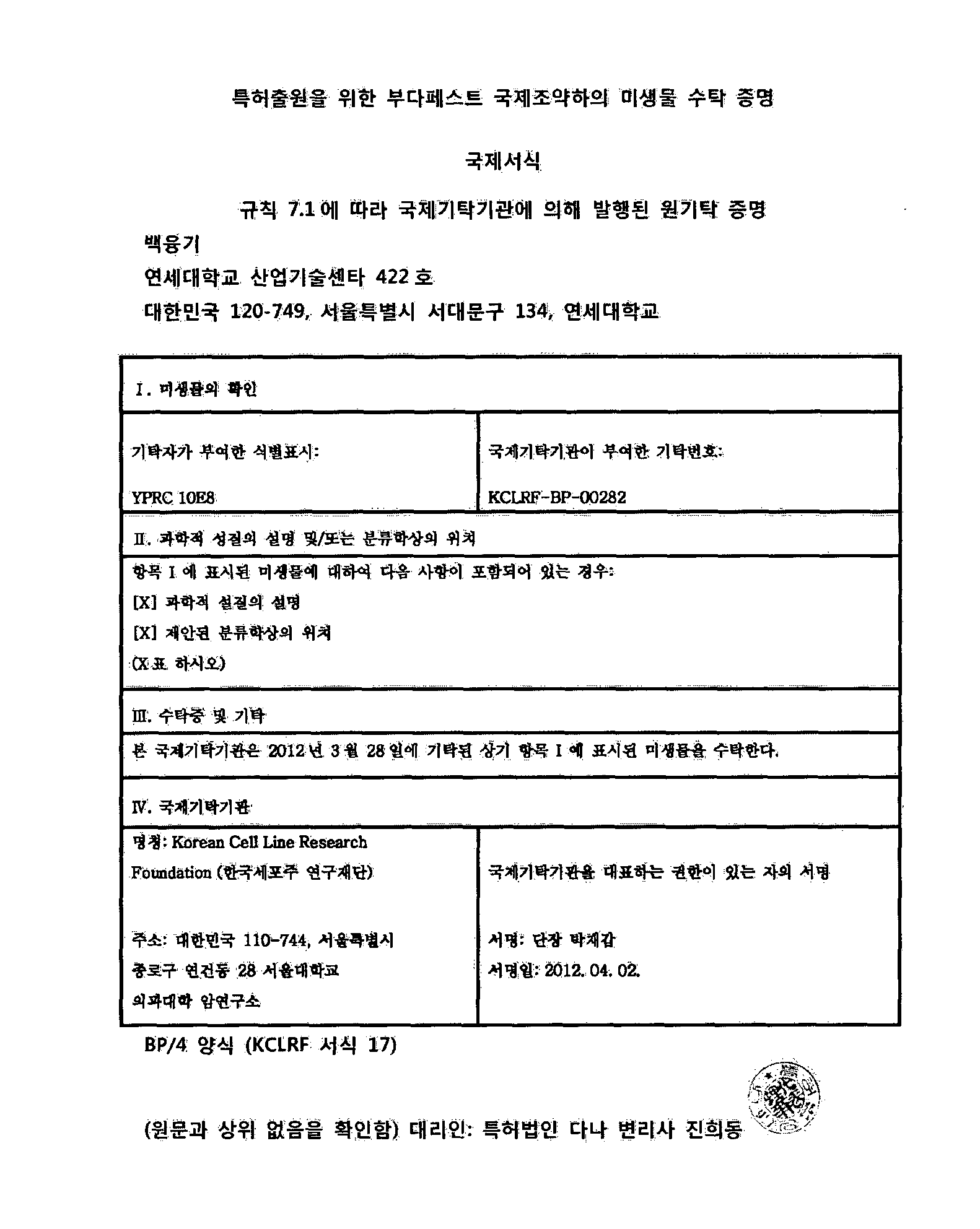
- the present invention provides a monoclonal antibody that specifically binds to human liver-carboxyesterase 1 protein, produced by hybridoma with accession number KCLRF-BP-00282.
- the present invention also provides hybridoma cells with Accession No. KCLRF-BP-00282 for producing monoclonal antibodies according to the present invention.
- the invention also provides a composition for the separation and purification of human liver-carboxyesterase 1 protein comprising a monoclonal antibody according to the invention.
- the present invention also provides a composition for detecting human liver-carboxyesterase 1 comprising the monoclonal antibody according to the present invention.
- the present invention also provides a method for detecting the concentration of human liver-carboxyesterase 1 in urine or blood, comprising contacting a monoclonal antibody according to the invention with a sample sample to detect antigen-antibody complex formation.
- the present invention also provides a liver cancer diagnostic kit comprising a monoclonal antibody according to the present invention.
- the monoclonal antibody of human hepatic-carboxyesterase 1 of the present invention is characterized in that hepatic carboxyesterase 1 is expressed in tissues, cells, blood and the like through high specific binding to human hepatic-carboxyesterase 1 protein. Can be used for detection. In particular, it can be usefully used for the early diagnosis of liver cancer simply in the blood.
- Figure 1 shows the results of analysis of liver-carboxyesterase 1 by separating the serum from the tail of the mouse after the immunization of the antigenic peptide group to the mouse 3rd.
- Figure 2 is a photograph showing the reactivity of splenocytes and Maeloma fused cells of the immunized mouse to the antigen peptide of liver-carboxyesterase 1.
- FIG. 3 shows hybridoma cell line selection results for producing liver-carboxyesterase 1 monoclonal antibody using splenocytes of immunized mice highly reactive by antigen-peptide of liver-carboxyesterase 1 will be.
- FIG. 4 is a result of comparing the antigen-antibody binding degree between the monoclonal antibody of the present invention and the polyclonal antibody of known liver-carboxyesterase 1.
- Figure 5 compares the antigen-antibody binding degree in the plasma of normal, hepatitis patients, cirrhosis transducer, liver cancer patients using a monoclonal antibody of the present invention and a known polyclonal antibody of liver-carboxyesterase 1 One result.
- Figure 6 shows the results of color development (a) of the sample according to antigen-antibody binding using a monoclonal antibody of the present invention and a known polyclonal antibody of hepatic-carboxyesterase 1, and hepatic-carboxyesterase 1 Absorbance values according to antigen-antibody binding for each concentration are shown (b).
- the inventors have developed a monoclonal antibody that is more efficient than the conventional commercially available human liver-carboxyl esterase 1 and a hybridoma cell line producing the antibody.
- the present invention has been completed by providing a means to isolate the first and objectively compare the amount of this enzyme.
- the present invention provides a monoclonal antibody specifically binding to human liver-carboxyesterase 1 protein, produced by hybridoma with Accession No. KCLRF-BP-00282, a hybridoma producing the monoclonal Provided are cells and methods for their preparation.
- 'human liver-carboxyesterase 1' includes the protein itself, its recombinant protein and its artificial variants and mutants, as well as the natural form of the protein and its functional equivalents.
- Monoclonal antibodies specific for human liver-carboxyesterase 1 of the invention can be prepared by fusion methods well known in the art (Kohler and Milstein (1976) European Jounral of Immunology 6: 511-519).
- One of two populations of cells fused to make a 'hybridoma' secreting monoclonal antibodies utilizes cells from an immunologically suitable host animal, such as a mouse injected with human liver-carboxyesterase 1
- the other population uses cancer or myeloma cell lines. These two populations of cells are fused by methods known in the art, such as polyethylene glycol, and then antibody-producing cells are propagated by standard tissue culture methods.
- Hybridoma cell lines capable of producing a specific cell population by subcloning by the limited dilution technique and then producing antibodies specific for human liver-carboxyesterase 1 were subjected to enzyme immunoassay. Selected by -linked immunosorbent assay (ELISA) or Western blot analysis, and culture in large quantities in vitro or in vivo according to standard techniques.
- the in vivo mass culture means the hybridoma cell line is injected into the abdominal cavity of the mouse to induce high concentration of antibody production, and then separated from the ascites.
- the monoclonal antibody produced by the hybridoma cell line may be used without purification, or may be purified and used in high purity (eg, 95% or more) according to a method well known in the art.
- purification techniques can be separated from the culture medium or ascites using methods such as gel electrophoresis, dialysis, salt precipitation, ion exchange chromatography, affinity antigen column chromatography, and the like.
- hybridoma producing monoclonal antibody is named as hybridoma YPRC 10E8, and the Korean Cell Line Research Foundation, an international depository institution within the Cancer Research Institute of Seoul National University, Yongun-dong, Jongno-gu, Seoul, on March 28, 2012. Deposited as KCLRF-BP-00282 with the Cell Line Research Foundation.
- the monoclonal antibody produced by the 'hybridoma YPRC 10E8' was named 'YPRC 10E8'.
- phage display peptide libraries are used for biopanning, immunoblotting and screening fragments generated by digesting antigen polypeptides with proteases to determine fragments comprising epitope sequences, activated membranes or solid phases such as polyethylene pins. Screening peptide arrays immobilized on, competitive ELISA assays using soluble peptides, and the importance of each amino acid sequence residue in an epitope sequence.
- Epitopes on human liver-carboxyesterase 1 recognized by the monoclonal antibodies of the present invention were analyzed on the tertiary structure of human liver-carboxyesterase 1 using AbFrontier's software and immunized on the analyzed peptide list. After confirming the difference from the amino acid sequence of the corresponding protein of the mouse to be selected, it was selected by reselection based on the peptide specific for the human liver-carboxyesterase 1 analyzed.
- the antigen binding affinity of the monoclonal antibody can be determined according to a conventional method, but is not particularly limited, but more specifically, radioimmunoassay (RIA), enzyme immunoassay (ELISA), immunoprecipitation, immunofluorescence, and color particle binding Binding affinity to the antigen is determined by methods, chemiluminescent binding or immunoblotting.
- Monoclonal antibodies of the present invention exhibit a binding affinity of at least 20-fold over polyclonal antibodies.
- the monoclonal antibody according to the invention may be an immunoglobulin isotype of IgG1.
- the present invention recognizes a linear epitope of SEQ ID NO: 1 corresponding to 554 to 567 of the C-terminus among the amino acid sequences of human liver-carboxyesterase 1, and to human liver-carboxyesterase 1 Provided are monoclonal antibodies that specifically bind.
- the term 'monoclonal antibody' refers to a highly specific antibody directed against a single antigenic site as it is known in the art. Typically, unlike polyclonal antibodies that include different antibodies directed against different epitopes (antigenic determinants), monoclonal antibodies are directed against a single determinant on an antigen. Monoclonal antibodies have the advantage of improving the selectivity and specificity of diagnostic and analytical assays using antigen-antibody binding, and also have the advantage that they are not contaminated by other immunoglobulins because they are synthesized by hybridoma culture. As used herein, antibodies against human liver-carboxyesterase 1 include not only whole antibody forms but also functional fragments of antibody molecules.
- the whole antibody is a structure having two full length light chains and two full length heavy chains, and each light chain is linked by a heavy chain and disulfide bond.
- the heavy chain constant region has gamma ( ⁇ ), mu ( ⁇ ), alpha ( ⁇ ), delta ( ⁇ ) and epsilon ( ⁇ ) types and subclasses gamma 1 ( ⁇ 1), gamma 2 ( ⁇ 2), and gamma 3 ( ⁇ 3). ), Gamma 4 ( ⁇ 4), alpha 1 ( ⁇ 1) and alpha 2 ( ⁇ 2).
- the constant regions of the light chains have kappa ( ⁇ ) and lambda ( ⁇ ) types (cellular and Molecular Immunology, Wonsiewicz, MJ, Ed., Chapter 45, pp. 41-50, WB Saunders Co. Philadelphia, PA (1991); Nisonoff, A., Introduction to Molecular Immunology, 2nd Ed., Chapter 4, pp. 45-65, sinauer Associates, Inc., Sunderland, MA (1984)).
- Functional fragments of antibody molecules refer to fragments having antigen binding functions and include Fab, F (ab '), F (ab') 2, Fv and the like.
- Fab in the antibody fragment has a structure having a variable region of the light and heavy chains, a constant region of the light chain and the first constant region (CH1) of the heavy chain has one antigen binding site.
- a cysteine residue in the hinge region of Fab' forms a disulfide bond.
- dsFv double-chain Fv
- scFv short chain Fv
- Such antibody fragments can be obtained using proteolytic enzymes (e.g., by limiting cleavage of the entire antibody to papain, Fab can be obtained and pepsin cleavage can yield F (ab ') 2 fragment). Can be produced through genetic recombination techniques.
- the antibody is preferably in the form of Fab or in the form of whole antibody.
- the heavy chain constant region may be selected from any one isotype of gamma ( ⁇ ), mu ( ⁇ ), alpha ( ⁇ ), delta ( ⁇ ) or epsilon ( ⁇ ).
- the light chain constant region may be of kappa or lambda type.
- the antibody of the present invention may be a humanized antibody or a CDR-grafted antibody to further reduce the immunogenicity of the chimeric antibody or chimeric antibody.
- Chimeric antibodies are antibody variable domains derived from animals other than humans (eg, mice, rabbits, poultry, etc.), and antibody constant domains refer to antibodies derived from humans. do. Such chimeric antibodies can be prepared by methods such as genetic recombination known in the art.
- Humanized antibodies or CDR-grafted antibodies may be used to maintain the high affinity and specificity of animal-derived monoclonal antibodies and to reduce immunogenicity in humans.
- the antibody refers to an antibody prepared by implanting an antigen binding region (CDR) into a human antibody.
- CDR antigen binding region
- the degree of humanization may be selected in consideration of the affinity for the antigen and the degree of immunity in the human body.
- the invention also relates to a composition for the separation and purification of human liver-carboxyesterase 1 protein comprising a monoclonal antibody according to the invention.
- the separation and purification composition comprising the antibody can be used for purification methods such as conventional ion exchange chromatography and affinity chromatography to easily separate and purify human liver-carboxyesterase 1 protein from a sample. To be.
- the present invention also provides a composition for detecting human liver-carboxyesterase 1 comprising the monoclonal antibody according to the present invention.
- the present invention provides a method for detecting the concentration of human liver-carboxylase 1 in urine or blood, comprising contacting a monoclonal antibody according to the present invention with a sample sample to detect antigen-antibody complex formation.
- the present invention can detect human liver-carboxyesterase 1 by contacting the monoclonal antibody with human urine, serum or plasma to detect antigen-antibody complex formation.
- the term 'antigen-antibody complex' refers to the enzyme and the monoclonal that recognizes the same to confirm the presence or absence of human liver-carboxyesterase 1 in a biological sample such as human urine, serum or plasma.
- a biological sample such as human urine, serum or plasma.
- the antigen-antibody complex can be detected using a detection label.
- a detection label may be selected from enzymes, fluorescent materials, ligands, light emitting materials, microparticles, redox molecules or radioisotopes, but is not particularly limited thereto.
- the enzyme is, for example, ⁇ -glucuronidase, ⁇ -D-glucosidase, ⁇ -D-galactosidase, urease, peroxidase, alkaline phosphatase, acetylcholinesterase, glucose oxidase, Hexokinase and GDPase, RNase, glucose oxidase and luciferase, phosphofructokinase, phosphoenolpyruvate carboxylase, aspartate aminotransferase, phosphphenolpyruvate decarboxylase, ⁇ -latama And the like.
- Fluorescent materials include, but are not limited to, fluorescein, isothiocyanate, rhodamine, phycoerythrin, phycocyanin, allophycocyanin, o-phthalaldehyde, fluorescamine, and the like.
- Ligands include, but are not limited to, biotin derivatives.
- Luminescent materials include, but are not limited to, acridinium ester, luciferin, luciferase, and the like.
- Microparticles include, but are not limited to, colloidal gold, colored latex, and the like.
- Redox molecules include ferrocene, ruthenium complex, biologen, quinone, Ti ion, Cs ion, diimide, 1,4-benzoquinone, hydroquinone, K 4 W (CN) 8 , [Os (bpy) 3 ] 2+ , [RU (bpy) 3 ] 2+ or [MO (CN) 8 ] 4-, and the like.
- Radioisotopes include, but are not limited to, 3 H, 14 C, 32 P, 35 S, 36 Cl, 51 Cr, 57 Co, 58 Co, 59 Fe, 90 Y, 125 I, 131 I or 186 Re.
- the antigen-antibody complex may be formed by colorimetric method, electrochemical method, fluorimetric method, luminometry, particle counting method, visual assessment, or visual assessment. It can be detected using a scintillation counting method. For example, flow cytometry, immunocytochemistry, radioimmunoassay (RIA), enzyme immunoassay (ELISA), western blotting, immunoprecipitation assay, immunodiffusion assay Can be detected by a complement fixation assay, a protein chip, or the like. More specifically, it can be detected through immunoprecipitation and immunoblotting useful for recovering small amounts of the desired protein.
- the antigen-antibody complex can be detected using an enzyme-linked immunosorbent assay (ELISA).
- ELISA is a direct ELISA using a labeled antibody that recognizes an antigen attached to a solid support, an indirect ELISA using a labeled antibody that recognizes a capture antibody in a complex of an antibody that recognizes an antigen attached to a solid support, and a solid support.
- Various ELISA methods include indirect sandwich ELISA using labeled secondary antibodies.
- a biological sample such as urine, blood, serum or plasma is contacted with a monoclonal antibody of the present invention coated on a solid support such as a microtiter plate, a membrane, a test strip or the like.
- a monoclonal antibody of the present invention coated on a solid support such as a microtiter plate, a membrane, a test strip or the like.
- the wells of the coated plate are incubated with the sample sample, The presence of the antigen-antibody complex can then be determined.
- the presence of the antigen-antibody complex can be confirmed using an antibody specific for the antigen of the antigen-antibody complex, for example a monoclonal or polyclonal antibody that specifically binds to human liver-carboxyesterase 1.
- the monoclonal or polyclonal antibody may have a detection label, and in the absence of the detection label, the monoclonal or polyclonal antibody may be identified by treatment with another antibody capable of detecting these monoclonal or polyclonal antibodies.
- a capture monoclonal antibody attached to a support is reacted with a biological sample, and then a monoclonal antibody that specifically binds to human liver-carboxyesterase 1 is bound to measure a detection label signal therefrom or these complexes.
- Human liver-carboxyesterase 1 can be detected by adding a secondary antibody having a label capable of producing a detectable signal to and measuring the detection label signal therefrom.
- the invention also relates to a liver cancer diagnostic kit comprising a monoclonal antibody according to the invention.
- the monoclonal antibody used in the liver cancer diagnostic kit of the present invention can also be used as a fragment of the monoclonal antibody as long as the antibody can selectively recognize human liver-carboxyesterase 1.
- Such antibody fragments may include F (ab ') 2, Fab, Fab', Fv fragments, and the like.
- the liver cancer diagnostic kit may be prepared in the form of a protein chip to which the monoclonal antibody of the present invention is attached.
- Liver cancer diagnostic kits of the present invention may include monoclonal antibodies or fragments thereof that selectively recognize human liver-carboxyesterase 1 and tools / reagents used for immunological analysis.
- Tools / reagents used in such immunological assays include suitable carriers, labeling substances capable of producing detectable signals, solubilizers, detergents and the like.
- the labeling substance when it is an enzyme, it may include a substrate capable of measuring enzyme activity and a reaction terminator.
- Suitable carriers include, but are not limited to, soluble carriers such as physiologically acceptable buffers known in the art, such as PBS, insoluble carriers such as polystyrene, polyethylene, polypropylene, polyesters, Polyacrylonitrile, fluorine resin, crosslinked dextran, polysaccharides, polymers such as magnetic fine particles plated with latex metal, other papers, glass, metals, agarose and combinations thereof.
- physiologically acceptable buffers known in the art, such as PBS, insoluble carriers such as polystyrene, polyethylene, polypropylene, polyesters, Polyacrylonitrile, fluorine resin, crosslinked dextran, polysaccharides, polymers such as magnetic fine particles plated with latex metal, other papers, glass, metals, agarose and combinations thereof.
- Assay systems for use in the detection methods and diagnostic kits of the present invention include, but are not limited to, ELISA plates, dip-stick devices, immunochromatography test strips and radiation split immunoassay devices, and flow-through through) devices and the like.
- liver tissue 50 mg was refrigerated at 4 ° C in cytolytic RIPA buffer [50 mM Tris, 150 mM sodium chloride (NaCl), 1% NonidetP-40, 0.25% sodium deoxycholate, pH 7.4]. The mixture was ground under conditions and centrifuged at 14000 rpm for 20 minutes to recover the protein solution.
- cytolytic RIPA buffer 50 mM Tris, 150 mM sodium chloride (NaCl), 1% NonidetP-40, 0.25% sodium deoxycholate, pH 7.4.
- Plasma collected from hepatitis patients, liver cirrhosis patients, and liver cancer patients with pathologically individual information was obtained from Yonsei Severance Hospital Gene Bank.
- Plasma from normal individuals was tested for HIV-1 and HIV-2 antibodies, HIV-1 antigen, hepatitis B surface antigen, and hepatitis B nuclear antigen (HIV) derived from immunodeficiency virus (HIV).
- HIV hepatitis B core antigen
- hepatitis C virus hepatitis C virus
- T cell leukemia virus (HTLV-I / II) antigen samples identified as negative in syphilis test were used. It was stored and used. The acquisition of plasma was in accordance with the rules of the Institutional Review Board of the Yonsei Medical Center.
- auxiliary antigen (Freund's adjuvant) of each peptide group was emulsified and immunized by first injection into the abdominal cavity of four female BALB / c mice, followed by a second injection four weeks later, and another three weeks later.
- Serum was isolated from the tails of all mice prior to each injection to determine the degree of antibody reaction produced in the mouse with the peptide antigen from enzyme immunoassay.
- NC nitrocelluose
- M in the gel picture means a standard marker (5 ⁇ g) by protein molecular weight, 5 ⁇ g represents a liver tissue protein it means.
- mice were selected because of their high antigen-antibody reactivity in # 3 mice.
- Splenocytes of mouse # 3 selected in Example 3 were isolated, fused with Sp2 / 0-Ag14 Maeloma cells, diluted in HAT medium and aliquoted into eight 96-well plates.
- the cultured cells were subjected to enzyme immunoassay using a peptide synthesized at 250 ng / well as an antigen to select 11 wells with the highest absorbance.
- Each cell was cultured in 24-well to obtain 5 wells from the enzyme immunoassay of the same method. Dog wells (# 1B2, # 1G11, # 2F4, # 3C1, # 3F2) were selected.
- Each cell was again cultured in a 96-well plate and subjected to three repeated enzyme-immunoassays, with five clone cells showing the highest reactivity (# 1A9, # 4H3, # 6A10, # 8F11, # 10E8). was selected.
- FIG. 2 is a photograph showing Western blot analysis of a culture solution of each # 3 well-cell having high titer in the enzyme immunoassay, and shows the position and degree of binding of liver-carboxyesterase 1.
- FIG. High reactivity was observed in # 3F2 fusion cells.
- FIG. 3 is a photograph showing the results of Western blot analysis of cells of the final selected wells by dispensing the selected five # well cells into 96-well plates and analyzing the wells showing high titers in a third iteration. It showed high reactivity in hybridoma cells.
- Subclass isotype of final monoclonal antibody # 10E8 was found to be IgGl type (see Table 2).
- Magnetic beads, 'Dynabead MyOne TM Tosylactivated' were prepared according to the manufacturer's manual for anti-inter-carboxyl esterase 1 polyclonal antibody (Abcam, # Ab1875) and anti-inter-carboxyl esterase 1 single. Each was coated with clone antibody (clone # 10E8). 10 ⁇ l, 20 ⁇ l, 30 ⁇ l, 40 ⁇ l, 50 ⁇ l of beads bound to each anti- liver-carboxyesterase 1 antibody were immunoprecipitated for 2 hours in a 1 mL tube with 100 ⁇ g of liver tissue protein.
- the protein bound to the antibody was recovered using PBS-T buffer adjusted to pH 2, and immunoblotting was performed to determine the binding of liver-carboxyesterase 1 bound from polyclonal antibody and monoclonal antibody (clone # 10E8). Changes were compared to polyclonal antibodies using the ImageQuant program.
- Figure 4 is a comparison of the antigen-antibody binding degree of polyclonal antibody and monoclonal antibody (clone # 10E8), pHCC is immunized with 30 ⁇ l of antibody-bound beads with 200 ⁇ l of a sample of plasma of 15 liver cancer patients Settled, 5 ⁇ g means liver tissue protein.
- monoclonal antibody # 10E8 showed 20-fold higher antigen-antibody reactivity than polyclonal antibody under the same conditions.
- liver cirrhosis After 10 plasma of normal, hepatitis, liver cirrhosis and liver cancer patients were mixed, the same 200 ⁇ l was immunoprecipitated with hepatic carboxyesterase 1 polyclonal antibody and monoclonal antibody # 10E8 used in Example 5. Western blot analysis was then performed. The signal intensity of the liver-carboxyesterase 1 analyzed when the scan resolution was 100 microns to scan under the same conditions was analyzed using the ImageQuant program.
- Figure 6 is a result of comparing the antigen-antibody binding degree of the monoclonal antibody # 10E8 and the two polyclonal antibodies (# Ab1875, # Ab77730) of the two antibody 1 and the antibody 2 of Abcam applicable to ELISA, according to the antigen-antibody binding
- the results of color development of the sample (a) and the absorbance values according to antigen-antibody binding for each concentration of liver-carboxyesterase 1 are shown (b).
- the present invention can be used in the field of protein or disease diagnosis.
Abstract
Description
| 종 | 아미노산 배열 | (n) | 아미노산 위치 |
| Human | KAVEKPPQTEHIEL | 13 | 554~567 |
| Mouse | LRAKKPPQTGHTEL | 13 | 554~567 |
| 클론 | IgG1 | IgG2a | IgG2b | IgG3 | IgA | IgM | κ | λ | H-체인 | L-체인 |
| 10E8 | 0.518 | 0.072 | 0.104 | 0.097 | 0.097 | 0.084 | 0.738 | 0.149 | IgG1 | κ |
| 항체 종류 | 정상인 | 간염환자 | 간경변환자 | 간암환자 |
| 다클론 항체 | 191,363 | 182,431 | 178,140 | 738,366 |
| 본 발명의 단일클론 항체 | 5,586,315 | 6,676,570 | 3,314,999 | 24,906,430 |

Claims (8)
- 기탁번호 KCLRF-BP-00282인 하이브리도마에 의해 생산되는, 인간 간-카르복실에스터라제 1 단백질에 특이적으로 결합하는 단일클론 항체.
- 제1항에 있어서,단일클론 항체는 인간 간-카르복실에스터라제 1 단백질의 C-말단의 아미노산 554 내지 567의 에피토프 서열을 인식하는 것인 단일클론 항체.
- 제1항에 있어서,IgG1의 면역글로불린 단위형(isotype)인 단일클론 항체.
- 제1항의 단일클론 항체를 생산하는 기탁번호 KCLRF-BP-00282인 하이브리도마 세포.
- 제1항에 따른 단일클론 항체를 포함하는 인간 간-카르복실에스터라제 1 단백질의 분리 및 정제용 조성물.
- 제1항에 따른 단일클론 항체를 포함하는 인간 간-카르복실에스터라제 1 검출용 조성물.
- 제1항에 따른 단일클론 항체를 시료 샘플과 접촉시켜 항원-항체 복합체 형성을 검출하는 단계를 포함하는 뇨 또는 혈중 인간 간-카르복실에스터라제 1의 농도 검출방법.
- 제1항에 따른 단일클론 항체를 포함하는 간암 진단 키트.
Priority Applications (4)
| Application Number | Priority Date | Filing Date | Title |
|---|---|---|---|
| EP12774256.7A EP2700650B1 (en) | 2011-04-18 | 2012-04-17 | Monoclonal antibodies which specifically recognize human liver-carboxylesterase 1, hybridoma cell lines which produce monoclonal antibodies, and uses thereof |
| JP2014506322A JP5798679B2 (ja) | 2011-04-18 | 2012-04-17 | ヒト肝−カルボキシルエステラーゼ1を特異的に認識するモノクローナル抗体、前記抗体を生産するハイブリドーマ細胞株及びその用途 |
| CN201280029813.2A CN104066749B (zh) | 2011-04-18 | 2012-04-17 | 用于特异性识别人肝羧酸酯酶1的单克隆抗体、生成单克隆抗体的杂交瘤细胞株及其用途 |
| US14/112,411 US9116152B2 (en) | 2011-04-18 | 2012-04-17 | Monoclonal antibodies which specifically recognize human liver-carboxylesterase 1, hybridoma cell lines which produce monoclonal antibodies, and uses thereof |
Applications Claiming Priority (4)
| Application Number | Priority Date | Filing Date | Title |
|---|---|---|---|
| KR20110035619 | 2011-04-18 | ||
| KR10-2011-0035619 | 2011-04-18 | ||
| KR10-2012-0034928 | 2012-04-04 | ||
| KR1020120034928A KR101338517B1 (ko) | 2011-04-18 | 2012-04-04 | 인간 간-카르복실에스터라제 1을 특이적으로 인식하는 단일클론 항체, 상기 항체를 생산하는 하이브리도마 세포주 및 이의 용도 |
Publications (2)
| Publication Number | Publication Date |
|---|---|
| WO2012144784A2 true WO2012144784A2 (ko) | 2012-10-26 |
| WO2012144784A3 WO2012144784A3 (ko) | 2013-01-17 |
Family
ID=47285895
Family Applications (1)
| Application Number | Title | Priority Date | Filing Date |
|---|---|---|---|
| PCT/KR2012/002911 WO2012144784A2 (ko) | 2011-04-18 | 2012-04-17 | 인간 간-카르복실에스터라제 1을 특이적으로 인식하는 단일클론 항체, 상기 항체를 생산하는 하이브리도마 세포주 및 이의 용도 |
Country Status (6)
| Country | Link |
|---|---|
| US (1) | US9116152B2 (ko) |
| EP (1) | EP2700650B1 (ko) |
| JP (1) | JP5798679B2 (ko) |
| KR (1) | KR101338517B1 (ko) |
| CN (1) | CN104066749B (ko) |
| WO (1) | WO2012144784A2 (ko) |
Families Citing this family (4)
| Publication number | Priority date | Publication date | Assignee | Title |
|---|---|---|---|---|
| KR101523077B1 (ko) * | 2013-07-08 | 2015-05-26 | 포항공과대학교 산학협력단 | pH 미터를 이용한 바이오 물질 검출 방법 |
| CN103642825A (zh) * | 2013-11-29 | 2014-03-19 | 湖南师范大学 | 一种重组hCE1蛋白的可溶性表达和纯化方法 |
| CN112255061A (zh) * | 2020-10-13 | 2021-01-22 | 南开大学 | 一种通过免疫沉淀分离和检测蛋白质的方法 |
| CN116535512B (zh) * | 2022-12-09 | 2024-03-15 | 中国医学科学院肿瘤医院 | 针对肝细胞癌患者的异常凝血酶原单克隆抗体的制备及应用 |
Citations (5)
| Publication number | Priority date | Publication date | Assignee | Title |
|---|---|---|---|---|
| WO1988001649A1 (en) | 1986-09-02 | 1988-03-10 | Genex Corporation | Single polypeptide chain binding molecules |
| WO1988006630A1 (en) | 1987-03-02 | 1988-09-07 | Genex Corporation | Method for the preparation of binding molecules |
| WO1988007085A1 (en) | 1987-03-20 | 1988-09-22 | Creative Biomolecules, Inc. | Process for the purification of recombinant polypeptides |
| WO1988007086A1 (en) | 1987-03-20 | 1988-09-22 | Creative Biomolecules, Inc. | Leader sequences for the production of recombinant proteins |
| WO1988009344A1 (en) | 1987-05-21 | 1988-12-01 | Creative Biomolecules, Inc. | Targeted multifunctional proteins |
Family Cites Families (4)
| Publication number | Priority date | Publication date | Assignee | Title |
|---|---|---|---|---|
| JP4288454B2 (ja) * | 2000-09-04 | 2009-07-01 | 株式会社大塚製薬工場 | 薬物代謝の第i相反応に関与する酵素の測定方法、そのためのプローブおよびキット |
| US7906637B2 (en) * | 2006-06-23 | 2011-03-15 | St. Jude Children's Research Hospital | Compositions and methods for inducing or inhibiting activities of selected human cells |
| WO2009077479A2 (en) | 2007-12-17 | 2009-06-25 | Novartis Ag | Altering high-density lipoprotein levels through carboxylesterase 1 modulation |
| KR100975852B1 (ko) * | 2008-03-14 | 2010-08-13 | 연세대학교 산학협력단 | 간-카르복실에스터라제 1 단백질과 특이적으로 결합하는 항체를 포함하는 간암 진단 조성물 |
-
2012
- 2012-04-04 KR KR1020120034928A patent/KR101338517B1/ko active IP Right Grant
- 2012-04-17 EP EP12774256.7A patent/EP2700650B1/en not_active Not-in-force
- 2012-04-17 JP JP2014506322A patent/JP5798679B2/ja not_active Expired - Fee Related
- 2012-04-17 WO PCT/KR2012/002911 patent/WO2012144784A2/ko active Application Filing
- 2012-04-17 US US14/112,411 patent/US9116152B2/en not_active Expired - Fee Related
- 2012-04-17 CN CN201280029813.2A patent/CN104066749B/zh not_active Expired - Fee Related
Patent Citations (5)
| Publication number | Priority date | Publication date | Assignee | Title |
|---|---|---|---|---|
| WO1988001649A1 (en) | 1986-09-02 | 1988-03-10 | Genex Corporation | Single polypeptide chain binding molecules |
| WO1988006630A1 (en) | 1987-03-02 | 1988-09-07 | Genex Corporation | Method for the preparation of binding molecules |
| WO1988007085A1 (en) | 1987-03-20 | 1988-09-22 | Creative Biomolecules, Inc. | Process for the purification of recombinant polypeptides |
| WO1988007086A1 (en) | 1987-03-20 | 1988-09-22 | Creative Biomolecules, Inc. | Leader sequences for the production of recombinant proteins |
| WO1988009344A1 (en) | 1987-05-21 | 1988-12-01 | Creative Biomolecules, Inc. | Targeted multifunctional proteins |
Non-Patent Citations (10)
| Title |
|---|
| IMAI T, DRUG METAB PHARMACOKINET., vol. 21, 2006, pages 173 - 185 |
| KOHLER; MILSTEIN, EUROPEAN JOURNAL OF IMMUNOLOGY, vol. 6, 1976, pages 511 - 519 |
| LI B., NIOCHEM. PHARMACOL., vol. 70, 2005, pages 1673 - 1684 |
| MAKI T., JPN. J CANCER RES., vol. 82, 1991, pages 800 - 806 |
| NA K. ET AL., PROTEOMICS, vol. 9, 2009, pages 3989 - 3999 |
| NISONOFF, A.: "Introduction to Molecular Immunology 2nd Ed.,", 1984, SINAUER ASSOCIATES, INC., pages: 45 - 65 |
| REDINBO MR., BIOCHEM. SOC. TRANS., vol. 31, 2003, pages 620 - 624 |
| REDINBO MR., DRUG DISCOV. TODAY., vol. 10, 2005, pages 313 - 325 |
| See also references of EP2700650A4 |
| WONSIEWICZ, M. J.,: "Cellular and Molecular Immunology", 1991, W. B. SAUNDERS CO., pages: 41 - 50 |
Also Published As
| Publication number | Publication date |
|---|---|
| US20140154699A1 (en) | 2014-06-05 |
| CN104066749A (zh) | 2014-09-24 |
| US9116152B2 (en) | 2015-08-25 |
| KR20120118412A (ko) | 2012-10-26 |
| EP2700650A2 (en) | 2014-02-26 |
| CN104066749B (zh) | 2016-03-30 |
| EP2700650B1 (en) | 2018-05-30 |
| KR101338517B1 (ko) | 2013-12-10 |
| EP2700650A4 (en) | 2014-12-03 |
| JP5798679B2 (ja) | 2015-10-21 |
| JP2014512379A (ja) | 2014-05-22 |
| WO2012144784A3 (ko) | 2013-01-17 |
Similar Documents
| Publication | Publication Date | Title |
|---|---|---|
| US4935343A (en) | Monoclonal antibodies for interleukin-1β | |
| WO2016080591A1 (ko) | 중동호흡기증후군 코로나바이러스 뉴클레오캡시드를 인식하는 항체 및 그의 용도 | |
| CA2406240C (en) | Hcv anti-core monoclonal antibodies | |
| WO2010038974A2 (ko) | 이뮤노글로블린 에이 신증과 티지비엠 신증의 진단용 조성물 및 키트 | |
| CN112679605B (zh) | 针对新型冠状病毒核衣壳蛋白的抗体或其抗原结合片段及其应用 | |
| JPH04503600A (ja) | 抗インターロイキン―1α及び―1βのモノクローナル抗体、その製法、ならびに前記抗体類の、インターロイキン―1α及び―1βの検出と治療への応用 | |
| WO2012144784A2 (ko) | 인간 간-카르복실에스터라제 1을 특이적으로 인식하는 단일클론 항체, 상기 항체를 생산하는 하이브리도마 세포주 및 이의 용도 | |
| US7575938B2 (en) | Method for diagnosing liver diseases | |
| WO2008032712A1 (en) | Monoclonal antibody and use thereof | |
| WO2019098603A2 (ko) | 소-임신 연관 글리코단백질 1에 특이적으로 결합하는 항체 및 이의 용도 | |
| JP6236864B2 (ja) | 癌の検出方法及び膵臓特異的リボヌクレアーゼ1を認識する抗体 | |
| US20060275849A1 (en) | Monoclonal antibody reagents | |
| JP4663831B2 (ja) | モノクローナル抗体、細胞株及びn1,n12−ジアセチルスペルミンの測定法 | |
| JP5553603B2 (ja) | 新規肝癌マーカー | |
| JP2878317B2 (ja) | ラミニン測定試薬 | |
| KR100493932B1 (ko) | 레지스틴에 대한 단클론 항체, 이의 제조 방법, 및 용도 | |
| JP4037933B2 (ja) | 抗ヒトカルシトニンモノクローナル抗体 | |
| KR20130133593A (ko) | 위암 진단용 바이오 마커 조성물 및 이를 이용한 위암 진단 방법 | |
| JP5448424B2 (ja) | ヒトIgGのFcを含有するタンパク質の測定試薬 | |
| JP2007093336A (ja) | 被検物質の測定方法 | |
| WO2005054296A1 (ja) | メチルリジンを認識する抗体及びその製造方法並びにその利用 | |
| US20130288279A1 (en) | Specific a1at monoclonal antibodies for detection of endometriosis | |
| EP4342909A1 (en) | Anti-norovirus antibody | |
| EP4342989A1 (en) | Anti-norovirus antibody | |
| JP2019210211A (ja) | ウシプロカルシトニンを特異的に認識する抗体、その抗原結合断片、および、その使用 |
Legal Events
| Date | Code | Title | Description |
|---|---|---|---|
| WWE | Wipo information: entry into national phase |
Ref document number: 201280029813.2 Country of ref document: CN |
|
| 121 | Ep: the epo has been informed by wipo that ep was designated in this application |
Ref document number: 12774256 Country of ref document: EP Kind code of ref document: A2 |
|
| ENP | Entry into the national phase |
Ref document number: 2014506322 Country of ref document: JP Kind code of ref document: A |
|
| NENP | Non-entry into the national phase |
Ref country code: DE |
|
| WWE | Wipo information: entry into national phase |
Ref document number: 2012774256 Country of ref document: EP |
|
| WWE | Wipo information: entry into national phase |
Ref document number: 14112411 Country of ref document: US |