WO2012141373A1 - 폐암 조기 진단용 XAGE-1a 마커 및 이의 용도 - Google Patents
폐암 조기 진단용 XAGE-1a 마커 및 이의 용도 Download PDFInfo
- Publication number
- WO2012141373A1 WO2012141373A1 PCT/KR2011/003309 KR2011003309W WO2012141373A1 WO 2012141373 A1 WO2012141373 A1 WO 2012141373A1 KR 2011003309 W KR2011003309 W KR 2011003309W WO 2012141373 A1 WO2012141373 A1 WO 2012141373A1
- Authority
- WO
- WIPO (PCT)
- Prior art keywords
- xage
- lung cancer
- protein
- antibody
- early diagnosis
- Prior art date
- Legal status (The legal status is an assumption and is not a legal conclusion. Google has not performed a legal analysis and makes no representation as to the accuracy of the status listed.)
- Ceased
Links
Images


Classifications
-
- G—PHYSICS
- G01—MEASURING; TESTING
- G01N—INVESTIGATING OR ANALYSING MATERIALS BY DETERMINING THEIR CHEMICAL OR PHYSICAL PROPERTIES
- G01N33/00—Investigating or analysing materials by specific methods not covered by groups G01N1/00 - G01N31/00
- G01N33/48—Biological material, e.g. blood, urine; Haemocytometers
- G01N33/50—Chemical analysis of biological material, e.g. blood, urine; Testing involving biospecific ligand binding methods; Immunological testing
- G01N33/53—Immunoassay; Biospecific binding assay; Materials therefor
- G01N33/563—Immunoassay; Biospecific binding assay; Materials therefor involving antibody fragments
-
- G—PHYSICS
- G01—MEASURING; TESTING
- G01N—INVESTIGATING OR ANALYSING MATERIALS BY DETERMINING THEIR CHEMICAL OR PHYSICAL PROPERTIES
- G01N33/00—Investigating or analysing materials by specific methods not covered by groups G01N1/00 - G01N31/00
- G01N33/48—Biological material, e.g. blood, urine; Haemocytometers
- G01N33/50—Chemical analysis of biological material, e.g. blood, urine; Testing involving biospecific ligand binding methods; Immunological testing
- G01N33/53—Immunoassay; Biospecific binding assay; Materials therefor
- G01N33/575—Immunoassay; Biospecific binding assay; Materials therefor for cancer
- G01N33/5752—Immunoassay; Biospecific binding assay; Materials therefor for cancer of the lungs
-
- G—PHYSICS
- G01—MEASURING; TESTING
- G01N—INVESTIGATING OR ANALYSING MATERIALS BY DETERMINING THEIR CHEMICAL OR PHYSICAL PROPERTIES
- G01N33/00—Investigating or analysing materials by specific methods not covered by groups G01N1/00 - G01N31/00
- G01N33/48—Biological material, e.g. blood, urine; Haemocytometers
- G01N33/50—Chemical analysis of biological material, e.g. blood, urine; Testing involving biospecific ligand binding methods; Immunological testing
- G01N33/53—Immunoassay; Biospecific binding assay; Materials therefor
- G01N33/531—Production of immunochemical test materials
- G01N33/532—Production of labelled immunochemicals
- G01N33/535—Production of labelled immunochemicals with enzyme label or co-enzymes, co-factors, enzyme inhibitors or enzyme substrates
-
- G—PHYSICS
- G01—MEASURING; TESTING
- G01N—INVESTIGATING OR ANALYSING MATERIALS BY DETERMINING THEIR CHEMICAL OR PHYSICAL PROPERTIES
- G01N33/00—Investigating or analysing materials by specific methods not covered by groups G01N1/00 - G01N31/00
- G01N33/48—Biological material, e.g. blood, urine; Haemocytometers
- G01N33/50—Chemical analysis of biological material, e.g. blood, urine; Testing involving biospecific ligand binding methods; Immunological testing
- G01N33/53—Immunoassay; Biospecific binding assay; Materials therefor
- G01N33/575—Immunoassay; Biospecific binding assay; Materials therefor for cancer
Definitions
- the present invention relates to an XAGE-1a marker for early diagnosis of lung cancer and its use, and more particularly, a composition for early diagnosis of lung cancer, kit for early diagnosis of lung cancer, and microarray for early diagnosis of lung cancer comprising an antibody specifically binding to XAGE-1a protein.
- the present invention relates to a method for diagnosing lung cancer by measuring the expression level of Xage-1a in a blood sample derived from the same.
- lung cancer was the second largest cancer in Korea between 2003 and 2005, accounting for 12.1% of all cancers, and the incidence rate per 100,000 population was 33.2 cases.
- the sex ratio of males and females was 3.83: 1, which occurred more frequently in males, and the number of occurrences was second in male cancers and fifth in female cancers.
- the 60s were the highest with 34.3%, followed by the 70s with 31.0% and the 50s with 14.6%.
- lung cancer is rapidly growing in Korea due to the increase of smoking population and air pollution.
- the mortality rate of cancer is 1st with 21.4%
- the mortality rate of men and women is 1st and 2nd with 24.9% and 15.1%, respectively. .
- Lung cancer usually causes symptoms such as surrounding tissue involvement or airway obstruction and lymph node metastasis due to the growth of cancer cells, but in about 10 to 15% of patients, lung cancer is diagnosed at regular checkups without any symptoms. In addition, since most lung cancers are in a fairly advanced state at the time of diagnosis, there is a problem that most cases are difficult to cure. Therefore, it is urgent to diagnose lung cancer early and reduce mortality from lung cancer (Wulfkuhle et al., Nat. Rev. Cancer 3, 267-275, 2003).
- CEA cancerembryionic antigen
- CYFRA 21-1 cytokeratin 19 fragment
- the inventors have discovered proteins that increase expression in lung cancer cells, and newly discovered that XAGE-1d protein is specifically present in the blood of patients with lung cancer.
- PCT was applied by confirming that lung cancer can be easily diagnosed by immunochemistry through the detection of XAGE-1d (PCT / KR2010 / 003710).
- the XAGE-1a of the present invention was originally known as XAGE-1b, but the existing XAGE-1a (146 amino acids) disappeared from the record and instead is a protein currently being recorded as XAGE-1a.
- the XAGE-1a of the present invention is clearly distinguished from XAGE-1d and also shows high expression in the blood of lung cancer patients to complete the present invention.
- Korean Patent No. 10-0661931 discloses a method for diagnosing cancer by measuring a change in a sugar chain of transferrin
- Korean Patent No. 10-0931997 discloses a method for diagnosing hepatocellular carcinoma using the KLK-6 gene.
- the present invention was derived by the above-described needs, and the present inventors have completed the present invention by confirming that the concentration of XAGE-1a protein in the blood of early stage lung cancer patients is significantly increased.
- the present invention provides a composition for the early diagnosis of lung cancer comprising an antibody that specifically binds to XAGE-1a protein.
- the present invention also provides a kit for early diagnosis of lung cancer comprising an antibody that specifically binds to XAGE-1a protein.
- the present invention also provides a microarray for the early diagnosis of lung cancer comprising an antibody that specifically binds to XAGE-1a protein.
- the present invention also provides a method for detecting a blood marker XAGE-1a for early diagnosis of lung cancer using the XAGE-1a protein specific antibody.
- the present invention also provides a composition for predicting prognosis of lung cancer comprising an antibody that specifically binds to XAGE-1a protein.
- the present invention also provides a method for diagnosing lung cancer by measuring the expression level of Xage-1a in a blood sample derived from a subject.
- the early diagnosis method of lung cancer using the XAGE-1a blood marker of the present invention collects samples from blood, so it is easier to implement and relatively less pain than other methods. It is also expected to be useful for lowering the mortality rate from lung cancer because it can specifically diagnose lung cancer at an early stage.
- HEK293T kidney cell line (control)
- A549, NCI-H460 and Hop-92 lung cancer cell line
- MCF-7, MDA-MB-453 and T-47D breast cancer cell line.
- Figure 2 shows the absorbance (A) according to the concentration of biotin-XAGE-1a peptide and the quantification standard curve (B) by antigen competition ELISA according to the concentration of XAGE-1a peptide.
- B Absorbance value obtained by adding the XAGE-1a peptide to the biotin-XAGE-1a peptide
- Bo Absorbance value only of the biotin-XAGE-1a peptide.
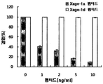
- Figure 3 shows the binding of biotin-XAGE-1a peptides to the concentration of the XAGE-1a peptide.
- Figure 4 shows the hourly concentration of XAGE-1a secreted in the media of lung and breast cancer cell lines.
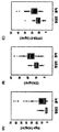
- Figure 5 shows the distribution of XAGE-1a (A) and control groups CEA (B) and CYFRA 21-1 (C) in normal and patient blood.
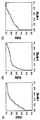
- Figure 6 shows the ROC graph of XAGE-1a (A) and the control CEA (B) and CYFRA 21-1 (C).
- the present invention provides a composition for the early diagnosis of lung cancer comprising an antibody that specifically binds to XAGE-1a protein.
- diagnosis means identifying the presence or characteristic of a pathological condition.
- diagnosis is to determine whether lung cancer has developed.
- lung cancer marker XAGE-1a protein of the present invention was detected higher than other lung cancer diagnostic markers CEA and CYFRA 21-1 in the early stage of lung cancer, it may be more effective for early diagnosis of lung cancer.
- the XAGE-1a protein may be XAGE-1a carboxyl terminal, but is not limited thereto.
- the XAGE-1a carboxyl terminal may preferably be composed of the amino acid sequence of SEQ ID NO: 1 (CATWKVIC KSCISQTPGI NLDLGSGVKV KIIPKEEHCK MPEAGEEQPQ V), but is not limited thereto.
- variants of such sequences are included within the scope of the present invention.
- a variant is a sequence that has a functional characteristic similar to that of the amino acid sequence of SEQ ID NO: 1, although the amino acid sequence changes.
- the XAGE-1a carboxyl terminal has at least 70%, more preferably at least 80%, even more preferably at least 90%, most preferably at least 95% homology with the amino acid sequence of SEQ ID NO: 1 May comprise an amino acid sequence.
- Antibodies that specifically bind to the XAGE-1a protein may specifically recognize only XAGE-1a.
- the antibody may be preferably a detection labeling agent capable of generating a detectable signal, and the detection labeling agent may be an enzyme, a prosthetic molecule, a fluorescent material, a luminescent material, a bioluminescent material or a radioactive material, but It is not limited.
- the lung cancer refers to a malignant tumor occurring in the lung, and may preferably be non-small cell lung cancer, but is not limited thereto.
- the composition may further comprise a secondary antibody that recognizes an antibody that specifically binds to XAGE-1a protein, and the detection labeling agent may be bound to the secondary antibody.
- the composition may further include a substrate and a reaction terminator capable of measuring enzymatic activity when the detection labeling agent is an enzyme.
- examples of enzymes suitable as detection markers include horseradish peroxidase, acetylcholine esterase, peroxidase, alkaline phosphatase, ⁇ -D-galactosidase, glucose oxidase, malate dehydro Geneases, glucose-6-phosphate dehydrogenases, invertases, and the like;
- suitable submolecular complexes include streptavidin / biotin or avidin / biotin;
- suitable fluorescent materials include umbelliferone, fluorescein, fluorescein isothiocyanate, rhodamine, dichlorotriazinylamine fluorescein, dansyl chloride, phycoerythrin or picobiliprotein, and the like;
- Examples of the luminescent material include luminol, isoleucinol or lucigenin and the like;
- bioluminescent materials include luciferase, luciferin or aequorin; Examples of bio
- antibody is a term known in the art and refers to a protein molecule that binds to a known antigen.
- Antibodies used in the present invention include monoclonal or polyclonal antibodies, immunologically active fragments (eg, Fab or (Fab) 2 fragments), antibody heavy chains, humanized antibodies, antibody light chains, genetically engineered single chain Fv molecules, Chimeric antibodies and the like, but is not limited thereto.
- Antibodies used in the present invention can be obtained by isolation from the serum of a mammal immunized with an immunogen (antigen).
- the mammal may preferably be mouse, rabbit or goat, but is not limited thereto.
- mammals used in the manufacture of antibodies may be preferentially selected from relatively large mammals with greater amounts of serum capable of separating more antibodies.
- an immunogen When the mammal is injected with an immunogen, it stimulates B cells of the immune system to produce immunoglobulins (Ig, immunoglobulin) specific for the immunogen.
- the immunoglobulins can be purified from serum using methods known in the art such as filtration, dialysis, ion exchange chromatography, size exclusion chromatography or affinity chromatography.
- an adjuvant such as Freund's, Alum, the Ribi Adjuvant System or Titermax may be injected for the purpose of enhancing the mammalian immune response to the injected immunogen.
- the adjuvant enhances antigenicity when mixed with the antigen and helps to generate a lot of antibodies.
- the present invention also provides a kit for early diagnosis of lung cancer comprising an antibody that specifically binds to XAGE-1a protein.
- the XAGE-1a protein may be XAGE-1a carboxyl terminus, but is not limited thereto.
- the kit may further comprise a secondary antibody that recognizes an antibody that specifically binds to XAGE-1a protein, and the detection labeling agent may be bound to the secondary antibody.
- the kit may include an antibody specifically binding to the XAGE-1a protein according to the present invention, a suitable carrier solution, a detection label capable of generating a detectable signal, a dissolving agent, a container containing a cleaning agent, and instructions for use.
- the kit may include a substrate and a reaction terminator capable of measuring enzymatic activity when the detection labeling agent is an enzyme.
- the present invention also provides a microarray for the early diagnosis of lung cancer comprising an antibody that specifically binds to XAGE-1a protein.
- the XAGE-1a protein may be XAGE-1a carboxyl terminus, but is not limited thereto.
- the microarray can detect a protein that specifically attaches to the antibody by antigen-antibody reaction by attaching an antibody that specifically binds to XAGE-1a protein on a slide glass surface treated with a specific reagent.
- It provides a method for detecting a blood marker XAGE-1a for the early diagnosis of lung cancer comprising the step of detecting the antigen-antibody complex.
- the XAGE-1a protein may be XAGE-1a carboxyl terminus, but is not limited thereto.
- the XAGE-1a carboxyl terminal may be composed of the amino acid sequence of SEQ ID NO: 1, but is not limited thereto.
- the method for detecting the antigen-antibody complex may be, but is not limited to, immunoblot, enzyme immunoassay, immunoprecipitation, fluorescence immunoassay, enzyme substrate coloration, or antigen antibody aggregation.
- the present invention also provides a composition for predicting prognosis of lung cancer comprising an antibody that specifically binds to XAGE-1a protein.
- Antibodies that specifically bind to the XAGE-1a protein of the present invention can be used not only for the early diagnosis of lung cancer, but also for the prognosis of lung cancer patients.
- step b) comparing the expression level of Xage-1a in step a) with the expression level of Xage-1a in the normal blood sample;
- c) providing a method for diagnosing lung cancer comprising determining that the subject's Xage-1a expression level is higher than that of a normal control group, and thus, the risk of developing lung cancer is high.
- the subject of step a) is a vertebrate, including a human, preferably a mammal, more preferably a human, ape, bovine, pig, rat, rabbit, guinea peak, hamster, dog or Cats can be used, but are not limited thereto.
- the expression level of Xage-1a is expressed by Western blotting, enzyme-linked immunosorbent assay (ELISA), immunohistochemical staining, immunoprecipitation and immunity. It is preferable to measure by any one selected from the group consisting of fluorescence (immunofluorescence), but is not limited thereto.
- XAGE1 was initially known as four transcription variants of a, b, c and d, but recently only two transcription variants with 81 amino acids and 69 amino acids have been identified (http: //www.ncbi. nlm.nih.gov/gene/653219?).
- the present invention relates to a transcription variant XAGE-1a consisting of 81 amino acids, wherein the amino acid terminal has the same 32 amino acid sequence as that of XAGE-1d consisting of 69 amino acids, but differs from the other carboxyl terminal (Table 1).
- the two proteins contain an underlined amino acid in common at the amino terminus.
- each cell line was inoculated into 100 mm culture dishes to be 5 ⁇ 10 5 cells and incubated for 2 days, and then cells were recovered by trypsin treatment. After the recovered cells were lysed with RIPA buffer, 10 ⁇ g of protein was electrophoresed on a 15% polyacrylamide gel and transferred to nitrocellulose membrane. A chlorine antibody (Abcam, ab21198) specific for the carboxyl terminus (CPEAGEEQPQV) of XAGE-1a was added to the membrane and reacted with it. Protein was detected.
- XAGE-1a In order to compare the expression of XAGE-1a with XAGE-1d, the expression of XAGE-1d was observed.
- a goat antibody specific for the carboxyl terminus (GFGFRRQGEDNT) of XAGE-1d (Abcam, ab27477) was used.
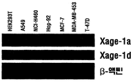
- the XAGE-1d protein (7.6 kD) showed similar expression in three lung and breast cancer cell lines except HEK293T kidney cells, whereas XAGE-1a (9 kD) was a lung cancer cell line NCI-H460 and breast cancer. Some less expression was observed in the cell line MCF-7 (FIG. 1).
- a peptide (CPEAGEEQPQV) corresponding to the carboxyl terminus of XAGE-1a and a biotin-binding biotin-CPEAGEEQPQV (Peptron) were prepared to perform antigen competition ELISA. . After recognizing the peptide as a specific antibody, the amount of XAGE-1a in solution was sought by biotin: streptavidin binding and HRP reaction conjugated to streptavidin.
- biotin-XAGE-1a peptide (biotin-CPEAGEEQPQV) specifically binds to the coated antibody, is also developed by STR-HRP bound to biotin, and the absorbance increases in proportion to the amount of biotin-peptide. It could be confirmed (FIG. 2A).
- Antigen competition enzyme immunoassay was also performed using 4 ng / mL Biotin-XAGE-1a peptide. Instead of adding the biotin-XAGE-1a peptide in the experiment of FIG. 2A, absorbance was obtained by mixing XAGE-1a peptide of various concentrations in 100 ⁇ l of 40 ng / mL biotin-XAGE-1a peptide (biotin-CPEAGEEQPQV). Measured. Standard curves were calculated using B / Bo (B: absorbance value of XAGE-1a peptide added to Biotin-XAGE-1a peptide, Bo: absorbance value of only biotin-XAGE-1a peptide) for the concentration of XAGE-1a peptide.
- the XAGE-1d peptide was found to be biotin- While XAGE-1a peptides did not inhibit binding to antibodies at all, XAGE-1a peptides inhibited the binding of biotin-XAGE-1a peptides in proportion to concentrations, and at about 10% binding at concentrations of 10 ng / mL Is shown (FIG. 3). The results show that ELISA with XAGE-1a specific antibody and biotin-XAGE-1a peptide will be able to detect XAGE-1a protein very specifically.
- the antibody (ab21198, Abcam) that specifically binds to the carboxyl terminal CPEAGEEQPQV of XAGE-1a was dissolved in coating solution (1.59 g of Na 2 CO 3 , 2.93 g of NaHCO 3 and 2 mL of 10% NaN 3 , pH 9.5). 1 ⁇ g per well was coated on 96-well plates (Nunc # 439454) for 24 hours. To inhibit nonspecific binding, 200 ⁇ l of blocking solution per well (PBS with 0.1% BSA and 0.02% thimerosal) was treated at room temperature for 2 hours.
- the wells were washed 5 times with washing solution (PBS containing 0.05% thimerosal and 0.05% Tween-20) and 100 ⁇ l of biotin-XAGE-1a peptide (40 ng / mL) per well and 100 ⁇ l of the cell line culture solution was added and mixed, followed by reaction at room temperature for 2 hours. After washing again with 200 ⁇ l of washing solution per well, HRP (horse radish peroxidase) bound streptavidin was added at a dilution of 1: 10,000 and bound for 2-3 hours.
- washing solution PBS containing 0.05% thimerosal and 0.05% Tween-20
- TMB substrate solution (GenDEPOT) was added to each well for peroxidase color development, followed by reaction at room temperature, and then 50 ⁇ l of stop solution (GenDEPOT) was added. Stop and absorbance at 465 nm wavelength was measured.
- the absorbance value was substituted into the quantification standard curve equation, and the ratio of the molecular weight was calculated to determine the concentration of the XAGE-1a protein contained in the solution, and then expressed as a relative value of the total protein of the cells.
- L549 cancer cell line A549 slowly secreted XAGE-1a over time, reaching 1 ng / ml ⁇ ⁇ g protein at 72 hours, whereas NCI-H460 cell line contained about 3 ng / mL ⁇ ⁇ g protein at the same time. Observed in the medium. Lung cancer cell line Hop-92 and three other breast cancer cell lines could not observe increased expression of XAGE-1a protein in the medium (FIG. 4).
- CEA carcinoembryionic antigen, Accubind, # 1825-300
- CYFRA 21-1 cytokeratin 19 fragment, Fujirebio, # 211-10) as controls were analyzed according to the manufacturer's assay kit manual.
- XAGE-1a was about four times more present in lung cancer patients (average 11.7 ng / ml) than normal people (average 3.0 ng / ml), which is 1.4-1.5 times greater than CEA and CYFRA 21-1. The value was significantly higher, and maintained at over three times except for the thirties with a small number of samples. The sex ratio of males and females showed similar values, while XAGE-1a showed more than two times higher males than females in normal and patients (Table 2). The results show that XAGE-1a may be a better diagnostic marker than the well-known controls CEA and CYFRA 21-1 (Stieber et al. Cancer, 1993, 72 (3), 707-713).
- Sensitivity and specificity of XAGE-1a were analyzed and compared with controls.
- the maximum value of XAGE-1a was about 6 times the mean value, which is larger than 2 ⁇ 2.6 times of CEA and CYFRA 21-1.
- the minimum value of XAGE-1a in lung cancer patients is very low.
- the average value (3.03 ng / ml) of the normal person is very low, and the average value of lung cancer patients is about 4 times higher than that of the normal person, which is particularly distinguished from the control group.
- Sensitivity is also slightly low, while specificity is very high compared to the control, especially CYFRA 21-1 (41.50 ng / ml), indicating that it may be a useful marker (Table 3).
- XAGE-1a Median and interquartile ranges of XAGE-1a were 3.03 and 0.68 to 3.72 in normal subjects and 11.66 and 2.59 to 16.28 in lung cancer patients.
- CEA was 10.35 and 7.95-12.40 in normal subjects and 16.81 and 13.10-20.83 in lung cancer patients.
- CYFRA21-1 was 11.92 and 7.96 to 13.65 in normal subjects and 17.50 and 12.96 to 21.22 in lung cancer patients (FIG. 5).
- Mann-Whitney U- test, p ⁇ 0.001 Mann-Whitney U- test, p ⁇ 0.001).
- ROC curve analysis was performed based on the analysis results of XAGE-1a.
- the area under the curve (AUC) was 0.787 for XAGE-1a, 0.815 for CEA, and 0.803 for CYFRA 21-1, and XAGE-1a showed similar values as the overall control (FIG. 6).
- ROC curves were analyzed by dividing the samples according to the stage of tumor development.
- XAGE-1a showed higher AUC than controls in IA ⁇ IIIA stages (Table 4). The results indicate that XAGE-1a can be used as a more effective diagnostic marker than the control in the early stages of lung cancer.
Landscapes
- Health & Medical Sciences (AREA)
- Life Sciences & Earth Sciences (AREA)
- Immunology (AREA)
- Engineering & Computer Science (AREA)
- Molecular Biology (AREA)
- Biomedical Technology (AREA)
- Chemical & Material Sciences (AREA)
- Hematology (AREA)
- Urology & Nephrology (AREA)
- Biotechnology (AREA)
- Biochemistry (AREA)
- Cell Biology (AREA)
- Food Science & Technology (AREA)
- Medicinal Chemistry (AREA)
- Physics & Mathematics (AREA)
- Analytical Chemistry (AREA)
- Microbiology (AREA)
- General Health & Medical Sciences (AREA)
- General Physics & Mathematics (AREA)
- Pathology (AREA)
- Peptides Or Proteins (AREA)
- Investigating Or Analysing Biological Materials (AREA)
- Measuring Or Testing Involving Enzymes Or Micro-Organisms (AREA)
Abstract
본 발명은 XAGE-1a 단백질에 특이적으로 결합하는 항체를 포함하는 폐암 조기 진단용 조성물, 폐암 조기 진단용 키트 및 폐암 조기 진단용 마이크로어레이, 상기 XAGE-1a 단백질 특이적 항체를 이용하여 폐암 조기 진단용 혈액 마커 XAGE-1a의 검출 방법, XAGE-1a 단백질에 특이적으로 결합하는 항체를 포함하는 폐암의 예후(prognosis) 예측용 조성물 및 피검체로부터 유래한 혈액시료에서 Xage-1a의 발현량을 측정하여 폐암을 진단하는 방법에 관한 것이다.
Description
본 발명은 폐암 조기 진단용 XAGE-1a 마커 및 이의 용도에 관한 것으로서, 더욱 상세하게는 XAGE-1a 단백질에 특이적으로 결합하는 항체를 포함하는 폐암 조기 진단용 조성물, 폐암 조기 진단용 키트 및 폐암 조기 진단용 마이크로어레이, 상기 XAGE-1a 단백질 특이적 항체를 이용하여 폐암 조기 진단용 혈액 마커 XAGE-1a의 검출 방법, XAGE-1a 단백질에 특이적으로 결합하는 항체를 포함하는 폐암의 예후(prognosis) 예측용 조성물 및 피검체로부터 유래한 혈액시료에서 Xage-1a의 발현량을 측정하여 폐암을 진단하는 방법에 관한 것이다.
2008년에 발표된 한국중앙암등록본부 자료에 의하면 2003~2005년 우리나라 연평균 암 발생 중 폐암은 남녀를 합쳐서 전체 암 발생의 12.1%로 2위를 차지하였으며, 인구 10만명 당 발생률은 33.2건이었다. 남녀의 성비는 3.83:1로 남자에게서 더 많이 발생하였고, 발생 건수로는 남성의 암 중에서 2위를 차지하였고 여성의 암 중에서는 5위를 차지하였다. 남녀를 합쳐서 본 연령대별로는 60대가 34.3%로 가장 많고, 70대가 31.0%, 50대가 14.6%의 순이다. 더욱이 폐암은 흡연 인구의 증가 및 대기오염 등의 원인으로 우리나라에서도 급속히 증가하고 있는 종양이며, 암 종별 사망률은 21.4%로 1위이고, 남녀 사망률은 24.9% 및 15.1%로 각각 1위 및 2위이다.
폐암은 암세포의 성장으로 인한 주변 조직 침범 또는 기도폐쇄 및 림프절 전이 등의 증상을 초래하는 것이 보통이지만, 환자의 10~15% 정도에서는 아무런 증상이 없이도 정기 검진에서 폐암으로 진단된다. 또한, 대부분의 폐암이 진단 당시에는 상당히 진행된 상태가 되므로 완치가 어려운 경우들이 대부분이라는 문제가 있다. 따라서 폐암을 조기에 진단하여 폐암으로 인한 사망률을 줄이는 것이 시급한 문제이다(Wulfkuhle et al., Nat. Rev. Cancer 3, 267-275, 2003).
폐암을 진단하기 위해서 여러 가지 방법이 사용되고 있는데, 현재까지는 종양의 크기를 측정하는 방법, 림프절로의 전이 유무를 조사하는 방법, 폐 종양 조직이나 림프절 등을 생검하여 면역조직화학방법으로 분석하는 방법, 흉부 전산화 단층 촬영 및 기관지 내시경 등을 이용하고 있다(Manser et al., Curr. Opin. Pulm. Med. 10, 266-271, 2004). 그러나, 흉부 단층 촬영의 경우 폐암의 크기가 약 0.1 cm 이상 되어야 측정가능한데, 이 시기는 이미 암이 다른 조직으로 전이되었을 가능성이 높으며, 내시경이 기관지로 삽입되어 폐 내부를 직접 관찰할 수 있는 기관지 내시경 방법은 폐 말단에 있는 종양을 관찰하기 곤란한 한계를 가지고 있다.
따라서, 폐암 마커를 이용하여 암을 진단하기 위한 연구들이 진행되었는데, 다중 표적유전자나 단백질의 발현을 조사함으로써 폐암 진단 마커를 발굴한 연구는 이미 보고되었으며, 마이크로어레이 기술을 이용하여 폐암 마커 유전자를 발굴하고 암을 진단하려는 보고도 있었다(Hibi et al., Am. J. Pathol. 1999, 155: 711-715). 또한, 환자의 혈액에서 AST(aspartate aminotransferase), ALT(alanine transaminase), 총 빌리루빈(total bilirubin) 또는 크레아티닌(creatinine)의 농도를 측정하여 폐암을 진단하려는 시도가 있었다(Nagamine et al., Clin. Chem. 29, 379-381, 1983). 그러나, 이러한 종양 마커들에 대한 연구에도 불구하고 아직까지 제한적으로 사용되고 있을 뿐 공식적으로 권장되고 있는 폐암 혈액마커는 없는 실정이다. 본 발명에서 대조군으로 사용한 CEA(carcinoembryionic antigen)는 결장암의 마커로 알려져 있지만 폐암 및 췌장암에서도 높은 친화도를 보여 간접적인 폐암 표지자로 보고되고 있다(Hansen et al. Human Pathology, 1974, 5(2), 139-147). 또한 CYFRA 21-1(사이토케라틴 19 절편)은 폐암 마커로서 발굴되었으나 최근 유방암에서도 발현이 확인되어 폐암에 대한 특이 마커는 부족한 형편이다(Nakata et al. British Journal of Cancer, 2004, 91, 873-878).
본 발명자들은 폐암 세포에서 발현이 증가하는 단백질들을 발굴하였고, 이 중 XAGE-1d 단백질이 폐암 특이적으로 환자의 혈액에 존재한다는 사실을 새로이 알아내었다. 상기 XAGE-1d의 검출을 통하여 면역화학법으로 폐암을 간편하게 진단할 수 있음을 확인함으로써 PCT를 출원하였다(PCT/KR2010/003710). 본 발명의 XAGE-1a는 원래 XAGE-1b로 알려졌으나 기존의 XAGE-1a(146개 아미노산)가 기록에서 없어지면서 대신 XAGE-1a로 현재 기록되고 있는 단백질이다. 본 발명의 XAGE-1a는 XAGE-1d와는 명확하게 구별되며 또한 폐암환자의 혈액에서 높은 발현을 보여 본 발명을 완성하게 되었다.
한국등록특허 제10-0661931호에는 트랜스페린의 당쇄 변화를 측정하여 암을 진단하는 방법이 개시되어 있으며, 한국등록특허 제10-0931997호에는 KLK-6 유전자를 이용한 간세포암 진단 방법이 개시되어 있다.
본 발명은 상기와 같은 요구에 의해 도출된 것으로서, 본 발명자들은 초기 단계의 폐암 환자의 혈액에서 XAGE-1a 단백질의 농도가 현저하게 증가하는 것을 확인함으로써 본 발명을 완성하게 되었다.
상기 과제를 해결하기 위해, 본 발명은 XAGE-1a 단백질에 특이적으로 결합하는 항체를 포함하는 폐암 조기 진단용 조성물을 제공한다.
또한, 본 발명은 XAGE-1a 단백질에 특이적으로 결합하는 항체를 포함하는 폐암 조기 진단용 키트를 제공한다.
또한, 본 발명은 XAGE-1a 단백질에 특이적으로 결합하는 항체를 포함하는 폐암 조기 진단용 마이크로어레이를 제공한다.
또한, 본 발명은 상기 XAGE-1a 단백질 특이적 항체를 이용하여 폐암 조기 진단용 혈액 마커 XAGE-1a의 검출 방법을 제공한다.
또한, 본 발명은 XAGE-1a 단백질에 특이적으로 결합하는 항체를 포함하는 폐암의 예후(prognosis) 예측용 조성물을 제공한다.
또한, 본 발명은 피검체로부터 유래한 혈액시료에서 Xage-1a의 발현량을 측정하여 폐암을 진단하는 방법을 제공한다.
본 발명의 XAGE-1a 혈액 마커를 이용한 폐암의 조기 진단 방법은 혈액으로부터 샘플을 수집하므로 다른 방법에 비해 실시가 용이하고 통증이 비교적 적다. 또한 초기 단계의 폐암을 특이적으로 진단할 수 있으므로, 폐암에 의한 사망률을 낮추는데 유용할 것으로 기대된다.
도 1은 종양 세포주에서 XAGE-1a 및 XAGE-1d의 세포내 발현을 나타낸 것이다. HEK293T: 신장 세포주(대조군), A549, NCI-H460 및 Hop-92: 폐암 세포주, MCF-7, MDA-MB-453 및 T-47D: 유방암 세포주.
도 2는 비오틴-XAGE-1a 펩티드의 농도에 따른 흡광도(A) 및 XAGE-1a 펩티드의 농도에 따른 항원 경쟁 ELISA에 의한 정량화 표준곡선(B)을 나타낸 것이다. B: 비오틴-XAGE-1a 펩티드에 XAGE-1a 펩티드를 첨가한 흡광도 값, Bo: 비오틴-XAGE-1a 펩티드만의 흡광도 값.
도 3은 XAGE-1a 펩티드의 농도에 따른 비오틴-XAGE-1a 펩티드의 결합을 나타낸 것이다.
도 4는 폐암 및 유방암 세포주의 배지에 분비되는 XAGE-1a의 시간별 농도를 나타낸 것이다.
도 5는 정상인과 환자 혈액에서 XAGE-1a (A)와 대조군인 CEA(B) 및 CYFRA 21-1(C)의 분포를 나타낸 것이다.
도 6은 XAGE-1a(A)와 대조군인 CEA(B) 및 CYFRA 21-1(C)의 ROC 그래프를 나타낸 것이다.
본 발명의 목적을 달성하기 위하여, 본 발명은 XAGE-1a 단백질에 특이적으로 결합하는 항체를 포함하는 폐암 조기 진단용 조성물을 제공한다.
본 발명에서, 용어 "진단"은 병리 상태의 존재 또는 특징을 확인하는 것을 의미한다. 본 발명의 목적상, 진단은 폐암 발병 여부를 확인하는 것이다.
본 발명의 폐암 조기 진단 마커 XAGE-1a 단백질은 폐암의 초기 단계에서 다른 폐암 진단 마커인 CEA 및 CYFRA 21-1보다 높게 검출되었으므로, 폐암의 조기 진단에 더욱 효과적일 수 있다.
바람직하게는 상기 XAGE-1a 단백질은 XAGE-1a 카르복실 말단일 수 있으나, 이에 제한되지 않는다. 상기 XAGE-1a 카르복실 말단은 바람직하게는 서열번호 1의 아미노산 서열(CATWKVIC KSCISQTPGI NLDLGSGVKV KIIPKEEHCK MPEAGEEQPQ V)로 이루어질 수 있으나, 이에 제한되지는 않는다. 또한, 상기 서열의 변이체가 본 발명의 범위 내에 포함된다. 변이체는 아미노산 서열은 변화되지만, 서열번호 1의 아미노산 서열과 유사한 기능적 특성을 갖는 서열이다. 구체적으로, XAGE-1a 카르복실 말단은 서열번호 1의 아미노산 서열과 70% 이상, 더욱 바람직하게는 80% 이상, 더 더욱 바람직하게는 90% 이상, 가장 바람직하게는 95% 이상의 서열 상동성을 가지는 아미노산 서열을 포함할 수 있다.
상기 XAGE-1a 단백질에 특이적으로 결합하는 항체는 XAGE-1a만을 특이적으로 인지할 수 있다. 상기 항체는 바람직하게는 검출 가능한 신호를 생성할 수 있는 검출 표지제가 결합될 수 있으며, 상기 검출 표지제는 효소, 보결분자족, 형광물질, 발광물질, 생물 발광물질 또는 방사성 물질일 수 있으나, 이에 제한되지는 않는다.
상기 폐암은 폐에 발생하는 악성 종양을 의미하며, 바람직하게는 비소세포폐암(non-small cell lung cancer)일 수 있으나, 이에 제한되지는 않는다. 바람직하게는, 상기 조성물은 XAGE-1a 단백질에 특이적으로 결합하는 항체를 인지하는 2차 항체를 추가로 포함할 수 있으며, 검출 표지제가 2차 항체에 결합될 수 있다. 바람직하게는, 상기 조성물은 검출 표지제가 효소인 경우에 효소활성을 측정할 수 있는 기질 및 반응 정지제를 추가로 포함할 수 있다.
본 발명에 있어서, 검출 표지제로 적합한 효소의 예는 서양고추냉이 퍼옥시다제, 아세틸콜린 에스터라제, 퍼옥시다아제, 알칼라인 포스파타아제, β-D-갈락토시다아제, 글루코스 옥시다아제, 말레이트 디하이드로게나아제, 글루코스-6-인산 디하이드로게나아제 또는 인버타제 등을 포함하고; 적합한 보결분자족 복합체의 예는 스트렙트아비딘/비오틴 또는 아비딘/비오틴을 포함하고; 적합한 형광물질의 예는 움벨리페론, 플루오레세인, 플루오레세인 이소티오시아네이트, 로다민, 디클로로트리아지닐아민 플루오레세인, 댄실 클로라이드, 피코에리트린 또는 피코빌리프로테인 등을 포함하고; 발광물질의 예는 루미놀, 이소루시놀 또는 루시제닌 등을 포함하고; 생물 발광물질의 예는 루시퍼라제, 루시페린 또는 아에쿠오린 등을 포함하며; 적합한 방사선 물질의 예는 125I, 131I, 111In, 99Tc 14C 또는 3H 등을 포함할 수 있으나, 이에 제한되지는 않는다.
상기 용어, "항체"는 당업계에 공지된 용어이며 공지된 항원에 결합하는 단백질 분자를 의미한다. 본 발명에 사용되는 항체는 단클론 또는 다클론 항체, 면역학적으로 활성인 단편(예를 들어, Fab 또는 (Fab)2 단편), 항체 중쇄, 인간화 항체, 항체 경쇄, 유전자 조작된 단일쇄 Fv 분자, 키메릭 항체 등일 수 있으나, 이에 제한되지는 않는다.
본 발명에 사용되는 항체는 면역원(항원)으로 면역시킨 포유동물의 혈청에서 분리하여 얻을 수 있다. 상기 포유동물은 바람직하게는 마우스, 토끼 또는 염소일 수 있으나, 이에 제한되지는 않는다. 일반적으로, 항체의 제조에 이용되는 포유동물은 더 많은 항체를 분리할 수 있는 더 많은 양의 혈청을 가지는 비교적 큰 포유동물이 우선적으로 선택될 수 있다. 상기 포유동물에 면역원이 주입되면, 면역계의 B 세포를 자극하여 면역원에 대해 특이적인 면역글로불린(Ig, immunoglobulin)이 생산된다. 상기 면역글로불린은 여과, 투석, 이온 교환 크로마토그래피, 크기배제 크로마토그래피 또는 친화성 크로마토그래피와 같은 당업계에 공지된 방법을 이용하여 혈청으로부터 정제될 수 있다. 상기 항체의 제조에 있어서, 주사되는 면역원에 대한 포유동물의 면역 반응을 증강시킬 목적으로 Freund's, Alum, the Ribi Adjuvant System 또는 Titermax와 같은 면역 보강제(adjuvant)가 주입될 수 있다. 상기 면역 보강제는 항원과 혼합하였을 때 항원성을 높여 항체가 많이 생성되도록 도와주는 역할을 한다.
또한, 본 발명은 XAGE-1a 단백질에 특이적으로 결합하는 항체를 포함하는 폐암 조기 진단용 키트를 제공한다. 바람직하게는 상기 XAGE-1a 단백질은 XAGE-1a 카르복실 말단일 수 있으나, 이에 제한되지는 않는다. 상기 키트는 XAGE-1a 단백질에 특이적으로 결합하는 항체를 인지하는 2차 항체를 추가로 포함할 수 있으며, 검출 표지제가 2차 항체에 결합될 수 있다. 상기 키트는 본 발명에 따른 XAGE-1a 단백질에 특이적으로 결합하는 항체, 적합한 담체 용액, 검출 가능한 신호를 생성할 수 있는 검출 표지제, 용해제, 세정제를 포함하는 용기 및 사용설명서를 포함할 수 있다. 또한, 상기 키트는 검출 표지제가 효소인 경우에 효소활성을 측정할 수 있는 기질 및 반응 정지제를 포함할 수 있다.
또한, 본 발명은 XAGE-1a 단백질에 특이적으로 결합하는 항체를 포함하는 폐암 조기 진단용 마이크로어레이를 제공한다. 바람직하게는 상기 XAGE-1a 단백질은 XAGE-1a 카르복실 말단일 수 있으나, 이에 제한되지는 않는다. 상기 마이크로어레이는 특정 시약으로 처리된 슬라이드 글라스 표면 위에 XAGE-1a 단백질에 특이적으로 결합하는 항체를 부착하여 항원-항체 반응에 의해 상기 항체에 특이적으로 부착하는 단백질을 검출할 수 있다.
또한, 본 발명은
환자로부터 혈액을 수집하는 단계;
상기 혈액과 XAGE-1a 단백질 특이적 항체를 반응시키는 단계; 및
상기 항원-항체 복합체를 검출하는 단계를 포함하는 폐암 조기 진단용 혈액 마커 XAGE-1a의 검출 방법을 제공한다.
바람직하게는 상기 XAGE-1a 단백질은 XAGE-1a 카르복실 말단일 수 있으나, 이에 제한되지는 않는다. 바람직하게는, 상기 XAGE-1a 카르복실 말단은 서열번호 1의 아미노산 서열로 이루어질 수 있으나, 이에 제한되지는 않는다. 상기 항원-항체 복합체를 검출하는 방법은 면역블롯법, 효소면역법, 면역침강법, 형광면역법, 효소기질발색법 또는 항원항체 응집법일 수 있으나, 이에 제한되지는 않는다.
본 발명에 있어서, 항원을 검출하고/하거나 정량하는데 사용되는 수많은 면역분석법들이 당업자에게 공지되어 있다.
또한, 본 발명은 XAGE-1a 단백질에 특이적으로 결합하는 항체를 포함하는 폐암의 예후(prognosis) 예측용 조성물을 제공한다. 본 발명의 XAGE-1a 단백질에 특이적으로 결합하는 항체는 폐암의 조기 진단뿐만 아니라, 폐암 환자의 예후 예측에도 이용될 수 있다.
또한, 본 발명은
a) 피검체로부터 유래한 혈액시료에서 Xage-1a의 발현량을 측정하는 단계;
b) 단계 a)의 Xage-1a의 발현량과 정상 혈액시료의 Xage-1a의 발현량을 비교하는 단계; 및
c) 피검체 유래의 Xage-1a 발현량이 정상 대조군에 비해 증가하는 경우 폐암에 걸릴 위험이 높은 것으로 판정하는 단계를 포함하는 폐암을 진단하는 방법을 제공한다.
상기 방법에 있어서, 단계 a)의 피검체는 인간을 포함한 척추동물이고, 바람직하게는 포유동물이며, 보다 바람직하게는 인간, 유인원류, 소, 돼지, 쥐, 토끼, 기니아피크, 햄스터, 개 또는 고양이를 사용할 수 있으나, 이에 한정되지 않는다.
상기 방법에 있어서, Xage-1a의 발현량은 웨스턴블랏(Western blotting), 효소-면역화학 검출법(Enzyme-linked immunosorbent assay, ELISA), 면역조직화학 염색법(immunohistochemical staining), 면역침강(immunoprecipitation) 및 면역형광법(immunofluorescence)으로 구성된 군으로부터 선택된 어느 하나로 측정하는 것이 바람직하나, 이에 한정되지 않는다.
이하, 본 발명을 실시예에 의해 상세히 설명한다. 단, 하기 실시예는 본 발명을 예시하는 것일 뿐, 본 발명의 내용이 하기 실시예에 한정되는 것은 아니다.
실시예 1: XAGE-1a 단백질의 구조
XAGE1은 처음에 a, b, c 및 d의 4종류의 전사 변이체로 알려졌으나, 최근에는 81개의 아미노산 및 69개의 아미노산을 갖는 2개의 전사 변이체만이 확인되고 있다(http://www.ncbi.nlm.nih.gov/gene/653219?). 본 발명은 81개 아미노산으로 구성된 전사 변이체 XAGE-1a에 관한 것으로, 69개 아미노산으로 구성된 XAGE-1d와는 아미노 말단의 32개 아미노산 서열은 동일하나 나머지 카르복실 말단의 서열이 서로 다르다(표 1).
표 1 XAGE1 전사 변이체의 단백질 서열
| 단백질명 | Ref_seq No. | 아미노산 서열 | 아미노산 수 |
| XAGE-1a | NP_001091061 | MESPKKKNQQ LKVGILHLGS RQKKIRIQLR SQCATWKVIC KSCISQTPGI NLDLGSGVKV KIIPKEEHCK MPEAGEEQPQ V (서열번호 2) | 81개 |
| XAGE-1d | NP_001091062 | MESPKKKNQQ LKVGILHLGS RQKKIRIQLR SQVLGREMRD MEGDLQELHQ SNTGDKSGFG FRRQGEDNT (서열번호 3) | 69개 |
* 상기 두 단백질은 밑줄친 아미노산을 아미노 말단에 공통으로 포함하고 있다.
실시예 2: XAGE-1a의 세포내 발현 확인
세포내 XAGE-1a의 발현을 관찰하기 위하여 세포주를 5×10⁵ 세포가 되도록 100 mm 배양 접시에 각각 접종하여 2일 동안 배양한 후, 트립신을 처리하여 세포를 회수하였다. 상기 회수한 세포를 RIPA 완충액으로 용균한 후, 10 ㎍의 단백질을 15% 폴리아크릴아마이드 겔에서 전기영동하고 니트로셀룰로즈 막으로 전이시켰다. XAGE-1a의 카르복실 말단(CPEAGEEQPQV)에 특이적인 염소항체(Abcam사, ab21198)를 막에 가하여 반응시키고, 다시 HRP (horseradish peroxidase)가 결합된 항-염소 생쥐항체 및 ECL을 이용하여 XAGE-1a 단백질을 검출하였다.
XAGE-1a의 발현을 XAGE-1d과 비교하기 위하여, XAGE-1d의 발현을 관찰하였는데, 상기 실험에는 XAGE-1d의 카르복실 말단(GFGFRRQGEDNT)에 특이적인 염소항체(Abcam사, ab27477)를 사용하였다. 상기 XAGE-1d 단백질(7.6 kD)은 HEK293T 신장세포를 제외한 3종의 폐암 및 유방암 세포주의 세포 내에서 유사한 정도의 발현을 나타낸 반면, XAGE-1a(9 kD)는 폐암 세포주인 NCI-H460 및 유방암 세포주인 MCF-7에서 발현이 다소 적게 관찰되었다(도 1).
실시예 3: 항원 경쟁 효소면역 측정법(antigen competition ELISA)를 이용한 표준곡선(standard curve)의 제작
XAGE-1a의 양을 정량적으로 표시하기 위하여 XAGE-1a의 카르복실 말단에 해당하는 펩티드(CPEAGEEQPQV) 및 상기 펩티드에 비오틴이 결합된 비오틴-CPEAGEEQPQV를 제조(Peptron사)하여, 항원 경쟁 ELISA를 수행하였다. 상기 펩티드를 특이적 항체로 인식한 후, 비오틴:스트렙트아비딘(streptavidin) 결합과 스트렙트아비딘에 컨쥬게이션(conjugation)된 HRP 반응에 의해 용액 내 XAGE-1a의 양을 검출하고자 하였다.
먼저, XAGE-1a를 정량화하기 위한 표준곡선을 그리기 위하여, 비오틴-XAGE-1a 펩티드(biotin-CPEAGEEQPQV)의 농도별 흡광도를 측정하였다. 상기 XAGE-1a의 카르복실 말단에 특이적으로 결합하는 항체(ab21998)를 코팅 용액(1L 당 Na2CO3 1.59 g, NaHCO3 2.93 g 및 10% NaN3 2 mL, pH 9.5)에 녹인 다음, 96-웰 플레이트(Nunc #439454)에 웰 당 1 ㎍으로 24시간 동안 처리하였다. 상기 코팅 후, 웰 당 200 ㎕의 차단(blocking) 용액(0.1% BSA (Bovine serum albumin) 및 0.02% 티메로살(thimerosal)이 포함된 PBS)을 상온에서 2시간 동안 처리하여 비특이적 결합을 억제하였다. 그 후, 세척 용액(0.02% 티메로살 및 0.05% 트윈-20(Tween-20)이 포함된 PBS)으로 코팅되지 않은 항체나 차단 용액을 세척하였다. 비오틴-XAGE-1a 펩티드(biotin-CPEAGEEQPQV)를 PBS에 농도별로 녹인 다음, 상기 플레이트에 100 ㎕씩 첨가하고 2~3시간 동안 상온에서 반응시켰다. 세척 용액으로 상기 플레이트를 5회 세척한 후, 각 웰에 비오틴에 특이적인 스트렙트아비딘(STR)에 HRP가 결합된 STR-HRP(Pierce사)를 1:10,000으로 희석하여 첨가하고, 2~3시간 동안 반응시킨 후 다시 웰을 세척 용액으로 세척하였다. 그 후, 퍼옥시다제 발색을 위하여 각 웰에 50 ㎕의 TMB(tetramethylbenzidine) 기질용액(GenDEPOT)을 첨가하여 상온에서 반응시킨 후, 다시 50 ㎕의 정지(stop)용액(GenDEPOT)를 첨가하여 반응을 정지시키고, 465 nm 파장에서 흡광도를 측정하였다. 그 결과, 비오틴-XAGE-1a 펩티드(biotin-CPEAGEEQPQV)가 코팅된 항체에 특이적으로 결합하며, 또한 비오틴에 결합한 STR-HRP에 의해 발색되고, 비오틴-펩티드의 양에 비례하여 흡광도가 증가함을 확인할 수 있었다(도 2A).
또한 4 ng/mL의 비오틴-XAGE-1a 펩티드를 사용하여 항원 경쟁 효소면역 측정을 수행하였다. 상기 도 2A 실험에서 비오틴-XAGE-1a 펩티드를 첨가하는 과정 대신, 40 ng/mL 비오틴-XAGE-1a 펩티드(biotin-CPEAGEEQPQV) 100 ㎕에 여러 농도의 XAGE-1a 펩티드를 혼합하여 반응시킨 후 흡광도를 측정하였다. XAGE-1a 펩티드의 농도에 대한 B/Bo (B: 비오틴-XAGE-1a 펩티드에 XAGE-1a 펩티드를 첨가한 흡광도 값, Bo: 비오틴-XAGE-1a 펩티드만의 흡광도 값)를 이용하여 표준곡선을 그리고 선형 회귀(linear regression)한 표준화 식(R2=0.9971)으로 흡광도에 대한 펩티드의 농도를 환산하였다(도 2B). 분자량의 비(4.067=(9,077: XAGE-1a 단백질)/(2,231: XAGE-1a 펩티드))를 농도에 환산하면 용액 속에 존재하는 XAGE-1a 단백질의 농도를 구할 수 있다.
실시예 4: XAGE-1a의 특이적 검출을 위한 ELISA
아미노 말단의 32개 아미노산이 동일하고 카르복실 말단의 45~49개 아미노산이 상이한 XAGE-1a를 XAGE-1d와 구별하여 선택적으로 검출할 수 있는지를 확인하기 위하여, XAGE-1a를 인식하는 항체(ab21198)를 ELISA 플레이트에 웰 당 1 ㎍씩 코팅한 후, 40 ng/mL의 비오틴-XAGE-1a 펩티드(biotin-CPEAGEEQPQV) 100 ㎕에 XAGE-1a 펩티드 및 XAGE-1d 펩티드를 농도별로 섞은 혼합용액을 첨가하였다. 상온에서 2시간 동안 결합시킨 후 STR-HRP를 사용하여 흡광도를 비교하였고, XAGE-1a 특이적 항체에 대한 XAGE-1a 펩티드의 결합 및 XAGE-1d의 비특이적 결합을 관찰하였다. XAGE-1a 또는 XAGE-1d 펩티드가 첨가되지 않고 비오틴-XAGE-1a 펩티드만을 결합시킨 경우를 기준(100%)으로 하여 첨가한 펩티드의 농도에 대한 흡광도를 관찰한 결과, XAGE-1d 펩티드는 비오틴-XAGE-1a 펩티드의 항체에 대한 결합을 전혀 억제하지 못한 반면, XAGE-1a 펩티드는 비오틴-XAGE-1a 펩티드의 결합을 농도에 비례하여 억제하였고, 10 ng/mL의 농도에서는 약 10% 정도의 결합을 나타내었다(도 3). 상기 결과는 XAGE-1a 특이적인 항체 및 비오틴-XAGE-1a 펩티드를 이용한 ELISA가 매우 특이적으로 XAGE-1a 단백질을 검출할 수 있을 것임을 보여준다.
실시예 5: 폐암 세포주 배양액에서 XAGE-1a의 검출
폐암환자의 혈액에서 검출할 수 있는 단백질인 경우 폐암 세포주의 배양액에서도 존재할 가능성을 갖는다. 따라서, A549, NCI-H460 및 HOP-92 등의 폐암 세포주를 배양한 후, 상기 세포 배양액 100 ㎕를 취하여 XAGE-1a의 존재를 확인하였다. 또한 XAGE-1a의 발현이 유방암 세포에서 높다고 알려져 있고(Egland et al. Molecular Cancer Therapeutics, 2002, 1, 441-450), 폐암 특이 혈액 마커가 부재한 현실에서 폐암 진단에 간접적으로 활용되고 있는 대조군 CEA(carcinoembryionic antigen) 및 CYFRA 21-1(사이토케라틴 19 절편)이 유방암 환자의 혈청에서 관찰된다는 보고가 있기 때문에, 유방암 세포주를 이용하여 XAGE-1a의 분비를 세포 배양액에서 관찰하였다(Nakata et al. British Journal of Cancer, 2004, 91, 873-878). 세포주 배양액을 얻기 위하여 5×10⁵ 세포를 100 mm 배양 용기에 접종한 후, 배양액 10 mL를 첨가하여 배양하면서 배양액을 12시간 간격으로 72시간까지 100 ㎕씩 샘플링하여 4℃에 보관하였다.
상기 목적을 달성하기 위하여, 정량화 항원 경쟁 ELISA를 수행하였다. XAGE-1a의 카르복실 말단 CPEAGEEQPQV에 특이적으로 결합하는 항체(ab21198, Abcam)를 코팅용액(1L 당 Na2CO3 1.59 g, NaHCO3 2.93 g 및 10% NaN3 2 mL, pH 9.5)에 녹여 웰 당 1 ㎍씩 96-웰 플레이트(Nunc #439454)에 24시간 동안 처리하여 코팅하였다. 비특이적 결합을 억제하기 위하여, 웰 당 200 ㎕의 차단용액(0.1% BSA 및 0.02% 티메로살이 포함된 PBS)을 상온에서 2시간 동안 처리하였다. 상기 차단용액을 버린 후, 웰을 세척용액(0.02% 티메로살 및 0.05% 트윈-20이 포함된 PBS)으로 5번 세척하고 웰 당 비오틴-XAGE-1a 펩티드(40 ng/mL) 100 ㎕ 및 세포주 배양액 100 ㎕를 혼합하여 첨가한 후 상온에서 2시간 동안 반응시켰다. 그 후, 웰 당 200 ㎕의 세척용액으로 다시 세척한 다음, HRP (horse radish peroxidase)가 결합된 스트렙트아비딘을 1:10,000으로 희석하여 첨가하고 2~3시간 동안 결합시켰다. 다시 웰을 세척용액으로 세척한 후, 퍼옥시다제 발색을 위하여 각 웰에 50 ㎕의 TMB 기질용액(GenDEPOT)을 첨가하여 상온에서 반응시킨 다음, 50 ㎕의 정지용액(GenDEPOT)을 첨가하여 반응을 정지시키고 465 nm 파장에서 흡광도를 측정하였다.
상기 실시예 3에서 기재한 것처럼 흡광도의 값을 정량화 표준곡선 식에 대입하고 분자량의 비를 환산하여 용액에 포함된 XAGE-1a 단백질의 농도를 구한 다음, 세포의 전체 단백질에 대한 상대적인 값으로 나타내었다. 폐암 세포주 A549는 시간에 따라 완만하게 XAGE-1a의 분비가 일어나 72시간에는 1 ng/ml·㎍ protein에 도달한 반면, NCI-H460 세포주는 동일한 시간대에 약 3 ng/mL·㎍ protein의 단백질이 배지에서 관찰되었다. 폐암 세포주 Hop-92 및 다른 3개의 유방암 세포주는 배지에서 XAGE-1a 단백질의 발현 증가를 관찰할 수 없었다(도 4).
실시예 6: 폐암 환자의 혈액에서 XAGE-1a의 검출
XAGE-1a가 혈청에서 검출되는지를 조사하기 위하여 정상 및 폐암 환자의 혈청을 각각 194개씩을 확보하고, 상기 실시예 5의 방법에서 세포 배양액 대신에 혈청 100 ㎕를 첨가하여 분석하였다. 대조군으로서 CEA(carcinoembryionic antigen, Accubind, #1825-300) 및 CYFRA 21-1(사이토케라틴 19 절편, Fujirebio, #211-10)을 제조사의 분석 키트 매뉴얼에 따라 분석하였다.
그 결과, XAGE-1a는 정상인(평균 3.0 ng/ml)에 비해 폐암 환자(평균 11.7 ng/ml)에서 약 4배 정도 더 많은 양이 존재하였는데, 이는 CEA 및 CYFRA 21-1의 1.4~1.5배 보다 현저하게 더 높은 비율이며, 샘플의 수가 적은 30대를 제외하면 3배 이상의 값을 유지하였다. 남녀 성비로는 대조군이 유사한 값을 나타낸 반면, XAGE-1a는 정상 및 환자에서 남성이 여성에 비해 2배 이상 높은 수치를 나타내었다(표 2). 상기 결과는 XAGE-1a가 잘 알려진 대조군 CEA 및 CYFRA 21-1보다 더 좋은 진단 마커가 될 수 있음을 보여 준다(Stieber et al. Cancer, 1993, 72(3), 707-713).
표 2 나이 및 성별에 따른 XAGE-1a의 혈청내 농도
| 인원 | XAGE-1a (ng/ml) | CEA (ng/ml) | CYFRA 21-1 (ng/ml) | ||||||||||||||
| 정상 | 환자 | 정상 | 환자 | 정상 | 환자 | 정상 | 환자 | ||||||||||
| 31~40세 | 남 | 1 | 6 | 1 | 6 | 0.6 | 3.3 | 18.3 | 7.7 | 13.9 | 8.8 | 20.3 | 20.3 | 8.9 | 9.8 | 20.3 | 18.6 |
| 여 | 5 | 5 | 3.9 | 5.6 | 8.2 | 21.0 | 9.3 | 18.3 | |||||||||
| 41~50세 | 남 | 9 | 27 | 10 | 27 | 1.6 | 1.2 | 6.8 | 7.7 | 11.3 | 8.2 | 14.9 | 16.6 | 12.1 | 9.7 | 18.3 | 17.9 |
| 여 | 18 | 17 | 1.0 | 8.3 | 6.4 | 17.5 | 7.5 | 16.8 | |||||||||
| 51~60세 | 남 | 28 | 45 | 27 | 45 | 3.5 | 2.8 | 12.3 | 8.6 | 11.2 | 11.6 | 14.6 | 13.8 | 11.1 | 12.5 | 16.6 | 16.8 |
| 여 | 17 | 18 | 1.8 | 3.1 | 12.3 | 13.9 | 12.9 | 17.3 | |||||||||
| 61~70세 | 남 | 88 | 116 | 88 | 116 | 3.9 | 3.5 | 16.0 | 14.0 | 10.3 | 10.1 | 17.2 | 17.7 | 11.0 | 11.7 | 17.2 | 17.9 |
| 여 | 28 | 28 | 2.2 | 7.7 | 10.8 | 19.0 | 11.6 | 19.2 | |||||||||
| 총(평균) | 남 | 126 | 126 | 3.6 | 14.5 | 10.6 | 16.5 | 11.1 | 17.2 | ||||||||
| 여 | 68 | 68 | 1.9 | 6.4 | 9.8 | 17.4 | 10.7 | 18.0 | |||||||||
| 총 | 194 | 194 | 3.0 | 11.7 | 10.2 | 16.7 | 11.6 | 17.7 | |||||||||
실시예 7: 폐암 혈액 마커 XAGE-1a의 민감도 및 특이도 분석
XAGE-1a의 민감도(sensitivity) 및 특이도(specificity)를 분석하여 대조군과 비교하였다. 정상인에서 XAGE-1a의 최대값은 평균값의 6배 정도로 나타났으며, 이는 CEA 및 CYFRA 21-1의 2~2.6배에 비해 큰 수치이다. 또한, 폐암 환자에서 XAGE-1a의 최소값도 매우 낮다. 그럼에도 불구하고 정상인의 평균값(3.03 ng/ml)이 매우 낮고, 폐암 환자의 평균값이 정상인에 비해 4배 정도 높은 것은 대조군과 특별히 구별되는 점이다. 또한 민감도는 약간 낮은 반면, 특이도는 대조군, 특히 CYFRA 21-1(41.50 ng/ml)에 비해 매우 높은 것은 유용한 마커가 될 수 있음을 나타낸다(표 3).
표 3 XAGE-1a의 민감도 및 특이도
| XAGE-1a | CEA | CYFRA 21-1 | ||||
| 정상 | 환자 | 정상 | 환자 | 정상 | 환자 | |
| 194 | 194 | 194 | 194 | 194 | 194 | |
| 최대량(ng/㎖) | 18.57 | 65.18 | 20.30 | 34.86 | 29.23 | 41.50 |
| 최소량(ng/㎖) | 0.06 | 0.13 | 1.44 | 2.57 | 0.47 | 5.01 |
| 평균(ng/㎖) | 3.03 | 11.66 | 10.35 | 16.81 | 10.92 | 17.50 |
| ≤ 정상평균 | 133 | 57 | 96 | 30 | 82 | 30 |
| > 정상평균 | 61 | 137 | 98 | 164 | 112 | 164 |
| 민감도(%) | (137/194)×100=70.62 | (163/194)×100=84.54 | (164/194)×100=84.54 | |||
| 특이도(%) | (133/194)×100=68.56 | (104/194)×100=49.48 | (82/194)×100=42.27 | |||
실시예 8: 혈청에서의 XAGE-1a 분포
XAGE-1a의 중간값 및 사분범위(median and interquartile range)는 정상인에서 3.03 및 0.68~3.72, 폐암 환자에서 11.66 및 2.59~16.28이었다. CEA의 경우는 정상인에서 10.35 및 7.95~12.40이었고, 폐암 환자에서는 16.81 및 13.10~20.83이었다. CYFRA21-1은 정상인에서 11.92 및 7.96~13.65이었고, 폐암 환자에서는 17.50 및 12.96~21.22이었다(도 5). 따라서, 상기 분석한 3개의 마커는 정상 및 폐암 환자에서 현저한 차이를 나타낸다는 것을 확인할 수 있었다(Mann-Whitney U-test, p<0.001).
실시예 9: ROC(receiver operating characteristics) 곡선 분석
상기 XAGE-1a의 분석 결과를 바탕으로 하여 ROC 곡선 분석을 수행하였다. AUC(area under the curve)는 XAGE-1a가 0.787, CEA가 0.815, 그리고 CYFRA 21-1이 0.803으로, XAGE-1a는 전반적으로 대조군과 유사한 값을 나타내었다(도 6).
종양의 발생 단계에 따라 샘플을 구분하여 ROC 곡선을 분석하였다. IA~IIIA 단계 및 IIIB~IV 단계를 구분하여 분석한 결과, XAGE-1a는 IA~IIIA 단계에서 대조군보다 높은 AUC를 나타내었다(표 4). 상기 결과는 XAGE-1a가 폐암의 초기 단계에서 대조군보다 효과적인 진단 마커로 사용될 수 있음을 나타낸다.
표 4 폐암의 발생 단계에 따른 ROC 곡선의 AUC
| 단계 | 환자수 | 정상인수 | XAGE-1a | CEA | CYFRA 21-1 |
| IA~IIIA | 84 | 194 | 0.815 | 0.799 | 0.783 |
| IIIB~IV | 110 | 194 | 0.739 | 0.822 | 0.821 |
Claims (14)
- XAGE-1a 단백질에 특이적으로 결합하는 항체를 포함하는 폐암 조기 진단용 조성물.
- 제1항에 있어서, 상기 XAGE-1a 단백질은 XAGE-1a 카르복실 말단인 것을 특징으로 하는 폐암 조기 진단용 조성물.
- 제2항에 있어서, 상기 XAGE-1a 카르복실 말단은 서열번호 1의 아미노산 서열로 이루어지는 것을 특징으로 하는 폐암 조기 진단용 조성물.
- 제1항에 있어서, 상기 조성물은 XAGE-1a 단백질에 특이적으로 결합하는 항체를 인지하는 2차 항체를 추가로 포함하며, 검출 표지제가 2차 항체에 결합됨을 특징으로 하는 폐암 조기 진단용 조성물.
- 제4항에 있어서, 상기 검출 표지제가 효소이고, 효소활성을 측정할 수 있는 기질 및 반응 정지제를 추가로 포함함을 특징으로 하는 폐암 조기 진단용 조성물.
- XAGE-1a 단백질에 특이적으로 결합하는 항체를 포함하는 폐암 조기 진단용 키트.
- 제6항에 있어서, 상기 XAGE-1a 단백질은 XAGE-1a 카르복실 말단인 것을 특징으로 하는 폐암 조기 진단용 키트.
- XAGE-1a 단백질에 특이적으로 결합하는 항체를 포함하는 폐암 조기 진단용 마이크로어레이.
- 제8항에 있어서, 상기 XAGE-1a 단백질은 XAGE-1a 카르복실 말단인 것을 특징으로 하는 폐암 조기 진단용 마이크로어레이.
- 환자로부터 혈액을 수집하는 단계;상기 혈액과 XAGE-1a 단백질 특이적 항체를 반응시키는 단계; 및상기 항원-항체 복합체를 검출하는 단계를 포함하는 폐암 조기 진단용 혈액 마커 XAGE-1a의 검출 방법.
- 제10항에 있어서, 상기 XAGE-1a 단백질은 XAGE-1a 카르복실 말단인 것을 특징으로 하는 방법.
- 제11항에 있어서, 상기 XAGE-1a 카르복실 말단은 서열번호 1의 아미노산 서열로 이루어지는 것을 특징으로 하는 방법.
- XAGE-1a 단백질에 특이적으로 결합하는 항체를 포함하는 폐암의 예후(prognosis) 예측용 조성물.
- a) 피검체로부터 유래한 혈액시료에서 Xage-1a의 발현량을 측정하는 단계;b) 단계 a)의 Xage-1a의 발현량과 정상 혈액시료의 Xage-1a의 발현량을 비교하는 단계; 및c) 피검체 유래의 Xage-1a 발현량이 정상 대조군에 비해 증가하는 경우 폐암에 걸릴 위험이 높은 것으로 판정하는 단계를 포함하는 폐암을 진단하는 방법.
Applications Claiming Priority (2)
| Application Number | Priority Date | Filing Date | Title |
|---|---|---|---|
| KR10-2011-0033078 | 2011-04-11 | ||
| KR1020110033078A KR20120116518A (ko) | 2011-04-11 | 2011-04-11 | 폐암 조기 진단용 XAGE-1a 마커 및 이의 용도 |
Publications (1)
| Publication Number | Publication Date |
|---|---|
| WO2012141373A1 true WO2012141373A1 (ko) | 2012-10-18 |
Family
ID=47009516
Family Applications (1)
| Application Number | Title | Priority Date | Filing Date |
|---|---|---|---|
| PCT/KR2011/003309 Ceased WO2012141373A1 (ko) | 2011-04-11 | 2011-05-03 | 폐암 조기 진단용 XAGE-1a 마커 및 이의 용도 |
Country Status (2)
| Country | Link |
|---|---|
| KR (1) | KR20120116518A (ko) |
| WO (1) | WO2012141373A1 (ko) |
Cited By (2)
| Publication number | Priority date | Publication date | Assignee | Title |
|---|---|---|---|---|
| CN113687076A (zh) * | 2021-07-14 | 2021-11-23 | 郑州大学 | 一种可用于肺腺癌早期诊断的联合检测血清标志物及其应用 |
| CN116482365A (zh) * | 2023-03-14 | 2023-07-25 | 南京芯原生物科技有限公司 | 用于检测血清抗体的蛋白组合物及其应用 |
Families Citing this family (2)
| Publication number | Priority date | Publication date | Assignee | Title |
|---|---|---|---|---|
| WO2022169267A1 (ko) * | 2021-02-02 | 2022-08-11 | 제노마인(주) | 폐암 혈액 바이오마커의 검출 방법 및 키트 |
| KR102613772B1 (ko) * | 2021-02-02 | 2023-12-14 | 제노마인(주) | 폐암 혈액 바이오마커의 검출 방법 및 키트 |
Citations (2)
| Publication number | Priority date | Publication date | Assignee | Title |
|---|---|---|---|---|
| US20060159694A1 (en) * | 2004-12-29 | 2006-07-20 | Chih-Sheng Chiang | Combinations of tumor-associated antigens in compositions for various types of cancers |
| US20070202553A1 (en) * | 2004-05-20 | 2007-08-30 | Gang Zeng | Dominant B Cell Epitopes and Methods of Making and Using Thereof |
-
2011
- 2011-04-11 KR KR1020110033078A patent/KR20120116518A/ko not_active Ceased
- 2011-05-03 WO PCT/KR2011/003309 patent/WO2012141373A1/ko not_active Ceased
Patent Citations (2)
| Publication number | Priority date | Publication date | Assignee | Title |
|---|---|---|---|---|
| US20070202553A1 (en) * | 2004-05-20 | 2007-08-30 | Gang Zeng | Dominant B Cell Epitopes and Methods of Making and Using Thereof |
| US20060159694A1 (en) * | 2004-12-29 | 2006-07-20 | Chih-Sheng Chiang | Combinations of tumor-associated antigens in compositions for various types of cancers |
Non-Patent Citations (4)
| Title |
|---|
| ALI MOHAMED ALI ELDIB ET AL.: "Immunoscreening of cDNA library from a lung cancer cell line using autologous patient serum: identification of XAGE-lb b as a dominant antigen and its immunogenicity in lung adenocarcinoma.", INT J CANCER., vol. 108, 2004, pages 558 - 563, XP055075155, DOI: doi:10.1002/ijc.11587 * |
| EIKI KIKUCHI ET AL.: "Prolonged survival of patients with lung adenocarcinoma expressing XAGE-lb and HLA class I antigens.", CANCER IMMUNITY., vol. 8, 2008, pages 1 - 6 * |
| KAZUHIKO NAKAGAWA ET AL.: "XAGE-1 expression in non-small cell lung cancer and antibody response in patients.", CLINICAL CANCER RESEARCH., vol. 11, no. 15, 2005, pages 5496 - 5503, XP008164200, DOI: doi:10.1158/1078-0432.CCR-05-0216 * |
| QUN LAI ET AL.: "Association of XAGE-1 Gene Expression with Clinical Characteristics of Lung Cancer.", CHINESE JOURNAL OF LUNG CANCER., vol. 13, no. 8, 2010, pages 786 - 789 * |
Cited By (4)
| Publication number | Priority date | Publication date | Assignee | Title |
|---|---|---|---|---|
| CN113687076A (zh) * | 2021-07-14 | 2021-11-23 | 郑州大学 | 一种可用于肺腺癌早期诊断的联合检测血清标志物及其应用 |
| CN113687076B (zh) * | 2021-07-14 | 2024-03-01 | 郑州大学 | 一种可用于肺腺癌早期诊断的联合检测血清标志物及其应用 |
| CN116482365A (zh) * | 2023-03-14 | 2023-07-25 | 南京芯原生物科技有限公司 | 用于检测血清抗体的蛋白组合物及其应用 |
| CN116482365B (zh) * | 2023-03-14 | 2023-10-20 | 南京芯原生物科技有限公司 | 用于检测血清抗体的蛋白组合物及其应用 |
Also Published As
| Publication number | Publication date |
|---|---|
| KR20120116518A (ko) | 2012-10-23 |
Similar Documents
| Publication | Publication Date | Title |
|---|---|---|
| WO2013133675A1 (ko) | 유방암 진단용 다중 바이오마커 세트, 이의 검출 방법 및 이에 대한 항체를 포함하는 유방암 진단키트 | |
| WO2017107974A1 (zh) | 血清psmd4蛋白的检测试剂盒及其检测方法与应用 | |
| JP2018136122A (ja) | 膵がんを診断するための血漿バイオマーカーパネル | |
| WO2012091465A2 (ko) | 혈청 아밀로이드 a에 대한 단일클론항체 및 이를 생산하는 하이브리도마 세포 | |
| WO2011074802A2 (ko) | 전립선암 진단용 키트 및 진단방법 | |
| CN110187111B (zh) | 一种用于早期贲门癌筛查elisa试剂盒 | |
| WO2010107158A1 (ko) | 티오레독신 1을 유효성분으로 하는 유방암 진단용 마커, 및 이를 이용한 유방암 진단 키트 | |
| CN105606811A (zh) | 一种鼻咽癌诊断试剂盒 | |
| KR20120116518A (ko) | 폐암 조기 진단용 XAGE-1a 마커 및 이의 용도 | |
| CN105259348B (zh) | 一种分泌型Sema4C蛋白及其应用 | |
| CN111077312A (zh) | 一组肿瘤相关抗原在制备贲门癌早期筛查试剂盒中的应用 | |
| KR101311717B1 (ko) | 대장암 진단용 단백질 마커 멜라노트랜스페린 및 이에 대한 항체를 포함하는 대장암 진단 키트 | |
| WO2015174585A1 (ko) | 보체인자 b 단백질에 특이적으로 결합하는 항체 및 카보하이드레이트 안티젠 19-9 단백질에 특이적으로 결합하는 항체를 포함하는 췌장암 진단용 키트 | |
| US20100047830A1 (en) | Compositions and methods for detecting cancers in a subject | |
| WO2015037907A1 (ko) | Ast 양의 측정을 통한 간 질환 진단, 예후 또는 모니터링 키트 및 방법 | |
| WO2019022371A1 (ko) | 대장암 진단용 조성물 및 상기 조성물을 이용한 대장암 진단 방법 | |
| CN103941016B (zh) | 半胱氨酸蛋白酶抑制剂sn与癌胚抗原的联合应用 | |
| KR101311718B1 (ko) | 대장암 진단용 단백질 마커 rpe-스폰딘 및 이에 대한 항체를 포함하는 대장암 진단 키트 | |
| WO2011129483A1 (ko) | 폐암 진단용 Xage-1d 마커 및 이를 이용한 진단 키트 | |
| WO2019098509A1 (ko) | 유방암 진단용 바이오마커 및 이의 용도 | |
| WO2013002434A1 (ko) | HPαR을 유효성분으로 포함하는 폐암 진단용 마커 | |
| US9435815B2 (en) | Method for rapid detection of hepatocyte growth factor in biological fluids | |
| WO2019022370A1 (ko) | 위암 진단용 조성물 및 상기 조성물을 이용한 위암 진단 방법 | |
| WO2022169267A1 (ko) | 폐암 혈액 바이오마커의 검출 방법 및 키트 | |
| CN103901206B (zh) | 半胱氨酸蛋白酶抑制剂sn与糖类抗原19-9的联合应用 |
Legal Events
| Date | Code | Title | Description |
|---|---|---|---|
| 121 | Ep: the epo has been informed by wipo that ep was designated in this application |
Ref document number: 11863564 Country of ref document: EP Kind code of ref document: A1 |
|
| NENP | Non-entry into the national phase |
Ref country code: DE |
|
| 122 | Ep: pct application non-entry in european phase |
Ref document number: 11863564 Country of ref document: EP Kind code of ref document: A1 |