WO2012128338A1 - タンパク質および核酸の超高感度測定法およびキット、並びに新規な酵素基質 - Google Patents
タンパク質および核酸の超高感度測定法およびキット、並びに新規な酵素基質 Download PDFInfo
- Publication number
- WO2012128338A1 WO2012128338A1 PCT/JP2012/057422 JP2012057422W WO2012128338A1 WO 2012128338 A1 WO2012128338 A1 WO 2012128338A1 JP 2012057422 W JP2012057422 W JP 2012057422W WO 2012128338 A1 WO2012128338 A1 WO 2012128338A1
- Authority
- WO
- WIPO (PCT)
- Prior art keywords
- group
- carbon atoms
- hydrogen
- enzyme
- thio
- Prior art date
- Legal status (The legal status is an assumption and is not a legal conclusion. Google has not performed a legal analysis and makes no representation as to the accuracy of the status listed.)
- Ceased
Links
Images








Classifications
-
- G—PHYSICS
- G01—MEASURING; TESTING
- G01N—INVESTIGATING OR ANALYSING MATERIALS BY DETERMINING THEIR CHEMICAL OR PHYSICAL PROPERTIES
- G01N21/00—Investigating or analysing materials by the use of optical means, i.e. using sub-millimetre waves, infrared, visible or ultraviolet light
- G01N21/75—Systems in which material is subjected to a chemical reaction, the progress or the result of the reaction being investigated
- G01N21/77—Systems in which material is subjected to a chemical reaction, the progress or the result of the reaction being investigated by observing the effect on a chemical indicator
- G01N21/78—Systems in which material is subjected to a chemical reaction, the progress or the result of the reaction being investigated by observing the effect on a chemical indicator producing a change of colour
-
- C—CHEMISTRY; METALLURGY
- C12—BIOCHEMISTRY; BEER; SPIRITS; WINE; VINEGAR; MICROBIOLOGY; ENZYMOLOGY; MUTATION OR GENETIC ENGINEERING
- C12Q—MEASURING OR TESTING PROCESSES INVOLVING ENZYMES, NUCLEIC ACIDS OR MICROORGANISMS; COMPOSITIONS OR TEST PAPERS THEREFOR; PROCESSES OF PREPARING SUCH COMPOSITIONS; CONDITION-RESPONSIVE CONTROL IN MICROBIOLOGICAL OR ENZYMOLOGICAL PROCESSES
- C12Q1/00—Measuring or testing processes involving enzymes, nucleic acids or microorganisms; Compositions therefor; Processes of preparing such compositions
- C12Q1/008—Measuring or testing processes involving enzymes, nucleic acids or microorganisms; Compositions therefor; Processes of preparing such compositions for determining co-enzymes or co-factors, e.g. NAD, ATP
-
- C—CHEMISTRY; METALLURGY
- C12—BIOCHEMISTRY; BEER; SPIRITS; WINE; VINEGAR; MICROBIOLOGY; ENZYMOLOGY; MUTATION OR GENETIC ENGINEERING
- C12Q—MEASURING OR TESTING PROCESSES INVOLVING ENZYMES, NUCLEIC ACIDS OR MICROORGANISMS; COMPOSITIONS OR TEST PAPERS THEREFOR; PROCESSES OF PREPARING SUCH COMPOSITIONS; CONDITION-RESPONSIVE CONTROL IN MICROBIOLOGICAL OR ENZYMOLOGICAL PROCESSES
- C12Q1/00—Measuring or testing processes involving enzymes, nucleic acids or microorganisms; Compositions therefor; Processes of preparing such compositions
- C12Q1/26—Measuring or testing processes involving enzymes, nucleic acids or microorganisms; Compositions therefor; Processes of preparing such compositions involving oxidoreductase
- C12Q1/28—Measuring or testing processes involving enzymes, nucleic acids or microorganisms; Compositions therefor; Processes of preparing such compositions involving oxidoreductase involving peroxidase
-
- C—CHEMISTRY; METALLURGY
- C12—BIOCHEMISTRY; BEER; SPIRITS; WINE; VINEGAR; MICROBIOLOGY; ENZYMOLOGY; MUTATION OR GENETIC ENGINEERING
- C12Q—MEASURING OR TESTING PROCESSES INVOLVING ENZYMES, NUCLEIC ACIDS OR MICROORGANISMS; COMPOSITIONS OR TEST PAPERS THEREFOR; PROCESSES OF PREPARING SUCH COMPOSITIONS; CONDITION-RESPONSIVE CONTROL IN MICROBIOLOGICAL OR ENZYMOLOGICAL PROCESSES
- C12Q1/00—Measuring or testing processes involving enzymes, nucleic acids or microorganisms; Compositions therefor; Processes of preparing such compositions
- C12Q1/34—Measuring or testing processes involving enzymes, nucleic acids or microorganisms; Compositions therefor; Processes of preparing such compositions involving hydrolase
- C12Q1/42—Measuring or testing processes involving enzymes, nucleic acids or microorganisms; Compositions therefor; Processes of preparing such compositions involving hydrolase involving phosphatase
-
- G—PHYSICS
- G01—MEASURING; TESTING
- G01N—INVESTIGATING OR ANALYSING MATERIALS BY DETERMINING THEIR CHEMICAL OR PHYSICAL PROPERTIES
- G01N21/00—Investigating or analysing materials by the use of optical means, i.e. using sub-millimetre waves, infrared, visible or ultraviolet light
- G01N21/17—Systems in which incident light is modified in accordance with the properties of the material investigated
- G01N21/25—Colour; Spectral properties, i.e. comparison of effect of material on the light at two or more different wavelengths or wavelength bands
-
- G—PHYSICS
- G01—MEASURING; TESTING
- G01N—INVESTIGATING OR ANALYSING MATERIALS BY DETERMINING THEIR CHEMICAL OR PHYSICAL PROPERTIES
- G01N2333/00—Assays involving biological materials from specific organisms or of a specific nature
- G01N2333/90—Enzymes; Proenzymes
- G01N2333/902—Oxidoreductases (1.)
- G01N2333/90209—Oxidoreductases (1.) acting on NADH or NADPH (1.6), e.g. those with a heme protein as acceptor (1.6.2) (general), Cytochrome-b5 reductase (1.6.2.2) or NADPH-cytochrome P450 reductase (1.6.2.4)
-
- G—PHYSICS
- G01—MEASURING; TESTING
- G01N—INVESTIGATING OR ANALYSING MATERIALS BY DETERMINING THEIR CHEMICAL OR PHYSICAL PROPERTIES
- G01N2333/00—Assays involving biological materials from specific organisms or of a specific nature
- G01N2333/90—Enzymes; Proenzymes
- G01N2333/914—Hydrolases (3)
- G01N2333/916—Hydrolases (3) acting on ester bonds (3.1), e.g. phosphatases (3.1.3), phospholipases C or phospholipases D (3.1.4)
-
- G—PHYSICS
- G01—MEASURING; TESTING
- G01N—INVESTIGATING OR ANALYSING MATERIALS BY DETERMINING THEIR CHEMICAL OR PHYSICAL PROPERTIES
- G01N2333/00—Assays involving biological materials from specific organisms or of a specific nature
- G01N2333/90—Enzymes; Proenzymes
- G01N2333/914—Hydrolases (3)
- G01N2333/924—Hydrolases (3) acting on glycosyl compounds (3.2)
Definitions
- the present invention relates to a highly sensitive assay method and kit for proteins and nucleic acids using enzyme immunoassay and nucleic acid probe assay.
- the present invention avoids inhibition in the thio-NAD cycling method used to measure the activity of these enzymes by using specific enzyme substrates for the enzymes that label antibodies or nucleic acid probes.
- the present invention relates to a method that enables highly sensitive measurement.
- the present invention also provides a novel enzyme substrate that can be used in a highly sensitive assay method and kit for the above protein and nucleic acid.
- the radioimmunoassay (RIA) method has been technically established as a high-sensitivity measurement method for proteins and nucleic acids. In addition to increasing the sensitivity of detection devices, at present, higher sensitivity of about 10 -16 mole or higher is technically required. Impossible. Moreover, in the RIA method, the field of measurement is not limited to the isotope experimental facility, but if a short-lived nuclide is used, the expiration date of the reagent is extremely shortened and the measurement sensitivity is drastically lowered. In addition, there is a problem of radioactive waste unique to radioactivity measurement methods. The problem of disposal is particularly great when long-lived nuclides are used. Therefore, in recent years, research on the development of high-sensitivity measurement methods for proteins and nucleic acids has been developed with the keyword “non-radioactive”. Therefore, there is almost no research and development or technical improvement on the RIA method.
- ELISA method enzyme immunoassay
- PCR method nucleic acid measurement
- Enzyme immunoassays have been developed with higher sensitivity (10 -15 mole) from the original colorimetric method (10 -13 mole) to the fluorescence method and luminescence method, and the development and improvement of dedicated measuring devices are also progressing.
- the sensitivity has reached its limit just by making the measurement operation simple.
- Patent Document 1 describes an enzyme measurement method using an antibody-enzyme complex using a thio-NAD cycling method and a nucleic acid probe measurement method using an enzyme-labeled nucleic acid probe.
- Patent Document 1 combines an enzyme immunoassay with an enzyme cycling method using a product of a labeled enzyme used in the enzyme immunoassay as a substrate, and thioNAD (P) H as a signal substance in a geometric series.
- thioNAD P
- P thioNAD
- an object of the present invention is to solve the problem of the substrate for the labeled enzyme in the method described in Patent Document 1 in which the enzyme cycling method using the enzyme-linked immunosorbent assay as a substrate is the product of the labeled enzyme. It is an object to provide a measurement method that uses a colorimetric method, which is the simplest colorimetric detection method by colorimetric measurement of protein or nucleic acid, and which has a sensitivity enhanced geometrically. Specifically, it is to provide a measurement method with sensitivity increased to 10 -18 moles or more.
- the present inventors have found a compound that can be a substrate for a labeling enzyme that has solved the above-mentioned problems or provided a new compound, and found that the above-described problems can be solved by using these substrates. Was completed.
- the present invention is as follows.
- An enzyme activity measurement method using an antibody-enzyme complex wherein alkaline phosphatase, glucosidase, galactosidase, fructosidase, mannosidase or peroxidase is used as an enzyme of the antibody-enzyme complex,
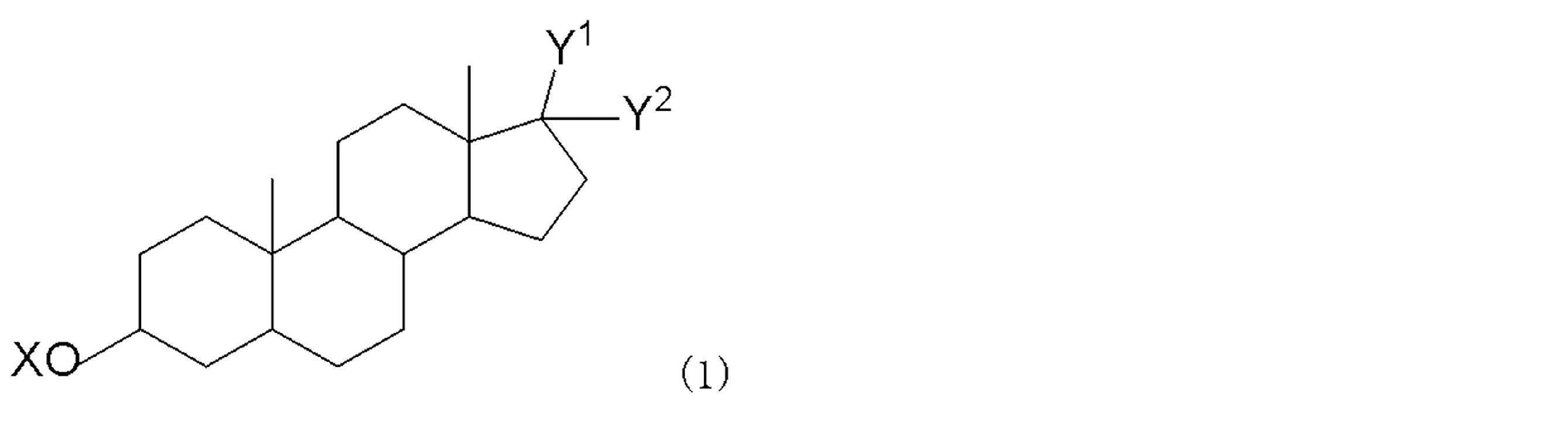
- an androsterone derivative represented by the following general formula (1) as a substrate of the enzyme,
- X is a phosphoric acid group
- Y 1 and Y 2 are jointly a methylene group
- Y 1 is hydrogen
- Y 2 is hydrogen, a hydroxyl group, an alkoxy group having 1 to 6 carbon atoms, or An alkyl group having 1 to 6 carbon atoms
- the androsterone derivative represented by the general formula (1) is used as a substrate for glucosidase, galactosi
- a nucleic acid probe measurement method using an enzyme-labeled nucleic acid probe Using alkaline phosphatase, glucosidase, galactosidase, fructosidase, mannosidase or peroxidase as the enzyme of the enzyme-labeled nucleic acid probe, and an androsterone derivative represented by the following general formula (1) as a substrate of the enzyme, (i) When the androsterone derivative represented by the general formula (1) is used as a substrate for alkaline phosphatase, In the formula, X is a phosphoric acid group, Y 1 and Y 2 are jointly a methylene group, or Y 1 is hydrogen, Y 2 is hydrogen, a hydroxyl group, an alkoxy group having 1 to 6
- the method is carried out by measuring the amount of thio-NADH and / or thio-NADPH produced, or measuring the color change caused by the produced thio-NADH and / or thio-NADPH.
- the enzyme is alkaline phosphatase;
- the androsterone derivative represented by the general formula (1) is In the formula, X is a phosphoric acid group, Y 1 and Y 2 are jointly a methylene group, or Y 1 is hydrogen, Y 2 is hydrogen, a hydroxyl group, an alkoxy group having 1 to 6 carbon atoms, or The method according to [1] or [2], which is an alkyl group having 1 to 6 carbon atoms.
- the enzyme is glucosidase, galactosidase, fructosidase or mannosidase, and androsterone derivative represented by the general formula (1),
- X is a sugar residue
- the sugar residue is one selected from the group consisting of glucose, galactose, fructose and mannose
- Y 1 and Y 2 are a methylene group or an oxygen atom together
- Y 1 is hydrogen
- Y 2 is hydrogen, a hydroxyl group, an alkoxy group having 1 to 6 carbon atoms, or an alkyl group having 1 to 6 carbon atoms, [1] or [2].
- the enzyme is peroxidase, and androsterone derivative represented by the general formula (1),
- X is —O—CO—R (wherein R 1 is an alkyl group having 1 to 6 carbon atoms or a phenyl group), and Y 1 and Y 2 together are a methylene group or an oxygen atom,
- R 1 is an alkyl group having 1 to 6 carbon atoms or a phenyl group
- Y 1 and Y 2 together are a methylene group or an oxygen atom
- Y 1 is hydrogen
- Y 2 is hydrogen, a hydroxyl group, an alkoxy group having 1 to 6 carbon atoms, or an alkyl group having 1 to 6 carbon atoms, [1] or [2].
- An enzyme immunoassay kit comprising the following reagents (1) to (5): (1) Alkaline phosphatase labeled with an antibody specific for the target protein antigen (2) an androsterone derivative represented by the general formula (1) which is a substrate of the enzyme (Wherein X is a phosphate group, Y 1 and Y 2 are jointly methylene groups, or Y 1 is hydrogen, Y 2 is hydrogen, a hydroxyl group, an alkoxy group having 1 to 6 carbon atoms, Or an alkyl group having 1 to 6 carbon atoms), (3) Hydroxysteroid dehydrogenase (HSD) (4) NADH and / or NADPH, (5) Thio NAD and / or Thio NADP [7] An enzyme immunoassay kit comprising the following reagents (1) to (5): (1) Glucosidase, galactosidase, fructosidase or mannosidase labeled with an antibody specific to the target protein antigen (2) an androsterone derivative
- Y 1 is hydrogen, and Y 2 is hydrogen, a hydroxyl group, an alkoxy group having 1 to 6 carbon atoms, or an alkyl group having 1 to 6 carbon atoms), (3) Hydroxysteroid dehydrogenase (HSD) (4) NADH and / or NADPH, (5) Thio NAD and / or Thio NADP [8]
- An enzyme immunoassay kit comprising the following reagents (1) to (5): (1) peroxidase labeled with an antibody specific for a target protein antigen, (2) an androsterone derivative represented by the general formula (1) which is a substrate of the enzyme (Wherein X is —O—CO—R (wherein R 1 is an alkyl group having 1 to 6 carbon atoms or a phenyl group), and Y 1 and Y 2 together are a methylene group or an oxygen atom) Or Y 1 is hydrogen, and Y 2 is hydrogen, a hydroxyl group, an alkoxy group having 1 to 6 carbon atoms
- Y 1 is hydrogen, and Y 2 is hydrogen, a hydroxyl group, an alkoxy group having 1 to 6 carbon atoms, or an alkyl group having 1 to 6 carbon atoms), (3) hydroxysteroid dehydrogenase (HSD), (4) NADH and / or NADPH, (5) Thio NAD and / or Thio NADP [11]
- a kit for measuring a nucleic acid probe comprising the following reagents (1) to (5): (1) peroxidase labeled with a nucleic acid probe that specifically binds to a target nucleic acid, (2) an androsterone derivative represented by the general formula (1) which is a substrate of the enzyme (Wherein X is —O—CO—R (wherein R 1 is an alkyl group having 1 to 6 carbon atoms or a phenyl group), and Y 1 and Y 2 together are a methylene group or an oxygen atom) Or Y 1 is hydrogen, and Y 2 is hydrogen, a hydroxyl group
- X is a phosphoric acid group
- Y 1 and Y 2 are jointly a methylene group
- Y 1 is hydrogen
- Y 2 is a hydroxyl group
- B X is one sugar residue selected from the group consisting of glucose, galactose, fructose and mannose
- Y 1 and Y 2 are jointly methylene groups
- Y 1 is hydrogen
- Y 2 is hydrogen, an alkoxy group having 1 to 6 carbon atoms, or an alkyl group having 1 to 6 carbon atoms
- C X is -O-CO-R, R is an alkyl group or phenyl group having 1 to 6 carbon atoms
- Y 1 and Y 2 are a methylene group or an oxygen atom, or
- X, Y 1 and Y 2 are defined as follows: (A) X is a phosphate group, Y 1 and Y 2 are jointly methylene groups, or Y 1 is hydrogen, Y 2 is a hydroxyl group, The androsterone derivative according to [12], which is an alkoxy group having 1 to 6 carbon atoms or an alkyl group having 1 to 6 carbon atoms. [14] The definitions of X, Y 1 and Y 2 are as follows: (B) X is one sugar residue selected from the group consisting of glucose, galactose, fructose and mannose, and Y 1 and Y 2 are methylene groups together.
- Y 1 is hydrogen and Y 2 is hydrogen, an alkoxy group having 1 to 6 carbon atoms, or an alkyl group having 1 to 6 carbon atoms.
- X, Y 1 and Y 2 are defined as follows: (C) X is —O—CO—R, R 1 is an alkyl group having 1 to 6 carbon atoms or a phenyl group, and Y 1 and Y 2 together represent methylene.
- the enzyme immunoassay which improved the measurement sensitivity to 10 ⁇ -18 > mols or more can be provided, and the highly sensitive measurement of target protein or target nucleic acid is possible.
- the present invention relates to an androsterone derivative represented by the following general formula (1).
- the definition of X, Y 1 and Y 2 is one of the following (A), (B) and (C).
- A) X is a phosphoric acid group, Y 1 and Y 2 are jointly a methylene group, or Y 1 is hydrogen, Y 2 is a hydroxyl group, an alkoxy group having 1 to 6 carbon atoms, or a carbon number 1 to 6 alkyl groups
- (B) X is one sugar residue selected from the group consisting of glucose, galactose, fructose and mannose, Y 1 and Y 2 are jointly methylene groups, or Y 1 is hydrogen, Y 2 is hydrogen, an alkoxy group having 1 to 6 carbon atoms, or an alkyl group having 1 to 6 carbon atoms
- C) X is -O-CO-R, R is an alkyl group or phenyl group having 1 to 6 carbon atoms
- Examples of Y 2 may be an alkoxy group having 1 to 6 carbon atoms such as a methoxy group, an ethoxy group, or a propoxy group, and an alkyl group having 1 to 6 carbon atoms may be, for example, a methyl group or an ethyl group. , Iso-propyl group, n-propyl group, tert-butyl group, n-butyl group, n-pentyl group, n-hexyl group and the like.
- the sugar of the sugar residue can be glucose, galactose, fructose or mannose, and can be appropriately selected depending on the labeling enzyme used.
- the alkyl group having 1 to 6 carbon atoms in R is, for example, a methyl group, an ethyl group, an iso-propyl group, n- It may be a propyl group, tert-butyl group, n-butyl group, n-pentyl group, n-hexyl group, and the like.
- R may be a phenyl group or may have a functional group.
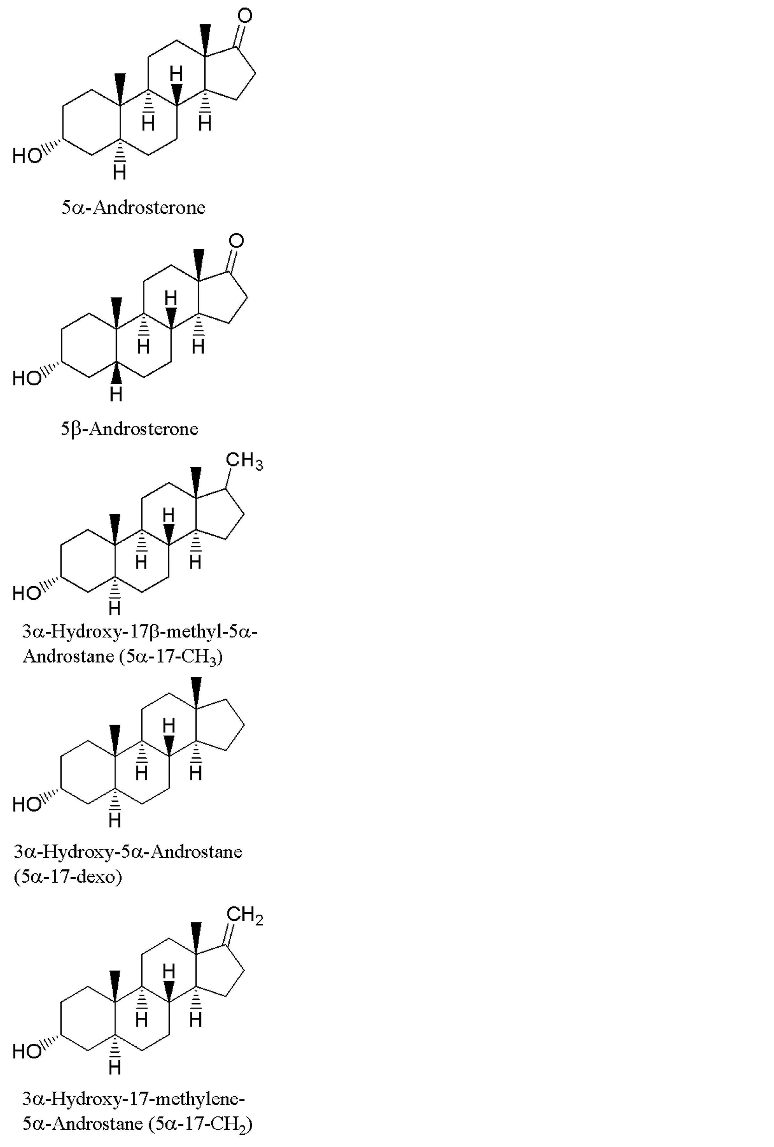
- X is a sugar residue or -O-CO-R, specifically a sugar derivative of 5 ⁇ -androsterone or 5 ⁇ -androsterone, such as a glucoside , Galactoside, fructoside or mannoside.
- glucoside, galactoside, fructoside and mannoside are formally called glucopyranoside, galactopyranoside, fructopyranoside and mannopyranoside, respectively.
- Table 1 Specific examples of the compound of the present invention are shown in Table 1 described later. However, Table 1 includes compounds that can be used in the methods and kits of the present invention in addition to specific examples of the compounds of the present invention.
- the compounds of the present invention can be synthesized based on the methods described in Examples or with reference to the methods described in Examples.
- the present invention is an enzyme measurement method using an antibody-enzyme complex, Enzymatic reaction product quantification of antibody-enzyme complex uses NADH and / or NADPH, thioNAD and / or thioNADP, and dehydrogenase (DH) to produce thioNADH and / or thioNADPH by enzyme cycling reaction And measuring the amount of thio-NADH and / or thio-NADPH produced, or measuring the color change caused by thio-NADH and / or thio-NADPH.
- the antibody-enzyme complex comprises an antibody specific for the target protein antigen and an enzyme labeled with this antibody, and is used for measuring the target protein.
- the present invention relates to a method for measuring a nucleic acid probe using an enzyme-labeled nucleic acid probe, wherein the enzyme-labeled nucleic acid probe quantifies the reaction product by NADH and / or NADPH, thio-NAD and / or thio-NADP, and dehydrogenase ( DH) is used to generate thio-NADH and / or thio-NADPH by an enzyme cycling reaction and measure the amount of thio-NADH and / or thio-NADPH generated, or the color of thio-NADH and / or thio-NADPH generated It is related with the said method performed by measuring a change.
- the enzyme-labeled nucleic acid probe quantifies the reaction product by NADH and / or NADPH, thio-NAD and / or thio-NADP, and dehydrogenase ( DH) is used to generate thio-NADH and / or thio-NADPH by an
- the enzyme-labeled nucleic acid probe comprises a nucleic acid probe that specifically binds to a target nucleic acid and an enzyme labeled with the nucleic acid probe, and is used for measuring the target nucleic acid.
- the enzyme (labeled enzyme) of the antibody-enzyme complex or enzyme-labeled nucleic acid probe used in the method of the present invention is alkaline phosphatase (ALP), galactosidase, glucosidase, fructosidase, mannosidase or peroxidase.
- ALP alkaline phosphatase
- galactosidase galactosidase
- glucosidase glucosidase
- fructosidase fructosidase
- mannosidase mannosidase
- peroxidase peroxidase
- An androsterone derivative represented by the following general formula (1) is used as the enzyme substrate.
- the androsterone derivative included in (ii) includes the above novel compound of the present invention, and further includes a compound in which Y 1 and Y 2 are oxygen atoms together.
- Y 1 and Y 2 are oxygen atoms together
- compounds in which X is glucose are described in Carbohydrate Research, 1971, Vol. 17, No. 1, p. 199-207. The entire description of this document is hereby specifically incorporated by reference.
- compounds in which Y 2 is a hydroxyl group compounds in which X is glucose are described in Phytochemistry, 1974, Vol. 13, No. 10, p.2135-2142. The entire description of this document is hereby specifically incorporated by reference.
- X is —O—CO—R (where R is an alkyl group having 1 to 6 carbon atoms or Y 1 and Y 2 are jointly a methylene group or an oxygen atom, or Y 1 is hydrogen, Y 2 is hydrogen, a hydroxyl group, and an alkoxy group having 1 to 6 carbon atoms. Or an alkyl group having 1 to 6 carbon atoms.
- R is an alkyl group having 1 to 6 carbon atoms or Y 1 and Y 2 are jointly a methylene group or an oxygen atom, or Y 1 is hydrogen, Y 2 is hydrogen, a hydroxyl group, and an alkoxy group having 1 to 6 carbon atoms. Or an alkyl group having 1 to 6 carbon atoms.
- R is an alkyl group having 1 to 6 carbon atoms or Y 1 and Y 2 are jointly a methylene group or an oxygen atom, or Y 1 is hydrogen, Y 2 is hydrogen, a hydroxyl group, and an alkoxy group having
- the ultrasensitive measurement method of the present invention can be carried out in the same manner as the usual enzyme immunoassay method and nucleic acid probe method.
- a solid phase carrier used for normal measurement such as a solid phase carrier to which an antibody or nucleic acid probe that specifically binds to the analyte is fixed, such as a microplate, plastic tube, or magnetic bead. Can do.
- Antibody and nucleic acid probe-enzyme complexes can be prepared by conventional methods.
- the antibody constituting the antibody-enzyme complex can be appropriately selected from antibodies that specifically bind to the analyte to be measured by the enzyme immunoassay method of the present invention.
- the enzyme immunoassay method of the present invention is used for protein analysis.
- the antibody of the antibody-enzyme complex is an antibody that specifically binds to the protein as the analyte.
- a substrate on which an antibody that specifically binds to the protein as the analyte is fixed to the surface is used.
- the antibody constituting the antibody-enzyme complex and the antibody to be fixed to the substrate may be antibody fragments.
- the analyte in the enzyme immunoassay method of the present invention is not limited to a protein, and can be any substance other than the protein to be measured by a normal enzyme immunoassay method.
- a probe complementary to the nucleic acid to be measured can be selected as appropriate for the nucleic acid probe enzyme complex.
- the quantification of the reaction product by the enzyme of the antibody-enzyme complex or the enzyme-labeled nucleic acid probe is performed using NADH and / or NADPH, thioNAD and / or thioNADP, and dehydrogenase (DH).
- NADH and / or NADPH NADH and / or NADPH
- thioNAD and / or thioNADP a dehydrogenase
- DH dehydrogenase
- the concentration of each component can be in the following range.
- Concentration range of antibody-enzyme complex or enzyme-labeled nucleic acid probe 0.01 ⁇ g / ml to 1 mg / ml
- Concentration range of the label enzyme substrate 1 ⁇ M to 500 mM
- NADH and / or NADPH concentration range 0.01 mM to 50 mM
- Thio NAD and / or Thio NADP concentration range 0.01 mM to 100 mM
- Dehydrogenase (DH) concentration 0.01 u / ml to 5000 u / ml
- the reaction conditions can be appropriately determined in consideration of the optimum temperature range of the labeling enzyme and dehydrogenase (DH).
- the reaction temperature is preferably room temperature since the operation is simple. However, it can be carried out at a temperature higher or lower than room temperature in consideration of the optimum temperature range of the labeling enzyme and dehydrogenase (DH).
- the reaction time accumulates thio-NADH and / or thio-NADPH to such an extent that the amount of thio-NADH and / or thio-NADPH produced can be measured or the color change due to the produced thio-NADH and / or thio-NADPH can be measured Enough time.
- the accumulation amount of thio-NADH and / or thio-NADPH necessary for measurement or measurement varies depending on the measurement or measurement conditions, and can be appropriately determined according to the conditions.
- Cycle cycling using thio NAD (P) is a unique cycling system that appeared relatively recently.
- dehydrogenase (DH) using NAD (P) / NAD (P) H as a coenzyme is used.
- the measurement principle of the thio NAD (P) cycling method is as follows.
- Cycling systems that use thio-NAD (P) can measure dehydrogenase activity based on increased absorption of NADH, taking advantage of the fact that it can be measured using popular absorption photometers and microplate readers for colorimetry.
- Some of the conventional methods such as a method for quantifying the substrate have been improved to a method using thio NAD.
- this cycling system as a detection system such as an enzyme immunoassay for high sensitivity.
- an amplification reaction can be made a geometrical reaction for the first time, and sufficient sensitivity can be increased.
- the product produced by the enzyme complex and the substrate combined therewith is used as a substrate for the next enzyme cycling reaction, and the thiol produced by the enzyme cycling reaction is used. Colorimetric determination of NAD (P) H absorption.
- the substrate for the enzyme cycling reaction may be a reduced substrate or an oxidized substrate.
- the labeled enzyme used as the enzyme complex used in the present invention, its substrate, and the dehydrogenase used in the subsequent cycling reaction have the following properties.
- Labeled enzyme product is androsterone or its derivatives
- Commercially available and widely used labeled enzymes can be used
- Enzymatic cycling turnover number is large
- Dehydrogenation used for enzymatic cycling reaction Enzyme does not have the same activity as labeling enzyme contamination or labeling enzyme
- DH dehydrogenase
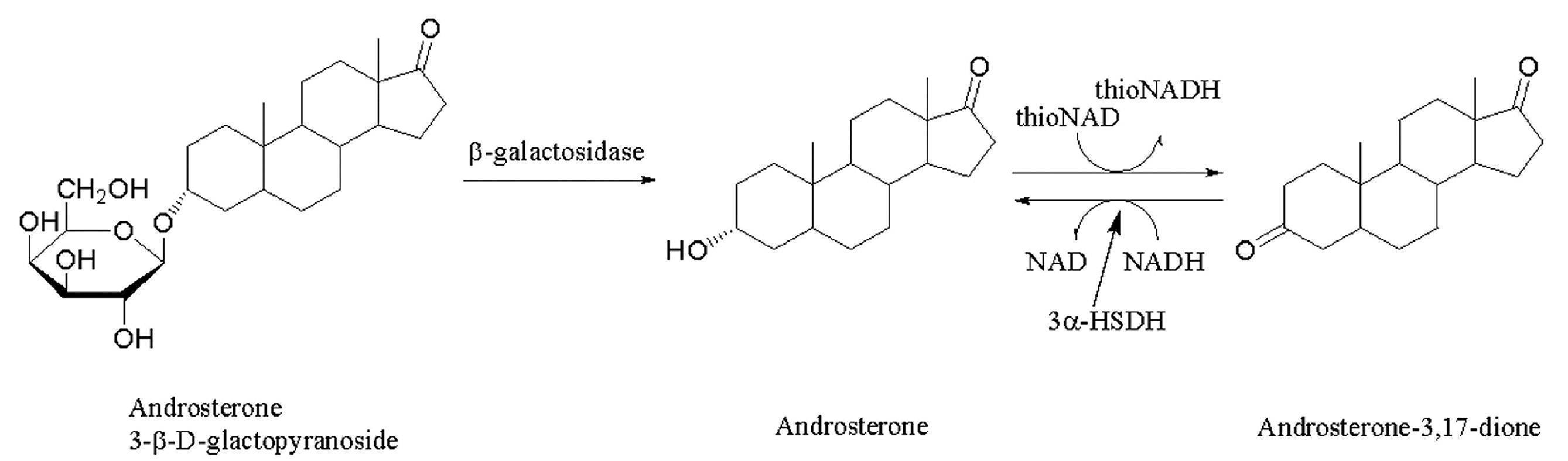
- Examples of dehydrogenase (DH) include 3 ⁇ -hydroxysteroid dehydrogenase.
- usable substrate candidates include androsterone, 11 ⁇ -hydroxyandrosterone, 11-oxoandrosterone, 11 ⁇ -hydroxyandrosterone. Is mentioned.
- a substrate having a high enzyme reaction rate (high activity against the substrate) and reacting even at a low concentration (small Km value) is preferable.
- a preferable property as a dehydrogenase is a reaction rate when thioNAD (P) is used as a coenzyme.
- the reaction rate when thio-NAD is used as a coenzyme is within several percent of that when NAD is used, whereas in the dehydrogenation reaction of androsterone with 3 ⁇ -hydroxysteroid dehydrogenase, The reaction rate with NAD is about 59% of the reaction rate with NAD, and it has favorable properties as the cycling enzyme of the present invention.
- alkaline phosphatase ALP
- the 3-position phosphate ester of an androsterone derivative which is a substrate for alkaline phosphatase is easily synthesized. It is.
- glucosidase and an androsterone derivative glucoside which are widely used as labeling enzymes
- galactosidase and an androsterone derivative galactoside are also easily combined and preferred combinations.
- peroxides of peroxidase and androsterone derivatives that are widely used as labeling enzymes such as tert-butyl peroxide, are also a combination that is easy to synthesize and is preferable.
- Alkaline phosphatase is an enzyme that is widely used as a labeling enzyme.
- this enzyme immunoassay method using ALP if a new androsterone derivative is used as a substrate for ALP, androsterone is produced as a product. .
- 3 ⁇ -hydroxysteroid dehydrogenase (3 ⁇ -HSD) which is a dehydrogenase using the reaction product of ALP as a substrate, is used for the enzyme cycling reaction.
- ThioNAD cycling allows amplification and quantification of dehydrogenase substrates with an efficiency of several hundred cycles per minute if an appropriate dehydrogenase reaction is selected. Therefore, the activity of an enzyme that gives such a substrate as a reaction product can be measured with extremely high sensitivity by thio-NAD cycling.
- the present invention includes an enzyme cycling method kit comprising an enzyme labeled on a reactive carrier, its substrate, an enzyme for cycling reaction, and its coenzymes thio NAD and NADH.
- the reactive carrier represents an antibody, a nucleic acid probe, a lectin or the like having an activity to bind to a measurement object.
- the reactive carrier is not particularly limited as long as it is suitable for the measurement target, and the labeling enzyme and its substrate are also suitable.
- the present invention includes a kit for an enzyme cycling method comprising a labeling enzyme, its substrate, an enzyme for cycling reaction, and its coenzymes thio NAD and NADH.
- the kit of the present invention is an enzyme immunoassay kit containing the following reagents (1) to (5).
- Alkaline phosphatase which is an enzyme labeled with an antibody specific for the target protein antigen, or glucosidase or galactosidase or fructosidase or mannosidase or peroxidase, (2) an androsterone derivative represented by the general formula (1) which is a substrate of the enzyme, (3) dehydrogenase (DH), (4) NADH and / or NADPH, (5) Thio NAD and / or Thio NADP
- the present invention is a kit for measuring a nucleic acid probe comprising the following reagents (1) to (5).
- Alkaline phosphatase which is an enzyme labeled with a nucleic acid probe that specifically binds to a target nucleic acid, or glucosidase or galactosidase or fructosidase or mannosidase or peroxidase, (2) an androsterone derivative represented by the general formula (1) which is a substrate of the enzyme, (3) dehydrogenase (DH), (4) NADH and / or NADPH, (5) Thio NAD and / or Thio NADP
- dehydrogenase (DH) and general formula (1) those described in the method of the present invention can be used as they are.
- kit of the present invention a commercially available enzyme-labeled antibody or the like can be used in combination with the constituent reagents of this kit.
- This kit can be used for an enzyme immunoassay method using an enzyme cycling method.
- Alkaline phosphatase and Pumilio using thio-NAD cycling of the Alkaline phosphatase (ALP) and Androsterone 3-phosphate (A3P) system as a detection system.
- ALP Alkaline phosphatase
- A3P Androsterone 3-phosphate
- Reference example 2 Preparation of insoluble carrier-immobilized antibody 1
- BSA bovine serum albumin
- Measurement Method A 0.1% BSA-containing TBS (pH) containing purified Pumilio (standard substance) in a range of 0 to 200 ng / ml on a microplate on which the anti-Pumilio guinea pig polyclonal antibody prepared by the method of Reference Example 2 is immobilized. 7.5) 50 ⁇ l of the solution was added and shaken at room temperature for 1 hour. Next, the solution in the well is removed by suction, washed with 0.05% Tween 20-containing TBS (pH 7.5), and contains the ALP-labeled anti-Pumilio mouse monoclonal antibody prepared by the method of Reference Example 1 at a concentration of 2.5 ⁇ g / ml.
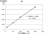
- FIG. 2 shows a straight line having a good concentration dependency obtained by plotting the amount of change in absorbance for 30 minutes against the concentration of the standard substance.
- Reaction reagent B 0.2 M glycine buffer (pH 9.6) containing 0.2 M disodium hydrogen phosphate 3 mM thio-NAD 1 mM NADH 40 U / ml 3 ⁇ -Hydroxysteroid dehydrogenase
- Measurement Method A 0.1% BSA-containing TBS (pH) containing purified Pumilio (standard substance) in the range of 0 to 100 ng / ml in a microplate on which the anti-Pumilio guinea pig polyclonal antibody prepared by the method of Reference Example 3 is immobilized. 7.5) 50 ⁇ l of the solution was added and shaken at room temperature for 1 hour. Next, the solution in the well is removed by suction, washed with 0.05% Tween 20-containing TBS (pH 7.5), and contains the ALP-labeled anti-Pumilio mouse monoclonal antibody prepared by the method of Reference Example 1 at a concentration of 2.5 ⁇ g / ml.
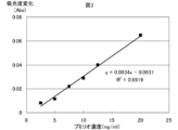
- FIG. 3 shows a straight line having good concentration dependency obtained by plotting the amount of change in absorbance for 30 minutes against the standard substance concentration.
- Reaction reagent 0.1 M Tris buffer (pH 8.6) 0.5 mM MgCl 2 1.5 mM thio-NAD 0.5 mM NADH 0.5 mM 5 ⁇ -Androsterone 3-phosphate 20 U / ml 3 ⁇ -Hydroxysteroid dehydrogenase
- Measurement Method A 0.1% BSA-containing TBS (pH) containing purified Pumilio (standard substance) in the range of 0 to 50 ng / ml in a microplate on which the anti-Pumilio guinea pig polyclonal antibody prepared by the method of Reference Example 3 is immobilized. 7.5) 50 ⁇ l of the solution was added and shaken at room temperature for 1 hour. Next, the solution in the well is removed by suction, washed with 0.05% Tween 20-containing TBS (pH 7.5), and contains the ALP-labeled anti-Pumilio mouse monoclonal antibody prepared by the method of Reference Example 1 at a concentration of 2.5 ⁇ g / ml.
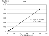
- FIG. 4 shows a straight line having good concentration dependency obtained by plotting the amount of change in absorbance for 30 minutes against the standard substance concentration.
- Reaction reagent A 0.1 M Tris buffer (pH 8.3) 1 mM MgCl 2 0.1 mM 5 ⁇ -Androsterone 3-phosphate
- Reaction reagent B 0.2 M glycine buffer (pH 9.6) containing 0.2 M disodium hydrogen phosphate 3 mM thio-NAD 1 mM NADH 40 U / ml 3 ⁇ -Hydroxysteroid dehydrogenase
- Measurement Method A 0.1% BSA-containing TBS (pH) containing purified Pumilio (standard substance) in the range of 0 to 50 ng / ml in a microplate on which the anti-Pumilio guinea pig polyclonal antibody prepared by the method of Reference Example 3 is immobilized. 7.5) 50 ⁇ l of the solution was added and shaken at room temperature for 1 hour. Next, the solution in the well is removed by suction, washed with 0.05% Tween 20-containing TBS (pH 7.5), and contains the ALP-labeled anti-Pumilio mouse monoclonal antibody prepared by the method of Reference Example 1 at a concentration of 2.5 ⁇ g / ml.
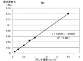
- FIG. 5 shows a straight line having good concentration dependency obtained by plotting the amount of change in absorbance for 10 minutes against the standard substance concentration.
- Reaction reagent 0.1 M Tris buffer (pH 9.5) 0.2 mM MgCl 2 0.6 mM thio-NAD 4 mM NADH 0.5 mM 3 ⁇ -Hydroxy-17 ⁇ -methoxy-5 ⁇ -Androstane 3-phosphate 20 U / ml 3 ⁇ -Hydoroxysteroid dehydrogenase
- Measurement method 50 ⁇ l of Tris buffer solution (pH 9.5) containing standard substance (Alkaline phosphatase) in the range of 0 to 2 mU / ml is added to a flat-bottomed microplate well, and then 50 ⁇ l each of reaction reagent B is added to the well.
- the absorbance was measured for 60 minutes using a 405 nm filter with a microplate reader (Corona MTP-500) while heating at 37 ° C.
- FIG. 7 shows a straight line having good concentration dependency obtained by plotting the amount of change in absorbance for 60 minutes against the standard substance concentration.
- Reaction reagent A 0.1 M Tris buffer (pH 9.5) 0.2 mM MgCl 2 1 mM 3 ⁇ -Hydroxy-17 ⁇ -methoxy-5 ⁇ -Androstane 3-phosphate
- Reaction reagent B 0.15 M pyrophosphate buffer (pH 9.5) 3 mM thio-NAD 1 mM NADH 40 U / ml 3 ⁇ -Hydoroxysteroid dehydrogenase
- the ALP The labeled antibody solution is present a complex of different molecular weights of ALP and Fab, since activity has variation, ALP-labeled antibody solution 1 mM Mgl 2 produced, 0.1 mM ZnCl 2, the 0.01% BSA Gel filtration using a column (Amersham Biosciences) packed with superdex 200 using Tris buffer (pH 7.0) as an eluent, and comparing the activity of each fraction, ALP-labeled antibody with a large S / N ratio Only recovered.
- Example 3 Measurement reaction reagent 0.1 M Tris buffer 3 ⁇ -Hydroxy-17 ⁇ -methoxy- 5 ⁇ -Androstane 3-phosphate Pumilio by 1 step method using (Pumirio) (pH 9.5) 1.5 mM thio-NAD 2.5 mM NADH 0.1 mM 3 ⁇ -Hydroxy-17 ⁇ -methoxy-5 ⁇ -Androstane 3-phosphate 20 U / ml 3 ⁇ -Hydoroxysteroid dehydrogenase
- Measurement Method A 0.1% BSA-containing TBS (pH) containing purified Pumilio (standard substance) in a range of 0 to 25 ng / ml in a microplate on which the anti-Pumilio guinea pig polyclonal antibody prepared by the method of Reference Example 5 is immobilized. 7.5) 50 ⁇ l of the solution was added and shaken at room temperature for 2 hours. Next, the solution in the well was removed by suction, washed with 0.5% Triton X-100-containing TBS (pH 7.5), and ALP-labeled anti-Pumilio mouse monoclonal antibody prepared by the method of Reference Example 4 at a concentration of about 0.8 ⁇ g / ml.
- FIG. 9 shows a straight line having good concentration dependency obtained by plotting the amount of change in absorbance for 60 minutes against the standard substance concentration.
- Measurement Method A 0.1% BSA-containing TBS (pH) containing purified Pumilio (standard substance) in the range of 0 to 0.5 ng / ml in a microplate on which the anti-Pumilio guinea pig polyclonal antibody prepared by the method of Reference Example 5 is immobilized. 7.5) 50 ⁇ l of the solution was added and shaken at room temperature for 2 hours.
- the solution in the well was removed by suction, washed with 0.5% Triton X-100-containing TBS (pH 7.5), and about 0.2 ⁇ g / ml of ALP-labeled anti-Pumilio mouse monoclonal antibody prepared by the method of Reference Example 4, rabbit 50 ⁇ l of 0.1% BSA-containing TBS (pH 7.5) solution containing F (ab ′) 2 at a concentration of 1 mg / ml was added and shaken at 4 ° C. for 2 hours.
- the solution in the well was removed by suction, washed with 0.5% Triton X-100-containing TBS (pH 7.5), and further washed with TBS.
- reaction reagent A 50 ⁇ l of reaction reagent A was added to each well and incubated at 37 ° C. for 30 minutes.
- 50 ⁇ l of reaction reagent B was added to the well, and the absorbance was measured for 30 minutes using a 405 nm filter with a microplate reader (MTP-500 manufactured by Corona) while heating at 37 ° C.
- FIG. 10 shows a straight line having good concentration dependency obtained by plotting the amount of change in absorbance for 30 minutes against the standard substance concentration.
- ⁇ -Galactosidase was used as the labeling enzyme for ELISA, and Androsterone ⁇ -D-Galactoside was synthesized as a substrate for further investigation.
- Reaction reagent A 0.1 M phosphate buffer (pH 7.3) 0.2 mM MgCl 2 0.1 mM 5 ⁇ -Androsterone ⁇ -D-Galactoside
- Reaction reagent B 0.1 M pyrophosphate buffer (pH 9.0) 1.2 mM thio-NAD 3.6 mM NADH 40 U / ml 3 ⁇ -Hydoroxysteroid dehydrogenase
- Measurement method 25 ⁇ l of phosphate buffer solution (pH 7.3) containing standard substance ( ⁇ -Galactosidase) in the range of 0 to 200 pg / ml is added to a flat-bottomed microplate well, and then 25 ⁇ l of reaction reagent A is added to the well. They were added and mixed, and incubated at 37 ° C for 60 minutes. Next, 50 ⁇ l of the reaction sample solution B was added to each well, and the absorbance was measured for 30 minutes using a 405 nm filter with a microplate reader (Corona MTP-500) while heating at 37 ° C.
- FIG. 11 shows a straight line having good concentration dependency obtained by plotting the amount of change in absorbance for 30 minutes against the standard substance concentration.
- Reaction reagent 0.1 M phosphate buffer (pH 7.7) 10 mM MgCl 2 6 mM thio-NAD 1 mM NADH 0.1 mM 5 ⁇ -Androsterone ⁇ -D-Galactoside 40 U / ml 3 ⁇ -Hydoroxysteroid dehydrogenase
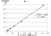
- FIG. 12 shows a straight line having good concentration dependency obtained by plotting the change in absorbance for 120 minutes against the concentration of the standard substance.
- Reaction reagent A 0.1 M phosphate buffer (pH 7.5) 10 mM MgCl 2 0.1 mM 5 ⁇ -Androsterone ⁇ -D-Galactoside
- Reaction reagent B 0.1 M pyrophosphate buffer (pH 9.0) 4.8 mM thio-NAD 0.4 mM NADH 40 U / ml 3 ⁇ -Hydoroxysteroid dehydrogenase
- Measurement method 25 ⁇ l of phosphate buffer solution (pH 7.5) containing standard substance ( ⁇ -Galactosidase) in the range of 0 to 200 pg / ml is added to a flat-bottomed microplate well, and then 25 ⁇ l of reaction reagent A is added to the well. They were added and mixed, and incubated at 37 ° C for 60 minutes. Next, 50 ⁇ l of the reaction sample solution B was added to each well, and the absorbance was measured for 30 minutes using a 405 nm filter with a microplate reader (Corona MTP-500) while heating at 37 ° C.
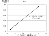
- FIG. 13 shows a straight line having good concentration dependency obtained by plotting the amount of change in absorbance for 30 minutes against the concentration of the standard substance.
- N-iodosuccinimide (435 mg) and trifluoromethanesulfonic acid (15 mg) were added, and the mixture was stirred at ⁇ 20 ° C. for 2 hours, and further reacted at room temperature for 2 hours.
- the reaction was stopped by adding 250 ⁇ L of triethylamine, and the reaction was washed successively with a 3: 1 mixture of saturated sodium carbonate and sodium thiosulfate, and saturated brine. After drying the organic layer with anhydrous sodium sulfate, the solvent was distilled off under reduced pressure. The residue was dissolved in methanol (15 mL), 28% sodium methoxide (500 ⁇ L) was added, and the mixture was stirred for 2 hr.
- Reaction reagent 0.1 M phosphate buffer (pH 8.5) 10 mM MgCl 2 6 mM thio-NAD 1 mM NADH 0.2 mM 3 ⁇ -Hydroxy-17 ⁇ -methoxy-5 ⁇ -Androstane ⁇ -D-Galactoside 20 U / ml 3 ⁇ -Hydoroxysteroid dehydrogenase
- FIG. 14 shows a straight line having good concentration dependency obtained by plotting the amount of change in absorbance for 120 minutes against the concentration of the standard substance.
- Reaction reagent A 0.1 M phosphate buffer (pH 7.5) 4 mM MgCl 2 0.1 mM 3 ⁇ -Hydroxy-17 ⁇ -methoxy-5 ⁇ -Androstane ⁇ -D-Galactoside
- Reaction reagent B 0.1 M pyrophosphate buffer (pH 9.8) 1.2 mM thio-NAD 1 mM NADH 40 U / ml 3 ⁇ -Hydoroxysteroid dehydrogenase
- Measurement method 25 ⁇ l of phosphate buffer solution (pH 7.5) containing standard substance ( ⁇ -Galactosidase) in the range of 0 to 200 pg / ml is added to a flat-bottomed microplate well, and then 25 ⁇ l of reaction reagent A is added to the well. They were added and mixed, and incubated at 37 ° C for 60 minutes. Next, 50 ⁇ l of the reaction sample solution B was added to each well, and the absorbance was measured for 30 minutes using a 405 nm filter with a microplate reader (Corona MTP-500) while heating at 37 ° C.
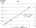
- FIG. 15 shows a straight line having good concentration dependency obtained by plotting the amount of change in absorbance for 30 minutes against the concentration of the standard substance.
- Reaction reagent 0.1 M phosphate buffer (pH 7.7) 5 mM MgCl 2 1.2 mM thio-NAD 0.8 mM NADH 0.05 mM 5 ⁇ -Androstane ⁇ -D-Galactoside 20 U / ml 3 ⁇ -Hydoroxysteroid dehydrogenase
- Measurement Method A 0.1% BSA-containing TBS (pH) containing purified Pumilio (standard substance) in the range of 0 to 2.5 ng / ml on a microplate immobilized with the anti-Pumilio guinea pig polyclonal antibody prepared by the method of Reference Example 5.
- 7.5 50 ⁇ l of the solution was added and shaken at room temperature for 2 hours. Next, the solution in the well was removed by suction, washed with 0.5% Triton X-100-containing TBS (pH 7.5), and the biotin-labeled anti-Pumilio mouse monoclonal antibody prepared by the method of Reference Example 6 was added at a concentration of approximately 0.1 ⁇ g / ml.
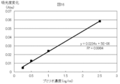
- FIG. 16 shows a straight line having good concentration dependency obtained by plotting the amount of change in absorbance for 60 minutes against the standard substance concentration.
- Reaction reagent A 0.1 M phosphate buffer (pH 7.5) 0.1 mM MgCl 2 0.05 mM 5 ⁇ -Androsterone ⁇ -D-Galactoside
- Reaction reagent B 0.1 M pyrophosphate buffer (pH 9.0) 3 mM thio-NAD 2 mM NADH 40 U / ml 3 ⁇ -Hydoroxysteroid dehydrogenase
- Measurement Method A 0.1% BSA-containing TBS (pH) containing purified Pumilio (standard substance) in a range of 0 to 5 ng / ml on a microplate immobilized with the anti-Pumilio guinea pig polyclonal antibody prepared by the method of Reference Example 5.
- 7.5 50 ⁇ l of the solution was added and shaken at room temperature for 2 hours. Next, the solution in the well was removed by suction, washed with 0.5% Triton X-100-containing TBS (pH 7.5), and the biotin-labeled anti-Pumilio mouse monoclonal antibody prepared by the method of Reference Example 6 was added at a concentration of approximately 0.1 ⁇ g / ml.
- reaction reagent A 50 ⁇ l of reaction reagent A was added to each well and incubated at 37 ° C. for 30 minutes.
- 50 ⁇ l of reaction reagent B was added to the well, and the absorbance was measured for 30 minutes using a 405 nm filter with a microplate reader (MTP-500 manufactured by Corona) while heating at 37 ° C.
- FIG. 17 shows a straight line having good concentration dependency obtained by plotting the amount of change in absorbance over 30 minutes against the standard substance concentration.
- Reaction reagent 0.1 M phosphate buffer (pH 8.5) 1.2 mM thio-NAD 0.8 mM NADH 0.05 mM 3 ⁇ -Hydroxy-17 ⁇ -methoxy-5 ⁇ -Androstane ⁇ -D-Galactoside 20 U / ml 3 ⁇ -Hydoroxysteroid dehydrogenase
- Measurement Method A 0.1% BSA-containing TBS (pH) containing purified Pumilio (standard substance) in the range of 0 to 10 ng / ml in a microplate on which the anti-Pumilio guinea pig polyclonal antibody prepared by the method of Reference Example 5 is immobilized. 7.5) 50 ⁇ l of the solution was added and shaken at room temperature for 2 hours. Next, the solution in the well was removed by suction, washed with 0.5% Triton X-100-containing TBS (pH 7.5), and the biotin-labeled anti-Pumilio mouse monoclonal antibody prepared by the method of Reference Example 6 was added at a concentration of approximately 0.1 ⁇ g / ml.
- FIG. 18 shows a straight line having good concentration dependency obtained by plotting the amount of change in absorbance for 60 minutes against the standard substance concentration.
- Reaction reagent A 0.1 M phosphate buffer (pH 7.0) 0.05 mM 3 ⁇ -Hydroxy-17 ⁇ -methoxy-5 ⁇ -Androstane ⁇ -D-Galactoside
- Reaction reagent B 0.1 M pyrophosphate buffer (pH 9.0) 2.4 mM thio-NAD 2 mM NADH 40 U / ml 3 ⁇ -Hydoroxysteroid dehydrogenase
- Measurement Method A 0.1% BSA-containing TBS (pH) containing purified Pumilio (standard substance) in the range of 0 to 10 ng / ml in a microplate on which the anti-Pumilio guinea pig polyclonal antibody prepared by the method of Reference Example 5 is immobilized. 7.5) 50 ⁇ l of the solution was added and shaken at room temperature for 2 hours. Next, the solution in the well was removed by suction, washed with 0.5% Triton X-100-containing TBS (pH 7.5), and the biotin-labeled anti-Pumilio mouse monoclonal antibody prepared by the method of Reference Example 6 was added at a concentration of approximately 0.3 ⁇ g / ml.
- reaction reagent A 50 ⁇ l of reaction reagent A was added to each well and incubated at 37 ° C. for 60 minutes.
- 50 ⁇ l of reaction reagent B was added to the wells, and the absorbance was measured for 30 minutes using a 405 nm filter with a microplate reader (Corona MTP-500) while heating at 37 ° C.
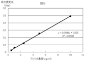
- FIG. 19 shows a straight line having a good concentration dependency obtained by plotting the amount of change in absorbance for 30 minutes against the standard substance concentration.
- Reaction reagent 0.1 M glycine buffer (pH 10.3) 1 mM MgCl 2 1 mM ZnCl 2 1 mg / ml p-NPP
- Measurement Method A 0.1% BSA-containing TBS (pH) containing purified Pumilio (standard substance) in a range of 0 to 25 ng / ml on a microplate immobilized with the anti-Pumilio guinea pig polyclonal antibody prepared by the method of Reference Example 3. 7.5) 50 ⁇ l of the solution was added and shaken at room temperature for 1 hour. Next, after removing the solution in the well by suction, it was washed with TBS (pH 7.5) containing 0.05% Tween 20, and the ALP-labeled anti-Pumilio mouse monoclonal antibody prepared by the method of Reference Example 1 was contained at a concentration of 1 ⁇ g / ml.
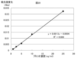
- FIG. 20 shows a straight line having a good concentration dependency obtained by plotting the amount of change in absorbance for 30 minutes against the concentration of the standard substance.
- Reaction reagent 0.1 M phosphate buffer (pH 7.3) 0.2 mM 2-mercaptoethanol 1 mM MgCl 2 10 mM ONPG
- FIG. 21 shows a straight line having a good concentration dependency obtained by plotting the amount of change in absorbance for 60 minutes against the standard substance concentration.
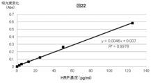
- FIG. 22 shows a straight line having good concentration dependency obtained by plotting the amount of change in absorbance for 120 minutes against the concentration of the standard substance.
- Example 15 Measurement of Pumilio by 1-step method using 3 ⁇ -tert-butyl-peroxy-5 ⁇ -Androsterone Reaction reagent 0.1M phosphate buffer (pH 7.0) 10 mM MgCl 2 6 mM thio-NAD 1mM NADH 0.1 mM 3 ⁇ -tert-butyl-peroxy-5 ⁇ -Androsterone 40U / ml 3 ⁇ -Hydroxysteroid dehydrogenase Sample 0.05, 0.1, 0.25, 0.5, 1, 2.5 ng / ml Pumilio
- Measurement Method A 0.1% BSA-containing TBS (pH 7) containing purified Pumilio (standard substance) in a range of 0 to 2.5 ng / ml on a microplate on which an anti-Pumilio guinea pig polyclonal antibody prepared by the method of Reference Example 5 is immobilized. .5) 50 ⁇ l of the solution was added and shaken at room temperature for 2 hours. Next, after removing the solution in the well by suction, it was washed with TBS (pH 7.5) containing 0.5% Triton X-100, and 0.1% BSA containing HRP-labeled anti-Pumilio mouse monoclonal antibody at a concentration of approximately 0.1 ⁇ g / ml.
- FIG. 23 shows a straight line having good concentration dependency obtained by plotting the amount of change in absorbance for 60 minutes against the standard substance concentration.
- ⁇ Reference Example 9 Synthesis method of 3 ⁇ -benzoyl-peroxy-5 ⁇ -Androsterone and 3 ⁇ -acetyl-peroxy-5 ⁇ -Androsterone
- tert-butyl-hydroperoxide was synthesized.
- the title compound was obtained in the same manner except that benzoyl-hydroperoxide or acetyl-hydroperoxide was used.
- the present invention can be suitably used in clinical inspection fields and food inspection fields where high sensitivity and simple measurement are required.
Landscapes
- Chemical & Material Sciences (AREA)
- Life Sciences & Earth Sciences (AREA)
- Organic Chemistry (AREA)
- Health & Medical Sciences (AREA)
- Engineering & Computer Science (AREA)
- Physics & Mathematics (AREA)
- Wood Science & Technology (AREA)
- Proteomics, Peptides & Aminoacids (AREA)
- Zoology (AREA)
- Analytical Chemistry (AREA)
- Biochemistry (AREA)
- General Health & Medical Sciences (AREA)
- Immunology (AREA)
- Genetics & Genomics (AREA)
- Bioinformatics & Cheminformatics (AREA)
- Microbiology (AREA)
- Molecular Biology (AREA)
- Biophysics (AREA)
- General Engineering & Computer Science (AREA)
- Biotechnology (AREA)
- General Physics & Mathematics (AREA)
- Pathology (AREA)
- Chemical Kinetics & Catalysis (AREA)
- Plasma & Fusion (AREA)
- Spectroscopy & Molecular Physics (AREA)
- Measuring Or Testing Involving Enzymes Or Micro-Organisms (AREA)
- Investigating Or Analysing Materials By The Use Of Chemical Reactions (AREA)
Abstract
Description
[1]
抗体酵素複合体を用いる酵素活性測定方法であって、抗体酵素複合体の酵素としてアルカリホスファターゼ、グルコシダーゼ、ガラクトシダーゼ、フルクトシダーゼ、マンノシダーゼまたはペルオキシダーゼを用い、
上記酵素の基質として下記一般式(1)で表されるアンドロステロン誘導体を用い、
式中、Xはリン酸基、Y1及びY2は共同でメチレン基であるか、または、Y1は水素であり、Y2は、水素、水酸基、炭素数1~6のアルコキシ基、または炭素数1~6のアルキル基であり、
(ii) 一般式(1)で表されるアンドロステロン誘導体は、グルコシダーゼ、ガラクトシダーゼ、フルクトシダーゼまたはマンノシダーゼの基質として用いる場合は、
式中、Xは糖残基であり、糖残基はグルコース、ガラクトース、フルクトースおよびマンノースから成る群から選ばれる1種であり、Y1及びY2は共同でメチレン基または酸素原子であるか、または、Y1は水素であり、Y2は、水素、水酸基、炭素数1~6のアルコキシ基、または炭素数1~6のアルキル基であり、
(iii) 一般式(1)で表されるアンドロステロン誘導体は、ペルオキシダーゼの基質として用いる場合は、
式中、Xは-O-CO-R(但し、R は炭素数1~6のアルキル基またはフェニル基である)であり、Y1及びY2は共同でメチレン基または酸素原子であるか、または、Y1は水素であり、Y2は、水素、水酸基、炭素数1~6のアルコキシ基、または炭素数1~6のアルキル基であり、
前記酵素反応の生成物の定量は、NADHおよび/またはNADPH、チオNADおよび/またはチオNADP、並びにヒドロキシステロイド脱水素酵素(HSD)を用いて、酵素サイクリング反応によりチオNADHおよび/またはチオNADPHを生成させ、生成したチオNADHおよび/またはチオNADPHの量を測定するか、または生成したチオNADHおよび/またはチオNADPHによる色の変化を計測することで行う、前記方法。
[2]
酵素標識核酸プローブを用いる核酸プローブ測定方法であって、
酵素標識核酸プローブの酵素としてアルカリホスファターゼ、グルコシダーゼ、ガラクトシダーゼ、フルクトシダーゼ、マンノシダーゼまたはペルオキシダーゼを用い、上記酵素の基質として下記一般式(1)で表されるアンドロステロン誘導体を用い、
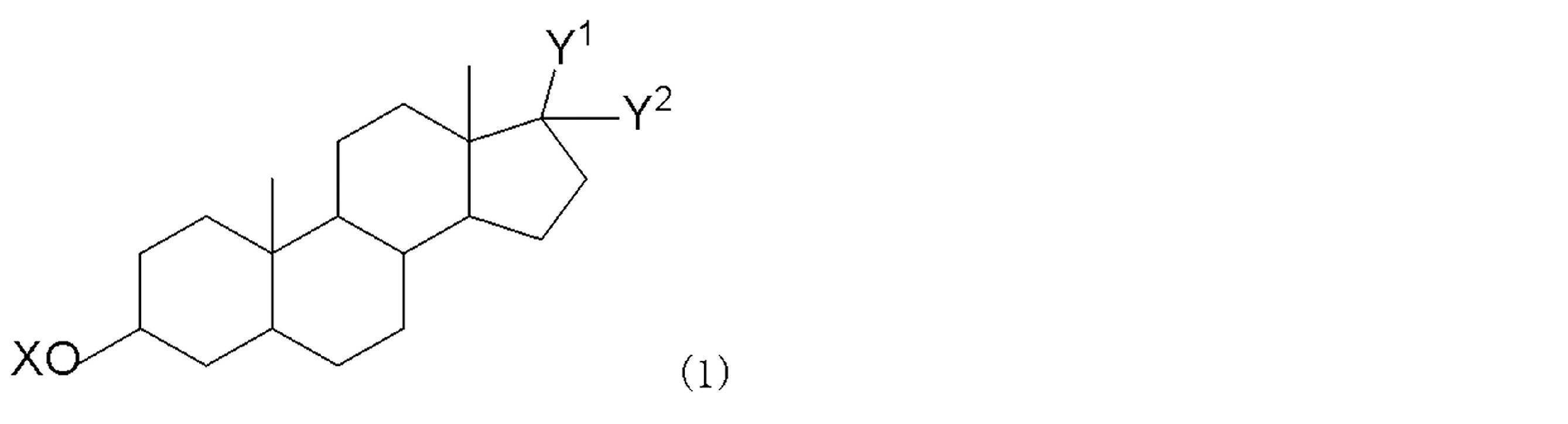
式中、Xはリン酸基、Y1及びY2は共同でメチレン基であるか、または、Y1は水素であり、Y2は、水素、水酸基、炭素数1~6のアルコキシ基、または炭素数1~6のアルキル基であり、
(ii) 一般式(1)で表されるアンドロステロン誘導体は、グルコシダーゼ、ガラクトシダーゼ、フルクトシダーゼまたはマンノシダーゼの基質として用いる場合は、
式中、Xは糖残基であり、糖残基はグルコース、ガラクトース、フルクトースおよびマンノースから成る群から選ばれる1種であり、Y1及びY2は共同でメチレン基または酸素原子であるか、または、Y1は水素であり、Y2は、水素、水酸基、炭素数1~6のアルコキシ基、または炭素数1~6のアルキル基であり、
(iii) 一般式(1)で表されるアンドロステロン誘導体は、ペルオキシダーゼの基質として用いる場合は、
式中、Xは-O-CO-R(但し、R は炭素数1~6のアルキル基またはフェニル基である)であり、Y1及びY2は共同でメチレン基または酸素原子であるか、または、Y1は水素であり、Y2は、水素、水酸基、炭素数1~6のアルコキシ基、または炭素数1~6のアルキル基であり、
前記酵素反応の反応生成物の定量は、NADHおよび/またはNADPH、チオNADおよび/またはチオNADP、並びにヒドロキシステロイド脱水素酵素(HSD)を用いて、酵素サイクリング反応によりチオNADHおよび/またはチオNADPHを生成させ、生成したチオNADHおよび/またはチオNADPHの量を測定するか、または生成したチオNADHおよび/またはチオNADPHによる色の変化を計測することで行う、前記方法。
[3]
前記酵素がアルカリホスファターゼであり、
一般式(1)で表されるアンドロステロン誘導体は、
式中、Xはリン酸基、Y1及びY2は共同でメチレン基であるか、または、Y1は水素であり、Y2は、水素、水酸基、炭素数1~6のアルコキシ基、または炭素数1~6のアルキル基である、[1]または[2]に記載の方法。
[4]
前記酵素がグルコシダーゼ、ガラクトシダーゼ、フルクトシダーゼまたはマンノシダーゼであり、一般式(1)で表されるアンドロステロン誘導体は、
式中、Xは糖残基であり、糖残基はグルコース、ガラクトース、フルクトースおよびマンノースから成る群から選ばれる1種であり、Y1及びY2は共同でメチレン基または酸素原子であるか、または、Y1は水素であり、Y2は、水素、水酸基、炭素数1~6のアルコキシ基、または炭素数1~6のアルキル基である、[1]または[2]に記載の方法。
[5]
前記酵素がペルオキシダーゼであり、一般式(1)で表されるアンドロステロン誘導体は、
式中、Xは-O-CO-R(但し、R は炭素数1~6のアルキル基またはフェニル基である)であり、Y1及びY2は共同でメチレン基または酸素原子であるか、または、Y1は水素であり、Y2は、水素、水酸基、炭素数1~6のアルコキシ基、または炭素数1~6のアルキル基である、[1]または[2]に記載の方法。
[6]
以下の(1)~(5)の試薬を含む酵素免疫測定用キット。
(1)標的タンパク質抗原に特異的な抗体を標識したアルカリホスファターゼ
(2)上記酵素の基質である一般式(1)で表されるアンドロステロン誘導体
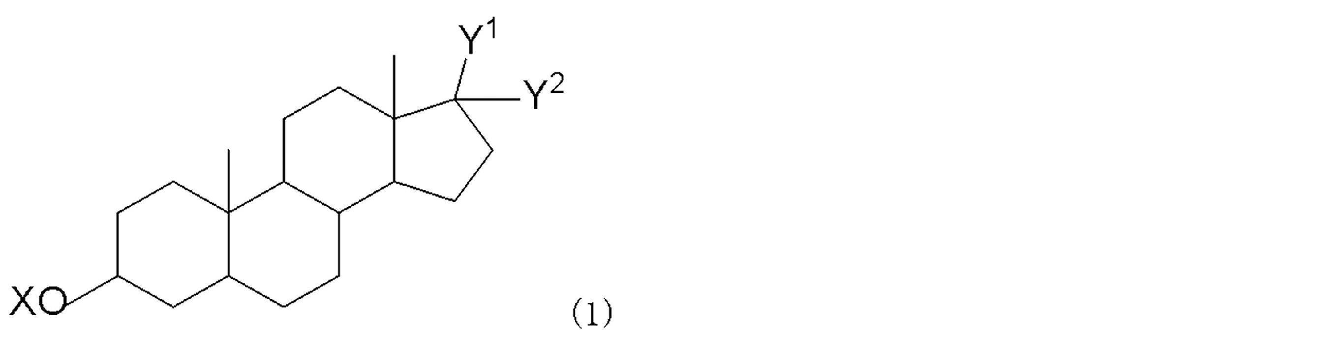
(3)ヒドロキシステロイド脱水素酵素(HSD)
(4)NADHおよび/またはNADPH、
(5)チオNADおよび/またはチオNADP
[7]
以下の(1)~(5)の試薬を含む酵素免疫測定用キット。
(1)標的タンパク質抗原に特異的な抗体を標識したグルコシダーゼ、ガラクトシダーゼ、フルクトシダーゼまたはマンノシダーゼ
(2)上記酵素の基質である一般式(1)で表されるアンドロステロン誘導体
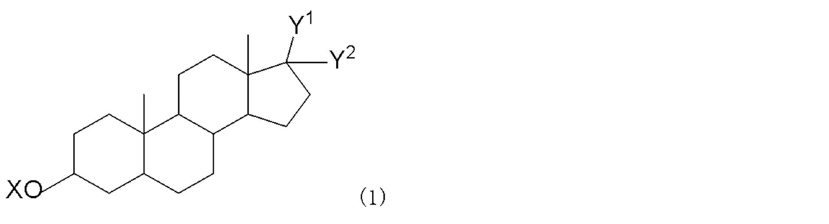
(3)ヒドロキシステロイド脱水素酵素(HSD)
(4)NADHおよび/またはNADPH、
(5)チオNADおよび/またはチオNADP
[8]
以下の(1)~(5)の試薬を含む酵素免疫測定用キット。
(1)標的タンパク質抗原に特異的な抗体を標識したペルオキシダーゼ、
(2)上記酵素の基質である一般式(1)で表されるアンドロステロン誘導体
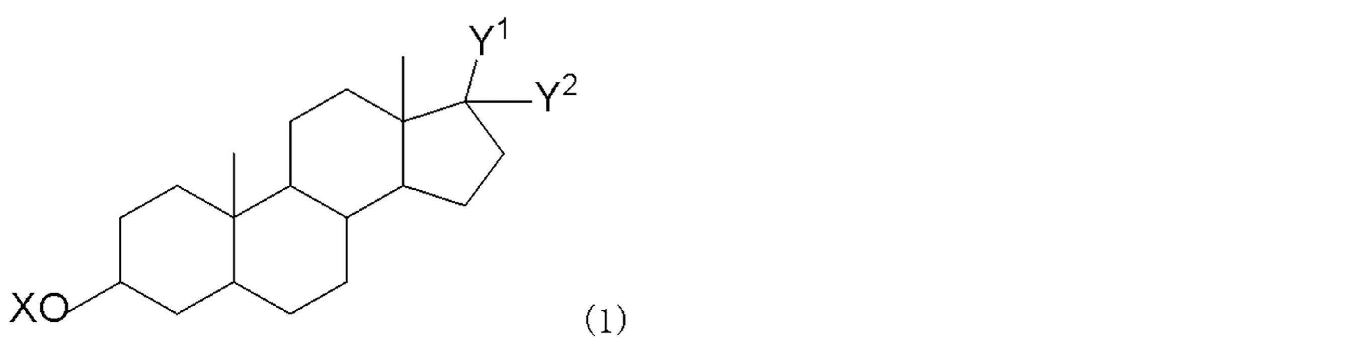
(3)ヒドロキシステロイド脱水素酵素(HSD)
(4)NADHおよび/またはNADPH、
(5)チオNADおよび/またはチオNADP
[9]
以下の(1)~(5)の試薬を含む核酸プローブ測定用キット。
(1)標的核酸に特異的に結合する核酸プローブで標識したアルカリホスファターゼ、
(2)上記酵素の基質である一般式(1)で表されるアンドロステロン誘導体

(3)ヒドロキシステロイド脱水素酵素(HSD)、
(4)NADHおよび/またはNADPH、
(5)チオNADおよび/またはチオNADP
[10]
以下の(1)~(5)の試薬を含む核酸プローブ測定用キット。
(1)標的核酸に特異的に結合する核酸プローブで標識したグルコシダーゼ、ガラクトシダーゼ、フルクトシダーゼまたはマンノシダーゼ、
(2)上記酵素の基質である一般式(1)で表されるアンドロステロン誘導体
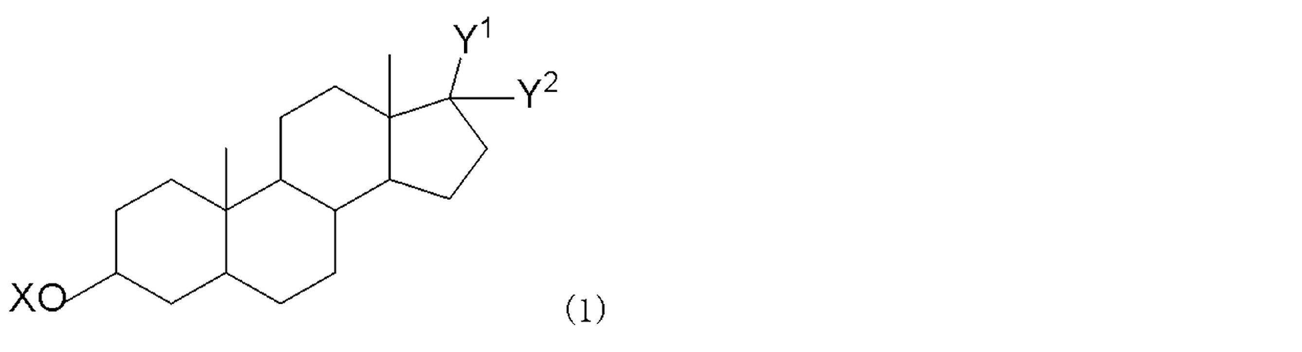
(3)ヒドロキシステロイド脱水素酵素(HSD)、
(4)NADHおよび/またはNADPH、
(5)チオNADおよび/またはチオNADP
[11]
以下の(1)~(5)の試薬を含む核酸プローブ測定用キット。
(1)標的核酸に特異的に結合する核酸プローブで標識したペルオキシダーゼ、
(2)上記酵素の基質である一般式(1)で表されるアンドロステロン誘導体
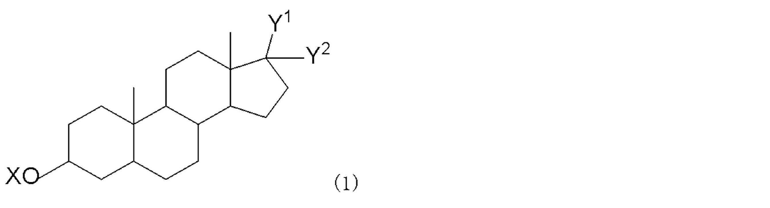
(3)ヒドロキシステロイド脱水素酵素(HSD)、
(4)NADHおよび/またはNADPH、
(5)チオNADおよび/またはチオNADP
[12]
下記一般式(1)で表されるアンドロステロン誘導体。
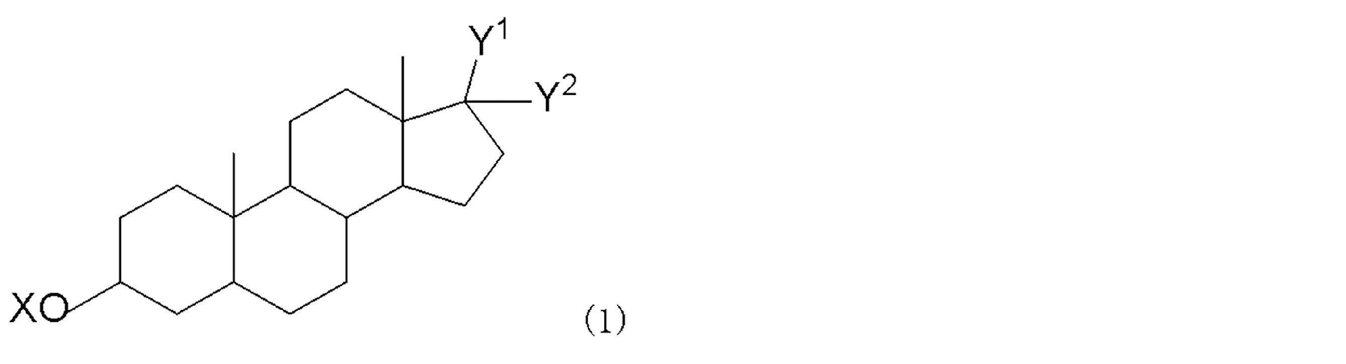
(A)Xはリン酸基であり、Y1及びY2は共同でメチレン基であるか、または、Y1は水素であり、Y2は水酸基、炭素数1~6のアルコキシ基または炭素数1~6のアルキル基である、
(B)Xはグルコース、ガラクトース、フルクトースおよびマンノースから成る群から選ばれる1種の糖残基であり、Y1及びY2は共同でメチレン基であるか、または、Y1は水素であり、Y2は水素、炭素数1~6のアルコキシ基、または炭素数1~6のアルキル基である、
(C)Xは-O-CO-Rであり、R は炭素数1~6のアルキル基またはフェニル基であり、Y1及びY2が共同でメチレン基または酸素原子であるか、または、Y1は水素であり、Y2は水素、水酸基、炭素数1~6のアルコキシ基、または炭素数1~6のアルキル基である。)
[13]
X、Y1およびY2の定義は、(A)Xはリン酸基であり、Y1及びY2は共同でメチレン基であるか、または、Y1は水素であり、Y2は水酸基、炭素数1~6のアルコキシ基または炭素数1~6のアルキル基である、[12]に記載のアンドロステロン誘導体。
[14]
X、Y1およびY2の定義は、 (B)Xはグルコース、ガラクトース、フルクトースおよびマンノースから成る群から選ばれる1種の糖残基であり、Y1及びY2は共同でメチレン基であるか、または、Y1は水素であり、Y2は水素、炭素数1~6のアルコキシ基、または炭素数1~6のアルキル基である、[12]に記載のアンドロステロン誘導体。
[15]
X、Y1およびY2の定義は、(C)Xは-O-CO-Rであり、R は炭素数1~6のアルキル基またはフェニル基であり、Y1及びY2が共同でメチレン基または酸素原子であるか、または、Y1は水素であり、Y2は水素、水酸基、炭素数1~6のアルコキシ基、または炭素数1~6のアルキル基である、[12]に記載のアンドロステロン誘導体。
本発明は下記一般式(1)で表されるアンドロステロン誘導体に関する。

(A)Xはリン酸基であり、Y1及びY2は共同でメチレン基であるか、または、Y1は水素であり、Y2は水酸基、炭素数1~6のアルコキシ基または炭素数1~6のアルキル基である、
(B)Xはグルコース、ガラクトース、フルクトースおよびマンノースから成る群から選ばれる1種の糖残基であり、Y1及びY2は共同でメチレン基であるか、または、Y1は水素であり、Y2は水素、炭素数1~6のアルコキシ基、または炭素数1~6のアルキル基である、
(C)Xは-O-CO-Rであり、R は炭素数1~6のアルキル基またはフェニル基であり、Y1及びY2が共同でメチレン基または酸素原子であるか、または、Y1は水素であり、Y2は水素、水酸基、炭素数1~6のアルコキシ基、または炭素数1~6のアルキル基である。)
本発明は、抗体酵素複合体を用いる酵素測定方法であって、
抗体酵素複合体の酵素による反応生成物の定量は、NADHおよび/またはNADPH、チオNADおよび/またはチオNADP、並びにデヒドロゲナーゼ(DH)を用いて、酵素サイクリング反応によりチオNADHおよび/またはチオNADPHを生成させ、生成したチオNADHおよび/またはチオNADPHの量を測定するか、または生成したチオNADHおよび/またはチオNADPHによる色の変化を計測することで行う、前記方法に関する。

法が測定対象とするタンパク質以外のすべての物質であることができる。
(1)抗体酵素複合体または酵素標識核酸プローブの濃度範囲
0.01μg/ml ~ 1 mg/ml
(2)標識酵素の基質の濃度範囲
1μM ~ 500 mM
(3)NADHおよび/またはNADPHの濃度範囲
0.01 mM ~ 50 mM
(4)チオNADおよび/またはチオNADPの濃度範囲
0.01 mM ~ 100 mM
(5)デヒドロゲナーゼ(DH)の濃度範囲
0.01 u/ml ~ 5000 u/ml
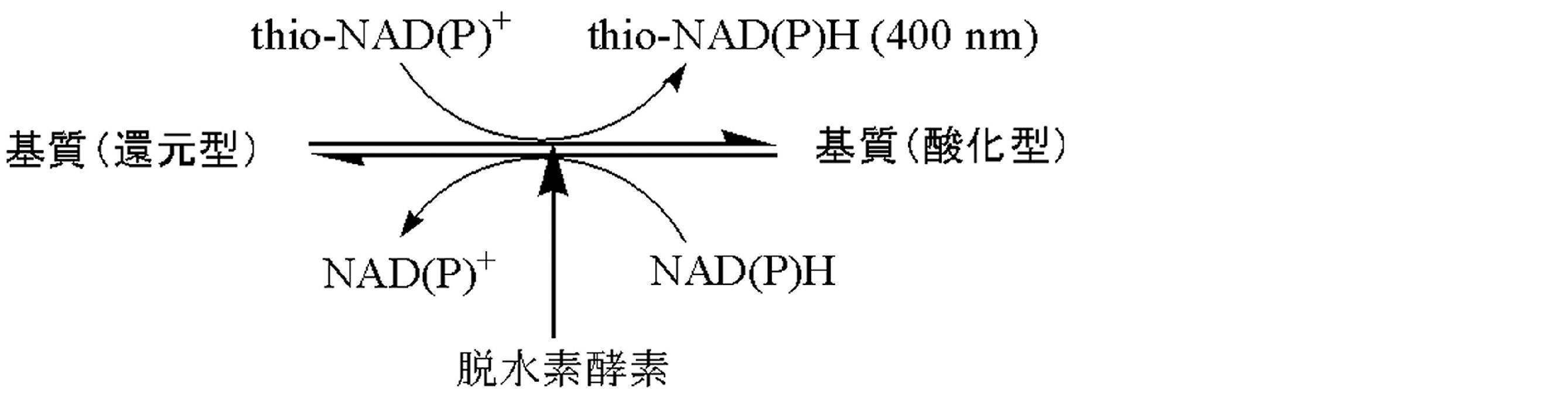
本発明では、標識酵素とサイクリング系を組み合わせることにより、初めて増幅反応を幾何級数的な反応とすることが可能となり、十分な高感度化を行うことが可能となった。
(1)標識酵素の生成物がアンドロステロンまたはその誘導体
(2)市販および汎用されている標識酵素が使用できる
(3)酵素サイクリングのターンオーバー数が大きい
(4)酵素サイクリング反応に使用する脱水素酵素に標識酵素のコンタミや標識酵素と同様の活性がない


本発明は、反応性担体に標識化された酵素およびその基質とサイクリング反応用の酵素とその補酵素のチオNADとNADHを含む酵素サイクリング法用キットを包含する。
反応性担体とは測定対象物と結合する活性を持った抗体・核酸プローブ・レクチン等を表す。
反応性担体は測定対象物に適した物を用いればよく、標識酵素およびその基質も適した物を用いればよく特に限定はされない。
(1)標的タンパク質抗原に特異的な抗体を標識した酵素であるアルカリホスファターゼ(ALP)またはグルコシダーゼまたはガラクトシダーゼまたはフルクトシダーゼまたはマンノシダーゼまたはペルオキシダーゼ、
(2)上記酵素の基質である一般式(1)で表されるアンドロステロン誘導体、
(3)デヒドロゲナーゼ(DH)、
(4)NADHおよび/またはNADPH、
(5)チオNADおよび/またはチオNADP
(1)標的核酸に特異的に結合する核酸プローブで標識した酵素であるアルカリホスファターゼ(ALP)またはグルコシダーゼまたはガラクトシダーゼまたはフルクトシダーゼまたはマンノシダーゼまたはペルオキシダーゼ、
(2)上記酵素の基質である一般式(1)で表されるアンドロステロン誘導体、
(3)デヒドロゲナーゼ(DH)、
(4)NADHおよび/またはNADPH、
(5)チオNADおよび/またはチオNADP
以下、本発明を実施例によりさらに詳細に説明する。
ALP標識抗体の作製
抗原に特異的反応性を有するマウス由来のモノクローナル抗体を0.1 Mクエン酸緩衝液(pH 3.5)で30分間の透析を3回行った。その抗体溶液に抗体量に対し0.5 %になるようにペプシンを添加し、37℃で1時間静置した後、1.5 Mトリス緩衝液(pH 8.8)で中性に調整した。この混合反応液をsuperdex 200を充填したカラム(Amersham Biosciences社)を用いてゲルろ過し、F(ab’)2を得た。
このF(ab’)2を100 μg用いて、ALP-labeling kit(同仁化学社)によりALPを標識した。
不溶性担体固定化抗体の調製1
抗原に特異的反応性を有するモルモット由来のポリクローナル抗体を50 mM炭酸ナトリウム緩衝液(pH 9.6)に100μg/mlの濃度で溶解した溶液を、平底マイクロプレート(Nunc社)の各ウェルに50μlずつ添加し、室温で1時間静置した後抗体溶液を回収し、2%ウシ血清アルブミン(BSA)含有TBSを300μl添加して室温で2時間静置してブロッキング処理を行い、ポリクローナル抗体固定化マイクロプレートを得た。
不溶性担体固定化抗体の調製2
抗原に特異的反応性を有するモルモット由来のポリクローナル抗体を50 mM炭酸ナトリウム緩衝液(pH 9.6)に10μg/mlの濃度で溶解した溶液を、平底マイクロプレート(Nunc社)の各ウェルに50μlずつ添加し、室温で1時間静置した後ウェル内の溶液を吸引除去し、0.1 %ウシ血清アルブミン(BSA)含有TBSを300μl添加して室温で2時間静置してブロッキング処理を行い、0.05 % Tween 20含有TBSで洗浄し、ポリクローナル抗体固定化マイクロプレートを得た。
反応試液
0.2 M グリシン緩衝液(pH 8.8)
1.5 mM thio-NAD
1 mM NADH
0.5 mM 5α-Androsterone 3-phosphate
20 U/ml 3α-Hydroxysteroid dehydrogenase
0.1、0.5、1、2、5、10、50、100、200 ng/ml Pumilio
参考例2の方法で調製した抗Pumilioモルモットポリクローナル抗体を固定化したマイクロプレートに、精製したPumilio(標準物質)を0~200 ng/mlの範囲で含有する、0.1 % BSA含有TBS(pH 7.5)溶液を50μl添加し、室温で1時間振とうさせた。次にウェル内の溶液を吸引除去後、0.05 % Tween 20含有TBS(pH 7.5)で洗浄し、参考例1の方法で作製したALP標識抗Pumilioマウスモノクローナル抗体を2.5μg/mlの濃度で含有する、0.1 % BSA含有TBS(pH 7.5)溶液を50μl加え、1時間振とうさせた。ウェル内の溶液を吸引除去後、0.05 % Tween 20含有TBS(pH 7.5)で洗浄、さらにTBSで洗浄した。次に各ウェルに反応試液を100μlずつ添加し、37℃で加温しながらマイクロプレートリーダー(コロナ社製MTP-500)で405 nmのフィルターを使用して30分間吸光度を測定した。30分間の吸光度変化量を標準物質濃度に対してプロットすることにより得られた、濃度依存性のよい直線を図2に示す。
反応試液A
0.1 M トリス緩衝液(pH 8.3)
1 mM MgCl2
0.1 mM 5α-Androsterone 3-phosphate
0.2 M リン酸水素二ナトリウム含有0.2 M グリシン緩衝液(pH 9.6)
3 mM thio-NAD
1 mM NADH
40 U/ml 3α-Hydroxysteroid dehydrogenase
0.5、0.75、1、2.5、5、7.5、10、12.5、20、50、75、100 ng/ml Pumilio
参考例3の方法で調製した抗Pumilioモルモットポリクローナル抗体を固定化したマイクロプレートに、精製したPumilio(標準物質)を0~100 ng/mlの範囲で含有する、0.1 % BSA含有TBS(pH 7.5)溶液を50μl添加し、室温で1時間振とうさせた。次にウェル内の溶液を吸引除去後、0.05 % Tween 20含有TBS(pH 7.5)で洗浄し、参考例1の方法で作製したALP標識抗Pumilioマウスモノクローナル抗体を2.5μg/mlの濃度で含有する、0.1 % BSA含有TBS(pH 7.5)溶液を50μl加え、1時間振とうさせた。ウェル内の溶液を吸引除去後、0.05 % Tween 20含有TBS(pH 7.5)で洗浄、さらにTBSで洗浄した。次に各ウェルに反応試液Aを50μlずつ添加し、37℃で30分間インキュベートした。続いてそのウェルに反応試液Bを50μl添加し、37℃で加温しながらマイクロプレートリーダー(コロナ社製MTP-500)で405 nmのフィルターを使用して30分間吸光度を測定した。30分間の吸光度変化量を標準物質濃度に対してプロットすることにより得られた、濃度依存性のよい直線を図3に示す。
0.1 M トリス緩衝液(pH 8.6)
0.5 mM MgCl2
1.5 mM thio-NAD
0.5 mM NADH
0.5 mM 5β-Androsterone 3-phosphate
20 U/ml 3α-Hydroxysteroid dehydrogenase
0.1、0.5、1、2、5、8、10、20、50 ng/ml Pumilio
参考例3の方法で調製した抗Pumilioモルモットポリクローナル抗体を固定化したマイクロプレートに、精製したPumilio(標準物質)を0~50 ng/mlの範囲で含有する、0.1 % BSA含有TBS(pH 7.5)溶液を50μl添加し、室温で1時間振とうさせた。次にウェル内の溶液を吸引除去後、0.05 % Tween 20含有TBS(pH 7.5)で洗浄し、参考例1の方法で作製したALP標識抗Pumilioマウスモノクローナル抗体を2.5μg/mlの濃度で含有する、0.1 % BSA含有TBS(pH 7.5)溶液を50 μl加え、1時間振とうさせた。ウェル内の溶液を吸引除去後、0.05 % Tween 20含有TBS(pH 7.5)で洗浄、さらにTBSで洗浄した。次に各ウェルに反応試液を50μlずつ添加し、37℃で加温しながらマイクロプレートリーダー(コロナ社製MTP-500)で405 nmのフィルターを使用して30分間吸光度を測定した。30分間の吸光度変化量を標準物質濃度に対してプロットすることにより得られた、濃度依存性のよい直線を図4に示す。
0.1 M トリス緩衝液(pH 8.3)
1 mM MgCl2
0.1 mM 5β-Androsterone 3-phosphate
0.2 M リン酸水素二ナトリウム含有0.2 M グリシン緩衝液(pH 9.6)
3 mM thio-NAD
1 mM NADH
40 U/ml 3α-Hydroxysteroid dehydrogenase
0.005、0.01、0.05、0.1、0.25、0.5、0.75、1、2.5、5、10、25、50 ng/ml Pumilio
参考例3の方法で調製した抗Pumilioモルモットポリクローナル抗体を固定化したマイクロプレートに、精製したPumilio(標準物質)を0~50 ng/mlの範囲で含有する、0.1 % BSA含有TBS(pH 7.5)溶液を50μl添加し、室温で1時間振とうさせた。次にウェル内の溶液を吸引除去後、0.05 % Tween 20含有TBS(pH 7.5)で洗浄し、参考例1の方法で作製したALP標識抗Pumilioマウスモノクローナル抗体を2.5μg/mlの濃度で含有する、0.1 % BSA含有TBS(pH 7.5)溶液を50μl加え、1時間振とうさせた。ウェル内の溶液を吸引除去後、0.05 % Tween 20含有TBS(pH 7.5)で洗浄、さらにTBSで洗浄した。次に各ウェルに反応試液Aを50μlずつ添加し、37℃で30分間インキュベートした。続いてそのウェルに反応試液Bを50μl添加し、37℃で加温しながらマイクロプレートリーダー(コロナ社製MTP-500)で405 nmのフィルターを使用して10分間吸光度を測定した。10分間の吸光度変化量を標準物質濃度に対してプロットすることにより得られた、濃度依存性のよい直線を図5に示す。


オキシ塩化リン(0.26mL)の無水ピリジン溶液(4 mL)に、氷冷下で3α-Hydroxy-17β-methoxy-5β-androstane (200mg)の無水ピリジン溶液 (4mL)を滴下し、氷冷下でさらに1時間攪拌した。反応液を氷水に注ぎ、溶液を強アルカリ性 (pH 約11)とした後、エーテルで洗浄した。次いで、水層に濃塩酸を加えpHを約2に調整した後、エーテルで抽出した。エーテル層を無水硫酸ナトリウムで乾燥後、減圧下溶媒を留去した。残渣をテトラヒドロフラン-ヘキサンで再結晶し無色結晶(210 mg)を得た。
ESI-HR-MS Calculated for C20H35O5P-H: m/z 385.2149 [M-H]-. Found m/z 385.2157
m.p. 187-188℃
0.1 Mトリス緩衝液(pH 9.5)
0.2 mM MgCl2
0.6 mM thio-NAD
4 mM NADH
0.5 mM 3α-Hydroxy-17β-methoxy-5β-Androstane 3-phosphate
20 U/ml 3α-Hydoroxysteroid dehydrogenase
0.1、0.2、0.5、1、2 mU/ml Alkaline phosphatase(最終濃度 0.05、0.1、0.25、0.5、1 mU/ml)
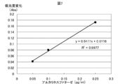
平底のマイクロプレートウェルに標準物質(Alkaline phosphatase)を0~2 mU/mlの範囲で含有するトリス緩衝溶液(pH 9.5)を50μl添加し、続いてそのウェルに反応試液Bを50μlずつ各ウェルに添加し、37℃で加温しながらマイクロプレートリーダー(コロナ社製MTP-500)で405 nmのフィルターを使用して60分間吸光度を測定した。60分間の吸光度変化量を標準物質濃度に対してプロットすることにより得られた、濃度依存性のよい直線を図7に示す。
0.1 M トリス緩衝液(pH 9.5)
0.2 mM MgCl2
1 mM 3α-Hydroxy-17β-methoxy-5β-Androstane 3-phosphate
0.15 M ピロリン酸緩衝液(pH 9.5)
3 mM thio-NAD
1 mM NADH
40 U/ml 3α-Hydoroxysteroid dehydrogenase
1、5、10、15、20 μU/ml Alkaline phosphatase(最終濃度 0.5、2.5、5、7.5、10 μU/ml)
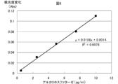
平底のマイクロプレートウェルに標準物質(Alkaline phosphatase)を0~20μU/mlの範囲で含有するトリス緩衝溶液(pH 9.5)を25μl添加し、続いてそのウェルに反応試液Aを25μlずつ添加して混合し、37℃のインキュベーターで1時間静置する。次に反応試液Bを50μlずつ各ウェルに添加し、37℃で加温しながらマイクロプレートリーダー(コロナ社製MTP-500)で405 nmのフィルターを使用して20分間吸光度を測定した。20分間の吸光度変化量を標準物質濃度に対してプロットすることにより得られた、濃度依存性のよい直線を図8に示す。
Alkaline phosphatase(ALP)標識抗体の精製
参考例1のように得たF(ab’)2を150μμg用いてALP-labeling-kit(同仁化学)によりALP標識抗体を作製した。このALP標識抗体液中にはALPとFabの様々な分子量の複合体が存在し、活性にバラつきがあるので、作製したALP標識抗体液を1 mM Mgl2、0.1 mM ZnCl2、0.01 % BSAを含有するトリス緩衝液(pH 7.0)を溶出液としてsuperdex 200を充填したカラム(Amersham Biosciences社)を用いてゲルろ過し、各フラクションの活性を比較した上で、S/N比の大きいALP標識抗体のみを回収した。
不溶性担体固定化抗体の調製
抗原に特異的反応性を有するモルモット由来のポリクローナル抗体を50 mM炭酸ナトリウム緩衝液(pH 9.6)に10μg/mlの濃度で溶解した溶液を、平底マイクロプレート(Nunc社)の各ウェルに50μlずつ添加し、室温で1時間静置した後ウェル内の溶液を吸引除去し、0.5 % Triton X-100含有TBS(pH 7.5)で洗浄を行った。次に5 %ウシ血清アルブミン(BSA)含有TBSを200μl添加して4℃で2時間から一晩静置してブロッキング処理を行い、0.5 % Triton X-100含有TBS(pH 7.5)で洗浄し、ポリクローナル抗体固定化マイクロプレートを得た。
反応試液
0.1 Mトリス緩衝液(pH 9.5)
1.5 mM thio-NAD
2.5 mM NADH
0.1 mM 3α-Hydroxy-17β-methoxy-5β-Androstane 3-phosphate
20 U/ml 3α-Hydoroxysteroid dehydrogenase
0.1、0.5、2、5、10、25 ng/ml Pumilio
参考例5の方法で調製した抗Pumilioモルモットポリクローナル抗体を固定化したマイクロプレートに、精製したPumilio(標準物質)を0~25 ng/mlの範囲で含有する、0.1 % BSA含有TBS(pH 7.5)溶液を50 μl添加し、室温で2時間振とうさせた。次にウェル内の溶液を吸引除去後、0.5 % Triton X-100含有TBS(pH 7.5)で洗浄し、参考例4の方法で作製したALP標識抗Pumilioマウスモノクローナル抗体をおよそ0.8μg/mlの濃度で含有する、0.1 % BSA含有TBS(pH 7.5)溶液を50μl加え、4℃で2時間振とうさせた。ウェル内の溶液を吸引除去後、0.5 % Triton X-100含有TBS(pH 7.5)で洗浄、さらにTBSで洗浄した。次に各ウェルに反応試液を50μlずつ添加し、37℃で加温しながらマイクロプレートリーダー(コロナ社製MTP-500)で405 nmのフィルターを使用して60分間吸光度を測定した。60分間の吸光度変化量を標準物質濃度に対してプロットすることにより得られた、濃度依存性のよい直線を図9に示す。
反応試液A
0.1 M トリス緩衝液(pH 9.5)
0.1mM MgCl2
0.2 mM 3α-Hydroxy-17β-methoxy-5β-Androstane 3-phosphate
反応試液B
0.15 M ピロリン酸緩衝液(pH 9.5)
3 mM thio-NAD
1 mM NADH
40 U/ml 3α-Hydoroxysteroid dehydrogenase
試料
0.01、0.025、0.05、0.1、0.25、0.5 ng/ml Pumilio
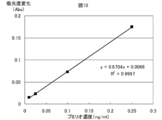
参考例5の方法で調製した抗Pumilioモルモットポリクローナル抗体を固定化したマイクロプレートに、精製したPumilio(標準物質)を0~0.5 ng/mlの範囲で含有する、0.1 % BSA含有TBS(pH 7.5)溶液を50μl添加し、室温で2時間振とうさせた。次にウェル内の溶液を吸引除去後、0.5 % Triton X-100含有TBS(pH 7.5)で洗浄し、参考例4の方法で作製したALP標識抗Pumilioマウスモノクローナル抗体をおよそ0.2μg/ml、ウサギF(ab’)2を1 mg/mlの濃度で含有する、0.1 % BSA含有TBS(pH 7.5)溶液を50 μl加え、4℃で2時間振とうさせた。ウェル内の溶液を吸引除去後、0.5 % Triton X-100含有TBS(pH 7.5)で洗浄、さらにTBSで洗浄した。次に各ウェルに反応試液Aを50μlずつ添加し、37℃で30分間インキュベートした。続いてそのウェルに反応試液Bを50μl添加し、37℃で加温しながらマイクロプレートリーダー(コロナ社製MTP-500)で405 nmのフィルターを使用して30分間吸光度を測定した。30分間の吸光度変化量を標準物質濃度に対してプロットすることにより得られた、濃度依存性のよい直線を図10に示す。
5β-Androsterone (290mg)のジクロロメタン溶液(15 mL)に、MS4A (1.25 g)、Phenyl 2,3,4,6-O-tetrabenzoyl-1-thio-β-D-garactopyranoside (1.03g)を加えて-20℃で15分間攪拌した。次いで、N-ヨードスクシンイミド (435mg), トリフルオロメタンスルホン酸 (15mg)を加え-20℃で2時間攪拌した後、室温にてさらに2時間反応した。反応液にトリエチルアミン250μLを添加し反応を停止し、反応液を飽和炭酸ナトリウムとチオ硫酸ナトリウムの3:1の混液、飽和食塩水で順次洗浄した。有機層を無水硫酸ナトリウムで乾燥した後、減圧下溶媒を留去した。残渣をメタノール(15 mL)に溶解し、28 % ナトリムメトキシド(500 μL)加え2時間攪拌した。反応液に酢酸を加えて中和した後、減圧乾固した。残渣をシリカゲルカラムクロマトグラフィー (24×240 mm, 10 % メタノール-クロロホルム)にて精製後、メタノールで再結晶し無色結晶(360mg)を得た。
ESI-HR-MS Calculated for C25H40O7-H: m/z 451.2701 [M-H]-. Found m/z 451.2711
m.p. 130-135℃
0.1 M リン酸緩衝液(pH 7.3)
0.2 mM MgCl2
0.1 mM 5β-Androsterone β-D-Galactoside
0.1 M ピロリン酸緩衝液(pH 9.0)
1.2 mM thio-NAD
3.6 mM NADH
40 U/ml 3α-Hydoroxysteroid dehydrogenase
1、4、10、40、100、200 pg/ml β-Galactosidase(最終濃度 0.5、2、5、20、50、100 pg/ml)
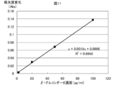
平底のマイクロプレートウェルに標準物質(β-Galactosidase)を0~200 pg/mlの範囲で含有するリン酸緩衝溶液(pH 7.3)を25μl添加し、続いてそのウェルに反応試液Aを25μlずつ添加して混合し、37℃で60分間インキュベートした。次に反応試液Bを50μlずつ各ウェルに添加し、37℃で加温しながらマイクロプレートリーダー(コロナ社製MTP-500)で405 nmのフィルターを使用して30分間吸光度を測定した。30分間の吸光度変化量を標準物質濃度に対してプロットすることにより得られた、濃度依存性のよい直線を図11に示す。
0.1 Mリン酸緩衝液(pH 7.7)
10 mM MgCl2
6 mM thio-NAD
1 mM NADH
0.1 mM 5β-Androsterone β-D-Galactoside
40 U/ml 3α-Hydoroxysteroid dehydrogenase
5、10、25、50、100、250 pg/ml β-Galactosidase(最終濃度 2.5、5、12.5、25、50、125 pg/ml)
平底のマイクロプレートウェルに標準物質(β-Galactosidase)を0~250 pg/mlの範囲で含有するリン酸緩衝溶液(pH 7.7)を50μl添加し、続いてそのウェルに反応試液Bを50μlずつ各ウェルに添加し、37℃で加温しながらマイクロプレートリーダー(コロナ社製MTP-500)で405 nmのフィルターを使用して120分間吸光度を測定した。120分間の吸光度変化量を標準物質濃度に対してプロットすることにより得られた、濃度依存性のよい直線を図12に示す。
0.1 M リン酸緩衝液(pH 7.5)
10 mM MgCl2
0.1 mM 5β-Androsterone β-D-Galactoside
0.1 M ピロリン酸緩衝液(pH 9.0)
4.8 mM thio-NAD
0.4 mM NADH
40 U/ml 3α-Hydoroxysteroid dehydrogenase
1、4、10、40、100、200 pg/ml β-Galactosidase(最終濃度 0.5、2、5、20、50、100 pg/ml)
平底のマイクロプレートウェルに標準物質(β-Galactosidase)を0~200 pg/mlの範囲で含有するリン酸緩衝溶液(pH 7.5)を25μl添加し、続いてそのウェルに反応試液Aを25μlずつ添加して混合し、37℃で60分間インキュベートした。次に反応試液Bを50μlずつ各ウェルに添加し、37℃で加温しながらマイクロプレートリーダー(コロナ社製MTP-500)で405 nmのフィルターを使用して30分間吸光度を測定した。30分間の吸光度変化量を標準物質濃度に対してプロットすることにより得られた、濃度依存性のよい直線を図13に示す。
3α-Hydroxy-17β-methoxy-5β-Androstane (306mg)のジクロロメタン溶液(15 mL)に、MS4A (1.25 g)、Phenyl 2,3,4,6-O-tetrabenzoyl-1-thio-β-D-garactopyranoside (1.03g)を加えて-20℃で15分間攪拌した。次いで、N-ヨードスクシンイミド(435mg), トリフルオロメタンスルホン酸 (15mg)を加え-20℃で2時間攪拌した後、室温にてさらに2時間反応した。反応液にトリエチルアミン250μLを添加し反応を停止し、反応液を飽和炭酸ナトリウムとチオ硫酸ナトリウムの3:1の混液、飽和食塩水で順次洗浄した。有機層を無水硫酸ナトリウムで乾燥した後、減圧下溶媒を留去した。残渣をメタノール(15 mL)に溶解し、28 % ナトリムメトキシド(500 μL)加え2時間攪拌した。反応液に酢酸を加えて中和した後、減圧乾固した。残渣をシリカゲルカラムクロマトグラフィー (24×240 mm, 10 % メタノール-クロロホルム)にて精製後、アセトン-ヘキサンで再結晶し無色結晶(383mg)を得た。
ESI-HR-MS Calculated for C26H44O7-H: m/z 467.3014 [M-H]-. Found m/z 451.3026
m.p. 197-199℃
0.1 Mリン酸緩衝液(pH 8.5)
10 mM MgCl2
6 mM thio-NAD
1 mM NADH
0.2 mM 3α-Hydroxy-17β-methoxy-5β-Androstane β-D-Galactoside
20 U/ml 3α-Hydoroxysteroid dehydrogenase
5、10、25、50、100、250 pg/ml β-Galactosidase(最終濃度 2.5、5、12.5、25、50、125 pg/ml)
平底のマイクロプレートウェルに標準物質(β-Galactosidase)を0~250 pg/mlの範囲で含有するリン酸緩衝溶液(pH 8.5)を50μl添加し、続いてそのウェルに反応試液Bを50μlずつ各ウェルに添加し、37℃で加温しながらマイクロプレートリーダー(コロナ社製MTP-500)で405 nmのフィルターを使用して120分間吸光度を測定した。120分間の吸光度変化量を標準物質濃度に対してプロットすることにより得られた、濃度依存性のよい直線を図14に示す。
0.1 M リン酸緩衝液(pH 7.5)
4 mM MgCl2
0.1 mM 3α-Hydroxy-17β-methoxy-5β-Androstane β-D-Galactoside
0.1 M ピロリン酸緩衝液(pH 9.8)
1.2 mM thio-NAD
1 mM NADH
40 U/ml 3α-Hydoroxysteroid dehydrogenase
1、4、10、40、100、200 pg/ml β-Galactosidase(最終濃度 0.5、2、5、20、50、100 pg/ml)
平底のマイクロプレートウェルに標準物質(β-Galactosidase)を0~200 pg/mlの範囲で含有するリン酸緩衝溶液(pH 7.5)を25μl添加し、続いてそのウェルに反応試液Aを25μlずつ添加して混合し、37℃で60分間インキュベートした。次に反応試液Bを50μlずつ各ウェルに添加し、37℃で加温しながらマイクロプレートリーダー(コロナ社製MTP-500)で405 nmのフィルターを使用して30分間吸光度を測定した。30分間の吸光度変化量を標準物質濃度に対してプロットすることにより得られた、濃度依存性のよい直線を図15に示す。
ビオチン標識抗体の作製
参考例1のように得たF(ab’)2を50μg用いてbiotin-labeling-kit(同仁化学)によりビオチン標識抗体を作製した。
0.1 Mリン酸緩衝液(pH 7.7)
5 mM MgCl2
1.2 mM thio-NAD
0.8 mM NADH
0.05 mM 5β-Androstaneβ-D-Galactoside
20 U/ml 3α-Hydoroxysteroid dehydrogenase
0.05、0.1、0.25、0.5、1、2.5 ng/ml Pumilio
参考例5の方法で調製した抗Pumilioモルモットポリクローナル抗体を固定化したマイクロプレートに、精製したPumilio(標準物質)を0~2.5 ng/mlの範囲で含有する、0.1 % BSA含有TBS(pH 7.5)溶液を50μl添加し、室温で2時間振とうさせた。次にウェル内の溶液を吸引除去後、0.5 % Triton X-100含有TBS(pH 7.5)で洗浄し、参考例6の方法で作製したビオチン標識抗Pumilioマウスモノクローナル抗体をおよそ0.1μg/mlの濃度で含有する、0.1 % BSA含有TBS(pH 7.5)溶液を50μl加え、室温で1時間振とうさせた。ウェル内の溶液を吸引除去後、0.5 % Triton X-100含有TBS(pH 7.5)で洗浄し、β-Galactosidase標識ストレプトアビジン(Roche社)を0.1 U conjugate/mlの濃度で含有する、0.1 % BSA含有TBS(pH 7.5)溶液を50 μl添加し、室温で30分間振とうさせた。ウェル内の溶液を吸引除去後、0.5 % Triton X-100含有TBS(pH 7.5)で洗浄し、さらにTBSで洗浄した。次に各ウェルに反応試液を50μlずつ添加し、37℃で加温しながらマイクロプレートリーダー(コロナ社製MTP-500)で405 nmのフィルターを使用して60分間吸光度を測定した。60分間の吸光度変化量を標準物質濃度に対してプロットすることにより得られた、濃度依存性のよい直線を図16に示す。
0.1 M リン酸緩衝液(pH 7.5)
0.1mM MgCl2
0.05 mM 5β-Androsterone β-D-Galactoside
0.1 M ピロリン酸緩衝液(pH 9.0)
3 mM thio-NAD
2 mM NADH
40 U/ml 3α-Hydoroxysteroid dehydrogenase
0.05、0. 1、0.5、1、2.5、5 ng/ml Pumilio
参考例5の方法で調製した抗Pumilioモルモットポリクローナル抗体を固定化したマイクロプレートに、精製したPumilio(標準物質)を0~5 ng/mlの範囲で含有する、0.1 % BSA含有TBS(pH 7.5)溶液を50μl添加し、室温で2時間振とうさせた。次にウェル内の溶液を吸引除去後、0.5 % Triton X-100含有TBS(pH 7.5)で洗浄し、参考例6の方法で作製したビオチン標識抗Pumilioマウスモノクローナル抗体をおよそ0.1μg/mlの濃度で含有する、0.1 % BSA含有TBS(pH 7.5)溶液を50μl加え、室温で1時間振とうさせた。ウェル内の溶液を吸引除去後、0.5 % Triton X-100含有TBS(pH 7.5)で洗浄し、β-Galactosidase標識ストレプトアビジン(Roche社)を0.1 U conjugate/mlの濃度で含有する、0.1 % BSA含有TBS(pH 7.5)溶液を50 μl添加し、室温で30分間振とうさせた。ウェル内の溶液を吸引除去後、0.5 % Triton X-100含有TBS(pH 7.5)で洗浄し、さらにTBSで洗浄した。次に各ウェルに反応試液Aを50 μlずつ添加し、37℃で30分間インキュベートした。続いてそのウェルに反応試液Bを50μl添加し、37℃で加温しながらマイクロプレートリーダー(コロナ社製MTP-500)で405 nmのフィルターを使用して30分間吸光度を測定した。30分間の吸光度変化量を標準物質濃度に対してプロットすることにより得られた、濃度依存性のよい直線を図17に示す。
0.1 Mリン酸緩衝液(pH 8.5)
1.2 mM thio-NAD
0.8 mM NADH
0.05 mM 3α-Hydroxy-17β-methoxy-5β-Androstaneβ-D-Galactoside
20 U/ml 3α-Hydoroxysteroid dehydrogenase
0.1、0.5、1、2.5、5、10 ng/ml Pumilio
参考例5の方法で調製した抗Pumilioモルモットポリクローナル抗体を固定化したマイクロプレートに、精製したPumilio(標準物質)を0~10 ng/mlの範囲で含有する、0.1 % BSA含有TBS(pH 7.5)溶液を50μl添加し、室温で2時間振とうさせた。次にウェル内の溶液を吸引除去後、0.5 % Triton X-100含有TBS(pH 7.5)で洗浄し、参考例6の方法で作製したビオチン標識抗Pumilioマウスモノクローナル抗体をおよそ0.1μg/mlの濃度で含有する、0.1 % BSA含有TBS(pH 7.5)溶液を50μl加え、室温で1時間振とうさせた。ウェル内の溶液を吸引除去後、0.5 % Triton X-100含有TBS(pH 7.5)で洗浄し、β-Galactosidase標識ストレプトアビジン(Roche社)を0.1 U conjugate/mlの濃度で含有する、0.1 % BSA含有TBS(pH 7.5)溶液を50 μl添加し、室温で30分間振とうさせた。ウェル内の溶液を吸引除去後、0.5 % Triton X-100含有TBS(pH 7.5)で洗浄し、さらにTBSで洗浄した。次に各ウェルに反応試液を50μlずつ添加し、37℃で加温しながらマイクロプレートリーダー(コロナ社製MTP-500)で405 nmのフィルターを使用して60分間吸光度を測定した。60分間の吸光度変化量を標準物質濃度に対してプロットすることにより得られた、濃度依存性のよい直線を図18に示す。
0.1 M リン酸緩衝液(pH 7.0)
0.05 mM 3α-Hydroxy-17β-methoxy-5β-Androstaneβ-D-Galactoside
0.1 M ピロリン酸緩衝液(pH 9.0)
2.4 mM thio-NAD
2 mM NADH
40 U/ml 3α-Hydoroxysteroid dehydrogenase
0.25、0.5、1、2.5、5、10 ng/ml Pumilio
参考例5の方法で調製した抗Pumilioモルモットポリクローナル抗体を固定化したマイクロプレートに、精製したPumilio(標準物質)を0~10 ng/mlの範囲で含有する、0.1 % BSA含有TBS(pH 7.5)溶液を50μl添加し、室温で2時間振とうさせた。次にウェル内の溶液を吸引除去後、0.5 % Triton X-100含有TBS(pH 7.5)で洗浄し、参考例6の方法で作製したビオチン標識抗Pumilioマウスモノクローナル抗体をおよそ0.3μg/mlの濃度で含有する、0.1 % BSA含有TBS(pH 7.5)溶液を50 μl加え、室温で1時間振とうさせた。ウェル内の溶液を吸引除去後、0.5 % Triton X-100含有TBS(pH 7.5)で洗浄し、β-Galactosidase標識ストレプトアビジン(Roche社)を0.1 U conjugate/mlの濃度で含有する、0.1 % BSA含有TBS(pH 7.5)溶液を50μl添加し、室温で30分間振とうさせた。ウェル内の溶液を吸引除去後、0.5 % Triton X-100含有TBS(pH 7.5)で洗浄し、さらにTBSで洗浄した。次に各ウェルに反応試液Aを50μlずつ添加し、37℃で60分間インキュベートした。続いてそのウェルに反応試液Bを50 μl添加し、37℃で加温しながらマイクロプレートリーダー(コロナ社製MTP-500)で405 nmのフィルターを使用して30分間吸光度を測定した。30分間の吸光度変化量を標準物質濃度に対してプロットすることにより得られた、濃度依存性のよい直線を図19に示す。
0.1 M グリシン緩衝液(pH 10.3)
1 mM MgCl2
1 mM ZnCl2
1 mg/ml p-NPP
0、1、2.5、5、10、25 ng/ml Pumilio
参考例3の方法で調製した抗Pumilioモルモットポリクローナル抗体を固定化したマイクロプレートに、精製したPumilio(標準物質)を0~25 ng/mlの範囲で含有する、0.1 % BSA含有TBS(pH 7.5)溶液を50μl添加し、室温で1時間振とうさせた。次にウェル内の溶液を吸引除去後、0.05 % Tween 20含有TBS(pH 7.5)で洗浄し、参考例1の方法で作製したALP標識抗Pumilioマウスモノクローナル抗体を1μg/mlの濃度で含有する、0.1 % BSA含有TBS(pH 7.5)溶液を50μl加え、1時間振とうさせた。ウェル内の溶液を吸引除去後、0.05 % Tween 20含有TBS(pH 7.5)で洗浄し、各ウェルに反応試液を100μl添加し、37℃で加温しながらマイクロプレートリーダー(コロナ社製MTP-500)で405 nmのフィルターを使用して30分間吸光度を測定した。30分間の吸光度変化量を標準物質濃度に対してプロットすることにより得られた、濃度依存性のよい直線を図20に示す。
0.1 M リン酸緩衝液(pH 7.3)
0.2 mM 2-mercaptoethanol
1 mM MgCl2
10 mM ONPG
2、10、20、50 ng/ml β-Galactosidase(最終濃度 1、5、10、25 ng/ml)
平底のマイクロプレートウェルに標準物質(β-Galactosidase)を0~25 ng/mlの範囲で含有するリン酸緩衝溶液(pH 7.3)を50μl添加し、続いてそのウェルに反応試液を50μlずつ各ウェルに添加し、37℃で加温しながらマイクロプレートリーダー(コロナ社製MTP-500)で415 nmのフィルターを使用して60分間吸光度を測定した。60分間の吸光度変化量を標準物質濃度に対してプロットすることにより得られた、濃度依存性のよい直線を図21に示す。
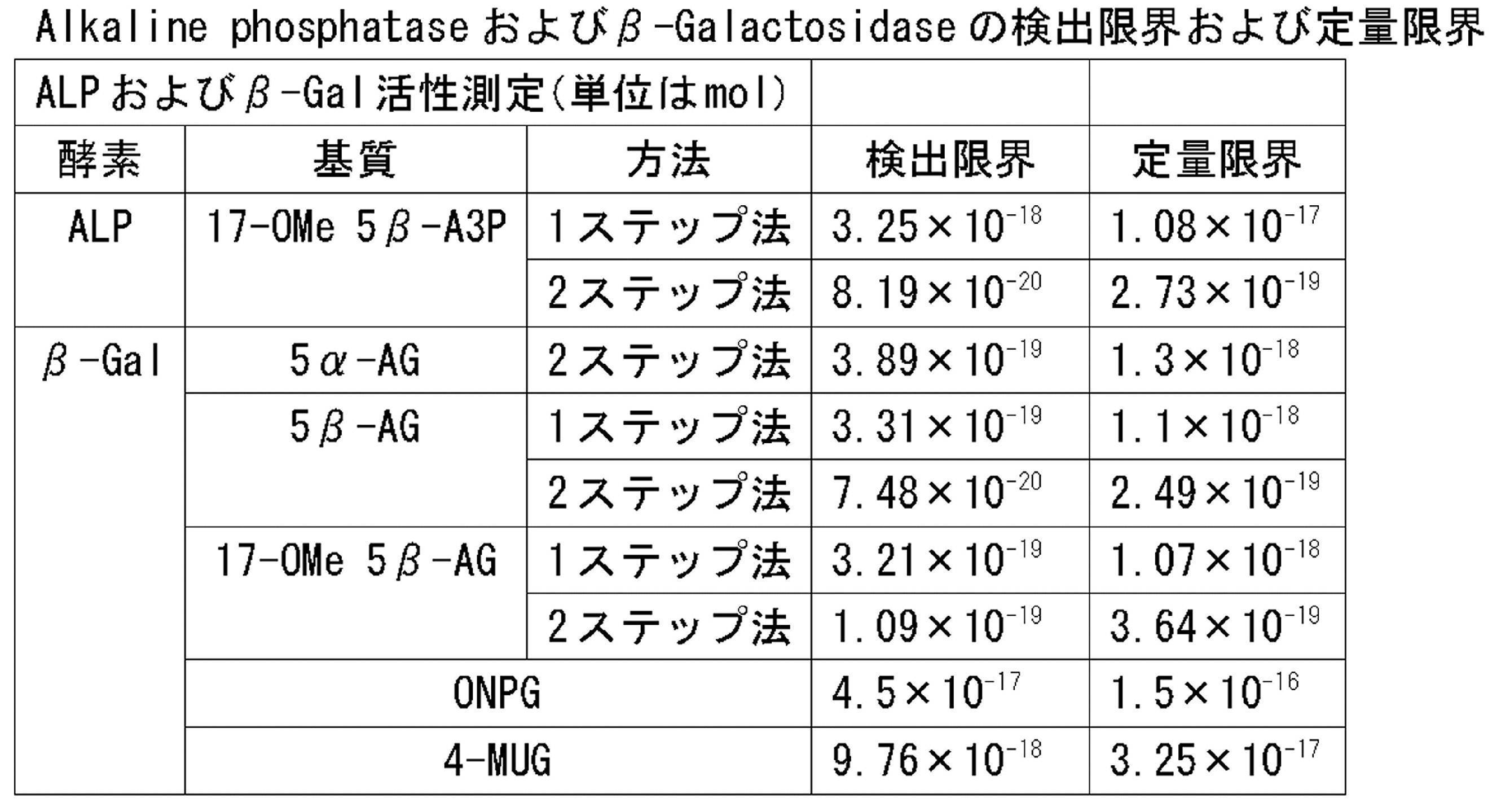
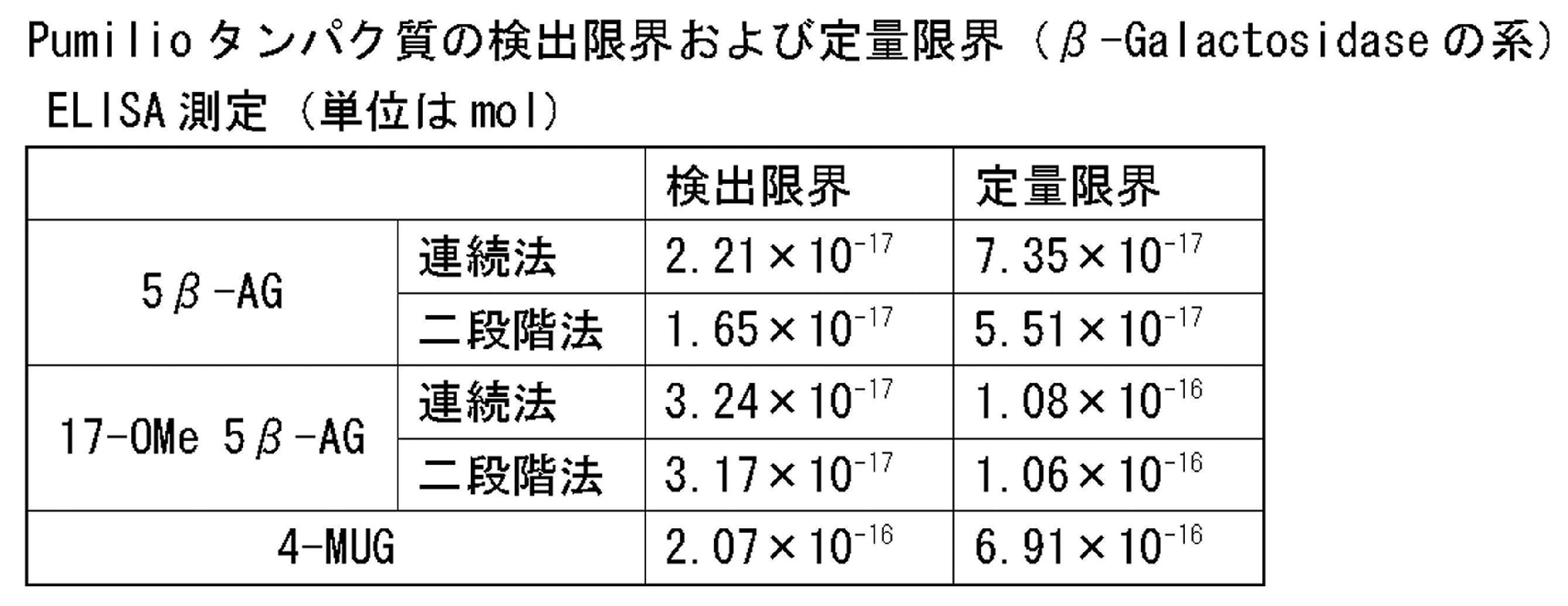
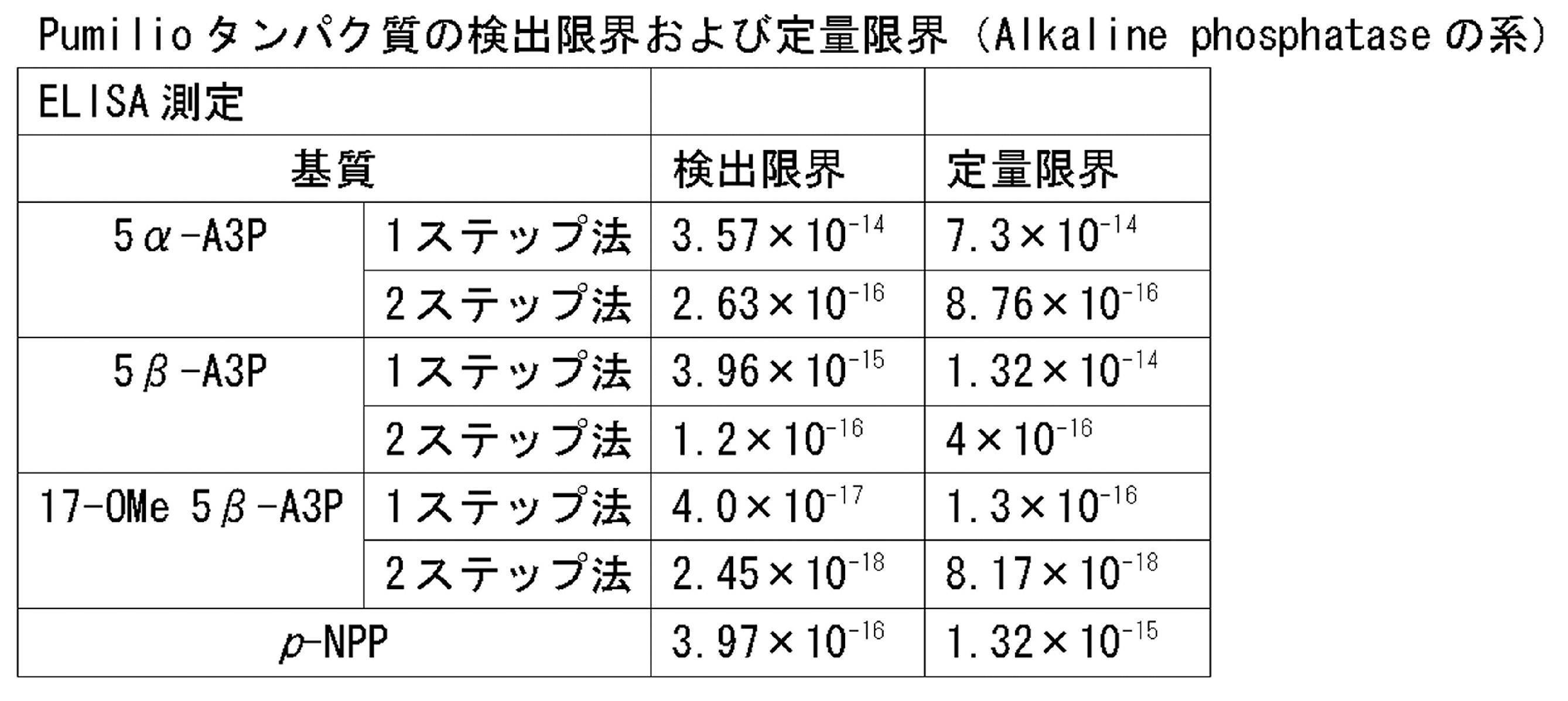
各測定結果の吸光度変化量を標準物質濃度に対してプロットし、直線上に並ぶ濃度のみを抽出して近似曲線を作成する。次にブランク(標準物質を含まない;n≧3)の標準偏差を算出し、その3倍値および10倍値を近似曲線の傾きで割り、得られる数値をそれぞれ検出限界および定量限界とする。
各実験結果より算出した検出限界および定量限界を表3、4、5にまとめた。
3α-tert-butyl-peroxy-5β-Androsteroneを用いた1ステップ法によるHorseradish peroxidaseの測定
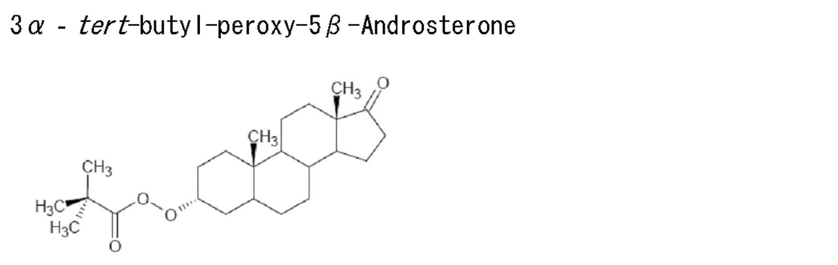
3α-chloro-5β-Androsterone(100mg)をジクロロメタン溶液(15mL)に溶解させ、反応試液を調製した。本溶液に対し、20%の水酸化ナトリウム水溶液(15mL)にtert-butyl-hydroperoxide(30mg)を溶解した溶液を加え、室温にて2時間撹拌させて反応させた。反応液から有機層を回収し、飽和炭酸水素ナトリウム水溶液、飽和食塩水で順次洗浄した。有機層を無水硫酸ナトリウムで脱水した後、減圧下溶媒を留去した。残渣をシリカゲルクロマトグラフィー(24×240 mm,10%メタノール-クロロホルム)にて精製後、本化合物88mgを得た。本化合物の同定は、1H-NMR、ESI-HR-MS およびm.p.(融点)により行った。
0.1Mリン酸緩衝液(pH7.0)
10mM MgCl2
6mM thio-NAD
1mM NADH
0.1mM 3α-tert-butyl-peroxy-5β-Androsterone
40U/ml 3α-Hydoroxysteroid dehydrogenase
試料
5、10、25、50、100、250 pg/ml Horseradish peroxidase(最終濃度 2.5、5、12.5、25、50、125pg/ml)
平底のマイクロプレートウェルに標準物質(Horseradish peroxidase)(以下HRPと称する。)を0~250pg/mlの範囲で含有するリン酸緩衝液(pH7.0)を50μl添加し、続いてそのウェルに反応試液を50μlずつ各ウェルに添加し、37℃で加温しながらマイクロプレートリーダー(コロナ社製MTP-500)で405nmのフィルターを使用して120分間吸光度を測定した。120分間の吸光度変化量を標準物質濃度に対してプロットすることにより得られた、濃度依存性の良い直線を図22に示す。
3α-tert-butyl-peroxy-5β-Androsteroneを用いた1ステップ法によるPumilio(プミリオ)の測定
反応試液
0.1Mリン酸緩衝液(pH7.0)
10mM MgCl2
6mM thio-NAD
1mM NADH
0.1mM 3α-tert-butyl-peroxy-5β-Androsterone
40U/ml 3α-Hydroxysteroid dehydrogenase
試料
0.05、0.1、0.25、0.5、1、2.5 ng/ml Pumilio
参考例5の方法で調製した抗Pumilioモルモットポリクローナル抗体を固定化したマイクロプレートに、精製したPumilio(標準物質)を0~2.5ng/mlの範囲で含有する、0.1%BSA含有TBS(pH7.5)溶液を50μl添加し、室温で2時間振とうさせた。次にウェル内の溶液を吸引除去後、0.5% Triton X-100含有TBS(pH7.5)で洗浄し、HRP標識抗Pumilioマウスモノクローナル抗体をおよそ0.1μg/mlの濃度で含有する、0.1% BSA含有TBS(pH7.5)溶液を50μl加え、室温で1時間振とうさせた。ウェル内の溶液を吸引除去後、0.5% Triton X-100含有TBS(pH7.5)で洗浄した。次に各ウェルに反応試液を50μlずつ添加し、37℃で加温しながらマイクロプレートリーダー(コロナ社製MTP-500)で405nmのフィルターを使用して60分間吸光度を測定した。60分間の吸光度変化量を標準物質濃度に対してプロットすることにより得られた、濃度依存性の良い直線を図23に示す。
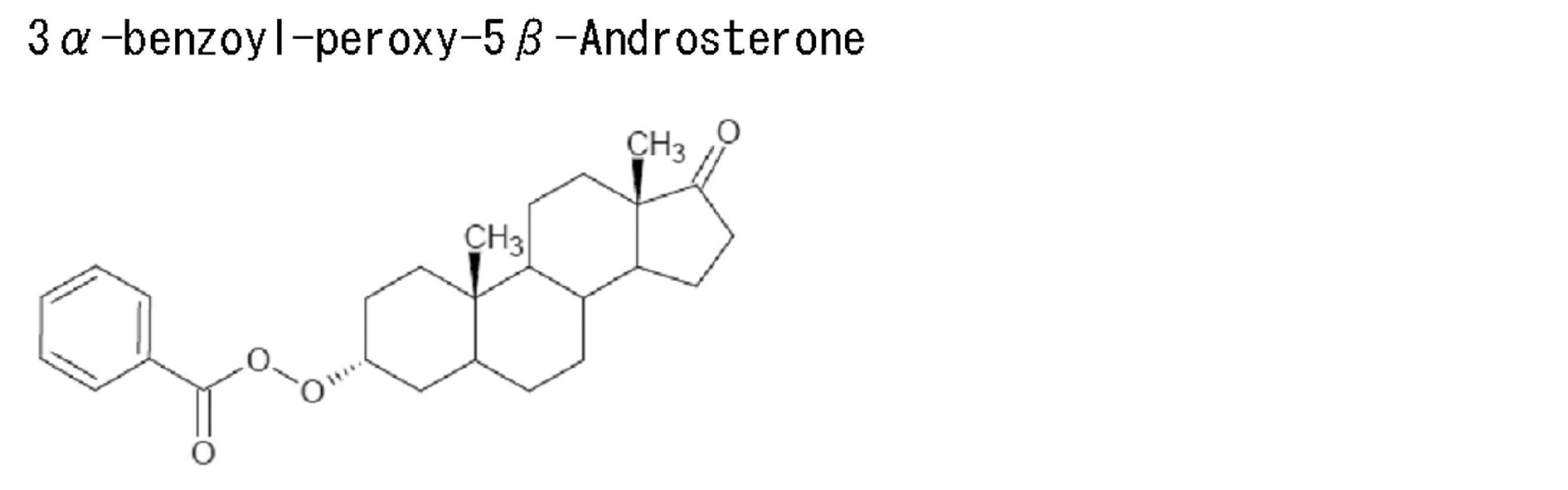
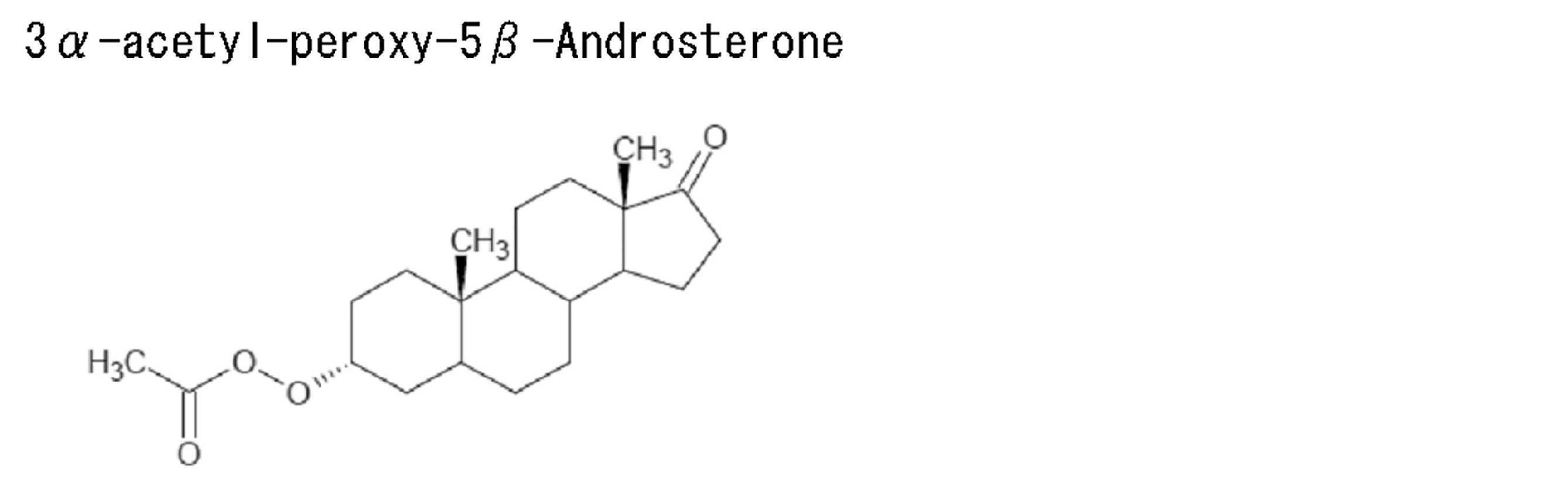
3α-benzoyl-peroxy-5β-Androsteroneおよび3α-acetyl-peroxy-5β- Androsteroneの合成方法
実施例14に示した3α-tert-butyl-peroxy-5β-Androsterone の合成方法において、tert-butyl-hydroperoxideをbenzoyl-hydroperoxide またはacetyl-hydroperoxideに替えた以外は同様にして表題化合物を得た。
Claims (15)
- 抗体酵素複合体を用いる酵素活性測定方法であって、抗体酵素複合体の酵素としてアルカリホスファターゼ、グルコシダーゼ、ガラクトシダーゼ、フルクトシダーゼ、マンノシダーゼまたはペルオキシダーゼを用い、
上記酵素の基質として下記一般式(1)で表されるアンドロステロン誘導体を用い、

式中、Xはリン酸基、Y1及びY2は共同でメチレン基であるか、または、Y1は水素であり、Y2は、水素、水酸基、炭素数1~6のアルコキシ基、または炭素数1~6のアルキル基であり、
(ii) 一般式(1)で表されるアンドロステロン誘導体は、グルコシダーゼ、ガラクトシダーゼ、フルクトシダーゼまたはマンノシダーゼの基質として用いる場合は、
式中、Xは糖残基であり、糖残基はグルコース、ガラクトース、フルクトースおよびマンノースから成る群から選ばれる1種であり、Y1及びY2は共同でメチレン基または酸素原子であるか、または、Y1は水素であり、Y2は、水素、水酸基、炭素数1~6のアルコキシ基、または炭素数1~6のアルキル基であり、
(iii) 一般式(1)で表されるアンドロステロン誘導体は、ペルオキシダーゼの基質として用いる場合は、
式中、Xは-O-CO-R(但し、R は炭素数1~6のアルキル基またはフェニル基である)であり、Y1及びY2は共同でメチレン基または酸素原子であるか、または、Y1は水素であり、Y2は、水素、水酸基、炭素数1~6のアルコキシ基、または炭素数1~6のアルキル基であり、
前記酵素反応の生成物の定量は、NADHおよび/またはNADPH、チオNADおよび/またはチオNADP、並びにヒドロキシステロイド脱水素酵素(HSD)を用いて、酵素サイクリング反応によりチオNADHおよび/またはチオNADPHを生成させ、生成したチオNADHおよび/またはチオNADPHの量を測定するか、または生成したチオNADHおよび/またはチオNADPHによる色の変化を計測することで行う、前記方法。 - 酵素標識核酸プローブを用いる核酸プローブ測定方法であって、
酵素標識核酸プローブの酵素としてアルカリホスファターゼ、グルコシダーゼ、ガラクトシダーゼ、フルクトシダーゼ、マンノシダーゼまたはペルオキシダーゼを用い、上記酵素の基質として下記一般式(1)で表されるアンドロステロン誘導体を用い、
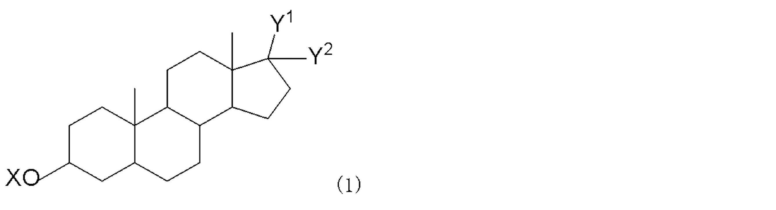
式中、Xはリン酸基、Y1及びY2は共同でメチレン基であるか、または、Y1は水素であり、Y2は、水素、水酸基、炭素数1~6のアルコキシ基、または炭素数1~6のアルキル基であり、
(ii) 一般式(1)で表されるアンドロステロン誘導体は、グルコシダーゼ、ガラクトシダーゼ、フルクトシダーゼまたはマンノシダーゼの基質として用いる場合は、
式中、Xは糖残基であり、糖残基はグルコース、ガラクトース、フルクトースおよびマンノースから成る群から選ばれる1種であり、Y1及びY2は共同でメチレン基または酸素原子であるか、または、Y1は水素であり、Y2は、水素、水酸基、炭素数1~6のアルコキシ基、または炭素数1~6のアルキル基であり、
(iii) 一般式(1)で表されるアンドロステロン誘導体は、ペルオキシダーゼの基質として用いる場合は、
式中、Xは-O-CO-R(但し、R は炭素数1~6のアルキル基またはフェニル基である)であり、Y1及びY2は共同でメチレン基または酸素原子であるか、または、Y1は水素であり、Y2は、水素、水酸基、炭素数1~6のアルコキシ基、または炭素数1~6のアルキル基であり、
前記酵素反応の反応生成物の定量は、NADHおよび/またはNADPH、チオNADおよび/またはチオNADP、並びにヒドロキシステロイド脱水素酵素(HSD)を用いて、酵素サイクリング反応によりチオNADHおよび/またはチオNADPHを生成させ、生成したチオNADHおよび/またはチオNADPHの量を測定するか、または生成したチオNADHおよび/またはチオNADPHによる色の変化を計測することで行う、前記方法。 - 前記酵素がアルカリホスファターゼであり、
一般式(1)で表されるアンドロステロン誘導体は、
式中、Xはリン酸基、Y1及びY2は共同でメチレン基であるか、または、Y1は水素であり、Y2は、水素、水酸基、炭素数1~6のアルコキシ基、または炭素数1~6のアルキル基である、請求項1または2に記載の方法。 - 前記酵素がグルコシダーゼ、ガラクトシダーゼ、フルクトシダーゼまたはマンノシダーゼであり、一般式(1)で表されるアンドロステロン誘導体は、
式中、Xは糖残基であり、糖残基はグルコース、ガラクトース、フルクトースおよびマンノースから成る群から選ばれる1種であり、Y1及びY2は共同でメチレン基または酸素原子であるか、または、Y1は水素であり、Y2は、水素、水酸基、炭素数1~6のアルコキシ基、または炭素数1~6のアルキル基である、請求項1または2に記載の方法。 - 前記酵素がペルオキシダーゼであり、一般式(1)で表されるアンドロステロン誘導体は、
式中、Xは-O-CO-R(但し、R は炭素数1~6のアルキル基またはフェニル基である)であり、Y1及びY2は共同でメチレン基または酸素原子であるか、または、Y1は水素であり、Y2は、水素、水酸基、炭素数1~6のアルコキシ基、または炭素数1~6のアルキル基である、請求項1または2に記載の方法。 - 以下の(1)~(5)の試薬を含む酵素免疫測定用キット。
(1)標的タンパク質抗原に特異的な抗体を標識したアルカリホスファターゼ
(2)上記酵素の基質である一般式(1)で表されるアンドロステロン誘導体
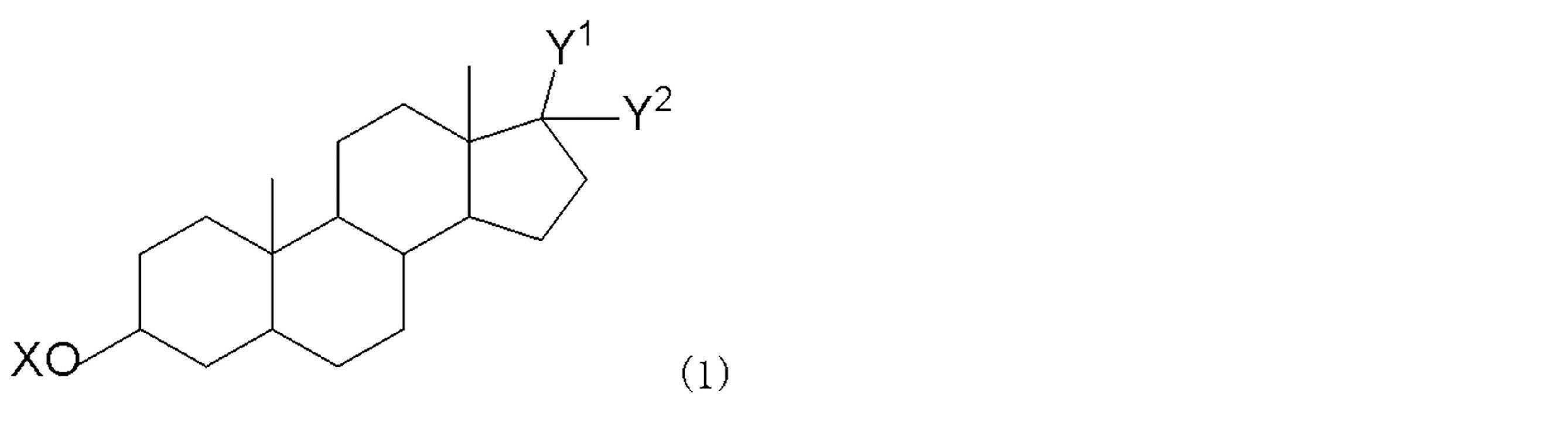
(3)ヒドロキシステロイド脱水素酵素(HSD)
(4)NADHおよび/またはNADPH、
(5)チオNADおよび/またはチオNADP - 以下の(1)~(5)の試薬を含む酵素免疫測定用キット。
(1)標的タンパク質抗原に特異的な抗体を標識したグルコシダーゼ、ガラクトシダーゼ、フルクトシダーゼまたはマンノシダーゼ
(2)上記酵素の基質である一般式(1)で表されるアンドロステロン誘導体
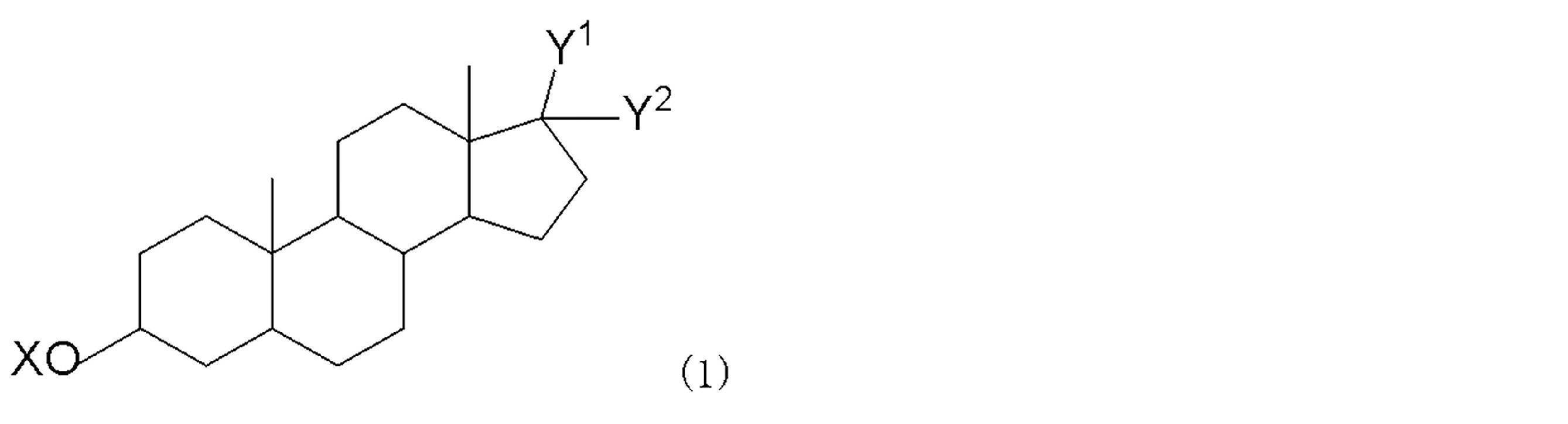
(3)ヒドロキシステロイド脱水素酵素(HSD)
(4)NADHおよび/またはNADPH、
(5)チオNADおよび/またはチオNADP - 以下の(1)~(5)の試薬を含む酵素免疫測定用キット。
(1)標的タンパク質抗原に特異的な抗体を標識したペルオキシダーゼ、
(2)上記酵素の基質である一般式(1)で表されるアンドロステロン誘導体
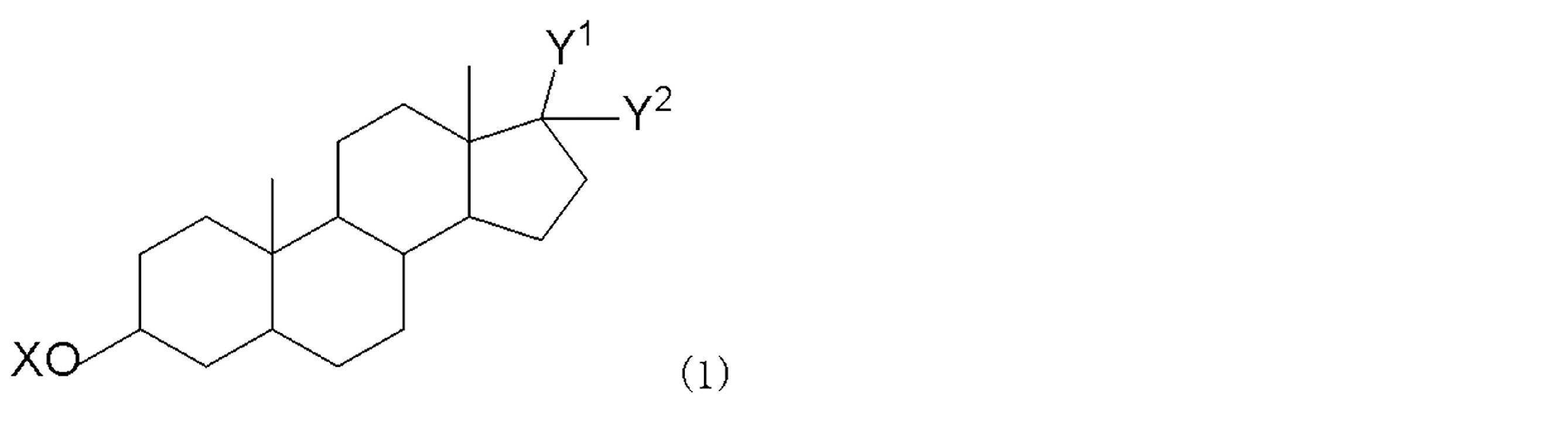
(3)ヒドロキシステロイド脱水素酵素(HSD)
(4)NADHおよび/またはNADPH、
(5)チオNADおよび/またはチオNADP - 以下の(1)~(5)の試薬を含む核酸プローブ測定用キット。
(1)標的核酸に特異的に結合する核酸プローブで標識したアルカリホスファターゼ、
(2)上記酵素の基質である一般式(1)で表されるアンドロステロン誘導体
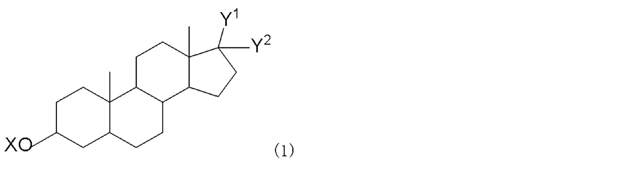
(3)ヒドロキシステロイド脱水素酵素(HSD)、
(4)NADHおよび/またはNADPH、
(5)チオNADおよび/またはチオNADP - 以下の(1)~(5)の試薬を含む核酸プローブ測定用キット。
(1)標的核酸に特異的に結合する核酸プローブで標識したグルコシダーゼ、ガラクトシダーゼ、フルクトシダーゼまたはマンノシダーゼ、
(2)上記酵素の基質である一般式(1)で表されるアンドロステロン誘導体
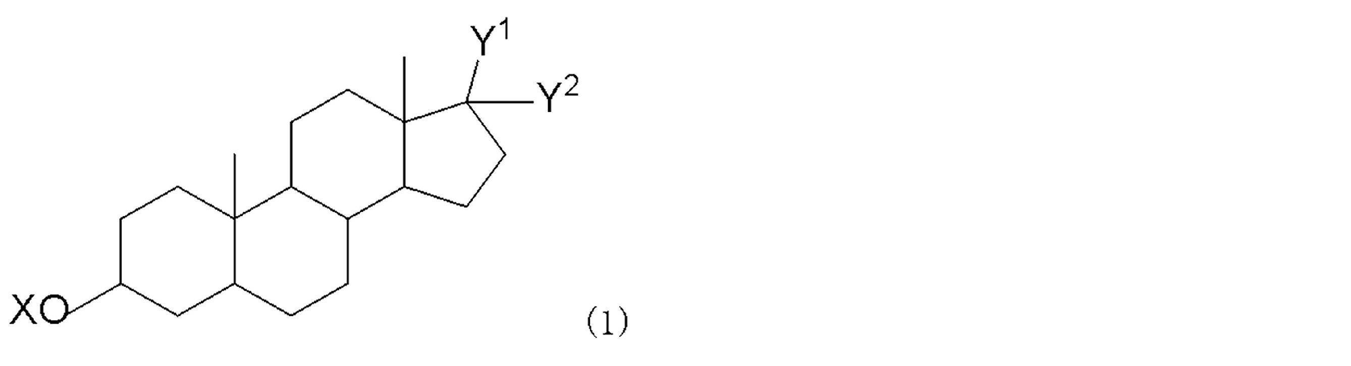
(3)ヒドロキシステロイド脱水素酵素(HSD)、
(4)NADHおよび/またはNADPH、
(5)チオNADおよび/またはチオNADP - 以下の(1)~(5)の試薬を含む核酸プローブ測定用キット。
(1)標的核酸に特異的に結合する核酸プローブで標識したペルオキシダーゼ、
(2)上記酵素の基質である一般式(1)で表されるアンドロステロン誘導体

(3)ヒドロキシステロイド脱水素酵素(HSD)、
(4)NADHおよび/またはNADPH、
(5)チオNADおよび/またはチオNADP - 下記一般式(1)で表されるアンドロステロン誘導体。

(A)Xはリン酸基であり、Y1及びY2は共同でメチレン基であるか、または、Y1は水素であり、Y2は水酸基、炭素数1~6のアルコキシ基または炭素数1~6のアルキル基である、
(B)Xはグルコース、ガラクトース、フルクトースおよびマンノースから成る群から選ばれる1種の糖残基であり、Y1及びY2は共同でメチレン基であるか、または、Y1は水素であり、Y2は水素、炭素数1~6のアルコキシ基、または炭素数1~6のアルキル基である、
(C)Xは-O-CO-Rであり、R は炭素数1~6のアルキル基またはフェニル基であり、Y1及びY2が共同でメチレン基または酸素原子であるか、または、Y1は水素であり、Y2は水素、水酸基、炭素数1~6のアルコキシ基、または炭素数1~6のアルキル基である。) - X、Y1およびY2の定義は、(A)Xはリン酸基であり、Y1及びY2は共同でメチレン基であるか、または、Y1は水素であり、Y2は水酸基、炭素数1~6のアルコキシ基または炭素数1~6のアルキル基である、請求項12に記載のアンドロステロン誘導体。
- X、Y1およびY2の定義は、 (B)Xはグルコース、ガラクトース、フルクトースおよびマンノースから成る群から選ばれる1種の糖残基であり、Y1及びY2は共同でメチレン基であるか、または、Y1は水素であり、Y2は水素、炭素数1~6のアルコキシ基、または炭素数1~6のアルキル基である、請求項12に記載のアンドロステロン誘導体。
- X、Y1およびY2の定義は、(C)Xは-O-CO-Rであり、R は炭素数1~6のアルキル基またはフェニル基であり、Y1及びY2が共同でメチレン基または酸素原子であるか、または、Y1は水素であり、Y2は水素、水酸基、炭素数1~6のアルコキシ基、または炭素数1~6のアルキル基である、請求項12に記載のアンドロステロン誘導体。
Priority Applications (6)
| Application Number | Priority Date | Filing Date | Title |
|---|---|---|---|
| JP2012533408A JP5265816B2 (ja) | 2011-03-23 | 2012-03-23 | タンパク質および核酸の超高感度測定法およびキット、並びに新規な酵素基質 |
| US14/006,317 US9851309B2 (en) | 2011-03-23 | 2012-03-23 | Ultra-high-sensitive assay of protein and nucleic acid and kit, and novel enzyme substrate |
| ES12759947.0T ES2613619T3 (es) | 2011-03-23 | 2012-03-23 | Método y kit para medición de superalta sensibilidad de proteína y ácido nucleico, y nuevo sustrato enzimático |
| EP17197044.5A EP3296405B1 (en) | 2011-03-23 | 2012-03-23 | Method and kit for super-high-sensitivity measurement of protein and nucleic acid, and novel enzyme substrate |
| EP12759947.0A EP2695948B1 (en) | 2011-03-23 | 2012-03-23 | Method and kit for super-high-sensitivity measurement of protein and nucleic acid, and novel enzyme substrate |
| US15/845,702 US11221299B2 (en) | 2011-03-23 | 2017-12-18 | Ultra-high-sensitive assay of protein and nucleic acid and kit, and novel enzyme substrate |
Applications Claiming Priority (2)
| Application Number | Priority Date | Filing Date | Title |
|---|---|---|---|
| JP2011063559 | 2011-03-23 | ||
| JP2011-063559 | 2011-03-23 |
Related Child Applications (2)
| Application Number | Title | Priority Date | Filing Date |
|---|---|---|---|
| US14/006,317 A-371-Of-International US9851309B2 (en) | 2011-03-23 | 2012-03-23 | Ultra-high-sensitive assay of protein and nucleic acid and kit, and novel enzyme substrate |
| US15/845,702 Division US11221299B2 (en) | 2011-03-23 | 2017-12-18 | Ultra-high-sensitive assay of protein and nucleic acid and kit, and novel enzyme substrate |
Publications (1)
| Publication Number | Publication Date |
|---|---|
| WO2012128338A1 true WO2012128338A1 (ja) | 2012-09-27 |
Family
ID=46879477
Family Applications (1)
| Application Number | Title | Priority Date | Filing Date |
|---|---|---|---|
| PCT/JP2012/057422 Ceased WO2012128338A1 (ja) | 2011-03-23 | 2012-03-23 | タンパク質および核酸の超高感度測定法およびキット、並びに新規な酵素基質 |
Country Status (5)
| Country | Link |
|---|---|
| US (2) | US9851309B2 (ja) |
| EP (3) | EP3296405B1 (ja) |
| JP (1) | JP5265816B2 (ja) |
| ES (3) | ES2762098T3 (ja) |
| WO (1) | WO2012128338A1 (ja) |
Cited By (3)
| Publication number | Priority date | Publication date | Assignee | Title |
|---|---|---|---|---|
| JP2014124165A (ja) * | 2012-12-27 | 2014-07-07 | Bl:Kk | タンパク質および核酸の高感度測定法、キット並びに酵素組成物 |
| US11702684B2 (en) | 2016-03-30 | 2023-07-18 | Biophenoma Inc. | Method for measuring protein and nucleic acid with ultrahigh sensitivity |
| WO2025004524A1 (ja) * | 2023-06-30 | 2025-01-02 | 学校法人早稲田大学 | 微量物質の分析方法および微量物質分析用キット |
Families Citing this family (19)
| Publication number | Priority date | Publication date | Assignee | Title |
|---|---|---|---|---|
| EP4717314A2 (en) | 2011-10-14 | 2026-04-01 | Sage Therapeutics, LLC | 3,3 disubstituted 19-nor pregnane compounds, compositions, and uses thereof |
| DK2935307T3 (en) | 2012-12-18 | 2018-07-30 | Univ Washington | NEUROACTIVE 19-ALKOXY-17-SUBSTITUTED STEROIDS USED IN METHODS OF TREATMENT |
| US9725481B2 (en) | 2013-04-17 | 2017-08-08 | Sage Therapeutics, Inc. | 19-nor C3, 3-disubstituted C21-C-bound heteroaryl steroids and methods of use thereof |
| CN117304245A (zh) | 2013-04-17 | 2023-12-29 | 萨奇治疗股份有限公司 | 刺激神经活性的19-去甲类固醇及其使用方法 |
| SMT202100509T1 (it) | 2013-04-17 | 2021-11-12 | Sage Therapeutics Inc | Steroide di c21-n-pirazolile 19-nor c3,3-disostituito per l'uso in terapia |
| WO2014169836A1 (en) | 2013-04-17 | 2014-10-23 | Sage Therapeutics, Inc. | 19-nor neuroactive steroids and methods of use thereof |
| CA2918735C (en) | 2013-07-19 | 2023-08-01 | Sage Therapeutics, Inc. | Neuroactive steroids, compositions, and uses thereof |
| CN105579043B (zh) | 2013-08-23 | 2021-09-14 | 萨奇治疗股份有限公司 | 神经活性类固醇、组合物和其用途 |
| US10246482B2 (en) | 2014-06-18 | 2019-04-02 | Sage Therapeutics, Inc. | Neuroactive steroids, compositions, and uses thereof |
| US20170233433A1 (en) | 2014-10-16 | 2017-08-17 | Sage Therapeutics, Inc. | Compositions and methods for treating cns disorders |
| MY190408A (en) | 2014-10-16 | 2022-04-21 | Sage Therapeutics Inc | Compositions and methods for treating cns disorders |
| WO2016082789A1 (en) | 2014-11-27 | 2016-06-02 | Sage Therapeutics, Inc. | Compositions and methods for treating cns disorders |
| LT3250210T (lt) | 2015-01-26 | 2021-04-12 | Sage Therapeutics, Inc. | Kompozicijos ir būdai, skirti gydyti cns sutrikimus |
| DK3258939T3 (da) | 2015-02-20 | 2022-12-12 | Sage Therapeutics Inc | Neuroaktive steroider, sammensætninger og anvendelser heraf |
| CA3030420C (en) | 2016-07-11 | 2025-11-18 | Sage Therapeutics, Inc. | C7, C12 AND C16 SUBSTITUTED NEUROACTIVE STEROIDS AND ASSOCIATED METHODS OF USE |
| WO2018013613A1 (en) | 2016-07-11 | 2018-01-18 | Sage Therapeutics, Inc. | C17, c20, and c21 substituted neuroactive steroids and their methods of use |
| JP7670626B2 (ja) | 2019-05-31 | 2025-04-30 | セージ セラピューティクス, インコーポレイテッド | 神経刺激性ステロイド及びその組成物 |
| JPWO2024185437A1 (ja) | 2023-03-09 | 2024-09-12 | ||
| WO2025142889A1 (ja) * | 2023-12-25 | 2025-07-03 | 株式会社BioPhenoMA | 細胞外小胞を分級する装置、および細胞外小胞を分級し、細胞外小胞に含まれるタンパク質を検出または分析するためのインビトロの方法 |
Citations (2)
| Publication number | Priority date | Publication date | Assignee | Title |
|---|---|---|---|---|
| WO2008117816A1 (ja) | 2007-03-28 | 2008-10-02 | Etsuro Ito | タンパク質および核酸の超高感度測定法 |
| JP2009207396A (ja) * | 2008-03-03 | 2009-09-17 | Tokushima Bunri Univ | ステロイド5α−レダクターゼ活性の測定方法およびそれに用いるキット |
Family Cites Families (3)
| Publication number | Priority date | Publication date | Assignee | Title |
|---|---|---|---|---|
| FR83554E (fr) | 1961-07-21 | 1964-09-04 | Roussel Uclaf | Procédé pour la préparation de dérivés de l'étiocholane |
| US6011023A (en) | 1997-08-27 | 2000-01-04 | Alcon Laboratories, Inc. | Angiostatic steroids |
| JP6960210B2 (ja) * | 2016-03-30 | 2021-11-05 | 悦朗 伊藤 | タンパク質及び核酸の超高感度測定方法 |
-
2012
- 2012-03-23 ES ES17197044T patent/ES2762098T3/es active Active
- 2012-03-23 EP EP17197044.5A patent/EP3296405B1/en active Active
- 2012-03-23 ES ES12759947.0T patent/ES2613619T3/es active Active
- 2012-03-23 US US14/006,317 patent/US9851309B2/en active Active
- 2012-03-23 JP JP2012533408A patent/JP5265816B2/ja active Active
- 2012-03-23 EP EP12759947.0A patent/EP2695948B1/en active Active
- 2012-03-23 ES ES15187872.5T patent/ES2657986T3/es active Active
- 2012-03-23 WO PCT/JP2012/057422 patent/WO2012128338A1/ja not_active Ceased
- 2012-03-23 EP EP15187872.5A patent/EP2987866B1/en active Active
-
2017
- 2017-12-18 US US15/845,702 patent/US11221299B2/en active Active
Patent Citations (2)
| Publication number | Priority date | Publication date | Assignee | Title |
|---|---|---|---|---|
| WO2008117816A1 (ja) | 2007-03-28 | 2008-10-02 | Etsuro Ito | タンパク質および核酸の超高感度測定法 |
| JP2009207396A (ja) * | 2008-03-03 | 2009-09-17 | Tokushima Bunri Univ | ステロイド5α−レダクターゼ活性の測定方法およびそれに用いるキット |
Non-Patent Citations (6)
| Title |
|---|
| ATSUSHI IWAI ET AL.: "Thio NAD Cycling o Riyo suru p-Galactosidase no Kokando Kassei Sokuteiho", KAGAKUKEIGAKU KYOKAI HOKKAIDO SHIBU 2010 NEN TOKI KENKYU HAPPYOKAI KOEN YOSHISHU, 26 January 2010 (2010-01-26), pages 65, XP008172120 * |
| CARBOHYDRATE RESEARCH, vol. 17, no. 1, 1971, pages 199 - 207 |
| PHYTOCHEMISTRY, vol. 13, no. 10, 1974, pages 2135 - 2142 |
| See also references of EP2695948A4 |
| SKALHEGG B.A.: "3a-hydroxysteroid dehydrogenase from Pseudomonas testosteroni: kinetic properties with NAD and its thionicotinamide analogue", EUR. J. BIOCHEM., vol. 50, no. 3, 15 January 1975 (1975-01-15), pages 603 - 609, XP055127238 * |
| TETRAHEDRON, vol. 55, no. 17, 1999, pages 5465 - 5482 |
Cited By (3)
| Publication number | Priority date | Publication date | Assignee | Title |
|---|---|---|---|---|
| JP2014124165A (ja) * | 2012-12-27 | 2014-07-07 | Bl:Kk | タンパク質および核酸の高感度測定法、キット並びに酵素組成物 |
| US11702684B2 (en) | 2016-03-30 | 2023-07-18 | Biophenoma Inc. | Method for measuring protein and nucleic acid with ultrahigh sensitivity |
| WO2025004524A1 (ja) * | 2023-06-30 | 2025-01-02 | 学校法人早稲田大学 | 微量物質の分析方法および微量物質分析用キット |
Also Published As
| Publication number | Publication date |
|---|---|
| ES2762098T3 (es) | 2020-05-22 |
| EP2987866A3 (en) | 2016-06-15 |
| JPWO2012128338A1 (ja) | 2014-07-24 |
| EP2695948B1 (en) | 2016-12-28 |
| HK1246360A1 (en) | 2018-09-07 |
| JP5265816B2 (ja) | 2013-08-14 |
| US20180106727A1 (en) | 2018-04-19 |
| US20140017675A1 (en) | 2014-01-16 |
| US11221299B2 (en) | 2022-01-11 |
| ES2657986T3 (es) | 2018-03-07 |
| US9851309B2 (en) | 2017-12-26 |
| ES2613619T3 (es) | 2017-05-24 |
| EP2695948A4 (en) | 2015-04-08 |
| EP2695948A1 (en) | 2014-02-12 |
| EP3296405A1 (en) | 2018-03-21 |
| EP2987866A2 (en) | 2016-02-24 |
| HK1221743A1 (en) | 2017-06-09 |
| EP2987866B1 (en) | 2017-12-06 |
| EP3296405B1 (en) | 2019-10-09 |
Similar Documents
| Publication | Publication Date | Title |
|---|---|---|
| JP5265816B2 (ja) | タンパク質および核酸の超高感度測定法およびキット、並びに新規な酵素基質 | |
| US7642064B2 (en) | Enzyme-catalyzed metal deposition for the enhanced detection of analytes of interest | |
| US7704752B2 (en) | Signalling compounds for use in methods of detecting hydrogen peroxide | |
| US6063581A (en) | Immunoassay for homocysteine | |
| AU622904B2 (en) | Enzyme activity determinations method characterized by the using of substrates whose fluorescent properties differs those of the converted products | |
| Lövgren et al. | Time-resolved fluoroimmunoassay, advantages | |
| US6162610A (en) | Xanthan-ester and acridan substrates for horseradish peroxidase | |
| WO2012006351A1 (en) | In situ chemiluminescent substrates and assays | |
| EP2705031B1 (en) | Flash and glow 1,2-dioxetanes | |
| US5605795A (en) | Assays using chemiluminescent, enzymatically cleavable substituted 1,2-dioxetanes and kits therefor | |
| JP5500985B2 (ja) | タンパク質および核酸の超高感度測定法 | |
| JPH0859686A (ja) | ウミホタルルシフェリン誘導体および糖加水分解酵素の定量方法 | |
| HK1246360B (en) | Method and kit for super-high-sensitivity measurement of protein and nucleic acid, and novel enzyme substrate | |
| CN111018924B (zh) | 一种柔红霉素衍生物及其制备方法与柔红霉素检测试剂 | |
| HK1221743B (en) | Method and kit for super-high-sensitivity measurement of protein and nucleic acid, and novel enzyme substrate | |
| JP2014124165A (ja) | タンパク質および核酸の高感度測定法、キット並びに酵素組成物 | |
| JP3471874B2 (ja) | 化学発光によるホスファターゼの酵素活性測定法 |
Legal Events
| Date | Code | Title | Description |
|---|---|---|---|
| ENP | Entry into the national phase |
Ref document number: 2012533408 Country of ref document: JP Kind code of ref document: A |
|
| 121 | Ep: the epo has been informed by wipo that ep was designated in this application |
Ref document number: 12759947 Country of ref document: EP Kind code of ref document: A1 |
|
| DPE1 | Request for preliminary examination filed after expiration of 19th month from priority date (pct application filed from 20040101) | ||
| WWE | Wipo information: entry into national phase |
Ref document number: 14006317 Country of ref document: US |
|
| NENP | Non-entry into the national phase |
Ref country code: DE |
|
| REEP | Request for entry into the european phase |
Ref document number: 2012759947 Country of ref document: EP |
|
| WWE | Wipo information: entry into national phase |
Ref document number: 2012759947 Country of ref document: EP |