WO2012118088A1 - 再燃性前立腺癌の検出方法 - Google Patents
再燃性前立腺癌の検出方法 Download PDFInfo
- Publication number
- WO2012118088A1 WO2012118088A1 PCT/JP2012/054962 JP2012054962W WO2012118088A1 WO 2012118088 A1 WO2012118088 A1 WO 2012118088A1 JP 2012054962 W JP2012054962 W JP 2012054962W WO 2012118088 A1 WO2012118088 A1 WO 2012118088A1
- Authority
- WO
- WIPO (PCT)
- Prior art keywords
- seq
- base sequence
- amino acid
- sequence
- oligonucleotide consisting
- Prior art date
- Legal status (The legal status is an assumption and is not a legal conclusion. Google has not performed a legal analysis and makes no representation as to the accuracy of the status listed.)
- Ceased
Links
Images











Classifications
-
- G—PHYSICS
- G01—MEASURING; TESTING
- G01N—INVESTIGATING OR ANALYSING MATERIALS BY DETERMINING THEIR CHEMICAL OR PHYSICAL PROPERTIES
- G01N33/00—Investigating or analysing materials by specific methods not covered by groups G01N1/00 - G01N31/00
- G01N33/48—Biological material, e.g. blood, urine; Haemocytometers
- G01N33/50—Chemical analysis of biological material, e.g. blood, urine; Testing involving biospecific ligand binding methods; Immunological testing
- G01N33/53—Immunoassay; Biospecific binding assay; Materials therefor
- G01N33/575—Immunoassay; Biospecific binding assay; Materials therefor for cancer
- G01N33/57555—Immunoassay; Biospecific binding assay; Materials therefor for cancer of the prostate
-
- C—CHEMISTRY; METALLURGY
- C12—BIOCHEMISTRY; BEER; SPIRITS; WINE; VINEGAR; MICROBIOLOGY; ENZYMOLOGY; MUTATION OR GENETIC ENGINEERING
- C12Q—MEASURING OR TESTING PROCESSES INVOLVING ENZYMES, NUCLEIC ACIDS OR MICROORGANISMS; COMPOSITIONS OR TEST PAPERS THEREFOR; PROCESSES OF PREPARING SUCH COMPOSITIONS; CONDITION-RESPONSIVE CONTROL IN MICROBIOLOGICAL OR ENZYMOLOGICAL PROCESSES
- C12Q1/00—Measuring or testing processes involving enzymes, nucleic acids or microorganisms; Compositions therefor; Processes of preparing such compositions
- C12Q1/68—Measuring or testing processes involving enzymes, nucleic acids or microorganisms; Compositions therefor; Processes of preparing such compositions involving nucleic acids
- C12Q1/6876—Nucleic acid products used in the analysis of nucleic acids, e.g. primers or probes
- C12Q1/6883—Nucleic acid products used in the analysis of nucleic acids, e.g. primers or probes for diseases caused by alterations of genetic material
- C12Q1/6886—Nucleic acid products used in the analysis of nucleic acids, e.g. primers or probes for diseases caused by alterations of genetic material for cancer
-
- C—CHEMISTRY; METALLURGY
- C12—BIOCHEMISTRY; BEER; SPIRITS; WINE; VINEGAR; MICROBIOLOGY; ENZYMOLOGY; MUTATION OR GENETIC ENGINEERING
- C12Q—MEASURING OR TESTING PROCESSES INVOLVING ENZYMES, NUCLEIC ACIDS OR MICROORGANISMS; COMPOSITIONS OR TEST PAPERS THEREFOR; PROCESSES OF PREPARING SUCH COMPOSITIONS; CONDITION-RESPONSIVE CONTROL IN MICROBIOLOGICAL OR ENZYMOLOGICAL PROCESSES
- C12Q2600/00—Oligonucleotides characterized by their use
- C12Q2600/112—Disease subtyping, staging or classification
-
- C—CHEMISTRY; METALLURGY
- C12—BIOCHEMISTRY; BEER; SPIRITS; WINE; VINEGAR; MICROBIOLOGY; ENZYMOLOGY; MUTATION OR GENETIC ENGINEERING
- C12Q—MEASURING OR TESTING PROCESSES INVOLVING ENZYMES, NUCLEIC ACIDS OR MICROORGANISMS; COMPOSITIONS OR TEST PAPERS THEREFOR; PROCESSES OF PREPARING SUCH COMPOSITIONS; CONDITION-RESPONSIVE CONTROL IN MICROBIOLOGICAL OR ENZYMOLOGICAL PROCESSES
- C12Q2600/00—Oligonucleotides characterized by their use
- C12Q2600/158—Expression markers
Definitions
- the present invention relates to a method for detecting relapsed prostate cancer.
- prostate cancer grows depending on androgen, which is a male hormone, hormone therapy that suppresses androgen secretion and action is used as an effective treatment. However, not only a few cancer cells survive this hormone therapy. Surviving cancer cells then repeat growth and cause hormone-resistant prostate cancer that is resistant to hormone therapy. Hormone refractory prostate cancer is a reactivated growth of prostate cancer once suppressed by hormone therapy, and is also called relapsed prostate cancer.
- An object of the present invention is to find a marker specific to relapsed prostate cancer and to provide a method for specifically detecting relapsed prostate cancer using this marker.
- the present inventors have focused on specific sugar chain-related genes that have been difficult to detect because the expression level is low so far, and among the genes that contain these genes, the higher in relapsed prostate cancer cells. Or it succeeded in specifying the gene which shows a lower expression level.
- the present invention has been completed based on this finding.
- a method for detecting relapsed prostate cancer comprising measuring the expression level of at least one gene comprising the DNA of any one of (a) to (d) below: (A) the base sequence of SEQ ID NO: 1, the base sequence of SEQ ID NO: 3, the base sequence of SEQ ID NO: 5, the base sequence of SEQ ID NO: 7, the base sequence of SEQ ID NO: 9, the base sequence of SEQ ID NO: 11, the base of SEQ ID NO: 15 Sequence, base sequence of SEQ ID NO: 17, base sequence of SEQ ID NO: 19, base sequence of SEQ ID NO: 21, base sequence of SEQ ID NO: 23, base sequence of SEQ ID NO: 25, base sequence of SEQ ID NO: 27, base sequence of SEQ ID NO: 31 , SEQ ID NO: 33 base sequence, SEQ ID NO: 35 base sequence, SEQ ID NO: 37 base sequence, SEQ ID NO: 39 base sequence, SEQ ID NO: 41 base sequence, SEQ ID NO: 43 base sequence, SEQ ID NO:
- ⁇ 2> The detection method according to ⁇ 1>, wherein the measurement of the expression level of the gene is a measurement of the amount of the transcription product of the gene.
- the expression level of at least one gene is measured using at least one primer set selected from the group consisting of the following (Aa) to (Bb): (Aa) a primer set consisting of an oligonucleotide consisting of the base sequence of SEQ ID NO: 63 and an oligonucleotide consisting of the base sequence of SEQ ID NO: 64; (Ab) a primer set consisting of an oligonucleotide consisting of the base sequence of SEQ ID NO: 65 and an oligonucleotide consisting of the base sequence of SEQ ID NO: 66; (Ac) a primer set consisting of an oligonucleotide consisting of the base sequence of SEQ ID NO: 67 and an oligonucleotide consisting of the base
- Ba A primer set consisting of an oligonucleotide consisting of the base sequence of SEQ ID NO: 121 and an oligonucleotide consisting of the base sequence of SEQ ID NO: 122.
- (Bb) A primer set consisting of an oligonucleotide consisting of the base sequence of SEQ ID NO: 123 and an oligonucleotide consisting of the base sequence of SEQ ID NO: 124.
- the amount of protein to be measured is the amount of at least one protein consisting of any one of the following amino acid sequences (e) to (g): (E) the amino acid sequence of SEQ ID NO: 2, the amino acid sequence of SEQ ID NO: 4, the amino acid sequence of SEQ ID NO: 6, the amino acid sequence of SEQ ID NO: 8, the amino acid sequence of SEQ ID NO: 10, the amino acid sequence of SEQ ID NO: 12, the amino acid of SEQ ID NO: 16 SEQ ID NO: 18, amino acid sequence SEQ ID NO: 20, amino acid sequence SEQ ID NO: 22, amino acid sequence SEQ ID NO: 24, amino acid sequence SEQ ID NO: 26, amino acid sequence SEQ ID NO: 28, amino acid sequence SEQ ID NO: 32 , Amino acid sequence of SEQ ID NO: 34, amino acid sequence of SEQ ID NO: 36, amino acid sequence of SEQ ID NO: 38, amino acid sequence of SEQ ID NO: 40, amino acid sequence of SEQ ID NO: 42, amino acid sequence of SEQ
- relapsed prostate cancer can be specifically detected by distinguishing it from hormone-sensitive prostate cancer.
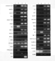
- FIG. 2 is a photograph of electrophoresis showing the results of RT-PCR described in Example 1.
- Lane 1 shows the results of PrE cells
- Lane 2 shows the results of LNCaP cells
- Lane 3 shows the results of Du145 cells
- Lane 4 shows the results of PC-3 cells.
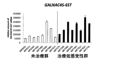
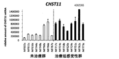
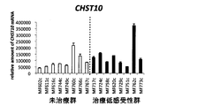
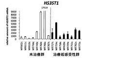
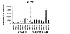
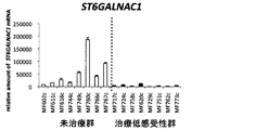
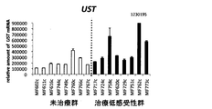
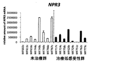
- 3 is a graph showing the results of real-time RT-PCR described in Example 2.
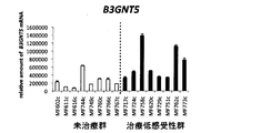
- 3 is a graph showing the results of real-time RT-PCR described in Example 2.
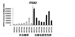
- 3 is a graph showing the results of real-time RT-PCR described in Example 2.
- 3 is a graph showing the results of real-time RT-PCR described in Example 2.
- 3 is a graph showing the results of real-time RT-PCR described in Example 2.
- 3 is a graph showing the results of real-time RT-PCR described in Example 2.
- 3 is a graph showing the results of real-time RT-PCR described in Example 2.
- 3 is a graph showing the results of real-time RT-PCR described in Example 2.
- 3 is a graph showing the results of real-time RT-PCR described in Example 2.
- 3 is a graph showing the results of real-time RT-PCR described in Example 2.
- 3 is a graph showing the results of real-time RT-PCR described in Example 2.
- 3 is a graph showing the results of real-time RT-PCR described in Example 2.
- 3 is a graph showing the results of real-time RT-PCR described in Example 2.
- 3 is a graph showing the results of real-time RT-PCR described in Example 2.
- 3 is a graph showing the results of real-time RT-PCR described in Example 2.
- 3 is a graph showing the results of real-time RT-PCR described in Example 2.
- 3 is a graph showing the results of real-time RT-PCR described in Example 2.
- 3 is a graph showing the results of real-time RT-PCR described in Example 2.
- 3 is a graph showing the results of real-time RT-PCR described in Example 2.
- 3 is a graph showing the results of real-time RT-PCR described in Example 2.
- 3 is a graph showing the results of real-time RT-PCR described in Example 2.
- 3 is a graph showing the results of real-time RT-PCR described in Example 2.
- 3 is a graph showing the results of real-time RT-PCR described in Example 2.
- 3 is a graph showing the results of real-time RT-PCR described in Example 2.
- 3 is a graph showing the results of real-time RT-PCR described in Example 2.
- 3 is a graph showing the results of real-time RT-PCR described in Example 2.
- 3 is a graph showing the results of real-time RT-PCR described in Example 2.
- 3 is a graph showing the results of real-time
- the method for detecting relapsed prostate cancer of the present invention measures the expression level of a specific gene and specifically detects relapsed prostate cancer based on the measurement result. Is the method.
- relapsed prostate cancer is synonymous with “hormone-insensitive prostate cancer”, “hormone-resistant prostate cancer”, and “hormone-independent prostate cancer”, and hormone therapy that blocks hormones such as androgens Cancer cells whose growth has been suppressed by the above are used to include not only hormone-resistant prostate cancer that has started to grow again, but also hormone-resistant prostate cancer cells that have resistance to hormone therapy from the beginning of cancer development .
- the gene whose expression level is measured by the detection method of the present invention is the base sequence of SEQ ID NO: 1, the base sequence of SEQ ID NO: 3, the base sequence of SEQ ID NO: 5, the base sequence of SEQ ID NO: 7, the base sequence of SEQ ID NO: 9,
- base sequence identity means that the two base sequences to be compared are aligned by introducing a gap as necessary, and the maximum base sequence identity obtained (%) ).
- the alignment for the purpose of determining the identity of the base sequence can be performed using a usual method.
- publicly available computer software such as BLAST, DNASIS Pro (manufactured by Hitachi Software Engineering)
- commercially available software such as GENETYX (manufactured by Genetics) can also be used.
- the gene whose expression level is measured by the detection method of the present invention includes the nucleotide sequence of SEQ ID NO: 1, the nucleotide sequence of SEQ ID NO: 3, the nucleotide sequence of SEQ ID NO: 5, the nucleotide sequence of SEQ ID NO: 7, and the nucleotide sequence of SEQ ID NO: 9.
- base sequence of SEQ ID NO: 11 base sequence of SEQ ID NO: 15, base sequence of SEQ ID NO: 17, base sequence of SEQ ID NO: 19, base sequence of SEQ ID NO: 21, base sequence of SEQ ID NO: 23, base sequence of SEQ ID NO: 25
- a base sequence of SEQ ID NO: 27, a base sequence of SEQ ID NO: 31, a base sequence of SEQ ID NO: 33, a base sequence of SEQ ID NO: 35, a base sequence of SEQ ID NO: 37, a base sequence of SEQ ID NO: 39, a base sequence of SEQ ID NO: 41 The base sequence of SEQ ID NO: 43, the base sequence of SEQ ID NO: 45, the base sequence of SEQ ID NO: 47, the base sequence of SEQ ID NO: 51, the base sequence of SEQ ID NO: 53, the base sequence of SEQ ID NO: 55, the base sequence of SEQ ID NO: 57
- “1 or (or several)” is preferably an integer of 1 to 50, more preferably an integer of 1 to 30, and still more preferably an integer of 1 to 20.
- An integer of 1 to 15 is particularly preferable, and an integer of 1 to 10 is particularly preferable.
- the gene whose expression level is measured by the detection method of the present invention includes the nucleotide sequence of SEQ ID NO: 1, the nucleotide sequence of SEQ ID NO: 3, the nucleotide sequence of SEQ ID NO: 5, the nucleotide sequence of SEQ ID NO: 7, and the nucleotide sequence of SEQ ID NO: 9.
- base sequence of SEQ ID NO: 11 base sequence of SEQ ID NO: 15, base sequence of SEQ ID NO: 17, base sequence of SEQ ID NO: 19, base sequence of SEQ ID NO: 21, base sequence of SEQ ID NO: 23, base sequence of SEQ ID NO: 25
- the base sequence of SEQ ID NO: 43, the base sequence of SEQ ID NO: 45, the base sequence of SEQ ID NO: 47, the base sequence of SEQ ID NO: 51, the base sequence of SEQ ID NO: 53, the base sequence of SEQ ID NO: 55, the base sequence of SEQ ID NO: 57 It may consist of hybridizing DNA with a base sequence and complementary DNA under stringent conditions to the nucleotide sequence and nucle
- stringent conditions means DNAs having high nucleotide sequence identity, for example, 70% or more, preferably 80% or more, more preferably 90% or more, still more preferably 95% or more, particularly preferably. Is a condition in which DNAs having an identity of 97% or more hybridize to each other and DNAs having lower identity than each other do not hybridize, or a washing condition for normal Southern hybridization, 60 ° C., 1 ⁇ SSC, 0.
- 1% SDS preferably 60 ° C., 0.1 ⁇ SSC, 0.1% SDS, more preferably 65 ° C., 0.1 ⁇ SSC, 0.1% SDS, more preferably 68 ° C., 0.1% XSSC, conditions corresponding to washing at a temperature and salt concentration corresponding to 0.1% SDS once, more preferably 2 to 3 times.
- the hybridization is described in J. Org. Sambrook et al. Molecular Cloning, A Laboratory Manual, 2nd Ed. , Cold Spring Harbor Laboratory (1989).
- the gene whose expression level is measured by the detection method of the present invention is a gene consisting of DNA present in at least the genome of human prostate cancer cells, and the expression level between hormone-sensitive prostate cancer and relapsed prostate cancer There is a difference in the genes.
- the sugar chain-related gene means a gene that can participate in the synthesis, metabolism, modification, etc. of the sugar chain.
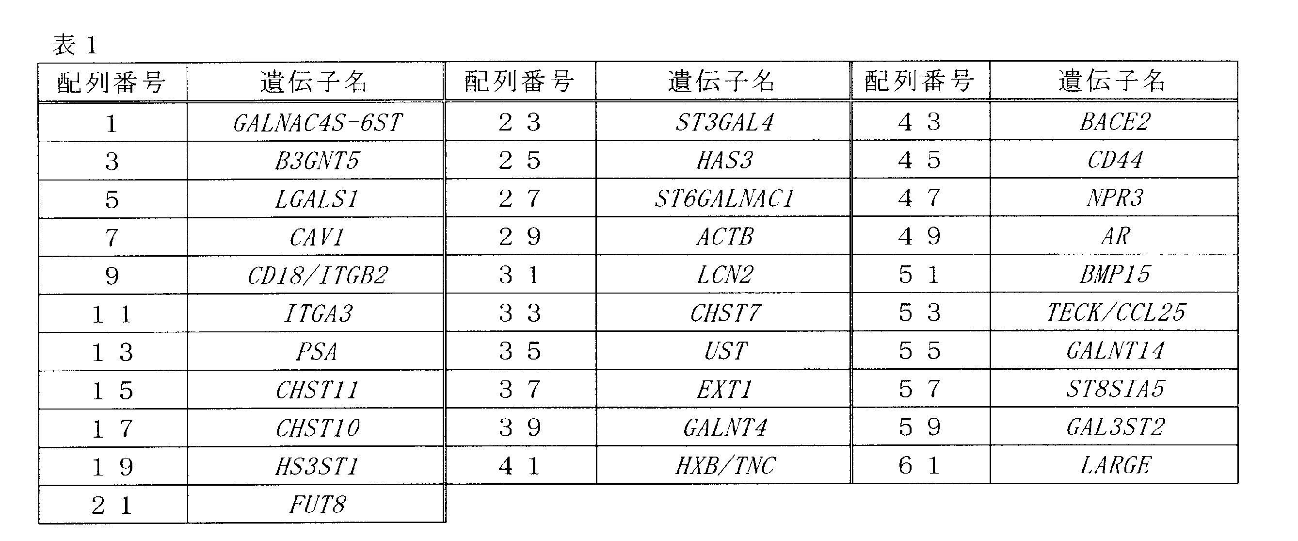
- Table 1 below shows the correspondence between the sequence numbers of the base sequences described in the present specification and the gene names encoded by the base sequences.
- the gene whose expression level is measured by the detection method of the present invention is the base sequence of SEQ ID NO: 1, the base sequence of SEQ ID NO: 3, the base sequence of SEQ ID NO: 5, the base sequence of SEQ ID NO: 7, the base sequence of SEQ ID NO: 9, DNA consisting of a base sequence selected from the group consisting of the base sequence of SEQ ID NO: 11 and the base sequence of SEQ ID NO: 27, or It consists of the base sequence of SEQ ID NO: 1, the base sequence of SEQ ID NO: 3, the base sequence of SEQ ID NO: 5, the base sequence of SEQ ID NO: 7, the base sequence of SEQ ID NO: 9, the base sequence of SEQ ID NO: 11 and the base sequence of SEQ ID NO: 27 DNA having 70% or more, preferably 80% or more, more preferably 90% or more, still more preferably 95% or more, particularly preferably 97% or more identity with a base sequence selected from the group, or It consists of the base sequence of SEQ ID NO: 1, the base sequence of SEQ ID NO: 3, the base sequence
- the expression level of at least one of the genes (or variants thereof) shown in Table 1 above is measured (except for PSA, ACTB, and AR). That is, in the detection method of the present invention, relapsed prostate cancer may be detected by measuring the expression level of one of the genes (or variants thereof) shown in Table 1 above, or two of the above genes The above expression level may be measured to detect relapsed prostate cancer.
- “measurement” includes both quantitative and qualitative concepts.
- examples of preferable gene combinations include the following combinations (h) to (m). Is not limited to this.
- (H) a gene having DNA consisting of the base sequence of SEQ ID NO: 1, A gene having a DNA comprising the nucleotide sequence of SEQ ID NO: 3, a gene having a DNA comprising the nucleotide sequence of SEQ ID NO: 5, a gene having a DNA comprising the nucleotide sequence of SEQ ID NO: 7, and a DNA comprising the nucleotide sequence of SEQ ID NO: 9.
- the expression level of a gene can be measured by measuring the amount of a transcription product of the gene, that is, the expression level of mRNA.
- the mRNA expression level can be measured by a normal method such as RT-PCR or real-time RT-PCR, but it is preferably measured by real-time RT-PCR from the viewpoint of rapid measurement.
- the real-time RT-PCR method can be performed by a usual method such as the method described in Examples described later.
- the primer for gene amplification used for the measurement of the expression level of mRNA is not particularly limited, and can be designed by an ordinary method based on the base sequence of the target gene. For example, by using one or more primer sets selected from the following (Aa) to (Bb), a nucleic acid amplification reaction can be performed using a cDNA synthesized from the mRNA as a template. The expression level can be measured.
- Ab a primer set consisting of an oligonucleotide consisting of the base sequence of SEQ ID NO: 65 and an oligonucleotide consisting of the base sequence of SEQ ID NO: 66;
- Ac a primer set consisting of an oligonucleotide consisting of the base sequence of SEQ ID NO: 67 and an oligonucleotide consisting of the base sequence of SEQ ID NO: 68;
- Ad a primer set consisting of an oligonucleotide consisting of the base sequence of SEQ ID NO: 69 and an oligonucleotide consisting of the base sequence of SEQ ID NO: 70;
- Ae a primer set consisting of an oligonucleotide consisting of an oligonu
- Ba A primer set consisting of an oligonucleotide consisting of the base sequence of SEQ ID NO: 121 and an oligonucleotide consisting of the base sequence of SEQ ID NO: 122.
- Bb A primer set consisting of an oligonucleotide consisting of the base sequence of SEQ ID NO: 123 and an oligonucleotide consisting of the base sequence of SEQ ID NO: 124.
- the expression level of a gene having a DNA consisting of the base sequence of SEQ ID NO: 1 or a variant thereof can be measured.
- the primer set (Ab) above the expression level of a gene having a DNA comprising the nucleotide sequence of SEQ ID NO: 3 or a variant thereof can be measured.
- the primer set (Ac) above the expression level of a gene having a DNA comprising the nucleotide sequence of SEQ ID NO: 5 or a variant thereof can be measured.
- the primer set (Ad) above the expression level of a gene having a DNA comprising the nucleotide sequence of SEQ ID NO: 7 or a variant thereof can be measured.
- the expression level of a gene having a DNA comprising the nucleotide sequence of SEQ ID NO: 9 or a variant thereof can be measured.
- the expression level of a gene having a DNA comprising the nucleotide sequence of SEQ ID NO: 11 or a variant thereof can be measured.
- the primer set (Ag) above the expression level of a gene having a DNA comprising the nucleotide sequence of SEQ ID NO: 15 or a variant thereof can be measured.
- the primer set (Ah) above the expression level of a gene having a DNA comprising the nucleotide sequence of SEQ ID NO: 17 or a variant thereof can be measured.
- the expression level of a gene having a DNA consisting of the nucleotide sequence of SEQ ID NO: 19 or a variant thereof can be measured.
- the expression level of a gene having a DNA consisting of the nucleotide sequence of SEQ ID NO: 21 or a variant thereof can be measured.
- the primer set (Ak) above the expression level of a gene having a DNA comprising the nucleotide sequence of SEQ ID NO: 23 or a variant thereof can be measured.
- the primer set (Al) above the expression level of a gene having a DNA comprising the nucleotide sequence of SEQ ID NO: 25 or a variant thereof can be measured.
- the expression level of a gene having a DNA comprising the nucleotide sequence of SEQ ID NO: 27 or a variant thereof can be measured.
- the primer set (An) above the expression level of a gene having a DNA consisting of the nucleotide sequence of SEQ ID NO: 31 or a variant thereof can be measured.
- the primer set (Ao) above the expression level of a gene having a DNA comprising the nucleotide sequence of SEQ ID NO: 33 or a variant thereof can be measured.
- the primer set (Ap) above the expression level of a gene having a DNA consisting of the nucleotide sequence of SEQ ID NO: 35 or a variant thereof can be measured.
- the expression level of a gene having a DNA comprising the nucleotide sequence of SEQ ID NO: 37 or a variant thereof can be measured.
- the expression level of a gene having a DNA consisting of the nucleotide sequence of SEQ ID NO: 39 or a variant thereof can be measured.
- the expression level of a gene having a DNA consisting of the nucleotide sequence of SEQ ID NO: 41 or a variant thereof can be measured.
- the expression level of a gene having a DNA consisting of the nucleotide sequence of SEQ ID NO: 43 or a variant thereof can be measured.
- the expression level of a gene having a DNA consisting of the nucleotide sequence of SEQ ID NO: 45 or a variant thereof can be measured.
- the expression level of a gene having a DNA consisting of the nucleotide sequence of SEQ ID NO: 47 or a variant thereof can be measured.
- the primer set (Aw) above the expression level of a gene having a DNA comprising the nucleotide sequence of SEQ ID NO: 51 or a variant thereof can be measured.
- the primer set (Ax) above the expression level of a gene having a DNA consisting of the nucleotide sequence of SEQ ID NO: 53 or a variant thereof can be measured.
- the expression level of a gene having a DNA comprising the nucleotide sequence of SEQ ID NO: 55 or a variant thereof can be measured.
- the expression level of a gene having a DNA comprising the nucleotide sequence of SEQ ID NO: 57 or a variant thereof can be measured.
- the primer set (Ba) above the expression level of a gene having a DNA consisting of the nucleotide sequence of SEQ ID NO: 59 or a variant thereof can be measured.
- the primer set (Bb) above the expression level of a gene having a DNA consisting of the nucleotide sequence of SEQ ID NO: 61 or a variant thereof can be measured.
- the expression level of a gene can also be measured by measuring the amount of protein that is an expression product of the gene.
- the protein whose amount is measured in the present invention is the amino acid sequence of SEQ ID NO: 2, the amino acid sequence of SEQ ID NO: 4, the amino acid sequence of SEQ ID NO: 6, the amino acid sequence of SEQ ID NO: 8, the amino acid sequence of SEQ ID NO: 12, Amino acid sequence, amino acid sequence of SEQ ID NO: 16, amino acid sequence of SEQ ID NO: 18, amino acid sequence of SEQ ID NO: 20, amino acid sequence of SEQ ID NO: 22, amino acid sequence of SEQ ID NO: 24, amino acid sequence of SEQ ID NO: 26, amino acid of SEQ ID NO: 28 Sequence, amino acid sequence of SEQ ID NO: 32, amino acid sequence of SEQ ID NO: 34, amino acid sequence of SEQ ID NO: 36, amino acid sequence of SEQ ID NO: 38, amino acid sequence of SEQ ID NO: 40, amino acid sequence of SEQ ID NO: 42, amino acid sequence of SEQ ID NO: 44 , SEQ ID NO: 46 amino acid sequence, SEQ ID NO: 48 amino acid sequence, SEQ ID NO: 52 An amino acid sequence selected
- amino acid sequence of SEQ ID No. 18 the amino acid sequence of SEQ ID No. 20
- amino acid sequence of SEQ ID No. 22 the amino acid sequence of SEQ ID No. 24, the amino acid sequence of SEQ ID No. 26, the amino acid sequence of SEQ ID No. 28, the SEQ ID No.
- amino acid sequence 32 amino acid sequence, SEQ ID NO: 34 amino acid sequence, SEQ ID NO: 36 amino acid sequence, SEQ ID NO: 38 amino acid sequence, SEQ ID NO: 40 amino acid sequence, SEQ ID NO: 42 amino acid sequence, SEQ ID NO: 44 amino acid sequence, SEQ ID NO: 46 Amino acid sequence of SEQ ID NO: 48, amino acid sequence of SEQ ID NO: 52, amino acid sequence of SEQ ID NO: 54, amino acid sequence of SEQ ID NO: 56, amino acid sequence of SEQ ID NO: 58, amino acid sequence of SEQ ID NO: 60 and SEQ ID NO: 62 1 or a number in an amino acid sequence selected from the group consisting of amino acid sequences Preferred amino acids are deleted, substituted, may consist of inserted or added in the amino acid sequence.
- amino acid sequence identity means that two amino acid sequences to be compared are aligned (aligned) by introducing a gap as necessary, and the maximum amino acid sequence identity (%) obtained.
- Alignment for the purpose of determining the identity of amino acid sequences can be performed using a conventional method.
- publicly available computer software such as BLAST, DNASIS Pro (manufactured by Hitachi Software Engineering)
- commercially available software such as GENETYX (manufactured by Genetics) can also be used.
- the “one or several” is as described above.
- the above protein is a protein consisting of an amino acid sequence encoded by DNA present in the genome of human prostate cancer cells, and has a difference in abundance between hormone-sensitive prostate cancer and relapsed prostate cancer. is there.
- the protein measured as the gene expression level is at least one of the above proteins. That is, the amount of two or more proteins may be measured. Examples of preferable combinations of proteins to be measured when measuring the amount of two or more proteins include the following combinations (n) to (s), but the present invention is not limited to these combinations: Absent.
- the amount of the protein is preferably measured using a molecule that specifically binds to the protein.
- the molecule that specifically binds to the protein of the present invention include antibodies and aptamers to the protein of the present invention, and it is preferable to use antibodies to the protein of the present invention.
- the antibody or aptamer can be used after being labeled with a radioactive substance, an enzyme, a fluorescent substance, a luminescent substance, or the like according to the purpose.
- means usually used in the field of immunoassay can be adopted, and for example, it can be detected by an immunoblot method, an enzyme immunoassay (ELISA method), an immunochromatography method or the like.
- the antibody used for detecting the protein can be obtained by a usual method.
- an animal can be immunized to obtain an antibody that selectively reacts with the protein from the animal.
- an amino acid sequence characteristic of the protein is selected from the amino acid sequence of the protein, a peptide comprising the amino acid sequence is chemically synthesized, this is immunized to the animal, and the animal selectively selects the protein.
- the measurement of the protein is preferably performed according to the principle of sandwich immunoassay using two types of antibodies having different epitopes in the protein.
- one antibody is immobilized on a solid phase as a capture antibody, and the other antibody is labeled and functions as a detection antibody.
- a well in which an antibody against the protein is immobilized is prepared, and the well is blocked with BSA, casein or the like in order to prevent nonspecific adsorption.
- a protein solution prepared from prostate cancer cells or the like is added here, and incubated for several minutes to several days at a temperature (eg, 4 to 40 ° C.) at which the antibody is hardly inactivated. Subsequently, discard the protein solution, wash the well with phosphate buffer, etc., add a labeled antibody that binds to an epitope different from the epitope to which the immobilized antibody binds, and incubate again for several minutes to several days. After washing, the label present in the well is detected. When the protein of interest is present in the protein solution, the labeling signal becomes high, so that relapsed prostate cancer can be detected.
- an immobilized antibody in the sandwich ELISA is immobilized on a membrane test line, and a test strip in a state where the labeled antibody in the sandwich ELISA is held on a conjugate pad is prepared.
- a protein solution prepared from prostate cancer cells or the like is added directly or indirectly to the conjugate pad, the solution proceeds from the conjugate pad to the membrane while reacting with the labeled antibody in a liquid phase by capillary action.
- the protein of interest is present in the protein solution, a complex composed of the labeled antibody and the protein of interest is trapped on the membrane test line. Therefore, relapsed prostate cancer can be detected by detecting a label present on the test line.
- the sample in which the expression level of the gene is measured is not particularly limited, and examples thereof include blood, plasma, serum, lymph, urine, saliva and other body fluids, prostate cancer tissue, prostate cancer cells and the like. It is preferable to use prostate cancer tissue or prostate cancer cells.
- isolated prostate cancer cells include prostate cancer tissue extracted from a living body (prostate cancer patient), a sample obtained by excising the tissue by laser digestion, and the like.
- the origin of the sample is not particularly limited, but is preferably derived from a human. That is, the detection method of the present invention is suitably used for detecting human relapsed prostate cancer.
- the detection method of the present invention By using the detection method of the present invention, the hormone sensitivity of prostate cancer can be determined easily and accurately, whereby relapsed prostate cancer can be specifically detected. Therefore, the detection method of the present invention is useful for diagnosis of relapsed prostate cancer.
- the analysis will focus on the following DNA microarray analysis and quantitative PCR array analysis to select candidate marker genes that can distinguish between hormone-sensitive and hormone-resistant prostate cancer. went.
- Prostate cancer cell lines LNCaP (hormone sensitivity (dependency)) and Du145 (hormone resistance (independence)) were obtained from RIKEN Cell Bank, RIKEN BioResource Research Center.
- PC-3 (hormone-independent) was obtained from the Human Science Promotion Foundation JCRB Cell Bank.
- the normal human prostate epithelial cell line PrE was purchased from DS Pharma Biomedical. All prostate cancer cell lines were cultured using RPMI 1640 medium (GIBCO) supplemented with 10% FBS (Hyclone) and 1% penicillin / streptomycin (GIBCO). Normal human prostate epithelial cell lines were cultured using CS-C medium kit R (DS Pharma Biomedical). All cells were cultured one day prior to RNA and protein extraction, substituting with RPMI 1640 plus 10% FBS.
- DNA microarray analysis The DNA microarray for comprehensive analysis of glycan-related genes used a tool developed by the RIKEN Laboratory for Disease Sugar Chain. Details of this tool are described in the literature (Molecular And Cellular Biology, Apr. 2007, p.3008-3022). Briefly, first, mRNA of logarithmically growing cells was prepared using mTRAP midi kit (manufactured by Active Motif), quality checked by Bioanalyzer 2100 manufactured by Agilent Technologies, and then CyScribe Post-Labelling Kit (GE Health). Fluorescently labeled with Care Bioscience).
- the fluorescently labeled sample was hybridized to a microarray carrying about 1200 sugar chain-related genes, and scanned with an Affymetrix 428 Array Scanner.
- the signal intensity of each spot was quantified (ImaGene; manufactured by BioDiscovery), and after correction, analysis was performed with clustering creation software MultiExperiment Viewer (MeV) (Opensource software tool, http://www.tm4.org/).
- the qPCR array which is a gene exhaustive analysis tool specialized for glycan synthases, used a tool developed by the National Institute of Advanced Industrial Science and Technology (AIST). Details of this tool are described in the literature (Jornal of Proteome Reserch. Vol.8, No.3, 2009, p.1358-1367). Briefly, first, a total RNA of cells in logarithmic growth phase was prepared using RNeasy plus mini kit (manufactured by QIAGEN), and QuantiTect Rev. First strand cDNA was synthesized using a transcription kit (QIAGEN).
- PCR primers were selected using ProbeFinder (https://www.roche-applied-science.com/), and made and purchased from Invitrogen. Hydrolysis probes were selected from Universal ProbeLibrary (Roche). The assay for all 189 genes was formatted in 384 plates with duplicate, and amplified and measured with LightCycler 480 (Roche) using qPCR Quick GoldStar Mastermix Plus (Eurogentec). After calculating each copy number, it was analyzed with a clustering creation software MultiExperiment Viewer (MeV) (Opensource software tool, http://www.tm4.org/) after correction.
- MeV clustering creation software MultiExperiment Viewer
- Example 1 Detection of Relapsed Prostate Cancer by RT-PCR
- Normal human prostate epithelial cell line PrE and prostate cancer cell lines LNCaP (hormone sensitive strain), Du145 (hormone resistant strain) and PC-3 (hormone resistant strain) Using each cell line, the amount of the transcription product of 31 kinds of genes shown in Table 1 was measured by RT-PCR.
- the primers to be selected were selected using ProbeFinder (https://www.roche-applied-science.com/), prepared and purchased by Invitrogen. The results are shown in FIG. Note that the measurement results of PSA, ACTB, AR, and GAPDH in FIG. 1 are provided for reference, and do not indicate the effects of the present invention.
- GALNAC4S-6ST (SEQ ID NO: 1), B3GNT5 (SEQ ID NO: 3), compared to LNCaP, which is a hormone-sensitive prostate cell line, LGALS1 (SEQ ID NO: 5), CAV1 (SEQ ID NO: 7), CD18 / ITGB2 (SEQ ID NO: 9), ITGA3 (SEQ ID NO: 11), CHST11 (SEQ ID NO: 15), CHST10 (SEQ ID NO: 17), FUT8 (SEQ ID NO: 21) , HAS3 (SEQ ID NO: 25), LCN2 (SEQ ID NO: 31), CHST7 (SEQ ID NO: 33), UST (SEQ ID NO: 35), EXT1 (SEQ ID NO: 37), GALNT4 (SEQ ID NO: 39), HXB / TNC (SEQ ID NO: 41) , BACE2 (SEQ ID NO: 43), CD44 (SEQ ID NO:
- Hormone-resistant prostate cancer relapsed prostate cancer
- Example 2 Detection of Relapsed Prostate Cancer by Real-Time RT-PCR Using the following human prostate cancer tissue samples, transcripts of 25 genes among the genes shown in Table 1 were analyzed by real-time RT-PCR described below. The amount was measured.
- a prostate cancer clinical tissue sample was provided by the Aichi Cancer Center. All samples were provided by patients with informed consent. Eight prostate tissues removed from prostate cancer patients who have not been treated with hormone therapy, and hormone therapy (neoadjuvant therapy: hormone-releasing hormone analogue (LH-RHa)) for 3 months or more before undergoing prostatectomy surgery Eight cases of prostate cancer patients who had received a non-steroidal antiandrogenic agent Bicalutamide (combined with Bicalutamide) and were excised from patients with low sensitivity to the treatment (therapeutic effect was weak) were used in this example.
- hormone therapy nuclear hormone-releasing hormone analogue
- prostate cancer cancer cells proliferate depending on male hormones such as androgens, and the growth of cancer cells is often effectively suppressed by suppressing the action of the hormones.
- hormone-resistant prostate cancer after receiving hormonal therapy, is often the one in which the surviving cancer cells have proliferated again even though growth has been suppressed, and has not received hormonal therapy Incidence in prostate cancer patients is relatively low. Therefore, the following tests were conducted assuming that prostate cancer patients who had not received hormone therapy were hormone sensitive prostate cancer patients.
- RNA extraction kit (trade name: RecoverAll TM). RNA was extracted using a Total Nucleic Acid Isolation Kit (Ambion).
- CDNA was prepared using High Capacity cDNA Reverse Transcription Kit (Applied Biosystems), preamplified with TaqMan (registered trademark) PreAmp Master Mix Kit (Applied Biosystems), and qPCR QuickMoldGoldMoldGoldSurGoldSoldGoldSurGold ABI PRISM (registered trademark) 7700 Sequence Detection System was used for amplification and measurement.
- the primer set used is the same as in Example 1. After calculating the number of each copy, it was standardized by the expression level of glyceraldehyde-3-phosphate dehydrogenase (GAPDH) mRNA used as a control. For statistical analysis, “KaleidaGraph” manufactured by Hulinks was used and t-test was performed.
- GPDH glyceraldehyde-3-phosphate dehydrogenase
- GALNAC4S-6ST (SEQ ID NO: 1), B3GNT5 (SEQ ID NO: 3), LGALS1 (SEQ ID NO: 5), CAV1 (SEQ ID NO: 7), ITGB2 (SEQ ID NO: 9), ITGA3 (SEQ ID NO: 9) 11) and ST6GALNAC1 (SEQ ID NO: 27), between cancer cells collected from 8 prostate cancer patients not receiving hormone therapy and cancer cells collected from 8 prostate cancer patients less sensitive to hormone therapy, A clear significant difference (P ⁇ 0.05) was observed in the expression level of mRNA (see the column of 8 vs 8 in Table 2).
- the expression level of PSA was significantly reduced in cancer cells collected from 8 prostate cancer patients with low sensitivity to hormonal therapy, which was caused by the blockage of androgen. Yes, it is not directly related to hormone resistance. There may also be patients who are not sensitive to hormonal therapy among untreated groups who have not received hormonal therapy. Therefore, for reference, out of samples collected from 8 prostate cancer patients who have not received hormone therapy, except for 2 patient samples with high mRNA expression levels, from 6 prostate cancer patients who have not received hormone therapy. The significant difference between the collected cancer cells and cancer cells collected from 8 prostate cancer patients with low sensitivity to hormone therapy was examined.
- Example 1 As for the genes for which no significant difference was observed in the above, as shown in Example 1, in the test using the prostate cancer cell line, hormone sensitive prostate cancer cells and hormone resistant prostate cancer cells were distinguished. It has been shown that Therefore, although it is difficult at present, if a patient specimen identified as a hormone sensitive patient can be used, a significant difference in the expression level may be observed with a prostate cancer patient specimen that is less sensitive to hormone therapy. It is done.
- the detection method of the present invention is a method that can specifically detect relapsed prostate cancer.
Landscapes
- Health & Medical Sciences (AREA)
- Life Sciences & Earth Sciences (AREA)
- Chemical & Material Sciences (AREA)
- Immunology (AREA)
- Engineering & Computer Science (AREA)
- Organic Chemistry (AREA)
- Proteomics, Peptides & Aminoacids (AREA)
- Pathology (AREA)
- Analytical Chemistry (AREA)
- Molecular Biology (AREA)
- Biochemistry (AREA)
- Wood Science & Technology (AREA)
- Zoology (AREA)
- Urology & Nephrology (AREA)
- Physics & Mathematics (AREA)
- Microbiology (AREA)
- Hematology (AREA)
- General Health & Medical Sciences (AREA)
- Biomedical Technology (AREA)
- Biotechnology (AREA)
- Genetics & Genomics (AREA)
- Oncology (AREA)
- Hospice & Palliative Care (AREA)
- Cell Biology (AREA)
- Biophysics (AREA)
- General Physics & Mathematics (AREA)
- Medicinal Chemistry (AREA)
- Food Science & Technology (AREA)
- Bioinformatics & Cheminformatics (AREA)
- General Engineering & Computer Science (AREA)
- Measuring Or Testing Involving Enzymes Or Micro-Organisms (AREA)
- Investigating Or Analysing Biological Materials (AREA)
Abstract
前立腺癌細胞で発現しうる特定の遺伝子の発現量を測定することを含む、再燃性前立腺癌の検出方法。
Description
本発明は、再燃性前立腺癌の検出方法に関する。
前立腺癌は、男性ホルモンであるアンドロゲンに依存して増殖するため、アンドロゲンの分泌や働きを抑制するホルモン療法が有効な治療法として用いられている。しかし、このホルモン療法によっても少なからず癌細胞が生き残る。生き残った癌細胞は、その後増殖を繰返し、ホルモン療法に耐性をもったホルモン抵抗性前立腺癌を引き起こす。ホルモン抵抗性前立腺癌は、ホルモン療法により一度抑制された前立腺癌の増殖が再び活発化したものであり、再燃性前立腺癌とも呼ばれる。
本発明者らは、再燃性前立腺癌特異的なマーカーを開発することができれば、当該マーカーを検出することにより再燃性前立腺癌を特異的に検出することができ、これにより再燃性前立腺癌に適した治療、すなわち非ホルモン療法をスムーズに実施できるため、患者の負担軽減及び医療経済の観点から有益であると考えた。
本発明は、再燃性前立腺癌に特異的なマーカーを見い出し、このマーカーを用いて再燃性前立腺癌を特異的に検出する方法を提供することを課題とする。
本発明は、再燃性前立腺癌に特異的なマーカーを見い出し、このマーカーを用いて再燃性前立腺癌を特異的に検出する方法を提供することを課題とする。
本発明者らは、これまで発現量が少ないが故に発現量の検出が困難であった特定の糖鎖関連遺伝子に着目し、これらを含む遺伝子群の中から、再燃性前立腺癌細胞においてより高い又はより低い発現量を示す遺伝子を特定することに成功した。本発明はこの知見に基づき完成するに至ったものである。
本発明の課題は下記の手段により達成された。
<1>下記(a)~(d)のいずれかのDNAからなる少なくとも1種の遺伝子の発現量を測定することを含む、再燃性前立腺癌の検出方法:
(a)配列番号1の塩基配列、配列番号3の塩基配列、配列番号5の塩基配列、配列番号7の塩基配列、配列番号9の塩基配列、配列番号11の塩基配列、配列番号15の塩基配列、配列番号17の塩基配列、配列番号19の塩基配列、配列番号21の塩基配列、配列番号23の塩基配列、配列番号25の塩基配列、配列番号27の塩基配列、配列番号31の塩基配列、配列番号33の塩基配列、配列番号35の塩基配列、配列番号37の塩基配列、配列番号39の塩基配列、配列番号41の塩基配列、配列番号43の塩基配列、配列番号45の塩基配列、配列番号47の塩基配列、配列番号51の塩基配列、配列番号53の塩基配列、配列番号55の塩基配列、配列番号57の塩基配列、配列番号59の塩基配列及び配列番号61の塩基配列からなる群から選ばれる塩基配列からなるDNA、
(b)上記(a)のDNAの塩基配列と80%以上の同一性を有する塩基配列からなるDNAであって、前立腺癌細胞のゲノム中に存在するDNA、
(c)上記(a)のDNAの塩基配列において、1又は数個の塩基が欠失、置換、挿入又は付加された塩基配列からなるDNAであって、前立腺癌細胞のゲノム中に存在するDNA、
(d)上記(a)のDNAの塩基配列と相補的な塩基配列からなるDNAにストリンジェントな条件下でハイブリダイズするDNAであって、前立腺癌細胞のゲノム中に存在するDNA。
<2>遺伝子の発現量の測定が、該遺伝子の転写産物の量の測定である、<1>に記載の検出方法。
<3>下記(Aa)~(Bb)からなる群から選ばれる少なくとも1種のプライマーセットを用いて少なくとも1種の遺伝子の発現量を測定する、<2>に記載の検出方法:
(Aa)配列番号63の塩基配列からなるオリゴヌクレオチド及び配列番号64の塩基配列からなるオリゴヌクレオチドからなるプライマーセット、
(Ab)配列番号65の塩基配列からなるオリゴヌクレオチド及び配列番号66の塩基配列からなるオリゴヌクレオチドからなるプライマーセット、
(Ac)配列番号67の塩基配列からなるオリゴヌクレオチド及び配列番号68の塩基配列からなるオリゴヌクレオチドからなるプライマーセット、
(Ad)配列番号69の塩基配列からなるオリゴヌクレオチド及び配列番号70の塩基配列からなるオリゴヌクレオチドからなるプライマーセット、
(Ae)配列番号71の塩基配列からなるオリゴヌクレオチド及び配列番号72の塩基配列からなるオリゴヌクレオチドからなるプライマーセット、
(Af)配列番号73の塩基配列からなるオリゴヌクレオチド及び配列番号74の塩基配列からなるオリゴヌクレオチドからなるプライマーセット、
(Ag)配列番号77の塩基配列からなるオリゴヌクレオチド及び配列番号78の塩基配列からなるオリゴヌクレオチドからなるプライマーセット、
(Ah)配列番号79の塩基配列からなるオリゴヌクレオチド及び配列番号80の塩基配列からなるオリゴヌクレオチドからなるプライマーセット、
(Ai)配列番号81の塩基配列からなるオリゴヌクレオチド及び配列番号82の塩基配列からなるオリゴヌクレオチドからなるプライマーセット、
(Aj)配列番号83の塩基配列からなるオリゴヌクレオチド及び配列番号84の塩基配列からなるオリゴヌクレオチドからなるプライマーセット、
(Ak)配列番号85の塩基配列からなるオリゴヌクレオチド及び配列番号86の塩基配列からなるオリゴヌクレオチドからなるプライマーセット、
(Al)配列番号87の塩基配列からなるオリゴヌクレオチド及び配列番号88の塩基配列からなるオリゴヌクレオチドからなるプライマーセット、
(Am)配列番号89の塩基配列からなるオリゴヌクレオチド及び配列番号90の塩基配列からなるオリゴヌクレオチドからなるプライマーセット、
(An)配列番号93の塩基配列からなるオリゴヌクレオチド及び配列番号94の塩基配列からなるオリゴヌクレオチドからなるプライマーセット、
(Ao)配列番号95の塩基配列からなるオリゴヌクレオチド及び配列番号96の塩基配列からなるオリゴヌクレオチドからなるプライマーセット、
(Ap)配列番号97の塩基配列からなるオリゴヌクレオチド及び配列番号98の塩基配列からなるオリゴヌクレオチドからなるプライマーセット、
(Aq)配列番号99の塩基配列からなるオリゴヌクレオチド及び配列番号100の塩基配列からなるオリゴヌクレオチドからなるプライマーセット、
(Ar)配列番号101の塩基配列からなるオリゴヌクレオチド及び配列番号102の塩基配列からなるオリゴヌクレオチドからなるプライマーセット、
(As)配列番号103の塩基配列からなるオリゴヌクレオチド及び配列番号104の塩基配列からなるオリゴヌクレオチドからなるプライマーセット、
(At)配列番号105の塩基配列からなるオリゴヌクレオチド及び配列番号106の塩基配列からなるオリゴヌクレオチドからなるプライマーセット、
(Au)配列番号107の塩基配列からなるオリゴヌクレオチド及び配列番号108の塩基配列からなるオリゴヌクレオチドからなるプライマーセット、
(Av)配列番号109の塩基配列からなるオリゴヌクレオチド及び配列番号110の塩基配列からなるオリゴヌクレオチドからなるプライマーセット、及び
(Aw)配列番号113の塩基配列からなるオリゴヌクレオチド及び配列番号114の塩基配列からなるオリゴヌクレオチドからなるプライマーセット。
(Ax)配列番号115の塩基配列からなるオリゴヌクレオチド及び配列番号116の塩基配列からなるオリゴヌクレオチドからなるプライマーセット。
(Ay)配列番号117の塩基配列からなるオリゴヌクレオチド及び配列番号118の塩基配列からなるオリゴヌクレオチドからなるプライマーセット。
(Az)配列番号119の塩基配列からなるオリゴヌクレオチド及び配列番号120の塩基配列からなるオリゴヌクレオチドからなるプライマーセット。
(Ba)配列番号121の塩基配列からなるオリゴヌクレオチド及び配列番号122の塩基配列からなるオリゴヌクレオチドからなるプライマーセット。
(Bb)配列番号123の塩基配列からなるオリゴヌクレオチド及び配列番号124の塩基配列からなるオリゴヌクレオチドからなるプライマーセット。
<4>遺伝子の発現量の測定が、該遺伝子の発現産物であるタンパク質の量の測定である、<1>に記載の検出方法。
<5>測定されるタンパク質の量が、下記(e)~(g)のいずれかのアミノ酸配列からなる少なくとも1種のタンパク質の量である、<4>に記載の検出方法:
(e)配列番号2のアミノ酸配列、配列番号4のアミノ酸配列、配列番号6のアミノ酸配列、配列番号8のアミノ酸配列、配列番号10のアミノ酸配列、配列番号12のアミノ酸配列、配列番号16のアミノ酸配列、配列番号18のアミノ酸配列、配列番号20のアミノ酸配列、配列番号22のアミノ酸配列、配列番号24のアミノ酸配列、配列番号26のアミノ酸配列、配列番号28のアミノ酸配列、配列番号32のアミノ酸配列、配列番号34のアミノ酸配列、配列番号36のアミノ酸配列、配列番号38のアミノ酸配列、配列番号40のアミノ酸配列、配列番号42のアミノ酸配列、配列番号44のアミノ酸配列、配列番号46のアミノ酸配列、配列番号48のアミノ酸配列、配列番号52のアミノ酸配列、配列番号54のアミノ酸配列、配列番号56のアミノ酸配列、配列番号58のアミノ酸配列、配列番号60のアミノ酸配列及び配列番号62のアミノ酸配列からなる群から選ばれるアミノ酸配列、
(f)上記(e)のアミノ酸配列と80%以上の同一性を有し、かつ前立腺癌細胞のゲノム中に存在するDNAにコードされるアミノ酸配列、
(g)上記(e)のアミノ酸配列において、1又は数個のアミノ酸が欠失、置換、挿入又は付加されたアミノ酸配列であって、前立腺癌細胞のゲノム中に存在するDNAにコードされるアミノ酸配列。
<6>単離した前立腺癌細胞中における遺伝子の発現量を測定する、<1>~<5>のいずれかに記載の検出方法。
<1>下記(a)~(d)のいずれかのDNAからなる少なくとも1種の遺伝子の発現量を測定することを含む、再燃性前立腺癌の検出方法:
(a)配列番号1の塩基配列、配列番号3の塩基配列、配列番号5の塩基配列、配列番号7の塩基配列、配列番号9の塩基配列、配列番号11の塩基配列、配列番号15の塩基配列、配列番号17の塩基配列、配列番号19の塩基配列、配列番号21の塩基配列、配列番号23の塩基配列、配列番号25の塩基配列、配列番号27の塩基配列、配列番号31の塩基配列、配列番号33の塩基配列、配列番号35の塩基配列、配列番号37の塩基配列、配列番号39の塩基配列、配列番号41の塩基配列、配列番号43の塩基配列、配列番号45の塩基配列、配列番号47の塩基配列、配列番号51の塩基配列、配列番号53の塩基配列、配列番号55の塩基配列、配列番号57の塩基配列、配列番号59の塩基配列及び配列番号61の塩基配列からなる群から選ばれる塩基配列からなるDNA、
(b)上記(a)のDNAの塩基配列と80%以上の同一性を有する塩基配列からなるDNAであって、前立腺癌細胞のゲノム中に存在するDNA、
(c)上記(a)のDNAの塩基配列において、1又は数個の塩基が欠失、置換、挿入又は付加された塩基配列からなるDNAであって、前立腺癌細胞のゲノム中に存在するDNA、
(d)上記(a)のDNAの塩基配列と相補的な塩基配列からなるDNAにストリンジェントな条件下でハイブリダイズするDNAであって、前立腺癌細胞のゲノム中に存在するDNA。
<2>遺伝子の発現量の測定が、該遺伝子の転写産物の量の測定である、<1>に記載の検出方法。
<3>下記(Aa)~(Bb)からなる群から選ばれる少なくとも1種のプライマーセットを用いて少なくとも1種の遺伝子の発現量を測定する、<2>に記載の検出方法:
(Aa)配列番号63の塩基配列からなるオリゴヌクレオチド及び配列番号64の塩基配列からなるオリゴヌクレオチドからなるプライマーセット、
(Ab)配列番号65の塩基配列からなるオリゴヌクレオチド及び配列番号66の塩基配列からなるオリゴヌクレオチドからなるプライマーセット、
(Ac)配列番号67の塩基配列からなるオリゴヌクレオチド及び配列番号68の塩基配列からなるオリゴヌクレオチドからなるプライマーセット、
(Ad)配列番号69の塩基配列からなるオリゴヌクレオチド及び配列番号70の塩基配列からなるオリゴヌクレオチドからなるプライマーセット、
(Ae)配列番号71の塩基配列からなるオリゴヌクレオチド及び配列番号72の塩基配列からなるオリゴヌクレオチドからなるプライマーセット、
(Af)配列番号73の塩基配列からなるオリゴヌクレオチド及び配列番号74の塩基配列からなるオリゴヌクレオチドからなるプライマーセット、
(Ag)配列番号77の塩基配列からなるオリゴヌクレオチド及び配列番号78の塩基配列からなるオリゴヌクレオチドからなるプライマーセット、
(Ah)配列番号79の塩基配列からなるオリゴヌクレオチド及び配列番号80の塩基配列からなるオリゴヌクレオチドからなるプライマーセット、
(Ai)配列番号81の塩基配列からなるオリゴヌクレオチド及び配列番号82の塩基配列からなるオリゴヌクレオチドからなるプライマーセット、
(Aj)配列番号83の塩基配列からなるオリゴヌクレオチド及び配列番号84の塩基配列からなるオリゴヌクレオチドからなるプライマーセット、
(Ak)配列番号85の塩基配列からなるオリゴヌクレオチド及び配列番号86の塩基配列からなるオリゴヌクレオチドからなるプライマーセット、
(Al)配列番号87の塩基配列からなるオリゴヌクレオチド及び配列番号88の塩基配列からなるオリゴヌクレオチドからなるプライマーセット、
(Am)配列番号89の塩基配列からなるオリゴヌクレオチド及び配列番号90の塩基配列からなるオリゴヌクレオチドからなるプライマーセット、
(An)配列番号93の塩基配列からなるオリゴヌクレオチド及び配列番号94の塩基配列からなるオリゴヌクレオチドからなるプライマーセット、
(Ao)配列番号95の塩基配列からなるオリゴヌクレオチド及び配列番号96の塩基配列からなるオリゴヌクレオチドからなるプライマーセット、
(Ap)配列番号97の塩基配列からなるオリゴヌクレオチド及び配列番号98の塩基配列からなるオリゴヌクレオチドからなるプライマーセット、
(Aq)配列番号99の塩基配列からなるオリゴヌクレオチド及び配列番号100の塩基配列からなるオリゴヌクレオチドからなるプライマーセット、
(Ar)配列番号101の塩基配列からなるオリゴヌクレオチド及び配列番号102の塩基配列からなるオリゴヌクレオチドからなるプライマーセット、
(As)配列番号103の塩基配列からなるオリゴヌクレオチド及び配列番号104の塩基配列からなるオリゴヌクレオチドからなるプライマーセット、
(At)配列番号105の塩基配列からなるオリゴヌクレオチド及び配列番号106の塩基配列からなるオリゴヌクレオチドからなるプライマーセット、
(Au)配列番号107の塩基配列からなるオリゴヌクレオチド及び配列番号108の塩基配列からなるオリゴヌクレオチドからなるプライマーセット、
(Av)配列番号109の塩基配列からなるオリゴヌクレオチド及び配列番号110の塩基配列からなるオリゴヌクレオチドからなるプライマーセット、及び
(Aw)配列番号113の塩基配列からなるオリゴヌクレオチド及び配列番号114の塩基配列からなるオリゴヌクレオチドからなるプライマーセット。
(Ax)配列番号115の塩基配列からなるオリゴヌクレオチド及び配列番号116の塩基配列からなるオリゴヌクレオチドからなるプライマーセット。
(Ay)配列番号117の塩基配列からなるオリゴヌクレオチド及び配列番号118の塩基配列からなるオリゴヌクレオチドからなるプライマーセット。
(Az)配列番号119の塩基配列からなるオリゴヌクレオチド及び配列番号120の塩基配列からなるオリゴヌクレオチドからなるプライマーセット。
(Ba)配列番号121の塩基配列からなるオリゴヌクレオチド及び配列番号122の塩基配列からなるオリゴヌクレオチドからなるプライマーセット。
(Bb)配列番号123の塩基配列からなるオリゴヌクレオチド及び配列番号124の塩基配列からなるオリゴヌクレオチドからなるプライマーセット。
<4>遺伝子の発現量の測定が、該遺伝子の発現産物であるタンパク質の量の測定である、<1>に記載の検出方法。
<5>測定されるタンパク質の量が、下記(e)~(g)のいずれかのアミノ酸配列からなる少なくとも1種のタンパク質の量である、<4>に記載の検出方法:
(e)配列番号2のアミノ酸配列、配列番号4のアミノ酸配列、配列番号6のアミノ酸配列、配列番号8のアミノ酸配列、配列番号10のアミノ酸配列、配列番号12のアミノ酸配列、配列番号16のアミノ酸配列、配列番号18のアミノ酸配列、配列番号20のアミノ酸配列、配列番号22のアミノ酸配列、配列番号24のアミノ酸配列、配列番号26のアミノ酸配列、配列番号28のアミノ酸配列、配列番号32のアミノ酸配列、配列番号34のアミノ酸配列、配列番号36のアミノ酸配列、配列番号38のアミノ酸配列、配列番号40のアミノ酸配列、配列番号42のアミノ酸配列、配列番号44のアミノ酸配列、配列番号46のアミノ酸配列、配列番号48のアミノ酸配列、配列番号52のアミノ酸配列、配列番号54のアミノ酸配列、配列番号56のアミノ酸配列、配列番号58のアミノ酸配列、配列番号60のアミノ酸配列及び配列番号62のアミノ酸配列からなる群から選ばれるアミノ酸配列、
(f)上記(e)のアミノ酸配列と80%以上の同一性を有し、かつ前立腺癌細胞のゲノム中に存在するDNAにコードされるアミノ酸配列、
(g)上記(e)のアミノ酸配列において、1又は数個のアミノ酸が欠失、置換、挿入又は付加されたアミノ酸配列であって、前立腺癌細胞のゲノム中に存在するDNAにコードされるアミノ酸配列。
<6>単離した前立腺癌細胞中における遺伝子の発現量を測定する、<1>~<5>のいずれかに記載の検出方法。
本発明によれば、再燃性前立腺癌を、ホルモン感受性前立腺癌と区別して特異的に検出することができる。
本発明の上記及び他の特徴及び利点は、適宜添付の図面を参照して、下記の記載からより明らかになるであろう。
以下、本発明について、その好ましい実施態様に基づき詳細に説明する。
本発明の再燃性前立腺癌の検出方法(以下、単に本発明の検出方法と呼ぶ。)は、特定の遺伝子の発現量を測定し、当該測定結果に基づき再燃性前立腺癌を特異的に検出する方法である。
本発明において「再燃性前立腺癌」とは、「ホルモン低感受性前立腺癌」、「ホルモン抵抗性前立腺癌」及び「ホルモン非依存性前立腺癌」と同義であり、アンドロゲン等のホルモンを遮断するホルモン療法により増殖が抑制されていた癌細胞が再び増殖を開始したホルモン抵抗性の前立腺癌のみならず、癌発生当初からホルモン療法に対する抵抗性を有するホルモン抵抗性の前立腺癌細胞をも含む意味で用いられる。
本発明において「再燃性前立腺癌」とは、「ホルモン低感受性前立腺癌」、「ホルモン抵抗性前立腺癌」及び「ホルモン非依存性前立腺癌」と同義であり、アンドロゲン等のホルモンを遮断するホルモン療法により増殖が抑制されていた癌細胞が再び増殖を開始したホルモン抵抗性の前立腺癌のみならず、癌発生当初からホルモン療法に対する抵抗性を有するホルモン抵抗性の前立腺癌細胞をも含む意味で用いられる。
本発明の検出方法により発現量が測定される遺伝子は、配列番号1の塩基配列、配列番号3の塩基配列、配列番号5の塩基配列、配列番号7の塩基配列、配列番号9の塩基配列、配列番号11の塩基配列、配列番号15の塩基配列、配列番号17の塩基配列、配列番号19の塩基配列、配列番号21の塩基配列、配列番号23の塩基配列、配列番号25の塩基配列、配列番号27の塩基配列、配列番号31の塩基配列、配列番号33の塩基配列、配列番号35の塩基配列、配列番号37の塩基配列、配列番号39の塩基配列、配列番号41の塩基配列、配列番号43の塩基配列、配列番号45の塩基配列、配列番号47の塩基配列、配列番号51の塩基配列、配列番号53の塩基配列、配列番号55の塩基配列、配列番号57の塩基配列、配列番号59の塩基配列及び配列番号61の塩基配列からなる群から選ばれる塩基配列からなるDNA、又は当該塩基酸配列と70%以上、好ましくは80%以上、より好ましくは90%以上、さらに好ましくは95%以上、特に好ましくは97%以上の同一性(identity)を有するDNAからなることが好ましい。
本明細書において「塩基配列の同一性(%)」とは、比較する2つの塩基配列を必要に応じて間隙を導入して整列(アラインメント)させ、得られる最大の塩基配列の同一性(%)をいう。塩基配列の同一性を決定する目的のためのアラインメントは、通常の方法を用いて行うことができ、例えばBLASTのような公に入手可能なコンピュータソフトウエアや、DNASIS Pro(日立ソフトウェアエンジニアリング社製)並びにGENETYX(ゼネティックス社製)等の市販のソフトウェアを使用することもできる。
本明細書において「塩基配列の同一性(%)」とは、比較する2つの塩基配列を必要に応じて間隙を導入して整列(アラインメント)させ、得られる最大の塩基配列の同一性(%)をいう。塩基配列の同一性を決定する目的のためのアラインメントは、通常の方法を用いて行うことができ、例えばBLASTのような公に入手可能なコンピュータソフトウエアや、DNASIS Pro(日立ソフトウェアエンジニアリング社製)並びにGENETYX(ゼネティックス社製)等の市販のソフトウェアを使用することもできる。
また、本発明の検出方法により発現量が測定される遺伝子は、配列番号1の塩基配列、配列番号3の塩基配列、配列番号5の塩基配列、配列番号7の塩基配列、配列番号9の塩基配列、配列番号11の塩基配列、配列番号15の塩基配列、配列番号17の塩基配列、配列番号19の塩基配列、配列番号21の塩基配列、配列番号23の塩基配列、配列番号25の塩基配列、配列番号27の塩基配列、配列番号31の塩基配列、配列番号33の塩基配列、配列番号35の塩基配列、配列番号37の塩基配列、配列番号39の塩基配列、配列番号41の塩基配列、配列番号43の塩基配列、配列番号45の塩基配列、配列番号47の塩基配列、配列番号51の塩基配列、配列番号53の塩基配列、配列番号55の塩基配列、配列番号57の塩基配列、配列番号59の塩基配列及び配列番号61の塩基配列からなる群から選ばれる塩基配列において1又は数個の塩基が置換、欠失、挿入又は付加された塩基配列からなるDNAからなるものであってもよい。
本明細書において「1又は(もしくは)数個」とは、1~50の整数であることが好ましく、1~30の整数であることがより好ましく、1~20の整数であることがさらに好ましく、1~15の整数であることが特に好ましく、1~10の整数であることが殊更好ましい。
本明細書において「1又は(もしくは)数個」とは、1~50の整数であることが好ましく、1~30の整数であることがより好ましく、1~20の整数であることがさらに好ましく、1~15の整数であることが特に好ましく、1~10の整数であることが殊更好ましい。
また、本発明の検出方法により発現量が測定される遺伝子は、配列番号1の塩基配列、配列番号3の塩基配列、配列番号5の塩基配列、配列番号7の塩基配列、配列番号9の塩基配列、配列番号11の塩基配列、配列番号15の塩基配列、配列番号17の塩基配列、配列番号19の塩基配列、配列番号21の塩基配列、配列番号23の塩基配列、配列番号25の塩基配列、配列番号27の塩基配列、配列番号31の塩基配列、配列番号33の塩基配列、配列番号35の塩基配列、配列番号37の塩基配列、配列番号39の塩基配列、配列番号41の塩基配列、配列番号43の塩基配列、配列番号45の塩基配列、配列番号47の塩基配列、配列番号51の塩基配列、配列番号53の塩基配列、配列番号55の塩基配列、配列番号57の塩基配列、配列番号59の塩基配列及び配列番号61の塩基配列からなる群から選ばれる塩基配列と相補的な塩基配列からなるDNAにストリンジェントな条件下でハイブリダイズするDNAからなるものでもよい。
本明細書において「ストリンジェントな条件」とは、塩基配列の同一性が高いDNA同士、例えば70%以上、好ましくは80%以上、より好ましくは90%以上、さらに好ましくは95%以上、特に好ましくは97%以上の同一性を有するDNA同士がハイブリダイズし、それより同一性が低いDNA同士がハイブリダイズしない条件、あるいは通常のサザンハイブリダイゼーションの洗浄条件である60℃、1×SSC、0.1%SDS、好ましくは、60℃、0.1×SSC、0.1%SDS、より好ましくは、65℃、0.1×SSC、0.1%SDS、さらに好ましくは68℃、0.1×SSC、0.1%SDSに相当する温度及び塩濃度で、1回、より好ましくは2~3回洗浄する条件が挙げられる。上記ハイブリダイゼーションは、J. Sambrook et al. Molecular Cloning, A Laboratory Manual,2nd Ed., Cold Spring Harbor Laboratory(1989)に記載されている方法等、通常の方法で行うことができる。
本明細書において「ストリンジェントな条件」とは、塩基配列の同一性が高いDNA同士、例えば70%以上、好ましくは80%以上、より好ましくは90%以上、さらに好ましくは95%以上、特に好ましくは97%以上の同一性を有するDNA同士がハイブリダイズし、それより同一性が低いDNA同士がハイブリダイズしない条件、あるいは通常のサザンハイブリダイゼーションの洗浄条件である60℃、1×SSC、0.1%SDS、好ましくは、60℃、0.1×SSC、0.1%SDS、より好ましくは、65℃、0.1×SSC、0.1%SDS、さらに好ましくは68℃、0.1×SSC、0.1%SDSに相当する温度及び塩濃度で、1回、より好ましくは2~3回洗浄する条件が挙げられる。上記ハイブリダイゼーションは、J. Sambrook et al. Molecular Cloning, A Laboratory Manual,2nd Ed., Cold Spring Harbor Laboratory(1989)に記載されている方法等、通常の方法で行うことができる。
本発明の検出方法により発現量が測定される上記遺伝子は、少なくともヒト前立腺癌細胞のゲノム中に存在するDNAからなる遺伝子であって、ホルモン感受性前立腺癌と再燃性前立腺癌との間で発現量に差がある遺伝子である。配列番号1、3、5、7、9、11、15、17、19、21、23、25、27、31、33、35、37、39、41、43、45、47、51、53、55、57、59及び61の塩基配列からなる各遺伝子の半数以上は、発現量が少ないが故に、これまでDNAマイクロアレイ等の網羅的解析手段によっては発現量の解析が困難であった糖鎖関連遺伝子である。ここで、糖鎖関連遺伝子とは、糖鎖の合成、代謝、修飾等に関与しうる遺伝子を意味する。
本明細書に記載される塩基配列の配列番号と当該塩基配列にコードされる遺伝子名との対応を下記表1に示す。
本発明の検出方法により発現量が測定される遺伝子は、配列番号1の塩基配列、配列番号3の塩基配列、配列番号5の塩基配列、配列番号7の塩基配列、配列番号9の塩基配列、配列番号11の塩基配列及び配列番号27の塩基配列からなる群から選ばれる塩基配列からなるDNA、又は、
配列番号1の塩基配列、配列番号3の塩基配列、配列番号5の塩基配列、配列番号7の塩基配列、配列番号9の塩基配列、配列番号11の塩基配列及び配列番号27の塩基配列からなる群から選ばれる塩基配列と70%以上、好ましくは80%以上、より好ましくは90%以上、さらに好ましくは95%以上、特に好ましくは97%以上の同一性(identity)を有するDNA、又は、
配列番号1の塩基配列、配列番号3の塩基配列、配列番号5の塩基配列、配列番号7の塩基配列、配列番号9の塩基配列、配列番号11の塩基配列及び配列番号27の塩基配列からなる群から選ばれる塩基配列において1もしくは数個の塩基が置換、欠失、挿入もしくは付加された塩基配列からなるDNA、又は、
配列番号1の塩基配列、配列番号3の塩基配列、配列番号5の塩基配列、配列番号7の塩基配列、配列番号9の塩基配列、配列番号11の塩基配列及び配列番号27の塩基配列からなる群から選ばれる塩基配列と相補的な塩基配列からなるDNAにストリンジェントな条件下でハイブリダイズするDNAからなることが好ましい。
配列番号1の塩基配列、配列番号3の塩基配列、配列番号5の塩基配列、配列番号7の塩基配列、配列番号9の塩基配列、配列番号11の塩基配列及び配列番号27の塩基配列からなる群から選ばれる塩基配列と70%以上、好ましくは80%以上、より好ましくは90%以上、さらに好ましくは95%以上、特に好ましくは97%以上の同一性(identity)を有するDNA、又は、
配列番号1の塩基配列、配列番号3の塩基配列、配列番号5の塩基配列、配列番号7の塩基配列、配列番号9の塩基配列、配列番号11の塩基配列及び配列番号27の塩基配列からなる群から選ばれる塩基配列において1もしくは数個の塩基が置換、欠失、挿入もしくは付加された塩基配列からなるDNA、又は、
配列番号1の塩基配列、配列番号3の塩基配列、配列番号5の塩基配列、配列番号7の塩基配列、配列番号9の塩基配列、配列番号11の塩基配列及び配列番号27の塩基配列からなる群から選ばれる塩基配列と相補的な塩基配列からなるDNAにストリンジェントな条件下でハイブリダイズするDNAからなることが好ましい。
本発明の検出方法においては、上記表1に示す遺伝子(又はそのバリアント)の少なくとも1種の発現量を測定する(但し、PSA、ACTB、ARを除く。)。すなわち、本発明の検出方法では、上記表1に示す遺伝子(又はそのバリアント)のうち1種の発現量を測定して再燃性前立腺癌の検出を行ってもよいし、上記遺伝子のうち2種以上の発現量を測定して再燃性前立腺癌の検出を行ってもよい。本発明において「測定」には、定量及び定性の両概念が含まれる。
本発明の検出方法において、発現量が測定される遺伝子が2種以上である場合において、その好ましい遺伝子の組み合わせの例として、下記(h)~(m)の組合わせが挙げられるが、本発明はこれに限定されるものではない。
(h)配列番号1の塩基配列からなるDNAを有する遺伝子と、
配列番号3の塩基配列からなるDNAを有する遺伝子、配列番号5の塩基配列からなるDNAを有する遺伝子、配列番号7の塩基配列からなるDNAを有する遺伝子、配列番号9の塩基配列からなるDNAを有する遺伝子、配列番号11の塩基配列からなるDNAを有する遺伝子及び配列番号27の塩基配列からなるDNAを有する遺伝子からなる群から選ばれる1種又は2種以上の遺伝子
との組み合わせ、
(i)配列番号3の塩基配列からなるDNAを有する遺伝子と、
配列番号5の塩基配列からなるDNAを有する遺伝子、配列番号7の塩基配列からなるDNAを有する遺伝子、配列番号9の塩基配列からなるDNAを有する遺伝子、配列番号11の塩基配列からなるDNAを有する遺伝子及び配列番号27の塩基配列からなるDNAを有する遺伝子からなる群から選ばれる1種又は2種以上の遺伝子
との組み合わせ、
(j)配列番号5の塩基配列からなるDNAを有する遺伝子と、
配列番号7の塩基配列からなるDNAを有する遺伝子、配列番号9の塩基配列からなるDNAを有する遺伝子、配列番号11の塩基配列からなるDNAを有する遺伝子及び配列番号27の塩基配列からなるDNAを有する遺伝子からなる群から選ばれる1種又は2種以上の遺伝子
との組み合わせ、
(k)配列番号7の塩基配列からなるDNAを有する遺伝子と、
配列番号9の塩基配列からなるDNAを有する遺伝子、配列番号11の塩基配列からなるDNAを有する遺伝子及び配列番号27の塩基配列からなるDNAを有する遺伝子からなる群から選ばれる1種又は2種以上の遺伝子
との組み合わせ、
(l)配列番号9の塩基配列からなるDNAを有する遺伝子と、
配列番号11の塩基配列からなるDNAを有する遺伝子又は配列番号27の塩基配列からなるDNAを有する遺伝子
との組み合わせ、
(m)配列番号11の塩基配列からなるDNAを有する遺伝子と、
配列番号27の塩基配列からなるDNAを有する遺伝子
との組み合わせ。
(h)配列番号1の塩基配列からなるDNAを有する遺伝子と、
配列番号3の塩基配列からなるDNAを有する遺伝子、配列番号5の塩基配列からなるDNAを有する遺伝子、配列番号7の塩基配列からなるDNAを有する遺伝子、配列番号9の塩基配列からなるDNAを有する遺伝子、配列番号11の塩基配列からなるDNAを有する遺伝子及び配列番号27の塩基配列からなるDNAを有する遺伝子からなる群から選ばれる1種又は2種以上の遺伝子
との組み合わせ、
(i)配列番号3の塩基配列からなるDNAを有する遺伝子と、
配列番号5の塩基配列からなるDNAを有する遺伝子、配列番号7の塩基配列からなるDNAを有する遺伝子、配列番号9の塩基配列からなるDNAを有する遺伝子、配列番号11の塩基配列からなるDNAを有する遺伝子及び配列番号27の塩基配列からなるDNAを有する遺伝子からなる群から選ばれる1種又は2種以上の遺伝子
との組み合わせ、
(j)配列番号5の塩基配列からなるDNAを有する遺伝子と、
配列番号7の塩基配列からなるDNAを有する遺伝子、配列番号9の塩基配列からなるDNAを有する遺伝子、配列番号11の塩基配列からなるDNAを有する遺伝子及び配列番号27の塩基配列からなるDNAを有する遺伝子からなる群から選ばれる1種又は2種以上の遺伝子
との組み合わせ、
(k)配列番号7の塩基配列からなるDNAを有する遺伝子と、
配列番号9の塩基配列からなるDNAを有する遺伝子、配列番号11の塩基配列からなるDNAを有する遺伝子及び配列番号27の塩基配列からなるDNAを有する遺伝子からなる群から選ばれる1種又は2種以上の遺伝子
との組み合わせ、
(l)配列番号9の塩基配列からなるDNAを有する遺伝子と、
配列番号11の塩基配列からなるDNAを有する遺伝子又は配列番号27の塩基配列からなるDNAを有する遺伝子
との組み合わせ、
(m)配列番号11の塩基配列からなるDNAを有する遺伝子と、
配列番号27の塩基配列からなるDNAを有する遺伝子
との組み合わせ。
本発明において、遺伝子の発現量の測定は、当該遺伝子の転写産物の量、すなわちmRNAの発現量を測定することで行うことができる。mRNAの発現量は、RT-PCR、リアルタイムRT-PCR等の通常の方法で行うことができるが、迅速な測定の観点からリアルタイムRT-PCRにより測定することが好ましい。リアルタイムRT-PCR法は、例えば、後述する実施例に記載の方法のような通常の方法で行うことができる。
上記mRNAの発現量の測定に用いる遺伝子増幅用プライマーに特に制限はなく、目的とする遺伝子の塩基配列に基づき通常の方法で設計することができる。例えば、下記(Aa)~(Bb)から選ばれる1種又は2種以上のプライマーセットを用いることで、上記mRNAから合成したcDNAを鋳型として核酸増幅反応を行うことができ、これにより上記mRNAの発現量を測定することができる。
(Aa)配列番号63の塩基配列からなるオリゴヌクレオチド及び配列番号64の塩基配列からなるオリゴヌクレオチドからなるプライマーセット、
(Ab)配列番号65の塩基配列からなるオリゴヌクレオチド及び配列番号66の塩基配列からなるオリゴヌクレオチドからなるプライマーセット、
(Ac)配列番号67の塩基配列からなるオリゴヌクレオチド及び配列番号68の塩基配列からなるオリゴヌクレオチドからなるプライマーセット、
(Ad)配列番号69の塩基配列からなるオリゴヌクレオチド及び配列番号70の塩基配列からなるオリゴヌクレオチドからなるプライマーセット、
(Ae)配列番号71の塩基配列からなるオリゴヌクレオチド及び配列番号72の塩基配列からなるオリゴヌクレオチドからなるプライマーセット、
(Af)配列番号73の塩基配列からなるオリゴヌクレオチド及び配列番号74の塩基配列からなるオリゴヌクレオチドからなるプライマーセット、
(Ag)配列番号77の塩基配列からなるオリゴヌクレオチド及び配列番号78の塩基配列からなるオリゴヌクレオチドからなるプライマーセット、
(Ah)配列番号79の塩基配列からなるオリゴヌクレオチド及び配列番号80の塩基配列からなるオリゴヌクレオチドからなるプライマーセット、
(Ai)配列番号81の塩基配列からなるオリゴヌクレオチド及び配列番号82の塩基配列からなるオリゴヌクレオチドからなるプライマーセット、
(Aj)配列番号83の塩基配列からなるオリゴヌクレオチド及び配列番号84の塩基配列からなるオリゴヌクレオチドからなるプライマーセット、
(Ak)配列番号85の塩基配列からなるオリゴヌクレオチド及び配列番号86の塩基配列からなるオリゴヌクレオチドからなるプライマーセット、
(Al)配列番号87の塩基配列からなるオリゴヌクレオチド及び配列番号88の塩基配列からなるオリゴヌクレオチドからなるプライマーセット、
(Am)配列番号89の塩基配列からなるオリゴヌクレオチド及び配列番号90の塩基配列からなるオリゴヌクレオチドからなるプライマーセット、
(An)配列番号93の塩基配列からなるオリゴヌクレオチド及び配列番号94の塩基配列からなるオリゴヌクレオチドからなるプライマーセット、
(Ao)配列番号95の塩基配列からなるオリゴヌクレオチド及び配列番号96の塩基配列からなるオリゴヌクレオチドからなるプライマーセット、
(Ap)配列番号97の塩基配列からなるオリゴヌクレオチド及び配列番号98の塩基配列からなるオリゴヌクレオチドからなるプライマーセット、
(Aq)配列番号99の塩基配列からなるオリゴヌクレオチド及び配列番号100の塩基配列からなるオリゴヌクレオチドからなるプライマーセット、
(Ar)配列番号101の塩基配列からなるオリゴヌクレオチド及び配列番号102の塩基配列からなるオリゴヌクレオチドからなるプライマーセット、
(As)配列番号103の塩基配列からなるオリゴヌクレオチド及び配列番号104の塩基配列からなるオリゴヌクレオチドからなるプライマーセット、
(At)配列番号105の塩基配列からなるオリゴヌクレオチド及び配列番号106の塩基配列からなるオリゴヌクレオチドからなるプライマーセット、
(Au)配列番号107の塩基配列からなるオリゴヌクレオチド及び配列番号108の塩基配列からなるオリゴヌクレオチドからなるプライマーセット、
(Av)配列番号109の塩基配列からなるオリゴヌクレオチド及び配列番号110の塩基配列からなるオリゴヌクレオチドからなるプライマーセット、及び
(Aw)配列番号113の塩基配列からなるオリゴヌクレオチド及び配列番号114の塩基配列からなるオリゴヌクレオチドからなるプライマーセット。
(Ax)配列番号115の塩基配列からなるオリゴヌクレオチド及び配列番号116の塩基配列からなるオリゴヌクレオチドからなるプライマーセット。
(Ay)配列番号117の塩基配列からなるオリゴヌクレオチド及び配列番号118の塩基配列からなるオリゴヌクレオチドからなるプライマーセット。
(Az)配列番号119の塩基配列からなるオリゴヌクレオチド及び配列番号120の塩基配列からなるオリゴヌクレオチドからなるプライマーセット。
(Ba)配列番号121の塩基配列からなるオリゴヌクレオチド及び配列番号122の塩基配列からなるオリゴヌクレオチドからなるプライマーセット。
(Bb)配列番号123の塩基配列からなるオリゴヌクレオチド及び配列番号124の塩基配列からなるオリゴヌクレオチドからなるプライマーセット。
(Aa)配列番号63の塩基配列からなるオリゴヌクレオチド及び配列番号64の塩基配列からなるオリゴヌクレオチドからなるプライマーセット、
(Ab)配列番号65の塩基配列からなるオリゴヌクレオチド及び配列番号66の塩基配列からなるオリゴヌクレオチドからなるプライマーセット、
(Ac)配列番号67の塩基配列からなるオリゴヌクレオチド及び配列番号68の塩基配列からなるオリゴヌクレオチドからなるプライマーセット、
(Ad)配列番号69の塩基配列からなるオリゴヌクレオチド及び配列番号70の塩基配列からなるオリゴヌクレオチドからなるプライマーセット、
(Ae)配列番号71の塩基配列からなるオリゴヌクレオチド及び配列番号72の塩基配列からなるオリゴヌクレオチドからなるプライマーセット、
(Af)配列番号73の塩基配列からなるオリゴヌクレオチド及び配列番号74の塩基配列からなるオリゴヌクレオチドからなるプライマーセット、
(Ag)配列番号77の塩基配列からなるオリゴヌクレオチド及び配列番号78の塩基配列からなるオリゴヌクレオチドからなるプライマーセット、
(Ah)配列番号79の塩基配列からなるオリゴヌクレオチド及び配列番号80の塩基配列からなるオリゴヌクレオチドからなるプライマーセット、
(Ai)配列番号81の塩基配列からなるオリゴヌクレオチド及び配列番号82の塩基配列からなるオリゴヌクレオチドからなるプライマーセット、
(Aj)配列番号83の塩基配列からなるオリゴヌクレオチド及び配列番号84の塩基配列からなるオリゴヌクレオチドからなるプライマーセット、
(Ak)配列番号85の塩基配列からなるオリゴヌクレオチド及び配列番号86の塩基配列からなるオリゴヌクレオチドからなるプライマーセット、
(Al)配列番号87の塩基配列からなるオリゴヌクレオチド及び配列番号88の塩基配列からなるオリゴヌクレオチドからなるプライマーセット、
(Am)配列番号89の塩基配列からなるオリゴヌクレオチド及び配列番号90の塩基配列からなるオリゴヌクレオチドからなるプライマーセット、
(An)配列番号93の塩基配列からなるオリゴヌクレオチド及び配列番号94の塩基配列からなるオリゴヌクレオチドからなるプライマーセット、
(Ao)配列番号95の塩基配列からなるオリゴヌクレオチド及び配列番号96の塩基配列からなるオリゴヌクレオチドからなるプライマーセット、
(Ap)配列番号97の塩基配列からなるオリゴヌクレオチド及び配列番号98の塩基配列からなるオリゴヌクレオチドからなるプライマーセット、
(Aq)配列番号99の塩基配列からなるオリゴヌクレオチド及び配列番号100の塩基配列からなるオリゴヌクレオチドからなるプライマーセット、
(Ar)配列番号101の塩基配列からなるオリゴヌクレオチド及び配列番号102の塩基配列からなるオリゴヌクレオチドからなるプライマーセット、
(As)配列番号103の塩基配列からなるオリゴヌクレオチド及び配列番号104の塩基配列からなるオリゴヌクレオチドからなるプライマーセット、
(At)配列番号105の塩基配列からなるオリゴヌクレオチド及び配列番号106の塩基配列からなるオリゴヌクレオチドからなるプライマーセット、
(Au)配列番号107の塩基配列からなるオリゴヌクレオチド及び配列番号108の塩基配列からなるオリゴヌクレオチドからなるプライマーセット、
(Av)配列番号109の塩基配列からなるオリゴヌクレオチド及び配列番号110の塩基配列からなるオリゴヌクレオチドからなるプライマーセット、及び
(Aw)配列番号113の塩基配列からなるオリゴヌクレオチド及び配列番号114の塩基配列からなるオリゴヌクレオチドからなるプライマーセット。
(Ax)配列番号115の塩基配列からなるオリゴヌクレオチド及び配列番号116の塩基配列からなるオリゴヌクレオチドからなるプライマーセット。
(Ay)配列番号117の塩基配列からなるオリゴヌクレオチド及び配列番号118の塩基配列からなるオリゴヌクレオチドからなるプライマーセット。
(Az)配列番号119の塩基配列からなるオリゴヌクレオチド及び配列番号120の塩基配列からなるオリゴヌクレオチドからなるプライマーセット。
(Ba)配列番号121の塩基配列からなるオリゴヌクレオチド及び配列番号122の塩基配列からなるオリゴヌクレオチドからなるプライマーセット。
(Bb)配列番号123の塩基配列からなるオリゴヌクレオチド及び配列番号124の塩基配列からなるオリゴヌクレオチドからなるプライマーセット。
上記(Aa)のプライマーセットを用いることにより、配列番号1の塩基配列からなるDNAを有する遺伝子又はそのバリアントの発現量を測定することができる。
上記(Ab)のプライマーセットを用いることにより、配列番号3の塩基配列からなるDNAを有する遺伝子又はそのバリアントの発現量を測定することができる。
上記(Ac)のプライマーセットを用いることにより、配列番号5の塩基配列からなるDNAを有する遺伝子又はそのバリアントの発現量を測定することができる。
上記(Ad)のプライマーセットを用いることにより、配列番号7の塩基配列からなるDNAを有する遺伝子又はそのバリアントの発現量を測定することができる。
上記(Ae)のプライマーセットを用いることにより、配列番号9の塩基配列からなるDNAを有する遺伝子又はそのバリアントの発現量を測定することができる。
上記(Af)のプライマーセットを用いることにより、配列番号11の塩基配列からなるDNAを有する遺伝子又はそのバリアントの発現量を測定することができる。
上記(Ag)のプライマーセットを用いることにより、配列番号15の塩基配列からなるDNAを有する遺伝子又はそのバリアントの発現量を測定することができる。
上記(Ah)のプライマーセットを用いることにより、配列番号17の塩基配列からなるDNAを有する遺伝子又はそのバリアントの発現量を測定することができる。
上記(Ai)のプライマーセットを用いることにより、配列番号19の塩基配列からなるDNAを有する遺伝子又はそのバリアントの発現量を測定することができる。
上記(Aj)のプライマーセットを用いることにより、配列番号21の塩基配列からなるDNAを有する遺伝子又はそのバリアントの発現量を測定することができる。
上記(Ak)のプライマーセットを用いることにより、配列番号23の塩基配列からなるDNAを有する遺伝子又はそのバリアントの発現量を測定することができる。
上記(Al)のプライマーセットを用いることにより、配列番号25の塩基配列からなるDNAを有する遺伝子又はそのバリアントの発現量を測定することができる。
上記(Am)のプライマーセットを用いることにより、配列番号27の塩基配列からなるDNAを有する遺伝子又はそのバリアントの発現量を測定することができる。
上記(An)のプライマーセットを用いることにより、配列番号31の塩基配列からなるDNAを有する遺伝子又はそのバリアントの発現量を測定することができる。
上記(Ao)のプライマーセットを用いることにより、配列番号33の塩基配列からなるDNAを有する遺伝子又はそのバリアントの発現量を測定することができる。
上記(Ap)のプライマーセットを用いることにより、配列番号35の塩基配列からなるDNAを有する遺伝子又はそのバリアントの発現量を測定することができる。
上記(Aq)のプライマーセットを用いることにより、配列番号37の塩基配列からなるDNAを有する遺伝子又はそのバリアントの発現量を測定することができる。
上記(Ar)のプライマーセットを用いることにより、配列番号39の塩基配列からなるDNAを有する遺伝子又はそのバリアントの発現量を測定することができる。
上記(As)のプライマーセットを用いることにより、配列番号41の塩基配列からなるDNAを有する遺伝子又はそのバリアントの発現量を測定することができる。
上記(At)のプライマーセットを用いることにより、配列番号43の塩基配列からなるDNAを有する遺伝子又はそのバリアントの発現量を測定することができる。
上記(Au)のプライマーセットを用いることにより、配列番号45の塩基配列からなるDNAを有する遺伝子又はそのバリアントの発現量を測定することができる。
上記(Av)のプライマーセットを用いることにより、配列番号47の塩基配列からなるDNAを有する遺伝子又はそのバリアントの発現量を測定することができる。
上記(Aw)のプライマーセットを用いることにより、配列番号51の塩基配列からなるDNAを有する遺伝子又はそのバリアントの発現量を測定することができる。
上記(Ax)のプライマーセットを用いることにより、配列番号53の塩基配列からなるDNAを有する遺伝子又はそのバリアントの発現量を測定することができる。
上記(Ay)のプライマーセットを用いることにより、配列番号55の塩基配列からなるDNAを有する遺伝子又はそのバリアントの発現量を測定することができる。
上記(Az)のプライマーセットを用いることにより、配列番号57の塩基配列からなるDNAを有する遺伝子又はそのバリアントの発現量を測定することができる。
上記(Ba)のプライマーセットを用いることにより、配列番号59の塩基配列からなるDNAを有する遺伝子又はそのバリアントの発現量を測定することができる。
上記(Bb)のプライマーセットを用いることにより、配列番号61の塩基配列からなるDNAを有する遺伝子又はそのバリアントの発現量を測定することができる。
上記(Ab)のプライマーセットを用いることにより、配列番号3の塩基配列からなるDNAを有する遺伝子又はそのバリアントの発現量を測定することができる。
上記(Ac)のプライマーセットを用いることにより、配列番号5の塩基配列からなるDNAを有する遺伝子又はそのバリアントの発現量を測定することができる。
上記(Ad)のプライマーセットを用いることにより、配列番号7の塩基配列からなるDNAを有する遺伝子又はそのバリアントの発現量を測定することができる。
上記(Ae)のプライマーセットを用いることにより、配列番号9の塩基配列からなるDNAを有する遺伝子又はそのバリアントの発現量を測定することができる。
上記(Af)のプライマーセットを用いることにより、配列番号11の塩基配列からなるDNAを有する遺伝子又はそのバリアントの発現量を測定することができる。
上記(Ag)のプライマーセットを用いることにより、配列番号15の塩基配列からなるDNAを有する遺伝子又はそのバリアントの発現量を測定することができる。
上記(Ah)のプライマーセットを用いることにより、配列番号17の塩基配列からなるDNAを有する遺伝子又はそのバリアントの発現量を測定することができる。
上記(Ai)のプライマーセットを用いることにより、配列番号19の塩基配列からなるDNAを有する遺伝子又はそのバリアントの発現量を測定することができる。
上記(Aj)のプライマーセットを用いることにより、配列番号21の塩基配列からなるDNAを有する遺伝子又はそのバリアントの発現量を測定することができる。
上記(Ak)のプライマーセットを用いることにより、配列番号23の塩基配列からなるDNAを有する遺伝子又はそのバリアントの発現量を測定することができる。
上記(Al)のプライマーセットを用いることにより、配列番号25の塩基配列からなるDNAを有する遺伝子又はそのバリアントの発現量を測定することができる。
上記(Am)のプライマーセットを用いることにより、配列番号27の塩基配列からなるDNAを有する遺伝子又はそのバリアントの発現量を測定することができる。
上記(An)のプライマーセットを用いることにより、配列番号31の塩基配列からなるDNAを有する遺伝子又はそのバリアントの発現量を測定することができる。
上記(Ao)のプライマーセットを用いることにより、配列番号33の塩基配列からなるDNAを有する遺伝子又はそのバリアントの発現量を測定することができる。
上記(Ap)のプライマーセットを用いることにより、配列番号35の塩基配列からなるDNAを有する遺伝子又はそのバリアントの発現量を測定することができる。
上記(Aq)のプライマーセットを用いることにより、配列番号37の塩基配列からなるDNAを有する遺伝子又はそのバリアントの発現量を測定することができる。
上記(Ar)のプライマーセットを用いることにより、配列番号39の塩基配列からなるDNAを有する遺伝子又はそのバリアントの発現量を測定することができる。
上記(As)のプライマーセットを用いることにより、配列番号41の塩基配列からなるDNAを有する遺伝子又はそのバリアントの発現量を測定することができる。
上記(At)のプライマーセットを用いることにより、配列番号43の塩基配列からなるDNAを有する遺伝子又はそのバリアントの発現量を測定することができる。
上記(Au)のプライマーセットを用いることにより、配列番号45の塩基配列からなるDNAを有する遺伝子又はそのバリアントの発現量を測定することができる。
上記(Av)のプライマーセットを用いることにより、配列番号47の塩基配列からなるDNAを有する遺伝子又はそのバリアントの発現量を測定することができる。
上記(Aw)のプライマーセットを用いることにより、配列番号51の塩基配列からなるDNAを有する遺伝子又はそのバリアントの発現量を測定することができる。
上記(Ax)のプライマーセットを用いることにより、配列番号53の塩基配列からなるDNAを有する遺伝子又はそのバリアントの発現量を測定することができる。
上記(Ay)のプライマーセットを用いることにより、配列番号55の塩基配列からなるDNAを有する遺伝子又はそのバリアントの発現量を測定することができる。
上記(Az)のプライマーセットを用いることにより、配列番号57の塩基配列からなるDNAを有する遺伝子又はそのバリアントの発現量を測定することができる。
上記(Ba)のプライマーセットを用いることにより、配列番号59の塩基配列からなるDNAを有する遺伝子又はそのバリアントの発現量を測定することができる。
上記(Bb)のプライマーセットを用いることにより、配列番号61の塩基配列からなるDNAを有する遺伝子又はそのバリアントの発現量を測定することができる。
本発明において、遺伝子の発現量の測定は、当該遺伝子の発現産物であるタンパク質の量を測定することで行うこともできる。
本発明において量が測定されるタンパク質は、配列番号2のアミノ酸配列、配列番号4のアミノ酸配列、配列番号6のアミノ酸配列、配列番号8のアミノ酸配列、配列番号10のアミノ酸配列、配列番号12のアミノ酸配列、配列番号16のアミノ酸配列、配列番号18のアミノ酸配列、配列番号20のアミノ酸配列、配列番号22のアミノ酸配列、配列番号24のアミノ酸配列、配列番号26のアミノ酸配列、配列番号28のアミノ酸配列、配列番号32のアミノ酸配列、配列番号34のアミノ酸配列、配列番号36のアミノ酸配列、配列番号38のアミノ酸配列、配列番号40のアミノ酸配列、配列番号42のアミノ酸配列、配列番号44のアミノ酸配列、配列番号46のアミノ酸配列、配列番号48のアミノ酸配列、配列番号52のアミノ酸配列、配列番号54のアミノ酸配列、配列番号56のアミノ酸配列、配列番号58のアミノ酸配列、配列番号60のアミノ酸配列及び配列番号62のアミノ酸配列からなる群から選ばれるアミノ酸配列、又は
配列番号2のアミノ酸配列、配列番号4のアミノ酸配列、配列番号6のアミノ酸配列、配列番号8のアミノ酸配列、配列番号10のアミノ酸配列、配列番号12のアミノ酸配列、配列番号16のアミノ酸配列、配列番号18のアミノ酸配列、配列番号20のアミノ酸配列、配列番号22のアミノ酸配列、配列番号24のアミノ酸配列、配列番号26のアミノ酸配列、配列番号28のアミノ酸配列、配列番号32のアミノ酸配列、配列番号34のアミノ酸配列、配列番号36のアミノ酸配列、配列番号38のアミノ酸配列、配列番号40のアミノ酸配列、配列番号42のアミノ酸配列、配列番号44のアミノ酸配列、配列番号46のアミノ酸配列、配列番号48のアミノ酸配列、配列番号52のアミノ酸配列、配列番号54のアミノ酸配列、配列番号56のアミノ酸配列、配列番号58のアミノ酸配列、配列番号60のアミノ酸配列及び配列番号62のアミノ酸配列からなる群から選ばれるアミノ酸配列と70%以上、好ましくは80%以上、より好ましくは90%以上、さらに好ましくは95%以上、特に好ましくは97%以上の同一性(identity)を有するアミノ酸配列、又は
配列番号2のアミノ酸配列、配列番号4のアミノ酸配列、配列番号6のアミノ酸配列、配列番号8のアミノ酸配列、配列番号10のアミノ酸配列、配列番号12のアミノ酸配列、配列番号16のアミノ酸配列、配列番号18のアミノ酸配列、配列番号20のアミノ酸配列、配列番号22のアミノ酸配列、配列番号24のアミノ酸配列、配列番号26のアミノ酸配列、配列番号28のアミノ酸配列、配列番号32のアミノ酸配列、配列番号34のアミノ酸配列、配列番号36のアミノ酸配列、配列番号38のアミノ酸配列、配列番号40のアミノ酸配列、配列番号42のアミノ酸配列、配列番号44のアミノ酸配列、配列番号46のアミノ酸配列、配列番号48のアミノ酸配列、配列番号52のアミノ酸配列、配列番号54のアミノ酸配列、配列番号56のアミノ酸配列、配列番号58のアミノ酸配列、配列番号60のアミノ酸配列及び配列番号62のアミノ酸配列からなる群から選ばれるアミノ酸配列において、1又は数個のアミノ酸が欠失、置換、挿入又は付加されたアミノ酸配列
からなることが好ましい。
配列番号2のアミノ酸配列、配列番号4のアミノ酸配列、配列番号6のアミノ酸配列、配列番号8のアミノ酸配列、配列番号10のアミノ酸配列、配列番号12のアミノ酸配列、配列番号16のアミノ酸配列、配列番号18のアミノ酸配列、配列番号20のアミノ酸配列、配列番号22のアミノ酸配列、配列番号24のアミノ酸配列、配列番号26のアミノ酸配列、配列番号28のアミノ酸配列、配列番号32のアミノ酸配列、配列番号34のアミノ酸配列、配列番号36のアミノ酸配列、配列番号38のアミノ酸配列、配列番号40のアミノ酸配列、配列番号42のアミノ酸配列、配列番号44のアミノ酸配列、配列番号46のアミノ酸配列、配列番号48のアミノ酸配列、配列番号52のアミノ酸配列、配列番号54のアミノ酸配列、配列番号56のアミノ酸配列、配列番号58のアミノ酸配列、配列番号60のアミノ酸配列及び配列番号62のアミノ酸配列からなる群から選ばれるアミノ酸配列と70%以上、好ましくは80%以上、より好ましくは90%以上、さらに好ましくは95%以上、特に好ましくは97%以上の同一性(identity)を有するアミノ酸配列、又は
配列番号2のアミノ酸配列、配列番号4のアミノ酸配列、配列番号6のアミノ酸配列、配列番号8のアミノ酸配列、配列番号10のアミノ酸配列、配列番号12のアミノ酸配列、配列番号16のアミノ酸配列、配列番号18のアミノ酸配列、配列番号20のアミノ酸配列、配列番号22のアミノ酸配列、配列番号24のアミノ酸配列、配列番号26のアミノ酸配列、配列番号28のアミノ酸配列、配列番号32のアミノ酸配列、配列番号34のアミノ酸配列、配列番号36のアミノ酸配列、配列番号38のアミノ酸配列、配列番号40のアミノ酸配列、配列番号42のアミノ酸配列、配列番号44のアミノ酸配列、配列番号46のアミノ酸配列、配列番号48のアミノ酸配列、配列番号52のアミノ酸配列、配列番号54のアミノ酸配列、配列番号56のアミノ酸配列、配列番号58のアミノ酸配列、配列番号60のアミノ酸配列及び配列番号62のアミノ酸配列からなる群から選ばれるアミノ酸配列において、1又は数個のアミノ酸が欠失、置換、挿入又は付加されたアミノ酸配列
からなることが好ましい。
本発明において「アミノ酸配列の同一性(%)」とは、比較する2つのアミノ酸配列を必要に応じて間隙を導入して整列(アラインメント)させ、得られる最大のアミノ酸配列の同一性(%)をいう。アミノ酸配列の同一性を決定する目的のためのアラインメントは、通常の方法を用いて行うことができ、例えばBLASTのような公に入手可能なコンピュータソフトウエアや、DNASIS Pro(日立ソフトウェアエンジニアリング社製)並びにGENETYX(ゼネティックス社製)等の市販のソフトウェアを使用することもできる。
また、上記「1又は数個」とは上述したとおりである。
また、上記「1又は数個」とは上述したとおりである。
上記タンパク質は、少なくともヒト前立腺癌細胞のゲノム中に存在するDNAにコードされるアミノ酸配列からなるタンパク質であって、ホルモン感受性前立腺癌と再燃性前立腺癌との間で存在量に差がある遺伝子である。
本発明の検出方法において、遺伝子の発現量として測定されるタンパク質は、上記タンパク質の少なくとも1種である。すなわち、2種以上のタンパク質の量を測定してもよい。2種以上のタンパク質の量を測定する際の、測定されるタンパク質の好ましい組み合わせの例として、下記(n)~(s)の組合わせが挙げられるが、本発明はこれに限定されるものではない。
(n)配列番号2のアミノ酸配列からなるタンパク質と、
配列番号4のアミノ酸配列からなるタンパク質、配列番号6のアミノ酸配列からなるタンパク質、配列番号8のアミノ酸配列からなるタンパク質、配列番号10のアミノ酸配列からなるタンパク質、配列番号12のアミノ酸配列からなるタンパク質及び配列番号28のアミノ酸配列からなるタンパク質からなる群から選ばれる1種又は2種以上のタンパク質
との組み合わせ、
(o)配列番号4のアミノ酸配列からなるタンパク質と、
配列番号6のアミノ酸配列からなるタンパク質、配列番号8のアミノ酸配列からなるタンパク質、配列番号10のアミノ酸配列からなるタンパク質、配列番号12のアミノ酸配列からなるタンパク質及び配列番号28のアミノ酸配列からなるタンパク質からなる群から選ばれる1種又は2種以上のタンパク質
との組み合わせ、
(p)配列番号6のアミノ酸配列からなるタンパク質と、
配列番号8のアミノ酸配列からなるタンパク質、配列番号10のアミノ酸配列からなるタンパク質、配列番号12のアミノ酸配列からなるタンパク質及び配列番号28のアミノ酸配列からなるタンパク質からなる群から選ばれる1種又は2種以上のタンパク質
との組み合わせ、
(q)配列番号8のアミノ酸配列からなるタンパク質と、
配列番号10のアミノ酸配列からなるタンパク質、配列番号12のアミノ酸配列からなるタンパク質及び配列番号28のアミノ酸配列からなるタンパク質からなる群から選ばれる1種又は2種以上のタンパク質
との組み合わせ、
(r)配列番号10のアミノ酸配列からなるタンパク質と、
配列番号12のアミノ酸配列からなるタンパク質又は配列番号28のアミノ酸配列からなるタンパク質
との組み合わせ、
(s)配列番号12のアミノ酸配列からなるタンパク質と、
配列番号28のアミノ酸配列からなるタンパク質
との組み合わせ。
配列番号4のアミノ酸配列からなるタンパク質、配列番号6のアミノ酸配列からなるタンパク質、配列番号8のアミノ酸配列からなるタンパク質、配列番号10のアミノ酸配列からなるタンパク質、配列番号12のアミノ酸配列からなるタンパク質及び配列番号28のアミノ酸配列からなるタンパク質からなる群から選ばれる1種又は2種以上のタンパク質
との組み合わせ、
(o)配列番号4のアミノ酸配列からなるタンパク質と、
配列番号6のアミノ酸配列からなるタンパク質、配列番号8のアミノ酸配列からなるタンパク質、配列番号10のアミノ酸配列からなるタンパク質、配列番号12のアミノ酸配列からなるタンパク質及び配列番号28のアミノ酸配列からなるタンパク質からなる群から選ばれる1種又は2種以上のタンパク質
との組み合わせ、
(p)配列番号6のアミノ酸配列からなるタンパク質と、
配列番号8のアミノ酸配列からなるタンパク質、配列番号10のアミノ酸配列からなるタンパク質、配列番号12のアミノ酸配列からなるタンパク質及び配列番号28のアミノ酸配列からなるタンパク質からなる群から選ばれる1種又は2種以上のタンパク質
との組み合わせ、
(q)配列番号8のアミノ酸配列からなるタンパク質と、
配列番号10のアミノ酸配列からなるタンパク質、配列番号12のアミノ酸配列からなるタンパク質及び配列番号28のアミノ酸配列からなるタンパク質からなる群から選ばれる1種又は2種以上のタンパク質
との組み合わせ、
(r)配列番号10のアミノ酸配列からなるタンパク質と、
配列番号12のアミノ酸配列からなるタンパク質又は配列番号28のアミノ酸配列からなるタンパク質
との組み合わせ、
(s)配列番号12のアミノ酸配列からなるタンパク質と、
配列番号28のアミノ酸配列からなるタンパク質
との組み合わせ。
上記タンパク質の量の測定は、上記タンパク質に特異的に結合する分子を用いて行うことが好ましい。本発明のタンパク質に特異的に結合する分子としては、本発明のタンパク質に対する抗体やアプタマー等が挙げられるが、本発明のタンパク質に対する抗体を用いることが好ましい。上記抗体やアプタマーは、目的に応じて、放射性物質、酵素、蛍光物質、発光物質等で標識して用いることができる。
抗体を用いた測定は、イムノアッセイの分野において通常用いられる手段を採用することができ、例えば、イムノブロット法、酵素免疫測定法(ELISA法)、イムノクロマト法などにより検出することができる。
抗体を用いた測定は、イムノアッセイの分野において通常用いられる手段を採用することができ、例えば、イムノブロット法、酵素免疫測定法(ELISA法)、イムノクロマト法などにより検出することができる。
上記タンパク質の検出に用いる抗体は、通常の方法で取得することができる。例えば、上記タンパク質を免疫原として用い、動物に免疫して、該動物から上記タンパク質に選択的に反応する抗体を取得することができる。また、上記タンパク質のアミノ酸配列から上記タンパク質に特徴的なアミノ酸配列を選択し、該アミノ酸配列からなるペプチドを化学的に合成して、これを動物に免疫して、該動物から上記タンパク質に選択的に反応する抗体を取得することができる。
上記タンパク質の測定は、上記タンパク質におけるエピトープの異なる2種類の抗体を用いて、サンドイッチイムノアッセイの原理により行うことが好ましい。この場合において、一方の抗体は捕捉抗体として固相に固定化され、もう一方の抗体は、標識され、検出用抗体として機能する。
サンドイッチELISA法では、上記タンパク質に対する抗体が固定化されたウェルを用意し、非特異的な吸着を防ぐために、BSAやカゼイン等により当該ウェルをブロッキングしておく。ここに前立腺癌細胞等から調製したタンパク質溶液を入れ、抗体が失活しにくい温度(例えば4~40℃)で数分~数日インキュベートする。続いてタンパク質溶液を捨て、リン酸緩衝液等でウェルを洗浄後、固定化抗体が結合するエピトープとは異なるエピトープに結合する標識された抗体を加え、再び数分~数日インキュベートし、ウェルを洗浄後、ウェル内に存在する標識を検出する。上記タンパク質溶液中に目的のタンパク質が存在する場合には、標識のシグナルが高くなるため、これにより、再燃性前立腺癌の検出を行うことができる。
また、イムノクロマト法においては、上記サンドイッチELISAにおける固定化抗体をメンブレンのテストライン上に固定化しておき、上記サンドイッチELISAにおける標識抗体をコンジュゲートパッドに保持した状態のテストストリップを調製する。前立腺癌細胞等から調製したタンパク質溶液をコンジュゲートパッドに直接又は間接的に添加すると、毛細管現象により、当該溶液が標識抗体と液相中で反応しながらコンジュゲートパッドからメンブレンへと進んでいく。タンパク質溶液中に目的のタンパク質が存在する場合には、メンブレンのテストライン上に標識抗体と目的のタンパク質とからなる複合体がトラップされる。したがって、テストライン上に存在する標識を検出することにより、再燃性前立腺癌の検出を行うことができる。
本発明において、遺伝子の発現量の測定が行われる試料に特に制限はなく、例えば、血液、血漿、血清、リンパ液、尿、唾液等の体液、前立腺癌組織、前立腺癌細胞等が挙げられるが、前立腺癌組織や前立腺癌細胞を使用することが好ましい。本発明において「単離した前立腺癌細胞」には、生体(前立腺癌患者)から摘出した前立腺癌組織、当該組織をレーザーダイゼクション等により切り出したサンプル等が含まれる。
上記試料の由来に特に制限はないが、ヒト由来であることが好ましい。すなわち、本発明の検出方法は、ヒトの再燃性前立腺癌の検出に好適に用いられる。
本発明の検出方法を用いることで、前立腺癌のホルモン感受性を、簡便に、しかも精度よく判定することができ、これにより再燃性前立腺癌を特異的に検出することができる。したがって、本発明の検出方法は、再燃性前立腺癌の診断に有用である。
以下、本発明を実施例に基づきさらに詳細に説明するが、本発明はこれに限定されるものではない。
下記のヒト前立腺癌細胞株を用いて、下記のDNAマイクロアレイ解析及び定量的PCRアレイ解析を中心に解析を行い、ホルモン感受性前立腺癌とホルモン抵抗性前立腺癌とを区別しうるマーカー候補遺伝子の選抜を行った。
[ヒト前立腺癌細胞株の培養]
前立腺癌細胞株LNCaP(ホルモン感受性(依存性))とDu145(ホルモン抵抗性(非依存性))は独立行政法人理化学研究所バイオリソース研究センター理研セルバンクより入手した。PC-3(ホルモン非依存性)は財団法人ヒューマンサイエンス振興財団JCRB細胞バンクより入手した。正常ヒト前立腺上皮細胞株PrEは、DSファーマバイオメディカル社より購入した。前立腺癌細胞株はすべて、RPMI 1640培地(GIBCO社製)に10% FBS(Hyclone社製)、1% ペニシリン/ストレプトマイシン(GIBCO社製)を添加したものを用いて培養した。正常ヒト前立腺上皮細胞株は、CS-C培地キットR(DSファーマバイオメディカル社製)を用い培養した。すべての細胞は、RNAおよびタンパク質を抽出する1日前にRPMI1640に10% FBSを加えた培地に置換して培養した。
前立腺癌細胞株LNCaP(ホルモン感受性(依存性))とDu145(ホルモン抵抗性(非依存性))は独立行政法人理化学研究所バイオリソース研究センター理研セルバンクより入手した。PC-3(ホルモン非依存性)は財団法人ヒューマンサイエンス振興財団JCRB細胞バンクより入手した。正常ヒト前立腺上皮細胞株PrEは、DSファーマバイオメディカル社より購入した。前立腺癌細胞株はすべて、RPMI 1640培地(GIBCO社製)に10% FBS(Hyclone社製)、1% ペニシリン/ストレプトマイシン(GIBCO社製)を添加したものを用いて培養した。正常ヒト前立腺上皮細胞株は、CS-C培地キットR(DSファーマバイオメディカル社製)を用い培養した。すべての細胞は、RNAおよびタンパク質を抽出する1日前にRPMI1640に10% FBSを加えた培地に置換して培養した。
[DNAマイクロアレイ解析]
糖鎖関連遺伝子網羅解析のDNAマイクロアレイは、独立行政法人理化学研究所 疾患糖鎖研究チームが開発したツールを用いた。本ツールの詳細は文献(Molecular And Cellular Biology, Apr.2007, p.3008-3022)に記載されている。簡潔に言えば、まず対数増殖期の細胞のmRNAをmTRAP midi kit(アクティブモチーフ社製)を用いて調製し、Agilent Technologies社製のバイオアナライザー2100でクオリティーチェック後、CyScribe Post-Labelling Kit(GEヘルスケアバイオサイエンス社製)で蛍光標識した。蛍光標識したサンプルを約1200の糖鎖関連遺伝子を搭載したマイクロアレイにハイブリダイズし、Affymetrix 428 Array Scannerでスキャニングした。各スポットのシグナル強度を定量(ImaGene; BioDiscovery社製)し、補正後にクラスタリング作成ソフトMultiExperiment Viewer (MeV)(Opensource software tool, http://www.tm4.org/)で解析した。
糖鎖関連遺伝子網羅解析のDNAマイクロアレイは、独立行政法人理化学研究所 疾患糖鎖研究チームが開発したツールを用いた。本ツールの詳細は文献(Molecular And Cellular Biology, Apr.2007, p.3008-3022)に記載されている。簡潔に言えば、まず対数増殖期の細胞のmRNAをmTRAP midi kit(アクティブモチーフ社製)を用いて調製し、Agilent Technologies社製のバイオアナライザー2100でクオリティーチェック後、CyScribe Post-Labelling Kit(GEヘルスケアバイオサイエンス社製)で蛍光標識した。蛍光標識したサンプルを約1200の糖鎖関連遺伝子を搭載したマイクロアレイにハイブリダイズし、Affymetrix 428 Array Scannerでスキャニングした。各スポットのシグナル強度を定量(ImaGene; BioDiscovery社製)し、補正後にクラスタリング作成ソフトMultiExperiment Viewer (MeV)(Opensource software tool, http://www.tm4.org/)で解析した。
[定量的PCRアレイ解析]
糖鎖合成酵素に特化した遺伝子網羅解析のツールであるqPCRアレイは、独立行政法人産業技術総合研究所 糖鎖医工学センターが開発したツールを用いた。本ツールの詳細は文献(Jornal of Proteome Reserch. Vol.8, No.3, 2009, p.1358-1367)に記載されている。簡潔に言えば、まず対数増殖期の細胞のtotal RNAを、RNeasy plus mini kit (QIAGEN社製)を用いて調製し、QuantiTect Rev. Transcription kit(QIAGEN社)を用いてFirst strand cDNAを合成した。PCRプライマーは、ProbeFinder(https://www.roche-applied-science.com/)を用いて選択し、インビトロジェン社にて作成、購入した。加水分解プローブは、Universal ProbeLibrary(ロシュ社)より選択した。全189遺伝子のアッセイをduplicateで384プレートにフォーマットし、qPCR Quick GoldStar Mastermix Plus (Eurogentec社製)を用いて、LightCycler 480(ロシュ社製)にて増幅、測定した。各コピー数を算出後、補正後にクラスタリング作成ソフトMultiExperiment Viewer (MeV)(Opensource software tool, http://www.tm4.org/)で解析した。
糖鎖合成酵素に特化した遺伝子網羅解析のツールであるqPCRアレイは、独立行政法人産業技術総合研究所 糖鎖医工学センターが開発したツールを用いた。本ツールの詳細は文献(Jornal of Proteome Reserch. Vol.8, No.3, 2009, p.1358-1367)に記載されている。簡潔に言えば、まず対数増殖期の細胞のtotal RNAを、RNeasy plus mini kit (QIAGEN社製)を用いて調製し、QuantiTect Rev. Transcription kit(QIAGEN社)を用いてFirst strand cDNAを合成した。PCRプライマーは、ProbeFinder(https://www.roche-applied-science.com/)を用いて選択し、インビトロジェン社にて作成、購入した。加水分解プローブは、Universal ProbeLibrary(ロシュ社)より選択した。全189遺伝子のアッセイをduplicateで384プレートにフォーマットし、qPCR Quick GoldStar Mastermix Plus (Eurogentec社製)を用いて、LightCycler 480(ロシュ社製)にて増幅、測定した。各コピー数を算出後、補正後にクラスタリング作成ソフトMultiExperiment Viewer (MeV)(Opensource software tool, http://www.tm4.org/)で解析した。
実施例1 RT-PCRによる再燃性前立腺癌の検出
正常ヒト前立腺上皮細胞株PrE並びに前立腺癌細胞株LNCaP(ホルモン感受性株)、Du145(ホルモン抵抗性株)及びPC-3(ホルモン抵抗性株)の各細胞株を用いて、RT-PCRにより、前記表1に示す31種類の遺伝子の転写産物の量を測定した。
具体的には、PrE、LNCaP、Du145及びPC-3の各前立腺癌細胞から上記と同様に合成したcDNAを鋳型とし、DNAポリメラーゼ(商品名:GoTaq(登録商標) Hot Start Polymerase、プロメガ社)と、上述した(Aa)~(Bb)で表されるプライマーセット、配列番号75及び76の塩基配列を有する各オリゴヌクレオチドからなるプライマーセット、配列番号91及び92の塩基配列を有する各オリゴヌクレオチドからなるプライマーセット並びに配列番号111及び112の塩基配列を有する各オリゴヌクレオチドからなるプライマーセットとを用い、サーマルサイクラー(商品名:GeneAmp(登録商標) PCR System 9700(アプライドバイオシステムズ社製)により各DNAの増幅反応を行った。2% アガロースゲルにて電気泳動をおこなった後、エチジウムブロミド染色してUVトランスイルミネーターで発光させ、写真を撮った(ポラロイド社製)。なお、上記プライマーセットを構成するプライマーは、ProbeFinder (https://www.roche-applied-science.com/)を用いて選択し、インビトロジェン社にて作成、購入したものである。
結果を図1に示す。なお、図1におけるPSA、ACTB、AR及びGAPDHの測定結果は参考として載せたものであり、本発明の効果を示すものではない。
正常ヒト前立腺上皮細胞株PrE並びに前立腺癌細胞株LNCaP(ホルモン感受性株)、Du145(ホルモン抵抗性株)及びPC-3(ホルモン抵抗性株)の各細胞株を用いて、RT-PCRにより、前記表1に示す31種類の遺伝子の転写産物の量を測定した。
具体的には、PrE、LNCaP、Du145及びPC-3の各前立腺癌細胞から上記と同様に合成したcDNAを鋳型とし、DNAポリメラーゼ(商品名:GoTaq(登録商標) Hot Start Polymerase、プロメガ社)と、上述した(Aa)~(Bb)で表されるプライマーセット、配列番号75及び76の塩基配列を有する各オリゴヌクレオチドからなるプライマーセット、配列番号91及び92の塩基配列を有する各オリゴヌクレオチドからなるプライマーセット並びに配列番号111及び112の塩基配列を有する各オリゴヌクレオチドからなるプライマーセットとを用い、サーマルサイクラー(商品名:GeneAmp(登録商標) PCR System 9700(アプライドバイオシステムズ社製)により各DNAの増幅反応を行った。2% アガロースゲルにて電気泳動をおこなった後、エチジウムブロミド染色してUVトランスイルミネーターで発光させ、写真を撮った(ポラロイド社製)。なお、上記プライマーセットを構成するプライマーは、ProbeFinder (https://www.roche-applied-science.com/)を用いて選択し、インビトロジェン社にて作成、購入したものである。
結果を図1に示す。なお、図1におけるPSA、ACTB、AR及びGAPDHの測定結果は参考として載せたものであり、本発明の効果を示すものではない。
図1の結果から、ホルモン抵抗性前立腺癌細胞株であるDu145及びPC-3では、ホルモン感受性前立腺細胞株であるLNCaPに比べて、GALNAC4S-6ST(配列番号1)、B3GNT5(配列番号3)、LGALS1(配列番号5)、CAV1(配列番号7)、CD18/ITGB2(配列番号9)、ITGA3(配列番号11)、CHST11(配列番号15)、CHST10(配列番号17)、FUT8(配列番号21)、HAS3(配列番号25)、LCN2(配列番号31)、CHST7(配列番号33)、UST(配列番号35)、EXT1(配列番号37)、GALNT4(配列番号39)、HXB/TNC(配列番号41、BACE2(配列番号43)、CD44(配列番号45)、NPR3(配列番号47)、BMP15(配列番号51)、TECK/CCL25(配列番号53)、GALNT14(配列番号55)及びST8SIA5(配列番号57)のmRNA発現量が明らかに増加していることがわかる。したがって、前立腺癌患者から採取した試料におけるこれらの遺伝子の少なくとも1つの発現量を測定し、その発現量が所定のレベルより高い場合には、ホルモン抵抗性の前立腺癌細胞が存在すると判定することができ、これによりホルモン抵抗性前立腺癌(再燃性前立腺癌)を検出することができる。
また、図1の結果から、ホルモン抵抗性前立腺癌細胞株であるDu145及びPC-3では、ホルモン感受性前立腺細胞株であるLNCaPに比べて、ST6GALNAC1(配列番号27)、GAL3ST2(配列番号59)及びLARGE(配列番号61)のmRNAの発現量が明らかに抑えられていることがわかる。したがって、前立腺癌患者から採取した試料におけるこれらの遺伝子の少なくとも1つの発現量を測定し、その発現量が所定のレベルより低い場合には、ホルモン抵抗性の前立腺癌細胞が存在すると判定することができ、これによりホルモン抵抗性前立腺癌(再燃性前立腺癌)を検出することができる。
実施例2 リアルタイムRT-PCRによる再燃性前立腺癌の検出
下記のヒト前立腺癌の組織サンプルを用いて、下記のリアルタイムRT-PCRにより、前記表1に示す遺伝子のうち25種類の遺伝子の転写産物の量を測定した。
下記のヒト前立腺癌の組織サンプルを用いて、下記のリアルタイムRT-PCRにより、前記表1に示す遺伝子のうち25種類の遺伝子の転写産物の量を測定した。
[ヒト前立腺癌組織サンプル]
前立腺癌臨床組織サンプルは、愛知県がんセンターより供与された。総てのサンプルは、インフォームドコンセントが取れた患者より供与されたものである。ホルモン療法を処置していない前立腺癌患者より摘出した前立腺組織を8例と、前立腺摘出手術を受ける前に3ヶ月以上のホルモン療法(ネオアジュバント療法:Leuteining hormone-releasing hormone analogue(LH-RHa)と非ステロイド性抗アンドロゲン剤のBicalutamideの併用)を受けた前立腺癌患者であって、当該治療に対して低感受性(治療効果が薄い)患者より摘出した組織8例を本実施例にて用いた。
前立腺癌臨床組織サンプルは、愛知県がんセンターより供与された。総てのサンプルは、インフォームドコンセントが取れた患者より供与されたものである。ホルモン療法を処置していない前立腺癌患者より摘出した前立腺組織を8例と、前立腺摘出手術を受ける前に3ヶ月以上のホルモン療法(ネオアジュバント療法:Leuteining hormone-releasing hormone analogue(LH-RHa)と非ステロイド性抗アンドロゲン剤のBicalutamideの併用)を受けた前立腺癌患者であって、当該治療に対して低感受性(治療効果が薄い)患者より摘出した組織8例を本実施例にて用いた。
一般に前立腺癌では、癌細胞はアンドロゲン等の雄性ホルモンに依存して増殖し、ホルモンの働きを抑えることにより癌細胞の増殖は効果的に抑制されることが多い。一方、ホルモン抵抗性の前立腺癌は、ホルモン療法を受けたのち、一度増殖が抑制されたにもかかわらず、生き残った癌細胞が再び増殖してきたものであることが多く、ホルモン療法を受けていない前立腺癌患者における発症率は比較的低い。そこで、ホルモン療法を受けていない前立腺癌患者をホルモン感受性前立腺癌患者であるものと仮定し、下記の試験を行った。ホルモン療法を受けていない前立腺癌患者から採取した癌細胞と、ホルモン療法に対する低感受性の前立腺癌患者から採取した癌細胞との間でもなお、発現量に有意な差が認められる遺伝子は、再燃性前立腺癌マーカーとしても非常に有用であると判断できると考えられる。
[リアルタイムRT-PCR解析]
インフォームドコンセントが取れている前立腺癌患者より摘出した前立腺臨床組織サンプルの薄切より、癌部をレーザーマイクロダイセクション(LMD6000;ライカ社製)により切り出し、RNA抽出キット(商品名:RecoverAll(商標) Total Nucleic Acid Isolation Kit、Ambion社製)にてRNAを抽出した。High Capacity cDNA Reverse Transcription Kit(アプライドバイオシステムズ社製)を用いてcDNAを調製後、TaqMan(登録商標) PreAmp Master Mix Kit(アプライドバイオシステムズ社製)で前増幅し、qPCR Quick GoldStar Mastermix Plus (Eurogentec社製)を用いて、ABI PRISM(登録商標) 7700 Sequence detection Systemで増幅、測定した。なお、使用したプライマーセットは実施例1と同様である。各コピー数を算出後、コントロールとして用いたglyceraldehyde-3-phosphate dehydrogenase(GAPDH)mRNAの発現量で標準化した。統計解析はヒューリンクス社の「KaleidaGraph」を用い、t-検定をおこなった。
結果をグラフ化したものを図2~26に示す。また、t-検定の結果(P値)を下記表2に示す。なお、図8のPSA、図16のACTB及び図26のARの結果は参考として載せたものであり、本発明の効果を示すものではない。
インフォームドコンセントが取れている前立腺癌患者より摘出した前立腺臨床組織サンプルの薄切より、癌部をレーザーマイクロダイセクション(LMD6000;ライカ社製)により切り出し、RNA抽出キット(商品名:RecoverAll(商標) Total Nucleic Acid Isolation Kit、Ambion社製)にてRNAを抽出した。High Capacity cDNA Reverse Transcription Kit(アプライドバイオシステムズ社製)を用いてcDNAを調製後、TaqMan(登録商標) PreAmp Master Mix Kit(アプライドバイオシステムズ社製)で前増幅し、qPCR Quick GoldStar Mastermix Plus (Eurogentec社製)を用いて、ABI PRISM(登録商標) 7700 Sequence detection Systemで増幅、測定した。なお、使用したプライマーセットは実施例1と同様である。各コピー数を算出後、コントロールとして用いたglyceraldehyde-3-phosphate dehydrogenase(GAPDH)mRNAの発現量で標準化した。統計解析はヒューリンクス社の「KaleidaGraph」を用い、t-検定をおこなった。
結果をグラフ化したものを図2~26に示す。また、t-検定の結果(P値)を下記表2に示す。なお、図8のPSA、図16のACTB及び図26のARの結果は参考として載せたものであり、本発明の効果を示すものではない。
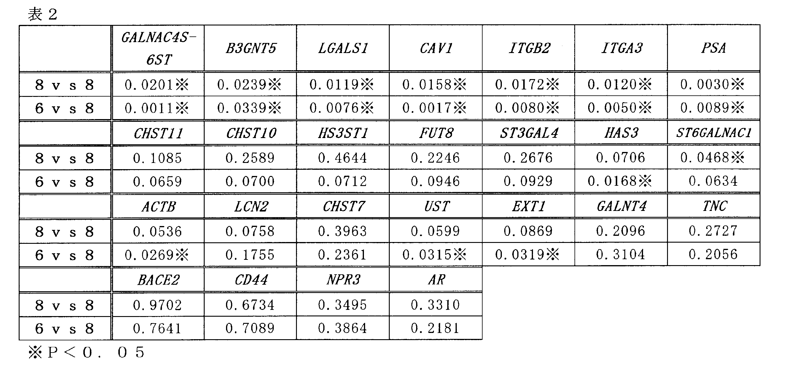
図2及び表2の結果から、GALNAC4S-6ST(配列番号1)、B3GNT5(配列番号3)、LGALS1(配列番号5)、CAV1(配列番号7)、ITGB2(配列番号9)、ITGA3(配列番号11)及びST6GALNAC1(配列番号27)において、ホルモン療法を受けていない前立腺癌患者8名から採取した癌細胞と、ホルモン療法に対する低感受性の前立腺癌患者8名から採取した癌細胞との間で、mRNAの発現量に明らかな有意差(P<0.05)が認められた(表2中、8vs8の欄参照)。なお既存マーカーであるPSAの発現量は、ホルモン療法に対する低感受性の前立腺癌患者8名から採取した癌細胞において有意に低下しているが、これは、アンドロゲンが遮断された結果引き起こされたものであり、ホルモン抵抗性との直接の関連性は低い。
また、ホルモン療法を受けていない未治療群の中にも、ホルモン療法に低感受性の患者が存在する可能性がある。したがって、参考のために、ホルモン療法を受けていない前立腺癌患者8名から採取した検体のうち、mRNAの発現量が高い患者検体2つを除き、ホルモン療法を受けていない前立腺癌患者6名から採取した癌細胞と、ホルモン療法に対する低感受性の前立腺癌患者8名から採取した癌細胞との間の有意差を調べた。その結果、HAS3(配列番号25)、UST(配列番号35)及びEXT1(配列番号37)でも有意差(P<0.05)を認めた(表2中、6vs8の欄参照)。
また、ホルモン療法を受けていない未治療群の中にも、ホルモン療法に低感受性の患者が存在する可能性がある。したがって、参考のために、ホルモン療法を受けていない前立腺癌患者8名から採取した検体のうち、mRNAの発現量が高い患者検体2つを除き、ホルモン療法を受けていない前立腺癌患者6名から採取した癌細胞と、ホルモン療法に対する低感受性の前立腺癌患者8名から採取した癌細胞との間の有意差を調べた。その結果、HAS3(配列番号25)、UST(配列番号35)及びEXT1(配列番号37)でも有意差(P<0.05)を認めた(表2中、6vs8の欄参照)。
なお、上記で有意差が認められなかった遺伝子に関しても、実施例1で示すように前立腺癌のセルラインを用いた試験では、ホルモン感受性前立腺癌細胞とホルモン抵抗性の前立腺癌細胞とを区別しうることが示されている。したがって、現状では困難であるが、ホルモン感受性患者として特定された患者検体を用いることができれば、ホルモン療法に対する低感受性の前立腺癌患者検体との間で発現量の有意差が認められうるものと考えられる。
以上の結果、本発明の検出方法は、再燃性前立腺癌を特異的に検出できる方法であることが示された。
本発明をその実施態様とともに説明したが、我々は特に指定しない限り我々の発明を説明のどの細部においても限定しようとするものではなく、添付の請求の範囲に示した発明の精神と範囲に反することなく幅広く解釈されるべきであると考える。
本願は、2011年2月28日に日本国で特許出願された特願2011-042967に基づく優先権を主張するものであり、これらはここに参照してその内容を本明細書の記載の一部として取り込む。
Claims (6)
- 下記(a)~(d)のいずれかのDNAからなる少なくとも1種の遺伝子の発現量を測定することを含む、再燃性前立腺癌の検出方法:
(a)配列番号1の塩基配列、配列番号3の塩基配列、配列番号5の塩基配列、配列番号7の塩基配列、配列番号9の塩基配列、配列番号11の塩基配列、配列番号15の塩基配列、配列番号17の塩基配列、配列番号19の塩基配列、配列番号21の塩基配列、配列番号23の塩基配列、配列番号25の塩基配列、配列番号27の塩基配列、配列番号31の塩基配列、配列番号33の塩基配列、配列番号35の塩基配列、配列番号37の塩基配列、配列番号39の塩基配列、配列番号41の塩基配列、配列番号43の塩基配列、配列番号45の塩基配列、配列番号47の塩基配列、配列番号51の塩基配列、配列番号53の塩基配列、配列番号55の塩基配列、配列番号57の塩基配列、配列番号59の塩基配列及び配列番号61の塩基配列からなる群から選ばれる塩基配列からなるDNA、
(b)上記(a)のDNAの塩基配列と80%以上の同一性を有する塩基配列からなるDNAであって、前立腺癌細胞のゲノム中に存在するDNA、
(c)上記(a)のDNAの塩基配列において、1又は数個の塩基が欠失、置換、挿入又は付加された塩基配列からなるDNAであって、前立腺癌細胞のゲノム中に存在するDNA、
(d)上記(a)のDNAの塩基配列と相補的な塩基配列からなるDNAにストリンジェントな条件下でハイブリダイズするDNAであって、前立腺癌細胞のゲノム中に存在するDNA。 - 遺伝子の発現量の測定が、該遺伝子の転写産物の量の測定である、請求項1に記載の検出方法。
- 下記(Aa)~(Bb)からなる群から選ばれる少なくとも1種のプライマーセットを用いて少なくとも1種の遺伝子の発現量を測定する、請求項2に記載の検出方法:
(Aa)配列番号63の塩基配列からなるオリゴヌクレオチド及び配列番号64の塩基配列からなるオリゴヌクレオチドからなるプライマーセット、
(Ab)配列番号65の塩基配列からなるオリゴヌクレオチド及び配列番号66の塩基配列からなるオリゴヌクレオチドからなるプライマーセット、
(Ac)配列番号67の塩基配列からなるオリゴヌクレオチド及び配列番号68の塩基配列からなるオリゴヌクレオチドからなるプライマーセット、
(Ad)配列番号69の塩基配列からなるオリゴヌクレオチド及び配列番号70の塩基配列からなるオリゴヌクレオチドからなるプライマーセット、
(Ae)配列番号71の塩基配列からなるオリゴヌクレオチド及び配列番号72の塩基配列からなるオリゴヌクレオチドからなるプライマーセット、
(Af)配列番号73の塩基配列からなるオリゴヌクレオチド及び配列番号74の塩基配列からなるオリゴヌクレオチドからなるプライマーセット、
(Ag)配列番号77の塩基配列からなるオリゴヌクレオチド及び配列番号78の塩基配列からなるオリゴヌクレオチドからなるプライマーセット、
(Ah)配列番号79の塩基配列からなるオリゴヌクレオチド及び配列番号80の塩基配列からなるオリゴヌクレオチドからなるプライマーセット、
(Ai)配列番号81の塩基配列からなるオリゴヌクレオチド及び配列番号82の塩基配列からなるオリゴヌクレオチドからなるプライマーセット、
(Aj)配列番号83の塩基配列からなるオリゴヌクレオチド及び配列番号84の塩基配列からなるオリゴヌクレオチドからなるプライマーセット、
(Ak)配列番号85の塩基配列からなるオリゴヌクレオチド及び配列番号86の塩基配列からなるオリゴヌクレオチドからなるプライマーセット、
(Al)配列番号87の塩基配列からなるオリゴヌクレオチド及び配列番号88の塩基配列からなるオリゴヌクレオチドからなるプライマーセット、
(Am)配列番号89の塩基配列からなるオリゴヌクレオチド及び配列番号90の塩基配列からなるオリゴヌクレオチドからなるプライマーセット、
(An)配列番号93の塩基配列からなるオリゴヌクレオチド及び配列番号94の塩基配列からなるオリゴヌクレオチドからなるプライマーセット、
(Ao)配列番号95の塩基配列からなるオリゴヌクレオチド及び配列番号96の塩基配列からなるオリゴヌクレオチドからなるプライマーセット、
(Ap)配列番号97の塩基配列からなるオリゴヌクレオチド及び配列番号98の塩基配列からなるオリゴヌクレオチドからなるプライマーセット、
(Aq)配列番号99の塩基配列からなるオリゴヌクレオチド及び配列番号100の塩基配列からなるオリゴヌクレオチドからなるプライマーセット、
(Ar)配列番号101の塩基配列からなるオリゴヌクレオチド及び配列番号102の塩基配列からなるオリゴヌクレオチドからなるプライマーセット、
(As)配列番号103の塩基配列からなるオリゴヌクレオチド及び配列番号104の塩基配列からなるオリゴヌクレオチドからなるプライマーセット、
(At)配列番号105の塩基配列からなるオリゴヌクレオチド及び配列番号106の塩基配列からなるオリゴヌクレオチドからなるプライマーセット、
(Au)配列番号107の塩基配列からなるオリゴヌクレオチド及び配列番号108の塩基配列からなるオリゴヌクレオチドからなるプライマーセット、
(Av)配列番号109の塩基配列からなるオリゴヌクレオチド及び配列番号110の塩基配列からなるオリゴヌクレオチドからなるプライマーセット、及び
(Aw)配列番号113の塩基配列からなるオリゴヌクレオチド及び配列番号114の塩基配列からなるオリゴヌクレオチドからなるプライマーセット。
(Ax)配列番号115の塩基配列からなるオリゴヌクレオチド及び配列番号116の塩基配列からなるオリゴヌクレオチドからなるプライマーセット。
(Ay)配列番号117の塩基配列からなるオリゴヌクレオチド及び配列番号118の塩基配列からなるオリゴヌクレオチドからなるプライマーセット。
(Az)配列番号119の塩基配列からなるオリゴヌクレオチド及び配列番号120の塩基配列からなるオリゴヌクレオチドからなるプライマーセット。
(Ba)配列番号121の塩基配列からなるオリゴヌクレオチド及び配列番号122の塩基配列からなるオリゴヌクレオチドからなるプライマーセット。
(Bb)配列番号123の塩基配列からなるオリゴヌクレオチド及び配列番号124の塩基配列からなるオリゴヌクレオチドからなるプライマーセット。 - 遺伝子の発現量の測定が、該遺伝子の発現産物であるタンパク質の量の測定である、請求項1に記載の検出方法。
- 測定されるタンパク質の量が、下記(e)~(g)のいずれかのアミノ酸配列からなる少なくとも1種のタンパク質の量である、請求項4に記載の検出方法:
(e)配列番号2のアミノ酸配列、配列番号4のアミノ酸配列、配列番号6のアミノ酸配列、配列番号8のアミノ酸配列、配列番号10のアミノ酸配列、配列番号12のアミノ酸配列、配列番号16のアミノ酸配列、配列番号18のアミノ酸配列、配列番号20のアミノ酸配列、配列番号22のアミノ酸配列、配列番号24のアミノ酸配列、配列番号26のアミノ酸配列、配列番号28のアミノ酸配列、配列番号32のアミノ酸配列、配列番号34のアミノ酸配列、配列番号36のアミノ酸配列、配列番号38のアミノ酸配列、配列番号40のアミノ酸配列、配列番号42のアミノ酸配列、配列番号44のアミノ酸配列、配列番号46のアミノ酸配列、配列番号48のアミノ酸配列、配列番号52のアミノ酸配列、配列番号54のアミノ酸配列、配列番号56のアミノ酸配列、配列番号58のアミノ酸配列、配列番号60のアミノ酸配列及び配列番号62のアミノ酸配列からなる群から選ばれるアミノ酸配列、
(f)上記(e)のアミノ酸配列と80%以上の同一性を有し、かつ前立腺癌細胞のゲノム中に存在するDNAにコードされるアミノ酸配列、
(g)上記(e)のアミノ酸配列において、1又は数個のアミノ酸が欠失、置換、挿入又は付加されたアミノ酸配列であって、前立腺癌細胞のゲノム中に存在するDNAにコードされるアミノ酸配列。 - 単離した前立腺癌細胞中における遺伝子の発現量を測定する、請求項1~5のいずれか1項に記載の検出方法。
Applications Claiming Priority (2)
| Application Number | Priority Date | Filing Date | Title |
|---|---|---|---|
| JP2011042967A JP2012178990A (ja) | 2011-02-28 | 2011-02-28 | 再燃性前立腺癌の検出方法 |
| JP2011-042967 | 2011-02-28 |
Publications (1)
| Publication Number | Publication Date |
|---|---|
| WO2012118088A1 true WO2012118088A1 (ja) | 2012-09-07 |
Family
ID=46758019
Family Applications (1)
| Application Number | Title | Priority Date | Filing Date |
|---|---|---|---|
| PCT/JP2012/054962 Ceased WO2012118088A1 (ja) | 2011-02-28 | 2012-02-28 | 再燃性前立腺癌の検出方法 |
Country Status (2)
| Country | Link |
|---|---|
| JP (1) | JP2012178990A (ja) |
| WO (1) | WO2012118088A1 (ja) |
Families Citing this family (1)
| Publication number | Priority date | Publication date | Assignee | Title |
|---|---|---|---|---|
| US12320812B2 (en) * | 2019-02-06 | 2025-06-03 | Bpgbio, Inc. | Markers for the diagnosis of biochemical recurrence in prostate cancer |
Citations (3)
| Publication number | Priority date | Publication date | Assignee | Title |
|---|---|---|---|---|
| WO2002031209A2 (en) * | 2000-10-13 | 2002-04-18 | The Government Of The United States Of America As Represented By The Secretary Of The Department Of Health And Human Services | Genes related to development of refractory prostate cancer |
| WO2004046358A1 (ja) * | 2002-11-21 | 2004-06-03 | Chugai Seiyaku Kabushiki Kaisha | 新規癌関連遺伝子 |
| WO2008102906A1 (en) * | 2007-02-20 | 2008-08-28 | Oncotherapy Science, Inc. | Hspc-hrpc transition genes |
-
2011
- 2011-02-28 JP JP2011042967A patent/JP2012178990A/ja active Pending
-
2012
- 2012-02-28 WO PCT/JP2012/054962 patent/WO2012118088A1/ja not_active Ceased
Patent Citations (3)
| Publication number | Priority date | Publication date | Assignee | Title |
|---|---|---|---|---|
| WO2002031209A2 (en) * | 2000-10-13 | 2002-04-18 | The Government Of The United States Of America As Represented By The Secretary Of The Department Of Health And Human Services | Genes related to development of refractory prostate cancer |
| WO2004046358A1 (ja) * | 2002-11-21 | 2004-06-03 | Chugai Seiyaku Kabushiki Kaisha | 新規癌関連遺伝子 |
| WO2008102906A1 (en) * | 2007-02-20 | 2008-08-28 | Oncotherapy Science, Inc. | Hspc-hrpc transition genes |
Non-Patent Citations (3)
| Title |
|---|
| HIDEWAKI NAKAGAWA: "Zenritsusengan no Genomikkusu: Genome Kaiseki karano Zenritsusengan no Rikai", THE JAPANESE JOURNAL OF UROLOGY, vol. 101, no. 2, 20 February 2010 (2010-02-20), pages 76 * |
| HITOSHI ISHIGURO: "Zenritsusengan no Shinten ni Okeru aPKCA/1 no Hatsugen Oyobi Kino Kaiseki", YOKOHAMA MEDICAL JOURNAL, vol. 61, 2010, pages 591 - 597 * |
| YUKO NAITO ET AL.: "Germinal Center Marker GL7 Probes Activation-Dependent Repression of N- Glycolylneuraminic Acid, a Sialic Acid Species Involved in the Negative Modulation of B-Cell Activation", MOL CELL BIOL., vol. 27, no. 8, 2007, pages 3008 - 3022 * |
Also Published As
| Publication number | Publication date |
|---|---|
| JP2012178990A (ja) | 2012-09-20 |
Similar Documents
| Publication | Publication Date | Title |
|---|---|---|
| EP2201134B1 (en) | Gene expression signatures in enriched tumor cell samples | |
| Liu et al. | Development of a multiplex system for the identification of forensically relevant body fluids | |
| EP2867376B1 (en) | Targeted rna-seq methods and materials for the diagnosis of prostate cancer | |
| JP6254975B2 (ja) | 院内感染罹患の患者の易罹患性を判定し、敗血症症候群の進行の予後を確立する方法 | |
| KR20190026769A (ko) | 유전자 발현 프로파일을 사용하여 폐암을 진단하기 위한 조성물 및 방법 | |
| CN108949969B (zh) | 长链非编码rna在结直肠癌中的应用 | |
| CN110964823A (zh) | 用于结直肠癌检测的dna甲基化试剂盒和检测方法 | |
| CN105838820A (zh) | 转录因子foxd1的新用途 | |
| CN111979315A (zh) | 环状tp63作为肺鳞癌诊断或治疗靶点的应用 | |
| US11519042B2 (en) | Method for the diagnosis or prognosis, in vitro, of lung cancer | |
| CN109504773B (zh) | 一种与口腔鳞癌分化等级相关的生物标志物 | |
| JP2012178990A (ja) | 再燃性前立腺癌の検出方法 | |
| KR20210024919A (ko) | BK 바이러스 신병증의 진단을 위한 소변 엑소좀 유래 miRNA 유전자 바이오마커 및 이의 용도 | |
| KR20170124683A (ko) | 담도암 진단용 신규 바이오마커로서의 융합 유전자 및 융합 단백질 | |
| KR101174369B1 (ko) | 유방암 진단용 바이오마커 및 유방암 진단제 | |
| CN110387423A (zh) | 前庭神经鞘瘤诊断用生物标志物 | |
| KR102505618B1 (ko) | 신장이식 후 항체 매개성 거부반응의 진단을 위한 소변 엑소좀 유래 miRNA 유전자 바이오마커 및 이의 용도 | |
| CN114107461B (zh) | 环状rna标志物及其在诊断和预测复发性流产病理妊娠中的应用 | |
| JPWO2002083899A1 (ja) | 癌関連遺伝子 | |
| CN115992217A (zh) | 用于诊断乳腺癌化疗导致的心肌损伤的环状rna标志物、试剂盒及其应用 | |
| JP5316749B2 (ja) | シスプラチン耐性遺伝子診断方法及びシスプラチン治療効果遺伝子診断キット | |
| EP4317458A1 (en) | Follicular thyroid cancer-specific marker | |
| KR20210024918A (ko) | 신장이식 후 T 세포 매개성 거부반응의 진단을 위한 소변 엑소좀 유래 miRNA 유전자 바이오마커 및 이의 용도 | |
| CN108384848A (zh) | 血清中circ_0021132作为深静脉血栓形成诊断标志物的应用 | |
| WO2019062429A1 (zh) | 分子标记物tcons_00016233、试剂盒及应用 |
Legal Events
| Date | Code | Title | Description |
|---|---|---|---|
| 121 | Ep: the epo has been informed by wipo that ep was designated in this application |
Ref document number: 12752727 Country of ref document: EP Kind code of ref document: A1 |
|
| NENP | Non-entry into the national phase |
Ref country code: DE |
|
| 122 | Ep: pct application non-entry in european phase |
Ref document number: 12752727 Country of ref document: EP Kind code of ref document: A1 |