WO2012043634A1 - ヒトプロスタグランジンe2受容体ep4に対する抗体 - Google Patents
ヒトプロスタグランジンe2受容体ep4に対する抗体 Download PDFInfo
- Publication number
- WO2012043634A1 WO2012043634A1 PCT/JP2011/072190 JP2011072190W WO2012043634A1 WO 2012043634 A1 WO2012043634 A1 WO 2012043634A1 JP 2011072190 W JP2011072190 W JP 2011072190W WO 2012043634 A1 WO2012043634 A1 WO 2012043634A1
- Authority
- WO
- WIPO (PCT)
- Prior art keywords
- antibody
- seq
- amino acid
- acid sequence
- sequence represented
- Prior art date
Links
- 102000008866 Prostaglandin E receptors Human genes 0.000 title 1
- 108010088540 Prostaglandin E receptors Proteins 0.000 title 1
- 239000012634 fragment Substances 0.000 claims abstract description 67
- 230000003834 intracellular effect Effects 0.000 claims abstract description 14
- 210000004027 cell Anatomy 0.000 claims description 187
- 101001117509 Homo sapiens Prostaglandin E2 receptor EP4 subtype Proteins 0.000 claims description 180
- 102100024450 Prostaglandin E2 receptor EP4 subtype Human genes 0.000 claims description 179
- 125000003275 alpha amino acid group Chemical group 0.000 claims description 88
- 206010028980 Neoplasm Diseases 0.000 claims description 37
- 208000037265 diseases, disorders, signs and symptoms Diseases 0.000 claims description 33
- 201000010099 disease Diseases 0.000 claims description 30
- 102000005962 receptors Human genes 0.000 claims description 27
- 108020003175 receptors Proteins 0.000 claims description 27
- 208000002193 Pain Diseases 0.000 claims description 26
- 210000004408 hybridoma Anatomy 0.000 claims description 26
- 230000036407 pain Effects 0.000 claims description 26
- 150000007523 nucleic acids Chemical class 0.000 claims description 24
- 239000008194 pharmaceutical composition Substances 0.000 claims description 24
- 239000013598 vector Substances 0.000 claims description 24
- 230000014509 gene expression Effects 0.000 claims description 12
- 210000004369 blood Anatomy 0.000 claims description 10
- 239000008280 blood Substances 0.000 claims description 10
- 108010021625 Immunoglobulin Fragments Proteins 0.000 claims description 8
- 102000008394 Immunoglobulin Fragments Human genes 0.000 claims description 8
- 208000026278 immune system disease Diseases 0.000 claims description 7
- 108020004707 nucleic acids Proteins 0.000 claims description 7
- 102000039446 nucleic acids Human genes 0.000 claims description 7
- 230000002265 prevention Effects 0.000 claims description 7
- FWMNVWWHGCHHJJ-SKKKGAJSSA-N 4-amino-1-[(2r)-6-amino-2-[[(2r)-2-[[(2r)-2-[[(2r)-2-amino-3-phenylpropanoyl]amino]-3-phenylpropanoyl]amino]-4-methylpentanoyl]amino]hexanoyl]piperidine-4-carboxylic acid Chemical compound C([C@H](C(=O)N[C@H](CC(C)C)C(=O)N[C@H](CCCCN)C(=O)N1CCC(N)(CC1)C(O)=O)NC(=O)[C@H](N)CC=1C=CC=CC=1)C1=CC=CC=C1 FWMNVWWHGCHHJJ-SKKKGAJSSA-N 0.000 claims description 5
- 210000001124 body fluid Anatomy 0.000 claims description 3
- 239000010839 body fluid Substances 0.000 claims description 3
- 108010047041 Complementarity Determining Regions Proteins 0.000 claims description 2
- 230000007488 abnormal function Effects 0.000 claims description 2
- 239000003814 drug Substances 0.000 abstract description 22
- 241000699670 Mus sp. Species 0.000 abstract description 6
- 229940079593 drug Drugs 0.000 abstract description 6
- XEYBRNLFEZDVAW-ARSRFYASSA-N dinoprostone Chemical compound CCCCC[C@H](O)\C=C\[C@H]1[C@H](O)CC(=O)[C@@H]1C\C=C/CCCC(O)=O XEYBRNLFEZDVAW-ARSRFYASSA-N 0.000 description 63
- 241000699666 Mus <mouse, genus> Species 0.000 description 61
- 108090000623 proteins and genes Proteins 0.000 description 57
- 230000027455 binding Effects 0.000 description 53
- 238000000034 method Methods 0.000 description 50
- 210000004978 chinese hamster ovary cell Anatomy 0.000 description 34
- 239000002609 medium Substances 0.000 description 30
- 239000012228 culture supernatant Substances 0.000 description 29
- 102100035360 Cerebellar degeneration-related antigen 1 Human genes 0.000 description 28
- 230000006870 function Effects 0.000 description 25
- 238000012360 testing method Methods 0.000 description 22
- 201000011510 cancer Diseases 0.000 description 21
- 238000000684 flow cytometry Methods 0.000 description 21
- 239000013612 plasmid Substances 0.000 description 19
- 108091028043 Nucleic acid sequence Proteins 0.000 description 17
- 108091007433 antigens Proteins 0.000 description 16
- 102000036639 antigens Human genes 0.000 description 16
- 241001465754 Metazoa Species 0.000 description 15
- 239000004480 active ingredient Substances 0.000 description 15
- 239000000427 antigen Substances 0.000 description 15
- 239000013604 expression vector Substances 0.000 description 14
- 210000004698 lymphocyte Anatomy 0.000 description 14
- 238000004519 manufacturing process Methods 0.000 description 14
- 239000002299 complementary DNA Substances 0.000 description 13
- 230000002401 inhibitory effect Effects 0.000 description 13
- 108091003079 Bovine Serum Albumin Proteins 0.000 description 12
- 108020004414 DNA Proteins 0.000 description 12
- 108090000765 processed proteins & peptides Proteins 0.000 description 12
- RXWNCPJZOCPEPQ-NVWDDTSBSA-N puromycin Chemical compound C1=CC(OC)=CC=C1C[C@H](N)C(=O)N[C@H]1[C@@H](O)[C@H](N2C3=NC=NC(=C3N=C2)N(C)C)O[C@@H]1CO RXWNCPJZOCPEPQ-NVWDDTSBSA-N 0.000 description 12
- -1 t-butyloxycarbonyl Chemical group 0.000 description 12
- 206010019233 Headaches Diseases 0.000 description 11
- 238000004113 cell culture Methods 0.000 description 11
- 238000002360 preparation method Methods 0.000 description 11
- 108091008146 restriction endonucleases Proteins 0.000 description 11
- 239000000126 substance Substances 0.000 description 11
- 230000000694 effects Effects 0.000 description 10
- 231100000869 headache Toxicity 0.000 description 10
- 230000005764 inhibitory process Effects 0.000 description 10
- 239000002953 phosphate buffered saline Substances 0.000 description 10
- 101001117519 Homo sapiens Prostaglandin E2 receptor EP2 subtype Proteins 0.000 description 9
- 108010004729 Phycoerythrin Proteins 0.000 description 9
- 239000008187 granular material Substances 0.000 description 9
- 102100024448 Prostaglandin E2 receptor EP2 subtype Human genes 0.000 description 8
- 238000007796 conventional method Methods 0.000 description 8
- 239000002158 endotoxin Substances 0.000 description 8
- 239000007924 injection Substances 0.000 description 8
- 238000002347 injection Methods 0.000 description 8
- 229920006008 lipopolysaccharide Polymers 0.000 description 8
- 239000007787 solid Substances 0.000 description 8
- PHEDXBVPIONUQT-UHFFFAOYSA-N Cocarcinogen A1 Natural products CCCCCCCCCCCCCC(=O)OC1C(C)C2(O)C3C=C(C)C(=O)C3(O)CC(CO)=CC2C2C1(OC(C)=O)C2(C)C PHEDXBVPIONUQT-UHFFFAOYSA-N 0.000 description 7
- 241000238631 Hexapoda Species 0.000 description 7
- 101001074035 Homo sapiens Zinc finger protein GLI2 Proteins 0.000 description 7
- 102100035558 Zinc finger protein GLI2 Human genes 0.000 description 7
- 235000001014 amino acid Nutrition 0.000 description 7
- 239000005557 antagonist Substances 0.000 description 7
- 238000006243 chemical reaction Methods 0.000 description 7
- 230000012010 growth Effects 0.000 description 7
- 210000004962 mammalian cell Anatomy 0.000 description 7
- PHEDXBVPIONUQT-RGYGYFBISA-N phorbol 13-acetate 12-myristate Chemical compound C([C@]1(O)C(=O)C(C)=C[C@H]1[C@@]1(O)[C@H](C)[C@H]2OC(=O)CCCCCCCCCCCCC)C(CO)=C[C@H]1[C@H]1[C@]2(OC(C)=O)C1(C)C PHEDXBVPIONUQT-RGYGYFBISA-N 0.000 description 7
- 150000003180 prostaglandins Chemical class 0.000 description 7
- 238000005406 washing Methods 0.000 description 7
- 210000005253 yeast cell Anatomy 0.000 description 7
- 102000004190 Enzymes Human genes 0.000 description 6
- 108090000790 Enzymes Proteins 0.000 description 6
- 239000012097 Lipofectamine 2000 Substances 0.000 description 6
- 229930182555 Penicillin Natural products 0.000 description 6
- JGSARLDLIJGVTE-MBNYWOFBSA-N Penicillin G Chemical compound N([C@H]1[C@H]2SC([C@@H](N2C1=O)C(O)=O)(C)C)C(=O)CC1=CC=CC=C1 JGSARLDLIJGVTE-MBNYWOFBSA-N 0.000 description 6
- 239000012979 RPMI medium Substances 0.000 description 6
- 230000004913 activation Effects 0.000 description 6
- 229940098773 bovine serum albumin Drugs 0.000 description 6
- 239000000872 buffer Substances 0.000 description 6
- 150000001875 compounds Chemical class 0.000 description 6
- 230000016396 cytokine production Effects 0.000 description 6
- 229940088598 enzyme Drugs 0.000 description 6
- 239000000203 mixture Substances 0.000 description 6
- 239000002773 nucleotide Substances 0.000 description 6
- 125000003729 nucleotide group Chemical group 0.000 description 6
- 229940049954 penicillin Drugs 0.000 description 6
- 102000004196 processed proteins & peptides Human genes 0.000 description 6
- 229950010131 puromycin Drugs 0.000 description 6
- 150000003839 salts Chemical class 0.000 description 6
- 150000003384 small molecules Chemical class 0.000 description 6
- 230000000638 stimulation Effects 0.000 description 6
- UCSJYZPVAKXKNQ-HZYVHMACSA-N streptomycin Chemical compound CN[C@H]1[C@H](O)[C@@H](O)[C@H](CO)O[C@H]1O[C@@H]1[C@](C=O)(O)[C@H](C)O[C@H]1O[C@@H]1[C@@H](NC(N)=N)[C@H](O)[C@@H](NC(N)=N)[C@H](O)[C@H]1O UCSJYZPVAKXKNQ-HZYVHMACSA-N 0.000 description 6
- 210000001519 tissue Anatomy 0.000 description 6
- XLYOFNOQVPJJNP-UHFFFAOYSA-N water Substances O XLYOFNOQVPJJNP-UHFFFAOYSA-N 0.000 description 6
- 102000010907 Cyclooxygenase 2 Human genes 0.000 description 5
- 108010037462 Cyclooxygenase 2 Proteins 0.000 description 5
- 238000009007 Diagnostic Kit Methods 0.000 description 5
- PEDCQBHIVMGVHV-UHFFFAOYSA-N Glycerine Chemical compound OCC(O)CO PEDCQBHIVMGVHV-UHFFFAOYSA-N 0.000 description 5
- 101001073427 Homo sapiens Prostaglandin E2 receptor EP1 subtype Proteins 0.000 description 5
- 101001117517 Homo sapiens Prostaglandin E2 receptor EP3 subtype Proteins 0.000 description 5
- 241000124008 Mammalia Species 0.000 description 5
- 102100024447 Prostaglandin E2 receptor EP3 subtype Human genes 0.000 description 5
- 230000002159 abnormal effect Effects 0.000 description 5
- 230000009471 action Effects 0.000 description 5
- 150000001413 amino acids Chemical class 0.000 description 5
- 230000003321 amplification Effects 0.000 description 5
- 238000004458 analytical method Methods 0.000 description 5
- 230000003042 antagnostic effect Effects 0.000 description 5
- 230000000259 anti-tumor effect Effects 0.000 description 5
- 230000003491 cAMP production Effects 0.000 description 5
- 108010030074 endodeoxyribonuclease MluI Proteins 0.000 description 5
- 239000012091 fetal bovine serum Substances 0.000 description 5
- 108020001507 fusion proteins Proteins 0.000 description 5
- 102000037865 fusion proteins Human genes 0.000 description 5
- 230000009390 immune abnormality Effects 0.000 description 5
- 230000003053 immunization Effects 0.000 description 5
- 210000002540 macrophage Anatomy 0.000 description 5
- 239000000463 material Substances 0.000 description 5
- 238000003199 nucleic acid amplification method Methods 0.000 description 5
- 210000005259 peripheral blood Anatomy 0.000 description 5
- 239000011886 peripheral blood Substances 0.000 description 5
- 239000000843 powder Substances 0.000 description 5
- 102000004169 proteins and genes Human genes 0.000 description 5
- 206010039073 rheumatoid arthritis Diseases 0.000 description 5
- 239000011534 wash buffer Substances 0.000 description 5
- 206010004146 Basal cell carcinoma Diseases 0.000 description 4
- 206010009944 Colon cancer Diseases 0.000 description 4
- YQYJSBFKSSDGFO-UHFFFAOYSA-N Epihygromycin Natural products OC1C(O)C(C(=O)C)OC1OC(C(=C1)O)=CC=C1C=C(C)C(=O)NC1C(O)C(O)C2OCOC2C1O YQYJSBFKSSDGFO-UHFFFAOYSA-N 0.000 description 4
- VEXZGXHMUGYJMC-UHFFFAOYSA-N Hydrochloric acid Chemical compound Cl VEXZGXHMUGYJMC-UHFFFAOYSA-N 0.000 description 4
- 102000018251 Hypoxanthine Phosphoribosyltransferase Human genes 0.000 description 4
- 108010091358 Hypoxanthine Phosphoribosyltransferase Proteins 0.000 description 4
- ZDXPYRJPNDTMRX-VKHMYHEASA-N L-glutamine Chemical compound OC(=O)[C@@H](N)CCC(N)=O ZDXPYRJPNDTMRX-VKHMYHEASA-N 0.000 description 4
- 206010058467 Lung neoplasm malignant Diseases 0.000 description 4
- 206010035226 Plasma cell myeloma Diseases 0.000 description 4
- 208000037062 Polyps Diseases 0.000 description 4
- 102100035842 Prostaglandin E2 receptor EP1 subtype Human genes 0.000 description 4
- 206010060862 Prostate cancer Diseases 0.000 description 4
- 208000000236 Prostatic Neoplasms Diseases 0.000 description 4
- QAOWNCQODCNURD-UHFFFAOYSA-N Sulfuric acid Chemical compound OS(O)(=O)=O QAOWNCQODCNURD-UHFFFAOYSA-N 0.000 description 4
- 239000011543 agarose gel Substances 0.000 description 4
- 125000000539 amino acid group Chemical group 0.000 description 4
- 229940125644 antibody drug Drugs 0.000 description 4
- 230000010056 antibody-dependent cellular cytotoxicity Effects 0.000 description 4
- YZXBAPSDXZZRGB-DOFZRALJSA-N arachidonic acid Chemical compound CCCCC\C=C/C\C=C/C\C=C/C\C=C/CCCC(O)=O YZXBAPSDXZZRGB-DOFZRALJSA-N 0.000 description 4
- 229930189065 blasticidin Natural products 0.000 description 4
- 239000003153 chemical reaction reagent Substances 0.000 description 4
- 239000002552 dosage form Substances 0.000 description 4
- 238000005516 engineering process Methods 0.000 description 4
- MHMNJMPURVTYEJ-UHFFFAOYSA-N fluorescein-5-isothiocyanate Chemical compound O1C(=O)C2=CC(N=C=S)=CC=C2C21C1=CC=C(O)C=C1OC1=CC(O)=CC=C21 MHMNJMPURVTYEJ-UHFFFAOYSA-N 0.000 description 4
- 238000002649 immunization Methods 0.000 description 4
- 230000002163 immunogen Effects 0.000 description 4
- JVTAAEKCZFNVCJ-UHFFFAOYSA-N lactic acid Chemical compound CC(O)C(O)=O JVTAAEKCZFNVCJ-UHFFFAOYSA-N 0.000 description 4
- 201000005202 lung cancer Diseases 0.000 description 4
- 208000020816 lung neoplasm Diseases 0.000 description 4
- HQKMJHAJHXVSDF-UHFFFAOYSA-L magnesium stearate Chemical compound [Mg+2].CCCCCCCCCCCCCCCCCC([O-])=O.CCCCCCCCCCCCCCCCCC([O-])=O HQKMJHAJHXVSDF-UHFFFAOYSA-L 0.000 description 4
- 230000036961 partial effect Effects 0.000 description 4
- 239000000546 pharmaceutical excipient Substances 0.000 description 4
- 229920001184 polypeptide Polymers 0.000 description 4
- 235000018102 proteins Nutrition 0.000 description 4
- 239000000243 solution Substances 0.000 description 4
- 230000010473 stable expression Effects 0.000 description 4
- 239000000758 substrate Substances 0.000 description 4
- QTBSBXVTEAMEQO-UHFFFAOYSA-N Acetic acid Chemical compound CC(O)=O QTBSBXVTEAMEQO-UHFFFAOYSA-N 0.000 description 3
- 208000023275 Autoimmune disease Diseases 0.000 description 3
- 208000008035 Back Pain Diseases 0.000 description 3
- 206010006187 Breast cancer Diseases 0.000 description 3
- 208000026310 Breast neoplasm Diseases 0.000 description 3
- 230000004544 DNA amplification Effects 0.000 description 3
- 238000002965 ELISA Methods 0.000 description 3
- LFQSCWFLJHTTHZ-UHFFFAOYSA-N Ethanol Chemical compound CCO LFQSCWFLJHTTHZ-UHFFFAOYSA-N 0.000 description 3
- LYCAIKOWRPUZTN-UHFFFAOYSA-N Ethylene glycol Chemical compound OCCO LYCAIKOWRPUZTN-UHFFFAOYSA-N 0.000 description 3
- 108010043121 Green Fluorescent Proteins Proteins 0.000 description 3
- 102000004144 Green Fluorescent Proteins Human genes 0.000 description 3
- 241000282412 Homo Species 0.000 description 3
- 101000836978 Homo sapiens Sperm-associated antigen 11B Proteins 0.000 description 3
- GUBGYTABKSRVRQ-QKKXKWKRSA-N Lactose Natural products OC[C@H]1O[C@@H](O[C@H]2[C@H](O)[C@@H](O)C(O)O[C@@H]2CO)[C@H](O)[C@@H](O)[C@H]1O GUBGYTABKSRVRQ-QKKXKWKRSA-N 0.000 description 3
- BAVYZALUXZFZLV-UHFFFAOYSA-N Methylamine Chemical compound NC BAVYZALUXZFZLV-UHFFFAOYSA-N 0.000 description 3
- MUBZPKHOEPUJKR-UHFFFAOYSA-N Oxalic acid Chemical compound OC(=O)C(O)=O MUBZPKHOEPUJKR-UHFFFAOYSA-N 0.000 description 3
- 239000002202 Polyethylene glycol Substances 0.000 description 3
- 229920001213 Polysorbate 20 Polymers 0.000 description 3
- DNIAPMSPPWPWGF-UHFFFAOYSA-N Propylene glycol Chemical compound CC(O)CO DNIAPMSPPWPWGF-UHFFFAOYSA-N 0.000 description 3
- 101150109738 Ptger4 gene Proteins 0.000 description 3
- 208000000453 Skin Neoplasms Diseases 0.000 description 3
- HEMHJVSKTPXQMS-UHFFFAOYSA-M Sodium hydroxide Chemical compound [OH-].[Na+] HEMHJVSKTPXQMS-UHFFFAOYSA-M 0.000 description 3
- GWEVSGVZZGPLCZ-UHFFFAOYSA-N Titan oxide Chemical compound O=[Ti]=O GWEVSGVZZGPLCZ-UHFFFAOYSA-N 0.000 description 3
- 108060008682 Tumor Necrosis Factor Proteins 0.000 description 3
- 102000000852 Tumor Necrosis Factor-alpha Human genes 0.000 description 3
- 239000002671 adjuvant Substances 0.000 description 3
- 239000002585 base Substances 0.000 description 3
- 239000002775 capsule Substances 0.000 description 3
- 238000005119 centrifugation Methods 0.000 description 3
- KRKNYBCHXYNGOX-UHFFFAOYSA-N citric acid Chemical compound OC(=O)CC(O)(C(O)=O)CC(O)=O KRKNYBCHXYNGOX-UHFFFAOYSA-N 0.000 description 3
- 208000029742 colonic neoplasm Diseases 0.000 description 3
- 208000010247 contact dermatitis Diseases 0.000 description 3
- 125000000151 cysteine group Chemical group N[C@@H](CS)C(=O)* 0.000 description 3
- 210000004443 dendritic cell Anatomy 0.000 description 3
- 230000001419 dependent effect Effects 0.000 description 3
- 235000014113 dietary fatty acids Nutrition 0.000 description 3
- 238000010494 dissociation reaction Methods 0.000 description 3
- 230000005593 dissociations Effects 0.000 description 3
- 201000006549 dyspepsia Diseases 0.000 description 3
- 239000000194 fatty acid Substances 0.000 description 3
- 229930195729 fatty acid Natural products 0.000 description 3
- 230000004927 fusion Effects 0.000 description 3
- 238000010353 genetic engineering Methods 0.000 description 3
- 239000011521 glass Substances 0.000 description 3
- ZDXPYRJPNDTMRX-UHFFFAOYSA-N glutamine Natural products OC(=O)C(N)CCC(N)=O ZDXPYRJPNDTMRX-UHFFFAOYSA-N 0.000 description 3
- 235000011187 glycerol Nutrition 0.000 description 3
- 239000005090 green fluorescent protein Substances 0.000 description 3
- 238000003018 immunoassay Methods 0.000 description 3
- 230000001965 increasing effect Effects 0.000 description 3
- 239000008101 lactose Substances 0.000 description 3
- 239000007788 liquid Substances 0.000 description 3
- 238000005259 measurement Methods 0.000 description 3
- 201000006417 multiple sclerosis Diseases 0.000 description 3
- 201000000050 myeloid neoplasm Diseases 0.000 description 3
- 210000003819 peripheral blood mononuclear cell Anatomy 0.000 description 3
- 239000002504 physiological saline solution Substances 0.000 description 3
- 229920001223 polyethylene glycol Polymers 0.000 description 3
- 235000010486 polyoxyethylene sorbitan monolaurate Nutrition 0.000 description 3
- 239000000256 polyoxyethylene sorbitan monolaurate Substances 0.000 description 3
- WGJJROVFWIXTPA-OALUTQOASA-N prostanoic acid Chemical compound CCCCCCCC[C@H]1CCC[C@@H]1CCCCCCC(O)=O WGJJROVFWIXTPA-OALUTQOASA-N 0.000 description 3
- 238000000746 purification Methods 0.000 description 3
- 238000011084 recovery Methods 0.000 description 3
- 230000002829 reductive effect Effects 0.000 description 3
- 230000000717 retained effect Effects 0.000 description 3
- 201000000849 skin cancer Diseases 0.000 description 3
- 239000002904 solvent Substances 0.000 description 3
- 229960005322 streptomycin Drugs 0.000 description 3
- 239000006228 supernatant Substances 0.000 description 3
- 239000000725 suspension Substances 0.000 description 3
- 208000024891 symptom Diseases 0.000 description 3
- 238000010361 transduction Methods 0.000 description 3
- 230000026683 transduction Effects 0.000 description 3
- 238000012546 transfer Methods 0.000 description 3
- 230000001052 transient effect Effects 0.000 description 3
- 108091032973 (ribonucleotides)n+m Proteins 0.000 description 2
- HZAXFHJVJLSVMW-UHFFFAOYSA-N 2-Aminoethan-1-ol Chemical compound NCCO HZAXFHJVJLSVMW-UHFFFAOYSA-N 0.000 description 2
- 208000003200 Adenoma Diseases 0.000 description 2
- 206010001233 Adenoma benign Diseases 0.000 description 2
- GUBGYTABKSRVRQ-XLOQQCSPSA-N Alpha-Lactose Chemical compound O[C@@H]1[C@@H](O)[C@@H](O)[C@@H](CO)O[C@H]1O[C@@H]1[C@@H](CO)O[C@H](O)[C@H](O)[C@H]1O GUBGYTABKSRVRQ-XLOQQCSPSA-N 0.000 description 2
- QGZKDVFQNNGYKY-UHFFFAOYSA-N Ammonia Chemical compound N QGZKDVFQNNGYKY-UHFFFAOYSA-N 0.000 description 2
- 206010003445 Ascites Diseases 0.000 description 2
- CIWBSHSKHKDKBQ-JLAZNSOCSA-N Ascorbic acid Chemical compound OC[C@H](O)[C@H]1OC(=O)C(O)=C1O CIWBSHSKHKDKBQ-JLAZNSOCSA-N 0.000 description 2
- 206010005003 Bladder cancer Diseases 0.000 description 2
- 229940124638 COX inhibitor Drugs 0.000 description 2
- 241000700199 Cavia porcellus Species 0.000 description 2
- 206010008342 Cervix carcinoma Diseases 0.000 description 2
- 241000699800 Cricetinae Species 0.000 description 2
- 229920000858 Cyclodextrin Polymers 0.000 description 2
- 102000010906 Cyclooxygenase 1 Human genes 0.000 description 2
- 108010037464 Cyclooxygenase 1 Proteins 0.000 description 2
- 102000004127 Cytokines Human genes 0.000 description 2
- 108090000695 Cytokines Proteins 0.000 description 2
- FBPFZTCFMRRESA-KVTDHHQDSA-N D-Mannitol Chemical compound OC[C@@H](O)[C@@H](O)[C@H](O)[C@H](O)CO FBPFZTCFMRRESA-KVTDHHQDSA-N 0.000 description 2
- 206010012442 Dermatitis contact Diseases 0.000 description 2
- 229920001353 Dextrin Polymers 0.000 description 2
- 239000004375 Dextrin Substances 0.000 description 2
- ROSDSFDQCJNGOL-UHFFFAOYSA-N Dimethylamine Chemical compound CNC ROSDSFDQCJNGOL-UHFFFAOYSA-N 0.000 description 2
- AOJJSUZBOXZQNB-TZSSRYMLSA-N Doxorubicin Chemical compound O([C@H]1C[C@@](O)(CC=2C(O)=C3C(=O)C=4C=CC=C(C=4C(=O)C3=C(O)C=21)OC)C(=O)CO)[C@H]1C[C@H](N)[C@H](O)[C@H](C)O1 AOJJSUZBOXZQNB-TZSSRYMLSA-N 0.000 description 2
- 208000000461 Esophageal Neoplasms Diseases 0.000 description 2
- 208000001640 Fibromyalgia Diseases 0.000 description 2
- VZCYOOQTPOCHFL-OWOJBTEDSA-N Fumaric acid Chemical compound OC(=O)\C=C\C(O)=O VZCYOOQTPOCHFL-OWOJBTEDSA-N 0.000 description 2
- 108010010803 Gelatin Proteins 0.000 description 2
- WQZGKKKJIJFFOK-GASJEMHNSA-N Glucose Natural products OC[C@H]1OC(O)[C@H](O)[C@@H](O)[C@@H]1O WQZGKKKJIJFFOK-GASJEMHNSA-N 0.000 description 2
- AEMRFAOFKBGASW-UHFFFAOYSA-N Glycolic acid Chemical compound OCC(O)=O AEMRFAOFKBGASW-UHFFFAOYSA-N 0.000 description 2
- 101000946889 Homo sapiens Monocyte differentiation antigen CD14 Proteins 0.000 description 2
- 108010001336 Horseradish Peroxidase Proteins 0.000 description 2
- 229920002153 Hydroxypropyl cellulose Polymers 0.000 description 2
- 208000004454 Hyperalgesia Diseases 0.000 description 2
- 208000035154 Hyperesthesia Diseases 0.000 description 2
- DGAQECJNVWCQMB-PUAWFVPOSA-M Ilexoside XXIX Chemical compound C[C@@H]1CC[C@@]2(CC[C@@]3(C(=CC[C@H]4[C@]3(CC[C@@H]5[C@@]4(CC[C@@H](C5(C)C)OS(=O)(=O)[O-])C)C)[C@@H]2[C@]1(C)O)C)C(=O)O[C@H]6[C@@H]([C@H]([C@@H]([C@H](O6)CO)O)O)O.[Na+] DGAQECJNVWCQMB-PUAWFVPOSA-M 0.000 description 2
- 208000005726 Inflammatory Breast Neoplasms Diseases 0.000 description 2
- 208000022559 Inflammatory bowel disease Diseases 0.000 description 2
- 206010021980 Inflammatory carcinoma of the breast Diseases 0.000 description 2
- 208000008930 Low Back Pain Diseases 0.000 description 2
- 229930195725 Mannitol Natural products 0.000 description 2
- 206010027476 Metastases Diseases 0.000 description 2
- AFVFQIVMOAPDHO-UHFFFAOYSA-N Methanesulfonic acid Chemical compound CS(O)(=O)=O AFVFQIVMOAPDHO-UHFFFAOYSA-N 0.000 description 2
- 208000019695 Migraine disease Diseases 0.000 description 2
- 102100035877 Monocyte differentiation antigen CD14 Human genes 0.000 description 2
- 208000003445 Mouth Neoplasms Diseases 0.000 description 2
- 101100407595 Mus musculus Ptger4 gene Proteins 0.000 description 2
- NWIBSHFKIJFRCO-WUDYKRTCSA-N Mytomycin Chemical compound C1N2C(C(C(C)=C(N)C3=O)=O)=C3[C@@H](COC(N)=O)[C@@]2(OC)[C@@H]2[C@H]1N2 NWIBSHFKIJFRCO-WUDYKRTCSA-N 0.000 description 2
- 108091007491 NSP3 Papain-like protease domains Proteins 0.000 description 2
- PVNIIMVLHYAWGP-UHFFFAOYSA-N Niacin Chemical compound OC(=O)C1=CC=CN=C1 PVNIIMVLHYAWGP-UHFFFAOYSA-N 0.000 description 2
- 208000001294 Nociceptive Pain Diseases 0.000 description 2
- 206010030155 Oesophageal carcinoma Diseases 0.000 description 2
- 238000012408 PCR amplification Methods 0.000 description 2
- 108090000526 Papain Proteins 0.000 description 2
- 102000057297 Pepsin A Human genes 0.000 description 2
- 108090000284 Pepsin A Proteins 0.000 description 2
- NBIIXXVUZAFLBC-UHFFFAOYSA-N Phosphoric acid Chemical compound OP(O)(O)=O NBIIXXVUZAFLBC-UHFFFAOYSA-N 0.000 description 2
- 229920000954 Polyglycolide Polymers 0.000 description 2
- 239000004793 Polystyrene Substances 0.000 description 2
- 241000700159 Rattus Species 0.000 description 2
- 208000006265 Renal cell carcinoma Diseases 0.000 description 2
- 229940124639 Selective inhibitor Drugs 0.000 description 2
- PXIPVTKHYLBLMZ-UHFFFAOYSA-N Sodium azide Chemical compound [Na+].[N-]=[N+]=[N-] PXIPVTKHYLBLMZ-UHFFFAOYSA-N 0.000 description 2
- 229920002472 Starch Polymers 0.000 description 2
- 229930006000 Sucrose Natural products 0.000 description 2
- CZMRCDWAGMRECN-UGDNZRGBSA-N Sucrose Chemical compound O[C@H]1[C@H](O)[C@@H](CO)O[C@@]1(CO)O[C@@H]1[C@H](O)[C@@H](O)[C@H](O)[C@@H](CO)O1 CZMRCDWAGMRECN-UGDNZRGBSA-N 0.000 description 2
- 206010043276 Teratoma Diseases 0.000 description 2
- IQFYYKKMVGJFEH-XLPZGREQSA-N Thymidine Chemical compound O=C1NC(=O)C(C)=CN1[C@@H]1O[C@H](CO)[C@@H](O)C1 IQFYYKKMVGJFEH-XLPZGREQSA-N 0.000 description 2
- 208000007097 Urinary Bladder Neoplasms Diseases 0.000 description 2
- 208000006105 Uterine Cervical Neoplasms Diseases 0.000 description 2
- 239000002250 absorbent Substances 0.000 description 2
- 230000002745 absorbent Effects 0.000 description 2
- 239000002253 acid Substances 0.000 description 2
- 150000007513 acids Chemical class 0.000 description 2
- 208000009956 adenocarcinoma Diseases 0.000 description 2
- 239000003708 ampul Substances 0.000 description 2
- 229940114079 arachidonic acid Drugs 0.000 description 2
- 235000021342 arachidonic acid Nutrition 0.000 description 2
- WPYMKLBDIGXBTP-UHFFFAOYSA-N benzoic acid Chemical compound OC(=O)C1=CC=CC=C1 WPYMKLBDIGXBTP-UHFFFAOYSA-N 0.000 description 2
- WQZGKKKJIJFFOK-VFUOTHLCSA-N beta-D-glucose Chemical compound OC[C@H]1O[C@@H](O)[C@H](O)[C@@H](O)[C@@H]1O WQZGKKKJIJFFOK-VFUOTHLCSA-N 0.000 description 2
- 230000003115 biocidal effect Effects 0.000 description 2
- 230000000903 blocking effect Effects 0.000 description 2
- 210000000601 blood cell Anatomy 0.000 description 2
- 239000011575 calcium Substances 0.000 description 2
- 230000009702 cancer cell proliferation Effects 0.000 description 2
- 239000001768 carboxy methyl cellulose Substances 0.000 description 2
- 229940084030 carboxymethylcellulose calcium Drugs 0.000 description 2
- 230000022534 cell killing Effects 0.000 description 2
- 210000003855 cell nucleus Anatomy 0.000 description 2
- 230000004663 cell proliferation Effects 0.000 description 2
- 230000001413 cellular effect Effects 0.000 description 2
- 229920002678 cellulose Polymers 0.000 description 2
- 239000001913 cellulose Substances 0.000 description 2
- 201000010881 cervical cancer Diseases 0.000 description 2
- 239000003795 chemical substances by application Substances 0.000 description 2
- HVYWMOMLDIMFJA-DPAQBDIFSA-N cholesterol Chemical compound C1C=C2C[C@@H](O)CC[C@]2(C)[C@@H]2[C@@H]1[C@@H]1CC[C@H]([C@H](C)CCCC(C)C)[C@@]1(C)CC2 HVYWMOMLDIMFJA-DPAQBDIFSA-N 0.000 description 2
- 230000001684 chronic effect Effects 0.000 description 2
- 238000010367 cloning Methods 0.000 description 2
- 239000003636 conditioned culture medium Substances 0.000 description 2
- 238000012790 confirmation Methods 0.000 description 2
- 230000008878 coupling Effects 0.000 description 2
- 238000010168 coupling process Methods 0.000 description 2
- 238000005859 coupling reaction Methods 0.000 description 2
- 230000007423 decrease Effects 0.000 description 2
- 239000003405 delayed action preparation Substances 0.000 description 2
- 238000001514 detection method Methods 0.000 description 2
- 235000019425 dextrin Nutrition 0.000 description 2
- 238000010790 dilution Methods 0.000 description 2
- 239000012895 dilution Substances 0.000 description 2
- 208000035475 disorder Diseases 0.000 description 2
- 230000004064 dysfunction Effects 0.000 description 2
- 239000000839 emulsion Substances 0.000 description 2
- 230000002708 enhancing effect Effects 0.000 description 2
- 201000004101 esophageal cancer Diseases 0.000 description 2
- 238000002474 experimental method Methods 0.000 description 2
- 238000007667 floating Methods 0.000 description 2
- MKXKFYHWDHIYRV-UHFFFAOYSA-N flutamide Chemical compound CC(C)C(=O)NC1=CC=C([N+]([O-])=O)C(C(F)(F)F)=C1 MKXKFYHWDHIYRV-UHFFFAOYSA-N 0.000 description 2
- 230000009454 functional inhibition Effects 0.000 description 2
- 208000015419 gastrin-producing neuroendocrine tumor Diseases 0.000 description 2
- 201000000052 gastrinoma Diseases 0.000 description 2
- 239000000499 gel Substances 0.000 description 2
- 229920000159 gelatin Polymers 0.000 description 2
- 239000008273 gelatin Substances 0.000 description 2
- 108010025899 gelatin film Proteins 0.000 description 2
- 235000019322 gelatine Nutrition 0.000 description 2
- 235000011852 gelatine desserts Nutrition 0.000 description 2
- 239000008103 glucose Substances 0.000 description 2
- 239000007902 hard capsule Substances 0.000 description 2
- 201000002735 hepatocellular adenoma Diseases 0.000 description 2
- 235000010977 hydroxypropyl cellulose Nutrition 0.000 description 2
- 239000001863 hydroxypropyl cellulose Substances 0.000 description 2
- FDGQSTZJBFJUBT-UHFFFAOYSA-N hypoxanthine Chemical compound O=C1NC=NC2=C1NC=N2 FDGQSTZJBFJUBT-UHFFFAOYSA-N 0.000 description 2
- 230000003100 immobilizing effect Effects 0.000 description 2
- 230000008076 immune mechanism Effects 0.000 description 2
- 238000012744 immunostaining Methods 0.000 description 2
- 238000001727 in vivo Methods 0.000 description 2
- 201000004653 inflammatory breast carcinoma Diseases 0.000 description 2
- 208000014674 injury Diseases 0.000 description 2
- 230000003704 interleukin-23 production Effects 0.000 description 2
- 238000002372 labelling Methods 0.000 description 2
- 239000004310 lactic acid Substances 0.000 description 2
- 235000014655 lactic acid Nutrition 0.000 description 2
- 229960000448 lactic acid Drugs 0.000 description 2
- 201000010260 leiomyoma Diseases 0.000 description 2
- 208000012987 lip and oral cavity carcinoma Diseases 0.000 description 2
- 150000002632 lipids Chemical class 0.000 description 2
- 239000002502 liposome Substances 0.000 description 2
- 230000007774 longterm Effects 0.000 description 2
- 235000019359 magnesium stearate Nutrition 0.000 description 2
- 239000000594 mannitol Substances 0.000 description 2
- 235000010355 mannitol Nutrition 0.000 description 2
- 230000007246 mechanism Effects 0.000 description 2
- 239000012528 membrane Substances 0.000 description 2
- 206010027191 meningioma Diseases 0.000 description 2
- 230000009401 metastasis Effects 0.000 description 2
- 210000000822 natural killer cell Anatomy 0.000 description 2
- 208000004296 neuralgia Diseases 0.000 description 2
- 208000021722 neuropathic pain Diseases 0.000 description 2
- 201000001119 neuropathy Diseases 0.000 description 2
- 230000007823 neuropathy Effects 0.000 description 2
- 239000004745 nonwoven fabric Substances 0.000 description 2
- 201000008482 osteoarthritis Diseases 0.000 description 2
- 229940094443 oxytocics prostaglandins Drugs 0.000 description 2
- 238000007911 parenteral administration Methods 0.000 description 2
- 208000033808 peripheral neuropathy Diseases 0.000 description 2
- 230000008823 permeabilization Effects 0.000 description 2
- 229920000515 polycarbonate Polymers 0.000 description 2
- 239000004417 polycarbonate Substances 0.000 description 2
- 229920000642 polymer Polymers 0.000 description 2
- 229920002223 polystyrene Polymers 0.000 description 2
- 229920000036 polyvinylpyrrolidone Polymers 0.000 description 2
- 239000001267 polyvinylpyrrolidone Substances 0.000 description 2
- 235000013855 polyvinylpyrrolidone Nutrition 0.000 description 2
- 239000003755 preservative agent Substances 0.000 description 2
- 230000035755 proliferation Effects 0.000 description 2
- 229940127293 prostanoid Drugs 0.000 description 2
- 150000003814 prostanoids Chemical class 0.000 description 2
- 238000003127 radioimmunoassay Methods 0.000 description 2
- 238000005215 recombination Methods 0.000 description 2
- 230000006798 recombination Effects 0.000 description 2
- 210000003289 regulatory T cell Anatomy 0.000 description 2
- YGSDEFSMJLZEOE-UHFFFAOYSA-N salicylic acid Chemical compound OC(=O)C1=CC=CC=C1O YGSDEFSMJLZEOE-UHFFFAOYSA-N 0.000 description 2
- HFHDHCJBZVLPGP-UHFFFAOYSA-N schardinger α-dextrin Chemical compound O1C(C(C2O)O)C(CO)OC2OC(C(C2O)O)C(CO)OC2OC(C(C2O)O)C(CO)OC2OC(C(O)C2O)C(CO)OC2OC(C(C2O)O)C(CO)OC2OC2C(O)C(O)C1OC2CO HFHDHCJBZVLPGP-UHFFFAOYSA-N 0.000 description 2
- 230000003248 secreting effect Effects 0.000 description 2
- 238000012163 sequencing technique Methods 0.000 description 2
- 210000002966 serum Anatomy 0.000 description 2
- RMAQACBXLXPBSY-UHFFFAOYSA-N silicic acid Chemical compound O[Si](O)(O)O RMAQACBXLXPBSY-UHFFFAOYSA-N 0.000 description 2
- 235000012239 silicon dioxide Nutrition 0.000 description 2
- 239000011734 sodium Substances 0.000 description 2
- 229910052708 sodium Inorganic materials 0.000 description 2
- 239000007901 soft capsule Substances 0.000 description 2
- 241000894007 species Species 0.000 description 2
- 210000003594 spinal ganglia Anatomy 0.000 description 2
- 210000004989 spleen cell Anatomy 0.000 description 2
- 206010041823 squamous cell carcinoma Diseases 0.000 description 2
- 238000010186 staining Methods 0.000 description 2
- 239000008107 starch Substances 0.000 description 2
- 235000019698 starch Nutrition 0.000 description 2
- 238000006467 substitution reaction Methods 0.000 description 2
- 239000005720 sucrose Substances 0.000 description 2
- 239000013589 supplement Substances 0.000 description 2
- 239000000829 suppository Substances 0.000 description 2
- 230000001629 suppression Effects 0.000 description 2
- 230000004083 survival effect Effects 0.000 description 2
- 239000006188 syrup Substances 0.000 description 2
- 235000020357 syrup Nutrition 0.000 description 2
- 239000000454 talc Substances 0.000 description 2
- 229910052623 talc Inorganic materials 0.000 description 2
- 235000012222 talc Nutrition 0.000 description 2
- 230000001225 therapeutic effect Effects 0.000 description 2
- RZWIIPASKMUIAC-VQTJNVASSA-N thromboxane Chemical compound CCCCCCCC[C@H]1OCCC[C@@H]1CCCCCCC RZWIIPASKMUIAC-VQTJNVASSA-N 0.000 description 2
- VZCYOOQTPOCHFL-UHFFFAOYSA-N trans-butenedioic acid Natural products OC(=O)C=CC(O)=O VZCYOOQTPOCHFL-UHFFFAOYSA-N 0.000 description 2
- 230000008733 trauma Effects 0.000 description 2
- GETQZCLCWQTVFV-UHFFFAOYSA-N trimethylamine Chemical compound CN(C)C GETQZCLCWQTVFV-UHFFFAOYSA-N 0.000 description 2
- 230000006433 tumor necrosis factor production Effects 0.000 description 2
- 201000005112 urinary bladder cancer Diseases 0.000 description 2
- 239000002759 woven fabric Substances 0.000 description 2
- QBYIENPQHBMVBV-HFEGYEGKSA-N (2R)-2-hydroxy-2-phenylacetic acid Chemical compound O[C@@H](C(O)=O)c1ccccc1.O[C@@H](C(O)=O)c1ccccc1 QBYIENPQHBMVBV-HFEGYEGKSA-N 0.000 description 1
- SDOFMBGMRVAJNF-VANKVMQKSA-N (2s,3s,4s,5r)-6-aminohexane-1,2,3,4,5-pentol Chemical compound NC[C@@H](O)[C@H](O)[C@@H](O)[C@@H](O)CO SDOFMBGMRVAJNF-VANKVMQKSA-N 0.000 description 1
- BJEPYKJPYRNKOW-REOHCLBHSA-N (S)-malic acid Chemical compound OC(=O)[C@@H](O)CC(O)=O BJEPYKJPYRNKOW-REOHCLBHSA-N 0.000 description 1
- 125000003088 (fluoren-9-ylmethoxy)carbonyl group Chemical group 0.000 description 1
- WBYWAXJHAXSJNI-VOTSOKGWSA-M .beta-Phenylacrylic acid Natural products [O-]C(=O)\C=C\C1=CC=CC=C1 WBYWAXJHAXSJNI-VOTSOKGWSA-M 0.000 description 1
- NWUYHJFMYQTDRP-UHFFFAOYSA-N 1,2-bis(ethenyl)benzene;1-ethenyl-2-ethylbenzene;styrene Chemical compound C=CC1=CC=CC=C1.CCC1=CC=CC=C1C=C.C=CC1=CC=CC=C1C=C NWUYHJFMYQTDRP-UHFFFAOYSA-N 0.000 description 1
- 229940058020 2-amino-2-methyl-1-propanol Drugs 0.000 description 1
- WFCSWCVEJLETKA-UHFFFAOYSA-N 2-piperazin-1-ylethanol Chemical compound OCCN1CCNCC1 WFCSWCVEJLETKA-UHFFFAOYSA-N 0.000 description 1
- UAIUNKRWKOVEES-UHFFFAOYSA-N 3,3',5,5'-tetramethylbenzidine Chemical compound CC1=C(N)C(C)=CC(C=2C=C(C)C(N)=C(C)C=2)=C1 UAIUNKRWKOVEES-UHFFFAOYSA-N 0.000 description 1
- BMYNFMYTOJXKLE-UHFFFAOYSA-N 3-azaniumyl-2-hydroxypropanoate Chemical compound NCC(O)C(O)=O BMYNFMYTOJXKLE-UHFFFAOYSA-N 0.000 description 1
- TVZGACDUOSZQKY-LBPRGKRZSA-N 4-aminofolic acid Chemical compound C1=NC2=NC(N)=NC(N)=C2N=C1CNC1=CC=C(C(=O)N[C@@H](CCC(O)=O)C(O)=O)C=C1 TVZGACDUOSZQKY-LBPRGKRZSA-N 0.000 description 1
- FHVDTGUDJYJELY-UHFFFAOYSA-N 6-{[2-carboxy-4,5-dihydroxy-6-(phosphanyloxy)oxan-3-yl]oxy}-4,5-dihydroxy-3-phosphanyloxane-2-carboxylic acid Chemical compound O1C(C(O)=O)C(P)C(O)C(O)C1OC1C(C(O)=O)OC(OP)C(O)C1O FHVDTGUDJYJELY-UHFFFAOYSA-N 0.000 description 1
- 206010000060 Abdominal distension Diseases 0.000 description 1
- 206010000087 Abdominal pain upper Diseases 0.000 description 1
- 244000215068 Acacia senegal Species 0.000 description 1
- 229920000178 Acrylic resin Polymers 0.000 description 1
- 239000004925 Acrylic resin Substances 0.000 description 1
- 206010049589 Afterbirth pain Diseases 0.000 description 1
- 229920000936 Agarose Polymers 0.000 description 1
- 208000007848 Alcoholism Diseases 0.000 description 1
- 201000003076 Angiosarcoma Diseases 0.000 description 1
- 239000004475 Arginine Substances 0.000 description 1
- 208000006820 Arthralgia Diseases 0.000 description 1
- BSYNRYMUTXBXSQ-UHFFFAOYSA-N Aspirin Chemical compound CC(=O)OC1=CC=CC=C1C(O)=O BSYNRYMUTXBXSQ-UHFFFAOYSA-N 0.000 description 1
- 241000416162 Astragalus gummifer Species 0.000 description 1
- IJGRMHOSHXDMSA-UHFFFAOYSA-N Atomic nitrogen Chemical compound N#N IJGRMHOSHXDMSA-UHFFFAOYSA-N 0.000 description 1
- 208000032116 Autoimmune Experimental Encephalomyelitis Diseases 0.000 description 1
- 208000000412 Avitaminosis Diseases 0.000 description 1
- 208000037157 Azotemia Diseases 0.000 description 1
- 208000005440 Basal Cell Neoplasms Diseases 0.000 description 1
- 239000005711 Benzoic acid Substances 0.000 description 1
- DWRXFEITVBNRMK-UHFFFAOYSA-N Beta-D-1-Arabinofuranosylthymine Natural products O=C1NC(=O)C(C)=CN1C1C(O)C(O)C(CO)O1 DWRXFEITVBNRMK-UHFFFAOYSA-N 0.000 description 1
- 241000283690 Bos taurus Species 0.000 description 1
- 101100180402 Caenorhabditis elegans jun-1 gene Proteins 0.000 description 1
- OYPRJOBELJOOCE-UHFFFAOYSA-N Calcium Chemical compound [Ca] OYPRJOBELJOOCE-UHFFFAOYSA-N 0.000 description 1
- 241000282472 Canis lupus familiaris Species 0.000 description 1
- 241000283707 Capra Species 0.000 description 1
- 229920002134 Carboxymethyl cellulose Polymers 0.000 description 1
- 208000024172 Cardiovascular disease Diseases 0.000 description 1
- 102000019034 Chemokines Human genes 0.000 description 1
- 108010012236 Chemokines Proteins 0.000 description 1
- 206010008479 Chest Pain Diseases 0.000 description 1
- 229920002101 Chitin Polymers 0.000 description 1
- 229920001661 Chitosan Polymers 0.000 description 1
- 201000005262 Chondroma Diseases 0.000 description 1
- 208000005243 Chondrosarcoma Diseases 0.000 description 1
- WBYWAXJHAXSJNI-SREVYHEPSA-N Cinnamic acid Chemical compound OC(=O)\C=C/C1=CC=CC=C1 WBYWAXJHAXSJNI-SREVYHEPSA-N 0.000 description 1
- 208000006561 Cluster Headache Diseases 0.000 description 1
- 108020004705 Codon Proteins 0.000 description 1
- 102000008186 Collagen Human genes 0.000 description 1
- 108010035532 Collagen Proteins 0.000 description 1
- 102000012422 Collagen Type I Human genes 0.000 description 1
- 108010022452 Collagen Type I Proteins 0.000 description 1
- 206010048832 Colon adenoma Diseases 0.000 description 1
- 208000001333 Colorectal Neoplasms Diseases 0.000 description 1
- 208000034656 Contusions Diseases 0.000 description 1
- 241000699802 Cricetulus griseus Species 0.000 description 1
- 208000011231 Crohn disease Diseases 0.000 description 1
- 230000006820 DNA synthesis Effects 0.000 description 1
- 206010012438 Dermatitis atopic Diseases 0.000 description 1
- 208000001154 Dermoid Cyst Diseases 0.000 description 1
- FEWJPZIEWOKRBE-JCYAYHJZSA-N Dextrotartaric acid Chemical compound OC(=O)[C@H](O)[C@@H](O)C(O)=O FEWJPZIEWOKRBE-JCYAYHJZSA-N 0.000 description 1
- 208000007342 Diabetic Nephropathies Diseases 0.000 description 1
- 208000032131 Diabetic Neuropathies Diseases 0.000 description 1
- 206010012689 Diabetic retinopathy Diseases 0.000 description 1
- 206010012735 Diarrhoea Diseases 0.000 description 1
- 101100364969 Dictyostelium discoideum scai gene Proteins 0.000 description 1
- XBPCUCUWBYBCDP-UHFFFAOYSA-N Dicyclohexylamine Chemical compound C1CCCCC1NC1CCCCC1 XBPCUCUWBYBCDP-UHFFFAOYSA-N 0.000 description 1
- BWGNESOTFCXPMA-UHFFFAOYSA-N Dihydrogen disulfide Chemical compound SS BWGNESOTFCXPMA-UHFFFAOYSA-N 0.000 description 1
- 238000011891 EIA kit Methods 0.000 description 1
- 241000588724 Escherichia coli Species 0.000 description 1
- 239000001856 Ethyl cellulose Substances 0.000 description 1
- ZZSNKZQZMQGXPY-UHFFFAOYSA-N Ethyl cellulose Chemical compound CCOCC1OC(OC)C(OCC)C(OCC)C1OC1C(O)C(O)C(OC)C(CO)O1 ZZSNKZQZMQGXPY-UHFFFAOYSA-N 0.000 description 1
- 206010016059 Facial pain Diseases 0.000 description 1
- 241000282326 Felis catus Species 0.000 description 1
- 102000003688 G-Protein-Coupled Receptors Human genes 0.000 description 1
- 108090000045 G-Protein-Coupled Receptors Proteins 0.000 description 1
- IAJILQKETJEXLJ-UHFFFAOYSA-N Galacturonsaeure Natural products O=CC(O)C(O)C(O)C(O)C(O)=O IAJILQKETJEXLJ-UHFFFAOYSA-N 0.000 description 1
- 208000012671 Gastrointestinal haemorrhages Diseases 0.000 description 1
- 206010017993 Gastrointestinal neoplasms Diseases 0.000 description 1
- WHUUTDBJXJRKMK-UHFFFAOYSA-N Glutamic acid Natural products OC(=O)C(N)CCC(O)=O WHUUTDBJXJRKMK-UHFFFAOYSA-N 0.000 description 1
- 201000005569 Gout Diseases 0.000 description 1
- 206010018634 Gouty Arthritis Diseases 0.000 description 1
- 229920000084 Gum arabic Polymers 0.000 description 1
- 208000001258 Hemangiosarcoma Diseases 0.000 description 1
- 206010019629 Hepatic adenoma Diseases 0.000 description 1
- 206010021135 Hypovitaminosis Diseases 0.000 description 1
- UGQMRVRMYYASKQ-UHFFFAOYSA-N Hypoxanthine nucleoside Natural products OC1C(O)C(CO)OC1N1C(NC=NC2=O)=C2N=C1 UGQMRVRMYYASKQ-UHFFFAOYSA-N 0.000 description 1
- 208000029462 Immunodeficiency disease Diseases 0.000 description 1
- 108060003951 Immunoglobulin Proteins 0.000 description 1
- 206010061218 Inflammation Diseases 0.000 description 1
- 206010065390 Inflammatory pain Diseases 0.000 description 1
- 208000003456 Juvenile Arthritis Diseases 0.000 description 1
- 206010059176 Juvenile idiopathic arthritis Diseases 0.000 description 1
- 208000007766 Kaposi sarcoma Diseases 0.000 description 1
- ODKSFYDXXFIFQN-BYPYZUCNSA-P L-argininium(2+) Chemical compound NC(=[NH2+])NCCC[C@H]([NH3+])C(O)=O ODKSFYDXXFIFQN-BYPYZUCNSA-P 0.000 description 1
- CKLJMWTZIZZHCS-REOHCLBHSA-N L-aspartic acid Chemical compound OC(=O)[C@@H](N)CC(O)=O CKLJMWTZIZZHCS-REOHCLBHSA-N 0.000 description 1
- WHUUTDBJXJRKMK-VKHMYHEASA-N L-glutamic acid Chemical compound OC(=O)[C@@H](N)CCC(O)=O WHUUTDBJXJRKMK-VKHMYHEASA-N 0.000 description 1
- 229930182816 L-glutamine Natural products 0.000 description 1
- 239000004166 Lanolin Substances 0.000 description 1
- 208000018142 Leiomyosarcoma Diseases 0.000 description 1
- 206010062038 Lip neoplasm Diseases 0.000 description 1
- 206010024612 Lipoma Diseases 0.000 description 1
- WHXSMMKQMYFTQS-UHFFFAOYSA-N Lithium Chemical compound [Li] WHXSMMKQMYFTQS-UHFFFAOYSA-N 0.000 description 1
- 208000002404 Liver Cell Adenoma Diseases 0.000 description 1
- 206010025323 Lymphomas Diseases 0.000 description 1
- 239000004472 Lysine Substances 0.000 description 1
- KDXKERNSBIXSRK-UHFFFAOYSA-N Lysine Natural products NCCCCC(N)C(O)=O KDXKERNSBIXSRK-UHFFFAOYSA-N 0.000 description 1
- 102100037611 Lysophospholipase Human genes 0.000 description 1
- FYYHWMGAXLPEAU-UHFFFAOYSA-N Magnesium Chemical compound [Mg] FYYHWMGAXLPEAU-UHFFFAOYSA-N 0.000 description 1
- 102000018697 Membrane Proteins Human genes 0.000 description 1
- 108010052285 Membrane Proteins Proteins 0.000 description 1
- 206010027193 Meningioma malignant Diseases 0.000 description 1
- 208000034578 Multiple myelomas Diseases 0.000 description 1
- 241001529936 Murinae Species 0.000 description 1
- 101100029148 Mus musculus Ptger1 gene Proteins 0.000 description 1
- 101100407588 Mus musculus Ptger3 gene Proteins 0.000 description 1
- 101100364971 Mus musculus Scai gene Proteins 0.000 description 1
- 206010028391 Musculoskeletal Pain Diseases 0.000 description 1
- 201000002481 Myositis Diseases 0.000 description 1
- MBBZMMPHUWSWHV-BDVNFPICSA-N N-methylglucamine Chemical compound CNC[C@H](O)[C@@H](O)[C@H](O)[C@H](O)CO MBBZMMPHUWSWHV-BDVNFPICSA-N 0.000 description 1
- 206010028813 Nausea Diseases 0.000 description 1
- 206010028836 Neck pain Diseases 0.000 description 1
- 208000007256 Nevus Diseases 0.000 description 1
- GRYLNZFGIOXLOG-UHFFFAOYSA-N Nitric acid Chemical compound O[N+]([O-])=O GRYLNZFGIOXLOG-UHFFFAOYSA-N 0.000 description 1
- 239000004677 Nylon Substances 0.000 description 1
- 239000012124 Opti-MEM Substances 0.000 description 1
- 241000283973 Oryctolagus cuniculus Species 0.000 description 1
- 102000016979 Other receptors Human genes 0.000 description 1
- 101710160107 Outer membrane protein A Proteins 0.000 description 1
- 206010033128 Ovarian cancer Diseases 0.000 description 1
- 206010061535 Ovarian neoplasm Diseases 0.000 description 1
- 206010061902 Pancreatic neoplasm Diseases 0.000 description 1
- 229930040373 Paraformaldehyde Natural products 0.000 description 1
- 241001494479 Pecora Species 0.000 description 1
- 208000008469 Peptic Ulcer Diseases 0.000 description 1
- 208000004983 Phantom Limb Diseases 0.000 description 1
- 206010056238 Phantom pain Diseases 0.000 description 1
- 108010058864 Phospholipases A2 Proteins 0.000 description 1
- 241000276498 Pollachius virens Species 0.000 description 1
- 229920003171 Poly (ethylene oxide) Polymers 0.000 description 1
- 229920002732 Polyanhydride Polymers 0.000 description 1
- 239000004698 Polyethylene Substances 0.000 description 1
- 229920001710 Polyorthoester Polymers 0.000 description 1
- 239000004743 Polypropylene Substances 0.000 description 1
- 239000004372 Polyvinyl alcohol Substances 0.000 description 1
- 206010036376 Postherpetic Neuralgia Diseases 0.000 description 1
- ZLMJMSJWJFRBEC-UHFFFAOYSA-N Potassium Chemical compound [K] ZLMJMSJWJFRBEC-UHFFFAOYSA-N 0.000 description 1
- OFOBLEOULBTSOW-UHFFFAOYSA-N Propanedioic acid Natural products OC(=O)CC(O)=O OFOBLEOULBTSOW-UHFFFAOYSA-N 0.000 description 1
- XBDQKXXYIPTUBI-UHFFFAOYSA-M Propionate Chemical compound CCC([O-])=O XBDQKXXYIPTUBI-UHFFFAOYSA-M 0.000 description 1
- 102000015433 Prostaglandin Receptors Human genes 0.000 description 1
- 108010050183 Prostaglandin Receptors Proteins 0.000 description 1
- 239000004365 Protease Substances 0.000 description 1
- 201000004681 Psoriasis Diseases 0.000 description 1
- IWYDHOAUDWTVEP-UHFFFAOYSA-N R-2-phenyl-2-hydroxyacetic acid Natural products OC(=O)C(O)C1=CC=CC=C1 IWYDHOAUDWTVEP-UHFFFAOYSA-N 0.000 description 1
- 238000011530 RNeasy Mini Kit Methods 0.000 description 1
- 102000007056 Recombinant Fusion Proteins Human genes 0.000 description 1
- 108010008281 Recombinant Fusion Proteins Proteins 0.000 description 1
- 208000015634 Rectal Neoplasms Diseases 0.000 description 1
- 208000005678 Rhabdomyoma Diseases 0.000 description 1
- 208000008765 Sciatica Diseases 0.000 description 1
- 208000021386 Sjogren Syndrome Diseases 0.000 description 1
- DBMJMQXJHONAFJ-UHFFFAOYSA-M Sodium laurylsulphate Chemical compound [Na+].CCCCCCCCCCCCOS([O-])(=O)=O DBMJMQXJHONAFJ-UHFFFAOYSA-M 0.000 description 1
- 201000002661 Spondylitis Diseases 0.000 description 1
- 208000010040 Sprains and Strains Diseases 0.000 description 1
- 208000005718 Stomach Neoplasms Diseases 0.000 description 1
- 208000007271 Substance Withdrawal Syndrome Diseases 0.000 description 1
- KDYFGRWQOYBRFD-UHFFFAOYSA-N Succinic acid Natural products OC(=O)CCC(O)=O KDYFGRWQOYBRFD-UHFFFAOYSA-N 0.000 description 1
- 241000282887 Suidae Species 0.000 description 1
- 210000001744 T-lymphocyte Anatomy 0.000 description 1
- FEWJPZIEWOKRBE-UHFFFAOYSA-N Tartaric acid Natural products [H+].[H+].[O-]C(=O)C(O)C(O)C([O-])=O FEWJPZIEWOKRBE-UHFFFAOYSA-N 0.000 description 1
- 208000008548 Tension-Type Headache Diseases 0.000 description 1
- 244000299461 Theobroma cacao Species 0.000 description 1
- 235000005764 Theobroma cacao ssp. cacao Nutrition 0.000 description 1
- 235000005767 Theobroma cacao ssp. sphaerocarpum Nutrition 0.000 description 1
- RTAQQCXQSZGOHL-UHFFFAOYSA-N Titanium Chemical compound [Ti] RTAQQCXQSZGOHL-UHFFFAOYSA-N 0.000 description 1
- 101710120037 Toxin CcdB Proteins 0.000 description 1
- 229920001615 Tragacanth Polymers 0.000 description 1
- 239000007983 Tris buffer Substances 0.000 description 1
- 206010067584 Type 1 diabetes mellitus Diseases 0.000 description 1
- 208000036029 Uterine contractions during pregnancy Diseases 0.000 description 1
- 208000036142 Viral infection Diseases 0.000 description 1
- JCABVIFDXFFRMT-DIPNUNPCSA-N [(2r)-1-[ethoxy(hydroxy)phosphoryl]oxy-3-hexadecanoyloxypropan-2-yl] octadec-9-enoate Chemical compound CCCCCCCCCCCCCCCC(=O)OC[C@H](COP(O)(=O)OCC)OC(=O)CCCCCCCC=CCCCCCCCC JCABVIFDXFFRMT-DIPNUNPCSA-N 0.000 description 1
- YKTSYUJCYHOUJP-UHFFFAOYSA-N [O--].[Al+3].[Al+3].[O-][Si]([O-])([O-])[O-] Chemical compound [O--].[Al+3].[Al+3].[O-][Si]([O-])([O-])[O-] YKTSYUJCYHOUJP-UHFFFAOYSA-N 0.000 description 1
- 230000005856 abnormality Effects 0.000 description 1
- 238000002835 absorbance Methods 0.000 description 1
- 235000010489 acacia gum Nutrition 0.000 description 1
- 239000000205 acacia gum Substances 0.000 description 1
- 230000035508 accumulation Effects 0.000 description 1
- 238000009825 accumulation Methods 0.000 description 1
- DPXJVFZANSGRMM-UHFFFAOYSA-N acetic acid;2,3,4,5,6-pentahydroxyhexanal;sodium Chemical compound [Na].CC(O)=O.OCC(O)C(O)C(O)C(O)C=O DPXJVFZANSGRMM-UHFFFAOYSA-N 0.000 description 1
- 229960001138 acetylsalicylic acid Drugs 0.000 description 1
- 230000002378 acidificating effect Effects 0.000 description 1
- 239000013543 active substance Substances 0.000 description 1
- 239000000654 additive Substances 0.000 description 1
- 230000000996 additive effect Effects 0.000 description 1
- 238000001042 affinity chromatography Methods 0.000 description 1
- IAJILQKETJEXLJ-QTBDOELSSA-N aldehydo-D-glucuronic acid Chemical compound O=C[C@H](O)[C@@H](O)[C@H](O)[C@H](O)C(O)=O IAJILQKETJEXLJ-QTBDOELSSA-N 0.000 description 1
- 229940072056 alginate Drugs 0.000 description 1
- 235000010443 alginic acid Nutrition 0.000 description 1
- 229920000615 alginic acid Polymers 0.000 description 1
- 229910052783 alkali metal Inorganic materials 0.000 description 1
- 150000001340 alkali metals Chemical class 0.000 description 1
- 229910052784 alkaline earth metal Inorganic materials 0.000 description 1
- BJEPYKJPYRNKOW-UHFFFAOYSA-N alpha-hydroxysuccinic acid Natural products OC(=O)C(O)CC(O)=O BJEPYKJPYRNKOW-UHFFFAOYSA-N 0.000 description 1
- PNEYBMLMFCGWSK-UHFFFAOYSA-N aluminium oxide Inorganic materials [O-2].[O-2].[O-2].[Al+3].[Al+3] PNEYBMLMFCGWSK-UHFFFAOYSA-N 0.000 description 1
- 229910000147 aluminium phosphate Inorganic materials 0.000 description 1
- WMGSQTMJHBYJMQ-UHFFFAOYSA-N aluminum;magnesium;silicate Chemical compound [Mg+2].[Al+3].[O-][Si]([O-])([O-])[O-] WMGSQTMJHBYJMQ-UHFFFAOYSA-N 0.000 description 1
- 150000001412 amines Chemical class 0.000 description 1
- CBTVGIZVANVGBH-UHFFFAOYSA-N aminomethyl propanol Chemical compound CC(C)(N)CO CBTVGIZVANVGBH-UHFFFAOYSA-N 0.000 description 1
- 229960003896 aminopterin Drugs 0.000 description 1
- 229910021529 ammonia Inorganic materials 0.000 description 1
- 238000002266 amputation Methods 0.000 description 1
- 210000004102 animal cell Anatomy 0.000 description 1
- 210000000628 antibody-producing cell Anatomy 0.000 description 1
- 239000003146 anticoagulant agent Substances 0.000 description 1
- 229940127219 anticoagulant drug Drugs 0.000 description 1
- 230000000890 antigenic effect Effects 0.000 description 1
- 230000004596 appetite loss Effects 0.000 description 1
- 239000012736 aqueous medium Substances 0.000 description 1
- ODKSFYDXXFIFQN-UHFFFAOYSA-N arginine Natural products OC(=O)C(N)CCCNC(N)=N ODKSFYDXXFIFQN-UHFFFAOYSA-N 0.000 description 1
- 206010003246 arthritis Diseases 0.000 description 1
- 235000010323 ascorbic acid Nutrition 0.000 description 1
- 239000011668 ascorbic acid Substances 0.000 description 1
- 229960005070 ascorbic acid Drugs 0.000 description 1
- 235000003704 aspartic acid Nutrition 0.000 description 1
- 208000006673 asthma Diseases 0.000 description 1
- 201000008937 atopic dermatitis Diseases 0.000 description 1
- 230000002567 autonomic effect Effects 0.000 description 1
- 210000003719 b-lymphocyte Anatomy 0.000 description 1
- 239000003855 balanced salt solution Substances 0.000 description 1
- OGBUMNBNEWYMNJ-UHFFFAOYSA-N batilol Chemical class CCCCCCCCCCCCCCCCCCOCC(O)CO OGBUMNBNEWYMNJ-UHFFFAOYSA-N 0.000 description 1
- 208000025718 benign teratoma Diseases 0.000 description 1
- 239000000440 bentonite Substances 0.000 description 1
- 229910000278 bentonite Inorganic materials 0.000 description 1
- 235000012216 bentonite Nutrition 0.000 description 1
- SVPXDRXYRYOSEX-UHFFFAOYSA-N bentoquatam Chemical compound O.O=[Si]=O.O=[Al]O[Al]=O SVPXDRXYRYOSEX-UHFFFAOYSA-N 0.000 description 1
- SRSXLGNVWSONIS-UHFFFAOYSA-N benzenesulfonic acid Chemical compound OS(=O)(=O)C1=CC=CC=C1 SRSXLGNVWSONIS-UHFFFAOYSA-N 0.000 description 1
- 229940092714 benzenesulfonic acid Drugs 0.000 description 1
- 235000010233 benzoic acid Nutrition 0.000 description 1
- IQFYYKKMVGJFEH-UHFFFAOYSA-N beta-L-thymidine Natural products O=C1NC(=O)C(C)=CN1C1OC(CO)C(O)C1 IQFYYKKMVGJFEH-UHFFFAOYSA-N 0.000 description 1
- OQFSQFPPLPISGP-UHFFFAOYSA-N beta-carboxyaspartic acid Natural products OC(=O)C(N)C(C(O)=O)C(O)=O OQFSQFPPLPISGP-UHFFFAOYSA-N 0.000 description 1
- 239000011230 binding agent Substances 0.000 description 1
- 229920000249 biocompatible polymer Polymers 0.000 description 1
- 229920002988 biodegradable polymer Polymers 0.000 description 1
- 230000004071 biological effect Effects 0.000 description 1
- 230000033228 biological regulation Effects 0.000 description 1
- 239000002981 blocking agent Substances 0.000 description 1
- 230000036772 blood pressure Effects 0.000 description 1
- 210000004556 brain Anatomy 0.000 description 1
- DQXBYHZEEUGOBF-UHFFFAOYSA-N but-3-enoic acid;ethene Chemical compound C=C.OC(=O)CC=C DQXBYHZEEUGOBF-UHFFFAOYSA-N 0.000 description 1
- KDYFGRWQOYBRFD-NUQCWPJISA-N butanedioic acid Chemical compound O[14C](=O)CC[14C](O)=O KDYFGRWQOYBRFD-NUQCWPJISA-N 0.000 description 1
- 235000014121 butter Nutrition 0.000 description 1
- 210000004899 c-terminal region Anatomy 0.000 description 1
- 238000010805 cDNA synthesis kit Methods 0.000 description 1
- 235000001046 cacaotero Nutrition 0.000 description 1
- 229960005069 calcium Drugs 0.000 description 1
- 229910052791 calcium Inorganic materials 0.000 description 1
- MKJXYGKVIBWPFZ-UHFFFAOYSA-L calcium lactate Chemical compound [Ca+2].CC(O)C([O-])=O.CC(O)C([O-])=O MKJXYGKVIBWPFZ-UHFFFAOYSA-L 0.000 description 1
- 235000010948 carboxy methyl cellulose Nutrition 0.000 description 1
- 239000008112 carboxymethyl-cellulose Substances 0.000 description 1
- 229940105329 carboxymethylcellulose Drugs 0.000 description 1
- 239000004203 carnauba wax Substances 0.000 description 1
- 235000013869 carnauba wax Nutrition 0.000 description 1
- 208000003295 carpal tunnel syndrome Diseases 0.000 description 1
- 239000004359 castor oil Substances 0.000 description 1
- 235000019438 castor oil Nutrition 0.000 description 1
- 229960000590 celecoxib Drugs 0.000 description 1
- RZEKVGVHFLEQIL-UHFFFAOYSA-N celecoxib Chemical compound C1=CC(C)=CC=C1C1=CC(C(F)(F)F)=NN1C1=CC=C(S(N)(=O)=O)C=C1 RZEKVGVHFLEQIL-UHFFFAOYSA-N 0.000 description 1
- 230000007910 cell fusion Effects 0.000 description 1
- 210000000170 cell membrane Anatomy 0.000 description 1
- 229920002301 cellulose acetate Polymers 0.000 description 1
- 239000000919 ceramic Substances 0.000 description 1
- 208000006990 cholangiocarcinoma Diseases 0.000 description 1
- 235000012000 cholesterol Nutrition 0.000 description 1
- 238000004587 chromatography analysis Methods 0.000 description 1
- 239000003593 chromogenic compound Substances 0.000 description 1
- 229930016911 cinnamic acid Natural products 0.000 description 1
- 235000013985 cinnamic acid Nutrition 0.000 description 1
- 235000015165 citric acid Nutrition 0.000 description 1
- 208000018912 cluster headache syndrome Diseases 0.000 description 1
- 206010009887 colitis Diseases 0.000 description 1
- 229920001436 collagen Polymers 0.000 description 1
- 238000004040 coloring Methods 0.000 description 1
- 230000000295 complement effect Effects 0.000 description 1
- 230000009850 completed effect Effects 0.000 description 1
- 235000008504 concentrate Nutrition 0.000 description 1
- 239000012141 concentrate Substances 0.000 description 1
- 238000010276 construction Methods 0.000 description 1
- 230000009519 contusion Effects 0.000 description 1
- 239000006071 cream Substances 0.000 description 1
- 238000012258 culturing Methods 0.000 description 1
- 230000009089 cytolysis Effects 0.000 description 1
- 230000001461 cytolytic effect Effects 0.000 description 1
- 230000003013 cytotoxicity Effects 0.000 description 1
- 231100000135 cytotoxicity Toxicity 0.000 description 1
- 230000002950 deficient Effects 0.000 description 1
- 238000012217 deletion Methods 0.000 description 1
- 230000037430 deletion Effects 0.000 description 1
- 230000001066 destructive effect Effects 0.000 description 1
- 239000003599 detergent Substances 0.000 description 1
- 238000011161 development Methods 0.000 description 1
- 206010012601 diabetes mellitus Diseases 0.000 description 1
- 208000033679 diabetic kidney disease Diseases 0.000 description 1
- 238000000502 dialysis Methods 0.000 description 1
- 230000004069 differentiation Effects 0.000 description 1
- 239000000539 dimer Substances 0.000 description 1
- 229910001873 dinitrogen Inorganic materials 0.000 description 1
- LOKCTEFSRHRXRJ-UHFFFAOYSA-I dipotassium trisodium dihydrogen phosphate hydrogen phosphate dichloride Chemical compound P(=O)(O)(O)[O-].[K+].P(=O)(O)([O-])[O-].[Na+].[Na+].[Cl-].[K+].[Cl-].[Na+] LOKCTEFSRHRXRJ-UHFFFAOYSA-I 0.000 description 1
- 239000007884 disintegrant Substances 0.000 description 1
- BNIILDVGGAEEIG-UHFFFAOYSA-L disodium hydrogen phosphate Chemical compound [Na+].[Na+].OP([O-])([O-])=O BNIILDVGGAEEIG-UHFFFAOYSA-L 0.000 description 1
- 239000012153 distilled water Substances 0.000 description 1
- 231100000673 dose–response relationship Toxicity 0.000 description 1
- 229960004679 doxorubicin Drugs 0.000 description 1
- 239000006196 drop Substances 0.000 description 1
- 239000003937 drug carrier Substances 0.000 description 1
- 230000000857 drug effect Effects 0.000 description 1
- 210000005069 ears Anatomy 0.000 description 1
- 238000006911 enzymatic reaction Methods 0.000 description 1
- 208000037828 epithelial carcinoma Diseases 0.000 description 1
- 210000002919 epithelial cell Anatomy 0.000 description 1
- 210000000981 epithelium Anatomy 0.000 description 1
- YSMODUONRAFBET-UHNVWZDZSA-N erythro-5-hydroxy-L-lysine Chemical compound NC[C@H](O)CC[C@H](N)C(O)=O YSMODUONRAFBET-UHNVWZDZSA-N 0.000 description 1
- 239000003797 essential amino acid Substances 0.000 description 1
- 235000020776 essential amino acid Nutrition 0.000 description 1
- 235000019325 ethyl cellulose Nutrition 0.000 description 1
- 229920001249 ethyl cellulose Polymers 0.000 description 1
- 239000005038 ethylene vinyl acetate Substances 0.000 description 1
- 238000011156 evaluation Methods 0.000 description 1
- 238000001704 evaporation Methods 0.000 description 1
- 230000008020 evaporation Effects 0.000 description 1
- 230000005284 excitation Effects 0.000 description 1
- 208000012997 experimental autoimmune encephalomyelitis Diseases 0.000 description 1
- 210000001508 eye Anatomy 0.000 description 1
- 230000001815 facial effect Effects 0.000 description 1
- 150000004665 fatty acids Chemical class 0.000 description 1
- 239000012894 fetal calf serum Substances 0.000 description 1
- 125000005519 fluorenylmethyloxycarbonyl group Chemical group 0.000 description 1
- 239000007850 fluorescent dye Substances 0.000 description 1
- 239000001530 fumaric acid Substances 0.000 description 1
- 108010074605 gamma-Globulins Proteins 0.000 description 1
- 239000007789 gas Substances 0.000 description 1
- 206010017758 gastric cancer Diseases 0.000 description 1
- 208000030304 gastrointestinal bleeding Diseases 0.000 description 1
- 238000007429 general method Methods 0.000 description 1
- 230000002068 genetic effect Effects 0.000 description 1
- 235000001727 glucose Nutrition 0.000 description 1
- 229940097043 glucuronic acid Drugs 0.000 description 1
- 239000004220 glutamic acid Substances 0.000 description 1
- 235000013922 glutamic acid Nutrition 0.000 description 1
- ZEMPKEQAKRGZGQ-XOQCFJPHSA-N glycerol triricinoleate Natural products CCCCCC[C@@H](O)CC=CCCCCCCCC(=O)OC[C@@H](COC(=O)CCCCCCCC=CC[C@@H](O)CCCCCC)OC(=O)CCCCCCCC=CC[C@H](O)CCCCCC ZEMPKEQAKRGZGQ-XOQCFJPHSA-N 0.000 description 1
- 229960004275 glycolic acid Drugs 0.000 description 1
- 208000030316 grade III meningioma Diseases 0.000 description 1
- 229940093915 gynecological organic acid Drugs 0.000 description 1
- 201000010536 head and neck cancer Diseases 0.000 description 1
- 208000014829 head and neck neoplasm Diseases 0.000 description 1
- 230000036541 health Effects 0.000 description 1
- 208000024798 heartburn Diseases 0.000 description 1
- 201000011066 hemangioma Diseases 0.000 description 1
- 206010073071 hepatocellular carcinoma Diseases 0.000 description 1
- 231100000844 hepatocellular carcinoma Toxicity 0.000 description 1
- 230000013632 homeostatic process Effects 0.000 description 1
- 102000052785 human PTGER1 Human genes 0.000 description 1
- 102000052775 human PTGER2 Human genes 0.000 description 1
- 210000003917 human chromosome Anatomy 0.000 description 1
- XMBWDFGMSWQBCA-UHFFFAOYSA-N hydrogen iodide Chemical compound I XMBWDFGMSWQBCA-UHFFFAOYSA-N 0.000 description 1
- 239000001341 hydroxy propyl starch Substances 0.000 description 1
- WGCNASOHLSPBMP-UHFFFAOYSA-N hydroxyacetaldehyde Natural products OCC=O WGCNASOHLSPBMP-UHFFFAOYSA-N 0.000 description 1
- 229910052588 hydroxylapatite Inorganic materials 0.000 description 1
- 125000004029 hydroxymethyl group Chemical group [H]OC([H])([H])* 0.000 description 1
- 235000010979 hydroxypropyl methyl cellulose Nutrition 0.000 description 1
- 239000001866 hydroxypropyl methyl cellulose Substances 0.000 description 1
- 229920003088 hydroxypropyl methyl cellulose Polymers 0.000 description 1
- UFVKGYZPFZQRLF-UHFFFAOYSA-N hydroxypropyl methyl cellulose Chemical compound OC1C(O)C(OC)OC(CO)C1OC1C(O)C(O)C(OC2C(C(O)C(OC3C(C(O)C(O)C(CO)O3)O)C(CO)O2)O)C(CO)O1 UFVKGYZPFZQRLF-UHFFFAOYSA-N 0.000 description 1
- 229920003132 hydroxypropyl methylcellulose phthalate Polymers 0.000 description 1
- 229940031704 hydroxypropyl methylcellulose phthalate Drugs 0.000 description 1
- 235000013828 hydroxypropyl starch Nutrition 0.000 description 1
- 208000003532 hypothyroidism Diseases 0.000 description 1
- 230000002989 hypothyroidism Effects 0.000 description 1
- 230000001900 immune effect Effects 0.000 description 1
- 210000000987 immune system Anatomy 0.000 description 1
- 230000036039 immunity Effects 0.000 description 1
- 102000018358 immunoglobulin Human genes 0.000 description 1
- 229940072221 immunoglobulins Drugs 0.000 description 1
- 230000002637 immunotoxin Effects 0.000 description 1
- 239000002596 immunotoxin Substances 0.000 description 1
- 231100000608 immunotoxin Toxicity 0.000 description 1
- 229940051026 immunotoxin Drugs 0.000 description 1
- 230000006872 improvement Effects 0.000 description 1
- 238000000338 in vitro Methods 0.000 description 1
- 238000011534 incubation Methods 0.000 description 1
- 208000015181 infectious disease Diseases 0.000 description 1
- 230000008595 infiltration Effects 0.000 description 1
- 238000001764 infiltration Methods 0.000 description 1
- 210000004969 inflammatory cell Anatomy 0.000 description 1
- 230000002757 inflammatory effect Effects 0.000 description 1
- 230000004054 inflammatory process Effects 0.000 description 1
- 206010022000 influenza Diseases 0.000 description 1
- 238000001802 infusion Methods 0.000 description 1
- 239000003112 inhibitor Substances 0.000 description 1
- 238000011081 inoculation Methods 0.000 description 1
- 229910010272 inorganic material Inorganic materials 0.000 description 1
- 239000011147 inorganic material Substances 0.000 description 1
- 229910052500 inorganic mineral Inorganic materials 0.000 description 1
- 238000003780 insertion Methods 0.000 description 1
- 230000037431 insertion Effects 0.000 description 1
- 230000031146 intracellular signal transduction Effects 0.000 description 1
- 238000007917 intracranial administration Methods 0.000 description 1
- 238000007918 intramuscular administration Methods 0.000 description 1
- 238000001990 intravenous administration Methods 0.000 description 1
- 239000003456 ion exchange resin Substances 0.000 description 1
- 229920003303 ion-exchange polymer Polymers 0.000 description 1
- 208000002551 irritable bowel syndrome Diseases 0.000 description 1
- 238000002955 isolation Methods 0.000 description 1
- 239000007951 isotonicity adjuster Substances 0.000 description 1
- 238000011813 knockout mouse model Methods 0.000 description 1
- 235000019388 lanolin Nutrition 0.000 description 1
- 229940039717 lanolin Drugs 0.000 description 1
- 230000000670 limiting effect Effects 0.000 description 1
- 201000006721 lip cancer Diseases 0.000 description 1
- 206010024627 liposarcoma Diseases 0.000 description 1
- 229940057995 liquid paraffin Drugs 0.000 description 1
- 229910052744 lithium Inorganic materials 0.000 description 1
- 201000007270 liver cancer Diseases 0.000 description 1
- 208000014018 liver neoplasm Diseases 0.000 description 1
- 244000144972 livestock Species 0.000 description 1
- 208000019017 loss of appetite Diseases 0.000 description 1
- 235000021266 loss of appetite Nutrition 0.000 description 1
- 239000000314 lubricant Substances 0.000 description 1
- 238000004020 luminiscence type Methods 0.000 description 1
- 210000001165 lymph node Anatomy 0.000 description 1
- 208000012804 lymphangiosarcoma Diseases 0.000 description 1
- 101150030901 mab-21 gene Proteins 0.000 description 1
- 229960003511 macrogol Drugs 0.000 description 1
- 208000002780 macular degeneration Diseases 0.000 description 1
- 229910052749 magnesium Inorganic materials 0.000 description 1
- 239000011777 magnesium Substances 0.000 description 1
- 238000012423 maintenance Methods 0.000 description 1
- VZCYOOQTPOCHFL-UPHRSURJSA-N maleic acid Chemical compound OC(=O)\C=C/C(O)=O VZCYOOQTPOCHFL-UPHRSURJSA-N 0.000 description 1
- 239000011976 maleic acid Substances 0.000 description 1
- 239000001630 malic acid Substances 0.000 description 1
- 235000011090 malic acid Nutrition 0.000 description 1
- 208000015486 malignant pancreatic neoplasm Diseases 0.000 description 1
- 201000000289 malignant teratoma Diseases 0.000 description 1
- 229960002510 mandelic acid Drugs 0.000 description 1
- 230000010534 mechanism of action Effects 0.000 description 1
- 201000001441 melanoma Diseases 0.000 description 1
- 229910052751 metal Inorganic materials 0.000 description 1
- 239000002184 metal Substances 0.000 description 1
- 239000007769 metal material Substances 0.000 description 1
- 230000001394 metastastic effect Effects 0.000 description 1
- 206010061289 metastatic neoplasm Diseases 0.000 description 1
- 229940098779 methanesulfonic acid Drugs 0.000 description 1
- IWVKTOUOPHGZRX-UHFFFAOYSA-N methyl 2-methylprop-2-enoate;2-methylprop-2-enoic acid Chemical compound CC(=C)C(O)=O.COC(=O)C(C)=C IWVKTOUOPHGZRX-UHFFFAOYSA-N 0.000 description 1
- 229920000609 methyl cellulose Polymers 0.000 description 1
- WBYWAXJHAXSJNI-UHFFFAOYSA-N methyl p-hydroxycinnamate Natural products OC(=O)C=CC1=CC=CC=C1 WBYWAXJHAXSJNI-UHFFFAOYSA-N 0.000 description 1
- 235000010981 methylcellulose Nutrition 0.000 description 1
- 239000001923 methylcellulose Substances 0.000 description 1
- 239000011325 microbead Substances 0.000 description 1
- 239000003094 microcapsule Substances 0.000 description 1
- 210000004088 microvessel Anatomy 0.000 description 1
- 206010027599 migraine Diseases 0.000 description 1
- 235000010755 mineral Nutrition 0.000 description 1
- 239000011707 mineral Substances 0.000 description 1
- 229960004857 mitomycin Drugs 0.000 description 1
- 210000005087 mononuclear cell Anatomy 0.000 description 1
- 229940035032 monophosphoryl lipid a Drugs 0.000 description 1
- 229910000403 monosodium phosphate Inorganic materials 0.000 description 1
- 235000019799 monosodium phosphate Nutrition 0.000 description 1
- 239000012120 mounting media Substances 0.000 description 1
- 229940126619 mouse monoclonal antibody Drugs 0.000 description 1
- 208000031225 myocardial ischemia Diseases 0.000 description 1
- 208000009091 myxoma Diseases 0.000 description 1
- 239000007923 nasal drop Substances 0.000 description 1
- 229940100662 nasal drops Drugs 0.000 description 1
- 201000009240 nasopharyngitis Diseases 0.000 description 1
- 229920005615 natural polymer Polymers 0.000 description 1
- 230000008693 nausea Effects 0.000 description 1
- 210000003739 neck Anatomy 0.000 description 1
- 230000007935 neutral effect Effects 0.000 description 1
- 239000011664 nicotinic acid Substances 0.000 description 1
- 235000001968 nicotinic acid Nutrition 0.000 description 1
- 229960003512 nicotinic acid Drugs 0.000 description 1
- 229910017604 nitric acid Inorganic materials 0.000 description 1
- 229910052757 nitrogen Inorganic materials 0.000 description 1
- 210000000929 nociceptor Anatomy 0.000 description 1
- 108091008700 nociceptors Proteins 0.000 description 1
- 239000012740 non-selective inhibitor Substances 0.000 description 1
- 208000002154 non-small cell lung carcinoma Diseases 0.000 description 1
- 239000002736 nonionic surfactant Substances 0.000 description 1
- 210000001331 nose Anatomy 0.000 description 1
- 229920001778 nylon Polymers 0.000 description 1
- 239000003921 oil Substances 0.000 description 1
- 235000019198 oils Nutrition 0.000 description 1
- 239000002674 ointment Substances 0.000 description 1
- 239000004006 olive oil Substances 0.000 description 1
- 235000008390 olive oil Nutrition 0.000 description 1
- 210000000056 organ Anatomy 0.000 description 1
- 235000005985 organic acids Nutrition 0.000 description 1
- 229920000620 organic polymer Polymers 0.000 description 1
- 208000008798 osteoma Diseases 0.000 description 1
- 201000008968 osteosarcoma Diseases 0.000 description 1
- 235000006408 oxalic acid Nutrition 0.000 description 1
- 125000004430 oxygen atom Chemical group O* 0.000 description 1
- 230000008533 pain sensitivity Effects 0.000 description 1
- 201000002528 pancreatic cancer Diseases 0.000 description 1
- 208000008443 pancreatic carcinoma Diseases 0.000 description 1
- FJKROLUGYXJWQN-UHFFFAOYSA-N papa-hydroxy-benzoic acid Natural products OC(=O)C1=CC=C(O)C=C1 FJKROLUGYXJWQN-UHFFFAOYSA-N 0.000 description 1
- 229940055729 papain Drugs 0.000 description 1
- 235000019834 papain Nutrition 0.000 description 1
- 208000003154 papilloma Diseases 0.000 description 1
- 229920002866 paraformaldehyde Polymers 0.000 description 1
- 230000001575 pathological effect Effects 0.000 description 1
- XYJRXVWERLGGKC-UHFFFAOYSA-D pentacalcium;hydroxide;triphosphate Chemical compound [OH-].[Ca+2].[Ca+2].[Ca+2].[Ca+2].[Ca+2].[O-]P([O-])([O-])=O.[O-]P([O-])([O-])=O.[O-]P([O-])([O-])=O XYJRXVWERLGGKC-UHFFFAOYSA-D 0.000 description 1
- 229940111202 pepsin Drugs 0.000 description 1
- 208000011906 peptic ulcer disease Diseases 0.000 description 1
- 102000013415 peroxidase activity proteins Human genes 0.000 description 1
- 108040007629 peroxidase activity proteins Proteins 0.000 description 1
- 235000020030 perry Nutrition 0.000 description 1
- 235000019271 petrolatum Nutrition 0.000 description 1
- 238000002823 phage display Methods 0.000 description 1
- WTJKGGKOPKCXLL-RRHRGVEJSA-N phosphatidylcholine Chemical compound CCCCCCCCCCCCCCCC(=O)OC[C@H](COP([O-])(=O)OCC[N+](C)(C)C)OC(=O)CCCCCCCC=CCCCCCCCC WTJKGGKOPKCXLL-RRHRGVEJSA-N 0.000 description 1
- 150000003904 phospholipids Chemical class 0.000 description 1
- 229920001200 poly(ethylene-vinyl acetate) Polymers 0.000 description 1
- 229920000747 poly(lactic acid) Polymers 0.000 description 1
- 239000002745 poly(ortho ester) Substances 0.000 description 1
- 229920002401 polyacrylamide Polymers 0.000 description 1
- 229920000728 polyester Polymers 0.000 description 1
- 229920000573 polyethylene Polymers 0.000 description 1
- 229940057838 polyethylene glycol 4000 Drugs 0.000 description 1
- 239000004633 polyglycolic acid Substances 0.000 description 1
- 239000004626 polylactic acid Substances 0.000 description 1
- 208000005987 polymyositis Diseases 0.000 description 1
- 235000010482 polyoxyethylene sorbitan monooleate Nutrition 0.000 description 1
- 239000000244 polyoxyethylene sorbitan monooleate Substances 0.000 description 1
- 229920001155 polypropylene Polymers 0.000 description 1
- 229940068968 polysorbate 80 Drugs 0.000 description 1
- 229920000053 polysorbate 80 Polymers 0.000 description 1
- 229920002635 polyurethane Polymers 0.000 description 1
- 239000004814 polyurethane Substances 0.000 description 1
- 229920002451 polyvinyl alcohol Polymers 0.000 description 1
- 235000019422 polyvinyl alcohol Nutrition 0.000 description 1
- 239000011148 porous material Substances 0.000 description 1
- 230000002980 postoperative effect Effects 0.000 description 1
- 229910052700 potassium Inorganic materials 0.000 description 1
- 239000011591 potassium Substances 0.000 description 1
- 230000002335 preservative effect Effects 0.000 description 1
- 230000003449 preventive effect Effects 0.000 description 1
- 239000000047 product Substances 0.000 description 1
- 230000002062 proliferating effect Effects 0.000 description 1
- YIBNHAJFJUQSRA-YNNPMVKQSA-N prostaglandin H2 Chemical compound C1[C@@H]2OO[C@H]1[C@H](/C=C/[C@@H](O)CCCCC)[C@H]2C\C=C/CCCC(O)=O YIBNHAJFJUQSRA-YNNPMVKQSA-N 0.000 description 1
- 102000017953 prostanoid receptors Human genes 0.000 description 1
- 108050007059 prostanoid receptors Proteins 0.000 description 1
- 231100000654 protein toxin Toxicity 0.000 description 1
- 210000003370 receptor cell Anatomy 0.000 description 1
- 238000010188 recombinant method Methods 0.000 description 1
- 206010038038 rectal cancer Diseases 0.000 description 1
- 201000001275 rectum cancer Diseases 0.000 description 1
- 238000003757 reverse transcription PCR Methods 0.000 description 1
- 201000009410 rhabdomyosarcoma Diseases 0.000 description 1
- 201000003068 rheumatic fever Diseases 0.000 description 1
- 229960000371 rofecoxib Drugs 0.000 description 1
- RZJQGNCSTQAWON-UHFFFAOYSA-N rofecoxib Chemical compound C1=CC(S(=O)(=O)C)=CC=C1C1=C(C=2C=CC=CC=2)C(=O)OC1 RZJQGNCSTQAWON-UHFFFAOYSA-N 0.000 description 1
- 229960004889 salicylic acid Drugs 0.000 description 1
- 238000005185 salting out Methods 0.000 description 1
- 239000000523 sample Substances 0.000 description 1
- 239000012898 sample dilution Substances 0.000 description 1
- 238000012216 screening Methods 0.000 description 1
- 230000035945 sensitivity Effects 0.000 description 1
- 230000001235 sensitizing effect Effects 0.000 description 1
- 238000000926 separation method Methods 0.000 description 1
- 239000008159 sesame oil Substances 0.000 description 1
- 235000011803 sesame oil Nutrition 0.000 description 1
- 230000008054 signal transmission Effects 0.000 description 1
- 235000020183 skimmed milk Nutrition 0.000 description 1
- 210000003625 skull Anatomy 0.000 description 1
- 201000002314 small intestine cancer Diseases 0.000 description 1
- 235000019812 sodium carboxymethyl cellulose Nutrition 0.000 description 1
- 229920001027 sodium carboxymethylcellulose Polymers 0.000 description 1
- AJPJDKMHJJGVTQ-UHFFFAOYSA-M sodium dihydrogen phosphate Chemical compound [Na+].OP(O)([O-])=O AJPJDKMHJJGVTQ-UHFFFAOYSA-M 0.000 description 1
- 235000019333 sodium laurylsulphate Nutrition 0.000 description 1
- YEENEYXBHNNNGV-XEHWZWQGSA-M sodium;3-acetamido-5-[acetyl(methyl)amino]-2,4,6-triiodobenzoate;(2r,3r,4s,5s,6r)-2-[(2r,3s,4s,5r)-3,4-dihydroxy-2,5-bis(hydroxymethyl)oxolan-2-yl]oxy-6-(hydroxymethyl)oxane-3,4,5-triol Chemical compound [Na+].CC(=O)N(C)C1=C(I)C(NC(C)=O)=C(I)C(C([O-])=O)=C1I.O[C@H]1[C@H](O)[C@@H](CO)O[C@]1(CO)O[C@@H]1[C@H](O)[C@@H](O)[C@H](O)[C@@H](CO)O1 YEENEYXBHNNNGV-XEHWZWQGSA-M 0.000 description 1
- 230000000392 somatic effect Effects 0.000 description 1
- 239000003549 soybean oil Substances 0.000 description 1
- 235000012424 soybean oil Nutrition 0.000 description 1
- 210000000952 spleen Anatomy 0.000 description 1
- 201000010159 squamous cell papilloma Diseases 0.000 description 1
- 208000017015 squamous papilloma Diseases 0.000 description 1
- 229910001220 stainless steel Inorganic materials 0.000 description 1
- 239000010935 stainless steel Substances 0.000 description 1
- 230000003068 static effect Effects 0.000 description 1
- 230000001954 sterilising effect Effects 0.000 description 1
- 238000004659 sterilization and disinfection Methods 0.000 description 1
- 201000011549 stomach cancer Diseases 0.000 description 1
- 239000012089 stop solution Substances 0.000 description 1
- 238000003860 storage Methods 0.000 description 1
- 238000007920 subcutaneous administration Methods 0.000 description 1
- 201000009032 substance abuse Diseases 0.000 description 1
- 231100000736 substance abuse Toxicity 0.000 description 1
- 208000011117 substance-related disease Diseases 0.000 description 1
- 239000002511 suppository base Substances 0.000 description 1
- 239000004094 surface-active agent Substances 0.000 description 1
- 238000001308 synthesis method Methods 0.000 description 1
- 238000003786 synthesis reaction Methods 0.000 description 1
- 229920001059 synthetic polymer Polymers 0.000 description 1
- 230000009885 systemic effect Effects 0.000 description 1
- 201000000596 systemic lupus erythematosus Diseases 0.000 description 1
- 239000003826 tablet Substances 0.000 description 1
- 239000011975 tartaric acid Substances 0.000 description 1
- 235000002906 tartaric acid Nutrition 0.000 description 1
- 229940124597 therapeutic agent Drugs 0.000 description 1
- 229940104230 thymidine Drugs 0.000 description 1
- 239000010936 titanium Substances 0.000 description 1
- 229910052719 titanium Inorganic materials 0.000 description 1
- OGIDPMRJRNCKJF-UHFFFAOYSA-N titanium oxide Inorganic materials [Ti]=O OGIDPMRJRNCKJF-UHFFFAOYSA-N 0.000 description 1
- JOXIMZWYDAKGHI-UHFFFAOYSA-N toluene-4-sulfonic acid Chemical compound CC1=CC=C(S(O)(=O)=O)C=C1 JOXIMZWYDAKGHI-UHFFFAOYSA-N 0.000 description 1
- 208000004371 toothache Diseases 0.000 description 1
- 239000003053 toxin Substances 0.000 description 1
- 231100000765 toxin Toxicity 0.000 description 1
- 108700012359 toxins Proteins 0.000 description 1
- 235000010487 tragacanth Nutrition 0.000 description 1
- 239000000196 tragacanth Substances 0.000 description 1
- 229940116362 tragacanth Drugs 0.000 description 1
- 230000001131 transforming effect Effects 0.000 description 1
- 206010044412 transitional cell carcinoma Diseases 0.000 description 1
- 208000010556 transitional cell papilloma Diseases 0.000 description 1
- 201000004420 transitional papilloma Diseases 0.000 description 1
- 238000002054 transplantation Methods 0.000 description 1
- 206010044652 trigeminal neuralgia Diseases 0.000 description 1
- 208000022271 tubular adenoma Diseases 0.000 description 1
- 210000004881 tumor cell Anatomy 0.000 description 1
- 230000005740 tumor formation Effects 0.000 description 1
- 230000004614 tumor growth Effects 0.000 description 1
- 208000029729 tumor suppressor gene on chromosome 11 Diseases 0.000 description 1
- 208000009852 uremia Diseases 0.000 description 1
- 208000019553 vascular disease Diseases 0.000 description 1
- 230000024883 vasodilation Effects 0.000 description 1
- 235000015112 vegetable and seed oil Nutrition 0.000 description 1
- 239000008158 vegetable oil Substances 0.000 description 1
- 230000009385 viral infection Effects 0.000 description 1
- 230000009278 visceral effect Effects 0.000 description 1
- 208000009935 visceral pain Diseases 0.000 description 1
- 230000000007 visual effect Effects 0.000 description 1
- 208000030401 vitamin deficiency disease Diseases 0.000 description 1
- 239000001993 wax Substances 0.000 description 1
- 230000004580 weight loss Effects 0.000 description 1
- 208000016261 weight loss Diseases 0.000 description 1
- 239000003871 white petrolatum Substances 0.000 description 1
Images


Classifications
-
- C—CHEMISTRY; METALLURGY
- C07—ORGANIC CHEMISTRY
- C07K—PEPTIDES
- C07K16/00—Immunoglobulins [IGs], e.g. monoclonal or polyclonal antibodies
- C07K16/18—Immunoglobulins [IGs], e.g. monoclonal or polyclonal antibodies against material from animals or humans
- C07K16/28—Immunoglobulins [IGs], e.g. monoclonal or polyclonal antibodies against material from animals or humans against receptors, cell surface antigens or cell surface determinants
-
- A—HUMAN NECESSITIES
- A61—MEDICAL OR VETERINARY SCIENCE; HYGIENE
- A61K—PREPARATIONS FOR MEDICAL, DENTAL OR TOILETRY PURPOSES
- A61K38/00—Medicinal preparations containing peptides
- A61K38/16—Peptides having more than 20 amino acids; Gastrins; Somatostatins; Melanotropins; Derivatives thereof
- A61K38/17—Peptides having more than 20 amino acids; Gastrins; Somatostatins; Melanotropins; Derivatives thereof from animals; from humans
- A61K38/18—Growth factors; Growth regulators
-
- A—HUMAN NECESSITIES
- A61—MEDICAL OR VETERINARY SCIENCE; HYGIENE
- A61K—PREPARATIONS FOR MEDICAL, DENTAL OR TOILETRY PURPOSES
- A61K39/00—Medicinal preparations containing antigens or antibodies
- A61K39/395—Antibodies; Immunoglobulins; Immune serum, e.g. antilymphocytic serum
-
- A—HUMAN NECESSITIES
- A61—MEDICAL OR VETERINARY SCIENCE; HYGIENE
- A61K—PREPARATIONS FOR MEDICAL, DENTAL OR TOILETRY PURPOSES
- A61K48/00—Medicinal preparations containing genetic material which is inserted into cells of the living body to treat genetic diseases; Gene therapy
-
- A—HUMAN NECESSITIES
- A61—MEDICAL OR VETERINARY SCIENCE; HYGIENE
- A61P—SPECIFIC THERAPEUTIC ACTIVITY OF CHEMICAL COMPOUNDS OR MEDICINAL PREPARATIONS
- A61P29/00—Non-central analgesic, antipyretic or antiinflammatory agents, e.g. antirheumatic agents; Non-steroidal antiinflammatory drugs [NSAID]
-
- A—HUMAN NECESSITIES
- A61—MEDICAL OR VETERINARY SCIENCE; HYGIENE
- A61P—SPECIFIC THERAPEUTIC ACTIVITY OF CHEMICAL COMPOUNDS OR MEDICINAL PREPARATIONS
- A61P35/00—Antineoplastic agents
-
- A—HUMAN NECESSITIES
- A61—MEDICAL OR VETERINARY SCIENCE; HYGIENE
- A61P—SPECIFIC THERAPEUTIC ACTIVITY OF CHEMICAL COMPOUNDS OR MEDICINAL PREPARATIONS
- A61P37/00—Drugs for immunological or allergic disorders
- A61P37/02—Immunomodulators
-
- C—CHEMISTRY; METALLURGY
- C07—ORGANIC CHEMISTRY
- C07K—PEPTIDES
- C07K14/00—Peptides having more than 20 amino acids; Gastrins; Somatostatins; Melanotropins; Derivatives thereof
- C07K14/435—Peptides having more than 20 amino acids; Gastrins; Somatostatins; Melanotropins; Derivatives thereof from animals; from humans
- C07K14/475—Growth factors; Growth regulators
-
- C—CHEMISTRY; METALLURGY
- C07—ORGANIC CHEMISTRY
- C07K—PEPTIDES
- C07K16/00—Immunoglobulins [IGs], e.g. monoclonal or polyclonal antibodies
- C07K16/18—Immunoglobulins [IGs], e.g. monoclonal or polyclonal antibodies against material from animals or humans
-
- C—CHEMISTRY; METALLURGY
- C07—ORGANIC CHEMISTRY
- C07K—PEPTIDES
- C07K16/00—Immunoglobulins [IGs], e.g. monoclonal or polyclonal antibodies
- C07K16/18—Immunoglobulins [IGs], e.g. monoclonal or polyclonal antibodies against material from animals or humans
- C07K16/22—Immunoglobulins [IGs], e.g. monoclonal or polyclonal antibodies against material from animals or humans against growth factors ; against growth regulators
-
- C—CHEMISTRY; METALLURGY
- C07—ORGANIC CHEMISTRY
- C07K—PEPTIDES
- C07K16/00—Immunoglobulins [IGs], e.g. monoclonal or polyclonal antibodies
- C07K16/18—Immunoglobulins [IGs], e.g. monoclonal or polyclonal antibodies against material from animals or humans
- C07K16/24—Immunoglobulins [IGs], e.g. monoclonal or polyclonal antibodies against material from animals or humans against cytokines, lymphokines or interferons
-
- C—CHEMISTRY; METALLURGY
- C07—ORGANIC CHEMISTRY
- C07K—PEPTIDES
- C07K16/00—Immunoglobulins [IGs], e.g. monoclonal or polyclonal antibodies
- C07K16/18—Immunoglobulins [IGs], e.g. monoclonal or polyclonal antibodies against material from animals or humans
- C07K16/28—Immunoglobulins [IGs], e.g. monoclonal or polyclonal antibodies against material from animals or humans against receptors, cell surface antigens or cell surface determinants
- C07K16/2869—Immunoglobulins [IGs], e.g. monoclonal or polyclonal antibodies against material from animals or humans against receptors, cell surface antigens or cell surface determinants against hormone receptors
-
- C—CHEMISTRY; METALLURGY
- C07—ORGANIC CHEMISTRY
- C07K—PEPTIDES
- C07K17/00—Carrier-bound or immobilised peptides; Preparation thereof
-
- C—CHEMISTRY; METALLURGY
- C07—ORGANIC CHEMISTRY
- C07K—PEPTIDES
- C07K19/00—Hybrid peptides, i.e. peptides covalently bound to nucleic acids, or non-covalently bound protein-protein complexes
-
- G—PHYSICS
- G01—MEASURING; TESTING
- G01N—INVESTIGATING OR ANALYSING MATERIALS BY DETERMINING THEIR CHEMICAL OR PHYSICAL PROPERTIES
- G01N33/00—Investigating or analysing materials by specific methods not covered by groups G01N1/00 - G01N31/00
- G01N33/48—Biological material, e.g. blood, urine; Haemocytometers
- G01N33/50—Chemical analysis of biological material, e.g. blood, urine; Testing involving biospecific ligand binding methods; Immunological testing
- G01N33/88—Chemical analysis of biological material, e.g. blood, urine; Testing involving biospecific ligand binding methods; Immunological testing involving prostaglandins or their receptors
-
- A—HUMAN NECESSITIES
- A61—MEDICAL OR VETERINARY SCIENCE; HYGIENE
- A61K—PREPARATIONS FOR MEDICAL, DENTAL OR TOILETRY PURPOSES
- A61K39/00—Medicinal preparations containing antigens or antibodies
- A61K2039/505—Medicinal preparations containing antigens or antibodies comprising antibodies
-
- C—CHEMISTRY; METALLURGY
- C07—ORGANIC CHEMISTRY
- C07K—PEPTIDES
- C07K2317/00—Immunoglobulins specific features
- C07K2317/20—Immunoglobulins specific features characterized by taxonomic origin
- C07K2317/24—Immunoglobulins specific features characterized by taxonomic origin containing regions, domains or residues from different species, e.g. chimeric, humanized or veneered
-
- C—CHEMISTRY; METALLURGY
- C07—ORGANIC CHEMISTRY
- C07K—PEPTIDES
- C07K2317/00—Immunoglobulins specific features
- C07K2317/50—Immunoglobulins specific features characterized by immunoglobulin fragments
- C07K2317/54—F(ab')2
-
- C—CHEMISTRY; METALLURGY
- C07—ORGANIC CHEMISTRY
- C07K—PEPTIDES
- C07K2317/00—Immunoglobulins specific features
- C07K2317/50—Immunoglobulins specific features characterized by immunoglobulin fragments
- C07K2317/55—Fab or Fab'
-
- C—CHEMISTRY; METALLURGY
- C07—ORGANIC CHEMISTRY
- C07K—PEPTIDES
- C07K2317/00—Immunoglobulins specific features
- C07K2317/50—Immunoglobulins specific features characterized by immunoglobulin fragments
- C07K2317/56—Immunoglobulins specific features characterized by immunoglobulin fragments variable (Fv) region, i.e. VH and/or VL
- C07K2317/565—Complementarity determining region [CDR]
-
- C—CHEMISTRY; METALLURGY
- C07—ORGANIC CHEMISTRY
- C07K—PEPTIDES
- C07K2317/00—Immunoglobulins specific features
- C07K2317/60—Immunoglobulins specific features characterized by non-natural combinations of immunoglobulin fragments
- C07K2317/62—Immunoglobulins specific features characterized by non-natural combinations of immunoglobulin fragments comprising only variable region components
- C07K2317/622—Single chain antibody (scFv)
-
- C—CHEMISTRY; METALLURGY
- C07—ORGANIC CHEMISTRY
- C07K—PEPTIDES
- C07K2317/00—Immunoglobulins specific features
- C07K2317/60—Immunoglobulins specific features characterized by non-natural combinations of immunoglobulin fragments
- C07K2317/62—Immunoglobulins specific features characterized by non-natural combinations of immunoglobulin fragments comprising only variable region components
- C07K2317/624—Disulfide-stabilized antibody (dsFv)
-
- C—CHEMISTRY; METALLURGY
- C07—ORGANIC CHEMISTRY
- C07K—PEPTIDES
- C07K2317/00—Immunoglobulins specific features
- C07K2317/70—Immunoglobulins specific features characterized by effect upon binding to a cell or to an antigen
- C07K2317/76—Antagonist effect on antigen, e.g. neutralization or inhibition of binding
Definitions
- the present invention relates to a monoclonal antibody against human prostaglandin E 2 receptor subtypes EP4.
- Prostaglandin is a physiologically active substance called a prostanoid together with thromboxane, and is a lipid having a prostanoic acid skeleton.
- Prostanoids such as prostaglandins are biosynthesized from arachidonic acid released from membrane phospholipids by phospholipase A2.
- Prostaglandins are classified into groups A to J depending on the difference between the oxygen atom attached to the 5-membered ring and the double bond. Further, it is classified into 1 to 3 groups depending on the number of double bonds in the side chain of the prostanoic acid skeleton.
- prostaglandin E has PGE 1 , PGE 2 , and PGE 3 groups that differ in the number of double bonds present in the side chain of the prostanoic acid skeleton.
- PG is produced from PGH 2 from PGG 2 biosynthesized from arachidonic acid by cyclooxygenase I (COX-I) or cyclooxygenase II (COX-II), and then PGD 2 , PGE 2 , PGF 2 ⁇ and the like are generated.
- PGA 2 , PGC 2 and the like are produced from PGE 2 .
- the production reaction of each PG is caused by the action of specific enzymes, and these enzymes are considered to have tissue specificity and produce PG corresponding to the function of each tissue.
- PGE is said to be responsible for various important biological activities and is involved in regulation of the immune system in addition to vasodilation, blood pressure lowering, uterine contraction through its specific receptors. It is believed that.
- the receptor for PGE 2 is a 7-transmembrane G protein coupled receptor, like other PG receptors.
- the PGE 2 receptor is abbreviated as EP, and the existence of four subtypes (EP1, EP2, EP3, EP4) has been revealed. In each subtype, EP1 is involved in an increase in intracellular Ca 2+ , EP2 and EP4 are associated with an increase in cAMP, and EP3 is associated with a decrease in cAMP (Non-patent Document 1).
- the four subtypes have high homology in terms of protein structure.
- Non-patent Document 2 When a low-molecular-weight compound antagonist highly selective for EP4 is administered to mice in which experimental autoimmune encephalomyelitis or contact hypersensitivity has been induced, the accumulation of both TH1 and TH17 cells in regional lymph nodes is reduced, and the disease It has been reported that the progression of is suppressed (Non-patent Document 2).
- PGE 2 In dendritic cells, PGE 2 has been shown to enhance IL-23 production by increasing cAMP through EP4 activation.
- PGE 2 In the TH17 cells, PGE 2 has been shown to be involved in the proliferation of TH17 cells in concert with IL-23. Also in TH17 cells, it has been shown that cAMP elevation through EP4 activation plays an important role in intracellular signal transduction (Non-patent Document 3).
- PGE 2 receptors in particular EP4 selective antagonists, are diseases caused by immune abnormalities involving TH1 or TH17, such as multiple sclerosis, rheumatoid arthritis, inflammatory bowel disease and contact dermatitis It is suggested that it is effective for the treatment (Non-patent Document 2).
- PGE 2 acts on cancer tissues or surrounding tissues and is involved in cancer progression.
- PGE 2 has been shown to be involved in infiltration of refractory inflammatory breast cancer cells or lung cancer cells into metastatic tissues (Non-Patent Documents 4 and 5).
- PGE 2 is known to be involved in proliferation of non-small cell lung cancer cells, colon cancer cells, inflammatory breast cancer cells, B lymphocytes, prostate cancer cells and melanoma via EP4.
- NK cells have an action of directly attacking cancer cells, but PGE 2 is known to inhibit the action.
- Non-patent Document 6 As a mechanism of inhibition of NK cell activity of PGE 2 , intracellular cAMP increase through activation of EP4 has been shown (Non-patent Document 6). Furthermore, Treg cells, which are thought to suppress cancer immunity, are known to be activated through EP4, and the possibility of reducing the in vivo immune mechanism against cancer cells has been shown (Non-Patent Document). 7). From such reports it is clear that PGE 2 is important for cancer progression. Thus, clinical studies using non-selective inhibitors of COX related to the production of PGE 2 have been attempted, but sufficient therapeutic results have not been achieved due to side effects.
- PGE 2 receptors particularly EP4 selective antagonists
- various antibodies that selectively bind to the EP4 receptor are available. It is expected to be effective in the treatment of various cancers such as breast cancer, colon cancer, lung cancer, prostate cancer, skin cancer and B lymphoma.
- non-specific COX inhibitors have been applied to reduce pain.
- nonspecific inhibitors are known to cause side effects such as heartburn, dyspepsia, nausea, abdominal distension, diarrhea, stomach pain, peptic ulcer, and gastrointestinal bleeding.
- COX-II selective inhibitors eg celecoxib, rofecoxib
- COX-II selective inhibitors have been developed for the treatment of pain.
- COX-II selective inhibitors have been suggested to cause serious cardiovascular disorders in certain patients, and drugs that alleviate pain with different mechanisms of action are desired.
- PGE 2 is known to enhance hyperalgesia.
- EP4 among PGE 2 receptors is involved in enhancing hyperalgesia.
- EP4 expression is enhanced in an inflammatory pain model, and a relatively selective antagonist of EP4 (AH23848) is known to reduce pain sensitivity in the model.
- Non-Patent Document 8 Moreover, the same result was obtained also in the analysis using the knockout mouse of EP4 (nonpatent literature 9).
- a drug that selectively blocks the function of EP4 can be effective in treating diseases, cancer, and pain accompanied by immune abnormalities and has few side effects.
- a plurality of antagonists based on low molecular weight compounds have been reported, but none have been successfully developed as pharmaceuticals.
- Small molecule antagonists have room for improvement, such as binding selectivity for PGE 2 receptor subtypes (EP1-4) and reduced binding affinity for thromboxane or other prostanoid receptors. If there is not sufficient receptor selectivity, there is a concern that side effects similar to those of a COX inhibitor occur.
- Antibodies that selectively bind to the EP4 receptor are expected to have higher receptor selectivity than low molecular weight compounds.
- antibody drugs generally have a long half-life in blood compared to low molecular weight compounds, and are expected to retain long-term efficacy with a single administration, such as rheumatoid arthritis, colitis, cancer, etc. ) Is useful.
- the main mechanism of action of an antibody drug against a membrane protein (receptor) is that the antibody recognizes cells expressing the protein, and then complement-dependent cytolytic action (CDC) and antibody-dependent cell-mediated cytotoxicity. What was removed based on (ADCC) was common.
- CDC and ADCC involve activation of inflammatory cells such as macrophages and are not necessarily appropriate for treatment of diseases and pain caused by immune abnormalities. Therefore, when a monoclonal antibody capable of selectively inhibiting EP4 is applied to treatment of a disease or pain caused by immune abnormality, a functional antibody not based on CDC or ADCC is desirable. That is, an antibody that selectively blocks EP4-dependent intracellular signal transmission is desirable.
- Patent Document 1 Patent Document 1 describes a method for obtaining an antibody against EP4.
- specific functions of EP4, EP2, and EP3 that specifically inhibit EP4 at low doses and do not bind to EP1, EP2, and EP3 are described.
- the present invention has an object to provide a pharmaceutical composition, etc. comprising an antibody and said anti-human EP4 antibodies to EP4 subtype of useful human PGE 2 receptor diseases caused by immune disorders, tumors, as pain treatment.
- the present inventors tried to produce a monoclonal antibody against the EP4 subtype of the human PGE 2 receptor. As a result, the present inventors specifically bound to the extracellular domain of the EP4 subtype and functions of EP4 (for example, intracellular cAMP concentration).
- the present invention was completed by successfully obtaining an antibody that suppresses the ability to increase the That is, the present invention includes the following (1) to (21).
- An antibody or a functional fragment thereof that binds to the extracellular domain of PGE 2 receptor subtype EP4 and inhibits the function of EP4.
- CDR1 to 3 It specifically binds to the extracellular domain of EP4, and the complementarity determining regions 1 to 3 (CDR1 to 3) satisfy any of the following (A), (B) or (C) The antibody or the functional fragment thereof according to any one of (1) to (3) above, (A) a heavy chain CDR1 comprising the amino acid sequence represented by SEQ ID NO: 5, Heavy chain CDR2, comprising the amino acid sequence represented by SEQ ID NO: 6, A heavy chain CDR3 comprising the amino acid sequence represented by SEQ ID NO: 7, A light chain CDR1 comprising the amino acid sequence represented by SEQ ID NO: 8, Having a light chain CDR2 comprising the amino acid sequence represented by SEQ ID NO: 9 and a light chain CDR3 comprising the amino acid sequence represented by SEQ ID NO: 10.
- (C) a heavy chain CDR1 comprising the amino acid sequence represented by SEQ ID NO: 45, Heavy chain CDR2, comprising the amino acid sequence represented by SEQ ID NO: 46; A heavy chain CDR3 comprising the amino acid sequence represented by SEQ ID NO: 47; A light chain CDR1, comprising the amino acid sequence represented by SEQ ID NO: 48, It has a light chain CDR2 comprising the amino acid sequence represented by SEQ ID NO: 49 and a light chain CDR3 comprising the amino acid sequence represented by SEQ ID NO: 50.
- (C) It has a heavy chain variable region comprising the amino acid sequence represented by SEQ ID NO: 43 and a light chain variable region comprising the amino acid sequence represented by SEQ ID NO: 44.
- the antibody or the functional fragment thereof according to any one of (1) to (9) above which is an antibody fragment, a single chain antibody, or a diabody.
- a vector comprising the nucleic acid according to (11) above.
- a pharmaceutical composition comprising the antibody or functional fragment thereof according to any one of (1) to (10) above.
- the pharmaceutical composition according to (15) above, wherein the disease to be treated is an immune disease.
- the pharmaceutical composition as described in (15) above, wherein the disease to be treated is a tumor.
- the pharmaceutical composition according to (15) above, wherein the disease to be treated is pain.
- An antibody-immobilized carrier obtained by immobilizing an anti-EP4 antibody or a functional fragment thereof according to any one of (1) to (10) on a carrier.
- (21) A kit for measuring an EP4 expression level on a cell surface comprising the anti-EP4 antibody according to any one of (1) to (10) above.
- the present invention provides for the first time an antibody that specifically suppresses its function in EP4.
- the medicament of the present invention related to EP4 can treat or prevent immune diseases, tumors and pain related to EP4.
- the antibody of the present invention having a higher binding selectivity for the EP4 subtype than a low molecular compound a therapeutic effect with fewer side effects can be provided.
- the antibody-immobilized carrier of the present invention can selectively remove EP4-expressing cells from the blood of patients suffering from cancer, autoimmune diseases and the like.
- the EP4 expression level measurement kit of the present invention can detect EP4-expressing cells from the blood of a patient suffering from cancer, autoimmune disease, etc., and evaluate the disease state.
- FIG. 1 shows the results of flow cytometry in which the binding of a mouse isotype control antibody and NBG016-mAb14 and NBG016-mAb21 to the parent strain Flp-In-CHO cells and the human EP4 stably expressing CHO cell line was analyzed.
- FIG. 2 shows the results of flow cytometry in which the binding of the mouse isotype control antibody and NBG016-mAb9 to the parent strain Flp-In-CHO cells and the human EP4 stably expressing CHO cell line was analyzed.
- Figure 3 is a result of analyzing the inhibitory effect of the mouse isotype control antibodies and NBG016-mAb14, NBG016-mAb21 for cAMP elevation of PGE 2 induced.
- FIG. 4 is a result of analyzing the inhibitory effect of the mouse isotype control antibodies and NBG016-mAb9 on cAMP increase of PGE 2 induced.
- FIG. 5 shows the results of flow cytometry in which the binding of NBG016-mAb14 to human EP1-4 and mouse EP1-4 gene-introduced 293FT cells was analyzed.
- FIG. 6 shows the results of flow cytometry in which the binding of NBG016-mAb9 to human EP1-4 and mouse EP1-4 gene-transferred 293FT cells was analyzed.
- FIG. 7 shows the results of flow cytometry in which the binding of mouse isotype control antibody and NBG016-mAb14 and NBG016-mAb21 to the lymphocyte fraction of human peripheral blood was analyzed.
- FIG. 8 shows the results of flow cytometry in which the binding of a mouse isotype control antibody and NBG016-mAb9 to a lymphocyte fraction of human peripheral blood was analyzed.
- FIG. 9 shows the result of immunostaining with human anti-EP4 antibody after treatment of human monocytic THP1 cell line with PMA.
- FIG. 10 shows a flow site in which the binding of the culture supernatant of a non-antibody gene-introduced cell and the culture supernatant of a recombinant antibody NBG016-mAb9 gene-expressing cell to the parent strain Flp-In-CHO cell and human EP4 -stable CHO cell strain is analyzed. It is a result of a measurement.
- FIG. 10 shows a flow site in which the binding of the culture supernatant of a non-antibody gene-introduced cell and the culture supernatant of a recombinant antibody NBG016-mAb9 gene-expressing cell to the parent strain Flp-
- FIG. 11 shows the results of flow cytometry in which the binding of mouse isotype control antibody, recombinant antibody NBG016-mAb14, and recombinant antibody NBG016-mAb21 to the parent strain Flp-In-CHO cells and human EP4 ⁇ ⁇ stably expressing CHO cell lines was analyzed.
- FIG. 12 (A) shows the results of flow cytometry in which the binding of the mouse isotype control antibody and the mouse IgG1-type antibody NBG016-mAb21 to the parent strain Flp-In-CHO cells and the human EP4 stably expressing CHO cell line was analyzed. Also FIG.
- FIG. 12 (B) is a result of analyzing the inhibitory effect of the mouse isotype control antibody and mouse IgG1 type antibodies NBG016-mAb21 for cAMP elevation of PGE 2 induced.
- FIG. 13 shows the culture supernatant of the antibody gene non-introduced cells and the culture supernatant of human chimeric antibody NBG016-mAb14 gene-expressing cells, human chimeric antibody NBG016-, for the parent strain Flp-In-CHO cells and human EP4 stably expressed CHO cell strain. It is the result of the flow cytometry which analyzed the coupling
- the present invention is an antibody or a functional fragment thereof that binds to the extracellular domain of human PGE 2 receptor subtype EP4 and suppresses the function of EP4, and a medicament containing these antibodies or functional fragments thereof.
- EP4 Protein As an EP4 protein that serves as an antigen of the present invention, a recombinant protein prepared from a cDNA or the like encoding an EP4 protein can be used. Alternatively, suitable cells that express EP4 on the cell surface may be used as the antigen. Nucleic acid sequences encoding human EP4 protein can be searched from public databases such as GenBank (for example, accession number: NM_000958). A DNA (such as cDNA) encoding EP4 can be prepared from an appropriate DNA library using a probe prepared based on this gene sequence or the like, or a primer pair for PCR amplification. Alternatively, total cDNA can be prepared by an artificial DNA synthesis method.
- SEQ ID NO: 21 shows the amino acid sequence corresponding to human EP4.
- Various variants such as amino acid substitutions are known for human EP4 other than the one shown in SEQ ID NO: 21.
- “Human EP4” in the present invention includes the variants as long as it has a function as EP4.
- the intracellular and extracellular domains of human EP4 are considered to correspond to the following parts in the amino acid sequence shown in SEQ ID NO: 21.
- the left side is the amino acid number
- the right side is each domain.
- some front and back (1-5 amino acid residues, preferably 1-3 amino acid residues, more preferably 1-2 amino acid residues may occur.
- N-terminal domain 44 to 54 intracellular first loop domain 80 to 96: extracellular first loop domain 116 to 135: intracellular second loop domain 161 to 184: extracellular second loop domain 212 to 267 : Intracellular third loop domain 296-312: extracellular third loop domain 333-488: C-terminal domain
- Antibodies that suppress the function of EP4 of the present invention include monoclonal antibodies, polyclonal antibodies, etc., and functional fragments of the antibodies include antibodies such as Fab or F (ab ′) 2 Any fragment or single-chain antibody, which is a polypeptide (or polypeptide complex) that forms part of the antibody and suppresses the function of EP4, can be used. Included in the range.
- EP4 examples include functions such as an increase in intracellular cAMP and activation of phosphoinoside 3-kinase (PI3K). These intracellular changes are known to control cancer cell proliferation, T lymphocyte cell proliferation, and cytokine production. That the antibody of the present invention specifically binds to the extracellular domain of human PGE 2 receptor subtype EP4 can be shown as follows. A nucleic acid sequence encoding human EP4 protein is inserted into an expression vector, and the vector is introduced into an appropriate host cell (eg, mammalian cells such as CHO cells, yeast cells, insect cells, etc.).
- an appropriate host cell eg, mammalian cells such as CHO cells, yeast cells, insect cells, etc.
- the antibody of the present invention is brought into contact with an EP4 expression host cell that does not have non-destructive amino acid insertions, deletions, substitutions, or the like, and the antibody of the present invention and allowed to react for a certain period of time. After washing excess antibody, the amount of antibody bound to the cells is measured by ELISA, RIA or flow cytometry. Binding of EP4 to the extracellular domain can be shown by more binding of the antibody of the present invention to EP4 expressing host cells compared to non-expressing host cells. Furthermore, an expression vector into which a nucleic acid sequence encoding a human or mouse-derived EP1, EP2, EP3 or EP4 protein is inserted is constructed, and receptor-expressing host cells are analyzed in the same manner as described above. The antibody of the present invention has more binding to human EP4-expressing host cells than other receptor cell-expressing cells, and preferably shows that binding to receptor-expressing cells other than human EP4 is not observed. it can.
- the antibody of the present invention is an antibody that inhibits the function of EP4.
- a nucleic acid sequence encoding human EP4 protein is inserted into an expression vector, and the vector is introduced into an appropriate host cell (for example, mammalian cells such as CHO cells, yeast cells, insect cells, etc.).
- Human EP4-expressing host cells are contacted with antibodies at 0.01 to 30 ⁇ g / mL, and then PGE 2 is further contacted at a concentration of 10 ⁇ 12 to 10 ⁇ 6 M. Thereafter, the increase in intracellular cAMP is measured by an appropriate method.
- the antibody of the present invention is added, the increase in cAMP induced by PGE 2 can be suppressed in a dose-dependent manner.
- PGE 2 is further contacted at a concentration of 10 ⁇ 12 to 10 ⁇ 6 M.
- cytokines and chemokines upon inflammation stimulation for example, lipopolysaccharide (LPS)
- LPS lipopolysaccharide
- PGE 2 is known to suppress cytokine production by LPS stimulation via EP4 or EP2.
- Antibodies of the present invention it is possible to evaluate the functional inhibition of EP4 an index to recover cytokine production inhibition by PGE 2 via EP4.
- inhibition of EP4 function of this antibody can be evaluated using the inhibitory effect of IL-23 production enhancing action by PGE 2 from human peripheral blood dendritic cells as an index.
- cancer cell lines derived from human bladder cancer, breast cancer, cervical cancer, colorectal cancer, esophageal cancer, head and neck cancer, skin cancer, lung cancer, oral cancer, prostate cancer, and multiple myeloma for example, MDA- When MB-231 cells, HCA-7 cells, HT-29 cells, etc.
- PGE 2 multiple myeloma
- the antibody of the present invention binds only to human EP4 and does not react to mouse EP4. Therefore, it is difficult to evaluate the drug effect on immune abnormalities and pain by animal tests.
- cells expressing high levels of EP4 established from the above human cancer tissues are inoculated into immunodeficient mice at a cell number of 10 6 to 10 7 per mouse.
- 0.1 to 0.5 mg / animal of the present invention is administered intraperitoneally or subcutaneously to mice.
- the antibody administration group of the present invention can exhibit an antitumor effect by significantly reducing tumor formation and metastasis frequency.
- Examples of the antibody of the present invention and functional fragments thereof include, for example, hybridomas of accession numbers FERM BP-11402 (NBG016-mAb14) and FERM BP-11403 (NBG016-mAb21) in international deposits.
- the monoclonal antibody prepared by the method shown in the Examples described later can be mentioned.
- an antibody having a heavy chain variable region comprising the amino acid sequence represented by SEQ ID NO: 2 and a light chain variable region comprising the amino acid sequence represented by SEQ ID NO: 4 SEQ ID NO: An antibody having a heavy chain variable region comprising the amino acid sequence represented by SEQ ID NO: 12 and a light chain variable region comprising the amino acid sequence represented by SEQ ID NO: 14, a heavy chain variable region comprising the amino acid sequence represented by SEQ ID NO: 43, and a sequence
- an antibody whose epitope overlaps (or is identical) with any of the monoclonal antibodies isolated in Examples is particularly preferred.
- Such an antibody is referred to as an antibody that binds to substantially the same site in the present invention.
- two antibodies bind to substantially the same site as the antigen protein can be determined, for example, by competition experiments. Specifically, when the binding between the anti-EP4 antibody of Example and EP4 is competitively inhibited by the second anti-EP4 antibody, the first antibody and the second antibody bind to substantially the same antigenic site. It is judged that Thus, antibodies that bind to substantially the same site as the EP4 binding site of the antibody isolated in the Examples and that have an action of inhibiting the function of EP4 are included in the present invention.
- the anti-EP4 antibody of the present invention may be a monoclonal antibody, a polyclonal antibody, or a functional fragment thereof.
- Monoclonal antibodies are preferred in that homogeneous antibodies can be stably produced as pharmaceutical compositions.
- “Monoclonal” is indicative of the properties of an antibody obtained from a substantially homogeneous population of antibodies and is not intended to limit the production of the antibody by any particular method.
- the monoclonal antibody used in the present invention may be produced, for example, by the hybridoma method (Kohler and Milstein, Nature 256: 495 (1975)) or the recombinant method (US Pat. No. 4,816,567).
- Monoclonal antibodies for use in the present invention may be isolated from a phage antibody library (Clackson et al., Nature 352: 624-628 (1991); Marks et al., J. Mol. Biol. 222: 581- 597 (1991)).
- the monoclonal antibody in the present invention includes a part of the heavy chain and / or light chain derived from a specific species, or a specific antibody class or subclass, and the remaining part of the chain from another species, or another antibody. Included are “chimeric” antibodies (immunoglobulins), antibody variants, and functional fragments thereof that are derived from a class or subclass (US Pat. No. 4,816,567; Morrison et al., Proc. Natl. Acad. Sci). USA 81: 6851-6855 (1984)).
- the antibody of the present invention is a polyclonal antibody
- it can be prepared, for example, by injecting a mixture of an immunogen and an adjuvant into a mammalian host animal.
- an antigen and / or adjuvant as an immunogen is injected multiple times subcutaneously or intraperitoneally into a host animal.
- adjuvants include complete Freud and monophosphoryl lipid A synthesis-trehalose dicorynomycolate (MPL-TDM).
- MPL-TDM monophosphoryl lipid A synthesis-trehalose dicorynomycolate
- a desired antibody can be obtained by using an antibody against EP4 produced in blood as an index of EP4 binding specificity, EP4 function inhibitory action, and the like.
- the antibody of the present invention is a monoclonal antibody
- it can be prepared, for example, using a hybridoma method.
- This method includes the following four steps: (i) immunizing a host animal or human EP4 protein from the host animal, (ii) monoclonal antibody secreting (or potentially secreting) lymphocytes (Iii) fuse lymphocytes to immortalized cells; (iv) select cells that secrete the desired monoclonal antibody.
- a mouse, rat, guinea pig, hamster, or other suitable host animal is selected as the immunized animal and the immunogen is injected.
- lymphocytes obtained from the host animal are fused with an immortalized cell line using a fusing agent such as polyethylene glycol in order to establish hybridoma cells.
- a fusing agent such as polyethylene glycol
- the fusion cell for example, a rat or mouse myeloma cell line is used.
- the cells are grown in a suitable medium containing one or more substrates that inhibit the growth or survival of unfused lymphocytes and immortalized cell lines.
- Conventional techniques use parental cells that lack the enzyme hypoxanthine guanine phosphoribosyltransferase (HGPRT or HPRT).
- hypoxanthine, aminopterin and thymidine are added to a medium (HAT medium) that inhibits the growth of HGPRT-deficient cells and allows the growth of hybridomas.
- HAT medium a medium that inhibits the growth of HGPRT-deficient cells and allows the growth of hybridomas.
- HAT medium a medium that inhibits the growth of HGPRT-deficient cells and allows the growth of hybridomas.
- a hybridoma that produces a desired antibody is selected, and the target monoclonal antibody can be obtained from the medium in which the hybridoma grows according to a conventional method.
- the hybridoma thus prepared can be cultured in vitro, or in vivo cultured in ascites such as mouse, rat, guinea pig, hamster, etc., and the desired antibody can be prepared from the culture supernatant or ascites.
- the nucleic acid of the present invention encodes the heavy chain variable region or the light chain variable region in the antibody of the present invention.
- a nucleic acid encoding a heavy chain variable region or a light chain variable region, which is a nucleic acid of the present invention may be inserted into a vector and expressed in a cell.
- the type of vector is not particularly limited, and may be appropriately selected depending on the type of host cell introduced thereafter.
- recombinant antibodies can be prepared by introducing them into appropriate host cells (for example, mammalian cells such as CHO cells, yeast cells, insect cells, etc.).
- an embodiment of the anti-EP4 antibody of the present invention includes a recombinant antibody.
- a recombinant antibody for example, a chimeric antibody, a humanized antibody, a human antibody etc. are mentioned.
- the chimeric antibody is an antibody in which a variable region derived from different animal species and a constant region are linked, particularly an antibody in which the variable region of a mouse-derived antibody is linked to a human-derived constant region (Proc. Natl. Acad. Sci.U.S.A.
- the heavy chain variable region is preferably composed of, for example, the amino acid sequence represented by SEQ ID NO: 2 or SEQ ID NO: 12, and the light chain variable region is, for example, SEQ ID NO: 4 or sequence What consists of an amino acid sequence shown by the number 14 is preferable.
- the chimeric heavy chain or chimeric light chain of the present invention is inserted into the vector.
- the type of vector is not particularly limited, and may be appropriately selected depending on the type of host cell introduced thereafter.
- recombinant antibodies can be prepared by introducing them into appropriate host cells (for example, mammalian cells such as CHO cells, yeast cells, insect cells, etc.).
- the chimeric antibody of the present invention includes a humanized antibody.
- the humanized antibody is an antibody whose framework region is derived from human and CDR is a region derived from mouse.
- the humanized antibody is prepared by first transplanting the CDR from the variable region of the mouse antibody to the human variable region, reconstituting the heavy chain and light chain variable regions, and then converting these humanized reshaped human variable regions to humans. It can be produced by linking to a constant region.
- Methods for producing such humanized antibodies are well known in the art (eg, Nature, 321, 522-525 (1986); J. Mol. Biol., 196, 901-917 (1987); Queen C et al., Proc. Natl. Acad.
- the mouse-derived CDR sequence used for the anti-EP4 antibody of the present invention is not limited.
- the amino acid sequences shown in SEQ ID NOs: 5 to 7 as heavy chain CDRs 1 to 3 are light chain CDRs 1 to 3.
- the amino acid sequences shown in SEQ ID NOs: 8 to 10 or the light chain CDRs 1 to 3 as shown in SEQ ID NOs: 18 to 20 are expressed in order to express a humanized antibody heavy chain or a humanized antibody light chain in a host cell.
- the humanized antibody heavy chain or humanized antibody light chain may be inserted into a vector.
- the type of the vector is not particularly limited, and can be appropriately selected depending on the type of host cell introduced thereafter.
- In order to express these as antibodies they are introduced into appropriate host cells (for example, mammalian cells such as CHO cells, yeast cells, insect cells, etc.), and the antibodies are reconstituted in the host cells. It can also be prepared.
- human antibodies of human antibody of the present invention (fully human antibodies), hypervariable region, an antigen-binding site of the variable region (Hyper Variable region), the structure of other parts and constant regions of the variable region, It has the same structure as a human antibody.
- the hypervariable site may be derived from another animal.
- a human antibody can be easily prepared by a person skilled in the art by a known technique.
- the human antibody can be obtained by, for example, a method using a human antibody-producing mouse having a human chromosome fragment containing the heavy and light chain genes of a human antibody (Tomizuka, K. et al., Nature Genetics, (1997) 16, 133-. Kuroiwa, Y. et al., Nuc.
- the functional fragment of the antibody of the present invention means a partial region of the anti-EP4 antibody, for example, Fab, Fab ′, F (ab ′) 2 , Fv (variable fragment of antibody).
- Single chain antibody (heavy chain, light chain, heavy chain variable region, light chain variable region, etc.), scFv, diabody (scFv dimer), dsFv (disulfide stabilized variable region), and at least one CDR Peptides contained in the part.
- Fab is an antibody fragment having antigen-binding activity in which about half of the heavy chain N-terminal side and the entire light chain are bound by a disulfide bond, among fragments obtained by treating antibody molecules with the proteolytic enzyme papain.
- an appropriate expression vector into which DNA encoding Fab is inserted is constructed, and this is used as an appropriate host cell (for example, CHO cell). (Mammalian cells, yeast cells, insect cells, etc.) and then Fab can be expressed in the cells.
- F (ab ′) 2 has an antigen binding activity that is slightly larger than that obtained by treating antibody molecules with proteolytic enzyme pepsin and Fab is bound via a disulfide bond in the hinge region. It is an antibody fragment.
- F (ab ′) 2 can be produced by treating with the antibody molecule pepsin to obtain a fragment, and can also be prepared by linking a Fab described below with a thioether bond or a disulfide bond. Can also be made.
- Fab ′ is an antibody fragment having an antigen-binding activity obtained by cleaving the disulfide bond in the hinge region of F (ab ′) 2 .
- Fab ′ can also be produced by genetic engineering techniques as in the case of Fab and the like.
- scFv is a VH-linker-VL or VL-linker-VH polypeptide in which one heavy chain variable region (VH) and one light chain variable region (VL) are linked using an appropriate peptide linker.
- the scFv can be prepared by obtaining a cDNA encoding the heavy chain variable region and the light chain variable region of an antibody and using a genetic engineering technique.
- Diabody is an antibody fragment in which scFv is dimerized and is an antibody fragment having a bivalent antigen-binding activity.
- the bivalent antigen binding activity may be the same antigen binding activity, or one may be a different antigen binding activity.
- Diabody obtains cDNA encoding the heavy chain variable region and the light chain variable region of an antibody, constructs cDNA expressing scFv in which the heavy chain variable region and the light chain variable region are linked by a peptide linker, It can be produced by a technique.
- dsFv refers to a polypeptide in which one amino acid residue in each of the heavy chain variable region and the light chain variable region is substituted with a cysteine residue, which are linked via a disulfide bond between the cysteine residues.
- the amino acid residue to be substituted with the cysteine residue can be selected based on the three-dimensional structure prediction of the antibody by the method shown by Reiter et al.
- dsFv can be prepared by obtaining a cDNA encoding the heavy chain variable region and light chain variable region of an antibody, constructing a DNA encoding dsFv, and genetic engineering techniques.
- a peptide comprising a CDR is configured to comprise at least one region of a heavy chain or light chain CDR (CDR1-3). Peptides containing multiple CDRs can be linked directly or via a suitable peptide linker.
- DNA encoding the heavy chain or light chain CDR of the antibody is constructed and inserted into an expression vector.
- the type of vector is not particularly limited, and may be appropriately selected depending on the type of host cell introduced thereafter.
- a peptide containing CDR can also be produced by chemical synthesis methods such as Fmoc method (fluorenylmethyloxycarbonyl method) and tBoc method (t-butyloxycarbonyl method).
- the method for purifying the antibody of the present invention is not particularly limited, and a known method can be employed.
- the culture supernatant of the hybridoma or the recombinant cell can be collected, and the antibody of the present invention can be purified by combining various known techniques such as chromatography, salting out, dialysis, membrane separation and the like.
- the antibody isotype is IgG, it can be easily purified by affinity chromatography using protein A.
- Medicament containing the antibody of the present invention can be used as a medicament comprising these as active ingredients.
- a medicament of the present invention can be used for the treatment or prevention of immune diseases, tumors and pain related to EP4.
- EP4 related immune diseases include, for example, psoriasis; multiple sclerosis; rheumatoid arthritis; systemic lupus erythematosus; inflammatory bowel diseases such as Crohn's disease; type I diabetes and its complications (eg diabetic retinopathy, diabetic microvessels) , Diabetic nephropathy, macular degeneration, etc.); polymyositis; Sjogren's syndrome; asthma atopic and contact dermatitis; immunodeficiency disease; organ transplantation.
- Nociceptive pain includes, for example, pain resulting from activation of somatic and visceral nociceptors, such as pathological deformities in the joints and chronic joint pain (eg, arthritis including rheumatoid arthritis, osteoarthritis, rheumatoid arthritis) (Including spondylitis, osteoarthritis, gouty arthritis and juvenile arthritis) (including disease relief and joint structure maintenance); low back and neck pain; musculoskeletal pain; myositis; fractures; sprains, contusions; fibromyalgia Concomitant pain; pain associated with tumors and tumor treatment; pain associated with influenza or other viral infections (such as the common cold); rheumatic fever; visceral pain; functional bowel disease (eg irritable bowel syndrome, noncardiac) Pain associated with chest pain, non-ulcer dyspepsia, etc
- Neuropathic pain includes, for example, physical trauma and amputation; phantom limb pain; pain due to chronic inflammatory symptoms; postherpetic neuralgia; diabetic neuropathy; nonspecific low back pain; back pain; sciatica; -Related neuropathy; HIV-related neuropathy; Carpal tunnel syndrome; Chronic alcoholism; Hypothyroidism; Trigeminal neuralgia; Trigeminal / autonomic headache; Uremia; Vitamin deficiency; Multiple sclerosis; Fibromyalgia Disease: Pain associated with toxins.
- the medicament of the present invention is useful for treating or preventing tumors.
- Tumor treatment includes not only full or partial inhibition of tumor growth, spread, metastasis or complete or partial removal of tumor cells, but also partial symptoms of the tumor (pain, loss of appetite, weight loss, etc.) Or including full resolution.
- tumor treatment or prevention of tumor abnormal growth and polyp of benign tumor, abnormal growth and polyp of malignant tumor, and neoplasm are targeted.
- Abnormal growth and polyps of benign tumors include, for example, squamous cell papilloma; basal cell tumor; transitional cell papilloma; adenoma; gastrinoma; cholangiocellular adenoma; hepatocellular adenoma; renal tubular adenoma; Nevi; fibroid; myxoma; lipoma; leiomyoma; rhabdomyomas; benign teratoma; hemangioma; osteoma; chondroma and meningioma.
- malignant tumors such as hepatocellular carcinoma; cholangiocellular carcinoma; renal cell carcinoma; squamous cell carcinoma; basal cell carcinoma; transitional cell carcinoma; adenocarcinoma; malignant gastrinoma; Including liposarcoma; leiomyosarcoma; rhabdomyosarcoma; malignant teratoma; hemangiosarcoma; Kaposi's sarcoma; osteosarcoma; chondrosarcoma; lymphangiosarcoma; malignant meningioma; .
- Neoplasms include epithelial cell-derived neoplasms (epithelial carcinomas), basal cell carcinomas, adenocarcinomas, and gastrointestinal cancers such as lip cancer, oral cancer, esophageal cancer, small intestine cancer and gastric cancer, colon cancer, rectal cancer, liver cancer, Includes bladder cancer, pancreatic cancer, ovarian cancer, cervical cancer, lung cancer, breast cancer, and skin cancer, such as squamous cell carcinoma and basal cell carcinoma, prostate cancer, renal cell carcinoma, and systemic epithelium, mesenchyme Or other known cancers that affect blood cells.
- An antibody-drug complex comprising the antibody of the present invention and a compound having antitumor activity and / or cell killing activity can be provided.
- a protein toxin as a compound having antitumor activity and / or cell killing activity is fused with an antibody gene on a gene and expressed as one protein, Generally referred to as immunotoxin.
- the compound having antitumor activity include doxorubicin, mitomycin C and the like.
- a method for producing the antibody-drug complex is not limited, and examples thereof include a method of coupling an antibody and a drug by a disulfide bond or a hydrazone bond.
- composition Comprising the Antibody of the Present Invention
- the present invention also includes a pharmaceutical or pharmaceutical composition.
- an active ingredient of the medicament of the present invention in addition to the above-described antibody of the present invention or a functional fragment thereof, physiologically acceptable salts thereof may be used.
- the salt examples include alkali metal and alkaline earth metal salts such as lithium, sodium, potassium, magnesium, and calcium; ammonia, methylamine, dimethylamine, trimethylamine, dicyclohexylamine, tris ( A salt of an amine such as hydroxymethyl) aminomethane, N, N-bis (hydroxyethyl) piperazine, 2-amino-2-methyl-1-propanol, ethanolamine, N-methylglucamine, L-glucamine; or lysine; Salts with basic amino acids such as ⁇ -hydroxylysine and arginine can be formed.
- alkali metal and alkaline earth metal salts such as lithium, sodium, potassium, magnesium, and calcium
- ammonia methylamine, dimethylamine, trimethylamine, dicyclohexylamine, tris
- a salt of an amine such as hydroxymethyl) aminomethane, N, N-bis (hydroxyethyl) piperazine, 2-amino-2-methyl-1-propanol,
- salts of mineral acids such as hydrochloric acid, hydrobromic acid, sulfuric acid, nitric acid, phosphoric acid; methanesulfonic acid, benzenesulfonic acid, paratoluenesulfonic acid, acetic acid, propionate, tartaric acid , Fumaric acid, maleic acid, malic acid, oxalic acid, succinic acid, citric acid, benzoic acid, mandelic acid, cinnamic acid, lactic acid, glycolic acid, glucuronic acid, ascorbic acid, nicotinic acid, salicylic acid and other organic acids Salts; or salts with acidic amino acids such as aspartic acid and glutamic acid.
- the medicament of the present invention may administer the antibody of the present invention which is an active ingredient or a functional fragment thereof, but generally one or more of the antibody or functional fragment thereof which is an active ingredient. It is desirable to administer in the form of a pharmaceutical composition containing the above pharmaceutical additives.
- a pharmaceutical composition containing the above pharmaceutical additives As the active ingredient of the medicament of the present invention, two or more of the antibodies of the invention or functional fragments thereof can be used in combination, and the above-mentioned pharmaceutical composition may be combined with other known drugs. .
- the type of the pharmaceutical composition is not particularly limited, and the dosage form includes tablets, capsules, granules, powders, syrups, suspensions, suppositories, ointments, creams, gels, patches, inhalants, An injection agent etc. are mentioned.
- preparations are prepared according to a conventional method.
- a liquid preparation it may be dissolved or suspended in water or other appropriate solvent at the time of use. Tablets and granules may be coated by a known method.
- injection it is prepared by dissolving the compound of the present invention in water, but it may be dissolved in physiological saline or glucose solution as necessary, and a buffer or preservative may be added. Good. It is provided in any dosage form for oral or parenteral administration.
- compositions for oral administration in the form of granules, fine granules, powders, hard capsules, soft capsules, syrups, emulsions, suspensions or liquids, for intravenous administration, for intramuscular administration
- it can be prepared as a pharmaceutical composition for parenteral administration in the form of injections, drops, transdermal absorbents, transmucosal absorbents, nasal drops, inhalants, suppositories and the like for subcutaneous administration.
- Injections, infusions and the like can be prepared as powdered dosage forms such as freeze-dried forms, and can be used by dissolving in an appropriate aqueous medium such as physiological saline at the time of use.
- a sustained-release preparation coated with a polymer directly into the brain.
- a person skilled in the art can appropriately select the type of pharmaceutical additive used for the production of the pharmaceutical composition, the ratio of the pharmaceutical additive to the active ingredient, or the method for producing the pharmaceutical composition depending on the form of the composition. It is.
- the additive for preparation an inorganic or organic substance or a solid or liquid substance can be used, and it can generally be blended in an amount of 1 to 90% by weight based on the weight of the active ingredient.
- examples of such substances are lactose, glucose, mannitol, dextrin, cyclodextrin, starch, sucrose, magnesium aluminate metasilicate, synthetic aluminum silicate, sodium carboxymethylcellulose, hydroxypropyl starch, carboxymethylcellulose calcium.
- Ion exchange resin methyl cellulose, gelatin, gum arabic, hydroxypropyl cellulose, hydroxypropyl methylcellulose, polyvinyl pyrrolidone, polyvinyl alcohol, light anhydrous silicic acid, magnesium stearate, talc, tragacanth, bentonite, bee gum, titanium oxide, sorbitan fatty acid ester, Sodium lauryl sulfate, glycerin, fatty acid glycerin ester, purified lanolin, glycerogelatin, polyso Bate, macrogol, vegetable oils, waxes, liquid paraffin, white petrolatum, fluorocarbons, nonionic surfactants, propylene glycol, water and the like.
- an active ingredient and excipient components such as lactose, starch, crystalline cellulose, calcium lactate, anhydrous silicic acid and the like are mixed to form a powder, or if necessary, sucrose, Add a binder such as hydroxypropylcellulose and polyvinylpyrrolidone, a disintegrant such as carboxymethylcellulose and carboxymethylcellulose calcium, and wet or dry granulate to form granules.
- these powders and granules may be tableted as they are, or by adding a lubricant such as magnesium stearate and talc.
- granules or tablets should be coated with an enteric solvent base such as hydroxypropylmethylcellulose phthalate, methacrylic acid-methyl methacrylate polymer, etc., and coated with an enteric solvent preparation, or with ethylcellulose, carnauba wax, hardened oil, etc. You can also.
- an enteric solvent base such as hydroxypropylmethylcellulose phthalate, methacrylic acid-methyl methacrylate polymer, etc.
- enteric solvent preparation or with ethylcellulose, carnauba wax, hardened oil, etc. You can also.
- powder capsules or granules are filled into hard capsules, or active ingredients are coated as they are or dissolved in glycerin, polyethylene glycol, sesame oil, olive oil, etc., and then coated with a gelatin film to form soft capsules. be able to.
- active ingredients such as hydrochloric acid, sodium hydroxide, lactose, lactic acid, sodium, sodium monohydrogen phosphate, sodium dihydrogen phosphate, etc. It can be dissolved in distilled water for injection together with an isotonic agent, filtered aseptically and filled into ampoules, or further lyophilized by adding mannitol, dextrin, cyclodextrin, gelatin, etc. .
- reticine, polysorbate 80, polyoxyethylene hydrogenated castor oil and the like can be added to the active ingredient and emulsified in water to give an emulsion for injection.
- the active ingredient is moistened with a suppository base material such as cacao butter, fatty acid tri, di- and monoglycerides, polyethylene glycol, etc., dissolved, poured into a mold and cooled, or the active ingredient What is necessary is just to coat
- a suppository base material such as cacao butter, fatty acid tri, di- and monoglycerides, polyethylene glycol, etc.
- the dose and frequency of administration of the medicament of the present invention are not particularly limited, and conditions such as prevention and / or progression of the disease to be treated and / or purpose of treatment, type of disease, patient weight and age, severity of the disease, etc. Depending on the situation, it is possible to make an appropriate selection based on the judgment of the doctor.
- the dose per day for an adult in oral administration is about 0.01 to 1000 mg (weight of active ingredient), and can be administered once or several times a day or every few days. it can.
- daily dosages of 0.001 to 400 mg (active ingredient weight) are preferably administered continuously or intermittently to adults.
- the medicament of the present invention can be prepared as a sustained-release preparation such as a delivery system encapsulated in implantable tablets and microcapsules, using a carrier that can prevent immediate removal from the body.
- a carrier for example, biodegradable and biocompatible polymers such as ethylene vinyl acetate, polyanhydride, polyglycolic acid, collagen, polyorthoester, and polylactic acid can be used. Such materials can be readily prepared by those skilled in the art. Liposome suspensions can also be used as pharmaceutically acceptable carriers.
- Useful liposomes are prepared as a lipid composition comprising, but not limited to, phosphatidylcholine, cholesterol and PEG-derivatized phosphatidylethanol (PEG-PE), through a filter of appropriate pore size so as to be of a size suitable for use. Purified by evaporation.
- the medicament of the present invention can be contained in the form of a kit as a pharmaceutical composition together with instructions for administration in a container or pack.
- a kit When the pharmaceutical composition of the present invention is supplied as a kit, different components of the composition are packaged in separate containers and mixed immediately before use. The reason why the components are packaged separately is to enable long-term storage without losing the function of the active component.
- the reagent contained in the kit is supplied into a container made of a material that maintains the activity of the component effectively for a long period of time, does not adsorb inside the container, and does not alter the component.
- a sealed glass ampoule may include a buffer sealed in the presence of a neutral and non-reactive gas such as nitrogen gas.
- the ampoule is composed of glass, polycarbonate, organic polymers such as polystyrene, ceramic, metal, or any other suitable material commonly used to hold reagents.
- instructions for use may be attached to the kit.
- Instructions for use of this kit are printed on paper and / or electrically and / or electromagnetically readable, such as floppy disk, CD-ROM, DVD-ROM, Zip disk, video tape, audio tape, etc. It may be stored on a medium and supplied to the user. Detailed instructions for use may be actually attached to the kit, or may be posted on a website designated by the manufacturer or distributor of the kit or notified by e-mail or the like.
- the present invention includes a method for preventing or treating immune diseases, tumors, pain and the like related to EP4 by administering the pharmaceutical or pharmaceutical composition of the present invention to a patient or the like.
- treatment means to prevent or alleviate the progression and worsening of the disease state in a mammal suffering from a disease or the like that develops due to an abnormal function of EP4 (eg, an abnormal increase in function).
- an abnormal function of EP4 eg, an abnormal increase in function
- the treatment aims to prevent or alleviate the progression and worsening of the disease.
- prevention refers to pre-preventing the onset or illness of a mammal that is likely to suffer from a disease or the like that develops due to an EP4 dysfunction (for example, increased function abnormality). This is a treatment aimed at preliminarily preventing the onset of various symptoms of the disease.
- the “mammal” to be treated means any animal classified as a mammal, and is not particularly limited.
- pet animals such as dogs, cats, rabbits, cows, pigs, sheep , Livestock animals such as horses.
- Particularly preferred “mammals” are humans.
- the present invention includes an antibody-immobilized carrier.
- the antibody-immobilized carrier of the present invention is obtained by immobilizing the anti-human EP4 antibody of the present invention on a carrier.
- the antibody-immobilized carrier of the present invention is used for removing EP4 expressing cells from the body fluid by contacting blood containing EP4 expressing cells. Only one type or two or more types of anti-human EP4 antibodies may be immobilized on the carrier.
- Specific examples of the antibody-immobilized carrier of the present invention include those in which the antibody of the present invention is immobilized on a water-insoluble carrier and filled in a container.
- any material can be used as the water-insoluble carrier, but polyethylene, polypropylene, polystyrene, acrylic resin, nylon, polyester, and polycarbonate are listed as preferable in view of low moldability, sterilization, and cytotoxicity.
- Synthetic polymers such as polyacrylamide and polyurethane, natural polymers such as agarose, cellulose, cellulose acetate, chitin, chitosan and alginate, inorganic materials such as hydroxyapatite, glass, alumina and titania, metal materials such as stainless steel and titanium Is mentioned.
- the shape of the carrier examples include granular, cotton-like, woven fabric, non-woven fabric, sponge-like porous body, flat plate shape, etc., but in terms of large surface area per volume, granular, cotton-like, woven fabric, non-woven fabric, A sponge-like porous body is preferred.
- the peripheral blood can be passed through a porous body filter in which a container is preliminarily filled with a water-insoluble carrier to which an antibody is immobilized, thereby efficiently removing EP4-expressing cells associated with the disease.
- a kit for removing EP4-expressing cells can be prepared by combining the antibody-immobilized carrier of the present invention and other components. Examples of the other components include an anticoagulant and an extracorporeal circuit.
- the anti-EP4 antibody of the present invention can be provided in the form of a diagnostic kit.
- the diagnostic kit of the present invention contains an antibody, but can additionally contain a labeling substance, or a secondary antibody or a labeled product thereof.
- the antibody labeling substance means a substance labeled with an enzyme, a radioisotope, a fluorescent compound, a chemiluminescent compound, or the like.
- the diagnostic kit of the present invention includes other reagents for carrying out the detection of the present invention, such as an enzyme substrate (such as a chromogenic substrate) and an enzyme substrate when the label is an enzyme label.
- a lysis solution may contain a lysis solution, an enzyme reaction stop solution, or a sample dilution solution.
- various buffers, sterilized water, various cell culture containers, various reaction containers (eppendorf tubes, etc.), blocking agents (serum components such as Bovine Serum Albumin (BSA), Skim milk, Goat serum), detergents, surfactants, Various plates, preservatives such as sodium azide, and experimental operation manuals (instructions) may be included.
- ELISA method EI method, RIA method, fluorescence immunoassay method (FIA), chemiluminescence immunoassay method (Luminescence immunoassay), flow cytometry method can be considered. Is preferable in terms of simplicity and high sensitivity.
- the diagnostic kit of the present invention can be reacted with blood cells of a patient suffering from cancer, autoimmune disease, etc., and the proportion of EP4-expressing cells in the blood can be detected.
- the proportion of EP4-expressing cells in a specific cell population for example, dendritic cells, TH17 cells, Treg cells
- the state of the disease can be evaluated by evaluating the increase / decrease in the ratio of EP4-expressing cells.
- Plasmid DNA of pcDNA-DEST40-hEP4 was amplified by transforming Escherichia coli (DH5 ⁇ strain) according to a conventional method, and prepared according to the instructions using PureLink HiPure Plasmid Filter Prep Kit (Invitrogen).
- EP4 gene-introduced 293FT cells and non-gene-introduced 293FT cells were subjected to cell membrane permeabilization using Cytofix / Cytoperper Kit (manufactured by BD), mixed and incubated with anti-V5 tag antibody (manufactured by Invitrogen) (4 ° C., 1 hour). After washing three times with a washing buffer (PBS containing 0.1% fetal bovine serum (Phosphate Buffered Saline, manufactured by Invitrogen)), staining with a secondary antibody Alexa488-labeled anti-mouse IgG antibody (Invitrogen) (4 ° C.
- a washing buffer PBS containing 0.1% fetal bovine serum (Phosphate Buffered Saline, manufactured by Invitrogen)
- Alexa488-labeled anti-mouse IgG antibody Invitrogen
- the well plate was seeded at a rate of 100 ⁇ L / well and cultured under conditions of 37 ° C. and 5% CO 2 . From the next day of the fusion, the medium was replaced with a medium obtained by adding HAT Supplement (manufactured by Invitrogen) to the above medium, and the culture was continued for 13 days after the fusion. As a result, about 700 clones of hybridoma colonies were obtained.
- NS0 cell line stably expressing human EP4 From pDONR-hEP4 described in (1), the pEF-DEST51 vector (manufactured by Invitrogen) was recombined with the Gateway system to obtain the pEF-DEST51-hEP4 plasmid. .
- Human EP4 expressed from this plasmid is a fusion protein with V5 and 6 ⁇ HIS tags added to the C-terminus.
- Lipoffectamine is added to 1 ⁇ 10 7 mouse myeloma NS0 cells (RIKEN cell bank) cultured in RPMI medium containing 10% fetal bovine serum, 100 units / mL penicillin, 100 ⁇ g / mL streptomycin (manufactured by Invitrogen).
- the cells were cultured in RPMI medium supplemented with 2.5 ⁇ g / mL antibiotic blasticidin (manufactured by Invitrogen) for 2 weeks while changing the medium every third day.
- Blasticidin-resistant NS0 cells were cloned from the formed colonies by the penicillin cap method.
- the obtained blasticidin-resistant NS0 cells were blocked with FcBlock (Becton Dickinson) at 4 ° C. for 15 minutes, and then the expression of EP4 fusion protein was confirmed by flow cytometry in the same manner as in (2). As a result, it was confirmed that the obtained blasticidin-resistant NS0 cells stably expressed human EP4.
- hybridomas were named NBG016-mAb14 and NBG016-mAb21, respectively.
- the obtained hybridoma cells NBG016-mAb14 and NBG016-mAb21 are dated June 29, 2010 (original deposit date), 1-1-1 Higashi 1-chome, Tsukuba City, Ibaraki Prefecture 6 Deposited to the Patent Organism Depositary, National Institute of Advanced Industrial Science and Technology as deposit numbers FERM P-21978 and FERM P-21979, respectively.
- the accession numbers are FERM BP-11402 and FERM BP-11403, respectively.
- NBG016-mAb14 and NBG016-mAb21 are each cultured in serum-free CD-Hybridoma Medium (manufactured by Invitrogen) until about 90% of the cells are killed. Was produced. Cells were removed from 100 mL of this culture supernatant by centrifugation (1,500 rpm, 15 minutes), and then passed through a HiTrap Protein G HP column (GE Healthcare Japan) to purify and concentrate IgG. About the obtained purified IgG, the type of the subclass and the light chain was determined using the Iso Strip mouse monoclonal antibody isotyping kit (Roche Diagnostics), and both were (IgG2a, ⁇ ).
- NBG016-mAb14 and NBG016-mAb21 refer to these purified antibodies, and when described as hybridomas or cells, they refer to hybridomas that produce these antibodies. Similar to (2), (3), (4), and (6), anti-EP4 antibody-producing hybridoma cells having different subclasses were obtained. From the culture supernatant of this hybridoma, purified IgG having the subclass and light chain type (IgG1, ⁇ ) was obtained in the same manner as in (7). This purified antibody was designated NBG016-mAb9.
- Lipase-In-CHO cells (Invitrogen) cultured in Ham's F-12 medium (Invitrogen) containing 10% fetal bovine serum, 100 units / mL penicillin, 100 ⁇ g / mL streptomycin
- the pEF-FRT-hEP4 plasmid and the pOG44 plasmid were simultaneously introduced using Perfectamine 2000. From the day after gene transfer, the medium was changed to Ham's F-12 medium supplemented with 500 ⁇ g / mL antibiotic hygromycin (manufactured by Invitrogen), and cultured for 2 weeks while changing the medium every third day.
- Hygromycin resistant cells were cloned from the formed colonies by the penicillin cap method.
- phycoerythrin (PE) -labeled anti-mouse IgG antibody (Beckman Coulter, Inc.) is used as the secondary antibody, and the resulting hygromycin-resistant cells are bound to the anti-V5 tag antibody.
- the obtained hygromycin-resistant Flp-In-CHO cells showed PE positive, confirming that human EP4 was stably expressed.
- This cell is hereinafter referred to as a human EP4 stably expressing CHO cell line.
- binding test between anti-human EP4 antibody and human EP4-expressing cell was performed by flow cytometry according to the method described in (6).
- Human EP4 stably expressing CHO cell line or parent strain Flp-In-CHO cell was used at 5 ⁇ 10 5 , and 1 ⁇ g of NBG016-mAb14, NBG016-mAb21 or mouse isotype control antibody (manufactured by BioRegend) was used as the primary antibody.
- a PE-labeled anti-mouse IgG antibody was used as the next antibody. The result is shown in FIG.
- NBG016-mAb14 and NBG016-mAb21 bound only to the human EP4 stably expressing CHO cell line, indicating that these antibodies bind specifically to human EP4.
- NBG016-mAb9 was subjected to a binding test with a human EP4 stably expressing CHO cell line. The results are shown in FIG. From this result, it was shown that NBG016-mAb9 also specifically binds to human EP4.
- NBG016-mAb14 and NBG016-mAb21 are functional antibodies having antagonistic activity against human EP4, and have strong receptors equivalent to or higher than those of existing substances having antagonistic activity against human EP4 (for example, low molecular weight compounds). It was shown to have a function inhibitory activity. Similarly, FIG.
- NBG016-mAb9 is also a functional antibody having antagonistic activity against human EP4.
- Human EP1-4 mouse EP1-4 expression vector preparation Human EP1 (GenBank accession number NM_000955), human EP2 (GenBank accession number NM_000956) human EP3a1 (GenBank accession number X83857), mouse EP1 (GenBank accession number NM_013641) , And the mouse EP2 (GenBank accession number NM_008964), mouse EP3 (GenBank accession number NM_011196), mouse EP4 (GenBank accession number NM_008965) ORF sequence excluding the stop codon at the 5 'end of the CATCATGGGACACACACACTCCTGTCTGTGTGTGTGTCTGCG DNA fragment was amplified by PCR using KOD FX (Toyobo Co., Ltd.) according to a conventional method.
- KOD FX Toyobo Co., Ltd.
- the amplified DNA was incorporated into a pENTR / D-TOPO vector (manufactured by Invitrogen), and pENTR-hEP1, pENTR-hEP2, pENTR-hEP3, pENTR-mEP1, pENTR-mEP2, pENTR-mEP3, and pENTR-mEP4, respectively.
- the base sequence of the insert was determined according to a conventional method, and it was confirmed that there was no error in the sequence. Using these seven types of plasmids and the pDONR-hEP4 plasmid prepared in (1), each insert was recombined into a pcDNA-DEST47 vector (Invitrogen) by the Gateway system.
- pcDNA-DEST47-hEP1, pcDNA-DEST47-hEP2, pcDNA-DEST47-hEP3, pcDNA-DEST47-hEP4, pcDNA-DEST47-mEP1, pcDNA-DEST47-mEP2, pcDNA-DEST47-mEP3, pcDNA-DEST47-mEP4 Got. From these plasmids, fusion proteins in which Cycle 3 Green Fluorescent Protein (GFP) is added to the C-terminus of each PGE 2 receptor are expressed.
- GFP Green Fluorescent Protein
- the binding test of the anti-EP4 antibody of the present invention and EP transiently expressed 293FT cells was performed by flow cytometry by the method described in (6). 5 ⁇ 10 5 transiently expressed 293FT cells for each of the 8 PGE 2 receptor subtypes were used, and 1 ⁇ g of anti-EP4 antibody NBG016-mAb14, NBG016-mAb21 or mouse isotype control antibody (BioRegend) was used as the primary antibody. PE-labeled anti-mouse IgG antibody was used as the secondary antibody. As an example, the result of NBG016-mAb14 is shown in FIG. Since there were fluorescence-positive cells derived from GFP, it was confirmed that each PGE 2 receptor was expressed on 293FT cells.
- NBG016-mAb21 had similar results, it was found that the anti-EP4 antibody of the present invention has a strong specificity for human-type EP4. PGE 2 receptor subtype binding specificity was shown to be higher than existing substances with antagonist activity against human EP4. Similarly, the results of examining the binding specificity of NBG016-mAb9 are shown in FIG. Of the cells expressing 8 types of PGE 2 receptor subtypes, only the cells expressing human EP4 showed positive PE fluorescence. From this result, it was found that NBG016-mAb9 also has a strong specificity for human type EP4.
- Binding test of human lymphocytes and anti-EP4 antibody Frozen human peripheral blood mononuclear cells (Cellular Technology Ltd.) were obtained from CTL-Anti-Aggregate-Wash Supplement (manufactured by Cellular Technology Ltd.). And thawed and prepared.
- the binding test between human lymphocytes and the anti-EP4 antibody of the present invention was performed by flow cytometry according to the method described in (6).
- NBG016-mAb14, NBG016-mAb21 or 1.5 ⁇ g of mouse isotype control antibody is used as the primary antibody to 9 ⁇ 10 5 human peripheral blood mononuclear cells prepared, and Alexa488-labeled anti-mouse IgG antibody is used as the secondary antibody. used.
- the cell population When analyzing with a flow cytometer, the cell population is divided into a lymphocyte fraction and a mononuclear cell / macrophage fraction based on a dot plot of forward scattered light and side scattered light. Alexa 488 fluorescence intensity was examined.
- the analysis result by flow cytometry is shown in FIG.
- the results obtained with the mouse isotype control antibody are shown in gray, and the results obtained with the anti-EP4 antibody are shown as a solid black line.
- Most of the human lymphocyte fractions were positive for Alexa 488 only when reacted with anti-EP4 antibody, indicating that human lymphocytes were bound to anti-EP4 antibody. From this result, it was revealed that the anti-EP4 antibody of the present invention has the ability to bind to human endogenous EP4.
- NBG016-mAb9 was similarly tested for binding to human lymphocytes.
- Human peripheral blood mononuclear cells were isolated from fresh human peripheral blood using Lymphoprep (manufactured by AXIS SHIELD) according to the instruction manual, and then CD14 negative using anti-human CD14 microbeads (manufactured by Miltenyi Biotec) The cell fraction was collected and used as a human lymphocyte fraction.
- the subsequent binding confirmation experiment was performed as described above using a fluorescein isothiocyanate (FITC) -labeled anti-mouse IgG antibody (manufactured by Beckman Coulter) as the secondary antibody.
- FITC fluorescein isothiocyanate
- FIG. 8 most of the human lymphocytes bound to NBG016-mAb9, and it was found that NBG016-mAb9 can also bind to human endogenous EP4.
- the human monocytic THP1 cell line is an RPMI medium containing 100 nM PMA (Phorbol 12-myristate 13-acetate, manufactured by Sigma-Aldrich). Becton Dickinson) was seeded at 1.5 ⁇ 10 5 cells per well and cultured for 3 days to differentiate into macrophages. After removing the medium, the cells were washed 3 times with PBS, and the cells were fixed with 200 ⁇ L of 1% paraformaldehyde solution (still at 4 ° C. for 30 minutes).
- PMA Phorbol 12-myristate 13-acetate
- the right image was stained with anti-EP4 antibody, and FITC fluorescence (white) was observed around the cell nucleus in granular form. From this result, it was found that the antibody of the present invention binds to native EP4 on the cell membrane surface of macrophage-like cells obtained by differentiating the THP1 cell line with PMA.
- PGE 2 Inhibition of cytokine inhibitory effect by PGE 2 by anti-EP4 antibody PGE 2 is known to suppress cytokine production by LPS stimulation in macrophages via EP4 or EP2.
- the THP1 cell line was seeded at 2.5 ⁇ 10 5 cells per well in a 48-well cell culture plate in RPMI medium containing 100 nM PMA.
- the medium was changed to RPMI medium containing NBG016-mAb14, NBG016-mAb21, and mouse isotype control antibody 3.0 ⁇ g / mL, and incubated for 30 minutes.
- PGE 2 was added to 20 nM and incubated for another 30 minutes.
- LPS was added to 100 ng / mL, and then cultured for 18 hours. The culture supernatant was collected, and the amount of TNF ⁇ in the culture supernatant was measured using TNF ⁇ Human DuoSet Kit (manufactured by R & D Systems) according to the instruction manual.
- Alamar Blue (MorphoSys) diluted 10-fold with RPMI medium was added to the cells after collecting the culture supernatant. After incubation for 4 hours, fluorescence was measured with a plate reader ARVO 1420 HTS under conditions of an excitation wavelength of 535 nm and a detection wavelength of 595 nm. Based on the measurement results with Alamar Blue, the relative ratio of viable cells between each well was determined, and the amount of TNF ⁇ produced per viable cell ratio was calculated. The extent to which the antibody of the present invention recovers the suppression of cytokine production by PGE 2 was calculated based on the following criteria.
- TNF [alpha] amount recovery rate of 100% of the wells was added only LPS stimulation, as the recovery rate of 0% TNF [alpha] amount of wells plus LPS stimulation and PGE 2, when the addition of LPS stimulation and PGE 2 and the antibody The recovery rate was calculated.
- the 3 ′ primers for amplification of the heavy chain variable region are 5′-AGGGGCCAGGTGATAGACCGATG-3 ′ (SEQ ID NO: 33) and 5′-GGCTGTTTTTGGCTGCAGAGAC-3 ′ (SEQ ID NO: 34).
- the 3 ′ primers for light chain variable region amplification are 5′-ACTGGATGGTGGGAAGATGGATAC-3 ′ (SEQ ID NO: 35) and 5′-TGGATACAGTTGGTGCAGCCATCAG-3 ′ (SEQ ID NO: 36).
- the obtained amplified fragment was electrophoresed on an agarose gel, the band was cut out, and the gel was dissolved to purify the DNA.
- the purified DNA was incorporated into T-Vector pMD20 (manufactured by Takara Bio Inc.), and the nucleotide sequence was analyzed to determine the amino acid sequence.
- the sequencing reaction was carried out using ABI Prism BigDye Terminator Cycle Sequencing Ready Reaction Kits Version 3.1 (Applied Biosystems), and Applied Biosystems 3130xl Genetic Sequence System (Applied Biosystems 3130xl Genetic Array).
- SEQ ID NO: 1 as a nucleic acid sequence encoding the heavy chain variable region of NBG016-mAb14
- SEQ ID NO: 3 as a nucleic acid sequence encoding the light chain variable region.
- SEQ ID NO: 2 was obtained as the amino acid sequence of the heavy chain variable region
- SEQ ID NO: 4 was obtained as the amino acid sequence of the light chain variable region.
- nucleic acid sequence encoding the heavy chain variable region of NBG016-mAb21 was SEQ ID NO: 11, and the nucleic acid sequence encoding the light chain variable region was SEQ ID NO: 13.
- SEQ ID NO: 12 was obtained as the amino acid sequence of the heavy chain variable region
- SEQ ID NO: 14 was obtained as the amino acid sequence of the light chain variable region.
- the heavy chain CDRs 1 to 3 of NBG016-mAb14 were SEQ ID NOs: 5 to 7, respectively.
- the light chain CDRs 1 to 3 were SEQ ID NOs: 8 to 10, respectively.
- the heavy chain CDRs 1 to 3 of NBG016-mAb21 were SEQ ID NOs: 15 to 17, respectively.
- the light chain CDRs 1 to 3 were SEQ ID NOs: 18 to 20, respectively. Both clones were in perfect agreement for heavy chain CDRs.
- the heavy chain variable region was amplified using the primers of SEQ ID NO: 33 and SEQ ID NO: 34, and the light chain variable region was amplified using the primers of SEQ ID NO: 35 and SEQ ID NO: 36. It was determined.
- the nucleic acid sequence encoding the heavy chain variable region of NBG016-mAb9 was SEQ ID NO: 41, and the nucleic acid sequence encoding the light chain variable region was SEQ ID NO: 42. Further, SEQ ID NO: 43 was obtained as the amino acid sequence of the heavy chain variable region, and SEQ ID NO: 44 was obtained as the amino acid sequence of the light chain variable region.
- the heavy chain CDRs 1 to 3 of NBG016-mAb9 were SEQ ID NOs: 45 to 47, respectively.
- the light chain CDRs 1 to 3 were SEQ ID NOs: 48 to 50, respectively.
- the 5 ′ primer for heavy chain gene amplification is 5′-CACTGACCCTACCGCGTATGGAATGGAGATGATCTTTCCTCTTC-3 ′ (SEQ ID NO: 37), and the 3 ′ primer is 5′-ATAAGAAATCGGCCCCTCATTTTACCAGGAGAGTGGGAGAG-3 ′ (SEQ ID NO: 38).
- the 5 ′ primers for light chain variable region amplification are 5′-TTGCAGCCAGGAACCGCGTATGGACATGAGGACCCCTGCT-3 ′ (SEQ ID NO: 39) and 5′-ATAGAATGCGGCCGCCTTAACACTCATTCCTGTTTGAAGCT-3 ′ (SEQ ID NO: 40).
- the amplified heavy chain and light chain amplified fragments were cleaved with restriction enzymes MluI and NotI, the heavy chain was pEHX1.1 (Toyobo), and the light chain was pELX2.1 (Toyobo) MluI and NotI sites.
- the amino acid sequence was determined by analyzing the nucleotide sequence. As a result of analyzing the nucleotide sequence, it was found that the nucleic acid sequence encoding NBG016-mAb14 heavy chain was SEQ ID NO: 22, and the nucleic acid sequence encoding light chain was SEQ ID NO: 24. Further, SEQ ID NO: 23 was obtained as the heavy chain amino acid sequence, and SEQ ID NO: 25 was obtained as the light chain amino acid sequence.
- the nucleic acid sequence encoding the heavy chain of NBG016-mAb21 was SEQ ID NO: 26, and the nucleic acid sequence encoding the light chain was SEQ ID NO: 28. Further, SEQ ID NO: 27 was obtained as the heavy chain amino acid sequence, and SEQ ID NO: 29 was obtained as the light chain amino acid sequence.
- the amino acid sequences of the heavy chain and light chain variable regions were identical to the amino acid sequences analyzed by the 5′-RACE method.
- NBG016-mAb9 cDNA was synthesized according to the above-described method, and the full length of the heavy chain and light chain genes of NBG016-mAb9 was PCR amplified using it as a template.
- the 5 ′ primer for heavy chain gene amplification is 5′-CACTAGAGCCCCCATACCGCGTATGGCTGTCCTGGTGCTGTTCC-3 ′ (SEQ ID NO: 51), and the 3 ′ primer is 5′-ATAAGAAATCGGCCCCTCTATTTACCCCGGAGATGGGAGAG-3 ′ (SEQ ID NO: 52).
- the 5 ′ primers for light chain gene amplification are 5′-TCCTCAGGTTGCCTCACGCGTATGAGAGTGTCCTGTTAG-3 ′ (SEQ ID NO: 53) and 5′-ATAAGAATGCGCCGCTTAACACTCATTCCTGTTTGAAGCT-3 ′ (SEQ ID NO: 40).
- the amplified heavy chain and light chain amplified fragments were cleaved with restriction enzymes MluI and NotI, the heavy chain was pEHX1.1 (Toyobo), and the light chain was pELX2.1 (Toyobo) MluI and NotI sites.
- the amino acid sequence was determined by analyzing the nucleotide sequence.
- the nucleic acid sequence encoding the heavy chain of NBG016-mAb9 was SEQ ID NO: 54
- the nucleic acid sequence encoding the light chain was SEQ ID NO: 55.
- SEQ ID NO: 56 was obtained as the heavy chain amino acid sequence
- SEQ ID NO: 57 was obtained as the light chain amino acid sequence.
- This plasmid was transduced into 293FT cells using Lipofectamine 2000 for transient antibody expression.
- a cell culture supernatant 72 hours after the transduction was collected, and a binding test with a human EP4 stably expressing CHO cell line was performed by flow cytometry by the method described in (6).
- As a control a culture supernatant of 293FT cells into which no antibody gene was introduced was used.
- the recombinant antibodies NBG016-mAb14 and NBG016-mAb21 secreted into the culture supernatant retained binding to human EP4. From this, it was confirmed that the antibody gene sequence obtained in (17) encodes an anti-EP4 antibody.
- a recombinant antibody of NBG016-mAb9 was also produced in the same manner as described above.
- PELX2.1 into which the light chain gene was inserted was cleaved with restriction enzymes SalI and SpeI and electrophoresed on an agarose gel to purify the fragment containing the light chain gene.
- the purified light chain gene fragment was incorporated into the SalI-SpeI site of pEHX1.1 into which the heavy chain gene was inserted, and a plasmid carrying both the light chain and heavy chain genes was prepared. This plasmid was transduced into 293FT cells for transient antibody expression.
- a culture supernatant of 293FT cells into which no antibody gene was introduced was used as a control.
- the results are shown in FIG. A histogram in which the parent strain Flp-In-CHO cells are grayed out and the human EP4 stably expressing CHO cell line is indicated by a solid black line. Since the recombinant antibody NBG016-mAb9 secreted into the culture supernatant retained binding to human EP4, the antibody gene sequence of NBG016-mAb9 obtained in (17) encodes an anti-EP4 antibody. It was confirmed that
- CHO-K1 cells were seeded on a 24-well plate and cultured in Ham's F12 medium containing 10 ⁇ g / mL puromycin for 3 days. Three days later, the medium was replaced with Ham's F12 medium (without fetal bovine serum added) containing 10 ⁇ g / mL puromycin, and the culture supernatant after 72 hours of culture was recovered. Mouse IgG in the culture supernatant was detected by ELISA. A dilution series of the culture supernatant was prepared using PBS, dispensed into Maxisorp 96-well plate (manufactured by Nunc), and allowed to stand at 4 ° C. overnight.
- Recombinant antibody NBG016-mAb14 and recombinant antibody NBG016-mAb21 were obtained from the cell line culture supernatant established in the binding test (19) of recombinant anti-EP4 antibody and human EP4 stably expressing CHO cell line in the same manner as (7). Purified. The binding test of the obtained purified recombinant anti-EP4 antibody and human EP4 stably expressing CHO cell line was performed by flow cytometry by the method described in (6). Purified recombinant anti-EP4 antibody or mouse isotype control antibody was used at 1 ⁇ g per 5 ⁇ 10 5 cells. A PE-labeled anti-mouse IgG antibody was used as the secondary antibody. FIG. 11 shows the result.
- a histogram in which the parent strain Flp-In-CHO cells are grayed out and the human EP4 stably expressing CHO cell line is indicated by a solid black line. Both recombinant antibody NBG016-mAb14 and recombinant antibody NBG016-mAb21 bind only to the human EP4 stable expression CHO cell line, confirming that the purified recombinant anti-EP4 antibody retains the binding to human EP4. It was.
- mice IgG1-type antibody NBG016-mAb21 expression vector NBG016-mAb21 purified in (7) had an IgG2a subclass. Therefore, the subclass of NBG016-mAb21 was modified to IgG1, and a mouse IgG1-type antibody NBG016-mAb21 was produced.
- the mouse IgG1-type antibody NBG016-mAb21 gene was prepared by Overwrapping PCR as follows.
- NBG016-mAb21 heavy chain gene Using NBG016-mAb21 heavy chain gene as a template, 5′-CACTGACCCTACCGCGTATGGAATGAGATGGATCTTTCCTCTTC-3 ′ (SEQ ID NO: 37) and 5′-GACAGATGGGGGTGTGTCGTTTTAGCGCTAGAGACAGTGGAGAN sequence was used. .
- 5′-GGGAACTCTGGTCACTGTCTCTAGCGCTAAAAACGAACCCCCCCATCGTCC-3 ′ (SEQ ID NO: 59) and 5′-ATAGAATAGCCGGCCCCTCATTTACCAGGAGGTGGG (AGGATGTGGG) using the cDNA synthesized from the total RNA of the hybridoma producing mouse IgG1 as a template PCR amplification was performed from CH1 of IgG1 to the constant region gene.
- the heavy chain variable region gene obtained by amplification and the CH1-constant region gene fragment were mixed and PCR-amplified using the primers of SEQ ID NO: 37 and SEQ ID NO: 38.
- the amplified DNA fragment was cleaved with restriction enzymes MluI and NotI and inserted into the MluI-NotI site of the expression vector pEHX1.1.
- the obtained expression vector was cleaved with restriction enzymes EcoRI and BglII, and a light chain gene fragment of NBG016-mAb21 (EcoRI-BglII fragment) was inserted to obtain a mouse IgG1-type antibody NBG016-mAb21 expression vector.
- the cells were suspended in 8 ml of EX-CELL CD CHO medium containing 8 mM glutamine, and 4 ml was dispensed into 2 wells of a 6-well plate. 3 ⁇ l of 10 mg / ml puromycin was added to one well and 4 ⁇ l was added to the other well, and cultured for 18 days while changing the medium every 3 or 4 days.
- Cells in the expanded wells were collected and conditioned medium (700 ml of EX-CELL CD CHO medium per 1 liter, 300 ml of floating CHO-KI cell culture supernatant, 10 mg / ml puromycin 1 ml or 0.75 ml so as to be 5 cells / ml. In a mixed medium), and 200 ⁇ l was dispensed into 96-well plates. One week later, 100 ⁇ l of Conditioned medium was added and further cultured for one week. After subculture for several times, 500 ⁇ l of drug resistant cells (4 ⁇ 10 4 cells / ml) were placed in a 24-well plate and cultured for 5 days.
- conditioned medium 700 ml of EX-CELL CD CHO medium per 1 liter, 300 ml of floating CHO-KI cell culture supernatant, 10 mg / ml puromycin 1 ml or 0.75 ml so as to be 5 cells / ml.
- 200 ⁇ l was dispensed
- mouse IgG1-type antibody NBG016-mAb21 stably expressing CHO cell line Five days later, antibody producing cells were screened by quantifying the amount of antibody in the supernatant using Mouse IgG EIA Kit (manufactured by Takara Bio Inc.). The cells were designated as mouse IgG1-type antibody NBG016-mAb21 stably expressing CHO cell line.
- mice IgG1-type antibody NBG016-mAb21 (23) Purification of mouse IgG1-type antibody NBG016-mAb21
- Mouse IgG1-type antibody NBG016-mAb21 stably expressing CHO cell line was cultured in EX-CELL CD CHO medium containing 8 mM glutamine and 7.5 ⁇ g / ml puromycin for 10 days. Antibody was produced. Purified IgG was obtained from 200 mL of this culture supernatant in the same manner as (7). Hereinafter, the obtained purified IgG was referred to as mouse IgG1-type antibody NBG016-mAb21.
- NBG016-mAb14 and 21 Production of human chimeric antibody NBG016-mAb14 and 21 A human chimeric antibody in which the CH1 region of NBG016-mAb14 and 21 was replaced with a human antibody gene was produced using Mammalian Power Express System (manufactured by Toyobo Co., Ltd.). NBG016-mAb14 and 21 heavy chain variable region genes were PCR amplified using 5′-CACTGACCCTAAGCTTATGGAATGAGATGGGATCTTTCTCTTC-3 ′ (SEQ ID NO: 60) and 5′-GGCTGTTGTGCTAGCTGCAGAGACAGTGGACAGAGT-3 ′ (SEQ ID NO: 61).
- the obtained heavy chain gene fragment was cleaved with restriction enzymes HindIII and NheI and inserted into the HindIII-NheI site of the expression vector pEH ⁇ X1.1.
- the light chain variable region genes of NBG016-mAb14 and 21 were amplified using 5′-ATTGCAGCCAGGAGAATTCATGACATGGAGACCCCTGCT-3 ′ (SEQ ID NO: 62) and 5′-GGTGCAGCCATCCGTACCTTTTATTTCCAACTTTGTCCCC-3 ′ (SEQ ID NO: 63).
- the obtained light chain gene fragment was cleaved with restriction enzymes BsiWI and EcoRI, and inserted into the BsiWI-EcoRI site of the expression vector pEL ⁇ X2.1.
- PEL ⁇ 2.1 into which the light chain gene was inserted was cleaved with restriction enzymes BglII, NotI and ScaI, and electrophoresed on an agarose gel to purify a fragment containing the light chain gene.
- the purified light chain gene fragment was incorporated into the BglII-NotI site of pEH ⁇ X1.1 into which the heavy chain gene was inserted to prepare a plasmid carrying both the light chain and heavy chain genes. This plasmid was transduced into 293FT cells using Lipofectamine 2000 for transient antibody expression.
- As a control 293FT cell culture supernatant into which no antibody gene had been introduced was used, and as a secondary antibody, a PE-labeled anti-human IgG antibody (manufactured by Abcam) was used.
- the results are shown in FIG. A histogram in which the parent strain Flp-In-CHO cells are grayed out and the human EP4 stably expressing CHO cell line is indicated by a solid black line.
- the antibody provided by the present invention specifically suppresses the function of human PGE 2 receptor subtype EP4, it provides a method for preventing or treating a disease related to EP4, and a preventive or therapeutic agent for the disease. Expected to play an important role in development.
Landscapes
- Health & Medical Sciences (AREA)
- Chemical & Material Sciences (AREA)
- Life Sciences & Earth Sciences (AREA)
- Organic Chemistry (AREA)
- Immunology (AREA)
- Medicinal Chemistry (AREA)
- General Health & Medical Sciences (AREA)
- Molecular Biology (AREA)
- Biochemistry (AREA)
- Proteomics, Peptides & Aminoacids (AREA)
- Genetics & Genomics (AREA)
- Engineering & Computer Science (AREA)
- Biophysics (AREA)
- Biomedical Technology (AREA)
- Public Health (AREA)
- Veterinary Medicine (AREA)
- Animal Behavior & Ethology (AREA)
- Pharmacology & Pharmacy (AREA)
- Urology & Nephrology (AREA)
- Hematology (AREA)
- Bioinformatics & Cheminformatics (AREA)
- Epidemiology (AREA)
- Zoology (AREA)
- Gastroenterology & Hepatology (AREA)
- Endocrinology (AREA)
- Microbiology (AREA)
- Biotechnology (AREA)
- Neurology (AREA)
- Chemical Kinetics & Catalysis (AREA)
- Nuclear Medicine, Radiotherapy & Molecular Imaging (AREA)
- General Chemical & Material Sciences (AREA)
- Cell Biology (AREA)
- Food Science & Technology (AREA)
- Physics & Mathematics (AREA)
- Analytical Chemistry (AREA)
- General Physics & Mathematics (AREA)
- Pathology (AREA)
- Toxicology (AREA)
- Pain & Pain Management (AREA)
- Rheumatology (AREA)
Abstract
Description
PGは、アラキドン酸からシクロオキシゲナーゼI(COX-I)あるいはシクロオキシゲナーゼII(COX-II)により生合成されるPGG2からPGH2が生じ、その後、酸素原子間の結合の切断の相違により、PGD2、PGE2、PGF2αなどが生じる。さらに、PGE2からは、PGA2、PGC2などが生産されていく。各PGの生成反応は、特異的な酵素の作用によって生じており、これらの酵素には組織特異性が存在し、各組織の機能に応じたPGを産生しているものと考えられている。
NK細胞は、直接癌細胞を攻撃する作用を有するが、PGE2はその作用を阻害することが知られている。PGE2のNK細胞活性阻害の機構としては、EP4の活性化を介した細胞内のcAMP上昇が示されている(非特許文献6)。さらに、癌免疫を抑制すると考えられているTreg細胞がEP4を介して活性化することが知られており、癌細胞に対する生体内の免疫機構を低下させる可能性が示されている(非特許文献7)。このような報告から、PGE2が癌の進展に重要であることが明らかである。そこでPGE2の産生にかかわるCOXの、非選択的阻害剤による臨床研究が試みられているが、副作用により充分な治療成績をあげることができていない。PGE2受容体、特にEP4選択的アンタゴニストが直接的に癌細胞の増殖を抑制し、かつ自己の癌免疫機構を賦活化させることができることから、EP4受容体に選択的に結合する抗体は、様々な癌、例えば、乳癌、大腸癌、肺癌、前立腺癌、皮膚癌、Bリンパ腫の治療に効果的であることが期待される。
EP4の機能を選択的に遮断する方法としては、低分子化合物によるアンタゴニストが複数報告されているが、医薬品として開発に成功しているものはない。低分子化合物のアンタゴニストは、PGE2受容体サブタイプ(EP1~4)に対する結合選択性やトロンボキサンあるいはその他のプロスタノイド受容体への結合親和性の軽減といった改良の余地がある。充分な受容体選択性がなければCOX阻害剤同様の副作用が発生する懸念がある。
EP4受容体に選択的に結合する抗体は、低分子化合物と比較して受容体選択性が高いことが期待される。さらに抗体医薬は、低分子化合物と比べて一般的に血中半減期が長いため、一回の投与で長期に薬効を保持することが期待され、慢性疾患(例えば関節リウマチ、大腸炎、癌など)においては有用である。
すなわち、本発明は以下の(1)~(21)である。
(1)PGE2受容体サブタイプEP4の細胞外ドメインに結合し、EP4の機能を阻害する抗体又はその機能的断片。
(2)前記抗体がモノクローナル抗体であることを特徴とする上記(1)に記載の抗体又はその機能的断片。
(3)上記抗体が国際寄託受託番号FERM BP-11402、受託番号FERM BP-11403のハイブリドーマから産生されることを特徴とする上記(2)に記載の抗体又はその機能的断片。
(4)上記EP4の機能が、細胞内cAMP濃度を上昇させることである、上記(1)乃至(3)のいずれかに記載の抗体又はその機能的断片。
(5)EP4の細胞外ドメインに特異的に結合し、その相補性決定領域1~3(CDR1~3)のアミノ酸配列について、下記(A)、(B)又は(C)のいずれかを満たすことを特徴とする上記(1)乃至(3)のいずれかに記載の抗体又はその機能的断片。
(A)配列番号5で表されるアミノ酸配列を含む重鎖CDR1、
配列番号6で表されるアミノ酸配列を含む重鎖CDR2、
配列番号7で表されるアミノ酸配列を含む重鎖CDR3、
配列番号8で表されるアミノ酸配列を含む軽鎖CDR1、
配列番号9で表されるアミノ酸配列を含む軽鎖CDR2、及び
配列番号10で表されるアミノ酸配列を含む軽鎖CDR3を有する、
(B)配列番号15で表されるアミノ酸配列を含む重鎖CDR1、
配列番号16で表されるアミノ酸配列を含む重鎖CDR2、
配列番号17で表されるアミノ酸配列を含む重鎖CDR3、
配列番号18で表されるアミノ酸配列を含む軽鎖CDR1、
配列番号19で表されるアミノ酸配列を含む軽鎖CDR2、及び
配列番号20で表されるアミノ酸配列を含む軽鎖CDR3を有する。
(C)配列番号45で表されるアミノ酸配列を含む重鎖CDR1、
配列番号46で表されるアミノ酸配列を含む重鎖CDR2、
配列番号47で表されるアミノ酸配列を含む重鎖CDR3、
配列番号48で表されるアミノ酸配列を含む軽鎖CDR1、
配列番号49で表されるアミノ酸配列を含む軽鎖CDR2、及び
配列番号50で表されるアミノ酸配列を含む軽鎖CDR3を有する。
(6)EP4の細胞外ドメインに特異的に結合し、その重鎖可変領域と軽鎖可変領域のアミノ酸配列について、下記(a)、(b)又は(c)のいずれかを満たすことを特徴とする上記(1)乃至(5)のいずれかに記載の抗体又はその機能的断片。
(a)配列番号2で表されるアミノ酸配列を含む重鎖可変領域、及び配列番号4で表されるアミノ酸配列を含む軽鎖可変領域を有する、
(b)配列番号12で表されるアミノ酸配列を含む重鎖可変領域、及び配列番号14で表されるアミノ酸配列を含む軽鎖可変領域を有する。
(c)配列番号43で表されるアミノ酸配列を含む重鎖可変領域、及び配列番号44で表されるアミノ酸配列を含む軽鎖可変領域を有する。
(7)EP4の細胞外ドメインに結合し、EP4の機能を阻害する抗体又はその機能的断片であって、上記(3)乃至(6)のいずれかに記載の抗体と同一のエピトープに結合する、抗体又はその機能的断片。
(8)ヒト化抗体又はキメラ抗体であることを特徴とする上記(1)乃至(7)のいずれかに記載の抗体又はその機能的断片。
(9)ヒト抗体であることを特徴とする上記(1)乃至(7)のいずれかに記載の抗体又はその機能的断片。
(10)抗体断片、一本鎖抗体、又はダイアボディであることを特徴とする上記(1)乃至(9)のいずれかに記載の抗体又はその機能的断片。
(11)配列番号1、配列番号3、配列番号11、配列番号13、配列番号41、配列番号42であらわされる上記(5)又は(6)に記載の抗体の重鎖可変領域又は軽鎖可変領域をコードする核酸。
(12)上記(11)に記載の核酸を含むベクター。
(13)上記(12)に記載のベクターが導入された細胞。
(14)上記(1)乃至(10)のいずれかに記載の抗体又はその機能的断片を含む医薬組成物。
(15)EP4の機能異常によって発症及び/又は進展する疾患の予防または治療に用いられる(14)に記載の医薬組成物。
(16)治療対象疾患が、免疫疾患であることを特徴とする上記(15)に記載の医薬組成物。
(17)治療対象疾患が、腫瘍であることを特徴とする上記(15)に記載の医薬組成物。
(18)治療対象疾患が、疼痛であることを特徴とする上記(15)に記載の医薬組成物。
(19)上記(1)乃至(10)のいずれかに記載の抗EP4抗体又はその機能的断片が担体に固定化されてなる抗体固定化担体。
(20)EP4発現細胞を含む血液を接触させて、前記体液からEP4発現細胞を除去するために用いられることを特徴とする上記(19)に記載の抗体固定化担体。
(21)上記(1)乃至(10)のいずれかに記載の抗EP4抗体を含む細胞表面上のEP4発現量を測定するキット。
本発明の抗原となるEP4タンパク質としては、EP4タンパク質をコードするcDNAなどから調製された組換えタンパク質などを使用することができる。あるいは、EP4を細胞表面に発現する適当な細胞などを抗原として使用してもよい。ヒトEP4タンパク質をコードする核酸配列は、GenBankなどの公開されたデータベースなどから検索できる(例えば、アクセッション番号:NM_000958)。この遺伝子配列などに基づいて作製したプローブ又はPCR増幅用のプライマーペアなどを使用して、適当なDNAライブラリーからEP4をコードするDNA(cDNAなど)を調製することができる。あるいは人工DNA合成法で、全cDNAを調整することができる。その一例として、配列番号21にヒトEP4に対応するアミノ酸配列を示す。ヒトEP4には、配列番号21に示すもの以外にアミノ酸置換体等の各種のバリアントが知られている。本発明における「ヒトEP4」には、EP4としての機能を有する限り、前記バリアントが含まれる。
ヒトEP4の細胞内および細胞外ドメインは、配列番号21に示すアミノ酸配列における以下の部分に相当すると考えられている。左側がアミノ酸番号、右側が各ドメインである。なお各ドメイン間の境界については、多少の前後(1~5アミノ酸残基、好ましくは、1~3アミノ酸残基、より好ましくは1~2アミノ酸残基)が生じ得る。
44~54:細胞内第1ループドメイン
80~96:細胞外第1ループドメイン
116~135:細胞内第2ループドメイン
161~184:細胞外第2ループドメイン
212~267:細胞内第3ループドメイン
296~312:細胞外第3ループドメイン
333~488:C末端ドメイン
本発明のEP4の機能を抑制する抗体には、モノクローナル抗体、ポリクローナル抗体などが含まれ、該抗体の機能的断片には、Fab又はF(ab’)2などの抗体断片、又は、単一鎖抗体などが含まれ、抗体の一部をなすポリペプチド(あるいは、ポリペプチド複合体)、であってEP4の機能を抑制するものであれば、如何なるものも本発明の範囲に含まれる。
EP4の機能としては、例えば、細胞内におけるcAMPの上昇、 Phosphoinositide3-kinase(PI3K)の活性化などの機能を挙げることができる。これらの細胞内の変化が癌細胞の増殖、Tリンパ球の細胞増殖、サイトカイン産生を制御することが知られている。本発明の抗体がヒトPGE2受容体サブタイプEP4の細胞外ドメインに特異的に結合することは次のように示すことができる。ヒトEP4タンパク質をコードする核酸配列を発現ベクターに挿入し、ベクターを適当な宿主細胞(例えば、CHO細胞などの哺乳類細胞、酵母細胞、昆虫細胞など)に導入する。非破壊状態のアミノ酸の挿入、欠失、置換等を有しないEP4発現宿主細胞又はEP4非発現宿主細胞と、本発明の抗体とを接触させ一定時間反応させる。過剰な抗体を洗浄後、細胞をELISA法、RIA法あるいはフローサイトメトリー法で細胞に結合している抗体量を測定する。非発現宿主細胞と比較してEP4発現宿主細胞に本発明の抗体がより多く結合することでEP4の細胞外ドメインへの結合を示すことができる。さらに、ヒト又はマウス由来EP1、EP2、EP3もしくはEP4タンパク質をコードする核酸配列を挿入した発現ベクターを構築し、上述と同様に受容体発現宿主細胞を解析する。本発明の抗体はヒトEP4発現宿主細胞への結合がその他の受容体細胞発現細胞と比較して多い、好ましくは、ヒトEP4以外の受容体発現細胞への結合が認められないことを示すことができる。
また、ヒトEP4を天然に発現している細胞株(例えばヒトマクロファージ細胞)に抗体を0.01~10μg/mLで接触させたのちPGE2を10-12~10-6Mの濃度でさらに接触させる。その後炎症刺激(例えばリポポリサッカライド(LPS))時のサイトカインやケモカイン産生を調べる。PGE2は、LPS刺激によるサイトカイン産生をEP4あるいはEP2を介して抑制することが知られている。本発明の抗体は、EP4を介したPGE2によるサイトカイン産生抑制を回復することを指標にEP4の機能阻害を評価することができる。同様に、ヒト末梢血樹状細胞からのPGE2によるIL-23産生増強作用の阻害効果を指標に本抗体のEP4機能阻害を評価することができる。
さらに、ヒト膀胱癌、乳癌、子宮頸癌、結腸直腸癌、食道癌、頭頸部癌、皮膚癌、肺癌、口腔癌、前立腺癌、また多発性骨髄腫に由来する癌細胞株(例えば、MDA-MB-231細胞、HCA-7細胞あるいはHT-29細胞など)をPGE2と接触させることで細胞の増殖性が高まる。これらの細胞に本発明の抗体をあらかじめ接触させたのち、PGE2による細胞の増殖性の増加が低減することを指標に機能阻害を評価することができる。
本発明の抗体及びその機能的断片の例として、例えば、国際寄託における受託番号FERM BP-11402(NBG016-mAb14)、FERM BP-11403(NBG016-mAb21)のハイブリドーマから産生されるモノクローナル抗体の他、後述の実施例に示される方法で調製されるモノクローナル抗体が挙げられる。
さらに、本発明の抗体及びその機能的断片として、配列番号2で表されるアミノ酸配列を含む重鎖可変領域及び配列番号4で表されるアミノ酸配列を含む軽鎖可変領域を有する抗体、配列番号12で表されるアミノ酸配列を含む重鎖可変領域及び配列番号14で表されるアミノ酸配列を含む軽鎖可変領域を有する抗体、配列番号43で表されるアミノ酸配列を含む重鎖可変領域及び配列番号44で表されるアミノ酸配列を含む軽鎖可変領域を有する抗体、配列番号23で表されるアミノ酸配列を含む重鎖及び配列番号25で表されるアミノ酸配列を含む軽鎖を有する抗体、配列番号27で表されるアミノ酸配列を含む重鎖及び配列番号29で表されるアミノ酸配列を含む軽鎖を有する抗体、配列番号56で表されるアミノ酸配列を含む重鎖及び配列番号57で表されるアミノ酸配列を含む軽鎖を有する抗体、及びこれらの抗体の機能的断片、並びに、これらの抗体を構成する重鎖及び/又は軽鎖の各アミノ酸配列において、1若しくは数個のアミノ酸が欠失、置換若しくは付加されたアミノ酸配列を有する重鎖及び/又は軽鎖からなる抗体、及びこれらの抗体の機能的断片であって、EP4の機能を抑制するものを挙げることができる。
さらに本発明の抗体及びその機能的断片としては、実施例において単離されたいずれかのモノクローナル抗体とエピトープが重複する(または同一の)抗体が特に好ましい。このような抗体を、本発明においては実質的に同じ部位に結合する抗体と呼ぶ。2つの抗体が抗原蛋白質と実質的に同じ部位に結合するかどうかは、例えば競合実験により決定することができる。具体的には、実施例の抗EP4抗体とEP4との結合が、第二の抗EP4抗体によって競合阻害を受けるとき、第一の抗体と第二の抗体は実質的に同じ抗原部位に結合していると判断される。このように、実施例で単離した抗体のEP4結合部位と実質的に同じ部位に結合する抗体であって、EP4の機能を阻害する作用を有する抗体は本発明に含まれる。
本発明の抗EP4抗体は、モノクローナル抗体、ポリクローナル抗体、またはそれら機能的断片であってもよい。医薬組成物として均質な抗体を安定に生産できる点でモノクローナル抗体が好ましい。「モノクローナル」というのは、実質的に均一な抗体の集団より得られた抗体の特性を示唆するものであって、抗体が特定の方法により製造されることを限定するものではない。例えば、本発明において用いられるモノクローナル抗体を、例えばハイブリドーマ法(Kohler and Milstein,Nature256:495(1975))、又は、組換え方法(米国特許第4,816,567号)により製造してもよい。本発明において使用するモノクローナル抗体は、ファージ抗体ライブラリーから単離してもよい(Clackson et al.,Nature 352:624-628(1991);Marks et al.,J.Mol.Biol.222:581-597(1991))。本発明におけるモノクローナル抗体には、特に、重鎖及び/又は軽鎖の一部が特定の種、又は特定の抗体クラス若しくはサブクラス由来であり、鎖の残りの部分が別の種、又は別の抗体クラス若しくはサブクラス由来である「キメラ」抗体(免疫グロブリン)、抗体変異体、並びにその機能的断片が含まれる(米国特許第4,816,567号;Morrison et al.,Proc.Natl.Acad.Sci.USA 81:6851-6855(1984))。
この方法には以下に示す4つの工程が含まれる:(i)宿主動物又は、宿主動物由来にヒトEP4タンパク質を免疫する、(ii)モノクローナル抗体分泌性(又は潜在的に分泌性)のリンパ球を回収する、(iii)リンパ球を不死化細胞に融合させる、(iv)所望のモノクローナル抗体を分泌する細胞を選択する。マウス、ラット、モルモット、ハムスター、又は他の適当な宿主動物が、免疫動物として選択され免疫原がインジェクトされる。
免疫後、宿主動物から得られたリンパ球はハイブリドーマ細胞を樹立するために、ポリエチレングリコールなどの融合剤を用いて不死化細胞株と融合する。融合細胞としては、例えば、ラットもしくはマウスのミエローマ細胞株が使用される。細胞融合を行った後、融合しなかったリンパ球及び不死化細胞株の成長又は生存を阻害する一又は複数の基質を含む適切な培地中で細胞を生育させる。通常の技術では、酵素のヒポキサンチングアニンホスホリボシルトランスフェラーゼ(HGPRT又はHPRT)を欠く親細胞を使用する。この場合、ヒポキサンチン、アミノプテリン及びチミジンがHGPRT欠損細胞の成長を阻害し、ハイブリドーマの成長を許容する培地(HAT培地)に添加される。このようにして得られたハイブリドーマから、所望の抗体を産生するハイブリドーマを選択し、該ハイブリドーマが生育する培地から、常法に従い、目的のモノクローナル抗体を取得することができる。
このようにして調製したハイブリドーマをインビトロ培養し、あるいは、マウス、ラット、モルモット、ハムスターなどの腹水中でインビボ培養し、目的の抗体を培養上清、あるいは、腹水から調製することができる。
ベクターの種類としては特に限定はなく、その後に導入される宿主細胞の種類等によって適宜選択すればよい。これらを抗体として発現させるために適当な宿主細胞(例えば、CHO細胞などの哺乳類細胞、酵母細胞、昆虫細胞など)に導入し、組換型の抗体を調製することもできる。
本発明の抗EP4抗体の実施形態には、遺伝子組換え抗体が含まれる。遺伝子組換え抗体としては、特に限定はされないが、例えば、キメラ抗体、ヒト型化抗体及びヒト抗体等が挙げられる。ここで、キメラ抗体とは、異なる動物種由来の可変領域と定常領域を連結した抗体、特に、マウス由来抗体の可変領域をヒト由来の定常領域に連結した抗体であり(Proc.Natl.Acad.Sci.U.S.A.81,6851-6855,(1984)等を参照)、キメラを作製する場合、そのように連結した抗体が得られるよう、当業者に周知の遺伝子組換え技術によって容易に構築できる。ここで、マウス由来抗体の可変領域として、重鎖可変領域は、例えば、配列番号2又は配列番号12で示されるアミノ酸配列からなるものが好ましく、軽鎖可変領域は、例えば、配列番号4又は配列番号14で示されるアミノ酸配列からなるものが好ましい。本発明のキメラ重鎖又はキメラ軽鎖をベクターに挿入する。ベクターの種類としては特に限定はなく、その後に導入される宿主細胞の種類等によって適宜選択すればよい。これらを抗体として発現させるために適当な宿主細胞(例えば、CHO細胞などの哺乳類細胞、酵母細胞、昆虫細胞など)に導入し、組換型の抗体を調製することもできる。
本発明のキメラ抗体には、ヒト(型)化抗体が含まれる。ヒト型化抗体は、フレームワーク領域はヒト由来で、CDRはマウス由来の領域からなる抗体のことである。ヒト型化抗体は、まず、マウス抗体の可変領域からそのCDRをヒト可変領域に移植し、重鎖及び軽鎖可変領域を再構成した後、これらヒト型化された再構成ヒト可変領域をヒト定常領域に連結することで作製することができる。このようなヒト型化抗体の作製法は、当分野において周知である(例えば、Nature,321,522-525(1986);J.Mol.Biol.,196,901-917(1987);Queen C et al.,Proc.Natl.Acad.Sci.USA,86:10029-10033(1989)等を参照)。ここで、本発明の抗EP4抗体に使用されるマウス由来CDR配列としては、限定はされないが、例えば、重鎖CDR1~3として配列番号5~7に示されるアミノ酸配列が、軽鎖CDR1~3として配列番号8~10に示されるアミノ酸配列あるいは軽鎖CDR1~3として配列番号18~20に示されるアミノ酸配列が挙げられる。
ヒト型化抗体重鎖又はヒト型化抗体軽鎖を宿主細胞内において発現させるために、ヒト型化抗体重鎖又はヒト型化抗体軽鎖をベクターに挿入してもよい。ベクターの種類としては特に限定はなく、その後に導入される宿主細胞の種類等によって適宜選択することができる。これらを抗体として発現させるために適当な宿主細胞(例えば、CHO細胞などの哺乳類細胞、酵母細胞、昆虫細胞など)に導入し、宿主細胞内で抗体を再構築させて、組換型の抗体を調製することもできる。
ヒト抗体(完全ヒト抗体)とは、可変領域の抗原結合部位である超過変領域(Hyper Variable region)、可変領域のその他の部分及び定常領域の構造が、ヒトの抗体と同じ構造を有するものである。ただし、超可変部位は他の動物由来であってもよい。ヒト抗体は、公知の技術により当業者であれば容易に作製することができる。ヒト抗体は、例えば、ヒト抗体の重鎖及び軽鎖の遺伝子を含むヒト染色体断片を有するヒト抗体産生マウスを用いた方法(Tomizuka,K.et al.,Nature Genetics,(1997)16,133-143;Kuroiwa,Y.et.al.,Nuc.Acids Res.,(1998)26,3447-3448;Yoshida,H.et.al.,Animal Cell Technology:Basic and Applied Aspects,(1999)10,69-73(Kitagawa,Y.,Matuda,T.and Iijima,S.eds.),Kluwer Academic Publishers;Tomizuka,K.et.al.,Proc.Natl.Acad.Sci.USA,(2000)97,722-727等を参照)や、ヒト抗体ライブラリーより選別したファージディスプレイ由来のヒト抗体を取得する方法(Wormstone,I.M.et.al,Investigative Ophthalmology & Visual Science.,(2002)43(7),2301-8;Carmen,S.et.al.,Briefings in Functional Genomics and Proteomics,(2002)1(2),189-203;Siriwardena,D.et.al.,Opthalmology,(2002)109(3),427-431等を参照)により取得することができる。
本発明の抗体の機能的断片としては、抗EP4抗体の一部分の領域を意味し、例えば、Fab、Fab’、F(ab’)2、Fv(variablefragment of antibody)、一本鎖抗体(重鎖、軽鎖、重鎖可変領域、及び軽鎖可変領域等)、scFv、diabody(scFv二量体)、dsFv(ジスルフィド安定化可変領域)、並びに、CDRを少なくとも一部に含むペプチド等が挙げられる。Fabは、抗体分子をタンパク質分解酵素パパインで処理して得られる断片のうち、重鎖のN末端側約半分と軽鎖全体とがジスルフィド結合で結合した、抗原結合活性を有する抗体断片である。
Fabの作製は、抗体分子をパパインで処理して断片を取得する他、例えば、FabをコードするDNAを挿入した適当な発現ベクターを構築し、これを適当な宿主細胞(例えば、CHO細胞などの哺乳類細胞、酵母細胞、昆虫細胞など)に導入後、細胞内でFabを発現させることで実施することができる。
また、F(ab’)2は、抗体分子をタンパク質分解酵素ペプシンで処理して得られる断片のうち、Fabがヒンジ領域のジスルフィド結合を介して結合されたものよりやや大きい、抗原結合活性を有する抗体断片である。F(ab’)2は、抗体分子ペプシンで処理して断片を取得する他、後述するFabをチオエーテル結合あるいはジスルフィド結合させて作製することも可能で、さらに、Fabと同様に遺伝子工学的手法によっても作製することができる。
Fab’は、上記F(ab’)2のヒンジ領域のジスルフィド結合を切断した、抗原結合活性を有する抗体断片である。Fab’も、Fab等と同様に遺伝子工学的な手法により作製することができる。
diabodyは、scFvが二量体化した抗体断片で、二価の抗原結合活性を有する抗体断片である。二価の抗原結合活性は、同一抗原結合活性であっても、又は、一方を異なる抗原結合活性であってもよい。diabodyは、抗体の重鎖可変領域および軽鎖可変領域をコードするcDNAを取得し、重鎖可変領域と軽鎖可変領域をペプチドリンカーで結合したscFvを発現するcDNAを構築して、遺伝子工学的手法により作製することができる。
dsFvは、重鎖可変領域及び軽鎖可変領域中のそれぞれ1アミノ酸残基をシステイン残基に置換したポリペプチドを、該システイン残基間のジスルフィド結合を介して結合させたものをいう。システイン残基に置換するアミノ酸残基は、Reiterらにより示された方法などにより、抗体の立体構造予測に基づいて選択することができる。dsFvは、抗体の重鎖可変領域、及び軽鎖可変領域をコードするcDNAを取得し、dsFvをコードするDNAを構築して遺伝子工学的手法により作製することができる。
CDRを含むペプチドは、重鎖又は軽鎖のCDR(CDR1~3)の少なくとも1領域以上を含むように構成される。複数のCDRを含むペプチドは、直接又は適当なペプチドリンカーを介して結合させることができる。CDRを含むペプチドは、抗体の重鎖又は軽鎖のCDRをコードするDNAを構築し、発現ベクターに挿入する。ベクターの種類としては特に限定はなく、その後に導入される宿主細胞の種類等によって適宜選択すればよい。これらを抗体として発現させるために適当な宿主細胞(例えば、CHO細胞などの哺乳類細胞、酵母細胞、昆虫細胞など)に導入し製造することができる。また、CDRを含むペプチドは、Fmoc法(フルオレニルメチルオキシカルボニル法)及びtBoc法(t-ブチルオキシカルボニル法)等の化学合成法によって製造することもできる。
本発明の抗体の精製方法としては特に限定はなく、公知の手法を採用することができる。例えば、前記ハイブリドーマ又は前記組換え細胞の培養上清を回収し、各種クロマトグラフィー、塩析、透析、膜分離等の公知の手法を組み合わせて、本発明の抗体を精製することができる。抗体のアイソタイプがIgGである場合には、プロテインAを用いたアフィニティクロマトグラフィーによって簡便に精製することもできる。
本発明の抗体又はその機能的断片は、これらを有効成分とした医薬として使用することができる。このような本発明の医薬は、EP4に関連する免疫疾患、腫瘍および疼痛の治療又は予防のために使用することができる。
神経障害性疼痛とは例えば物理的外傷や切断;幻肢痛;慢性炎症症状に起因する疼痛;帯状疱疹後神経痛;糖尿病性神経障害;非特異的腰痛;背部痛;坐骨神経痛;腫瘍とその治療に関連する神経障害;HIV関連神経障害;手根管症候群;慢性アルコール中毒;甲状腺機能低下症;三叉神経痛;三叉神経・自律神経性頭痛;尿毒症;ビタミン欠乏症;多発性硬化症;線維筋痛症;毒素等に付随する疼痛等を指す。
腫瘍の治療又は予防としては、良性の腫瘍の異常増殖およびポリープ、悪性の腫瘍の異常増殖およびポリープ、新生物を対象とする。
良性の腫瘍の異常増殖及びポリープとは例えば扁平上皮細胞乳頭腫;基底細胞腫瘍;移行細胞乳頭腫;腺腫;ガストリノーマ;胆管細胞腺腫;肝細胞腺腫;腎管状腺腫;膨大細胞腫;グロムス腫瘍;メラノサイト母斑;線維腫;粘液腫;脂肪腫;平滑筋腫;横紋筋腫;良性奇形腫;血管腫;骨腫;軟骨腫および髄膜腫等を指す。
悪性の腫瘍の異常増殖及びポリープ、例えば肝細胞癌;胆管細胞癌;腎細胞癌;扁平上皮癌;基底細胞癌;移行細胞癌;腺癌;悪性ガストリノーマ;悪性黒色腫;線維肉腫;粘液肉腫;脂肪肉腫;平滑筋肉腫;横紋筋肉腫;悪性奇形腫;血管肉腫;カポジ肉腫;骨肉腫;軟骨肉腫;リンパ管肉腫;悪性髄膜腫;非ホジキンリンパ腫;ホジキンリンパ腫;白血病及び脳腫瘍等を含む。
新生物は、上皮細胞由来新生物(上皮癌腫)、基底細胞癌腫、腺癌腫、並びに、口唇癌、口腔癌、食道癌、小腸癌及び胃癌のような胃腸癌、結腸癌、直腸癌、肝癌、膀胱癌、膵臓癌、卵巣癌、子宮頚癌、肺癌、乳癌、並びに、扁平上皮細胞癌及び基底細胞癌のような皮膚癌、前立腺癌、腎細胞癌腫を含み、また、全身の上皮、間葉又は血液細胞を冒す他の既知の癌を含む。
本発明の抗体と、抗腫瘍活性及び/又は殺細胞活性を有する化合物とを含む、抗体-薬剤複合体を提供することができる。また、遺伝子組換え技術を用い、抗腫瘍活性及び/又は殺細胞活性を有する化合物としてのタンパク質トキシンを、遺伝子上で抗体遺伝子と融合させて、1つのタンパク質として発現させて得られたものは、一般に、イムノトキシンと称される。抗腫瘍活性を有する化合物としては、例えば、ドキソルビシン、マイトマイシンCなどが挙げられる。抗体-薬剤複合体の作製方法としては、限定はされないが、例えば、ジスルフィド結合やヒドラゾン結合によって抗体と薬剤とをカップリングする方法などが挙げられる。
本発明には、医薬又は医薬組成物も含まれる。本発明の医薬の有効成分としては、上述の本発明の抗体又はその機能的断片のほか、生理学的に許容されるその塩を用いてもよい。塩としては、例えば、酸性基が存在する場合には、リチウム、ナトリウム、カリウム、マグネシウム、カルシウム等のアルカリ金属及びアルカリ土類金属塩;アンモニア、メチルアミン、ジメチルアミン、トリメチルアミン、ジシクロヘキシルアミン、トリス(ヒドロキシメチル)アミノメタン、N,N-ビス(ヒドロキシエチル)ピペラジン、2-アミノ-2-メチル-1-プロパノール、エタノールアミン、N-メチルグルカミン、L-グルカミン等のアミンの塩;又はリジン、δ-ヒドロキシリジン、アルギニンなどの塩基性アミノ酸との塩を形成することができる。塩基性基が存在する場合には、塩酸、臭化水素酸、硫酸、硝酸、リン酸等の鉱酸の塩;メタンスルホン酸、ベンゼンスルホン酸、パラトルエンスルホン酸、酢酸、プロピオン酸塩、酒石酸、フマル酸、マレイン酸、リンゴ酸、シュウ酸、コハク酸、クエン酸、安息香酸、マンデル酸、ケイ皮酸、乳酸、グリコール酸、グルクロン酸、アスコルビン酸、ニコチン酸、サリチル酸等の有機酸との塩;又はアスパラギン酸、グルタミン酸などの酸性アミノ酸との塩などを挙げることができる。
医薬組成物の種類は特に限定されず、剤型としては、錠剤、カプセル剤、顆粒剤、散剤、シロップ剤、懸濁剤、座剤、軟膏、クリーム剤、ゲル剤、貼付剤、吸入剤、注射剤等が挙げられる。これらの製剤は常法に従って調製される。なお、液体製剤にあっては、用時、水又は他の適当な溶媒に溶解又は懸濁する形であってもよい。また錠剤、顆粒剤は周知の方法でコーティングしてもよい。注射剤の場合には、本発明の化合物を水に溶解させて調製されるが、必要に応じて生理食塩水あるいはブドウ糖溶液に溶解させてもよく、また緩衝剤や保存剤を添加してもよい。経口投与用又は非経口投与用の任意の製剤形態で提供される。例えば、顆粒剤、細粒剤、散剤、硬カプセル剤、軟カプセル剤、シロップ剤、乳剤、懸濁剤又は液剤等の形態の経口投与用医薬組成物、静脈内投与用、筋肉内投与用、若しくは皮下投与用などの注射剤、点滴剤、経皮吸収剤、経粘膜吸収剤、点鼻剤、吸入剤、坐剤などの形態の非経口投与用医薬組成物として調製することができる。注射剤や点滴剤などは、凍結乾燥形態などの粉末状の剤形として調製し、用時に生理食塩水などの適宜の水性媒体に溶解して用いることもできる。また、高分子などで被覆した徐放製剤を脳内に直接投与することも可能である。
医薬組成物の製造に用いられる製剤用添加物の種類、有効成分に対する製剤用添加物の割合、又は医薬組成物の製造方法は、組成物の形態に応じて当業者が適宜選択することが可能である。製剤用添加物としては無機又は有機物質あるいは固体又は液体の物質を用いることができ、一般的には、有効成分重量に対して1重量%から90重量%の間で配合することができる。具体的には、その様な物質の例として乳糖、ブドウ糖、マンニット、デキストリン、シクロデキストリン、デンプン、蔗糖、メタケイ酸アルミン酸マグネシウム、合成ケイ酸アルミニウム、カルボキシメチルセルロースナトリウム、ヒドロキシプロピルデンプン、カルボキシメチルセルロースカルシウム、イオン交換樹脂、メチルセルロース、ゼラチン、アラビアゴム、ヒドロキシプロピルセルロース、ヒドロキシプロピルメチルセルロース、ポリビニルピロリドン、ポリビニルアルコール、軽質無水ケイ酸、ステアリン酸マグネシウム、タルク、トラガント、ベントナイト、ビーガム、酸化チタン、ソルビタン脂肪酸エステル、ラウリル硫酸ナトリウム、グリセリン、脂肪酸グリセリンエステル、精製ラノリン、グリセロゼラチン、ポリソルベート、マクロゴール、植物油、ロウ、流動パラフィン、白色ワセリン、フルオロカーボン、非イオン性界面活性剤、プロピレングルコール、水等が挙げられる。
キット中に含まれる試薬は、構成成分の活性を長期間有効に持続し、容器内側に吸着することなく、また、構成成分を変質することのない材質で製造された容器中に供給される。例えば、封着されたガラスアンプルは、窒素ガスのような中性で不反応性を示すガスの存在下で封入されたバッファーなどを含んでもよい。アンプルは、ガラス、ポリカーボネート、ポリスチレンなどの有機ポリマー、セラミック、金属、又は試薬を保持するために通常用いられる他の何れかの適切な材料によって構成される。
ここで「治療」とは、EP4の機能異常(例えば、機能の異常亢進など)に起因して発症する疾患等に罹患した哺乳動物において、その病態の進行及び悪化を阻止又は緩和することを意味し、これによって該疾患の進行及び悪化を阻止又は緩和することを目的とする処置のことである。
また、「予防」とは、EP4の機能異常(例えば、機能の異常亢進など)に起因して発症する疾患等に罹患するおそれがある哺乳動物について、該疾患の発症又は罹患を予め阻止することを意味し、これによって該疾患の諸症状等の発症を予め阻止することを目的とする処置のことである。
本発明には、抗体固定化担体が含まれる。本発明の抗体固定化担体は、本発明の抗ヒトEP4抗体が担体に固定化されてなるものである。好ましい実施形態では、本発明の抗体固定化担体は、EP4発現細胞を含む血液を接触させて、前記体液からEP4発現細胞を除去するために用いられる。担体に固定化される抗ヒトEP4抗体は、1種類のみでもよいし、2種以上でもよい。
本発明の抗体固定化担体の具体的形態としては、例えば、本発明の抗体が水不溶性担体に固定され、容器に充填されたものが挙げられる。ここで、水不溶性担体としてはいかなる材質も使用可能であるが、成型性、滅菌性や細胞毒性が低いという点で好ましいものを列記すると、ポリエチレン、ポリプロピレン、ポリスチレン、アクリル樹脂、ナイロン、ポリエステル、ポリカーボネート、ポリアクリルアミド、ポリウレタン等の合成高分子、アガロース、セルロース、酢酸セルロース、キチン、キトサン、アルギン酸塩等の天然高分子、ハイドロキシアパタイト、ガラス、アルミナ、チタニア等の無機材料、ステンレス、チタン等の金属材料が挙げられる。
担体の形状としては、粒状、綿状、織布、不織布、スポンジ状多孔質体、平板状、などが挙げられるが、体積当たりの表面積が大きいという点で粒状、綿状、織布、不織布、スポンジ状多孔質体が好ましい。例えば、抗体が固定された水不溶性担体を予め容器に充填した多孔質体フィルターに末梢血液を通過させ、疾患に関わるEP4発現細胞を効率よく除去することができる。
本発明の抗体固定化担体と他の構成要素とを組み合わせて、EP4発現細胞除去用キットを作製することができる。当該他の構成要素としては、抗凝固剤、体外循環回路が挙げられる。
本発明の抗EP4抗体は、診断用キットの形態で提供することができる。本発明の診断用キットは抗体を含むが、その他に、標識物質、又は2次抗体もしくはその標識物含めることができる。抗体の標識物質とは、酵素、放射性同位体、蛍光化合物及び化学発光化合物等によって標識されたものを意味する。本発明の診断用キットは、上記の構成要素のほか、本発明の検出を実施するための他の試薬、例えば標識物が酵素標識物の場合は、酵素基質(発色性基質等)、酵素基質溶解液、酵素反応停止液、あるいは検体用希釈液等を含んでいてもよい。また、各種バッファー、滅菌水、各種細胞培養容器、各種反応容器(エッペンドルフチューブ等)、ブロッキング剤(Bovine Serum Albumin(BSA),Skim milk,Goat血清等の血清成分)、洗浄剤、界面活性剤、各種プレート、アジ化ナトリウム等の防腐剤、及び実験操作マニュアル(説明書)等を含んでいてもよい。測定法としては、例えばELISA法、EI法、RIA法、蛍光免疫測定法(FIA)、化学発光免疫測定法(Luminescence immunoassay)、フローサイトメトリー法が考えられるが、なかでも特に、フローサイトメトリー法が、簡便かつ高感度という点で好ましい。また、その他の細胞表面抗原を認識する抗体キットと組み合わせて使用することができる。
本発明の診断用キットを、癌、自己免疫疾患などを罹患している患者の血液細胞と反応させ、EP4発現細胞の血液中での割合を検出することができる。その他の細胞表面抗原抗体と組み合わせることにより特定の細胞集団(例えば樹状細胞、TH17細胞、Treg細胞)でのEP4発現細胞の割合を検出することができる。EP4発現細胞の割合の増減を評価することで、疾病の状態を評価することができる。
以下に実施例を示してさらに詳細に説明するが、本発明は以下の実施例により何ら限定されるものではない。
GenBankに登録済みのヒトEP4遺伝子(登録番号NM_000958)の、ORF配列から終止コドンを除いた配列の 5’末端に5’-GGGGACAAGTTTGTACAAAAAAGCAGGCTTCGAAGGAGATAGAACCATGGAGACAGACACACTCCTGCTATGGGTACTGCTGCTCTGGGTTCCAGGTTCCACTGGTGAC-3’配列(配列番号30)を、3’末端に5’-GACCCAGCTTTCTTGTACAAAGTGGTCCCC-3’配列(配列番号31)を付加したDNAを合成し、 pDONR221ベクター(インビトロジェン社製)にGatewayシステム(インビトロジェン社)を使用して組み込み、pDONR-hEP4とした。インサートの塩基配列は常法に従って決定し、配列に誤りがないことを確認した。次にGatewayシステムにより、ヒトEP4遺伝子を含む配列をpcDNA-DEST40 ベクター(インビトロジェン社製)に組み替え、pcDNA-DEST40-hEP4 を得た。このプラスミドから発現するヒトEP4は、C末端にV5と6×HISタグが付加された融合タンパク質である。pcDNA-DEST40-hEP4のプラスミドDNAは、常法に従って大腸菌(DH5α株)を形質転換して増幅し、PureLink HiPure Plasmid Filter Maxiprep Kit(インビトロジェン社製)を用いて使用説明書に従い調製した。
100mmのコラーゲンIコート細胞培養用ディッシュに播種した293FT 細胞(インビトロジェン社製)に、上述のpcDNA-DEST40-hEP4 プラスミドDNA10μgを、リポフェクトアミン2000(インビトロジェン社製)25μLを用いて、使用説明書に従い遺伝子導入した。遺伝子導入24時間後に細胞をHBSS(Hanks’ Balanced Salt Solutions、インビトロジェン社製) で洗浄し、酵素不含細胞解離バッファー(インビトロジェン社製)によって細胞培養用ディッシュから剥がし取り、遠心操作により回収した。EP4遺伝子導入293FT細胞および遺伝子未導入293FT細胞は、Cytofix/Cytoperm Kit(BD社製)を用いて細胞膜透過処理を施し、抗V5タグ抗体(インビトロジェン社製)と混合してインキュベートした(4℃、1時間)。洗浄バッファー(0.1%ウシ胎児血清を含むPBS(Phosphate Buffered Saline,インビトロジェン社製))で3回洗浄した後、2次抗体のAlexa488標識抗マウスIgG抗体(インビトロジェン社製)によって染色(4℃、1時間)し、再度洗浄バッファーで3回洗浄した後、フローサイトメーターQuanta SC MPL(ベックマン・コールター社製)で解析した。その結果、遺伝子導入293FT細胞のみがAlexa488蛍光陽性を示したことから、ヒトEP4を発現していることが確認できた。
そこで、このヒトEP4発現293FT細胞を感作抗原として使用した。
抗原免疫は 129/Ola 系統の7週齢のメスマウスに対して行った。(2)で記載したヒトEP4 発現293FT細胞を生理食塩水に懸濁した後、10~14日の間隔を空けて5回腹腔内投与を行った。
5回目の免疫から3日後にマウスの脾臓を摘出し、脾細胞を調製した。脾細胞とマウスミエローマP3X63Ag8.653細胞(ECACC)とをポリエチレングリコール4000(メルク社製)を用いて、常法に従って細胞融合させた。融合細胞は、100単位/mL ペニシリン、100μg/mL ストレプトマイシン、非必須アミノ酸、2mM L-グルタミン、NCTC-109培地(以上インビトロジェン社製)を含むGIT培地(和光純薬社製)に懸濁し、96ウェルプレートに100μL/ウェルの割合で播種、37℃、5%CO2条件下で培養した。融合の翌日からは、上述の培地にHAT Supplement(インビトロジェン社製)を加えた培地に交換して、融合後13日間培養を継続した。その結果、約700クローンのハイブリドーマのコロニーを得た。
(1)で述べたpDONR-hEP4からpEF-DEST51ベクター(インビトロジェン社製)へGatewayシステムによる配列の組み換えを行い、pEF-DEST51-hEP4 プラスミドを得た。このプラスミドから発現するヒトEP4は、C末端にV5と6×HISタグが付加された融合タンパク質である。
10%のウシ胎児血清と、100単位/mL ペニシリン、100μg/mL ストレプトマイシン(インビトロジェン社製)を含む RPMI培地で培養した、マウスミエローマNS0細胞(理化学研究所細胞バンク)1×10⁷細胞に、Lipofectamin LTX (インビトロジェン社製)35μLとPlusReagent(インビトロジェン社製) 14μLを使って、使用説明書に従ってpEF-DEST51-hEP4プラスミド 14μgを遺伝子導入した。遺伝子導入の翌日からは、2.5μg/mL の抗生物質ブラストシジン(インビトロジェン社製)を加えたRPMI培地で、3日目ごとに培地を交換しながら2週間培養した。形成されたコロニーからペニシリンキャップ法により、ブラストシジン耐性 NS0細胞をクローニングした。
得られたブラストシジン耐性 NS0細胞を、FcBlock(ベクトン・ディッキンソン社製)で4℃15分間ブロッキングした後、(2)と同様の方法でフローサイトメトリーによりEP4融合タンパク質の発現を確認した。その結果、得られたブラストシジン耐性 NS0細胞がヒトEP4を安定発現していることが確認できた。
(5)で作製したヒトEP4安定発現NS0細胞株 2×10⁵細胞を、(2)と同様の方法で染色しフローサイトメーターによる解析を行った。細胞膜透過処理は施さず、1次抗体として、(4)で得られたハイブリドーマの培養上清50μLを使用した。その結果、21ウェルの上清でAlexa488蛍光陽性の反応が見られた。陽性だったウェルの細胞は限界希釈法によりクローニングし、2週間後の培養上清についても同様の方法で、フローサイトメトリーによるヒトEP4安定発現NS0細胞株との結合試験を行った。同様のクローニングと結合試験を再度繰り返し、最終的に2クローンの抗EP4抗体産生ハイブリドーマを得た。これらのハイブリドーマを、それぞれNBG016-mAb14、NBG016-mAb21と名付けた。
得られたハイブリドーマ細胞 NBG016-mAb14、NBG016-mAb21は、2010年6月29日(原寄託日)付で、茨城県つくば市東1丁目1番地1 つくばセンター中央第6 (郵便番号305-8566)、独立行政法人 産業技術総合研究所 特許生物寄託センターに、それぞれ受託番号FERM P-21978及びFERM P-21979として、寄託された。その後、ブダペスト条約に基づき、原寄託より国際寄託に移管された(「原寄託についての受託証」及び「生存に関する証明書」の通知日;2011年9月5日)。受託番号はそれぞれFERM BP-11402及びFERM BP-11403である。
ハイブリドーマ細胞 NBG016-mAb14、NBG016-mAb21それぞれを、無血清のCD-Hybridoma Medium (インビトロジェン社製)中で、細胞の約9割が死滅するまで培養を継続し、抗体を産生させた。この培養上清100mLから遠心操作(1,500rpm、15分間)により細胞をとり除いた後、HiTrap Protein G HPカラム(GEヘルスケア・ジャパン社製)に通すことでIgGを精製・濃縮した。得られた精製IgGについて、Iso Strip マウスモノクローナル抗体アイソタイピングキット(ロシュ・ダイアグノスティックス社製)を用いてサブクラスと軽鎖の種類を決定したところ、共に(IgG2a、κ)であった。以降、NBG016-mAb14、NBG016-mAb21とはこれらの精製抗体のことを指し、ハイブリドーマもしくは細胞と記載した場合はこれらの抗体を産生するハイブリドーマのことを指す。
(2)、(3)、(4)、(6)と同様に、サブクラスの異なる抗EP4抗体産生ハイブリドーマ細胞を取得した。このハイブリドーマの培養上清から、(7)と同様にしてサブクラスと軽鎖の種類が(IgG1、κ)である精製IgGを得た。この精製抗体をNBG016-mAb9とした。
pDONR-hEP4 から、Gateway システムで pEF5/FRT/V5-DEST ベクター(インビトロジェン社製)に ヒトEP4 遺伝子を乗せ換え、 pEF-FRT-hEP4 を得た。このプラスミドから発現するヒトEP4は、C末端にV5と6×HISタグを付加した融合タンパク質である。
10%のウシ胎児血清と、100単位/mL ペニシリン、100μg/mL ストレプトマイシンを含むHam’s F-12培地(インビトロジェン社製)で培養した、Flp-In-CHO 細胞(インビトロジェン社製)に、リポフェクトアミン2000を用いてpEF-FRT-hEP4 プラスミドとpOG44プラスミド(インビトロジェン社製)を同時に遺伝子導入した。遺伝子導入の翌日から、500μg/mL の抗生物質ハイグロマイシン(インビトロジェン社製)を加えたHam’s F-12培地に培地を交換し、3日目ごとに培地を交換しながら2週間培養した。形成されたコロニーから、ペニシリンキャップ法によりハイグロマイシン耐性細胞をクローニングした。
(2)に記載した方法で、2次抗体としてはフィコエリスリン(PE)標識抗マウスIgG抗体(ベックマン・コールター社)を使用し、得られたハイグロマイシン耐性細胞と抗V5タグ抗体との結合をフローサイトメーターで解析した。その結果、得られたハイグロマイシン耐性 Flp-In-CHO細胞がPE陽性を示したことから、ヒトEP4を安定発現していることが確認できた。この細胞を以下、ヒトEP4 安定発現CHO細胞株と記載する。
抗ヒトEP4抗体と、ヒトEP4 安定発現CHO細胞株の結合試験を(6)に記載した方法でフローサイトメトリーにより実施した。ヒトEP4 安定発現CHO細胞株もしくは親株Flp-In-CHO細胞を5×10⁵使用し、1次抗体としては1μgのNBG016-mAb14、NBG016-mAb21もしくはマウスアイソタイプコントロール抗体(BioRegend社製)を、2次抗体としてはPE標識抗マウスIgG抗体を使用した。
図1にその結果を示す。親株Flp-In-CHO細胞を灰色に塗りつぶしたヒストグラムで、ヒトEP4安定発現株細胞を黒の実線で示している。NBG016-mAb14、NBG016-mAb21が共に、ヒトEP4安定発現CHO細胞株にのみ結合していることから、これらの抗体がヒトEP4に特異的に結合することが示された。
同様にしてNBG016-mAb9についても、ヒトEP4 安定発現CHO細胞株との結合試験を行った。結果を図2に示す。この結果よりNBG016-mAb9もヒトEP4に特異的に結合することが示された。
ヒトEP4 安定発現CHO細胞株もしくは親株Flp-In-CHO細胞を、アセチルサリチル酸1mMを含む培地で18時間培養した後、細胞解離バッファーで細胞培養ディッシュから回収して、CulturPlate-96(パーキンエルマー社製)に1ウェルあたり2,500個分注した。各ウェルにNBG016-mAb14、NBG016-mAb21もしくはマウスアイソタイプコントロール抗体を0.05~30μg/mLになるように加え、室温で15分間静置した。次にPGE2(Cayman社製)を5×10-11Mになるように加え、さらに室温で30分間静置した。細胞内で産生されたcAMP量は、LANCE Ultra cAMP Kit(パーキンエルマー社製)を用いて、使用説明書に従った反応を行い、プレートリーダーARVO 1420 HTS(パーキンエルマー社製)で測定した。
その結果を図3に示す。マウスアイソタイプコントロール抗体を加えても、PGE2により誘導されたcAMP産生量に関して有意な阻害効果が見られなかったのに対し、NBG016-mAb14、NBG016-mAb21を加えた場合には、抗体濃度依存的にcAMP産生阻害効果が見られた。データ解析ソフトウェア OriginPro 8.1(OriginLab社製)を用いてロジスティック関数による解析を行ったところ、IC50値はNBG016-mAb14では0.15μg/mL(約1.0nM)、NBG016-mAb21では0.24μg/mL(約1.6nM)であった。この結果からNBG016-mAb14、NBG016-mAb21はヒトEP4に対するアンタゴニスト活性を有する機能性抗体であり、ヒトEP4に対するアンタゴニスト活性を有する既存の物質(例えば低分子化合物)に比して同等以上の強い受容体機能阻害活性を有していることが示された。
同様にしてNBG016-mAb9について、PGE2を1.5×10⁻¹⁰M添加して行った試験の結果を図4に示す。上述の解析を行ったところ、NBG016-mAb9のIC50値は4.6μg/mL(約28.8nM)であった。この結果から、NBG016-mAb9もヒトEP4に対するアンタゴニスト活性を有する機能性抗体であることが示された。
ヒトEP1(GenBank登録番号NM_000955)、ヒトEP2(GenBank登録番号NM_000956)ヒトEP3a1(GenBank登録番号X83857)、マウスEP1(GenBank登録番号NM_013641)、マウスEP2(GenBank登録番号NM_008964)、マウスEP3(GenBank登録番号NM_011196)、マウスEP4(GenBank登録番号NM_008965)の終止コドンを除くORF配列の5’末端に、CACCATGGAGACAGACACACTCCTGCTATGGGTACTGCTGCTCTGGGTTCCAGGTTCCACTGGTGAC配列(配列番号32)を付加したDNA断片を、KOD FX(東洋紡績社製)を用いて常法に従ってPCR増幅した。増幅したDNAは、pENTR/D-TOPOベクター(インビトロジェン社製)に組み込み、それぞれpENTR-hEP1、pENTR-hEP2、pENTR-hEP3、pENTR-mEP1、pENTR-mEP2、pENTR-mEP3、pENTR-mEP4とした。インサートの塩基配列は常法に従って決定し、配列に誤りがないことを確認した。これら7種類のプラスミドと、(1)で作製したpDONR-hEP4プラスミドを用いて、GatewayシステムによりそれぞれのインサートをpcDNA-DEST47ベクター(インビトロジェン社製)に組み替えた。その結果、pcDNA-DEST47-hEP1、pcDNA-DEST47-hEP2、pcDNA-DEST47-hEP3、pcDNA-DEST47-hEP4、pcDNA-DEST47-mEP1、pcDNA-DEST47-mEP2、pcDNA-DEST47-mEP3、pcDNA-DEST47-mEP4を得た。これらのプラスミドからは、それぞれのPGE2受容体のC末端にCycle 3 Green Fluorescent Protein(GFP)を付加した融合タンパク質が発現される。
(11)で作製したpcDNA-DEST47-hEP1、pcDNA-DEST47-hEP2、pcDNA-DEST47-hEP3、pcDNA-DEST47-hEP4、pcDNA-DEST47-mEP1、pcDNA-DEST47-mEP2、pcDNA-DEST47-mEP3、pcDNA-DEST47-mEP4、各10μgを、リポフェクトアミン2000を使用して293FT細胞に遺伝子導入した。その翌日に細胞をHBSSで洗浄し、酵素不含細胞解離バッファーによって細胞培養用ディッシュから剥がし取り、遠心操作により回収した。これらの細胞をEP一過性発現293FT細胞と記載する。
本発明の抗EP4抗体と、EP一過性発現293FT細胞の結合試験を(6)に記載した方法でフローサイトメトリーにより実施した。8種類のPGE2受容体サブタイプそれぞれの一過性発現293FT細胞は5×10⁵個使用し、1次抗体としては1μgの抗EP4抗体 NBG016-mAb14、NBG016-mAb21もしくはマウスアイソタイプコントロール抗体(BioRegend社製)を、2次抗体としてはPE標識抗マウスIgG抗体を使用した。
例としてNBG016-mAb14の結果を図5に示す。GFP由来の蛍光陽性細胞が存在することから、それぞれのPGE2受容体が293FT細胞上に発現していることが確認できた。しかし8種類の細胞の中でPE蛍光陽性を示したのは、ヒトEP4の発現細胞のみであった。NBG016-mAb21も同様の結果であったことから、本発明の抗EP4抗体はヒト型のEP4に強い特異性を有することが分かった。PGE2受容体サブタイプ結合特異性が、ヒトEP4に対するアンタゴニスト活性を有する既存の物質よりも高いことが示された。
同様にNBG016-mAb9の結合特異性を調べた結果を図6に示す。8種類のPGE2受容体サブタイプをそれぞれ発現する細胞のうち、PE蛍光陽性を示したのはヒトEP4の発現細胞のみであった。この結果から、NBG016-mAb9もヒト型のEP4に強い特異性を有することが分かった。
凍結ヒト末梢血単核細胞(Cellular Technology Ltd.社製)は、使用説明書に従ってCTL-Anti-Aggregate-Wash Supplement(Cellular Technology Ltd.社製)を使用して解凍、調製した。
ヒトリンパ球と本発明の抗EP4抗体の結合性試験は、(6)に記載した方法でフローサイトメトリーにより実施した。調製したヒト末梢血単核細胞9×10⁵個に、1次抗体としてNBG016-mAb14、NBG016-mAb21もしくはマウスアイソタイプコントロール抗体を1.5μg使用し、2次抗体にはAlexa488標識抗マウスIgG抗体を使用した。フローサイトメーターによる解析の際には、前方散乱光と側方散乱光のドットプロットに基づき、細胞集団をリンパ球画分と単核球・マクロファージ画分とに区分して、リンパ球画分のAlexa488蛍光強度を調べた。
フローサイトメトリーによる解析結果を図7に示す。マウスアイソタイプコントロール抗体による結果を灰色で塗りつぶしたヒストグラムで、抗EP4抗体による結果を黒の実線で示している。抗EP4抗体と反応させた場合にのみ、ヒトリンパ球画分の大部分がAlexa488陽性を示したことから、ヒトリンパ球と抗EP4抗体が結合していることが分かる。この結果より、本発明の抗EP4抗体はヒトの内在性EP4に結合する能力を有していることが明らかとなった。
NBG016-mAb9についても同様に、ヒトリンパ球との結合確認実験を行った。ヒト新鮮末梢血から Lymphoprep(AXIS SHIELD社製)を使用して、使用説明書に従いヒト末梢血単核細胞を単離した後、抗ヒトCD14マイクロビーズ(Miltenyi Biotec社製)を使用してCD14陰性の細胞画分を分取し、ヒトリンパ球画分とした。以降の結合確認実験は2次抗体としてフルオレセインイソチオシアネート(FITC)標識抗マウスIgG抗体(ベックマン・コールター社製)を用いて、上述の通り行った。図8に示した通り、ヒトリンパ球の大部分がNBG016-mAb9と結合しており、NBG016-mAb9もまたヒトの内在性EP4に結合できることがわかった。
ヒト単球系THP1細胞株は、PMA(Phorbol 12-myristate 13?acetate、シグマ・アルドリッチ社製)100nMを含むRPMI培地で、4ウェルカルチャースライド(ベクトン・ディッキンソン社製)に1ウェルあたり1.5×10⁵細胞になるように播種し、3日間培養してマクロファージ様に分化させた。培地を除いてからPBSで3回洗浄し、1%パラホルムアルデヒド溶液200μLにより細胞を固定(4℃で30分間静置)した。再度PBSで3回洗浄した後、ヒトガンマグロブリン(和光純薬社製)1mg/mLを含む1%BSA(Bovine Serum Albumin,和光純薬社製)を300μL加えてブロッキングした(室温、20分間静置)。次に0.1%Tween20(MPバイオ社製)を含むPBS(以下、免染洗浄バッファーと記載する)で3回洗浄してから、1mg/mLに調製したNBG016-mAb14、NBG016-mAb21もしくはマウスアイソタイプコントロール抗体を200μL加えて、4℃で1時間インキュベートした。免染洗浄バッファーで3回洗浄後、2次抗体のFITC標識抗マウスIgG抗体によって染色(4℃、1時間)した。免染洗浄バッファーで3回洗浄したスライドは、最後にPropidium Iodide (PI)を含むVECTASHIELD Mounting Medium(Vector Laboratories 社製)で封入した。作製したスライドは蛍光顕微鏡を用いて観察した。
免疫染色したTHP1細胞の蛍光顕微鏡観察像を図9に示す。左の像は、マウスアイソタイプコントロール抗体での染色像で、中央にPIで染められた細胞核(灰色)のみが観察できる。右の像は抗EP4抗体での染色像で、細胞核の周辺にFITCの蛍光(白色)が粒状に観察された。この結果から、THP1細胞株をPMAによって分化させたマクロファージ様細胞の細胞膜表面上の天然型EP4に本発明の抗体が結合することが分かった。
PGE2は、マクロファージにおいてLPS刺激によるサイトカイン産生をEP4あるいはEP2を介して抑制することが知られている。本発明の抗体が、EP4を介したPGE2によるサイトカイン産生抑制を回復させるかを、EP4受容体を発現しているPMA分化THP1を用いて調べた。THP1細胞株は、PMA 100nMを含むRPMI培地で48ウェル細胞培養プレートに1ウェルあたり2.5×10⁵細胞播種した。3日間培養後、NBG016-mAb14、NBG016-mAb21、マウスアイソタイプコントロール抗体3.0μg/mLを含むRPMI培地に培地交換し、30分間インキュベートした。次に、PGE2を20nMになるように加えてさらに30分間インキュベートした。さらにLPSを100ng/mLになるように加えてから18時間培養した。培養上清を回収し、TNFα Human DuoSet Kit(R&Dシステムズ社製)を使用して使用説明書に従い、培養上清中のTNFαの量を測定した。
一方、培養上清を回収した後の細胞には、RPMI培地で10倍希釈したアラマーブルー(MorphoSys社製)を0.5mL加えた。4時間インキュベートした後に、励起波長535nm、検出波長595nmの条件でプレートリーダーARVO 1420 HTSで蛍光測定した。このアラマーブルーでの測定結果を基に、各ウェル間の相対的な生存細胞数比を求め、生存細胞数比当たりのTNFα生産量を算出した。本発明の抗体が、どの程度PGE2によるサイトカイン産生抑制を回復するかは、以下の基準に基づいて算出した。すなわち、LPS刺激のみを加えたウェルのTNFα量を回復率100%、LPS刺激とPGE2を加えたウェルのTNFα量を回復率0%として、LPS刺激とPGE2と当該抗体を加えた場合の回復率を算出した。
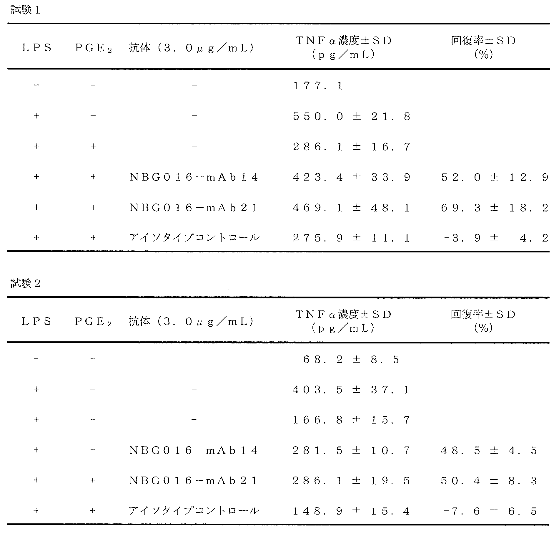
抗EP4抗体を産生するハイブリドーマ(NBG016-mAb14、NBG016-mAb21)約1×10⁷個よりRneasy Mini Kit (キアゲン社製)を用いて、添付の使用説明書に従ってTotal RNAの抽出を行った。5’/3’ RACEキット、2nd ジェネレーション(ロシュ・ダイアグノスティックス社製)にて5’-RACE(rapid amplification of cDNA ends)法によるPCRを行い、重鎖または軽鎖の可変領域の増幅を行った。3’プライマーはマウス定常領域γ1とκに相当するプライマーを使用した。重鎖可変領域増幅のための3’ プライマーは5’-AGGGGCCAGTGGATAGACCGATG-3’(配列番号33)と5’-GGCTGTTGTTTTGGCTGCAGAGAC-3’(配列番号34)である。軽鎖可変領域増幅のための3’プライマーは5’-ACTGGATGGTGGGAAGATGGATAC-3’(配列番号35)と5’-TGGATACAGTTGGTGCAGCATCAG-3’(配列番号36)である。次に、得られた増幅断片をアガロースゲルによって電気泳動し、バンドを切り出し、ゲルを溶かし出すことによってDNAの精製を行った。精製したDNAをT-Vector pMD20(タカラバイオ社製)に組み込み、塩基配列を解析してアミノ酸配列を決定した。シーケンス反応はABI Prism BigDye Terminator Cycle Sequencing Ready Reaction Kits Version 3.1(アプライドバイオシステムズ社)を使用し、Applied Biosystems 3130xl Genetic Analyzer(アプライドバイオシステムズ社)により塩基配列を決定した。 塩基配列を解析した結果、NBG016-mAb14の重鎖可変領域をコードする核酸配列として配列番号1、軽鎖可変領域をコードする核酸配列として配列番号3であった。また重鎖可変領域のアミノ酸配列として配列番号2、軽鎖可変領域のアミノ酸配列として配列番号4が得られた。
さらに、NBG016-mAb21の重鎖可変領域をコードする核酸配列として配列番号11、軽鎖可変領域をコードする核酸配列としてとして配列番号13であった。また重鎖可変領域のアミノ酸配列として配列番号12、軽鎖可変領域のアミノ酸配列として配列番号14が得られた。
NBG016-mAb14の重鎖CDR1~3はそれぞれ、配列番号5~7であった。また軽鎖CDR1~3はそれぞれ配列番号8~10であった。また、NBG016-mAb21の重鎖CDR1~3はそれぞれ、配列番号15~17であった。また軽鎖CDR1~3はそれぞれ配列番号18~20であった。重鎖CDRについて両クローンは完全一致していた。
NBG016-mAb9に関しても上述の方法に従って、重鎖可変領域は配列番号33と配列番号34のプライマーを、軽鎖可変領域は配列番号35と配列番号36のプライマーを用いて増幅し、可変領域の配列を決定した。NBG016-mAb9の重鎖可変領域をコードする核酸配列として配列番号41、軽鎖可変領域をコードする核酸配列として配列番号42であった。また重鎖可変領域のアミノ酸配列として配列番号43、軽鎖可変領域のアミノ酸配列として配列番号44が得られた。
NBG016-mAb9の重鎖CDR1~3はそれぞれ、配列番号45~47であった。また軽鎖CDR1~3はそれぞれ配列番号48~50であった。
First Strand cDNA Synthesis Kit For RT?PCR (AMV) (ロシュ・ダイアグノスティックス社製)に付属のOligo-dT Primerを用いて、使用説明書に従ってcDNAを合成した。合成したcDNAを鋳型として、本発明の抗EP4抗体の重鎖および軽鎖遺伝子全長をPCR増幅した。重鎖及び軽鎖の5’末端側は5’-RACEで判明した塩基配列を参考に、3’末端側は定常領域特異的配列を参考に設計した。重鎖遺伝子増幅のための5’プライマーは5’-CACTGACCCTACGCGTATGGAATGGAGATGGATCTTTCTCTTC-3’(配列番号37)、3’プライマーは5’-ATAAGAATGCGGCCGCTCATTTACCAGGAGAGTGGGAGAG-3’ (配列番号38)である。軽鎖可変領域増幅のための5’プライマーは5’-TTGCAGCCAGGAACGCGTATGGACATGAGGACCCCTGCT-3’(配列番号39)と5’-ATAAGAATGCGGCCGCTTAACACTCATTCCTGTTGAAGCT-3’(配列番号40)である。得られた重鎖および軽鎖増幅断片は制限酵素MluIとNotIで切断し、重鎖はpEHX1.1(東洋紡績社製)、軽鎖はpELX2.1(東洋紡績社製)のMluIとNotIサイトに組み込み、塩基配列を解析してアミノ酸配列を決定した。
塩基配列を解析した結果、NBG016-mAb14の重鎖をコードする核酸配列として配列番号22、軽鎖をコードする核酸配列として配列番号24となった。また重鎖のアミノ酸配列として配列番号23、軽鎖のアミノ酸配列として配列番号25が得られた。
NBG016-mAb21の重鎖をコードする核酸配列として配列番号26、軽鎖をコードする核酸配列として配列番号28となった。また重鎖のアミノ酸配列として配列番号27、軽鎖のアミノ酸配列として配列番号29が得られた。
重鎖ならびに軽鎖可変領域のアミノ酸配列は上記5’-RACE法で解析したアミノ酸配列と同一であった。
NBG016-mAb9に関しても、上述の方法に従ってcDNAを合成し、それを鋳型としてNBG016―mAb9の重鎖および軽鎖遺伝子全長をPCR増幅した。重鎖遺伝子増幅のための5’プライマーは5’-CACTAGAGCCCCCATACGCGTATGGCTGTCCTGGTGCTGTTCC-3’(配列番号51)、3’プライマーは5’-ATAAGAATGCGGCCGCTCATTTACCCGGAGAGTGGGAGAG-3’ (配列番号52)である。軽鎖遺伝子増幅のための5’プライマーは5’-TCCTCAGGTTGCCTCACGCGTATGAAGTTGCCTGTTAG-3’(配列番号53)と5’-ATAAGAATGCGGCCGCTTAACACTCATTCCTGTTGAAGCT-3’(配列番号40)である。得られた重鎖および軽鎖増幅断片は制限酵素MluIとNotIで切断し、重鎖はpEHX1.1(東洋紡績社製)、軽鎖はpELX2.1(東洋紡績社製)のMluIとNotIサイトに組み込み、塩基配列を解析してアミノ酸配列を決定した。
NBG016-mAb9の重鎖をコードする核酸配列として配列番号54、軽鎖をコードする核酸配列として配列番号55となった。また重鎖のアミノ酸配列として配列番号56、軽鎖のアミノ酸配列として配列番号57が得られた。
NBG016-mAb14、NBG016-mAb21のリコンビナント抗体は、Mammalian PowerExpress System(東洋紡績社製)を用いて生産した。軽鎖遺伝子が挿入されたpELX2.1を制限酵素EcoRIとBglIIで切断し、アガロースゲルによって電気泳動して軽鎖遺伝子を含む断片を精製した。精製した軽鎖遺伝子断片を、重鎖遺伝子を挿入されたpEHX1.1のEcoRI-BglIIサイトに組み込み、軽鎖および重鎖両方の遺伝子を保持するプラスミドを作製した。このプラスミドをリポフェクトアミン2000を用いて293FT細胞に形質導入し、一過性での抗体発現を行った。
形質導入から72時間後の細胞培養上清を採取し、ヒトEP4 安定発現CHO細胞株との結合試験を(6)に記載した方法でフローサイトメトリーにより実施した。コントロールとしては抗体遺伝子未導入の293FT細胞の培養上清を使用した。その結果、培養上清中に分泌されたリコンビナント抗体NBG016-mAb14、NBG016-mAb21は、ヒトEP4に対する結合性を保持していた。このことから、(17)で得られた抗体遺伝子配列は、抗EP4抗体をコードしていることが確認できた。
NBG016―mAb9のリコンビナント抗体も、上述の方法と同様に生産した。軽鎖遺伝子が挿入されたpELX2.1は制限酵素SalIとSpeIで切断し、アガロースゲルによって電気泳動して軽鎖遺伝子を含む断片を精製した。精製した軽鎖遺伝子断片を、重鎖遺伝子が挿入されたpEHX1.1のSalI-SpeIサイトに組み込み、軽鎖および重鎖両方の遺伝子を保持するプラスミドを作製した。このプラスミドを293FT細胞に形質導入し、一過性での抗体発現を行った。
形質導入から72時間後の細胞培養上清を採取し、ヒトEP4 安定発現CHO細胞株との結合試験を(6)に記載した方法でフローサイトメトリーにより実施した。コントロールとしては抗体遺伝子未導入の293FT細胞の培養上清を使用した。結果を図10に示す。親株Flp-In-CHO細胞を灰色に塗りつぶしたヒストグラムで、ヒトEP4 安定発現CHO細胞株を黒の実線で示している。培養上清中に分泌されたリコンビナント抗体NBG016―mAb9がヒトEP4に対する結合性を保持していたことから、(17)で得られたNBG016―mAb9の抗体遺伝子配列は、抗EP4抗体をコードしていることが確認できた。
(18)で作製した、NBG016-mAb14、NBG016-mAb21の軽鎖および重鎖の遺伝子を保持するベクターを制限酵素SspIで切断し、エタノール沈殿法で精製した。リポフェクトアミン2000を用いてCHO-K1細胞(理化学研究所細胞バンク)に形質導入し、10%ウシ胎児血清を含むHam’s F12培地で24時間培養した。24時間後、10% ウシ胎児血清および10μg/mLピューロマイシンを含むHam’s F12培地に交換し、3日毎に培地交換をしながら12日間培養した。12日後にペニシリンキャップ法でコロニーを分離した。
分離したCHO―K1細胞を24well plateに播種し、10μg/mL ピューロマイシンを含むHam’s F12培地で3日間培養した。3日後に10μg/mLピューロマイシンを含むHam’s F12培地(ウシ胎児血清無添加)に交換し、72時間培養後の培養上清を回収した。
培養上清中のマウスIgGはELISA法により検出した。PBSを用いて培養上清の希釈系列を作製し、Maxisorp 96well plate (Nunc社製)に分注して4℃で一晩静置した。翌日、3% BSA(Sigma社製)を含むPBSを加えて室温で1時間静置してブロッキングした。0.1% Tween 20を含むPBSで洗浄した後、1% BSAを含むPBSで4,000倍に希釈したホースラディッシュペルオキシダーゼ(HRP)標識抗マウスIgG抗体(ミリポア社製)を加え、室温で1時間静置した。0.1% Tween 20を含むPBSで洗浄した後、発色試薬(Sureblue TMB microwell peroxidase substrate、Kirkegaard & Perry Laboratories社製)を100μL加えて室温で5分静置した後、100μLの1N硫酸を加えて反応を停止し、450nmの吸光度を測定した。その結果、軽鎖および重鎖の遺伝子を保持するベクターを導入して株化したCHO-K1細胞の上清で、IgGの発現を確認した。
(19)で樹立した細胞株培養上清から(7)と同様にして、リコンビナント抗体NBG016-mAb14、リコンビナント抗体NBG016-mAb21を精製した。得られた精製リコンビナント抗EP4抗体と、ヒトEP4 安定発現CHO細胞株の結合試験を(6)に記載した方法でフローサイトメトリーにより実施した。精製リコンビナント抗EP4抗体もしくはマウスアイソタイプコントロール抗体は細胞5×10⁵個あたり1μgを使用した。2次抗体にはPE標識抗マウスIgG抗体を使用した。
図11にその結果を示す。親株Flp-In-CHO細胞を灰色に塗りつぶしたヒストグラムで、ヒトEP4 安定発現CHO細胞株を黒の実線で示している。リコンビナント抗体NBG016-mAb14、リコンビナント抗体NBG016-mAb21が共に、ヒトEP4安定発現CHO細胞株にのみ結合していることから、精製リコンビナント抗EP4抗体が、ヒトEP4に対する結合性を保持していることが確認された。
(7)で精製したNBG016-mAb21はサブクラスがIgG2aであった。そこで、NBG016-mAb21のサブクラスをIgG1に改変し、マウスIgG1型抗体NBG016-mAb21を作製した。マウスIgG1型抗体NBG016-mAb21遺伝子はOverlapping PCR法で以下の通り作製した。NBG016―mAb21の重鎖遺伝子を鋳型として、5’-CACTGACCCTACGCGTATGGAATGGAGATGGATCTTTCTCTTC-3’(配列番号37)と5’-GACAGATGGGGGTGTCGTTTTAGCGCTAGAGACAGTGACCAGAGTCCC-3’(配列番号58)を用い、NBG016―mAb21の可変領域遺伝子をPCR増幅した。一方で、マウスIgG1を産生するハイブリドーマのTotal RNAから合成したcDNAを鋳型にして、5’-GGGACTCTGGTCACTGTCTCTAGCGCTAAAACGACACCCCCATCTGTC-3’(配列番号59)と5’-ATAAGAATGCGGCCGCTCATTTACCAGGAGAGTGGGAGAG-3’(配列番号38)を用い、マウスIgG1のCH1から定常領域遺伝子までをPCR増幅した。増幅して得られた重鎖可変領域遺伝子とCH1―定常領域遺伝子断片を混合し、配列番号37と配列番号38のプライマーを用いてPCR増幅した。増幅して得られたDNA断片を制限酵素MluIとNotIで切断し、発現ベクターpEHX1.1のMluI-NotIサイトに挿入した。得られた発現ベクターを制限酵素EcoRIとBglIIで切断し、NBG016-mAb21の軽鎖遺伝子断片(EcoRI-BglII断片)を挿入してマウスIgG1型抗体NBG016-mAb21発現ベクターとした。
8mMグルタミンを含むEX-CELL CD CHO培地(SAFCBioscience社製)で培養した浮遊CHO-KI細胞(東洋紡績社)(2.5×10⁵cells/ml)を24穴プレートの2ウェルに1mlずつ分注した。Opti-MEM 136μl、リポフェクトアミン2000 15μl、および制限酵素SspIで切断したマウスIgG1型抗体NBG016-mAb21発現ベクター4μgを混合し、室温で20分放置した後、CHO-K1を入れたウェルに68μlずつ添加し、CO2インキュベーターで24時間インキュベートした。24時間後の細胞を8mMグルタミンを含むEX-CELL CD CHO培地8mlに細胞を懸濁し、6穴プレートの2ウェルに4mlずつ分注した。一方のウェルに10mg/mlのピューロマイシンを3μl、もう一方のウェルに4μl添加し、3日または4日毎に培地を交換しながら18日間培養した。増殖したウェルの細胞を回収して、5cells/mlになるようにConditioned medium (1l当たりEX-CELL CD CHO培地700ml、浮遊CHO-KI細胞培養上清300ml、10mg/ml ピューロマイシン1mlまたは0.75mlを混合した培地)に懸濁して、96穴プレートに200μlずつ分注した。1週間後にConditioned mediumを100μl添加し、さらに1週間培養した。数回継代培養した後、薬剤耐性細胞(4×10⁴cells/ml)を24穴プレートに500μl入れ、5日間培養した。5日後にMouse IgG EIA Kit (タカラバイオ社製)を用いて上清中の抗体量を定量して抗体生産細胞をスクリーニングした。この細胞をマウスIgG1型抗体NBG016-mAb21安定発現CHO細胞株とした。
マウスIgG1型抗体NBG016-mAb21安定発現CHO細胞株を、8mM グルタミン、7.5μg/ml ピューロマイシンを含むEX-CELL CD CHO培地で10日間培養して抗体を産生させた。この培養上清200mLから(7)と同様にして精製IgGを得た。以降、得られた精製IgGをマウスIgG1型抗体NBG016-mAb21とした。
マウスIgG1型抗体NBG016-mAb21と、ヒトEP4 安定発現CHO細胞株の結合試験を(9)と同様に実施した。結果を図12(A)に示す。親株Flp-In-CHO細胞を灰色に塗りつぶしたヒストグラムで、ヒトEP4 安定発現CHO細胞株を黒の実線で示している。マウスIgG1型抗体NBG016-mAb21がヒトEP4安定発現CHO細胞株にのみ結合していることから、作製したマウスIgG1型抗体NBG016-mAb21が、ヒトEP4に対する結合性を保持していることが確認された。
マウスIgG1型抗体NBG016-mAb21が、PGE2による cAMP産生を阻害するかを(10)と同様に調べた。細胞と抗体を室温で15分間反応させた後、PGE2を1.5×10⁻¹⁰Mになるように加え、さらに室温で30分間静置した。cAMP量は、LANCE Ultra cAMP Kit(パーキンエルマー社製)を用いて、使用説明書に従った反応を行って測定した。
その結果を図12(B)に示す。マウスアイソタイプコントロール抗体を加えても、PGE2により誘導されたcAMP産生量に関して有意な阻害効果が見られなかったのに対し、マウスIgG1型抗体NBG016-mAb21を加えた場合には、その抗体濃度に依存的なcAMP産生阻害効果が見られた。IC50値は1.1μg/mL(約6.9nM)であった。この結果からNBG016-mAb21をマウスIgG1型抗体に改変してもEP4に対するアンタゴニスト活性を保持していることが示された。
NBG016―mAb14および21のCH1領域以降をヒト抗体遺伝子に置換したヒトキメラ型抗体は、Mammalian PowerExpress System(東洋紡績社製)を用いて生産した。NBG016―mAb14および21の重鎖可変領域遺伝子を、5’-CACTGACCCTAAGCTTATGGAATGGAGATGGATCTTTCTCTTC-3’(配列番号60)と5’-GGCTGTTGTGCTAGCTGCAGAGACAGTGACCAGAGT-3’(配列番号61)を用いてPCR増幅した。得られた重鎖遺伝子断片は制限酵素HindIIIとNheIで切断し、発現ベクターpEHγX1.1のHindIII-NheIサイトに挿入した。一方で、NBG016―mAb14および21の軽鎖可変領域遺伝子を5’-ATTGCAGCCAGGAGAATTCATGGACATGAGGACCCCTGCT-3’(配列番号62)と5’-GGTGCAGCATCCGTACGTTTTATTTCCAACTTTGTCCCC-3’(配列番号63)を用いてPCR増幅した。得られた軽鎖遺伝子断片は制限酵素BsiWIとEcoRIで切断し、発現ベクターpELκX2.1のBsiWI-EcoRIサイトに挿入した。
軽鎖遺伝子が挿入されたpELκ2.1を制限酵素BglII、NotI、ScaIで切断し、アガロースゲルによって電気泳動して軽鎖遺伝子を含む断片を精製した。精製した軽鎖遺伝子断片を、重鎖遺伝子が挿入されたpEHγX1.1のBglII―NotIサイトに組み込み、軽鎖および重鎖両方の遺伝子を保持するプラスミドを作製した。このプラスミドをリポフェクトアミン2000を用いて293FT細胞に形質導入し、一過性での抗体発現を行った。
形質導入から72時間後の細胞培養上清を採取し、ヒトEP4 安定発現CHO細胞株との結合試験を(6)に記載した方法でフローサイトメトリーにより実施した。コントロールとしては抗体遺伝子未導入の293FT細胞の培養上清を、2次抗体としてはPE標識抗ヒトIgG抗体(アブカム社製)を使用した。図13に結果を示す。親株Flp-In-CHO細胞を灰色に塗りつぶしたヒストグラムで、ヒトEP4 安定発現CHO細胞株を黒の実線で示している。この結果より、培養上清中に分泌されたヒトキメラ型抗体NBG016―mAb14および21は、ヒトEP4に対する結合性を保持していることが確認された。
これらの結果から、本発明により提供される抗体をコードする核酸配列を用いて、その機能を保持するリコンビナント抗体(例えば、キメラ抗体、ヒト型化抗体及びヒト抗体等)の生産が可能であることが示された。
Claims (21)
- PGE2受容体サブタイプEP4の細胞外ドメインに結合し、EP4の機能を阻害する抗体又はその機能的断片。
- 前記抗体がモノクローナル抗体であることを特徴とする請求項1に記載の抗体又はその機能的断片。
- 前記抗体が国際寄託受託番号FERM BP-11402(NBG016-mAb14)、 受託番号FERM BP-11403(NBG016-mAb21)のハイブリドーマから産生されることを特徴とする請求項2に記載の抗体又はその機能的断片。
- 前記EP4の機能が、細胞内cAMP濃度を上昇させることである、請求項1乃至3のいずれかに記載の抗体又はその機能的断片。
- EP4の細胞外ドメインに特異的に結合し、その相補性決定領域1~3(CDR1~3)のアミノ酸配列について、下記(A)、(B)又は(C)のいずれかを満たすことを特徴とする請求項1乃至3のいずれかに記載の抗体又はその機能的断片。
(A)配列番号5で表されるアミノ酸配列を含む重鎖CDR1、
配列番号6で表されるアミノ酸配列を含む重鎖CDR2、
配列番号7で表されるアミノ酸配列を含む重鎖CDR3、
配列番号8で表されるアミノ酸配列を含む軽鎖CDR1、
配列番号9で表されるアミノ酸配列を含む軽鎖CDR2、及び
配列番号10で表されるアミノ酸配列を含む軽鎖CDR3を有する、
(B)配列番号15で表されるアミノ酸配列を含む重鎖CDR1、
配列番号16で表されるアミノ酸配列を含む重鎖CDR2、
配列番号17で表されるアミノ酸配列を含む重鎖CDR3、
配列番号18で表されるアミノ酸配列を含む軽鎖CDR1、
配列番号19で表されるアミノ酸配列を含む軽鎖CDR2、及び
配列番号20で表されるアミノ酸配列を含む軽鎖CDR3を有する。
(C)配列番号45で表されるアミノ酸配列を含む重鎖CDR1、
配列番号46で表されるアミノ酸配列を含む重鎖CDR2、
配列番号47で表されるアミノ酸配列を含む重鎖CDR3、
配列番号48で表されるアミノ酸配列を含む軽鎖CDR1、
配列番号49で表されるアミノ酸配列を含む軽鎖CDR2、及び
配列番号50で表されるアミノ酸配列を含む軽鎖CDR3を有する。 - EP4の細胞外ドメインに特異的に結合し、その重鎖可変領域と軽鎖可変領域のアミノ酸配列について、下記(a)、(b)又は(c)のいずれかを満たすことを特徴とする請求項1乃至5のいずれかに記載の抗体又はその機能的断片。
(a)配列番号2で表されるアミノ酸配列を含む重鎖可変領域、及び配列番号4で表されるアミノ酸配列を含む軽鎖可変領域を有する、
(b)配列番号12で表されるアミノ酸配列を含む重鎖可変領域、及び配列番号14で表されるアミノ酸配列を含む軽鎖可変領域を有する。
(c)配列番号43で表されるアミノ酸配列を含む重鎖可変領域、及び配列番号44で表されるアミノ酸配列を含む軽鎖可変領域を有する。 - EP4の細胞外ドメインに結合し、EP4の機能を阻害する抗体又はその機能的断片であって、請求項3乃至6のいずれかに記載の抗体と同一のエピトープに結合する、抗体又はその機能的断片。
- ヒト化抗体又はキメラ抗体であることを特徴とする請求項1乃至7のいずれかに記載の抗体又はその機能的断片。
- ヒト抗体であることを特徴とする請求項1乃至7のいずれかに記載の抗体又はその機能的断片。
- 抗体断片、一本鎖抗体、又はダイアボディであることを特徴とする請求項1乃至9のいずれかに記載の抗体又はその機能的断片。
- 配列番号1、配列番号3、配列番号11、配列番号13、配列番号41、配列番号42であらわされる請求項5又は6に記載の抗体の重鎖可変領域又は軽鎖可変領域をコードする核酸。
- 請求項11に記載の核酸を含むベクター。
- 請求項12に記載のベクターが導入された細胞。
- 請求項1乃至10のいずれかに記載の抗体又はその機能的断片を含む医薬組成物。
- EP4の機能異常によって発症及び/又は進展する疾患の予防あるいは治療に用いられる、請求項14に記載の医薬組成物。
- 治療対象疾患が、免疫疾患であることを特徴とする請求項15に記載の医薬組成物。
- 治療対象疾患が、腫瘍であることを特徴とする請求項15に記載の医薬組成物。
- 治療対象疾患が、疼痛であることを特徴とする請求項15に記載の医薬組成物。
- 請求項1乃至10のいずれかに記載の抗EP4抗体又はその機能的断片が担体に固定化されてなる抗体固定化担体。
- EP4発現細胞を含む血液を接触させて、前記体液からEP4発現細胞を除去するために用いられることを特徴とする請求項19に記載の抗体固定化担体。
- 請求項1乃至10のいずれかに記載の抗EP4抗体を含む細胞表面上のEP4発現量を測定するキット。
Priority Applications (14)
| Application Number | Priority Date | Filing Date | Title |
|---|---|---|---|
| KR1020137010557A KR101857310B1 (ko) | 2010-09-29 | 2011-09-28 | 인간 프로스타글란딘 e2 수용체 ep4 에 대한 항체 |
| AU2011309028A AU2011309028B2 (en) | 2010-09-29 | 2011-09-28 | Antibody against human prostaglandin E2 receptor EP4 |
| EP11829187.1A EP2623594B9 (en) | 2010-09-29 | 2011-09-28 | Antibody against human prostaglandin e2 receptor ep4 |
| NZ609887A NZ609887A (en) | 2010-09-29 | 2011-09-28 | Antibody against human prostaglandin e2 receptor ep4 |
| DK11829187.1T DK2623594T5 (en) | 2010-09-29 | 2011-09-28 | Antibody against human prostaglandin E2 receptor EP4 |
| CN201180046998.3A CN103459595B (zh) | 2010-09-29 | 2011-09-28 | 针对人前列腺素e2受体ep4的抗体 |
| CA2812756A CA2812756C (en) | 2010-09-29 | 2011-09-28 | Antibody against human prostaglandin e2 receptor ep4 |
| JP2012536508A JP5273689B2 (ja) | 2010-09-29 | 2011-09-28 | ヒトプロスタグランジンe2受容体ep4に対する抗体 |
| US13/876,763 US9175080B2 (en) | 2010-09-29 | 2011-09-28 | Antibody against human prostaglandin E2 receptor EP4 |
| ES11829187.1T ES2537017T3 (es) | 2010-09-29 | 2011-09-28 | Anticuerpo dirigido contra el receptor EP4 de la prostaglandina E2 humano |
| IL225447A IL225447A (en) | 2010-09-29 | 2013-03-24 | 4ep antibody of human prostate 2e 2e receptor |
| HK14104750.6A HK1191378A1 (zh) | 2010-09-29 | 2014-05-21 | 針對人前列腺素 受體 的抗體 |
| US14/864,707 US10435465B2 (en) | 2010-09-29 | 2015-09-24 | Antibody against human prostaglandin E2 receptor EP4 |
| US16/555,524 US11649281B2 (en) | 2010-09-29 | 2019-08-29 | Antibody against human prostaglandin E2 receptor EP4 |
Applications Claiming Priority (2)
| Application Number | Priority Date | Filing Date | Title |
|---|---|---|---|
| JP2010-218158 | 2010-09-29 | ||
| JP2010218158 | 2010-09-29 |
Related Child Applications (2)
| Application Number | Title | Priority Date | Filing Date |
|---|---|---|---|
| US13/876,763 A-371-Of-International US9175080B2 (en) | 2010-09-29 | 2011-09-28 | Antibody against human prostaglandin E2 receptor EP4 |
| US14/864,707 Division US10435465B2 (en) | 2010-09-29 | 2015-09-24 | Antibody against human prostaglandin E2 receptor EP4 |
Publications (1)
| Publication Number | Publication Date |
|---|---|
| WO2012043634A1 true WO2012043634A1 (ja) | 2012-04-05 |
Family
ID=45893073
Family Applications (1)
| Application Number | Title | Priority Date | Filing Date |
|---|---|---|---|
| PCT/JP2011/072190 WO2012043634A1 (ja) | 2010-09-29 | 2011-09-28 | ヒトプロスタグランジンe2受容体ep4に対する抗体 |
Country Status (13)
| Country | Link |
|---|---|
| US (3) | US9175080B2 (ja) |
| EP (1) | EP2623594B9 (ja) |
| JP (1) | JP5273689B2 (ja) |
| KR (1) | KR101857310B1 (ja) |
| CN (2) | CN103459595B (ja) |
| AU (1) | AU2011309028B2 (ja) |
| CA (1) | CA2812756C (ja) |
| DK (1) | DK2623594T5 (ja) |
| ES (1) | ES2537017T3 (ja) |
| HK (1) | HK1191378A1 (ja) |
| IL (1) | IL225447A (ja) |
| NZ (1) | NZ609887A (ja) |
| WO (1) | WO2012043634A1 (ja) |
Cited By (8)
| Publication number | Priority date | Publication date | Assignee | Title |
|---|---|---|---|---|
| WO2018210995A1 (en) | 2017-05-18 | 2018-11-22 | Idorsia Pharmaceuticals Ltd | N-substituted indole derivatives |
| WO2020171020A1 (ja) | 2019-02-18 | 2020-08-27 | 株式会社エヌビィー健康研究所 | 細胞の選抜方法、核酸の製造方法、組換え細胞の製造方法、目的物質の製造方法、医薬組成物の製造方法、及び試薬 |
| US11241431B2 (en) | 2015-11-20 | 2022-02-08 | Idorsia Pharmaceuticals Ltd | N-substituted indole derivatives as PGE2 receptor modulators |
| US11325899B2 (en) | 2017-05-18 | 2022-05-10 | Idorsia Pharmaceuticals Ltd | Benzofurane and benzothiophene derivatives as PGE2 receptor modulators |
| WO2022102731A1 (ja) | 2020-11-13 | 2022-05-19 | 小野薬品工業株式会社 | Ep4拮抗薬と免疫チェックポイント阻害物質との併用によるがん治療 |
| US11446298B2 (en) | 2017-05-18 | 2022-09-20 | Idorsia Pharmaceuticals Ltd | Pyrimidine derivatives |
| US11712438B2 (en) | 2017-05-18 | 2023-08-01 | Idorsia Pharmaceuticals Ltd | Phenyl derivatives as PGE2 receptor modulators |
| US11839613B2 (en) | 2017-05-18 | 2023-12-12 | Idorsia Pharmaceuticals Ltd | Pyrimidine derivatives as PGE2 receptor modulators |
Families Citing this family (3)
| Publication number | Priority date | Publication date | Assignee | Title |
|---|---|---|---|---|
| CN106632673A (zh) * | 2015-11-01 | 2017-05-10 | 钮晓音 | 抗pge2单克隆抗体及其使用 |
| CN106929477B (zh) * | 2017-03-20 | 2020-01-10 | 江南大学 | 一株抗前列腺素F2α的特异性单克隆抗体杂交瘤细胞株WXX-2及其应用 |
| GB2596803A (en) * | 2020-07-06 | 2022-01-12 | Petmedix Ltd | Expression vector production & high-throughput cell screening |
Citations (3)
| Publication number | Priority date | Publication date | Assignee | Title |
|---|---|---|---|---|
| US4816567A (en) | 1983-04-08 | 1989-03-28 | Genentech, Inc. | Recombinant immunoglobin preparations |
| JP3118460B2 (ja) | 1993-08-31 | 2000-12-18 | メルク フロスト カナダ アンド カンパニー | プロスタグランジンレセプターep▲下2▼をコードするdna |
| WO2010013498A1 (ja) * | 2008-08-01 | 2010-02-04 | 学校法人聖マリアンナ医科大学 | 変形性関節症治療剤又は予防剤 |
Family Cites Families (6)
| Publication number | Priority date | Publication date | Assignee | Title |
|---|---|---|---|---|
| SE9202892D0 (sv) | 1992-10-02 | 1992-10-02 | Kabi Pharmacia Ab | Dna encoding a prostaglandin receptor, a host cell transformed therewith and an expression product thereof |
| US6395878B1 (en) | 1994-05-05 | 2002-05-28 | Allergan Sales, Inc. | Nucleic acid encoding a human EP prostaglandin receptor |
| CA2485485A1 (en) | 2002-05-23 | 2003-12-04 | Theratechnologies Inc. | Antagonistic peptides of prostaglandin e2 receptor subtype ep4 |
| CA2515081A1 (en) * | 2003-02-07 | 2004-08-19 | Protein Design Labs, Inc. | Amphiregulin antibodies and their use to treat cancer and psoriasis |
| WO2004073589A2 (en) * | 2003-02-24 | 2004-09-02 | Bayer Healthcare Ag | Diagnostics and therapeutics for diseases associated with g-protein coupled receptor prostaglandin e2 ep4 (prostaglandin e2 ep4) |
| SG192496A1 (en) * | 2008-07-08 | 2013-08-30 | Abbott Lab | Prostaglandin e2 binding proteins and uses thereof |
-
2011
- 2011-09-28 WO PCT/JP2011/072190 patent/WO2012043634A1/ja active Application Filing
- 2011-09-28 CN CN201180046998.3A patent/CN103459595B/zh active Active
- 2011-09-28 DK DK11829187.1T patent/DK2623594T5/en active
- 2011-09-28 ES ES11829187.1T patent/ES2537017T3/es active Active
- 2011-09-28 EP EP11829187.1A patent/EP2623594B9/en active Active
- 2011-09-28 US US13/876,763 patent/US9175080B2/en active Active
- 2011-09-28 CN CN201610161807.2A patent/CN106046158A/zh active Pending
- 2011-09-28 AU AU2011309028A patent/AU2011309028B2/en active Active
- 2011-09-28 KR KR1020137010557A patent/KR101857310B1/ko active IP Right Grant
- 2011-09-28 NZ NZ609887A patent/NZ609887A/en unknown
- 2011-09-28 CA CA2812756A patent/CA2812756C/en active Active
- 2011-09-28 JP JP2012536508A patent/JP5273689B2/ja active Active
-
2013
- 2013-03-24 IL IL225447A patent/IL225447A/en active IP Right Grant
-
2014
- 2014-05-21 HK HK14104750.6A patent/HK1191378A1/zh unknown
-
2015
- 2015-09-24 US US14/864,707 patent/US10435465B2/en active Active
-
2019
- 2019-08-29 US US16/555,524 patent/US11649281B2/en active Active
Patent Citations (3)
| Publication number | Priority date | Publication date | Assignee | Title |
|---|---|---|---|---|
| US4816567A (en) | 1983-04-08 | 1989-03-28 | Genentech, Inc. | Recombinant immunoglobin preparations |
| JP3118460B2 (ja) | 1993-08-31 | 2000-12-18 | メルク フロスト カナダ アンド カンパニー | プロスタグランジンレセプターep▲下2▼をコードするdna |
| WO2010013498A1 (ja) * | 2008-08-01 | 2010-02-04 | 学校法人聖マリアンナ医科大学 | 変形性関節症治療剤又は予防剤 |
Non-Patent Citations (32)
| Title |
|---|
| BISWAS, S. ET AL.: "Prostaglandin E2 receptor subtypes, EPI, EP2, EP3 and EP4 in human and mouse ocular tissues-a comparative immunohistochemical study", PROSTAGLANDINS LEUKOTRIENES AND ESSENTIAL FATTY ACIDS, vol. 71, 2004, pages 277 - 288, XP004571548 * |
| CARMEN, S., BRIEFINGS IN FUNCTIONAL GENOMICS AND PROTEOMICS, vol. 1, no. 2, 2002, pages 189 - 203 |
| CLACKSON ET AL., NATURE, vol. 352, 1991, pages 624 - 628 |
| FEDYK, E. R. ET AL.: "Prostaglandin E2 receptors of the EP2 and EP4 subtypes regulate activation and differentiation of mouse B lymphocytes to IgE-secreting cells", PROC. NATL. ACAD. SCI. USA, vol. 93, 1996, pages 10978 - 10983, XP001040639 * |
| FORTIER I. ET AL.: "Immunolocalization of the prostaglandin E2 receptor subtypes in human bone tissue: differences in foetal, adult normal, osteoporotic and pagetic bone", PROSTAGLANDINS LEUKOTRIENES AND ESSENTIAL FATTY ACIDS, vol. 70, 2004, pages 431 - 439, XP055084723 * |
| J. MOL. BIOL., vol. 196, 1987, pages 901 - 917 |
| KABAT ET AL.: "Sequences of Proteins of Immunological Interest, Fifth edition,", 1991, U. S. DEPARTMENT OF HEALTH AND HUMAN SERVICES |
| KOHLER; MILSTEIN, NATURE, vol. 256, 1975, pages 495 |
| KUROIWA, Y., NUC. ACIDS RES., vol. 26, 1998, pages 3447 - 3448 |
| LIN C.-R. ET AL., J PHARMACOLOGY AND EXPERIMENTAL THERAPEUTICS, vol. 319, no. 3, 2006, pages 1096 - 1103 |
| LIN, C. ET AL.: "Prostaglandin E2 receptor EP4 contributes to inflammatory pain hypersensitivity", THE JOURNAL OF PHARMACOLOGY AND EXPERIMENTAL THERAPEUTICS, vol. 319, no. 3, 2006, pages 1096 - 1103, XP008163202 * |
| MARK ET AL., J. MOL. BIOL., vol. 222, 1991, pages 581 - 597 |
| MARTINET L., BIOCHEM PHARMACOL., vol. 80, no. 6, 15 September 2010 (2010-09-15), pages 838 - 45 |
| MORRISON ET AL., PROC. NATL. ACAD. SCI. USA, vol. 81, 1984, pages 6851 - 6855 |
| NATURE, vol. 321, 1986, pages 522 - 525 |
| POPP L. ET AL., EUROPEAN JOURNAL OF PAIN., vol. 13, no. 7, 2009, pages 691 - 703 |
| PROC. NATL. ACAD. SCI. U.S.A., vol. 81, 1984, pages 6851 - 6855 |
| QUEEN C ET AL., PROC. NATL. ACAD. SCI. USA, vol. 86, 1989, pages 10029 - 10033 |
| ROBERTSON FM, CANCER., vol. 116, no. 11, 1 June 2010 (2010-06-01), pages 2806 - 14 |
| SAKATA ET AL., J PHARMACOL SCI., vol. 112, no. 1, 2010, pages 1 - 5 |
| See also references of EP2623594A4 |
| SHARMA S ET AL., CANCER RES., vol. 65, no. 12, 15 June 2005 (2005-06-15), pages 5211 - 20 |
| SHARMA SD, MOL CANCER THER., vol. 9, no. 3, March 2010 (2010-03-01), pages 569 - 80 |
| SIRIWARDENA, D., OPTHALMOLOGY, vol. 109, no. 3, 2002, pages 427 - 431 |
| STILLMAN B.A. ET AL.: "A conserved threonine in the second extracellular loop of the human EP2 and EP4 receptors is required for ligand binding", EUROPEAN JOURNAL OF PHARMACOLOGY, vol. 357, 1998, pages 73 - 82, XP055084727 * |
| SUGIMOTO ET AL., J. BIOL. CHEM., vol. 282, 2007, pages 11613 - 11617 |
| TAKITA, M. ET AL.: "Prostaglandin E receptor EP4 antagonist suppresses osteolysis due to bone metastasis of mouse malignant melanoma cells", FEBS LETTERS, vol. 581, 2007, pages 565 - 571, XP005907872 * |
| TOMIZUKA, K. ET AL., NATURE GENETICS, vol. 16, 1997, pages 133 - 143 |
| TOMIZUKA, K., PROC. NATL. ACAD. SCI. USA, vol. 97, 2000, pages 722 - 727 |
| WORMSTONE, I. M., INVESTIGATIVE OPHTHALMOLOGY & VISUAL SCIENCE, vol. 43, no. 7, 2002, pages 2301 - 8 |
| YAO ET AL., NAT. MED., vol. 15, 2009, pages 633 - 640 |
| YOSHIDA, H.: "Animal Cell Technology: Basic and Applied Aspects", vol. 10, 1999, KLUWER ACADEMIC PUBLISHERS, pages: 69 - 73 |
Cited By (12)
| Publication number | Priority date | Publication date | Assignee | Title |
|---|---|---|---|---|
| US11241431B2 (en) | 2015-11-20 | 2022-02-08 | Idorsia Pharmaceuticals Ltd | N-substituted indole derivatives as PGE2 receptor modulators |
| WO2018210995A1 (en) | 2017-05-18 | 2018-11-22 | Idorsia Pharmaceuticals Ltd | N-substituted indole derivatives |
| US11325899B2 (en) | 2017-05-18 | 2022-05-10 | Idorsia Pharmaceuticals Ltd | Benzofurane and benzothiophene derivatives as PGE2 receptor modulators |
| US11446298B2 (en) | 2017-05-18 | 2022-09-20 | Idorsia Pharmaceuticals Ltd | Pyrimidine derivatives |
| US11712438B2 (en) | 2017-05-18 | 2023-08-01 | Idorsia Pharmaceuticals Ltd | Phenyl derivatives as PGE2 receptor modulators |
| US11839613B2 (en) | 2017-05-18 | 2023-12-12 | Idorsia Pharmaceuticals Ltd | Pyrimidine derivatives as PGE2 receptor modulators |
| WO2020171020A1 (ja) | 2019-02-18 | 2020-08-27 | 株式会社エヌビィー健康研究所 | 細胞の選抜方法、核酸の製造方法、組換え細胞の製造方法、目的物質の製造方法、医薬組成物の製造方法、及び試薬 |
| KR20210128444A (ko) | 2019-02-18 | 2021-10-26 | 가부시키가이샤 에누비 켄코우겡큐쇼 | 세포의 선발 방법, 핵산의 제조 방법, 재조합 세포의 제조 방법, 목적 물질의 제조 방법, 의약 조성물의 제조 방법, 및 시약 |
| US11357787B2 (en) | 2019-02-18 | 2022-06-14 | Nb Health Laboratory Co., Ltd. | Method for selecting cells, method for producing nucleic acid, method for producing recombinant cells, method for producing target substance, method for producing pharmaceutical composition, and reagent |
| EP3928794A4 (en) * | 2019-02-18 | 2022-11-30 | NB Health Laboratory Co. Ltd. | CELL SELECTION METHOD, NUCLEIC ACID PRODUCTION METHOD, RECOMBINANT CELL PRODUCTION METHOD, TARGET SUBSTANCE PRODUCTION METHOD, PHARMACEUTICAL COMPOSITION AND REAGENT PRODUCTION METHOD |
| JP7456627B2 (ja) | 2019-02-18 | 2024-03-27 | 株式会社エヌビィー健康研究所 | 細胞の選抜方法、核酸の製造方法、組換え細胞の製造方法、目的物質の製造方法、医薬組成物の製造方法、及び試薬 |
| WO2022102731A1 (ja) | 2020-11-13 | 2022-05-19 | 小野薬品工業株式会社 | Ep4拮抗薬と免疫チェックポイント阻害物質との併用によるがん治療 |
Also Published As
| Publication number | Publication date |
|---|---|
| JP5273689B2 (ja) | 2013-08-28 |
| CN103459595B (zh) | 2016-04-13 |
| IL225447A0 (en) | 2013-06-27 |
| ES2537017T9 (es) | 2015-08-21 |
| CN106046158A (zh) | 2016-10-26 |
| KR20140027051A (ko) | 2014-03-06 |
| CA2812756A1 (en) | 2012-04-05 |
| DK2623594T5 (en) | 2015-09-21 |
| DK2623594T3 (da) | 2015-05-18 |
| EP2623594A4 (en) | 2013-11-13 |
| US20160075780A1 (en) | 2016-03-17 |
| EP2623594A1 (en) | 2013-08-07 |
| EP2623594B1 (en) | 2015-02-25 |
| US9175080B2 (en) | 2015-11-03 |
| EP2623594B9 (en) | 2015-07-22 |
| AU2011309028A1 (en) | 2013-05-23 |
| CN103459595A (zh) | 2013-12-18 |
| HK1191378A1 (zh) | 2014-07-25 |
| US11649281B2 (en) | 2023-05-16 |
| AU2011309028B2 (en) | 2016-03-17 |
| ES2537017T3 (es) | 2015-06-01 |
| CA2812756C (en) | 2020-06-30 |
| US10435465B2 (en) | 2019-10-08 |
| US20190389949A1 (en) | 2019-12-26 |
| US20130197199A1 (en) | 2013-08-01 |
| IL225447A (en) | 2017-02-28 |
| NZ609887A (en) | 2015-01-30 |
| KR101857310B1 (ko) | 2018-05-11 |
| JPWO2012043634A1 (ja) | 2014-02-24 |
Similar Documents
| Publication | Publication Date | Title |
|---|---|---|
| JP5273689B2 (ja) | ヒトプロスタグランジンe2受容体ep4に対する抗体 | |
| US11498972B2 (en) | Anti-OX40 antibody and use thereof | |
| JP6847037B2 (ja) | 抗cd73抗体とa2a受容体阻害薬とを含む併用治療薬及びその使用 | |
| JP5982698B2 (ja) | Dll4に特異的に結合する新規モノクローナル抗体及びその用途 | |
| KR102362609B1 (ko) | 항garp 단백질 항체와 그 용도 | |
| KR102662915B1 (ko) | 항-b7-h3 항체 | |
| CA3015938A1 (en) | Anti-lag-3 antibodies | |
| KR101838310B1 (ko) | 암의 치료를 위한 인간화 항-cxcr4 항체들 | |
| JP6280040B2 (ja) | invivoで抗腫瘍活性を有する抗ヒトDlk−1抗体 | |
| TW202136307A (zh) | 抗lilrb1抗體和彼之用途 | |
| JP6159010B2 (ja) | テトラスパニン8に対するハイブリドーマクローンおよびモノクローナル抗体 | |
| JP2018513141A (ja) | ヒトカドヘリン−17、ヒトカドヘリン−5、ヒトカドヘリン−6およびヒトカドヘリン−20のrgdモチーフに特異的に結合する薬剤 | |
| JP6159011B2 (ja) | Cd9に対するハイブリドーマクローンおよびモノクローナル抗体 | |
| JP2022514786A (ja) | Muc18に特異的な抗体 | |
| JP2016005449A (ja) | Cd9に対するハイブリドーマクローンおよびモノクローナル抗体 | |
| KR20210096627A (ko) | 항-b7s1 폴리펩티드 및 이의 용도 | |
| TW202010753A (zh) | Spns2中和抗體 |
Legal Events
| Date | Code | Title | Description |
|---|---|---|---|
| 121 | Ep: the epo has been informed by wipo that ep was designated in this application |
Ref document number: 11829187 Country of ref document: EP Kind code of ref document: A1 |
|
| WWE | Wipo information: entry into national phase |
Ref document number: 2012536508 Country of ref document: JP |
|
| WWE | Wipo information: entry into national phase |
Ref document number: 225447 Country of ref document: IL |
|
| ENP | Entry into the national phase |
Ref document number: 2812756 Country of ref document: CA |
|
| WWE | Wipo information: entry into national phase |
Ref document number: 13876763 Country of ref document: US |
|
| NENP | Non-entry into the national phase |
Ref country code: DE |
|
| WWE | Wipo information: entry into national phase |
Ref document number: 2011829187 Country of ref document: EP |
|
| ENP | Entry into the national phase |
Ref document number: 20137010557 Country of ref document: KR Kind code of ref document: A |
|
| ENP | Entry into the national phase |
Ref document number: 2011309028 Country of ref document: AU Date of ref document: 20110928 Kind code of ref document: A |