WO2010147146A1 - 標的細胞の検出方法 - Google Patents
標的細胞の検出方法 Download PDFInfo
- Publication number
- WO2010147146A1 WO2010147146A1 PCT/JP2010/060208 JP2010060208W WO2010147146A1 WO 2010147146 A1 WO2010147146 A1 WO 2010147146A1 JP 2010060208 W JP2010060208 W JP 2010060208W WO 2010147146 A1 WO2010147146 A1 WO 2010147146A1
- Authority
- WO
- WIPO (PCT)
- Prior art keywords
- cell
- measurement
- target cell
- cells
- particles
- Prior art date
- Legal status (The legal status is an assumption and is not a legal conclusion. Google has not performed a legal analysis and makes no representation as to the accuracy of the status listed.)
- Ceased
Links
Images

Classifications
-
- G—PHYSICS
- G01—MEASURING; TESTING
- G01N—INVESTIGATING OR ANALYSING MATERIALS BY DETERMINING THEIR CHEMICAL OR PHYSICAL PROPERTIES
- G01N33/00—Investigating or analysing materials by specific methods not covered by groups G01N1/00 - G01N31/00
- G01N33/48—Biological material, e.g. blood, urine; Haemocytometers
- G01N33/50—Chemical analysis of biological material, e.g. blood, urine; Testing involving biospecific ligand binding methods; Immunological testing
- G01N33/5005—Chemical analysis of biological material, e.g. blood, urine; Testing involving biospecific ligand binding methods; Immunological testing involving human or animal cells
- G01N33/5008—Chemical analysis of biological material, e.g. blood, urine; Testing involving biospecific ligand binding methods; Immunological testing involving human or animal cells for testing or evaluating the effect of chemical or biological compounds, e.g. drugs, cosmetics
- G01N33/5044—Chemical analysis of biological material, e.g. blood, urine; Testing involving biospecific ligand binding methods; Immunological testing involving human or animal cells for testing or evaluating the effect of chemical or biological compounds, e.g. drugs, cosmetics involving specific cell types
- G01N33/5047—Cells of the immune system
-
- G—PHYSICS
- G01—MEASURING; TESTING
- G01N—INVESTIGATING OR ANALYSING MATERIALS BY DETERMINING THEIR CHEMICAL OR PHYSICAL PROPERTIES
- G01N33/00—Investigating or analysing materials by specific methods not covered by groups G01N1/00 - G01N31/00
- G01N33/48—Biological material, e.g. blood, urine; Haemocytometers
- G01N33/50—Chemical analysis of biological material, e.g. blood, urine; Testing involving biospecific ligand binding methods; Immunological testing
- G01N33/53—Immunoassay; Biospecific binding assay; Materials therefor
- G01N33/543—Immunoassay; Biospecific binding assay; Materials therefor with an insoluble carrier for immobilising immunochemicals
- G01N33/54313—Immunoassay; Biospecific binding assay; Materials therefor with an insoluble carrier for immobilising immunochemicals the carrier being characterised by its particulate form
-
- G—PHYSICS
- G01—MEASURING; TESTING
- G01N—INVESTIGATING OR ANALYSING MATERIALS BY DETERMINING THEIR CHEMICAL OR PHYSICAL PROPERTIES
- G01N15/00—Investigating characteristics of particles; Investigating permeability, pore-volume or surface-area of porous materials
- G01N15/10—Investigating individual particles
- G01N15/1031—Investigating individual particles by measuring electrical or magnetic effects
- G01N15/12—Investigating individual particles by measuring electrical or magnetic effects by observing changes in resistance or impedance across apertures when traversed by individual particles, e.g. by using the Coulter principle
-
- G—PHYSICS
- G01—MEASURING; TESTING
- G01N—INVESTIGATING OR ANALYSING MATERIALS BY DETERMINING THEIR CHEMICAL OR PHYSICAL PROPERTIES
- G01N15/00—Investigating characteristics of particles; Investigating permeability, pore-volume or surface-area of porous materials
- G01N15/10—Investigating individual particles
- G01N15/14—Optical investigation techniques, e.g. flow cytometry
- G01N15/1456—Optical investigation techniques, e.g. flow cytometry without spatial resolution of the texture or inner structure of the particle, e.g. processing of pulse signals
- G01N15/1459—Optical investigation techniques, e.g. flow cytometry without spatial resolution of the texture or inner structure of the particle, e.g. processing of pulse signals the analysis being performed on a sample stream
-
- G—PHYSICS
- G01—MEASURING; TESTING
- G01N—INVESTIGATING OR ANALYSING MATERIALS BY DETERMINING THEIR CHEMICAL OR PHYSICAL PROPERTIES
- G01N15/00—Investigating characteristics of particles; Investigating permeability, pore-volume or surface-area of porous materials
- G01N15/01—Investigating characteristics of particles; Investigating permeability, pore-volume or surface-area of porous materials specially adapted for biological cells, e.g. blood cells
- G01N2015/016—White blood cells
-
- G—PHYSICS
- G01—MEASURING; TESTING
- G01N—INVESTIGATING OR ANALYSING MATERIALS BY DETERMINING THEIR CHEMICAL OR PHYSICAL PROPERTIES
- G01N15/00—Investigating characteristics of particles; Investigating permeability, pore-volume or surface-area of porous materials
- G01N15/10—Investigating individual particles
- G01N2015/1024—Counting particles by non-optical means
-
- G—PHYSICS
- G01—MEASURING; TESTING
- G01N—INVESTIGATING OR ANALYSING MATERIALS BY DETERMINING THEIR CHEMICAL OR PHYSICAL PROPERTIES
- G01N15/00—Investigating characteristics of particles; Investigating permeability, pore-volume or surface-area of porous materials
- G01N15/10—Investigating individual particles
- G01N15/14—Optical investigation techniques, e.g. flow cytometry
- G01N2015/1477—Multiparameters
-
- G—PHYSICS
- G01—MEASURING; TESTING
- G01N—INVESTIGATING OR ANALYSING MATERIALS BY DETERMINING THEIR CHEMICAL OR PHYSICAL PROPERTIES
- G01N15/00—Investigating characteristics of particles; Investigating permeability, pore-volume or surface-area of porous materials
- G01N15/10—Investigating individual particles
- G01N15/14—Optical investigation techniques, e.g. flow cytometry
- G01N2015/1486—Counting the particles
Definitions
- the present invention relates to a method for detecting a target cell.
- a cell to be examined has a specific molecule serving as a marker (sometimes called a cell surface marker, cell surface antigen or CD antigen) on its surface.
- a marker sometimes called a cell surface marker, cell surface antigen or CD antigen
- flow cytometry and other methods for detecting a cell group having these cell surface markers Japanese Patent Laid-Open No. 2008-187932, Japanese Translation of PCT International Publication No. 2008-538609.
- these analysis results are used as data for disease diagnosis, for example.
- a cell having a specific cell surface marker contained in a cell dispersion to be examined is referred to as a “target cell”.
- a flow cytometer uses cell surface markers of cells to be examined, labeled molecules such as fluorescently labeled monoclonal antibodies (fluorescent antibody method) and enzyme-labeled monoclonal antibodies (enzyme antibody method), fluorescent particles bound with labeled molecules, This is a device that can be labeled with inorganic semiconductor fluorescent nanoparticles (Japanese Patent Application Laid-Open No. 2004-077389), and that the target cells can be identified using the label.
- These labeled monoclonal antibodies MoAb
- the cell dispersion can be objectively and definitely characterized. Therefore, the obtained result can be used as more reliable data for diagnosing a disease by a doctor or the like.
- flow cytometry is more expensive than the blood cell counter, and the labeled monoclonal antibody used as a reagent is still expensive. For this reason, flow cytometers are often installed in specialized inspection institutions, and there are still few examples of introduction to hospitals and clinics. Therefore, at present, flow cytometry is performed by outsourcing to a specialized organization such as an inspection company, and it takes time to obtain results, and in order to quickly provide data for diagnosing diseases by doctors, etc. Is still inadequate.
- the method described in Japanese Patent Application Laid-Open No. 2008-187932 in which an antibody is immobilized on a substrate, a plate, or the like and cells are adsorbed thereto, can only be used to separate target cells and confirm the presence or absence of target cells.
- leukocytes are classified into granulocytes, lymphocytes and monocytes, or eosinophils, neutrophils, basophils, lymphocytes and monocytes. Can be measured by classifying them into blood test devices classified into the following five categories (sometimes referred to as “white blood cell 3 class blood test device” and “white blood cell 5 class blood test device”), respectively.
- white blood cell 3 class blood test device and “white blood cell 5 class blood test device”
- white blood cell 5 class blood test device There is a device. These devices are equipped with a mechanism that irradiates a sample with laser light to detect scattered light such as forward scattered intensity and side scattered intensity, and information that reflects the size of the cell and the internal structure of the cell, It can be obtained simultaneously with the counting of blood cells.
- the existence ratio for each type of white blood cell can be obtained.
- abnormalities in which the abundance ratio of five types of leukocytes becomes abnormal in leukemia or immature leukocytes (juvenile leukocytes) appear can be detected. It largely depends on the performance of the apparatus, and at present, it is said that a blood sample cannot be sufficiently characterized only by the measurement results of these apparatuses.
- An object of the present invention is to provide a simple and highly accurate method for detecting a target cell. According to the method of the present invention, it is easy to detect a target cell and analyze the size of a cell group constituting the target cell without using flow cytometry. Useful for inspection.
- the present invention has been made to solve the above-described problems, and can be realized as the following aspects or application examples.
- One aspect of the method for detecting a target cell according to the present invention is as follows. Measurement of dispersion liquid containing particles (hereinafter referred to as “labeled particles”) to which a substance that specifically binds to a specific molecule present on the cell surface is fixed and cells to be examined by an optical or electromagnetic method. (Hereinafter referred to as “inspection measurement”) to obtain measurement result 1; A step of obtaining the measurement result 2 by performing the same measurement as the test measurement for the dispersion liquid that does not contain the labeled particles and contains the cells to be tested, and compares the measurement result 1 and the measurement result 2 Process.
- labeled particles particles to which a substance that specifically binds to a specific molecule present on the cell surface is fixed and cells to be examined by an optical or electromagnetic method.
- Application example 2 In application example 1, A method for detecting a target cell, wherein the test measurement does not include fluorescence measurement.
- Application example 3 In application example 1 or application example 2, A method for detecting a target cell, wherein the inspection measurement includes measurement of scattered light.
- Application example 4 In any one of Application Examples 1 to 3, A method for detecting a target cell, wherein the test measurement includes measurement of a cell number and a cell size by an electromagnetic method.
- Application example 5 In any one of Application Examples 1 to 4, A method for detecting a target cell, wherein the labeled particle has a polar group.
- Application example 6 In application example 5, A method for detecting a target cell, wherein the polar group is at least one group selected from a hydroxyl group, an epoxy group, a carboxyl group, an alkylene oxide group, a keto group, and a substituted or unsubstituted amino group.
- test measurement includes a step of measuring a binding amount between each target cell and a labeled particle and obtaining a distribution of the number of target cells with respect to the binding amount.
- Application example 8 In any one of Application Examples 1 to 7, A method for detecting a target cell, wherein the test measurement is performed for a plurality of measurement items.
- Application example 9 In any one of Application Examples 1 to 8, A method for detecting a target cell, wherein the cell dispersion contains a body fluid.
- the cell to be examined is a blood cell containing white blood cells
- the test measurement is performed using a blood test apparatus that classifies leukocytes into three classifications of granulocytes, lymphocytes and monocytes or five classifications of eosinophils, neutrophils, basophils, lymphocytes and monocytes. And a method for detecting a target cell.
- the labeled particle has a substance that specifically binds to a second antigen different from the first antigen and the labeled particle 1 to which a substance that specifically binds to the first antigen is immobilized.
- the method of the present invention it is possible to easily and rapidly perform detection of target cells and analysis of the size of a cell group constituting the target cells.
- the data for diagnosing various diseases can be provided simply and rapidly.
- the measurement conventionally performed using flow cytometry can be performed using a leukocyte 5-class blood test apparatus or a leukocyte 3-class blood test apparatus.
- the target cells in the cell dispersion can be analyzed more easily.
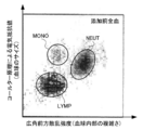
- FIG. 1 is a scatter diagram illustrating an example of a blood analysis process.
- FIG. 2 is a scatter diagram illustrating an example of an analysis process of the cell analysis method of the example.
- the target cell detection method includes a particle (hereinafter referred to as “labeled particle”) to which a substance that specifically binds to a specific molecule existing on the cell surface is fixed and a test object.
- Step 1 of obtaining a measurement result 1 by performing measurement by optical or electromagnetic method (hereinafter referred to as “inspection measurement”) on a dispersion containing a certain cell, including the cell to be inspected without the labeled particle It has the process 2 which performs the same test
- the cell dispersion prepared in the present embodiment is not limited as long as at least target cells are dispersed (suspended) in the liquid.
- Examples of such a cell dispersion liquid include body fluids of animals such as humans, that is, blood, lymph fluid, tissue fluid, body cavity fluid, and the like.
- a cell dispersion prepared by diluting a body fluid with an appropriate dispersion medium such as an isotonic buffer may be prepared.
- the cell dispersion according to the present embodiment is not limited to those derived from living bodies, and may be various cell dispersions prepared by artificially dispersing cells for testing, research, etc. Good.
- the cell dispersion medium is not limited and is typically water, plasma, or the like, and may be an organic solvent such as glycerin or alcohol.
- medical agent may be contained as a solute.
- target cells of the present embodiment include leukocytes, erythrocytes, platelets, artificial cells (such as genetically manipulated cells), cancer cells, and the like, cells having an antigen on the surface, and the like.
- the cell surface marker possessed by the target cell include at least one of protein, sugar chain, complex carbohydrate, and lipid.
- One target cell may have a plurality of cell surface markers.
- the type of cell surface marker is not particularly limited, and examples of the protein include various receptors (receptors), CD antigens (based on international cluster CD (cluster of differentiation) display), and the like.
- sugar chain examples include at least one of a glycoprotein sugar chain, a glycolipid sugar chain, a glycosaminoglycan sugar chain, and a polysaccharide-derived oligosaccharide chain.
- complex carbohydrates include in vivo polymers having sugar chains.
- the complex carbohydrate includes at least one of glycoprotein (including glycopeptide), proteoglycan, and glycolipid.
- the cell dispersion may contain either target cells or cells other than the target cells.
- the cell dispersion liquid is blood and the white blood cells are target cells
- red blood cells and platelets correspond to other cells.
- white blood cells and platelets correspond to other cells.
- a specific disease for example, when a cell having a specific cell surface marker is a target cell due to infection with a virus that causes the disease, cells infected with the virus are not included. Normal cell specimens may not contain target cells.
- the concentration of the cells dispersed in the cell dispersion is not particularly limited.
- the concentration of cells dispersed in the cell dispersion is, for example, 1000 (cells / ⁇ l) or more and 20000 (cells / ⁇ l) or less when the cell dispersion is blood and the target cells are leukocytes. It is preferable to do. For this reason, blood and other body fluids can be diluted as necessary.
- the concentration of target cells dispersed in the cell dispersion is, for example, about 10 (cells / ⁇ l) or more and about 1 ⁇ 10 7 (cells / ⁇ l) or less. It can be.
- Labeled particle and method for preparing the same The labeled particle used in the present invention is prepared by immobilizing a substance that specifically binds to a specific molecule present on the cell surface to the base particle described below.
- the shape of the base particle is not particularly limited.
- the shape of the base particles can be a shape such as a sphere, a spheroid, a cylinder, etc., and an irregular shape.
- spherical polymer particles can be produced by, for example, emulsion polymerization, etc., so that the production can be facilitated.
- by arranging the shapes of the particles for example, it is possible to provide functions such as imparting characteristics to scattered light by laser irradiation and facilitating identification of particles by optical observation.
- the size of the base particles is preferably 0.04 to 10 ⁇ m, more preferably 0.5 to 10 ⁇ m, and particularly preferably 1 to 5 ⁇ m as the number average particle diameter. If the number average particle diameter (diameter) is less than 0.04 ⁇ m, the measurement result may not be clearly changed when the particles are adsorbed to target cells. When the cell dispersion is blood and the white blood cells are target cells, if the number average particle diameter exceeds 10 ⁇ m, the particles become the same size or larger than the target cells, and are adsorbed on the target cells. It is difficult to distinguish between the particles and target cells that have not been used, and when adsorbed to the target cells, the measurement result may change excessively, which may hinder acquisition of information in the measurement process. . In addition, when the number average particle diameter is 0.5 ⁇ m or more, inspection measurement by the light scattering method becomes easy.
- the number average particle diameter is determined as a number average particle diameter in terms of polystyrene particles by the light scattering method or the light blocking method, or by electron microscopy. Any of these measuring methods may be used, and if the number average particle diameter obtained by any of the measuring methods is within the above range, it can be suitably used in the present invention. From the relationship with measurement accuracy, the light scattering method is preferable when the particle size is about 0.04 to 1 ⁇ m, the light blocking method is preferable when the particle size is about 1 to 5 ⁇ m, and the case where the particle size is about 5 ⁇ m or more. Is preferably electron microscopy.
- the material of the base particles is not particularly limited, and examples thereof include organic particles and inorganic particles.
- the organic particles include polystyrene, polylactic acid, acrylic, polyethyleneimine, agarose, iminodiacetic acid chelate, magnetic latex, magnetic polylactic acid, magnetic dextran, magnetic chitosan, magnetic agarose, magnetic polyethyleneimine, and the like.
- the inorganic particles include silica, magnetic silica, iron oxide, and inorganic semiconductor particles.
- the base particles may be a mixture of particles of different materials. Specifically, one or more organic particles or inorganic particles, or organic particles and inorganic particles may be used in combination. .
- the base particles preferably have a polar group, and more preferably have a polar group on the particle surface.
- the polar group is preferably at least one group selected from a hydroxyl group, an epoxy group, a carboxyl group, an alkylene oxide group, a keto group, and a substituted or unsubstituted amino group.
- Such a base particle having a hydrophilic group is prepared by emulsion polymerization using a monomer having a hydrophilic group (hydrophilic monomer) as a part of the raw material, and the particle is coated with a monomer part containing a hydrophilic monomer. It can be prepared by a method of polymerizing the monomer part.
- hydrophilic monomers include hydrophilic functional groups such as glycerol acrylate, glycerol methacrylate, 2-hydroxyethyl acrylate, 2-hydroxyethyl methacrylate, methoxyethyl acrylate, methoxyethyl methacrylate, polyethylene glycol monoacrylate, and polyethylene glycol monomethacrylate.
- hydrophilic functional groups such as glycerol acrylate, glycerol methacrylate, 2-hydroxyethyl acrylate, 2-hydroxyethyl methacrylate, methoxyethyl acrylate, methoxyethyl methacrylate, polyethylene glycol monoacrylate, and polyethylene glycol monomethacrylate.
- hydrophilic functional groups such as glycerol acrylate, glycerol methacrylate, 2-hydroxyethyl acrylate, 2-hydroxyethyl methacrylate, methoxyethyl acrylate, methoxyethyl me
- the monomer which has epoxy groups such as glycidyl acrylate and glycidyl methacrylate, is hydrolyzed and produces
- these monomers can also be used suitably.
- the base particles may be colored so as to exhibit colors in the visible light region such as red, blue, and green. By coloring, for example, the visibility when observing particles with an optical microscope can be enhanced. Thereby, when the number of target cells (cells to which particles are bound) in the cell dispersion liquid is small, it is possible to facilitate the search for the target cells using a microscope.
- Substances that specifically bind to specific molecules on the cell surface are not particularly limited, but antibodies and other protein molecules such as monoclonal antibodies and polyclonal antibodies Alternatively, aggregates thereof, Fab ′ fragments of antibodies, and other polynucleotides can be mentioned.
- the antibody has subclasses such as IgG, IgM, IgA, IgE, and IgD, and any of them may be used.
- a monoclonal antibody that specifically binds to the CD antigen is preferably immobilized.
- the anti-CD antigen antibody include an anti-CD3 antigen antibody (sometimes abbreviated as CD3 antibody), an anti-CD4 antigen antibody (sometimes abbreviated as CD4 antibody), and an anti-CD8 antigen antibody (abbreviated as CD8 antibody).
- anti-human CD antigen monoclonal antibodies are anti-human CD antigen monoclonal antibodies, they can be suitably prepared using, for example, mice, rats, rabbits, sheep and the like.
- the method of fixing a substance that specifically binds to a specific molecule present on the cell surface to the base particle is not particularly limited, and is a method of physically fixing by adsorption or the like and a covalent bond And a method of chemically fixing by hydrogen bonding or the like. Furthermore, as a method of immobilizing a substance such as the antibody on the base particle, a method of immobilizing a substance such as the antibody directly on the base particle (hereinafter sometimes referred to as a direct method), a base particle and the antibody or the like And a method of fixing another substance between them (hereinafter sometimes referred to as an indirect method).
- a method for directly immobilizing an antibody on a base particle there may be mentioned a method in which an Fc site of an antibody is covalently bound to a functional group on the particle surface with a coupling agent.
- a coupling agent for example, carbodiimides such as EDC (1-Ethyl-3- (3-Dimethylaminopropyryl) -Carbodiimide hydrochloride) can be used.
- EDC is used as a coupling agent
- the antibody can be directly immobilized on the base particle by a covalent bond by a carbodiimide coupling reaction.
- a blocking agent may be used in combination.
- BSA bovine serum albumin
- gelatin gelatin
- skim milk ovalbumin and the like
- the amount of the antibody is preferably 0.1 to 100 ⁇ g / mg particle based on the weight of the base particle.
- a method of indirectly immobilizing an antibody on the base particle a method of binding a primary antibody to the base particle via a secondary antibody can be mentioned.
- the antibody that specifically binds to the cell surface marker is the primary antibody
- the antibody that specifically binds to the primary antibody is the secondary antibody.
- a protein having a property of specifically binding to an Fc site of IgG such as protein G or protein A or a derivative thereof is bound to a base particle, and the cell surface is bound to the protein or derivative thereof. Examples include a method of binding an antibody that specifically binds to a marker.
- an antibody corresponding to the animal species from which the primary antibody immunoglobulin is derived is selected as the secondary antibody.
- the primary antibody is an anti-human CD antigen monoclonal antibody and is derived from a mouse (hereinafter sometimes referred to as an anti-human CD antigen mouse antibody)
- the secondary antibody is an anti-mouse immunoglobulin. It is preferable to select an antibody (such as an anti-mouse IgG antibody). More specifically, for example, when the primary antibody is an anti-human CD antigen mouse IgG antibody, it is preferable to select an anti-mouse IgG antibody as the secondary antibody.
- the animal species derived from the anti-mouse IgG antibody is preferably rat, rabbit, sheep or the like.
- the amount of the anti-mouse IgG antibody when the anti-mouse IgG antibody is bound to the base particle is preferably 0.1 to 10 ⁇ g / mg particle based on the weight of the base particle.
- the amount of protein A or protein G bound to the particles is preferably 0.1 to 100 ⁇ g / mg particles based on the weight of the base particles.
- the amount of the primary antibody is preferably 0.1 to 100 ⁇ g / mg particles with respect to the weight of the particles.
- anti-mouse IgG antibody or the like is used as a masking agent, and protein G or protein Can be bound to A.
- the amount of anti-mouse IgG antigen antibody and the like in this case is preferably 0.1 to 100 ⁇ g / mg particle, although it depends on the particle diameter of the particle and the amount of protein A and protein G bound.
- the labeled particles are used by being dispersed in a dispersion medium such as physiological saline or a buffer solution (borate buffer, EDTA buffer, Tris buffer, phosphate buffer, etc.).
- a dispersion medium such as physiological saline or a buffer solution (borate buffer, EDTA buffer, Tris buffer, phosphate buffer, etc.).
- Step 1 is a step of obtaining a measurement result by performing inspection measurement on the dispersion liquid containing the labeled particles and the cells to be inspected.
- the measurement result obtained in step 1 is referred to as measurement result 1.
- the labeled particles and the target cells are specifically bound by an antigen-antibody reaction or the like.
- cells other than the labeled particles and the target cells cannot specifically bind. For this reason, in the step 1, only the target cells are bound to one or more labeled particles.
- Dispersion liquid of cells and the like The amount of labeled particles in the dispersion liquid used in this step is 1 ⁇ 10 2 particles / ⁇ l to 9 ⁇ 10 10 particles / ⁇ l, depending on the type of cell dispersion liquid. preferable.
- the cell dispersion liquid is blood and the target cells are leukocytes having a specific cell surface marker, a sufficient number of leukocytes in a general blood is sufficient if the labeling particle concentration is less than 1 ⁇ 10 2 cells / ⁇ l. Even if sufficient incubation time is provided, the probability that the particles will bind to the target cells may be very small.
- the cell dispersion is blood and the target cells are leukocytes having a specific cell surface marker
- the particle concentration exceeds 9 ⁇ 10 10 cells / ⁇ l
- the number of particles becomes much larger than the number of leukocytes, and the target Since a large amount of particles that are not bound to the cells are present in the cell dispersion, the test measurement may be hindered and accurate measurement of the target cell may not be performed.
- the cell dispersion is blood and the labeled particles are supplied as a dispersion dispersed at a concentration of about 1 g / l, about 10 ⁇ l of particle dispersion is performed for 100 ⁇ l of blood. It is preferable to add a body.
- the target cells when using colored labeling particles, different types of antibodies or the like can be fixed to the labeling particles for each of different color particles.
- the target cells can be modified in multiple ways with labeled particles of different colors, and the antigens possessed by the target cells can be easily observed, for example, by observing with an optical microscope or performing spectroscopic measurements. Can be identified. Thereby, differentiation of a target cell can be performed more easily.
- the first substance that specifically binds to the first cell surface marker among the plurality of cell surface markers is Adding a fixed first labeling particle; and a second labeling particle to which a second substance that specifically binds to a second cell surface marker of a plurality of cell surface markers is fixed Can be added.
- inspection measurement can be performed for every cell surface marker.
- Inspection measurement is measurement which performs cell dispersion liquid by an optical or electromagnetic method.
- the electromagnetic method means an electronic method, a magnetic method, or other methods that cannot be recognized by human perception.
- Specific methods of inspection and measurement include, for example, an optical method such as a light scattering method, a light blocking method, a fluorescence method, the number of cells by an electromagnetic method based on the so-called Coulter principle, which is the name of Beckman Coulter, Inc. Measurement of cell size can be mentioned.
- an optical method other than the fluorescence method, an electromagnetic method, or a combination thereof is preferable, and a light scattering method is preferable as the optical method other than the fluorescence method.
- the fluorescence method it is necessary to use labeled particles that can emit fluorescence, and a method such as flow cytometry is required, which causes problems such as expensive equipment and reagents. It is.
- the inspection measurement method may be a single type of inspection measurement method or a plurality of inspection measurement items.
- the obtained measurement results can be used in combination. For example, when measuring the cell size by light scattering method etc. and measuring the number of cells, the correlation between the cell size and the cell number can be known, so more information can be obtained. .
- the amount of binding between the target cells and the labeled particles may be measured, and the distribution of the number of target cells with respect to the amount of binding may be obtained.
- Examples of the light scattering method include a forward scattered light intensity measurement method and a side scattered light intensity measurement method.
- Forward scattered light is light scattered forward with respect to the laser optical axis among light scattered by laser light hitting a cell and scattered around.
- the intensity of forward scattered light is proportional to the projected area of the cell. That is, when a large cell is irradiated with laser light, a large forward scattered light intensity is obtained, and a small forward scattered light intensity is obtained for a small cell. Therefore, the cell size and the like can be estimated by measuring the forward scattered light intensity.
- the forward scattered light intensity may be larger than the forward scattered light intensity of the target cell before modification. Therefore, from the measurement of the forward scattered light intensity, it is observed that the modified target cell has a larger apparent size than the target cell before modification. That is, the apparent size of the target cell can be estimated by measuring the forward scattered light intensity. Thereby, the presence or absence, the number of bonds, and the like of the labeled particles to the target cells can be evaluated.
- the intensity of the forward scattered light on the wide-angle side includes information such as the presence / absence of granules inside the cell, the number of granules, and the density inside the cell. If the wide-angle scattered light intensity is high, the cell internal structure tends to be complex, and if the wide-angle scattered light intensity is low, the cell internal structure tends to be simple. Therefore, by measuring the wide-angle scattered light intensity, it is possible to estimate the presence / absence and number of granules in the cell, the density and structure information in the cell.
- the number average particle diameter of the labeled particles is more preferably 2 to 5 ⁇ m.
- Side scattered light is light scattered in a direction perpendicular to the laser optical axis among light scattered by laser light on a cell and scattered around.
- the intensity of the side scattered light changes due to the presence of a scatterer with a smaller scale than the cells. Therefore, the side scattered light includes information such as the degree of nucleation of the nucleus inside the cell, as with the light on the wide angle side of the forward scattered light. That is, if the side scattered light intensity is high, the internal structure of the cell is complex, and if the side scattered light intensity is low, the internal structure of the cell tends to be simple. For this reason, the presence / absence and number of young cells with a small degree of leaflet can usually be estimated by measuring the side scattered light intensity.
- the side scattered light intensity may be larger than the side scattered light intensity of the target cell before modification. Therefore, from the measurement of the side scattered light intensity, a change in the intracellular structure of the modified target cell relative to the target cell before the modification is observed. That is, by measuring the side scattered light intensity, it is possible to estimate the structure such as the apparent nucleation degree of the nucleus in the target cell. Thereby, the presence / absence of the binding of the labeled particles to the target cell, the number of binding, the complexity of the intracellular structure, and the like can be evaluated.
- the number average particle diameter of the labeled particles is more preferably 300 nm to 5 ⁇ m.
- Side fluorescence is fluorescence that appears in a direction perpendicular to the laser optical axis, among the fluorescence emitted to the periphery when the laser light hits the fluorescent material of the cell.
- the intensity of side fluorescence varies depending on the amount of fluorescent substance contained in or bound to the cells.
- the side fluorescence intensity can be measured, for example, as follows.
- the stained substance When a cell component is stained with a fluorescent substance that specifically binds to intracellular DNA or RNA, the stained substance absorbs laser light, and the fluorescent substance emits fluorescence.
- the lateral fluorescence intensity depends on the amount of DNA or RNA stained with the fluorescent substance, it includes information on the amount of DNA or RNA contained in the cell.
- the fluorescent substance When a target particle containing a fluorescent substance is bound to the surface of the target cell, the fluorescent substance emits fluorescence by irradiating the modified labeled cell with laser light.
- the side fluorescence intensity includes information on the amount of labeled particles bound to the target cells.
- the fluorescent substance for example, propidium iodide, ethidium bromide, acridine orange and the like can be used, and a plurality of fluorescent substances can be used in combination.
- a method for imparting a fluorescent material to the particles include a method of binding a fluorescent material to the surface of the particle, a method of adding a fluorescent material into the particle by adding it together with a monomer during the synthesis of the particle, and the like.
- the measurement of the side fluorescence intensity can be additionally performed as necessary in a leukocyte 5 class blood test apparatus or the like.
- the time from the preparation of the dispersion by mixing the cells and the labeled particles to the time when the test measurement is performed is not particularly limited, but is preferably 30 seconds to 60 minutes.
- the cell dispersion is blood, the blood coagulation or the like may occur if the cell dispersion exceeds this range.
- the flow cytometer is a measuring device that uses an optical method centered on a fluorescence method.
- the blood cell counter using the so-called Coulter principle utilizes the fact that when cells pass through the pores in the electric field, the electrolyte solution and cells are replaced, and the electrical resistance, impedance, electromagnetic field, etc. change, The number of cells that have passed through the pores is counted.
- a large number of cells dispersed in the cell dispersion can be measured one by one in principle.
- the size of a cell whose number or the like can be measured by a blood cell counter is typically about 1 to 30 ⁇ m in diameter.
- the white blood cell 3 class blood test device and the white blood cell 5 class blood test device are configured, for example, by adding a laser irradiation device and a scattered light receiving device to a blood cell counter.
- the intensity of laser scattered light can be measured for each of a large number of cells dispersed in the cell dispersion liquid. Examples of the scattered light intensity detected include forward scattered light intensity, side scattered light intensity, and side fluorescence intensity.
- the measurement result of step 1 may be obtained as a numerical value such as light scattering intensity.
- a scatter diagram (Scattergram).
- a numerical value related to the number of cells in the cell dispersion and a numerical value related to the cell size may be represented in a scatter diagram, or a numerical value related to the cell size in the cell dispersion and internal information of the cell (for example, the number of granules) ) Are expressed in a scatter diagram.
- Such a scatter diagram may be a three-dimensional or higher scatter diagram.
- Such a scatter diagram is more useful as, for example, data for diagnosis by a doctor or the like because the expressive power of the characteristics of the cell dispersion liquid is high.
- step 1 From the measurement result of step 1, information such as the presence or absence of target cells in the cell dispersion and the number of target cells can be obtained. Thereby, for example, the target cells can be classified according to the degree of modification, and the cell dispersion can be characterized based on this classification.
- the analysis performed in the analysis step can include comparing measurement results regarding target cells modified in the modification step and target cells not modified. Thereby, for example, the number of antigens present on the surface of the target cell can be grasped, and the cell dispersion can be characterized based on this analysis.
- the analysis performed in the analysis process can include comparing information measured before and after the modification process.
- the identification of the target cell and the classification of the target cell can be performed at the same time, and the cell dispersion can be identified based on this.
- the cells can be classified according to the type of antigen present on the surface of the cells contained in the cell dispersion, and the cell dispersion can be characterized based on this.
- Step 2 is a step of obtaining the measurement result by performing the same inspection measurement as in Step 1 for the dispersion liquid that does not include the labeled particles and includes the cells to be inspected.
- the measurement result obtained in step 2 is referred to as measurement result 2.
- the same measurement as the inspection measurement refers to, for example, an inspection measurement in which at least one of the measurement items and the measurement conditions is the same.
- Step 2 is different from Step 1 only in that the dispersion liquid does not contain labeled particles, and the cell dispersion used in Step 2 is the same as that used in Step 1. Step 2 may be performed before or after step 1.
- Step 3 is a step of comparing measurement result 1 and measurement result 2. Although it does not specifically limit as a method to compare, For example, the method of comparing the obtained measurement result as numerical data, the method of representing numerical data on a scatter diagram etc., etc. are mentioned. The comparison may be performed quantitatively or may be performed qualitatively, for example, by visually comparing the scatter diagrams. It is also possible to perform inspection measurement on a plurality of measurement items, analyze the correlation between the obtained measurement items, and compare the measurement result 1 and the measurement result 2. Step 3 can be performed using the blood cell counter, the white blood cell 3 class blood test device, the white blood cell 5 class blood test device, or the flow cytometer together with the test measurement in steps 1 and 2.
- the target cell detection method of the present invention may have other steps in addition to steps 1 to 3.
- the observation process which observes a cell dispersion liquid with an optical microscope is mentioned, for example.
- the observation step is a step of creating a smear of the cell dispersion and observing it with an optical microscope.
- the smear can be prepared by a known method.
- the incubation time from the addition of the antibody-immobilized particles to the preparation of a blood smear is preferably 30 seconds to 60 minutes, but is not limited thereto.
- the observation process can be added, for example, to confirm the information obtained in the above measurement process. In this case, in the observation step, it can be confirmed whether or not the type of the modified target cell is the target cell.
- the observation process can be added to obtain different types of information from the measurement results obtained in the above measurement process. For example, as described above, when the colored particles corresponding to the antigen on the surface of the target cell are applied, in the observation step, information on the cell type can be obtained more directly. That is, in this case, the cells can be classified in more detail based on the antigen on the cell surface. Moreover, when observing a blood smear under a microscope, the objectivity of a morphological examination can be improved, for example, by performing such multi-color analysis using a plurality of markers (plural colors).
- first labeling particles and second labeling particles that are colored in different colors in the visible light region are used, and in the observation step, the first labeling particles and the second labeling particles are used.
- a target cell can be identified as a label, and the target cell can be identified based on the number of binding of the first label particle and the second label particle to the target cell.
- the target cell detection method of the present invention described above can classify target cells in a cell dispersion very easily. Moreover, according to this method, a target cell can be detected simply and rapidly. Thereby, for example, when the cell dispersion is blood, data for diagnosis of a disease by a doctor or the like can be provided easily and quickly.
- Example of preparation of labeled particles 2.1.1.
- Base particles MS300 / Tosyl manufactured by JSR Corporation was prepared as base particles. These particles are magnetic latex having a number average particle diameter of 3 ⁇ m measured by a light scattering method, and are dispersed in a phosphate buffer to form a dispersion having a concentration of 10 mg / ml. This particle was used as the base particle in each example.
- washing buffer physiological Tris buffer solution TBS-T (hereinafter referred to as a washing buffer) was added, and the mixture was stirred and dispersed with a Vortex mixer. Then, the removal of the supernatant and the addition of the washing buffer were repeated three times.
- PCD4 phosphate buffer solution
- Labeled particle preparation example 2 In place of the anti-human CD4 antigen antibody, an anti-mouse IgG antibody as a secondary antibody was used, and the anti-mouse IgG antibody was immobilized on the base particle in the same manner as in Labeled Particle Preparation Example 1.
- the particle dispersion obtained as described above is referred to as “PIM”.
- an anti-CD4 rabbit monoclonal antibody manufactured by Cell Marque Corporation was immobilized on the particle dispersion PPA in the same manner as the primary antibody binding step in Preparation Example 2 of labeled particles.
- the labeled particle dispersion obtained as described above is referred to as “PPACD4”.
- Labeled particle preparation example 4 (indirect method) A labeled particle dispersion “PPGCD4” was obtained in the same manner as in Labeled Particle Preparation Example 3 except that Protein G was used instead of Protein A.
- the lymphocyte ratio (LYMP%), neutrophil ratio (NEUT%), monocyte ratio (MONO%), eosinophil ratio (EOS%) , And basophil ratio (BASO%) data were collected.
- the ratio of each blood cell is the ratio of the number of corresponding blood cells to the total number of white blood cells in the measured blood.
- the rate of change (%) is the ratio of each blood cell in each example / the ratio of the same blood cell in the comparison target ⁇ 100 (%).
- the ratio of lymphocytes and monocytes decreased and the ratio of neutrophils and eosinophils, which are granulocytes, increased compared to the comparison target.
- this result indicates that the blood cell analyzer determines that a part of the lymphocytes to which the particles to which the anti-human CD4 antigen antibody of each Example is immobilized are specifically bound based on the size of the cells and the presence or absence of granules. It can be considered that it occurs because it is recognized as a sphere (eosinophil or neutrophil).
- FIG. 1 is a scattergram obtained for a comparison target
- FIG. 2 is a scattergram obtained for Example 2.
- 1 and 2 were obtained using a Beckman Coulter blood analyzer (model: LH750).
- the granule increased (value on the horizontal axis) without changing the cell size (value on the vertical axis).
- the scattergram has shifted to a lower part (labeled with A) of a group classified as neutrophils (denoted NEUT in the figure) on the scattergram.
- each example uses particles to which an antibody that specifically binds to an antigen on the cell surface is immobilized, the characterization of the cell dispersion can be quickly performed using a leukocyte 5 classification blood test apparatus. could be done.
- the present invention includes configurations that are substantially the same as the configurations described in the embodiments (for example, configurations that have the same functions, methods, and results, or configurations that have the same purposes and effects).
- the invention includes a configuration in which a non-essential part of the configuration described in the embodiment is replaced.
- the present invention includes a configuration that achieves the same effect as the configuration described in the embodiment or a configuration that can achieve the same object.
- the invention includes a configuration in which a known technique is added to the configuration described in the embodiment.
- the target cells in the cell dispersion can be classified very simply, and the cell dispersion can be quickly characterized. Further, according to this method, since a device other than the flow cytometer can be used, the cell dispersion can be characterized easily and rapidly. Further, the present invention can be easily applied to various blood test devices.
Landscapes
- Health & Medical Sciences (AREA)
- Life Sciences & Earth Sciences (AREA)
- Immunology (AREA)
- Engineering & Computer Science (AREA)
- Biomedical Technology (AREA)
- Molecular Biology (AREA)
- Chemical & Material Sciences (AREA)
- Urology & Nephrology (AREA)
- Hematology (AREA)
- Cell Biology (AREA)
- General Physics & Mathematics (AREA)
- Physics & Mathematics (AREA)
- Pathology (AREA)
- Biotechnology (AREA)
- Analytical Chemistry (AREA)
- Food Science & Technology (AREA)
- Microbiology (AREA)
- General Health & Medical Sciences (AREA)
- Medicinal Chemistry (AREA)
- Biochemistry (AREA)
- Bioinformatics & Cheminformatics (AREA)
- Toxicology (AREA)
- Tropical Medicine & Parasitology (AREA)
- Investigating Or Analysing Materials By Optical Means (AREA)
- Investigating Or Analysing Biological Materials (AREA)
Abstract
Description
本発明にかかる標的細胞の検出方法の一態様は、
細胞表面に存在する特定の分子に特異的に結合する物質が固定された粒子(以下、「標識粒子」という。)及び検査対象である細胞を含む分散液について、光学的又は電磁的方法による測定(以下、「検査測定」という。)を行って測定結果1を得る工程、
前記標識粒子を含まず、前記検査対象である細胞を含む分散液について、前記検査測定と同一の測定を行って測定結果2を得る工程、及び
前記測定結果1と前記測定結果2とを比較する工程、を有する。
適用例1において、
前記検査測定が、蛍光測定を含まない、標的細胞の検出方法。
適用例1または適用例2において、
前記検査測定が、散乱光の測定を含む、標的細胞の検出方法。
適用例1ないし適用例3のいずれか一例において、
前記検査測定が、電磁的方法による細胞数と細胞の大きさの測定を含む、標的細胞の検出方法。
適用例1ないし適用例4のいずれか一例において、
前記標識粒子が、極性基を有する、標的細胞の検出方法。
適用例5において、
前記極性基が、水酸基、エポキシ基、カルボキシル基、アルキレンオキシド基、ケト基、および、置換または非置換のアミノ基から選ばれる少なくとも一つの基である、標的細胞の検出方法。
適用例1ないし適用例6のいずれか一例において、
前記検査測定が、各標的細胞と標識粒子の結合量を測定し、該結合量に対する標的細胞数の分布を求める工程を含む、標的細胞の検出方法。
適用例1ないし適用例7のいずれか一例において、
前記検査測定を複数の測定項目について行う、標的細胞の検出方法。
適用例1ないし適用例8のいずれか一例において、
前記細胞分散液が体液を含む、標的細胞の検出方法。
適用例1ないし適用例9のいずれか一例において、
前記検査対象である細胞は、白血球を含む血液細胞であり、
前記検査測定は、白血球を顆粒球、リンパ球及び単球の3分類又は好酸球、好中球、好塩基球、リンパ球及び単球の5分類に分類する血液検査装置を用いて行われる、標的細胞の検出方法。
適用例1ないし適用例10のいずれか一例において、
前記標識粒子は、第1の抗原に対して特異的に結合する物質が固定された標識粒子1と第1の抗原とは異なる第2の抗原に対して特異的に結合する物質が固定された標識粒子2とを含んでなる、標的細胞の検出方法。
本発明にかかる標的細胞の検出方法は、細胞表面に存在する特定の分子に特異的に結合する物質が固定された粒子(以下、「標識粒子」という。)及び検査対象である細胞を含む分散液について光学的又は電磁的方法による測定(以下、「検査測定」という。)を行って測定結果1を得る工程1、標識粒子を含まず、前記検査対象である細胞を含む分散液について工程1と同一の検査測定を行って測定結果2を得る工程2、及び測定結果1と測定結果2を比較する工程3を有する。
本実施形態で準備される細胞分散液は、液体中に少なくとも標的細胞が分散(懸濁)されているものであるかぎり限定されない。このような細胞分散液としては、たとえば、人間等の動物の体液、すなわち、血液、リンパ液、組織液、体腔液などを挙げることができる。また、体液を等張緩衝液等の適当な分散媒で希釈して調製した細胞分散液を調製しても良い。また、本実施形態にかかる細胞分散液としては、生体由来のものに限定されず、試験、研究等のために人工的に細胞を分散させて調製された各種の細胞の分散体であってもよい。また、細胞の分散媒についても限定されず、典型的には水、血しょう等であり、グリセリン、アルコールなどの有機溶剤であってもよい。さらに、溶質として、食塩、緩衝剤、その他の薬剤が含まれていてもよい。
本発明で用いられる標識粒子は、以下に説明するベース粒子に、細胞表面に存在する特定の分子に特異的に結合する物質を固定して調製される。
ベース粒子の形状は、特に限定されない。たとえば、ベース粒子の形状は、球、回転楕円体、円柱等の形状、および不定形の形状であることができる。また、粒子全体として、形状が揃っている必要はなく、また例示した形状の粒子の混合物であってもよい。これらのうち、球状のポリマー粒子は、たとえば、エマルション重合等によって製造することができるため、製造を容易化することができる。また、粒子の形状を揃えることにより、たとえば、レーザー照射による散乱光に特徴を付与すること、および光学的な観察による粒子の特定を容易化すること、などの機能を付与することができる。
細胞表面に存在する特定の分子に特異的に結合する物質としては、特に限定されないが、モノクローナル抗体、ポリクローナル抗体などの抗体その他のタンパク質分子またはその集合体、および抗体のFab’フラグメントその他のポリヌクレオチド等が挙げられる。また、抗体は、IgG、IgM、IgA、IgE、IgDなどのサブクラスを有するが、いずれであってもよい。
ベース粒子に上記細胞表面に存在する特定の分子に特異的に結合する物質を固定する方法としては、特に限定されず、吸着等によって物理的に固定する方法、および、共有結合、水素結合等によって化学的に固定する方法などが挙げられる。さらに、ベース粒子に上記抗体等の物質を固定する方法としては、ベース粒子に直接、上記抗体等の物質を固定する方法(以下、直接法と称することがある)、およびベース粒子と上記抗体等の物質との間に他の物質を介在させて固定する方法(以下、間接法と称することがある)を挙げることができる。
工程1は、標識粒子及び検査対象である細胞を含む分散液について検査測定を行って測定結果を得る工程である。工程1で得られる測定結果を測定結果1という。細胞分散液の中に標的細胞が存在していた場合には、標識粒子と標的細胞が抗原抗体反応等により特異的に結合する。一方、標識粒子と標的細胞以外の細胞は特異的に結合することはできない。このため、工程1において、標的細胞だけが1個又は2個以上の標識粒子と結合することになる。
本工程で用いられる分散液中における標識粒子の配合量は、細胞分散液の種類にもよるが、1×10²個/μl~9×1010個/μlであることが好ましい。細胞分散液が血液であって標的細胞が特定の細胞表面マーカーを有する白血球である場合には、標識粒子濃度が1×102個/μl未満であると、一般的な血液の白血球数の十分の一レベルになり、十分なインキュベーション時間を与えても、標的細胞に粒子が結合する確率が非常に小さくなってしまうことがある。また細胞分散液が血液であって標的細胞が特定の細胞表面マーカーを有する白血球である場合、粒子濃度が9×1010個/μlを超えると、粒子数が白血球数に比べ大過剰となり、標的細胞に結合していない粒子が細胞分散液に大量に存在することになるため、検査測定が阻害されて標的細胞の正確な測定が実施できなくなる場合がある。また、たとえば、細胞分散液が血液である場合であって、標識粒子が1g/l程度の濃度で分散された分散体として供給される場合には、血液100μlに対して、10μl程度の粒子分散体を添加することが好ましい。
検査測定は、細胞分散液を光学的又は電磁的な方法等により行う測定である。ここで電磁的方法とは、電子的方法、磁気的方法その他の人の知覚によって認識することができない方法をいう。検査測定の具体的方法としては、例えば、光散乱法、光遮断法、蛍光法等の光学的方法、下記のベックマン・コールター社の呼称であるいわゆるコールター原理等による電磁気的方法等による細胞数と細胞の大きさの測定が挙げられる。これらのうち、蛍光法以外の光学的方法や電磁気的方法またはこれらの組合せが好ましく、蛍光法以外の光学的方法としては、光散乱法が好ましい。蛍光法を用いた場合には、蛍光を発することのできる標識粒子を用いる必要があり、また、フローサイトメトリー等の方法が必要となるため、設備と試薬が高価となる等の問題を生じるためである。
前方散乱光は、レーザー光が細胞に当たって周囲に散乱した光のうち、レーザー光軸に対して前方に散乱される光である。前方散乱光の強度は細胞の投影面積に比例する。すなわち、大きな細胞に対してレーザー光を照射した場合は大きな前方散乱光強度が得られ、小さな細胞に対しては小さな前方散乱光強度が得られる。そのため、前方散乱光強度の測定によって、細胞のサイズなどを見積もることができる。
側方散乱光は、レーザー光が細胞に当たって周囲に散乱した光のうち、レーザー光軸に対して直交する方向に散乱される光である。側方散乱光の強度は、細胞よりも小さいスケールの散乱体の存在により変化する。そのため、側方散乱光は、前方散乱光の広角側の光と同様に、たとえば、細胞の内部の核の分葉度などの情報を含んでいる。すなわち、側方散乱光強度が大きければ細胞の内部構造は複雑であり、側方散乱光強度が小さければ細胞の内部構造は単純である傾向がある。そのため、通常は側方散乱光強度の測定によって、分葉度の小さい幼弱な細胞の有無および多寡の情報を見積もることができる。
側方蛍光は、レーザー光が細胞の蛍光物質に当たって周囲に発せられた蛍光のうち、レーザー光軸に対して直交する方向に現れる蛍光である。側方蛍光の強度は、細胞に含まれるまたは細胞に結合した蛍光物質の量によって変化する。側方蛍光強度は、たとえば、以下のようにして測定することができる。
工程2は、標識粒子を含まず、前記検査対象である細胞を含む分散液について工程1と同一の検査測定を行って測定結果を得る工程である。工程2で得られる測定結果を測定結果2という。ここで、検査測定と同一の測定とは、たとえば、測定項目、および測定条件の少なくとも一種が同一である検査測定のことをいう。
工程3は、測定結果1と測定結果2を比較する工程である。比較する方法としては、特に限定されないが、例えば、得られた測定結果を数値データとして比較する方法、数値データを散布図等に表して比較する方法等が挙げられる。比較は定量的に行ってもよいし、散布図を目視で比較するなど定性的に行ってもよい。複数の測定項目について検査測定を行い、得られた各測定項目についての相関関係を解析し、測定結果1と測定結果2を比較することもできる。工程3は、工程1および工程2の検査測定と共に、血球計数装置、白血球3分類血液検査装置、白血球5分類血液検査装置、又はフローサイトメーターを用いて行うことができる。
本発明の標的細胞の検出方法は、工程1~工程3の他に、他の工程を有していてもよい。他の工程としては、たとえば、細胞分散液を光学顕微鏡によって観察する観察工程が挙げられる。観察工程は、細胞分散液の塗抹標本を作成して、これを光学顕微鏡で観察する工程である。塗抹標本は、公知の方法で作成することができる。細胞分散液がヒトの血液である場合、抗体が固定された粒子を添加した後、血液塗抹標本を作製するまでのインキュベーション時間は30秒~60分であることが好ましいがこれに限らない。
次に、本発明を実施例により説明するが、本発明はこれらの実施例によりなんら限定されるものではない。
2.1.1. ベース粒子
ベース粒子として、MS300/Tosyl(JSR株式会社製)を準備した。この粒子は、光散乱法で測定した数平均粒子径3μmの磁性ラテックスであり、リン酸バッファーに分散され、10mg/mlの濃度の分散液となっている。この粒子を各実施例のベース粒子として使用した。
前記ベース粒子の分散液をVortexミキサーでよく分散し、ベース粒子分散液1.0ml(粒子10mg分に相当する)をマイクロチューブに取った。次いで、このマイクロチューブを、磁気スタンドに約1分間セットし、上清を除去し、ベース粒子を濃縮した。次に、濃縮されたベース粒子に、ホウ酸バッファー(0.1M、pH9.5)(以下、反応バッファーという)を0.5ml加え、Vortexミキサーで粒子を分散させた。この濃縮操作と、反応バッファーによる分散操作を2回繰り返した。その後、さらに反応バッファーを0.5ml加え、Vortexミキサーで粒子を分散し、ベース粒子分散体Aを得た。
抗ヒトCD4抗原抗体に替えて、二次抗体である抗マウスIgG抗体を用いた以外は、標識粒子の調製例1と同様にして抗マウスIgG抗体をベース粒子に固定した。以上により得られた粒子分散体を「PIM」という。
抗ヒトCD4抗原抗体に替えて、プロテインAを用いた以外は、標識粒子の調製例1と同様にしてプロテインAをベース粒子に固定した。以上により得られた粒子分散体を「PPA」という。
プロテインAに替えてプロテインGを用いた他は標識粒子の調製例3と同様にして、標識粒子の分散体「PPGCD4」を得た。
健常人の静脈からEDTA添加採血管を用いて採血した血液2mlをマイクロチューブに採り、標識粒子の調製例1~4で得られた標識粒子の分散体200μlを加えて、各実施例で用いる分散液とし、マイクロチューブを20回転倒混和した後、室温(15-25℃)で20分静置して保存して各実施例に使用した。分散液中における標識粒子の濃度は、5×10⁷個/μlである。比較対象では、上記血液100μlを分散液とした他は、実施例と同様とした。
ベックマン・コールター社製血液分析装置(型式:LH750)(白血球5分類血液検査装置に該当する)を用いて、広角前方散乱光強度、およびコールター原理による電気抵抗を測定し、それぞれの値を、2つの軸にとり、各血球についての二次元プロットを行った。そして、血球の大きさの分布と、血球の内部構造の複雑さの分布の相関をとり、各血球のプロットされる位置(集団)により血球を分類した。分類された各集団に属する血球の数および全血球の数から、リンパ球比率(LYMP%)、好中球比率(NEUT%)、単球比率(MONO%)、好酸球比率(EOS%)、および好塩基球比率(BASO%)のデータを採取した。ここで、各血球の比率とは、測定した血液中の白血球全体の個数に対する該当する血球の個数の比である。これらの結果を表1に記載した。変化率(%)とは、各実施例における各血球の比率÷比較対象における同血球の比率×100(%)である。

Claims (11)
- 細胞表面に存在する特定の分子に特異的に結合する物質が固定された粒子(以下、「標識粒子」という。)及び検査対象である細胞を含む分散液について光学的又は電磁的方法による測定(以下、検査測定という。)を行って測定結果1を得る工程、
前記標識粒子を含まず、前記検査対象である細胞を含む分散液について前記検査測定と同一の測定を行って測定結果2を得る工程、及び
前記測定結果1と前記測定結果2とを比較する工程、を有する、前記特定の分子を有する細胞(以下、「標的細胞」という。)の検出方法。 - 請求項1において、
前記検査測定が、蛍光測定を含まない、標的細胞の検出方法。 - 請求項1または請求項2において、
前記検査測定が、散乱光の測定を含む、標的細胞の検出方法。 - 請求項1ないし請求項3のいずれか一項において、
前記検査測定が、電磁的方法による細胞数と細胞の大きさの測定を含む、標的細胞の検出方法。 - 請求項1ないし請求項4のいずれか一項において、
前記標識粒子が、極性基を有する、標的細胞の検出方法。 - 請求項5において、
前記極性基が、水酸基、エポキシ基、カルボキシル基、アルキレンオキシド基、ケト基、および、置換または非置換のアミノ基から選ばれる少なくとも一つの基である、標的細胞の検出方法。 - 請求項1ないし請求項6のいずれか一項において、
前記検査測定が、各標的細胞と標識粒子の結合量を測定し、該結合量に対する標的細胞数の分布を求める工程を含む、標的細胞の検出方法。 - 請求項1ないし請求項7のいずれか一項において、
前記検査測定を複数の測定項目について行う、標的細胞の検出方法。 - 請求項1ないし請求項8のいずれか一項において、
前記細胞分散液が体液を含む、標的細胞の検出方法。 - 請求項1ないし請求項9のいずれか一項において、
前記検査対象である細胞は、白血球を含む血液細胞であり、
前記検査測定は、白血球を顆粒球、リンパ球及び単球の3分類又は好酸球、好中球、好塩基球、リンパ球及び単球の5分類に分類する血液検査装置を用いて行われる、標的細胞の検出方法。 - 請求項1ないし請求項10のいずれか一項において、
前記標識粒子は、第1の抗原に対して特異的に結合する物質が固定された標識粒子1と第1の抗原とは異なる第2の抗原に対して特異的に結合する物質が固定された標識粒子2とを含んでなる、標的細胞の検出方法。
Priority Applications (2)
| Application Number | Priority Date | Filing Date | Title |
|---|---|---|---|
| US13/378,133 US20120115130A1 (en) | 2009-06-16 | 2010-06-16 | Method for detecting target cell |
| JP2011519814A JP5800149B2 (ja) | 2009-06-16 | 2010-06-16 | 標的細胞の検出方法 |
Applications Claiming Priority (4)
| Application Number | Priority Date | Filing Date | Title |
|---|---|---|---|
| JP2009142929 | 2009-06-16 | ||
| JP2009-142929 | 2009-06-16 | ||
| JP2009-259518 | 2009-11-13 | ||
| JP2009259518 | 2009-11-13 |
Publications (1)
| Publication Number | Publication Date |
|---|---|
| WO2010147146A1 true WO2010147146A1 (ja) | 2010-12-23 |
Family
ID=43356462
Family Applications (1)
| Application Number | Title | Priority Date | Filing Date |
|---|---|---|---|
| PCT/JP2010/060208 Ceased WO2010147146A1 (ja) | 2009-06-16 | 2010-06-16 | 標的細胞の検出方法 |
Country Status (3)
| Country | Link |
|---|---|
| US (1) | US20120115130A1 (ja) |
| JP (1) | JP5800149B2 (ja) |
| WO (1) | WO2010147146A1 (ja) |
Cited By (4)
| Publication number | Priority date | Publication date | Assignee | Title |
|---|---|---|---|---|
| JP2014020938A (ja) * | 2012-07-19 | 2014-02-03 | Sumitomo Bakelite Co Ltd | バイオチップの製造方法及びバイオチップ |
| CN104075981A (zh) * | 2013-03-29 | 2014-10-01 | 希森美康株式会社 | 血细胞分析装置及血细胞分析方法 |
| JP2024537883A (ja) * | 2021-10-05 | 2024-10-16 | ロボットドリームズ ゲー・エム・ベー・ハー | 血液細胞の形態学的特徴および細胞質の複雑度に影響を及ぼす疾患を判定するためのコンピュータ実装方法およびシステム |
| JP7743138B1 (ja) * | 2024-12-31 | 2025-09-24 | ▲華▼中科技大学同▲済▼医学院附属同▲済▼医院 | 医学検査における血液細胞検査の品質管理方法 |
Families Citing this family (2)
| Publication number | Priority date | Publication date | Assignee | Title |
|---|---|---|---|---|
| JP5951527B2 (ja) * | 2013-03-07 | 2016-07-13 | 株式会社東芝 | 検体検出装置及び検出方法 |
| JP6151128B2 (ja) | 2013-08-12 | 2017-06-21 | 株式会社東芝 | 半導体マイクロ分析チップ及びその製造方法 |
Citations (4)
| Publication number | Priority date | Publication date | Assignee | Title |
|---|---|---|---|---|
| JP2004125775A (ja) * | 2002-07-29 | 2004-04-22 | Sysmex Corp | 血液分析装置及び方法 |
| JP2008500558A (ja) * | 2004-05-21 | 2008-01-10 | ベックマン コールター,インコーポレイティド | 完全自動化したモノクローナル抗体による広範な識別法 |
| JP2008081574A (ja) * | 2006-09-27 | 2008-04-10 | Jsr Corp | 磁性粒子およびその製造方法、ならびにプローブ結合粒子 |
| JP2008268186A (ja) * | 2007-03-27 | 2008-11-06 | Canon Inc | 磁気センサーの感度向上材料及び方法 |
Family Cites Families (4)
| Publication number | Priority date | Publication date | Assignee | Title |
|---|---|---|---|---|
| DE69132373T2 (de) * | 1990-11-23 | 2001-03-29 | Coulter Corp., Hialeah | Verfahren und vorrichtung zur reihensichtung mikroskopischer zellen unter verwendung von lichtstreuenden techniken |
| US20070059680A1 (en) * | 2005-09-15 | 2007-03-15 | Ravi Kapur | System for cell enrichment |
| WO2007033669A1 (en) * | 2005-09-22 | 2007-03-29 | Chempaq A/S | Detection and subsequent removal of an aperture blockage |
| EP1890147B1 (en) * | 2005-11-01 | 2010-01-27 | JSR Corporation | Organic polymer particles and process for producing the same, magnetic particles for diagnostics, carboxyl group-containing particles and process for producing the same, and probe-bound particles and process for producing the same |
-
2010
- 2010-06-16 JP JP2011519814A patent/JP5800149B2/ja active Active
- 2010-06-16 US US13/378,133 patent/US20120115130A1/en not_active Abandoned
- 2010-06-16 WO PCT/JP2010/060208 patent/WO2010147146A1/ja not_active Ceased
Patent Citations (4)
| Publication number | Priority date | Publication date | Assignee | Title |
|---|---|---|---|---|
| JP2004125775A (ja) * | 2002-07-29 | 2004-04-22 | Sysmex Corp | 血液分析装置及び方法 |
| JP2008500558A (ja) * | 2004-05-21 | 2008-01-10 | ベックマン コールター,インコーポレイティド | 完全自動化したモノクローナル抗体による広範な識別法 |
| JP2008081574A (ja) * | 2006-09-27 | 2008-04-10 | Jsr Corp | 磁性粒子およびその製造方法、ならびにプローブ結合粒子 |
| JP2008268186A (ja) * | 2007-03-27 | 2008-11-06 | Canon Inc | 磁気センサーの感度向上材料及び方法 |
Cited By (5)
| Publication number | Priority date | Publication date | Assignee | Title |
|---|---|---|---|---|
| JP2014020938A (ja) * | 2012-07-19 | 2014-02-03 | Sumitomo Bakelite Co Ltd | バイオチップの製造方法及びバイオチップ |
| CN104075981A (zh) * | 2013-03-29 | 2014-10-01 | 希森美康株式会社 | 血细胞分析装置及血细胞分析方法 |
| JP2024537883A (ja) * | 2021-10-05 | 2024-10-16 | ロボットドリームズ ゲー・エム・ベー・ハー | 血液細胞の形態学的特徴および細胞質の複雑度に影響を及ぼす疾患を判定するためのコンピュータ実装方法およびシステム |
| JP7814717B2 (ja) | 2021-10-05 | 2026-02-17 | ロボットドリームズ ゲー・エム・ベー・ハー | 血液細胞の形態学的特徴および細胞質の複雑度に影響を及ぼす疾患を判定するためのコンピュータ実装方法およびシステム |
| JP7743138B1 (ja) * | 2024-12-31 | 2025-09-24 | ▲華▼中科技大学同▲済▼医学院附属同▲済▼医院 | 医学検査における血液細胞検査の品質管理方法 |
Also Published As
| Publication number | Publication date |
|---|---|
| JPWO2010147146A1 (ja) | 2012-12-06 |
| US20120115130A1 (en) | 2012-05-10 |
| JP5800149B2 (ja) | 2015-10-28 |
Similar Documents
| Publication | Publication Date | Title |
|---|---|---|
| US6682940B2 (en) | Products and methods for single parameter and multiparameter phenotyping of cells | |
| JP5587179B2 (ja) | 赤血球によって保有される抗原および抗赤血球抗体の検出 | |
| JP2022078344A (ja) | 生物学的サンプルの画像分析および測定 | |
| CN102089418B (zh) | 用于对细胞和生物分子进行计数的系统和方法 | |
| CN104769415B (zh) | 生物学样本的图像分析及测量 | |
| JP5800149B2 (ja) | 標的細胞の検出方法 | |
| JPH07101218B2 (ja) | 絶対カウントのための一段試験 | |
| US20030129665A1 (en) | Methods for qualitative and quantitative analysis of cells and related optical bio-disc systems | |
| US20080131917A1 (en) | Products and methods for single parameter and multiparameter phenotyping of cells | |
| JPS61225656A (ja) | 検体検査装置 | |
| EP4377671A1 (en) | Detection of cell aggregates using quantitative phase-contrast microscopy | |
| JP5348357B1 (ja) | 血中の目的細胞の定量方法および該細胞を定量するシステムの評価方法 | |
| CN110187115A (zh) | 一种荧光编码磁珠及其制备和应用 | |
| WO2023115389A1 (zh) | 一种样本分析装置和样本分析方法 | |
| CN120490496A (zh) | 一种快速区分细菌病毒感染的试剂及应用 | |
| CN1668922A (zh) | 用于循环性巨噬细胞的性质确定和/或分类的方法以及施行该方法的分析安排 | |
| JP4590109B2 (ja) | 細胞の単一パラメータ及び複数パラメーター表現型解析のための生成物と方法 | |
| JPS6281567A (ja) | 粒子凝集反応を用いる定量方法 | |
| US20240377310A1 (en) | Detection of molecular biological objects, cellular biological objects and cell aggregates using quantitative phase-contrast microscopy | |
| KR101424720B1 (ko) | 신규한 혈소판 활성 측정 방법 및 그를 이용한 장치 | |
| WO2023125940A1 (zh) | 血液细胞分析仪、方法以及感染标志参数的用途 | |
| JP3916189B2 (ja) | イムノアッセイ用試薬及びイムノアッセイ | |
| WO2010019227A1 (en) | Ex vivo therapeutic screening of living bone marrow cells for multiple myeloma | |
| Heredia | Microfluidic biochip for optical detection of sepsis biomarkers from whole blood samples | |
| WO2024168532A1 (zh) | 一种动物用血液分析装置及方法 |
Legal Events
| Date | Code | Title | Description |
|---|---|---|---|
| 121 | Ep: the epo has been informed by wipo that ep was designated in this application |
Ref document number: 10789520 Country of ref document: EP Kind code of ref document: A1 |
|
| WWE | Wipo information: entry into national phase |
Ref document number: 2011519814 Country of ref document: JP |
|
| NENP | Non-entry into the national phase |
Ref country code: DE |
|
| WWE | Wipo information: entry into national phase |
Ref document number: 13378133 Country of ref document: US |
|
| 122 | Ep: pct application non-entry in european phase |
Ref document number: 10789520 Country of ref document: EP Kind code of ref document: A1 |