WO2010147117A1 - ペプチドおよびその用途 - Google Patents
ペプチドおよびその用途 Download PDFInfo
- Publication number
- WO2010147117A1 WO2010147117A1 PCT/JP2010/060136 JP2010060136W WO2010147117A1 WO 2010147117 A1 WO2010147117 A1 WO 2010147117A1 JP 2010060136 W JP2010060136 W JP 2010060136W WO 2010147117 A1 WO2010147117 A1 WO 2010147117A1
- Authority
- WO
- WIPO (PCT)
- Prior art keywords
- present
- peptide
- amino acid
- acid sequence
- receptor
- Prior art date
- Legal status (The legal status is an assumption and is not a legal conclusion. Google has not performed a legal analysis and makes no representation as to the accuracy of the status listed.)
- Ceased
Links
Images



Classifications
-
- G—PHYSICS
- G01—MEASURING; TESTING
- G01N—INVESTIGATING OR ANALYSING MATERIALS BY DETERMINING THEIR CHEMICAL OR PHYSICAL PROPERTIES
- G01N33/00—Investigating or analysing materials by specific methods not covered by groups G01N1/00 - G01N31/00
- G01N33/48—Biological material, e.g. blood, urine; Haemocytometers
- G01N33/50—Chemical analysis of biological material, e.g. blood, urine; Testing involving biospecific ligand binding methods; Immunological testing
- G01N33/68—Chemical analysis of biological material, e.g. blood, urine; Testing involving biospecific ligand binding methods; Immunological testing involving proteins, peptides or amino acids
-
- A—HUMAN NECESSITIES
- A61—MEDICAL OR VETERINARY SCIENCE; HYGIENE
- A61P—SPECIFIC THERAPEUTIC ACTIVITY OF CHEMICAL COMPOUNDS OR MEDICINAL PREPARATIONS
- A61P1/00—Drugs for disorders of the alimentary tract or the digestive system
-
- A—HUMAN NECESSITIES
- A61—MEDICAL OR VETERINARY SCIENCE; HYGIENE
- A61P—SPECIFIC THERAPEUTIC ACTIVITY OF CHEMICAL COMPOUNDS OR MEDICINAL PREPARATIONS
- A61P11/00—Drugs for disorders of the respiratory system
-
- A—HUMAN NECESSITIES
- A61—MEDICAL OR VETERINARY SCIENCE; HYGIENE
- A61P—SPECIFIC THERAPEUTIC ACTIVITY OF CHEMICAL COMPOUNDS OR MEDICINAL PREPARATIONS
- A61P13/00—Drugs for disorders of the urinary system
-
- A—HUMAN NECESSITIES
- A61—MEDICAL OR VETERINARY SCIENCE; HYGIENE
- A61P—SPECIFIC THERAPEUTIC ACTIVITY OF CHEMICAL COMPOUNDS OR MEDICINAL PREPARATIONS
- A61P31/00—Antiinfectives, i.e. antibiotics, antiseptics, chemotherapeutics
- A61P31/04—Antibacterial agents
-
- A—HUMAN NECESSITIES
- A61—MEDICAL OR VETERINARY SCIENCE; HYGIENE
- A61P—SPECIFIC THERAPEUTIC ACTIVITY OF CHEMICAL COMPOUNDS OR MEDICINAL PREPARATIONS
- A61P37/00—Drugs for immunological or allergic disorders
-
- C—CHEMISTRY; METALLURGY
- C07—ORGANIC CHEMISTRY
- C07K—PEPTIDES
- C07K14/00—Peptides having more than 20 amino acids; Gastrins; Somatostatins; Melanotropins; Derivatives thereof
- C07K14/435—Peptides having more than 20 amino acids; Gastrins; Somatostatins; Melanotropins; Derivatives thereof from animals; from humans
- C07K14/575—Hormones
- C07K14/655—Somatostatins
-
- C—CHEMISTRY; METALLURGY
- C07—ORGANIC CHEMISTRY
- C07K—PEPTIDES
- C07K14/00—Peptides having more than 20 amino acids; Gastrins; Somatostatins; Melanotropins; Derivatives thereof
- C07K14/435—Peptides having more than 20 amino acids; Gastrins; Somatostatins; Melanotropins; Derivatives thereof from animals; from humans
- C07K14/575—Hormones
- C07K14/655—Somatostatins
- C07K14/6555—Somatostatins at least 1 amino acid in D-form
-
- A—HUMAN NECESSITIES
- A61—MEDICAL OR VETERINARY SCIENCE; HYGIENE
- A61K—PREPARATIONS FOR MEDICAL, DENTAL OR TOILETRY PURPOSES
- A61K38/00—Medicinal preparations containing peptides
Definitions
- the present invention relates to a screening method for MRGX2 ligand peptides, antibacterial agents, preventive / therapeutic agents for various diseases, a screening method for MRGX2 antagonists, and the like.
- Important functional regulation such as maintaining homeostasis in the body, reproduction, individual development, metabolism, growth, nervous system, circulatory system, immune system, digestive system, metabolic system and sensory reception, is a variety of hormones and nerves
- the cells receive endogenous factors such as transmitter substances or sensory stimuli such as light and smell through specific receptors present on the cell membrane of the living body, and react accordingly.
- G protein guanine nucleotide-binding protein
- G protein-coupled receptors 7-transmembrane receptors.
- various hormones and neurotransmitters and their receptor proteins exist and interact to play an important role in the regulation of biological functions. It is still unclear whether there is a neurotransmitter) and a receptor for it.
- MRGX2 One such G protein-coupled receptor is MRGX2.
- Human MRGX2 (the base sequence of human MRGX2 is shown in SEQ ID NO: 2 and the amino acid sequence is shown in SEQ ID NO: 3) is known by other names such as GPCRx11, TGR12, and GPCR44.
- Non-patent Document 1 European Journal of Pharmacology, 519, 191-193, 2005;
- Non-patent Document 2 Biochemical and Biophysical Research Communications, 349, 1322-1328, 2006).
- MRGX2 is activated by cortisatin, somatostatin, BAM (bovine adrenal medulepteptide) 13-22, ⁇ melanocyte stimulating hormone ( ⁇ MSH), neuropeptide FF, dynorphin A (dynorphin A) and substance P MRGX2 agonist or antagonist is a treatment for sleep disorder, pain, cerebral infarction, memory disorder, diabetes, cancer, obesity, heart disease, depression, sexual dysfunction, urinary tract / bladder disease, infection treatment and digestive disorders It has been reported that it can be used as a medicine (Patent Document 1: WO 03/073107).
- Patent Document 2 WO01 / 98330.
- MRGX2 or a polynucleotide encoding the same can be used to determine the ligand of MRGX2, obtain an antibody against MRGX2, construct an expression system for MRGX2, develop a receptor binding assay system using the expression system, and screen for drug candidate compounds It has been reported to be useful (Patent Document 4: WO02 / 04641).
- MRGX2 is involved in the degranulation of human mast cells dependent on neuropeptides (substance P, cortisatin, pituitary adenylate cyclase activating polypeptide, vasoactive intestinal peptide or prodrenomedullin N-terminal 20 peptide).
- antagonists, antibodies, antisense nucleotides, siRNA, etc. against MRGX2 are useful as mast cell degranulation inhibitors and preventive / therapeutic agents for immune diseases, urological diseases, gastrointestinal diseases, cardiovascular diseases, etc. It has been reported (Patent Document 5: WO 2006/118328).
- IGF insulin-like growth factor
- IGFBP specific binding protein
- the present inventors have found that the peptide comprising the amino acid sequence represented by SEQ ID NO: 1 has an antibacterial action, and the amino acid represented by SEQ ID NO: 1. It was found that the peptide consisting of the sequence is a ligand peptide that binds to MRGX2. Based on these findings, the present invention has been completed. That is, the present invention provides the following peptides, antibacterial agents, immunological diseases, urological diseases, digestive organ diseases or respiratory disease screening methods and the like.
- a peptide consisting of an amino acid sequence identical or substantially identical to the amino acid sequence represented by SEQ ID NO: 1, or a salt thereof.
- An antibacterial agent containing the peptide of the above [1] or a salt thereof.
- a method for preventing or treating a disease caused by a pathogenic bacterium in a mammal comprising administering an effective amount of the peptide or the salt thereof according to [1] to the mammal.
- [5] (a) a peptide consisting of an amino acid sequence identical or substantially identical to the amino acid sequence represented by SEQ ID NO: 1, or a salt thereof, and (b) identical or substantial to the amino acid sequence represented by SEQ ID NO: 3.
- [5a] (i) (a) a peptide consisting of an amino acid sequence identical or substantially identical to the amino acid sequence represented by SEQ ID NO: 1 or a salt thereof; and (b) an amino acid sequence represented by SEQ ID NO: 3.
- a protein containing the same or substantially the same amino acid sequence or a salt thereof in the absence of the test compound, and (b) the same or substantially the same as the amino acid sequence represented by SEQ ID NO: 3 A step of measuring the binding amount of a peptide comprising the same or substantially the same amino acid sequence as the amino acid sequence represented by (a) SEQ ID NO: 1 or a salt thereof with respect to a protein containing the same amino acid sequence or a salt thereof; (Ii) (a) a peptide consisting of an amino acid sequence identical or substantially identical to the amino acid sequence represented by SEQ ID NO: 1 or a salt thereof, and (b) identical to the amino acid sequence represented by SEQ ID NO: 3 or A protein containing substantially the same amino acid sequence or a salt thereof is contacted in the presence of a test compound, and the amino acid sequence identical or substantially identical to the amino acid sequence represented by (b) SEQ ID NO: 3 above (A) a step of measuring the binding amount of a peptide consisting of an amino acid sequence identical or substantially
- [5b] (i) (a) a peptide consisting of an amino acid sequence identical or substantially identical to the amino acid sequence represented by SEQ ID NO: 1 or a salt thereof; (b) an amino acid sequence represented by SEQ ID NO: 3 In contact with a cell containing a protein containing the same or substantially the same amino acid sequence or a salt thereof or a membrane fraction of the cell in the absence of a test compound, and represented by (b) SEQ ID NO: 3.
- test compound When the amount of binding in the step (ii) is lower than the amount of binding in the step (i), the test compound is selected as a prophylactic / therapeutic agent for immune diseases, urological diseases, digestive diseases or respiratory diseases. The method according to [5a] or [5b] above.
- test compound When the amount of binding in step (ii) is 50% or less of the amount of binding in step (i), the test compound is selected as an agent for preventing / treating immune disease, urological disease, gastrointestinal disease or respiratory disease The method according to [5c] above.
- step (i) a step (i) between step (i) and step (ii), wherein (b) SEQ ID NO:
- the method according to [5] above comprising the step of comparing cell stimulating activity via a protein containing the same or substantially the same amino acid sequence as the amino acid sequence represented by 3 or a salt thereof.
- [6] (a) a peptide consisting of an amino acid sequence identical or substantially identical to the amino acid sequence represented by SEQ ID NO: 1, or a salt thereof, and (b) identical or substantial to the amino acid sequence represented by SEQ ID NO: 3.
- a screening kit for a prophylactic / therapeutic agent for immune diseases, urological diseases, gastrointestinal diseases or respiratory diseases characterized by containing a protein having the same amino acid sequence or a salt thereof.
- [7] (a) a peptide consisting of an amino acid sequence identical or substantially identical to the amino acid sequence represented by SEQ ID NO: 1, or a salt thereof, and (b) identical or substantial to the amino acid sequence represented by SEQ ID NO: 3.
- a protein containing the same amino acid sequence or a salt thereof characterized by using a protein containing the same amino acid sequence represented by SEQ ID NO: 3 or a salt thereof Screening method.
- [7a] (i) (a) a peptide consisting of an amino acid sequence identical or substantially identical to the amino acid sequence represented by SEQ ID NO: 1 or a salt thereof; and (b) an amino acid sequence represented by SEQ ID NO: 3.
- a protein containing the same or substantially the same amino acid sequence or a salt thereof in the absence of the test compound, and (b) the same or substantially the same as the amino acid sequence represented by SEQ ID NO: 3 A step of measuring the binding amount of a peptide comprising the same or substantially the same amino acid sequence as the amino acid sequence represented by (a) SEQ ID NO: 1 or a salt thereof with respect to a protein containing the same amino acid sequence or a salt thereof; (Ii) (a) a peptide consisting of an amino acid sequence identical or substantially identical to the amino acid sequence represented by SEQ ID NO: 1 or a salt thereof, and (b) identical to the amino acid sequence represented by SEQ ID NO: 3 or A protein containing substantially the same amino acid sequence or a salt thereof is contacted in the presence of a test compound, and the amino acid sequence identical or substantially identical to the amino acid sequence represented by (b) SEQ ID NO: 3 above (A) a step of measuring the binding amount of a peptide consisting of an amino acid sequence identical or substantially
- [7b] (i) (a) a peptide comprising an amino acid sequence identical or substantially identical to the amino acid sequence represented by SEQ ID NO: 1 or a salt thereof; (b) an amino acid sequence represented by SEQ ID NO: 3 In contact with a cell containing a protein containing the same or substantially the same amino acid sequence or a salt thereof or a membrane fraction of the cell in the absence of a test compound, and represented by (b) SEQ ID NO: 3.
- the test compound contains the same or substantially the same amino acid sequence as the amino acid sequence represented by SEQ ID NO: 3.
- step (i) a step (i) between step (i) and step (ii), wherein (b) SEQ ID NO:
- the method according to [7] above comprising the step of comparing the cell stimulating activity via a protein containing the same or substantially the same amino acid sequence as the amino acid sequence represented by 3 or a salt thereof.
- [8a] The antibody according to [8] above, which specifically recognizes a peptide consisting of an amino acid sequence identical or substantially identical to the amino acid sequence represented by SEQ ID NO: 1 or a salt thereof.
- [8b] The above-mentioned [8] or [8a], which is a neutralizing antibody that inactivates the activity of a peptide consisting of the same or substantially the same amino acid sequence as the amino acid sequence represented by SEQ ID NO: 1 or a salt thereof Antibodies.
- the present invention provides a novel peptide.
- the peptide of the preferable aspect of this invention can be used as an antibacterial agent.
- the peptide of another preferred embodiment of the present invention can be used for screening for preventive / therapeutic agents for immune diseases, urological diseases, gastrointestinal diseases or respiratory diseases.
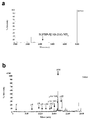
- FIGS. 1a to c show elution patterns of gel filtration column (a), cation exchange column (b), and reverse-phase nano LC (c), respectively, of fractions containing IGFBP-5 [193-214] -NH 2 .
- Bold lines (a, b) and arrows (c) indicate fractions containing IGFBP-5 [193-214] -NH 2 .
- the molecular weight markers in FIG. 1a are as follows.
- FIG. 2a shows the MS1 of the spot containing IGFBP-5 [193-214] -NH 2
- FIG. 2b shows the MS2 of IGFBP-5 [193-214] -NH 2 . It is a diagram illustrating a coordinated action in other antimicrobial peptides and IGFBP-5 [193-214] -NH 2 of antimicrobial activity.
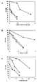
- Figure 3a shows the IGFBP-5 [193-214] bacterial viability in -NH 2 the presence of horizontal axis concentration on the vertical axis ( ⁇ ).
- the vertical axis represents the bacterial viability in the presence of ⁇ -defensin-2 (3.15 ⁇ M, ⁇ ) or cathelicidin (0.3 ⁇ M, ⁇ ) at half the concentration of IC 50 .
- FIG. 3b shows the bacterial viability in the presence of ⁇ -defensin-2 at the horizontal axis on the vertical axis ( ⁇ ).
- the vertical axis represents the bacterial survival rate in the presence of IGFBP-5 [193-214] -NH 2 (0.45 ⁇ M, ⁇ ) or cathelicidin (0.3 ⁇ M, ⁇ ) at a concentration of half the IC 50 .
- FIG. 3c shows the bacterial viability in the presence of cathelicidin at the horizontal axis on the vertical axis ( ⁇ ).
- IGFBP-5 [193-214] -NH 2 It is a diagram illustrating the identification of IGFBP-5 [193-214] -NH 2 present in rat tissues.
- 4a and 4b show the analysis results of immunoreactive IGFBP-5 [193-214] -NH 2 by gel filtration column (a) and reverse phase HPLC (b) of brain extract, respectively.
- the arrow in FIG. 4b indicates the elution position of synthetic IGFBP-5 [193-214] -NH 2 .
- FIG. 4c shows the analysis result of the immunoreactive IGFBP-5 [193-214] -NH 2 by a mass spectrometer.
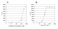
- IGFBP-5 [193-214] is a diagram showing the results of measurement of the HeLa / SRE-LUC reporter activity against MRGX2 of -NH 2.
- a peptide consisting of an amino acid sequence identical or substantially identical to the amino acid sequence represented by SEQ ID NO: 1 or a salt thereof may be abbreviated as “the peptide of the present invention”.
- a protein or a salt thereof containing the same or substantially the same amino acid sequence as the amino acid sequence represented by SEQ ID NO: 3 may be abbreviated as “the receptor of the present invention”.
- a peptide consisting of an amino acid sequence identical or substantially identical to the amino acid sequence represented by SEQ ID NO: 1 is a human or warm-blooded animal (eg, guinea pig, rat, mouse, chicken, rabbit, pig, sheep, cow or monkey).
- Etc. cells eg, retinal cells, hepatocytes, spleen cells, neurons, glial cells, pancreatic ⁇ cells, bone marrow cells, mesangial cells, Langerhans cells, epidermal cells, epithelial cells, endothelial cells, fibroblasts, fiber cells , Muscle cells, adipocytes, immune cells (eg, macrophages, T cells, B cells, natural killer cells, mast cells, neutrophils, basophils, eosinophils or monocytes), megakaryocytes, synoviocytes, Chondrocytes, bone cells, osteoblasts, osteoclasts, breast cells, hepatocytes or stromal cells, or precursor cells, stem cells or cancer cells of these cells) or Any tissue in which these cells are present, eg, brain, brain regions (eg, retina, olfactory bulb, amygdala, basal ganglia, hippocampus, thalamus, hypothalamus,
- amino acid sequence substantially identical to the amino acid sequence represented by SEQ ID NO: 1 is about 70% or more, preferably about 80% or more, more preferably about 90%, with the amino acid sequence represented by SEQ ID NO: 1. More preferred are amino acid sequences having homology of 95% or more (eg, 95% or more, 96% or more, 97% or more, 98% or more, 99% or more).
- the amino acid sequence substantially identical to the amino acid sequence represented by SEQ ID NO: 1 includes (i) 1 to 5 (preferably 1 to 3) of the amino acid sequence represented by SEQ ID NO: 1, More preferably, it is an amino acid sequence in which 1 to 2, more preferably 1 amino acid has been deleted. (Ii) 1 to 5 (preferably 1 to 3) in the amino acid sequence represented by SEQ ID NO: 1.
- Examples of the peptide consisting of the amino acid sequence substantially the same as the amino acid sequence represented by SEQ ID NO: 1, include, for example, the amino acid sequence substantially identical to the amino acid sequence represented by SEQ ID NO: 1, A peptide having substantially the same activity as the amino acid sequence represented by No. 1 is preferred.
- the substantially equivalent activity includes, for example, the activity of the peptide of the present invention (for example, the antibacterial activity described later, the binding activity with the receptor of the present invention, and the cell stimulating activity with respect to cells expressing the receptor of the present invention (for example, arachidonic acid release, acetylcholine release, intracellular Ca 2+ release, intracellular cAMP generation, intracellular cGMP generation, inositol phosphate generation, cell membrane potential fluctuation, intracellular protein phosphorylation, c-fos activation, pH Reduction, activity for promoting GTP ⁇ S binding activity, etc.)) and the like.
- the activity of the peptide of the present invention for example, the antibacterial activity described later, the binding activity with the receptor of the present invention, and the cell stimulating activity with respect to cells expressing the receptor of the present invention
- Substantially the same quality of activity means that the activities are qualitatively (eg, physiochemically or pharmacologically) equivalent (eg, about 0.01 to 100 times, preferably about 0.5 to 20 times). Times, more preferably about 0.5 to 2 times).
- the measurement of activities such as antibacterial activity, binding activity to the receptor of the present invention, and cell stimulating activity for cells expressing the receptor of the present invention can be performed according to a method known per se.
- peptide of the present invention examples include a peptide consisting of the amino acid sequence represented by SEQ ID NO: 1, a peptide consisting of the amino acid sequence represented by SEQ ID NO: 4, or an amino acid sequence represented by SEQ ID NO: 5. Peptide etc. are mentioned. Especially, the peptide which consists of an amino acid sequence represented by sequence number 1 is preferable.
- the receptor for the peptide of the present invention has binding activity with the peptide of the present invention, and cell stimulation activity (for example, arachidonic acid release, Acetylcholine release, intracellular Ca 2+ release, intracellular cAMP generation, intracellular cGMP generation, inositol phosphate production, cell membrane potential fluctuation, intracellular protein phosphorylation, c-fos activation, pH decrease, GTP ⁇ S binding activity And the like that are observed to promote activity etc.).
- cell stimulation activity for example, arachidonic acid release, Acetylcholine release, intracellular Ca 2+ release, intracellular cAMP generation, intracellular cGMP generation, inositol phosphate production, cell membrane potential fluctuation, intracellular protein phosphorylation, c-fos activation, pH decrease, GTP ⁇ S binding activity And the like that are observed to promote activity etc.
- MRGX2 protein consisting of the amino acid sequence represented by SEQ ID NO: 3 which is a G protein-coupled receptor, or an amino acid sequence substantially identical to MRGX2, that is, represented by SEQ ID NO: 3
- a protein containing an amino acid sequence substantially identical to the amino acid sequence to be prepared is, represented by SEQ ID NO: 3 and a protein containing an amino acid sequence substantially identical to the amino acid sequence to be prepared.
- a protein containing the same or substantially the same amino acid sequence as the amino acid sequence represented by SEQ ID NO: 3 is used in humans and warm-blooded animals (eg, guinea pigs, rats, mice, chickens, rabbits, pigs, sheep, cows, Monkeys (eg, retinal cells, hepatocytes, spleen cells, neurons, glial cells, pancreatic ⁇ cells, bone marrow cells, mesangial cells, Langerhans cells, epidermal cells, epithelial cells, endothelial cells, fibroblasts, fibers Cells, muscle cells, adipocytes, immune cells (eg, macrophages, T cells, B cells, natural killer cells, mast cells, neutrophils, basophils, eosinophils, monocytes), megakaryocytes, synoviocytes Chondrocytes, bone cells, osteoblasts, osteoclasts, mammary cells, hepatocytes or stromal cells, or precursor cells, stem
- the amino acid sequence substantially identical to the amino acid sequence represented by SEQ ID NO: 3 is about 70% or more, preferably about 80% or more, more preferably about 90%, with the amino acid sequence represented by SEQ ID NO: 3.
- Amino acids having homology as described above eg, 90% or more, 91% or more, 92% or more, 93% or more, 94% or more, 95% or more, 96% or more, 97% or more, 98% or more, 99% or more
- Examples include arrays.
- the amino acid sequence substantially identical to the amino acid sequence represented by SEQ ID NO: 3 includes (i) one or more (for example, about 1 to 100) in the amino acid sequence represented by SEQ ID NO: 3.
- amino acid sequence to which several (1 to 5) amino acids are preferably added (iii) one or two or more (for example, about 1 to 100, preferably 1) in the amino acid sequence represented by SEQ ID NO: 3.
- ⁇ 50 preferably An amino acid sequence in which about 1 to 30, more preferably about 1 to 10, more preferably several (1 to 5) amino acids are substituted with other amino acids, (iv) represented by SEQ ID NO: 3 1 or 2 or more (for example, about 1 to 100, preferably about 1 to 50, preferably about 1 to 30, more preferably about 1 to 10, even more preferably several (1 To 5)) amino acid sequences inserted, or (v) a protein containing an amino acid sequence combining them.
- the protein containing the amino acid sequence substantially the same as the amino acid sequence represented by SEQ ID NO: 3 has, for example, the amino acid sequence substantially the same as the amino acid sequence represented by SEQ ID NO: 3, A protein having substantially the same activity as the amino acid sequence represented by No. 3 is preferred.
- the substantially homogeneous activity includes, for example, a ligand (eg, peptide of the present invention, substance P, cortisatin, pituitary adenylate cyclase activating polypeptide, vasoactive intestinal peptide or prodrenomedullin N-terminal 20 peptide). Examples thereof include binding activity, signal signal transduction action, and cell stimulation activity. “Substantially homogeneous” means that their activities are homogeneous in nature.
- activities such as ligand binding activity, signal transduction activity or cell stimulation activity are equivalent (eg, about 0.01 to 100 times, preferably about 0.5 to 20 times, more preferably about 0.5 to 2 times).
- the quantitative factors such as the degree of activity and the molecular weight of the protein may be different.
- the measurement of activities such as ligand binding activity, signal signal transduction activity, or cell stimulation activity can be performed according to a method known per se.
- the left end is the N-terminus (amino terminus) and the right end is the C-terminus (carboxyl terminus) according to the convention of peptide designation.
- the C-terminus may be carboxy (—COOH), carboxylate (—COO ⁇ ), amide (—CONH 2 ) or ester (—COOR).
- the peptide of some embodiments of the present invention is one in which the C-terminus is amidated. In some other aspects of the invention, the peptide is one in which the C-terminus is not amidated.
- R in the ester is, for example, C 1-6 alkyl such as methyl, ethyl, n-propyl, isopropyl or n-butyl, for example, C 3-8 cycloalkyl such as cyclopentyl, cyclohexyl, for example, phenyl, ⁇ -C 6-12 aryl such as naphthyl, for example, C 7-14 aralkyl such as phenyl-C 1-2 alkyl such as benzyl, phenethyl or ⁇ -naphthyl-C 1-2 alkyl such as ⁇ -naphthylmethyl, Pivaloyloxymethyl, which is widely used as an oral ester, is used.
- C 1-6 alkyl such as methyl, ethyl, n-propyl, isopropyl or n-butyl
- C 3-8 cycloalkyl such as cyclopentyl, cyclohe
- the peptide or receptor of the present invention has a carboxy (or carboxylate) other than the C-terminus
- those in which the carboxy is amidated or esterified are also included in the peptide or receptor of the present invention.
- the ester in this case, for example, the above-mentioned C-terminal ester or the like is used.
- amino acid residues (eg, methionine residue) of the N-terminal amino group protecting group e.g., formyl, such as C 1-6 alkanoyl such as acetyl C 1-6 Protected by acyl, etc.
- an N-terminal glutamine residue generated by cleavage in vivo, pyroglutamine oxidized, a substituent on the side chain of an amino acid in the molecule eg, —OH, —SH Amino group, imidazole group, indole group, guanidino group, etc.
- an appropriate protecting group eg, C 1-6 acyl such as C 1-6 alkanoyl such as formyl, acetyl, etc.
- complex proteins such as so-called glycoproteins with chains attached.
- a salt with a physiologically acceptable acid eg, inorganic acid, organic acid
- base eg, alkali metal salt
- the acid addition salts that are acceptable are preferred.
- examples of such salts include salts with inorganic acids (eg, hydrochloric acid, phosphoric acid, hydrobromic acid, sulfuric acid, etc.) or organic acids (eg, acetic acid, formic acid, propionic acid, fumaric acid, maleic acid, And salts with succinic acid, tartaric acid, citric acid, malic acid, succinic acid, benzoic acid, methanesulfonic acid, benzenesulfonic acid, and the like.
- inorganic acids eg, hydrochloric acid, phosphoric acid, hydrobromic acid, sulfuric acid, etc.
- organic acids eg, acetic acid, formic acid, propionic acid, fumaric acid, maleic acid
- succinic acid tartaric acid, citric acid, malic acid,
- the peptide or receptor of the present invention can be produced from the aforementioned human or warm-blooded animal cells or tissues by a known polypeptide purification method, or transformed with a DNA encoding the polypeptide described below. It can also be produced by culturing a transformant. Moreover, it can also manufacture according to the below-mentioned polypeptide synthesis method. For example, it can be produced by the method described in Genomics, 56, 12-21, 1999, Biochim. Biophys. Acta, 1446, 57-70, 1999, or the like.
- the human or mammalian tissue or cells are homogenized, and then extracted with an acid or the like, and the extract is subjected to reverse phase chromatography. It can be purified and isolated by combining chromatography such as ion exchange chromatography.
- a commercially available polypeptide synthesis resin can be usually used.
- resins include chloromethyl resin, hydroxymethyl resin, benzhydrylamine resin, aminomethyl resin, 4-benzyloxybenzyl alcohol resin, 4-methylbenzhydrylamine resin, PAM resin, 4-hydroxymethylmethyl.
- an amino acid having an ⁇ -amino group and a side chain functional group appropriately protected (protected amino acid) is condensed on the resin according to various known condensation methods according to the sequence of the target polypeptide.
- the polypeptide is cut out from the resin, and at the same time, various protecting groups are removed, and an intramolecular disulfide bond-forming reaction is performed in a highly diluted solution to obtain the target peptide, receptor, or amide form thereof.
- carbodiimides are particularly preferable.
- the carbodiimide DCC, N, N′-diisopropylcarbodiimide, N-ethyl-N ′-(3-dimethylaminoprolyl) carbodiimide or the like is used.
- a protected amino acid is added directly to the resin together with a racemization inhibitor (eg, HOBt or HOBt), or the protected amino acid is activated in advance as a symmetric acid anhydride, HOBt ester or HOBt ester. It can later be added to the resin.
- a racemization inhibitor eg, HOBt or HOBt
- the solvent used for the activation of the protected amino acid and the condensation with the resin can be appropriately selected from solvents known to be usable for the polypeptide condensation reaction.
- acid amides such as N, N-dimethylformamide, N, N-dimethylacetamide, N-methylpyrrolidone, halogenated hydrocarbons such as methylene chloride and chloroform, alcohols such as trifluoroethanol, dimethyl sulfoxide, etc.
- Examples include sulfoxides, ethers such as pyridine, dioxane, and tetrahydrofuran, nitriles such as acetonitrile and propionitrile, esters such as methyl acetate and ethyl acetate, and appropriate mixtures thereof.
- the reaction temperature is appropriately selected from a range known to be usable for polypeptide bond formation reaction, and is usually selected from the range of about -20 ° C to 50 ° C.
- the activated amino acid derivative is usually used in an excess of 1.5 to 4 times.
- Examples of the protective group for the amino group of the raw material include Z, Boc, t-pentyloxycarbonyl, isobornyloxycarbonyl, 4-methoxybenzyloxycarbonyl, Cl-Z, Br-Z, adamantyloxycarbonyl, trifluoroacetyl. , Phthaloyl, formyl, 2-nitrophenylsulfenyl, diphenylphosphinothioyl or Fmoc.
- Carboxyl groups are, for example, alkyl esterified (eg, linear, branched or cyclic alkyl esters such as methyl, ethyl, propyl, butyl, t-butyl, cyclopentyl, cyclohexyl, cycloheptyl, cyclooctyl or 2-adamantyl).
- alkyl esterified eg, linear, branched or cyclic alkyl esters such as methyl, ethyl, propyl, butyl, t-butyl, cyclopentyl, cyclohexyl, cycloheptyl, cyclooctyl or 2-adamantyl.
- Aralkyl esterification eg, benzyl ester, 4-nitrobenzyl ester, 4-methoxybenzyl ester, 4-chlorobenzyl ester or benzhydryl esterification
- phenacyl esterification eg, benzyloxycarbonylhydrazide, t-butoxy It can be protected by carbonyl hydrazation or trityl hydrazation.
- the hydroxyl group of serine can be protected, for example, by esterification or etherification.
- groups suitable for esterification include groups derived from carbonic acid such as lower (C 1-6 ) alkanoyl groups such as acetyl groups, aroyl groups such as benzoyl groups, benzyloxycarbonyl groups, and ethoxycarbonyl groups. Used.
- groups suitable for etherification include benzyl group, tetrahydropyranyl group, t-butyl group and the like.
- Examples of the protecting group for the phenolic hydroxyl group of tyrosine include Bzl, Cl 2 -Bzl, 2-nitrobenzyl, Br-Z, t-butyl and the like.
- Examples of the protecting group for imidazole of histidine include Tos, 4-methoxy-2,3,6-trimethylbenzenesulfonyl, DNP, benzyloxymethyl, Bum, Boc, Trt, Fmoc and the like.
- Examples of the activated carboxyl group of the raw material include the corresponding acid anhydride, azide, active ester [alcohol (eg, pentachlorophenol, 2,4,5-trichlorophenol, 2,4-dinitrophenol, And esters thereof with cyanomethyl alcohol, paranitrophenol, HONB, N-hydroxysuccinimide, N-hydroxyphthalimide, and HOBt).
- active ester alcohol (eg, pentachlorophenol, 2,4,5-trichlorophenol, 2,4-dinitrophenol, And esters thereof with cyanomethyl alcohol, paranitrophenol, HONB, N-hydroxysuccinimide, N-hydroxyphthalimide, and HOBt).
- alcohol eg, pentachlorophenol, 2,4,5-trichlorophenol, 2,4-dinitrophenol, And esters thereof with cyanomethyl alcohol, paranitrophenol, HONB, N-hydroxysuccinimide, N-hydroxyphthalimide, and HOBt.
- Examples of the method for removing (eliminating) the protecting group include catalytic reduction in a hydrogen stream in the presence of a catalyst such as Pd-black or Pd-carbon, anhydrous hydrogen fluoride, methanesulfonic acid, trifluoro.
- a catalyst such as Pd-black or Pd-carbon, anhydrous hydrogen fluoride, methanesulfonic acid, trifluoro.
- Acid treatment with romethanesulfonic acid, trifluoroacetic acid or a mixture thereof, base treatment with diisopropylethylamine, triethylamine, piperidine, piperazine, etc., reduction with sodium in liquid ammonia, and the like are also used.
- the elimination reaction by the acid treatment is generally performed at a temperature of about ⁇ 20 ° C. to 40 ° C.
- Protection of a functional group that should not be involved in the reaction of the raw material, protection group, elimination of the protective group, activation of the functional group involved in the reaction, etc. can be appropriately selected from known groups or known means.
- the ⁇ -carboxyl group of the carboxy terminal amino acid is amidated and protected, and then the peptide (polypeptide) chain is formed on the amino group side with a desired chain length. And then producing a polypeptide from which only the N-terminal ⁇ -amino protecting group of the peptide chain has been removed and a polypeptide from which only the C-terminal carboxyl protecting group has been removed. Condensation in a mixed solvent as described above. The details of the condensation reaction are the same as described above. After purifying the protected polypeptide obtained by condensation, all the protecting groups are removed by the above-described method to obtain the desired crude polypeptide. This crude polypeptide can be purified by using various known purification means, and the main fraction can be lyophilized to obtain the desired peptide or amide of the receptor.
- ester of the peptide or receptor of the present invention for example, the ⁇ -carboxyl group of the carboxy terminal amino acid is condensed with a desired alcohol to form an amino acid ester, and then the same as the peptide or receptor amide.
- an ester form of a desired peptide or receptor can be obtained.
- the peptide or receptor of the present invention can be produced according to a peptide synthesis method known per se, for example, by cleaving a polypeptide with an appropriate peptidase.
- a polypeptide synthesis method for example, either a solid phase synthesis method or a liquid phase synthesis method may be used. That is, the desired polypeptide is produced by condensing a partial peptide or amino acid capable of constituting the peptide or receptor of the present invention and the remaining portion, and removing the protective group when the product has a protective group. Can do. Examples of known condensation methods and protecting group elimination include the methods described in the following (i) to (v).
- the peptide or receptor of the present invention can be purified and isolated by combining ordinary purification methods such as solvent extraction, distillation, column chromatography, liquid chromatography, recrystallization and the like.
- the peptide or receptor obtained by the above method is a free form, it can be converted to an appropriate salt by a known method or a method analogous thereto, and conversely, when obtained by a salt, the known method or It can be converted into a free form or other salt by a method according to it.
- the polynucleotide encoding the peptide or receptor of the present invention may be any as long as it contains a base sequence encoding the peptide or receptor of the present invention.
- DNA is preferable, and the DNA may be any of genomic DNA, genomic DNA library, cDNA derived from the cells / tissues described above, cDNA library derived from the cells / tissues described above, and synthetic DNA.
- the vector used for the library may be any of bacteriophage, plasmid, cosmid, phagemid and the like. In addition, it can be amplified directly by Reverse-Transcriptase-Polymerase-Chain Reaction (hereinafter abbreviated as RT-PCR method) using a total RNA or mRNA fraction prepared from the cells / tissues described above.
- RT-PCR method Reverse-Transcriptase-Polymerase-Chain Reaction
- Examples of the DNA encoding the peptide of the present invention include a DNA consisting of the base sequence represented by SEQ ID NO: 6, or a DNA consisting of a base sequence complementary to the DNA consisting of the base sequence represented by SEQ ID NO: 6. Any DNA that has a base sequence that hybridizes under highly stringent conditions and that encodes a peptide having substantially the same activity as the protein containing the amino acid sequence represented by SEQ ID NO: 1 It may be a thing.
- DNA that can hybridize under high stringency conditions with DNA consisting of a base sequence complementary to the DNA consisting of the base sequence represented by SEQ ID NO: 6 include the base sequence represented by SEQ ID NO: 6 About 70% or more, preferably about 80% or more, more preferably about 90% or more, more preferably about 95% or more (eg, 95% or more, 96% or more, 97% or more, 98% or more, 99% or more) DNA containing a base sequence having the homology of
- Hybridization can be performed according to a method known per se or a method analogous thereto, for example, the method described in Molecular Cloning 2nd (J. Sambrook et al., Cold Spring Harbor Lab Press, 1989). Moreover, when using a commercially available library, it can carry out according to the method as described in an attached instruction manual. More preferably, it can be carried out according to highly stringent conditions.
- the highly stringent conditions are, for example, conditions in which the sodium concentration is about 19 to 40 mM, preferably about 19 to 20 mM, and the temperature is about 50 to 70 ° C., preferably about 60 to 65 ° C. In particular, the case where the sodium concentration is about 19 mM and the temperature is about 65 ° C. is most preferable.
- DNA encoding the peptide consisting of the amino acid sequence represented by SEQ ID NO: 1 DNA containing the base sequence represented by SEQ ID NO: 6 is used.
- the DNA encoding the receptor of the present invention comprises, for example, a DNA containing the base sequence represented by SEQ ID NO: 2 or a base sequence complementary to the DNA comprising the base sequence represented by SEQ ID NO: 2.
- Examples of DNA that can hybridize under high stringency conditions with a DNA consisting of a base sequence complementary to the DNA consisting of the base sequence represented by SEQ ID NO: 2 include the base sequence represented by SEQ ID NO: 2.
- Hybridization can be performed according to a method known per se or a method analogous thereto, for example, the method described in Molecular Cloning 2nd (J. Sambrook et al., Cold Spring Harbor Lab Press, 1989). Moreover, when using a commercially available library, it can carry out according to the method as described in an attached instruction manual. More preferably, it can be carried out according to highly stringent conditions.
- the highly stringent conditions are, for example, conditions in which the sodium concentration is about 19 to 40 mM, preferably about 19 to 20 mM, and the temperature is about 50 to 70 ° C., preferably about 60 to 65 ° C. In particular, the case where the sodium concentration is about 19 mM and the temperature is about 65 ° C. is most preferable.
- DNA encoding the receptor containing the amino acid sequence represented by SEQ ID NO: 3 DNA containing the base sequence represented by SEQ ID NO: 2 is used.
- the polynucleotide (eg, DNA) encoding the peptide or receptor of the present invention may be labeled by a method known per se.
- the labeling substance include radioisotopes, fluorescent substances (eg, fluorescein), luminescent substances, enzymes, biotin, lanthanide elements, and the like.
- amplification by a known PCR method using a synthetic DNA primer having a partial base sequence of the peptide or receptor of the present invention, or DNA incorporated in an appropriate vector can be selected by hybridization with a peptide fragment of the present invention or a DNA fragment encoding a part or the entire region of the receptor, or labeled with a synthetic DNA.
- the hybridization method can be carried out, for example, according to the method described in Molecular Cloning 2nd (J. Sambrook et al., Cold Spring Harbor Lab Press, 1989).
- it can carry out according to the method as described in an attached instruction manual.
- the DNA base sequence can be converted using a known kit such as Mutan TM -super Express Km (Takara Shuzo Co., Ltd.), Mutan TM -K (Takara Shuzo Co., Ltd.) or the like using the ODA-LA PCR method, Gapped duplex
- the method can be carried out according to a method known per se, such as the Kunkel method or the like, or a method analogous thereto.
- the DNA encoding the cloned peptide can be used as it is depending on the purpose, or can be digested with a restriction enzyme or added with a linker if desired.
- the DNA may have ATG as a translation initiation codon on the 5 'end side, and may have TAA, TGA or TAG as a translation termination codon on the 3' end side. These translation initiation codon and translation termination codon can be added using an appropriate synthetic DNA adapter.
- the expression vector of the peptide or receptor of the present invention is, for example, (a) excising the target DNA fragment from the DNA encoding the peptide or receptor of the present invention, and (b) extracting the DNA fragment in an appropriate expression vector. It can be produced by linking downstream of the promoter.
- Examples of the vector include plasmids derived from E. coli (eg, pBR322, pBR325, pUC12, pUC13), plasmids derived from Bacillus subtilis (eg, pUB110, pTP5, pC194), plasmids derived from yeast (eg, pSH19, pSH15), ⁇ phage, and the like.
- E. coli eg, pBR322, pBR325, pUC12, pUC13
- Bacillus subtilis eg, pUB110, pTP5, pC19
- yeast eg, pSH19, pSH15
- ⁇ phage e.g, ⁇ phage, and the like.
- animal viruses such as bacteriophage, retrovirus, vaccinia virus and baculovirus, pA1-11, pXT1, pRc / CMV, pRc / RSV,
- the promoter may be any promoter as long as it is appropriate for the host used for gene expression.
- SR ⁇ promoter when animal cells are used as a host, SR ⁇ promoter, SV40 promoter, HIV / LTR promoter, CMV promoter, HSV-TK promoter and the like can be mentioned.
- CMV cytomegalovirus
- SR ⁇ promoter a CMV (cytomegalovirus) promoter
- the host is Escherichia, trp promoter, lac promoter, recA promoter, ⁇ PL promoter, lpp promoter, T7 promoter, etc., and when the host is Bacillus, SPO1, SPO2 promoter, penP promoter, etc.
- the host is yeast, PHO5 promoter, PGK promoter, GAP promoter, ADH promoter, etc. are preferable.
- polyhedrin promoter, P10 promoter and the like are preferable.
- an expression vector containing an enhancer, a splicing signal, a poly A addition signal, a selection marker, an SV40 replication origin (hereinafter sometimes abbreviated as SV40ori) and the like is used as desired.
- selectable markers include dihydrofolate reductase (hereinafter sometimes abbreviated as dhfr) gene [methotrexate (MTX) resistance], ampicillin resistance gene (hereinafter sometimes abbreviated as Amp r ), neomycin resistance gene (hereinafter sometimes abbreviated as Neo r, G418 resistance) and the like.
- dhfr gene-deficient Chinese hamster cell is used and the dhfr gene is used as a selection marker
- the target gene can also be selected by a medium not containing thymidine.
- a signal sequence suitable for the host is added to the N terminal side of the peptide or receptor of the present invention.
- the host is Escherichia
- Bacillus ⁇ -amylase signal sequence, subtilisin signal sequence, etc.
- MF ⁇ signal sequence, SUC2 signal sequence, etc. when the host is an animal cell, insulin signal sequence, ⁇ -interferon signal sequence, antibody molecule signal sequence, etc. can be used.
- a transformant can be produced using a vector containing a DNA encoding the peptide or receptor of the present invention thus constructed.
- Escherichia for example, Escherichia, Bacillus, yeast, insect cells, insects, animal cells and the like are used.
- Escherichia examples include, for example, Escherichia coli K12 ⁇ DH1 [Proc. Natl. Acad. Sci. USA, 60, 160 (1968)], JM103 [Nucleic Acids Research, 9, 309 (1981)], JA221 [Journal of Molecular Biology, 120, 517 (1978)], HB101 [Journal of Molecular Biology, 41,459 (1969)], C600 [Genetics, 39,440 (1954)] Etc. are used.
- Bacillus subtilis MI114 Gene, 24, 255 (1983)]
- 207-21 Journal of Biochemistry, 95, 87 (1984)].
- yeast examples include Saccharomyces cerevisiae AH22, AH22R ⁇ , NA87-11A, DKD-5D, 20B-12, Schizosaccharomyces pombe NCYC1913, NCYC2036, Used.
- Insect cells include, for example, when the virus is AcNPV, Spodoptera frugiperda cell (Sf cell), Trichoplusia ni midgut-derived MG1 cells, Trichoplusia ni egg-derived High Five TM cells A cell derived from Mamestra brassicae or a cell derived from Estigmena acrea is used.
- Sf cells include Sf9 cells (ATCC CRL 1711), Sf21 cells (Vaughn, JL et al., In Vivo, 13, 213-217, (1977)) and the like.
- insects for example, silkworm larvae are used [Maeda et al., Nature, 315, 592 (1985)].
- animal cells examples include monkey cells COS-7 (COS7), Vero, Chinese hamster cells CHO (hereinafter abbreviated as CHO cells), dhfr gene-deficient Chinese hamster cells CHO (hereinafter abbreviated as CHO (dhfr ⁇ ) cells).
- COS-7 monkey cells
- Vero Chinese hamster cells CHO
- CHO cells Chinese hamster cells CHO
- dhfr gene-deficient Chinese hamster cells CHO hereinafter abbreviated as CHO (dhfr ⁇ ) cells.
- Mouse L cells mouse AtT-20, mouse myeloma cells, rat GH3, human FL cells, and the like.
- Transformation of the genus Escherichia can be performed, for example, according to the method described in Proc. Natl. Acad. Sci. USA, 69, 2110 (1972), Gene, 17, 107 (1982).
- Transformation of Bacillus can be carried out, for example, according to the method described in Molecular & General Genetics, 168, 111 (1979).
- Insect cells or insects can be transformed, for example, according to the method described in Bio / Technology, 6, 47-55 (1988).
- a transformant transformed with an expression vector containing DNA encoding the peptide or receptor of the present invention can be obtained.
- a liquid medium is suitable as a medium used for the culture, including a carbon source necessary for the growth of the transformant, Nitrogen sources, inorganic substances, etc. are contained.
- the carbon source include glucose, dextrin, soluble starch, and sucrose.
- the nitrogen source include ammonium salts, nitrates, corn steep liquor, peptone, casein, meat extract, soybean cake, and potato extract.
- the inorganic or organic substance and inorganic substance include calcium chloride, sodium dihydrogen phosphate, and magnesium chloride.
- yeast extract, vitamins, growth promoting factors and the like may be added.
- the pH of the medium is preferably about 5-8.
- an M9 medium containing glucose and casamino acids is preferable.
- a drug such as 3 ⁇ -indolylacrylic acid can be added to make the promoter work efficiently if necessary.
- the culture is usually carried out at about 15 to 43 ° C. for about 3 to 24 hours, and if necessary, aeration or agitation can be added.
- the culture is usually performed at about 30 to 40 ° C. for about 6 to 24 hours, and if necessary, aeration or agitation can be added.
- examples of the medium include a Burkholder minimum medium [Bostian, K. L., Proc. Natl. Acad. Sci. USA, vol. 77, 4505 ( 1980)] and SD medium containing 0.5% casamino acid [Bitter, G. A., Proc. Natl. Acad. Sci. USA, Vol. 81, 5330 (1984)].
- the pH of the medium is preferably adjusted to about 5-8. Cultivation is usually carried out at about 20 ° C. to 35 ° C. for about 24 to 72 hours, with aeration and agitation as necessary.
- an appropriate medium such as 10% bovine serum immobilized in Grace'sceInsect Medium (Grace, TCC, Nature, 195,788 (1962)) is appropriately used. Additions etc. are used.
- the pH of the medium is preferably adjusted to about 6.2 to 6.4.
- the culture is usually carried out at about 27 ° C. for about 3 to 5 days, and aeration and agitation are added as necessary.
- examples of the medium include MEM medium (Science, Vol. 122, 501 (1952)) containing about 5 to 20% fetal bovine serum, DMEM medium [Virology, 8, 396 (1959)], RPMI-1640 medium [The Journal of America, Medical American Association, 199, 519 (1967)], 199 medium (Proceeding, Society, for Society, Biological, Medicine, 73, 1 (1950)), etc. Is used.
- the pH is preferably about 6-8. Cultivation is usually carried out at about 30 ° C. to 40 ° C. for about 15 to 60 hours, with aeration and agitation as necessary.
- the peptide or receptor of the present invention can be produced in the cell, cell membrane, or extracellular area of the transformant.
- Separation and purification of the peptide or receptor of the present invention from the culture can be performed, for example, by the following method.
- the cells or cells are collected by a known method after culturing, suspended in an appropriate buffer, and subjected to ultrasound, lysozyme and / or A method of obtaining a crude polypeptide extract by centrifugation or filtration after disrupting cells or cells by freezing and thawing is appropriately used.
- the buffer may contain a protein denaturant such as urea or guanidine hydrochloride, or a surfactant such as Triton X-100 TM .
- Purification of the polypeptide contained in the culture supernatant or the extract thus obtained can be performed by appropriately combining known separation / purification methods.
- These known separation and purification methods include mainly molecular weights such as methods utilizing solubility such as salting out and solvent precipitation, dialysis, ultrafiltration, gel filtration, and SDS-polyacrylamide gel electrophoresis.
- Method using difference in charge method using difference in charge such as ion exchange chromatography, method using specific affinity such as affinity chromatography, and difference in hydrophobicity such as reverse phase high performance liquid chromatography
- a method using a difference in isoelectric point, such as a method, isoelectric focusing method, or the like is used.
- the polypeptide thus obtained when obtained in a free form, it can be converted into a salt by a method known per se or a method analogous thereto, and conversely when it is obtained as a salt, a method known per se or a method analogous thereto. Depending on the method, it can be converted to the free form or other salts.
- polypeptide produced by the recombinant can be arbitrarily modified or partially removed by allowing an appropriate protein modifying enzyme to act before or after purification.
- protein modifying enzymes include trypsin, chymotrypsin, arginyl endopeptidase, protein kinase, glycosidase, and the like.
- an antibody against a peptide consisting of an amino acid sequence identical or substantially identical to the amino acid sequence represented by SEQ ID NO: 1 or a salt thereof recognizes the peptide of the present invention.
- Any antibody that can be used may be a polyclonal antibody or a monoclonal antibody.
- the antibody of the present invention include an antibody that specifically recognizes the peptide of the present invention, and / or a neutralizing antibody that inactivates the activity of the peptide of the present invention.
- the activity of the peptide of the present invention include an activity such as receptor binding activity, signal information transmission activity or cell stimulation activity.
- the antibody of the present invention can be produced according to a known antibody or antiserum production method using the peptide of the present invention as an antigen.
- the peptide of the present invention is administered to a warm-blooded animal by itself, together with a carrier and a diluent, at a site where antibody production is possible by administration.
- Complete Freund's adjuvant or incomplete Freund's adjuvant may be administered in order to enhance antibody production ability upon administration. Administration is usually once every 2 to 6 weeks, for a total of about 2 to 10 times.
- Examples of the warm-blooded animal used include monkeys, rabbits, dogs, guinea pigs, mice, rats, sheep, goats and chickens, and mice and rats are preferably used.
- Monoclonal antibody-producing hybridomas can be prepared by fusing the antibody-producing cells to be fused with allogeneic or xenogeneic myeloma cells.
- the antibody titer in the antiserum can be measured, for example, by reacting the labeled polypeptide described below with the antiserum and then measuring the activity of the labeling agent bound to the antibody.
- the fusion operation can be carried out according to a known method, for example, the method of Kohler and Milstein [Nature, 256, 495 (1975)].
- the fusion promoter include polyethylene glycol (PEG) and Sendai virus.
- PEG polyethylene glycol
- Sendai virus Preferably, PEG is used.
- myeloma cells include warm-blooded animal myeloma cells such as NS-1, P3U1, SP2 / 0, AP-1, and P3U1 is preferably used.
- the preferred ratio between the number of antibody-producing cells (spleen cells) and the number of myeloma cells used is about 1: 1 to 20: 1, and PEG (preferably PEG1000 to PEG6000) is added at a concentration of about 10 to 80%.
- the cell fusion can be efficiently carried out by incubating at 20 to 40 ° C., preferably 30 to 37 ° C. for 1 to 10 minutes.
- hybridoma culture supernatant is added to a solid phase (eg, a microplate) on which a polypeptide (protein) antigen is adsorbed directly or together with a carrier.
- a solid phase eg, a microplate
- a polypeptide (protein) antigen is adsorbed directly or together with a carrier.
- Monoclonal antibody selection can be performed according to a known method or a similar method. Usually, it can be performed in a medium for animal cells supplemented with HAT (hypoxanthine, aminopterin, thymidine).
- HAT hyperxanthine, aminopterin, thymidine
- any medium may be used as long as the hybridoma can grow.
- RPMI 1640 medium containing 1 to 20%, preferably 10 to 20% fetal bovine serum, GIT medium (Wako Pure Chemical Industries, Ltd.) containing 1 to 10% fetal bovine serum, or serum-free for hybridoma culture A medium (SFM-101, Nissui Pharmaceutical Co., Ltd.) or the like can be used.
- the culture temperature is usually 20 to 40 ° C., preferably about 37 ° C.
- the culture time is usually 5 days to 3 weeks, preferably 1 to 2 weeks. Culturing can usually be performed under 5% carbon dioxide gas.
- the antibody titer of the hybridoma culture supernatant can be measured in the same manner as the antibody titer in the above antiserum.
- Polyclonal antibodies can be produced according to known methods or similar methods. For example, an immune antigen (polypeptide antigen) itself or a complex of it and a carrier protein is prepared, and a warm-blooded animal is immunized in the same manner as the above-described monoclonal antibody production method, and the antibody against the peptide of the present invention is immunized from the immunized animal. It can be produced by collecting the contents and carrying out separation and purification of the antibody.
- an immune antigen polypeptide antigen itself or a complex of it and a carrier protein
- the type of carrier protein and the mixing ratio of carrier and hapten are such that antibodies are more effective against hapten immunized by cross-linking to carrier.
- any substance may be cross-linked at any ratio.
- bovine serum albumin, bovine thyroglobulin, hemocyanin and the like are about 0.1 to 20, preferably about 1 by weight ratio to hapten 1.
- a method of coupling at a rate of ⁇ 5 is used.
- various condensing agents can be used for coupling of the hapten and the carrier, but an active ester reagent containing glutaraldehyde, carbodiimide, maleimide active ester, thiol group, dithiobilidyl group, or the like is used.
- Condensation products are administered to warm-blooded animals either at the site where antibody production is possible, or together with carriers and diluents.
- Complete Freund's adjuvant or incomplete Freund's adjuvant may be administered in order to enhance antibody production ability upon administration. Administration is usually about once every 2 to 6 weeks, about 3 to 10 times in total.
- Polyclonal antibodies can be collected from blood, ascites, etc., preferably from blood of warm-blooded animals immunized by the above method.
- the measurement of the polyclonal antibody titer in the antiserum can be performed in the same manner as the measurement of the antibody titer in the antiserum. Separation and purification of the polyclonal antibody can be performed according to the same immunoglobulin separation and purification method as the above-described monoclonal antibody separation and purification.
- a nucleotide sequence complementary to or substantially complementary to a polynucleotide eg, DNA
- a polynucleotide eg, DNA
- a polynucleotide comprising a part thereof is composed of a base sequence complementary to or substantially complementary to the polynucleotide, and has an action capable of suppressing the expression of the polynucleotide.
- Any polynucleotide may be used (an antisense polynucleotide (hereinafter sometimes abbreviated as the antisense polynucleotide of the present invention)).
- a base complementary to or substantially complementary to a polynucleotide eg, DNA
- these DNAs may be abbreviated as the DNA of the present invention.
- Antisense DNA consisting of a sequence or a part thereof (hereinafter, these DNAs may be abbreviated as antisense DNA), and a base sequence complementary to or substantially complementary to the DNA of the present invention Any antisense DNA may be used as long as it is a part thereof and has an action capable of suppressing the expression of the DNA.
- the base sequence substantially complementary to the DNA of the present invention is, for example, about 70 of the total base sequence or partial base sequence of the base sequence complementary to the DNA of the present invention (that is, the complementary strand of the DNA of the present invention). % Or more, preferably about 80% or more, more preferably about 90% or more, and most preferably about 95% or more of the base sequence having homology.
- Antisense DNA having a homology of preferably about 80% or more, more preferably about 90% or more, and most preferably about 95% or more is suitable. These antisense DNAs can be produced using a known DNA synthesizer.
- antisense polynucleotides having a base sequence complementary to or substantially complementary to DNA consisting of the base sequence represented by SEQ ID NO: 6, or a part thereof include an antisense polynucleotide having a base sequence complementary to the base sequence of DNA consisting of the base sequence represented by SEQ ID NO: 6, or a part thereof.
- the antisense polynucleotide is usually composed of about 10 to 40 bases, preferably about 15 to 30 bases.
- the phosphate residues (phosphates) of each nucleotide constituting the antisense DNA are changed to chemically modified phosphate residues such as phosphorothioate, methylphosphonate and phosphorodithionate. May be substituted.
- These antisense polynucleotides can be produced using a known DNA synthesizer.
- an antisense polynucleotide capable of inhibiting the replication or expression of the gene of the peptide of the present invention (antisense polynucleotide of the present invention) (nucleic acid) was cloned or the amino acid was determined. It can be designed and synthesized based on the base sequence information of the DNA encoding the protein. Such a polynucleotide (nucleic acid) can hybridize with the RNA of the gene of the peptide of the present invention, inhibit the synthesis or function of the RNA, or interact with the peptide-related RNA of the present invention. Thus, the gene expression of the peptide of the present invention can be regulated and controlled.
- a polynucleotide that is complementary to a selected sequence of the peptide-related RNA of the present invention and a polynucleotide that can specifically hybridize with the peptide-related RNA of the present invention is an in vivo and in vitro of the peptide of the present invention. It is useful for regulating and controlling gene expression, and is useful for treatment or diagnosis of diseases and the like.
- the term “corresponding” means homologous to or complementary to a specific sequence of nucleotides, base sequences or nucleic acids including genes.
- “Corresponding” between a nucleotide, base sequence or nucleic acid and peptide (protein) usually refers to the amino acid of the peptide (protein) in the instruction derived from the nucleotide (nucleic acid) sequence or its complement. . 5 'end hairpin loop of protein gene, 5' end 6-base pair repeat, 5 'end untranslated region, protein translation stop codon, protein coding region, ORF translation stop codon, 3' end untranslated region, 3 'end
- the palindromic region and the 3 ′ end hairpin loop can be selected as preferred target regions, but any region within the protein gene can be selected as the target.
- the relationship between the target nucleic acid and a polynucleotide complementary to at least a part of the target region is a relationship with a polynucleotide that can hybridize with the target, and can be said to be “antisense”.
- Antisense polynucleotides are polynucleotides containing 2-deoxy-D-ribose, polynucleotides containing D-ribose, other types of polynucleotides that are N-glycosides of purine or pyrimidine bases, or Other polymers with non-nucleotide backbones (eg, commercially available protein nucleic acids and synthetic sequence specific nucleic acid polymers) or other polymers containing special linkages, provided that the polymer is a base as found in DNA or RNA And a nucleotide having a configuration that allows attachment of a base).
- RNA hybrids and unmodified polynucleotides (or unmodified oligonucleotides), and more Modified, such as those with labels known in the art, capped, methylated, one or more natural nucleotides replaced with analogs, intramolecular nucleotides Modified, such as those having uncharged bonds (eg, methylphosphonates, phosphotriesters, phosphoramidates, carbamates, etc.), charged or sulfur-containing bonds (eg, phosphorothioates, phosphorodithioates, etc.) ),
- proteins nucleases, nuclease inhibitors, and toxins
- Antibodies signal peptides, poly-L-lysine, etc.
- sugars eg, monosaccharides
- nucleoside may include not only purine and pyrimidine bases but also those having other modified heterocyclic bases. Such modifications may include methylated purines and pyrimidines, acylated purines and pyrimidines, or other heterocycles. Modified nucleotides and modified nucleotides may also be modified at the sugar moiety, for example, one or more hydroxyl groups are replaced by halogens, aliphatic groups, etc., or functional groups such as ethers, amines, etc. It may have been converted.
- the antisense polynucleotide (nucleic acid) of the present invention is RNA, DNA, or modified nucleic acid (RNA, DNA).
- modified nucleic acid include, but are not limited to, nucleic acid sulfur derivatives and thiophosphate derivatives, and those resistant to degradation of polynucleoside amides and oligonucleoside amides.
- the antisense polynucleotide of the present invention can be preferably designed according to the following policy.
- the antisense polynucleotide more stable in the cell, increase the cell permeability of the antisense polynucleotide, increase the affinity for the target sense strand, and if it is toxic This reduces the toxicity of the antisense polynucleotide.
- the antisense polynucleotide of the present invention may be altered or contain modified sugars, bases and bonds, and may be provided in special forms such as liposomes, microspheres, or applied by gene therapy. Can be provided in an added form.
- the additional form includes polycationic substances such as polylysine that acts to neutralize the charge of the phosphate group skeleton, lipids that enhance interaction with cell membranes and increase nucleic acid uptake ( For example, hydrophobic substances such as phospholipids and cholesterols).
- Preferred lipids to be added include cholesterol and derivatives thereof (for example, cholesteryl chloroformate, cholic acid, etc.).
- nucleic acid can be attached via a base, sugar, or intramolecular nucleoside bond.
- the other group include a cap group specifically arranged at the 3 ′ end or 5 ′ end of a nucleic acid, which prevents degradation by a nuclease such as exonuclease or RNase.
- capping groups include, but are not limited to, hydroxyl protecting groups known in the art, including glycols such as polyethylene glycol and tetraethylene glycol.
- the inhibitory activity of the antisense polynucleotide can be examined using the transformant of the present invention, the in vivo or in vitro gene expression system of the present invention, or the in vivo or in vitro translation system of the peptide of the present invention.
- the nucleic acid can be applied to cells by various known methods.
- a peptide of the present invention (ii) a polynucleotide encoding the peptide of the present invention (polynucleotide of the present invention), (iii) an antibody against the peptide of the present invention (antibody of the present invention), (iv) The use of the antisense polynucleotide (eg, antisense DNA) of the present invention will be described.
- a compound that inhibits cell activation promoting action or the like or a salt thereof is, for example, a mast cell degranulation inhibitor, an eicosanoid production inhibitor, a cytokine production inhibitor, a mast cell proliferation inhibitor, a mast cell activation inhibitor (eg, Including MAPK activation inhibitors, etc., such as immune diseases [eg, inflammatory diseases (pituitary abscess, thyroiditis, peritonitis, Crohn's disease, ulcerative colitis, erythema nodosum, chronic joints, etc.) Rheumatism, systemic lupus erythematosus, etc.), allergies (eg, allergic conjunctivitis, allergic rhinitis, hay
- mast cell degranulation inhibitor preferably used are preventive / therapeutic agents for immune diseases, urological diseases, digestive organ diseases, cardiovascular diseases and the like.
- a preventive / therapeutic agent for immune diseases is preferable.
- cytokine production inhibitor As the cytokine production inhibitor, a preventive / therapeutic agent for immune diseases is preferable.
- a preventive / therapeutic agent for urological diseases is preferable.
- a prophylactic / therapeutic agent for gastrointestinal diseases is preferable.
- the receptor of the present invention By using the peptide of the present invention and the receptor of the present invention, or by using the ligand receptor assay system using the expression system of the peptide of the present invention and the recombinant receptor of the present invention, the receptor of the present invention, A compound (for example, peptide, protein, antibody, non-peptidic compound, synthetic compound, fermentation product, cell extract, plant extract, animal tissue extract or plasma) that changes the binding to the peptide of the present invention, or The salt can be screened efficiently.
- a compound for example, peptide, protein, antibody, non-peptidic compound, synthetic compound, fermentation product, cell extract, plant extract, animal tissue extract or plasma
- the compound or a salt thereof includes (i) cell stimulating activity via the receptor of the present invention (for example, arachidonic acid release, acetylcholine release, intracellular Ca 2+ release, intracellular cAMP production, intracellular cAMP production inhibition, Intracellular cGMP production, inositol phosphate production, cell membrane potential fluctuation, intracellular protein phosphorylation, c-fos activation, pH reduction, GTP ⁇ S binding activity, cAMP-dependent protein kinase activation, cGMP-dependent protein kinase Activation of phospholipid-dependent protein kinase, mitogen-activated protein kinase (MAP kinase) (eg, ERK1 / 2 (p42 / 44 MAP kinase), p38 MAPK, JNK / SAPK, ERK5, etc.) A compound having an activity that promotes activation, etc.
- MAP kinase mitogen-activated protein kinas
- the comparison is performed, for example, by measuring the binding amount of the peptide of the present invention to the receptor of the present invention, cell stimulating activity, and the like.
- a method for screening a compound or a salt thereof that changes the binding between the peptide of the present invention and the receptor of the present invention which comprises measuring and comparing the amount of binding to
- the screening method according to (b) above which is a receptor of the present invention
- (D) a receptor binding assay system such as the screening method of (a) to (c) above, wherein the peptide of the present invention is a labeled peptide
- a method for screening a compound or a salt thereof that alters the binding between the peptide of the present invention and the receptor of the present invention which comprises measuring and comparing the activity
- the binding activity of the peptide of the present invention and the receptor of the present invention is characterized
- the receptor of the present invention a membrane fraction of human or warm-blooded animal organs is preferably used.
- human-derived organs are particularly difficult to obtain, the receptor of the present invention expressed in a large amount using a recombinant is suitable for use in screening.
- the preparation method described below when a cell containing the receptor of the present invention or the cell membrane fraction is used, the preparation method described below may be followed.
- the cells containing the receptor of the present invention When cells containing the receptor of the present invention are used, the cells may be fixed with glutaraldehyde, formalin or the like.
- the immobilization method can be performed according to a method known per se.
- the cell containing the receptor of the present invention refers to a host cell that expresses the receptor of the present invention, and examples of the host cell include the aforementioned Escherichia coli, Bacillus subtilis, yeast, insect cells, animal cells and the like. .
- the manufacturing method is the same as described above.
- the membrane fraction refers to a fraction containing a large amount of cell membrane obtained by a method known per se after cell disruption.
- Cell disruption methods include crushing cells with a Potter-Elvehjem homogenizer, disrupting with a Waring blender or polytron (manufactured by Kinematica), disrupting with ultrasonic waves, and pressurizing with a French press while ejecting cells from a thin nozzle. Crushing.
- a fractionation method using centrifugal force such as a fractional centrifugation method or a density gradient centrifugation method is mainly used.
- the membrane fraction contains a lot of membrane components such as the expressed receptor of the present invention and cell-derived phospholipids and membrane proteins.
- the amount of the receptor of the present invention in the cell or membrane fraction containing the receptor of the present invention is preferably 10 3 to 10 8 molecules per cell, and preferably 10 5 to 10 7 molecules. Is preferred.
- the higher the expression level the higher the ligand binding activity (specific activity) per membrane fraction, making it possible not only to construct a highly sensitive screening system, but also to measure a large amount of samples in the same lot. .
- the receptor fraction of the present invention and the peptide of the present invention are used. It is done.
- the receptor fraction of the present invention is preferably a natural fraction of the receptor of the present invention, or a recombinant fraction of the present invention having an equivalent activity.
- the equivalent activity indicates an equivalent ligand binding activity and the like.
- the labeled peptide of the present invention include radioisotopes (eg, [ 3 H], [ 125 I], [ 14 C], [ 32 P], [ 33 P], [ 35 S], etc.), fluorescence
- a peptide of the present invention labeled with a substance (eg, fluorescein), a luminescent substance (eg, luminol), an enzyme (eg, peroxidase) or a lanthanide element can be used.
- the cell containing the receptor of the present invention or the membrane fraction of the cell is suitable for screening.
- Receptor preparations are prepared by suspending in a buffer.
- the buffer may be any buffer that does not inhibit the binding between the peptide and the receptor, such as a phosphate buffer having a pH of 4 to 10 (preferably pH 6 to 8) or a Tris-HCl buffer.
- a surfactant such as CHAPS, Tween-80 TM (Kao-Atlas), digitonin, deoxycholate, etc. may be added to the buffer.
- protease inhibitors such as PMSF, leupeptin, E-64 (manufactured by Peptide Institute) and pepstatin can be added for the purpose of suppressing the degradation of the receptor of the present invention by protease.
- a fixed amount (5000 to 500000 cpm) of the labeled peptide of the present invention is added to 0.01 to 10 ml of the receptor solution, and 10 ⁇ 10 to 10 ⁇ 7 M of the test compound is allowed to coexist at the same time.
- NBS non-specific binding
- a reaction tube to which a large excess of unlabeled peptide of the present invention is added is also prepared. The reaction is carried out at 0 ° C.
- the solution is filtered with a glass fiber filter or the like, washed with an appropriate amount of the same buffer, and then the radioactivity remaining on the glass fiber filter is measured with a liquid scintillation counter or a ⁇ -counter.
- the specific binding amount (B-NSB) is, for example, 50 % Or less of the test compound can be selected as a compound that decreases the binding between the receptor of the present invention and the peptide of the present invention.
- compounds that bind to the receptor of the present invention can be screened by utilizing the surface plasmon sensor technology.
- the test compound that binds to the receptor of the present invention is selected by measuring the change in. For example, a test compound that gives a measured value of change in surface plasmon of 5 resonance units or more is selected as a substance having binding properties to the receptor of the present invention.
- cell stimulation activity for example, arachidonic acid release, acetylcholine release, intracellular Ca 2+ release, intracellular cAMP generation, intracellular cAMP production inhibition, intracellular cGMP production, inositol phosphate production, cell membrane potential fluctuation, intracellular protein phosphorylation, c-fos activation, pH reduction, GTP ⁇ S binding activity, cAMP-dependent protein kinase activation, cGMP Activating or inhibiting the activation of dependent protein kinases, activation of phospholipid-dependent protein kinases, activation of mitogen-activated protein kinase (MAP kinase), etc.
- MAP kinase mitogen-activated protein kinase
- ERK1 / 2 p42 / 44 MAP kinase
- p38 MAPK p38 MAPK
- JNK / SAPK ERK5
- ERK5 p38 MAPK
- JNK / SAPK p38 MAPK
- JNK / SAPK p38 MAPK
- JNK / SAPK p38 MAPK
- JNK / SAPK p38 MAPK
- JNK / SAPK ERK5
- an assay may be performed by adding an inhibitor to the degrading enzyme. Further, the activity such as cAMP production suppression can be detected as a production inhibitory action on cells whose basic production amount has been increased with forskolin or the like.
- cells expressing the appropriate receptor of the present invention are required.
- the cells expressing the receptor of the present invention are preferably the above-described cell lines expressing the receptor of the present invention.
- test compound examples include peptides, proteins, antibodies, non-peptide compounds, synthetic compounds, fermentation products, cell extracts, plant extracts, animal tissue extracts, plasma and the like.
- the screening method for the cell stimulation assay system will be described more specifically in the following (1) to (13).
- the compound of the present invention is screened for a compound that changes the binding property between the peptide of the present invention and the receptor of the present invention by measuring the stimulating activity of the peptide of the present invention on the cells expressing the receptor of the present invention. be able to.
- This method is performed using a membrane fraction containing the receptor of the present invention.
- the substance showing the activity of promoting GTP ⁇ S binding to the membrane fraction containing the receptor of the present invention is an agonist.
- the peptide of the present invention when the peptide of the present invention is brought into contact with the cell membrane fraction containing the receptor of the present invention in the presence of labeled GTP ⁇ S, and the cell membrane fraction containing the peptide of the present invention and the test compound containing the receptor of the present invention.
- GTP ⁇ S binding-promoting activity to the cell membrane fraction containing the receptor of the present invention when contacted with the mixture is changed and the binding between the peptide of the present invention and the receptor of the present invention is changed. Screen compounds.
- a test compound showing an activity of suppressing GTP ⁇ S binding promoting activity to the cell membrane fraction containing the receptor of the present invention by the peptide of the present invention can be selected as an antagonist candidate compound.
- an agonist can be screened by bringing only the test compound into contact with the cell membrane fraction containing the receptor of the present invention and measuring the GTP ⁇ S binding promoting activity to the cell membrane fraction containing the receptor of the present invention.
- a specific example of the screening method is described below.
- a cell membrane fraction containing the receptor of the present invention prepared according to a known method is diluted with a membrane dilution buffer (50 mM Tris, 5 mM MgCl 2 , 150 mM NaCl, 1 ⁇ M GDP, 0.1% BSA; pH 7.4).
- the dilution rate varies depending on the amount of receptor expressed. This is dispensed in 0.2 ml each to Falcon2053, the peptide of the present invention or the peptide of the present invention and the test compound are added, and [ 35 S] GTP ⁇ S is further added to a final concentration of 200 pM. After incubating at 25 ° C.
- the compound of the present invention is screened for compounds that alter the binding properties of the peptide of the present invention and the receptor of the present invention by measuring the stimulating activity of the peptide of the present invention on the cells expressing the receptor of the present invention. can do.
- the peptide of the present invention when the peptide of the present invention is brought into contact with cells expressing the receptor of the present invention in the presence of a substance that increases the amount of intracellular cAMP, the peptide of the present invention and the test compound are bound to the receptor of the present invention.
- the intracellular cAMP production inhibitory activity of the cells of the present invention when they are brought into contact with the cells expressing the above, to screen for compounds that alter the binding properties of the peptides of the present invention and the receptors of the present invention .
- forskolin and calcitonin are used as substances that increase the amount of intracellular cAMP.
- the amount of cAMP produced in the cells expressing the receptor of the present invention uses an anti-cAMP antibody obtained by immunizing mice, rats, rabbits, goats, cattle, etc. and [ 125 I] -labeled cAMP (both commercially available). It is possible to measure with an RIA system or an EIA system in which an anti-cAMP antibody and labeled cAMP are combined.
- SPA Scintillation Proximity Assay
- Quantification using a competitive method cAMP detection kit PerkinElmer
- AlphaScreen PerkinElmer
- a test compound showing an activity of inhibiting the cAMP production inhibitory activity of the cell expressing the receptor of the present invention by the peptide of the present invention can be selected as an antagonist candidate compound.
- a compound showing agonist activity can be screened by contacting only the test compound with cells expressing the receptor of the present invention and examining cAMP production inhibitory activity.
- Cells expressing the receptor of the present invention eg, animal cells such as CHO cells
- the cells are washed with Hank's buffer (pH 7.4) containing 0.2 mM 3-isobutyl-methylxanthine, 0.05% BSA and 20 mM HEPES (hereinafter abbreviated as reaction buffer).
- reaction buffer 0.2 mM 3-isobutyl-methylxanthine, 0.05% BSA and 20 mM HEPES
- reaction buffer 0.25 ml of reaction containing 20 ⁇ M of the peptide of the present invention or 20 ⁇ M of the peptide of the present invention and 2 ⁇ M forskolin added with the test compound.
- the buffer is added to the cells and allowed to react for 24 minutes at 37 ° C.
- the reaction is stopped by adding 100 ⁇ l of 20% perchloric acid, and then intracellular cAMP is extracted by placing on ice for 1 hour. The amount of cAMP in the extract is measured using a cAMP EIA kit (Amersham Pharmacia Biotech).
- the amount of cAMP produced by forskolin stimulation was taken as 100%, and the amount of cAMP inhibited by the addition of 20 ⁇ M of the peptide of the present invention was taken as 0%. calculate.
- a test compound that inhibits the activity of the peptide of the present invention and has a cAMP production activity of, for example, 50% or more can be selected as an antagonist candidate compound.
- the peptide of the present invention when using the receptor-expressing cell of the present invention that shows the property that the amount of intracellular cAMP increases by stimulation of the peptide of the present invention, the peptide of the present invention is brought into contact with the cell expressing the receptor of the present invention.
- the intracellular cAMP production promoting activity of the cell is measured and compared. It is possible to screen for a compound that changes the binding property to the receptor.
- a test compound showing an activity of inhibiting the cAMP production promoting activity of the cell expressing the receptor of the present invention by the peptide of the present invention can be selected as an antagonist candidate compound.
- a compound showing agonist activity can be screened by contacting only the test compound with the cell expressing the receptor of the present invention and examining cAMP production promoting activity.
- the cAMP production promoting activity is achieved by adding the peptide of the present invention or the peptide of the present invention and a test compound to the receptor-expressing cells of the present invention (eg, animal cells such as CHO cells) without adding forskolin in the above screening method.
- the cAMP produced in this manner is quantified and measured by the above method.
- the binding activity between the peptide of the present invention and the receptor of the present invention is determined by measuring the stimulating activity of the peptide of the present invention against the cells expressing the receptor of the present invention. Compounds that are altered can be screened.
- the peptide of the present invention is brought into contact with the cell expressing the receptor of the present invention into which the CRE-reporter gene vector has been introduced, and the peptide of the present invention.
- the test compound is contacted with cells expressing the receptor of the present invention into which the CRE-reporter gene vector has been introduced, and the enzyme activity of the reporter gene protein is measured and compared, whereby the peptide of the present invention and the present invention are compared.
- a compound that changes the binding property to the receptor is screened.
- forskolin and calcitonin are used as substances that increase the amount of intracellular cAMP.
- a DNA containing CRE is inserted into a reporter gene of the above vector, for example, a multicloning site upstream of the luciferase gene to obtain a CRE-reporter gene vector.
- a test compound that recovers inhibition of the enzyme activity of the reporter gene protein by the peptide of the present invention can be selected as an antagonist candidate compound.
- an agonist can be screened by contacting only the test compound with cells expressing the receptor of the present invention and measuring the suppression of the luminescence increased by forskolin stimulation in the same manner as the peptide of the present invention.
- Cells expressing the receptor of the present invention into which a CRE-reporter gene (luciferase) has been introduced are seeded in a 24-well plate at 5 ⁇ 10 3 cells / well and cultured for 48 hours. The cells are washed with Hank's buffer (pH 7.4) containing 0.2 mM 3-isobutyl-methylxanthine, 0.05% BSA and 20 mM HEPES (hereinafter abbreviated as reaction buffer). Then add 0.5 ml of reaction buffer and incubate in an incubator for 30 minutes.
- Hank's buffer pH 7.4
- reaction buffer containing 0.2 mM 3-isobutyl-methylxanthine, 0.05% BSA and 20 mM HEPES
- reaction buffer is added to the cells and reacted at 37 ° C. for 24 minutes.
- the cells are dissolved with a cell lysis agent for Picker Gene (Toyo Ink Manufacturing Co., Ltd.), and a luminescent substrate (Toyo Ink Manufacturing Co., Ltd.) is added to the lysate.
- Luminescence by luciferase is measured with a luminometer, liquid scintillation counter or top counter. The amount of luminescence by luciferase when the peptide of the present invention alone is added and when 100 ⁇ M of the peptide of the present invention and a test compound are added is measured and compared.
- the peptide of the present invention suppresses an increase in light emission by luciferase based on forskolin stimulation.
- a compound that restores the suppression can be selected as an antagonist candidate compound.
- reporter gene genes such as alkaline phosphatase, chloramphenicol acetyltransferase, ⁇ -galactosidase and the like may be used.
- the enzyme activity of these reporter gene proteins is measured according to a known method or using a commercially available measurement kit.
- Alkaline phosphatase activity is, for example, using Lumi-Phos 530 manufactured by Wako Pure Chemical Industries
- chloramphenicol acetyltransferase activity is, for example, using FAST CAT chrolamphenicol Acetyltransferase Assay KiT
- ⁇ -galactosidase activity is, for example, Measure using Aurora Gal-XE manufactured by Wako Pure Chemical.
- the binding activity between the peptide of the present invention and the receptor of the present invention is determined by measuring the stimulating activity of the peptide of the present invention against the cells expressing the receptor of the present invention. Compounds that are altered can be screened.
- a proliferation signal such as MAP kinase activation by serum stimulation
- reporter gene expression via SRE and subsequent reporter gene genes Induces the production of products (proteins). That is, by measuring the enzyme activity of the reporter gene protein, the activation of the proliferation signal in the SRE-reporter gene vector-introduced cell can be detected.
- the peptide of the present invention when the peptide of the present invention is brought into contact with an expression cell of the receptor of the present invention into which the SRE-reporter gene vector is introduced, the peptide of the present invention and the test compound are introduced into the SRE-reporter gene vector.
- a compound that changes the binding activity between the peptide of the present invention and the receptor of the present invention by measuring and comparing the enzyme activity of the reporter gene protein when contacted with the cell expressing the receptor of the present invention. Screen.
- a DNA containing SRE is inserted into a reporter gene of the above vector, for example, a multicloning site upstream of the luciferase gene, to obtain an SRE-reporter gene vector.
- a test compound that suppresses enzyme activation of the reporter gene protein by the peptide of the present invention can be selected as an antagonist candidate compound.
- an agonist can be screened by contacting only the test compound with cells expressing the receptor of the present invention and measuring the increase in the amount of luminescence similar to the peptide of the present invention.
- Cells expressing the receptor of the present invention into which the SRE-reporter gene (luciferase) has been introduced are seeded in a 24-well plate at 5 ⁇ 10 3 cells / well and cultured for 48 hours.
- the cells are washed with Hanks buffer (pH 7.4) containing 0.05% BSA and 20 mM HEPES (hereinafter abbreviated as reaction buffer).
- reaction buffer Hanks buffer
- BSA bovine serum
- reaction buffer 20 mM HEPES
- Luminescence by luciferase is measured with a luminometer, liquid scintillation counter or top counter. The amount of luminescence by luciferase when the peptide of the present invention alone is added and when 100 ⁇ M of the peptide of the present invention and a test compound are added is measured and compared.
- the peptide of the present invention increases the amount of luminescence by luciferase.
- a compound that suppresses the increase can be selected as an antagonist candidate compound.
- reporter gene genes such as alkaline phosphatase, chloramphenicol acetyltransferase, ⁇ -galactosidase and the like may be used.
- the enzyme activity of these reporter gene proteins is measured according to a known method or using a commercially available measurement kit.
- Alkaline phosphatase activity is, for example, using Lumi-Phos 530 manufactured by Wako Pure Chemical Industries
- chloramphenicol acetyltransferase activity is, for example, using FAST CAT chrolamphenicol Acetyltransferase Assay KiT
- ⁇ -galactosidase activity is, for example, Measure using Aurora Gal-XE manufactured by Wako Pure Chemical.
- the cell expressing the receptor of the present invention releases an arachidonic acid metabolite to the outside of the cell upon stimulation with the peptide of the present invention.
- the compound of the present invention is screened for compounds that alter the binding properties of the peptide of the present invention and the receptor of the present invention by measuring the stimulating activity of the peptide of the present invention on the cells expressing the receptor of the present invention. can do.
- the arachidonic acid metabolite release activity is measured by measuring the labeled arachidonic acid metabolite released outside the cell. Can be measured.
- the peptide of the present invention when the peptide of the present invention is contacted with cells expressing the receptor of the present invention containing labeled arachidonic acid, the peptide of the present invention and the test compound contain a labeled arachidonic acid. Screening for compounds that alter the binding properties of the peptide of the present invention and the receptor of the present invention by measuring and comparing the release activity of the arachidonic acid metabolite when contacted with cells expressing the receptor of the invention To do.
- a test compound that inhibits the arachidonic acid metabolite release activity by the peptide of the present invention can be selected as an antagonist candidate compound.
- screening for compounds exhibiting agonist activity is carried out by contacting only test compounds with cells expressing the receptor of the present invention and examining the arachidonic acid metabolite release activity of cells expressing the receptor of the present invention by a known method. You can also.
- the amount of free [ 3 H] arachidonic acid metabolite is 0% and the reaction buffer containing 20 ⁇ M of the peptide of the present invention is added.
- the amount of free [ 3 H] arachidonic acid metabolite when added (test compound not added) is defined as 100%, and the amount of free [ 3 H] arachidonic acid metabolite when added with the test compound is calculated.
- a test compound having an arachidonic acid metabolite release activity of, for example, 50% or less can be selected as an antagonist candidate compound.
- intracellular Ca concentration is increased by stimulation of the peptide of the present invention.
- the compound of the present invention is screened for compounds that alter the binding properties of the peptide of the present invention and the receptor of the present invention by measuring the stimulating activity of the peptide of the present invention on the cells expressing the receptor of the present invention. can do.
- a compound that changes the binding property between the peptide of the present invention and the receptor of the present invention is screened by measuring and comparing the intracellular calcium concentration increasing activity. The measurement is performed according to a known method.
- a test compound that suppresses an increase in intracellular calcium concentration by the peptide of the present invention can be selected as an antagonist candidate compound.
- agonists can be screened by measuring the increase in intracellular calcium concentration (eg, increase in fluorescence intensity) due to the addition of only the test compound.
- Cells expressing the receptor of the present invention are seeded on a sterilized microscope cover glass, and two days later, the culture medium is replaced with HBSS suspended in 4 mM Fura-2 AM (Dojindo Laboratories), and 2 cells at room temperature. Leave for 30 minutes. After washing with HBSS, set the cover glass in the cuvette, add the peptide of the present invention or the peptide of the present invention and the test compound, and measure the increase in the ratio of the fluorescence intensity at 505 nm at the excitation wavelengths of 340 nm and 380 nm. Measure and compare with.
- FLIPR Molecular Device
- Fluo-3 AM manufactured by Dojindo Laboratories
- the cell suspension that expresses the receptor of the present invention
- After washing the supernatant several times by centrifugation place the cells in a 96-well plate. Sowing.
- the peptide of the present invention or the peptide of the present invention and a test compound are added, and the increase in the ratio of fluorescence intensities is measured with a fluorometer and compared.
- a gene of a protein that emits light when the intracellular Ca ion is increased is co-expressed in the cell expressing the receptor of the present invention, and the gene is increased by increasing the intracellular Ca ion concentration.
- a compound that changes the binding property between the peptide of the present invention and the receptor of the present invention can be screened using the fact that a protein (eg, aequorin) becomes Ca-binding and emits light.
- Cells expressing the receptor of the present invention co-expressed with a gene for a protein that emits light by an increase in intracellular Ca ions are seeded in a 96-well plate, and in the same manner as described above, the peptide of the present invention or the peptide of the present invention And a test compound are added, and an increase in the ratio of fluorescence intensities is measured with a fluorometer and compared.
- test compound that suppresses the increase in fluorescence intensity by the peptide of the present invention can be selected as an antagonist candidate compound.
- the peptide of the present invention when the peptide of the present invention is contacted with the cell expressing the receptor of the present invention in the presence of labeled inositol, the peptide of the present invention and the test compound are expressed in the cell expressing the receptor of the present invention.
- Inositol triphosphate production activity in the case of contact with the compound is measured and compared to screen for a compound that changes the binding property between the peptide of the present invention and the receptor of the present invention. The measurement is performed according to a known method.
- a test compound that suppresses inositol triphosphate production activity can be selected as an antagonist candidate compound.
- Cells expressing the receptor of the present invention are seeded in a 24-well plate and cultured for 1 day. Then, cultured myo- [2- 3 H] inositol 1 days (2.5 ⁇ Ci / well) medium supplemented with washed well inositol having cell radioactivity in medium without additives. After adding the peptide of the present invention or the peptide of the present invention and the test compound, 10% perchloric acid is added to stop the reaction.
- a test compound having an inositol triphosphate production activity of, for example, 50% or less can be selected as an antagonist candidate compound.
- the binding activity between the peptide of the present invention and the receptor of the present invention is determined by measuring the stimulating activity of the peptide of the present invention against the cells expressing the receptor of the present invention. Compounds that are altered can be screened.
- TPA response element Insert DNA containing TRE (TPA response element) upstream of the reporter gene of the vector to obtain a TRE-reporter gene vector.
- stimulation accompanied by an increase in intracellular calcium concentration is caused by TRE-mediated reporter gene expression and subsequent gene product (protein) of the reporter gene. Induces production. That is, by measuring the enzyme activity of the reporter gene protein, it is possible to detect a change in the amount of calcium in the TRE-reporter gene vector-introduced cell.
- the peptide of the present invention when the peptide of the present invention is brought into contact with a cell expressing the receptor of the present invention into which the TRE-reporter gene vector has been introduced, the peptide of the present invention and the test compound are introduced into the TRE-reporter gene vector.
- a compound that changes the binding activity between the peptide of the present invention and the receptor of the present invention by measuring and comparing the enzyme activity of the reporter gene protein when contacted with the cell expressing the receptor of the present invention. Screen.
- a DNA containing TRE is inserted into a reporter gene of the above vector, for example, a multicloning site upstream of a luciferase gene, to obtain a TRE-reporter gene vector.
- a test compound that suppresses the enzyme activity of the reporter gene protein by the peptide of the present invention can be selected as an antagonist candidate compound.
- luciferase As a reporter gene.
- Cells expressing the receptor of the present invention into which a TRE-reporter gene (luciferase) has been introduced are seeded at 5 ⁇ 10 3 cells / well in a 24-well plate and cultured for 48 hours. After washing the cells with Hank's buffer (pH 7.4) containing 0.05% BSA and 20 mM HEPES, 100 ⁇ M of the peptide of the present invention or 100 ⁇ M of the peptide of the present invention and the test compound are added and reacted at 37 ° C. for 60 minutes.
- Hank's buffer pH 7.4
- BSA Hank's buffer
- 20 mM HEPES 100 ⁇ M of the peptide of the present invention or 100 ⁇ M of the peptide of the present invention and the test compound are added and reacted at 37 ° C. for 60 minutes.
- the cells are dissolved with a cell lysis agent for Picker Gene (Toyo Ink Manufacturing Co., Ltd.), and a luminescent substrate (Toyo Ink Manufacturing Co., Ltd.) is added to the lysate.
- Luminescence by luciferase is measured with a luminometer, liquid scintillation counter or top counter. The amount of luminescence by luciferase when the peptide of the present invention is added and when 100 ⁇ M of the peptide of the present invention and a test compound are added are measured and compared.
- the amount of luminescence by luciferase increases due to the increase of intracellular calcium by the peptide of the present invention.
- a compound that suppresses this increase can be selected as an antagonist candidate compound.
- reporter gene genes such as alkaline phosphatase, chloramphenicol acetyltransferase, ⁇ -galactosidase and the like may be used.
- the enzyme activity of these reporter gene proteins is measured according to a known method or using a commercially available measurement kit.
- Alkaline phosphatase activity is, for example, using Lumi-Phos 530 manufactured by Wako Pure Chemical Industries
- chloramphenicol acetyltransferase activity is, for example, using FAST CAT chrolamphenicol Acetyltransferase Assay KiT
- ⁇ -galactosidase activity is, for example, Measure using Aurora Gal-XE manufactured by Wako Pure Chemical.
- MAP kinase is activated by the stimulation of the peptide of the present invention and proliferates.
- the compound of the present invention is screened for compounds that alter the binding properties of the peptide of the present invention and the receptor of the present invention by measuring the stimulating activity of the peptide of the present invention on the cells expressing the receptor of the present invention. can do.
- a compound that changes the binding between the peptide of the present invention and the receptor of the present invention is screened.
- the proliferation of cells expressing the receptor of the present invention may be determined by measuring, for example, MAP kinase activity, thymidine uptake activity, ATP amount, cell number, and the like.
- the peptide of the present invention or the peptide of the present invention and a test compound are added to cells expressing the receptor of the present invention, and then immunized with an anti-MAP kinase antibody from the cell lysate. After obtaining the MAP kinase fraction by sedimentation, the MAP kinase activity is measured and compared using a known method such as MAP Kinase Assay Kit and ⁇ - [ 32 P] -ATP manufactured by Wako Pure Chemical Industries.
- thymidine uptake activity cells expressing the receptor of the present invention were seeded in a 24-well plate, cultured, added with the peptide of the present invention or the peptide of the present invention and a test compound, and then thymidine labeled with radioactivity (example) , [methyl- 3 H] - thymidine, etc.) was added, then the cells were lysed and the radioactivity of the thymidine incorporated into the cells, by counting in a liquid scintillation counter, to measure the thymidine uptake activity, compared To do.
- cells expressing the receptor of the present invention are seeded in a 96-well plate, cultured, and after adding the peptide of the present invention or the peptide of the present invention and a test compound, for example, CellTiter-Glo (Promega) Is used to measure and compare the amount of ATP in cells.
- a test compound for example, CellTiter-Glo (Promega) Is used to measure and compare the amount of ATP in cells.
- MTT 3- (4, Add 5-dimethyl-2-thiazolyl) -2,5-diphenyl-2H-tetrazolium bromide).
- MTT formazan which has been incorporated into cells and has changed MTT, is lysed with an aqueous isopropanol solution acidified with hydrochloric acid, and then measured by 570 nm absorption for comparison.
- a test compound that suppresses the proliferation of cells expressing the receptor of the present invention can be selected as an antagonist candidate compound.
- an agonist can be screened by contacting only the test compound with cells expressing the receptor of the present invention and measuring the cell proliferation activity similar to that of the peptide of the present invention.
- a specific example of a screening method using thymidine incorporation activity is described below.
- the cells expressing the receptor of the present invention are seeded in a 24-well plate at 5000 cells / well and cultured for 1 day. The cells are then starved for 2 days in serum-free medium.
- the peptide of the present invention or the peptide of the present invention and a test compound are added to the cells and cultured for 24 hours, and then [methyl- 3 H] -thymidine is added at 0.015 MBq per well and cultured for 6 hours. After washing the cells with PBS, methanol is added and left for 10 minutes.
- a test compound that suppresses an increase in radioactivity when the peptide of the present invention is added can be selected as an antagonist candidate compound.
- the potassium channel is activated by the stimulation of the peptide of the present invention, and K ions in the cell flow out of the cell.
- the compound of the present invention is screened for compounds that alter the binding properties of the peptide of the present invention and the receptor of the present invention by measuring the stimulating activity of the peptide of the present invention on the cells expressing the receptor of the present invention. can do.
- Rb ions rubberidium ions
- the Rb [ 86 Rb]
- the Rb is incorporated into the cells expressing the receptor of the present invention, and then the flow of 86 Rb that flows out by stimulation with the peptide of the present invention (efflux activity).
- efflux activity By measuring the stimulating activity of the peptide of the present invention on the cells expressing the receptor of the present invention.
- the peptide of the present invention when the peptide of the present invention is contacted with the cell expressing the receptor of the present invention in the presence of 86 Rb, the peptide of the present invention and the test compound are added to the cell expressing the receptor of the present invention.
- a compound that changes the binding property between the peptide of the present invention and the receptor of the present invention is screened by measuring and comparing the efflux activity of 86 Rb when contacted.
- a test compound that suppresses the increase in 86 Rb efflux activity by peptide stimulation of the present invention can be selected as an antagonist candidate compound.
- an agonist can be screened by contacting only the test compound with cells expressing the receptor of the present invention and measuring the increase in 86 Rb efflux activity similar to the peptide of the present invention.
- Cells expressing the receptor of the present invention are seeded in a 24-well plate and cultured for 2 days. Then, it is incubated for 2 hours in a medium containing 1 mCi / ml of 86 RbCl. Wash the cells thoroughly with medium and completely remove 86 RbCl in the external solution.
- the peptide of the present invention or the peptide of the present invention and a test compound are added to the cells, and after 30 minutes, the external solution is collected, and the radioactivity is measured with a ⁇ counter and compared.
- test compound that suppresses an increase in 86 Rb efflux activity by peptide stimulation of the present invention can be selected as an antagonist candidate compound.
- Cells expressing the receptor of the present invention react with the peptide of the present invention, and the extracellular pH changes. Utilizing this reaction, the compound of the present invention is screened for compounds that alter the binding properties of the peptide of the present invention and the receptor of the present invention by measuring the stimulating activity of the peptide of the present invention on the cells expressing the receptor of the present invention. can do.
- the extracellular pH change is measured using, for example, a Cytosensor device (Molecular Device).
- a test compound that suppresses extracellular pH change by the peptide of the present invention can be selected as an antagonist candidate compound.
- an agonist can be screened by bringing only the test compound into contact with cells expressing the receptor of the present invention and measuring the extracellular pH change similar to that of the peptide of the present invention.
- Cells expressing the receptor of the present invention are cultured overnight in a capsule for a Cytosensor device, set in the chamber of the device, and RPMI1640 medium containing 0.1% BSA (manufactured by Molecular Devices) for about 2 hours until the extracellular pH is stabilized. ). After the pH is stabilized, a medium containing the peptide of the present invention or the peptide of the present invention and a test compound is perfused onto the cells. Measure and compare the media pH change caused by perfusion.
- a compound that suppresses extracellular pH change by the peptide of the present invention can be selected as an antagonist candidate compound.
- the sex pheromone receptor Ste2 of haploid ⁇ -mating type (MAT ⁇ ) ⁇ of yeast (Saccharomyces cerevisiae) is coupled to G protein Gpa1, and activates MAP kinase in response to the sex pheromone ⁇ -mating factor, Following this, Far1 (cell-cyclecellarrest) and the transcriptional activator Ste12 are activated. Ste12 induces the expression of various proteins (eg, FUS1 involved in conjugation). On the other hand, the control factor Sst2 functions suppressively in the above process.
- a yeast into which a receptor gene is introduced is prepared, a signal transduction system in the yeast cell is activated by stimulation of the receptor agonist, and the resulting proliferation is used as an index. Attempts have been made to measure the reaction (Trends in Biotechnology, Vol. 15, pp. 487-494, 1997).
- a compound that changes the binding property between the peptide of the present invention and the receptor of the present invention can be screened using the above-described receptor gene-transferred yeast system.
- a gene encoding Ste2 and Gpa1 of MAT ⁇ yeast is removed, and a gene encoding a receptor of the present invention and a gene encoding a Gpa1-Gai2 fusion protein are introduced instead.
- the gene encoding Far1 is removed to prevent cell-cycle arrest, and the gene encoding Sst2 is removed to improve the sensitivity of the response to the peptide of the present invention.
- the FUS1-HIS3 gene in which the histidine biosynthesis gene HIS3 is linked to FUS1 is introduced. This gene recombination operation is performed, for example, by replacing the somatostatin receptor type 2 (SSTR2) gene with the receptor of the present invention in the method described in Molecular and Cellular Biology, 15, 6188-6195, 1995. can do.
- SSTR2 somatostatin receptor type 2
- the transformed yeast constructed in this way reacts with high sensitivity to the peptide of the present invention.
- activation of MAP kinase occurs, histidine biosynthetic enzyme is synthesized, and grows in a histidine-deficient medium. It becomes possible.
- the above-described yeast expressing the receptor of the present invention (the Ste2 gene and Gpa1 gene are removed, the gene encoding the receptor of the present invention and the Gpa1-Gai2 fusion protein-encoding gene are introduced, and the Far gene and the Sst2 gene are removed.
- MAT ⁇ yeast introduced with the FUS1-HIS3 gene is cultured in a histidine-deficient medium, the peptide of the present invention or the peptide of the present invention and a test compound are contacted, and the growth of the yeast is measured and compared.
- a compound that changes the binding between the peptide of the present invention and the receptor of the present invention can be screened.
- a test compound that suppresses the growth of the yeast can be selected as an antagonist candidate compound.
- an agonist can be screened by contacting only the test compound with the yeast expressing the receptor of the present invention and measuring the growth of the same yeast as the peptide of the present invention.
- the above-described yeast expressing the receptor of the present invention is cultured overnight in a liquid medium of a completely synthetic medium, and then added to a dissolved agar medium from which histidine has been removed to a concentration of 2 ⁇ 10 4 cells / ml. Then sow in a 9x9cm square petri dish. After the agar solidifies, a sterile filter paper impregnated with the peptide of the present invention or the peptide of the present invention and a test compound is placed on the surface of the agar and cultured at 30 ° C. for 3 days.
- the influence of the test compound compares the growth of the yeast around the filter paper with the case of using sterile filter paper soaked with only the peptide of the present invention.
- the peptide of the present invention is added in advance to an agar medium from which histidine has been removed, the test compound is soaked in sterile filter paper, and the yeast is cultured. You may observe receiving.
- a compound that suppresses yeast growth can be selected as an antagonist candidate compound.
- RNA of the gene encoding the receptor of the present invention is injected into Xenopus oocytes and stimulated with the peptide of the present invention, the concentration of cellular calcium rises and calcium-activated chloride current is generated. This can be viewed as a change in membrane potential (even when there is a change in the K ion concentration gradient).
- the stimulating activity of the peptide of the present invention on the cells expressing the receptor of the present invention is measured.
- a compound that changes the binding property between the peptide of the present invention and the receptor of the present invention can be screened.
- the peptide of the present invention when the peptide of the present invention is brought into contact with Xenopus oocytes into which RNA of the gene encoding the receptor of the present invention has been introduced, the peptide of the present invention and the test compound are allowed to react with the receptor of the present invention.
- the binding properties of the peptide of the present invention and the receptor of the present invention can be compared. Screen for compounds to be altered.
- a test compound that suppresses changes in cell membrane potential can be selected as an antagonist candidate compound.
- an agonist is screened by contacting only the test compound with Xenopus oocytes into which RNA of the gene encoding the receptor of the present invention has been introduced, and measuring the change in cell membrane potential similar to the peptide of the present invention. You can also.
- the oocyte mass removed from the female Xenopus laevis that was unable to move due to ice-cooling was prepared using MBS solution (88 mM NaCl, 1 mM KCl, 0.41 mM CaCl 2 , 0.33 mM Ca (NO 3 ) 2 , 0.82 mM MgSO 4 , 2.4 Treat with collagenase (0.5 mg / ml) dissolved in mM NaHCO 3 , 10 mM HEPES; pH 7.4) at 150 ° C. for 1-6 hours at 19 ° C. until the egg mass is loosened.
- MBS solution 88 mM NaCl, 1 mM KCl, 0.41 mM CaCl 2 , 0.33 mM Ca (NO 3 ) 2 , 0.82 mM MgSO 4 , 2.4 Treat with collagenase (0.5 mg / ml) dissolved in mM NaHCO 3 , 10 mM HEPES; pH 7.4) at 150
- the external solution is washed three times by substituting the MBS solution, and the poly A-added cRNA (50 ng / 50 nl) of the gene encoding the receptor of the present invention is microinjected into the oocyte using a micromanipulator.
- the mRNA of the gene encoding the receptor of the present invention may be prepared from tissues or cells, or may be transcribed in vitro from a plasmid.
- the mRNA of the gene encoding the receptor of the present invention is cultured in MBS solution at 20 ° C. for 3 days, and this is placed in the depression of a voltage-clamp device in which Ringer solution is flowing, and the potential fixing glass microelectrode and potential measurement A glass microelectrode is inserted into the cell, and the ( ⁇ ) electrode is placed outside the cell.
- a Ring solution containing the peptide of the present invention or the peptide of the present invention and a test compound is flowed to record the potential change.
- test compound The effect of the test compound is measured by comparing the change in cell membrane potential of Xenopus oocytes into which RNA of the gene encoding the receptor of the present invention has been introduced, compared with the case where the Ringer solution containing only the peptide of the present invention is passed. be able to.
- a compound that suppresses changes in cell membrane potential can be selected as an antagonist candidate compound.
- poly-A-added RNAs of various G protein genes may be introduced.
- a poly A-added RNA of a protein (eg, aequorin) gene that generates luminescence in the presence of calcium the amount of luminescence can be measured instead of a change in membrane potential.
- mast cells derived from mammals preferably humans, chimpanzees, monkeys, etc.
- mast cell lines preferably using mast cells derived from skin, bladder, stomach, small intestine, lungs, etc.
- a compound having an inhibitory action on mast cell degranulation, an eicosanoid production inhibitory action, a cytokine production inhibitory action, an inhibitory action on mast cell proliferation, an inhibitory action on mast cell activation, etc. can be screened.
- a peptide of the present invention is contacted with mast cells in the presence and absence of a test compound, RNA is prepared from the cells, and cytokines (eg, TNF- ⁇ , IL-4, IL-5, IL-6) , IL-8, IL-13, TGF- ⁇ , etc.) RNA levels (eg, RT-PCR, Quantitative real-time-PCR, etc.) are measured and compared to produce cytokine production inhibitors (cytokine production inhibitory effects) Etc.) are screened.
- cytokines eg, TNF- ⁇ , IL-4, IL-5, IL-6) , IL-8, IL-13, TGF- ⁇ , etc.
- RNA levels eg, RT-PCR, Quantitative real-time-PCR, etc.
- cytokine production inhibitors cytokine production inhibitory effects
- a peptide of the present invention is contacted with mast cells in the presence and absence of a test compound, and cytokines (eg, TNF- ⁇ , IL-4, IL-5, IL-6, IL in the culture supernatant) -8, IL-13, TGF- ⁇ , etc.)
- cytokines eg, TNF- ⁇ , IL-4, IL-5, IL-6, IL in the culture supernatant
- the peptide of the present invention is contacted with mast cells in the presence and absence of a test compound, and cytokines (eg, TNF- ⁇ , IL-4, IL-5, IL-6, IL-8, IL- 13.
- cytokines eg, TNF- ⁇ , IL-4, IL-5, IL-6, IL-8, IL- 13.
- the number of producing cells eg, TGF- ⁇ , etc.
- cytokine production inhibitors such as compounds having a cytokine production inhibitory effect.
- siRNA vectors eg, siRNA vectors, antisense oligonucleotides, antisense oligonucleotide expression vectors, etc.
- siRNA vectors eg, siRNA vectors, antisense oligonucleotides, antisense oligonucleotide expression vectors, etc.
- siRNA against the polynucleotide encoding the receptor of the present invention in the presence and absence of the test compound
- cytokines in the cells eg, TNF- ⁇ , IL-4, IL-5, IL-6, IL-8) , IL-13, TGF- ⁇ , etc.
- expression level eg, cytokine RNA level, cytokine protein level, etc.
- a cytokine production inhibitor Screening for compounds having an inhibitory effect on cytokine production).
- peptide of the present invention When contacting with the peptide of the present invention, for example, when mast cells are actively or passively sensitized, anti-IgE antibody, antibody-specific antigen, etc., otherwise NGF, Stem Cell Factor (SCF), cytokine, lectin You may costimulate using etc.
- anti-IgE antibody for example, when mast cells are actively or passively sensitized, anti-IgE antibody, antibody-specific antigen, etc., otherwise NGF, Stem Cell Factor (SCF), cytokine, lectin You may costimulate using etc.
- SCF Stem Cell Factor
- a compound having an eicosanoid production inhibitory action (such as a compound having an eicosanoid production inhibitory action) is screened.
- the peptide of the present invention is contacted with mast cells in the presence and absence of a test compound, and the granule content in the culture supernatant (eg, histamine, serotonin, ⁇ -hexosaminidase, ⁇ -glucuronidase, The amount of tryptase, chymase, carboxypeptidase, etc.) is measured and compared to screen for compounds having a degranulation inhibitory action (compounds having a degranulation inhibitory action, etc.).
- the amount of the granule-containing material can be quantified by a known method such as EIA, RIA, HPLC, or enzyme activity measurement.
- mast cells may be visualized (eg, video device, etc.) and degranulated cells may be counted.
- the peptide of the present invention is contacted with mast cells in the presence and absence of the test compound, the cell lysate is separated on a polyacrylamide gel, transferred to a filter, MAP kinase specific antibody and chemiluminescent reagent Are detected with an image analyzer, etc., and the intensity of luminescence is compared to screen for mast cell activation inhibitors (eg, compounds having an inhibitory effect on mast cell activation, including MAPK activation inhibitors). .
- mast cell activation inhibitors eg, compounds having an inhibitory effect on mast cell activation, including MAPK activation inhibitors.
- a peptide of the present invention is contacted with a mast cell (eg, a mast cell cultured after seeding in a 24-well plate) in the presence and absence of a test compound, and thymidine labeled with a radioisotope (eg, [methyl- 3 H] - after the addition of thymidine, etc.), the cells were lysed, and the radioactivity of thymidine incorporated into the cells, by counting in a liquid scintillation counter, which measures the thymidine uptake activity, compared to Thus, a mast cell growth inhibitor (such as a compound having a mast cell growth inhibitory effect) is screened.
- a mast cell growth inhibitor such as a compound having a mast cell growth inhibitory effect
- the peptide of the present invention is contacted with mast cells (eg, mast cells cultured after seeding in 24-well plates) in the presence and absence of the test compound, and MTT (3- (4,5-dimethyl- 2-thiazolyl) -2,5-diphenyl-2H-tetrazolium bromide) was added to the cells, and then the cells were lysed with an aqueous isopropanol solution in which MTT formazan, which had been incorporated into the cells and changed MTT, was acidified with hydrochloric acid, and then 570 nm.
- a mast cell growth inhibitor (compound having a mast cell growth inhibitory effect) is screened by measuring and comparing the absorption of the mast cell.
- a screening kit for a compound or a salt thereof that alters the binding property between the peptide of the present invention and the receptor of the present invention a screening kit for a prophylactic / therapeutic agent for immune diseases, urological diseases, gastrointestinal diseases or respiratory diseases, or
- the kit for screening an antagonist for the receptor of the present invention contains the receptor of the present invention or a cell fraction containing the receptor of the present invention or a membrane fraction of the cell, and the peptide of the present invention.
- screening kit of the present invention examples include the following. 1. Screening reagent (i) Measurement buffer and washing buffer Hanks' Balanced Salt Solution (Gibco) plus 0.05% bovine serum albumin (Sigma). The solution is sterilized by filtration through a 0.45 ⁇ m filter and stored at 4 ° C. or may be prepared at the time of use. (Ii) Receptor preparation of the present invention CHO cells expressing the receptor of the present invention are subcultured at 5 ⁇ 10 5 cells / well in a 12-well plate, 37 ° C., 5% CO 2 , 95% air. Cultured for 2 days.
- the compound or salt thereof obtainable by using the screening method or screening kit of the present invention promotes or inhibits the activity of the compound that changes the binding between the peptide of the present invention and the receptor of the present invention or the receptor of the present invention.
- a compound having a cell stimulating activity via the receptor of the present invention or a salt thereof receptor agonist of the present invention
- a compound not having the stimulating activity (Receptor antagonist of the present invention)
- a compound that promotes the binding force between the receptor of the present invention and the peptide of the present invention and
- the binding force of the receptor of the present invention and the peptide of the present invention is inhibited.
- Such as a compound include compounds selected from peptides, proteins, non-peptidic compounds, synthetic compounds, fermentation products, cell extracts, plant extracts, animal tissue extracts, plasma, etc. These compounds are novel Or a known compound.
- the same salt as the receptor salt of the present invention described above can be used.
- the method for evaluating whether it is a receptor agonist or an antagonist of the present invention may be performed according to, for example, the following i) or ii).
- a binding assay shown by the screening methods (i) to (iii) above is performed to obtain a compound that changes the binding property of the peptide of the present invention and the receptor of the present invention (in particular, inhibits binding). Thereafter, it is determined whether or not the compound has a cell stimulating activity via the receptor of the present invention described above.
- a compound having a cell stimulating activity or a salt thereof is a receptor agonist (agonist) of the present invention, and a compound having no such activity or a salt thereof is a receptor antagonist (antagonist) of the present invention.
- a test compound is brought into contact with a cell containing the receptor of the present invention, and the cell stimulating activity via the receptor of the present invention is measured.
- a compound having a cell stimulating activity or a salt thereof is the receptor agonist of the present invention.
- the present invention when the peptide of the present invention is contacted with a cell containing the receptor of the present invention and when the peptide of the present invention and a test compound are contacted with the cell containing the receptor of the present invention. Measure and compare cell-stimulating activity via the receptors.
- a compound or a salt thereof that can reduce the cell-stimulating activity of a compound that activates the receptor of the present invention is the receptor antagonist of the present invention.
- the receptor antagonist of the present invention is a physiological activity of the receptor of the present invention or the peptide of the present invention (eg, mast cell degranulation promoting action, eicosanoid production promoting action, cytokine production promoting action, mast cell proliferation promoting action, obesity Can suppress cell activation, etc., and is an excellent mast cell degranulation inhibitor, eicosanoid production inhibitor, cytokine production inhibitor, mast cell proliferation inhibitor, mast cell activation inhibitor.
- agents include immune diseases (eg, inflammatory diseases (such as pituitary abscess, thyroiditis, peritonitis, Crohn's disease, ulcerative colitis, erythema nodosum, rheumatoid arthritis, systemic lupus erythematosus), allergies (eg , Allergic conjunctivitis, allergic rhinitis, hay fever, metal allergy, etc.), asthma, exudative otitis media, Meniere's disease, contact Dermatitis, anaphylaxis, urticaria, myasthenia gravis, glomerulonephritis, Sjogren's syndrome, Basedow's disease, insulin resistant diabetes, atopic dermatitis, leukocyte abnormalities, etc.), urological diseases (eg, renal tubular interstitial disorders ( Fibrosis), interstitial cystitis, allergic cystitis, etc.), digestive disorders (eg, irritable bowel syndrome, chronic liver disease, food allergy, allergic
- the compound that inhibits the binding force between the receptor of the present invention and the peptide of the present invention is used in the same manner as the receptor antagonist of the present invention.
- the compound that promotes the binding force between the receptor of the present invention and the peptide of the present invention is used in the same manner as the receptor agonist of the present invention.
- the present invention also provides a screening method for a compound or a salt thereof that promotes or inhibits the expression of the gene of the peptide of the present invention, characterized by using the polynucleotide of the present invention encoding the peptide of the present invention.
- the expression level of the gene of the peptide of the present invention (specifically, the amount of the peptide of the present invention or the amount of mRNA encoding the peptide of the present invention, etc.) ) And compare.
- test compound examples include peptides, proteins, antibodies, non-peptidic compounds, synthetic compounds, fermentation products, cell extracts, plant extracts, animal tissue extracts, plasma, etc. These compounds are novel compounds. It may be a known compound.
- the buffer may be any buffer that does not inhibit the activity of the peptide of the present invention, such as a phosphate buffer or borate buffer having a pH of about 4 to 10 (preferably about pH 6 to 8).
- a host transformed with a vector containing the above-described DNA encoding the peptide of the present invention is used.
- the host for example, animal cells such as CHO cells are preferably used.
- a transformant in which the peptide of the present invention is expressed on the cell membrane by culturing by the aforementioned method is preferably used.
- the protein amount of the peptide of the present invention can be measured by a known method, for example, using the antibody of the present invention, the polypeptide or peptide present in a cell extract or the like using a method such as Western analysis, ELISA method or the like. It can be measured in accordance with a similar method.
- the expression level of the gene of the peptide of the present invention can be determined by a known method such as Northern blotting, reverse transcription-polymerase chain reaction (RT-PCR), real-time PCR analysis system (ABI, TaqMan polymerase chain reaction) or the like. It can measure according to the method according to it.
- the compound can be selected as a compound or a salt thereof that promotes the expression of the gene of the peptide of the present invention.
- the compound can be selected as a compound that inhibits the expression of the gene of the peptide of the present invention or a salt thereof.
- the compound that inhibits the expression of the gene of the peptide of the present invention or a salt thereof is used in the same manner as the receptor antagonist of the present invention.
- the compound that promotes the expression of the gene of the peptide of the present invention (increases the expression level) or a salt thereof is used in the same manner as the receptor agonist of the present invention.
- the compound or salt thereof obtained using the screening method or screening kit of the present invention is, for example, a peptide, protein, non-peptide compound, synthetic compound, fermentation product, cell extract, plant extract, or animal tissue extract.
- a compound selected from plasma and the like, which alters the binding property between the peptide of the present invention and the peptide of the present invention, a compound that promotes or inhibits the activity or function of the peptide of the present invention, and the gene of the peptide of the present invention Such as a compound that promotes or inhibits expression (increases or decreases the expression level).
- the salt of the compound the same salts as those of the peptide of the present invention described above can be used.
- the compound or salt thereof obtained using the screening method or screening kit of the present invention is used as the above-mentioned pharmaceutical (such as a prophylactic / therapeutic agent), it can be carried out according to conventional means.
- the compound or a salt thereof is, for example, orally as a tablet, capsule, elixir, microcapsule or the like with sugar coating as necessary, or aseptically with water or other pharmaceutically acceptable liquid. It can be used parenterally in the form of an injectable solution such as an aqueous solution or suspension.
- the compound or salt thereof is admixed in a unit dosage form generally required for the practice of a recognized formulation with a physiologically acceptable carrier, flavoring agent, excipient, vehicle, preservative, stabilizer, binder, and the like. Can be manufactured.
- the amount of active ingredient in these preparations is such that an appropriate dose within the indicated range can be obtained.
- Additives that can be mixed into tablets, capsules and the like include binders such as gelatin, corn starch, tragacanth, gum arabic, excipients such as crystalline cellulose, corn starch, gelatin, alginic acid and the like. Leavening agents, lubricants such as magnesium stearate, sweeteners such as sucrose, lactose or saccharin, flavorings such as peppermint, red oil and cherry.
- a liquid carrier such as fats and oils can be further contained in the above-mentioned type of material.
- Sterile compositions for injection can be formulated according to conventional pharmaceutical practice such as dissolving or suspending active substances in vehicles such as water for injection, naturally occurring vegetable oils such as sesame oil, coconut oil and the like.
- aqueous solutions for injection examples include isotonic solutions (eg, D-sorbitol, D-mannitol, sodium chloride, etc.) containing physiological saline, glucose and other adjuvants, and suitable solubilizing agents.
- an alcohol eg, ethanol
- a polyalcohol eg, propylene glycol, polyethylene glycol, etc.
- a nonionic surfactant eg, polysorbate 80 TM , HCO-50, etc.
- the oily liquid examples include sesame oil and soybean oil, and may be used in combination with benzyl benzoate, benzyl alcohol or the like as a solubilizing agent.
- the compound or a salt thereof is added to a buffer (eg, phosphate buffer, sodium acetate buffer, etc.), a soothing agent (eg, benzalkonium chloride, procaine, etc.), a stabilizer (eg, human serum). Albumin, polyethylene glycol, etc.), preservatives (eg, benzyl alcohol, phenol, etc.), antioxidants and the like.
- a buffer eg, phosphate buffer, sodium acetate buffer, etc.
- a soothing agent eg, benzalkonium chloride, procaine, etc.
- a stabilizer eg, human serum
- Albumin polyethylene glycol, etc.
- preservatives eg, benzyl alcohol, phenol, etc.
- the preparation thus obtained is safe and has low toxicity, for example, humans or warm-blooded animals (eg, mice, rats, rabbits, sheep, pigs, cows, horses, birds, cats, dogs, monkeys, chimpanzees, etc. ).
- warm-blooded animals eg, mice, rats, rabbits, sheep, pigs, cows, horses, birds, cats, dogs, monkeys, chimpanzees, etc.
- the dose of the compound or its salt varies depending on its action, target disease, administration subject, administration route and the like.
- an antagonist is orally administered to an interstitial cystitis patient (per body weight of 60 kg) per day in an amount of about 0.1 to 100 mg, preferably about 1.0 to 50 mg, more preferably about 1.0 to 20 mg.
- the antagonist is about 0.01-30 mg per day, preferably about 0.1 Conveniently, ⁇ 20 mg, more preferably about 0.1-10 mg, is administered by intravenous injection. In the case of other animals, an amount converted per 60 kg can be administered.
- the antibody against the peptide of the present invention can specifically recognize the peptide of the present invention, so that the quantification of the peptide of the present invention in a test solution, In particular, it can be used for quantitative determination by sandwich immunoassay. That is, the present invention (I) The antibody of the present invention is competitively reacted with a test solution and the labeled peptide of the present invention, and the ratio of the labeled peptide of the present invention bound to the antibody is measured. And (ii) the test solution and the antibody of the present invention insolubilized on the carrier and the labeled another antibody of the present invention simultaneously or successively. Provided is a method for quantifying the peptide of the present invention in a test solution, characterized by measuring the activity of a labeling agent on an insolubilized carrier after the reaction.
- one antibody is an antibody that recognizes the N-terminal part of the peptide of the present invention and the other antibody is an antibody that reacts with the C-terminal part of the peptide of the present invention.
- the monoclonal antibody of the present invention In addition to quantification of the peptide of the present invention using a monoclonal antibody against the peptide of the present invention (hereinafter sometimes abbreviated as the monoclonal antibody of the present invention), detection by tissue staining or the like can also be performed. .
- the antibody molecule itself may be used, or F (ab ′) 2 , Fab ′, or Fab fraction of the antibody molecule may be used.
- the method for quantifying the peptide of the present invention using the antibody of the present invention is not particularly limited, and an antibody, antigen or antibody-antigen complex corresponding to the amount of antigen (eg, amount of polypeptide) in the solution to be measured.
- Any measurement method may be used as long as it is a measurement method in which the amount is detected by a chemical or physical means and calculated from a standard curve prepared using a standard solution containing a known amount of antigen.
- nephrometry, competition method, immunometric method and sandwich method are preferably used, but the sandwich method described later is particularly preferable in view of sensitivity and specificity.
- a labeling agent used in a measurement method using a labeling substance for example, a radioisotope, an enzyme, a fluorescent substance, a luminescent substance, or the like is used.
- the radioisotope for example, [ 125 I], [ 131 I], [ 3 H], [ 14 C] and the like are used.
- the enzyme is preferably stable and has a large specific activity.
- ⁇ -galactosidase, ⁇ -glucosidase, alkaline phosphatase, peroxidase, malate dehydrogenase and the like are used.
- the fluorescent material for example, fluorescamine, fluorescein isothiocyanate and the like are used.
- luminescent substance for example, luminol, luminol derivatives, luciferin, lucigenin and the like are used.
- a biotin-avidin system can be used for binding of an antibody or antigen to a labeling agent.
- the carrier include insoluble polysaccharides such as agarose, dextran, and cellulose, synthetic resins such as polystyrene, polyacrylamide, and silicon, or glass.
- the test solution is reacted with the insolubilized monoclonal antibody of the present invention (primary reaction), and further labeled with another monoclonal antibody of the present invention (secondary reaction), and then on the insolubilized carrier.
- primary reaction the insolubilized monoclonal antibody of the present invention
- secondary reaction another monoclonal antibody of the present invention
- the primary reaction and the secondary reaction may be performed in the reverse order, or may be performed simultaneously or at different times.
- the labeling agent and the insolubilization method can be the same as those described above.
- the antibody used for the solid phase antibody or the labeling antibody is not necessarily one type, and a mixture of two or more types of antibodies is used for the purpose of improving measurement sensitivity. May be.
- the monoclonal antibody of the present invention used for the primary reaction and the secondary reaction is preferably an antibody having a different site to which the peptide of the present invention binds. That is, the antibody used in the primary reaction and the secondary reaction is preferably the antibody used in the primary reaction when the antibody used in the secondary reaction recognizes the C-terminal part of the peptide of the present invention, for example. For example, an antibody that recognizes the N end other than the C end is used.
- the monoclonal antibody of the present invention can be used in measurement systems other than the sandwich method, such as a competitive method, an immunometric method, or nephrometry.
- the antigen in the test solution and the labeled antigen are reacted competitively with the antibody, and then the unreacted labeled antigen (F) and the labeled antigen (B) bound to the antibody are separated. (B / F separation), the labeling amount of either B or F is measured, and the amount of antigen in the test solution is quantified.
- a soluble antibody is used as an antibody
- B / F separation is performed using polyethylene glycol
- a liquid phase method using a second antibody against the antibody and a solid-phased antibody is used as the first antibody
- a soluble method is used
- a solid phase method using a solid phase antibody as the second antibody is used.
- the antigen in the test solution and the immobilized antigen are subjected to a competitive reaction with a certain amount of labeled antibody, and then the solid phase and the liquid phase are separated, or the antigen in the test solution is separated. Are reacted with an excess amount of labeled antibody, and then a solid phased antigen is added to bind the unreacted labeled antibody to the solid phase, and then the solid phase and the liquid phase are separated. Next, the amount of label in any phase is measured to quantify the amount of antigen in the test solution.
- nephrometry the amount of insoluble precipitate produced as a result of antigen-antibody reaction in gel or solution is measured.
- Laser nephrometry using laser scattering is preferably used even when the amount of antigen in the test solution is small and only a small amount of precipitate is obtained.
- the peptide of the present invention can be quantified with high sensitivity by using the antibody of the present invention.
- an increase in the concentration of the peptide of the present invention is detected by quantifying the concentration of the peptide of the present invention using the antibody of the present invention, for example, the degranulation promoting action of mast cells, the eicosanoid production promoting action , Cytokine production promotion action, mast cell proliferation promotion action, mast cell activation action, etc.
- immune diseases eg, inflammatory diseases (pituitary abscess, thyroiditis, peritonitis, Crohn's disease, ulcerative colitis, nodularity) Erythema, rheumatoid arthritis, systemic lupus erythematosus), allergies (eg, allergic conjunctivitis, allergic rhinitis, hay fever, metal allergy), asthma, exudative otitis media, Meniere's disease, contact dermatitis, anaphylaxis, urticaria, Myasthenia gravis, glomerulonephritis, Sjogren's syndrome, Basedow's disease, insulin resistant diabetes, atopic dermatitis, white Sphere abnormalities, etc.), urological diseases (eg, renal tubulointerstitial disorder (fibrosis), interstitial cystitis, allergic cystitis, etc.), gastrointestinal diseases (eg, irritable bowel syndrome, chronic liver disease, food) Allergy, allergic enter
- the antibody of the present invention can also be used to detect the peptide of the present invention present in a subject such as a body fluid or tissue.
- a subject such as a body fluid or tissue.
- detection of the peptide of the present invention in each fraction during purification, analysis of the behavior of the peptide of the present invention in a test cell, etc. Can be used.
- DNA of the present invention can be used, for example, as a probe to allow humans and warm-blooded animals (eg, rats, mice, guinea pigs, rabbits, birds, sheep, pigs, cows, horses). , Cats, dogs, monkeys, etc.), in particular, abnormalities (gene abnormalities) in DNA or mRNA encoding the peptide of the present invention can be detected, for example, damage, mutation or expression of the DNA or mRNA. It is useful as a genetic diagnostic agent for reduction, increase or overexpression of the DNA or mRNA.
- the gene diagnosis using the DNA of the present invention may be performed by, for example, the known Northern hybridization or PCR-SSCP method (Genomics, Vol. 5, pp. 874-879, 1989, Proceedings of the National Academy of Sciences of the United States of America, 86, 2766-2770, 1989).
- mast cell degranulation inhibitory action eicosanoid production inhibitory action
- cytokine production inhibitory action eicosanoid production inhibitory action
- mast cell proliferation inhibitory action eicosanoid production inhibitory action
- mast cell activation inhibitory action Immunity (eg, pituitary abscess, thyroiditis, peritonitis, Crohn's disease, ulcerative colitis, erythema nodosum, rheumatoid arthritis, systemic lupus erythematosus), allergy (eg, allergic) Conjunctivitis, allergic rhinitis, hay fever, metal allergy, etc.), asthma, exudative otitis media, Meniere's disease, contact dermatitis, anaphylaxis, urticaria, myasthenia gravis, glomerulonephritis, Sjogren's syndrome, Basedow's disease, insulin resistance Diabetes, atopic dermatitis, leukocyte abnormalities, etc
- Sense polynucleotides (eg, antisense DNA) include, for example, mast cell degranulation inhibitor, eicosanoid production inhibitor, cytokine production inhibitor, mast cell proliferation inhibitor, mast cell activation inhibitor (eg, MAPK activation) Including immunity diseases (eg, pituitary abscess, thyroiditis, peritonitis, Crohn's disease, ulcerative colitis, erythema nodosum, rheumatoid arthritis, systemic lupus erythematosus, etc.) , Allergies (eg, allergic conjunctivitis, allergic rhinitis, hay fever, metal allergy, etc.), asthma, exudative o
- the antisense DNA can be carried out according to conventional means after being inserted alone or into an appropriate vector such as a retrovirus vector, adenovirus vector, adenovirus associated virus vector.
- the antisense DNA can be formulated as it is or with a physiologically recognized carrier such as an adjuvant for promoting intake, and can be administered by a gene gun or a catheter such as a hydrogel catheter.
- the antisense DNA can also be used as a diagnostic oligonucleotide probe for examining the presence and expression status of the DNA of the present invention in tissues and cells.
- double-stranded RNA containing a part of the RNA encoding the peptide of the present invention can also suppress the expression of the polynucleotide of the present invention, and the function of the peptide of the present invention or the polynucleotide of the present invention in vivo
- RNA small (short) interfering RNA
- shRNA small (short) ⁇ ⁇ ⁇ ⁇ hairpin
- ribozymes containing a part of the RNA encoding the peptide of the present invention can also suppress the expression of the polynucleotide of the present invention, and the function of the peptide of the present invention or the polynucleotide of the present invention in vivo
- mast cell degranulation inhibitor eicosanoid production inhibitor, cytokine production inhibitor, mast cell proliferation inhibitor, mast cell activation inhibitor (eg, MAPK activation inhibitor, etc.)
- immune diseases eg, inflammatory diseases (pituitary abscess, thyroidit
- Double-stranded RNA can be designed and manufactured based on the sequence of the polynucleotide of the present invention according to a known method (eg, Nature, Vol. 411, pp. 494, 2001).
- the ribozyme can be designed and produced based on the sequence of the polynucleotide of the present invention according to a known method (eg, TRENDS in Molecular Medicine, 7, 221, 2001). For example, it can be produced by linking a known ribozyme to a part of RNA encoding the peptide of the present invention. As a part of RNA encoding the peptide of the present invention of the present invention, a part (RNA fragment) close to the cleavage site on the RNA of the present invention that can be cleaved by a known ribozyme can be mentioned. When the above double-stranded RNA or ribozyme is used as the prophylactic / therapeutic agent, it can be formulated and administered in the same manner as the antisense polynucleotide.
- an antibody against the peptide of the present invention is, for example, a mast cell degranulation inhibitor, an eicosanoid production inhibitor Agents, cytokine production inhibitors, mast cell proliferation inhibitors, mast cell activation inhibitors (eg, including MAPK activation inhibitors, etc.), for example, immune diseases [eg, inflammatory diseases (pituitary abscess, thyroid gland) Inflammation, peritonitis, Crohn's disease, ulcerative colitis, erythema nodosum, rheumatoid arthritis, systemic lupus erythematosus, etc., allergy (eg, allergic conjunctivitis, allergic rhinitis, hay fever, metal allergy, etc.), asthma, exudative Otitis media, Meniere's disease, contact dermatitis, anaphylaxis, urticaria
- the above-mentioned medicament containing the antibody of the present invention can be used as a liquid or as a pharmaceutical composition of an appropriate dosage form as a human or warm-blooded animal (eg, rat, rabbit, sheep, pig, cow, cat, dog, monkey, etc.). ), Particularly orally or parenterally to humans.
- the dose varies depending on the administration subject, target disease, symptom, administration route, etc.
- the antibody of the present invention is administered once.
- the amount is usually about 0.01 to 20 mg / kg body weight, preferably about 0.1 to 10 mg / kg body weight, more preferably about 0.1 to 5 mg / kg body weight, about 1 to 5 times a day, preferably It is convenient to administer by intravenous injection about 1 to 3 times a day. In the case of other parenteral administration and oral administration, an equivalent amount can be administered. If symptoms are particularly severe, the dose may be increased according to the symptoms.
- the antibody of the present invention can be administered per se or as an appropriate pharmaceutical composition.
- the pharmaceutical composition used for the administration comprises the above or a salt thereof and a pharmacologically acceptable carrier, diluent or excipient.
- Such compositions are provided as dosage forms suitable for oral or parenteral administration.
- compositions for oral administration include solid or liquid dosage forms, specifically tablets (including sugar-coated tablets and film-coated tablets), pills, granules, powders, capsules (soft capsules). Syrups, emulsions, suspensions and the like.
- Such a composition is produced by a known method and contains a carrier, diluent or excipient usually used in the pharmaceutical field.
- a carrier for example, lactose, starch, sucrose, magnesium stearate and the like are used as carriers and excipients for tablets.
- composition for parenteral administration for example, an injection, a suppository and the like are used, and the injection is a dosage form such as an intravenous injection, a subcutaneous injection, an intradermal injection, an intramuscular injection, or an infusion. Is included.
- an injection is prepared according to a known method, for example, by dissolving, suspending or emulsifying the antibody or a salt thereof in a sterile aqueous or oily liquid usually used for injection.
- aqueous solution for injection for example, isotonic solutions containing physiological saline, glucose and other adjuvants are used, and suitable solubilizers such as alcohol (eg, ethanol), polyalcohol (eg, Propylene glycol, polyethylene glycol), nonionic surfactants (eg polysorbate 80, HCO-50 (polyoxyethylene (50 mol) adduct of hydrogenated castor oil)) and the like may be used in combination.
- alcohol eg, ethanol
- polyalcohol eg, Propylene glycol, polyethylene glycol
- nonionic surfactants eg polysorbate 80, HCO-50 (polyoxyethylene (50 mol) adduct of hydrogenated castor oil)
- oily liquid for example, sesame oil, soybean oil and the like are used, and benzyl benzoate, benzyl alcohol and the like may be used in combination as a solubilizing agent.
- the prepared injection solution is usually filled in a suitable ampoule
- the above-mentioned oral or parenteral pharmaceutical composition is conveniently prepared in a dosage unit form suitable for the dose of the active ingredient.
- dosage form of such a dosage unit include tablets, pills, capsules, injections (ampoules), suppositories, etc., and usually 5 to 500 mg, particularly 5 to 100 mg for injections, Other dosage forms preferably contain 10 to 250 mg of the above antibody.
- compositions may contain other active ingredients as long as an undesirable interaction is not caused by blending with the antibody.
- the present invention is a DNA encoding an exogenous peptide of the present invention (hereinafter abbreviated as exogenous DNA of the present invention) or a mutant DNA thereof (abbreviated as exogenous mutant DNA of the present invention).
- exogenous DNA of the present invention an exogenous peptide of the present invention
- mutant DNA thereof abbreviated as exogenous mutant DNA of the present invention
- the present invention (1) a non-human mammal having the foreign DNA of the present invention or a mutant DNA thereof, (2) the animal according to (1) above, wherein the non-human mammal is a rodent; (3) An animal according to (2) above, wherein the rodent is a mouse or a rat, and (4) a recombinant vector that contains the exogenous DNA of the present invention or a mutant DNA thereof and can be expressed in a mammal To do.
- a non-human mammal having an exogenous DNA of the present invention or a mutant DNA thereof can be used for embryos including unfertilized eggs, fertilized eggs, spermatozoa and primordial cells thereof.
- the calcium phosphate method, electric pulse method, lipofection method, aggregation method it can be produced by transferring the target DNA by the microinjection method, particle gun method, DEAE-dextran method or the like.
- the target exogenous DNA of the present invention can be transferred to somatic cells, living organs, tissue cells, etc., and can be used for cell culture, tissue culture, etc.
- the DNA-transferred animal of the present invention can also be produced by fusing the above-mentioned embryo cells with a known cell fusion method.
- non-human mammals examples include cows, pigs, sheep, goats, rabbits, dogs, cats, guinea pigs, hamsters, mice, rats and the like.
- rodents especially mice (for example, C57BL / 6 strain, DBA2 strain, etc. as pure strains, etc.) that are relatively short in ontogenesis and biological cycle from the viewpoint of creating a disease animal model system and that are easy to reproduce as a system, B6C3F 1 strain, BDF 1 strain, B6D2F 1 strain, BALB / c strain, ICR strain, etc.) or rats (e.g., Wistar, etc. SD) and the like are preferable.
- mammals in a recombinant vector that can be expressed in mammals include humans and the like in addition to the above non-human mammals.
- the exogenous DNA of the present invention is not the DNA of the present invention inherently possessed by a non-human mammal but the DNA of the present invention once isolated and extracted from the mammal.
- mutant DNA a DNA in which a mutation (for example, mutation) has occurred in the base sequence of the original DNA of the present invention, specifically, a DNA in which addition of a base, deletion, substitution to another base, etc. has occurred. (It may be abbreviated as the mutant DNA of the present invention) and the like, and abnormal DNA (sometimes abbreviated as the abnormal DNA of the present invention) is also included.
- the abnormal DNA of the present invention means DNA that expresses the abnormal peptide of the present invention, and for example, DNA that expresses a peptide that suppresses the function of the normal peptide of the present invention is used.
- the exogenous DNA of the present invention may be derived from a mammal of the same or different species as the target animal.
- a promoter that can be expressed in animal cells.
- the expression of DNA derived from various mammals for example, rabbits, dogs, cats, guinea pigs, hamsters, rats, mice, etc. having the DNA of the present invention having high homology with this.
- the DNA of the present invention is highly expressed by microinjecting a DNA construct (eg, a vector) conjugated with the human DNA of the present invention downstream of various promoters into a fertilized egg of a target mammal, for example, a mouse fertilized egg. DNA transfer mammals can be created.
- a DNA construct eg, a vector conjugated with the human DNA of the present invention downstream of various promoters into a fertilized egg of a target mammal, for example, a mouse fertilized egg.
- DNA transfer mammals can be created.
- Examples of the expression vector for the peptide of the present invention include plasmids derived from Escherichia coli, plasmids derived from Bacillus subtilis, plasmids derived from yeast, bacteriophages such as ⁇ phage, retroviruses such as Moloney leukemia virus, and animal viruses such as vaccinia virus or baculovirus. Etc. are used. Of these, plasmids derived from Escherichia coli, plasmids derived from Bacillus subtilis or plasmids derived from yeast are preferably used.
- promoters that regulate DNA expression include (i) promoters of DNA derived from viruses (eg, simian virus, cytomegalovirus, Moloney leukemia virus, JC virus, breast cancer virus, poliovirus, etc.) ) Promoters derived from various mammals (human, rabbit, dog, cat, guinea pig, hamster, rat, mouse, etc.) such as albumin, insulin II, uroplakin II, elastase, erythropoietin, endothelin, muscle creatine kinase, glial fibrillary acidity Protein, glutathione S-transferase, platelet-derived growth factor ⁇ , keratin K1, K10 and K14, collagen type I and type II, cyclic AMP-dependent protein kinase ⁇ I subunit, dystrophin, Tartrate-resistant alkaline phosphatase, atrial natriuretic factor, endothelial receptor tyrosine
- the vector preferably has a sequence (generally called a terminator) that terminates transcription of a target messenger RNA in a DNA-transferred mammal.
- a sequence generally called a terminator
- the sequence of each DNA derived from viruses and various mammals is preferably used.
- the normal translation region of the peptide of the present invention includes liver, kidney, thyroid cell, fibroblast-derived DNA derived from humans or various mammals (eg, rabbit, dog, cat, guinea pig, hamster, rat, mouse, etc.) and commercially available DNA.
- a raw material complementary DNA prepared by a known method as a whole or a part of genomic DNA or from RNA derived from liver, kidney, thyroid cells, or fibroblasts can be obtained.
- exogenous abnormal DNA can produce a translation region obtained by mutating a translation region of a normal polypeptide obtained from the above cells or tissues by a point mutagenesis method.
- the translation region can be prepared as a DNA construct that can be expressed in a metastatic animal by a conventional DNA engineering technique in which it is linked downstream of the promoter and optionally upstream of the transcription termination site.
- Transfer of the foreign DNA of the present invention at the fertilized egg cell stage is ensured to be present in all germ cells and somatic cells of the subject mammal.
- the presence of the exogenous DNA of the present invention in the embryo cells of the produced animal after the DNA transfer indicates that all the progeny of the produced animal retain the exogenous DNA of the present invention in all the germ cells and somatic cells. means.
- the offspring of this type of animal that has inherited the foreign DNA of the present invention has the foreign DNA of the present invention in all germ cells and somatic cells.
- the non-human mammal to which the foreign DNA of the present invention has been transferred can be subcultured in a normal breeding environment as the DNA-bearing animal after confirming that the foreign DNA is stably retained by mating.
- the transfer of the exogenous DNA of the present invention at the fertilized egg cell stage is ensured to be excessively present in all germ cells and somatic cells of the target mammal.
- Excessive exogenous DNA of the present invention is present in the embryo cells of the produced animal after the DNA transfer. This means that all the offspring of the produced animal have the exogenous DNA of the present invention in all the germ cells and somatic cells. Means.
- the offspring of this type of animal that has inherited the foreign DNA of the present invention has an excess of the foreign DNA of the present invention in all germ cells and somatic cells.
- the non-human mammal having the DNA of the present invention has high expression of the DNA of the present invention, and eventually develops hyperfunction of the peptide of the present invention by promoting the function of the endogenous DNA. And can be used as a model animal for the disease state. For example, by using the DNA-transferred animal of the present invention, it is possible to elucidate the mechanism of hyperfunction of the peptide of the present invention and the diseases related to the peptide of the present invention, and to examine methods for treating these diseases. It is.
- the mammal to which the foreign DNA of the present invention has been transferred has an increased symptom of the released peptide of the present invention, for example, mast cell degranulation promoting animal, eicosanoid production promoting animal, cytokine production promoting animal, Mast cell proliferation-promoting animals, mast cell-activated animals (eg, including MAPK-activated (promoted) animals), etc.
- immune diseases eg, inflammatory diseases (pituitary abscess, thyroiditis, peritonitis, Crohn's disease) , Ulcerative colitis, erythema nodosum, rheumatoid arthritis, systemic lupus erythematosus, etc.), allergies (eg, allergic conjunctivitis, allergic rhinitis, hay fever, metal allergy, etc.), asthma, exudative otitis media, Meniere's disease, contact Dermatitis, anaphylaxis, urticaria, myasth
- the non-human mammal having the abnormal DNA of the present invention can be subcultured in a normal breeding environment as the DNA-bearing animal after confirming that the foreign DNA is stably retained by mating.
- the desired exogenous DNA can be incorporated into the aforementioned plasmid and used as a raw material.
- a DNA construct with a promoter can be prepared by a normal DNA engineering technique.
- the abnormal DNA transfer of the present invention at the fertilized egg cell stage is ensured to be present in all germ cells and somatic cells of the target mammal.
- the presence of the abnormal DNA of the present invention in the embryo cell of the produced animal after DNA transfer means that all the offspring of the produced animal have the abnormal DNA of the present invention in all the germ cells and somatic cells.
- the offspring of this type of animal that has inherited the foreign DNA of the present invention has the abnormal DNA of the present invention in all of its germ cells and somatic cells.
- the abnormal DNA of the present invention is highly expressed, and the function of the endogenous DNA is finally inhibited by inhibiting the function of the endogenous DNA. It can be responsive and can be used as a model animal for the disease state. For example, using the non-human mammal having the abnormal DNA of the present invention, it is possible to elucidate the pathologic mechanism of the functional inactive refractory of the peptide of the present invention and to examine a method for treating this disease.
- a non-human mammal that highly expresses the abnormal DNA of the present invention can inhibit the function of the normal peptide by the abnormal peptide of the present invention in the functional inactive refractory of the peptide of the present invention ( It becomes a model to elucidate dominant (negative) action.
- the mammal to which the abnormal DNA of the present invention has been transferred since the mammal to which the abnormal DNA of the present invention has been transferred has an increased symptom of the released peptide of the present invention, it can be used in a therapeutic drug screening test for the functional inactive refractory of the peptide of the present invention. is there.
- each organ can be taken out from the DNA-transferred animal of the present invention, and after minced, free DNA-transferred cells can be obtained, cultured, or the cultured cells can be organized using a protease such as trypsin. .
- the peptide-producing cells of the present invention can be identified, their relationship with apoptosis, differentiation or proliferation, or their signal transduction mechanisms can be investigated, and their abnormalities can be investigated. It becomes an effective research material for.
- the above-described test methods and quantification methods are used. It is possible to provide an effective and rapid screening method for a therapeutic agent for the disease using a method or the like. Moreover, it is possible to examine and develop a DNA therapy for a disease associated with the peptide of the present invention using the DNA-transferred animal of the present invention or the exogenous DNA expression vector of the present invention.
- the present invention is an antibacterial agent comprising a peptide consisting of an amino acid sequence identical or substantially identical to the amino acid sequence represented by SEQ ID NO: 1 or a salt thereof (hereinafter referred to as the antibacterial agent of the present invention).
- the antibacterial agent of the present invention can be used in humans and mammals (eg, mouse, rat, rabbit, dog, cat, It can be used for the prevention and treatment of various diseases caused by pathogenic bacteria in cattle, pigs, etc., such as respiratory tract infections or urinary tract infections.
- the target bacteria when the peptide of the present invention is used as an antibacterial agent is not particularly limited as long as the peptide of the present invention exhibits antibacterial activity, and a wide range of gram positive bacteria and gram negative bacteria are targeted.
- gram positive bacteria and gram negative bacteria are targeted.
- Staphylococcus ureaureus 209P Staphylococcus epidermidis K3
- E. coli eg Escherichia coli strain B, Escherichia coli K12, Escherichia cok ⁇ ⁇ ⁇
- Examples include Micrococcus spp., Pichia spp. (Eg, Pichia pastoris GS115).
- the peptide of the present invention can be administered parenterally or orally as an injection, capsule, tablet, granule or the like in the same manner as a known penicillin or cephalosporin.
- the dose is usually 0.5 to 80 mg / day, more preferably 2 to 40 mg / day as the peptide of the present invention per kg body weight of humans and animals infected with pathogenic bacteria as described above. Dosage is divided into 2-3 times a day.
- the antibacterial agent of the present invention usually contains pharmaceutically acceptable carriers, diluents and bulking agents, such as excipients (for example, calcium carbonate, kaolin, sodium bicarbonate, lactose, D-mannitol, starches, crystalline cellulose, talc, granulated sugar, porous materials, etc.), binders (eg, dextrin, gums, pregelatinized starch, gelatin, hydroxypropylcellulose, hydroxypropylmethylcellulose, pullulan, etc.), increase Sticking agents (eg, natural gums, cellulose derivatives, acrylic acid derivatives, etc.), disintegrating agents (eg, carboxymethylcellulose calcium, croscarmellose sodium, crospovidone, low-substituted hydroxypropylcellulose, partially pregelatinized starch, etc.), solvents (Eg water for injection, alcohol Propylene glycol, macrogol, sesame oil, corn oil, etc.), dispersant
- the antibacterial agent of the present invention which may contain the pharmaceutically acceptable carrier, diluent, bulking agent and the like, contains an effective amount of the peptide of the present invention.
- the content of the peptide of the present invention in the antibacterial agent of the present invention is usually about 0.1% to about 100% by weight of the whole preparation.
- the preparation used in the present invention may contain a pharmaceutical component other than the peptide of the present invention, and these components are not particularly limited as long as the purpose as an antibacterial agent is achieved, and an appropriate blending ratio is appropriately selected. It can be used as a mixture.
- dosage forms include tablets (including sugar-coated tablets and film-coated tablets), pills, capsules (including microcapsules), granules, fine granules, powders, drops, syrups, emulsions, suspensions, and the like.
- Suspending agents, injections, inhalants, ointments, suppositories, troches, poultices, sustained-release agents and the like are used. These preparations are prepared according to a conventional method (for example, the method described in Japanese Pharmacopoeia 12th revision).
- the carrier when used as an injection, is, for example, distilled water or physiological saline, and when used as a capsule, powder, granule or tablet, it is a known pharmacologically acceptable excipient.
- Agents eg starch, lactose, sucrose, calcium carbonate, calcium phosphate etc.
- binders eg starch, gum arabic, carboxymethylcellulose, hydroxypropylcellulose, crystalline cellulose etc.
- lubricants eg magnesium stearate, talc etc.
- Used in combination with a disrupting agent for example, carboxymethyl calcium, talc, etc.
- Carriers used as injections include, for example, distilled water and physiological saline, and when used as capsules, powders, granules, tablets, known pharmacologically acceptable excipients (for example, starch, lactose, sucrose, calcium carbonate, calcium phosphate, etc.), binder (eg, starch, gum arabic, carboxymethyl cellulose, hydroxypropyl cellulose, crystalline cellulose, etc.), lubricant (eg, magnesium stearate, talc, etc.), breaker (For example, carboxymethyl calcium, talc, etc.) are used in combination.
- known pharmacologically acceptable excipients for example, starch, lactose, sucrose, calcium carbonate, calcium phosphate, etc.
- binder eg, starch, gum arabic, carboxymethyl cellulose, hydroxypropyl cellulose, crystalline cellulose, etc.
- lubricant eg, magnesium stearate, talc, etc.
- DNA deoxyribonucleic acid
- cDNA complementary deoxyribonucleic acid
- A adenine T: thymine
- G guanine
- C cytosine I: inosine RNA: ribonucleic acid
- mRNA messenger ribonucleic acid
- dATP deoxyadenosine triphosphate
- dTTP deoxythymidine triphosphate
- dGTP Deoxyguanosine triphosphate
- dCTP Deoxycytidine triphosphate ATP: Adenosine triphosphate
- EDTA Ethylenediaminetetraacetic acid
- SDS Sodium dodecyl sulfate
- BHA Benzhydrylamine pMBHA: p-methylbenzhydrylamine
- Bzl benzyl Bom: benzyloxymethyl Boc: t-butyloxycarbon
- sequence numbers in the sequence listing in the present specification indicate the following sequences.
- [SEQ ID NO: 1] The amino acid sequence of peptide 1 (IGFBP-5 [193-214] -NH 2 ) is shown.
- [SEQ ID NO: 2] This shows the base sequence encoding the amino acid sequence of MRGX2.
- [SEQ ID NO: 3] The amino acid sequence of MRGX2 is shown.
- [SEQ ID NO: 4] The amino acid sequence of peptide 2 (IGFBP-5 [193-215]) is shown.
- [SEQ ID NO: 5] The amino acid sequence of peptide 3 (Y-IGFBP-5 [193-214] -NH 2 ) is shown.
- [SEQ ID NO: 6] This shows the base sequence encoding the amino acid sequence of peptide 1 (IGFBP-5 [193-214] -NH 2 ).
- Example 1 The culture supernatant IGFBP-5 [193-214] isolated and its structure determined embodiment of -NH 2 human pancreatic cancer derived cell line, searching for a physiologically active peptide as an evaluation criterion and biological activity, prior to identify Unlike the above method, the peptides produced and secreted by cells etc. are recovered as they are, and as many structures as possible are determined for the peptides contained in the recovered ones, and the biological activity may be higher than the structure determined peptides Bioactive substances were searched and identified using a new method of selecting, synthesizing, and verifying biological activity. Therefore, in this example, it was necessary to prevent peptide and protein degradation and to prevent protein contamination.
- the substances secreted from the culture supernatant of the human pancreatic cancer-derived cultured cell line were collected under the condition that the degradation of the protein and peptide was suppressed as much as possible, and the peptide fraction and the protein fraction were separated using a gel filtration column. .
- a mass spectrometer it was necessary to separate the peptide mixture as widely as possible. Therefore, in this example, after separation by a gel filtration column, the peptides were sufficiently separated by a cation exchange column and a reverse phase column, and then a thorough peptide structure analysis was performed by a tandem mass spectrometer.
- ⁇ -defensin-2 and cathelicidin which are typical antibacterial peptides of mammals, are highly basic with a charge of +6 under physiological conditions.
- IGFBP-5 [193-214] -NH 2 was applicable, and thus the antibacterial activity of this peptide was examined.
- the peptide contains two Cys, forms a cyclic structure with an intramolecular S—S bond, has a characteristic structure in which the C-terminal is amidated, and is active by binding to insulin-like growth factor (IGF) It was a peptide derived from IGFBP-5, a secreted protein that regulates.
- IGF insulin-like growth factor
- sequence of the peptide, the sequence that cleaves and generates the peptide from the precursor, and the sequence that generates amidation were completely conserved in humans, rats, mice, cows, and pigs.
- IGFBP-5 [193-214] -NH 2 of human, rat, mouse, cow and pig are shown below.
- IGFBP-5 [193-214] -NH 2 AVYLPNCDRKGFYKRKQCKPSR-NH 2 (SEQ ID NO: 1)
- Peptide separation method 1 Collect the culture supernatant containing the peptide secreted from the pancreatic cancer-derived cultured cells, and desalt and extract using Sep-Pak C18 (Waters) as usual.
- MS1 Automatically measure MS1 in reflector mode of the mass spectrometer. This time, as an example, MS1 of the spot where the monoisotopic mass (2770.4) of IGFBP-5 [193-214] -NH 2 was detected is shown (FIG. 2a).
- Peptide structure is identified by database search by mascot.
- Example 2 Prepared according structure of IGFBP-5 [193-214] synthesis described in Example 5 Antimicrobial activity measurement described later -NH 2 and IGFBP-5 [193-214] -NH 2 , IGFBP-5 [193-215] Antibacterial activity was measured using the synthesized peptides (peptides 1 and 2, respectively). At the same time, the cysteine residue of peptide 1 was reductively alkylated (CAM peptide 1), and antibacterial activity was measured.
- the peptide (peptide 1) is against gram-positive bacteria (Micrococcus luteus, Staphylococcus aureus 209P, Staphylococcus epidermidis K3), gram-negative bacteria (Escherichia coli strain B, Escherichia coli K12, Escherichia coli kp), and fungi (Pichia pastoris GS115). It showed wide and strong antibacterial activity (Table 1). Antibacterial activity against peptides M. luteus, S. epidermidis K3, and P.
- Sample reduction activity (%) (117,216 x Sample A570-80,586 x Sample A600) x 100 / (117,216 x Control A570 -80,586 x Control A600) v)
- the 50% growth inhibitory concentration is expressed as IC 50 (Table 1).
- Example 3 Synergistic action on antibacterial activity of IGFBP-5 [193-214] -NH 2 with other antibacterial peptides ⁇ br/> IGFBP-5 [193-214] -NH 2 synthetic peptide described above, human ⁇ -defensin- 2 Antimicrobial activity was measured using Peptide Institute, cathelicidin (LL-37) (Bachem). The antibacterial activity was determined by an antibacterial activity measurement method using the reducing activity of live bacteria using Example 2 (I) 2. Alama Blue as an index.
- IGFBP-5 [193-214] -NH 2 peptide cooperated with human ⁇ -defensin-2 and cathelicidin (LL-37), and showed antibacterial activity against E. coli K12 (FIG. 3).
- Antibacterial activity of various concentrations of IGFBP-5 [193-214] -NH 2 peptide was measured in the presence of ⁇ -defensin-2 (3.15 ⁇ M) or cathelicidin (0.3 ⁇ M) at half the concentration of IC 50 (Fig. 3a).
- antibacterial activity of various concentrations of ⁇ -defensin-2 was measured in the presence of IGFBP-5 [193-214] -NH 2 peptide (0.45 ⁇ M) or cathelicidin (0.3 ⁇ M) at half the IC 50 concentration ( Figure 3b).
- IGFBP-5 [193-214]
- IGFBP- 5 [193-214] described hemolytic activity measurement ⁇ br/> aforementioned -NH 2 Synthesis of -NH 2 peptide (peptide 1), human ⁇ - defensin-2 (peptides Laboratories) and cathelicidin (LL-37) (Bachem) were used to measure hemolytic activity against sheep erythrocytes for the purpose of examining cytotoxicity.
- Cathelicidin showed hemolytic activity on sheep erythrocytes (4.1% at 10 ⁇ M), but IGFBP-5 [193-214] -NH 2 (peptide 1) and human ⁇ -defensin-2 showed little hemolytic activity. It was.
- Example 5 Peptide synthesis The following three peptides were synthesized.
- Example 6 Preparation of IGFBP-5 [193-214] -NH 2 Measurement System (Radioimmunoassay) A specific antibody against the C-terminus of IGFBP-5 [193-214] -NH 2 was prepared. Specifically, the peptide and bovine thyroglobulin were bound by the action of water-soluble carbodiimide (hereinafter referred to as WSC). Using the antibody obtained by immunizing rabbits with the prepared conjugate, a system capable of measuring the peptide with high sensitivity and specificity was created. Since this peptide has a constant structure regardless of animal species such as rats and humans, it is considered that this measurement system can be widely used.
- WSC water-soluble carbodiimide
- Rabbit immunization i) Of the above antigen solutions, 0.5 mL of an antigen solution containing peptide equivalent to 0.02 mg was mixed with an equal volume of Freund's complete adjuvant and micellized, and the rabbit (New Zealand White, male) was placed subcutaneously on the back. Immunize. ii) Immunize every 3 weeks and collect blood 10 days later. iii) Antiserum titer is measured using peptide 3 (YAVYLPNCDRKGFYKRKQCKPSR-NH 2 ; SEQ ID NO: 5) radiolabeled with 125 I described later, and antiserum is obtained from a rabbit with increased titer.
- peptide 3 YAVYLPNCDRKGFYKRKQCKPSR-NH 2 ; SEQ ID NO: 5
- Tracer is a radioactive 125 I (NaI) by the lactoperoxidase method on peptide 3 (YAVYLPNCDRKGFYKRKQCKPSR-NH 2 ; SEQ ID NO: 5) in which a tyrosine residue is bound to the N-terminus of the peptide. And hydrogen peroxide.
- the reaction mixture is separated by reversed-phase high performance liquid chromatography, and a peptide in which 125 I is introduced into one molecule N-terminal tyrosine residue is isolated and used as a tracer.
- iii Set the concentration at which about 30% of the added tracer binds to the antibody, and change the concentration of the peptide from 1 fmol / tube to 1000 fmol / tube at that antibody concentration to obtain a standard curve. iv) Change the concentration of a peptide with a known structure, react with the above measurement system, measure the inhibition of binding, and examine the specificity of the antibody.
- the prepared antiserum is a high-titer antibody with a final dilution concentration of 210,000 times, and the midpoint of the standard curve is 20 fmol / tube, with a good concentration dependence in the range of 2 fmol / tube to 500 fmol / tube Sex was recognized.
- peptide 2 (IGFBP-5 [193-215]), which has an IGFBP-5 sequence extended as it is without taking a C-terminal amide structure, has a midpoint of 20 pmol / tube, and its cross-reaction is It was about 1/670 of that.
- the peptide obtained by reducing and alkylating the peptide showed the same inhibition of tracer binding as the peptide.
- IGFBP-5 which is the precursor of the peptide, showed no binding inhibition.
- the prepared antibody specifically recognizes the structure of IGFBP-5 [193-214] -NH 2 , especially the C-terminal amide structure and the surrounding primary structure, and has good properties that do not recognize the precursor. I think that.
- Example 7 Tissue distribution of IGFBP-5 [193-214] -NH 2 -like immune activity in rats Peptide fractions were extracted from various tissues of SD rats (female), concentrated and then IGFBP-5 prepared in Example 6 above. The tissue distribution of the peptide immunoreactivity was examined using the [193-214] -NH 2 measurement system. Significant immune activity was observed in the pituitary and brain tissues (Table 5). On the other hand, only low immune activity was detected in the peripheral tissues as a whole, but significant immune activity was observed in the small intestine. From these results, it was estimated that the peptide exhibited functions in the cranial nervous system, pituitary gland, digestive tract and the like.
- Example 8 Identification of IGFBP-5 [193-214] -NH 2 present in rat tissue
- the tissue concentration is high.
- concentration of the immunologically active substance in the brain tissue that can be obtained with a reversed phase cartridge, gel filtration, and separation with a reversed phase column (Fig. 4a, b)
- the immunoactive substance was synthesized in two types of separation systems. It was confirmed to show the same behavior as IGFBP-5 [193-214] -NH 2 .
- immunoprecipitation of the immunoactive fraction and mass spectrometry confirmed that the main immunoactive substance was derived from IGFBP-5 [193-214] -NH 2 itself (FIG. 4c).
- Immunologically active substances in the small intestine tissue which have a high tissue concentration in the peripheral tissue and obtain a tissue weight, were similarly concentrated with a reverse phase cartridge and separated by gel filtration.
- synthetic IGFBP-5 [193-214] -NH 2 and It was confirmed that the same behavior was exhibited.
- the immunoactive fraction was immunoprecipitated and mass spectrometry was performed to confirm that the main immunoactive substance was derived from IGFBP-5 [193-214] -NH 2 itself. From these results, it was estimated that the peptide exists and functions in tissues such as rat brain and intestine.
- the small intestine extract is separated by a gel filtration column, and the amount of IR-IGFBP-5 [193-214] -NH 2 contained in each fraction is measured using the system prepared in Example 6 (II). . i) Separate the small intestine extract (extracted from 1.08 g) with a gel filtration column.
- IGFBP-5 [193-214] -NH 2 HeLa / SRE-LUC reporter activity HeLa cells using MRGX2-expressing HeLa cells were seeded at 4 ⁇ 10 3 cells / well in 384-well plates and cultured for 24 hours. Transfection into HeLa cells was performed as follows. The expression plasmids of SRE-LUC, G protein (G ⁇ OA , G ⁇ 1 , G ⁇ 2 ) and MrgX2 in which the luciferase gene was linked to SRE were adjusted to 240 ng / ⁇ l with PBS.
- Each plasmid SRE-LUC: G ⁇ OA: G ⁇ 1: G ⁇ 2: Mrgx2 9: 1: 1: 1: those mixed was used as a plasmid mix 1.
- 83.2 ⁇ l of plasmid mix was added to 1.6 ml of OPTI-MEM medium and stirred well with Vortex to obtain a plasmid mix solution.
- prepare another tube add 66.6 ⁇ l of Lipofectamine 2000 (Invitrogen) to 1.6 ml of OPTI-MEM medium, and stir well with Vortex to make Lipofectamine 2000 solution. After leaving at room temperature for 5 minutes, the plasmid mix solution and Lipofectamine 2000 solution were mixed and stirred.
- the SRE activity by the sample was evaluated by measuring the luciferase activity by chemiluminescence using a plate reader (EnVision). As a result, IGFBP-5 [193-214] -NH 2 of 2 ⁇ M or more showed HeLa / SRE reporter activity against MrgX2 (FIG. 5).
- IGFBP-5 [193-214] -NH 2 and S1P (manufactured by Wako Pure Chemical Industries, Ltd.) synthesized by the method described in [Example 5] above were dissolved in 1 mM using DMSO.
- 1 mM IGFBP-5 [193-214] -NH 2 solution was diluted with DMSO to prepare 5-fold, 25-fold, 125-fold, 625-fold, 3125-fold, and 15625-fold solutions.
- MRGX2-expressing CHO cells are the human TGR12-expressing CHO of Example 5 of WO 2006/118328.
- CHO / dhfr-cells (hereinafter referred to as CHO) were cultured using ⁇ -MEM medium (Invitrogen) containing 10% fetal bovine serum.
- Animal cell expression vector plasmid pAKKO-111H Biochim. Biophys. Acta, Vol. 1219, pp.
- MRGX2-expressing CHO cells were established by stably expressing the same vector plasmid as pAKKO1.11H described in the year. MRGX2-expressing CHO cells were cultured using a nucleic acid-free ⁇ -MEM medium (Invitrogen) containing 10% fetal bovine dialysis serum. Intracellular Ca 2+ release promoting activity was measured using FLIPR (Molecular Devices). MRGX2-expressing CHO cells were seeded at 3 ⁇ 10 4 cells / well in a 96-well plate and cultured for 24 hours.
- washing buffer Hank's solution containing 2.5 mM probenecid and 20 mM HEPES.
- the culture solution on the culture plate was removed, 100 ⁇ l / well of loading buffer was added, and the mixture was incubated at a gas phase of 37 ° C. for 60 minutes.
- the culture buffer was removed from the loading buffer, washed with a washing buffer, and then placed at a predetermined position in the FLIPR.
- the sample was prepared as follows. IGFBP-5 [193-214] -NH 2 and substance P (manufactured by Peptide Institute) synthesized by the method described in [Example 5] above were dissolved in 1 mM using DMSO. 1 mM IGFBP-5 [193-214] -NH 2 solution and substance P solution were diluted with DMSO to prepare 10-fold, 100-fold, 1000-fold, and 10000-fold solutions, respectively. Each concentration of IGFBP-5 [193-214] -NH 2 solution and substance P solution was diluted 33-fold in the sample buffer to obtain a sample solution.
- IGFBP-5 gene Expression analysis of IGFBP-5 gene was performed as follows using NCBI gene expression database and the University of Tokyo gene expression database. IGFBP on the NCBI gene expression database (http://www.ncbi.nlm.nih.gov/sites/entrez) and the gene expression database (http://www.lsbm.org/database/index.html#) -5 was searched, and the intensity of expression due to inter-organ, disease, and sex differences was analyzed.
- IGFBP-5 [193-214] -NH 2 which is a partial peptide of IGFBP-5, exhibited MRGX2-specific activity
- expression analysis of the IGFBP-5 gene was performed.
- peripheral tissues IGFBP-5 gene was expressed in ovary, uterus, myometrium, cervix, fallopian tube, skin, skeletal muscle, kidney, adrenal gland, retina, etc., and the highest expression was observed in ovary. .
- central tissues almost no expression was observed, but partial expression was observed in ganglia.
- each tissue was analyzed by pathology (tumor, inflammation, etc.) and donor (medical history, drug administration, etc.), and IGFBP-5 gene was found in samples that are expected to have increased cell proliferation such as tumor and inflammation.
Landscapes
- Health & Medical Sciences (AREA)
- Life Sciences & Earth Sciences (AREA)
- Chemical & Material Sciences (AREA)
- Organic Chemistry (AREA)
- Medicinal Chemistry (AREA)
- General Health & Medical Sciences (AREA)
- Engineering & Computer Science (AREA)
- Nuclear Medicine, Radiotherapy & Molecular Imaging (AREA)
- General Chemical & Material Sciences (AREA)
- Chemical Kinetics & Catalysis (AREA)
- Pharmacology & Pharmacy (AREA)
- Animal Behavior & Ethology (AREA)
- Public Health (AREA)
- Veterinary Medicine (AREA)
- Molecular Biology (AREA)
- Bioinformatics & Cheminformatics (AREA)
- Biochemistry (AREA)
- Urology & Nephrology (AREA)
- Immunology (AREA)
- Proteomics, Peptides & Aminoacids (AREA)
- Hematology (AREA)
- Endocrinology (AREA)
- Toxicology (AREA)
- Zoology (AREA)
- Gastroenterology & Hepatology (AREA)
- Biophysics (AREA)
- Genetics & Genomics (AREA)
- Biomedical Technology (AREA)
- Cell Biology (AREA)
- Analytical Chemistry (AREA)
- Biotechnology (AREA)
- Microbiology (AREA)
- Communicable Diseases (AREA)
- Food Science & Technology (AREA)
- Physics & Mathematics (AREA)
- Oncology (AREA)
- General Physics & Mathematics (AREA)
- Pathology (AREA)
- Pulmonology (AREA)
- Medicines That Contain Protein Lipid Enzymes And Other Medicines (AREA)
Abstract
本発明は、配列番号:1で表されるアミノ酸配列と同一もしくは実質的に同一のアミノ酸配列からなるペプチドまたはその塩;該ペプチドまたはその塩を含有する抗菌剤;および該ペプチドまたはその塩および(b)配列番号:3で表されるアミノ酸配列と同一もしくは実質的に同一のアミノ酸配列を含有する蛋白質またはその塩を用いることを特徴とする、免疫疾患、泌尿器疾患、消化器疾患または呼吸器疾患の予防・治療剤のスクリーニング方法を提供する。これにより、優れた抗菌剤、または優れた免疫疾患、泌尿器疾患、消化器疾患または呼吸器疾患の予防・治療剤などが提供される。
Description
本発明は、MRGX2のリガンドペプチド、抗菌剤、各種疾患の予防・治療剤のスクリーニング方法、MRGX2アンタゴニストのスクリーニング方法などに関する。
生体のホメオスタシスの維持、生殖、個体の発達、代謝、成長、神経系、循環器系、免疫系、消化器系、代謝系の調節および感覚受容などの重要な機能調節は、様々なホルモンや神経伝達物質のような内在性因子あるいは光や匂いなどの感覚刺激をこれらに対して生体が備えている細胞膜に存在する特異的な受容体を介して細胞が受容し、それに応じた反応をすることによって行われている。このような機能調節によるホルモンや神経伝達物質の受容体の多くはguanine nucleotide-binding protein(以下、G蛋白質と略称する場合がある)と共役しており、このG蛋白質の活性化によって細胞内にシグナルを伝達して様々な機能を発現させることを特徴とする。また、これらの受容体蛋白質は共通して7個の膜貫通領域を有する。これらのことからこうした受容体はG蛋白質共役型受容体あるいは7回膜貫通型受容体と総称される。このように生体機能の調節には様々なホルモンや神経伝達物質およびそれに対する受容体蛋白質が存在して相互作用し、重要な役割を果たしていることがわかっているが、未知の作用物質(ホルモンや神経伝達物質など)およびそれに対する受容体が存在するかどうかについてはいまだ不明なことが多い。
このようなG蛋白質共役型受容体の一つに、MRGX2がある。ヒトMRGX2(ヒトMRGX2の塩基配列を配列番号:2に、アミノ酸配列を配列番号:3に示す。)は、GPCRx11、TGR12およびGPCR44などの別名が知られている。
MRGX2は、多くのペプチドをリガンドとすることが報告されている(非特許文献1:European Journal of Pharmacology、519、191-193頁、2005年;非特許文献2:Biochemical and Biophysical Research Communications、349、1322-1328、2006年)。
MRGX2は、コルチスタチン(cortistatin)、ソマトスタチン、BAM(bovine adrenal medullapeptide)13-22、αメラニン細胞刺激ホルモン(αMSH)、neuropeptide FF、ダイノルフィンA(dynorphin A)およびサブスタンスPによって活性化されること、MRGX2のアゴニストまたはアンタゴニストが、睡眠障害、疼痛、脳梗塞、記憶障害、糖尿病、癌、肥満症、心疾患、鬱病、性機能障害、尿路・膀胱疾患、感染症治療および消化器疾患などの治療薬となりうることなどが報告されている(特許文献1:WO03/073107号公報)。
GPCRx11にangiopeptinが特異的に反応したことが報告されている(特許文献2:WO01/98330号公報)。
MRGX2に、PAMP-12(proadrenomedullin N-terminal 20 peptideの9-20位)がアゴニスト作用を示したこと、MRGX2が後根神経節(DRG)のC繊維に特異的に発現していることが報告されている(非特許文献3:Biochem.Biophys.Res.Commun.、330巻、1146-1152頁、2005年)。
MRGX2が、ヒト臍帯血由来肥満細胞において発現していること、IgEによって発現が変動することが報告されている(特許文献3:WO2005/028667号公報)。
MRGX2またはそれをコードするポリヌクレオチドが、MRGX2のリガンドの決定、MRGX2に対する抗体の入手、MRGX2の発現系の構築および該発現系を用いた受容体結合アッセイ系の開発と医薬品候補化合物のスクリーニングなどに有用であることが報告されている(特許文献4:WO02/04641号公報)。
さらに、ヒト肥満細胞の神経ペプチド(サブスタンスP、コルチスタチン、脳下垂体アデニル酸シクラーゼ活性化ポリペプチド、血管作動性腸管ペプチドまたはproadrenomedullin N-terminal 20 peptide)依存的脱顆粒にはMRGX2が関与していること、MRGX2に対するアンタゴニスト、抗体、アンチセンスヌクレオチド、siRNAなどが肥満細胞の脱顆粒抑制剤や、免疫疾患、泌尿器疾患、消化器疾患、循環器疾患などの予防・治療剤として有用であることが報告されている(特許文献5:WO2006/118328号公報)。
ここで、インスリン様増殖因子(IGF)は、培養系で細胞増殖を促進させる作用を示す因子であり、IGF-IおよびIGF-IIの2種類が知られている。IGF-IおよびIGF-IIは、両者とも血中ではその大部分が特異的な結合タンパク質(IGFBP)と結合して存在している。現在6種類のIGFBPが知られており、そのうちの一つがIGFBP-5である。このIGFBP-5は、IGFと結合することによりIGF受容体を介したIGFの活性を阻害することが報告されている(非特許文献4:J.Clin.Invest.、100巻、10号、2596-2605頁、1997年11月)。また、IGFBP-5自身にも受容体があり、IGFBP-5が受容体を介してシグナルを伝えることが報告されている(非特許文献5:Journal of Endocrinology、172、423-440頁、2002年)。
European Journal of Pharmacology、519、191-193頁、2005年
Biochemical and Biophysical Research Communications、349、1322-1328、2006年
Biochem.Biophys.Res.Commun.、330巻、1146-1152頁、2005年
J.Clin.Invest.、100巻、10号、2596-2605頁、1997年11月
Journal of Endocrinology、172、423-440頁、2002年
上記状況において、優れた抗菌剤、または優れた免疫疾患、泌尿器疾患、消化器疾患または呼吸器疾患の予防・治療剤などが求められていた。
本発明者らは、上記課題を解決するために鋭意研究を重ねた結果、配列番号:1で表されるアミノ酸配列からなるペプチドに抗菌作用があること、および配列番号:1で表されるアミノ酸配列からなるペプチドがMRGX2と結合するリガンドペプチドであること等を見出した。これらの知見に基づいて、本発明を完成するに至った。
すなわち、本発明は、以下に示すペプチド、抗菌剤、免疫疾患、泌尿器疾患、消化器疾患または呼吸器疾患の予防・治療剤のスクリーニング方法などを提供する。
すなわち、本発明は、以下に示すペプチド、抗菌剤、免疫疾患、泌尿器疾患、消化器疾患または呼吸器疾患の予防・治療剤のスクリーニング方法などを提供する。
〔1〕 配列番号:1で表されるアミノ酸配列と同一もしくは実質的に同一のアミノ酸配列からなるペプチドまたはその塩。
〔2〕 上記〔1〕記載のペプチドまたはその塩を含有する抗菌剤。
〔3〕 哺乳動物に対して上記〔1〕記載のペプチドまたはその塩の有効量を投与することを含む、該哺乳動物において病原性細菌により生ずる疾患の予防または治療方法。
〔4〕病原性細菌により生ずる疾患の予防または治療剤を製造するための上記〔1〕記載のペプチドまたはその塩の使用。
〔5〕 (a)配列番号:1で表されるアミノ酸配列と同一もしくは実質的に同一のアミノ酸配列からなるペプチドまたはその塩および(b)配列番号:3で表されるアミノ酸配列と同一もしくは実質的に同一のアミノ酸配列を含有する蛋白質またはその塩を用いることを特徴とする、免疫疾患、泌尿器疾患、消化器疾患または呼吸器疾患の予防・治療剤のスクリーニング方法。
〔5a〕 (i)(a)配列番号:1で表されるアミノ酸配列と同一もしくは実質的に同一のアミノ酸配列からなるペプチドまたはその塩と、(b)配列番号:3で表されるアミノ酸配列と同一もしくは実質的に同一のアミノ酸配列を含有する蛋白質またはその塩とを、試験化合物の非存在下で接触させ、前記(b)配列番号:3で表されるアミノ酸配列と同一もしくは実質的に同一のアミノ酸配列を含有する蛋白質またはその塩に対する前記(a)配列番号:1で表されるアミノ酸配列と同一もしくは実質的に同一のアミノ酸配列からなるペプチドまたはその塩の結合量を測定する工程、
(ii)(a)配列番号:1で表されるアミノ酸配列と同一もしくは実質的に同一のアミノ酸配列からなるペプチドまたはその塩と、(b)配列番号:3で表されるアミノ酸配列と同一もしくは実質的に同一のアミノ酸配列を含有する蛋白質またはその塩とを、試験化合物の存在下で接触させ、前記(b)配列番号:3で表されるアミノ酸配列と同一もしくは実質的に同一のアミノ酸配列を含有する蛋白質またはその塩に対する前記(a)配列番号:1で表されるアミノ酸配列と同一もしくは実質的に同一のアミノ酸配列からなるペプチドまたはその塩の結合量を測定する工程、および
(iii)工程(i)と工程(ii)との間で、前記(b)配列番号:3で表されるアミノ酸配列と同一もしくは実質的に同一のアミノ酸配列を含有する蛋白質またはその塩に対する前記(a)配列番号:1で表されるアミノ酸配列と同一もしくは実質的に同一のアミノ酸配列からなるペプチドまたはその塩の結合量を比較する工程を含む、上記〔5〕記載の方法。
〔5b〕 (i)(a)配列番号:1で表されるアミノ酸配列と同一もしくは実質的に同一のアミノ酸配列からなるペプチドまたはその塩を、(b)配列番号:3で表されるアミノ酸配列と同一もしくは実質的に同一のアミノ酸配列を含有する蛋白質またはその塩を含有する細胞または該細胞の膜画分に試験化合物の非存在下で接触させ、前記(b)配列番号:3で表されるアミノ酸配列と同一もしくは実質的に同一のアミノ酸配列を含有する蛋白質またはその塩に対する前記(a)配列番号:1で表されるアミノ酸配列と同一もしくは実質的に同一のアミノ酸配列からなるペプチドまたはその塩の結合量を測定する工程、
(ii)(a)配列番号:1で表されるアミノ酸配列と同一もしくは実質的に同一のアミノ酸配列からなるペプチドまたはその塩を、(b)配列番号:3で表されるアミノ酸配列と同一もしくは実質的に同一のアミノ酸配列を含有する蛋白質またはその塩を含有する細胞または該細胞の膜画分に試験化合物の存在下で接触させ、前記(b)配列番号:3で表されるアミノ酸配列と同一もしくは実質的に同一のアミノ酸配列を含有する蛋白質またはその塩に対する前記(a)配列番号:1で表されるアミノ酸配列と同一もしくは実質的に同一のアミノ酸配列からなるペプチドまたはその塩の結合量を測定する工程、および
(iii)工程(i)と工程(ii)との間で、前記(b)配列番号:3で表されるアミノ酸配列と同一もしくは実質的に同一のアミノ酸配列を含有する蛋白質またはその塩に対する前記(a)配列番号:1で表されるアミノ酸配列と同一もしくは実質的に同一のアミノ酸配列からなるペプチドまたはその塩の結合量を比較する工程を含む、上記〔5〕記載の方法。
〔5c〕 工程(ii)における結合量が工程(i)における結合量よりも低い場合に、前記試験化合物を免疫疾患、泌尿器疾患、消化器疾患または呼吸器疾患の予防・治療剤として選択する、上記〔5a〕または〔5b〕記載の方法。
〔5d〕 工程(ii)における結合量が工程(i)における結合量の50%以下の場合に、前記試験化合物を免疫疾患、泌尿器疾患、消化器疾患または呼吸器疾患の予防・治療剤として選択する、上記〔5c〕記載の方法。
〔5e〕 (i)(a)配列番号:1で表されるアミノ酸配列と同一もしくは実質的に同一のアミノ酸配列からなるペプチドまたはその塩と、(b)配列番号:3で表されるアミノ酸配列と同一もしくは実質的に同一のアミノ酸配列を含有する蛋白質またはその塩とを、試験化合物の非存在下で接触させ、前記(b)配列番号:3で表されるアミノ酸配列と同一もしくは実質的に同一のアミノ酸配列を含有する蛋白質を介した細胞刺激活性を測定する工程、
(ii)(a)配列番号:1で表されるアミノ酸配列と同一もしくは実質的に同一のアミノ酸配列からなるペプチドまたはその塩と、(b)配列番号:3で表されるアミノ酸配列と同一もしくは実質的に同一のアミノ酸配列を含有する蛋白質またはその塩とを、試験化合物の存在下で接触させ、前記(b)配列番号:3で表されるアミノ酸配列と同一もしくは実質的に同一のアミノ酸配列を含有する蛋白質を介した細胞刺激活性を測定する工程、および
(iii)工程(i)と工程(ii)との間で、前記(b)配列番号:3で表されるアミノ酸配列と同一もしくは実質的に同一のアミノ酸配列を含有する蛋白質またはその塩を介した細胞刺激活性を比較する工程を含む、上記〔5〕記載の方法。
〔5f〕 (i)(a)配列番号:1で表されるアミノ酸配列と同一もしくは実質的に同一のアミノ酸配列からなるペプチドまたはその塩を、(b)配列番号:3で表されるアミノ酸配列と同一もしくは実質的に同一のアミノ酸配列を含有する蛋白質またはその塩を含有する細胞または該細胞の膜画分に、試験化合物の非存在下で接触させ、前記(b)配列番号:3で表されるアミノ酸配列と同一もしくは実質的に同一のアミノ酸配列を含有する蛋白質を介した細胞刺激活性を測定する工程、
(ii)(a)配列番号:1で表されるアミノ酸配列と同一もしくは実質的に同一のアミノ酸配列からなるペプチドまたはその塩を、(b)配列番号:3で表されるアミノ酸配列と同一もしくは実質的に同一のアミノ酸配列を含有する蛋白質またはその塩を含有する細胞または該細胞の膜画分に、試験化合物の存在下で接触させ、前記(b)配列番号:3で表されるアミノ酸配列と同一もしくは実質的に同一のアミノ酸配列を含有する蛋白質を介した細胞刺激活性を測定する工程、および
(iii)工程(i)と工程(ii)との間で、前記(b)配列番号:3で表されるアミノ酸配列と同一もしくは実質的に同一のアミノ酸配列を含有する蛋白質またはその塩を介した細胞刺激活性を比較する工程を含む、上記〔5〕記載の方法。
〔6〕 (a)配列番号:1で表されるアミノ酸配列と同一もしくは実質的に同一のアミノ酸配列からなるペプチドまたはその塩および(b)配列番号:3で表されるアミノ酸配列と同一もしくは実質的に同一のアミノ酸配列を含有する蛋白質またはその塩を含有することを特徴とする、免疫疾患、泌尿器疾患、消化器疾患または呼吸器疾患の予防・治療剤のスクリーニング用キット。
〔7〕 (a)配列番号:1で表されるアミノ酸配列と同一もしくは実質的に同一のアミノ酸配列からなるペプチドまたはその塩および(b)配列番号:3で表されるアミノ酸配列と同一もしくは実質的に同一のアミノ酸配列を含有する蛋白質またはその塩を用いることを特徴とする、配列番号:3で表されるアミノ酸配列と同一もしくは実質的に同一のアミノ酸配列を含有する蛋白質またはその塩に対するアンタゴニストのスクリーニング方法。
〔7a〕 (i)(a)配列番号:1で表されるアミノ酸配列と同一もしくは実質的に同一のアミノ酸配列からなるペプチドまたはその塩と、(b)配列番号:3で表されるアミノ酸配列と同一もしくは実質的に同一のアミノ酸配列を含有する蛋白質またはその塩とを、試験化合物の非存在下で接触させ、前記(b)配列番号:3で表されるアミノ酸配列と同一もしくは実質的に同一のアミノ酸配列を含有する蛋白質またはその塩に対する前記(a)配列番号:1で表されるアミノ酸配列と同一もしくは実質的に同一のアミノ酸配列からなるペプチドまたはその塩の結合量を測定する工程、
(ii)(a)配列番号:1で表されるアミノ酸配列と同一もしくは実質的に同一のアミノ酸配列からなるペプチドまたはその塩と、(b)配列番号:3で表されるアミノ酸配列と同一もしくは実質的に同一のアミノ酸配列を含有する蛋白質またはその塩とを、試験化合物の存在下で接触させ、前記(b)配列番号:3で表されるアミノ酸配列と同一もしくは実質的に同一のアミノ酸配列を含有する蛋白質またはその塩に対する前記(a)配列番号:1で表されるアミノ酸配列と同一もしくは実質的に同一のアミノ酸配列からなるペプチドまたはその塩の結合量を測定する工程、および
(iii)工程(i)と工程(ii)との間で、前記(b)配列番号:3で表されるアミノ酸配列と同一もしくは実質的に同一のアミノ酸配列を含有する蛋白質またはその塩に対する前記(a)配列番号:1で表されるアミノ酸配列と同一もしくは実質的に同一のアミノ酸配列からなるペプチドまたはその塩の結合量を比較する工程を含む、上記〔7〕記載の方法。
〔7b〕 (i)(a)配列番号:1で表されるアミノ酸配列と同一もしくは実質的に同一のアミノ酸配列からなるペプチドまたはその塩を、(b)配列番号:3で表されるアミノ酸配列と同一もしくは実質的に同一のアミノ酸配列を含有する蛋白質またはその塩を含有する細胞または該細胞の膜画分に試験化合物の非存在下で接触させ、前記(b)配列番号:3で表されるアミノ酸配列と同一もしくは実質的に同一のアミノ酸配列を含有する蛋白質またはその塩に対する前記(a)配列番号:1で表されるアミノ酸配列と同一もしくは実質的に同一のアミノ酸配列からなるペプチドまたはその塩の結合量を測定する工程、
(ii)(a)配列番号:1で表されるアミノ酸配列と同一もしくは実質的に同一のアミノ酸配列からなるペプチドまたはその塩を、(b)配列番号:3で表されるアミノ酸配列と同一もしくは実質的に同一のアミノ酸配列を含有する蛋白質またはその塩を含有する細胞または該細胞の膜画分に試験化合物の存在下で接触させ、前記(b)配列番号:3で表されるアミノ酸配列と同一もしくは実質的に同一のアミノ酸配列を含有する蛋白質またはその塩に対する前記(a)配列番号:1で表されるアミノ酸配列と同一もしくは実質的に同一のアミノ酸配列からなるペプチドまたはその塩の結合量を測定する工程、および
(iii)工程(i)と工程(ii)との間で、前記(b)配列番号:3で表されるアミノ酸配列と同一もしくは実質的に同一のアミノ酸配列を含有する蛋白質またはその塩に対する前記(a)配列番号:1で表されるアミノ酸配列と同一もしくは実質的に同一のアミノ酸配列からなるペプチドまたはその塩の結合量を比較する工程を含む、上記〔7〕記載の方法。
〔7c〕 工程(ii)における結合量が工程(i)における結合量よりも低い場合に、前記試験化合物を配列番号:3で表されるアミノ酸配列と同一もしくは実質的に同一のアミノ酸配列を含有する蛋白質またはその塩に対するアンタゴニストとして選択する、上記〔7a〕または〔7b〕記載の方法。
〔7d〕 工程(ii)における結合量が工程(i)における結合量の50%以下の場合に、配列番号:3で表されるアミノ酸配列と同一もしくは実質的に同一のアミノ酸配列を含有する蛋白質またはその塩に対するアンタゴニストとして選択する、上記〔7c〕記載の方法。
〔7e〕 (i)(a)配列番号:1で表されるアミノ酸配列と同一もしくは実質的に同一のアミノ酸配列からなるペプチドまたはその塩と、(b)配列番号:3で表されるアミノ酸配列と同一もしくは実質的に同一のアミノ酸配列を含有する蛋白質またはその塩とを、試験化合物の非存在下で接触させ、前記(b)配列番号:3で表されるアミノ酸配列と同一もしくは実質的に同一のアミノ酸配列を含有する蛋白質を介した細胞刺激活性を測定する工程、
(ii)(a)配列番号:1で表されるアミノ酸配列と同一もしくは実質的に同一のアミノ酸配列からなるペプチドまたはその塩と、(b)配列番号:3で表されるアミノ酸配列と同一もしくは実質的に同一のアミノ酸配列を含有する蛋白質またはその塩とを、試験化合物の存在下で接触させ、前記(b)配列番号:3で表されるアミノ酸配列と同一もしくは実質的に同一のアミノ酸配列を含有する蛋白質を介した細胞刺激活性を測定する工程、および
(iii)工程(i)と工程(ii)との間で、前記(b)配列番号:3で表されるアミノ酸配列と同一もしくは実質的に同一のアミノ酸配列を含有する蛋白質またはその塩を介した細胞刺激活性を比較する工程を含む、上記〔7〕記載の方法。
〔7f〕 (i)(a)配列番号:1で表されるアミノ酸配列と同一もしくは実質的に同一のアミノ酸配列からなるペプチドまたはその塩を、(b)配列番号:3で表されるアミノ酸配列と同一もしくは実質的に同一のアミノ酸配列を含有する蛋白質またはその塩を含有する細胞または該細胞の膜画分に、試験化合物の非存在下で接触させ、前記(b)配列番号:3で表されるアミノ酸配列と同一もしくは実質的に同一のアミノ酸配列を含有する蛋白質を介した細胞刺激活性を測定する工程、
(ii)(a)配列番号:1で表されるアミノ酸配列と同一もしくは実質的に同一のアミノ酸配列からなるペプチドまたはその塩を、(b)配列番号:3で表されるアミノ酸配列と同一もしくは実質的に同一のアミノ酸配列を含有する蛋白質またはその塩を含有する細胞または該細胞の膜画分に、試験化合物の存在下で接触させ、前記(b)配列番号:3で表されるアミノ酸配列と同一もしくは実質的に同一のアミノ酸配列を含有する蛋白質を介した細胞刺激活性を測定する工程、および
(iii)工程(i)と工程(ii)との間で、前記(b)配列番号:3で表されるアミノ酸配列と同一もしくは実質的に同一のアミノ酸配列を含有する蛋白質またはその塩を介した細胞刺激活性を比較する工程を含む、上記〔7〕記載の方法。
〔8〕 配列番号:1で表されるアミノ酸配列と同一もしくは実質的に同一のアミノ酸配列からなるペプチドまたはその塩を認識する抗体。
〔8a〕 配列番号:1で表されるアミノ酸配列と同一もしくは実質的に同一のアミノ酸配列からなるペプチドまたはその塩を特異的に認識する上記〔8〕記載の抗体。
〔8b〕 配列番号:1で表されるアミノ酸配列と同一もしくは実質的に同一のアミノ酸配列からなるペプチドまたはその塩の活性を不活性化する中和抗体である上記〔8〕または〔8a〕記載の抗体。
〔9〕 上記〔8〕、〔8a〕または〔8b〕記載の抗体を含有してなる免疫疾患、泌尿器疾患、消化器疾患または呼吸器疾患の予防・治療剤。
〔10〕 上記〔8〕、〔8a〕または〔8b〕記載の抗体と、被検液および標識化された配列番号:1で表されるアミノ酸配列と同一もしくは実質的に同一のアミノ酸配列からなるペプチドまたはその塩とを競合的に反応させ、上記抗体に結合した上記標識化されたペプチドまたはその塩の割合を測定することを特徴とする、被検液中の配列番号:1で表されるアミノ酸配列と同一もしくは実質的に同一のアミノ酸配列からなるペプチドまたはその塩の定量法。
〔11〕哺乳動物に対して上記〔8〕、〔8a〕または〔8b〕記載の抗体の有効量を投与することを含む、免疫疾患、泌尿器疾患、消化器疾患または呼吸器疾患の予防または治療方法。
〔12〕 免疫疾患、泌尿器疾患、消化器疾患または呼吸器疾患の予防・治療剤を製造するための、上記〔8〕、〔8a〕または〔8b〕記載の抗体の使用。
〔2〕 上記〔1〕記載のペプチドまたはその塩を含有する抗菌剤。
〔3〕 哺乳動物に対して上記〔1〕記載のペプチドまたはその塩の有効量を投与することを含む、該哺乳動物において病原性細菌により生ずる疾患の予防または治療方法。
〔4〕病原性細菌により生ずる疾患の予防または治療剤を製造するための上記〔1〕記載のペプチドまたはその塩の使用。
〔5〕 (a)配列番号:1で表されるアミノ酸配列と同一もしくは実質的に同一のアミノ酸配列からなるペプチドまたはその塩および(b)配列番号:3で表されるアミノ酸配列と同一もしくは実質的に同一のアミノ酸配列を含有する蛋白質またはその塩を用いることを特徴とする、免疫疾患、泌尿器疾患、消化器疾患または呼吸器疾患の予防・治療剤のスクリーニング方法。
〔5a〕 (i)(a)配列番号:1で表されるアミノ酸配列と同一もしくは実質的に同一のアミノ酸配列からなるペプチドまたはその塩と、(b)配列番号:3で表されるアミノ酸配列と同一もしくは実質的に同一のアミノ酸配列を含有する蛋白質またはその塩とを、試験化合物の非存在下で接触させ、前記(b)配列番号:3で表されるアミノ酸配列と同一もしくは実質的に同一のアミノ酸配列を含有する蛋白質またはその塩に対する前記(a)配列番号:1で表されるアミノ酸配列と同一もしくは実質的に同一のアミノ酸配列からなるペプチドまたはその塩の結合量を測定する工程、
(ii)(a)配列番号:1で表されるアミノ酸配列と同一もしくは実質的に同一のアミノ酸配列からなるペプチドまたはその塩と、(b)配列番号:3で表されるアミノ酸配列と同一もしくは実質的に同一のアミノ酸配列を含有する蛋白質またはその塩とを、試験化合物の存在下で接触させ、前記(b)配列番号:3で表されるアミノ酸配列と同一もしくは実質的に同一のアミノ酸配列を含有する蛋白質またはその塩に対する前記(a)配列番号:1で表されるアミノ酸配列と同一もしくは実質的に同一のアミノ酸配列からなるペプチドまたはその塩の結合量を測定する工程、および
(iii)工程(i)と工程(ii)との間で、前記(b)配列番号:3で表されるアミノ酸配列と同一もしくは実質的に同一のアミノ酸配列を含有する蛋白質またはその塩に対する前記(a)配列番号:1で表されるアミノ酸配列と同一もしくは実質的に同一のアミノ酸配列からなるペプチドまたはその塩の結合量を比較する工程を含む、上記〔5〕記載の方法。
〔5b〕 (i)(a)配列番号:1で表されるアミノ酸配列と同一もしくは実質的に同一のアミノ酸配列からなるペプチドまたはその塩を、(b)配列番号:3で表されるアミノ酸配列と同一もしくは実質的に同一のアミノ酸配列を含有する蛋白質またはその塩を含有する細胞または該細胞の膜画分に試験化合物の非存在下で接触させ、前記(b)配列番号:3で表されるアミノ酸配列と同一もしくは実質的に同一のアミノ酸配列を含有する蛋白質またはその塩に対する前記(a)配列番号:1で表されるアミノ酸配列と同一もしくは実質的に同一のアミノ酸配列からなるペプチドまたはその塩の結合量を測定する工程、
(ii)(a)配列番号:1で表されるアミノ酸配列と同一もしくは実質的に同一のアミノ酸配列からなるペプチドまたはその塩を、(b)配列番号:3で表されるアミノ酸配列と同一もしくは実質的に同一のアミノ酸配列を含有する蛋白質またはその塩を含有する細胞または該細胞の膜画分に試験化合物の存在下で接触させ、前記(b)配列番号:3で表されるアミノ酸配列と同一もしくは実質的に同一のアミノ酸配列を含有する蛋白質またはその塩に対する前記(a)配列番号:1で表されるアミノ酸配列と同一もしくは実質的に同一のアミノ酸配列からなるペプチドまたはその塩の結合量を測定する工程、および
(iii)工程(i)と工程(ii)との間で、前記(b)配列番号:3で表されるアミノ酸配列と同一もしくは実質的に同一のアミノ酸配列を含有する蛋白質またはその塩に対する前記(a)配列番号:1で表されるアミノ酸配列と同一もしくは実質的に同一のアミノ酸配列からなるペプチドまたはその塩の結合量を比較する工程を含む、上記〔5〕記載の方法。
〔5c〕 工程(ii)における結合量が工程(i)における結合量よりも低い場合に、前記試験化合物を免疫疾患、泌尿器疾患、消化器疾患または呼吸器疾患の予防・治療剤として選択する、上記〔5a〕または〔5b〕記載の方法。
〔5d〕 工程(ii)における結合量が工程(i)における結合量の50%以下の場合に、前記試験化合物を免疫疾患、泌尿器疾患、消化器疾患または呼吸器疾患の予防・治療剤として選択する、上記〔5c〕記載の方法。
〔5e〕 (i)(a)配列番号:1で表されるアミノ酸配列と同一もしくは実質的に同一のアミノ酸配列からなるペプチドまたはその塩と、(b)配列番号:3で表されるアミノ酸配列と同一もしくは実質的に同一のアミノ酸配列を含有する蛋白質またはその塩とを、試験化合物の非存在下で接触させ、前記(b)配列番号:3で表されるアミノ酸配列と同一もしくは実質的に同一のアミノ酸配列を含有する蛋白質を介した細胞刺激活性を測定する工程、
(ii)(a)配列番号:1で表されるアミノ酸配列と同一もしくは実質的に同一のアミノ酸配列からなるペプチドまたはその塩と、(b)配列番号:3で表されるアミノ酸配列と同一もしくは実質的に同一のアミノ酸配列を含有する蛋白質またはその塩とを、試験化合物の存在下で接触させ、前記(b)配列番号:3で表されるアミノ酸配列と同一もしくは実質的に同一のアミノ酸配列を含有する蛋白質を介した細胞刺激活性を測定する工程、および
(iii)工程(i)と工程(ii)との間で、前記(b)配列番号:3で表されるアミノ酸配列と同一もしくは実質的に同一のアミノ酸配列を含有する蛋白質またはその塩を介した細胞刺激活性を比較する工程を含む、上記〔5〕記載の方法。
〔5f〕 (i)(a)配列番号:1で表されるアミノ酸配列と同一もしくは実質的に同一のアミノ酸配列からなるペプチドまたはその塩を、(b)配列番号:3で表されるアミノ酸配列と同一もしくは実質的に同一のアミノ酸配列を含有する蛋白質またはその塩を含有する細胞または該細胞の膜画分に、試験化合物の非存在下で接触させ、前記(b)配列番号:3で表されるアミノ酸配列と同一もしくは実質的に同一のアミノ酸配列を含有する蛋白質を介した細胞刺激活性を測定する工程、
(ii)(a)配列番号:1で表されるアミノ酸配列と同一もしくは実質的に同一のアミノ酸配列からなるペプチドまたはその塩を、(b)配列番号:3で表されるアミノ酸配列と同一もしくは実質的に同一のアミノ酸配列を含有する蛋白質またはその塩を含有する細胞または該細胞の膜画分に、試験化合物の存在下で接触させ、前記(b)配列番号:3で表されるアミノ酸配列と同一もしくは実質的に同一のアミノ酸配列を含有する蛋白質を介した細胞刺激活性を測定する工程、および
(iii)工程(i)と工程(ii)との間で、前記(b)配列番号:3で表されるアミノ酸配列と同一もしくは実質的に同一のアミノ酸配列を含有する蛋白質またはその塩を介した細胞刺激活性を比較する工程を含む、上記〔5〕記載の方法。
〔6〕 (a)配列番号:1で表されるアミノ酸配列と同一もしくは実質的に同一のアミノ酸配列からなるペプチドまたはその塩および(b)配列番号:3で表されるアミノ酸配列と同一もしくは実質的に同一のアミノ酸配列を含有する蛋白質またはその塩を含有することを特徴とする、免疫疾患、泌尿器疾患、消化器疾患または呼吸器疾患の予防・治療剤のスクリーニング用キット。
〔7〕 (a)配列番号:1で表されるアミノ酸配列と同一もしくは実質的に同一のアミノ酸配列からなるペプチドまたはその塩および(b)配列番号:3で表されるアミノ酸配列と同一もしくは実質的に同一のアミノ酸配列を含有する蛋白質またはその塩を用いることを特徴とする、配列番号:3で表されるアミノ酸配列と同一もしくは実質的に同一のアミノ酸配列を含有する蛋白質またはその塩に対するアンタゴニストのスクリーニング方法。
〔7a〕 (i)(a)配列番号:1で表されるアミノ酸配列と同一もしくは実質的に同一のアミノ酸配列からなるペプチドまたはその塩と、(b)配列番号:3で表されるアミノ酸配列と同一もしくは実質的に同一のアミノ酸配列を含有する蛋白質またはその塩とを、試験化合物の非存在下で接触させ、前記(b)配列番号:3で表されるアミノ酸配列と同一もしくは実質的に同一のアミノ酸配列を含有する蛋白質またはその塩に対する前記(a)配列番号:1で表されるアミノ酸配列と同一もしくは実質的に同一のアミノ酸配列からなるペプチドまたはその塩の結合量を測定する工程、
(ii)(a)配列番号:1で表されるアミノ酸配列と同一もしくは実質的に同一のアミノ酸配列からなるペプチドまたはその塩と、(b)配列番号:3で表されるアミノ酸配列と同一もしくは実質的に同一のアミノ酸配列を含有する蛋白質またはその塩とを、試験化合物の存在下で接触させ、前記(b)配列番号:3で表されるアミノ酸配列と同一もしくは実質的に同一のアミノ酸配列を含有する蛋白質またはその塩に対する前記(a)配列番号:1で表されるアミノ酸配列と同一もしくは実質的に同一のアミノ酸配列からなるペプチドまたはその塩の結合量を測定する工程、および
(iii)工程(i)と工程(ii)との間で、前記(b)配列番号:3で表されるアミノ酸配列と同一もしくは実質的に同一のアミノ酸配列を含有する蛋白質またはその塩に対する前記(a)配列番号:1で表されるアミノ酸配列と同一もしくは実質的に同一のアミノ酸配列からなるペプチドまたはその塩の結合量を比較する工程を含む、上記〔7〕記載の方法。
〔7b〕 (i)(a)配列番号:1で表されるアミノ酸配列と同一もしくは実質的に同一のアミノ酸配列からなるペプチドまたはその塩を、(b)配列番号:3で表されるアミノ酸配列と同一もしくは実質的に同一のアミノ酸配列を含有する蛋白質またはその塩を含有する細胞または該細胞の膜画分に試験化合物の非存在下で接触させ、前記(b)配列番号:3で表されるアミノ酸配列と同一もしくは実質的に同一のアミノ酸配列を含有する蛋白質またはその塩に対する前記(a)配列番号:1で表されるアミノ酸配列と同一もしくは実質的に同一のアミノ酸配列からなるペプチドまたはその塩の結合量を測定する工程、
(ii)(a)配列番号:1で表されるアミノ酸配列と同一もしくは実質的に同一のアミノ酸配列からなるペプチドまたはその塩を、(b)配列番号:3で表されるアミノ酸配列と同一もしくは実質的に同一のアミノ酸配列を含有する蛋白質またはその塩を含有する細胞または該細胞の膜画分に試験化合物の存在下で接触させ、前記(b)配列番号:3で表されるアミノ酸配列と同一もしくは実質的に同一のアミノ酸配列を含有する蛋白質またはその塩に対する前記(a)配列番号:1で表されるアミノ酸配列と同一もしくは実質的に同一のアミノ酸配列からなるペプチドまたはその塩の結合量を測定する工程、および
(iii)工程(i)と工程(ii)との間で、前記(b)配列番号:3で表されるアミノ酸配列と同一もしくは実質的に同一のアミノ酸配列を含有する蛋白質またはその塩に対する前記(a)配列番号:1で表されるアミノ酸配列と同一もしくは実質的に同一のアミノ酸配列からなるペプチドまたはその塩の結合量を比較する工程を含む、上記〔7〕記載の方法。
〔7c〕 工程(ii)における結合量が工程(i)における結合量よりも低い場合に、前記試験化合物を配列番号:3で表されるアミノ酸配列と同一もしくは実質的に同一のアミノ酸配列を含有する蛋白質またはその塩に対するアンタゴニストとして選択する、上記〔7a〕または〔7b〕記載の方法。
〔7d〕 工程(ii)における結合量が工程(i)における結合量の50%以下の場合に、配列番号:3で表されるアミノ酸配列と同一もしくは実質的に同一のアミノ酸配列を含有する蛋白質またはその塩に対するアンタゴニストとして選択する、上記〔7c〕記載の方法。
〔7e〕 (i)(a)配列番号:1で表されるアミノ酸配列と同一もしくは実質的に同一のアミノ酸配列からなるペプチドまたはその塩と、(b)配列番号:3で表されるアミノ酸配列と同一もしくは実質的に同一のアミノ酸配列を含有する蛋白質またはその塩とを、試験化合物の非存在下で接触させ、前記(b)配列番号:3で表されるアミノ酸配列と同一もしくは実質的に同一のアミノ酸配列を含有する蛋白質を介した細胞刺激活性を測定する工程、
(ii)(a)配列番号:1で表されるアミノ酸配列と同一もしくは実質的に同一のアミノ酸配列からなるペプチドまたはその塩と、(b)配列番号:3で表されるアミノ酸配列と同一もしくは実質的に同一のアミノ酸配列を含有する蛋白質またはその塩とを、試験化合物の存在下で接触させ、前記(b)配列番号:3で表されるアミノ酸配列と同一もしくは実質的に同一のアミノ酸配列を含有する蛋白質を介した細胞刺激活性を測定する工程、および
(iii)工程(i)と工程(ii)との間で、前記(b)配列番号:3で表されるアミノ酸配列と同一もしくは実質的に同一のアミノ酸配列を含有する蛋白質またはその塩を介した細胞刺激活性を比較する工程を含む、上記〔7〕記載の方法。
〔7f〕 (i)(a)配列番号:1で表されるアミノ酸配列と同一もしくは実質的に同一のアミノ酸配列からなるペプチドまたはその塩を、(b)配列番号:3で表されるアミノ酸配列と同一もしくは実質的に同一のアミノ酸配列を含有する蛋白質またはその塩を含有する細胞または該細胞の膜画分に、試験化合物の非存在下で接触させ、前記(b)配列番号:3で表されるアミノ酸配列と同一もしくは実質的に同一のアミノ酸配列を含有する蛋白質を介した細胞刺激活性を測定する工程、
(ii)(a)配列番号:1で表されるアミノ酸配列と同一もしくは実質的に同一のアミノ酸配列からなるペプチドまたはその塩を、(b)配列番号:3で表されるアミノ酸配列と同一もしくは実質的に同一のアミノ酸配列を含有する蛋白質またはその塩を含有する細胞または該細胞の膜画分に、試験化合物の存在下で接触させ、前記(b)配列番号:3で表されるアミノ酸配列と同一もしくは実質的に同一のアミノ酸配列を含有する蛋白質を介した細胞刺激活性を測定する工程、および
(iii)工程(i)と工程(ii)との間で、前記(b)配列番号:3で表されるアミノ酸配列と同一もしくは実質的に同一のアミノ酸配列を含有する蛋白質またはその塩を介した細胞刺激活性を比較する工程を含む、上記〔7〕記載の方法。
〔8〕 配列番号:1で表されるアミノ酸配列と同一もしくは実質的に同一のアミノ酸配列からなるペプチドまたはその塩を認識する抗体。
〔8a〕 配列番号:1で表されるアミノ酸配列と同一もしくは実質的に同一のアミノ酸配列からなるペプチドまたはその塩を特異的に認識する上記〔8〕記載の抗体。
〔8b〕 配列番号:1で表されるアミノ酸配列と同一もしくは実質的に同一のアミノ酸配列からなるペプチドまたはその塩の活性を不活性化する中和抗体である上記〔8〕または〔8a〕記載の抗体。
〔9〕 上記〔8〕、〔8a〕または〔8b〕記載の抗体を含有してなる免疫疾患、泌尿器疾患、消化器疾患または呼吸器疾患の予防・治療剤。
〔10〕 上記〔8〕、〔8a〕または〔8b〕記載の抗体と、被検液および標識化された配列番号:1で表されるアミノ酸配列と同一もしくは実質的に同一のアミノ酸配列からなるペプチドまたはその塩とを競合的に反応させ、上記抗体に結合した上記標識化されたペプチドまたはその塩の割合を測定することを特徴とする、被検液中の配列番号:1で表されるアミノ酸配列と同一もしくは実質的に同一のアミノ酸配列からなるペプチドまたはその塩の定量法。
〔11〕哺乳動物に対して上記〔8〕、〔8a〕または〔8b〕記載の抗体の有効量を投与することを含む、免疫疾患、泌尿器疾患、消化器疾患または呼吸器疾患の予防または治療方法。
〔12〕 免疫疾患、泌尿器疾患、消化器疾患または呼吸器疾患の予防・治療剤を製造するための、上記〔8〕、〔8a〕または〔8b〕記載の抗体の使用。
本発明は、新規ペプチドを提供する。本発明の好ましい態様のペプチドは、抗菌剤として使用することができる。本発明の別の好ましい態様のペプチドは、免疫疾患、泌尿器疾患、消化器疾患または呼吸器疾患の予防・治療剤のスクリーニングなどのために用いることができる。
以下、「配列番号:1で表されるアミノ酸配列と同一もしくは実質的に同一のアミノ酸配列からなるペプチドまたはその塩」を「本発明のペプチド」と略記する場合がある。また、「配列番号:3で表されるアミノ酸配列と同一もしくは実質的に同一のアミノ酸配列を含有する蛋白質またはその塩」を「本発明の受容体」と略記する場合がある。
配列番号:1で表されるアミノ酸配列と同一もしくは実質的に同一のアミノ酸配列からなるペプチドは、ヒトまたは温血動物(例えば、モルモット、ラット、マウス、ニワトリ、ウサギ、ブタ、ヒツジ、ウシまたはサルなど)の細胞(例えば、網膜細胞、肝細胞、脾細胞、神経細胞、グリア細胞、膵臓β細胞、骨髄細胞、メサンギウム細胞、ランゲルハンス細胞、表皮細胞、上皮細胞、内皮細胞、繊維芽細胞、繊維細胞、筋細胞、脂肪細胞、免疫細胞(例、マクロファージ、T細胞、B細胞、ナチュラルキラー細胞、肥満細胞、好中球、好塩基球、好酸球または単球)、巨核球、滑膜細胞、軟骨細胞、骨細胞、骨芽細胞、破骨細胞、乳腺細胞、肝細胞もしくは間質細胞、またはこれら細胞の前駆細胞、幹細胞もしくは癌細胞など)もしくはそれらの細胞が存在するあらゆる組織、例えば、脳、脳の各部位(例、網膜、嗅球、扁桃核、大脳基底核、海馬、視床、視床下部、大脳皮質、延髄または小脳)、脊髄、下垂体、胃、膵臓、腎臓、肝臓、生殖腺、甲状腺、胆のう、骨髄、副腎、皮膚、筋肉、肺、消化管(例、大腸または小腸)、血管、心臓、胸腺、脾臓、顎下腺、末梢血、前立腺、睾丸、卵巣、胎盤、子宮、骨、関節または骨格筋など、または血球系の細胞もしくはその培養細胞(例、MEL、M1、CTLL-2、HT-2、WEHI-3、HL-60、JOSK-1、K562、ML-1、MOLT-3、MOLT-4、MOLT-10、CCRF-CEM、TALL-1、Jurkat、CCRT-HSB-2、KE-37、SKW-3、HUT-78、HUT-102、H9、U937、THP-1、HEL、JK-1、CMK、KO-812、MEG-01、LAD 1またはLAD 2など)に由来するペプチドであってもよく、あるいは合成ペプチドであってもよい。
配列番号:1で表されるアミノ酸配列と実質的に同一のアミノ酸配列としては、配列番号:1で表されるアミノ酸配列と約70%以上、好ましくは約80%以上、より好ましくは約90%以上、さらに好ましくは95%以上(例、95%以上、96%以上、97%以上、98%以上、99%以上)の相同性を有するアミノ酸配列などがあげられる。
特に、配列番号:1で表されるアミノ酸配列と実質的に同一のアミノ酸配列としては、(i) 配列番号:1で表されるアミノ酸配列中の1~5個(好ましくは1~3個、さらに好ましくは1~2個、より好ましくは、1個)のアミノ酸が欠失したアミノ酸配列、(ii) 配列番号:1で表されるアミノ酸配列に1~5個(好ましくは1~3個、さらに好ましくは1~2個、より好ましくは、1個)のアミノ酸が付加したアミノ酸配列、(iii) 配列番号:1で表されるアミノ酸配列に1~5個(好ましくは1~3個、さらに好ましくは1~2個、より好ましくは、1個)のアミノ酸が挿入されたアミノ酸配列、(iv) 配列番号:1で表されるアミノ酸配列中の1~5個(好ましくは1~3個、さらに好ましくは1~2個、より好ましくは、1個)のアミノ酸が他のアミノ酸で置換されたアミノ酸配列、または(v) 上記(i)~(iv)を組み合わせたアミノ酸配列などがあげられる。
配列番号:1で表されるアミノ酸配列と実質的に同一のアミノ酸配列からなるペプチドとしては、例えば、配列番号:1で表されるアミノ酸配列と実質的に同一のアミノ酸配列からなり、かつ、配列番号:1で表されるアミノ酸配列と実質的に同質の活性を有するペプチドなどが好ましい。実質的に同質の活性としては、例えば、本発明のペプチドの有する活性(例えば、後述の抗菌活性、本発明の受容体との結合活性、および本発明の受容体の発現細胞に対する細胞刺激活性(例えば、アラキドン酸遊離、アセチルコリン遊離、細胞内Ca2+遊離、細胞内cAMP生成、細胞内cGMP生成、イノシトールリン酸産生、細胞膜電位変動、細胞内蛋白質のリン酸化、c-fosの活性化、pHの低下、GTPγS結合活性などを促進する活性等))などがあげられる。実質的に同質の活性とは、それらの活性が性質的に(例、生理化学的に、または薬理学的に)同質(例、約0.01~100倍、好ましくは約0.5~20倍、より好ましくは約0.5~2倍)であることを示す。抗菌活性、本発明の受容体との結合活性、および本発明の受容体の発現細胞に対する細胞刺激活性などの活性の測定は、自体公知の方法に準じて行なうことができる。
本発明のペプチドの具体例として、配列番号:1で表されるアミノ酸配列からなるペプチド、配列番号:4で表されるアミノ酸配列からなるペプチド、または配列番号:5で表されるアミノ酸配列からなるペプチドなどが挙げられる。なかでも、配列番号:1で表されるアミノ酸配列からなるペプチドが好ましい。
本発明のペプチドに対する受容体としては、種々の受容体のうち、本発明のペプチドと結合活性を有し、本発明のペプチドにより該受容体の発現細胞の細胞刺激活性(例えば、アラキドン酸遊離、アセチルコリン遊離、細胞内Ca2+遊離、細胞内cAMP生成、細胞内cGMP生成、イノシトールリン酸産生、細胞膜電位変動、細胞内蛋白質のリン酸化、c-fosの活性化、pHの低下、GTPγS結合活性などを促進する活性等)が観察されるものなどがあげられる。具体的には、G蛋白質共役型受容体であるMRGX2(配列番号:3で表されるアミノ酸配列からなる蛋白質)、または、MRGX2と実質的に同一のアミノ酸配列、即ち、配列番号:3で表されるアミノ酸配列と実質的に同一のアミノ酸配列を含有する蛋白質などがあげられる。配列番号:3で表されるアミノ酸配列と同一もしくは実質的に同一のアミノ酸配列を含有する蛋白質は、ヒトや温血動物(例えば、モルモット、ラット、マウス、ニワトリ、ウサギ、ブタ、ヒツジ、ウシ、サルなど)の細胞(例えば、網膜細胞、肝細胞、脾細胞、神経細胞、グリア細胞、膵臓β細胞、骨髄細胞、メサンギウム細胞、ランゲルハンス細胞、表皮細胞、上皮細胞、内皮細胞、繊維芽細胞、繊維細胞、筋細胞、脂肪細胞、免疫細胞(例、マクロファージ、T細胞、B細胞、ナチュラルキラー細胞、肥満細胞、好中球、好塩基球、好酸球、単球)、巨核球、滑膜細胞、軟骨細胞、骨細胞、骨芽細胞、破骨細胞、乳腺細胞、肝細胞もしくは間質細胞、またはこれら細胞の前駆細胞、幹細胞もしくは癌細胞など)もしくはそれらの細胞が存在するあらゆる組織、例えば、脳、脳の各部位(例、網膜、嗅球、扁桃核、大脳基底核、海馬、視床、視床下部、大脳皮質、延髄、小脳)、脊髄、下垂体、胃、膵臓、腎臓、肝臓、生殖腺、甲状腺、胆のう、骨髄、副腎、皮膚、筋肉、肺、消化管(例、大腸、小腸)、血管、心臓、胸腺、脾臓、顎下腺、末梢血、前立腺、睾丸、卵巣、胎盤、子宮、骨、関節、骨格筋など、または血球系の細胞もしくはその培養細胞(例、MEL、M1、CTLL-2、HT-2、WEHI-3、HL-60、JOSK-1、K562、ML-1、MOLT-3、MOLT-4、MOLT-10、CCRF-CEM、TALL-1、Jurkat、CCRT-HSB-2、KE-37、SKW-3、HUT-78、HUT-102、H9、U937、THP-1、HEL、JK-1、CMK、KO-812、MEG-01、LAD 1、LAD 2など)に由来する蛋白質であってもよく、合成蛋白質であってもよい。
配列番号:3で表されるアミノ酸配列と実質的に同一のアミノ酸配列としては、配列番号:3で表されるアミノ酸配列と約70%以上、好ましくは約80%以上、より好ましくは約90%以上(例、90%以上、91%以上、92%以上、93%以上、94%以上、95%以上、96%以上、97%以上、98%以上、99%以上)の相同性を有するアミノ酸配列などがあげられる。特に、配列番号:3で表されるアミノ酸配列と実質的に同一のアミノ酸配列としては、(i)配列番号:3で表されるアミノ酸配列中の1または2個以上(例えば1~100個程度、好ましくは1~50個程度、好ましくは1~30個程度、より好ましくは1~10個程度、さらに好ましくは数個(1~5個))のアミノ酸が欠失したアミノ酸配列、(ii)配列番号:3で表されるアミノ酸配列に1または2個以上(例えば1~100個程度、好ましくは1~50個程度、好ましくは1~30個程度、より好ましくは1~10個程度、さらに好ましくは数個(1~5個))のアミノ酸が付加したアミノ酸配列、(iii)配列番号:3で表されるアミノ酸配列中の1または2個以上(例えば1~100個程度、好ましくは1~50個程度、好ましくは1~30個程度、より好ましくは1~10個程度、さらに好ましくは数個(1~5個))のアミノ酸が他のアミノ酸で置換されたアミノ酸配列、(iv)配列番号:3で表されるアミノ酸配列中の1または2個以上(例えば1~100個程度、好ましくは1~50個程度、好ましくは1~30個程度、より好ましくは1~10個程度、さらに好ましくは数個(1~5個))のアミノ酸が挿入されたアミノ酸配列、または(v)それらを組み合わせたアミノ酸配列を含有する蛋白質などが挙げられる。
配列番号:3で表されるアミノ酸配列と実質的に同一のアミノ酸配列を含有する蛋白質としては、例えば、配列番号:3で表されるアミノ酸配列と実質的に同一のアミノ酸配列を有し、配列番号:3で表されるアミノ酸配列と実質的に同質の活性を有する蛋白質などが好ましい。実質的に同質の活性としては、例えば、リガンド(例、本発明のペプチド、サブスタンスP、コルチスタチン、脳下垂体アデニル酸シクラーゼ活性化ポリペプチド、血管作動性腸管ペプチドまたはproadrenomedullin N-terminal 20 peptide)結合活性、シグナル情報伝達作用、または細胞刺激活性などが挙げられる。実質的に同質とは、それらの活性が性質的に同質であることを示す。したがって、リガンド結合活性、シグナル情報伝達作用または細胞刺激活性などの活性が同等(例、約0.01~100倍、好ましくは約0.5~20倍、より好ましくは約0.5~2倍)であることが好ましいが、これらの活性の程度や蛋白質の分子量などの量的要素は異なっていてもよい。リガンド結合活性、シグナル情報伝達作用、または細胞刺激活性などの活性の測定は、自体公知の方法に準じて行なうことができる。
本発明のペプチドまたは受容体は、ペプチド標記の慣例に従って左端がN末端(アミノ末端)、右端がC末端(カルボキシル末端)である。C末端はカルボキシ(-COOH)、カルボキシレート(-COO-)、アミド(-CONH2)またはエステル(-COOR)であってもよい。本発明のいくつかの態様のペプチドは、C末端がアミド化されたものである。本発明の別のいくつかの態様のペプチドは、C末端がアミド化されていないものである。
ここでエステルにおけるRとしては、例えば、メチル、エチル、n-プロピル、イソプロピルもしくはn-ブチルなどのC1-6アルキル、例えば、シクロペンチル、シクロヘキシルなどのC3-8シクロアルキル、例えば、フェニル、α-ナフチルなどのC6-12アリール、例えば、ベンジル、フェネチルなどのフェニル-C1-2アルキルもしくはα-ナフチルメチルなどのα-ナフチル-C1-2アルキルなどのC7-14アラルキルのほか、経口用エステルとして汎用されるピバロイルオキシメチルなどが用いられる。
ここでエステルにおけるRとしては、例えば、メチル、エチル、n-プロピル、イソプロピルもしくはn-ブチルなどのC1-6アルキル、例えば、シクロペンチル、シクロヘキシルなどのC3-8シクロアルキル、例えば、フェニル、α-ナフチルなどのC6-12アリール、例えば、ベンジル、フェネチルなどのフェニル-C1-2アルキルもしくはα-ナフチルメチルなどのα-ナフチル-C1-2アルキルなどのC7-14アラルキルのほか、経口用エステルとして汎用されるピバロイルオキシメチルなどが用いられる。
本発明のペプチドまたは受容体がC末端以外にカルボキシ(またはカルボキシレート)を有している場合、カルボキシがアミド化またはエステル化されているものも、本発明のペプチドまたは受容体に含まれる。この場合のエステルとしては、例えば上記したC末端のエステルなどが用いられる。
さらに、本発明のペプチドまたは受容体には、N末端のアミノ酸残基(例、メチオニン残基)のアミノ基が保護基(例えば、ホルミル、アセチルなどのC1-6アルカノイルなどのC1-6アシルなど)で保護されているもの、生体内で切断されて生成するN末端のグルタミン残基がピログルタミン酸化したもの、分子内のアミノ酸の側鎖上の置換基(例えば、-OH、-SH、アミノ基、イミダゾール基、インドール基、グアニジノ基など)が適当な保護基(例えば、ホルミル、アセチルなどのC1-6アルカノイルなどのC1-6アシルなど)で保護されているもの、あるいは糖鎖が結合したいわゆる糖蛋白質などの複合蛋白質なども含まれる。
本発明のペプチドまたは受容体のうちの塩としては、生理学的に許容される酸(例、無機酸、有機酸)や塩基(例、アルカリ金属塩)などとの塩が用いられ、とりわけ生理学的に許容される酸付加塩が好ましい。このような塩としては、例えば、無機酸(例、塩酸、リン酸、臭化水素酸、硫酸など)との塩、あるいは有機酸(例、酢酸、ギ酸、プロピオン酸、フマル酸、マレイン酸、コハク酸、酒石酸、クエン酸、リンゴ酸、蓚酸、安息香酸、メタンスルホン酸、ベンゼンスルホン酸など)との塩などが用いられる。
本発明のペプチドまたは受容体は、前述したヒトや温血動物の細胞または組織から自体公知のポリペプチドの精製方法によって製造することもできるし、後述するポリペプチドをコードするDNAで形質転換された形質転換体を培養することによっても製造することができる。また、後述のポリペプチド合成法に準じて製造することもできる。例えば、Genomics、56巻、12-21頁、1999年、Biochim. Biophys. Acta、1446巻、57-70頁、1999年などに記載の方法またはこれに準じた方法により、製造することもできる。
ヒトや哺乳動物の組織または細胞から本発明のペプチドまたは受容体を製造する場合、ヒトや哺乳動物の組織または細胞をホモジナイズした後、酸などで抽出を行ない、該抽出液を逆相クロマトグラフィー、イオン交換クロマトグラフィーなどのクロマトグラフィーを組み合わせることにより精製単離することができる。
本発明のペプチドまたは受容体の合成には、通常、市販のポリペプチド合成用樹脂を用いることができる。そのような樹脂としては、例えば、クロロメチル樹脂、ヒドロキシメチル樹脂、ベンズヒドリルアミン樹脂、アミノメチル樹脂、4-ベンジルオキシベンジルアルコール樹脂、4-メチルベンズヒドリルアミン樹脂、PAM樹脂、4-ヒドロキシメチルメチルフェニルアセトアミドメチル樹脂、ポリアクリルアミド樹脂、4-(2',4'-ジメトキシフェニル-ヒドロキシメチル)フェノキシ樹脂または4-(2',4'-ジメトキシフェニル-Fmocアミノエチル)フェノキシ樹脂などをあげることができる。このような樹脂を用い、α-アミノ基と側鎖官能基を適当に保護したアミノ酸(保護アミノ酸)を、目的とするポリペプチドの配列通りに、自体公知の各種縮合方法に従い、樹脂上で縮合させる。反応の最後に樹脂からポリペプチドを切り出すと同時に各種保護基を除去し、さらに高希釈溶液中で分子内ジスルフィド結合形成反応を実施し、目的のペプチド、受容体、またはそのアミド体を取得する。
上記した保護アミノ酸の縮合に関しては、ポリペプチド合成に使用できる各種活性化試薬を用いることができるが、特に、カルボジイミド類がよい。カルボジイミド類としては、DCC、N,N'-ジイソプロピルカルボジイミドまたはN-エチル-N'-(3-ジメチルアミノプロリル)カルボジイミドなどが用いられる。これらによる活性化にはラセミ化抑制添加剤(例、HOBtまたはHOOBt)とともに保護アミノ酸を直接樹脂に添加するかまたは、対称酸無水物またはHOBtエステルあるいはHOOBtエステルとしてあらかじめ保護アミノ酸の活性化を行なった後に樹脂に添加することができる。
保護アミノ酸の活性化や樹脂との縮合に用いられる溶媒としては、ポリペプチド縮合反応に使用しうることが知られている溶媒から適宜選択されうる。例えば、N,N-ジメチルホルムアミド、N,N-ジメチルアセトアミド、N-メチルピロリドンなどの酸アミド類、塩化メチレン、クロロホルムなどのハロゲン化炭化水素類、トリフルオロエタノールなどのアルコール類、ジメチルスルホキシドなどのスルホキシド類、ピリジン,ジオキサン,テトラヒドロフランなどのエーテル類、アセトニトリル,プロピオニトリルなどのニトリル類、酢酸メチル,酢酸エチルなどのエステル類あるいはこれらの適宜の混合物などが用いられる。反応温度はポリペプチド結合形成反応に使用され得ることが知られている範囲から適宜選択され、通常約-20℃~50℃の範囲から適宜選択される。活性化されたアミノ酸誘導体は通常1.5~4倍過剰で用いられる。ニンヒドリン反応を用いたテストの結果、縮合が不十分な場合には保護基の脱離を行なうことなく縮合反応を繰り返すことにより十分な縮合を行なうことができる。反応を繰り返しても十分な縮合が得られないときには、無水酢酸またはアセチルイミダゾールを用いて未反応アミノ酸をアセチル化することによって、後の反応に影響を与えないようにすることができる。
原料のアミノ基の保護基としては、例えば、Z、Boc、t-ペンチルオキシカルボニル、イソボルニルオキシカルボニル、4-メトキシベンジルオキシカルボニル、Cl-Z、Br-Z、アダマンチルオキシカルボニル、トリフルオロアセチル、フタロイル、ホルミル、2-ニトロフェニルスルフェニル、ジフェニルホスフィノチオイルまたはFmocなどが用いられる。
カルボキシル基は、例えば、アルキルエステル化(例えば、メチル、エチル、プロピル、ブチル、t-ブチル、シクロペンチル、シクロヘキシル、シクロヘプチル、シクロオクチルもしくは2-アダマンチルなどの直鎖状、分枝状もしくは環状アルキルエステル化)、アラルキルエステル化(例えば、ベンジルエステル、4-ニトロベンジルエステル、4-メトキシベンジルエステル、4-クロロベンジルエステルもしくはベンズヒドリルエステル化)、フェナシルエステル化、ベンジルオキシカルボニルヒドラジド化、t-ブトキシカルボニルヒドラジド化またはトリチルヒドラジド化などによって保護することができる。
セリンの水酸基は、例えば、エステル化またはエーテル化によって保護することができる。このエステル化に適する基としては、例えば、アセチル基などの低級(C1-6)アルカノイル基、ベンゾイル基などのアロイル基、ベンジルオキシカルボニル基、エトキシカルボニル基などの炭酸から誘導される基などが用いられる。また、エーテル化に適する基としては、例えば、ベンジル基、テトラヒドロピラニル基、t-ブチル基などである。
チロシンのフェノール性水酸基の保護基としては、例えば、Bzl、Cl2-Bzl、2-ニトロベンジル、Br-Z、t-ブチルなどが用いられる。
ヒスチジンのイミダゾールの保護基としては、例えば、Tos、4-メトキシ-2,3,6-トリメチルベンゼンスルホニル、DNP、ベンジルオキシメチル、Bum、Boc、Trt、Fmocなどが用いられる。
原料のカルボキシル基の活性化されたものとしては、例えば、対応する酸無水物、アジド、活性エステル〔アルコール(例えば、ペンタクロロフェノール、2,4,5-トリクロロフェノール、2,4-ジニトロフェノール、シアノメチルアルコール、パラニトロフェノール、HONB、N-ヒドロキシスクシミド、N-ヒドロキシフタルイミド、HOBt)とのエステル〕などが用いられる。原料のアミノ基の活性化されたものとしては、例えば、対応するリン酸アミドが用いられる。
保護基の除去(脱離)方法としては、例えば、Pd-黒あるいはPd-炭素などの触媒の存在下での水素気流中での接触還元や、また、無水フッ化水素、メタンスルホン酸、トリフルオロメタンスルホン酸、トリフルオロ酢酸あるいはこれらの混合液などによる酸処理や、ジイソプロピルエチルアミン、トリエチルアミン、ピペリジン、ピペラジンなどによる塩基処理、また液体アンモニア中ナトリウムによる還元なども用いられる。上記酸処理による脱離反応は、一般に約-20℃~40℃の温度で行なわれるが、酸処理においては、例えば、アニソール、フェノール、チオアニソール、メタクレゾール、パラクレゾール、ジメチルスルフィド、1,4-ブタンジチオール、1,2-エタンジチオールなどのようなカチオン捕捉剤の添加が有効である。また、ヒスチジンのイミダゾール保護基として用いられる2,4-ジニトロフェニル基はチオフェノール処理により除去され、トリプトファンのインドール保護基として用いられるホルミル基は上記の1,2-エタンジチオール、1,4-ブタンジチオールなどの存在下の酸処理による脱保護以外に、希水酸化ナトリウム溶液、希アンモニアなどによるアルカリ処理によっても除去される。
原料の反応に関与すべきでない官能基の保護ならびに保護基、およびその保護基の脱離、反応に関与する官能基の活性化などは公知の基または公知の手段から適宜選択しうる。
本発明のペプチドまたは受容体を得る別の方法としては、例えば、まず、カルボキシ末端アミノ酸のα-カルボキシル基をアミド化して保護した後、アミノ基側にペプチド(ポリペプチド)鎖を所望の鎖長まで延ばした後、該ペプチド鎖のN末端のα-アミノ基の保護基のみを除いたポリペプチドとC末端のカルボキシル基の保護基のみを除去したポリペプチドとを製造し、この両ポリペプチドを上記したような混合溶媒中で縮合させる。縮合反応の詳細については上記と同様である。縮合により得られた保護ポリペプチドを精製した後、上記方法によりすべての保護基を除去し、所望の粗ポリペプチドを得ることができる。この粗ポリペプチドは既知の各種精製手段を駆使して精製し、主要画分を凍結乾燥することで所望のペプチドまたは受容体のアミド体を得ることができる。
本発明のペプチドまたは受容体のエステル体を得るには、例えば、カルボキシ末端アミノ酸のα-カルボキシル基を所望のアルコール類と縮合しアミノ酸エステルとした後、上記ペプチドまたは受容体のアミド体と同様にして、所望のペプチドまたは受容体のエステル体を得ることができる。
本発明のペプチドまたは受容体は、自体公知のペプチドの合成法に従って、例えばポリペプチドを適当なペプチダーゼで切断することによって製造することができる。ポリペプチドの合成法としては、例えば、固相合成法、液相合成法のいずれによっても良い。すなわち、本発明のペプチドまたは受容体を構成し得る部分ペプチドもしくはアミノ酸と残余部分とを縮合させ、生成物が保護基を有する場合は保護基を脱離することにより目的のポリペプチドを製造することができる。公知の縮合方法や保護基の脱離としては、例えば、以下の(i)~(v)に記載された方法があげられる。
(i)M. Bodanszky および M.A. Ondetti、ペプチド・シンセシス (Peptide Synthesis), Interscience Publishers, New York (1966年)
(ii)SchroederおよびLuebke、ザ・ペプチド(The Peptide), Academic Press, New York (1965年)
(iii)泉屋信夫他、ペプチド合成の基礎と実験、 丸善(株) (1975年)
(iv)矢島治明 および榊原俊平、生化学実験講座 1、 タンパク質の化学IV、 205、(1977年)
(v)矢島治明監修、続医薬品の開発、第14巻、ペプチド合成、広川書店
(ii)SchroederおよびLuebke、ザ・ペプチド(The Peptide), Academic Press, New York (1965年)
(iii)泉屋信夫他、ペプチド合成の基礎と実験、 丸善(株) (1975年)
(iv)矢島治明 および榊原俊平、生化学実験講座 1、 タンパク質の化学IV、 205、(1977年)
(v)矢島治明監修、続医薬品の開発、第14巻、ペプチド合成、広川書店
また、反応後は通常の精製法、例えば、溶媒抽出、蒸留、カラムクロマトグラフィー、液体クロマトグラフィー、再結晶などを組み合わせて本発明のペプチドまたは受容体を精製単離することができる。上記方法で得られるペプチドまたは受容体が遊離体である場合は、公知の方法あるいはそれに準じる方法によって適当な塩に変換することができるし、逆に塩で得られた場合は、公知の方法あるいはそれに準じる方法によって遊離体または他の塩に変換することができる。
本発明のペプチドまたは受容体をコードするポリヌクレオチドとしては、本発明のペプチドまたは受容体をコードする塩基配列を含有するものであればいかなるものであってもよい。このうちDNAが好ましく、該DNAは、ゲノムDNA、ゲノムDNAライブラリー、前記した細胞・組織由来のcDNA、前記した細胞・組織由来のcDNAライブラリー、合成DNAのいずれでもよい。
ライブラリーに使用するベクターは、バクテリオファージ、プラスミド、コスミド、ファージミドなどいずれであってもよい。また、前記した細胞・組織よりtotal RNAまたはmRNA画分を調製したものを用いて直接Reverse Transcriptase Polymerase Chain Reaction(以下、RT-PCR法と略称する)によって増幅することもできる。
本発明のペプチドをコードするDNAとしては、例えば配列番号:6で表される塩基配列からなるDNA、または配列番号:6で表される塩基配列からなるDNAと相補的な塩基配列からなるDNAとハイストリンジェントな条件下でハイブリダイズする塩基配列を有し、配列番号:1で表されるアミノ酸配列を含有する蛋白質と実質的に同質の活性を有するペプチドをコードするDNAなどであれば何れのものでもよい。
配列番号:6で表される塩基配列からなるDNAと相補的な塩基配列からなるDNAとハイストリンジェントな条件下でハイブリダイズできるDNAとしては、例えば、配列番号:6で表される塩基配列と約70%以上、好ましくは約80%以上、より好ましくは約90%以上、さらに好ましくは約95%以上(例、95%以上、96%以上、97%以上、98%以上、99%以上)の相同性を有する塩基配列を含有するDNAなどが用いられる。
ハイブリダイゼーションは、自体公知の方法あるいはそれに準じる方法、例えば、Molecular Cloning 2nd(J. Sambrook et al., Cold Spring Harbor Lab. Press, 1989)に記載の方法などに従って行なうことができる。また、市販のライブラリーを使用する場合、添付の使用説明書に記載の方法に従って行なうことができる。より好ましくは、ハイストリンジェントな条件に従って行なうことができる。
ハイストリンジェントな条件とは、例えば、ナトリウム濃度が約19~40mM、好ましくは約19~20mMで、温度が約50~70℃、好ましくは約60~65℃の条件を示す。特に、ナトリウム濃度が約19mMで温度が約65℃の場合が最も好ましい。
より具体的には、配列番号:1で表されるアミノ酸配列からなるペプチドをコードするDNAとしては、配列番号:6で表される塩基配列を含有するDNAなどが用いられる。
本発明の受容体をコードするDNAとしては、例えば配列番号:2で表される塩基配列を含有するDNA、または配列番号:2で表される塩基配列からなるDNAと相補的な塩基配列からなるDNAとハイストリンジェントな条件下でハイブリダイズする塩基配列を有し、配列番号:3で表されるアミノ酸配列を含有する蛋白質と実質的に同質の活性を有する受容体をコードするDNAなどであれば何れのものでもよい。
配列番号:2で表される塩基配列からなるDNAと相補的な塩基配列からなるDNAとハイストリンジェントな条件下でハイブリダイズできるDNAとしては、例えば、配列番号:2で表される塩基配列と約70%以上、好ましくは約80%以上、より好ましくは約90%以上、さらに好ましくは約95%以上(例、95%以上、96%以上、97%以上、98%以上、99%以上)の相同性を有する塩基配列を含有するDNAなどが用いられる。
ハイブリダイゼーションは、自体公知の方法あるいはそれに準じる方法、例えば、Molecular Cloning 2nd(J. Sambrook et al., Cold Spring Harbor Lab. Press, 1989)に記載の方法などに従って行なうことができる。また、市販のライブラリーを使用する場合、添付の使用説明書に記載の方法に従って行なうことができる。より好ましくは、ハイストリンジェントな条件に従って行なうことができる。
ハイストリンジェントな条件とは、例えば、ナトリウム濃度が約19~40mM、好ましくは約19~20mMで、温度が約50~70℃、好ましくは約60~65℃の条件を示す。特に、ナトリウム濃度が約19mMで温度が約65℃の場合が最も好ましい。
より具体的には、配列番号:3で表されるアミノ酸配列を含有する受容体をコードするDNAとしては、配列番号:2で表される塩基配列を含有するDNAなどが用いられる。
本発明のペプチドまたは受容体をコードするポリヌクレオチド(例、DNA)は、自体公知の方法で標識化されていてもよい。標識物質としては、放射性同位元素、蛍光物質(例、フルオレセインなど)、発光物質、酵素、ビオチン、ランタニド元素などがあげられる。
本発明のペプチドまたは受容体を完全にコードするDNAのクローニングの手段としては、本発明のペプチドまたは受容体の部分塩基配列を有する合成DNAプライマーを用いて自体公知のPCR法によって増幅するか、または適当なベクターに組み込んだDNAを本発明のペプチドまたは受容体の一部あるいは全領域をコードするDNA断片もしくは合成DNAを用いて標識したものとのハイブリダイゼーションによって選別することができる。ハイブリダイゼーションの方法は、例えば、モレキュラー・クローニング(Molecular Cloning)2nd(J. Sambrook et al., Cold Spring Harbor Lab. Press, 1989)に記載の方法などに従って行なうことができる。また、市販のライブラリーを使用する場合、添付の使用説明書に記載の方法に従って行なうことができる。
DNAの塩基配列の変換は、公知のキット、例えば、MutanTM-super Express Km(宝酒造(株))、MutanTM-K(宝酒造(株))等を用いて、ODA-LA PCR法、Gapped duplex法、Kunkel法等の自体公知の方法あるいはそれらに準じる方法に従って行なうことができる。
クローン化されたペプチドをコードするDNAは目的によりそのまま、または所望により制限酵素で消化したり、リンカーを付加したりして使用することができる。該DNAはその5’末端側に翻訳開始コドンとしてのATGを有し、また3’末端側には翻訳終止コドンとしてのTAA、TGAまたはTAGを有していてもよい。これらの翻訳開始コドンや翻訳終止コドンは、適当な合成DNAアダプターを用いて付加することもできる。
本発明のペプチドまたは受容体の発現ベクターは、例えば、(a)本発明のペプチドまたは受容体をコードするDNAから目的とするDNA断片を切り出し、(b)該DNA断片を適当な発現ベクター中のプロモーターの下流に連結することにより製造することができる。
ベクターとしては、大腸菌由来のプラスミド(例、pBR322,pBR325,pUC12,pUC13)、枯草菌由来のプラスミド(例、pUB110,pTP5,pC194)、酵母由来プラスミド(例、pSH19,pSH15)、λファージなどのバクテリオファージ、レトロウィルス,ワクシニアウィルス,バキュロウィルスなどの動物ウィルスなどの他、pA1-11、pXT1、pRc/CMV、pRc/RSV、pcDNAI/Neoなどが用いられる。
プロモーターとしては、遺伝子の発現に用いる宿主に対応して適切なプロモーターであればいかなるものでもよい。例えば、動物細胞を宿主として用いる場合は、SRαプロモーター、SV40プロモーター、HIV・LTRプロモーター、CMVプロモーター、HSV-TKプロモーターなどがあげられる。
これらのうち、CMV(サイトメガロウィルス)プロモーター、SRαプロモーターなどを用いるのが好ましい。宿主がエシェリヒア属菌である場合は、trpプロモーター、lacプロモーター、recAプロモーター、λPLプロモーター、lppプロモーター、T7プロモーターなどが、宿主がバチルス属菌である場合は、SPO1プロモーター、SPO2プロモーター、penPプロモーターなど、宿主が酵母である場合は、PHO5プロモーター、PGKプロモーター、GAPプロモーター、ADHプロモーターなどが好ましい。宿主が昆虫細胞である場合は、ポリヘドリンプロモーター、P10プロモーターなどが好ましい。
発現ベクターには、以上の他に、所望によりエンハンサー、スプライシングシグナル、ポリA付加シグナル、選択マーカー、SV40複製オリジン(以下、SV40oriと略称する場合がある)などを含有しているものを用いることができる。選択マーカーとしては、例えば、ジヒドロ葉酸還元酵素(以下、dhfrと略称する場合がある)遺伝子〔メソトレキセート(MTX)耐性〕、アンピシリン耐性遺伝子(以下、Amprと略称する場合がある)、ネオマイシン耐性遺伝子(以下、Neorと略称する場合がある、G418耐性)等があげられる。特に、dhfr遺伝子欠損チャイニーズハムスター細胞を用いてdhfr遺伝子を選択マーカーとして使用する場合、目的遺伝子をチミジンを含まない培地によっても選択できる。
また、必要に応じて、宿主に合ったシグナル配列を、本発明のペプチドまたは受容体のN端末側に付加する。宿主がエシェリヒア属菌である場合は、PhoA・シグナル配列、OmpA・シグナル配列などが、宿主がバチルス属菌である場合は、α-アミラーゼ・シグナル配列、サブチリシン・シグナル配列などが、宿主が酵母である場合は、MFα・シグナル配列、SUC2・シグナル配列など、宿主が動物細胞である場合には、インシュリン・シグナル配列、α-インターフェロン・シグナル配列、抗体分子・シグナル配列などがそれぞれ利用できる。
このようにして構築された本発明のペプチドまたは受容体をコードするDNAを含有するベクターを用いて、形質転換体を製造することができる。
宿主としては、例えば、エシェリヒア属菌、バチルス属菌、酵母、昆虫細胞、昆虫、動物細胞などが用いられる。
エシェリヒア属菌の具体例としては、例えば、エシェリヒア・コリ(Escherichia coli)K12・DH1〔Proc. Natl. Acad. Sci. USA,60巻,160(1968)〕,JM103〔Nucleic Acids Research,9巻,309(1981)〕,JA221〔Journal of Molecular Biology,120巻,517(1978)〕,HB101〔Journal of Molecular Biology,41巻,459(1969)〕,C600〔Genetics,39巻,440(1954)〕などが用いられる。
バチルス属菌としては、例えば、バチルス・サブチルス(Bacillus subtilis)MI114〔Gene,24巻,255(1983)〕,207-21〔Journal of Biochemistry,95巻,87(1984)〕などが用いられる。
酵母としては、例えば、サッカロマイセス セレビシエ(Saccharomyces cerevisiae)AH22,AH22R-,NA87-11A,DKD-5D,20B-12、シゾサッカロマイセス ポンベ(Schizosaccharomyces pombe)NCYC1913,NCYC2036、ピキア パストリス(Pichia pastoris)KM71などが用いられる。
昆虫細胞としては、例えば、ウィルスがAcNPVの場合は、ヨトウガの幼虫由来株化細胞(Spodoptera frugiperda cell;Sf細胞)、Trichoplusia niの中腸由来のMG1細胞、Trichoplusia niの卵由来のHigh FiveTM細胞、Mamestra brassicae由来の細胞またはEstigmena acrea由来の細胞などが用いられる。ウィルスがBmNPVの場合は、蚕由来株化細胞(Bombyx mori N 細胞;BmN細胞)などが用いられる。該Sf細胞としては、例えば、Sf9細胞(ATCC CRL1711)、Sf21細胞(以上、Vaughn, J.L.ら、In Vivo,13, 213-217,(1977))などが用いられる。
昆虫としては、例えば、カイコの幼虫などが用いられる〔前田ら、Nature,315巻,592(1985)〕。
動物細胞としては、例えば、サル細胞COS-7(COS7),Vero,チャイニーズハムスター細胞CHO(以下、CHO細胞と略記),dhfr遺伝子欠損チャイニーズハムスター細胞CHO(以下、CHO(dhfr-)細胞と略記),マウスL細胞,マウスAtT-20,マウスミエローマ細胞,ラットGH3,ヒトFL細胞などが用いられる。
エシェリヒア属菌を形質転換するには、例えば、Proc. Natl. Acad. Sci. USA,69巻,2110(1972)やGene,17巻,107(1982)などに記載の方法に従って行なうことができる。
バチルス属菌を形質転換するには、例えば、Molecular & General Genetics,168巻,111(1979)などに記載の方法に従って行なうことができる。
酵母を形質転換するには、例えば、Methods in Enzymology,194巻,182-187(1991)、Proc. Natl. Acad. Sci. USA,75巻,1929(1978)などに記載の方法に従って行なうことができる。
昆虫細胞または昆虫を形質転換するには、例えば、Bio/Technology, 6, 47-55(1988)などに記載の方法に従って行なうことができる。
動物細胞を形質転換するには、例えば、細胞工学別冊8 新細胞工学実験プロトコール 263-267(1995)(秀潤社発行)、Virology,52巻,456(1973)に記載の方法に従って行なうことができる。
このようにして、本発明のペプチドまたは受容体をコードするDNAを含有する発現ベクターで形質転換された形質転換体を得ることができる。
ここで、本発明の受容体をコードするDNAを含有する発現ベクターで形質転換された形質転換体としては、後述の実施例に記載のものの他、国際公開第02/04641号パンフレットに記載のEscherichia coli TOP10/pCR2.1-hTGR12を挙げることができる。
ここで、本発明の受容体をコードするDNAを含有する発現ベクターで形質転換された形質転換体としては、後述の実施例に記載のものの他、国際公開第02/04641号パンフレットに記載のEscherichia coli TOP10/pCR2.1-hTGR12を挙げることができる。
宿主がエシェリヒア属菌、バチルス属菌である形質転換体を培養する際、培養に使用される培地としては液体培地が適当であり、その中には該形質転換体の生育に必要な炭素源、窒素源、無機物その他が含有せしめられる。炭素源としては、例えば、グルコース、デキストリン、可溶性澱粉、ショ糖など、窒素源としては、例えば、アンモニウム塩類、硝酸塩類、コーンスチープ・リカー、ペプトン、カゼイン、肉エキス、大豆粕、バレイショ抽出液などの無機または有機物質、無機物としては、例えば、塩化カルシウム、リン酸二水素ナトリウム、塩化マグネシウムなどがあげられる。また、酵母エキス、ビタミン類、成長促進因子などを添加してもよい。培地のpHは約5~8が望ましい。
エシェリヒア属菌を培養する際の培地としては、例えば、グルコース、カザミノ酸を含むM9培地〔Miller,Journal of Experiments in Molecular Genetics,431-433,Cold Spring Harbor Laboratory, New York 1972〕が好ましい。ここに必要によりプロモーターを効率よく働かせるために、例えば、3β-インドリルアクリル酸のような薬剤を加えることができる。
宿主がエシェリヒア属菌の場合、培養は通常約15~43℃で約3~24時間行ない、必要により、通気や撹拌を加えることもできる。
宿主がバチルス属菌の場合、培養は通常約30~40℃で約6~24時間行ない、必要により通気や撹拌を加えることもできる。
宿主が酵母である形質転換体を培養する際、培地としては、例えば、バークホールダー(Burkholder)最小培地〔Bostian, K. L. ら、Proc. Natl. Acad. Sci. USA,77巻,4505(1980)〕や0.5%カザミノ酸を含有するSD培地〔Bitter, G. A. ら、Proc. Natl. Acad. Sci. USA,81巻,5330(1984)〕があげられる。培地のpHは約5~8に調整するのが好ましい。培養は通常約20℃~35℃で約24~72時間行ない、必要に応じて通気や撹拌を加える。
宿主が昆虫細胞または昆虫である形質転換体を培養する際、培地としては、Grace's Insect Medium(Grace, T.C.C., Nature,195,788(1962))に非動化した10%ウシ血清等の添加物を適宜加えたものなどが用いられる。培地のpHは約6.2~6.4に調整するのが好ましい。培養は通常約27℃で約3~5日間行ない、必要に応じて通気や撹拌を加える。
宿主が動物細胞である形質転換体を培養する際、培地としては、例えば、約5~20%の胎児牛血清を含むMEM培地〔Science,122巻,501(1952)〕,DMEM培地〔Virology,8巻,396(1959)〕,RPMI 1640培地〔The Journal of the American Medical Association 199巻,519(1967)〕,199培地〔Proceeding of the Society for the Biological Medicine,73巻,1(1950)〕などが用いられる。pHは約6~8であるのが好ましい。培養は通常約30℃~40℃で約15~60時間行ない、必要に応じて通気や撹拌を加える。
以上のようにして、形質転換体の細胞内、細胞膜または細胞外などに本発明のペプチドまたは受容体を生成せしめることができる。
上記培養物から本発明のペプチドまたは受容体を分離精製するには、例えば、下記の方法により行なうことができる。
本発明のペプチドまたは受容体を培養菌体あるいは細胞から抽出するに際しては、培養後、公知の方法で菌体あるいは細胞を集め、これを適当な緩衝液に懸濁し、超音波、リゾチームおよび/または凍結融解などによって菌体あるいは細胞を破壊したのち、遠心分離やろ過によりポリペプチドの粗抽出液を得る方法などが適宜用いられる。緩衝液の中に尿素や塩酸グアニジンなどの蛋白質変性剤や、トリトンX-100TMなどの界面活性剤が含まれていてもよい。培養液中にポリペプチドが分泌される場合には、培養終了後、それ自体公知の方法で菌体あるいは細胞と上清とを分離し、上清を集める。
このようにして得られた培養上清、あるいは抽出液中に含まれるポリペプチドの精製は、自体公知の分離・精製法を適宜組み合わせて行なうことができる。これらの公知の分離、精製法としては、塩析や溶媒沈澱法などの溶解度を利用する方法、透析法、限外ろ過法、ゲルろ過法、およびSDS-ポリアクリルアミドゲル電気泳動法などの主として分子量の差を利用する方法、イオン交換クロマトグラフィーなどの荷電の差を利用する方法、アフィニティークロマトグラフィーなどの特異的親和性を利用する方法、逆相高速液体クロマトグラフィーなどの疎水性の差を利用する方法、等電点電気泳動法などの等電点の差を利用する方法などが用いられる。
かくして得られるポリペプチドが遊離体で得られた場合には、自体公知の方法あるいはそれに準じる方法によって塩に変換することができ、逆に塩で得られた場合には自体公知の方法あるいはそれに準じる方法により、遊離体または他の塩に変換することができる。
なお、組換え体が産生するポリペプチドを、精製前または精製後に適当な蛋白修飾酵素を作用させることにより、任意に修飾を加えたり、ポリペプチドを部分的に除去することもできる。蛋白修飾酵素としては、例えば、トリプシン、キモトリプシン、アルギニルエンドペプチダーゼ、プロテインキナーゼ、グリコシダーゼなどが用いられる。
配列番号:1で表されるアミノ酸配列と同一もしくは実質的に同一のアミノ酸配列からなるペプチドまたはその塩に対する抗体(以下、単に本発明の抗体と称する場合がある)は、本発明のペプチドを認識し得る抗体であれば、ポリクローナル抗体、モノクローナル抗体の何れであってもよい。本発明の抗体としては、例えば、本発明のペプチドを特異的に認識する抗体、および/または本発明のペプチドの活性を不活性化する中和抗体などが挙げられる。本発明のペプチドの活性としては、例えば、受容体結合活性、シグナル情報伝達作用または細胞刺激活性などの活性が挙げられる。
本発明の抗体は、本発明のペプチドを抗原として用い、公知の抗体または抗血清の製造法に従って製造することができる。
〔モノクローナル抗体の作製〕
(a)モノクローナル抗体産生細胞の作製
本発明のペプチドは、温血動物に対して投与により抗体産生が可能な部位にそれ自体あるいは担体、希釈剤とともに投与される。投与に際して抗体産生能を高めるため、完全フロイントアジュバントや不完全フロイントアジュバントを投与してもよい。投与は通常2~6週毎に1回ずつ、計2~10回程度行われる。用いられる温血動物としては、例えば、サル、ウサギ、イヌ、モルモット、マウス、ラット、ヒツジ、ヤギ、ニワトリがあげられるが、マウスおよびラットが好ましく用いられる。
(a)モノクローナル抗体産生細胞の作製
本発明のペプチドは、温血動物に対して投与により抗体産生が可能な部位にそれ自体あるいは担体、希釈剤とともに投与される。投与に際して抗体産生能を高めるため、完全フロイントアジュバントや不完全フロイントアジュバントを投与してもよい。投与は通常2~6週毎に1回ずつ、計2~10回程度行われる。用いられる温血動物としては、例えば、サル、ウサギ、イヌ、モルモット、マウス、ラット、ヒツジ、ヤギ、ニワトリがあげられるが、マウスおよびラットが好ましく用いられる。
モノクローナル抗体産生細胞の作製に際しては、抗原で免疫された温血動物、例えばマウスから抗体価の認められた個体を選択し最終免疫の2~5日後に脾臓またはリンパ節を採取し、それらに含まれる抗体産生細胞を同種または異種動物の骨髄腫細胞と融合させることにより、モノクローナル抗体産生ハイブリドーマを調製することができる。抗血清中の抗体価の測定は、例えば、後記の標識化ポリペプチドと抗血清とを反応させたのち、抗体に結合した標識剤の活性を測定することにより行なうことができる。融合操作は既知の方法、例えば、ケーラーとミルスタインの方法〔ネイチャー(Nature)、256、495 (1975)〕に従い実施することができる。融合促進剤としては、例えば、ポリエチレングリコール(PEG)やセンダイウィルスなどがあげられるが、好ましくはPEGが用いられる。
骨髄腫細胞としては、例えば、NS-1、P3U1、SP2/0、AP-1などの温血動物の骨髄腫細胞があげられるが、P3U1が好ましく用いられる。用いられる抗体産生細胞(脾臓細胞)数と骨髄腫細胞数との好ましい比率は1:1~20:1程度であり、PEG(好ましくはPEG1000~PEG6000)が10~80%程度の濃度で添加され、20~40℃、好ましくは30~37℃で1~10分間インキュベートすることにより効率よく細胞融合を実施できる。
モノクローナル抗体産生ハイブリドーマのスクリーニングには種々の方法が使用できるが、例えば、ポリペプチド(蛋白質)抗原を直接あるいは担体とともに吸着させた固相(例、マイクロプレート)にハイブリドーマ培養上清を添加し、次に放射性物質や酵素などで標識した抗免疫グロブリン抗体(細胞融合に用いられる細胞がマウスの場合、抗マウス免疫グロブリン抗体が用いられる)またはプロテインAを加え、固相に結合したモノクローナル抗体を検出する方法、抗免疫グロブリン抗体またはプロテインAを吸着させた固相にハイブリドーマ培養上清を添加し、放射性物質や酵素などで標識したポリペプチドを加え、固相に結合したモノクローナル抗体を検出する方法などがあげられる。
モノクローナル抗体の選別は、公知あるいはそれに準じる方法に従って行なうことができる。通常HAT(ヒポキサンチン、アミノプテリン、チミジン)を添加した動物細胞用培地で行なうことができる。選別および育種用培地としては、ハイブリドーマが生育できるものならばどのような培地を用いても良い。例えば、1~20%、好ましくは10~20%の牛胎児血清を含むRPMI 1640培地、1~10%の牛胎児血清を含むGIT培地(和光純薬工業(株))あるいはハイブリドーマ培養用無血清培地(SFM-101、日水製薬(株))などを用いることができる。培養温度は、通常20~40℃、好ましくは約37℃である。培養時間は、通常5日~3週間、好ましくは1週間~2週間である。培養は、通常5%炭酸ガス下で行なうことができる。ハイブリドーマ培養上清の抗体価は、上記の抗血清中の抗体価の測定と同様にして測定できる。
(b)モノクローナル抗体の精製
モノクローナル抗体の分離精製は、公知の方法、例えば、免疫グロブリンの分離精製法〔例、塩析法、アルコール沈殿法、等電点沈殿法、電気泳動法、イオン交換体(例、DEAE)による吸脱着法、超遠心法、ゲルろ過法、抗原結合固相あるいはプロテインAあるいはプロテインGなどの活性吸着剤により抗体のみを採取し、結合を解離させて抗体を得る特異的精製法〕に従って行なうことができる。
モノクローナル抗体の分離精製は、公知の方法、例えば、免疫グロブリンの分離精製法〔例、塩析法、アルコール沈殿法、等電点沈殿法、電気泳動法、イオン交換体(例、DEAE)による吸脱着法、超遠心法、ゲルろ過法、抗原結合固相あるいはプロテインAあるいはプロテインGなどの活性吸着剤により抗体のみを採取し、結合を解離させて抗体を得る特異的精製法〕に従って行なうことができる。
〔ポリクローナル抗体の作製〕
ポリクローナル抗体は、公知あるいはそれに準じる方法に従って製造することができる。例えば、免疫抗原(ポリペプチド抗原)自体、あるいはそれとキャリアー蛋白質との複合体をつくり、上記のモノクローナル抗体の製造法と同様に温血動物に免疫を行い、該免疫動物から本発明のペプチドに対する抗体含有物を採取して、抗体の分離精製を行なうことにより製造することができる。
ポリクローナル抗体は、公知あるいはそれに準じる方法に従って製造することができる。例えば、免疫抗原(ポリペプチド抗原)自体、あるいはそれとキャリアー蛋白質との複合体をつくり、上記のモノクローナル抗体の製造法と同様に温血動物に免疫を行い、該免疫動物から本発明のペプチドに対する抗体含有物を採取して、抗体の分離精製を行なうことにより製造することができる。
温血動物を免疫するために用いられる免疫抗原とキャリアー蛋白質との複合体に関し、キャリアー蛋白質の種類およびキャリアーとハプテンとの混合比は、キャリアーに架橋させて免疫したハプテンに対して抗体が効率良くできれば、どの様なものをどの様な比率で架橋させてもよいが、例えば、ウシ血清アルブミンやウシサイログロブリン、ヘモシアニン等を重量比でハプテン1に対し、約0.1~20、好ましくは約1~5の割合でカプルさせる方法が用いられる。
また、ハプテンとキャリアーのカプリングには、種々の縮合剤を用いることができるが、グルタルアルデヒドやカルボジイミド、マレイミド活性エステル、チオール基、ジチオビリジル基を含有する活性エステル試薬等が用いられる。
縮合生成物は、温血動物に対して、抗体産生が可能な部位にそれ自体あるいは担体、希釈剤とともに投与される。投与に際して抗体産生能を高めるため、完全フロイントアジュバントや不完全フロイントアジュバントを投与してもよい。投与は、通常約2~6週毎に1回ずつ、計約3~10回程度行なわれる。
ポリクローナル抗体は、上記の方法で免疫された温血動物の血液、腹水など、好ましくは血液から採取することができる。
抗血清中のポリクローナル抗体価の測定は、上記の抗血清中の抗体価の測定と同様にして測定できる。ポリクローナル抗体の分離精製は、上記のモノクローナル抗体の分離精製と同様の免疫グロブリンの分離精製法に従って行なうことができる。
配列番号:1で表されるアミノ酸配列と同一もしくは実質的に同一のアミノ酸配列からなるペプチドまたはその塩をコードするポリヌクレオチド(例、DNA)に相補的な、または実質的に相補的な塩基配列またはその一部からなるポリヌクレオチド(例、DNA)としては、該ポリヌクレオチドに相補的な、または実質的に相補的な塩基配列からなり、該ポリヌクレオチドの発現を抑制し得る作用を有するものであれば、いずれのポリヌクレオチド(アンチセンスポリヌクレオチド(以下、本発明のアンチセンスポリヌクレオチドと略記する場合がある))であってもよい。
具体的には、本発明のペプチドをコードするポリヌクレオチド(例、DNA)(以下、これらのDNAを本発明のDNAと略記する場合がある)に相補的な、または実質的に相補的な塩基配列またはその一部からなるアンチセンスDNA(以下、これらのDNAをアンチセンスDNAと略記する場合がある)が挙げられ、本発明のDNAに相補的な、または実質的に相補的な塩基配列またはその一部からなり、該DNAの発現を抑制し得る作用を有するものであれば、いずれのアンチセンスDNAであってもよい。
本発明のDNAに実質的に相補的な塩基配列とは、例えば、本発明のDNAに相補的な塩基配列(すなわち、本発明のDNAの相補鎖)の全塩基配列あるいは部分塩基配列と約70%以上、好ましくは約80%以上、より好ましくは約90%以上、最も好ましくは約95%以上の相同性を有する塩基配列などがあげられる。特に、本発明のDNAの相補鎖の全塩基配列うち、本発明のペプチドのN末端部位をコードする部分の塩基配列(例えば、開始コドン付近の塩基配列など)の相補鎖と約70%以上、好ましくは約80%以上、より好ましくは約90%以上、最も好ましくは約95%以上の相同性を有するアンチセンスDNAが好適である。これらのアンチセンスDNAは、公知のDNA合成装置などを用いて製造することができる。
具体的には、配列番号:6で表される塩基配列からなるDNAに相補的な、もしくは実質的に相補的な塩基配列、またはその一部分を有するアンチセンスポリヌクレオチドなどが挙げられる。好ましくは例えば、配列番号:6で表される塩基配列からなるDNAの塩基配列に相補な塩基配列、またはその一部分を有するアンチセンスポリヌクレオチドなどが挙げられる。
アンチセンスポリヌクレオチドは通常、10~40個程度、好ましくは15~30個程度の塩基から構成される。
ヌクレアーゼなどの加水分解酵素による分解を防ぐために、アンチセンスDNAを構成する各ヌクレオチドのりん酸残基(ホスフェート)は、例えば、ホスホロチオエート、メチルホスホネート、ホスホロジチオネートなどの化学修飾りん酸残基に置換されていてもよい。これらのアンチセンスポリヌクレオチドは、公知のDNA合成装置などを用いて製造することができる。
本発明に従えば、本発明のペプチドの遺伝子の複製または発現を阻害することのできるアンチセンスポリヌクレオチド(本発明のアンチセンスポリヌクレオチド)(核酸)を、クローン化した、あるいはアミノ酸が決定された蛋白質をコードするDNAの塩基配列情報に基づき設計し、合成しうる。かかるポリヌクレオチド(核酸)は、本発明のペプチドの遺伝子のRNAとハイブリダイズすることができ、該RNAの合成または機能を阻害することができるか、あるいは本発明のペプチド関連RNAとの相互作用を介して本発明のペプチドの遺伝子の発現を調節・制御することができる。本発明のペプチド関連RNAの選択された配列に相補的なポリヌクレオチド、および本発明のペプチド関連RNAと特異的にハイブリダイズすることができるポリヌクレオチドは、生体内および生体外で本発明のペプチドの遺伝子の発現を調節・制御するのに有用であり、また病気などの治療または診断に有用である。用語「対応する」とは、遺伝子を含めたヌクレオチド、塩基配列または核酸の特定の配列に相同性を有するあるいは相補的であることを意味する。ヌクレオチド、塩基配列または核酸とペプチド(蛋白質)との間で「対応する」とは、ヌクレオチド(核酸)の配列またはその相補体から誘導される指令にあるペプチド(蛋白質)のアミノ酸を通常指している。蛋白質遺伝子の5'端ヘアピンループ、5’端6-ベースペア・リピート、5’端非翻訳領域、蛋白質翻訳終止コドン、蛋白質コード領域、ORF翻訳終止コドン、3’端非翻訳領域、3’端パリンドローム領域、および3’端ヘアピンループは好ましい対象領域として選択しうるが、蛋白質遺伝子内の如何なる領域も対象として選択しうる。
目的核酸と、対象領域の少なくとも一部に相補的なポリヌクレオチドとの関係は、対象物とハイブリダイズすることができるポリヌクレオチドとの関係であり、「アンチセンス」であるということができる。アンチセンスポリヌクレオチドは、2-デオキシ-D-リボースを含有しているポリヌクレオチド、D-リボースを含有しているポリヌクレオチド、プリンまたはピリミジン塩基のN-グリコシドであるその他のタイプのポリヌクレオチド、あるいは非ヌクレオチド骨格を有するその他のポリマー(例えば、市販の蛋白質核酸および合成配列特異的な核酸ポリマー)または特殊な結合を含有するその他のポリマー(但し、該ポリマーはDNAやRNA中に見出されるような塩基のペアリングや塩基の付着を許容する配置をもつヌクレオチドを含有する)などが挙げられる。それらは、二本鎖DNA、一本鎖DNA、二本鎖RNA、一本鎖RNA、さらにDNA:RNAハイブリッドであることができ、さらに非修飾ポリヌクレオチド(または非修飾オリゴヌクレオチド)、さらには公知の修飾の付加されたもの、例えば当該分野で知られた標識のあるもの、キャップの付いたもの、メチル化されたもの、1個以上の天然のヌクレオチドを類縁物で置換したもの、分子内ヌクレオチド修飾のされたもの、例えば非荷電結合(例えば、メチルホスホネート、ホスホトリエステル、ホスホルアミデート、カルバメートなど)を持つもの、電荷を有する結合または硫黄含有結合(例えば、ホスホロチオエート、ホスホロジチオエートなど)を持つもの、例えば蛋白質(ヌクレアーゼ、ヌクレアーゼ・インヒビター、トキシン、抗体、シグナルペプチド、ポリ-L-リジンなど)や糖(例えば、モノサッカライドなど)などの側鎖基を有しているもの、インターカレート化合物(例えば、アクリジン、ソラレンなど)を持つもの、キレート化合物(例えば、金属、放射活性をもつ金属、ホウ素、酸化性の金属など)を含有するもの、アルキル化剤を含有するもの、修飾された結合を持つもの(例えば、αアノマー型の核酸など)であってもよい。ここで「ヌクレオシド」、「ヌクレオチド」および「核酸」とは、プリンおよびピリミジン塩基を含有するのみでなく、修飾されたその他の複素環型塩基をもつようなものを含んでいて良い。こうした修飾物は、メチル化されたプリンおよびピリミジン、アシル化されたプリンおよびピリミジン、あるいはその他の複素環を含むものであってよい。修飾されたヌクレオチドおよび修飾されたヌクレオチドはまた糖部分が修飾されていてよく、例えば、1個以上の水酸基がハロゲンとか、脂肪族基などで置換されていたり、あるいはエーテル、アミンなどの官能基に変換されていてよい。
本発明のアンチセンスポリヌクレオチド(核酸)は、RNA、DNA、あるいは修飾された核酸(RNA、DNA)である。修飾された核酸の具体例としては核酸の硫黄誘導体やチオホスフェート誘導体、そしてポリヌクレオシドアミドやオリゴヌクレオシドアミドの分解に抵抗性のものが挙げられるが、それに限定されるものではない。本発明のアンチセンスポリヌクレオチドは次のような方針で好ましく設計されうる。すなわち、細胞内でのアンチセンスポリヌクレオチドをより安定なものにする、アンチセンスポリヌクレオチドの細胞透過性をより高める、目標とするセンス鎖に対する親和性をより大きなものにする、そしてもし毒性があるならアンチセンスポリヌクレオチドの毒性をより小さなものにする。
こうした修飾は当該分野で数多く知られており、例えば J. Kawakami et al., Pharm Tech Japan, Vol. 8, pp.247, 1992; Vol. 8, pp.395, 1992; S. T. Crooke et al. ed., Antisense Research and Applications, CRC Press, 1993 などに開示がある。
本発明のアンチセンスポリヌクレオチドは、変化せしめられたり、修飾された糖、塩基、結合を含有していて良く、リポゾーム、ミクロスフェアのような特殊な形態で供与されたり、遺伝子治療により適用されたり、付加された形態で与えられることができうる。こうして付加形態で用いられるものとしては、リン酸基骨格の電荷を中和するように働くポリリジンのようなポリカチオン体、細胞膜との相互作用を高めたり、核酸の取込みを増大せしめるような脂質(例えば、ホスホリピド、コレステロールなど)といった疎水性のものが挙げられる。付加するに好ましい脂質としては、コレステロールやその誘導体(例えば、コレステリルクロロホルメート、コール酸など)が挙げられる。こうしたものは、核酸の3'端あるいは5'端に付着させることができ、塩基、糖、分子内ヌクレオシド結合を介して付着させることができうる。その他の基としては、核酸の3'端あるいは5'端に特異的に配置されたキャップ用の基で、エキソヌクレアーゼ、RNaseなどのヌクレアーゼによる分解を阻止するためのものが挙げられる。こうしたキャップ用の基としては、ポリエチレングリコール、テトラエチレングリコールなどのグリコールをはじめとした当該分野で知られた水酸基の保護基が挙げられるが、それに限定されるものではない。
アンチセンスポリヌクレオチドの阻害活性は、本発明の形質転換体、本発明の生体内や生体外の遺伝子発現系、あるいは本発明のペプチドの生体内や生体外の翻訳系を用いて調べることができる。該核酸は公知の各種の方法で細胞に適用できる。
以下に、(i)本発明のペプチド、(ii)本発明のペプチドをコードするポリヌクレオチド(本発明のポリヌクレオチド)、(iii)本発明のペプチドに対する抗体(本発明の抗体)、(iv)本発明のアンチセンスポリヌクレオチド(例、アンチセンスDNA)などの用途を説明する。
〔1〕各種疾患の予防・治療剤のスクリーニング方法
本発明の受容体の機能・活性(例、肥満細胞の脱顆粒促進作用、エイコサノイド産生促進作用、サイトカイン産生促進作用、肥満細胞増殖促進作用、肥満細胞活性化促進作用など)を阻害する化合物またはその塩は、例えば、肥満細胞の脱顆粒抑制剤、エイコサノイド産生抑制剤、サイトカイン産生抑制剤、肥満細胞増殖抑制剤、肥満細胞活性化抑制剤(例、MAPK活性化抑制剤なども含む)などとして有用であり、例えば、免疫疾患〔例、炎症性疾患(下垂体膿瘍、甲状腺炎、腹膜炎、クローン病、潰瘍性大腸炎、結節性紅斑、慢性関節リウマチ、全身性エリテマトーデスなど)、アレルギー(例、アレルギー性結膜炎、アレルギー性鼻炎、花粉症、金属アレルギーなど)、喘息、滲出性中耳炎、メニエール病、接触皮膚炎、アナフィラキシー、蕁麻疹、重症筋無力症、糸球体腎炎、シェーグレン症候群、バセドー病、インスリン抵抗性糖尿病、アトピー性皮膚炎、白血球異常など〕、泌尿器疾患(例、腎尿細管間質障害(繊維化)、間質性膀胱炎、アレルギー性膀胱炎など)、消化器疾患〔例、過敏性腸症候群、慢性肝疾患、食物アレルギー、アレルギー性腸炎、牛乳タンパク誘発性直腸炎、消化性潰瘍(例、胃潰瘍、十二指腸潰瘍、吻合部潰瘍、Zollinger-Ellison症候群など)、胃炎、逆流性食道炎、NUD(Non Ulcer Dyspepsia)、胃MALTリンパ腫、非ステロイド系抗炎症剤に起因する潰瘍、胃酸過多、手術後ストレスによる胃酸過多および潰瘍など〕、呼吸器疾患〔例、慢性閉塞性肺疾患(例、慢性気管支炎、肺気腫)、びまん性汎細気管支炎、嚢胞性線維症、過敏性肺炎、突発性間質性肺炎、肺線維症など〕、循環器疾患(例、動脈硬化症、急性冠状動脈症候群、粥状硬化性大動脈瘤、心臓アナフィラキシー、心不全、心筋梗塞、狭心症、不整脈、深部静脈血栓症、PTCA後再狭窄など)、眼科疾患(例、翼状片、春期カタル、ドライアイなど)、癌(例、甲状腺乳頭癌、非小細胞性肺癌、子宮内膜癌、子宮頸癌、胃癌、膵臓癌、肺癌、腎臓癌、肝臓癌、卵巣癌、前立腺癌、膀胱癌、乳癌、結腸癌、直腸癌、カポジ肉腫、肥満細胞種など)、脳梗塞、高脂血症、急性腎不全、糖尿病、肥満症、浮腫、肉芽種、アトピー性脊髄炎、神経線維腫、鼻粘膜過敏症、ホジキン病、または子宮内膜増殖症などの予防・治療剤などとして、特には、免疫疾患、泌尿器疾患、消化器疾患または呼吸器疾患の予防・治療剤などとして使用できる。
本発明の受容体の機能・活性(例、肥満細胞の脱顆粒促進作用、エイコサノイド産生促進作用、サイトカイン産生促進作用、肥満細胞増殖促進作用、肥満細胞活性化促進作用など)を阻害する化合物またはその塩は、例えば、肥満細胞の脱顆粒抑制剤、エイコサノイド産生抑制剤、サイトカイン産生抑制剤、肥満細胞増殖抑制剤、肥満細胞活性化抑制剤(例、MAPK活性化抑制剤なども含む)などとして有用であり、例えば、免疫疾患〔例、炎症性疾患(下垂体膿瘍、甲状腺炎、腹膜炎、クローン病、潰瘍性大腸炎、結節性紅斑、慢性関節リウマチ、全身性エリテマトーデスなど)、アレルギー(例、アレルギー性結膜炎、アレルギー性鼻炎、花粉症、金属アレルギーなど)、喘息、滲出性中耳炎、メニエール病、接触皮膚炎、アナフィラキシー、蕁麻疹、重症筋無力症、糸球体腎炎、シェーグレン症候群、バセドー病、インスリン抵抗性糖尿病、アトピー性皮膚炎、白血球異常など〕、泌尿器疾患(例、腎尿細管間質障害(繊維化)、間質性膀胱炎、アレルギー性膀胱炎など)、消化器疾患〔例、過敏性腸症候群、慢性肝疾患、食物アレルギー、アレルギー性腸炎、牛乳タンパク誘発性直腸炎、消化性潰瘍(例、胃潰瘍、十二指腸潰瘍、吻合部潰瘍、Zollinger-Ellison症候群など)、胃炎、逆流性食道炎、NUD(Non Ulcer Dyspepsia)、胃MALTリンパ腫、非ステロイド系抗炎症剤に起因する潰瘍、胃酸過多、手術後ストレスによる胃酸過多および潰瘍など〕、呼吸器疾患〔例、慢性閉塞性肺疾患(例、慢性気管支炎、肺気腫)、びまん性汎細気管支炎、嚢胞性線維症、過敏性肺炎、突発性間質性肺炎、肺線維症など〕、循環器疾患(例、動脈硬化症、急性冠状動脈症候群、粥状硬化性大動脈瘤、心臓アナフィラキシー、心不全、心筋梗塞、狭心症、不整脈、深部静脈血栓症、PTCA後再狭窄など)、眼科疾患(例、翼状片、春期カタル、ドライアイなど)、癌(例、甲状腺乳頭癌、非小細胞性肺癌、子宮内膜癌、子宮頸癌、胃癌、膵臓癌、肺癌、腎臓癌、肝臓癌、卵巣癌、前立腺癌、膀胱癌、乳癌、結腸癌、直腸癌、カポジ肉腫、肥満細胞種など)、脳梗塞、高脂血症、急性腎不全、糖尿病、肥満症、浮腫、肉芽種、アトピー性脊髄炎、神経線維腫、鼻粘膜過敏症、ホジキン病、または子宮内膜増殖症などの予防・治療剤などとして、特には、免疫疾患、泌尿器疾患、消化器疾患または呼吸器疾患の予防・治療剤などとして使用できる。
肥満細胞の脱顆粒抑制剤として、好ましくは、免疫疾患、泌尿器疾患、消化器疾患、循環器疾患などの予防・治療剤が挙げられる。
エイコサノイド産生抑制剤として、好ましくは、免疫疾患などの予防・治療剤が挙げられる。
サイトカイン産生抑制剤として、好ましくは、免疫疾患などの予防・治療剤が挙げられる。
肥満細胞増殖抑制剤として、好ましくは、泌尿器疾患などの予防・治療剤が挙げられる。
肥満細胞活性化抑制剤(例、MAPK活性化抑制剤なども含む)として、好ましくは、消化器疾患などの予防・治療剤が挙げられる。
本発明のペプチドと本発明の受容体を用いること、または本発明のペプチドと組換え型本発明の受容体の発現系を用いたリガンドレセプターアッセイ系を用いることにより、本発明の受容体と、本発明のペプチドとの結合性を変化させる化合物(例えば、ペプチド、タンパク質、抗体、非ペプチド性化合物、合成化合物、発酵生産物、細胞抽出液、植物抽出液、動物組織抽出液または血漿など)またはその塩を効率よくスクリーニングすることができる。
該化合物またはその塩には、(i)本発明の受容体を介して細胞刺激活性(例えば、アラキドン酸遊離、アセチルコリン遊離、細胞内Ca2+遊離、細胞内cAMP生成、細胞内cAMP産生抑制、細胞内cGMP生成、イノシトールリン酸産生、細胞膜電位変動、細胞内蛋白質のリン酸化、c-fosの活性化、pHの低下、GTPγS結合活性、cAMP依存性プロテインキナーゼの活性化、cGMP依存性プロテインキナーゼの活性化、リン脂質依存性プロテインキナーゼの活性化、マイトジェン活性化蛋白質リン酸化酵素(MAPキナーゼ)(例、ERK1/2(p42/44 MAP キナーゼ)、p38 MAPK、JNK/SAPK、ERK5など)の活性化などを促進する活性など)を有する化合物(アゴニスト)、(ii)上記細胞刺激活性を有しない化合物(アンタゴニスト)、(iii)本発明の受容体と本発明のペプチドとの結合力を促進する化合物、(iv)本発明の受容体と本発明のペプチドとの結合力を阻害する化合物などが含まれる。
具体的には、(i)本発明の受容体に、本発明のペプチドを接触させた場合と(ii)本発明の受容体に、本発明のペプチドおよび試験化合物を接触させた場合との比較を行なう。比較は、例えば、本発明の受容体に対する本発明のペプチドの結合量、細胞刺激活性などを測定して行う。
スクリーニング方法の具体例としては、例えば、
(a)本発明のペプチドを本発明の受容体に接触させた場合と、本発明のペプチドおよび試験化合物を本発明の受容体に接触させた場合における、本発明のペプチドの本発明の受容体に対する結合量を測定し、比較することを特徴とする、本発明のペプチドと本発明の受容体との結合性を変化させる化合物またはその塩のスクリーニング方法、
(b)本発明のペプチドを、本発明の受容体を含有する細胞または該細胞の膜画分に接触させた場合と、本発明のペプチドおよび試験化合物を本発明の受容体を含有する細胞または該細胞の膜画分に接触させた場合における、本発明のペプチドの該細胞または該膜画分に対する結合量を測定し、比較することを特徴とする、本発明のペプチドと本発明の受容体との結合性を変化させる化合物またはその塩のスクリーニング方法、および
(c)本発明の受容体が、本発明の受容体をコードするDNAを含有する形質転換体を培養することによって細胞膜上に発現した本発明の受容体である上記(b)記載のスクリーニング方法、
(d)本発明のペプチドが、標識したペプチドである上記(a)~(c)のスクリーニング方法などのレセプター結合アッセイ系、
(e)本発明のペプチドを本発明の受容体に接触させた場合と、本発明のペプチドおよび試験化合物を本発明の受容体に接触させた場合における、本発明の受容体を介した細胞刺激活性を測定し、比較することを特徴とする、本発明のペプチドと本発明の受容体との結合性を変化させる化合物またはその塩のスクリーニング方法、
(f)本発明のペプチドを本発明の受容体を含有する細胞または該細胞の膜画分に接触させた場合と、本発明のペプチドおよび試験化合物を本発明の受容体を含有する細胞または該細胞の膜画分に接触させた場合における、本発明の受容体を介した細胞刺激活性を測定し、比較することを特徴とする、本発明のペプチドと本発明の受容体との結合性を変化させる化合物またはその塩のスクリーニング方法、および
(g)本発明の受容体が、本発明の受容体をコードするDNAを含有する形質転換体を培養することによって細胞膜上に発現した本発明の受容体である上記(f)のスクリーニング方法などの細胞刺激アッセイ系などが挙げられる。
(a)本発明のペプチドを本発明の受容体に接触させた場合と、本発明のペプチドおよび試験化合物を本発明の受容体に接触させた場合における、本発明のペプチドの本発明の受容体に対する結合量を測定し、比較することを特徴とする、本発明のペプチドと本発明の受容体との結合性を変化させる化合物またはその塩のスクリーニング方法、
(b)本発明のペプチドを、本発明の受容体を含有する細胞または該細胞の膜画分に接触させた場合と、本発明のペプチドおよび試験化合物を本発明の受容体を含有する細胞または該細胞の膜画分に接触させた場合における、本発明のペプチドの該細胞または該膜画分に対する結合量を測定し、比較することを特徴とする、本発明のペプチドと本発明の受容体との結合性を変化させる化合物またはその塩のスクリーニング方法、および
(c)本発明の受容体が、本発明の受容体をコードするDNAを含有する形質転換体を培養することによって細胞膜上に発現した本発明の受容体である上記(b)記載のスクリーニング方法、
(d)本発明のペプチドが、標識したペプチドである上記(a)~(c)のスクリーニング方法などのレセプター結合アッセイ系、
(e)本発明のペプチドを本発明の受容体に接触させた場合と、本発明のペプチドおよび試験化合物を本発明の受容体に接触させた場合における、本発明の受容体を介した細胞刺激活性を測定し、比較することを特徴とする、本発明のペプチドと本発明の受容体との結合性を変化させる化合物またはその塩のスクリーニング方法、
(f)本発明のペプチドを本発明の受容体を含有する細胞または該細胞の膜画分に接触させた場合と、本発明のペプチドおよび試験化合物を本発明の受容体を含有する細胞または該細胞の膜画分に接触させた場合における、本発明の受容体を介した細胞刺激活性を測定し、比較することを特徴とする、本発明のペプチドと本発明の受容体との結合性を変化させる化合物またはその塩のスクリーニング方法、および
(g)本発明の受容体が、本発明の受容体をコードするDNAを含有する形質転換体を培養することによって細胞膜上に発現した本発明の受容体である上記(f)のスクリーニング方法などの細胞刺激アッセイ系などが挙げられる。
スクリーニング方法の具体的な説明を以下にする。
本発明の受容体としては、ヒトや温血動物の臓器の膜画分などが好適に用いられる。しかし、特にヒト由来の臓器は入手が極めて困難なことから、スクリーニングに用いられるものとしては、組換え体を用いて大量発現させた本発明の受容体などが適している。
本発明の受容体を製造するには、前述の本発明の受容体の製造方法などが用いられる。
スクリーニング方法において、本発明の受容体を含有する細胞あるいは該細胞膜画分などを用いる場合、後述の調製法に従えばよい。
本発明の受容体を含有する細胞を用いる場合、該細胞をグルタルアルデヒド、ホルマリンなどで固定化してもよい。固定化方法はそれ自体公知の方法に従って行うことができる。
本発明の受容体を含有する細胞としては、本発明の受容体を発現した宿主細胞をいうが、該宿主細胞としては、前述の大腸菌、枯草菌、酵母、昆虫細胞、動物細胞などが挙げられる。製造方法は前述と同様である。
膜画分としては、細胞を破砕した後、それ自体公知の方法で得られる細胞膜が多く含まれる画分のことをいう。細胞の破砕方法としては、Potter-Elvehjem型ホモジナイザーで細胞を押し潰す方法、ワーリングブレンダーやポリトロン(Kinematica社製)による破砕、超音波による破砕、フレンチプレスなどで加圧しながら細胞を細いノズルから噴出させることによる破砕などがあげられる。細胞膜の分画には、分画遠心分離法や密度勾配遠心分離法などの遠心力による分画法が主として用いられる。例えば、細胞破砕液を低速(500rpm~3000rpm)で短時間(通常、約1分~10分)遠心し、上清をさらに高速(15000rpm~30000rpm)で通常30分~2時間遠心し、得られる沈澱を膜画分とする。該膜画分中には、発現した本発明の受容体と細胞由来のリン脂質や膜蛋白質などの膜成分が多く含まれる。
該本発明の受容体を含有する細胞や膜画分中の本発明の受容体の量は、1細胞当たり103~108分子であるのが好ましく、105~107分子であるのが好適である。なお、発現量が多いほど膜画分当たりのリガンド結合活性(比活性)が高くなり、高感度なスクリーニング系の構築が可能になるばかりでなく、同一ロットで大量の試料を測定できるようになる。
前記のレセプター結合アッセイ系や細胞刺激アッセイ系などのスクリーニング方法を実施するためには、例えば、本発明の受容体画分と、本発明のペプチド(例、標識した本発明のペプチド)などが用いられる。本発明の受容体画分としては、天然型の本発明の受容体画分か、またはそれと同等の活性を有する組換え型本発明の受容体画分などが望ましい。ここで、同等の活性とは、同等のリガンド結合活性などを示す。標識した本発明のペプチドとしては、例えば、放射性同位元素(例、〔3H〕、〔125I〕、〔14C〕、〔32P〕、〔33P〕、〔35S〕など)、蛍光物質(例、フルオレセインなど)、発光物質(例、ルミノールなど)、酵素(例、ペルオキシダーゼなど)またはランタニド元素などで標識された本発明のペプチドなどを用いることができる。
前記のレセプター結合アッセイ系や細胞刺激アッセイ系などのスクリーニング方法を実施するためには、例えば、本発明の受容体画分と、本発明のペプチド(例、標識した本発明のペプチド)などが用いられる。本発明の受容体画分としては、天然型の本発明の受容体画分か、またはそれと同等の活性を有する組換え型本発明の受容体画分などが望ましい。ここで、同等の活性とは、同等のリガンド結合活性などを示す。標識した本発明のペプチドとしては、例えば、放射性同位元素(例、〔3H〕、〔125I〕、〔14C〕、〔32P〕、〔33P〕、〔35S〕など)、蛍光物質(例、フルオレセインなど)、発光物質(例、ルミノールなど)、酵素(例、ペルオキシダーゼなど)またはランタニド元素などで標識された本発明のペプチドなどを用いることができる。
具体的には、本発明のペプチドと本発明の受容体との結合性を変化させる化合物のスクリーニングを行うには、本発明の受容体を含有する細胞または細胞の膜画分を、スクリーニングに適したバッファーに懸濁することによりレセプター標品を調製する。バッファーには、pH4~10(望ましくはpH6~8)のリン酸バッファー、トリス-塩酸バッファーなどのペプチドと受容体との結合を阻害しないバッファーであればいずれでもよい。また、非特異的結合を低減させる目的で、CHAPS、Tween-80TM(花王-アトラス社)、ジギトニン、デオキシコレートなどの界面活性剤をバッファーに加えることもできる。さらに、プロテアーゼによる本発明の受容体の分解を抑える目的でPMSF、ロイペプチン、E-64(ペプチド研究所製)、ペプスタチンなどのプロテアーゼ阻害剤を添加することもできる。0.01~10mlの該レセプター溶液に、一定量(5000~500000cpm)の標識した本発明のペプチドを添加し、同時に10-10~10-7Mの試験化合物を共存させる。非特異的結合量(NSB)を知るために大過剰の未標識の本発明のペプチドを加えた反応チューブも用意する。反応は0℃~50℃、望ましくは4℃~37℃で20分~24時間、望ましくは30分~3時間行う。反応後、ガラス繊維濾紙等で濾過し、適量の同バッファーで洗浄した後、ガラス繊維濾紙に残存する放射活性を液体シンチレーションカウンターまたはγ-カウンターで計測する。拮抗する物質がない場合のカウント(B0)から非特異的結合量(NSB)を引いたカウント(B0-NSB)を100%とした時、特異的結合量(B-NSB)が例えば50%以下になる試験化合物を本発明の受容体と本発明のペプチドとの結合性を低下させる化合物として選択することができる。
さらに、表面プラズモンセンサー技術を利用することによって、本発明の受容体に結合する化合物をスクリーニングすることもできる。
具体的には、ビアコア3000(ビアコア社)のセンサーチップ表面に、本発明の受容体を固定化後、チップ表面にリン酸緩衝液(PBS)などに溶解した試験化合物を流したときの表面プラズモンの変化を測定することにより、本発明の受容体に結合する試験化合物を選択する。例えば、表面プラズモンの変化の測定値が5レゾナンスユニット以上与える試験化合物を本発明の受容体に結合性を有する物質として選択する。
前記の細胞刺激アッセイ系のスクリーニング方法を実施するためには、本発明の受容体を介する細胞刺激活性(例えば、アラキドン酸遊離、アセチルコリン遊離、細胞内Ca2+遊離、細胞内cAMP生成、細胞内cAMP産生抑制、細胞内cGMP生成、イノシトールリン酸産生、細胞膜電位変動、細胞内蛋白質のリン酸化、c-fosの活性化、pHの低下、GTPγS結合活性、cAMP依存性プロテインキナーゼの活性化、cGMP依存性プロテインキナーゼの活性化、リン脂質依存性プロテインキナーゼの活性化、マイトジェン活性化蛋白質リン酸化酵素(MAPキナーゼ)の活性化などを促進する活性または抑制する活性など)(例、ERK1/2 (p42/44 MAP キナーゼ)、p38 MAPK、JNK/SAPK、ERK5 など)を、自体公知の方法または市販の測定用キットを用いて測定することができる。具体的には、まず、本発明の受容体を含有する細胞をマルチウェルプレート等に培養する。スクリーニングを行うにあたっては前もって新鮮な培地あるいは細胞に毒性を示さない適当なバッファーに交換し、試験化合物などを添加して一定時間インキュベートした後、細胞を抽出あるいは上清液を回収して、生成した産物をそれぞれの方法に従って定量する。細胞刺激活性の指標とする物質(例えば、アラキドン酸など)の生成が、細胞が含有する分解酵素によって検定困難な場合は、該分解酵素に対する阻害剤を添加してアッセイを行なってもよい。また、cAMP産生抑制などの活性については、フォルスコリンなどで細胞の基礎的産生量を増大させておいた細胞に対する産生抑制作用として検出することができる。
前記の細胞刺激アッセイ系のスクリーニング方法を実施するためには、本発明の受容体を介する細胞刺激活性(例えば、アラキドン酸遊離、アセチルコリン遊離、細胞内Ca2+遊離、細胞内cAMP生成、細胞内cAMP産生抑制、細胞内cGMP生成、イノシトールリン酸産生、細胞膜電位変動、細胞内蛋白質のリン酸化、c-fosの活性化、pHの低下、GTPγS結合活性、cAMP依存性プロテインキナーゼの活性化、cGMP依存性プロテインキナーゼの活性化、リン脂質依存性プロテインキナーゼの活性化、マイトジェン活性化蛋白質リン酸化酵素(MAPキナーゼ)の活性化などを促進する活性または抑制する活性など)(例、ERK1/2 (p42/44 MAP キナーゼ)、p38 MAPK、JNK/SAPK、ERK5 など)を、自体公知の方法または市販の測定用キットを用いて測定することができる。具体的には、まず、本発明の受容体を含有する細胞をマルチウェルプレート等に培養する。スクリーニングを行うにあたっては前もって新鮮な培地あるいは細胞に毒性を示さない適当なバッファーに交換し、試験化合物などを添加して一定時間インキュベートした後、細胞を抽出あるいは上清液を回収して、生成した産物をそれぞれの方法に従って定量する。細胞刺激活性の指標とする物質(例えば、アラキドン酸など)の生成が、細胞が含有する分解酵素によって検定困難な場合は、該分解酵素に対する阻害剤を添加してアッセイを行なってもよい。また、cAMP産生抑制などの活性については、フォルスコリンなどで細胞の基礎的産生量を増大させておいた細胞に対する産生抑制作用として検出することができる。
細胞刺激活性を測定してスクリーニングを行なうには、適当な本発明の受容体を発現した細胞が必要である。本発明の受容体を発現した細胞としては、前述の本発明の受容体の発現細胞株などが望ましい。
試験化合物としては、例えばペプチド、タンパク質、抗体、非ペプチド性化合物、合成化合物、発酵生産物、細胞抽出液、植物抽出液、動物組織抽出液、血漿などがあげられる。
上記細胞刺激アッセイ系のスクリーニング方法について、さらに具体的に以下(1)~(13)に記載する。
(1)受容体発現細胞が受容体アゴニストによって刺激されると細胞内のG蛋白質が活性化されてGTPが結合する。この現象は受容体発現細胞の膜画分においても観察される。通常、GTPは加水分解されてGDPへと変化するが、このとき反応液中にGTPγSを添加しておくと、GTPγSはGTPと同様にG蛋白質に結合するが、加水分解されずにG蛋白質を含む細胞膜に結合した状態が維持される。標識したGTPγSを用いると細胞膜に残存した標識されたGTPγSを測定することにより、受容体アゴニストの受容体発現細胞刺激活性を測定することができる。
この反応を利用して、本発明のペプチドの本発明の受容体の発現細胞に対する刺激活性を測定することにより、本発明のペプチドと本発明の受容体との結合性を変化させる化合物をスクリーニングすることができる。
この方法は、本発明の受容体を含む膜画分を用いて行う。本測定法において本発明の受容体を含む膜画分へのGTPγS結合促進活性を示す物質はアゴニストである。
具体的には、標識したGTPγSの存在下、本発明のペプチドを本発明の受容体を含む細胞膜画分に接触させた場合と本発明のペプチドおよび試験化合物を本発明の受容体を含む細胞膜画分に接触させた場合における、本発明の受容体を含む細胞膜画分へのGTPγS結合促進活性を測定し、比較することにより、本発明のペプチドと本発明の受容体との結合性を変化させる化合物をスクリーニングする。
本方法において、本発明のペプチドによる本発明の受容体を含む細胞膜画分へのGTPγS結合促進活性を抑制する活性を示す試験化合物を、アンタゴニスト候補化合物として選択することができる。
一方、試験化合物のみを本発明の受容体を含む細胞膜画分に接触させ、本発明の受容体を含む細胞膜画分へのGTPγS結合促進活性を測定することにより、アゴニストのスクリーニングを行なうこともできる。
スクリーニング法の一具体例を以下に述べる。
公知の方法に従って調製した本発明の受容体を含む細胞膜画分を、膜希釈緩衝液(50mM Tris、5mM MgCl2、150mM NaCl、1μM GDP、0.1% BSA;pH7.4)で希釈する。希釈率は、受容体の発現量により異なる。これをFalcon2053に0.2mlずつ分注し、本発明のペプチドまたは本発明のペプチドおよび試験化合物を加え、さらに終濃度200pMとなるように[35S]GTPγSを加える。25℃で1時間保温した後、氷冷した洗浄用緩衝液(50mM Tris,5mM MgCl2,150mM NaCl,0.1% BSA,0.05% CHAPS;pH7.4)1.5mlを加えて、ガラス繊維ろ紙GF/Fでろ過する。65℃、30分保温して乾燥後、液体シンチレーションカウンターでろ紙上に残った膜画分に結合した[35S]GTPγSの放射活性を測定する。本発明のペプチドのみを加えた実験区の放射活性を100%、本発明のペプチドを加えなかった実験区の放射活性を0%とし、本発明のペプチドによるGTPγS結合促進活性に対する試験化合物の影響を算出する。GTPγS結合促進活性が例えば50%以下になる試験化合物をアンタゴニスト候補化合物として選択することができる。
公知の方法に従って調製した本発明の受容体を含む細胞膜画分を、膜希釈緩衝液(50mM Tris、5mM MgCl2、150mM NaCl、1μM GDP、0.1% BSA;pH7.4)で希釈する。希釈率は、受容体の発現量により異なる。これをFalcon2053に0.2mlずつ分注し、本発明のペプチドまたは本発明のペプチドおよび試験化合物を加え、さらに終濃度200pMとなるように[35S]GTPγSを加える。25℃で1時間保温した後、氷冷した洗浄用緩衝液(50mM Tris,5mM MgCl2,150mM NaCl,0.1% BSA,0.05% CHAPS;pH7.4)1.5mlを加えて、ガラス繊維ろ紙GF/Fでろ過する。65℃、30分保温して乾燥後、液体シンチレーションカウンターでろ紙上に残った膜画分に結合した[35S]GTPγSの放射活性を測定する。本発明のペプチドのみを加えた実験区の放射活性を100%、本発明のペプチドを加えなかった実験区の放射活性を0%とし、本発明のペプチドによるGTPγS結合促進活性に対する試験化合物の影響を算出する。GTPγS結合促進活性が例えば50%以下になる試験化合物をアンタゴニスト候補化合物として選択することができる。
(2)本発明の受容体の発現細胞は、本発明のペプチドの刺激により、細胞内cAMPの産生が抑制される。この反応を利用して、本発明のペプチドの、本発明の受容体の発現細胞に対する刺激活性を測定することにより、本発明のペプチドと本発明の受容体との結合性を変化させる化合物をスクリーニングすることができる。
具体的には、細胞内cAMP量を増加させる物質の存在下、本発明のペプチドを本発明の受容体の発現細胞に接触させた場合と、本発明のペプチドおよび試験化合物を本発明の受容体の発現細胞に接触させた場合における、該細胞の細胞内cAMPの産生抑制活性を測定し、比較することにより、本発明のペプチドと本発明の受容体との結合性を変化させる化合物をスクリーニングする。
細胞内cAMP量を増加させる物質としては、例えば、フォルスコリン、カルシトニンなどが用いられる。
本発明の受容体の発現細胞内のcAMP産生量は、マウス、ラット、ウサギ、ヤギ、ウシなどを免疫して得られた抗cAMP抗体と〔125I〕標識cAMP(ともに市販品)を使用することによるRIA系、または抗cAMP抗体と標識cAMPとを組み合わせたEIA系で測定することができる。また、抗cAMP抗体を、protein Aまたは抗cAMP抗体産生に用いた動物のIgGなどに対する抗体などを使用して固定したシンチラントを含むビーズと〔125I〕標識cAMPとを使用するSPA(Scintillation Proximity Assay)法による定量、化学増幅型ルミネッセンスプロキシミティーホモジニアスアッセイ系であるAlphaScreen(PerkinElmer社)を応用した競合法cAMP検出キット(PerkinElmer社)による定量も可能である。
本方法において、本発明のペプチドによる本発明の受容体の発現細胞のcAMP産生抑制活性を阻害する活性を示す試験化合物を、アンタゴニスト候補化合物として選択することができる。
一方、試験化合物のみを本発明の受容体の発現細胞に接触させて、cAMP産生抑制活性を調べることによりアゴニスト活性を示す化合物のスクリーニングを行なうことができる。
スクリーニング法の一具体例を以下に述べる。
本発明の受容体の発現細胞(例、CHO細胞などの動物細胞)を24穴プレートに5x10⁴cell/wellで播種し、48時間培養する。細胞を0.2mM 3-イソブチル-メチルキサンチン、0.05% BSAおよび20mM HEPESを含むハンクスバッファー(pH7.4)で洗浄する(以下、反応用バッファーと略記する)。その後、0.5mlの反応用バッファーを加えて30分間培養器で保温する。反応用バッファーを除き、新たに0.25mlの反応用バッファーを細胞に加えた後、20 μMの本発明のペプチドまたは20μMの本発明のペプチドおよび試験化合物を添加した2μM フォルスコリンを含む0.25mlの反応用バッファーを、細胞に加え、37℃で24分間反応させる。100μlの20%過塩素酸を加えて反応を停止させ、その後氷上で1時間置くことにより細胞内cAMPを抽出する。抽出液中のcAMP量を、cAMP EIAキット(アマシャムファルマシアバイオテク)を用いて測定する。フォルスコリンの刺激によって産生されたcAMP量を100%とし、20 μMの本発明のペプチドの添加によって抑制されたcAMP量を0%として、本発明のペプチドによるcAMP産生抑制活性に対する試験化合物の影響を算出する。本発明のペプチドの活性を阻害して、cAMP産生活性が例えば50%以上になる試験化合物を、アンタゴニスト候補化合物として選択することができる。
本発明の受容体の発現細胞(例、CHO細胞などの動物細胞)を24穴プレートに5x10⁴cell/wellで播種し、48時間培養する。細胞を0.2mM 3-イソブチル-メチルキサンチン、0.05% BSAおよび20mM HEPESを含むハンクスバッファー(pH7.4)で洗浄する(以下、反応用バッファーと略記する)。その後、0.5mlの反応用バッファーを加えて30分間培養器で保温する。反応用バッファーを除き、新たに0.25mlの反応用バッファーを細胞に加えた後、20 μMの本発明のペプチドまたは20μMの本発明のペプチドおよび試験化合物を添加した2μM フォルスコリンを含む0.25mlの反応用バッファーを、細胞に加え、37℃で24分間反応させる。100μlの20%過塩素酸を加えて反応を停止させ、その後氷上で1時間置くことにより細胞内cAMPを抽出する。抽出液中のcAMP量を、cAMP EIAキット(アマシャムファルマシアバイオテク)を用いて測定する。フォルスコリンの刺激によって産生されたcAMP量を100%とし、20 μMの本発明のペプチドの添加によって抑制されたcAMP量を0%として、本発明のペプチドによるcAMP産生抑制活性に対する試験化合物の影響を算出する。本発明のペプチドの活性を阻害して、cAMP産生活性が例えば50%以上になる試験化合物を、アンタゴニスト候補化合物として選択することができる。
また、本発明のペプチドの刺激により、細胞内cAMP量が増加する性質を示す本発明の受容体発現細胞を使用する場合、本発明のペプチドを本発明の受容体の発現細胞に接触させた場合と、本発明のペプチドおよび試験化合物を本発明の受容体発現細胞に接触させた場合における、該細胞の細胞内cAMPの産生促進活性を測定し、比較することにより、本発明のペプチドと本発明の受容体との結合性を変化させる化合物をスクリーニングすることができる。
本方法において、本発明のペプチドによる本発明の受容体の発現細胞のcAMP産生促進活性を阻害する活性を示す試験化合物を、アンタゴニスト候補化合物として選択することができる。
一方、試験化合物のみを本発明の受容体の発現細胞に接触させてcAMP産生促進活性を調べることによりアゴニスト活性を示す化合物のスクリーニングを行なうことができる。
cAMP産生促進活性は、上記のスクリーニング法においてフォルスコリンを添加せずに本発明の受容体発現細胞(例、CHO細胞などの動物細胞)に本発明のペプチドまたは本発明のペプチドおよび試験化合物を添加して産生されたcAMPを上記の方法で定量して測定する。
(3)CRE-レポーター遺伝子ベクターを用いて、本発明のペプチドの、本発明の受容体の発現細胞に対する刺激活性を測定することにより、本発明のペプチドと本発明の受容体との結合性を変化させる化合物をスクリーニングすることができる。
CRE(cAMP response element)を含むDNAを、ベクターのレポーター遺伝子上流に挿入し、CRE-レポーター遺伝子ベクターを得る。CRE-レポーター遺伝子ベクターを導入した、本発明の受容体の発現細胞において、cAMPの上昇を伴う刺激は、CREを介したレポーター遺伝子発現と、それに引き続くレポーター遺伝子の遺伝子産物(蛋白質)の産生を誘導する。つまり、レポーター遺伝子蛋白質の酵素活性を測定することにより、CRE-レポーター遺伝子ベクター導入細胞内のcAMP量の変動を検出することができる。
具体的には、細胞内cAMP量を増加させる物質の存在下、本発明のペプチドを、CRE-レポーター遺伝子ベクターを導入した本発明の受容体の発現細胞に接触させた場合と、本発明のペプチドおよび試験化合物を、CRE-レポーター遺伝子ベクターを導入した本発明の受容体の発現細胞に接触させた場合における、レポーター遺伝子蛋白質の酵素活性を測定し、比較することにより、本発明のペプチドと本発明の受容体との結合性を変化させる化合物をスクリーニングする。
細胞内cAMP量を増加させる物質としては、例えば、フォルスコリン、カルシトニンなどが用いられる。
ベクターとしては、例えば、ピッカジーン ベイシックベクター、ピッカジーン エンハンサーベクター(東洋インキ製造(株))などが用いられる。CREを含むDNAを、上記ベクターのレポーター遺伝子、例えばルシフェラーゼ遺伝子上流のマルチクローニングサイトに挿入し、CRE-レポーター遺伝子ベクターとする。
本方法において、本発明のペプチドによるレポーター遺伝子蛋白質の酵素活性抑制を回復させる試験化合物を、アンタゴニスト候補化合物として選択することができる。
一方、試験化合物のみを本発明の受容体の発現細胞に接触させて、フォルスコリン刺激によって上昇した発光量の本発明のペプチドと同様な抑制を測定することによりアゴニストのスクリーニングを行なうこともできる。
レポーター遺伝子として、ルシフェラーゼを利用する例を用いて、このスクリーニング方法の具体例を以下に述べる。
CRE-レポーター遺伝子(ルシフェラーゼ)を導入した本発明の受容体の発現細胞を、24穴プレートに5x10³cell/wellで播種し、48時間培養する。細胞を0.2mM 3-イソブチル-メチルキサンチン、0.05% BSAおよび20mM HEPESを含むハンクスバッファー(pH7.4)で洗浄する(以下、反応用バッファーと略記する)。その後0.5mlの反応用バッファーを加えて30分間培養器で保温する。反応用バッファーを除き、新たに0.25mlの反応用バッファーを細胞に加えた後、100 μMの本発明のペプチドまたは100 μMの本発明のペプチドおよび試験化合物を添加した2μM フォルスコリンを含む0.25mlの反応用バッファーを、細胞に加え、37℃で24分間反応させる。細胞をピッカジーン用細胞溶解剤(東洋インキ製造(株))で溶かし、溶解液に発光基質(東洋インキ製造(株))を添加する。ルシフェラーゼによる発光は、ルミノメーター、液体シンチレーションカウンターまたはトップカウンターにより測定する。本発明のペプチド単独を添加した場合と、100 μMの本発明のペプチドおよび試験化合物を添加した場合のルシフェラーゼによる発光量を測定して、比較する。
本発明のペプチドは、フォルスコリン刺激に基づくルシフェラーゼによる発光量の増加を抑制する。該抑制を回復させる化合物をアンタゴニスト候補化合物として選択することができる。
レポーター遺伝子として、例えば、アルカリフォスファターゼ、クロラムフェニコール・アセチルトランスフェラーゼ(chloramphenicol acetyltransferase)、β-ガラクトシダーゼなどの遺伝子を用いてもよい。これらのレポーター遺伝子蛋白質の酵素活性は、公知の方法に従い、または市販の測定キットを用いて測定する。アルカリフォスファターゼ活性は、例えば和光純薬製Lumi-Phos 530を用いて、クロラムフェニコール・アセチルトランスフェラーゼ活性は、例えば和光純薬製FAST CAT chrolamphenicol Acetyltransferase Assay KiTを用いて、β-ガラクトシダーゼ活性は、例えば和光純薬製Aurora Gal-XEを用いて測定する。
(4)SRE-レポーター遺伝子ベクターを用いて、本発明のペプチドの、本発明の受容体の発現細胞に対する刺激活性を測定することにより、本発明のペプチドと本発明の受容体との結合性を変化させる化合物をスクリーニングすることができる。
SRE(serum response element)を含むDNAを、ベクターのレポーター遺伝子上流に挿入し、SRE-レポーター遺伝子ベクターを得る。SRE-レポーター遺伝子ベクターを導入した、本発明の受容体の発現細胞において、血清刺激によるMAPキナーゼ活性化などの増殖シグナルの活性化は、SREを介したレポーター遺伝子発現と、それに引き続くレポーター遺伝子の遺伝子産物(蛋白質)の産生を誘導する。つまり、レポーター遺伝子蛋白質の酵素活性を測定することにより、SRE-レポーター遺伝子ベクター導入細胞内の増殖シグナルの活性化を検出することができる。
具体的には、本発明のペプチドを、SRE-レポーター遺伝子ベクターを導入した本発明の受容体の発現細胞に接触させた場合と、本発明のペプチドおよび試験化合物を、SRE-レポーター遺伝子ベクターを導入した本発明の受容体の発現細胞に接触させた場合における、レポーター遺伝子蛋白質の酵素活性を測定し、比較することにより、本発明のペプチドと本発明の受容体との結合性を変化させる化合物をスクリーニングする。
ベクターとしては、例えば、ピッカジーン ベイシックベクター、ピッカジーン エンハンサーベクター(東洋インキ製造(株))などが用いられる。SREを含むDNAを、上記ベクターのレポーター遺伝子、例えばルシフェラーゼ遺伝子上流のマルチクローニングサイトに挿入し、SRE-レポーター遺伝子ベクターとする。
本方法において、本発明のペプチドによるレポーター遺伝子蛋白質の酵素活性化を抑制させる試験化合物を、アンタゴニスト候補化合物として選択することができる。
一方、試験化合物のみを本発明の受容体の発現細胞に接触させて、本発明のペプチドと同様な発光量の上昇を測定することによりアゴニストのスクリーニングを行なうこともできる。
レポーター遺伝子として、ルシフェラーゼを利用する例を用いて、このスクリーニング方法の具体例を以下に述べる。
SRE-レポーター遺伝子(ルシフェラーゼ)を導入した本発明の受容体の発現細胞を、24穴プレートに5x10³cell/wellで播種し、48時間培養する。細胞を0.05% BSAおよび20mM HEPESを含むハンクスバッファー(pH7.4)で洗浄する(以下、反応用バッファーと略記する)。その後0.5mlの反応用バッファーを加えて30分間培養器で保温する。反応用バッファーを除き、新たに0.25mlの反応用バッファーを細胞に加えた後、100 μMの本発明のペプチドまたは100 μMの本発明のペプチドおよび試験化合物を添加した0.25mlの反応用バッファーを、細胞に加え、37℃で24分間反応させる。細胞をピッカジーン用細胞溶解剤(東洋インキ製造(株))で溶かし、溶解液に発光基質(東洋インキ製造(株))を添加する。ルシフェラーゼによる発光は、ルミノメーター、液体シンチレーションカウンターまたはトップカウンターにより測定する。本発明のペプチド単独を添加した場合と、100 μMの本発明のペプチドおよび試験化合物を添加した場合のルシフェラーゼによる発光量を測定して、比較する。
本発明のペプチドは、ルシフェラーゼによる発光量を増加させる。該増加を抑制させる化合物をアンタゴニスト候補化合物として選択することができる。
レポーター遺伝子として、例えば、アルカリフォスファターゼ、クロラムフェニコール・アセチルトランスフェラーゼ(chloramphenicol acetyltransferase)、β-ガラクトシダーゼなどの遺伝子を用いてもよい。これらのレポーター遺伝子蛋白質の酵素活性は、公知の方法に従い、または市販の測定キットを用いて測定する。アルカリフォスファターゼ活性は、例えば和光純薬製Lumi-Phos 530を用いて、クロラムフェニコール・アセチルトランスフェラーゼ活性は、例えば和光純薬製FAST CAT chrolamphenicol Acetyltransferase Assay KiTを用いて、β-ガラクトシダーゼ活性は、例えば和光純薬製Aurora Gal-XEを用いて測定する。
(5)本発明の受容体の発現細胞は、本発明のペプチドの刺激により、アラキドン酸代謝物を細胞外に放出する。この反応を利用して、本発明のペプチドの、本発明の受容体の発現細胞に対する刺激活性を測定することにより、本発明のペプチドと本発明の受容体との結合性を変化させる化合物をスクリーニングすることができる。
あらかじめ、標識したアラキドン酸を、本発明の受容体の発現細胞に取り込ませておくことによって、アラキドン酸代謝物放出活性を、細胞外に放出された標識されたアラキドン酸代謝物を測定することによって測定することができる。
具体的には、本発明のペプチドを、標識したアラキドン酸を含有する本発明の受容体の発現細胞に接触させた場合と、本発明のペプチドおよび試験化合物を、標識したアラキドン酸を含有する本発明の受容体の発現細胞に接触させた場合における、アラキドン酸代謝物の放出活性を測定し、比較することにより、本発明のペプチドと本発明の受容体との結合性を変化させる化合物をスクリーニングする。
本方法において、本発明のペプチドによるアラキドン酸代謝物放出活性を阻害する試験化合物を、アンタゴニスト候補化合物として選択することができる。
また、試験化合物のみを本発明の受容体の発現細胞に接触させ、本発明の受容体の発現細胞のアラキドン酸代謝物放出活性を公知の方法で調べることによりアゴニスト活性を示す化合物のスクリーニングを行なうこともできる。
スクリーニング法の一具体例を以下に述べる。
本発明の受容体の発現細胞を24穴プレートに5x10⁴cell/wellで播種し、24時間培養後、[3H]アラキドン酸を0.25μCi/wellとなるよう添加し、16時間後、細胞を0.05% BSAおよび20mM HEPESを含むハンクスバッファー(pH7.4)(以下、反応用バッファーと略記する)で洗浄する。終濃度20μMの本発明のペプチドまたは終濃度20μMの本発明のペプチドおよび試験化合物を含む反応用バッファー 500μlを、各wellに添加する。37℃で60分間インキュベートした後、反応液400μlをシンチレーターに加え、反応液中に遊離した[3H]アラキドン酸代謝物の量をシンチレーションカウンターにより測定する。
本発明の受容体の発現細胞を24穴プレートに5x10⁴cell/wellで播種し、24時間培養後、[3H]アラキドン酸を0.25μCi/wellとなるよう添加し、16時間後、細胞を0.05% BSAおよび20mM HEPESを含むハンクスバッファー(pH7.4)(以下、反応用バッファーと略記する)で洗浄する。終濃度20μMの本発明のペプチドまたは終濃度20μMの本発明のペプチドおよび試験化合物を含む反応用バッファー 500μlを、各wellに添加する。37℃で60分間インキュベートした後、反応液400μlをシンチレーターに加え、反応液中に遊離した[3H]アラキドン酸代謝物の量をシンチレーションカウンターにより測定する。
反応用バッファー 500μlのみを添加した場合(本発明のペプチド非添加・試験化合物非添加)の遊離[3H]アラキドン酸代謝物の量を0%、20μMの本発明のペプチドを含む反応用バッファーを添加した場合(試験化合物非添加)の遊離[3H]アラキドン酸代謝物の量を100%として、試験化合物を添加した場合の遊離[3H]アラキドン酸代謝物の量を算出する。
アラキドン酸代謝物放出活性が、例えば50%以下になる試験化合物をアンタゴニスト候補化合物として選択することができる。
(6)本発明の受容体の発現細胞は、本発明のペプチドの刺激により、細胞内のCa濃度が上昇する。この反応を利用して、本発明のペプチドの、本発明の受容体の発現細胞に対する刺激活性を測定することにより、本発明のペプチドと本発明の受容体との結合性を変化させる化合物をスクリーニングすることができる。
具体的には、本発明のペプチドを、本発明の受容体の発現細胞に接触させた場合と、本発明のペプチドおよび試験化合物を、本発明の受容体の発現細胞に接触させた場合における、細胞内カルシウム濃度上昇活性を測定し、比較することにより、本発明のペプチドと本発明の受容体との結合性を変化させる化合物をスクリーニングする。測定は公知の方法に従って行う。
本方法において、本発明のペプチドによる細胞内カルシウム濃度の上昇を抑制する試験化合物を、アンタゴニスト候補化合物として選択することができる。
一方、試験化合物のみの添加による細胞内カルシウム濃度の上昇(例、蛍光強度の上昇)を測定することによってアゴニストのスクリーニングを行なうこともできる。
スクリーニング法の一具体例を以下に述べる。
本発明の受容体の発現細胞を、滅菌した顕微鏡用カバーグラス上に播き、2日後、培養液を、4mM Fura-2 AM(同仁化学研究所)を縣濁したHBSSに置換し、室温で2時間30分おく。HBSSで洗浄した後、キュベットにカバーグラスをセットし、本発明のペプチドまたは本発明のペプチドおよび試験化合物を添加し、励起波長340nmおよび380nmでの、505nmの蛍光強度の比の上昇を蛍光測定器で測定し、比較する。
本発明の受容体の発現細胞を、滅菌した顕微鏡用カバーグラス上に播き、2日後、培養液を、4mM Fura-2 AM(同仁化学研究所)を縣濁したHBSSに置換し、室温で2時間30分おく。HBSSで洗浄した後、キュベットにカバーグラスをセットし、本発明のペプチドまたは本発明のペプチドおよび試験化合物を添加し、励起波長340nmおよび380nmでの、505nmの蛍光強度の比の上昇を蛍光測定器で測定し、比較する。
また、FLIPR(モレキュラーデバイス社製)を使って行ってもよい。本発明の受容体の発現細胞縣濁液にFluo-3 AM(同仁化学研究所製)を添加し、細胞に取り込ませた後、上清を遠心により数度洗浄後、96穴プレートに細胞を播く。FLIPR装置にセットし、Fura-2の場合と同様に、本発明のペプチドまたは本発明のペプチドおよび試験化合物を添加し、蛍光強度の比の上昇を蛍光測定器で測定し、比較する。
さらに、本発明の受容体の発現細胞に、細胞内Caイオンの上昇によって発光するような蛋白質の遺伝子(例、aequorinなど)を共発現させておき、細胞内Caイオン濃度の上昇によって、該遺伝子蛋白質(例、aequorinなど)がCa結合型となり発光することを利用して、本発明のペプチドと本発明の受容体との結合性を変化させる化合物をスクリーニングすることもできる。
細胞内Caイオンの上昇によって発光するような蛋白質の遺伝子を共発現させた、本発明の受容体の発現細胞を、96穴プレートに播き、上記と同様に、本発明のペプチドまたは本発明のペプチドおよび試験化合物を添加し、蛍光強度の比の上昇を蛍光測定器で測定し、比較する。
本発明のペプチドによる蛍光強度の上昇を、抑制する試験化合物をアンタゴニスト候補化合物として選択することができる。
(7)受容体を発現する細胞に、受容体アゴニストを添加すると、細胞内イノシトール三リン酸濃度は上昇する。本発明のペプチドの、本発明の受容体の発現細胞における細胞内イノシトール三リン酸産生活性を利用することにより、本発明のペプチドと本発明の受容体との結合性を変化させる化合物をスクリーニングすることができる。
具体的には、標識したイノシトールの存在下、本発明のペプチドを、本発明の受容体の発現細胞に接触させた場合と、本発明のペプチドおよび試験化合物を、本発明の受容体の発現細胞に接触させた場合における、イノシトール三リン酸産生活性を測定し、比較することにより、本発明のペプチドと本発明の受容体との結合性を変化させる化合物をスクリーニングする。測定は公知の方法に従って行う。
本方法において、イノシトール三リン酸産生活性を抑制する試験化合物を、アンタゴニスト候補化合物として選択することができる。
一方、試験化合物のみを本発明の受容体の発現細胞に接触させ、イノシトール三リン酸産生上昇を測定することによってアゴニストのスクリーニングを行なうこともできる。
スクリーニング法の一具体例を以下に述べる。
本発明の受容体の発現細胞を24穴プレートに播き、1日間培養する。その後、myo-[2-3H]inositol(2.5μCi/well)を添加した培地で1日間培養し、細胞を放射活性を有するイノシトールを無添加の培地でよく洗浄する。本発明のペプチドまたは本発明のペプチドおよび試験化合物を添加後、10%過塩素酸を加え、反応を止める。1.5M 水酸化カリウムおよび60mM HEPES溶液で中和し、0.5mlのAG1x8樹脂(Bio-Rad)を詰めたカラムに通し、5mM 四ホウ酸ナトリウム(Na2B4O7)および60mM ギ酸アンモニウムで洗浄した後、1M ギ酸アンモニウムおよび0.1M ギ酸で溶出した放射活性を、液体シンチレーションカウンターで測定する。本発明のペプチドを添加しない場合の放射活性を0%、本発明のペプチドを添加した場合の放射活性を100%とし、試験化合物の、本発明のペプチドと本発明の受容体の結合に対する影響を算出する。
本発明の受容体の発現細胞を24穴プレートに播き、1日間培養する。その後、myo-[2-3H]inositol(2.5μCi/well)を添加した培地で1日間培養し、細胞を放射活性を有するイノシトールを無添加の培地でよく洗浄する。本発明のペプチドまたは本発明のペプチドおよび試験化合物を添加後、10%過塩素酸を加え、反応を止める。1.5M 水酸化カリウムおよび60mM HEPES溶液で中和し、0.5mlのAG1x8樹脂(Bio-Rad)を詰めたカラムに通し、5mM 四ホウ酸ナトリウム(Na2B4O7)および60mM ギ酸アンモニウムで洗浄した後、1M ギ酸アンモニウムおよび0.1M ギ酸で溶出した放射活性を、液体シンチレーションカウンターで測定する。本発明のペプチドを添加しない場合の放射活性を0%、本発明のペプチドを添加した場合の放射活性を100%とし、試験化合物の、本発明のペプチドと本発明の受容体の結合に対する影響を算出する。
イノシトール三リン酸産生活性が、例えば50%以下になる試験化合物をアンタゴニスト候補化合物として選択することができる。
(8)TRE-レポーター遺伝子ベクターを用いて、本発明のペプチドの、本発明の受容体の発現細胞に対する刺激活性を測定することにより、本発明のペプチドと本発明の受容体との結合性を変化させる化合物をスクリーニングすることができる。
TRE(TPA response element)を含むDNAを、ベクターのレポーター遺伝子上流に挿入し、TRE-レポーター遺伝子ベクターを得る。TRE-レポーター遺伝子ベクターを導入した、本発明の受容体の発現細胞において、細胞内カルシウム濃度の上昇を伴う刺激は、TREを介したレポーター遺伝子発現と、それに引き続くレポーター遺伝子の遺伝子産物(蛋白質)の産生を誘導する。つまり、レポーター遺伝子蛋白質の酵素活性を測定することにより、TRE-レポーター遺伝子ベクター導入細胞内のカルシウム量の変動を検出することができる。
具体的には、本発明のペプチドを、TRE-レポーター遺伝子ベクターを導入した本発明の受容体の発現細胞に接触させた場合と、本発明のペプチドおよび試験化合物を、TRE-レポーター遺伝子ベクターを導入した本発明の受容体の発現細胞に接触させた場合における、レポーター遺伝子蛋白質の酵素活性を測定し、比較することにより、本発明のペプチドと本発明の受容体との結合性を変化させる化合物をスクリーニングする。
ベクターとしては、例えば、ピッカジーン ベイシックベクター、ピッカジーン エンハンサーベクター(東洋インキ製造(株))などが用いられる。TREを含むDNAを、上記ベクターのレポーター遺伝子、例えばルシフェラーゼ遺伝子上流のマルチクローニングサイトに挿入し、TRE-レポーター遺伝子ベクターとする。
本方法において、本発明のペプチドによるレポーター遺伝子蛋白質の酵素活性を抑制する試験化合物を、アンタゴニスト候補化合物として選択することができる。
一方、試験化合物のみをTRE-レポーター遺伝子ベクターを導入した本発明の受容体の発現細胞に接触させ、本発明のペプチドと同様な発光量の増加を測定することによりアゴニストのスクリーニングを行なうこともできる。
レポーター遺伝子として、ルシフェラーゼを利用する例を用いて、このスクリーニング方法の具体例を以下に述べる。
TRE-レポーター遺伝子(ルシフェラーゼ)を導入した本発明の受容体の発現細胞を、24穴プレートに5x10³cell/wellで播種し、48時間培養する。細胞を0.05% BSAおよび20mM HEPESを含むハンクスバッファー(pH7.4)で洗浄した後、100μMの本発明のペプチドまたは100μMの本発明のペプチドおよび試験化合物を添加し、37℃で60分間反応させる。細胞をピッカジーン用細胞溶解剤(東洋インキ製造(株))で溶かし、溶解液に発光基質(東洋インキ製造(株))を添加する。ルシフェラーゼによる発光は、ルミノメーター、液体シンチレーションカウンターまたはトップカウンターにより測定する。本発明のペプチドを添加した場合と、100μMの本発明のペプチドおよび試験化合物を添加した場合のルシフェラーゼによる発光量を測定して、比較する。
TRE-レポーター遺伝子(ルシフェラーゼ)を導入した本発明の受容体の発現細胞を、24穴プレートに5x10³cell/wellで播種し、48時間培養する。細胞を0.05% BSAおよび20mM HEPESを含むハンクスバッファー(pH7.4)で洗浄した後、100μMの本発明のペプチドまたは100μMの本発明のペプチドおよび試験化合物を添加し、37℃で60分間反応させる。細胞をピッカジーン用細胞溶解剤(東洋インキ製造(株))で溶かし、溶解液に発光基質(東洋インキ製造(株))を添加する。ルシフェラーゼによる発光は、ルミノメーター、液体シンチレーションカウンターまたはトップカウンターにより測定する。本発明のペプチドを添加した場合と、100μMの本発明のペプチドおよび試験化合物を添加した場合のルシフェラーゼによる発光量を測定して、比較する。
本発明のペプチドによる細胞内カルシウムの上昇によって、ルシフェラーゼによる発光量が増加する。この増加を抑制する化合物をアンタゴニスト候補化合物として選択することができる。
レポーター遺伝子として、例えば、アルカリフォスファターゼ、クロラムフェニコール・アセチルトランスフェラーゼ(chloramphenicol acetyltransferase)、β-ガラクトシダーゼなどの遺伝子を用いてもよい。これらのレポーター遺伝子蛋白質の酵素活性は、公知の方法に従い、または市販の測定キットを用いて測定する。アルカリフォスファターゼ活性は、例えば和光純薬製Lumi-Phos 530を用いて、クロラムフェニコール・アセチルトランスフェラーゼ活性は、例えば和光純薬製FAST CAT chrolamphenicol Acetyltransferase Assay KiTを用いて、β-ガラクトシダーゼ活性は、例えば和光純薬製Aurora Gal-XEを用いて測定する。
(9)本発明の受容体の発現細胞は、本発明のペプチドの刺激により、MAPキナーゼが活性化され、増殖する。この反応を利用して、本発明のペプチドの、本発明の受容体の発現細胞に対する刺激活性を測定することにより、本発明のペプチドと本発明の受容体との結合性を変化させる化合物をスクリーニングすることができる。
具体的には、本発明のペプチドを、本発明の受容体の発現細胞に接触させた場合と、本発明のペプチドおよび試験化合物を、本発明の受容体の発現細胞に接触させた場合における、細胞増殖を測定し、比較することにより、本発明のペプチドと本発明の受容体との結合性を変化させる化合物をスクリーニングする。
本発明の受容体の発現細胞の増殖は、例えば、MAPキナーゼ活性、チミジン取り込み活性、ATP量、細胞数などを測定すればよい。
具体例としては、MAPキナーゼ活性については、本発明のペプチドまたは本発明のペプチドおよび試験化合物を、本発明の受容体の発現細胞に添加した後、細胞溶解液から抗MAPキナーゼ抗体を用いた免疫沈降によりMAPキナーゼ分画を得た後、公知の方法、例えば和光純薬製MAP Kinase Assay Kitおよびγ-[32P]-ATPを使用してMAPキナーゼ活性を測定し、比較する。
チミジン取り込み活性については、本発明の受容体の発現細胞を24穴プレートに播種し、培養し、本発明のペプチドまたは本発明のペプチドおよび試験化合物を添加した後、放射活性により標識したチミジン(例、[methyl-3H]-チミジンなど)を加え、その後、細胞を溶解し、細胞内に取り込まれたチミジンの放射活性を、液体シンチレーションカウンターで計数することにより、チミジン取り込み活性を測定し、比較する。
ATP量の測定については、本発明の受容体の発現細胞を96穴プレートに播種し、培養し、本発明のペプチドまたは本発明のペプチドおよび試験化合物を添加した後、例えばCellTiter-Glo (Promega) を用いて細胞内のATP量を測定し、比較する。
細胞数の測定については、本発明の受容体の発現細胞を24穴プレートに播種し、培養し、本発明のペプチドまたは本発明のペプチドおよび試験化合物を添加した後、MTT(3-(4,5-dimethyl-2-thiazolyl)-2,5-diphenyl-2H-tetrazolium bromide)を添加する。細胞内に取り込まれてMTTが変化したMTTホルマザンを、塩酸にて酸性としたイソプロパノール水溶液で細胞を溶解した後、570nmの吸収によって測定し、比較する。
本方法において、本発明の受容体の発現細胞の増殖を抑制する試験化合物を、アンタゴニスト候補化合物として選択することができる。
一方、試験化合物のみを本発明の受容体の発現細胞に接触させ、本発明のペプチドと同様な細胞増殖活性を測定することによりアゴニストのスクリーニングを行なうこともできる。
チミジン取り込み活性を利用するスクリーニング法の一具体例を以下に述べる。
本発明の受容体の発現細胞を24穴プレートに5000個/ウェル播き、1日間培養する。次に血清を含まない培地で2日間培養し、細胞を飢餓状態にする。本発明のペプチドまたは本発明のペプチドおよび試験化合物を、細胞に添加して24時間培養した後、[methyl-3H]-チミジンをウェル当たり0.015MBq添加し、6時間培養する。細胞をPBSで洗った後、メタノールを添加して10分間放置する。次に5%トリクロロ酢酸を添加して15分間放置後、固定された細胞を蒸留水で4回洗う。0.3N水酸化ナトリウム溶液で細胞を溶解し、溶解液中の放射活性を液体シンチレーションカウンターで測定する。
本発明の受容体の発現細胞を24穴プレートに5000個/ウェル播き、1日間培養する。次に血清を含まない培地で2日間培養し、細胞を飢餓状態にする。本発明のペプチドまたは本発明のペプチドおよび試験化合物を、細胞に添加して24時間培養した後、[methyl-3H]-チミジンをウェル当たり0.015MBq添加し、6時間培養する。細胞をPBSで洗った後、メタノールを添加して10分間放置する。次に5%トリクロロ酢酸を添加して15分間放置後、固定された細胞を蒸留水で4回洗う。0.3N水酸化ナトリウム溶液で細胞を溶解し、溶解液中の放射活性を液体シンチレーションカウンターで測定する。
本発明のペプチドを添加した場合の放射活性の増加を抑制する試験化合物を、アンタゴニスト候補化合物として選択することができる。
(10)本発明の受容体の発現細胞は、本発明のペプチドの刺激により、カリウムチャネルが活性化し、細胞内にあるKイオンが、細胞外に流出する。この反応を利用して、本発明のペプチドの、本発明の受容体の発現細胞に対する刺激活性を測定することにより、本発明のペプチドと本発明の受容体との結合性を変化させる化合物をスクリーニングすることができる。
Kイオンと同族元素であるRbイオン(ルビジウムイオン)は、Kイオンと区別無く、カリウムチャネルを通って細胞外に流出する。よって、本発明の受容体の発現細胞に、放射活性同位体であるRb([86Rb])を取り込ませておいた後、本発明のペプチドの刺激によって流出する86Rbの流れ(流出活性)を測定することにより、本発明のペプチドの本発明の受容体の発現細胞に対する刺激活性を測定する。
具体的には、86Rbの存在下、本発明のペプチドを、本発明の受容体の発現細胞に接触させた場合と、本発明のペプチドおよび試験化合物を、本発明の受容体の発現細胞に接触させた場合における、86Rbの流出活性を測定し、比較することにより、本発明のペプチドと本発明の受容体との結合性を変化させる化合物をスクリーニングする。
本方法において、本発明のペプチド刺激による86Rbの流出活性の上昇を抑制する試験化合物を、アンタゴニスト候補化合物として選択することができる。
一方、試験化合物のみを本発明の受容体の発現細胞に接触させ、本発明のペプチドと同様な86Rbの流出活性の上昇を測定することによりアゴニストのスクリーニングを行なうこともできる。
スクリーニング法の一具体例を以下に述べる。
本発明の受容体の発現細胞を24穴プレートに播き、2日間培養する。その後、1mCi/mlの86RbClを含む培地中で2時間保温する。細胞を培地でよく洗浄し、外液中の86RbClを完全に除く。本発明のペプチドまたは本発明のペプチドおよび試験化合物を細胞に添加し、30分後外液を回収し、γカウンターで放射活性を測定し、比較する。
本発明の受容体の発現細胞を24穴プレートに播き、2日間培養する。その後、1mCi/mlの86RbClを含む培地中で2時間保温する。細胞を培地でよく洗浄し、外液中の86RbClを完全に除く。本発明のペプチドまたは本発明のペプチドおよび試験化合物を細胞に添加し、30分後外液を回収し、γカウンターで放射活性を測定し、比較する。
本発明のペプチド刺激による86Rbの流出活性の上昇を抑制する試験化合物を、アンタゴニスト候補化合物として選択することができる。
(11)本発明の受容体の発現細胞が本発明のペプチドに反応し、細胞外のpHが変化する。この反応を利用して、本発明のペプチドの、本発明の受容体の発現細胞に対する刺激活性を測定することにより、本発明のペプチドと本発明の受容体との結合性を変化させる化合物をスクリーニングすることができる。
具体的には、本発明のペプチドを、本発明の受容体発現細胞に接触させた場合と、本発明のペプチドおよび試験化合物を、本発明の受容体発現細胞に接触させた場合における、細胞外のpH変化を測定し、比較することにより、本発明のペプチドと本発明の受容体との結合性を変化させる化合物をスクリーニングする。
細胞外pH変化は、例えば、Cytosensor装置(モレキュラーデバイス社)を使用して測定する。
本方法において、本発明のペプチドによる細胞外pH変化を抑制する試験化合物を、アンタゴニスト候補化合物として選択することができる。
一方、試験化合物のみを本発明の受容体の発現細胞に接触させ、本発明のペプチドと同様な細胞外pH変化を測定することによりアゴニストのスクリーニングを行なうこともできる。
スクリーニング法の一具体例を以下に述べる。
本発明の受容体の発現細胞をCytosensor装置用のカプセル内で終夜培養し、装置のチャンバーにセットして細胞外pHが安定するまで約2時間、0.1% BSAを含むRPMI1640培地(モレキュラーデバイス社製)を灌流させる。pHが安定した後、本発明のペプチドまたは本発明のペプチドおよび試験化合物を含む培地を細胞上に灌流させる。灌流によって生じた培地のpH変化を測定し、比較する。
本発明の受容体の発現細胞をCytosensor装置用のカプセル内で終夜培養し、装置のチャンバーにセットして細胞外pHが安定するまで約2時間、0.1% BSAを含むRPMI1640培地(モレキュラーデバイス社製)を灌流させる。pHが安定した後、本発明のペプチドまたは本発明のペプチドおよび試験化合物を含む培地を細胞上に灌流させる。灌流によって生じた培地のpH変化を測定し、比較する。
本発明のペプチドによる細胞外pH変化を抑制する化合物をアンタゴニスト候補化合物として選択することができる。
(12)酵母(Saccharomyces cerevisiae)のhaploid α-mating type (MATα) の性フェロモン受容体Ste2は、G蛋白質Gpa1と共役しており、性フェロモンα-mating factorに応答してMAPキナーゼを活性化し、これに引き続き、Far1(cell-cycle arrest)および転写活性化因子Ste12が活性化される。Ste12は、種々の蛋白質(例えば、接合に関与するFUS1)の発現を誘導する。一方、制御因子Sst2は上記の過程に抑制的に機能する。この系において、受容体遺伝子を導入した酵母を作製し、受容体アゴニストの刺激により酵母細胞内のシグナル伝達系を活性化し、その結果生じる増殖などを指標として用いる、受容体アゴニストと受容体との反応の測定系の試みが行なわれている(Trends in Biotechnology, 15巻, 487-494頁, 1997年)。上記の受容体遺伝子導入酵母の系を利用して、本発明のペプチドと本発明の受容体との結合性を変化させる化合物をスクリーニングすることができる。
具体例を以下に示す。
MATα酵母のSte2およびGpa1をコードする遺伝子を除去し、代わりに、本発明の受容体をコードする遺伝子およびGpa1-Gai2融合蛋白質をコードする遺伝子を導入する。Far1をコードする遺伝子を除去してcell-cycle arrestが生じないようにし、また、Sst2をコードする遺伝子を除去して本発明のペプチドに対する応答の感度を向上させておく。さらに、FUS1にヒスチジン生合成遺伝子HIS3を結合したFUS1-HIS3遺伝子を導入する。この遺伝子組換え操作は、例えば、Molecular and Cellular Biology, 15巻, 6188-6195頁, 1995年に記載の方法において、ソマトスタチン受容体タイプ2(SSTR2)遺伝子を、本発明の受容体に置き換えて実施することができる。
MATα酵母のSte2およびGpa1をコードする遺伝子を除去し、代わりに、本発明の受容体をコードする遺伝子およびGpa1-Gai2融合蛋白質をコードする遺伝子を導入する。Far1をコードする遺伝子を除去してcell-cycle arrestが生じないようにし、また、Sst2をコードする遺伝子を除去して本発明のペプチドに対する応答の感度を向上させておく。さらに、FUS1にヒスチジン生合成遺伝子HIS3を結合したFUS1-HIS3遺伝子を導入する。この遺伝子組換え操作は、例えば、Molecular and Cellular Biology, 15巻, 6188-6195頁, 1995年に記載の方法において、ソマトスタチン受容体タイプ2(SSTR2)遺伝子を、本発明の受容体に置き換えて実施することができる。
このように構築された形質変換酵母は、本発明のペプチドに高感度で反応し、その結果、MAPキナーゼの活性化が起き、ヒスチジン生合成酵素が合成されるようになり、ヒスチジン欠乏培地で生育可能になる。
従って、上記の本発明の受容体の発現酵母(Ste2遺伝子およびGpa1遺伝子が除去され、本発明の受容体をコードする遺伝子およびGpa1-Gai2融合蛋白質コード遺伝子が導入され、Far遺伝子およびSst2遺伝子が除去され、FUS1-HIS3遺伝子が導入されたMATα酵母)を、ヒスチジン欠乏培地で培養し、本発明のペプチドまたは本発明のペプチドおよび試験化合物を接触させ、該酵母の生育を測定し、比較することにより、本発明のペプチドと本発明の受容体との結合性を変化させる化合物をスクリーニングすることができる。
本方法において、該酵母の生育を抑制する試験化合物を、アンタゴニスト候補化合物として選択することができる。
一方、試験化合物のみを上記本発明の受容体の発現酵母に接触させ、本発明のペプチドと同様な酵母の生育を測定することによりアゴニストのスクリーニングを行なうこともできる。
スクリーニング法の一具体例を以下に述べる。
上記の本発明の受容体の発現酵母を完全合成培地の液体培地で終夜培養し、その後、ヒスチジンを除去した溶解寒天培地に、2x10⁴cell/mlの濃度になるように加える。ついで、9x9cmの角形シャーレに播く。寒天が固化した後、本発明のペプチドまたは本発明のペプチドおよび試験化合物をしみこませた滅菌濾紙を寒天表面におき、30℃で3日間培養する。試験化合物の影響は、濾紙の周囲の酵母の生育を、本発明のペプチドのみをしみこませた滅菌濾紙を用いた場合と比較する。また、あらかじめ、ヒスチジンを除去した寒天培地に本発明のペプチドを添加しておき、滅菌濾紙に試験化合物のみをしみこませて酵母を培養し、シャーレ全面での酵母の生育が濾紙の周囲で影響を受けることを観察してもよい。
上記の本発明の受容体の発現酵母を完全合成培地の液体培地で終夜培養し、その後、ヒスチジンを除去した溶解寒天培地に、2x10⁴cell/mlの濃度になるように加える。ついで、9x9cmの角形シャーレに播く。寒天が固化した後、本発明のペプチドまたは本発明のペプチドおよび試験化合物をしみこませた滅菌濾紙を寒天表面におき、30℃で3日間培養する。試験化合物の影響は、濾紙の周囲の酵母の生育を、本発明のペプチドのみをしみこませた滅菌濾紙を用いた場合と比較する。また、あらかじめ、ヒスチジンを除去した寒天培地に本発明のペプチドを添加しておき、滅菌濾紙に試験化合物のみをしみこませて酵母を培養し、シャーレ全面での酵母の生育が濾紙の周囲で影響を受けることを観察してもよい。
酵母の生育を抑制する化合物をアンタゴニスト候補化合物として選択することができる。
(13)本発明の受容体をコードする遺伝子のRNAをアフリカツメガエル卵母細胞に注入し、本発明のペプチドによって刺激すると細胞カルシウム濃度が上昇して、calcium-activated chloride currentが生じる。これは、膜電位の変化としてとらえることができる(Kイオン濃度勾配に変化がある場合も同様)。本発明のペプチドによって生じる、本発明の受容体の遺伝子を導入したアフリカツメガエル卵母細胞における上記反応を利用して、本発明のペプチドの本発明の受容体の発現細胞に対する刺激活性を測定することにより、本発明のペプチドと本発明の受容体との結合性を変化させる化合物をスクリーニングすることができる。
具体的には、本発明のペプチドを、本発明の受容体をコードする遺伝子のRNAを導入したアフリカツメガエル卵母細胞に接触させた場合と、本発明のペプチドおよび試験化合物を、本発明の受容体をコードする遺伝子のRNAを導入したアフリカツメガエル卵母細胞に接触させた場合における、細胞膜電位の変化を測定し、比較することにより、本発明のペプチドと本発明の受容体との結合性を変化させる化合物をスクリーニングする。
本方法において、細胞膜電位変化を抑制する試験化合物を、アンタゴニスト候補化合物として選択することができる。
一方、試験化合物のみを本発明の受容体をコードする遺伝子のRNAを導入したアフリカツメガエル卵母細胞に接触させ、本発明のペプチドと同様な細胞膜電位変化を測定することによりアゴニストのスクリーニングを行なうこともできる。
スクリーニング法の一具体例を以下に述べる。
氷冷して動けなくなった雌のアフリカツメガエルから取り出した、卵母細胞塊を、MBS液(88mM NaCl, 1mM KCl, 0.41mM CaCl2, 0.33mM Ca(NO3)2, 0.82mM MgSO4, 2.4mM NaHCO3, 10mM HEPES; pH7.4)に溶かしたコラーゲナーゼ(0.5mg/ml)で卵塊がほぐれるまで19℃、1~6時間、150rpmで処理する。外液をMBS液に置換することで3度洗浄し、マイクロマニピュレーターで本発明の受容体をコードする遺伝子のpoly A付加cRNA(50ng/50nl)を卵母細胞にマイクロインジェクションする。
氷冷して動けなくなった雌のアフリカツメガエルから取り出した、卵母細胞塊を、MBS液(88mM NaCl, 1mM KCl, 0.41mM CaCl2, 0.33mM Ca(NO3)2, 0.82mM MgSO4, 2.4mM NaHCO3, 10mM HEPES; pH7.4)に溶かしたコラーゲナーゼ(0.5mg/ml)で卵塊がほぐれるまで19℃、1~6時間、150rpmで処理する。外液をMBS液に置換することで3度洗浄し、マイクロマニピュレーターで本発明の受容体をコードする遺伝子のpoly A付加cRNA(50ng/50nl)を卵母細胞にマイクロインジェクションする。
本発明の受容体をコードする遺伝子のmRNAは、組織や細胞から調製してもよく、プラスミドからin vitroで転写してもよい。本発明の受容体をコードする遺伝子のmRNAをMBS液中で20℃で3日培養し、これをRinger液を流しているvoltage clamp装置のくぼみに置き、電位固定用ガラス微小電極および電位測定用ガラス微小電極を細胞内に刺入し、(-)極は細胞外に置く。電位が安定したら、本発明のペプチドまたは本発明のペプチドおよび試験化合物を含むRinger液を流して電位変化を記録する。試験化合物の影響は、本発明の受容体をコードする遺伝子のRNAを導入したアフリカツメガエル卵母細胞の細胞膜電位変化を、本発明のペプチドのみ含むRinger液を流した場合と比較することによって測定することができる。
細胞膜電位変化を抑制する化合物をアンタゴニスト候補化合物として選択することができる。
上記の系において、電位の変化量を増大させると、測定しやすくなるため、各種のG蛋白質遺伝子のpoly A付加RNAを導入してもよい。また、カルシウム存在下で発光を生じるような蛋白質(例、aequorinなど)の遺伝子のpoly A付加RNAを共インジェクションすることにより、膜電位変化ではなく発光量を測定することもできる。
さらに、哺乳動物由来の肥満細胞を使用したスクリーニング方法について、以下に述べる。
哺乳動物(好ましくはヒト、チンパンジー、サルなど)由来の肥満細胞(肥満細胞株も含む)、好ましくは皮膚、膀胱、胃、小腸、肺などの由来の肥満細胞を用いて以下の方法などにより、例えば肥満細胞の脱顆粒抑制作用、エイコサノイド産生抑制作用、サイトカイン産生抑制作用、肥満細胞増殖抑制作用、肥満細胞活性化抑制作用などを有する化合物をスクリーニングすることができる。
哺乳動物(好ましくはヒト、チンパンジー、サルなど)由来の肥満細胞(肥満細胞株も含む)、好ましくは皮膚、膀胱、胃、小腸、肺などの由来の肥満細胞を用いて以下の方法などにより、例えば肥満細胞の脱顆粒抑制作用、エイコサノイド産生抑制作用、サイトカイン産生抑制作用、肥満細胞増殖抑制作用、肥満細胞活性化抑制作用などを有する化合物をスクリーニングすることができる。
(1)試験化合物の存在下および非存在下、本発明のペプチドを肥満細胞に接触させ、細胞からRNAを調製し、サイトカイン(例、TNF-α、IL-4、IL-5、IL-6、IL-8、IL-13、TGF-βなど)RNA量を(例、RT-PCR、Quantitative real-time-PCRなどにより)測定し、比較することにより、サイトカイン産生抑制剤(サイトカイン産生抑制作用を有する化合物など)をスクリーニングする。
(2)試験化合物の存在下および非存在下、本発明のペプチドを肥満細胞に接触させ、培養上清中のサイトカイン(例、TNF-α、IL-4、IL-5、IL-6、IL-8、IL-13、TGF-βなど)タンパク質量を(例、EIA、RIAなどにより)測定し、比較することにより、サイトカイン(例、TNF-α、IL-4、IL-5、IL-6、IL-8、IL-13、TGF-βなど)産生抑制剤(サイトカイン産生抑制作用を有する化合物など)をスクリーニングする。
(3)試験化合物の存在下および非存在下、本発明のペプチドを肥満細胞に接触させ、サイトカイン(例、TNF-α、IL-4、IL-5、IL-6、IL-8、IL-13、TGF-βなど)産生細胞数を(例、ELISPOT法により)測定し、比較することにより、サイトカイン産生抑制剤(サイトカイン産生抑制作用を有する化合物など)をスクリーニングする。
(4)試験化合物の存在下および非存在下、本発明の受容体をコードするポリヌクレオチドに対するsiRNAを含むsiRNAベクター(例、siRNAベクター、アンチセンスオリゴヌクレオチド、アンチセンスオリゴヌクレオチド発現ベクターなど)を多数作製し、siRNAライブラリーを作製後、肥満細胞にトランスフェクションし、本発明のペプチドと接触させ、細胞におけるサイトカイン(例、TNF-α、IL-4、IL-5、IL-6、IL-8、IL-13、TGF-βなど)の発現量(例、サイトカインRNA量、サイトカインタンパク質量など)を測定し、発現量の低下を誘導するsiRNAの塩基配列を調べることにより、サイトカイン産生抑制剤(サイトカイン産生抑制作用を有する化合物など)をスクリーニングする。本発明のペプチドと接触させる際、例えば肥満細胞を能動または受動感作した場合は、抗IgE 抗体、抗体特異的抗原などを、その他の場合は、NGF、Stem Cell Factor(SCF)、サイトカイン、レクチンなどを用いて共刺激してもよい。
(5)試験化合物の存在下および非存在下、本発明のペプチドを、放射標識したアラキドン酸を取り込ませた肥満細胞に接触させ、培養上清中の放射活性を測定し、比較することにより、エイコサノイド産生抑制作用(エイコサノイド産生抑制作用を有する化合物など)のある化合物をスクリーニングする。
(6a)試験化合物の存在下および非存在下、本発明のペプチドを肥満細胞に接触させ、培養上清中の顆粒含有物(例、ヒスタミン、セロトニン、β-ヘキソサミニダーゼ、β-グルクロニダーゼ、トリプターゼ、キマーゼ、カルボキシペプチダーゼなど)量を測定し、比較することにより、脱顆粒抑制作用(脱顆粒抑制作用を有する化合物など)のある化合物をスクリーニングする。顆粒含有物の量は、公知の方法、例えばEIA、RIA、HPLC、 酵素活性測定等で定量できる。あるいは、肥満細胞を視覚化し(例、ビデオ装置など)、脱顆粒した細胞を数えてもよい。
(6b)試験化合物の存在下および非存在下、本発明のペプチドを、放射標識した顆粒含有物を取り込ませた肥満細胞に接触させ、培養上清中の放射活性を測定し、比較することにより、脱顆粒抑制剤(脱顆粒抑制作用を有する化合物など)をスクリーニングする。
(6c)試験化合物の存在下および非存在下、本発明のペプチドを肥満細胞に接触させ、アネキシンVの肥満細胞への結合量を測定(例、FACSなど)し、比較することにより、脱顆粒抑制剤(脱顆粒抑制作用を有する化合物など)をスクリーニングする。
(7)試験化合物の存在下および非存在下、本発明のペプチドを肥満細胞に接触させ、細胞溶解液をポリアクリルアミドゲルで分離し、フィルターに転写後、MAPキナーゼ特異的な抗体および化学発光試薬を用いてイメージアナライザー等で検出し、発光の強度を比較することにより、肥満細胞活性化抑制剤(例、MAPK活性化抑制剤なども含む、肥満細胞活性化抑制作用を有する化合物)をスクリーニングする。
(8)試験化合物の存在下および非存在下、本発明のペプチドを肥満細胞(例、24穴プレートに播種後培養された肥満細胞)に接触させ、放射性同位元素で標識されたチミジン(例、[methyl-3H]-チミジンなど)を添加後、細胞を溶解し、細胞内に取り込まれたチミジンの放射活性を、液体シンチレーションカウンターで計数することにより、チミジン取り込み活性を測定し、比較することにより、肥満細胞増殖抑制剤(肥満細胞増殖抑制作用を有する化合物など)をスクリーニングする。
(9)試験化合物の存在下および非存在下、本発明のペプチドを肥満細胞(例、24穴プレートに播種後培養された肥満細胞)に接触させ、MTT(3-(4,5-dimethyl -2-thiazolyl)-2,5-diphenyl-2H-tetrazolium bromide)を添加後、細胞内に取り込まれてMTTが変化したMTTホルマザンを、塩酸にて酸性としたイソプロパノール水溶液で細胞を溶解した後、570nmの吸収によって測定して比較することにより、肥満細胞増殖抑制剤(肥満細胞増殖抑制作用を有する化合物)をスクリーニングする。
本発明のペプチドと本発明の受容体との結合性を変化させる化合物またはその塩のスクリーニング用キット、免疫疾患、泌尿器疾患、消化器疾患または呼吸器疾患の予防・治療剤のスクリーニング用キット、または本発明の受容体に対するアンタゴニストのスクリーニング用キットは、本発明の受容体または本発明の受容体を含有する細胞もしくは細胞の膜画分、および本発明のペプチドを含有する。
本発明のスクリーニング用キットの例としては、次のものが挙げられる。
1.スクリーニング用試薬
(i)測定用緩衝液および洗浄用緩衝液
Hanks' Balanced Salt Solution(ギブコ社製)に、0.05%のウシ血清アルブミン(シグマ社製)を加えたもの。
孔径0.45μmのフィルターで濾過滅菌し、4℃で保存するか、または用時調製しても良い。
(ii)本発明の受容体標品
本発明の受容体を発現させたCHO細胞を、12穴プレートに5×10⁵個/穴で継代し、37℃、5%CO2、95%airで2日間培養したもの。
(iii)標識リガンド
〔3H〕、〔125I〕、〔14C〕、〔32P〕、〔33P〕、〔35S〕などの放射性同位元素で標識した本発明のペプチドを適当な溶媒または緩衝液に溶解したものを、4℃または-20℃にて保存し、用時に測定用緩衝液にて1μMに希釈する。
(iv)リガンド標準液
本発明のペプチドを0.1%ウシ血清アルブミン(シグマ社製)を含むPBSで1mMとなるように溶解し、-20℃で保存する。
1.スクリーニング用試薬
(i)測定用緩衝液および洗浄用緩衝液
Hanks' Balanced Salt Solution(ギブコ社製)に、0.05%のウシ血清アルブミン(シグマ社製)を加えたもの。
孔径0.45μmのフィルターで濾過滅菌し、4℃で保存するか、または用時調製しても良い。
(ii)本発明の受容体標品
本発明の受容体を発現させたCHO細胞を、12穴プレートに5×10⁵個/穴で継代し、37℃、5%CO2、95%airで2日間培養したもの。
(iii)標識リガンド
〔3H〕、〔125I〕、〔14C〕、〔32P〕、〔33P〕、〔35S〕などの放射性同位元素で標識した本発明のペプチドを適当な溶媒または緩衝液に溶解したものを、4℃または-20℃にて保存し、用時に測定用緩衝液にて1μMに希釈する。
(iv)リガンド標準液
本発明のペプチドを0.1%ウシ血清アルブミン(シグマ社製)を含むPBSで1mMとなるように溶解し、-20℃で保存する。
2.測定法
(i)12穴組織培養用プレートにて培養した本発明の受容体を発現させた細胞を、測定用緩衝液1mlで2回洗浄した後、490μlの測定用緩衝液を各穴に加える。
(ii)10-3~10-10Mの試験化合物溶液を5μl加えた後、標識した本発明のペプチドを5μl加え、室温にて1時間反応させる。非特異的結合量を知るためには試験化合物の代わりに10-3Mの本発明のペプチドを5μl加えておく。
(iii)反応液を除去し、1mlの洗浄用緩衝液で3回洗浄する。細胞に結合した標識された本発明のペプチドを0.2N NaOH-1%SDSで溶解し、4mlの液体シンチレーターA(和光純薬製)と混合する。
(iv)液体シンチレーションカウンター(ベックマン社製)を用いて放射活性を測定し、Percent Maximum Binding(PMB)を次式で求める。
PMB=[(B-NSB)/(B0-NSB)]×100
PMB:Percent Maximum Binding
B :検体を加えた時の値
NSB:Non-specific Binding(非特異的結合量)
B0 :最大結合量
(i)12穴組織培養用プレートにて培養した本発明の受容体を発現させた細胞を、測定用緩衝液1mlで2回洗浄した後、490μlの測定用緩衝液を各穴に加える。
(ii)10-3~10-10Mの試験化合物溶液を5μl加えた後、標識した本発明のペプチドを5μl加え、室温にて1時間反応させる。非特異的結合量を知るためには試験化合物の代わりに10-3Mの本発明のペプチドを5μl加えておく。
(iii)反応液を除去し、1mlの洗浄用緩衝液で3回洗浄する。細胞に結合した標識された本発明のペプチドを0.2N NaOH-1%SDSで溶解し、4mlの液体シンチレーターA(和光純薬製)と混合する。
(iv)液体シンチレーションカウンター(ベックマン社製)を用いて放射活性を測定し、Percent Maximum Binding(PMB)を次式で求める。
PMB=[(B-NSB)/(B0-NSB)]×100
PMB:Percent Maximum Binding
B :検体を加えた時の値
NSB:Non-specific Binding(非特異的結合量)
B0 :最大結合量
本発明のスクリーニング方法またはスクリーニング用キットを用いて得られうる化合物またはその塩は、本発明のペプチドと本発明の受容体との結合を変化させる化合物あるいは本発明の受容体の活性を促進または阻害する化合物であり、具体的には、(i)本発明の受容体を介して細胞刺激活性を有する化合物またはその塩(本発明の受容体アゴニスト)、(ii)該刺激活性を有しない化合物(本発明の受容体アンタゴニスト)、(iii)本発明の受容体と本発明のペプチドとの結合力を促進する化合物、(iv)本発明の受容体と本発明のペプチドとの結合力を阻害する化合物などである。該化合物としては、ペプチド、蛋白質、非ペプチド性化合物、合成化合物、発酵生産物、細胞抽出液、植物抽出液、動物組織抽出液、血漿などから選ばれた化合物などが挙げられ、これら化合物は新規な化合物であってもよいし、公知の化合物であってもよい。
該化合物の塩としては、前記した本発明の受容体の塩と同様のものが用いられる。
上記本発明の受容体アゴニストであるか、またはアンタゴニストであるかの評価方法は、例えば、以下の i)または ii)に従えばよい。
i)前記(i)~(iii)のスクリーニング方法で示されるバインディング・アッセイを行い、本発明のペプチドと本発明の受容体との結合性を変化させる(特に、結合を阻害する)化合物を得た後、該化合物が上記した本発明の受容体を介する細胞刺激活性を有しているか否かを測定する。細胞刺激活性を有する化合物またはその塩は本発明の受容体アゴニスト(アゴニスト)であり、該活性を有しない化合物またはその塩は本発明の受容体アンタゴニスト(アンタゴニスト)である。
ii)(a)試験化合物を本発明の受容体を含有する細胞に接触させ、本発明の受容体を介した細胞刺激活性を測定する。細胞刺激活性を有する化合物またはその塩は本発明の受容体アゴニストである。
(b)本発明のペプチドを本発明の受容体を含有する細胞に接触させた場合と、本発明のペプチドおよび試験化合物を本発明の受容体を含有する細胞に接触させた場合における、本発明の受容体を介した細胞刺激活性を測定し、比較する。本発明の受容体を活性化する化合物による細胞刺激活性を減少させ得る化合物またはその塩は本発明の受容体アンタゴニストである。
i)前記(i)~(iii)のスクリーニング方法で示されるバインディング・アッセイを行い、本発明のペプチドと本発明の受容体との結合性を変化させる(特に、結合を阻害する)化合物を得た後、該化合物が上記した本発明の受容体を介する細胞刺激活性を有しているか否かを測定する。細胞刺激活性を有する化合物またはその塩は本発明の受容体アゴニスト(アゴニスト)であり、該活性を有しない化合物またはその塩は本発明の受容体アンタゴニスト(アンタゴニスト)である。
ii)(a)試験化合物を本発明の受容体を含有する細胞に接触させ、本発明の受容体を介した細胞刺激活性を測定する。細胞刺激活性を有する化合物またはその塩は本発明の受容体アゴニストである。
(b)本発明のペプチドを本発明の受容体を含有する細胞に接触させた場合と、本発明のペプチドおよび試験化合物を本発明の受容体を含有する細胞に接触させた場合における、本発明の受容体を介した細胞刺激活性を測定し、比較する。本発明の受容体を活性化する化合物による細胞刺激活性を減少させ得る化合物またはその塩は本発明の受容体アンタゴニストである。
本発明の受容体アンタゴニストは、本発明の受容体または本発明のペプチドが有する生理活性(例、肥満細胞の脱顆粒促進作用、エイコサノイド産生促進作用、サイトカイン産生促進作用、肥満細胞増殖促進作用、肥満細胞活性化作用など)を抑制することができるので、低毒性で安全な優れた肥満細胞の脱顆粒抑制剤、エイコサノイド産生抑制剤、サイトカイン産生抑制剤、肥満細胞増殖抑制剤、肥満細胞活性化抑制作用剤などとして、例えば、免疫疾患〔例、炎症性疾患(下垂体膿瘍、甲状腺炎、腹膜炎、クローン病、潰瘍性大腸炎、結節性紅斑、慢性関節リウマチ、全身性エリテマトーデスなど)、アレルギー(例、アレルギー性結膜炎、アレルギー性鼻炎、花粉症、金属アレルギーなど)、喘息、滲出性中耳炎、メニエール病、接触皮膚炎、アナフィラキシー、蕁麻疹、重症筋無力症、糸球体腎炎、シェーグレン症候群、バセドー病、インスリン抵抗性糖尿病、アトピー性皮膚炎、白血球異常など〕、泌尿器疾患(例、腎尿細管間質障害(繊維化)、間質性膀胱炎、アレルギー性膀胱炎など)、消化器疾患〔例、過敏性腸症候群、慢性肝疾患、食物アレルギー、アレルギー性腸炎、牛乳タンパク誘発性直腸炎、消化性潰瘍(例、胃潰瘍、十二指腸潰瘍、吻合部潰瘍、Zollinger-Ellison症候群など)、胃炎、逆流性食道炎、NUD(Non Ulcer Dyspepsia)、胃MALTリンパ腫、非ステロイド系抗炎症剤に起因する潰瘍、胃酸過多、手術後ストレスによる胃酸過多および潰瘍など〕、呼吸器疾患〔例、慢性閉塞性肺疾患(例、慢性気管支炎、肺気腫)、びまん性汎細気管支炎、嚢胞性線維症、過敏性肺炎、突発性間質性肺炎、肺線維症など〕、循環器疾患(例、動脈硬化症、急性冠状動脈症候群、粥状硬化性大動脈瘤、心臓アナフィラキシー、心不全、心筋梗塞、狭心症、不整脈、深部静脈血栓症、PTCA後再狭窄など)、眼科疾患(例、翼状片、春期カタル、ドライアイなど)、癌(例、甲状腺乳頭癌、非小細胞性肺癌、子宮内膜癌、子宮頸癌、胃癌、膵臓癌、肺癌、腎臓癌、肝臓癌、卵巣癌、前立腺癌、膀胱癌、乳癌、結腸癌、直腸癌、カポジ肉腫、肥満細胞種など)、脳梗塞、高脂血症、急性腎不全、糖尿病、肥満症、浮腫、肉芽種、アトピー性脊髄炎、神経線維腫、鼻粘膜過敏症、ホジキン病、子宮内膜増殖症などの予防・治療剤などとして、特には、免疫疾患、泌尿器疾患、消化器疾患または呼吸器疾患の予防・治療剤などとして使用できる。
本発明の受容体と本発明のペプチドとの結合力を阻害する化合物は、本発明の受容体アンタゴニストと同様に用いられる。
本発明の受容体と本発明のペプチドとの結合力を促進する化合物は、本発明の受容体アゴニストと同様に用いられる。
また、本発明は、本発明のペプチドをコードする本発明のポリヌクレオチドを用いることを特徴とする本発明のペプチドの遺伝子の発現を促進または阻害する化合物またはその塩のスクリーニング方法なども提供する。
具体的には、(i)本発明のペプチドを産生する能力を有する細胞を培養した場合と(ii)本発明のペプチドを産生する能力を有する細胞と試験化合物の混合物を培養した場合との比較を行い、本発明のペプチドの遺伝子の発現を促進または阻害する化合物またはその塩をスクリーニングする。
上記スクリーニング方法においては、例えば、(i)と(ii)の場合における、本発明のペプチドの遺伝子の発現量(具体的には、本発明のペプチド量または本発明のペプチドをコードするmRNA量など)を測定して、比較する。
試験化合物としては、例えば、ペプチド、タンパク質、抗体、非ペプチド性化合物、合成化合物、発酵生産物、細胞抽出液、植物抽出液、動物組織抽出液、血漿などが挙げられ、これら化合物は新規な化合物であってもよいし、公知の化合物であってもよい。
上記のスクリーニング方法を実施するには、本発明ポリペプチドまたは本発明のペプチドを産生する能力を有する細胞をスクリーニングに適したバッファーに浮遊して調製する。バッファーには、pH約4~10(望ましくは、pH約6~8)のリン酸バッファー、ほう酸バッファーなどの、本発明のペプチドの活性を阻害しないバッファーであればいずれでもよい。
本発明のペプチドを産生する能力を有する細胞としては、例えば、前述した本発明のペプチドをコードするDNAを含有するベクターで形質転換された宿主(形質転換体)などが用いられる。宿主としては、例えば、CHO細胞などの動物細胞が好ましく用いられる。該スクリーニングには、例えば、前述の方法で培養することによって、本発明のペプチドを細胞膜上に発現させた形質転換体などが好ましく用いられる。
本発明のペプチドの蛋白質量の測定は、公知の方法、例えば、本発明の抗体を用いて、細胞抽出液中などに存在する前記ポリペプチドまたはペプチドを、ウェスタン解析、ELISA法などの方法またはそれに準じる方法に従い測定することができる。
本発明のペプチドの遺伝子の発現量は、公知の方法、例えば、ノーザンブロッティングやReverse transcription-polymerase chain reaction(RT-PCR)、リアルタイムPCR解析システム(ABI社製、TaqMan polymerase chain reaction)などの方法あるいはそれに準じる方法にしたがって測定することができる。
本発明のペプチドの遺伝子の発現量は、公知の方法、例えば、ノーザンブロッティングやReverse transcription-polymerase chain reaction(RT-PCR)、リアルタイムPCR解析システム(ABI社製、TaqMan polymerase chain reaction)などの方法あるいはそれに準じる方法にしたがって測定することができる。
例えば、上記(ii)の場合における本発明のペプチドの遺伝子の発現を、上記(i)の場合に比べて、約20%以上、好ましくは30%以上、より好ましくは約50%以上促進する試験化合物を本発明のペプチドの遺伝子の発現を促進する化合物またはその塩として選択することができる。
例えば、上記(ii)の場合における本発明のペプチドの遺伝子の発現を、上記(i)の場合に比べて、約20%以上、好ましくは30%以上、より好ましくは約50%以上阻害する試験化合物を本発明のペプチドの遺伝子の発現を阻害する化合物またはその塩として選択することができる。
本発明のペプチドの遺伝子の発現を阻害する化合物またはその塩は、本発明の受容体アンタゴニストと同様に用いられる。
本発明のペプチドの遺伝子の発現を促進(発現量を増加)する化合物またはその塩は、本発明の受容体アゴニストと同様に用いられる。
本発明のスクリーニング方法またはスクリーニング用キットを用いて得られる化合物またはその塩は、例えば、ペプチド、蛋白、非ペプチド性化合物、合成化合物、発酵生産物、細胞抽出液、植物抽出液、動物組織抽出液、血漿などから選ばれた化合物であり、本発明のペプチドと本発明のペプチドとの結合性を変化させる化合物、本発明のペプチドの活性または機能を促進または阻害する化合物、本発明のペプチドの遺伝子の発現を促進または阻害(発現量を増加または減少)する化合物などである。
該化合物の塩としては、前記した本発明のペプチドの塩と同様のものが用いられる。
本発明のスクリーニング方法またはスクリーニング用キットを用いて得られる化合物またはその塩を上記の医薬(予防・治療剤など)として使用する場合、常套手段に従って実施することができる。
本発明のスクリーニング方法またはスクリーニング用キットを用いて得られる化合物またはその塩を上記の医薬(予防・治療剤など)として使用する場合、常套手段に従って実施することができる。
該化合物またはその塩は、例えば、必要に応じて糖衣を施した錠剤、カプセル剤、エリキシル剤、マイクロカプセル剤などとして経口的に、あるいは水もしくはそれ以外の薬学的に許容し得る液との無菌性溶液、または懸濁液剤などの注射剤の形で非経口的に使用できる。例えば、該化合物またはその塩を生理学的に認められる担体、香味剤、賦形剤、ベヒクル、防腐剤、安定剤、結合剤などとともに一般に認められた製剤実施に要求される単位用量形態で混和することによって製造することができる。これら製剤における有効成分量は指示された範囲の適当な用量が得られるようにするものである。
錠剤、カプセル剤などに混和することができる添加剤としては、例えば、ゼラチン、コーンスターチ、トラガント、アラビアゴムのような結合剤、結晶性セルロースのような賦形剤、コーンスターチ、ゼラチン、アルギン酸などのような膨化剤、ステアリン酸マグネシウムのような潤滑剤、ショ糖、乳糖またはサッカリンのような甘味剤、ペパーミント、アカモノ油またはチェリーのような香味剤などが用いられる。調剤単位形態がカプセルである場合には、前記タイプの材料にさらに油脂のような液状担体を含有することができる。注射のための無菌組成物は注射用水のようなベヒクル中の活性物質、胡麻油、椰子油などのような天然産出植物油などを溶解または懸濁させるなどの通常の製剤実施に従って処方することができる。
注射用の水性液としては、例えば、生理食塩水、ブドウ糖やその他の補助薬を含む等張液(例えば、D-ソルビトール、D-マンニトール、塩化ナトリウムなど)などがあげられ、適当な溶解補助剤、例えば、アルコール(例えば、エタノールなど)、ポリアルコール(例えば、プロピレングリコール、ポリエチレングリコールなど)、非イオン性界面活性剤(例えば、ポリソルベート80TM、HCO-50など)などと併用してもよい。油性液としては、例えば、ゴマ油、大豆油などがあげられ、溶解補助剤として安息香酸ベンジル、ベンジルアルコールなどと併用してもよい。また、該化合物またはその塩を、緩衝剤(例えば、リン酸塩緩衝液、酢酸ナトリウム緩衝液など)、無痛化剤(例えば、塩化ベンザルコニウム、塩酸プロカインなど)、安定剤(例えば、ヒト血清アルブミン、ポリエチレングリコールなど)、保存剤(例えば、ベンジルアルコール、フェノールなど)、酸化防止剤などと配合してもよい。調製された注射液は、通常、適当なアンプルに充填される。
このようにして得られる製剤は安全で低毒性であるので、例えば、ヒトまたは温血動物(例えば、マウス、ラット、ウサギ、ヒツジ、ブタ、ウシ、ウマ、トリ、ネコ、イヌ、サル、チンパンジーなど)に対して投与することができる。
該化合物またはその塩の投与量は、その作用、対象疾患、投与対象、投与ルートなどにより差異はある。
例えば、間質性膀胱炎患者(体重60kg当たり)に、一日につきアンタゴニストを約0.1~100mg、好ましくは約1.0~50mg、より好ましくは約1.0~20mg経口投与する。非経口的に投与する場合、例えば、アンタゴニストを注射剤の形で間質性膀胱炎患者(60kg当たり)に投与する場合、一日につきアンタゴニストを約0.01~30mg、好ましくは約0.1~20mg、より好ましくは約0.1~10mgを静脈注射により投与するのが好都合である。他の動物の場合も、60kg当たりに換算した量を投与することができる。
〔2〕本発明のペプチドの定量
本発明のペプチドに対する抗体(本発明の抗体)は、本発明のペプチドを特異的に認識することができるので、被検液中の本発明のペプチドの定量、特にサンドイッチ免疫測定法による定量などに使用することができる。
すなわち、本発明は、
(i)本発明の抗体と、被検液および標識化された本発明のペプチドとを競合的に反応させ、該抗体に結合した標識化された本発明のペプチドの割合を測定することを特徴とする被検液中の本発明のペプチドの定量法、および
(ii)被検液と担体上に不溶化した本発明の抗体および標識化された本発明の別の抗体とを同時あるいは連続的に反応させたのち、不溶化担体上の標識剤の活性を測定することを特徴とする被検液中の本発明のペプチドの定量法を提供する。
本発明のペプチドに対する抗体(本発明の抗体)は、本発明のペプチドを特異的に認識することができるので、被検液中の本発明のペプチドの定量、特にサンドイッチ免疫測定法による定量などに使用することができる。
すなわち、本発明は、
(i)本発明の抗体と、被検液および標識化された本発明のペプチドとを競合的に反応させ、該抗体に結合した標識化された本発明のペプチドの割合を測定することを特徴とする被検液中の本発明のペプチドの定量法、および
(ii)被検液と担体上に不溶化した本発明の抗体および標識化された本発明の別の抗体とを同時あるいは連続的に反応させたのち、不溶化担体上の標識剤の活性を測定することを特徴とする被検液中の本発明のペプチドの定量法を提供する。
上記(ii)の定量法においては、一方の抗体が本発明のペプチドのN端部を認識する抗体で、他方の抗体が本発明のペプチドのC端部に反応する抗体であることが望ましい。
また、本発明のペプチドに対するモノクローナル抗体(以下、本発明のモノクローナル抗体と略記する場合がある)を用いて本発明のペプチドの定量を行うことができるほか、組織染色等による検出を行なうこともできる。これらの目的には、抗体分子そのものを用いてもよく、また、抗体分子のF(ab')2、Fab'、あるいはFab画分を用いてもよい。
本発明の抗体を用いる本発明のペプチドの定量法は、 特に制限されるべきものではなく、被測定液中の抗原量(例えば、ポリペプチド量)に対応した抗体、抗原もしくは抗体-抗原複合体の量を化学的または物理的手段により検出し、これを既知量の抗原を含む標準液を用いて作製した標準曲線より算出する測定法であれば、いずれの測定法を用いてもよい。例えば、ネフロメトリー、競合法、イムノメトリック法およびサンドイッチ法が好適に用いられるが、感度、特異性の点で、後述するサンドイッチ法を用いるのが特に好ましい。
標識物質を用いる測定法に用いられる標識剤としては、例えば、放射性同位元素、酵素、蛍光物質、発光物質などが用いられる。放射性同位元素としては、例えば、〔125I〕、〔131I〕、〔3H〕、〔14C〕などが用いられる。上記酵素としては、安定で比活性の大きなものが好ましく、例えば、β-ガラクトシダーゼ、β-グルコシダーゼ、アルカリフォスファターゼ、パーオキシダーゼ、リンゴ酸脱水素酵素などが用いられる。蛍光物質としては、例えば、フルオレスカミン、フルオレッセンイソチオシアネートなどが用いられる。発光物質としては、例えば、ルミノール、ルミノール誘導体、ルシフェリン、ルシゲニンなどが用いられる。さらに、抗体あるいは抗原と標識剤との結合にビオチン-アビジン系を用いることもできる。
抗原あるいは抗体の不溶化に当っては、物理吸着を用いてもよく、また通常ポリペプチドあるいは酵素等を不溶化、固定化するのに用いられる化学結合を用いる方法でもよい。担体としては、アガロース、デキストラン、セルロースなどの不溶性多糖類、ポリスチレン、ポリアクリルアミド、シリコン等の合成樹脂、あるいはガラス等があげられる。
サンドイッチ法においては不溶化した本発明のモノクローナル抗体に被検液を反応させ(1次反応)、さらに標識化した別の本発明のモノクローナル抗体を反応させ(2次反応)たのち、不溶化担体上の標識剤の活性を測定することにより被検液中のペプチド量を定量することができる。1次反応と2次反応は逆の順序に行っても、また、同時に行なってもよいし時間をずらして行なってもよい。標識化剤および不溶化の方法は前記のそれらに準じることができる。また、サンドイッチ法による免疫測定法において、固相用抗体あるいは標識用抗体に用いられる抗体は必ずしも1種類である必要はなく、測定感度を向上させる等の目的で2種類以上の抗体の混合物を用いてもよい。
サンドイッチ法による本発明のペプチドの測定法においては、1次反応と2次反応に用いられる本発明のモノクローナル抗体は、本発明のペプチドの結合する部位が相異なる抗体が好ましく用いられる。すなわち、1次反応および2次反応に用いられる抗体は、例えば、2次反応で用いられる抗体が、本発明のペプチドのC端部を認識する場合、1次反応で用いられる抗体は、好ましくはC端部以外、例えばN端部を認識する抗体が用いられる。
本発明のモノクローナル抗体をサンドイッチ法以外の測定システム、例えば、競合法、イムノメトリック法あるいはネフロメトリーなどに用いることができる。
競合法では、被検液中の抗原と標識抗原とを抗体に対して競合的に反応させたのち、未反応の標識抗原(F)と、抗体と結合した標識抗原(B)とを分離し(B/F分離)、B,Fいずれかの標識量を測定し、被検液中の抗原量を定量する。本反応法には、抗体として可溶性抗体を用い、B/F分離をポリエチレングリコール、前記抗体に対する第2抗体などを用いる液相法、および、第1抗体として固相化抗体を用いるか、あるいは、第1抗体は可溶性のものを用い第2抗体として固相化抗体を用いる固相化法とが用いられる。
イムノメトリック法では、被検液中の抗原と固相化抗原とを一定量の標識化抗体に対して競合反応させた後固相と液相を分離するか、あるいは、被検液中の抗原と過剰量の標識化抗体とを反応させ、次に固相化抗原を加え未反応の標識化抗体を固相に結合させたのち、固相と液相を分離する。次に、いずれかの相の標識量を測定し被検液中の抗原量を定量する。
また、ネフロメトリーでは、ゲル内あるいは溶液中で抗原抗体反応の結果生じた不溶性の沈降物の量を測定する。被検液中の抗原量が僅かであり、少量の沈降物しか得られない場合にもレーザーの散乱を利用するレーザーネフロメトリーなどが好適に用いられる。
これら個々の免疫学的測定法を本発明の定量方法に適用するにあたっては、特別の条件、操作等の設定は必要とされない。それぞれの方法における通常の条件、操作法に当業者の通常の技術的配慮を加えて本発明のペプチドの測定系を構築すればよい。これらの一般的な技術手段の詳細については、総説、成書などを参照することができる。
例えば、入江 寛編「ラジオイムノアッセイ」(講談社、昭和49年発行)、入江 寛編「続ラジオイムノアッセイ」(講談社、昭和54年発行)、石川栄治ら編「酵素免疫測定法」(医学書院、昭和53年発行)、石川栄治ら編「酵素免疫測定法」(第2版)(医学書院、昭和57年発行)、石川栄治ら編「酵素免疫測定法」(第3版)(医学書院、昭和62年発行)、「Methods in ENZYMOLOGY」 Vol. 70(Immunochemical Techniques(Part A))、同書 Vol. 73(Immunochemical Techniques(Part B))、同書 Vol. 74(Immunochemical Techniques(Part C))、同書 Vol. 84(Immunochemical Techniques(Part D : Selected Immunoassays))、同書 Vol. 92(Immunochemical Techniques(Part E : Monoclonal Antibodies and General Immunoassay Methods))、同書 Vol. 121(Immunochemical Techniques(Part I : Hybridoma Technology and Monoclonal Antibodies))(以上、アカデミックプレス社発行)などを参照することができる。
以上のようにして、本発明の抗体を用いることによって、本発明のペプチドを感度良く定量することができる。
さらには、本発明の抗体を用いて本発明のペプチドの濃度を定量することによって、本発明のペプチドの濃度の増加が検出された場合、例えば、肥満細胞の脱顆粒促進作用、エイコサノイド産生促進作用、サイトカイン産生促進作用、肥満細胞増殖促進作用、肥満細胞活性化作用などが亢進し、免疫疾患〔例、炎症性疾患(下垂体膿瘍、甲状腺炎、腹膜炎、クローン病、潰瘍性大腸炎、結節性紅斑、慢性関節リウマチ、全身性エリテマトーデスなど)、アレルギー(例、アレルギー性結膜炎、アレルギー性鼻炎、花粉症、金属アレルギーなど)、喘息、滲出性中耳炎、メニエール病、接触皮膚炎、アナフィラキシー、蕁麻疹、重症筋無力症、糸球体腎炎、シェーグレン症候群、バセドー病、インスリン抵抗性糖尿病、アトピー性皮膚炎、白血球異常など〕、泌尿器疾患(例、腎尿細管間質障害(繊維化)、間質性膀胱炎、アレルギー性膀胱炎など)、消化器疾患〔例、過敏性腸症候群、慢性肝疾患、食物アレルギー、アレルギー性腸炎、牛乳タンパク誘発性直腸炎、消化性潰瘍(例、胃潰瘍、十二指腸潰瘍、吻合部潰瘍、Zollinger-Ellison症候群など)、胃炎、逆流性食道炎、NUD(Non Ulcer Dyspepsia)、胃MALTリンパ腫、非ステロイド系抗炎症剤に起因する潰瘍、胃酸過多、手術後ストレスによる胃酸過多および潰瘍など〕、呼吸器疾患〔例、慢性閉塞性肺疾患(例、慢性気管支炎、肺気腫)、びまん性汎細気管支炎、嚢胞性線維症、過敏性肺炎、突発性間質性肺炎、肺線維症など〕、循環器疾患(例、動脈硬化症、急性冠状動脈症候群、粥状硬化性大動脈瘤、心臓アナフィラキシー、心不全、心筋梗塞、狭心症、不整脈、深部静脈血栓症、PTCA後再狭窄など)、眼科疾患(例、翼状片、春期カタル、ドライアイなど)、癌(例、甲状腺乳頭癌、非小細胞性肺癌、子宮内膜癌、子宮頸癌、胃癌、膵臓癌、肺癌、腎臓癌、肝臓癌、卵巣癌、前立腺癌、膀胱癌、乳癌、結腸癌、直腸癌、カポジ肉腫、肥満細胞種など)、脳梗塞、高脂血症、急性腎不全、糖尿病、肥満症、浮腫、肉芽種、アトピー性脊髄炎、神経線維腫、鼻粘膜過敏症、ホジキン病、または子宮内膜増殖症などに、特には、免疫疾患、泌尿器疾患、消化器疾患または呼吸器疾患などに将来罹患する可能性が高いと診断することができる。
また、本発明の抗体は、体液や組織などの被検体中に存在する本発明のペプチドを検出するために使用することができる。また、本発明のペプチドを精製するために使用する抗体カラムの作製、精製時の各分画中の本発明のペプチドの検出、被検細胞内における本発明のペプチドの挙動の分析などのために使用することができる。
〔3〕遺伝子診断薬
本発明のポリヌクレオチド(DNA)は、例えば、プローブとして使用することにより、ヒトや温血動物(例えば、ラット、マウス、モルモット、ウサギ、トリ、ヒツジ、ブタ、ウシ、ウマ、ネコ、イヌ、サル、など)、特にヒトにおける本発明のペプチドをコードするDNAまたはmRNAの異常(遺伝子異常)を検出することができるので、例えば、該DNAまたはmRNAの損傷、突然変異あるいは発現低下や、該DNAまたはmRNAの増加あるいは発現過多などの遺伝子診断薬として有用である。
本発明のポリヌクレオチド(DNA)は、例えば、プローブとして使用することにより、ヒトや温血動物(例えば、ラット、マウス、モルモット、ウサギ、トリ、ヒツジ、ブタ、ウシ、ウマ、ネコ、イヌ、サル、など)、特にヒトにおける本発明のペプチドをコードするDNAまたはmRNAの異常(遺伝子異常)を検出することができるので、例えば、該DNAまたはmRNAの損傷、突然変異あるいは発現低下や、該DNAまたはmRNAの増加あるいは発現過多などの遺伝子診断薬として有用である。
本発明のDNAを用いる上記の遺伝子診断は、例えば、公知のノーザンハイブリダイゼーションやPCR-SSCP法(Genomics,第5巻,874~879頁, 1989年、Proceedings of the National Academy of Sciences of the United States of America,第86巻,2766~2770頁,1989年)などにより実施することができる。
例えば、本発明のペプチドの遺伝子の発現過多が検出された場合は、例えば、肥満細胞の脱顆粒抑制作用、エイコサノイド産生抑制作用、サイトカイン産生抑制作用、肥満細胞増殖抑制作用、肥満細胞活性化抑制作用が亢進し、免疫疾患〔例、炎症性疾患(下垂体膿瘍、甲状腺炎、腹膜炎、クローン病、潰瘍性大腸炎、結節性紅斑、慢性関節リウマチ、全身性エリテマトーデスなど)、アレルギー(例、アレルギー性結膜炎、アレルギー性鼻炎、花粉症、金属アレルギーなど)、喘息、滲出性中耳炎、メニエール病、接触皮膚炎、アナフィラキシー、蕁麻疹、重症筋無力症、糸球体腎炎、シェーグレン症候群、バセドー病、インスリン抵抗性糖尿病、アトピー性皮膚炎、白血球異常など〕、泌尿器疾患(例、腎尿細管間質障害(繊維化)、間質性膀胱炎、アレルギー性膀胱炎など)、消化器疾患〔例、過敏性腸症候群、慢性肝疾患、食物アレルギー、アレルギー性腸炎、牛乳タンパク誘発性直腸炎、消化性潰瘍(例、胃潰瘍、十二指腸潰瘍、吻合部潰瘍、Zollinger-Ellison症候群など)、胃炎、逆流性食道炎、NUD(Non Ulcer Dyspepsia)、胃MALTリンパ腫、非ステロイド系抗炎症剤に起因する潰瘍、胃酸過多、手術後ストレスによる胃酸過多および潰瘍など〕、呼吸器疾患〔例、慢性閉塞性肺疾患(例、慢性気管支炎、肺気腫)、びまん性汎細気管支炎、嚢胞性線維症、過敏性肺炎、突発性間質性肺炎、肺線維症など〕、循環器疾患(例、動脈硬化症、急性冠状動脈症候群、粥状硬化性大動脈瘤、心臓アナフィラキシー、心不全、心筋梗塞、狭心症、不整脈、深部静脈血栓症、PTCA後再狭窄など)、眼科疾患(例、翼状片、春期カタル、ドライアイなど)、癌(例、甲状腺乳頭癌、非小細胞性肺癌、子宮内膜癌、子宮頸癌、胃癌、膵臓癌、肺癌、腎臓癌、肝臓癌、卵巣癌、前立腺癌、膀胱癌、乳癌、結腸癌、直腸癌、カポジ肉腫、肥満細胞種など)、脳梗塞、高脂血症、急性腎不全、糖尿病、肥満症、浮腫、肉芽種、アトピー性脊髄炎、神経線維腫、鼻粘膜過敏症、ホジキン病、または子宮内膜増殖症などに、特には、免疫疾患、泌尿器疾患、消化器疾患または呼吸器疾患などに将来罹患する可能性が高いと診断することができる。
〔4〕アンチセンスポリヌクレオチド(例、DNA)を含有する医薬
本発明のポリヌクレオチド(例、DNA)に相補的に結合し、該ポリヌクレオチド(例、DNA)の発現を抑制することができるアンチセンスポリヌクレオチド(例、アンチセンスDNA)は、例えば、肥満細胞の脱顆粒抑制剤、エイコサノイド産生抑制剤、サイトカイン産生抑制剤、肥満細胞増殖抑制剤、肥満細胞活性化抑制剤(例、MAPK活性化抑制剤なども含む)などとして、例えば、免疫疾患〔例、炎症性疾患(下垂体膿瘍、甲状腺炎、腹膜炎、クローン病、潰瘍性大腸炎、結節性紅斑、慢性関節リウマチ、全身性エリテマトーデスなど)、アレルギー(例、アレルギー性結膜炎、アレルギー性鼻炎、花粉症、金属アレルギーなど)、喘息、滲出性中耳炎、メニエール病、接触皮膚炎、アナフィラキシー、蕁麻疹、重症筋無力症、糸球体腎炎、シェーグレン症候群、バセドー病、インスリン抵抗性糖尿病、アトピー性皮膚炎、白血球異常など〕、泌尿器疾患(例、腎尿細管間質障害(繊維化)、間質性膀胱炎、アレルギー性膀胱炎など)、消化器疾患〔例、過敏性腸症候群、慢性肝疾患、食物アレルギー、アレルギー性腸炎、牛乳タンパク誘発性直腸炎、消化性潰瘍(例、胃潰瘍、十二指腸潰瘍、吻合部潰瘍、Zollinger-Ellison症候群など)、胃炎、逆流性食道炎、NUD(Non Ulcer Dyspepsia)、胃MALTリンパ腫、非ステロイド系抗炎症剤に起因する潰瘍、胃酸過多、手術後ストレスによる胃酸過多および潰瘍など〕、呼吸器疾患〔例、慢性閉塞性肺疾患(例、慢性気管支炎、肺気腫)、びまん性汎細気管支炎、嚢胞性線維症、過敏性肺炎、突発性間質性肺炎、肺線維症など〕、循環器疾患(例、動脈硬化症、急性冠状動脈症候群、粥状硬化性大動脈瘤、心臓アナフィラキシー、心不全、心筋梗塞、狭心症、不整脈、深部静脈血栓症、PTCA後再狭窄など)、眼科疾患(例、翼状片、春期カタル、ドライアイなど)、癌(例、甲状腺乳頭癌、非小細胞性肺癌、子宮内膜癌、子宮頸癌、胃癌、膵臓癌、肺癌、腎臓癌、肝臓癌、卵巣癌、前立腺癌、膀胱癌、乳癌、結腸癌、直腸癌、カポジ肉腫、肥満細胞種など)、脳梗塞、高脂血症、急性腎不全、糖尿病、肥満症、浮腫、肉芽種、アトピー性脊髄炎、神経線維腫、鼻粘膜過敏症、ホジキン病、または子宮内膜増殖症などの予防・治療剤など、特には、免疫疾患、泌尿器疾患、消化器疾患または呼吸器疾患の予防・治療剤などの低毒性で安全な医薬として有用である。
本発明のポリヌクレオチド(例、DNA)に相補的に結合し、該ポリヌクレオチド(例、DNA)の発現を抑制することができるアンチセンスポリヌクレオチド(例、アンチセンスDNA)は、例えば、肥満細胞の脱顆粒抑制剤、エイコサノイド産生抑制剤、サイトカイン産生抑制剤、肥満細胞増殖抑制剤、肥満細胞活性化抑制剤(例、MAPK活性化抑制剤なども含む)などとして、例えば、免疫疾患〔例、炎症性疾患(下垂体膿瘍、甲状腺炎、腹膜炎、クローン病、潰瘍性大腸炎、結節性紅斑、慢性関節リウマチ、全身性エリテマトーデスなど)、アレルギー(例、アレルギー性結膜炎、アレルギー性鼻炎、花粉症、金属アレルギーなど)、喘息、滲出性中耳炎、メニエール病、接触皮膚炎、アナフィラキシー、蕁麻疹、重症筋無力症、糸球体腎炎、シェーグレン症候群、バセドー病、インスリン抵抗性糖尿病、アトピー性皮膚炎、白血球異常など〕、泌尿器疾患(例、腎尿細管間質障害(繊維化)、間質性膀胱炎、アレルギー性膀胱炎など)、消化器疾患〔例、過敏性腸症候群、慢性肝疾患、食物アレルギー、アレルギー性腸炎、牛乳タンパク誘発性直腸炎、消化性潰瘍(例、胃潰瘍、十二指腸潰瘍、吻合部潰瘍、Zollinger-Ellison症候群など)、胃炎、逆流性食道炎、NUD(Non Ulcer Dyspepsia)、胃MALTリンパ腫、非ステロイド系抗炎症剤に起因する潰瘍、胃酸過多、手術後ストレスによる胃酸過多および潰瘍など〕、呼吸器疾患〔例、慢性閉塞性肺疾患(例、慢性気管支炎、肺気腫)、びまん性汎細気管支炎、嚢胞性線維症、過敏性肺炎、突発性間質性肺炎、肺線維症など〕、循環器疾患(例、動脈硬化症、急性冠状動脈症候群、粥状硬化性大動脈瘤、心臓アナフィラキシー、心不全、心筋梗塞、狭心症、不整脈、深部静脈血栓症、PTCA後再狭窄など)、眼科疾患(例、翼状片、春期カタル、ドライアイなど)、癌(例、甲状腺乳頭癌、非小細胞性肺癌、子宮内膜癌、子宮頸癌、胃癌、膵臓癌、肺癌、腎臓癌、肝臓癌、卵巣癌、前立腺癌、膀胱癌、乳癌、結腸癌、直腸癌、カポジ肉腫、肥満細胞種など)、脳梗塞、高脂血症、急性腎不全、糖尿病、肥満症、浮腫、肉芽種、アトピー性脊髄炎、神経線維腫、鼻粘膜過敏症、ホジキン病、または子宮内膜増殖症などの予防・治療剤など、特には、免疫疾患、泌尿器疾患、消化器疾患または呼吸器疾患の予防・治療剤などの低毒性で安全な医薬として有用である。
例えば、上記アンチセンスDNAを用いる場合、該アンチセンスDNAを単独あるいはレトロウィルスベクター、アデノウィルスベクター、アデノウィルスアソシエーテッドウィルスベクターなどの適当なベクターに挿入した後、常套手段に従って実施することができる。該アンチセンスDNAは、そのままで、あるいは摂取促進のために補助剤などの生理学的に認められる担体とともに製剤化し、遺伝子銃やハイドロゲルカテーテルのようなカテーテルによって投与できる。
さらに、該アンチセンスDNAは、組織や細胞における本発明のDNAの存在やその発現状況を調べるための診断用オリゴヌクレオチドプローブとして使用することもできる。
上記アンチセンスポリヌクレオチドと同様に、本発明のペプチドをコードするRNAの一部を含有する二重鎖RNA〔本発明のペプチドに対するsiRNA(small (short) interfering RNA)、shRNA(small (short) hairpin RNA)、本発明のペプチドをコードするRNAの一部を含有するリボザイムなども、本発明のポリヌクレオチドの発現を抑制することができ、生体内における本発明のペプチドまたは本発明のポリヌクレオチドの機能を抑制することができるので、例えば、肥満細胞の脱顆粒抑制剤、エイコサノイド産生抑制剤、サイトカイン産生抑制剤、肥満細胞増殖抑制剤、肥満細胞活性化抑制剤(例、MAPK活性化抑制剤なども含む)などとして、例えば、免疫疾患〔例、炎症性疾患(下垂体膿瘍、甲状腺炎、腹膜炎、クローン病、潰瘍性大腸炎、結節性紅斑、慢性関節リウマチ、全身性エリテマトーデスなど)、アレルギー(例、アレルギー性結膜炎、アレルギー性鼻炎、花粉症、金属アレルギーなど)、喘息、滲出性中耳炎、メニエール病、接触皮膚炎、アナフィラキシー、蕁麻疹、重症筋無力症、糸球体腎炎、シェーグレン症候群、バセドー病、インスリン抵抗性糖尿病、アトピー性皮膚炎、白血球異常など〕、泌尿器疾患(例、腎尿細管間質障害(繊維化)、間質性膀胱炎、アレルギー性膀胱炎など)、消化器疾患〔例、過敏性腸症候群、慢性肝疾患、食物アレルギー、アレルギー性腸炎、牛乳タンパク誘発性直腸炎、消化性潰瘍(例、胃潰瘍、十二指腸潰瘍、吻合部潰瘍、Zollinger-Ellison症候群など)、胃炎、逆流性食道炎、NUD(Non Ulcer Dyspepsia)、胃MALTリンパ腫、非ステロイド系抗炎症剤に起因する潰瘍、胃酸過多、手術後ストレスによる胃酸過多および潰瘍など〕、呼吸器疾患〔例、慢性閉塞性肺疾患(例、慢性気管支炎、肺気腫)、びまん性汎細気管支炎、嚢胞性線維症、過敏性肺炎、突発性間質性肺炎、肺線維症など〕、循環器疾患(例、動脈硬化症、急性冠状動脈症候群、粥状硬化性大動脈瘤、心臓アナフィラキシー、心不全、心筋梗塞、狭心症、不整脈、深部静脈血栓症、PTCA後再狭窄など)、眼科疾患(例、翼状片、春期カタル、ドライアイなど)、癌(例、甲状腺乳頭癌、非小細胞性肺癌、子宮内膜癌、子宮頸癌、胃癌、膵臓癌、肺癌、腎臓癌、肝臓癌、卵巣癌、前立腺癌、膀胱癌、乳癌、結腸癌、直腸癌、カポジ肉腫、肥満細胞種など)、脳梗塞、高脂血症、急性腎不全、糖尿病、肥満症、浮腫、肉芽種、アトピー性脊髄炎、神経線維腫、鼻粘膜過敏症、ホジキン病、または子宮内膜増殖症などの予防・治療剤など、特には、免疫疾患、泌尿器疾患、消化器疾患または呼吸器疾患の予防・治療剤などの低毒性で安全な医薬として有用である。
二重鎖RNAは、公知の方法(例、Nature, 411巻, 494頁, 2001年)に準じて、本発明のポリヌクレオチドの配列を基に設計して製造することができる。
リボザイムは、公知の方法(例、TRENDS in Molecular Medicine, 7巻, 221頁, 2001年)に準じて、本発明のポリヌクレオチドの配列を基に設計して製造することができる。例えば、本発明のペプチドをコードするRNAの一部に公知のリボザイムを連結することによって製造することができる。本発明の本発明のペプチドをコードするRNAの一部としては、公知のリボザイムによって切断され得る本発明のRNA上の切断部位に近接した部分(RNA断片)が挙げられる。
上記の二重鎖RNAまたはリボザイムを上記予防・治療剤として使用する場合、アンチセンスポリヌクレオチドと同様にして製剤化し、投与することができる。
上記の二重鎖RNAまたはリボザイムを上記予防・治療剤として使用する場合、アンチセンスポリヌクレオチドと同様にして製剤化し、投与することができる。
〔5〕本発明の抗体を含有する医薬
本発明のペプチドに対する抗体(例、本発明のペプチドの活性を不活性化する中和抗体)は、例えば、肥満細胞の脱顆粒抑制剤、エイコサノイド産生抑制剤、サイトカイン産生抑制剤、肥満細胞増殖抑制剤、肥満細胞活性化抑制剤(例、MAPK活性化抑制剤なども含む)などとして、例えば、免疫疾患〔例、炎症性疾患(下垂体膿瘍、甲状腺炎、腹膜炎、クローン病、潰瘍性大腸炎、結節性紅斑、慢性関節リウマチ、全身性エリテマトーデスなど)、アレルギー(例、アレルギー性結膜炎、アレルギー性鼻炎、花粉症、金属アレルギーなど)、喘息、滲出性中耳炎、メニエール病、接触皮膚炎、アナフィラキシー、蕁麻疹、重症筋無力症、糸球体腎炎、シェーグレン症候群、バセドー病、インスリン抵抗性糖尿病、アトピー性皮膚炎、白血球異常など〕、泌尿器疾患(例、腎尿細管間質障害(繊維化)、間質性膀胱炎、アレルギー性膀胱炎など)、消化器疾患〔例、過敏性腸症候群、慢性肝疾患、食物アレルギー、アレルギー性腸炎、牛乳タンパク誘発性直腸炎、消化性潰瘍(例、胃潰瘍、十二指腸潰瘍、吻合部潰瘍、Zollinger-Ellison症候群など)、胃炎、逆流性食道炎、NUD(Non Ulcer Dyspepsia)、胃MALTリンパ腫、非ステロイド系抗炎症剤に起因する潰瘍、胃酸過多、手術後ストレスによる胃酸過多および潰瘍など〕、呼吸器疾患〔例、慢性閉塞性肺疾患(例、慢性気管支炎、肺気腫)、びまん性汎細気管支炎、嚢胞性線維症、過敏性肺炎、突発性間質性肺炎、肺線維症など〕、循環器疾患(例、動脈硬化症、急性冠状動脈症候群、粥状硬化性大動脈瘤、心臓アナフィラキシー、心不全、心筋梗塞、狭心症、不整脈、深部静脈血栓症、PTCA後再狭窄など)、眼科疾患(例、翼状片、春期カタル、ドライアイなど)、癌(例、甲状腺乳頭癌、非小細胞性肺癌、子宮内膜癌、子宮頸癌、胃癌、膵臓癌、肺癌、腎臓癌、肝臓癌、卵巣癌、前立腺癌、膀胱癌、乳癌、結腸癌、直腸癌、カポジ肉腫、肥満細胞種など)、脳梗塞、高脂血症、急性腎不全、糖尿病、肥満症、浮腫、肉芽種、アトピー性脊髄炎、神経線維腫、鼻粘膜過敏症、ホジキン病、子宮内膜増殖症などの予防・治療剤などの低毒性で安全な医薬として有用である。
本発明の抗体は、特に免疫疾患、泌尿器疾患、消化器疾患または呼吸器疾患の予防・治療剤として有用である。
本発明のペプチドに対する抗体(例、本発明のペプチドの活性を不活性化する中和抗体)は、例えば、肥満細胞の脱顆粒抑制剤、エイコサノイド産生抑制剤、サイトカイン産生抑制剤、肥満細胞増殖抑制剤、肥満細胞活性化抑制剤(例、MAPK活性化抑制剤なども含む)などとして、例えば、免疫疾患〔例、炎症性疾患(下垂体膿瘍、甲状腺炎、腹膜炎、クローン病、潰瘍性大腸炎、結節性紅斑、慢性関節リウマチ、全身性エリテマトーデスなど)、アレルギー(例、アレルギー性結膜炎、アレルギー性鼻炎、花粉症、金属アレルギーなど)、喘息、滲出性中耳炎、メニエール病、接触皮膚炎、アナフィラキシー、蕁麻疹、重症筋無力症、糸球体腎炎、シェーグレン症候群、バセドー病、インスリン抵抗性糖尿病、アトピー性皮膚炎、白血球異常など〕、泌尿器疾患(例、腎尿細管間質障害(繊維化)、間質性膀胱炎、アレルギー性膀胱炎など)、消化器疾患〔例、過敏性腸症候群、慢性肝疾患、食物アレルギー、アレルギー性腸炎、牛乳タンパク誘発性直腸炎、消化性潰瘍(例、胃潰瘍、十二指腸潰瘍、吻合部潰瘍、Zollinger-Ellison症候群など)、胃炎、逆流性食道炎、NUD(Non Ulcer Dyspepsia)、胃MALTリンパ腫、非ステロイド系抗炎症剤に起因する潰瘍、胃酸過多、手術後ストレスによる胃酸過多および潰瘍など〕、呼吸器疾患〔例、慢性閉塞性肺疾患(例、慢性気管支炎、肺気腫)、びまん性汎細気管支炎、嚢胞性線維症、過敏性肺炎、突発性間質性肺炎、肺線維症など〕、循環器疾患(例、動脈硬化症、急性冠状動脈症候群、粥状硬化性大動脈瘤、心臓アナフィラキシー、心不全、心筋梗塞、狭心症、不整脈、深部静脈血栓症、PTCA後再狭窄など)、眼科疾患(例、翼状片、春期カタル、ドライアイなど)、癌(例、甲状腺乳頭癌、非小細胞性肺癌、子宮内膜癌、子宮頸癌、胃癌、膵臓癌、肺癌、腎臓癌、肝臓癌、卵巣癌、前立腺癌、膀胱癌、乳癌、結腸癌、直腸癌、カポジ肉腫、肥満細胞種など)、脳梗塞、高脂血症、急性腎不全、糖尿病、肥満症、浮腫、肉芽種、アトピー性脊髄炎、神経線維腫、鼻粘膜過敏症、ホジキン病、子宮内膜増殖症などの予防・治療剤などの低毒性で安全な医薬として有用である。
本発明の抗体は、特に免疫疾患、泌尿器疾患、消化器疾患または呼吸器疾患の予防・治療剤として有用である。
本発明の抗体を含有する上記医薬は、そのまま液剤として、または適当な剤型の医薬組成物として、ヒトや温血動物(例、ラット、ウサギ、ヒツジ、ブタ、ウシ、ネコ、イヌ、サルなど)、特にヒトに対して経口的または非経口的に投与することができる。投与量は、投与対象、対象疾患、症状、投与ルートなどによっても異なるが、例えば、成人の間質性膀胱炎患者の治療・予防のために使用する場合には、本発明の抗体を1回量として、通常0.01~20mg/kg体重程度、好ましくは0.1~10mg/kg体重程度、さらに好ましくは0.1~5mg/kg体重程度を、1日1~5回程度、好ましくは1日1~3回程度、静脈注射により投与するのが好都合である。他の非経口投与および経口投与の場合もこれに準ずる量を投与することができる。症状が特に重い場合には、その症状に応じて増量してもよい。
本発明の抗体は、それ自体または適当な医薬組成物として投与することができる。上記投与に用いられる医薬組成物は、上記またはその塩と薬理学的に許容され得る担体、希釈剤もしくは賦形剤とを含むものである。かかる組成物は、経口または非経口投与に適する剤形として提供される。
すなわち、例えば、経口投与のための組成物としては、固体または液体の剤形、具体的には錠剤(糖衣錠、フィルムコーティング錠を含む)、丸剤、顆粒剤、散剤、カプセル剤(ソフトカプセル剤を含む)、シロップ剤、乳剤、懸濁剤などがあげられる。かかる組成物は公知の方法によって製造され、製剤分野において通常用いられる担体、希釈剤もしくは賦形剤を含有するものである。例えば、錠剤用の担体、賦形剤としては、乳糖、でんぷん、蔗糖、ステアリン酸マグネシウムなどが用いられる。
非経口投与のための組成物としては、例えば、注射剤、坐剤などが用いられ、注射剤は静脈注射剤、皮下注射剤、皮内注射剤、筋肉注射剤、点滴注射剤などの剤形を包含する。かかる注射剤は、公知の方法に従って、例えば、上記抗体またはその塩を通常注射剤に用いられる無菌の水性もしくは油性液に溶解、懸濁または乳化することによって調製する。注射用の水性液としては、例えば、生理食塩水、ブドウ糖やその他の補助薬を含む等張液などが用いられ、適当な溶解補助剤、例えば、アルコール(例、エタノール)、ポリアルコール(例、プロピレングリコール、ポリエチレングリコール)、非イオン界面活性剤〔例、ポリソルベート80、HCO-50(polyoxyethylene(50mol)adduct of hydrogenated castor oil)〕などと併用してもよい。油性液としては、例えば、ゴマ油、大豆油などが用いられ、溶解補助剤として安息香酸ベンジル、ベンジルアルコールなどを併用してもよい。調製された注射液は、通常、適当なアンプルに充填される。直腸投与に用いられる坐剤は、上記抗体またはその塩を通常の坐薬用基剤に混合することによって調製される。
上記の経口用または非経口用医薬組成物は、活性成分の投与量に適合するような投薬単位の剤形に調製されることが好都合である。かかる投薬単位の剤形としては、錠剤、丸剤、カプセル剤、注射剤(アンプル)、坐剤などが例示され、それぞれの投薬単位剤形当たり通常5~500mg、とりわけ注射剤では5~100mg、その他の剤形では10~250mgの上記抗体が含有されていることが好ましい。
なお前記した各組成物は、上記抗体との配合により好ましくない相互作用を生じない限り他の活性成分を含有してもよい。
〔6〕DNA転移動物
本発明は、外来性の本発明のペプチドをコードするDNA(以下、本発明の外来性DNAと略記する)またはその変異DNA(本発明の外来性変異DNAと略記する場合がある)を有する非ヒト哺乳動物を提供する。
本発明は、外来性の本発明のペプチドをコードするDNA(以下、本発明の外来性DNAと略記する)またはその変異DNA(本発明の外来性変異DNAと略記する場合がある)を有する非ヒト哺乳動物を提供する。
すなわち、本発明は、
(1)本発明の外来性DNAまたはその変異DNAを有する非ヒト哺乳動物、
(2)非ヒト哺乳動物がゲッ歯動物である上記(1)記載の動物、
(3)ゲッ歯動物がマウスまたはラットである上記(2)記載の動物、および
(4)本発明の外来性DNAまたはその変異DNAを含有し、哺乳動物において発現しうる組換えベクターなどを提供する。
(1)本発明の外来性DNAまたはその変異DNAを有する非ヒト哺乳動物、
(2)非ヒト哺乳動物がゲッ歯動物である上記(1)記載の動物、
(3)ゲッ歯動物がマウスまたはラットである上記(2)記載の動物、および
(4)本発明の外来性DNAまたはその変異DNAを含有し、哺乳動物において発現しうる組換えベクターなどを提供する。
本発明の外来性DNAまたはその変異DNAを有する非ヒト哺乳動物(以下、本発明のDNA転移動物と略記する)は、未受精卵、受精卵、精子およびその始原細胞を含む胚芽細胞などに対して、好ましくは、非ヒト哺乳動物の発生における胚発生の段階(さらに好ましくは、単細胞または受精卵細胞の段階でかつ一般に8細胞期以前)に、リン酸カルシウム法、電気パルス法、リポフェクション法、凝集法、マイクロインジェクション法、パーティクルガン法、DEAE-デキストラン法などにより目的とするDNAを転移することによって作出することができる。また、該DNA転移方法により、体細胞、生体の臓器、組織細胞などに目的とする本発明の外来性DNAを転移し、細胞培養、組織培養などに利用することもでき、さらに、これら細胞を上述の胚芽細胞と公知の細胞融合法により融合させることにより本発明のDNA転移動物を作出することもできる。
非ヒト哺乳動物としては、例えば、ウシ、ブタ、ヒツジ、ヤギ、ウサギ、イヌ、ネコ、モルモット、ハムスター、マウス、ラットなどが用いられる。なかでも、病体動物モデル系の作成の面から個体発生および生物サイクルが比較的短く、また、繁殖が容易なゲッ歯動物、とりわけマウス(例えば、純系として、C57BL/6系統,DBA2系統など、交雑系として、B6C3F1系統,BDF1系統,B6D2F1系統,BALB/c系統,ICR系統など)またはラット(例えば、Wistar,SDなど)などが好ましい。
哺乳動物において発現しうる組換えベクターにおける「哺乳動物」としては、上記の非ヒト哺乳動物の他にヒトなどがあげられる。
本発明の外来性DNAとは、非ヒト哺乳動物が本来有している本発明のDNAではなく、いったん哺乳動物から単離・抽出された本発明のDNAをいう。
変異DNAとしては、元の本発明のDNAの塩基配列に変異(例えば、突然変異など)が生じたもの、具体的には、塩基の付加、欠損、他の塩基への置換などが生じたDNA(本発明の変異DNAと略記する場合がある)などが用いられ、また、異常DNA(本発明の異常DNAと略記する場合がある)も含まれる。
本発明の異常DNAとしては、異常な本発明のペプチドを発現させるDNAを意味し、例えば、正常な本発明のペプチドの機能を抑制するペプチドを発現させるDNAなどが用いられる。
本発明の外来性DNAは、対象とする動物と同種あるいは異種のどちらの哺乳動物由来のものであってもよい。本発明のDNAを対象動物に転移させるにあたっては、該DNAを動物細胞で発現させうるプロモーターの下流に結合したDNAコンストラクトとして用いるのが一般に有利である。例えば、本発明のヒトDNAを転移させる場合、これと相同性が高い本発明のDNAを有する各種哺乳動物(例えば、ウサギ、イヌ、ネコ、モルモット、ハムスター、ラット、マウスなど)由来のDNAを発現させうる各種プロモーターの下流に、本発明のヒトDNAを結合したDNAコンストラクト(例、ベクターなど)を対象哺乳動物の受精卵、例えば、マウス受精卵へマイクロインジェクションすることによって本発明のDNAを高発現するDNA転移哺乳動物を作出することができる。
本発明のペプチドの発現ベクターとしては、大腸菌由来のプラスミド、枯草菌由来のプラスミド、酵母由来のプラスミド、λファージなどのバクテリオファージ、モロニー白血病ウィルスなどのレトロウィルス、ワクシニアウィルスまたはバキュロウィルスなどの動物ウィルスなどが用いられる。なかでも、大腸菌由来のプラスミド、枯草菌由来のプラスミドまたは酵母由来のプラスミドなどが好ましく用いられる。
上記のDNA発現調節を行なうプロモーターとしては、例えば、(i)ウィルス(例、シミアンウィルス、サイトメガロウィルス、モロニー白血病ウィルス、JCウィルス、乳癌ウィルス、ポリオウィルスなど)に由来するDNAのプロモーター、(ii)各種哺乳動物(ヒト、ウサギ、イヌ、ネコ、モルモット、ハムスター、ラット、マウスなど)由来のプロモーター、例えば、アルブミン、インスリンII、ウロプラキンII、エラスターゼ、エリスロポエチン、エンドセリン、筋クレアチンキナーゼ、グリア線維性酸性蛋白質、グルタチオンS-トランスフェラーゼ、血小板由来成長因子β、ケラチンK1,K10およびK14、コラーゲンI型およびII型、サイクリックAMP依存蛋白質キナーゼβIサブユニット、ジストロフィン、酒石酸抵抗性アルカリフォスファターゼ、心房ナトリウム利尿性因子、内皮レセプターチロシンキナーゼ(一般にTie2と略される)、ナトリウムカリウムアデノシン3リン酸化酵素(Na,K-ATPase)、ニューロフィラメント軽鎖、メタロチオネインIおよびIIA、メタロプロティナーゼ1組織インヒビター、MHCクラスI抗原(H-2L)、H-ras、レニン、ドーパミンβ-水酸化酵素、甲状腺ペルオキシダーゼ(TPO)、ポリペプチド鎖延長因子1α(EF-1α)、βアクチン、αおよびβミオシン重鎖、ミオシン軽鎖1および2、ミエリン基礎蛋白質、チログロブリン、Thy-1、免疫グロブリン、H鎖可変部(VNP)、血清アミロイドPコンポーネント、ミオグロビン、トロポニンC、平滑筋αアクチン、プレプロエンケファリンA、バソプレシンなどのプロモーターなどが用いられる。なかでも、全身で高発現することが可能なサイトメガロウィルスプロモーター、ヒトポリペプチド鎖延長因子1α(EF-1α)のプロモーター、ヒトおよびニワトリβアクチンプロモーターなどが好適である。
上記ベクターは、DNA転移哺乳動物において目的とするメッセンジャーRNAの転写を終結する配列(一般にターミネーターと呼ばれる)を有していることが好ましく、例えば、ウィルス由来および各種哺乳動物由来の各DNAの配列を用いることができ、好ましくは、シミアンウィルスのSV40ターミネーターなどが用いられる。
その他、目的とする外来性DNAをさらに高発現させる目的で各DNAのスプライシングシグナル、エンハンサー領域、真核DNAのイントロンの一部などをプロモーター領域の5’上流、プロモーター領域と翻訳領域間あるいは翻訳領域の3’下流 に連結することも目的により可能である。
正常な本発明のペプチドの翻訳領域は、ヒトまたは各種哺乳動物(例えば、ウサギ、イヌ、ネコ、モルモット、ハムスター、ラット、マウスなど)由来の肝臓、腎臓、甲状腺細胞、線維芽細胞由来DNAおよび市販の各種ゲノムDNAライブラリーよりゲノムDNAの全てあるいは一部として、または肝臓、腎臓、甲状腺細胞、線維芽細胞由来RNAより公知の方法により調製された相補DNAを原料として取得することが出来る。また、外来性の異常DNAは、上記の細胞または組織より得られた正常なポリペプチドの翻訳領域を点突然変異誘発法により変異した翻訳領域を作製することができる。
該翻訳領域は転移動物において発現しうるDNAコンストラクトとして、前記のプロモーターの下流および所望により転写終結部位の上流に連結させる通常のDNA工学的手法により作製することができる。
受精卵細胞段階における本発明の外来性DNAの転移は、対象哺乳動物の胚芽細胞および体細胞のすべてに存在するように確保される。DNA転移後の作出動物の胚芽細胞において、本発明の外来性DNAが存在することは、作出動物の後代がすべて、その胚芽細胞および体細胞のすべてに本発明の外来性DNAを保持することを意味する。本発明の外来性DNAを受け継いだこの種の動物の子孫はその胚芽細胞および体細胞のすべてに本発明の外来性DNAを有する。
本発明の外来性DNAを転移させた非ヒト哺乳動物は、交配により外来性DNAを安定に保持することを確認して、該DNA保有動物として通常の飼育環境で継代飼育することが出来る。
受精卵細胞段階における本発明の外来性DNAの転移は、対象哺乳動物の胚芽細胞および体細胞の全てに過剰に存在するように確保される。DNA転移後の作出動物の胚芽細胞において本発明の外来性DNAが過剰に存在することは、作出動物の子孫が全てその胚芽細胞および体細胞の全てに本発明の外来性DNAを過剰に有することを意味する。本発明の外来性DNAを受け継いだこの種の動物の子孫はその胚芽細胞および体細胞の全てに本発明の外来性DNAを過剰に有する。
本発明の外来性DNAを転移させた非ヒト哺乳動物は、交配により外来性DNAを安定に保持することを確認して、該DNA保有動物として通常の飼育環境で継代飼育することが出来る。
受精卵細胞段階における本発明の外来性DNAの転移は、対象哺乳動物の胚芽細胞および体細胞の全てに過剰に存在するように確保される。DNA転移後の作出動物の胚芽細胞において本発明の外来性DNAが過剰に存在することは、作出動物の子孫が全てその胚芽細胞および体細胞の全てに本発明の外来性DNAを過剰に有することを意味する。本発明の外来性DNAを受け継いだこの種の動物の子孫はその胚芽細胞および体細胞の全てに本発明の外来性DNAを過剰に有する。
導入DNAを相同染色体の両方に持つホモザイゴート動物を取得し、この雌雄の動物を交配することによりすべての子孫が該DNAを過剰に有するように繁殖継代することができる。
本発明のDNAを有する非ヒト哺乳動物は、本発明のDNAが高発現させられており、内在性のDNAの機能を促進することにより最終的に本発明のペプチドの機能亢進症を発症することがあり、その病態モデル動物として利用することができる。例えば、本発明のDNA転移動物を用いて、本発明のペプチドの機能亢進症や、本発明のペプチドが関連する疾患の病態機序の解明およびこれらの疾患の治療方法の検討を行なうことが可能である。
また、本発明の外来性DNAを転移させた哺乳動物は、遊離した本発明のペプチドの増加症状を有することから、例えば、肥満細胞の脱顆粒促進動物、エイコサノイド産生促進動物、サイトカイン産生促進動物、肥満細胞増殖促進動物、肥満細胞活性化動物〔例、MAPK活性化(促進)動物なども含む〕などとして、例えば、免疫疾患〔例、炎症性疾患(下垂体膿瘍、甲状腺炎、腹膜炎、クローン病、潰瘍性大腸炎、結節性紅斑、慢性関節リウマチ、全身性エリテマトーデスなど)、アレルギー(例、アレルギー性結膜炎、アレルギー性鼻炎、花粉症、金属アレルギーなど)、喘息、滲出性中耳炎、メニエール病、接触皮膚炎、アナフィラキシー、蕁麻疹、重症筋無力症、糸球体腎炎、シェーグレン症候群、バセドー病、インスリン抵抗性糖尿病、アトピー性皮膚炎、白血球異常など〕、泌尿器疾患(例、腎尿細管間質障害(繊維化)、間質性膀胱炎、アレルギー性膀胱炎など)、消化器疾患〔例、過敏性腸症候群、慢性肝疾患、食物アレルギー、アレルギー性腸炎、牛乳タンパク誘発性直腸炎、消化性潰瘍(例、胃潰瘍、十二指腸潰瘍、吻合部潰瘍、Zollinger-Ellison症候群など)、胃炎、逆流性食道炎、NUD(Non Ulcer Dyspepsia)、胃MALTリンパ腫、非ステロイド系抗炎症剤に起因する潰瘍、胃酸過多、手術後ストレスによる胃酸過多および潰瘍など〕、呼吸器疾患〔例、慢性閉塞性肺疾患(例、慢性気管支炎、肺気腫)、びまん性汎細気管支炎、嚢胞性線維症、過敏性肺炎、突発性間質性肺炎、肺線維症など〕、循環器疾患(例、動脈硬化症、急性冠状動脈症候群、粥状硬化性大動脈瘤、心臓アナフィラキシー、心不全、心筋梗塞、狭心症、不整脈、深部静脈血栓症、PTCA後再狭窄など)、眼科疾患(例、翼状片、春期カタル、ドライアイなど)、癌(例、甲状腺乳頭癌、非小細胞性肺癌、子宮内膜癌、子宮頸癌、胃癌、膵臓癌、肺癌、腎臓癌、肝臓癌、卵巣癌、前立腺癌、膀胱癌、乳癌、結腸癌、直腸癌、カポジ肉腫、肥満細胞種など)、脳梗塞、高脂血症、急性腎不全、糖尿病、肥満症、浮腫、肉芽種、アトピー性脊髄炎、神経線維腫、鼻粘膜過敏症、ホジキン病、または子宮内膜増殖症などの予防・治療剤、特には、免疫疾患、泌尿器疾患、消化器疾患または呼吸器疾患の予防・治療剤などのスクリーニング試験にも利用可能である。
また、本発明の外来性DNAを転移させた哺乳動物は、遊離した本発明のペプチドの増加症状を有することから、例えば、肥満細胞の脱顆粒促進動物、エイコサノイド産生促進動物、サイトカイン産生促進動物、肥満細胞増殖促進動物、肥満細胞活性化動物〔例、MAPK活性化(促進)動物なども含む〕などとして、例えば、免疫疾患〔例、炎症性疾患(下垂体膿瘍、甲状腺炎、腹膜炎、クローン病、潰瘍性大腸炎、結節性紅斑、慢性関節リウマチ、全身性エリテマトーデスなど)、アレルギー(例、アレルギー性結膜炎、アレルギー性鼻炎、花粉症、金属アレルギーなど)、喘息、滲出性中耳炎、メニエール病、接触皮膚炎、アナフィラキシー、蕁麻疹、重症筋無力症、糸球体腎炎、シェーグレン症候群、バセドー病、インスリン抵抗性糖尿病、アトピー性皮膚炎、白血球異常など〕、泌尿器疾患(例、腎尿細管間質障害(繊維化)、間質性膀胱炎、アレルギー性膀胱炎など)、消化器疾患〔例、過敏性腸症候群、慢性肝疾患、食物アレルギー、アレルギー性腸炎、牛乳タンパク誘発性直腸炎、消化性潰瘍(例、胃潰瘍、十二指腸潰瘍、吻合部潰瘍、Zollinger-Ellison症候群など)、胃炎、逆流性食道炎、NUD(Non Ulcer Dyspepsia)、胃MALTリンパ腫、非ステロイド系抗炎症剤に起因する潰瘍、胃酸過多、手術後ストレスによる胃酸過多および潰瘍など〕、呼吸器疾患〔例、慢性閉塞性肺疾患(例、慢性気管支炎、肺気腫)、びまん性汎細気管支炎、嚢胞性線維症、過敏性肺炎、突発性間質性肺炎、肺線維症など〕、循環器疾患(例、動脈硬化症、急性冠状動脈症候群、粥状硬化性大動脈瘤、心臓アナフィラキシー、心不全、心筋梗塞、狭心症、不整脈、深部静脈血栓症、PTCA後再狭窄など)、眼科疾患(例、翼状片、春期カタル、ドライアイなど)、癌(例、甲状腺乳頭癌、非小細胞性肺癌、子宮内膜癌、子宮頸癌、胃癌、膵臓癌、肺癌、腎臓癌、肝臓癌、卵巣癌、前立腺癌、膀胱癌、乳癌、結腸癌、直腸癌、カポジ肉腫、肥満細胞種など)、脳梗塞、高脂血症、急性腎不全、糖尿病、肥満症、浮腫、肉芽種、アトピー性脊髄炎、神経線維腫、鼻粘膜過敏症、ホジキン病、または子宮内膜増殖症などの予防・治療剤、特には、免疫疾患、泌尿器疾患、消化器疾患または呼吸器疾患の予防・治療剤などのスクリーニング試験にも利用可能である。
一方、本発明の異常DNAを有する非ヒト哺乳動物は、交配により外来性DNAを安定に保持することを確認して該DNA保有動物として通常の飼育環境で継代飼育することが出来る。さらに、目的とする外来性DNAを前述のプラスミドに組み込んで原科として用いることができる。プロモーターとのDNAコンストラク卜は、通常のDNA工学的手法によって作製することができる。受精卵細胞段階における本発明の異常DNAの転移は、対象哺乳動物の胚芽細胞および体細胞の全てに存在するように確保される。DNA転移後の作出動物の胚芽細胞において本発明の異常DNAが存在することは、作出動物の子孫が全てその胚芽細胞および体細胞の全てに本発明の異常DNAを有することを意味する。本発明の外来性DNAを受け継いだこの種の動物の子孫は、その胚芽細胞および体細胞の全てに本発明の異常DNAを有する。導入DNAを相同染色体の両方に持つホモザイゴート動物を取得し、この雌雄の動物を交配することによりすべての子孫が該DNAを有するように繁殖継代することができる。
本発明の異常DNAを有する非ヒト哺乳動物は、本発明の異常DNAが高発現させられており、内在性のDNAの機能を阻害することにより最終的に本発明のペプチドの機能不活性型不応症となることがあり、その病態モデル動物として利用することができる。例えば、本発明の異常DNAを有する非ヒト哺乳動物を用いて、本発明のペプチドの機能不活性型不応症の病態機序の解明およびこの疾患を治療方法の検討を行なうことが可能である。
また、具体的な利用可能性としては、本発明の異常DNAを高発現する非ヒト哺乳動物は、本発明のペプチドの機能不活性型不応症における本発明の異常ペプチドによる正常ペプチドの機能阻害(dominant negative作用)を解明するモデルとなる。
また、本発明の異常DNAを転移させた哺乳動物は、遊離した本発明のペプチドの増加症状を有することから、本発明のペプチドの機能不活性型不応症に対する治療薬スクリーニング試験にも利用可能である。
また、上記2種類の本発明のDNA転移動物のその他の利用可能性として、例えば、
(i)組織培養のための細胞源としての使用、
(ii)本発明のDNA転移動物の組織中のDNAもしくはRNAを直接分析するか、またはDNAにより発現されたペプチド組織を分析することによる、本発明のペプチドにより特異的に発現あるいは活性化するペプチドとの関連性についての解析、
(iii)DNAを有する組織の細胞を標準組織培養技術により培養し、これらを使用して、一般に培養困難な組織からの細胞の機能の研究、
(iv)上記(iii)記載の細胞を用いることによる細胞の機能を高めるような薬剤のスクリーニング、および
(v)本発明の変異ペプチドを単離精製およびその抗体作製
などが考えられる。
(i)組織培養のための細胞源としての使用、
(ii)本発明のDNA転移動物の組織中のDNAもしくはRNAを直接分析するか、またはDNAにより発現されたペプチド組織を分析することによる、本発明のペプチドにより特異的に発現あるいは活性化するペプチドとの関連性についての解析、
(iii)DNAを有する組織の細胞を標準組織培養技術により培養し、これらを使用して、一般に培養困難な組織からの細胞の機能の研究、
(iv)上記(iii)記載の細胞を用いることによる細胞の機能を高めるような薬剤のスクリーニング、および
(v)本発明の変異ペプチドを単離精製およびその抗体作製
などが考えられる。
さらに、本発明のDNA転移動物を用いて、本発明のペプチドの機能不活性型不応症などを含む、本発明のペプチドに関連する疾患の臨床症状を調べることができ、また、本発明のペプチドに関連する疾患モデルの各臓器におけるより詳細な病理学的所見が得られ、新しい治療方法の開発、さらには、該疾患による二次的疾患の研究および治療に貢献することができる。
また、本発明のDNA転移動物から各臓器を取り出し、細切後、トリプシンなどの蛋白質分解酵素により、遊離したDNA転移細胞の取得、その培養またはその培養細胞の系統化を行なうことが可能である。さらに、本発明のペプチド産生細胞の特定化、アポトーシス、分化あるいは増殖との関連性、またはそれらにおけるシグナル伝達機構を調べ、それらの異常を調べることなどができ、本発明のペプチドおよびその作用解明のための有効な研究材料となる。
さらに、本発明のDNA転移動物を用いて、本発明のペプチドの機能不活性型不応症を含む、本発明のペプチドに関連する疾患の治療薬の開発を行なうために、上述の検査法および定量法などを用いて、有効で迅速な該疾患治療薬のスクリーニング法を提供することが可能となる。また、本発明のDNA転移動物または本発明の外来性DNA発現ベクターを用いて、本発明のペプチドが関連する疾患のDNA治療法を検討、開発することが可能である。
〔7〕抗菌剤
本発明は、配列番号:1で表されるアミノ酸配列と同一もしくは実質的に同一のアミノ酸配列からなるペプチドまたはその塩を含有する抗菌剤(以下、本発明の抗菌剤という場合がある。)を提供する。本発明の好ましい態様のペプチドは、優れた抗菌力を有しており、且つ低毒性であるため、本発明の抗菌剤は、人および哺乳動物(例、マウス、ラット、ウサギ、犬、ネコ、牛、豚等)における病原性細菌により生ずる種々の疾病、たとえば気道感染または尿路感染の予防・治療のために使用されうる。
本発明は、配列番号:1で表されるアミノ酸配列と同一もしくは実質的に同一のアミノ酸配列からなるペプチドまたはその塩を含有する抗菌剤(以下、本発明の抗菌剤という場合がある。)を提供する。本発明の好ましい態様のペプチドは、優れた抗菌力を有しており、且つ低毒性であるため、本発明の抗菌剤は、人および哺乳動物(例、マウス、ラット、ウサギ、犬、ネコ、牛、豚等)における病原性細菌により生ずる種々の疾病、たとえば気道感染または尿路感染の予防・治療のために使用されうる。
抗菌剤として本発明のペプチドを使用する場合に対象となる菌は、本発明のペプチドが抗菌活性を示す菌であれば特に限定されず、広範囲のグラム陽性菌およびグラム陰性菌がその対象となるが、例えば、ブドウ球菌(例、Staphylococcus aureus 209P, Staphylococcus epidermidis K3)、メチシリン耐性黄色ブドウ球菌(MRSA)、大腸菌(例、Escherichia coli strain B, Escherichia coli K12, Escherichia coli kp)、シュードモナス属の菌、ミクロコッカス属の菌、ピキア属の菌(例、Pichia pastoris GS115)などが挙げられる。
本発明のペプチドは公知のペニシリン剤またはセフアロスポリン剤と同様に注射剤、カプセル剤、錠剤または顆粒剤などとして非経口または経口的に投与できる。投与量は、例えば、前記したような病原性細菌に感染した人および動物の体重1kgあたり本発明のペプチドとして通常0.5~80mg/日、より好ましくは2~40mg/日であり、通常1日2~3回に分割して投与される。本発明の抗菌剤は、本発明のペプチドの他に、通常、薬学的に許容しうる担体、希釈剤および増量剤など、例えば賦形剤(例えば、炭酸カルシウム、カオリン、炭酸水素ナトリウム、乳糖、D-マンニトール、澱粉類、結晶セルロース、タルク、グラニュー糖、多孔性物質など)、結合剤(例えば、デキストリン、ゴム類、アルファ化澱粉、ゼラチン、ヒドロキシプロピルセルロース、ヒドロキシプロピルメチルセルロース、プルランなど)、増粘剤(例えば、天然ガム類、セルロース誘導体、アクリル酸誘導体など)、崩壊剤(例えば、カルボキシメチルセルロースカルシウム、クロスカルメロースナトリウム、クロスポビドン、低置換度ヒドロキシプロピルセルロース、部分アルファ化澱粉など)、溶剤(例えば、注射用水、アルコール、プロピレングリコール、マクロゴール、ゴマ油、トウモロコシ油など)、分散剤(例えば、ツイーン80、HCO60、ポリエチレングリコール、カルボキシメチルセルロース、アルギン酸ナトリウムなど)、溶解補助剤(例えば、ポリエチレングリコール、プロピレングリコール、D-マンニトール、安息香酸ベンジル、エタノール、トリスアミノメタン、トリエタノールアミン、炭酸ナトリウム、クエン酸ナトリウムなど)、懸濁化剤(例えば、ステアリルトリエタノールアミン、ラウリル硫酸ナトリウム、塩化ベンザルコニウム、ポリビニルアルコール、ポリビニルピロリドン、ヒドロキシメチルセルロースなど)、無痛化剤(例えば、ベンジルアルコールなど)、等張化剤(例えば、塩化ナトリウム、グリセリンなど)、緩衝剤(例えば、リン酸塩、酢酸塩、炭酸塩、クエン酸塩など)、滑沢剤(例えば、ステアリン酸マグネシウム、ステアリン酸カルシウム、タルク、澱粉、安息香酸ナトリウムなど)、着色剤(例えば、タール色素、カラメル、三二酸化鉄、酸化チタン、リボフラビン類など)、矯味剤(例えば、甘味類、香料など)、安定剤(例えば、亜硫酸ナトリウム、アスコルビン酸など)および保存剤(例えば、パラベン類、ソルビン酸など)などの中から適宜、適量を用いて、常法に従って調製される。
前記薬学的に許容しうる担体、希釈剤、増量剤などを含んでいてもよい本発明の抗菌剤は、本発明のペプチドの有効量を含有する。本発明のペプチドの本発明の抗菌剤中の含有量は、通常、製剤全体の約0.1重量%~約100重量%である。
前記薬学的に許容しうる担体、希釈剤、増量剤などを含んでいてもよい本発明の抗菌剤は、本発明のペプチドの有効量を含有する。本発明のペプチドの本発明の抗菌剤中の含有量は、通常、製剤全体の約0.1重量%~約100重量%である。
また、本発明で用いられる製剤は、本発明のペプチド以外の医薬成分を含有していてもよく、これらの成分は抗菌剤としての目的が達成される限り特に限定されず、適宜適当な配合割合で含有させて合剤とすることが可能である。
剤型の具体例としては、例えば錠剤(糖衣錠、フィルムコーティング錠を含む)、丸剤、カプセル剤(マイクロカプセルを含む)、顆粒剤、細粒剤、散剤、点滴剤、シロップ剤、乳剤、懸濁剤、注射剤、吸入剤、軟膏剤、座剤、トローチ剤、パップ剤、徐放剤などが用いられる。これらの製剤は常法(例えば、日本薬局方第12改正に記載の方法など)に従って調製される。特に、注射剤として用いられる場合の担体は、たとえば蒸留水、生理食塩水などが用いられ、カプセル剤、粉剤、顆粒剤、錠剤として用いられる場合は、公知の薬理学的に許容される賦形剤(たとえばデンプン、乳糖、白糖、炭酸カルシウム、リン酸カルシウム等)、結合剤(たとえばデンプン、アラビアゴム、カルボキシメチルセルロース、ヒドロキシプロピルセルロース、結晶セルロース等)、滑沢剤(たとえばステアリン酸マグネシウム、タルク等)、破壊剤(たとえばカルボキシメチルカルシウム、タルク等)と混合して用いられる。
注射剤として用いられる場合の担体は、たとえば蒸留水,生理食塩水などが用いられ、カプセル剤,粉剤,顆粒剤,錠剤として用いられる場合は、公知の薬理学的に許容される賦形剤(たとえばデンプン,乳糖,白糖,炭酸カルシウム,リン酸カルシウム等),結合剤(たとえばデンプン,アラビアゴム,カルボキシメチルセルロース,ヒドロキシプロピルセルロース,結晶セルロース等),滑沢剤(たとえばステアリン酸マグネシウム,タルク等),破壊剤(たとえばカルボキシメチルカルシウム,タルク等)と混合して用いられる。
本明細書において、塩基やアミノ酸などを略号で表示する場合、IUPAC-IUB Commission on Biochemical Nomenclatureによる略号あるいは当該分野における慣用略号に基づくものであり、その例を下記する。またアミノ酸に関し光学異性体があり得る場合は、特に明示しなければL体を示すものとする。
DNA :デオキシリボ核酸
cDNA :相補的デオキシリボ核酸
A :アデニン
T :チミン
G :グアニン
C :シトシン
I :イノシン
RNA :リボ核酸
mRNA :メッセンジャーリボ核酸
dATP :デオキシアデノシン三リン酸
dTTP :デオキシチミジン三リン酸
dGTP :デオキシグアノシン三リン酸
dCTP :デオキシシチジン三リン酸
ATP :アデノシン三リン酸
EDTA :エチレンジアミン四酢酸
SDS :ドデシル硫酸ナトリウム
BHA :ベンズヒドリルアミン
pMBHA :p-メチルベンズヒドリルアミン
Tos :p-トルエンスルフォニル
Bzl :ベンジル
Bom :ベンジルオキシメチル
Boc :t-ブチルオキシカルボニル
DCM :ジクロロメタン
HOBt :1-ヒドロキシベンズトリアゾール
DCC :N,N'-ジシクロヘキシルカルボジイミド
TFA :トリフルオロ酢酸
DIEA :ジイソプロピルエチルアミン
Gly又はG :グリシン
Ala又はA :アラニン
Val又はV :バリン
Leu又はL :ロイシン
Ile又はI :イソロイシン
Ser又はS :セリン
Thr又はT :スレオニン
Cys又はC :システイン
Met又はM :メチオニン
Glu又はE :グルタミン酸
Asp又はD :アスパラギン酸
Lys又はK :リジン
Arg又はR :アルギニン
His又はH :ヒスチジン
Phe又はF :フェニルアラニン
Tyr又はY :チロシン
Trp又はW :トリプトファン
Pro又はP :プロリン
Asn又はN :アスパラギン
Gln又はQ :グルタミン
pGlu :ピログルタミン酸
Tyr(I) :3-ヨードチロシン
DMF :N,N-ジメチルホルムアミド
Fmoc :N-9-フルオレニルメトキシカルボニル
Trt :トリチル
Pbf :2,2,4,6,7-ペンタメチルジヒドロベンゾ
フラン-5-スルホニル
Clt :2-クロロトリチル
But :t-ブチル
Met(O) :メチオニンスルフォキシド
DNP :ジニトロフェノール
DNA :デオキシリボ核酸
cDNA :相補的デオキシリボ核酸
A :アデニン
T :チミン
G :グアニン
C :シトシン
I :イノシン
RNA :リボ核酸
mRNA :メッセンジャーリボ核酸
dATP :デオキシアデノシン三リン酸
dTTP :デオキシチミジン三リン酸
dGTP :デオキシグアノシン三リン酸
dCTP :デオキシシチジン三リン酸
ATP :アデノシン三リン酸
EDTA :エチレンジアミン四酢酸
SDS :ドデシル硫酸ナトリウム
BHA :ベンズヒドリルアミン
pMBHA :p-メチルベンズヒドリルアミン
Tos :p-トルエンスルフォニル
Bzl :ベンジル
Bom :ベンジルオキシメチル
Boc :t-ブチルオキシカルボニル
DCM :ジクロロメタン
HOBt :1-ヒドロキシベンズトリアゾール
DCC :N,N'-ジシクロヘキシルカルボジイミド
TFA :トリフルオロ酢酸
DIEA :ジイソプロピルエチルアミン
Gly又はG :グリシン
Ala又はA :アラニン
Val又はV :バリン
Leu又はL :ロイシン
Ile又はI :イソロイシン
Ser又はS :セリン
Thr又はT :スレオニン
Cys又はC :システイン
Met又はM :メチオニン
Glu又はE :グルタミン酸
Asp又はD :アスパラギン酸
Lys又はK :リジン
Arg又はR :アルギニン
His又はH :ヒスチジン
Phe又はF :フェニルアラニン
Tyr又はY :チロシン
Trp又はW :トリプトファン
Pro又はP :プロリン
Asn又はN :アスパラギン
Gln又はQ :グルタミン
pGlu :ピログルタミン酸
Tyr(I) :3-ヨードチロシン
DMF :N,N-ジメチルホルムアミド
Fmoc :N-9-フルオレニルメトキシカルボニル
Trt :トリチル
Pbf :2,2,4,6,7-ペンタメチルジヒドロベンゾ
フラン-5-スルホニル
Clt :2-クロロトリチル
But :t-ブチル
Met(O) :メチオニンスルフォキシド
DNP :ジニトロフェノール
本明細書の配列表の配列番号は、以下の配列を示す。
〔配列番号:1〕
ペプチド1(IGFBP-5[193-214]-NH2)のアミノ酸配列を示す。
〔配列番号:2〕
MRGX2のアミノ酸配列をコードする塩基配列を示す。
〔配列番号:3〕
MRGX2のアミノ酸配列を示す。
〔配列番号:4〕
ペプチド2(IGFBP-5[193-215])のアミノ酸配列を示す。
〔配列番号:5〕
ペプチド3(Y-IGFBP-5[193-214]-NH2)のアミノ酸配列を示す。
〔配列番号:6〕
ペプチド1(IGFBP-5[193-214]-NH2)のアミノ酸配列をコードする塩基配列を示す。
〔配列番号:1〕
ペプチド1(IGFBP-5[193-214]-NH2)のアミノ酸配列を示す。
〔配列番号:2〕
MRGX2のアミノ酸配列をコードする塩基配列を示す。
〔配列番号:3〕
MRGX2のアミノ酸配列を示す。
〔配列番号:4〕
ペプチド2(IGFBP-5[193-215])のアミノ酸配列を示す。
〔配列番号:5〕
ペプチド3(Y-IGFBP-5[193-214]-NH2)のアミノ酸配列を示す。
〔配列番号:6〕
ペプチド1(IGFBP-5[193-214]-NH2)のアミノ酸配列をコードする塩基配列を示す。
なお、本明細書に記載した全ての文献及び刊行物は、その目的にかかわらず参照によりその全体を本明細書に組み込むものとする。また、本明細書は、本願優先権主張の基礎となる日本特許出願2009-142232号(2009年6月15日出願)の特許請求の範囲、明細書、および図面の開示内容全体を包含する。
前述の形質転換体エシェリヒア コリ(Escherichia coli)TOP10/pCR2.1-hTGR12は、2001年(平成13年)3月5日から茨城県つくば市東1丁目1番1号 中央第6(郵便番号305-8566)の独立行政法人産業技術総合研究所 特許生物寄託センター (旧 経済産業省産業技術総合研究所生命工学工業技術研究所:NIBH)に寄託番号FERM BP-7485として、2001年(平成13年)2月20日から大阪府大阪市淀川区十三本町2丁目17番85号(郵便番号532-8686)の財団法人・発酵研究所(IFO)に寄託番号IFO 16572として寄託されている。
前述の形質転換体エシェリヒア コリ(Escherichia coli)TOP10/pCR2.1-hTGR12は、2001年(平成13年)3月5日から茨城県つくば市東1丁目1番1号 中央第6(郵便番号305-8566)の独立行政法人産業技術総合研究所 特許生物寄託センター (旧 経済産業省産業技術総合研究所生命工学工業技術研究所:NIBH)に寄託番号FERM BP-7485として、2001年(平成13年)2月20日から大阪府大阪市淀川区十三本町2丁目17番85号(郵便番号532-8686)の財団法人・発酵研究所(IFO)に寄託番号IFO 16572として寄託されている。
以下に実施例を示して、本発明をより詳細に説明するが、これらは本発明の範囲を限定するものではない。
[実施例1]
ヒト膵臓癌由来培養細胞株の培養上清からIGFBP-5[193-214]-NH 2 の分離およびその構造決定
本実施例では、生物活性などを評価基準として生理活性ペプチドを探索、同定する従来の方法とは異なり、細胞などが産生、分泌するペプチドをそのままの状態で回収し、回収したものの中に含まれるペプチドについてできるだけ数多く構造決定し、構造決定されたペプチド群より生物活性を有する可能性があるものを選択、合成し、生物活性を検証するという新しい手法を用いて、生理活性物質の探索および同定を行った。そのため本実施例では、ペプチドやタンパク質の分解を抑制するとともに、タンパク質の混入を防ぐ必要があった。そこで、ヒト膵臓癌由来培養細胞株の培養上清より分泌される物質を、タンパク質やペプチドの分解を極力押さえた条件で収集し、ゲルろ過カラムによりペプチド画分とタンパク質画分の分離を行った。また、微量ペプチドまでを含めてできるだけ多数のペプチドを質量分析計で構造解析、同定するためには、ペプチド混合物をできるだけ広範に分離する必要があった。したがって、本実施例ではゲルろ過カラムによる分離後、さらに陽イオン交換カラム、逆相カラムでペプチドを十分に分離した後、タンデム質量分析計によって徹底的なペプチド構造解析を実施した。
ヒト膵臓癌由来培養細胞株の培養上清からIGFBP-5[193-214]-NH 2 の分離およびその構造決定
本実施例では、生物活性などを評価基準として生理活性ペプチドを探索、同定する従来の方法とは異なり、細胞などが産生、分泌するペプチドをそのままの状態で回収し、回収したものの中に含まれるペプチドについてできるだけ数多く構造決定し、構造決定されたペプチド群より生物活性を有する可能性があるものを選択、合成し、生物活性を検証するという新しい手法を用いて、生理活性物質の探索および同定を行った。そのため本実施例では、ペプチドやタンパク質の分解を抑制するとともに、タンパク質の混入を防ぐ必要があった。そこで、ヒト膵臓癌由来培養細胞株の培養上清より分泌される物質を、タンパク質やペプチドの分解を極力押さえた条件で収集し、ゲルろ過カラムによりペプチド画分とタンパク質画分の分離を行った。また、微量ペプチドまでを含めてできるだけ多数のペプチドを質量分析計で構造解析、同定するためには、ペプチド混合物をできるだけ広範に分離する必要があった。したがって、本実施例ではゲルろ過カラムによる分離後、さらに陽イオン交換カラム、逆相カラムでペプチドを十分に分離した後、タンデム質量分析計によって徹底的なペプチド構造解析を実施した。
哺乳類の代表的な抗菌ペプチドであるβ-ディフェンシン-2やカテリシジンは、生理的条件下での電荷が+6と塩基性が高いことが知られている。この基準で今回同定されたペプチドを検索すると、IGFBP-5[193-214]-NH2のみが該当したため、本ペプチドの抗菌活性を検討した。当該ペプチドはCysを2つ含み分子内S-S結合で環状構造を形成し、C末端がアミド化された特徴的な構造を有しており、インスリン様成長因子(IGF)と結合して活性を調節する分泌タンパク質、IGFBP-5に由来するペプチドであった。また、生理活性ペプチドにはC末端がアミド化されたペプチド、分子内S-S結合を有するものが多いため、構造的には生理活性ペプチドとして機能する可能性も示唆された。
当該ペプチドの配列、並びに当該ペプチドを前駆体より切断、生成する配列、アミド化を生成する配列は、ヒト、ラット、マウス、ウシ、ブタで完全に保存されていた。
ヒト、ラット、マウス、ウシ、ブタの当該ペプチドIGFBP-5[193-214]-NH2のアミノ酸配列を以下に示す。
IGFBP-5[193-214]-NH2:AVYLPNCDRKGFYKRKQCKPSR-NH2(配列番号:1)
IGFBP-5[193-214]-NH2:AVYLPNCDRKGFYKRKQCKPSR-NH2(配列番号:1)
(I) ペプチドの分離法
1. 膵臓癌由来培養細胞から分泌されたペプチドを含む培養上清を回収し、定法通りSep-Pak C18 (Waters)を用いて脱塩、抽出を行う。
1. 膵臓癌由来培養細胞から分泌されたペプチドを含む培養上清を回収し、定法通りSep-Pak C18 (Waters)を用いて脱塩、抽出を行う。
2. ペプチド画分とタンパク質画分を分離するために、ゲルろ過カラムにて分離する (図1 a)。
カラム: TSK-gel G2000SWXL (21.5 x 300 mm) (東ソー)
溶媒:0.1% TFA/60% acetonitrile (TFA: trifluoroacetic acid)
流速:1.5 ml/min
ゲルろ過より得られたペプチド画分を5分画して、各画分を陽イオン交換カラムにて分離し、できるだけ多数のペプチドを同定する。
カラム: TSK-gel G2000SWXL (21.5 x 300 mm) (東ソー)
溶媒:0.1% TFA/60% acetonitrile (TFA: trifluoroacetic acid)
流速:1.5 ml/min
ゲルろ過より得られたペプチド画分を5分画して、各画分を陽イオン交換カラムにて分離し、できるだけ多数のペプチドを同定する。
3. ペプチド画分のイオン交換カラムによる精製、分離例として、IGFBP-5[193-214]-NH2を含む分子量3,000-4,000のペプチド画分(図1a, 太線部)の例を記載する。
分子量3,000-4,000のペプチド画分を凍結乾燥後、還元アルキル化し、陽イオン交換カラム
によって分離する (図1b)。
カラム: TSK-gel SP-2SW (1.0 x 50 mm) (東ソー)
溶媒: A: 10 mMギ酸アンモニウム, pH 3.8:acetonitrile = 9:1(vol/vol)
B: 1.0 Mギ酸アンモニウム, pH 3.8:acetonitrile = 9:1(vol/vol)
溶出: A→B 30 分リニアグラジエント
陽イオン交換カラムで分離されたペプチドは10個に分画する。
分子量3,000-4,000のペプチド画分を凍結乾燥後、還元アルキル化し、陽イオン交換カラム
によって分離する (図1b)。
カラム: TSK-gel SP-2SW (1.0 x 50 mm) (東ソー)
溶媒: A: 10 mMギ酸アンモニウム, pH 3.8:acetonitrile = 9:1(vol/vol)
B: 1.0 Mギ酸アンモニウム, pH 3.8:acetonitrile = 9:1(vol/vol)
溶出: A→B 30 分リニアグラジエント
陽イオン交換カラムで分離されたペプチドは10個に分画する。
4. イオン交換カラムによる精製、分離例として、IGFBP-5[193-214]-NH2を含む1.0 Mギ酸アンモニウム, pH 3.8:acetonitrile = 9:1で溶出される強陽イオンペプチド画分(図1b, 太線)の例を記載する。溶出された画分は、逆相カートリッジで濃縮、脱塩後、以下の溶媒Aに溶解し、逆相ナノLCカラムにて分離する (図1c)。
カラム: C18 PepMap (0.075 x 150 mm) (LC Packings, Holland)
溶媒: A: 0.1% TFA/5% acetonitrile
B: 0.1% TFA/95% acetonitrile
溶出: 5%Bで5分間溶出後、同濃度から60%Bまでの55 分間のリニアグラジエント
溶出時間10分から20秒毎に60分間、質量分析計のターゲットプレート上に溶出液を順次スポットする。同時にマトリックスもスポットする。
カラム: C18 PepMap (0.075 x 150 mm) (LC Packings, Holland)
溶媒: A: 0.1% TFA/5% acetonitrile
B: 0.1% TFA/95% acetonitrile
溶出: 5%Bで5分間溶出後、同濃度から60%Bまでの55 分間のリニアグラジエント
溶出時間10分から20秒毎に60分間、質量分析計のターゲットプレート上に溶出液を順次スポットする。同時にマトリックスもスポットする。
5. ターゲットプレート上の試料を、質量分析計4800 MALDI TOF/TOF Analyzer (Applied Biosystems)により構造解析を行う。
(II) ペプチドの構造決定
質量分析計: 4800 MALDI TOF/TOF Analyzer, Applied Biosystems, USA
Matrix: 50% saturated solution of alpha-cyano-4-hydroxy-cinnamic acid (ナカライ) in 0.1% TFA/50% acetonitrile (7 mg/4 ml)
質量分析計: 4800 MALDI TOF/TOF Analyzer, Applied Biosystems, USA
Matrix: 50% saturated solution of alpha-cyano-4-hydroxy-cinnamic acid (ナカライ) in 0.1% TFA/50% acetonitrile (7 mg/4 ml)
質量分析計によるペプチド構造決定法
1. 逆相ナノLCの溶出液をProbot (LC Packings, Holland)で自動的に20秒毎にターゲットプレート上に溶出液を順次スポットする。同時に0.5 μlのマトリックス溶液もスポットし、ペプチドとマトリックスとの共結晶を作る。
1. 逆相ナノLCの溶出液をProbot (LC Packings, Holland)で自動的に20秒毎にターゲットプレート上に溶出液を順次スポットする。同時に0.5 μlのマトリックス溶液もスポットし、ペプチドとマトリックスとの共結晶を作る。
2. 質量分析計のリフレクターモードでMS1を自動測定する。今回はその一例としてIGFBP-5[193-214]-NH2のモノイソトピックマス(2770.4)が検出されたスポットのMS1を示す(図2a)。
3. S/N 20以上のサンプルについてMS2(MS/MS)を測定する。今回はその一例としてIGFBP-5[193-214]-NH2のMS2を示す(図2b)。
4. マスコットによるデータベース検索により、ペプチド構造を同定する。
[実施例2]
IGFBP-5[193-214]-NH 2 の抗菌活性測定
後述の実施例5で記載した合成法とIGFBP-5[193-214]-NH2、IGFBP-5[193-215]の構造に従い調製した合成ペプチド(それぞれペプチド1、2)を用いて抗菌活性を測定した。同時に、ペプチド1のシステイン残基を還元アルキル化し(CAM化ペプチド1)、抗菌活性を測定した。
IGFBP-5[193-214]-NH 2 の抗菌活性測定
後述の実施例5で記載した合成法とIGFBP-5[193-214]-NH2、IGFBP-5[193-215]の構造に従い調製した合成ペプチド(それぞれペプチド1、2)を用いて抗菌活性を測定した。同時に、ペプチド1のシステイン残基を還元アルキル化し(CAM化ペプチド1)、抗菌活性を測定した。
陽性コントロールとしてヒトβ-ディフェンシン-2 (ペプチド研究所)、カテリシジン(LL-37)(Bachem)を用いた。
当該ペプチド(ペプチド1)は、グラム陽性菌(Micrococcus luteus, Staphylococcus aureus 209P, Staphylococcus epidermidis K3)、グラム陰性菌 (Escherichia coli strain B, Escherichia coli K12, Escherichia coli kp)、真菌 (Pichia pastoris GS115)に対し幅広く、かつ強力な抗菌活性を示した(表1)。
システイン残基が修飾されたペプチド(CAM化ペプチド1)やC末端がアミド化されていないペプチド(ペプチド2)のM. luteus、S. epidermidis K3、P. pastoris GS115に対する抗菌活性は、ペプチド1とほとんど変わらなかったので、これらの細菌や真菌に対する抗菌活性には、システイン残基の架橋やC末端のアミド化は重要ではない。
一方、CAM化ペプチド1やペプチド2のS. aureus 209P、E. coli K12、E. coli kpに対する抗菌活性は、ペプチド1に対して1/2から1/10に低下していたので、これらの細菌に対する抗菌活性には、システイン残基の架橋やC末端のアミド化は重要である。
また、E. coli Bに対する抗菌活性では、CAM化ペプチド1の活性はペプチド1とほとんど変わらなかったが、ペプチド2は活性が低下していたので、この細菌に対する抗菌活性は、システイン残基の架橋は重要でなく、C末端のアミド化が重要である。
当該ペプチド(ペプチド1)は、グラム陽性菌(Micrococcus luteus, Staphylococcus aureus 209P, Staphylococcus epidermidis K3)、グラム陰性菌 (Escherichia coli strain B, Escherichia coli K12, Escherichia coli kp)、真菌 (Pichia pastoris GS115)に対し幅広く、かつ強力な抗菌活性を示した(表1)。
システイン残基が修飾されたペプチド(CAM化ペプチド1)やC末端がアミド化されていないペプチド(ペプチド2)のM. luteus、S. epidermidis K3、P. pastoris GS115に対する抗菌活性は、ペプチド1とほとんど変わらなかったので、これらの細菌や真菌に対する抗菌活性には、システイン残基の架橋やC末端のアミド化は重要ではない。
一方、CAM化ペプチド1やペプチド2のS. aureus 209P、E. coli K12、E. coli kpに対する抗菌活性は、ペプチド1に対して1/2から1/10に低下していたので、これらの細菌に対する抗菌活性には、システイン残基の架橋やC末端のアミド化は重要である。
また、E. coli Bに対する抗菌活性では、CAM化ペプチド1の活性はペプチド1とほとんど変わらなかったが、ペプチド2は活性が低下していたので、この細菌に対する抗菌活性は、システイン残基の架橋は重要でなく、C末端のアミド化が重要である。
(I) 抗菌活性の評価法
1. 用いた細菌
・グラム陽性菌
Enterococcus hirae, Micrococcus luteus, Staphylococcus aureus 209P, Staphylococcus epidermidis K3, Staphylococcus saprophyticus KD
・グラム陰性細菌
Escherichia coli strain B, Escherichia coli K12, Escherichia coli kp
・真菌
Pichia pastoris GS115
1. 用いた細菌
・グラム陽性菌
Enterococcus hirae, Micrococcus luteus, Staphylococcus aureus 209P, Staphylococcus epidermidis K3, Staphylococcus saprophyticus KD
・グラム陰性細菌
Escherichia coli strain B, Escherichia coli K12, Escherichia coli kp
・真菌
Pichia pastoris GS115
2. アラマブルーを用いた生菌の還元活性を指標とする抗菌活性測定法
・試薬: アラマブルー(alamarBlue, BioSource International)
バッファー: 10 mM リン酸バッファー, pH 7.0
・方法
i) 2 x 10⁴ colony forming unit (CFU)の細菌あるいは真菌とペプチドを50 μlの反応系(n=3)で1時間反応させる(E. hirae, S. aureus 209P, S. epidermidis K3, E. coli strain B, E. coli K12, E. coli kpは37℃、 M. luteus, S. saprophyticus KD, P. pastoris GS115は30℃)。陰性コントロールとしてペプチドなし溶液も作成、反応させる。
ii) 10%アラマブルーを加えた3%Tryptosoy Broth (培地)を反応液に加える。
E. hirae, S. aureus 209P, S. epidermidis K3, E. coli strain B, E. coli K12, E. coli kpは37℃、M. luteus, S. saprophyticus KD, P. pastoris GS115は30℃で生育させ、陰性コントロールの反応液が青色から赤色に変わったところ (S. aureus 209Pの場合4時間)で、570 nm、600 nmの吸光度 (A570, A600)を測定する。
iii) 還元状態のアラマブルーのモル吸光係数 ε570 = 155,677 、ε600 = 14,652
酸化状態のアラマブルーのモル吸光係数 ε570 = 80,586、ε600 = 117,216
より下記計算式が成り立つ。
還元状態のアラマブルー濃度 (M) =
(117,216 x A570 - 80,586 x A600)/(155,677 x 117,216 - 14,652 x 80,586)。
iv) 上記計算式より、コントロールを100%としてペプチドの還元活性を算出する。
サンプルの還元活性 (%) =
(117,216 x サンプルA570 - 80,586 x サンプルA600)
x 100/ (117,216 x コントロールA570 -80,586 x コントロールA600)
v) サンプルの還元活性(%)は生菌の数に比例する。すなわち、還元活性50% = 50%生育阻害濃度である。50%生育阻害濃度をIC50として表記する(表1)。
・試薬: アラマブルー(alamarBlue, BioSource International)
バッファー: 10 mM リン酸バッファー, pH 7.0
・方法
i) 2 x 10⁴ colony forming unit (CFU)の細菌あるいは真菌とペプチドを50 μlの反応系(n=3)で1時間反応させる(E. hirae, S. aureus 209P, S. epidermidis K3, E. coli strain B, E. coli K12, E. coli kpは37℃、 M. luteus, S. saprophyticus KD, P. pastoris GS115は30℃)。陰性コントロールとしてペプチドなし溶液も作成、反応させる。
ii) 10%アラマブルーを加えた3%Tryptosoy Broth (培地)を反応液に加える。
E. hirae, S. aureus 209P, S. epidermidis K3, E. coli strain B, E. coli K12, E. coli kpは37℃、M. luteus, S. saprophyticus KD, P. pastoris GS115は30℃で生育させ、陰性コントロールの反応液が青色から赤色に変わったところ (S. aureus 209Pの場合4時間)で、570 nm、600 nmの吸光度 (A570, A600)を測定する。
iii) 還元状態のアラマブルーのモル吸光係数 ε570 = 155,677 、ε600 = 14,652
酸化状態のアラマブルーのモル吸光係数 ε570 = 80,586、ε600 = 117,216
より下記計算式が成り立つ。
還元状態のアラマブルー濃度 (M) =
(117,216 x A570 - 80,586 x A600)/(155,677 x 117,216 - 14,652 x 80,586)。
iv) 上記計算式より、コントロールを100%としてペプチドの還元活性を算出する。
サンプルの還元活性 (%) =
(117,216 x サンプルA570 - 80,586 x サンプルA600)
x 100/ (117,216 x コントロールA570 -80,586 x コントロールA600)
v) サンプルの還元活性(%)は生菌の数に比例する。すなわち、還元活性50% = 50%生育阻害濃度である。50%生育阻害濃度をIC50として表記する(表1)。

3. プレート培地に生育するコロニー数を指標に抗菌活性測定法
・方法
i) 1 x 10⁴ colony forming unit (CFU)の細菌あるいは真菌とペプチドを100 μlの反応系で1時間反応させる(S. aureus 209P, E. coli K12は37℃、P. pastoris GS115は30℃)。
ii) 10 mM リン酸バッファー3.65 mlを反応液に加え、1/5量(0.75 ml)ずつ寒天培地(10 cmディッシュ)にプレーティングする(n=3)。
iii) 一晩培養し(S. aureus 209P, E. coli K12は37℃、P. pastoris GS115は30℃)、コロニー数をカウントする。
iv) 50%生育阻害濃度をIC50として表記する(表2)。
・方法
i) 1 x 10⁴ colony forming unit (CFU)の細菌あるいは真菌とペプチドを100 μlの反応系で1時間反応させる(S. aureus 209P, E. coli K12は37℃、P. pastoris GS115は30℃)。
ii) 10 mM リン酸バッファー3.65 mlを反応液に加え、1/5量(0.75 ml)ずつ寒天培地(10 cmディッシュ)にプレーティングする(n=3)。
iii) 一晩培養し(S. aureus 209P, E. coli K12は37℃、P. pastoris GS115は30℃)、コロニー数をカウントする。
iv) 50%生育阻害濃度をIC50として表記する(表2)。
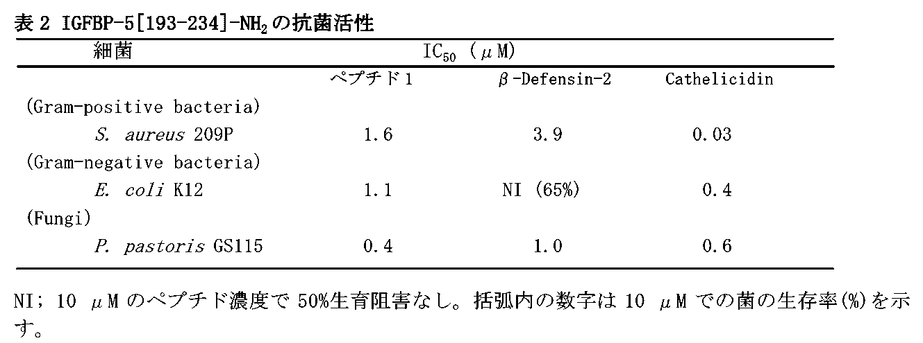
[実施例3]
他の抗菌ペプチドとIGFBP-5[193-214]-NH 2 の抗菌活性における協調作用
上述に記載したIGFBP-5[193-214]-NH2の合成ペプチド、ヒトβ-ディフェンシン-2 (ペプチド研究所)、カテリシジン(LL-37)(Bachem)を用いて抗菌活性を測定した。抗菌活性は、実施例2(I)2.アラマブルーを用いた生菌の還元活性を指標とする抗菌活性測定法により求めた。
他の抗菌ペプチドとIGFBP-5[193-214]-NH 2 の抗菌活性における協調作用
上述に記載したIGFBP-5[193-214]-NH2の合成ペプチド、ヒトβ-ディフェンシン-2 (ペプチド研究所)、カテリシジン(LL-37)(Bachem)を用いて抗菌活性を測定した。抗菌活性は、実施例2(I)2.アラマブルーを用いた生菌の還元活性を指標とする抗菌活性測定法により求めた。
IGFBP-5[193-214]-NH2ペプチドはヒトβ-ディフェンシン-2、カテリシジン(LL-37)と協調的に働き、E. coli K12に対する抗菌活性を示した (図3)。
(I) 抗菌活性の評価法
1. 用いた細菌
・グラム陰性細菌
Escherichia coli K12
2. アラマブルーを用いた生菌の還元活性を指標とする抗菌活性測定法
・試薬: アラマブルー(alamarBlue, BioSource International)
バッファー: 10 mM リン酸バッファー, pH 7.0
・方法
i) 2 x 10⁴ colony forming unit (CFU)の細菌あるいは真菌とペプチドを50 μlの反応系(n=3)で37℃、1時間反応させる。IC50の半分の濃度のβ-ディフェンシン-2 (3.15 μM)、あるいはカテリシジン(0.3 μM)存在下で様々な濃度のIGFBP-5[193-214]-NH2ペプチドの抗菌活性を測定した(図3a)。一方、IC50の半分の濃度のIGFBP-5[193-214]-NH2ペプチド (0.45 μM) あるいはカテリシジン(0.3 μM)存在下で様々な濃度のβ-ディフェンシン-2の抗菌活性を測定した(図3b)。また、IC50の半分の濃度のIGFBP-5[193-214]-NH2ペプチド (0.45 μM)、あるいはβ-ディフェンシン-2 (3.15 μM)存在下で様々な濃度のカテリシジンの抗菌活性を測定した(図3c)。陰性コントロールとしてペプチドなし溶液も作成、反応させる。
ii) 10%アラマブルーを加えた3%Tryptosoy Broth (培地)を反応液に加える。
37℃で生育させ、陰性コントロールの反応液が青色から赤色に変わったところで、570 nm、600 nmの吸光度(A570, A600)を測定する。
iii) 還元活性の算出は実施例2の、2. iii)-iv)に従った。サンプルの還元活性(%)は生菌の数に比例する。すなわち、還元活性=細菌生存率である。それぞれのペプチド濃度における細菌生存率を求めた(図3)。
1. 用いた細菌
・グラム陰性細菌
Escherichia coli K12
2. アラマブルーを用いた生菌の還元活性を指標とする抗菌活性測定法
・試薬: アラマブルー(alamarBlue, BioSource International)
バッファー: 10 mM リン酸バッファー, pH 7.0
・方法
i) 2 x 10⁴ colony forming unit (CFU)の細菌あるいは真菌とペプチドを50 μlの反応系(n=3)で37℃、1時間反応させる。IC50の半分の濃度のβ-ディフェンシン-2 (3.15 μM)、あるいはカテリシジン(0.3 μM)存在下で様々な濃度のIGFBP-5[193-214]-NH2ペプチドの抗菌活性を測定した(図3a)。一方、IC50の半分の濃度のIGFBP-5[193-214]-NH2ペプチド (0.45 μM) あるいはカテリシジン(0.3 μM)存在下で様々な濃度のβ-ディフェンシン-2の抗菌活性を測定した(図3b)。また、IC50の半分の濃度のIGFBP-5[193-214]-NH2ペプチド (0.45 μM)、あるいはβ-ディフェンシン-2 (3.15 μM)存在下で様々な濃度のカテリシジンの抗菌活性を測定した(図3c)。陰性コントロールとしてペプチドなし溶液も作成、反応させる。
ii) 10%アラマブルーを加えた3%Tryptosoy Broth (培地)を反応液に加える。
37℃で生育させ、陰性コントロールの反応液が青色から赤色に変わったところで、570 nm、600 nmの吸光度(A570, A600)を測定する。
iii) 還元活性の算出は実施例2の、2. iii)-iv)に従った。サンプルの還元活性(%)は生菌の数に比例する。すなわち、還元活性=細菌生存率である。それぞれのペプチド濃度における細菌生存率を求めた(図3)。
[実施例4]
IGFBP-5[193-214]-NH 2 の溶血活性測定
上述に記載したIGFBP-5[193-214]-NH2の合成ペプチド(ペプチド1)、ヒトβ-ディフェンシン-2 (ペプチド研究所)、カテリシジン(LL-37)(Bachem)を用いて、細胞傷害性を調べる目的でヒツジ赤血球に対する溶血活性を測定した。
IGFBP-5[193-214]-NH 2 の溶血活性測定
上述に記載したIGFBP-5[193-214]-NH2の合成ペプチド(ペプチド1)、ヒトβ-ディフェンシン-2 (ペプチド研究所)、カテリシジン(LL-37)(Bachem)を用いて、細胞傷害性を調べる目的でヒツジ赤血球に対する溶血活性を測定した。
カテリシジンはヒツジ赤血球に対して溶血活性を示した (10 μMで4.1%)が、IGFBP-5[193-214]-NH2(ペプチド1)、ヒトβ-ディフェンシン-2はほとんど溶血活性を示さなかった。
(I) 溶血活性の測定法
溶血活性の測定
・用いた赤血球:ヒツジ脱繊血(日本バイオテスト)
・バッファー: 10 mM リン酸バッファー, pH 7.0, 0.15 M NaCl
・方法
i) ヒツジ脱繊血をバッファーで3回洗浄後、2%になるようにバッファーで希釈する。
ii) 上記赤血球50 μlにバッファーで希釈したペプチド溶液(20 μM)50 μl加え、37℃、4時間反応させる。
iii) 3,000回転、5分遠心後、上清の546 nmの吸光度 (A546)を測定する。
iv) 純水で完全溶血させたときのA546を100%として、溶血活性を算出する(表3)。
溶血活性の測定
・用いた赤血球:ヒツジ脱繊血(日本バイオテスト)
・バッファー: 10 mM リン酸バッファー, pH 7.0, 0.15 M NaCl
・方法
i) ヒツジ脱繊血をバッファーで3回洗浄後、2%になるようにバッファーで希釈する。
ii) 上記赤血球50 μlにバッファーで希釈したペプチド溶液(20 μM)50 μl加え、37℃、4時間反応させる。
iii) 3,000回転、5分遠心後、上清の546 nmの吸光度 (A546)を測定する。
iv) 純水で完全溶血させたときのA546を100%として、溶血活性を算出する(表3)。

[実施例5]
ペプチド合成
以下の3種類のペプチドについて合成を行った。
ペプチド1 IGFBP-5[193-214]-NH2: AVYLPNCDRKGFYKRKQCKPSR-NH2(配列番号:1)
ペプチド2 IGFBP-5[193-215]: AVYLPNCDRKGFYKRKQCKPSRG(配列番号:4)
ペプチド3 Y-IGFBP-5[193-214]-NH2: YAVYLPNCDRKGFYKRKQCKPSR-NH2(配列番号:5)
ペプチド合成
以下の3種類のペプチドについて合成を行った。
ペプチド1 IGFBP-5[193-214]-NH2: AVYLPNCDRKGFYKRKQCKPSR-NH2(配列番号:1)
ペプチド2 IGFBP-5[193-215]: AVYLPNCDRKGFYKRKQCKPSRG(配列番号:4)
ペプチド3 Y-IGFBP-5[193-214]-NH2: YAVYLPNCDRKGFYKRKQCKPSR-NH2(配列番号:5)
ペプチド合成は、シグマ・ジェノシス社で実施した。
・合成機:Abacus peptide synthesizer (製造会社名:Sigma-Genosys) ペプチド1
SyroII peptide synthesizer (製造会社名:MultiSynTech GmbH) ペプチド2、ペプチド3
SyroII peptide synthesizer (製造会社名:MultiSynTech GmbH) ペプチド2、ペプチド3
・樹脂:TentaGelS RAM (製造会社名:Rapp Polymer GmbH) ペプチド1
TentaGelS PHB (同上) ペプチド1
Fmoc-Gly-NovaSyn TGT (製造会社名:EMD Chemicals, Inc.) ペプチド2、ペプチド3
NovaSyn TGR resin (同上) ペプチド2、ペプチド3
TentaGelS PHB (同上) ペプチド1
Fmoc-Gly-NovaSyn TGT (製造会社名:EMD Chemicals, Inc.) ペプチド2、ペプチド3
NovaSyn TGR resin (同上) ペプチド2、ペプチド3
・保護アミノ酸: (製造会社名:EMD Chemicals, Inc.)
9- Fluorenylmethoxycarbonyl- L- Alanine
Nα-9- Fluorenylmethoxycarbonyl- Nγ- 2,2,4,6,7-Pentamethyldihydrobenzofuran- 5- Sulfonyl-L-Arginine
9- Fluorenylmethoxycarbonyl- L- Asparagine
9- Fluorenylmethoxycarbonyl- L- Aspartic Acid β- t- Butyl Ester
9- Fluorenylmethoxycarbonyl-S- Trityl-L-Cysteine
9- Fluorenylmethoxycarbonyl- L- Glutamine
9- Fluorenylmethoxycarbonyl-Glycine
9- Fluorenylmethoxycarbonyl- L- Leucine
Nα-9- Fluorenylmethoxycarbonyl- Nε- t- Butyloxycarbonyl- L- Lysine
9- Fluorenylmethoxycarbonyl- L- Phenylalanine
9- Fluorenylmethoxycarbonyl- L- Proline
9- Fluorenylmethoxycarbonyl- O-t-Butyl-L-Serine
9- Fluorenylmethoxycarbonyl- O-t-Butyl-L-Tyrosine
9- Fluorenylmethoxycarbonyl- L- Valine
9- Fluorenylmethoxycarbonyl- L- Alanine
Nα-9- Fluorenylmethoxycarbonyl- Nγ- 2,2,4,6,7-Pentamethyldihydrobenzofuran- 5- Sulfonyl-L-Arginine
9- Fluorenylmethoxycarbonyl- L- Asparagine
9- Fluorenylmethoxycarbonyl- L- Aspartic Acid β- t- Butyl Ester
9- Fluorenylmethoxycarbonyl-S- Trityl-L-Cysteine
9- Fluorenylmethoxycarbonyl- L- Glutamine
9- Fluorenylmethoxycarbonyl-Glycine
9- Fluorenylmethoxycarbonyl- L- Leucine
Nα-9- Fluorenylmethoxycarbonyl- Nε- t- Butyloxycarbonyl- L- Lysine
9- Fluorenylmethoxycarbonyl- L- Phenylalanine
9- Fluorenylmethoxycarbonyl- L- Proline
9- Fluorenylmethoxycarbonyl- O-t-Butyl-L-Serine
9- Fluorenylmethoxycarbonyl- O-t-Butyl-L-Tyrosine
9- Fluorenylmethoxycarbonyl- L- Valine
・合成法:通常のfmoc strategyに従い実施
・脱保護:
クリベージカクテル
TFA: H2O: DTT: triisopropylsilane = 94:2.5:2.5:1
クリベージカクテル
TFA: H2O: DTT: triisopropylsilane = 94:2.5:2.5:1
・合成ペプチドの精製: 逆相高速液体クロマトグラフィーによる
カラム: Discovery HS C18 column, 10 micron, 250 x 21.2 mm, Sigma-Aldrich)
溶媒: A) 0.1% TFA, B) 0.1% TFA-acetonitrile, グラジエント溶出(時間などの条件は個々に変更)
流速: 15ml/min
合成品は一度上記条件で精製、酸化してS-S結合形成後、再度上記条件にて精製を実施
カラム: Discovery HS C18 column, 10 micron, 250 x 21.2 mm, Sigma-Aldrich)
溶媒: A) 0.1% TFA, B) 0.1% TFA-acetonitrile, グラジエント溶出(時間などの条件は個々に変更)
流速: 15ml/min
合成品は一度上記条件で精製、酸化してS-S結合形成後、再度上記条件にて精製を実施
・合成ペプチドの品質管理
i) 逆相高速液体クロマトグラフィーによるペプチドの純度検定
装置:東ソー 高圧グラジエントシステム
カラム:Supelco Discovery HS C18, 4.6 x 75 mm, particle 3 micron
溶媒:(A) 0.1% trifluoroacetic acid(TFA) in water, (B) 0.1% TFA in acetonitrile
injectionの2min後から(B) 5% → 55% 8 min linear gradient elution
流速:1.0 ml/min, 測定波長:220 nm(東ソー,UV-8020)
UV吸収に基づく純度
ペプチド1: IGFBP-5[193-214]-NH2 97.7%
ペプチド2: IGFBP-5[193-215] 94.6%
ペプチド3: Y-IGFBP-5[193-214]-NH2 96.7%
装置:東ソー 高圧グラジエントシステム
カラム:Supelco Discovery HS C18, 4.6 x 75 mm, particle 3 micron
溶媒:(A) 0.1% trifluoroacetic acid(TFA) in water, (B) 0.1% TFA in acetonitrile
injectionの2min後から(B) 5% → 55% 8 min linear gradient elution
流速:1.0 ml/min, 測定波長:220 nm(東ソー,UV-8020)
UV吸収に基づく純度
ペプチド1: IGFBP-5[193-214]-NH2 97.7%
ペプチド2: IGFBP-5[193-215] 94.6%
ペプチド3: Y-IGFBP-5[193-214]-NH2 96.7%
ii)アミノ酸分析:
加水分解:PicoTag ワークステーション(Waters社)にて塩化水素蒸気で110℃、22時間加水分解分析計: L-8500 amino acid analyzer (日立)
アミノ酸分析による純度検定の結果を以下の表4に示す。
加水分解:PicoTag ワークステーション(Waters社)にて塩化水素蒸気で110℃、22時間加水分解分析計: L-8500 amino acid analyzer (日立)
アミノ酸分析による純度検定の結果を以下の表4に示す。
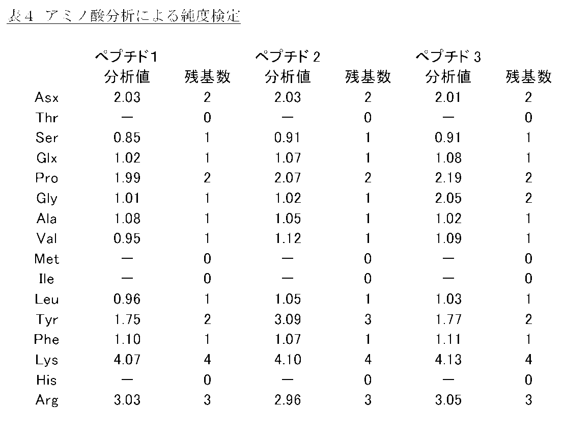
[実施例6]
IGFBP-5[193-214]-NH 2 の測定システム(ラジオイムノアッセイ)の作成
IGFBP-5[193-214]-NH2のC末端に対する特異的抗体を作成した。具体的には、当該ペプチドとウシサイログロブリンを水溶性カルボジイミド(Water soluble carbodiimide, 以下WSC)の作用により結合させた。調製したコンジュゲートをウサギに免疫することにより得られた抗体を用いて、当該ペプチドを高感度、特異的に測定可能なシステムを作成した。当該ペプチドはラット、ヒトなどの動物種によらず一定の構造であるため、本測定システムは幅広い利用が可能と考えられる。
IGFBP-5[193-214]-NH 2 の測定システム(ラジオイムノアッセイ)の作成
IGFBP-5[193-214]-NH2のC末端に対する特異的抗体を作成した。具体的には、当該ペプチドとウシサイログロブリンを水溶性カルボジイミド(Water soluble carbodiimide, 以下WSC)の作用により結合させた。調製したコンジュゲートをウサギに免疫することにより得られた抗体を用いて、当該ペプチドを高感度、特異的に測定可能なシステムを作成した。当該ペプチドはラット、ヒトなどの動物種によらず一定の構造であるため、本測定システムは幅広い利用が可能と考えられる。
(I) 抗体の作成法
1. 抗原の作成
i) サイログロブリン (Sigma, 6.7 mg)、合成IGFBP-5[193-214]-NH2(4 mg)をそれぞれ1 mlの生理食塩水に溶解する。
ii) サイログロブリン溶液にWSC (ペプチド研究所, 33.3.mg)を添加し、そこにペプチド溶液を添加する。
iii) 撹拌下に室温で3時間反応後、WSC (33.3.mg)を添加し室温でさらに3時間させる。さらに4℃で一晩反応する。
iv) 試薬、未反応のペプチドを除去するため、4℃で生理的食塩水に対して繰り返し透析を行う。
1. 抗原の作成
i) サイログロブリン (Sigma, 6.7 mg)、合成IGFBP-5[193-214]-NH2(4 mg)をそれぞれ1 mlの生理食塩水に溶解する。
ii) サイログロブリン溶液にWSC (ペプチド研究所, 33.3.mg)を添加し、そこにペプチド溶液を添加する。
iii) 撹拌下に室温で3時間反応後、WSC (33.3.mg)を添加し室温でさらに3時間させる。さらに4℃で一晩反応する。
iv) 試薬、未反応のペプチドを除去するため、4℃で生理的食塩水に対して繰り返し透析を行う。
2. ウサギの免疫
i) 上記抗原溶液の内、ペプチド0.02mg相当を含む抗原溶液0.5mLを等量のフロイント完全アジュバントと混合、ミセル化して、ウサギ(New Zealand White, オス)の背部、皮下に免疫する。
ii) 3週間おきに免疫を行い、その10日後に採血を行う。
iii) 後述の125Iで放射性標識したペプチド3(YAVYLPNCDRKGFYKRKQCKPSR-NH2;配列番号:5)を用いて抗血清力価を測定し、力価の上昇したウサギより抗血清を得る。
i) 上記抗原溶液の内、ペプチド0.02mg相当を含む抗原溶液0.5mLを等量のフロイント完全アジュバントと混合、ミセル化して、ウサギ(New Zealand White, オス)の背部、皮下に免疫する。
ii) 3週間おきに免疫を行い、その10日後に採血を行う。
iii) 後述の125Iで放射性標識したペプチド3(YAVYLPNCDRKGFYKRKQCKPSR-NH2;配列番号:5)を用いて抗血清力価を測定し、力価の上昇したウサギより抗血清を得る。
(II) ラジオイムノアッセイシステムの作成法
i) トレーサーは当該ペプチドのN末端にチロシン残基を結合したペプチド3(YAVYLPNCDRKGFYKRKQCKPSR-NH2;配列番号:5)にラクトペルオキシダーゼ法により、放射性125I(NaI)と過酸化水素を反応させて調製する。
ii) 反応混合物は逆相高速液体クロマトグラフィーで分離し、125Iが1分子N末端チロシン残基に導入されたペプチドを単離し、トレーサーとして使用する。
iii) 添加したトレーサーの30%程度が抗体と結合する濃度を設定し、その抗体濃度で当該ペプチドの濃度を1 fmol/tubeより1000 fmol/tubeまで変化させ、標準曲線を得る。
iv) 構造既知のペプチドの濃度を変化させて上記測定系で反応させ、結合の抑制を測定し、抗体の特異性を検討する。
i) トレーサーは当該ペプチドのN末端にチロシン残基を結合したペプチド3(YAVYLPNCDRKGFYKRKQCKPSR-NH2;配列番号:5)にラクトペルオキシダーゼ法により、放射性125I(NaI)と過酸化水素を反応させて調製する。
ii) 反応混合物は逆相高速液体クロマトグラフィーで分離し、125Iが1分子N末端チロシン残基に導入されたペプチドを単離し、トレーサーとして使用する。
iii) 添加したトレーサーの30%程度が抗体と結合する濃度を設定し、その抗体濃度で当該ペプチドの濃度を1 fmol/tubeより1000 fmol/tubeまで変化させ、標準曲線を得る。
iv) 構造既知のペプチドの濃度を変化させて上記測定系で反応させ、結合の抑制を測定し、抗体の特異性を検討する。
作成された抗血清は、最終希釈濃度210,000倍の高力価の抗体であり、標準曲線の中点は20 fmol/tubeであり、2 fmol/tubeから500 fmol/tubeの範囲で良好な濃度依存性が認められた。
このラジオイムノアッセイ(RIA)において、既知の生理活性ペプチドであるヒトカルシトニン、サブスタンスK、[Arg8]バソプッレシン、アドレノメジュリン[22-52]、Leu-エンケファリン、アンジオテンシンIIは100 pmol/tubeでもトレーサーの結合を阻害せず、交差反応は全く認められなかった。当該ペプチドとC末端アミド構造を共有するPAMP-20は、中点が100 pmol/tubeで、その交差反応は当該ペプチドの約1/3,000であった。また、C末端アミド構造を取らずにIGFBP-5配列がそのまま1残基延長したペプチド2(IGFBP-5[193-215])は、中点が20 pmol/tubeで、その交差反応は当該ペプチドの約1/670であった。また、当該ペプチドを還元アルキル化したペプチドは、当該ペプチドと全く同様のトレーサーの結合阻害を示した。一方、当該ペプチドの前駆体であるIGFBP-5は、全く結合阻害を示さなかった。これらの結果より、作成した抗体はIGFBP-5[193-214]-NH2の構造、特にC末端アミド構造と周辺1次構造を特異的に認識し、前駆体を認識しない良好な特性を有すると考えられる。
以上より、作製したIGFBP-5[193-214]-NH2のラジオイムノアッセイは、当該ペプチドに極めて特異性の高いものであり、当該ペプチド濃度を正確に測定できると考えられる。
[実施例7]
ラットにおけるIGFBP-5[193-214]-NH 2 様免疫活性の組織分布
SDラット(雌)の様々な組織よりペプチド画分の抽出を行い、濃縮後、前記実施例6で作成したIGFBP-5[193-214]-NH2測定システムを用いて当該ペプチド免疫活性の組織分布を検討した。下垂体、脳組織で有意な免疫活性が観測された(表5)。一方末梢組織では全体に低い免疫活性しか検出されなかったが、小腸では有意な免疫活性が観測された。これらの結果より、当該ペプチドは脳神経系、下垂体、消化管などで機能を発揮すると推定された。
ラットにおけるIGFBP-5[193-214]-NH 2 様免疫活性の組織分布
SDラット(雌)の様々な組織よりペプチド画分の抽出を行い、濃縮後、前記実施例6で作成したIGFBP-5[193-214]-NH2測定システムを用いて当該ペプチド免疫活性の組織分布を検討した。下垂体、脳組織で有意な免疫活性が観測された(表5)。一方末梢組織では全体に低い免疫活性しか検出されなかったが、小腸では有意な免疫活性が観測された。これらの結果より、当該ペプチドは脳神経系、下垂体、消化管などで機能を発揮すると推定された。
(I) 組織からのペプチド抽出法
i) 雌SDラット(n=3)から表5の組織を摘出し、生理食塩水で洗浄後、秤量、細片化する。
ii) 予め加熱した10倍量の蒸留水入り試験管に組織片を加え、15分間90℃以上で加熱する。
iii) 氷冷後、終濃度1 Mになるように酢酸を加える。
iv) ホモジナイザー(ポリトロン)で組織片を破砕し、破砕液を遠心分離する(13,000 x g、15分、4℃)。
v) 上清の一部を Sep-Pak C18カートリッジで濃縮、脱塩し、ペプチドを回収する。
i) 雌SDラット(n=3)から表5の組織を摘出し、生理食塩水で洗浄後、秤量、細片化する。
ii) 予め加熱した10倍量の蒸留水入り試験管に組織片を加え、15分間90℃以上で加熱する。
iii) 氷冷後、終濃度1 Mになるように酢酸を加える。
iv) ホモジナイザー(ポリトロン)で組織片を破砕し、破砕液を遠心分離する(13,000 x g、15分、4℃)。
v) 上清の一部を Sep-Pak C18カートリッジで濃縮、脱塩し、ペプチドを回収する。
(II) ラジオイムノアッセイシステムをもちいた組織中の免疫活性物質の測定法
実施例6 (II)で作成したシステムで測定を行う。
i) ラジオイムノアッセイシステムにおける当該ペプチドの標準曲線から(I)で抽出した各組織抽出物に含まれる免疫活性物質の量を算出する。
ii) 組織湿重量1 g当たりの免疫活性物質の含量を換算する(表5)。
iii) ラジオイムノアッセイのバッファー、手法は既報の通りである。
(T. Katafuchi, et al., J. Biol. Chem., 278, 12046-12054, 2003)
実施例6 (II)で作成したシステムで測定を行う。
i) ラジオイムノアッセイシステムにおける当該ペプチドの標準曲線から(I)で抽出した各組織抽出物に含まれる免疫活性物質の量を算出する。
ii) 組織湿重量1 g当たりの免疫活性物質の含量を換算する(表5)。
iii) ラジオイムノアッセイのバッファー、手法は既報の通りである。
(T. Katafuchi, et al., J. Biol. Chem., 278, 12046-12054, 2003)
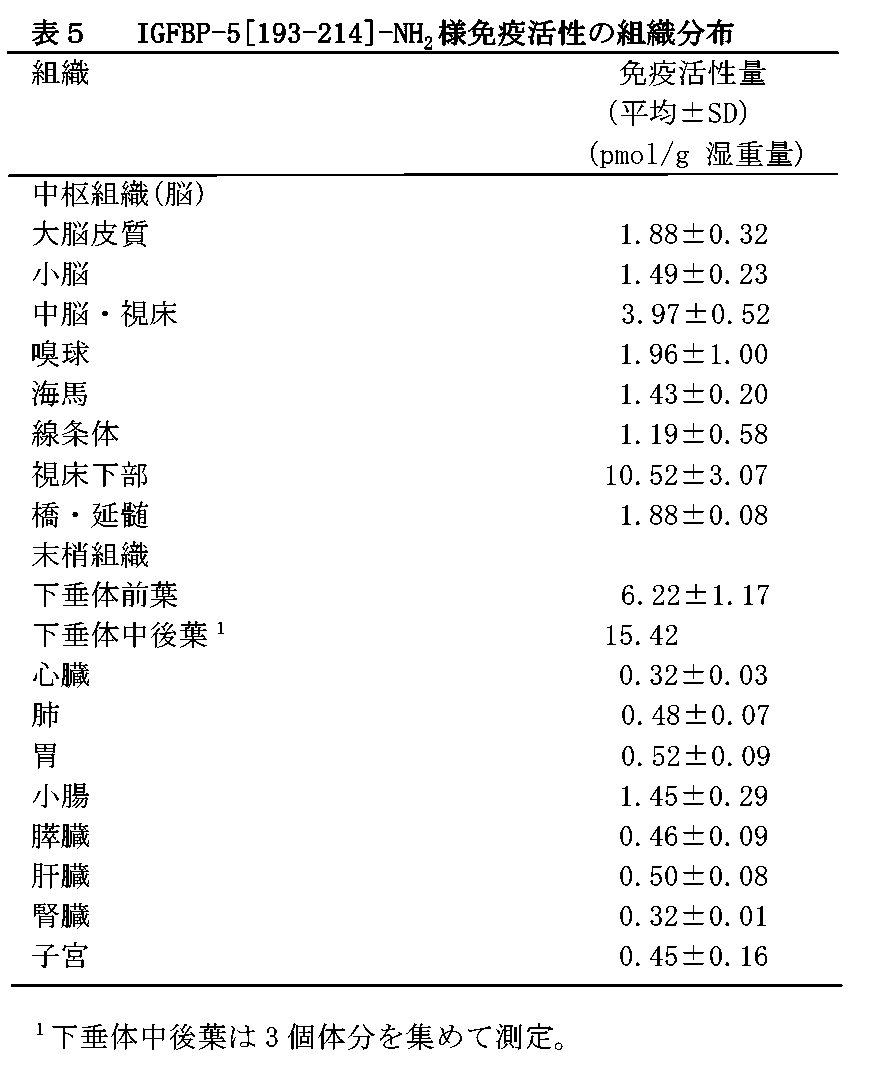
[実施例8]
ラット組織中に存在するIGFBP-5[193-214]-NH 2 の同定
実施例7で検出したIGFBP-5[193-214]-NH2様免疫活性物質を同定するため、組織濃度が高く組織重量が得られる脳組織中の免疫活性物質を、逆相カートリッジで濃縮後、ゲルろ過、逆相カラムで分離した結果(図4 a, b)、免疫活性物質が2種の分離系において、合成IGFBP-5[193-214]-NH2と同じ挙動を示すことを確認した。さらにその免疫活性画分を免疫沈降し、質量分析を実施することにより、主たる免疫活性物質がIGFBP-5[193-214]-NH2自身に由来することを確定した(図4 c)。末梢組織で組織濃度が高く組織重量が得られる小腸組織中の免疫活性物質を、同様に逆相カートリッジで濃縮後、ゲルろ過で分離した結果、合成IGFBP-5[193-214]-NH2と同じ挙動を示すことを確認した。さらにその免疫活性画分を免疫沈降し、質量分析を実施することにより、主たる免疫活性物質がIGFBP-5[193-214]-NH2自身に由来することを確定した。これらの結果より、当該ペプチドがラット脳、腸などの組織内に実在し、機能すると推定された。
ラット組織中に存在するIGFBP-5[193-214]-NH 2 の同定
実施例7で検出したIGFBP-5[193-214]-NH2様免疫活性物質を同定するため、組織濃度が高く組織重量が得られる脳組織中の免疫活性物質を、逆相カートリッジで濃縮後、ゲルろ過、逆相カラムで分離した結果(図4 a, b)、免疫活性物質が2種の分離系において、合成IGFBP-5[193-214]-NH2と同じ挙動を示すことを確認した。さらにその免疫活性画分を免疫沈降し、質量分析を実施することにより、主たる免疫活性物質がIGFBP-5[193-214]-NH2自身に由来することを確定した(図4 c)。末梢組織で組織濃度が高く組織重量が得られる小腸組織中の免疫活性物質を、同様に逆相カートリッジで濃縮後、ゲルろ過で分離した結果、合成IGFBP-5[193-214]-NH2と同じ挙動を示すことを確認した。さらにその免疫活性画分を免疫沈降し、質量分析を実施することにより、主たる免疫活性物質がIGFBP-5[193-214]-NH2自身に由来することを確定した。これらの結果より、当該ペプチドがラット脳、腸などの組織内に実在し、機能すると推定された。
(I) カラムクロマトグラフィーによる免疫活性物質の生化学的解析法
1-1. 脳抽出物をゲルろ過カラムで分離し、各画分に含まれるIGFBP-5[193-214]-NH2様免疫活性物質の量を実施例6(II)で作成したシステムをもちいて測定する(図4a)。
i) 全脳抽出物(15 g組織より抽出)をゲルろ過カラムで分離する。
カラム: Sephadex G-50 fine (GE Healthcare, 1.8 x 135 cm)
溶媒:1 M 酢酸、流速:7 ml/h、フラクション: 6 ml/tube
ii) 各画分の一部(1/42量)に含まれる免疫活性(IR-)IGFBP-5[193-214]-NH2量を実施例6(II)で作成したシステムを用いて測定する。
免疫活性は4 kDより小さい画分で検出され、当該ペプチドの溶出位置と一致した。
1-1. 脳抽出物をゲルろ過カラムで分離し、各画分に含まれるIGFBP-5[193-214]-NH2様免疫活性物質の量を実施例6(II)で作成したシステムをもちいて測定する(図4a)。
i) 全脳抽出物(15 g組織より抽出)をゲルろ過カラムで分離する。
カラム: Sephadex G-50 fine (GE Healthcare, 1.8 x 135 cm)
溶媒:1 M 酢酸、流速:7 ml/h、フラクション: 6 ml/tube
ii) 各画分の一部(1/42量)に含まれる免疫活性(IR-)IGFBP-5[193-214]-NH2量を実施例6(II)で作成したシステムを用いて測定する。
免疫活性は4 kDより小さい画分で検出され、当該ペプチドの溶出位置と一致した。
1-2.ゲルろ過でIR-IGFBP-5[193-214]-NH2量が多く検出された画分(図4a、太線)をさらに逆相高速液体クロマトグラフィーにて分離し、各画分に含まれる免疫活性物質の量を実施例6(II)で作成したラジオイムノアッセイシステムを用いて測定する(図4b)。
i) ゲルろ過でIR-IGFBP-5[193-214]-NH2量が多く検出された画分(図4a、太線) の一部(1/8量)を逆相高速液体クロマトグラフィーで分離する。
カラム: Symmetry 300 5C18 (Waters, 4.6 x 250 mm)
溶媒: A: 0.1% TFA/10% acetonitrile
B: 0.1% TFA/60% acetonitrile
溶出: A→B 60 分リニアグラジエント。1分毎に60分間分取する (60本)。
ii) 各画分の一部(1/5量)に含まれる免疫活性量を前記実施例6(II)で作成したラジオイムノアッセイシステムを用いて測定する。
IR-IGFBP-5[193-214]-NH2の最も高いピーク(図4b、矢印)は当該ペプチドの溶出位置と一致した。
i) ゲルろ過でIR-IGFBP-5[193-214]-NH2量が多く検出された画分(図4a、太線) の一部(1/8量)を逆相高速液体クロマトグラフィーで分離する。
カラム: Symmetry 300 5C18 (Waters, 4.6 x 250 mm)
溶媒: A: 0.1% TFA/10% acetonitrile
B: 0.1% TFA/60% acetonitrile
溶出: A→B 60 分リニアグラジエント。1分毎に60分間分取する (60本)。
ii) 各画分の一部(1/5量)に含まれる免疫活性量を前記実施例6(II)で作成したラジオイムノアッセイシステムを用いて測定する。
IR-IGFBP-5[193-214]-NH2の最も高いピーク(図4b、矢印)は当該ペプチドの溶出位置と一致した。
2-1 小腸抽出物をゲルろ過カラムで分離し、各画分に含まれるIR-IGFBP-5[193-214]-NH2量を実施例6(II)で作成したシステムをもちいて測定する。
i) 小腸抽出物(1.08 gより抽出)をゲルろ過カラムで分離する。
カラム: TSK-gel G2000SWXL (21.5 x 300 mm)
溶媒:0.1% TFA/60% acetonitrile、流速:1.5 ml/min、フラクション: 1.5 ml/tube
ii) 各画分の一部(1/5量)のIR-IGFBP-5[193-214]-NH2量を実施例6(II)で作成したラジオイムノアッセイシステムを用いて測定する。
免疫活性量のピークは当該ペプチドの溶出位置と一致した。
i) 小腸抽出物(1.08 gより抽出)をゲルろ過カラムで分離する。
カラム: TSK-gel G2000SWXL (21.5 x 300 mm)
溶媒:0.1% TFA/60% acetonitrile、流速:1.5 ml/min、フラクション: 1.5 ml/tube
ii) 各画分の一部(1/5量)のIR-IGFBP-5[193-214]-NH2量を実施例6(II)で作成したラジオイムノアッセイシステムを用いて測定する。
免疫活性量のピークは当該ペプチドの溶出位置と一致した。
(II)質量分析計による免疫活性物質の同定法
1 逆相高速液体クロマトグラフィーで分離したIR-IGFBP-5[193-214]-NH2の最も高いピーク(図4b、矢印)画分に含まれる脳由来免疫活性物質を、免疫沈降後、質量分析計を用いて分析、同定する。
1-1. 免疫活性物質の免疫沈降
i) 当該ペプチドの溶出位置と同じ画分 (図4b、矢印)の1/2量を凍結乾燥する。
ii) 50 μl 10 mMリン酸バッファー, 0.15 M NaCl, pH 7.0, 0.1% Triton X-100に溶解する。
iii) 前記実施例6 (I)で作成した抗血清(10倍希釈) 40 μlとProtein A Sepharose (GE Healthcare) 20 μlを混合する。
iv) 4℃で一晩反応後、10 mMリン酸バッファー, 0.15 M NaCl, pH 7.0で4回洗浄する。
v) 蒸留水で1回洗浄後、1%TFA 20 μlで溶出する(室温2分)。
1 逆相高速液体クロマトグラフィーで分離したIR-IGFBP-5[193-214]-NH2の最も高いピーク(図4b、矢印)画分に含まれる脳由来免疫活性物質を、免疫沈降後、質量分析計を用いて分析、同定する。
1-1. 免疫活性物質の免疫沈降
i) 当該ペプチドの溶出位置と同じ画分 (図4b、矢印)の1/2量を凍結乾燥する。
ii) 50 μl 10 mMリン酸バッファー, 0.15 M NaCl, pH 7.0, 0.1% Triton X-100に溶解する。
iii) 前記実施例6 (I)で作成した抗血清(10倍希釈) 40 μlとProtein A Sepharose (GE Healthcare) 20 μlを混合する。
iv) 4℃で一晩反応後、10 mMリン酸バッファー, 0.15 M NaCl, pH 7.0で4回洗浄する。
v) 蒸留水で1回洗浄後、1%TFA 20 μlで溶出する(室温2分)。
1-2. 質量分析計による同定
質量分析計、マトリックスは前記実施例1(II)で記載したものを使用した。
i) 前記実施例8 (II) 1-1. v) の溶出液をZip‐tip (Millipore) で脱塩処理する。
ii) 0.5 μlのマトリックス溶液をスポットし、ペプチドとマトリックスとの共結晶を作る。
iii) 質量分析計でMS1を測定する(図4c)。
分子内S‐S架橋、C末端アミド化された当該ペプチドの[M+H]+に相当する2654.4のピークが質量分析の結果得られた。
質量分析計、マトリックスは前記実施例1(II)で記載したものを使用した。
i) 前記実施例8 (II) 1-1. v) の溶出液をZip‐tip (Millipore) で脱塩処理する。
ii) 0.5 μlのマトリックス溶液をスポットし、ペプチドとマトリックスとの共結晶を作る。
iii) 質量分析計でMS1を測定する(図4c)。
分子内S‐S架橋、C末端アミド化された当該ペプチドの[M+H]+に相当する2654.4のピークが質量分析の結果得られた。
2. ゲルろ過で分離した小腸由来IR-IGFBP-5[193-214]-NH2の最も高いピーク画分の3/5量を、免疫沈降後、質量分析計を用いて分析、同定する。
方法は前項の1-1. 免疫活性物質の免疫沈降、および1-2. 質量分析計による同定に従う。
脳組織由来のIGFBP-5[193-214]-NH2様免疫活性物質と同様に、分子内S‐S架橋、C末端アミド化された当該ペプチドの[M+H]+に相当する2654.4のピークが質量分析の結果得られた。
方法は前項の1-1. 免疫活性物質の免疫沈降、および1-2. 質量分析計による同定に従う。
脳組織由来のIGFBP-5[193-214]-NH2様免疫活性物質と同様に、分子内S‐S架橋、C末端アミド化された当該ペプチドの[M+H]+に相当する2654.4のピークが質量分析の結果得られた。
これらの結果より、当該ペプチド本体がラット脳、小腸などの組織内に存在することが確認された。
[実施例9]
MRGX2発現HeLa細胞を用いたIGFBP-5[193-214]-NH 2 のHeLa/SRE-LUCレポーター活性
HeLa細胞を384穴プレートに4 x 10³ cells/wellで播種し、24時間培養した。HeLa細胞へのトランスフェクトは以下のように行った。SREにルシフェラーゼ遺伝子を連結させたSRE-LUC、G蛋白質 (GαOA、Gβ1、Gγ2)およびMrgX2の発現プラスミドをPBSで240ng/μlに調整した。それぞれのプラスミドをSRE-LUC:GαOA:Gβ1:Gγ2:Mrgx2を9:1:1:1:1で混合させたものをプラスミドミックスとして用いた。1.6mlのOPTI-MEM培地にプラスミドミックス83.2μlを加え、Vortexによりよく攪拌し、プラスミドミックス液とした。別のチューブを用意し、1.6mlのOPTI-MEM培地に66.6μlのリポフェクタミン2000(Invitrogen)を加え、Vortexによりよく攪拌し、リポフェクタミン2000液とした。5分間室温に放置後、プラスミドミックス液とリポフェクタミン2000液を混合し、攪拌した。さらに20分間室温に放置後、この溶液をプレートに5μlずつ分注し、炭酸ガスインキュベーターで、37℃、5%CO2にて培養した。18時間後、無血清DMEM培地で細胞を洗浄し、無血清DMEM培地で2時間培養した。無血清DMEM培地で細胞を洗浄し、0.05%CHAPSを含む無血清DMEM培地に希釈したサンプル溶液を添加し炭酸ガスインキュベーターで、37℃、5%CO2にて培養した。4時間後、プレートを室温に戻し、培地を除き8μlのピッカジーンLT液を加え、遮光にて20分間室温に放置した。サンプルによるSRE活性はルシフェラーゼ活性をプレートリーダー(EnVision)で化学発光を測定することによって評価した。その結果、2μM以上のIGFBP-5[193-214]-NH2はMrgX2に対するHeLa/SREレポーター活性を示した(図5)。
MRGX2発現HeLa細胞を用いたIGFBP-5[193-214]-NH 2 のHeLa/SRE-LUCレポーター活性
HeLa細胞を384穴プレートに4 x 10³ cells/wellで播種し、24時間培養した。HeLa細胞へのトランスフェクトは以下のように行った。SREにルシフェラーゼ遺伝子を連結させたSRE-LUC、G蛋白質 (GαOA、Gβ1、Gγ2)およびMrgX2の発現プラスミドをPBSで240ng/μlに調整した。それぞれのプラスミドをSRE-LUC:GαOA:Gβ1:Gγ2:Mrgx2を9:1:1:1:1で混合させたものをプラスミドミックスとして用いた。1.6mlのOPTI-MEM培地にプラスミドミックス83.2μlを加え、Vortexによりよく攪拌し、プラスミドミックス液とした。別のチューブを用意し、1.6mlのOPTI-MEM培地に66.6μlのリポフェクタミン2000(Invitrogen)を加え、Vortexによりよく攪拌し、リポフェクタミン2000液とした。5分間室温に放置後、プラスミドミックス液とリポフェクタミン2000液を混合し、攪拌した。さらに20分間室温に放置後、この溶液をプレートに5μlずつ分注し、炭酸ガスインキュベーターで、37℃、5%CO2にて培養した。18時間後、無血清DMEM培地で細胞を洗浄し、無血清DMEM培地で2時間培養した。無血清DMEM培地で細胞を洗浄し、0.05%CHAPSを含む無血清DMEM培地に希釈したサンプル溶液を添加し炭酸ガスインキュベーターで、37℃、5%CO2にて培養した。4時間後、プレートを室温に戻し、培地を除き8μlのピッカジーンLT液を加え、遮光にて20分間室温に放置した。サンプルによるSRE活性はルシフェラーゼ活性をプレートリーダー(EnVision)で化学発光を測定することによって評価した。その結果、2μM以上のIGFBP-5[193-214]-NH2はMrgX2に対するHeLa/SREレポーター活性を示した(図5)。
尚、各サンプルは、次のように調製した。上記[実施例5]に記載の方法で合成したIGFBP-5[193-214]-NH2およびS1P(和光純薬社製)はDMSOを用い1 mMに溶解した。1 mMの IGFBP-5[193-214]-NH2溶液をDMSOで希釈し、5倍、25倍、125倍、625倍、3125倍、15625倍溶液を作成した。0.05%CHAPSを含む無血清DMEM培地に各濃度のIGFBP-5[193-214]-NH2溶液は50倍に、S1P溶液は250倍に、FBS(Invitrogen社製)は25倍に希釈しサンプル溶液とした。
[実施例10]
MRGX2発現CHO細胞を用いたIGFBP-5[193-214]-NH 2 の細胞内Ca 2+ 濃度上昇活性
MRGX2発現CHO細胞は、国際公開第2006/118328号パンフレットの実施例5のヒトTGR12発現CHO細胞(=ヒトMRGX2発現CHO細胞)の作製方法に準じて作製した。CHO/dhfr-細胞(以下、CHO)を、10%ウシ胎児血清を含むα-MEM培地(インビトロジェン社)を用いて培養した。CHOに、MRGX2の塩基配列(配列番号:2に表される塩基配列を有するDNA)を組み込んだ動物細胞発現用ベクタープラスミドpAKKO-111H(Biochim. Biophys. Acta, 1219巻、251-259頁、1994年記載のpAKKO1.11Hと同一のベクタープラスミド)を安定的に発現させることにより、MRGX2発現CHO細胞を樹立した。MRGX2発現CHO細胞は、10%ウシ胎児透析血清を含む核酸不含α-MEM培地(インビトロジェン社)を用いて培養した。
細胞内Ca2+放出促進活性はFLIPR(モレキュラーデバイス社)を用いて測定した。MRGX2発現CHO細胞を96穴プレートに3 x 10⁴ cells/wellで播種し、24時間培養した。2.5 mMプロベネシドと20 mM HEPESを含むハンクス氏液(pH 7.4)(以下、洗浄用バッファーと略す)10 mlに50μg Fluo 3-AM (DOJINDO)を添加し、ローディングバッファーを作成した。培養プレートの培養液を除き、ローディングバッファーを100μl/well添加して気相37℃で60分間保温した。培養プレートのローディングバッファーを除き、洗浄用バッファーで洗浄してから、FLIPR内の所定の位置に設置した。試料にサンプルバッファー(洗浄用バッファーにBSAを2 mg/ml、CHAPSを1mg/ml添加したもの)を添加し、攪拌後、96穴サンプルプレートに移し替えた。このサンプルプレートもFLIPR内の所定の位置に設置し、測定を行なった。細胞内Ca2+放出促進活性は、試料添加によって惹起された蛍光強度の増加として計測した。本実験系において、IGFBP-5[193-214]-NH2のEC50値は1.7μMであった(図6A)。一方、サブスタンスPのEC50値は0.82μMであったことから、IGFBP-5[193-214]-NH2のMrgX2特異的な活性はサブスタンスP に比べて1/20程度であった(図6B)。
MRGX2発現CHO細胞を用いたIGFBP-5[193-214]-NH 2 の細胞内Ca 2+ 濃度上昇活性
MRGX2発現CHO細胞は、国際公開第2006/118328号パンフレットの実施例5のヒトTGR12発現CHO細胞(=ヒトMRGX2発現CHO細胞)の作製方法に準じて作製した。CHO/dhfr-細胞(以下、CHO)を、10%ウシ胎児血清を含むα-MEM培地(インビトロジェン社)を用いて培養した。CHOに、MRGX2の塩基配列(配列番号:2に表される塩基配列を有するDNA)を組み込んだ動物細胞発現用ベクタープラスミドpAKKO-111H(Biochim. Biophys. Acta, 1219巻、251-259頁、1994年記載のpAKKO1.11Hと同一のベクタープラスミド)を安定的に発現させることにより、MRGX2発現CHO細胞を樹立した。MRGX2発現CHO細胞は、10%ウシ胎児透析血清を含む核酸不含α-MEM培地(インビトロジェン社)を用いて培養した。
細胞内Ca2+放出促進活性はFLIPR(モレキュラーデバイス社)を用いて測定した。MRGX2発現CHO細胞を96穴プレートに3 x 10⁴ cells/wellで播種し、24時間培養した。2.5 mMプロベネシドと20 mM HEPESを含むハンクス氏液(pH 7.4)(以下、洗浄用バッファーと略す)10 mlに50μg Fluo 3-AM (DOJINDO)を添加し、ローディングバッファーを作成した。培養プレートの培養液を除き、ローディングバッファーを100μl/well添加して気相37℃で60分間保温した。培養プレートのローディングバッファーを除き、洗浄用バッファーで洗浄してから、FLIPR内の所定の位置に設置した。試料にサンプルバッファー(洗浄用バッファーにBSAを2 mg/ml、CHAPSを1mg/ml添加したもの)を添加し、攪拌後、96穴サンプルプレートに移し替えた。このサンプルプレートもFLIPR内の所定の位置に設置し、測定を行なった。細胞内Ca2+放出促進活性は、試料添加によって惹起された蛍光強度の増加として計測した。本実験系において、IGFBP-5[193-214]-NH2のEC50値は1.7μMであった(図6A)。一方、サブスタンスPのEC50値は0.82μMであったことから、IGFBP-5[193-214]-NH2のMrgX2特異的な活性はサブスタンスP に比べて1/20程度であった(図6B)。
尚、試料は、次のように調製した。上記[実施例5]に記載の方法で合成したIGFBP-5[193-214]-NH2およびサブスタンスP(ペプチド研究所製)はDMSOを用い1 mMに溶解した。1 mMのIGFBP-5[193-214]-NH2溶液とサブスタンスP溶液をDMSOで希釈し、それぞれ10倍、100倍、1000倍、10000倍溶液を作成した。各濃度のIGFBP-5[193-214]-NH2溶液とサブスタンスP溶液はサンプルバッファーにそれぞれは33倍に希釈しサンプル溶液とした。
[実施例11]
IGFBP-5遺伝子の発現解析
IGFBP-5遺伝子の発現解析を、NCBI遺伝子発現データベースや東大の遺伝子発現データベースを利用して次のようにして行った。
NCBI遺伝子発現データベース(http://www.ncbi.nlm.nih.gov/sites/entrez)や東大の遺伝子発現データベース(http://www.lsbm.org/database/index.html#)上でIGFBP-5を検索し、臓器間、疾患、性差による発現の強度を解析した。
IGFBP-5遺伝子の発現解析
IGFBP-5遺伝子の発現解析を、NCBI遺伝子発現データベースや東大の遺伝子発現データベースを利用して次のようにして行った。
NCBI遺伝子発現データベース(http://www.ncbi.nlm.nih.gov/sites/entrez)や東大の遺伝子発現データベース(http://www.lsbm.org/database/index.html#)上でIGFBP-5を検索し、臓器間、疾患、性差による発現の強度を解析した。
IGFBP-5の部分ペプチドであるIGFBP-5[193-214]-NH2がMRGX2特異的な活性を示したことからIGFBP-5遺伝子の発現解析を行った。末梢組織においてIGFBP-5遺伝子は卵巣、子宮、子宮筋層、子宮頚部、卵管、皮膚、骨格筋、腎臓、副腎、網膜などに発現が見られ、なかでも卵巣で最も高い発現が認められた。また中枢組織においては、ほとんど発現は認められなかったが、神経節で一部発現が認められた。次に各組織について病理(腫瘍、炎症等)やドナー(病歴、薬剤投与等)別に解析を行ったところ、IGFBP-5遺伝子は腫瘍や炎症など細胞増殖が亢進していると予想されるサンプルにおいて著しい発現低下を示した。また動脈硬化サンプルにおいて正常サンプルの2倍程度の発現上昇が認められた。しかし、動脈サンプルは4サンプルずつと数が少ないため解釈に注意が必要である。なお、各組織のドナーの病歴や薬剤投与、非投与および男性、女性ではほとんど発現変動を示さなかった。
Claims (12)
- 配列番号:1で表されるアミノ酸配列と同一もしくは実質的に同一のアミノ酸配列からなるペプチドまたはその塩。
- 請求項1記載のペプチドまたはその塩を含有する抗菌剤。
- 哺乳動物に対して請求項1記載のペプチドまたはその塩の有効量を投与することを含む、該哺乳動物において病原性細菌により生ずる疾患の予防または治療方法。
- 病原性細菌により生ずる疾患の予防または治療剤を製造するための請求項1記載のペプチドまたはその塩の使用。
- (a)配列番号:1で表されるアミノ酸配列と同一もしくは実質的に同一のアミノ酸配列からなるペプチドまたはその塩および(b)配列番号:3で表されるアミノ酸配列と同一もしくは実質的に同一のアミノ酸配列を含有する蛋白質またはその塩を用いることを特徴とする、免疫疾患、泌尿器疾患、消化器疾患または呼吸器疾患の予防・治療剤のスクリーニング方法。
- (a)配列番号:1で表されるアミノ酸配列と同一もしくは実質的に同一のアミノ酸配列からなるペプチドまたはその塩および(b)配列番号:3で表されるアミノ酸配列と同一もしくは実質的に同一のアミノ酸配列を含有する蛋白質またはその塩を含有することを特徴とする、免疫疾患、泌尿器疾患、消化器疾患または呼吸器疾患の予防・治療剤のスクリーニング用キット。
- (a)配列番号:1で表されるアミノ酸配列と同一もしくは実質的に同一のアミノ酸配列からなるペプチドまたはその塩および(b)配列番号:3で表されるアミノ酸配列と同一もしくは実質的に同一のアミノ酸配列を含有する蛋白質またはその塩を用いることを特徴とする、配列番号:3で表されるアミノ酸配列と同一もしくは実質的に同一のアミノ酸配列を含有する蛋白質またはその塩に対するアンタゴニストのスクリーニング方法。
- 配列番号:1で表されるアミノ酸配列と同一もしくは実質的に同一のアミノ酸配列からなるペプチドまたはその塩を認識する抗体。
- 請求項8記載の抗体を含有してなる免疫疾患、泌尿器疾患、消化器疾患または呼吸器疾患の予防・治療剤。
- 請求項8記載の抗体と、被検液および標識化された配列番号:1で表されるアミノ酸配列と同一もしくは実質的に同一のアミノ酸配列からなるペプチドまたはその塩とを競合的に反応させ、上記抗体に結合した上記標識化されたペプチドまたはその塩の割合を測定することを特徴とする、被検液中の配列番号:1で表されるアミノ酸配列と同一もしくは実質的に同一のアミノ酸配列からなるペプチドまたはその塩の定量法。
- 哺乳動物に対して請求項8記載の抗体の有効量を投与することを含む、免疫疾患、泌尿器疾患、消化器疾患または呼吸器疾患の予防または治療方法。
- 免疫疾患、泌尿器疾患、消化器疾患または呼吸器疾患の予防または治療剤を製造するための、請求項8記載の抗体の使用。
Applications Claiming Priority (2)
| Application Number | Priority Date | Filing Date | Title |
|---|---|---|---|
| JP2009142232A JP2012167017A (ja) | 2009-06-15 | 2009-06-15 | ペプチドおよびその用途 |
| JP2009-142232 | 2009-06-15 |
Publications (1)
| Publication Number | Publication Date |
|---|---|
| WO2010147117A1 true WO2010147117A1 (ja) | 2010-12-23 |
Family
ID=43356434
Family Applications (1)
| Application Number | Title | Priority Date | Filing Date |
|---|---|---|---|
| PCT/JP2010/060136 Ceased WO2010147117A1 (ja) | 2009-06-15 | 2010-06-15 | ペプチドおよびその用途 |
Country Status (2)
| Country | Link |
|---|---|
| JP (1) | JP2012167017A (ja) |
| WO (1) | WO2010147117A1 (ja) |
Cited By (2)
| Publication number | Priority date | Publication date | Assignee | Title |
|---|---|---|---|---|
| US12247015B2 (en) | 2019-04-29 | 2025-03-11 | Solent Therapeutics, Llc | 3-amino-4H-benzo[e][1,2,4]thiadiazine 1,1-dioxide derivatives as inhibitors of MRGX2 |
| CN120463776A (zh) * | 2025-07-17 | 2025-08-12 | 三亚华大生命科学研究院 | 多肽及其应用 |
Families Citing this family (1)
| Publication number | Priority date | Publication date | Assignee | Title |
|---|---|---|---|---|
| EP2977384A1 (en) * | 2014-07-25 | 2016-01-27 | Fraunhofer Gesellschaft zur Förderung der angewandten Forschung e.V. | N-terminally truncated interleukin-38 |
-
2009
- 2009-06-15 JP JP2009142232A patent/JP2012167017A/ja not_active Withdrawn
-
2010
- 2010-06-15 WO PCT/JP2010/060136 patent/WO2010147117A1/ja not_active Ceased
Non-Patent Citations (8)
Cited By (2)
| Publication number | Priority date | Publication date | Assignee | Title |
|---|---|---|---|---|
| US12247015B2 (en) | 2019-04-29 | 2025-03-11 | Solent Therapeutics, Llc | 3-amino-4H-benzo[e][1,2,4]thiadiazine 1,1-dioxide derivatives as inhibitors of MRGX2 |
| CN120463776A (zh) * | 2025-07-17 | 2025-08-12 | 三亚华大生命科学研究院 | 多肽及其应用 |
Also Published As
| Publication number | Publication date |
|---|---|
| JP2012167017A (ja) | 2012-09-06 |
Similar Documents
| Publication | Publication Date | Title |
|---|---|---|
| EP1424392A1 (en) | Novel secretory proteins and dna thereof | |
| WO2010147117A1 (ja) | ペプチドおよびその用途 | |
| JP5317318B2 (ja) | 新規ポリペプチドおよびその用途 | |
| US7390625B2 (en) | Apoptosis-associated protein and use thereof | |
| US20050176632A1 (en) | Angiogenesis inhibitors | |
| EP1357129A2 (en) | Novel physiologically active peptide and use thereof | |
| JPWO2006118328A1 (ja) | 脱顆粒抑制剤 | |
| US7943137B2 (en) | FPRL1 ligands and use thereof | |
| JP4772684B2 (ja) | スクリーニング方法 | |
| JP4184697B2 (ja) | スクリーニング方法 | |
| JP4488720B2 (ja) | アポトーシス関連蛋白質およびその用途 | |
| JP4965776B2 (ja) | 新規生理活性ペプチドおよびその用途 | |
| JP4128030B2 (ja) | 新規リガンドおよびそのdna | |
| EP1457557A1 (en) | Novel proteins and dnas thereof | |
| JP2003292455A (ja) | 血管新生阻害剤 | |
| JP4404605B2 (ja) | 新規なfprl1リガンドおよびその用途 | |
| US20090227501A1 (en) | Agents for preventing and/or treating upper digestive tract disorders | |
| EP1428877A1 (en) | Novel protein and dna thereof | |
| JP4300008B2 (ja) | 新規タンパク質およびそのdna | |
| JP4378083B2 (ja) | スクリーニング方法 | |
| JP4167868B2 (ja) | 新規分泌蛋白質およびそのdna | |
| JP4799837B2 (ja) | スクリーニング方法 | |
| JP2005110658A (ja) | 新規スクリーニング方法 | |
| JP2004026692A (ja) | Mepeの新規用途 | |
| US20040072293A1 (en) | Novel physiologically active peptide and use thereof |
Legal Events
| Date | Code | Title | Description |
|---|---|---|---|
| 121 | Ep: the epo has been informed by wipo that ep was designated in this application |
Ref document number: 10789492 Country of ref document: EP Kind code of ref document: A1 |
|
| NENP | Non-entry into the national phase |
Ref country code: DE |
|
| 122 | Ep: pct application non-entry in european phase |
Ref document number: 10789492 Country of ref document: EP Kind code of ref document: A1 |
|
| NENP | Non-entry into the national phase |
Ref country code: JP |