WO2009154174A1 - Endoscope électronique - Google Patents
Endoscope électronique Download PDFInfo
- Publication number
- WO2009154174A1 WO2009154174A1 PCT/JP2009/060885 JP2009060885W WO2009154174A1 WO 2009154174 A1 WO2009154174 A1 WO 2009154174A1 JP 2009060885 W JP2009060885 W JP 2009060885W WO 2009154174 A1 WO2009154174 A1 WO 2009154174A1
- Authority
- WO
- WIPO (PCT)
- Prior art keywords
- electronic endoscope
- lens
- lens holder
- main body
- imaging
- Prior art date
Links
Images


Classifications
-
- H—ELECTRICITY
- H04—ELECTRIC COMMUNICATION TECHNIQUE
- H04N—PICTORIAL COMMUNICATION, e.g. TELEVISION
- H04N7/00—Television systems
- H04N7/18—Closed-circuit television [CCTV] systems, i.e. systems in which the video signal is not broadcast
-
- A—HUMAN NECESSITIES
- A61—MEDICAL OR VETERINARY SCIENCE; HYGIENE
- A61B—DIAGNOSIS; SURGERY; IDENTIFICATION
- A61B1/00—Instruments for performing medical examinations of the interior of cavities or tubes of the body by visual or photographical inspection, e.g. endoscopes; Illuminating arrangements therefor
- A61B1/00064—Constructional details of the endoscope body
- A61B1/00071—Insertion part of the endoscope body
- A61B1/0008—Insertion part of the endoscope body characterised by distal tip features
- A61B1/00096—Optical elements
-
- A—HUMAN NECESSITIES
- A61—MEDICAL OR VETERINARY SCIENCE; HYGIENE
- A61B—DIAGNOSIS; SURGERY; IDENTIFICATION
- A61B1/00—Instruments for performing medical examinations of the interior of cavities or tubes of the body by visual or photographical inspection, e.g. endoscopes; Illuminating arrangements therefor
- A61B1/00163—Optical arrangements
- A61B1/00172—Optical arrangements with means for scanning
-
- A—HUMAN NECESSITIES
- A61—MEDICAL OR VETERINARY SCIENCE; HYGIENE
- A61B—DIAGNOSIS; SURGERY; IDENTIFICATION
- A61B1/00—Instruments for performing medical examinations of the interior of cavities or tubes of the body by visual or photographical inspection, e.g. endoscopes; Illuminating arrangements therefor
- A61B1/00163—Optical arrangements
- A61B1/00174—Optical arrangements characterised by the viewing angles
- A61B1/00177—Optical arrangements characterised by the viewing angles for 90 degrees side-viewing
-
- A—HUMAN NECESSITIES
- A61—MEDICAL OR VETERINARY SCIENCE; HYGIENE
- A61B—DIAGNOSIS; SURGERY; IDENTIFICATION
- A61B1/00—Instruments for performing medical examinations of the interior of cavities or tubes of the body by visual or photographical inspection, e.g. endoscopes; Illuminating arrangements therefor
- A61B1/00163—Optical arrangements
- A61B1/00174—Optical arrangements characterised by the viewing angles
- A61B1/00183—Optical arrangements characterised by the viewing angles for variable viewing angles
-
- A—HUMAN NECESSITIES
- A61—MEDICAL OR VETERINARY SCIENCE; HYGIENE
- A61B—DIAGNOSIS; SURGERY; IDENTIFICATION
- A61B1/00—Instruments for performing medical examinations of the interior of cavities or tubes of the body by visual or photographical inspection, e.g. endoscopes; Illuminating arrangements therefor
- A61B1/04—Instruments for performing medical examinations of the interior of cavities or tubes of the body by visual or photographical inspection, e.g. endoscopes; Illuminating arrangements therefor combined with photographic or television appliances
- A61B1/042—Instruments for performing medical examinations of the interior of cavities or tubes of the body by visual or photographical inspection, e.g. endoscopes; Illuminating arrangements therefor combined with photographic or television appliances characterised by a proximal camera, e.g. a CCD camera
-
- A—HUMAN NECESSITIES
- A61—MEDICAL OR VETERINARY SCIENCE; HYGIENE
- A61B—DIAGNOSIS; SURGERY; IDENTIFICATION
- A61B1/00—Instruments for performing medical examinations of the interior of cavities or tubes of the body by visual or photographical inspection, e.g. endoscopes; Illuminating arrangements therefor
- A61B1/04—Instruments for performing medical examinations of the interior of cavities or tubes of the body by visual or photographical inspection, e.g. endoscopes; Illuminating arrangements therefor combined with photographic or television appliances
- A61B1/045—Control thereof
-
- A—HUMAN NECESSITIES
- A61—MEDICAL OR VETERINARY SCIENCE; HYGIENE
- A61B—DIAGNOSIS; SURGERY; IDENTIFICATION
- A61B1/00—Instruments for performing medical examinations of the interior of cavities or tubes of the body by visual or photographical inspection, e.g. endoscopes; Illuminating arrangements therefor
- A61B1/04—Instruments for performing medical examinations of the interior of cavities or tubes of the body by visual or photographical inspection, e.g. endoscopes; Illuminating arrangements therefor combined with photographic or television appliances
- A61B1/05—Instruments for performing medical examinations of the interior of cavities or tubes of the body by visual or photographical inspection, e.g. endoscopes; Illuminating arrangements therefor combined with photographic or television appliances characterised by the image sensor, e.g. camera, being in the distal end portion
Definitions
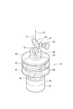
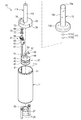
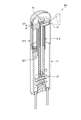
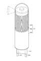
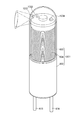
- FIG. 6A It is a perspective view explaining the drive part which moves the lens holder holding an objective lens in the endoscope of FIG. It is a perspective view which fractures
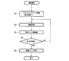
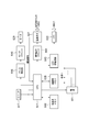
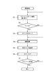
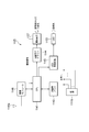
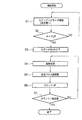
- an imaging process is performed (step S2).
- the LED 55 is driven to irradiate illumination light
- the subject light is taken into the endoscope 1 from the objective lens 17 to form an image on the light receiving surface of the imaging element 23, and read out from the imaging element 23.
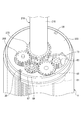
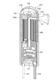
- Subject light is collected by the objective lens 17 through the cylindrical portion 313c of the translucent cover 313 and travels toward the objective mirror 16 as a parallel light flux. Then, the subject light is reflected by the reflecting surface of the objective mirror 16 and travels on the central axis of the cylindrical portion 315, in other words, on the central axis of the outer shell as a parallel light flux.
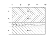
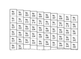
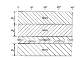
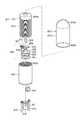
- the stepping motor 61 is driven with the designated number of pulses in step S3, the lens holder 319 is lowered and rotated, and the next field of view is “No. 002 ". Then, imaging is performed in the field of view of “No. 002”, and image data of the field of view of “No. 002” is generated from the imaging signal read from the image sensor 23.
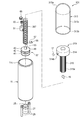
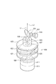
- An illumination lens 557 that deflects the illumination light so as to be a parallel light beam with respect to the half mirror 553 is provided between the LED 555 and the half mirror 553.
- the half mirror 553, the illumination lens 557, and the LED 555 are fixed in the main body 511 by appropriate support members.
- the endoscope 500 can be configured to send captured image data to an external monitor so that the captured image can be observed on-line on the external monitor and to input an operation instruction from the outside.
- the control unit 581 sends the image signal acquired from the image sensor 523 to the external video processor as it is without performing image processing, and displays the subject image image-processed by the video processor on the external monitor.
- Communication between the external video processor or external monitor and the control unit 581 may be wired or wireless. When performing wired communication, an external power source can be used by inserting a power line in the wiring.
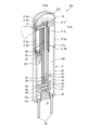
- the lens holder in the translucent cover is moved forward and backward by the drive unit, so that images can be taken from different positions on the central axis of the cylindrical portion of the lens holder and taken in from the wide-angle lens.
- the acquired image information can be accurately acquired within the moving range of the lens holder. Thereby, it is possible to easily acquire a wide range of continuous images without moving the electronic endoscope within the subject.
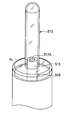
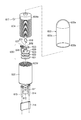
- the stepping motor 628 having the specified number of pulses is driven in step S5, and thus the cylindrical member 604b rotates by the specified number of pulses.
- the cylindrical member 604b is screwed into the main body 602 and retracted, and the next field of view is “No. 002” in FIG. 53.
- a subject image in this field of view is captured, and the image data is imaged. It is stored in the memory 626.
- the moving lens frame portion 604 is rotationally driven by the stepping motor 628.
- a motor that can accurately control the rotation angle and the rotation length is acceptable even if it is not a stepping motor. Needless to say.
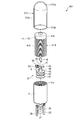
- the outer shell is formed in a cylindrical shape
- a thread groove is formed on the inner peripheral surface of the peripheral wall
- the drive mechanism includes a lens holder that supports the objective lens, and the outer A motor for rotating the lens holder about the axis of the shell as a rotation axis, wherein the lens holder is engaged with the screw groove of the outer shell.
- the present specification discloses an electronic endoscope characterized in that an image memory for receiving and storing the image data transmitted wirelessly is provided in the main body.
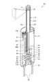
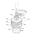
- a stepping motor 1291 is installed on the uppermost substrate (on the opposite side to the bottom 1211a side) 1243.
- a motor gear (spur gear) 1293 is attached to the rotating shaft of the stepping motor 1291.
- control unit that performs image processing on an image signal obtained by the imaging unit and an image memory that stores image data processed by the control unit are incorporated in the main body unit.
- An electronic endoscope characterized by this is disclosed.
Abstract
L'invention porte sur un endoscope électronique dotée d’une nouvelle structure permettant d’obtenir des informations d'image détaillées d'une vaste zone de façon simple et précise. L'endoscope électronique (1) comporte une enveloppe extérieure cylindrique dont la paroi périphérique est munie d’une section cylindrique transparente (13c) s'étendant en direction axiale ; un élément d'imagerie à semi-conducteurs (23) disposé dans l'enveloppe extérieure ; un système optique d'objectif comportant une lentille de focalisation (17) qui collecte une lumière d'objet à travers une section de fenêtre et forme une image sur un élément d'imagerie à semi-conducteurs ; et un mécanisme d'entraînement destiné à déplacer au moins la lentille de focalisation du système optique d'objectif le long de l'axe de l'enveloppe extérieure.
Priority Applications (5)
| Application Number | Priority Date | Filing Date | Title |
|---|---|---|---|
| US12/999,815 US8512231B2 (en) | 2008-06-17 | 2009-06-15 | Electronic endoscope including lens holder and objective mirror |
| EP09766622.6A EP2294965A4 (fr) | 2008-06-17 | 2009-06-15 | Endoscope électronique |
| US13/763,485 US20130155214A1 (en) | 2008-06-17 | 2013-02-08 | Electronic endoscope |
| US13/763,468 US20130147934A1 (en) | 2008-06-17 | 2013-02-08 | Electronic endoscope |
| US13/763,502 US20130147935A1 (en) | 2008-06-17 | 2013-02-08 | Electronic endoscope |
Applications Claiming Priority (20)
| Application Number | Priority Date | Filing Date | Title |
|---|---|---|---|
| JP2008-158013 | 2008-06-17 | ||
| JP2008158005A JP5171417B2 (ja) | 2008-06-17 | 2008-06-17 | 電子内視鏡 |
| JP2008-157991 | 2008-06-17 | ||
| JP2008-158000 | 2008-06-17 | ||
| JP2008157993A JP2009297411A (ja) | 2008-06-17 | 2008-06-17 | 電子内視鏡 |
| JP2008158006A JP2009297424A (ja) | 2008-06-17 | 2008-06-17 | 電子内視鏡 |
| JP2008158013A JP5171418B2 (ja) | 2008-06-17 | 2008-06-17 | 内視鏡 |
| JP2008-157992 | 2008-06-17 | ||
| JP2008-157999 | 2008-06-17 | ||
| JP2008157999A JP5193694B2 (ja) | 2008-06-17 | 2008-06-17 | 内視鏡及び内視鏡の作動方法 |
| JP2008-158004 | 2008-06-17 | ||
| JP2008-158002 | 2008-06-17 | ||
| JP2008157992A JP2009297410A (ja) | 2008-06-17 | 2008-06-17 | 内視鏡 |
| JP2008158002A JP2009297420A (ja) | 2008-06-17 | 2008-06-17 | 内視鏡 |
| JP2008158000A JP2009297418A (ja) | 2008-06-17 | 2008-06-17 | 内視鏡 |
| JP2008157991A JP5276907B2 (ja) | 2008-06-17 | 2008-06-17 | 内視鏡 |
| JP2008-157993 | 2008-06-17 | ||
| JP2008-158005 | 2008-06-17 | ||
| JP2008-158006 | 2008-06-17 | ||
| JP2008158004A JP5171416B2 (ja) | 2008-06-17 | 2008-06-17 | 内視鏡 |
Related Child Applications (3)
| Application Number | Title | Priority Date | Filing Date |
|---|---|---|---|
| US13/763,485 Continuation US20130155214A1 (en) | 2008-06-17 | 2013-02-08 | Electronic endoscope |
| US13/763,468 Continuation US20130147934A1 (en) | 2008-06-17 | 2013-02-08 | Electronic endoscope |
| US13/763,502 Continuation US20130147935A1 (en) | 2008-06-17 | 2013-02-08 | Electronic endoscope |
Publications (1)
| Publication Number | Publication Date |
|---|---|
| WO2009154174A1 true WO2009154174A1 (fr) | 2009-12-23 |
Family
ID=41434090
Family Applications (1)
| Application Number | Title | Priority Date | Filing Date |
|---|---|---|---|
| PCT/JP2009/060885 WO2009154174A1 (fr) | 2008-06-17 | 2009-06-15 | Endoscope électronique |
Country Status (3)
| Country | Link |
|---|---|
| US (4) | US8512231B2 (fr) |
| EP (1) | EP2294965A4 (fr) |
| WO (1) | WO2009154174A1 (fr) |
Cited By (2)
| Publication number | Priority date | Publication date | Assignee | Title |
|---|---|---|---|---|
| CZ306728B6 (cs) * | 2011-02-03 | 2017-05-31 | Karel Dušek | Zařízení pro lékařské vyšetření, zejména koloskopii |
| JP2022051712A (ja) * | 2020-09-21 | 2022-04-01 | プキョン ナショナル ユニバーシティ インダストリー-ユニバーシティ コーポレーション ファウンデーション | 子宮頸癌早期診断のためのモバイル膣拡大鏡装置 |
Families Citing this family (32)
| Publication number | Priority date | Publication date | Assignee | Title |
|---|---|---|---|---|
| CN101744601B (zh) * | 2008-12-05 | 2013-04-24 | 德昌电机(深圳)有限公司 | 胶囊式成像装置和体内图像获取系统 |
| DE102010022430A1 (de) * | 2010-06-01 | 2011-12-01 | Karl Storz Gmbh & Co. Kg | Sichtfeldvorrichtung für ein Endoskop |
| US9538908B2 (en) | 2010-09-08 | 2017-01-10 | Covidien Lp | Catheter with imaging assembly |
| WO2012078853A2 (fr) * | 2010-12-08 | 2012-06-14 | Cornell University | Appareil de type microscope, procédé et application associés |
| US8886449B2 (en) * | 2012-01-13 | 2014-11-11 | Qualcomm Incorporated | Calibrated hardware sensors for estimating real-world distances |
| US20130289362A1 (en) * | 2012-04-30 | 2013-10-31 | Empire Technology Development Llc | Infrared guide stars for endoscopic orienteering |
| US9517184B2 (en) | 2012-09-07 | 2016-12-13 | Covidien Lp | Feeding tube with insufflation device and related methods therefor |
| USD735343S1 (en) | 2012-09-07 | 2015-07-28 | Covidien Lp | Console |
| US9198835B2 (en) | 2012-09-07 | 2015-12-01 | Covidien Lp | Catheter with imaging assembly with placement aid and related methods therefor |
| USD716841S1 (en) | 2012-09-07 | 2014-11-04 | Covidien Lp | Display screen with annotate file icon |
| USD717340S1 (en) | 2012-09-07 | 2014-11-11 | Covidien Lp | Display screen with enteral feeding icon |
| ES2815566T3 (es) | 2013-10-10 | 2021-03-30 | Nitinotes Ltd | Gastroplastia con manga endoluminal |
| US10779979B2 (en) | 2013-10-10 | 2020-09-22 | Nitinotes Ltd. | Endoluminal sleeve gastroplasty |
| US9801563B2 (en) * | 2013-11-06 | 2017-10-31 | The Charles Stark Draper Laboratory, Inc. | Micro-magnetic reporter and systems |
| TWI533834B (zh) * | 2013-12-10 | 2016-05-21 | Ying-Jie Su | Magnetic manipulation of the surgical lighting device and a hand with a lighting function Assisted system |
| US20160338578A1 (en) * | 2014-01-17 | 2016-11-24 | The General Hospital Corporation | Method and apparatus for acquisition of volumetric imaging data within an anatomic structure |
| US20150346115A1 (en) * | 2014-05-30 | 2015-12-03 | Eric J. Seibel | 3d optical metrology of internal surfaces |
| CN104880410A (zh) * | 2015-06-12 | 2015-09-02 | 上海良友(集团)有限公司 | 储粮生态显微图像采集专用探视管 |
| CN205179451U (zh) * | 2015-11-10 | 2016-04-20 | 常州市巨泰电子有限公司 | 灯具的立式驱动电源 |
| CN105338703B (zh) * | 2015-11-10 | 2018-05-22 | 常州市巨泰电子有限公司 | 灯具的立式驱动电源 |
| GB2546747B (en) * | 2016-01-26 | 2021-04-07 | Ev Offshore Ltd | Optical cap |
| CN106264427B (zh) * | 2016-08-04 | 2018-03-16 | 北京千安哲信息技术有限公司 | 胶囊内窥镜及其控制装置、系统和检测方法 |
| CN106175896A (zh) * | 2016-09-09 | 2016-12-07 | 江苏科沁光电科技有限公司 | 一种电子可视取异物钳 |
| CN106345077A (zh) * | 2016-11-10 | 2017-01-25 | 广州奥方美容设备有限公司 | 超声刀手柄及超声刀美容美体仪 |
| US20200237200A1 (en) * | 2017-09-18 | 2020-07-30 | Veena Moktali | A digital device facilitating body cavity screening and diagnosis |
| US11596313B2 (en) | 2017-10-13 | 2023-03-07 | Arizona Board Of Regents On Behalf Of Arizona State University | Photoacoustic targeting with micropipette electrodes |
| US11944270B1 (en) * | 2017-11-17 | 2024-04-02 | PhotonEdge Inc. | Systems and methods of rotation compensation for brain optical imaging and stimulation |
| US20190282069A1 (en) * | 2018-03-16 | 2019-09-19 | Barbara Smith | Deep brain stimulation electrode with photoacoustic and ultrasound imaging capabilities |
| CN109106323B (zh) * | 2018-08-31 | 2024-03-26 | 上海澳华内镜股份有限公司 | 一种内窥镜照明结构及内窥镜 |
| CN110393499B (zh) * | 2018-08-31 | 2021-12-07 | 上海微创医疗机器人(集团)股份有限公司 | 电子内窥镜及电子内窥镜系统 |
| US11768182B2 (en) | 2019-04-26 | 2023-09-26 | Arizona Board Of Regents On Behalf Of Arizona State University | Photoacoustic and optical microscopy combiner and method of generating a photoacoustic image of a sample |
| CN113080833B (zh) * | 2019-12-23 | 2023-01-03 | 财团法人工业技术研究院 | 光纤扫描探头及内视镜 |
Citations (8)
| Publication number | Priority date | Publication date | Assignee | Title |
|---|---|---|---|---|
| JPH06335450A (ja) * | 1993-05-31 | 1994-12-06 | Olympus Optical Co Ltd | 電子内視鏡装置 |
| JPH10248844A (ja) * | 1997-03-12 | 1998-09-22 | Olympus Optical Co Ltd | 超音波画像診断装置 |
| JPH116904A (ja) * | 1997-06-16 | 1999-01-12 | Reatsukusu:Kk | 孔内観察用回転体レンズとこれを用いたプローブ |
| JP2002095632A (ja) * | 2000-09-22 | 2002-04-02 | Minolta Co Ltd | 内視鏡装置 |
| JP2003093339A (ja) * | 1991-03-11 | 2003-04-02 | Olympus Optical Co Ltd | 画像処理装置 |
| JP2005323889A (ja) * | 2004-05-14 | 2005-11-24 | Olympus Corp | 内視鏡 |
| JP2006235346A (ja) * | 2005-02-25 | 2006-09-07 | Mitsubishi Electric Corp | 内視鏡 |
| JP2008040468A (ja) * | 2006-07-10 | 2008-02-21 | Olympus Corp | 透過光学素子及びそれを用いた光学系 |
Family Cites Families (45)
| Publication number | Priority date | Publication date | Assignee | Title |
|---|---|---|---|---|
| US3090378A (en) * | 1960-05-16 | 1963-05-21 | Bausch & Lomb | Focusing endoscope |
| US3804081A (en) * | 1971-07-29 | 1974-04-16 | Olympus Optical Co | Endoscope |
| JPS6048011A (ja) * | 1983-08-27 | 1985-03-15 | Olympus Optical Co Ltd | 内視鏡装置 |
| JPS6349125A (ja) * | 1986-08-16 | 1988-03-01 | 奥津 一郎 | 内視鏡用案内管 |
| JP2848570B2 (ja) | 1989-12-21 | 1999-01-20 | オリンパス光学工業株式会社 | 側視型電子内視鏡 |
| US6134003A (en) | 1991-04-29 | 2000-10-17 | Massachusetts Institute Of Technology | Method and apparatus for performing optical measurements using a fiber optic imaging guidewire, catheter or endoscope |
| US5191879A (en) * | 1991-07-24 | 1993-03-09 | Welch Allyn, Inc. | Variable focus camera for borescope or endoscope |
| US5976076A (en) * | 1995-02-22 | 1999-11-02 | Kolff; Jack | Stereo laparoscope with synchronized optics |
| US6428470B1 (en) * | 1995-09-15 | 2002-08-06 | Pinotage, Llc | Imaging system and components thereof |
| US6413209B1 (en) * | 1995-09-15 | 2002-07-02 | Med Images, Inc. | Imaging system with condensation control |
| US6007484A (en) * | 1995-09-15 | 1999-12-28 | Image Technologies Corporation | Endoscope having elevation and azimuth control of camera |
| JP3549321B2 (ja) | 1996-01-24 | 2004-08-04 | 株式会社町田製作所 | 内視鏡 |
| US5782752A (en) * | 1996-04-05 | 1998-07-21 | Vista Medical Technologies, Inc. | Device for carrying two units in end to end disposition and for moving one of the units alongside the other of the units |
| JP3662072B2 (ja) | 1996-06-07 | 2005-06-22 | オリンパス株式会社 | 医療用カプセル装置 |
| US5879289A (en) * | 1996-07-15 | 1999-03-09 | Universal Technologies International, Inc. | Hand-held portable endoscopic camera |
| JPH1156774A (ja) | 1997-06-09 | 1999-03-02 | Matsushita Electric Ind Co Ltd | 撮像装置 |
| US6577339B1 (en) * | 1997-07-30 | 2003-06-10 | Pinotage, Llc | Aircraft monitoring and analysis system and method |
| US6478730B1 (en) * | 1998-09-09 | 2002-11-12 | Visionscope, Inc. | Zoom laparoscope |
| US6641530B2 (en) * | 1998-12-14 | 2003-11-04 | Fuji Photo Optical Co., Ltd. | Endoscope with objective lens drive mechanism |
| US6687010B1 (en) * | 1999-09-09 | 2004-02-03 | Olympus Corporation | Rapid depth scanning optical imaging device |
| US6767321B2 (en) * | 1999-10-04 | 2004-07-27 | Robert Czarnek | Stereo laparoscope with discrete working distance |
| US20020103420A1 (en) * | 2001-01-26 | 2002-08-01 | George Coleman | Endoscope with alterable viewing angle |
| US20020133077A1 (en) * | 2001-03-14 | 2002-09-19 | Edwardsen Stephen Dodge | Transesophageal ultrasound probe having a rotating endoscope shaft |
| JP2002282201A (ja) * | 2001-03-28 | 2002-10-02 | Asahi Optical Co Ltd | 対物光学系移動機構付き電子内視鏡 |
| WO2003038410A1 (fr) * | 2001-10-31 | 2003-05-08 | Olympus Corporation | Dispositif d'observation de type lecteur optique |
| FR2832516B1 (fr) * | 2001-11-19 | 2004-01-23 | Tokendo Sarl | Endoscopes rotatifs a visee distale deviee |
| JP2003279862A (ja) * | 2002-03-25 | 2003-10-02 | Machida Endscope Co Ltd | 全方位内視鏡装置 |
| US20040097791A1 (en) * | 2002-11-13 | 2004-05-20 | Olympus Corporation | Endoscope |
| US7559890B2 (en) * | 2003-02-26 | 2009-07-14 | Ikona Medical Corporation | Endoscopic imaging of an organ system |
| WO2004096008A2 (fr) * | 2003-05-01 | 2004-11-11 | Given Imaging Ltd. | Dispositif d'imagerie a champ panoramique |
| JP4398184B2 (ja) | 2003-06-24 | 2010-01-13 | オリンパス株式会社 | 内視鏡 |
| WO2005110189A1 (fr) * | 2004-05-14 | 2005-11-24 | Olympus Corporation | Endoscope |
| US8556806B2 (en) * | 2004-09-24 | 2013-10-15 | Vivid Medical, Inc. | Wavelength multiplexing endoscope |
| US8858425B2 (en) * | 2004-09-24 | 2014-10-14 | Vivid Medical, Inc. | Disposable endoscope and portable display |
| US8602971B2 (en) * | 2004-09-24 | 2013-12-10 | Vivid Medical. Inc. | Opto-Electronic illumination and vision module for endoscopy |
| US8827899B2 (en) * | 2004-09-24 | 2014-09-09 | Vivid Medical, Inc. | Disposable endoscopic access device and portable display |
| US8878924B2 (en) * | 2004-09-24 | 2014-11-04 | Vivid Medical, Inc. | Disposable microscope and portable display |
| US8480566B2 (en) | 2004-09-24 | 2013-07-09 | Vivid Medical, Inc. | Solid state illumination for endoscopy |
| US9033870B2 (en) * | 2004-09-24 | 2015-05-19 | Vivid Medical, Inc. | Pluggable vision module and portable display for endoscopy |
| DE102005063539B4 (de) * | 2004-11-09 | 2012-09-06 | Denso Corporation | Verfahren und Vorrichtung zum Herstellen eines Rillenrohrs und dessen Konstruktion |
| JP2007061296A (ja) | 2005-08-30 | 2007-03-15 | Pentax Corp | 電子内視鏡用受信モジュール及び画像処理装置 |
| JP4545696B2 (ja) * | 2005-09-30 | 2010-09-15 | 富士フイルム株式会社 | 光プローブ |
| US20070191682A1 (en) * | 2006-02-15 | 2007-08-16 | Jannick Rolland | Optical probes for imaging narrow vessels or lumens |
| US7627208B2 (en) * | 2007-04-23 | 2009-12-01 | Fujifilm Corporation | Optical probe and optical tomography apparatus |
| US20100013910A1 (en) * | 2008-07-21 | 2010-01-21 | Vivid Medical | Stereo viewer |
-
2009
- 2009-06-15 EP EP09766622.6A patent/EP2294965A4/fr not_active Withdrawn
- 2009-06-15 US US12/999,815 patent/US8512231B2/en not_active Expired - Fee Related
- 2009-06-15 WO PCT/JP2009/060885 patent/WO2009154174A1/fr active Application Filing
-
2013
- 2013-02-08 US US13/763,485 patent/US20130155214A1/en not_active Abandoned
- 2013-02-08 US US13/763,468 patent/US20130147934A1/en not_active Abandoned
- 2013-02-08 US US13/763,502 patent/US20130147935A1/en not_active Abandoned
Patent Citations (8)
| Publication number | Priority date | Publication date | Assignee | Title |
|---|---|---|---|---|
| JP2003093339A (ja) * | 1991-03-11 | 2003-04-02 | Olympus Optical Co Ltd | 画像処理装置 |
| JPH06335450A (ja) * | 1993-05-31 | 1994-12-06 | Olympus Optical Co Ltd | 電子内視鏡装置 |
| JPH10248844A (ja) * | 1997-03-12 | 1998-09-22 | Olympus Optical Co Ltd | 超音波画像診断装置 |
| JPH116904A (ja) * | 1997-06-16 | 1999-01-12 | Reatsukusu:Kk | 孔内観察用回転体レンズとこれを用いたプローブ |
| JP2002095632A (ja) * | 2000-09-22 | 2002-04-02 | Minolta Co Ltd | 内視鏡装置 |
| JP2005323889A (ja) * | 2004-05-14 | 2005-11-24 | Olympus Corp | 内視鏡 |
| JP2006235346A (ja) * | 2005-02-25 | 2006-09-07 | Mitsubishi Electric Corp | 内視鏡 |
| JP2008040468A (ja) * | 2006-07-10 | 2008-02-21 | Olympus Corp | 透過光学素子及びそれを用いた光学系 |
Non-Patent Citations (1)
| Title |
|---|
| See also references of EP2294965A4 * |
Cited By (2)
| Publication number | Priority date | Publication date | Assignee | Title |
|---|---|---|---|---|
| CZ306728B6 (cs) * | 2011-02-03 | 2017-05-31 | Karel Dušek | Zařízení pro lékařské vyšetření, zejména koloskopii |
| JP2022051712A (ja) * | 2020-09-21 | 2022-04-01 | プキョン ナショナル ユニバーシティ インダストリー-ユニバーシティ コーポレーション ファウンデーション | 子宮頸癌早期診断のためのモバイル膣拡大鏡装置 |
Also Published As
| Publication number | Publication date |
|---|---|
| EP2294965A1 (fr) | 2011-03-16 |
| US20130147935A1 (en) | 2013-06-13 |
| US20110098530A1 (en) | 2011-04-28 |
| US20130155214A1 (en) | 2013-06-20 |
| US20130147934A1 (en) | 2013-06-13 |
| US8512231B2 (en) | 2013-08-20 |
| EP2294965A4 (fr) | 2014-03-12 |
Similar Documents
| Publication | Publication Date | Title |
|---|---|---|
| WO2009154174A1 (fr) | Endoscope électronique | |
| JP4550048B2 (ja) | パノラマ視野の撮像装置 | |
| US20100016662A1 (en) | Radial Scanner Imaging System | |
| JP2009240634A (ja) | 内視鏡装置 | |
| JP2005319315A (ja) | パノラミックビューを有する内視鏡 | |
| CN201042429Y (zh) | 可控移动无线电药丸式内窥系统 | |
| JP5193694B2 (ja) | 内視鏡及び内視鏡の作動方法 | |
| JP5276907B2 (ja) | 内視鏡 | |
| JP2009297426A (ja) | 電子内視鏡 | |
| JP5210719B2 (ja) | 電子内視鏡及び画像処理プログラム | |
| JP2009297415A (ja) | 電子内視鏡及び画像処理プログラム | |
| JP5244471B2 (ja) | 電子内視鏡 | |
| JP5481549B2 (ja) | 内視鏡 | |
| JP5171417B2 (ja) | 電子内視鏡 | |
| JP2009297420A (ja) | 内視鏡 | |
| JP5171418B2 (ja) | 内視鏡 | |
| JP2009297410A (ja) | 内視鏡 | |
| JP5634478B2 (ja) | 内視鏡 | |
| JP5103293B2 (ja) | 電子内視鏡 | |
| JP5171416B2 (ja) | 内視鏡 | |
| JP2009297427A (ja) | 電子内視鏡 | |
| JP2009297424A (ja) | 電子内視鏡 | |
| JP4491316B2 (ja) | 口腔内撮影装置 | |
| JP2009297425A (ja) | 電子内視鏡 | |
| JP2009297412A (ja) | 電子内視鏡 |
Legal Events
| Date | Code | Title | Description |
|---|---|---|---|
| 121 | Ep: the epo has been informed by wipo that ep was designated in this application |
Ref document number: 09766622 Country of ref document: EP Kind code of ref document: A1 |
|
| REEP | Request for entry into the european phase |
Ref document number: 2009766622 Country of ref document: EP |
|
| WWE | Wipo information: entry into national phase |
Ref document number: 12999815 Country of ref document: US Ref document number: 2009766622 Country of ref document: EP |
|
| NENP | Non-entry into the national phase |
Ref country code: DE |