US9975983B2 - Bio-reducible self-assembled liquid crystalline block copolymer for drug delivery - Google Patents
Bio-reducible self-assembled liquid crystalline block copolymer for drug delivery Download PDFInfo
- Publication number
- US9975983B2 US9975983B2 US15/519,085 US201515519085A US9975983B2 US 9975983 B2 US9975983 B2 US 9975983B2 US 201515519085 A US201515519085 A US 201515519085A US 9975983 B2 US9975983 B2 US 9975983B2
- Authority
- US
- United States
- Prior art keywords
- copolymer
- dox
- nps
- encapsulated
- peo
- Prior art date
- Legal status (The legal status is an assumption and is not a legal conclusion. Google has not performed a legal analysis and makes no representation as to the accuracy of the status listed.)
- Active
Links
- 0 C.C.[1*]*(C)C Chemical compound C.C.[1*]*(C)C 0.000 description 9
- LJWUWAGVOUWCFK-UHFFFAOYSA-N CC(C)(C)C(=O)COC(=O)C(C)(C)C.CC(C)(C)C(=O)COC(=O)C(C)(C)C.CC(C)(C)COC(=O)C(C)(C)C.CC(C)(C)COC(=O)C(C)(C)C.CC(OC(=O)C(C)OC(=O)C(C)C(C)(C)C)C(=O)OC(C)(C)C Chemical compound CC(C)(C)C(=O)COC(=O)C(C)(C)C.CC(C)(C)C(=O)COC(=O)C(C)(C)C.CC(C)(C)COC(=O)C(C)(C)C.CC(C)(C)COC(=O)C(C)(C)C.CC(OC(=O)C(C)OC(=O)C(C)C(C)(C)C)C(=O)OC(C)(C)C LJWUWAGVOUWCFK-UHFFFAOYSA-N 0.000 description 3
- AMQIDTYZUOWFMR-UHFFFAOYSA-N CC(C)(C)C(=O)COC(=O)C(C)(C)C.CC(C)(C)COC(=O)C(C)(C)C Chemical compound CC(C)(C)C(=O)COC(=O)C(C)(C)C.CC(C)(C)COC(=O)C(C)(C)C AMQIDTYZUOWFMR-UHFFFAOYSA-N 0.000 description 2
- ANVYVNUDKBUJMG-UHFFFAOYSA-N CC(C)(C)OC(=O)CSSOC(=O)C(C)(C)C(C)(C)C Chemical compound CC(C)(C)OC(=O)CSSOC(=O)C(C)(C)C(C)(C)C ANVYVNUDKBUJMG-UHFFFAOYSA-N 0.000 description 2
- XXLVMBLZXUDZHQ-USLSFYNUSA-N [H][C@]12CC[C@]3(C)[C@@]4(C)CCC(OC(=O)COC(=O)C(C)(C)CC(C)(C)C(=O)OSSCC(=O)OCCOC)CC4=CC[C@@]3([H])[C@]1(C)CC[C@]2(C)C(C)(C)CCCC(C)C.[H][C@]12CC[C@]3(C)[C@@]4(C)CCC(OC(=O)COC(=O)C(C)CC(C)(C)C(=O)OSSCC(=O)OCCOC)CC4=CC[C@@]3([H])[C@]1(C)CC[C@]2(C)C(C)(C)CCCC(C)C Chemical compound [H][C@]12CC[C@]3(C)[C@@]4(C)CCC(OC(=O)COC(=O)C(C)(C)CC(C)(C)C(=O)OSSCC(=O)OCCOC)CC4=CC[C@@]3([H])[C@]1(C)CC[C@]2(C)C(C)(C)CCCC(C)C.[H][C@]12CC[C@]3(C)[C@@]4(C)CCC(OC(=O)COC(=O)C(C)CC(C)(C)C(=O)OSSCC(=O)OCCOC)CC4=CC[C@@]3([H])[C@]1(C)CC[C@]2(C)C(C)(C)CCCC(C)C XXLVMBLZXUDZHQ-USLSFYNUSA-N 0.000 description 2
- VQGJFGWPDDMUNN-ORDZVYLRSA-N [H][C@]12CC[C@]3(C)[C@@]4(C)CCC(OCOC(=O)C(C)(C)CC(C)(C)C(=O)OSSCC(=O)OCCOC)CC4=CC[C@@]3([H])[C@]1(C)CC[C@]2(C)C(C)(C)CCCC(C)C.[H][C@]12CC[C@]3(C)[C@@]4(C)CCC(OCOC(=O)C(C)CC(C)(C)C(=O)OSSCC(=O)OCCOC)CC4=CC[C@@]3([H])[C@]1(C)CC[C@]2(C)C(C)(C)CCCC(C)C Chemical compound [H][C@]12CC[C@]3(C)[C@@]4(C)CCC(OCOC(=O)C(C)(C)CC(C)(C)C(=O)OSSCC(=O)OCCOC)CC4=CC[C@@]3([H])[C@]1(C)CC[C@]2(C)C(C)(C)CCCC(C)C.[H][C@]12CC[C@]3(C)[C@@]4(C)CCC(OCOC(=O)C(C)CC(C)(C)C(=O)OSSCC(=O)OCCOC)CC4=CC[C@@]3([H])[C@]1(C)CC[C@]2(C)C(C)(C)CCCC(C)C VQGJFGWPDDMUNN-ORDZVYLRSA-N 0.000 description 2
- DCZAGZYEJQCZEA-UHFFFAOYSA-N CC(C)(C)C(=O)CCSSCNC(=O)C(C)(C)C.CC(C)(C)C(=O)OCCSSOC(=O)C(C)(C)C.CC(C)(C)OC(=O)CSSOC(=O)C(C)(C)C(C)(C)C.CC(C)SSC(C)(C)C Chemical compound CC(C)(C)C(=O)CCSSCNC(=O)C(C)(C)C.CC(C)(C)C(=O)OCCSSOC(=O)C(C)(C)C.CC(C)(C)OC(=O)CSSOC(=O)C(C)(C)C(C)(C)C.CC(C)SSC(C)(C)C DCZAGZYEJQCZEA-UHFFFAOYSA-N 0.000 description 1
- GCENZUWEDQPVSB-UHFFFAOYSA-N CC(C)(C)C(=O)COC(=O)C(C)(C)C.CC(C)(C)C(=O)COC(=O)C(C)(C)C Chemical compound CC(C)(C)C(=O)COC(=O)C(C)(C)C.CC(C)(C)C(=O)COC(=O)C(C)(C)C GCENZUWEDQPVSB-UHFFFAOYSA-N 0.000 description 1
- PCSDUQJLSTXHRI-UHFFFAOYSA-N CC(C)(C)C(=O)OCCSSC1=CC=CC=N1.CC(C)(C)SSC1=CC=CC=N1 Chemical compound CC(C)(C)C(=O)OCCSSC1=CC=CC=N1.CC(C)(C)SSC1=CC=CC=N1 PCSDUQJLSTXHRI-UHFFFAOYSA-N 0.000 description 1
- UIWKNVUAIIPEQA-UHFFFAOYSA-N CC(C)(C)C(OCCSSc1ccccn1)=O Chemical compound CC(C)(C)C(OCCSSc1ccccn1)=O UIWKNVUAIIPEQA-UHFFFAOYSA-N 0.000 description 1
- FSRAGORHGUZQKI-UHFFFAOYSA-N CC(C)(C)SSc1ccccn1 Chemical compound CC(C)(C)SSc1ccccn1 FSRAGORHGUZQKI-UHFFFAOYSA-N 0.000 description 1
- SJFPEMAPDCOVIZ-NIWPXVBPSA-N CCCCCCCCCCCCSC(=S)SC(C)(C)C(=O)O.CCCCCCCCCCCCSC(=S)SC(C)(C)C(=O)OCCOC.COCCO.[H][C@@]12CC=C3CC(OC(=O)CCCCCOC(=O)C(=C)C)CC[C@]3(C)[C@@]1([H])CC[C@@]1(C)[C@@]2([H])CC[C@]1([H])[C@]([H])(C)CCCC(C)C.[H][C@@]12CC=C3CC(OC(=O)COC(=O)C(C)(CC(C)(C)C(=O)OCCOC)SC(=S)SCCCCCCCCCCCC)CC[C@]3(C)[C@@]1([H])CC[C@@]1(C)[C@@]2([H])CC[C@]1([H])C(C)(C)CCCC(C)C Chemical compound CCCCCCCCCCCCSC(=S)SC(C)(C)C(=O)O.CCCCCCCCCCCCSC(=S)SC(C)(C)C(=O)OCCOC.COCCO.[H][C@@]12CC=C3CC(OC(=O)CCCCCOC(=O)C(=C)C)CC[C@]3(C)[C@@]1([H])CC[C@@]1(C)[C@@]2([H])CC[C@]1([H])[C@]([H])(C)CCCC(C)C.[H][C@@]12CC=C3CC(OC(=O)COC(=O)C(C)(CC(C)(C)C(=O)OCCOC)SC(=S)SCCCCCCCCCCCC)CC[C@]3(C)[C@@]1([H])CC[C@@]1(C)[C@@]2([H])CC[C@]1([H])C(C)(C)CCCC(C)C SJFPEMAPDCOVIZ-NIWPXVBPSA-N 0.000 description 1
- MGCQKFPGDFTHCJ-DFVIDUIVSA-N CCCCCCCCCCCCSC(=S)SC(C)(C)C(=O)OCCSSC1=NC=CC=C1.CCCCCCCCCCCCSC(=S)SC(C)(C)C(=O)SSCC(=O)OCCOC.COCCO.COCCOC(=O)CS.COCCOC(=O)CS.O=C(O)CS.[H][C@@]12CC=C3CC(OC(=O)CCCCCOC(=O)C(=C)C)CC[C@]3(C)[C@@]1([H])CC[C@@]1(C)[C@@]2([H])CC[C@]1([H])[C@]([H])(C)CCCC(C)C Chemical compound CCCCCCCCCCCCSC(=S)SC(C)(C)C(=O)OCCSSC1=NC=CC=C1.CCCCCCCCCCCCSC(=S)SC(C)(C)C(=O)SSCC(=O)OCCOC.COCCO.COCCOC(=O)CS.COCCOC(=O)CS.O=C(O)CS.[H][C@@]12CC=C3CC(OC(=O)CCCCCOC(=O)C(=C)C)CC[C@]3(C)[C@@]1([H])CC[C@@]1(C)[C@@]2([H])CC[C@]1([H])[C@]([H])(C)CCCC(C)C MGCQKFPGDFTHCJ-DFVIDUIVSA-N 0.000 description 1
- DDBKFMBTYXUAFB-JFDJFCAFSA-N [H][C@@]12CC=C3CC(OC(=O)COC(=O)C(C)(C)CC)CC[C@]3(C)[C@@]1(C)CC[C@@]1([H])[C@@]2(C)CC[C@]1(C)[C@]([H])(C)CCCC(C)C.[H][C@@]12CC=C3CC(OC(=O)COC(=O)C(C)CC)CC[C@]3(C)[C@@]1(C)CC[C@@]1([H])[C@@]2(C)CC[C@]1(C)[C@]([H])(C)CCCC(C)C.[H][C@@]12CC=C3CC(OCOC(=O)C(C)(C)CC)CC[C@]3(C)[C@@]1(C)CC[C@@]1([H])[C@@]2(C)CC[C@]1(C)[C@]([H])(C)CCCC(C)C.[H][C@@]12CC=C3CC(OCOC(=O)C(C)CC)CC[C@]3(C)[C@@]1(C)CC[C@@]1([H])[C@@]2(C)CC[C@]1(C)[C@]([H])(C)CCCC(C)C Chemical compound [H][C@@]12CC=C3CC(OC(=O)COC(=O)C(C)(C)CC)CC[C@]3(C)[C@@]1(C)CC[C@@]1([H])[C@@]2(C)CC[C@]1(C)[C@]([H])(C)CCCC(C)C.[H][C@@]12CC=C3CC(OC(=O)COC(=O)C(C)CC)CC[C@]3(C)[C@@]1(C)CC[C@@]1([H])[C@@]2(C)CC[C@]1(C)[C@]([H])(C)CCCC(C)C.[H][C@@]12CC=C3CC(OCOC(=O)C(C)(C)CC)CC[C@]3(C)[C@@]1(C)CC[C@@]1([H])[C@@]2(C)CC[C@]1(C)[C@]([H])(C)CCCC(C)C.[H][C@@]12CC=C3CC(OCOC(=O)C(C)CC)CC[C@]3(C)[C@@]1(C)CC[C@@]1([H])[C@@]2(C)CC[C@]1(C)[C@]([H])(C)CCCC(C)C DDBKFMBTYXUAFB-JFDJFCAFSA-N 0.000 description 1
- XIUARFQDURQJID-GFGUIPKPSA-N [H][C@@]12CC=C3CC(OC(=O)COC(=O)C(C)(CC(C)(C)C(=O)OCCOC)SC(=S)SCCCCCCCCCCCC)CC[C@]3(C)[C@@]1([H])CC[C@@]1(C)[C@@]2([H])CC[C@]1([H])C(C)(C)CCCC(C)C Chemical compound [H][C@@]12CC=C3CC(OC(=O)COC(=O)C(C)(CC(C)(C)C(=O)OCCOC)SC(=S)SCCCCCCCCCCCC)CC[C@]3(C)[C@@]1([H])CC[C@@]1(C)[C@@]2([H])CC[C@]1([H])C(C)(C)CCCC(C)C XIUARFQDURQJID-GFGUIPKPSA-N 0.000 description 1
- GEIIVDVFIXKPJG-MYURHXKKSA-N [H][C@@]12CC=C3CC(OC(=O)COC(=O)C(C)(CC(C)(C)C(=O)OSSCC(=O)OCCOC)SC(=S)SCCCCCCCCCCCC)CC[C@]3(C)[C@@]1([H])CC[C@@]1(C)[C@@]2([H])CC[C@]1([H])C(C)(C)CCCC(C)C Chemical compound [H][C@@]12CC=C3CC(OC(=O)COC(=O)C(C)(CC(C)(C)C(=O)OSSCC(=O)OCCOC)SC(=S)SCCCCCCCCCCCC)CC[C@]3(C)[C@@]1([H])CC[C@@]1(C)[C@@]2([H])CC[C@]1([H])C(C)(C)CCCC(C)C GEIIVDVFIXKPJG-MYURHXKKSA-N 0.000 description 1
- CFFJCVZAZTVRJE-LESWKBAUSA-N [H][C@]12CC[C@]3(C)[C@@]4(C)CCC(OC(=O)COC(=O)C(C)(C)CC)CC4=CC[C@@]3([H])[C@]1(C)CC[C@]2(C)C(C)(C)CCCC(C)C.[H][C@]12CC[C@]3(C)[C@@]4(C)CCC(OC(=O)COC(=O)C(C)CC)CC4=CC[C@@]3([H])[C@]1(C)CC[C@]2(C)C(C)(C)CCCC(C)C.[H][C@]12CC[C@]3(C)[C@@]4(C)CCC(OCOC(=O)C(C)(C)CC)CC4=CC[C@@]3([H])[C@]1(C)CC[C@]2(C)C(C)(C)CCCC(C)C.[H][C@]12CC[C@]3(C)[C@@]4(C)CCC(OCOC(=O)C(C)(C)CC)CC4=CC[C@@]3([H])[C@]1(C)CC[C@]2(C)C(C)(C)CCCC(C)C Chemical compound [H][C@]12CC[C@]3(C)[C@@]4(C)CCC(OC(=O)COC(=O)C(C)(C)CC)CC4=CC[C@@]3([H])[C@]1(C)CC[C@]2(C)C(C)(C)CCCC(C)C.[H][C@]12CC[C@]3(C)[C@@]4(C)CCC(OC(=O)COC(=O)C(C)CC)CC4=CC[C@@]3([H])[C@]1(C)CC[C@]2(C)C(C)(C)CCCC(C)C.[H][C@]12CC[C@]3(C)[C@@]4(C)CCC(OCOC(=O)C(C)(C)CC)CC4=CC[C@@]3([H])[C@]1(C)CC[C@]2(C)C(C)(C)CCCC(C)C.[H][C@]12CC[C@]3(C)[C@@]4(C)CCC(OCOC(=O)C(C)(C)CC)CC4=CC[C@@]3([H])[C@]1(C)CC[C@]2(C)C(C)(C)CCCC(C)C CFFJCVZAZTVRJE-LESWKBAUSA-N 0.000 description 1
Images




Classifications
-
- C—CHEMISTRY; METALLURGY
- C08—ORGANIC MACROMOLECULAR COMPOUNDS; THEIR PREPARATION OR CHEMICAL WORKING-UP; COMPOSITIONS BASED THEREON
- C08F—MACROMOLECULAR COMPOUNDS OBTAINED BY REACTIONS ONLY INVOLVING CARBON-TO-CARBON UNSATURATED BONDS
- C08F293/00—Macromolecular compounds obtained by polymerisation on to a macromolecule having groups capable of inducing the formation of new polymer chains bound exclusively at one or both ends of the starting macromolecule
- C08F293/005—Macromolecular compounds obtained by polymerisation on to a macromolecule having groups capable of inducing the formation of new polymer chains bound exclusively at one or both ends of the starting macromolecule using free radical "living" or "controlled" polymerisation, e.g. using a complexing agent
-
- A—HUMAN NECESSITIES
- A61—MEDICAL OR VETERINARY SCIENCE; HYGIENE
- A61K—PREPARATIONS FOR MEDICAL, DENTAL OR TOILETRY PURPOSES
- A61K31/00—Medicinal preparations containing organic active ingredients
- A61K31/70—Carbohydrates; Sugars; Derivatives thereof
- A61K31/7028—Compounds having saccharide radicals attached to non-saccharide compounds by glycosidic linkages
- A61K31/7034—Compounds having saccharide radicals attached to non-saccharide compounds by glycosidic linkages attached to a carbocyclic compound, e.g. phloridzin
- A61K31/704—Compounds having saccharide radicals attached to non-saccharide compounds by glycosidic linkages attached to a carbocyclic compound, e.g. phloridzin attached to a condensed carbocyclic ring system, e.g. sennosides, thiocolchicosides, escin, daunorubicin
-
- A—HUMAN NECESSITIES
- A61—MEDICAL OR VETERINARY SCIENCE; HYGIENE
- A61K—PREPARATIONS FOR MEDICAL, DENTAL OR TOILETRY PURPOSES
- A61K9/00—Medicinal preparations characterised by special physical form
- A61K9/48—Preparations in capsules, e.g. of gelatin, of chocolate
- A61K9/50—Microcapsules having a gas, liquid or semi-solid filling; Solid microparticles or pellets surrounded by a distinct coating layer, e.g. coated microspheres, coated drug crystals
- A61K9/51—Nanocapsules; Nanoparticles
- A61K9/5107—Excipients; Inactive ingredients
- A61K9/513—Organic macromolecular compounds; Dendrimers
- A61K9/5138—Organic macromolecular compounds; Dendrimers obtained by reactions only involving carbon-to-carbon unsaturated bonds, e.g. polyvinyl pyrrolidone, poly(meth)acrylates
-
- A—HUMAN NECESSITIES
- A61—MEDICAL OR VETERINARY SCIENCE; HYGIENE
- A61K—PREPARATIONS FOR MEDICAL, DENTAL OR TOILETRY PURPOSES
- A61K9/00—Medicinal preparations characterised by special physical form
- A61K9/48—Preparations in capsules, e.g. of gelatin, of chocolate
- A61K9/50—Microcapsules having a gas, liquid or semi-solid filling; Solid microparticles or pellets surrounded by a distinct coating layer, e.g. coated microspheres, coated drug crystals
- A61K9/51—Nanocapsules; Nanoparticles
- A61K9/5107—Excipients; Inactive ingredients
- A61K9/513—Organic macromolecular compounds; Dendrimers
- A61K9/5146—Organic macromolecular compounds; Dendrimers obtained otherwise than by reactions only involving carbon-to-carbon unsaturated bonds, e.g. polyethylene glycol, polyamines, polyanhydrides
-
- A—HUMAN NECESSITIES
- A61—MEDICAL OR VETERINARY SCIENCE; HYGIENE
- A61K—PREPARATIONS FOR MEDICAL, DENTAL OR TOILETRY PURPOSES
- A61K9/00—Medicinal preparations characterised by special physical form
- A61K9/48—Preparations in capsules, e.g. of gelatin, of chocolate
- A61K9/50—Microcapsules having a gas, liquid or semi-solid filling; Solid microparticles or pellets surrounded by a distinct coating layer, e.g. coated microspheres, coated drug crystals
- A61K9/51—Nanocapsules; Nanoparticles
- A61K9/5192—Processes
-
- A—HUMAN NECESSITIES
- A61—MEDICAL OR VETERINARY SCIENCE; HYGIENE
- A61P—SPECIFIC THERAPEUTIC ACTIVITY OF CHEMICAL COMPOUNDS OR MEDICINAL PREPARATIONS
- A61P35/00—Antineoplastic agents
-
- C—CHEMISTRY; METALLURGY
- C08—ORGANIC MACROMOLECULAR COMPOUNDS; THEIR PREPARATION OR CHEMICAL WORKING-UP; COMPOSITIONS BASED THEREON
- C08F—MACROMOLECULAR COMPOUNDS OBTAINED BY REACTIONS ONLY INVOLVING CARBON-TO-CARBON UNSATURATED BONDS
- C08F290/00—Macromolecular compounds obtained by polymerising monomers on to polymers modified by introduction of aliphatic unsaturated end or side groups
- C08F290/02—Macromolecular compounds obtained by polymerising monomers on to polymers modified by introduction of aliphatic unsaturated end or side groups on to polymers modified by introduction of unsaturated end groups
- C08F290/06—Polymers provided for in subclass C08G
- C08F290/062—Polyethers
-
- B—PERFORMING OPERATIONS; TRANSPORTING
- B82—NANOTECHNOLOGY
- B82Y—SPECIFIC USES OR APPLICATIONS OF NANOSTRUCTURES; MEASUREMENT OR ANALYSIS OF NANOSTRUCTURES; MANUFACTURE OR TREATMENT OF NANOSTRUCTURES
- B82Y5/00—Nanobiotechnology or nanomedicine, e.g. protein engineering or drug delivery
-
- C—CHEMISTRY; METALLURGY
- C08—ORGANIC MACROMOLECULAR COMPOUNDS; THEIR PREPARATION OR CHEMICAL WORKING-UP; COMPOSITIONS BASED THEREON
- C08F—MACROMOLECULAR COMPOUNDS OBTAINED BY REACTIONS ONLY INVOLVING CARBON-TO-CARBON UNSATURATED BONDS
- C08F2438/00—Living radical polymerisation
- C08F2438/03—Use of a di- or tri-thiocarbonylthio compound, e.g. di- or tri-thioester, di- or tri-thiocarbamate, or a xanthate as chain transfer agent, e.g . Reversible Addition Fragmentation chain Transfer [RAFT] or Macromolecular Design via Interchange of Xanthates [MADIX]
Definitions
- the disclosure provides biodegradable amphiphilic liquid crystalline copolymers that can readily self-assemble to nanoparticles in aqueous solutions and also allow for encapsulation of hydrophobic pharmaceutically active molecules.
- Copolymers with cholesterol end-groups have also generated interest for various biomedical applications including serving as membranes for cell attachment and proliferation, forming the basis of polymeric scaffolds, and as materials with improved blood compatibility. But, reported amphiphilic polymer architectures that contain cholesterol are conjugates or linear copolymers with only one or a few cholesterol molecules. This results in low stability, limited drug loading capacity to 20% (w/w) with low encapsulation efficiency and fast drug release for these cholesterol-containing copolymers.
- Redox-Sensitive nanocarriers containing disulfide bonds have received much attention for intracellular drug delivery due to the existence of a high glutathione (GSH) concentration in the tumor microenvironment and cancer cells.
- GSH glutathione
- several groups have reported redox-sensitive polymer/DNA complexes, polyion complex micelles for siRNA delivery, crosslinked micelles, and degradable nanogels with good stability under the physiological conditions that, rapidly released encapsulated drugs in the intracellular reductive environment.
- limited information is available on the in vivo behavior of nanoparticles comprised of reductive-sensitive polymers and the interaction between the nanoparticles and tumor tissue.
- LCPs Liquid crystalline polymers
- the present invention provides novel a biodegradable amphiphilic liquid crystalline copolymer (“copolymer” or “block copolymer”).
- copolymer or “block copolymer”.
- the block copolymers of the disclosure readily formed self-assembled nanoparticles in aqueous solutions. These nanoparticles allowed for loading of hydrophobic drugs simply via self-assembly without sonication or homogenization procedure.
- the nanoparticles of the disclosure also showed excellent stability of the high steroid content hydrophobic core and demonstrated a high capacity for encapsulation of hydrophobic drugs.
- the hydrophilic surface also protected from reticuloendo-thelial system (RES) uptake and facilitated long circulation in body.
- RES reticuloendo-thelial system
- the self-assembled block copolymer nanoparticles of the invention have good biocompatibility, high drug loading capacity, excellent stability, and can be easily manufactured in large scale, which make them suitable for drug delivery especially delivering anti-cancer drugs to tumors.
- the nanoparticles of the invention while being stable under physiological conditions rapidly release the encapsulated drug by cleavage of disulfide linkage under a cytosolic reducing environment following cellular entry, resulting in effective cytotoxicity to cancer cells ( FIG. 1 ).
- the nanoparticles are capable of rapidly releasing the drugs inside the cells to yield significantly enhanced drug efficacy as compared to the non-reductive nanoparticles due to the cleavage of disulfide bonds when exposed to an intracellular reductive environment. Furthermore, the nanoparticles significantly increased the duration of the drug in the circulation, improved tumor accumulation and antitumor efficacy, significantly reduced toxicity compared to the free anticancer drug, and decreased cardiac accumulation of the drug. These properties make the nanoparticles of the disclosure especially suitable for use in anti-cancer drug delivery.
- the copolymers of the disclosure may be functionalized (for example, with thiol, phosphate, carboxylic acid groups, etc.), and such copolymers also self-assembled in aqueous media to form well-defined nanoparticles.
- the thiol functionalized nanoparticles served as a multifunctional carrier for dual encapsulation of hydrophobic anticancer drug via physical entrapment and gold nanoparticles (Au NPs) via covalent bonding to the thiol groups.
- Au NPs gold nanoparticles
- High drug loading and high encapsulation efficiency, along with uniform size distribution and good stability, allow the functionalized nanoparticles to be used for the delivery of anticancer drug and metal nanoparticles, for example in photothermal cancer therapy and biological sensing.
- the disclosure provides a biodegradable amphiphilic liquid crystalline copolymer (“copolymer” or “block copolymer”) that can readily self-assemble to nanoparticles in aqueous solutions.
- copolymer or “block copolymer”
- a copolymer comprising:
- n is an integer about 3 to about 500;
- A is independently selected from polyacrylate, polymethacrylate, polynorbonene, polycyclopentene, polycyclooctene, polysiloxane, polyester, or polypeptide;
- R 1 is a steroid moiety optionally comprising a linker R 11 ;
- R 2 is polyalkylene oxide, polyester, or polypeptide moiety
- R 3 is a disulfide linker moiety.
- the disclosure provides the block copolymers of the disclosure in a core/shell nanoparticle form.
- the core/shell nanoparticle form is wherein the block copolymers of the disclosure self-assembled in aqueous solutions.
- the disclosure provides a nanoparticle comprising the block copolymer of the disclosure and a hydrophobic pharmaceutically active molecule.
- Another aspect provides a therapeutic delivery system comprising this nanoparticle.
- Yet another aspect provides a method of delivering a pharmaceutically active molecule, comprising administering to a subject this nanoparticle.
- Yet another aspect provides a method of treating a disease or disorder, comprising administering to a subject this nanoparticle. For example, if the hydrophobic pharmaceutically active molecule is an anti-cancer drug, then the disease or disorder is cancer.
- the disclosure also provides a process for preparing a nanoparticle of the disclosure: comprising (a) dissolving a block copolymer of the disclosure in an organic solvent to obtain a copolymer solution; and (b) mixing the copolymer solution in an aqueous solution to form the nanoparticle.
- FIG. 1 illustrates reduction-sensitive in aqueous media of PC5MA-SS-PEO NPs for intracellular drug release.
- FIG. 2 shows TEM images of ( a ) blank thioester-NPs, ( b ) blank SS-NPs, ( c ) DOX-encapsulated thioester-NPs, and ( d ) DOX-encapsulated SS-NPs.
- FIG. 3 shows the particle size distribution of PC5MA-SS-PEO NPs after incubation with or without 10 mM of DTT solution.
- FIG. 4 ( a ) shows stability of DOX-encapsulated thioester-NPs and DOX-encapsulated SS-NPs in PBS/FBS (1:1) stored at 4° C.
- FIG. 4 ( b ) shows release profiles of DOX-encapsulated thioester NPs and DOX-encapsulated SS-NPs with and without DTT in PBS buffer (pH 7.4, 10 mM) at 37° C.
- FIG. 5 shows confocal laser scanning microscopy (CLSM) images of A549 and fibroblast (NIH3T3) cells incubated with free DOX, DOX-encapsulated thioester-NPs, and DOX-encapsulated SS-NPs for 30 min ( a ), 2 h ( b ) and 4 h ( c ) at 10 ⁇ g/mL DOX equivalence. Scale bars are 10 ⁇ m. Blue-nuclei stained with DAPI; red-DOX.
- CLSM confocal laser scanning microscopy
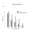
- FIG. 6 shows viability of A549 cells incubated with blank thioester-NPs and blank SS-NPs for 24 h ( a ), with free DOX, DOX-encapsulated thioester-NPs, and DOX-encapsulated SS-NPs ( b ), and cell viability of NIH3T3 cells incubated with free DOX, DOX-encapsulated thioester-NPs, and DOX-encapsulated SS-NPs ( c ) at different concentrations of DOX for 24 h.
- FIG. 7 shows CLSM images of HeLa cells incubated with free DOX, DOX-encapsulated thioester nanoparticles, and DOX-encapsulated SS nanoparticles for 2 h and 4 h at 10 ⁇ g/mL DOX equivalence.
- FIG. 8 shows viability of Hela cells incubated with free DOX, DOX-encapsulated thioester-NPs, and DOX-encapsulated SS-NPs at different concentrations of DOX for 4 h.
- FIG. 9 shows ( a ) in vivo fluorescence images of DiR-encapsulated SS-NPs in tumor-bearing SCID mice at 1 h, 3 h, 6 h, 24 h, 48 h, and 72 h-post intravenous injection; and ( b ) ex vivo images of tumors and organs at 72 h-post injection.
- the methods described herein can be configured by the person of ordinary skill in the art to meet the desired need.
- the copolymers of disclosure comprise of a steroid-containing block and a polyalkylene oxide-, polyester-, or polypeptide moiety-containing block.
- Such copolymers readily self-assemble as nanoparticles in aqueous solutions without sonication or homogenization, and have good biocompatibility, high drug loading capacity, long retention in the circulation, multimodality potential and can be easily manufactured in large scale.
- the nanostructures of the disclosure may be used to encapsulate a hydrophobic therapeutically active molecule, such as anti-cancer drugs.
- the nanoparticles encapsulating anticancer drug showed high tumor accumulation and antitumor efficacy with significantly reduced toxicity compared to the free anticancer drug.
- the block copolymers of the disclosure may be functionalized (for example, with thiol), and such copolymers also self-assembled in aqueous media to form well-defined nanoparticles with the functional group.
- the thiol functionalized nanoparticles served as a multifunctional carrier for dual encapsulation of hydrophobic anticancer drug via physical entrapment and gold nanoparticles (Au NPs) via covalent bonding to the thiol groups. These dual nanoparticles exhibited high drug loading, high encapsulation efficiency, uniform size distribution, and good stability.
- a copolymer that doesn't contain disulfide bond was also synthesized and compared in vitro and in vivo. Both amphiphilic liquid crystalline polymers self-assembled in aqueous media to form bioreducible and non-reducible nanoparticles. The resulting disulfide-containing nanoparticles of the disclosure possessed enhanced stability under extracellular environment and exhibited rapid drug release under an intracellular reductive condition.
- the block copolymers of the disclosure require that the first block comprises a steroid moiety optionally comprising a linker.
- Suitable steroids may be selected to meet the desired need.
- the steroid moiety suitable in the materials of the disclosure comprises cholesterol, cholic acid, deoxycholic acid, taurocholic acid, lanosterol, estradiol, testosterone, bile acid, dexamethasone, secosteroid, phytosterol, or the like.
- the steroid moiety is selected from cholesterol, cholic acid, deoxycholic acid, and taurocholic acid.
- the steroid moiety comprises cholesterol.
- the steroid-containing first block may be present from about 1% to about 80% of the total weight of the block copolymer (i.e., weight fraction of about 1% to about 80%.)
- the weight fraction of the first block may be more that 50%, or less than 50%, or from about 5% to about 70%, or about 40% to about 70%, or about 40% to about 50%, or about 60% to about 70%, or about 2% to about 30%, or about 3% to about 30%, or about 5% to about 30%, or about 2% to about 20%, or about 3% to about 20%, or about 5% to about 20%, or about 7% to about 20%, based on the total weight of the block copolymer.
- the steroid moiety may be connected to the polymer back bone via a suitable linker R 11 .
- linker R 11 include, but are not limited to:
- the linker at R 11 is
- linker at R 11 is:
- the linker at R 11 is N-(2-aminoethyl)-2-aminoethyl-N-(2-aminoethyl)-2-aminoethyl-N-(2-aminoethyl)-2-aminoethyl-N-(2-aminoethyl)-2-aminoethyl-N-(2-aminoethyl)-2-aminoe
- the linker at R 11 is
- the block copolymers of the disclosure require a backbone moiety A.
- the block copolymers described herein may contain, for example, polyacrylate, polymethacrylate, polynorbonene, polycyclopentene, polycyclooctene, polysiloxane, polyester, and polypeptide backbone A available to one skill in the art, and may be varied depending on the desired product.
- the block copolymers of disclosure are those wherein each A is independently polyacrylate, polymethacrylate, or polyester.
- each A is independently polyacrylate or polymethacrylate.
- each A is independently polyacrylate.
- each A is independently polymethacrylate.
- each A is independently polyester.
- the first block is of formula:
- the block copolymers of the disclosure require that the second block is
- the second block comprises R 2 moiety, which may be polyalkylene oxide, polyester, or polypeptide moiety.
- R 2 is polyalkylene oxide moiety. Suitable polyalkylene oxides may be selected to meet the desired need.
- the polyalkylene oxide moiety comprises polyethylene oxide, polyethylene oxide thiolate, polypropylene oxide, or polypropylene oxide thiolate.
- the polyalkylene oxide moiety comprises polyethylene oxide or polyethylene oxide thiolate.
- the polyalkylene oxide moiety comprises polyethylene oxide.
- R 2 is polyester moiety.
- Suitable polyesters include polymers that contain the ester functional group in their main chain. Examples include, but are not limited to, polylactides, polyglycolides, polycaprolactones, and the like.
- R 2 is polypeptide moiety.
- Suitable polypeptides include one or more chains of amino acid monomers linked together by peptide (amide) bonds, and may comprise L-amino acids, D-amino acids (which are resistant to L-amino acid-specific proteases in vivo), or a combination of D- and L-amino acids.
- polypeptides described herein refer to a chain less than about 100 amino acids in length.
- the polypeptides described herein may be chemically synthesized or recombinantly expressed.
- the second block may be present from about 20% to about 99% of the total weight of the block copolymer (i.e., weight fraction of about 20% to about 99%.)
- the weight fraction of the second block may be more that 50%, or less than 50%, or from about 30% to about 95%, or about 30% to about 60%, or about 50% to about 60%, or about 30% to about 40%, or about 70% to about 98%, or about 70% to about 97%, or about 70% to about 95%, or about 80% to about 98%, or about 8% to about 97%, or about 80% to about 95%, or about 80% to about 93%, based on the total weight of the block copolymer.
- the second block also comprises R 3 linker moiety comprising reducible disulfide bonds.
- R 3 is selected from the group consisting of:
- R 3 is
- R 3 is derived from
- the copolymer of the disclosure may further comprise a chain terminus moiety X:
- X is a trithiocarbonate, dithiocarbamate, or dithioester. In another embodiment, X is —SC(S)S—(C 1 -C 24 alkyl). In another embodiment, X is —SC(S)S—C 12 H 25 .
- the copolymer of the disclosure comprises polyacrylate or polymethacrylate bearing cholesterol block and polyalkylene oxide block with reducible disulfide bonds. In one embodiment, the copolymer of the disclosure comprises polyacrylate or polymethacrylate bearing cholesterol block and polyethylene glycol block with reducible disulfide bonds.
- block copolymers of the disclosure comprise the structure:
- n is an integer between about 5 and about 100.
- m and n may be selected by one of skill in the art and may be varied depending on the desired product.
- m may be between about 10 and about 100; and/or n may be between about 15 and about 85.
- the molecular weight of the block copolymer of the disclosure may be between about 5,000 to about 200,000 Da.
- the block copolymer of the disclosure is about 5,000 to about 150,000 Da, or about 5,000 to about 100,000 Da, about 5,000 to about 60,000 Da, or about 10,000 to about 150,000 Da, or about 10,000 to about 100,000 Da, or about 10,000 to about 60,000 Da, or about 20,000 to about 150,000 Da, or about 20,000 to about 100,000 Da, or about 20,000 to about 60,000 Da.
- the block copolymers of the disclosure may be further comprise one or more additional functional groups.
- functional groups include, but are not limited to thiol, phosphate, carboxylic acid groups, etc.
- thiol-functionalized block copolymer may serve as a multifunctional carrier for dual encapsulation of hydrophobic anticancer drug (i.e., via physical entrapment) and gold nanoparticles (Au NPs) via covalent bonding to the thiol groups.
- phosphate- or carboxylic acid-functionalized block copolymer may be used to encapsulate the quantum dots (e.g., CdSe or the like) or magnetic nanoparticles.
- the block copolymers disclosed herein have a number of desirable qualities including for example, a relatively low polydispersity.
- the polymer chains exhibit a polydispersity index such that M w /M n is between about 1.0 and about 2.5.
- the polydispersity index is between about 1.0 and about 2.0, or between about 1.0 and about 1.9, or between about 1.1 and about 1.9, or between about 1.0 and about 1.8, or between about 1.1 and about 1.8, or between about 1.0 and about 1.5, or between about 1.5 and about 1.5, or between about 1.0 and about 1.3, or between about 1.0 and about 1.2, or about 1.0, or about 1.1, or about 1.2, or about 1.3, or about 1.4, or about 1.5, or about 1.6, or about 1.7, or about 1.8, or about 1.9, or even about 2.0.
- the polymer exhibits a polydispersity of M w /M n between about 1.0 and about 1.5. In some other embodiments, the polymer exhibits a polydispersity of M w /M n between about 1.0 and about 1.2.
- the copolymers of the disclosure may be present in a nanoparticle form (e.g., core/shell nanoparticle form).
- the core/shell nanoparticle form is wherein the block copolymers of the disclosure self-assembled in aqueous solutions.
- Such nanoparticles are able to encapsulate large amount of hydrophobic drug molecules into the nanoparticles during the self-assembling process.
- the disclosure provides a nanoparticulate system for drug delivery using the amphiphilic copolymers of the disclosure.
- the disclosure provides a nanoparticle comprising the block copolymer of the disclosure and a hydrophobic pharmaceutically active molecule.
- hydrophobic pharmaceutically active molecule may be used depending on the desired therapeutic effect.
- Some examples include, but are not limited to doxorubicin, daunorubicin, vincristin, paclitaxel, docetaxel, cisplatin, camptothecin, irinotecan, 5-fluorouracil, methotrexate, or dexamethasone.
- the nanoparticles of the disclosure may further comprise one or more of metal nanoparticles, such as gold nanoparticles and/or magnetic nanoparticles and/or quantum dots (for example, near infrared (NIR) quantum dot, CdSe and the like.).
- metal nanoparticles such as gold nanoparticles and/or magnetic nanoparticles and/or quantum dots (for example, near infrared (NIR) quantum dot, CdSe and the like.).
- NIR near infrared
- the block copolymers disclosed herein have a number of desirable qualities including for example, well-defined with uniform size distribution.
- the nanoparticles of the disclosure may be anywhere from about 5 to about 900 nm in size.
- the nanoparticles may be between about 5 and about 200 nm, or between about 10 and about 100 nm, or between about 10 and about 200 nm, or between about 50 and about 150 nm, or between about 100 and about 250 nm, or between about 100 and about 200 nm, or between about 120 and about 150 nm, or between about 110 and about 150 nm, or between about 120 and about 180 nm, or between about 150 and about 250 nm, or between about 150 and about 200 nm.
- the disclosure also provides methods for preparing the nanoparticles of the disclosure, comprising: (a) dissolving the copolymer of any one of the claims in an organic solvent to obtain the copolymer solution; and (b) mixing the copolymer solution in an aqueous solution to form a nanoparticle.
- Organic solvent suitable in preparation of the nanoparticles include, but are not limited to, dimethylformamide, dimethyl sulfoxide, dioxane, tetrahydrofurane, or any combination thereof.
- Mixing the copolymer solution may be performed by dialysis in the aqueous solution.
- Ranges can be expressed herein as from “about” one particular value, and/or to “about” another particular value. When such a range is expressed, another aspect includes from the one particular value and/or to the other particular value. Similarly, when values are expressed as approximations, by use of the antecedent “about,” it will be understood that the particular value forms another aspect. It will be further understood that the endpoints of each of the ranges are significant both in relation to the other endpoint, and independently of the other endpoint. In some embodiments, the term “about” means ⁇ 10% of the recited value. In another embodiment, term “about” means ⁇ 5% of the recited value.
- combining includes adding one or more items to a reaction mixture.
- the term “dispersity,” “polydispersity,” “polydispersity index”, “PDI,” and “M w /M n ” are used interchangeably and refer to measure of the polymer uniformity with respect to distribution of molecular mass.
- the dispersity may be calculated by dividing weight average molecular weight (M w ) by the number average molecular weight (M n ) (i.e., M w /M n ). In certain embodiments, the dispersity may be calculated according to degree of polymerization, where the dispersity equals X w /X n , where X w is the weight-average degree of polymerization and X n is the number-average degree of polymerization.
- weight percent is based on the total weight of the composition in which the component is included (e.g., on the total amount of the reaction mixture).
- Doxorubicin hydrochloride (DOX.HCl) was purchased from Biotang Inc (Waltham, Mass., USA). D,L-dithiothreitol (DTT), thioglycolic acid (98%), p-toluenesulfonic acid monohydrate (PTSA), N,N′-dicyclohexylcarbodiimide (DCC), 4-(dimethylamino)pyridine (DMAP), pyrene and other conventional reagents were obtained from Sigma-Aldrich Chemical Co. (St. Louis, Mo., USA). Triethylamine (TEA) and dimethyl formamide (DMF) were purchased from Fisher Scientific (Boston, Mass., USA).
- DTT D,L-dithiothreitol
- PTSA p-toluenesulfonic acid monohydrate
- DCC N,N′-dicyclohexylcarbodiimide
- DMAP 4-(dimethylamino
- Penicillin-streptomycin, 0.25% (w/v) trypsine-0.03% (w/v) EDTA solution, RPMI 1640, and DMEM medium were purchased from American Type Culture Collection (Rockville, Md., USA).
- Mouse fibroblasts (NIH3T3) and human lung cancer cell lines (A549) were purchased from the National Cancer Institute (Frederick, Md., USA).
- Fetal bovine serum (FBS) was purchased from Atlanta Biologicals (Norcross, Ga., USA).
- 1,1′-Dioctadecyl-3,3,3′,3′-tetramethylindotricarbocyanine iodide (DiR) and in vitro toxicology assay kits (MTT based) were obtained from Invitrogen (Carlsbad, Calif., USA). Spectra/Pro membranes were purchased from Spectrum Laboratories, Inc. (Rancho Dominguez, Calif., USA). All chemicals were analytical grade and used without purification.
- the liquid crystalline monomer, cholesteryl 6-methacryloyloxyhexaneoate (C5MA) was prepared according to Hamley et al. ( Soft Matter.
- RAFT agent S-1-dodecyl-S′— ( ⁇ , ⁇ ′-dimethyl-acetic acid) trithiocarbonate (CTA) was synthesized according to Lai et al. ( Macromolecules. 2002; 35:6754-6756).
- Example 1 Synthesis and Purification of Cholesterol-Based Block Copolymer with Disulfide Linkage PEO-SS-PC5MA
- the crude reaction mixture was purifiedby column chromatography using silica gel as the stationary phase and mixture of ethylacetate/hexane (4:1 v/v ratio) as eluent to yield 4.4 g (83%) of RAFT agent as a yellow liquid.
- Methoxypolyethylene glycol 20000 (5.0 g, 0.25 mmol) and PTSA (17 mg, 0.01 mmol) were added to a round bottom flask in freshly distilled toluene. To this solution, thioglycolic acid (150 mg, 1.0 mmol) was then added slowly. The solution was then refluxed under Ar atmosphere overnight. The reaction mixture was cooled down and concentrated under vacuumn. The residue was partitioned using dicholoromethane/water, and the organic layer was dried over MgSO 4 . The organic layer was collected and concentrated. The crude product was then dissolved in 20 mL of methanol followed by adding DTT (303 mg, 2.0 mmol) to reduce the possibility of forming disulfide.
- DTT 303 mg, 2.0 mmol
- RAFT agent (3.0 g, 5.62 mmol), PEO-SH (4.5 g, 0.25 mmol), and 0.5 mL glacial acetic acid were dissolved in methanol (50 mL) and the reaction mixture was stirred at room temperature for 6 h under nitrogen atmosphere. The reaction was stopped and the solvent was evaporated.
- the crude PEO macro chain transfer agent product was purifiedby column chromatography using silica gel as stationary phase and mixture of ethylacetate/hexane (4:1 v/v ratio) and methylene chloride/methanol (4:1 v/v ratio) as eluents. 5.8 g of the pure was obtained as light yellow solid in 76% yield.
- Methoxypolyethylene glycol 20000 (5.0 g, 0.25 mmol), RAFT agent (3.0 g, 5.62 mmol) and dicyclohexylcarbodiimide (1.20 g, 6 mmol) were dissolved in 40 mL of dry CH 2 Cl 2 at room temperature and the reaction mixture was stirred 10 min. After the additions of 4-di(methylamino)pyridine (73.2 mg, 0.6 mmol), the reaction mixture was further stirred 20 h at room temperature. The reaction was stopped and the solvent was evaporated. The resulting solution was poured in diethylether to precipitate the product.
- Blank self-assembled NPs were prepared by a dialysis method. Briefly, PC5MA-SS-PEO or PC5MA-PEO-thioester was dissolved in DMF with the aid of sonication. The solution was then transferred to a dialysis bag (MWCO: 10,000 Da) and dialyzed against distilled water for 48 h. To prepare DOX-loaded SS-NPs and thioester-NPs, DOX.HCl was first dissolved in DMF containing 2 equivalents of triethylamine (TEA) and stirred overnight in the dark to form hydrophobic DOX and TEN.HCl. Each copolymer was added, and then the solution was stirred for another 1 h in the dark.
- TAA triethylamine
- the solution was then dialyzed against distilled water for 48 h to remove solvents. Since DOX is hydrophobic with limited solubility, un-encapsulated DOX beyond the solubility will precipate in water.
- the precipitated DOX was removed by centrifugation at 8000 rpm for 10 min, followed by filtration through 0.45 ⁇ m syringe to collect clear red solution of DOX-encapsulated nanoparticles. The final products were collected after lyophilization.
- the average particle size, size distribution and zeta-potential of the DOX-loaded SS or thioester NPs (1 mg/mL) were measured using a dynamic light scattering (DLS) instrument (Malvern Zetasizer).
- the morphologies of DOX-loaded SS or thioester NPs were imaged by TEM (FEI Tecnai Biotwin, Eindhoven, Netherlands). Specimens were prepared by adding a suspension of the nanoparticles dropwise to a Formvar/carbon film grid followed by air-drying.
- the Critical Aggregation Concentration (CAC) of PC5MA-SS-PEO or PC5MA-PEO-thioester copolymers was determined by fluorescence measurements using pyrene as a hydrophobic probe. Pyrene solutions (3 ⁇ 10 ⁇ 4 M) in acetone were added to glass tubes and were subsequently evaporated to remove the organic solvent. Various concentrations of copolymer solutions (10 mL) were added to the tubes and sonicated for 3 h at 60° C. to equilibrate the pyrene and the nanoparticles. The copolymer concentrations ranged from 0.005 to 0.5 mg/mL and the final concentration of pyrene was 6.0 ⁇ 10 ⁇ 7 M.
- the emission spectra of pyrene were recorded from 350-450 nm using a fluorescence spectrophotometer (Perkin Elmer LS-55B, USA) at an excitation wavelength of 336 nm.
- the slit opening for the excitation and emission spectra was set at 5 nm.
- the amount of DOX-encapsulated into nanoparticles was determined by a colorimetric method.
- the lyophilized DOX-loaded SS or thioester NPs (0.5 mg) were dissolved in DMF (2 mL) to obtain clear solutions.
- the absorbance at 480 nm was detected with a UV-VIS spectrophotometer (Shimadzu, Japan).
- DOX solutions were prepared at various concentrations and the absorbance at 480 nm was measured to generate a calibration curve for calculating drug-loading content.
- the drug-loading content (DLC) and encapsulation efficiency (EE) were calculated using the following equations:
- the self-assembly behavior of the PC5MA-SS-PEO copolymers in aqueous solution was characterized by measuring the critical micelle concentration (CMC) using pyrene as a hydrophobic fluorescence probe.
- CMC critical micelle concentration
- the hydrophobic DOX was encapsulated into the PC5MA-SS-PEO and PC5MA-PEO-thioester self-assembled NPs through hydrophobic interactions between DOX and cholesterol moieties.
- DOX.HCl feed ratio 20% (w/w)
- the drug loading content (DLC) in the PC5MA-PEO-thioester NPs was about 17.1% with encapsulation efficiency (EE) of 88.1%.
- the DLC and EE values were slightly higher in the PC5MA-SS-PEO copolymer (18.2% w/w and 94.9%, respectively) (Table 2).
- the average particle size of blank PC5MA-SS-PEONPs and blank PC5MA-PEO-thioester NPs measured by DLS was about 85.1 nm and 92.3 nm, respectively with narrow size distribution (PDI less than 0.2) (Table 2). These were larger than the sizes measured by TEM, which might be due to the existence of the self-assembled NPs in a swollen state in aqueous media. Encapsulation of DOX slightly increased the particle size of the nanoparticles to 89.4 nm in DOX-encapsulated PC5MA-SS-PEO NPs, and 101.3 nm in DOX-encapsulated PC5MA-PEO-thioester NPs.
- the SS-NPs, thioester-NPs, and DOX-loaded NPs were negatively charged at their surface as reflected by zeta potential values in the range of ⁇ 22 mV to ⁇ 26 mV.
- the physical loading of DOX slightly decreased the negative surface charge of the nanoparticles.
- PC5MA-SS-PEO NPs were incubated with 10 mM DTT in PBS buffer (pH 7.4, 10 mM) at 37° C. with shaking, and the size change of the NPs was examined at different time intervals.
- the initial size of PC5MA-SS-PEO NPs displayed a unimodal distribution with an average hydrodynamic diameter of 85.1 ⁇ 3.1 nm.
- the particles size of the NPs dramatically increased to 235 nm after 30 min and to 540 nm after 2 h incubation with DTT. After 4 h of incubation with DTT, no particle size was measurable, indicating completed dissociation of the SS-NPs.
- Lyophilized DOX-loaded SS or thioester NPs (1 mg/mL) were suspended in the serum-containing phosphate-buffered saline (PBS) solution (50% FBS), followed by sonication for 10 min and filtration through a 0.45 ⁇ m syringe filter membrane.
- PBS serum-containing phosphate-buffered saline
- the particle size of the nanoparticles stored at 4° C. was monitored over the storage time using a Malvern Zetasizer. Reduction-triggered destabilization of DOX-encapsulated SS-NPs was observed by detecting the change in size of the NPs in responseto 10 mM DTT in aqueous medium.
- DOX-encapsulated SS-NPs were dissolved in 10 mM DTT containing PBS at a concentration of 1 mg/mL and then kept in a shaking bath at 37° C. The particle size was measured by DLS at a pre-determined time. The average particle size of the DOX-encapsulated PC5MA-SS-PEO NPs and PC5MA-PEO-thioester NPs did not change significantly after 1 week storage at 4° C. in 50% FBS ( FIG. 4 a ), and no precipitation or aggregation was observed.
- DOX-loaded NPs (6 mg) were suspended in 3 mL of PBS (0.01 M, pH 7.4), followed by sonication for 10 min to yield an optically clear suspension.
- the suspensions were introduced into 5 mL-dialyzers (MWCO: 10,000 Da) and immersed in 20 mL of PBS or PBS containing 10 mM DTT at 37° C. in a shaking bath at 100 rpm. At selected time intervals, aliquots (10 mL) were removed from the dissolution medium and an equivalent volume of fresh medium was compensated.
- the concentration of DOX was immediately measured by UV at 480 nm. The percentage of DOX released was calculated based on a standard curve of known DOX concentrations.
- minimal drug release ( ⁇ 10%) was observed after 24 h for the non-reducible PC5MA-PEO-thioester NPs under the same conditions as well as for PC5MA-SS-PEO NPs in the absence of DTT ( FIG. 4 b ).
- Example 6 Intracellular Uptake and Release Behavior of DOX-Encapsulated NPs is A549 and NIH-3T3 Cells
- A549 (cancer cells) and NIH3T3 (normal cells) cells were seeded at a density of 1.0 ⁇ 10 5 cells/well in an 8-well chamber of a Lab-Tek II chamber slide and preincubated for 24 h at 37° C. and 5% CO 2 .
- Serum-free DMEM containing free DOX and DOX-NPs at equivalent doses (10 ⁇ g/mL) was added to each well, followed by incubation for 30 min, 2 h and 4 h at 37° C. The cells were then rinsed with PBS, and fixed with 4% formaldehyde solution for 10 min. Cover glasses were then placed on glass slides.
- DOX-encapsulated SS or thioester NPs were imaged by confocal laser scanning microscopy (CLSM) (Leica, England) at an excitation wavelength of 488 nm for DOX. Since DOX itself is fluorescent, it was used directly to investigate cellular uptake without additional fluorescent labeling in of the nanoparticles.
- NIH3T3 cells were utilized to compare cellular uptake of DOX-encapsulated thioester-NPs and DOX-encapsulated SS-NPs.
- red signal was observed in the cellular nuclei at all the time points from 30 min to 4 h.
- the red fluorescent signal was visualized mainly in the cytoplasm up to 4 h ( FIG. 5 a - c ). The results indicated that DOX release from the redox-sensitive nanoparticles was negligible in normal cells such as NIH3T3 during 4 h of incubation.
- A549 and NIH3T3 cells (7500 cells/well) were seeded on 96-well plates and cultured in 200 ⁇ L of DMEM supplemented with 10% FBS, 1% antibiotics, and 1% L-glutamine for 24 h at 37° C. and 5% CO 2 . After incubation, various concentrations of DOX-encapsulated SS or thioester NPs and free DOX (1-50 ⁇ g/mL of DOX equivalents) dissolved in DMEM without supplements were added.
- cytotoxicity was determined using 3-[4,5-dimethylthiazol-2-yl]-3,5-diphenyltetrazolium bromide dye (MTT dye, final concentration of 0.5 mg/mL) uptake at 540 nm on a microplate reader (Tecan group Ltd., Mannedorf, Switzerland).
- MTT dye 3-[4,5-dimethylthiazol-2-yl]-3,5-diphenyltetrazolium bromide dye
- FIG. 6 show the viability of A549 and NIH3T3 cells treated with blank nanoparticles, free DOX, DOX-encapsulated thioester-NPs and DOX-encapsulated SS-NPs at different equivalent DOX concentrations.
- Blank thioester-NPs and SS-NPs showed negligible toxicity to A549 cells even at a concentration of 1 mg/mL, with a cell viability >90% after 24 h treatment, indicating good compatibility of the nanoparticles to A549 cells ( FIG. 6 a ).
- Free DOX and DOX-encapsulated nanoparticles decreased the cell viability by 5-82% after 24 h of incubation in a dose-dependent manner (1-50 ⁇ g/mL DOX) ( FIG. 6 b ).
- HeLa cells (cancer cells) were seeded at a density of 1.0 ⁇ 10 5 cells/well in an 8-well chamber of a Lab-Tek II chamber slide and preincubated for 24 h at 37° C. and 5% CO 2 .
- the cells were then rinsed with PBS, stained with 10 ⁇ M Draq5 and fixed with 4% formaldehyde solution for 10 min. Cover glasses were then placed on glass slides.
- the cellular uptake of free DOX and DOX-NPs was imaged by CLSM at an excitation wavelength of 488 nm for DOX.
- HeLa cells (5 ⁇ 10 5 cells/well) in 0.5 mL were grown on a 24-well plate at 37° C. in a humidified atmosphere of 5% CO 2 for 24 h.
- HeLa cells (7500 cells/well) were seeded on 96-well plates and cultured in 200 ⁇ L of DMEM supplemented with 10% FBS, 1% antibiotics, and 1% L-glutamine for 24 h at 37° C. and 5% CO 2 . After incubation, various concentrations of the blank nanoparticles (0.2-1 mg/mL), DOX-NPs, and free DOX (1-50 ⁇ g/mL of DOX equivalents) dissolved in DMEM without supplements were added.
- cytotoxicity was determined using 3-[4,5-dimethylthiazol-2-yl]-3,5-diphenyltetrazolium bromide dye (MTT dye, final concentration of 0.5 mg/mL) uptake at 540 nm on a microplate reader (Tecan group Ltd., Switzerland).
- FIG. 8 shows the viability of Hela cells treated with free DOX, DOX-encapsulated thioester-NPs and DOX-encapsulated SS-NPs at different nanoparticles and equivalent DOX concentrations.
- Increasing DOX concentration from 1 ⁇ g/mL to 5 ⁇ g/mL and up to 50 ⁇ g/mL, free DOX significantly decreased the cell viability while the DOX-encapsulated NPs gradually decreased the cell viability.
- DOX-encapsulated NPs At all DOX concentrations, free DOX showed significantly higher cytotoxicity than DOX-encapsulated NPs, possibly due to the difference in the uptake pathway of free DOX and DOX-encapsulated NPs, and the sustained-release property of DOX-encapsulated NPs. At similar DOX level, the cytotoxicity of DOX-encapsulated SS-NPs was significantly lower than that of DOX-encapsulated thioester-NPs, indicating that DOX-encapsulated SS-NPs taken up by cells were release DOX in the free form.
- PC5MA-SS-PEO NPs The biodistribution of PC5MA-SS-PEO NPs was assessed by in-vivo near-infrared (NIR) imaging.
- NIR fluorophore, DiR A NIR fluorophore, was loaded into the nanoparticles by a dialysis method. Briefly, the PC5MA-SS-PEO (10 mg) and DiR (0.6 mg) were dissolved in DMF (3 mL). The resulting solution was dialyzed against distilled water for 48 h, and then filtered through a 0.45 ⁇ m membrane before lyophilization. The loading content of DiR was determined spectrophotometrically at a wavelength of 750 nm.
- the tumor models were established by subcutaneous injection of A549 cells (2 ⁇ 10 6 cells in 100 ⁇ L of PBS) into the flank of male SCID mice. When the tumor reached an acceptable size, the mice were treated with the DIR-loaded NPs (5 ⁇ g/kg of equivalent DiR) via tail-vein injection.
- Whole body images were obtained at 1 h, 3 h, 6 h, 24 h, 48 h and 72 h after injection using an IVIS imaging system (PerkinElmer, Hopkinton, Mass., USA). Images of various organs, including heart, kidney, liver, spleen, lung, antitumor, were also obtained after sacrifice of the mice at 72 h post-injection.
- DiR A hydrophobic NIRF dye, DiR was used as a model drug for encapsulation into PC5MA-SS-PEO NPs (5% wiw). DiR release from SS-NPs was investigated in simulated physiological condition and the result showed less than 7% of DiR release during 24 h without any burst release, indicating the stability of DiR in the nanoparticles. This dye emits strong NIR fluorescence that is minimally absorbed by water or hemoglobin. This enables less interference from background fluorescence during non-invasive animal imaging. The DiR-loaded PC5MA-SS-PEO NPs were administrated to the A549 tumor-bearing SCID mice via tail-vein injection.
- a fluorescence signal could be detected throughout the entire animal.
- the fluorescent signal in the liver was relatively weaker than the tumor signal ( FIG. 9 a ).
- the contrast of the DiR signal in the tumor compared to the surrounding tissues of the animal was already apparent 6 h post-injection of the PC5MA-SS-PEO NPs in two mice, and even at 1 h post-injection in one mouse.
- a strong red signal was observed in tumor tissues and only a low signal was observed in the liver. This strong fluorescence signal in the tumors was maintained up to 72 h post-injection.
- mice were sacrificed and the major organs were isolated to analyze the tissue distribution of the DiR-loaded nanoparticles. As shown in FIG. 9 b , the highest NIRF intensity was observed in tumor tissues, while the signal intensities were lower for other tissues such as liver, without any detectable fluorescence signal in the heart.
- the as-synthesized block copolymers readily self-assembled in aqueous solution to form nanoparticles due to the hydrophobic interaction between the cholesterol moieties.
- the CMC values were in the typical range of PEG-based block copolymers, suggesting that the PC5MA-SS-PEO copolymers may circulate as self-assembled nanoparticles in vivo for an extended period of time.
- These nanoparticles have a core/shell structure bearing a hydrophobic cholesterol inner core that could encapsulate hydrophobic drugs.
- DOX-encapsulated PC5MA-SS-PEO and PC5MA-PEO-thioester NPs had size less than 100 nm and negatively charged surface.
- the ideal size for self-assembled nanoparticles to evade renal clearance and liver capture is >10 nm and ⁇ 100 nm, respectively. Therefore, the reducible NPs have sizes and surface charges that are suitable for tumor targeting via the EPR effect.
- PC5MA-SS-PEO NPs were destabilized in response to 10 mM DTT. It should be further noted that the DOX-encapsulated SS-NPs released DOX in a sustained release pattern without an initial burst release in PBS in the absence of DTT.
- the release behavior of the PC5MA-SS-PEO NPs is useful for delivery of anticancer drugs, in which limited amounts of the drug are released in the blood stream until the nanoparticles reach the tumor tissues where the drug release is triggered inside cancer cells due to the dissociation of the nanoparticles in the presence of high intracellular GSH concentration.
Landscapes
- Health & Medical Sciences (AREA)
- Chemical & Material Sciences (AREA)
- Medicinal Chemistry (AREA)
- Life Sciences & Earth Sciences (AREA)
- Bioinformatics & Cheminformatics (AREA)
- Engineering & Computer Science (AREA)
- Veterinary Medicine (AREA)
- Pharmacology & Pharmacy (AREA)
- Animal Behavior & Ethology (AREA)
- General Health & Medical Sciences (AREA)
- Public Health (AREA)
- Epidemiology (AREA)
- Physics & Mathematics (AREA)
- Optics & Photonics (AREA)
- Nanotechnology (AREA)
- Biomedical Technology (AREA)
- Organic Chemistry (AREA)
- Chemical Kinetics & Catalysis (AREA)
- Polymers & Plastics (AREA)
- Molecular Biology (AREA)
- General Chemical & Material Sciences (AREA)
- Nuclear Medicine, Radiotherapy & Molecular Imaging (AREA)
- Medicinal Preparation (AREA)
- Pharmaceuticals Containing Other Organic And Inorganic Compounds (AREA)
- Graft Or Block Polymers (AREA)
- Other Resins Obtained By Reactions Not Involving Carbon-To-Carbon Unsaturated Bonds (AREA)
- Peptides Or Proteins (AREA)
Priority Applications (1)
| Application Number | Priority Date | Filing Date | Title |
|---|---|---|---|
| US15/519,085 US9975983B2 (en) | 2014-10-15 | 2015-10-15 | Bio-reducible self-assembled liquid crystalline block copolymer for drug delivery |
Applications Claiming Priority (3)
| Application Number | Priority Date | Filing Date | Title |
|---|---|---|---|
| US201462064041P | 2014-10-15 | 2014-10-15 | |
| PCT/US2015/055660 WO2016061310A2 (en) | 2014-10-15 | 2015-10-15 | Bio-reducible self-assembled liquid crystalline block copolymer for drug delivery |
| US15/519,085 US9975983B2 (en) | 2014-10-15 | 2015-10-15 | Bio-reducible self-assembled liquid crystalline block copolymer for drug delivery |
Publications (2)
| Publication Number | Publication Date |
|---|---|
| US20170240680A1 US20170240680A1 (en) | 2017-08-24 |
| US9975983B2 true US9975983B2 (en) | 2018-05-22 |
Family
ID=55747549
Family Applications (1)
| Application Number | Title | Priority Date | Filing Date |
|---|---|---|---|
| US15/519,085 Active US9975983B2 (en) | 2014-10-15 | 2015-10-15 | Bio-reducible self-assembled liquid crystalline block copolymer for drug delivery |
Country Status (7)
| Country | Link |
|---|---|
| US (1) | US9975983B2 (de) |
| EP (1) | EP3207069B1 (de) |
| JP (1) | JP6751084B2 (de) |
| CN (1) | CN107922559B (de) |
| CA (1) | CA2974195A1 (de) |
| IL (1) | IL251728A0 (de) |
| WO (1) | WO2016061310A2 (de) |
Families Citing this family (2)
| Publication number | Priority date | Publication date | Assignee | Title |
|---|---|---|---|---|
| CA2993970A1 (en) * | 2015-07-29 | 2017-02-02 | Linheng Li | Methods for treating chemoresistant cancer-initiating cells |
| CN109381708A (zh) * | 2018-07-18 | 2019-02-26 | 温州医科大学 | 一种地塞米松-多肽纳米颗粒滴眼液及其制备方法 |
Citations (1)
| Publication number | Priority date | Publication date | Assignee | Title |
|---|---|---|---|---|
| US20130096288A1 (en) | 2009-05-14 | 2013-04-18 | Bioneer Corporation | siRNA conjugate and preparation method thereof |
Family Cites Families (7)
| Publication number | Priority date | Publication date | Assignee | Title |
|---|---|---|---|---|
| JPH05105730A (ja) * | 1991-10-16 | 1993-04-27 | Fuji Xerox Co Ltd | フオトクロミツク性液晶共重合体 |
| US7649023B2 (en) * | 2002-06-11 | 2010-01-19 | Novartis Ag | Biodegradable block copolymeric compositions for drug delivery |
| WO2007106997A1 (en) * | 2006-03-21 | 2007-09-27 | The Governors Of The University Of Alberta | Novel poly(ethylene oxide)-block-poly(ester) block copolymers |
| JPWO2009022477A1 (ja) * | 2007-08-10 | 2010-11-11 | 学校法人日本大学 | 生理活性物質を結合したノルボルネン系高分子重合体、その製法及び用途 |
| KR101564796B1 (ko) * | 2008-03-10 | 2015-10-30 | 고쿠리츠다이가쿠호우진 도쿄다이가쿠 | 비하전성 친수성 블록 및 측사슬의 일부에 소수성 기가 도입된 카티온성 폴리아미노산 블록을 포함하여 이루어지는 공중합체, 그 사용 |
| CN102971002B (zh) * | 2010-07-09 | 2019-04-05 | 国立大学法人东京大学 | 核酸递送用组合物及载体组合物、使用其的药物组合物以及核酸递送方法 |
| JP5817055B2 (ja) * | 2010-12-28 | 2015-11-18 | 国立大学法人 東京大学 | TNF−α、CD40L及びGM−CSF併用遺伝子治療剤 |
-
2015
- 2015-10-15 WO PCT/US2015/055660 patent/WO2016061310A2/en not_active Ceased
- 2015-10-15 JP JP2017519810A patent/JP6751084B2/ja not_active Expired - Fee Related
- 2015-10-15 US US15/519,085 patent/US9975983B2/en active Active
- 2015-10-15 CA CA2974195A patent/CA2974195A1/en not_active Abandoned
- 2015-10-15 CN CN201580058172.7A patent/CN107922559B/zh not_active Expired - Fee Related
- 2015-10-15 EP EP15851120.4A patent/EP3207069B1/de not_active Not-in-force
-
2017
- 2017-04-13 IL IL251728A patent/IL251728A0/en unknown
Patent Citations (1)
| Publication number | Priority date | Publication date | Assignee | Title |
|---|---|---|---|---|
| US20130096288A1 (en) | 2009-05-14 | 2013-04-18 | Bioneer Corporation | siRNA conjugate and preparation method thereof |
Non-Patent Citations (30)
| Title |
|---|
| Chen X, Conti PS, Moats RA.In vivo near-infrared fluorescence imaging of intergrin avβ3 in brain tumor xenografts. Cancer Res 2004;64: 8009-14. |
| Cheng R, Feng F, Meng F, Deng C, Feijen J, Zhong Z. Glutathione-responsive nano-vehicles as a promising platform for targeted intracellular drug and gene delivery. J Control Release 2011; 152: 2-12. |
| Ebrahim Attia AB, Yang C, Tan JP, Gao S, Williams DF, Hedrick JL, et al. The effect of kinetic stability on biodistribution and anti-tumor efficacy of drug-loaded biodegradable polymeric micelles. Biomaterials 2013;34:3132-40. |
| Heino S, Lusa S, Somerharju P. Dissecting the role of the Golgi complex and lipid rafts in biosynthetic transport of cholesterol to the cell surface. Proc Natl Acad Sci USA 2000;97:8375-80. |
| International Search Report and Written Opinion for PCT/US2015/055660, dated Jan. 28, 2016. |
| Kim JH, Kim YS, Park K, Lee S, Nam HY, Min KH, et al. Antitumor efficacy of cisplatin-loaded glycol chitosan nanoparticles in tumor-bearing mice. J Control Release 2008;127:41-49. |
| Kim SH, Jeong JH, Lee SH, Kim SW, Park TG. PEG conjugated VEGF siRNA for anti-angiogenic gene therapy. J Control Release 2006;116(2):123-9. |
| Kwon GS and Forrest ML. Amphiphilic block copolymer micelles for nanoscale drug delivery. Drug Dev Res 2006; 67:15-22. |
| Lee H, Mok H, Lee S, Oh YK, Park TG. Target-specific intracellular delivery of siRNA using degradable hyaluronic acid nanogels. J Control Release 2007;119(2):245-52. |
| Lee, A.L.; Venkataraman, S.; Gao, S.; Hedrick, J. L.; Yang, Y.Y. The use of cholesterol-containing biodegradable block copolymers to exploit hydrophobic interactions for the delivery of anticancer drugs. Biomaterials, 2012, 33(6):1921-8. |
| Lin C, Zhong ZY, Lok MC, Jiang XL, Hennink WE, Feijen J, et al. Novel bioreducible poly(amido amine)s for highly efficient gene delivery. Bioconjugate Chem 2007;18(1)138-45. |
| Matsumoto S, Christie RJ, Nishiyama N, Miyata K, Ishii A, Oba M, et al. Environment-responsive block copolymer micelles with a disulfide cross-linked core for enhanced siRNA delivery. Biomacromolecules 2009;10(1)119-27. |
| Meng FH, Hennink WE, Zhong ZY. Reduction sensitive polymers and bioconjugates for biomedical applications. Biomaterials 2009;30:2180-98. |
| Meng FH, Zhong ZY, Feijen J. Stimuli-responsive polymersomes for programmed drug delivery. Biomacromolecules 2009;10(2):197-209. |
| Nagahama K, Ueda Y, Ouchi T, Ohya Y. Exhibition of Soft and Tenacious Characteristics Based on Liquid Crystal Formation by Introduction of Cholesterol Groups on Biodegradable Lactide Copolymer. Biomacromolecules 2007;8:3938-43. |
| Nguyen, Chi Thanh et al. Redox-sensitive nanoparticles from amphiphilic cholesterol-based block copolymers for enhanced tumor intracellular release of doxorubicin. Nanomedicine: Nanotechnology, Biology and Medicine. 2015 (E-pub. Jul. 11, 2015), vol. 11, Issue 8. pp. 2071-2082. |
| Nguyen, T. H. Tran, X. Lu, R. M. Kasi, Self-assembled nanoparticles from thiol functionalized liquid crystalline brush block copolymers for dual encapsulation of doxorubicin and gold nanoparticles Polymer Chemistry 2014,5 (8), 2774-2783. |
| Park J, Fong PM, Lu J, Russell KS, Booth CJ, Saltzman WM, et al. PEGylated PLGA nanoparticles for the improved delivery of doxorubicin. Nanomedicine 2009;5:410-18. |
| Peer D, Karp JM, Hong S, Farokhzad OC, Margalit R, Langer R. Nanocarriers as an emerging platform for cancer therapy. Nat Nanotechnol 2007;2: 751-60. |
| Ranucci. Elisabetta et al. "Poly (amidoamine) conjugates with disulfide- linked cholesterol pendants self-assembling into redox-sensitive nanoparticles." Biomacromolecules. 2008. vol. 9. Issue 10. pp. 2693-2704. |
| Rapoport N. Physical stimuli-responsive polymeric micelles for anti-cancer drug delivery. Prog Polym Sci 2007;32(8-9):962-90. |
| Read ES, Armes SP. Recent advances in shell cross-linked micelles. Chem Commun 2007;29:3021-35. |
| Rijcken CJF, Soga O, Hennink WE, van Nostrum CF. Triggered destabilisation of polymeric micelles and vesicles by changing polymers polarity: an attractive tool for drug delivery. J Control Release 2007;120(3):131-48. |
| Schmaljohann D. Thermo- and pH-responsive polymers in drug delivery. Adv Drug Deliv Rev 2006; 58:1655-70. |
| Thou, Yuxiang et al. "Polymers comprising cholesterol: synthesis, self-assembly, and applications." Materials. 2009 vol. 2. Issue 2, pp. 636-660. |
| Wan T, Zou T, Cheng SX, Zhuo RX. Synthesis and Characterization of Biodegradable Cholesteryl End-Capped Polycarbonates. Biomacromolecules 2005;6:524-29. |
| Wang X, Li J, Wang Y, Cho KJ, Kim G, Gjyrezi A, et al. HFT-T, a targeting nanoparticles, enhances specific delivery of pactitaxel to folate receptor-positive tumors. ACS nano 2009;3: 3165-74. |
| Yinsong W, Lingrong L, Jian W, Zhang Q. Preparation and characterization of self-aggregated nanoparticles of cholesterol-modified O-carboxymethyl chitosan conjugates. Carbohydr Polym 2007;69:597-6. |
| Zhou et al. (Synthesis and characterization of Polycholesteryl methacrylate-polyhydroxyethyl methacrylate block copolymers; Journal of Polymer Science Part A: Polymer Chemistry 2008, vol. 46, Issue 20, pp. 6801-6809). * |
| Zhou. Yuxiang et al. "Synthesis and characterization of polycholesteryl methacrylate-polyhydroxyethyl methyacrylate block copolymers." Journal of Polymer Science Part A: Polymer Chemistry. 2008, vol. 46, Issue 20, pp. 6801-6809. |
Also Published As
| Publication number | Publication date |
|---|---|
| EP3207069A4 (de) | 2018-06-20 |
| JP6751084B2 (ja) | 2020-09-02 |
| JP2017532417A (ja) | 2017-11-02 |
| CA2974195A1 (en) | 2016-04-21 |
| EP3207069A2 (de) | 2017-08-23 |
| US20170240680A1 (en) | 2017-08-24 |
| CN107922559A (zh) | 2018-04-17 |
| EP3207069B1 (de) | 2021-03-10 |
| WO2016061310A3 (en) | 2017-05-26 |
| WO2016061310A2 (en) | 2016-04-21 |
| IL251728A0 (en) | 2017-06-29 |
| CN107922559B (zh) | 2020-07-24 |
Similar Documents
| Publication | Publication Date | Title |
|---|---|---|
| Xu et al. | Smart and hyper-fast responsive polyprodrug nanoplatform for targeted cancer therapy | |
| Zou et al. | Self-crosslinkable and intracellularly decrosslinkable biodegradable micellar nanoparticles: A robust, simple and multifunctional nanoplatform for high-efficiency targeted cancer chemotherapy | |
| Guan et al. | Folate-conjugated and pH-responsive polymeric micelles for target-cell-specific anticancer drug delivery | |
| Zhu et al. | The effect of hydrophilic chain length and iRGD on drug delivery from poly (ε-caprolactone)-poly (N-vinylpyrrolidone) nanoparticles | |
| Chen et al. | The accumulation of dual pH and temperature responsive micelles in tumors | |
| Chen et al. | Multi-functional self-fluorescent unimolecular micelles for tumor-targeted drug delivery and bioimaging | |
| Tsai et al. | Graft and diblock copolymer multifunctional micelles for cancer chemotherapy and imaging | |
| Li et al. | Dual redox/pH-responsive hybrid polymer-lipid composites: Synthesis, preparation, characterization and application in drug delivery with enhanced therapeutic efficacy | |
| Wu et al. | Reversing of multidrug resistance breast cancer by co-delivery of P-gp siRNA and doxorubicin via folic acid-modified core-shell nanomicelles | |
| Prabaharan et al. | Folate-conjugated amphiphilic hyperbranched block copolymers based on Boltorn® H40, poly (l-lactide) and poly (ethylene glycol) for tumor-targeted drug delivery | |
| Xiao et al. | Multifunctional unimolecular micelles for cancer-targeted drug delivery and positron emission tomography imaging | |
| Guo et al. | pH-triggered intracellular release from actively targeting polymer micelles | |
| Hu et al. | PEGylated chitosan-based polymer micelle as an intracellular delivery carrier for anti-tumor targeting therapy | |
| Guo et al. | Image-guided and tumor-targeted drug delivery with radiolabeled unimolecular micelles | |
| Yan et al. | Stepwise targeted drug delivery to liver cancer cells for enhanced therapeutic efficacy by galactose-grafted, ultra-pH-sensitive micelles | |
| US10016517B2 (en) | Bioactive amphiphilic polymer stabilized nanoparticles with enhanced stability and activity | |
| AU2014332069B2 (en) | Self-assembled brush block copolymer-nanoparticles for drug delivery | |
| Tian et al. | Dextran-doxorubicin prodrug nanoparticles conjugated with CD147 monoclonal antibody for targeted drug delivery in hepatoma therapy | |
| Garg et al. | Traceable PEO-poly (ester) micelles for breast cancer targeting: The effect of core structure and targeting peptide on micellar tumor accumulation | |
| Zhu et al. | Enhanced tumor targeting and antitumor efficacy via hydroxycamptothecin-encapsulated folate-modified N-succinyl-N′-octyl chitosan micelles | |
| Nguyen et al. | Redox-sensitive nanoparticles from amphiphilic cholesterol-based block copolymers for enhanced tumor intracellular release of doxorubicin | |
| Ghamkhari et al. | Novel thermoresponsive star-liked nanomicelles for targeting of anticancer agent | |
| Dai et al. | Effect of polymer side chains on drug delivery properties for cancer therapy | |
| Wang et al. | Polymeric micelles with α-glutamyl-terminated PEG shells show low non-specific protein adsorption and a prolonged in vivo circulation time | |
| US9975983B2 (en) | Bio-reducible self-assembled liquid crystalline block copolymer for drug delivery |
Legal Events
| Date | Code | Title | Description |
|---|---|---|---|
| AS | Assignment |
Owner name: UNIVERSITY OF CONNECTICUT, CONNECTICUT Free format text: ASSIGNMENT OF ASSIGNORS INTEREST;ASSIGNORS:LU, XIULING;KASI, RAJESWARI;TRAN, THANH-HUYEN;AND OTHERS;SIGNING DATES FROM 20141020 TO 20170414;REEL/FRAME:042033/0839 |
|
| STCF | Information on status: patent grant |
Free format text: PATENTED CASE |
|
| MAFP | Maintenance fee payment |
Free format text: PAYMENT OF MAINTENANCE FEE, 4TH YR, SMALL ENTITY (ORIGINAL EVENT CODE: M2551); ENTITY STATUS OF PATENT OWNER: SMALL ENTITY Year of fee payment: 4 |
|
| FEPP | Fee payment procedure |
Free format text: MAINTENANCE FEE REMINDER MAILED (ORIGINAL EVENT CODE: REM.); ENTITY STATUS OF PATENT OWNER: SMALL ENTITY |