US9795965B2 - System and method for a biomimetic fluid processing - Google Patents
System and method for a biomimetic fluid processing Download PDFInfo
- Publication number
- US9795965B2 US9795965B2 US14/758,915 US201314758915A US9795965B2 US 9795965 B2 US9795965 B2 US 9795965B2 US 201314758915 A US201314758915 A US 201314758915A US 9795965 B2 US9795965 B2 US 9795965B2
- Authority
- US
- United States
- Prior art keywords
- channel
- series
- biological
- columns
- input
- Prior art date
- Legal status (The legal status is an assumption and is not a legal conclusion. Google has not performed a legal analysis and makes no representation as to the accuracy of the status listed.)
- Active, expires
Links
Images


Classifications
-
- B—PERFORMING OPERATIONS; TRANSPORTING
- B01—PHYSICAL OR CHEMICAL PROCESSES OR APPARATUS IN GENERAL
- B01L—CHEMICAL OR PHYSICAL LABORATORY APPARATUS FOR GENERAL USE
- B01L3/00—Containers or dishes for laboratory use, e.g. laboratory glassware; Droppers
- B01L3/50—Containers for the purpose of retaining a material to be analysed, e.g. test tubes
- B01L3/502—Containers for the purpose of retaining a material to be analysed, e.g. test tubes with fluid transport, e.g. in multi-compartment structures
- B01L3/5027—Containers for the purpose of retaining a material to be analysed, e.g. test tubes with fluid transport, e.g. in multi-compartment structures by integrated microfluidic structures, i.e. dimensions of channels and chambers are such that surface tension forces are important, e.g. lab-on-a-chip
- B01L3/502753—Containers for the purpose of retaining a material to be analysed, e.g. test tubes with fluid transport, e.g. in multi-compartment structures by integrated microfluidic structures, i.e. dimensions of channels and chambers are such that surface tension forces are important, e.g. lab-on-a-chip characterised by bulk separation arrangements on lab-on-a-chip devices, e.g. for filtration or centrifugation
-
- B—PERFORMING OPERATIONS; TRANSPORTING
- B01—PHYSICAL OR CHEMICAL PROCESSES OR APPARATUS IN GENERAL
- B01L—CHEMICAL OR PHYSICAL LABORATORY APPARATUS FOR GENERAL USE
- B01L3/00—Containers or dishes for laboratory use, e.g. laboratory glassware; Droppers
- B01L3/50—Containers for the purpose of retaining a material to be analysed, e.g. test tubes
- B01L3/502—Containers for the purpose of retaining a material to be analysed, e.g. test tubes with fluid transport, e.g. in multi-compartment structures
- B01L3/5027—Containers for the purpose of retaining a material to be analysed, e.g. test tubes with fluid transport, e.g. in multi-compartment structures by integrated microfluidic structures, i.e. dimensions of channels and chambers are such that surface tension forces are important, e.g. lab-on-a-chip
- B01L3/502746—Containers for the purpose of retaining a material to be analysed, e.g. test tubes with fluid transport, e.g. in multi-compartment structures by integrated microfluidic structures, i.e. dimensions of channels and chambers are such that surface tension forces are important, e.g. lab-on-a-chip characterised by the means for controlling flow resistance, e.g. flow controllers, baffles
-
- B—PERFORMING OPERATIONS; TRANSPORTING
- B01—PHYSICAL OR CHEMICAL PROCESSES OR APPARATUS IN GENERAL
- B01L—CHEMICAL OR PHYSICAL LABORATORY APPARATUS FOR GENERAL USE
- B01L3/00—Containers or dishes for laboratory use, e.g. laboratory glassware; Droppers
- B01L3/50—Containers for the purpose of retaining a material to be analysed, e.g. test tubes
- B01L3/502—Containers for the purpose of retaining a material to be analysed, e.g. test tubes with fluid transport, e.g. in multi-compartment structures
- B01L3/5027—Containers for the purpose of retaining a material to be analysed, e.g. test tubes with fluid transport, e.g. in multi-compartment structures by integrated microfluidic structures, i.e. dimensions of channels and chambers are such that surface tension forces are important, e.g. lab-on-a-chip
- B01L3/502769—Containers for the purpose of retaining a material to be analysed, e.g. test tubes with fluid transport, e.g. in multi-compartment structures by integrated microfluidic structures, i.e. dimensions of channels and chambers are such that surface tension forces are important, e.g. lab-on-a-chip characterised by multiphase flow arrangements
- B01L3/502776—Containers for the purpose of retaining a material to be analysed, e.g. test tubes with fluid transport, e.g. in multi-compartment structures by integrated microfluidic structures, i.e. dimensions of channels and chambers are such that surface tension forces are important, e.g. lab-on-a-chip characterised by multiphase flow arrangements specially adapted for focusing or laminating flows
-
- C—CHEMISTRY; METALLURGY
- C12—BIOCHEMISTRY; BEER; SPIRITS; WINE; VINEGAR; MICROBIOLOGY; ENZYMOLOGY; MUTATION OR GENETIC ENGINEERING
- C12M—APPARATUS FOR ENZYMOLOGY OR MICROBIOLOGY; APPARATUS FOR CULTURING MICROORGANISMS FOR PRODUCING BIOMASS, FOR GROWING CELLS OR FOR OBTAINING FERMENTATION OR METABOLIC PRODUCTS, i.e. BIOREACTORS OR FERMENTERS
- C12M23/00—Constructional details, e.g. recesses, hinges
- C12M23/02—Form or structure of the vessel
- C12M23/16—Microfluidic devices; Capillary tubes
-
- C—CHEMISTRY; METALLURGY
- C12—BIOCHEMISTRY; BEER; SPIRITS; WINE; VINEGAR; MICROBIOLOGY; ENZYMOLOGY; MUTATION OR GENETIC ENGINEERING
- C12M—APPARATUS FOR ENZYMOLOGY OR MICROBIOLOGY; APPARATUS FOR CULTURING MICROORGANISMS FOR PRODUCING BIOMASS, FOR GROWING CELLS OR FOR OBTAINING FERMENTATION OR METABOLIC PRODUCTS, i.e. BIOREACTORS OR FERMENTERS
- C12M23/00—Constructional details, e.g. recesses, hinges
- C12M23/20—Material Coatings
-
- C—CHEMISTRY; METALLURGY
- C12—BIOCHEMISTRY; BEER; SPIRITS; WINE; VINEGAR; MICROBIOLOGY; ENZYMOLOGY; MUTATION OR GENETIC ENGINEERING
- C12M—APPARATUS FOR ENZYMOLOGY OR MICROBIOLOGY; APPARATUS FOR CULTURING MICROORGANISMS FOR PRODUCING BIOMASS, FOR GROWING CELLS OR FOR OBTAINING FERMENTATION OR METABOLIC PRODUCTS, i.e. BIOREACTORS OR FERMENTERS
- C12M25/00—Means for supporting, enclosing or fixing the microorganisms, e.g. immunocoatings
- C12M25/02—Membranes; Filters
-
- C—CHEMISTRY; METALLURGY
- C12—BIOCHEMISTRY; BEER; SPIRITS; WINE; VINEGAR; MICROBIOLOGY; ENZYMOLOGY; MUTATION OR GENETIC ENGINEERING
- C12M—APPARATUS FOR ENZYMOLOGY OR MICROBIOLOGY; APPARATUS FOR CULTURING MICROORGANISMS FOR PRODUCING BIOMASS, FOR GROWING CELLS OR FOR OBTAINING FERMENTATION OR METABOLIC PRODUCTS, i.e. BIOREACTORS OR FERMENTERS
- C12M29/00—Means for introduction, extraction or recirculation of materials, e.g. pumps
- C12M29/04—Filters; Permeable or porous membranes or plates, e.g. dialysis
-
- C—CHEMISTRY; METALLURGY
- C12—BIOCHEMISTRY; BEER; SPIRITS; WINE; VINEGAR; MICROBIOLOGY; ENZYMOLOGY; MUTATION OR GENETIC ENGINEERING
- C12M—APPARATUS FOR ENZYMOLOGY OR MICROBIOLOGY; APPARATUS FOR CULTURING MICROORGANISMS FOR PRODUCING BIOMASS, FOR GROWING CELLS OR FOR OBTAINING FERMENTATION OR METABOLIC PRODUCTS, i.e. BIOREACTORS OR FERMENTERS
- C12M29/00—Means for introduction, extraction or recirculation of materials, e.g. pumps
- C12M29/10—Perfusion
-
- C—CHEMISTRY; METALLURGY
- C12—BIOCHEMISTRY; BEER; SPIRITS; WINE; VINEGAR; MICROBIOLOGY; ENZYMOLOGY; MUTATION OR GENETIC ENGINEERING
- C12M—APPARATUS FOR ENZYMOLOGY OR MICROBIOLOGY; APPARATUS FOR CULTURING MICROORGANISMS FOR PRODUCING BIOMASS, FOR GROWING CELLS OR FOR OBTAINING FERMENTATION OR METABOLIC PRODUCTS, i.e. BIOREACTORS OR FERMENTERS
- C12M29/00—Means for introduction, extraction or recirculation of materials, e.g. pumps
- C12M29/20—Degassing; Venting; Bubble traps
-
- C—CHEMISTRY; METALLURGY
- C12—BIOCHEMISTRY; BEER; SPIRITS; WINE; VINEGAR; MICROBIOLOGY; ENZYMOLOGY; MUTATION OR GENETIC ENGINEERING
- C12M—APPARATUS FOR ENZYMOLOGY OR MICROBIOLOGY; APPARATUS FOR CULTURING MICROORGANISMS FOR PRODUCING BIOMASS, FOR GROWING CELLS OR FOR OBTAINING FERMENTATION OR METABOLIC PRODUCTS, i.e. BIOREACTORS OR FERMENTERS
- C12M41/00—Means for regulation, monitoring, measurement or control, e.g. flow regulation
- C12M41/30—Means for regulation, monitoring, measurement or control, e.g. flow regulation of concentration
- C12M41/36—Means for regulation, monitoring, measurement or control, e.g. flow regulation of concentration of biomass, e.g. colony counters or by turbidity measurements
-
- C—CHEMISTRY; METALLURGY
- C12—BIOCHEMISTRY; BEER; SPIRITS; WINE; VINEGAR; MICROBIOLOGY; ENZYMOLOGY; MUTATION OR GENETIC ENGINEERING
- C12M—APPARATUS FOR ENZYMOLOGY OR MICROBIOLOGY; APPARATUS FOR CULTURING MICROORGANISMS FOR PRODUCING BIOMASS, FOR GROWING CELLS OR FOR OBTAINING FERMENTATION OR METABOLIC PRODUCTS, i.e. BIOREACTORS OR FERMENTERS
- C12M41/00—Means for regulation, monitoring, measurement or control, e.g. flow regulation
- C12M41/40—Means for regulation, monitoring, measurement or control, e.g. flow regulation of pressure
-
- G—PHYSICS
- G01—MEASURING; TESTING
- G01N—INVESTIGATING OR ANALYSING MATERIALS BY DETERMINING THEIR CHEMICAL OR PHYSICAL PROPERTIES
- G01N33/00—Investigating or analysing materials by specific methods not covered by groups G01N1/00 - G01N31/00
- G01N33/48—Biological material, e.g. blood, urine; Haemocytometers
- G01N33/50—Chemical analysis of biological material, e.g. blood, urine; Testing involving biospecific ligand binding methods; Immunological testing
- G01N33/86—Chemical analysis of biological material, e.g. blood, urine; Testing involving biospecific ligand binding methods; Immunological testing involving blood coagulating time or factors, or their receptors
-
- B—PERFORMING OPERATIONS; TRANSPORTING
- B01—PHYSICAL OR CHEMICAL PROCESSES OR APPARATUS IN GENERAL
- B01L—CHEMICAL OR PHYSICAL LABORATORY APPARATUS FOR GENERAL USE
- B01L2200/00—Solutions for specific problems relating to chemical or physical laboratory apparatus
- B01L2200/06—Fluid handling related problems
- B01L2200/0647—Handling flowable solids, e.g. microscopic beads, cells, particles
- B01L2200/0668—Trapping microscopic beads
-
- B—PERFORMING OPERATIONS; TRANSPORTING
- B01—PHYSICAL OR CHEMICAL PROCESSES OR APPARATUS IN GENERAL
- B01L—CHEMICAL OR PHYSICAL LABORATORY APPARATUS FOR GENERAL USE
- B01L2300/00—Additional constructional details
- B01L2300/06—Auxiliary integrated devices, integrated components
- B01L2300/0681—Filter
-
- B—PERFORMING OPERATIONS; TRANSPORTING
- B01—PHYSICAL OR CHEMICAL PROCESSES OR APPARATUS IN GENERAL
- B01L—CHEMICAL OR PHYSICAL LABORATORY APPARATUS FOR GENERAL USE
- B01L2300/00—Additional constructional details
- B01L2300/08—Geometry, shape and general structure
- B01L2300/0809—Geometry, shape and general structure rectangular shaped
- B01L2300/0816—Cards, e.g. flat sample carriers usually with flow in two horizontal directions
-
- B—PERFORMING OPERATIONS; TRANSPORTING
- B01—PHYSICAL OR CHEMICAL PROCESSES OR APPARATUS IN GENERAL
- B01L—CHEMICAL OR PHYSICAL LABORATORY APPARATUS FOR GENERAL USE
- B01L2400/00—Moving or stopping fluids
- B01L2400/08—Regulating or influencing the flow resistance
- B01L2400/084—Passive control of flow resistance
- B01L2400/086—Passive control of flow resistance using baffles or other fixed flow obstructions
-
- G—PHYSICS
- G01—MEASURING; TESTING
- G01N—INVESTIGATING OR ANALYSING MATERIALS BY DETERMINING THEIR CHEMICAL OR PHYSICAL PROPERTIES
- G01N2800/00—Detection or diagnosis of diseases
- G01N2800/22—Haematology
- G01N2800/222—Platelet disorders
-
- G—PHYSICS
- G01—MEASURING; TESTING
- G01N—INVESTIGATING OR ANALYSING MATERIALS BY DETERMINING THEIR CHEMICAL OR PHYSICAL PROPERTIES
- G01N33/00—Investigating or analysing materials by specific methods not covered by groups G01N1/00 - G01N31/00
- G01N33/48—Biological material, e.g. blood, urine; Haemocytometers
- G01N33/483—Physical analysis of biological material
- G01N33/487—Physical analysis of biological material of liquid biological material
- G01N33/49—Blood
- G01N33/491—Blood by separating the blood components
-
- Y—GENERAL TAGGING OF NEW TECHNOLOGICAL DEVELOPMENTS; GENERAL TAGGING OF CROSS-SECTIONAL TECHNOLOGIES SPANNING OVER SEVERAL SECTIONS OF THE IPC; TECHNICAL SUBJECTS COVERED BY FORMER USPC CROSS-REFERENCE ART COLLECTIONS [XRACs] AND DIGESTS
- Y10—TECHNICAL SUBJECTS COVERED BY FORMER USPC
- Y10T—TECHNICAL SUBJECTS COVERED BY FORMER US CLASSIFICATION
- Y10T436/00—Chemistry: analytical and immunological testing
- Y10T436/25—Chemistry: analytical and immunological testing including sample preparation
- Y10T436/25375—Liberation or purification of sample or separation of material from a sample [e.g., filtering, centrifuging, etc.]
Definitions
- the present disclosure generally relates to fluid systems, including microfluidic devices, systems that include such devices, and methods that use such devices and systems. More particularly, the present disclosure relates to devices, systems, and methods for generating functional biological material, substances, or agents based on biomimetic platforms.
- PKTs Blood platelets
- Some causes for low platelet count include surgery, cancer, cancer treatments, aplastic anemia, toxic chemicals, alcohol, viruses, infection, pregnancy, and idiopathic immune thrombocytopenia.
- MKs progenitor cells
- BM bone marrow
- proPLTs branching cellular structures
- nearly all adult MKs in humans must produce roughly 1,000-10,000 PLTs each to account for the number of circulating PLTs. This constitutes a significant bottleneck in the ex vivo production of platelet transfusion unit.
- the present invention overcomes the aforementioned drawbacks by providing a biomimetic fluidic system and method, for example, for generating functional human blood platelets using a platform representative of physiologically accurate conditions, environments, structures, and dynamic flows.
- the approach is amenable for infusive treatment of platelet-deficient conditions, such as thrombocytopenia, as well as for drug development applications.
- a biomimetic microfluidic system in accordance with one aspect of the present invention, includes a substrate.
- the system also includes a first channel formed in the substrate that extends from a first input to a first output and along a longitudinal dimension and extends along a first transverse dimension.
- the system also includes a second channel formed in the substrate that extends from a second input to a second output along the longitudinal dimension and extends along a second transverse dimension.
- the first and second channels extend substantially parallel.
- the system further includes a series of gaps extending from the first channel to the second channel to create a fluid communication path passing between a series of columns. The columns are longitudinally separated by a predetermined separation distance.
- the predetermined distance may be uniform or may vary within a range of predetermined distances such that the gaps have varying widths.
- the system also includes a first source connected to the first input and configured to selectively introduce into the first channel at least one first biological composition at a first channel flow rate.
- the system next includes a second source connected to the second input and configured to selectively introduce into the second channel at least one second biological composition at a second channel flow rate.
- the first channel flow rate and the second channel flow rate create a differential configured to generate physiological shear rates along the second channel bounded within a predetermined range and to influence the flow within the first channel through the series of gaps.
- a method for producing a physical model of at least one of a bone marrow and blood vessel structure.
- the method includes providing a biomimetic microfluidic system that includes a substrate and a first channel formed in the substrate that extends from a first input to a first output along a longitudinal dimension and extends along a first transverse dimension.
- the system also includes a second channel formed in the substrate that extends from a second input to a second output along the longitudinal dimension and extends along a second transverse dimension.
- the first and second channels extend substantially parallel.
- the system further includes a series of gaps extending from the first channel to the second channel to create a fluid communication path passing between a series of columns. The columns are longitudinally separated by a predetermined separation distance.
- the system also includes a first source connected to the first input and a second source connected to the second input.
- the method includes introducing the first biological substance into the upper channel at a first channel flow rate using the first source and introducing the second biological substance into the lower channel at a second channel flow rate using the second source to create a differential between the first and second channel flow rates to generate physiological shear rates along the second channel that are bounded within a predetermined range.
- the method also includes harvesting a target biological substance produced proximate to the gaps by the physiological shear rates.
- FIG. 1 is a schematic illustration of a biomimetic microfluidic system in accordance with the present invention.
- FIG. 2A shows microscopy images depicting a coating of each microfluidic channel with bone marrow and blood vessel proteins to reproduce extra-cellular matrix (ECM) composition, in accordance with the present invention.
- FIG. 2B shows microscopy images depicting megakaryocytes (MKs) trapped in gaps or microchannels selectively embedded in alginate gel (white arrow), modeling 3-dimensional ECM organization and physiological bone marrow (BM) stiffness, in accordance with the present invention.
- MKs megakaryocytes
- FIG. 2C shows microscopy images of human umbilical vein endothelial cells (HUVECs) selectively cultured in the fibrinogen-coated second channel to produce a functional blood vessel, in accordance to the present invention.
- HUVECs human umbilical vein endothelial cells
- FIG. 2D shows a combined image of the complete system.
- FIG. 2E is a graphical depiction of a simulated distribution of shear rates within a biomimetic microfluidic system in accordance with the present invention.
- FIG. 2F is a graphical depiction of shear rates as a function of transverse (axial) distance from first channel for several infusion rates, in accordance with the present invention.
- FIG. 2G is a graphical depiction of shear rates as a function of the number of block microchannels (slits or pores), in accordance with the present invention.
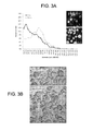
- FIG. 3A is a graphical of depiction the diameter distribution for cultured MKs at 0 and 18 hours, in accordance with the present invention.
- FIG. 3B shows microscopy images of MKs in static culture illustrating the production of proPLTs at 6 hours post-purification, in accordance with the present invention.
- FIG. 3C shows microscopy images of MKs under physiological shear illustrating the production proPLTs immediately upon trapping, in accordance with the present invention.
- FIG. 3D is a graphical depiction of an increased percentage of proPLT-producing MKs under physiological shear over those of static cultures, in accordance with the present invention.
- FIG. 3E is a graphical depiction illustration that proPLT extension rates under physiological shear are increased significantly as compared to static cultures, in accordance with the present invention.
- FIG. 4A shows microscopy images illustrating MKs squeezing through 3 ⁇ m-wide microchannels, supporting a model of vascular PLT production, in accordance with the present invention.
- FIG. 4B shows microscopy images illustrating MKs extending large fragments through 3 ⁇ m-wide microchannels, supporting a model of vascular PLT production, in accordance with the present invention.
- FIG. 4C shows microscopy images illustrating proPLT extension, in accordance with the present invention.
- FIG. 4D shows microscopy images illustrating proPLT extension and abscission events at different positions along the proPLT shaft, in accordance with the present invention.
- FIG. 4E shows microscopy images illustrating that following abscission, the resulting proPLT end formed a new PLT-size swelling at the tip, which was subsequently extended and released, with the cycle repeated, in accordance with the present invention.
- FIG. 4F is a graphical depiction illustrating that increased shear rates within physiological ranges do not increase proPLT extension rate, in accordance with the present invention.
- FIG. 4G shows miscrocopy images illustrating that MKs retrovirally transfected to express GFP- ⁇ 1 tubulin showed proPLT extensions and were comprised of peripheral MTs that form coils at the PLT-sized ends, in accordance with the present invention.
- FIG. 4H is a graphical depiction illustrating that 5 ⁇ M Jasplankinolide (Jas, actin stabilizer) and 1 mM erythro-9-(3-[2-hydroxynonyl] (EHNA, cytoplasmic dynein inhibitor) inhibit shear-induced proPLT production, in accordance with the present invention.
- FIG. 4I shows microscopy images illustrating drug-induced inhibition of proPLT production under physiological shear, in accordance with the present invention.
- FIG. 5A is a graphical depiction illustrating that microfluidic device-derived mFLC-PLTs manifest structural and functional properties of blood PLTs, in accordance with the present invention.
- FIG. 5B is a graphical depiction illustrating biomarker expression, and forward/side scatter and relative concentration of GPIX+mFLC-MKs infused into the microfluidic device following isolation on culture day 4, and effluent collected from the microfluidic device 2 hours post infusion, in accordance with the present invention.
- FIG. 5C is a graphical depiction illustrating that the application of shear shifts GPIX+ produce toward more PLT-sized cells relative to static culture supernatant, in accordance with the present invention.
- FIG. 5D shows microscopy images illustrating that in the microfluidic device, mFLC-MKs are converted into PLTs over a period of 2 hours, in accordance with the present invention.
- FIG. 5E is a graphical depiction illustrating that the application of shear shifts product toward more PLT-sized ⁇ 1 tubulin+ Hoescht ⁇ cells relative to static culture supernatant, in accordance with the present invention.
- the insert shows quantitation of free nuclei in the effluent.
- FIG. 5F shows microscopy images illustrating that microfluidic device-mPLTs are ultrastructurally similar to mouse blood PLTs and contain a cortical MT coil, open canalicular system, dense tubular system, mitochondria, and characteristic secretory granules, in accordance with the present invention.
- FIG. 5G shows microscopy images illustrating that microfluidic device-mPLTs and PLT intermediates are morphologically similar to mouse blood PLTs and display comparable MT and actin expression, in accordance with the present invention.
- FIG. 6A is a graphical depiction illustrating that microfluidic device-derived hiPSC-PLTs manifest structural and functional properties of blood PLTs, where hiPSC-MKs reach maximal diameter (20-60 ⁇ m) on culture day 15, in accordance with the present invention
- FIG. 6B shows a microscopy image illustrating that hiPSC-MKs are ultrastructurally similar to primary human MKs and contain a lobulated nuclei, invaginated membrane system, glycogen stores, organelles, and characteristic secretory granules, in accordance with the present invention.
- FIG. 6C shows microscopy images illustrating that hiPSC-MKs in static culture begin producing proPLTs at 6 hours post-purification, and reach maximal proPLT production at 18 hours, in accordance with the present invention.
- FIG. 6D shows a microscopy image illustrating that hiPSC-MKs under physiological shear ( ⁇ 500 s ⁇ 1 ) begin producing proPLTs immediately upon trapping and extend/release proPLTs within the first 2 hours of culture, in accordance with the present invention.
- FIG. 6E is a graphical depiction illustrating that percent proPLT-producing hiPSC-MKs under physiological shear are increased significantly over static cultures, in accordance with the present invention.
- FIG. 6F is a graphical depiction illustrating that proPLT extension rates under physiological shear are ⁇ 19 ⁇ m/min, in accordance with the present invention.
- FIG. 6G shows microscopy images illustrating that microfluidic device derived-hPLTs are ultrastructurally similar to human blood PLTs and contain a cortical MT coil, open canalicular system, dense tubular system, mitochondria, and characteristic secretory granules in accordance with the present invention.
- Top-right insert shows peripheral MT coil.
- FIG. 6H shows microscopy images illustrating that microfluidic device derived-hPLTs are morphologically similar to human blood PLTs and display comparable MT and actin expression, in accordance with the present invention.
- FIG. 6I shows microscopy images illustrating that microfluidic device derived-mPLTs form filpodia/lamellipodia on activation and spread on glass surface, in accordance with the present invention.
- FIG. 7 shows a live-cell microscopy image illustrating that T-DM1 inhibits MK differentiation and disrupts proPLT formation by inducing abnormal tubulin organization, accordance with the present invention.
- PLTs Blood platelets
- Morbidity and mortality from bleeding due to low PLT count is a major clinical problem encountered across a number of conditions including chemotherapy or radiation treatment, trauma, immune thrombocytopenic purpura (ITP), organ transplant surgery, severe burns, sepsis, and genetic disorders.
- ITP immune thrombocytopenic purpura
- PLT transfusions total more than 10 million units per year in the United States.
- PLT production involves the differentiation of megakaryocyte (MKs), which sit outside sinusoidal blood vessels in the bone marrow (BM) and extend long, branching cellular structures (designated proPLTs) into the circulation.
- MKs megakaryocyte
- proPLTs experience vascular shear and function as the assembly lines for PLT production, containing PLT-sized swellings in tandem arrays that are connected by thin cytoplasmic bridges.
- proPLTs Although detailed characterization of proPLTs remains incomplete, these structures have been recognized both in vitro and in vivo and proPLT-producing MKs in culture yield PLTs that are structurally and functionally similar to blood PLTs. PLTs are released sequentially from proPLT tips.
- Thrombopoietin has been identified as the major regulator of MK differentiation, and it has been used to produce enriched populations of MKs in culture.
- TPO Thrombopoietin
- human PLTs generated in vitro from proPLT-producing MKs were functional. Since then, MKs have been differentiated from multiple sources, including fetal liver cells (FLCs), cord blood stem cells (CBSCs), embryonic stem cells (ESCs), and induced pluripotent stem cells (iPSCs).
- FLCs fetal liver cells
- CBSCs cord blood stem cells
- ESCs embryonic stem cells
- iPSCs induced pluripotent stem cells
- current 2-D and liquid MK cultures fall orders of magnitude short of the estimated ⁇ 2000 PLTs generated per MK in vivo.
- 3-D knitted polyester scaffolds have been applied under continuous fluid flow to produce up to 6 ⁇ 10 6 PLTs/day from 1 million CD34 + human cord blood cells in culture. While suggestive that clinically useful PLT numbers may be attained, those 3-D perfusion bioreactors do not accurately reproduce the complex structure and fluid characteristics of the BM microenvironment, and their closed modular design prevents visualization of proPLT production, offering little insight into the mechanism of PLT release.
- 3-D polydimethylsiloxane (PDMS) biochips adjacent ECM-coated silk-based tubes have been proposed to reproduce BM sinusoids and study MK differentiation and PLT production in vitro. Although such devices recapitulate MK migration during maturation, they are not amenable to high resolution live-cell microscopy, and fail to reproduce endothelial cell contacts necessary to drive MK differentiation.
- PDMS polydimethylsiloxane
- MK differentiation has been studied in culture, the conditions that stimulate proPLT production remain poorly understood, particularly in vivo. MKs are found in BM niches, and some evidence suggests that cell-cell, cell-matrix, and soluble factor interactions of the BM stroma contribute to proPLT formation and PLT release. Indeed, the chemokine SDF-1 and growth factor FGF-4 recruit MKs to sinusoid endothelial cells. Extracellular matrix (ECM) proteins are another major constituent of the BM vascular niche, and evidence suggests that signaling through trans-membrane glycoprotein (GP) receptors regulate proPLT formation, PLT number and size (defects seen in e.g. Bernard-Soulier syndrome, Glanzmann's thrombasthenia).
- GP trans-membrane glycoprotein
- Collagen IV and vitronectin promote proPLT production, which can be inhibited by antibodies directed against their conjugate integrin receptor, GPIb ⁇ .
- fibrinogen regulates proPLT formation and PLT release through GPIIbIII ⁇ . While these findings shed light on the environmental determinants of proPLT production, they are limited by a reductionist approach. Therefore, new models that incorporate the defining attributes of BM stroma complexity are necessary to elucidate the physiological regulation of MKs into PLTs.
- proPLTs experience wall shear rates ranging from, 100 to 10,000 s ⁇ 1 or, more particularly, from 500 to 2500 s ⁇ 1 . While the role of continuous blood flow on PLT thrombus formation has been studied, surprisingly little attention has been paid to the mechanism by which shear forces regulate PLT release. When investigated, experiments have not been physiologically representative. Some preliminary studies have perfused MKs over ECM-coated glass slides, which select for immobilized/adhered MKs without discriminating ECM-contact activation from shear.
- released proPLTs have been centripetally agitated in an incubator shaker, which does not recapitulate circulatory laminar shear flow, does not provide precise control of vascular shear rates, and is not amenable to high-resolution live-cell microscopy.
- exposure of MKs to high shear rates appears to accelerate proPLT production and while proPLTs cultured in the absence of shear release fewer PLTs than those maintained at fluid shear stresses.
- Microfluidic devices provide excellent platforms to generate and precisely tune dynamic fluid flows, and thus mimic blood vessel conditions to deliver chemical cues to cells.
- Embedding microfluidic networks within cell-laden hydrogels has been shown to support efficient convective transport of soluble factors through 3D scaffolds.
- Viable 3D tissue contacts have been produced consisting of hepatocytes encapsulated in agarose, calcium alginate hydrogels seeded with primary chondrocytes, and endothelial cells embedded in 3D tubular poly(ethylene glycol) hydrogels. Accordingly, the technology has been applied to the development of organs-on-a-chip, including liver, kidney, intestine, and lung.
- microvasculature-on-a-chip models have been used to study cardiovascular biology and pathophysiology in vitro. These studies emphasize the importance of mimicking the physical microenvironment and natural chemical cues of living organs to study cellular and physiological development. For example, this is particularly important for drug-mediated inhibition of PLT production. Since proPLT-producing MKs sit just outside blood vessels in the BM, interacting with both the semi-solid ECM microenvironment of BM and fluid microenvironment of the circulation, biomimetic microfluidic biochips may achieve a model system to elucidating the relevant physiological mechanisms, such as those responsible for drug-induced thrombocytopenia.
- FIG. 1 a schematic is shown illustrating an example of a biomimetic system 100 in accordance with various embodiments of the present invention.
- the system 100 includes a substrate 101 , a first channel 102 and a second channel 104 , wherein each channel is configured to carry a flow of any fluid medium transporting or consisting of but not limited to, for example, particles, cells, substances, particulates, materials, compositions and the like.
- the system 100 and/or substrate 101 may be constructed using cell-inert silicon-based organic polymers, such as polydimethylsiloxane (PDMS).
- PDMS polydimethylsiloxane
- the first channel 102 includes a first channel input 106 and first channel output 108 .
- the second channel 104 includes a second channel input 110 and second channel output 112 .
- the first channel 102 and second channel 104 extend along a substantially longitudinal direction, and are longitudinally and transversally dimensioned, as will be explained, to achieve desired flow profiles, velocities, or rates, such as those present in a physiological system.
- the size of the longitudinal 130 and transverse 132 dimensions describing the channels may be in a range consistent with an anatomical or physiological structure, assembly, configuration or arrangement, such as in the case of bone marrow and blood vessels.
- each channel may be prepared, conditioned, or manufactured to receive, localize, trap, or accumulate for example, particles, cells, substances, particulates, materials, compositions, and the like, from a traversing fluid medium.
- the system 100 also includes a series of columns 114 that separate the first channel 102 and second channel 104 .
- the long axes of the columns 114 are generally arranged parallel to the longitudinal 130 dimension of the channels, the series of columns 114 extending for a distance substantially equal to the longitudinal 130 dimension of the channels.
- the columns 114 are separated by gaps, creating a series of gaps that, as illustrated, may be microchannels 116 that extend from the first channel 102 to the second channel 104 to create a partial fluid communication path passing between the columns 114 .
- the term “microchannel” when referring to the gaps does not connote a particular width. For example, the gaps may be substantially greater or smaller than the micrometer range.
- the columns 114 and microchannels 116 are dimensioned such that particles, cells, substances, particulates, materials, compositions, and the like, may bind, adhere to or otherwise be confined to an area generally in the vicinity of the columns 114 and microchannels 116 and, thereby, harvested from an area proximate to the microchannels 116 .
- the longitudinal 130 and transverse 132 dimensions of the columns 114 may be in the range of 1 to 200 micrometers, while the longitudinal 130 dimension of the microchannels 116 , defined by the separation distances or gaps between the columns 114 , may be in the range of 0.1 to 20 micrometers, although other values are possible.
- Flow in the first channel 102 is established through a first source or input 118 configured for deliver a first medium, and a first outlet 120 , configured for extracting the first fluid medium.
- flow of a second fluid medium in the second channel 104 is established through a second source or inlet 122 to a second outlet 124 .
- the first input or source 118 and the second source of inlet 122 may be arranged to include a pump or other system for delivering a controlled flow.
- the first outlet 120 or second outlet 124 may also be fitted with or followed by elements, components, devices or systems designed to capture, store and/or separate a desired material or substance from a first or second fluid medium, such as for example, human blood platelets, or thrombocytes.
- flow velocities or flow rates of the first fluid medium between the first channel input 106 and first channel output 108 , and of the second fluid medium between the second channel input 110 and second channel output 112 may be established by way of fluid communication of system 100 with any number of sources, such as microfluidic pumps and drains, and may be sustained for any desired or required amount of time. Control and manipulation of flow may be realized by integrating elements, such valves, sources and drains, with the system 100 , or may be achieved by external interfacing or coupling of the system 100 with various components for fluid actuation and flow regulation.
- flow velocities or rates in the first channel 102 may be configured to be substantially different from flow velocities or rates in the second channel 104 , as desired, or as required for recapitulating, modeling, or duplicating physiological elements, constituents and conditions such as, for example, those found in bone marrow and blood vessels.
- flow velocities or rates may be controlled in a manner that duplicates physiological shear rates and profiles, such as vascular shear rates and profiles.
- the system 100 may also include filtration elements 126 , which may take any shape or form, arranged along the paths of each of the first and second fluid mediums and designed to capture or remove from the traversing fluid mediums any kind of debris, dust and any other contaminants or undesirable materials, elements, or particulates. In one configuration, filtration elements 126 are situated in proximity to the first inlet 118 and second inlet 122 .
- the system 100 further includes flow resistive elements 128 , which may take a variety of shapes or forms, arranged along the paths of each of the first and second fluid mediums and designed to control flow forces or damp fluctuations in flow rate. In one configuration, flow resistive elements 128 may be situated following each of the filtration elements 126 along the paths of each of the first and second fluid mediums.
- recreating human bone marrow (BM) vascular niche ex vivo may be achieved by selectively filling the first channel 102 with bone powder, proteins, such as CI, CIV, FG, FN, VN, LN and VWF, gels such as agarose, alginate, and matrigel or solutions such as PBS, HBS, DMEM EGM or other media, alone and in combination.
- ECM proteins may be patterned directly onto glass surfaces prior to adhesion of biochips to surface slides using protein micro/nano-stamping, or following microfluidic device assembly using parallel microfluidic streams. Local component concentration may be adjusted by regulating microfluidic stream flow rate during infusion, with focus on alignment and 3-D arrangement.
- recapitulating human BM vasculature may be achieved by selectively coating the second channel 104 by culturing with endothelial cells at 37 degrees C. and 5 percent CO 2 .
- Endothelial cells may be fixed with 4% formaldehyde, and probed for cellular biomarkers to resolve cellular localization and architecture.
- the second channel 104 of endothelialized BM biochips may be perfused with a fluorescent or colorimetric medium such as FITC-dextran or with beads, and visualized by live-cell microscopy to assess sample/cell/molecule diffusion and determine vascular permeability.
- the system 100 in accordance with the present invention can provide a platform for recapitulating physiological conditions, such as those of human BM, by replicating the dimensions, environments and conditions found in human venules using a biomimetic microfluidic device.
- the microfluidic channels separated by columns spaced closely apart experiencing controlled environments and flow conditions represent a realistic physiological model that may be employed to produce functional PLTs.
- MK trapping, BM stiffness, ECM composition, micro-channel size, hemodynamic vascular shear, and endothelial cell contacts may be tailored to reproduce human BM in vitro.
- microfluidic devices were fabricated using soft lithography, consisting of two channels containing passive filters, for trapping air bubbles and dust, followed by fluid resistors, used to damp fluctuations in flow rate arising during operation.
- the channels merge to a rectangular area 1300 micrometers long, 130 micrometers wide, and 30 micrometers deep, separated by a series of columns (10 micrometers wide and 90 micrometers long) spaced 3 micrometers apart.
- microfluidic devices were constructed from a cell-inert silicon-based organic polymer bonded to glass slides.
- AutoDesk software in AutoCAD was used to design the desired 2D pattern and printed on a photolithography chrome mask.
- Silicon wafers Universal Wafers, Boston, Mass.
- SU-8 3025 photoresist Mochrochem, Newton, Mass.
- Laurell Technologies, North Wales, Pa. baked at 65 degrees C. for 1 minute and 95 degrees C. for 5 minutes, and exposed to UV light ( ⁇ 10 mJ cm ⁇ 2 ) through the chrome mask for 30 seconds.
- the unbound SU-8 photoresist was removed by submerging the substrate into propylene glycol monomethyl ether acetate for 7 minutes.
- Polydimethylsiloxane was poured onto the patterned side of the silicon wafer, degassed, and cross-linked at 65 degrees C. for ⁇ 12 hours. After curing, the PDMS layer was peeled off the mold and the inlet and outlet holes were punched with a 0.75 mm diameter biopsy punch. The channels were sealed by bonding the PDMS slab to a glass cover slide (#1.5, 0.17 ⁇ 22 ⁇ 50 mm, Dow Corning, Seneffe, Belgium) following treatment with oxygen plasma (PlasmaPrep 2, GaLa Instrumente GmbH, Bad Schwalbach, Germany).
- Samples were infused into the microfluidic device via PE/2 tubing (Scientific Commodities, Lake Havasu City, Ariz.) using 1 mL syringes equipped with 27-gauge needles (Beckton Dickinson, Franklin Lakes, N.J.). Flow rates of liquids were controlled by syringe pumps (PHD 2000, Harvard Apparatus, Holliston, Mass.).
- Microfluidic devices were selectively coated with extracellular matrix proteins by perfusing the channels with rhodamine-conjugated fibrinogen (1 mg/mL) or fibronectin (50 ⁇ g/mL, Cytoskeleon Inc., Denver, Colo.) for 30 minutes. Samples were perfused in parallel through both inlets and collected through both outlets so that laminar flow streams did not mix. Devices were washed with 1 ⁇ PBS and coated with 0.22 ⁇ m filtered (Millipore, Billerica, Mass.) 10% BSA solution (Roche, South San Francisco, Calif.) for 30 minutes to coat any remaining exposed glass.
- Microfluidic devices were selectively coated with 50 ⁇ g/mL fibronectin (Cytoskeleon Inc., Denver Colo.) and 10 percent BSA (Roche, South San Francisco, Calif.), as described above, and transferred to a 37 degrees C., 5 percent CO2 incubator. 10,000,000 HUVECs/mL in EBM media (Lonza, Basel, Switzerland) were seeded over the fibronectin-coated channel at 12.5 uL/hour and permitted to adhere to this surface over a period of 3 hours. The inlet sample was replaced with cell-free EBM media and perfused through the channel until HUVECs reached confluency (2-8 days).
- Cells were stained with 5 ⁇ M CellTracker Red and 1 ⁇ g/mL Hoescht 33342 (Invitrogen, Carlsbad, Calif.) for 45 minutes, washed in fresh media or fixed in 4% formaldehyde and visualized by confocal-fluorescence microscopy.
- Equation (1) was solved in a three dimensional computational domain replicating the exact dimensions of the microfluidic device. It was assumed that the fluid within the device had a viscosity and density of water (0.001 Pa s and 1000 kg/m3, respectively). No slip boundary conditions were assumed at the walls of the channels. The infusion flow rates ranged from 12.5-200 ⁇ L/hr. A triangular mesh, which was made finer at the slits, was used to discretize the domain. The model contained 315,317 degrees of freedom. Mesh independence, as was confirmed by obtaining less than a 10 percent difference between shear rates, was found between 251,101 and 415,309 degrees of freedom. The steady state solutions were obtained using the UMFPACK linear system solver.
- Mouse FLCs were collected from WT CD1 mice (Charles River Laboratories, Wilmington, Mass.) and MKs were cultured.
- samples were permeabilized with 0.5 percent Triton-X-100, and blocked in immunofluorescence blocking buffer (0.5 g BSA, 0.25 ml 10% sodium azide, 5 ml FCS, in 50 ml 1 ⁇ PBS) overnight before antibody labeling(55).
- immunofluorescence blocking buffer 0.5 g BSA, 0.25 ml 10% sodium azide, 5 ml FCS, in 50 ml 1 ⁇ PBS
- samples were incubated with a rabbit polyclonal primary antibody for mouse or human ⁇ 1-tubulin.
- samples were incubated with Alexa 568 phalloidin (Invitrogen, Carlsbad, Calif.). Cell nuclei were labeled with 1 ⁇ g/mL Hoescht 33342 (Invitrogen, Carlsbad, Calif.).
- MK diameters were calculated from area measurements to account for non-circular cells. More than 2000 cells were counted for each condition, and analysis of MK area and effluent composition was performed for at least three independent samples. Statistical significance was established using a 2-tailed Student t test for paired samples. Error bars represent one standard deviation about the mean.
- MKs were loaded onto ‘naked’ microfluidic devices (only BSA-coated), and the infusion rate was doubled incrementally from 12.5 ⁇ L/hr to 200 ⁇ L/hr over a 2 hour period.
- isolated MKs were pipetted into chambers formed by mounting a glass coverslide coated with 3% BSA onto a 10 mm petri dish with a 1 cm hole and cultured for 24 hours. Both static and shear cultures were maintained at 37 degrees C. and 5 percent CO2 and examined on a Zeiss Axiovert 200 (Carl Zeiss, Thornwood, N.Y.) equipped with 10 ⁇ (numerical aperature, 0.30) Plan-Neofluar air objective.
- DIC Differential interference contrast
- Dendra2-fused ⁇ 1 tubulin was cloned into pMSCV plasmids.
- HEK 293 cells packaging cells were cultured in DMEM supplemented with 10% fetal bovine serum (FBS) to 30-50 percent confluency.
- FBS fetal bovine serum
- Transfection of HEK 293 cells was performed using 2 ⁇ g of DNA plasmids encoding gag/pol, vsvG, and the ⁇ 1 tubulin fused with Dendra2 in the pMSCV vector. After medium exchange the following day, cells were incubated for 72 hours for virus production. The supernatant was filtered through a 0.22 ⁇ m filter (Millipore, Billerica, Mass.), and aliquots were stored at ⁇ 80 degrees C.
- MKs isolated from fetal liver cultures described above were resuspended in DMEM containing 10 percent FBS, 8 ⁇ g/mL polybrene (Sigma), and the retroviral supernatant. Samples were transferred to a 6-well plate, centrifuged at 800 ⁇ g for 90 minutes at 25 degrees C. and then incubated at 37 degrees C. for 90 minutes. Following incubation, MKs were washed by centrifugation and resuspended in fresh DMEM containing 10 percent FBS and TPO. MKs were allowed to mature until day 4 of culture and then isolated by a BSA gradient, as previously described.
- Platelets were collected from the released proPLT fraction of static MK cultures or bioreactor effluent and examined under resting conditions. Samples were probed with FITC-conjugated antibodies against CD42a or CD41/61 (Emfret Analytics, Eibelstadt, Germany) and run on a FACSCalibur flow cytometer (Beckton Dickinson). PLTs were gated by their characteristic forward- and side-scattering as they passed through the detector, and their total fluorescence intensity was calculated after subtraction of a FITC-conjugated IgG antibody specificity control (Emfret Analytics). Quantization of PLT yield was determined by dividing net GP IX+ PLT production by net GP IX+ MK depletion over effluent collection period, and was performed for at least 3 independent samples Results were identical for GP IIbIIIa+ cells.
- Metamorph The digital images acquired in Metamorph were analyzed using ImageJ and Adobe Photoshop CS3 (Adobe Systems, San Jose, Calif.). Dividing lines explicitly separate different images, or separate regions of the same image. No specific features within an image were enhanced, obscured, moved, removed, or introduced, and adjustments made to the brightness, contrast, and color balance were linearly applied to the whole image.
- each of the channels were selectively coated with fibrinogen and fibronectin, respectively to reproduce ECM composition of the BM and blood vessel microenvironments (shown in FIG. 2A ).
- primary MKs infused along a first channel would become sequentially trapped between the columns and extend proPLTs into the second channel (shown in FIG. 2B ), recapitulating physiological proPLT extension.
- MKs were infused in a 1 percent alginate solution that was polymerized within the microfluidic device, selectively embedding the MKs in alginate gel within the first channel while retaining vascular flow in the second channel. Alginate did not inhibit proPLT production, and MK distance from the second channel could be controlled.
- FIG. 2C Human umbilical vein endothelial cells (HUVECs) were selectively seeded along the second channel, and grown to confluency to produce a functional blood vessel (shown in FIG. 2C ).
- MK behavior was monitored by 10 ⁇ -150 ⁇ magnification, high-resolution live-cell microscopy, and the released PLTs were collected from the effluent.
- FIG. 2D shows the complete system illustrating operation.
- Laminar fluid shear rates were characterized (shown in FIG. 2E ), and were tightly controlled using two microfluidic pumps (one for the first channel and one for the second channel). Shear rates within the device were linearly proportional to infusion rates and were adjusted to span the physiological range (500-2500 s ⁇ 1 ).
- In vivo BM MKs extend proPLTs in the direction of blood flow and release PLTs, proPLTs, large cytoplasmic fragments (prePLTs), and even whole MKs into sinusoidal blood vessels which may be trapping in the pulmonary microvascular bed, or otherwise maturing in the circulation.
- mouse fetal liver culture-derived (mFLC) MKs were isolated on culture day 4 and characterized by size and ploidy before being infused into the microfluidic device (shown in FIG. 3A ).
- MKs begin producing proPLTs ⁇ 6 hours post-isolation, and reach maximal proPLT production at 18 hours (shown in FIG. 3B ).
- MKs under physiological shear shown in FIG. 3C at roughly 500 s ⁇ 1 ) began producing proPLTs within seconds of trapping, reaching maximal proPLT production and biochip saturation within the first 2 hours of culture.
- MKs cultured under physiological shear produced fewer, longer proPLTs that were less highly branched relative to static cultures.
- ProPLTs in shear cultures were uniformly extended into the lower channel and aligned in the direction of flow against the vascular channel wall, recapitulating physiological proPLT production.
- the percent of proPLT-producing MKs under physiological shear were doubled over static cultures to roughly 90% (shown in FIG. 3D ).
- abscission events were routinely captured by high-resolution live-cell microscopy and occurred at variable positions along the proPLT shaft, releasing both prePLT-sized intermediates (3-10 ⁇ m diameter) and PLTs (1.5-3 ⁇ m diameter) (shown in FIG. 4C and FIG. 4D ). Following each abscission, the resulting proPLT end formed a new PLT-sized swelling at the tip, which was subsequently extended and released, repeating the cycle (shown in FIG. 4E ).
- proPLT extension rates While shear rates were kept constant, proPLT extension rates varied at different positions along the shaft, predictive of a regulated cytoskeletal driven mechanism of proPLT elongation (shown in FIG. 4C ). Increasing microfluidic shear rates within the physiological range did not affect the median proPLT extension rate or the distribution of proPLT extension rates in culture (shown in FIG. 4F ), and proPLT projections in MKs retrovirally transfected to express GFP- ⁇ 1 were comprised of peripheral microtubules (MTs) that formed coils at the PLT-sized ends (shown in FIG. 4G ).
- MTs peripheral microtubules
- ProPLTs reached lengths exceeding 5 mm, and resisted shear rates up to 1000 s ⁇ 1 in vitro; recapitulating physiological examples of proPLT production from intravital microscopy, and demonstrating that abcission events were not caused by shear.
- MKs were incubated with 5 ⁇ M Jasplankinolide (Jas, actin stabilizer) or 1 mM erythro-9-(3-[2-hydroxynonyl] (EHNA, cytoplasmic dynein inhibitor) prior to infusion in microfluidic device. Both Jas and EHNA inhibited shear-induced proPLT production (shown in FIG. 4H and FIG. 4I ) and PLT release under both static and physiological shear conditions.
- PLTs are anucleate discoid cells ⁇ 1-3 ⁇ m in diameter that express biomarkers GP IX and IIbIIIa on their surface, and are characterized by a cortical MT coil of 6-8 MTs encircling an actin-based cytoskeletal network.
- biomarker expression, and forward/side scatter and relative concentration of glycoprotein (GP) IX+ mFLC-MKs were measured by flow cytometry immediately before infusion in our microfluidic device on culture day 4 (shown in FIG. 5A ). Effluent was collected 2 hours post infusion and compared to mFLC-MK input (shown in FIG. 5B ).
- Input MKs and effluent PLTs both expressed GP IX and IIbIIIa on their surface, and displayed characteristic forward/side scatter.
- the application of shear shifted the cellular composition of the effluent toward more PLT-sized GPIX+ cells relative to static culture supernatant isolated on culture day 5 (shown in FIG. 5C ).
- 85 ⁇ 1% of MKs were converted into PLTs over 2 hours, which agreed with our quantitation of percent proPLT production ( FIG. 3D ) and constitutes a significant improvement over static cultures ( FIG. 5D ).
- Resting PLTs contain characteristic invaginations of the surface membrane that form the open canalicular system, a closed channel network of residual endoplasmic reticulum that form the dense tubular system, organelles, specialized secretory granules, and will flatten/spread on contact activation with glass.
- Microfluidic device-generated PLTs were ultrastructurally indistinguishable from mouse blood PLTs by thin-section transmission electron; and contained a cortical MT coil, open canalicular system, dense tubular system, mitochondria, alpha- and dense-granules (as shown in FIG. 5F ).
- Microfluidic device-generated PLTs and PLT intermediates displayed comparable MT and actin organization to mouse blood PLTs by immunofluorescence microscopy (as shown in FIG. 5G ), and spread normally on contact-activation with glass, forming both filpodia and lamellipodia.
- hiPSC-MKs were isolated on culture day 15, once they had reached maximal diameter of 20-60 ⁇ m (shown in FIG. 6A ), and were ultrastructurally similar to primary human MKs (shown in FIG. 6B ). In static culture, hiPSC-MKs began producing proPLTs at 6 hours post-isolation, and reached maximal proPLT production at 18 hours (shown in FIG. 6C ).
- hiPSC-MKs under physiological shear (about 500 s ⁇ 1 ) began producing proPLTs immediately upon trapping, and extended/released proPLTs within the first 2 hours of culture (shown in FIG. 6D ).
- the percent proPLT-producing hiPSC-MKs under shear were increased significantly over static cultures ( ⁇ 10%) to roughly 90% (as shown in FIG. 6E ).
- ProPLT extension rates were slightly lower than mFLC-MK controls ( ⁇ 19 ⁇ m/min versus 30 ⁇ m/min) (shown in FIG. 6F ) and more closely approximated physiological controls.
- Microfluidic device-generated PLTs displayed forward and side scatter, and surface biomarker expression characteristic of human blood PLTs, were ultrastructurally indistinguishable from human blood PLTs by thin-section transmission electron (shown in FIG. 6G ), displaying comparable MT and actin expression to human blood PLTs by immunofluorescence microscopy (shown in FIG. 6H ), spreading normally on contact-activation with glass, and forming both filpodia and lamellipodia (shown in FIG. 6I ). Taken together these data demonstrate that hiPSC-MKs can be applied to our biomimetic microfluidic device to generate potentially unlimited numbers of functional human PLTs.
- Thrombocytopenia may appear suddenly and often unintentionally, potentially causing major bleeding and death. Antibody and cell-mediated autoimmune responses have been shown to cause thrombocytopenia. In addition, thrombocytopenia may also be triggered by a wide range of medications, including cancer drugs, such as dasatinib. Animal models are generally poor predictors of safety and efficacy of medications in humans, and clinical studies are time-consuming, expensive, and potentially harmful. Microfluidic devices designed to mimic human BM represent an area of innovation of major clinical importance, offering an efficient and realistic platform to investigate the effects of a variety of medications upon BM and MK biology.
- PLT survival and clearance rates are usually measured through infusion studies using flow cytometry. Quantification of the rate and extent of proPLT production, however, is not amenable to this approach, and requires direct visualization to establish at what stage thrombocytopoiesis is affected. By contrast, the application of microfluidic devices offers a great platform to study drug effects on PLT production, one that may facilitate the identification of new regulators of PLT production and elucidate the mechanism of clinically significant drug-induced thrombocytopenias.
- T-DM1 trastuzumab emtansine
- the approach of the present invention capitalizes on a highly novel microfluidic design to recapitulate human BM and blood vessel physiology ex vivo, and generate an alternative source of functional human PLTs for infusion. While clinically desirable to meet growing transfusion needs and obviate risks currently associated with platelet procurement and storage, 2 major quantitative roadblocks have thus far persisted in the development of donor-independent PLTs for therapeutic use: (1) generating sufficient numbers ( ⁇ 3 ⁇ 10 8 ) of human MKs to support the production of one PLT transfusion unit ( ⁇ 3 ⁇ 10 11 PLTs), and (2) generating physiological numbers of functional human PLTs ( ⁇ 10 3 -10 4 ) per MK.
- hESC human embryonic stem cell cultures
- hiPSC human induced pluripotent stem cell cultures
- proPLTs have been centripetally agitated in an incubator shaker, which does not recapitulate laminar shear flow in BM blood vessels, does not provide precise control of vascular shear rates, and is not amenable to high-resolution live-cell microscopy. Nonetheless, despite these limitations, exposure of MKs to high shear rates (1800 s ⁇ 1 ) accelerated proPLT production, and proPLTs cultured in the absence of shear released significantly fewer PLTs than those maintained at fluid shear stresses of ⁇ 0.5 Pa for 2 hours. Moreover, recent advances in multiphoton intravital microscopy have provided increasing resolution of proPLT production in vivo and confirmed the importance of vascular flow on proPLT extension and PLT release. While these studies have provided physiologically accurate examples of in vivo proPLT production, poor resolution and limited control of the microenvironment has prohibited detailed study of how the BM microenvironment contributes to PLT release.
- 3D perfusion bioreactors While suggestive that clinically useful numbers of culture-derived human PLTs are attainable, 3D perfusion bioreactors have not accurately reproduced the complex structure and fluid characteristics of the BM microenvironment, and their closed modular design has prevented direct visualization of proPLT production, offering little insight into the mechanism of PLT release.
- 3D PDMS biochips adjacent ECM-coated silk-based tubes have been proposed to reproduce BM sinusoids and study MK differentiation and PLT production in vitro. While capable of recapitulating MK migration during maturation, this design is not amenable to high resolution live-cell microscopy, and does not reproduce endothelial cell contacts necessary to drive MK differentiation.
- the microfluidic device design of the present invention offers the complete package, allowing significant improvement in time to PLT release and an increased total PLT yield.
- application of vascular shear rates within the microfluidic device induces proPLT production, and reproduces physiological proPLT extension and release.
- MKs are capable of squeezing through small gaps to enter the circulation and releasing prePLT intermediates under physiological flow conditions.
- the product resulting from continuous perfusion of MKs in the microfluidic device of the present invention approached physiological PLT concentrations, and manifested both structural and functional properties of blood PLTs.
- PLT microfluidic devices could be applied to human MK cultures to produce functional human PLTs.
- the approach of the present invention offers unprecedented control of ex vivo microenvironments and a biomimetic platform for drug development.
- support of high-resolution live-cell microscopy permits direct visualization of cells during culture and provides a window into poorly characterized physiological processes.
- the microfluidic device design can be easily scaled by mirroring effluent channels on either side of a central channel, elongating the device to support greater numbers of columns, and positioning multiple units in parallel within a larger microfluidic device matrix.
- the biomimetic microfluidic system may be fabricated using PDMS bonded to glass in a configuration that includes microfluidic channels separated by a series of columns.
- the channels can be selectively coated with ECM and human endothelial cells to simulate realistic physiological conditions.
- Round or proPLT-producing MKs infused along one channel can sequentially become trapped between the columns, and extend platelet-producing proplatelets into the other channel.
- the released PLTs entering the fluid stream can be collected from the effluent, and the process may be visualized by high-resolution live-cell microscopy.
Landscapes
- Health & Medical Sciences (AREA)
- Chemical & Material Sciences (AREA)
- Life Sciences & Earth Sciences (AREA)
- Engineering & Computer Science (AREA)
- Organic Chemistry (AREA)
- Bioinformatics & Cheminformatics (AREA)
- Wood Science & Technology (AREA)
- Zoology (AREA)
- General Health & Medical Sciences (AREA)
- Biomedical Technology (AREA)
- Biochemistry (AREA)
- Biotechnology (AREA)
- Microbiology (AREA)
- General Engineering & Computer Science (AREA)
- Sustainable Development (AREA)
- Genetics & Genomics (AREA)
- Hematology (AREA)
- Analytical Chemistry (AREA)
- Clinical Laboratory Science (AREA)
- Dispersion Chemistry (AREA)
- Immunology (AREA)
- Chemical Kinetics & Catalysis (AREA)
- Molecular Biology (AREA)
- Urology & Nephrology (AREA)
- Cell Biology (AREA)
- Food Science & Technology (AREA)
- Medicinal Chemistry (AREA)
- Physics & Mathematics (AREA)
- General Physics & Mathematics (AREA)
- Pathology (AREA)
- Apparatus Associated With Microorganisms And Enzymes (AREA)
- Micro-Organisms Or Cultivation Processes Thereof (AREA)
- Physical Or Chemical Processes And Apparatus (AREA)
Priority Applications (2)
| Application Number | Priority Date | Filing Date | Title |
|---|---|---|---|
| US14/758,915 US9795965B2 (en) | 2013-01-03 | 2013-11-20 | System and method for a biomimetic fluid processing |
| US17/848,523 US11850589B2 (en) | 2013-01-03 | 2022-06-24 | System and method for a biomimetic fluid processing |
Applications Claiming Priority (3)
| Application Number | Priority Date | Filing Date | Title |
|---|---|---|---|
| US201361848424P | 2013-01-03 | 2013-01-03 | |
| US14/758,915 US9795965B2 (en) | 2013-01-03 | 2013-11-20 | System and method for a biomimetic fluid processing |
| PCT/US2013/070910 WO2014107240A1 (en) | 2013-01-03 | 2013-11-20 | System and method for a biomimetic fluid processing |
Related Parent Applications (1)
| Application Number | Title | Priority Date | Filing Date |
|---|---|---|---|
| PCT/US2013/070910 A-371-Of-International WO2014107240A1 (en) | 2013-01-03 | 2013-11-20 | System and method for a biomimetic fluid processing |
Related Child Applications (1)
| Application Number | Title | Priority Date | Filing Date |
|---|---|---|---|
| US15/709,989 Continuation US10343163B2 (en) | 2013-01-03 | 2017-09-20 | System and method for a biomimetic fluid processing |
Publications (2)
| Publication Number | Publication Date |
|---|---|
| US20150336095A1 US20150336095A1 (en) | 2015-11-26 |
| US9795965B2 true US9795965B2 (en) | 2017-10-24 |
Family
ID=51062414
Family Applications (5)
| Application Number | Title | Priority Date | Filing Date |
|---|---|---|---|
| US14/758,915 Active 2034-05-20 US9795965B2 (en) | 2013-01-03 | 2013-11-20 | System and method for a biomimetic fluid processing |
| US15/709,989 Active US10343163B2 (en) | 2013-01-03 | 2017-09-20 | System and method for a biomimetic fluid processing |
| US16/422,030 Active US10710073B2 (en) | 2013-01-03 | 2019-05-24 | System and method for biomimetic fluid processing |
| US16/904,523 Active 2034-01-17 US11396016B2 (en) | 2013-01-03 | 2020-06-17 | System and method for a biomimetic fluid processing |
| US17/848,523 Active US11850589B2 (en) | 2013-01-03 | 2022-06-24 | System and method for a biomimetic fluid processing |
Family Applications After (4)
| Application Number | Title | Priority Date | Filing Date |
|---|---|---|---|
| US15/709,989 Active US10343163B2 (en) | 2013-01-03 | 2017-09-20 | System and method for a biomimetic fluid processing |
| US16/422,030 Active US10710073B2 (en) | 2013-01-03 | 2019-05-24 | System and method for biomimetic fluid processing |
| US16/904,523 Active 2034-01-17 US11396016B2 (en) | 2013-01-03 | 2020-06-17 | System and method for a biomimetic fluid processing |
| US17/848,523 Active US11850589B2 (en) | 2013-01-03 | 2022-06-24 | System and method for a biomimetic fluid processing |
Country Status (8)
| Country | Link |
|---|---|
| US (5) | US9795965B2 (zh) |
| EP (2) | EP2941642B1 (zh) |
| JP (1) | JP6429794B2 (zh) |
| CN (1) | CN105308452B (zh) |
| AU (1) | AU2013371589C1 (zh) |
| CA (1) | CA2896997C (zh) |
| HK (1) | HK1218958A1 (zh) |
| WO (1) | WO2014107240A1 (zh) |
Cited By (3)
| Publication number | Priority date | Publication date | Assignee | Title |
|---|---|---|---|---|
| US10343163B2 (en) * | 2013-01-03 | 2019-07-09 | The Brigham And Women's Hospital, Inc. | System and method for a biomimetic fluid processing |
| US11135581B2 (en) | 2016-04-29 | 2021-10-05 | Creoptix Sa | Methods and assemblies for molecule recovery |
| US11566214B2 (en) | 2014-03-31 | 2023-01-31 | Brigham And Women's Hospital, Inc. | Systems and methods for biomimetic fluid processing |
Families Citing this family (13)
| Publication number | Priority date | Publication date | Assignee | Title |
|---|---|---|---|---|
| CA2930131A1 (en) | 2013-11-19 | 2015-05-28 | Platod | Fluidic device for producing platelets |
| JP6815985B2 (ja) * | 2014-07-14 | 2021-01-20 | プレジデント アンド フェローズ オブ ハーバード カレッジ | 流体システムおよびマイクロ流体システムの改善された性能のためのシステムおよび方法 |
| WO2016073486A1 (en) | 2014-11-03 | 2016-05-12 | The General Hospital Corporation | Concentrating particles in a microfluidic device |
| CN113075113A (zh) * | 2014-12-09 | 2021-07-06 | 伯克利之光生命科技公司 | 微流体装置中微物体的自动检测和重新定位 |
| EP3298161B1 (en) * | 2015-05-22 | 2020-07-29 | President and Fellows of Harvard College | Methods, systems, and compositions for determining blood clot formation, and uses thereof |
| AU2016321116B2 (en) * | 2015-09-08 | 2023-05-18 | Brigham And Women's Hospital, Inc. | System and method for producing blood platelets |
| EP3545074A1 (en) | 2016-11-23 | 2019-10-02 | The Charles Stark Draper Laboratory Inc. | Bi-layer multi-well cell culture platform |
| EP3592838A4 (en) * | 2017-03-07 | 2021-01-13 | Platelet Biogenesis, Inc. | RECIRCULATING BIOREACTOR |
| CN107723207B (zh) * | 2017-11-01 | 2019-01-01 | 深圳市瑞格生物科技有限公司 | 一种分离捕获细胞的芯片及其在肿瘤细胞分选中的应用 |
| KR102214461B1 (ko) * | 2018-03-06 | 2021-02-09 | 사회복지법인 삼성생명공익재단 | 약물 반응성 검사 장치 |
| US20210238523A1 (en) * | 2018-07-19 | 2021-08-05 | Platelet Biogenesis, Inc. | Stacked Recirculating Bioreactor |
| EP4288520A1 (en) | 2021-02-08 | 2023-12-13 | HemostOD SA | Use of 3d porous structure for platelet production |
| FR3122185A1 (fr) | 2021-04-21 | 2022-10-28 | Institut National De La Sante Et De La Recherche Medicale (Inserm) | Procede de liberation de plaquettes en regime turbulent et systeme de liberation de plaquettes pour la mise en œuvre de ce procede |
Citations (27)
| Publication number | Priority date | Publication date | Assignee | Title |
|---|---|---|---|---|
| RU2076742C1 (ru) | 1991-06-17 | 1997-04-10 | Тютрин Иван Илларионович | Колонка для гемосорбции |
| US6819736B1 (en) | 2002-02-22 | 2004-11-16 | Siemens Aktiengesellschaft | Computed tomography method and computed tomography apparatus |
| US20050009101A1 (en) | 2001-05-17 | 2005-01-13 | Motorola, Inc. | Microfluidic devices comprising biochannels |
| RU2246258C1 (ru) | 2004-03-16 | 2005-02-20 | Центральный научно-исследовательский рентгено-радиологический институт МЗ РФ | Способ диагностики стенозов коронарных артерий |
| US20050069459A1 (en) | 2003-09-26 | 2005-03-31 | The University Of Cincinnati | On-chip sample preparation for whole blood analysis |
| US20050112184A1 (en) | 2003-11-26 | 2005-05-26 | Andreas Jahn | Controlled vesicle self-assembly in continuous two phase flow microfluidic channels |
| CN2726112Y (zh) | 2004-03-12 | 2005-09-14 | 南京理工大学 | 仿蜂巢结构分形微管道网络换热器 |
| WO2006001954A2 (en) | 2004-05-20 | 2006-01-05 | Puget Sound Blood Center And Program | Methods for promoting the formation of platelets and for treating blood and bone marrow disorders |
| US20070243608A1 (en) | 2006-04-14 | 2007-10-18 | Board Of Regents, The University Of Texas System | Platelet bioreactor |
| US20100009430A1 (en) * | 2008-07-09 | 2010-01-14 | Micropoint Bioscience Inc | Analytical cartridge with fluid flow control |
| US20100041128A1 (en) | 2008-01-08 | 2010-02-18 | Medtrain Technologies, Llc | Microfluidic Device for Application of Shear Stress and Tensile Strain |
| US7718420B2 (en) | 2006-10-10 | 2010-05-18 | Postech Academy-Industry Foundation | Microfluidic biochip for blood typing based on agglutination reaction |
| WO2010063823A1 (en) | 2008-12-04 | 2010-06-10 | INSERM (Institut National de la Santé et de la Recherche Médicale) | Method for producing platelets |
| RU2392314C2 (ru) | 2003-12-30 | 2010-06-20 | Аугустинус БАДЕР | Способ регенерации ткани |
| WO2010123594A2 (en) * | 2009-01-15 | 2010-10-28 | Children's Medical Center Corporation | Device for filtration of fluids there through and accompanying method |
| US20110000823A1 (en) | 2009-07-01 | 2011-01-06 | Feras Hamad | Membrane desulfurization of liquid hydrocarbons using an extractive liquid membrane contactor system and method |
| US20110039285A1 (en) | 2007-09-20 | 2011-02-17 | Inaki Sadaba Champetier De Ribes | Microfluidic device and method for fluid clotting time determination |
| CN102058399A (zh) | 2009-11-18 | 2011-05-18 | 中国科学院化学研究所 | 一种基于微流控芯片的仿生号脉系统 |
| US20120039434A1 (en) | 2009-10-28 | 2012-02-16 | Ge Wang | Cardiac computed tomography methods and systems using fast exact/quasi-exact filtered back projection algorithms |
| US20120238020A1 (en) * | 2011-03-18 | 2012-09-20 | Mitchell W Beau | Megakaryocyte and Platelet Production from Stem Cells |
| US8279996B2 (en) | 2009-10-30 | 2012-10-02 | Siemens Aktiengesellschaft | Beam hardening correction for CT perfusion measurements |
| CN102698672A (zh) | 2012-05-21 | 2012-10-03 | 中南大学 | 一种微流体反应器 |
| US8306304B2 (en) | 2009-08-31 | 2012-11-06 | Siemens Aktiengesellschaft | Precise image reconstruction of spiral CT images at optional pitch values |
| WO2013013220A2 (en) | 2011-07-20 | 2013-01-24 | University Of Washington Through Its Center For Commercialization | Photonic blood typing |
| US20130295601A1 (en) * | 2012-05-03 | 2013-11-07 | Purdue Research Foundation | Systems and Methods for Testing Drugs and Drug Delivery Systems |
| US20160002586A1 (en) | 2011-03-18 | 2016-01-07 | New York Blood Center, Inc. | Megakaryocyte and Platelet Production from Stem Cells |
| US20160272941A1 (en) | 2013-11-19 | 2016-09-22 | Platod | Fluidic device for producing platelets |
Family Cites Families (14)
| Publication number | Priority date | Publication date | Assignee | Title |
|---|---|---|---|---|
| WO1996032178A1 (en) | 1995-04-13 | 1996-10-17 | Travenol Laboratories (Israel) Ltd. | Leukocyte filtration method and apparatus |
| US7790443B2 (en) * | 2002-08-27 | 2010-09-07 | Vanderbilt University | Bioreactors with substance injection capacity |
| JP2008301746A (ja) | 2007-06-06 | 2008-12-18 | Canon Inc | 培養装置 |
| JP5419076B2 (ja) * | 2008-05-15 | 2014-02-19 | 旭化成メディカル株式会社 | 血小板の誘導方法 |
| CA2730928C (en) * | 2008-07-16 | 2023-06-20 | Children's Medical Center Corporation | Organ mimic device with microchannels and methods of use and manufacturing thereof |
| US8540434B2 (en) | 2009-01-15 | 2013-09-24 | Mayo Foundation For Medical Education And Research | Optical edge connector |
| WO2010128157A1 (en) | 2009-05-07 | 2010-11-11 | Universite De Strasbourg | Microfluidic system and methods for highly selective droplet fusion |
| SG192931A1 (en) * | 2011-02-28 | 2013-09-30 | Harvard College | Cell culture system |
| JP2012235749A (ja) | 2011-05-12 | 2012-12-06 | National Institute Of Advanced Industrial Science & Technology | マイクロチップの製造方法、物理マスク、及びマイクロチップ |
| JP6108426B2 (ja) | 2011-06-28 | 2017-04-05 | 国立大学法人名古屋大学 | 血小板産生方法及び血小板産生装置 |
| JP6429794B2 (ja) * | 2013-01-03 | 2018-11-28 | ブリガム・アンド・ウイミンズ・ホスピタル・インコーポレイテッド | バイオミメティック流体プロセスの系および方法 |
| CA2944314C (en) * | 2014-03-31 | 2023-09-19 | Brigham And Women's Hospital, Inc. | Systems and methods for biomimetic fluid processing |
| AU2016321116B2 (en) * | 2015-09-08 | 2023-05-18 | Brigham And Women's Hospital, Inc. | System and method for producing blood platelets |
| EP3592838A4 (en) * | 2017-03-07 | 2021-01-13 | Platelet Biogenesis, Inc. | RECIRCULATING BIOREACTOR |
-
2013
- 2013-11-20 JP JP2015551674A patent/JP6429794B2/ja active Active
- 2013-11-20 WO PCT/US2013/070910 patent/WO2014107240A1/en active Application Filing
- 2013-11-20 CA CA2896997A patent/CA2896997C/en active Active
- 2013-11-20 AU AU2013371589A patent/AU2013371589C1/en active Active
- 2013-11-20 US US14/758,915 patent/US9795965B2/en active Active
- 2013-11-20 CN CN201380074324.3A patent/CN105308452B/zh active Active
- 2013-11-20 EP EP13870350.9A patent/EP2941642B1/en active Active
- 2013-11-20 EP EP20174012.3A patent/EP3712612A1/en active Pending
-
2016
- 2016-06-17 HK HK16106975.8A patent/HK1218958A1/zh unknown
-
2017
- 2017-09-20 US US15/709,989 patent/US10343163B2/en active Active
-
2019
- 2019-05-24 US US16/422,030 patent/US10710073B2/en active Active
-
2020
- 2020-06-17 US US16/904,523 patent/US11396016B2/en active Active
-
2022
- 2022-06-24 US US17/848,523 patent/US11850589B2/en active Active
Patent Citations (29)
| Publication number | Priority date | Publication date | Assignee | Title |
|---|---|---|---|---|
| RU2076742C1 (ru) | 1991-06-17 | 1997-04-10 | Тютрин Иван Илларионович | Колонка для гемосорбции |
| US20050009101A1 (en) | 2001-05-17 | 2005-01-13 | Motorola, Inc. | Microfluidic devices comprising biochannels |
| US6819736B1 (en) | 2002-02-22 | 2004-11-16 | Siemens Aktiengesellschaft | Computed tomography method and computed tomography apparatus |
| US20050069459A1 (en) | 2003-09-26 | 2005-03-31 | The University Of Cincinnati | On-chip sample preparation for whole blood analysis |
| US20050112184A1 (en) | 2003-11-26 | 2005-05-26 | Andreas Jahn | Controlled vesicle self-assembly in continuous two phase flow microfluidic channels |
| RU2392314C2 (ru) | 2003-12-30 | 2010-06-20 | Аугустинус БАДЕР | Способ регенерации ткани |
| CN2726112Y (zh) | 2004-03-12 | 2005-09-14 | 南京理工大学 | 仿蜂巢结构分形微管道网络换热器 |
| RU2246258C1 (ru) | 2004-03-16 | 2005-02-20 | Центральный научно-исследовательский рентгено-радиологический институт МЗ РФ | Способ диагностики стенозов коронарных артерий |
| WO2006001954A2 (en) | 2004-05-20 | 2006-01-05 | Puget Sound Blood Center And Program | Methods for promoting the formation of platelets and for treating blood and bone marrow disorders |
| US20070243608A1 (en) | 2006-04-14 | 2007-10-18 | Board Of Regents, The University Of Texas System | Platelet bioreactor |
| US7718420B2 (en) | 2006-10-10 | 2010-05-18 | Postech Academy-Industry Foundation | Microfluidic biochip for blood typing based on agglutination reaction |
| US20110039285A1 (en) | 2007-09-20 | 2011-02-17 | Inaki Sadaba Champetier De Ribes | Microfluidic device and method for fluid clotting time determination |
| US20100041128A1 (en) | 2008-01-08 | 2010-02-18 | Medtrain Technologies, Llc | Microfluidic Device for Application of Shear Stress and Tensile Strain |
| US20100009430A1 (en) * | 2008-07-09 | 2010-01-14 | Micropoint Bioscience Inc | Analytical cartridge with fluid flow control |
| WO2010063823A1 (en) | 2008-12-04 | 2010-06-10 | INSERM (Institut National de la Santé et de la Recherche Médicale) | Method for producing platelets |
| US20120014933A1 (en) * | 2008-12-04 | 2012-01-19 | INSERM (Institut National de la Sante et al Recherche Medicate ) | Method for Producing Platelets |
| WO2010123594A2 (en) * | 2009-01-15 | 2010-10-28 | Children's Medical Center Corporation | Device for filtration of fluids there through and accompanying method |
| WO2011002745A1 (en) | 2009-07-01 | 2011-01-06 | Saudi Arabian Oil Company | Membrane desulfurization of liquid hydrocarbons using an extractive liquid membrane contactor system and method |
| US20110000823A1 (en) | 2009-07-01 | 2011-01-06 | Feras Hamad | Membrane desulfurization of liquid hydrocarbons using an extractive liquid membrane contactor system and method |
| US8306304B2 (en) | 2009-08-31 | 2012-11-06 | Siemens Aktiengesellschaft | Precise image reconstruction of spiral CT images at optional pitch values |
| US20120039434A1 (en) | 2009-10-28 | 2012-02-16 | Ge Wang | Cardiac computed tomography methods and systems using fast exact/quasi-exact filtered back projection algorithms |
| US8279996B2 (en) | 2009-10-30 | 2012-10-02 | Siemens Aktiengesellschaft | Beam hardening correction for CT perfusion measurements |
| CN102058399A (zh) | 2009-11-18 | 2011-05-18 | 中国科学院化学研究所 | 一种基于微流控芯片的仿生号脉系统 |
| US20120238020A1 (en) * | 2011-03-18 | 2012-09-20 | Mitchell W Beau | Megakaryocyte and Platelet Production from Stem Cells |
| US20160002586A1 (en) | 2011-03-18 | 2016-01-07 | New York Blood Center, Inc. | Megakaryocyte and Platelet Production from Stem Cells |
| WO2013013220A2 (en) | 2011-07-20 | 2013-01-24 | University Of Washington Through Its Center For Commercialization | Photonic blood typing |
| US20130295601A1 (en) * | 2012-05-03 | 2013-11-07 | Purdue Research Foundation | Systems and Methods for Testing Drugs and Drug Delivery Systems |
| CN102698672A (zh) | 2012-05-21 | 2012-10-03 | 中南大学 | 一种微流体反应器 |
| US20160272941A1 (en) | 2013-11-19 | 2016-09-22 | Platod | Fluidic device for producing platelets |
Non-Patent Citations (2)
| Title |
|---|
| International Search Report and Written Opinion as mailed on May 7, 2014 for International Application No. PCT/US2014/010307. |
| PCT International Search Report, PCT/U52013/070910, dated Mar. 20, 2014. |
Cited By (7)
| Publication number | Priority date | Publication date | Assignee | Title |
|---|---|---|---|---|
| US10343163B2 (en) * | 2013-01-03 | 2019-07-09 | The Brigham And Women's Hospital, Inc. | System and method for a biomimetic fluid processing |
| US10710073B2 (en) | 2013-01-03 | 2020-07-14 | Brigham And Women's Hospital, Inc. | System and method for biomimetic fluid processing |
| US11396016B2 (en) * | 2013-01-03 | 2022-07-26 | The Brigham And Women's Hospital, Inc. | System and method for a biomimetic fluid processing |
| US20230009208A1 (en) * | 2013-01-03 | 2023-01-12 | The Brigham And Women’S Hospital, Inc. | System and method for a biomimetic fluid processing |
| US11850589B2 (en) * | 2013-01-03 | 2023-12-26 | The Brigham and Women's Hospital, Inc., Boston, MA; | System and method for a biomimetic fluid processing |
| US11566214B2 (en) | 2014-03-31 | 2023-01-31 | Brigham And Women's Hospital, Inc. | Systems and methods for biomimetic fluid processing |
| US11135581B2 (en) | 2016-04-29 | 2021-10-05 | Creoptix Sa | Methods and assemblies for molecule recovery |
Also Published As
| Publication number | Publication date |
|---|---|
| EP2941642A1 (en) | 2015-11-11 |
| WO2014107240A1 (en) | 2014-07-10 |
| US11396016B2 (en) | 2022-07-26 |
| AU2013371589C1 (en) | 2020-09-24 |
| CN105308452A (zh) | 2016-02-03 |
| CA2896997C (en) | 2023-10-03 |
| US20190283027A1 (en) | 2019-09-19 |
| AU2013371589A1 (en) | 2015-07-23 |
| CN105308452B (zh) | 2018-02-06 |
| EP2941642B1 (en) | 2020-05-13 |
| JP6429794B2 (ja) | 2018-11-28 |
| EP3712612A1 (en) | 2020-09-23 |
| US20200316597A1 (en) | 2020-10-08 |
| CA2896997A1 (en) | 2014-07-10 |
| US11850589B2 (en) | 2023-12-26 |
| JP2016510212A (ja) | 2016-04-07 |
| US10710073B2 (en) | 2020-07-14 |
| HK1218958A1 (zh) | 2017-03-17 |
| EP2941642A4 (en) | 2016-10-05 |
| US20150336095A1 (en) | 2015-11-26 |
| US10343163B2 (en) | 2019-07-09 |
| US20180021780A1 (en) | 2018-01-25 |
| AU2013371589B2 (en) | 2019-11-28 |
| US20230009208A1 (en) | 2023-01-12 |
Similar Documents
| Publication | Publication Date | Title |
|---|---|---|
| US11396016B2 (en) | System and method for a biomimetic fluid processing | |
| US11566214B2 (en) | Systems and methods for biomimetic fluid processing | |
| Kim et al. | Engineering of functional, perfusable 3D microvascular networks on a chip | |
| CN105980057A (zh) | 生产血小板的射流装置 | |
| McCloskey et al. | The modular µSiM: a mass produced, rapidly assembled, and reconfigurable platform for the study of barrier tissue models in vitro | |
| Chitteti et al. | In vitro construction of 2D and 3D simulations of the murine hematopoietic niche | |
| Mazzaglia et al. | Modelling Structural Elements and Functional Responses to Lymphatic‐Delivered Cues in a Murine Lymph Node on a Chip | |
| Shanti | Engineering an Artificial Biomimetic Lymph Node | |
| Mahadik | Hydrogel platform to investigate the coordinated impact of niche signals on hematopoietic stem cell fate | |
| Kang | Liver on a Chip: Engineering a Liver Sinusoid Functional Unit |
Legal Events
| Date | Code | Title | Description |
|---|---|---|---|
| AS | Assignment |
Owner name: BRIGHAM AND WOMEN'S HOSPITAL, INC., MASSACHUSETTS Free format text: ASSIGNMENT OF ASSIGNORS INTEREST;ASSIGNORS:ITALIANO, JOSEPH;WEITZ, DAVID A.;MAZUTIS, LINAS;AND OTHERS;SIGNING DATES FROM 20151015 TO 20161212;REEL/FRAME:040705/0662 |
|
| STCF | Information on status: patent grant |
Free format text: PATENTED CASE |
|
| AS | Assignment |
Owner name: BRIGHAM AND WOMEN'S HOSPITAL INC, MASSACHUSETTS Free format text: ASSIGNMENT OF ASSIGNORS INTEREST;ASSIGNORS:ITALIANO, JOSEPH;MAZUTIS, LINAS;THON, JONATHAN;AND OTHERS;SIGNING DATES FROM 20151015 TO 20161212;REEL/FRAME:046253/0335 |
|
| AS | Assignment |
Owner name: NATIONAL INSTITUTES OF HEALTH (NIH), U.S. DEPT. OF Free format text: CONFIRMATORY LICENSE;ASSIGNOR:BRIGHAM AND WOMEN'S HOSPITAL;REEL/FRAME:047768/0322 Effective date: 20181129 |
|
| MAFP | Maintenance fee payment |
Free format text: PAYMENT OF MAINTENANCE FEE, 4TH YR, SMALL ENTITY (ORIGINAL EVENT CODE: M2551); ENTITY STATUS OF PATENT OWNER: SMALL ENTITY Year of fee payment: 4 |