US5169835A - Pregancy specific proteins applications - Google Patents
Pregancy specific proteins applications Download PDFInfo
- Publication number
- US5169835A US5169835A US07/390,409 US39040989A US5169835A US 5169835 A US5169835 A US 5169835A US 39040989 A US39040989 A US 39040989A US 5169835 A US5169835 A US 5169835A
- Authority
- US
- United States
- Prior art keywords
- proteins
- protein
- sequence
- cdna
- genes
- Prior art date
- Legal status (The legal status is an assumption and is not a legal conclusion. Google has not performed a legal analysis and makes no representation as to the accuracy of the status listed.)
- Expired - Fee Related
Links
- 108090000623 proteins and genes Proteins 0.000 title claims abstract description 177
- 102000004169 proteins and genes Human genes 0.000 title claims abstract description 132
- 230000003169 placental effect Effects 0.000 claims abstract description 42
- 238000000034 method Methods 0.000 claims description 35
- 210000004027 cell Anatomy 0.000 claims description 25
- 230000012010 growth Effects 0.000 claims description 5
- 210000003958 hematopoietic stem cell Anatomy 0.000 claims description 5
- 230000035755 proliferation Effects 0.000 claims description 4
- 108010085648 Pregnancy-Specific beta 1-Glycoproteins Proteins 0.000 claims description 3
- 102100022018 Pregnancy-specific beta-1-glycoprotein 8 Human genes 0.000 claims 1
- 230000004936 stimulating effect Effects 0.000 claims 1
- 239000002299 complementary DNA Substances 0.000 abstract description 73
- 239000002773 nucleotide Substances 0.000 abstract description 54
- 125000003729 nucleotide group Chemical group 0.000 abstract description 54
- 241000282414 Homo sapiens Species 0.000 abstract description 39
- 230000013595 glycosylation Effects 0.000 abstract description 32
- 238000006206 glycosylation reaction Methods 0.000 abstract description 32
- 210000002826 placenta Anatomy 0.000 abstract description 31
- 210000001550 testis Anatomy 0.000 abstract description 19
- 230000035935 pregnancy Effects 0.000 abstract description 17
- 241000894007 species Species 0.000 abstract description 14
- 230000000968 intestinal effect Effects 0.000 abstract description 9
- 108091008146 restriction endonucleases Proteins 0.000 abstract description 8
- 241000283690 Bos taurus Species 0.000 abstract description 5
- 241000700159 Rattus Species 0.000 abstract description 5
- 241001494479 Pecora Species 0.000 abstract description 3
- 241000282898 Sus scrofa Species 0.000 abstract description 3
- 241000251468 Actinopterygii Species 0.000 abstract description 2
- 241000283707 Capra Species 0.000 abstract description 2
- 102000003886 Glycoproteins Human genes 0.000 abstract description 2
- 108090000288 Glycoproteins Proteins 0.000 abstract description 2
- 241001504519 Papio ursinus Species 0.000 abstract description 2
- 230000036541 health Effects 0.000 abstract description 2
- 238000013507 mapping Methods 0.000 abstract description 2
- 235000018102 proteins Nutrition 0.000 description 124
- 235000001014 amino acid Nutrition 0.000 description 57
- 150000001413 amino acids Chemical class 0.000 description 57
- 108020004635 Complementary DNA Proteins 0.000 description 46
- 108010022366 Carcinoembryonic Antigen Proteins 0.000 description 43
- 102100025475 Carcinoembryonic antigen-related cell adhesion molecule 5 Human genes 0.000 description 41
- 108091026890 Coding region Proteins 0.000 description 27
- 125000003275 alpha amino acid group Chemical group 0.000 description 22
- 239000012634 fragment Substances 0.000 description 20
- 239000000523 sample Substances 0.000 description 18
- 108090000765 processed proteins & peptides Proteins 0.000 description 17
- 108020004414 DNA Proteins 0.000 description 16
- 210000004899 c-terminal region Anatomy 0.000 description 15
- 230000002209 hydrophobic effect Effects 0.000 description 15
- 238000004458 analytical method Methods 0.000 description 14
- 108020004705 Codon Proteins 0.000 description 13
- 125000000151 cysteine group Chemical group N[C@@H](CS)C(=O)* 0.000 description 12
- 229920001817 Agar Polymers 0.000 description 11
- 239000008272 agar Substances 0.000 description 11
- 108091035707 Consensus sequence Proteins 0.000 description 10
- 108091028043 Nucleic acid sequence Proteins 0.000 description 10
- 238000012217 deletion Methods 0.000 description 10
- 230000037430 deletion Effects 0.000 description 10
- 230000014509 gene expression Effects 0.000 description 10
- 108020004999 messenger RNA Proteins 0.000 description 10
- 238000009396 hybridization Methods 0.000 description 9
- 239000012528 membrane Substances 0.000 description 9
- 108700005091 Immunoglobulin Genes Proteins 0.000 description 8
- 239000003153 chemical reaction reagent Substances 0.000 description 8
- 230000000694 effects Effects 0.000 description 8
- 210000004897 n-terminal region Anatomy 0.000 description 8
- 238000012512 characterization method Methods 0.000 description 7
- 238000002955 isolation Methods 0.000 description 7
- 239000000047 product Substances 0.000 description 7
- 241000588724 Escherichia coli Species 0.000 description 6
- 108700026244 Open Reading Frames Proteins 0.000 description 6
- 108010076504 Protein Sorting Signals Proteins 0.000 description 6
- 238000003556 assay Methods 0.000 description 6
- 210000003593 megakaryocyte Anatomy 0.000 description 6
- 238000012216 screening Methods 0.000 description 6
- 102000012440 Acetylcholinesterase Human genes 0.000 description 5
- 108010022752 Acetylcholinesterase Proteins 0.000 description 5
- 102000039968 CEA family Human genes 0.000 description 5
- 108091069214 CEA family Proteins 0.000 description 5
- 102100037850 Interferon gamma Human genes 0.000 description 5
- 108010074328 Interferon-gamma Proteins 0.000 description 5
- 108091092195 Intron Proteins 0.000 description 5
- 238000000636 Northern blotting Methods 0.000 description 5
- FAPWRFPIFSIZLT-UHFFFAOYSA-M Sodium chloride Chemical compound [Na+].[Cl-] FAPWRFPIFSIZLT-UHFFFAOYSA-M 0.000 description 5
- 229940022698 acetylcholinesterase Drugs 0.000 description 5
- 239000000427 antigen Substances 0.000 description 5
- 108091007433 antigens Proteins 0.000 description 5
- 102000036639 antigens Human genes 0.000 description 5
- 239000002158 endotoxin Substances 0.000 description 5
- 210000003754 fetus Anatomy 0.000 description 5
- 230000001506 immunosuppresive effect Effects 0.000 description 5
- BPHPUYQFMNQIOC-NXRLNHOXSA-N isopropyl beta-D-thiogalactopyranoside Chemical compound CC(C)S[C@@H]1O[C@H](CO)[C@H](O)[C@H](O)[C@H]1O BPHPUYQFMNQIOC-NXRLNHOXSA-N 0.000 description 5
- 229920006008 lipopolysaccharide Polymers 0.000 description 5
- 239000000203 mixture Substances 0.000 description 5
- 238000012544 monitoring process Methods 0.000 description 5
- 210000005059 placental tissue Anatomy 0.000 description 5
- 101100310663 Homo sapiens SP1 gene Proteins 0.000 description 4
- 241001529936 Murinae Species 0.000 description 4
- 206010028980 Neoplasm Diseases 0.000 description 4
- 102100022019 Pregnancy-specific beta-1-glycoprotein 2 Human genes 0.000 description 4
- DTQVDTLACAAQTR-UHFFFAOYSA-N Trifluoroacetic acid Chemical compound OC(=O)C(F)(F)F DTQVDTLACAAQTR-UHFFFAOYSA-N 0.000 description 4
- 238000000211 autoradiogram Methods 0.000 description 4
- 230000001086 cytosolic effect Effects 0.000 description 4
- 238000001514 detection method Methods 0.000 description 4
- 230000001605 fetal effect Effects 0.000 description 4
- 230000008488 polyadenylation Effects 0.000 description 4
- 102000004196 processed proteins & peptides Human genes 0.000 description 4
- 239000000243 solution Substances 0.000 description 4
- 210000001519 tissue Anatomy 0.000 description 4
- 238000013519 translation Methods 0.000 description 4
- WEVYAHXRMPXWCK-UHFFFAOYSA-N Acetonitrile Chemical compound CC#N WEVYAHXRMPXWCK-UHFFFAOYSA-N 0.000 description 3
- 108700024394 Exon Proteins 0.000 description 3
- 108060003951 Immunoglobulin Proteins 0.000 description 3
- 101100285000 Neurospora crassa (strain ATCC 24698 / 74-OR23-1A / CBS 708.71 / DSM 1257 / FGSC 987) his-3 gene Proteins 0.000 description 3
- 239000000020 Nitrocellulose Substances 0.000 description 3
- 239000004677 Nylon Substances 0.000 description 3
- 108010035746 Pregnancy Proteins Proteins 0.000 description 3
- 102000008217 Pregnancy Proteins Human genes 0.000 description 3
- 238000012300 Sequence Analysis Methods 0.000 description 3
- 238000002105 Southern blotting Methods 0.000 description 3
- 101150081457 Sp1 gene Proteins 0.000 description 3
- 230000004071 biological effect Effects 0.000 description 3
- 238000012047 cause and effect analysis Methods 0.000 description 3
- 238000006243 chemical reaction Methods 0.000 description 3
- 238000004590 computer program Methods 0.000 description 3
- 238000012043 cost effectiveness analysis Methods 0.000 description 3
- 239000003937 drug carrier Substances 0.000 description 3
- 238000001962 electrophoresis Methods 0.000 description 3
- 239000003102 growth factor Substances 0.000 description 3
- 102000018358 immunoglobulin Human genes 0.000 description 3
- 229940072221 immunoglobulins Drugs 0.000 description 3
- 230000001939 inductive effect Effects 0.000 description 3
- 210000000936 intestine Anatomy 0.000 description 3
- 210000004698 lymphocyte Anatomy 0.000 description 3
- 229920001220 nitrocellulos Polymers 0.000 description 3
- 150000007523 nucleic acids Chemical class 0.000 description 3
- 229920001778 nylon Polymers 0.000 description 3
- 238000002264 polyacrylamide gel electrophoresis Methods 0.000 description 3
- 238000012163 sequencing technique Methods 0.000 description 3
- 210000002966 serum Anatomy 0.000 description 3
- 239000007787 solid Substances 0.000 description 3
- 238000012360 testing method Methods 0.000 description 3
- 230000001573 trophoblastic effect Effects 0.000 description 3
- 238000011144 upstream manufacturing Methods 0.000 description 3
- 239000013598 vector Substances 0.000 description 3
- FJQZXCPWAGYPSD-UHFFFAOYSA-N 1,3,4,6-tetrachloro-3a,6a-diphenylimidazo[4,5-d]imidazole-2,5-dione Chemical compound ClN1C(=O)N(Cl)C2(C=3C=CC=CC=3)N(Cl)C(=O)N(Cl)C12C1=CC=CC=C1 FJQZXCPWAGYPSD-UHFFFAOYSA-N 0.000 description 2
- QKNYBSVHEMOAJP-UHFFFAOYSA-N 2-amino-2-(hydroxymethyl)propane-1,3-diol;hydron;chloride Chemical compound Cl.OCC(N)(CO)CO QKNYBSVHEMOAJP-UHFFFAOYSA-N 0.000 description 2
- 229920000936 Agarose Polymers 0.000 description 2
- 210000001239 CD8-positive, alpha-beta cytotoxic T lymphocyte Anatomy 0.000 description 2
- 101150045267 CEA gene Proteins 0.000 description 2
- PHEDXBVPIONUQT-UHFFFAOYSA-N Cocarcinogen A1 Natural products CCCCCCCCCCCCCC(=O)OC1C(C)C2(O)C3C=C(C)C(=O)C3(O)CC(CO)=CC2C2C1(OC(C)=O)C2(C)C PHEDXBVPIONUQT-UHFFFAOYSA-N 0.000 description 2
- 108020003215 DNA Probes Proteins 0.000 description 2
- 108050009160 DNA polymerase 1 Proteins 0.000 description 2
- 239000003298 DNA probe Substances 0.000 description 2
- 241000701959 Escherichia virus Lambda Species 0.000 description 2
- 241000289695 Eutheria Species 0.000 description 2
- 101000993347 Gallus gallus Ciliary neurotrophic factor Proteins 0.000 description 2
- TWRXJAOTZQYOKJ-UHFFFAOYSA-L Magnesium chloride Chemical compound [Mg+2].[Cl-].[Cl-] TWRXJAOTZQYOKJ-UHFFFAOYSA-L 0.000 description 2
- MQUQNUAYKLCRME-INIZCTEOSA-N N-tosyl-L-phenylalanyl chloromethyl ketone Chemical compound C1=CC(C)=CC=C1S(=O)(=O)N[C@H](C(=O)CCl)CC1=CC=CC=C1 MQUQNUAYKLCRME-INIZCTEOSA-N 0.000 description 2
- 108010047620 Phytohemagglutinins Proteins 0.000 description 2
- 108091036407 Polyadenylation Proteins 0.000 description 2
- 108010046644 Polymeric Immunoglobulin Receptors Proteins 0.000 description 2
- 102100035187 Polymeric immunoglobulin receptor Human genes 0.000 description 2
- 108010034615 SP1 antigen Proteins 0.000 description 2
- DBMJMQXJHONAFJ-UHFFFAOYSA-M Sodium laurylsulphate Chemical compound [Na+].CCCCCCCCCCCCOS([O-])(=O)=O DBMJMQXJHONAFJ-UHFFFAOYSA-M 0.000 description 2
- 108091081024 Start codon Proteins 0.000 description 2
- 241000251539 Vertebrata <Metazoa> Species 0.000 description 2
- 210000001766 X chromosome Anatomy 0.000 description 2
- 239000002253 acid Substances 0.000 description 2
- 125000000539 amino acid group Chemical group 0.000 description 2
- 239000012472 biological sample Substances 0.000 description 2
- 210000001185 bone marrow Anatomy 0.000 description 2
- 244000309464 bull Species 0.000 description 2
- 150000001720 carbohydrates Chemical class 0.000 description 2
- 230000008859 change Effects 0.000 description 2
- 210000000349 chromosome Anatomy 0.000 description 2
- 238000003776 cleavage reaction Methods 0.000 description 2
- 238000010367 cloning Methods 0.000 description 2
- 230000007423 decrease Effects 0.000 description 2
- 230000002950 deficient Effects 0.000 description 2
- 230000029087 digestion Effects 0.000 description 2
- 230000002708 enhancing effect Effects 0.000 description 2
- 210000001035 gastrointestinal tract Anatomy 0.000 description 2
- 238000004128 high performance liquid chromatography Methods 0.000 description 2
- 230000001900 immune effect Effects 0.000 description 2
- 230000005764 inhibitory process Effects 0.000 description 2
- 230000000977 initiatory effect Effects 0.000 description 2
- 229960003130 interferon gamma Drugs 0.000 description 2
- 210000001161 mammalian embryo Anatomy 0.000 description 2
- 238000004519 manufacturing process Methods 0.000 description 2
- 230000004048 modification Effects 0.000 description 2
- 238000012986 modification Methods 0.000 description 2
- 230000035772 mutation Effects 0.000 description 2
- 108020004707 nucleic acids Proteins 0.000 description 2
- 102000039446 nucleic acids Human genes 0.000 description 2
- PHEDXBVPIONUQT-RGYGYFBISA-N phorbol 13-acetate 12-myristate Chemical compound C([C@]1(O)C(=O)C(C)=C[C@H]1[C@@]1(O)[C@H](C)[C@H]2OC(=O)CCCCCCCCCCCCC)C(CO)=C[C@H]1[C@H]1[C@]2(OC(C)=O)C1(C)C PHEDXBVPIONUQT-RGYGYFBISA-N 0.000 description 2
- 230000001885 phytohemagglutinin Effects 0.000 description 2
- 102000054765 polymorphisms of proteins Human genes 0.000 description 2
- 238000002360 preparation method Methods 0.000 description 2
- 125000002924 primary amino group Chemical group [H]N([H])* 0.000 description 2
- 230000001737 promoting effect Effects 0.000 description 2
- 238000000746 purification Methods 0.000 description 2
- 238000011160 research Methods 0.000 description 2
- 230000007017 scission Effects 0.000 description 2
- 239000011780 sodium chloride Substances 0.000 description 2
- 235000019333 sodium laurylsulphate Nutrition 0.000 description 2
- 239000000126 substance Substances 0.000 description 2
- 230000002381 testicular Effects 0.000 description 2
- 238000012546 transfer Methods 0.000 description 2
- 230000014621 translational initiation Effects 0.000 description 2
- 208000029387 trophoblastic neoplasm Diseases 0.000 description 2
- 238000005406 washing Methods 0.000 description 2
- MTCFGRXMJLQNBG-REOHCLBHSA-N (2S)-2-Amino-3-hydroxypropansäure Chemical compound OC[C@H](N)C(O)=O MTCFGRXMJLQNBG-REOHCLBHSA-N 0.000 description 1
- PQMRRAQXKWFYQN-UHFFFAOYSA-N 1-phenyl-2-sulfanylideneimidazolidin-4-one Chemical compound S=C1NC(=O)CN1C1=CC=CC=C1 PQMRRAQXKWFYQN-UHFFFAOYSA-N 0.000 description 1
- 206010000234 Abortion spontaneous Diseases 0.000 description 1
- ATRRKUHOCOJYRX-UHFFFAOYSA-N Ammonium bicarbonate Chemical compound [NH4+].OC([O-])=O ATRRKUHOCOJYRX-UHFFFAOYSA-N 0.000 description 1
- 229910000013 Ammonium bicarbonate Inorganic materials 0.000 description 1
- 241000282472 Canis lupus familiaris Species 0.000 description 1
- 102100025473 Carcinoembryonic antigen-related cell adhesion molecule 6 Human genes 0.000 description 1
- 241000282693 Cercopithecidae Species 0.000 description 1
- XUJNEKJLAYXESH-UWTATZPHSA-N D-Cysteine Chemical group SC[C@@H](N)C(O)=O XUJNEKJLAYXESH-UWTATZPHSA-N 0.000 description 1
- 108010017826 DNA Polymerase I Proteins 0.000 description 1
- 102000004594 DNA Polymerase I Human genes 0.000 description 1
- 239000003155 DNA primer Substances 0.000 description 1
- 102000016911 Deoxyribonucleases Human genes 0.000 description 1
- 108010053770 Deoxyribonucleases Proteins 0.000 description 1
- 239000003109 Disodium ethylene diamine tetraacetate Substances 0.000 description 1
- ZGTMUACCHSMWAC-UHFFFAOYSA-L EDTA disodium salt (anhydrous) Chemical compound [Na+].[Na+].OC(=O)CN(CC([O-])=O)CCN(CC(O)=O)CC([O-])=O ZGTMUACCHSMWAC-UHFFFAOYSA-L 0.000 description 1
- 238000002965 ELISA Methods 0.000 description 1
- 102000004190 Enzymes Human genes 0.000 description 1
- 108090000790 Enzymes Proteins 0.000 description 1
- 241000283086 Equidae Species 0.000 description 1
- 241001524679 Escherichia virus M13 Species 0.000 description 1
- 241000282326 Felis catus Species 0.000 description 1
- WQZGKKKJIJFFOK-GASJEMHNSA-N Glucose Natural products OC[C@H]1OC(O)[C@H](O)[C@@H](O)[C@@H]1O WQZGKKKJIJFFOK-GASJEMHNSA-N 0.000 description 1
- 229920000209 Hexadimethrine bromide Polymers 0.000 description 1
- 241000282412 Homo Species 0.000 description 1
- 101000914326 Homo sapiens Carcinoembryonic antigen-related cell adhesion molecule 6 Proteins 0.000 description 1
- 101000652338 Homo sapiens Transcription factor Sp1 Proteins 0.000 description 1
- 102000004889 Interleukin-6 Human genes 0.000 description 1
- 108090001005 Interleukin-6 Proteins 0.000 description 1
- LRQKBLKVPFOOQJ-YFKPBYRVSA-N L-norleucine Chemical compound CCCC[C@H]([NH3+])C([O-])=O LRQKBLKVPFOOQJ-YFKPBYRVSA-N 0.000 description 1
- AYFVYJQAPQTCCC-GBXIJSLDSA-N L-threonine Chemical compound C[C@@H](O)[C@H](N)C(O)=O AYFVYJQAPQTCCC-GBXIJSLDSA-N 0.000 description 1
- 108010054278 Lac Repressors Proteins 0.000 description 1
- 102000015930 Lon proteases Human genes 0.000 description 1
- 102000012750 Membrane Glycoproteins Human genes 0.000 description 1
- 108010090054 Membrane Glycoproteins Proteins 0.000 description 1
- 102000018697 Membrane Proteins Human genes 0.000 description 1
- 108010052285 Membrane Proteins Proteins 0.000 description 1
- 241001465754 Metazoa Species 0.000 description 1
- 230000004988 N-glycosylation Effects 0.000 description 1
- 125000001429 N-terminal alpha-amino-acid group Chemical group 0.000 description 1
- 108091092724 Noncoding DNA Proteins 0.000 description 1
- 241000283973 Oryctolagus cuniculus Species 0.000 description 1
- 241000282577 Pan troglodytes Species 0.000 description 1
- 241000282520 Papio Species 0.000 description 1
- 108010021757 Polynucleotide 5'-Hydroxyl-Kinase Proteins 0.000 description 1
- 102000008422 Polynucleotide 5'-hydroxyl-kinase Human genes 0.000 description 1
- 241000288906 Primates Species 0.000 description 1
- 108010023294 Protease La Proteins 0.000 description 1
- 102000007056 Recombinant Fusion Proteins Human genes 0.000 description 1
- 108010008281 Recombinant Fusion Proteins Proteins 0.000 description 1
- 101150108015 STR6 gene Proteins 0.000 description 1
- 101100386054 Saccharomyces cerevisiae (strain ATCC 204508 / S288c) CYS3 gene Proteins 0.000 description 1
- 108091058545 Secretory proteins Proteins 0.000 description 1
- 102000040739 Secretory proteins Human genes 0.000 description 1
- MTCFGRXMJLQNBG-UHFFFAOYSA-N Serine Natural products OCC(N)C(O)=O MTCFGRXMJLQNBG-UHFFFAOYSA-N 0.000 description 1
- VMHLLURERBWHNL-UHFFFAOYSA-M Sodium acetate Chemical compound [Na+].CC([O-])=O VMHLLURERBWHNL-UHFFFAOYSA-M 0.000 description 1
- 241000282887 Suidae Species 0.000 description 1
- AYFVYJQAPQTCCC-UHFFFAOYSA-N Threonine Natural products CC(O)C(N)C(O)=O AYFVYJQAPQTCCC-UHFFFAOYSA-N 0.000 description 1
- 239000004473 Threonine Substances 0.000 description 1
- 239000007983 Tris buffer Substances 0.000 description 1
- 108090000631 Trypsin Proteins 0.000 description 1
- 102000004142 Trypsin Human genes 0.000 description 1
- 230000002159 abnormal effect Effects 0.000 description 1
- 230000005856 abnormality Effects 0.000 description 1
- 230000009471 action Effects 0.000 description 1
- 208000009956 adenocarcinoma Diseases 0.000 description 1
- 239000003463 adsorbent Substances 0.000 description 1
- 235000012538 ammonium bicarbonate Nutrition 0.000 description 1
- 239000001099 ammonium carbonate Substances 0.000 description 1
- 230000000890 antigenic effect Effects 0.000 description 1
- 238000013459 approach Methods 0.000 description 1
- 238000000376 autoradiography Methods 0.000 description 1
- 108010058966 bacteriophage T7 induced DNA polymerase Proteins 0.000 description 1
- 230000000903 blocking effect Effects 0.000 description 1
- 210000004369 blood Anatomy 0.000 description 1
- 239000008280 blood Substances 0.000 description 1
- 230000037396 body weight Effects 0.000 description 1
- 210000004958 brain cell Anatomy 0.000 description 1
- 239000000872 buffer Substances 0.000 description 1
- 230000005859 cell recognition Effects 0.000 description 1
- 239000013611 chromosomal DNA Substances 0.000 description 1
- 238000005352 clarification Methods 0.000 description 1
- 239000013599 cloning vector Substances 0.000 description 1
- 210000001072 colon Anatomy 0.000 description 1
- 230000000295 complement effect Effects 0.000 description 1
- 230000000536 complexating effect Effects 0.000 description 1
- 239000002131 composite material Substances 0.000 description 1
- 238000010276 construction Methods 0.000 description 1
- 230000037029 cross reaction Effects 0.000 description 1
- 230000009089 cytolysis Effects 0.000 description 1
- 230000007812 deficiency Effects 0.000 description 1
- 230000001419 dependent effect Effects 0.000 description 1
- 238000009795 derivation Methods 0.000 description 1
- 239000008121 dextrose Substances 0.000 description 1
- 238000003745 diagnosis Methods 0.000 description 1
- 235000019301 disodium ethylene diamine tetraacetate Nutrition 0.000 description 1
- 239000003814 drug Substances 0.000 description 1
- 229940088598 enzyme Drugs 0.000 description 1
- 230000000925 erythroid effect Effects 0.000 description 1
- 239000013604 expression vector Substances 0.000 description 1
- 230000035558 fertility Effects 0.000 description 1
- 230000004927 fusion Effects 0.000 description 1
- 108020001507 fusion proteins Proteins 0.000 description 1
- 102000037865 fusion proteins Human genes 0.000 description 1
- 239000000499 gel Substances 0.000 description 1
- 238000002523 gelfiltration Methods 0.000 description 1
- 230000004545 gene duplication Effects 0.000 description 1
- 230000002068 genetic effect Effects 0.000 description 1
- 238000010353 genetic engineering Methods 0.000 description 1
- 230000010005 growth-factor like effect Effects 0.000 description 1
- 230000003394 haemopoietic effect Effects 0.000 description 1
- 239000008241 heterogeneous mixture Substances 0.000 description 1
- HNDVDQJCIGZPNO-UHFFFAOYSA-N histidine Natural products OC(=O)C(N)CC1=CN=CN1 HNDVDQJCIGZPNO-UHFFFAOYSA-N 0.000 description 1
- 125000000487 histidyl group Chemical group [H]N([H])C(C(=O)O*)C([H])([H])C1=C([H])N([H])C([H])=N1 0.000 description 1
- 102000052282 human Sp1 Human genes 0.000 description 1
- 238000002169 hydrotherapy Methods 0.000 description 1
- 230000036039 immunity Effects 0.000 description 1
- 238000003018 immunoassay Methods 0.000 description 1
- 239000003018 immunosuppressive agent Substances 0.000 description 1
- 229940125721 immunosuppressive agent Drugs 0.000 description 1
- 238000000338 in vitro Methods 0.000 description 1
- 238000011534 incubation Methods 0.000 description 1
- 238000003780 insertion Methods 0.000 description 1
- 230000037431 insertion Effects 0.000 description 1
- 230000003993 interaction Effects 0.000 description 1
- 230000008611 intercellular interaction Effects 0.000 description 1
- 229940100601 interleukin-6 Drugs 0.000 description 1
- 238000002372 labelling Methods 0.000 description 1
- 101150066555 lacZ gene Proteins 0.000 description 1
- 238000002794 lymphocyte assay Methods 0.000 description 1
- 229910001629 magnesium chloride Inorganic materials 0.000 description 1
- 239000000463 material Substances 0.000 description 1
- 230000008774 maternal effect Effects 0.000 description 1
- 238000005259 measurement Methods 0.000 description 1
- 238000010369 molecular cloning Methods 0.000 description 1
- 230000001575 pathological effect Effects 0.000 description 1
- 239000008177 pharmaceutical agent Substances 0.000 description 1
- 230000007505 plaque formation Effects 0.000 description 1
- 239000013612 plasmid Substances 0.000 description 1
- 238000007747 plating Methods 0.000 description 1
- 229920002401 polyacrylamide Polymers 0.000 description 1
- 229920001184 polypeptide Polymers 0.000 description 1
- 108010005636 polypeptide C Proteins 0.000 description 1
- 239000002243 precursor Substances 0.000 description 1
- 239000013615 primer Substances 0.000 description 1
- 230000008569 process Effects 0.000 description 1
- 238000000734 protein sequencing Methods 0.000 description 1
- 238000000163 radioactive labelling Methods 0.000 description 1
- 238000003127 radioimmunoassay Methods 0.000 description 1
- 108020003175 receptors Proteins 0.000 description 1
- 230000001105 regulatory effect Effects 0.000 description 1
- 210000001626 skin fibroblast Anatomy 0.000 description 1
- 239000001632 sodium acetate Substances 0.000 description 1
- 235000017281 sodium acetate Nutrition 0.000 description 1
- 239000001509 sodium citrate Substances 0.000 description 1
- NLJMYIDDQXHKNR-UHFFFAOYSA-K sodium citrate Chemical compound O.O.[Na+].[Na+].[Na+].[O-]C(=O)CC(O)(CC([O-])=O)C([O-])=O NLJMYIDDQXHKNR-UHFFFAOYSA-K 0.000 description 1
- 229940083575 sodium dodecyl sulfate Drugs 0.000 description 1
- 238000002415 sodium dodecyl sulfate polyacrylamide gel electrophoresis Methods 0.000 description 1
- 208000000995 spontaneous abortion Diseases 0.000 description 1
- 239000008223 sterile water Substances 0.000 description 1
- 230000000638 stimulation Effects 0.000 description 1
- 101150035983 str1 gene Proteins 0.000 description 1
- 238000006467 substitution reaction Methods 0.000 description 1
- 239000006228 supernatant Substances 0.000 description 1
- 230000001629 suppression Effects 0.000 description 1
- 239000000725 suspension Substances 0.000 description 1
- 230000001225 therapeutic effect Effects 0.000 description 1
- 231100000331 toxic Toxicity 0.000 description 1
- 230000002588 toxic effect Effects 0.000 description 1
- LENZDBCJOHFCAS-UHFFFAOYSA-N tris Chemical compound OCC(N)(CO)CO LENZDBCJOHFCAS-UHFFFAOYSA-N 0.000 description 1
- 210000002993 trophoblast Anatomy 0.000 description 1
- 239000012588 trypsin Substances 0.000 description 1
- 108010087967 type I signal peptidase Proteins 0.000 description 1
- 238000005199 ultracentrifugation Methods 0.000 description 1
- 230000035899 viability Effects 0.000 description 1
- XLYOFNOQVPJJNP-UHFFFAOYSA-N water Chemical compound O XLYOFNOQVPJJNP-UHFFFAOYSA-N 0.000 description 1
- 239000003643 water by type Substances 0.000 description 1
- 238000001262 western blot Methods 0.000 description 1
Images


Classifications
-
- C—CHEMISTRY; METALLURGY
- C07—ORGANIC CHEMISTRY
- C07K—PEPTIDES
- C07K16/00—Immunoglobulins [IGs], e.g. monoclonal or polyclonal antibodies
- C07K16/18—Immunoglobulins [IGs], e.g. monoclonal or polyclonal antibodies against material from animals or humans
-
- A—HUMAN NECESSITIES
- A61—MEDICAL OR VETERINARY SCIENCE; HYGIENE
- A61P—SPECIFIC THERAPEUTIC ACTIVITY OF CHEMICAL COMPOUNDS OR MEDICINAL PREPARATIONS
- A61P13/00—Drugs for disorders of the urinary system
- A61P13/02—Drugs for disorders of the urinary system of urine or of the urinary tract, e.g. urine acidifiers
-
- A—HUMAN NECESSITIES
- A61—MEDICAL OR VETERINARY SCIENCE; HYGIENE
- A61P—SPECIFIC THERAPEUTIC ACTIVITY OF CHEMICAL COMPOUNDS OR MEDICINAL PREPARATIONS
- A61P15/00—Drugs for genital or sexual disorders; Contraceptives
-
- A—HUMAN NECESSITIES
- A61—MEDICAL OR VETERINARY SCIENCE; HYGIENE
- A61P—SPECIFIC THERAPEUTIC ACTIVITY OF CHEMICAL COMPOUNDS OR MEDICINAL PREPARATIONS
- A61P37/00—Drugs for immunological or allergic disorders
-
- C—CHEMISTRY; METALLURGY
- C07—ORGANIC CHEMISTRY
- C07K—PEPTIDES
- C07K14/00—Peptides having more than 20 amino acids; Gastrins; Somatostatins; Melanotropins; Derivatives thereof
- C07K14/435—Peptides having more than 20 amino acids; Gastrins; Somatostatins; Melanotropins; Derivatives thereof from animals; from humans
- C07K14/46—Peptides having more than 20 amino acids; Gastrins; Somatostatins; Melanotropins; Derivatives thereof from animals; from humans from vertebrates
- C07K14/47—Peptides having more than 20 amino acids; Gastrins; Somatostatins; Melanotropins; Derivatives thereof from animals; from humans from vertebrates from mammals
- C07K14/4701—Peptides having more than 20 amino acids; Gastrins; Somatostatins; Melanotropins; Derivatives thereof from animals; from humans from vertebrates from mammals not used
- C07K14/4715—Pregnancy proteins, e.g. placenta proteins, alpha-feto-protein, pregnancy specific beta glycoprotein
-
- A—HUMAN NECESSITIES
- A61—MEDICAL OR VETERINARY SCIENCE; HYGIENE
- A61K—PREPARATIONS FOR MEDICAL, DENTAL OR TOILETRY PURPOSES
- A61K38/00—Medicinal preparations containing peptides
Definitions
- the present invention generally relates to proteins, particularly a group of pregnancy-specific ⁇ -1 glycoprotein-like proteins, the genes encoding them, and methods for their use.
- SP1 Pregnancy-specific ⁇ -1 glycoprotein
- SP1 is found in the serum of pregnant women and has been isolated in pure form, as reported by Tatarinov and Masyukevich, Bull. Exo. Biol. Med. 69, 66-68 (1970). It is synthesized by the syncytiotrophoblast of the placenta and secreted into maternal serum, as studied by Bohn, Placental Proteins, pp. 71-18, A. Klopper and T. Chard, editors (Springer Verlag, N.Y. 1979), becoming detectable during the first two to three weeks of pregnancy and increasing as pregnancy progresses to levels of about 200 to 400 ⁇ g/ml, as reported by Tatarinov and Masyukevich Bull. Exp. Biol. Med. 69, 66-68 (1970) and Lin, et al., J. Clin. Invest. 54, 576-582 (1974).
- SP1 isolated from placenta consists of a single polypeptide chain with an N-terminal histidine, having a molecular weight of 90,000, of which 30% is carbohydrate.
- N-terminal histidine having a molecular weight of 90,000, of which 30% is carbohydrate.
- this "protein” is actually a heterogeneous mixture of several components.
- molecular weights for human placental SP1. Values determined by gel filtration, ultracentrifugation and SDS-PAGE vary from 110,000 to 42,300, respectively. Carbohydrate varies from 28 to 32%.
- electrophoretic mobility ranges from ⁇ , ⁇ , to gamma.
- Genetic variants of SP1 have also been reported in both normal and abnormal pregnancies as well as in tumors. It has been suggested that these variants involve a change in protein size or sequence.
- SP1 determinations have great potential clinical application, even though the function of SP1 is still not known.
- the medical relevance of SP1 in a number of situations has been extensively investigated. The most important use of SP1 is for monitoring various conditions, both normal and pathological, during pregnancy, reviewed by Bischof (1984). SP1 measurements have also shown promise for the diagnosis and monitoring of trophoblastic and some nontrophoblastic tumors, as described by Sorensen, Clin. Chim. Acta 121, 199-208 (1984).
- SP1 has not been well utilized in clinical medicine because of the lack of reliable quantitation methods.
- the majority of the SP1 assays depend on antiqen-antibody interaction, dependent on the nature of the SP1 molecule and on the specificity of the antibody involved. Heterogeneity of the SP1 molecules decreases the reliability of these immunoassays.
- SP1 pregnancy specific protein
- PSBG pregnancy specific beta glycoprotein
- hPS12 An exemplary clone of the first group of placental specific SP1-like proteins is hPS12.
- Another clone having a sequence found in placenta is hPS11, which is very closely related to clone PSG16 of Watanabe and Chou, J. Biol. Chem. 263 (4), 2049-2056 (1988), and clones PSBGC and PSBGD of Streydio, et al., Biochem. Biophys. Res. Comm. 154(1), 130-137 (1988).
- Clones isolated from an intestinal library include hIS1, hIS2 and hIS3.
- a clone common to both placenta and testis is hPS3.
- Clones isolated from a testis cDNA library include hTS1, hTS2 and hTS3.
- Clones isolated from a HeLa cell library include hHS1, hHS2, hHS8, hHS11, hHS4, hHS3, hHS6, hHS9 , hHS12, and hHS14.
- These cDNAs are at least 65% homologous with some members of the immunoglobulin gene superfamily such as Carcinoembryonic Antigen (CEA).
- CEA Carcinoembryonic Antigen
- Methods for making and using the DNA sequences, proteins, and antibodies to the proteins are also described, particularly for diagnostic work, as in making and purifying reagents for use in pregnancy assays, in detection and monitoring of trophoblastic and some non-trophoblastic tumors and in purification of reagents used to assay for SP1 and for CEA.
- Immunosuppressive activity and growth promoting activities have been demonstrated by suppression of lymphocyte proliferation and inducement of megakaryocytopoiesis, respectively.
- FIG. 1 summarizes the relationship between the SP1 genes, CEA and the immunoglobulin gene superfamily.
- FIG. 2 is the nucleotide and predicted amino acid sequence of hPS11. The amino acid numbers are above and nucleotide numbers below the sequence. Potential glycosylation sites are underlined. Deduced amino acid sequences identical to that of tryptic fragments of the pure protein are boxed. ( ⁇ ) Amino acid residue different from those determined by amino-terminal analysis of pure SP1; ( ) cysteine residues; (***) stop codon. The ends of the different subdomains are indicated by R1n, R2n, and R2c.
- FIG. 3 is the nucleotide sequence and predicted amino acid sequence of hPS2. Nucleotide numbers are indicated. Potential glycosylation sites are underlined. Open inverted triangles indicated conserved Cys residues. Solid squares indicated polyadenylation sites. * indicates stop codon. Boundaries of the different domains are indicated by: N, N-terminal domain; Rn, n-subdomain of repeat unite; Rc, c-subdomain of repeat unit; C, C-terminal domain.
- FIG. 4 is the nucleotide sequence and predicted amino acids of hPS12 and the 5' extended sequence derived from hPS90. The nucleotide numbers are indicated. Potential glycosylation sites are underlined. Open inverted triangles indicate conserved Cys residues. Solid squares indicate polyadenylation site. * indicates stop codon. Boundaries of the different domains are indicated by: N, N-terminal domain; Rn, n-subdomain of repeat unite; Rc, c-subdomain of repeat unit; C, C-terminal domain.
- FIG. 5 is the nucleotide sequence and the derived amino acid sequence of hPS3.
- the nucleotide numbers are shown beneath the sequence.
- the potential glycosylation sites are underlined.
- the amino acid sequence determined by protein sequencing is boxed (Peptide C).
- FIG. 6 is a comparison of the nucleotide sequence of the 3' EcoRI fragment of hTS1 with hPS11.
- FIG. 7 is the nucelotide sequence of hTS16. Number is the nucleotide base number. Amino acids encoded by exons are shown. Polyadenylation signal sequence is scored by a broken underline. Potential glycosylation sites are underlined and conserved cysteine residues are boxed. Large empty triangle indicates deletion. Boundaries of exon/intron and domain regions are shown. IVS: intervening sequence; N: N-terminal region; Rn: n-subdomain; Rc: c-subdomain; C: C-terminal domain.
- FIG. 8 is the nucleotide sequence and derived amino acid sequence of hHS2. The nucleotide numbers are indicated. Potential glycosylation sites are underlined. Cysteine residues are boxed. The boundaries of the different domains are marked: N-Term (N-terminal domain), R1n (n-subdomain of repeat 1), R2n (n-subdomain of repeat 2).
- FIG. 9 is the nucleotide sequence and derived amino acid sequence of hIS1. Nucleotide numbers are indicated. Potential glycosylation sites are underlined. Cysteine residues are boxed. The boundaries of different domains are marked: N-Term (N-terminal domain), R1n (n-subdomain of repeat 1), R1c (c-subdomain of repeat 1). ( ) indicates potential signal peptidase cleavage point.
- FIG. 10 is a comparison of the domain structure of hPS11, hIS1 and CEA. The following legend is used:
- FIG. 11 is a comparison of the aligned consensus amino acid sequences encoded by human placental SP1 cDNAs with that encoded by human CEA and normal crossualting antigen (NCA) cDNAs. Potential glycosylation sites are underlined. Solid inverted triangles indicate conserved Cys residues. Amino acids are numbered with reference to the beginning of each domain or subdomain.
- SP1 NCon Consensus sequence of N-terminal domain of SP1 cDNAs
- SP1R1nCon consensus sequence of R1n-subdomain of SP1 cDNAs
- SP1R2cCon consensus sequence of R2c-subdomain of SP1 cDNAs. Domain notation of CEA and NCA is the same as in published references.
- FIG. 12 is a graph of the immunosuppressive activity of human placental SP1, plotting percent inhibition of mixed lympohocyte reaction versus SP1 (micrograms per ml).
- FIG. 13 is a graph of the growth promoting activity of human placental SP1 plotting % murine megakaryocyte acetylcholinesterase activity of the control versus SP1 (micrograms per ml).
- U.S. Ser. No. 07,298,638 describes the isolation and characterization of cDNA clones encoding several distinct, but closely related, proteins. At least seven genes are identified which encode proteins specifically found in placenta (which can be grouped by the presence or absence of specific restriction enzyme sites and hydrophobic regions into at least three groups), genes found in intestinal cells, genes found in cells of both testis and placental origin, genes found only in tissue of testis origin, and genes found in HeLa cells.
- An exemplary clone of the first group of placental specific SP1-like proteins is hPS12.
- hPS2 A clone encoding a SP1-like protein which is specifically expressed in placenta and which appears to have a hydrophobic C-terminal region, indicating that it is membrane bound, is hPS2.
- Another clone having a sequence expressed in placenta is hPS11, which is very closely related to clone PSG16 of Watanabe and Chou, J. Biol. Chem. 263 (4), 2049-2056 (1988), and clones PSBGC and PSBGD of Streydio., et al., Biochem. Biophys. Res. Comm. 154(1 ), 130-137 (1988).
- Clones isolated from an intestinal library include hIS1, hIS2 and hIS3.
- a clone common to both placenta and testis is hPS3.
- Clones isolated from a testis cDNA library include hTS1, hTS2 and hTS3.
- Clones isolated from a HeLa cell library include hHS1, hHS2, hHS8, hHS11, hHS4, hHS3, hHS6, hHS9, hHS12, and hHS14.
- These cDNAs are at least 65% homologous with some members of the immunoglobulin gene superfamily such as Carcinoembryonic Antigen (CEA).
- CEA Carcinoembryonic Antigen
- CEA family differs from the SP1 family by having a high degree of glycosylation.
- the proteins encoded by the cDNA of intestinal cell origin appear to be more closely related to the CEA proteins than to the other SP1 proteins.
- SPI genes of the present invention include the cDNA sequences described in the figures and examples, homologous sequences thereof isolated from any naturally-occurring genome, and any analog thereof in which nucleotides in the sequence are substituted, deleted or added while encoding a protein having at least a portion of the specific biological activity or unique structure of the encoded peptide.
- the present invention also includes a substantially pure peptide or protein as described in the figures and examples, or as expressed from the described cDNAs, analogues thereof in which amino aids in the sequence are substituted, deleted or added while maintaining at least a portion of the specific biological activity or structure of the peptide, and conjugates of any such peptide or analog.
- cloning vector lambda, lambda phage; p, plasmid; m, M13 phage
- tissue of origin of cDNA P, placenta; I, intestine; H, HeLa cells; T, testis
- hPS11 has been referred to as hPSP11.
- hHS2 has been referred to as hHSP2.
- hPS11 is analogous to the clone described by Watanabe and Chou, J.Biol.Chem. 263(4), 2049 (1988), PSG16, and that by Streydio, et al., Biochem. Biophys. Res. Comm. 154(1), 130-137 (1988), PSBGC and D.
- hPS11 is an example of one of the clones of the present invention encoding a placental SP1-like protein that is detected in placenta and in testis.
- the amino-terminal 143 amino acids of hPS11 besides having a characteristic secondary structure, appears to constitute a specific domain of the protein containing three of the seven glycosylation sites.
- the rest of the protein is composed of repeating units. There are two internal repeats, R1n and R2n, each of 279 bp, encoding 93 amino acids, that are identical in 73% of their nucleotides and 48% of their amino acids.
- the glycosylation sites of the repeats are not conserved.
- the second repeat is more hydrophilic than the first repeat. After the internal repeats there are 90 amino acids before the stop codon TGA, designated R2c. This region contains no glycosylation sites.
- the SP1 family was formed by duplication of a primordial gene, which developed into the SP1 genes, the Carcinoembryonic Antigen genes, and the immunoglobulin genes. The relationship is depicted in FIG. 1.
- One characteristic of the members of the SP1 gene family is the deletion of the c-subdomain of the first repeating unit (R1c) in several of the cDNAs cloned, indicating that hPS11, hPS12, pSG16 and hHS2 all originate from one ancestor which contains two of the repeating units with one c-subdomain deleted.
- hIS1 even though discovered by hybridization with an SP1 probe, is closer to the CEA family than the SP1 family, and may therefore be intermediate between the two families of genes.
- CEA is believed to be involved in cell-cell interaction and growth factor like activities.
- SP1 proteins especially those in the group isolated from an intestine gene library, are expected to have analogous functions.
- SP1 genes and immunoglobulin genes have similar overall domain structure, conserved disulphide bridges and ⁇ -sheet structure and approximately 65% homology in some part of their nucleotide sequence. This relationship has a very important implication as to the function of SP1 proteins. Many immunoglobulin gene family members have receptor or cell recognition functions, suggesting that SP1 might also have similar physiological roles.
- the similarity in structure between the MHC antigen and the membrane bound form of SP1 specific to placenta (hPS2) suggests that SP1 might compete with the MHC antigens in presentation of the fetal antigens to killer T cells. If so, SP1 may prevent fetal allograft rejection by blocking the action of MHC antigens and killer T cells and provide local immunity to the implanted embryo.
- SP1 The similarity in structure between the poly Ig receptor and the cytoplasmic form of SP1 (hPS11 and hPS12) suggests that SP1 might have functions similar to that of poly Ig receptor, i.e., transcellular transfer of immunoglobulins from the mother to the fetus.
- cDNA clones encoding genes for each of the several groups of SP1-like proteins have been isolated and characterized, as described in the following non-limiting examples. It is understood that specific cDNA sequences can be modified by those skilled in the art, for example, by labelling, fusion with regulatory sequences, insertion into expression vectors, and substitution or deletion of nucleotides encoding specific amino acids, without departing from the scope of the nucleotide and amino acid sequences of the present invention, and the methods for their use.
- compositions of the present invention are further illustrated by the following non-limiting examples, using the methods and reagents described below.
- Vector libraries used in the examples and which are suitable for use in the present invention are sold by Clontech Laboratories, Palo Alto, Calif., or may be prepared in accordance with known procedures, such as those described in "Construction and Screening cDNA Libraries in lambda gt10 and lambda gt11", A Practical Approach, DNA Cloning (IRL Press, Oxford, England, 1985) Vol. 1, pp. 49-79.
- a lambda gt11 human placental expression library obtained from M. D. Anderson Hospital and Tumor Institute was used in examples of the present invention. This library includes double-strand cDNA with over 500 base pairs (bp), cloned into the EcoRI site of the lambda gt11 phage.
- Genes isolated from the library can be expressed by plating 2 ⁇ 10 6 phages from a human placental lambda gt11 library on E. coli Y1090 at a density of 5 ⁇ 10 5 plaques per 150 mm L-agar plates and inducing expression by adding IPTG.
- E. coli Y1090 contains: lac repressor which prevents lacZ-directed gene expression until it is derepressed by the addition of IPTG (Isopropyl- ⁇ -D-thiogalactopyranoside) to the medium; a deficiency in the lon protease which increases the stability of the recombinant fusion protein; and supF to suppress the phage mutation causing defective lysis.
- plaque formation is initiated without expression from the lacZ gene promoter. After the number of infected cells surrounding the plaques is pin-size, lacZ-directed gene expression is switched on by the addition of IPTG. The production of the protein from the plaque is induced by overnight incubation at 42° C. in the presence of IPTG.
- sequence-carrying vectors it is preferable to plate out only a sufficient number of the sequence-carrying vectors to proportionally represent groups of sequences found in the library. This can be accomplished by following the procedure in Genetic Engineering, Vol. 1 (Academic Press, New York 1981).
- a filter is contacted with the expressed proteins in an agar plate, so that the proteins adhere to the filter.
- the filter is prehybridized so that the antibodies will not bind non-specifically to the filter.
- the filter is then incubated in a labeled antibody solution and an autoradiogram made.
- the DNA encoding the proteins complexing with the antibody are identified by superimposing the marked autoradiogram over the agar plate.
- Antibody against SP1 protein is commercially available from Calbiochem of San Diego, Calif. This antibody is raised in rabbit, adsorbed with other placental proteins and affinity purified.
- the antibody (100-150 ⁇ g) is labeled with 1 mCi of 125 I in the presence of iodogen (1,3,4,6-tetrachloro-3 ⁇ , 6 ⁇ -diphenylglycouril) from Sigma Chem. Co. of St. Louis, Mo., and 1M Tris-HCl, pH 8.0.
- the SP 1 protein antibody can bind to proteins which are selected members or fragments of members of SP 1 proteins.
- the antibody-protein complex is identified by autoradiography which detects the radioactivity of the labeled antibody.
- the autoradiogram then is disposed over the agar dish in the same position as that of the filter which was originally used to adsorb the proteins. In this manner, the phage containing the insert producing the marked protein can be identified.
- the E. coli colonies containing those phages are cut out of the agar, and placed in a microfuge tube with 0.5 ml of L-Broth. The suspension is vortexed to break up the agar and release the phage from the E. coli colony. The supernatant portion of the L-Broth containing the phages is replated on E. coli Y1090 at a density of 5 ⁇ 10 5 plaques per 150 mm L-agar plates.
- the cDNA screening process is carried out by exposing the autoradiogram to radiolabeled DNA probe, under conditions which promote DNA hybridization.
- the DNA probe is characterized by a nucleotide sequence that is complementary to at least a portion of the nucleotide sequence of a DNA fragment coding for the selected member of the SPI family of related proteins.
- E. coli DNA polymerase 1 and T4 polynucleotide kinase were purchased from Boehringer Mannheim Biochemicals, Indianapolis, Ind. DNAse 1 was purchased from Pharmacia, Piscataway, N.J. All restriction enzymes were supplied by either Bethesda Research Laboratories, Gaithersburg, Md., or New England Biolab, Beverly, Mass. LambdaSorb phage adsorbent was purchased from Promega Biotec, Madison, Wis. The Phage Lambda Mapping Quik-Kit was supplied by Collaborative Research, Inc., Bedford, Mass.
- Nylon filters for library screening and radiochemicals including ⁇ and gamma 32 P deoxynucleotides were purchased form Amersham, Arlington Heights, Ill. Nitrocellulose filters were supplied by Schleicher & Schuell, Keene, N.H. Highly purified human placental genomic DNA was purchased from Sigma, St. Louis, Mo. All other chemicals were reagent or molecular biology grade.
- cDNA clones SP1 proteins were isolated from a group of fifteen positive clones obtained by screening a human placental cDNA library, initially with SP1 protein antibody and then with a partial SP1 cDNA probe, described by Chan, et al., in Human Reproduction 3(5), 677-685 (1988).
- the cDNA insert was released from a lambda gt11 vector by complete and partial digestion with EcoRI and subcloned into M13mp18 and M13mp19.
- the DNA sequence was determined by a modified dideoxy chain termination method using Klenow fragment at 50° C. or Sequenase from USB, Cleveland, O.H., at 37° C., as described in L.
- Each clone was Southern blot analyzed and sized and placed into one of three groups.
- the DNA was digested to completion with EcoRI.
- the digested DNA fragments were separated by electrophoresis on 1% agarose in 1 ⁇ TA buffer (40 mM Tris base, 20 mM sodium acetate, 18 mM sodium chloride, 1 mM disodium EDTA, pH 8.0), and transferred to a nitrocellulose filter as described by Smith in Anal. Biochem. 109, 123-129 (1980).
- Blots were analyzed with cDNA inserts previously labeled with 32 P by nick translation as described by Rixon, et al., in Biochemistry 21, 3237-3244 (1983).
- Hybridization and washing conditions were as described by Kan, Proc. Natl. Acad. Sci. USA 75, 5631-5635 (1978). Hybridization and washing use high stringency conditions, with 2 ⁇ SSC at 65° C. for 15 minutes and repeated, 2 ⁇ SSC containing 0.1% sodium dodecylsulfate at 65° C. for 30 minutes and 0.1 ⁇ SSC at 65° C. for 10 minutes, as recommended by the manufacturer of the filters. Under these conditions only the highly homologous cDNAs are identified. When nylon filters were used instead of nitrocellulose, the procedures were modified as recommended by the manufacturer (Amersham, 1985). Nylon blots were washed twice with 2 ⁇ SSC (0.3M sodium citrate, 0.3M sodium chloride) at 65° C. for 15 minutes, twice with 0.1 ⁇ SSC-0.1% SDS at 65° C. for 30 minutes and once with 0.1 ⁇ SSC at 65° C. for 30 minutes.
- human placental genomic DNA predigested with EcoRI, BamHI and BamHI/HindIII was purchased from Oncor, Inc., Gaithersburg, Md. Twelve micrograms of each type of DNA were separated by electrophoresis through 0.8% agarose and Southern blotted.
- hPS12 is a representative clone.
- Group 2 clones had cDNA inserts with one internal EcoRI site, and a length of no longer than 1958 bp with EcoRI fragments of 622 and 1336 bp.
- hPS11 is a representative clone.
- Group 3 clones had cDNA inserts with two internal EcoRI sites, and a length of no longer than 2215 bp with EcoRI fragments of 450, 645 and 1120 bp.
- hPS2 is a representative clone.
- the nucleotide sequences of the cDNA insert of the six clones in group 2 were determined by Sanger's dideoxy chain termination method, as described in the Amersham (1985) cloning and sequencing manual. The DNA sequences were analyzed by the computer programs described in Nucleic Acids Res. 12, 605-614 (1984).
- the nucleotide sequence for hPS11, which is expressed in placental tissue, is set out on the lower series of lines in FIG. 2.
- the peptide sequence of hPS11 is shown in the top series of lines in FIG. 2, with each amino acid residue positioned directly above the sequence of DNA bases that codes for it.
- the tryptic fragments were then separated by HPLC using a reverse-phase C18 column, equilibrated with 0.05% trifluoroacetic acid (TFA). Peptides were eluted from the column by an acetonitrile gradient of 0-40% in 80 minutes at a flow rate of 1 ml/min. Fractions of 1 ml were collected. Peptide peaks were randomly selected and concentrated for further purification on the same column. The procedure for rechromatography was as follows: 0% for 10 minutes, 1 ml/min; 0-20% for 20 minutes, 1 ml/min; 20% constant for 60 minutes, 0.5 ml/min.
- TFA trifluoroacetic acid
- the hPS11 cDNA has 1958 bp with a 5' non-coding sequence of 73 bp and an open reading frame encoding a protein of 419 amino acids with a calculated molecular mass of 47.2 kDa. Even though no upstream stop codon can be identified, the sequence around the presumed translation initiation codon ACCATGG agrees with the consensus sequence for initiation of translation in vertebrates as suggested by M. Kozack, Nucl. Acid Res. 15: 8125-8148 (1987).
- Amino-terminal analysis of pure SP1 protein demonstrates a sequence of N--X--Thr--Ile--Glu--Ala--Gln--Pro--Pro--Lys--Val--Ser--Glu, corresponding to the predicted amino acids 37-47 of hPS11 except Glu-41 and Thr-43. These differences could be due to the majority of the SP1 proteins having blocked N-terminals, so that this procedure only gave the N-terminal sequence of a minor component of SP1 preparation with unblocked N-terminal.
- N-terminal 34-35 amino acids demonstrate the characteristics of a putative signal peptide having a hydrophobic core, a helix breaker (Pro) and a small uncharged amino acid at the site of cleavage (Ala), as suggested by G. Blobel et al., J. Cell. Biol. 67, 835-85I (1975) and T. Yamamoto et al., Cell 39, 27-38 (1984).
- N-glycosylation sites of the structure Asn-X-Thr/Ser in the predicted amino acid sequence there are seven possible N-glycosylation sites of the structure Asn-X-Thr/Ser in the predicted amino acid sequence, underlined in FIG. 2. All potential glycosylation sites are present within the N-terminal three-fourths portion of the protein.
- Analysis of the predicted secondary structure with the computer algorithm of B. A. Jameson et al., CABIOS 4: 181-186 (1988) shows that the protein is in the form of beta-sheets except for two small areas near the N-terminal (amino acids 35-50 and 160-170), which are in the form of alpha-helices. With the PEPPLOT computer program, it is possible to determine that the protein is largely hydrophilic, except for the amino-terminal 10 amino acids
- the amino-terminal 143 amino acids besides having a characteristic secondary structure, appears to constitute a specific domain of the protein containing three of the seven glycosylation sites. Analysis with the REPEAT program indicates that the rest of the protein is composed of repeating units. There are two internal repeats, each of 279 bp, encoding 93 amino acids. R1n starts at nucleotide 503 (Leu-144) and runs through 781 (Leu-236). R2n starts at nucleotide 782 (Pro-237) and runs through 1060 (Leu-329). These two repeats are identical in 73% of their nucleotides and 48% of their amino acids.
- glycosylation site in the first repeat there is only one potential glycosylation site in the first repeat in contrast to the second repeat which has three glycosylation sites.
- the glycosylation sites of the repeats are not conserved.
- the two cysteine residues in the repeats are conserved and both are separated by 47 amino acids in their respective domains.
- the second repeat is shown to be more hydrophilic than the first repeat upon analysis of the predicted amino acid sequence with the PEPPLOT program.
- R2c begins with nucleotide 1061 (Tyr-330) and continues through 1315 (Ser-414), and contains no glycosylation sites. There are two cysteine residues 39 amino acids apart. There is 655 bp of 3' non-coding sequence following the stop codon.
- Clone hPS2 expressed uniquely in placenta and containing two internal EcoRI sites, was sequenced and characterized as described in Example 1. This clone represented partial cDNA with an incomplete 5' coding sequence. To obtain more 5' sequence, the most 5' EcoRI-BamHI fragment of hPS2 was used as a probe to rescreen the same cDNA library. No more 5' sequence was found.
- the nucleotide and predicted amino acid sequence of hPS2 is shown in FIG. 3. It has 1744 bp with an open reading frame of 1053 bp encoding 351 amino acids, a stop codon TGA, and a 3' non-coding sequence of 659 bp with the polyadenylation signal ATTAAA 14 bp upstream from a 29 bp poly(A) tail.
- the peptide encoded by hPS2 contains part of the N-terminal domain including 92 amino acids that are 91.2% homologous at the nucleotide level and 82.2% homologous at the amino acid level. Two of the three glycosylation sites are also conserved (a total of five potential glycosylation sites in the encoded protein can be identified).
- the R1n subdomain containing 93 amino acids is 91.3% homologous at the nucleotide level and 82.3% homologous at the amino acid level.
- When compared to R1n of hPS11 both the glycosylation site and the positions of the cysteine residues are conserved.
- the c-subdomain has 81 amino acids.
- n-subdomain there is 91.3% homology at the nucleotide level and 92.3% homology at the amino acid level.
- the cysteine residues but not the glycosylation sites are conserved. However, no homology is observed beyond the c-subdomain.
- hPS11 and hPS2 are important differences between the two cDNAs, hPS11 and hPS2, in the hydrophobicity of their C-terminal regions.
- the secondary structure of the encoded protein of hPS11 shows the protein to be largely hydrophilic, indicating that it is not a membrane protein. The only hydrophobic part is at the N-terminal 34-35 amino acids which corresponds to the signal peptide.
- the hydrophobic C-terminal region of hPS2 is comparable with the membrane anchor region of a number of membrane bond proteins including that of CEA and some of the immunoglobulins. Prediction of secondary structure using programs available in the Sequence Analysis Software Package of the Genetics Computer Group also show that the protein is mainly in the form of beta-sheets.
- FIG. 4 is the nucleotide sequence and the encoded amino acid sequences of hPS12, the clone described in Example 1 as representative of "group 1" placenta-specific SP1 -like proteins characterized by no internal EcoRI sites.
- the open reading frame encodes 395 amino acids, a stop codon and 3' non-coding sequence of 252 bp. hPS12 has only been detected in the placenta.
- This clone also represented only partial cDNA, with an incomplete 5' coding sequence.
- the most 5' EcoRI-BAMHI fragment of hPS12 was used as a probe to rescreen the cDNA library.
- the composite cDNA has 1573 bp with a 5' non-coding sequence of 45 bp, an open reading frame of 1272 bp encoding 424 amino acids with a calculated molecular mass of 47.5 kD, a stop codon of TAA and a 3' non-coding sequence of 253 bp.
- hPS12 is mainly in the form of beta sheets.
- the PEPPLOT program Goldman, et al., Ann. Rev. Biophys. Chem. 15, 321-353 (1986)
- the PLOTSTRUCTURE program Crop, et al., Adv. Enzymol. 47, 145-147 (1978)
- hPS12 is largely hydrophilic except for the N-terminal amino acids.
- the N-terminal 34-35 amino acids have the characteristics of a putative signal peptide as observed for hPS11 .
- the N-terminal domain of hPS12, as in hPS2, is hydrophobic.
- hPS12 is very similar to hPS11. There is 90.6% homology at the amino acid level and 93.7% homology at nucleotide level between hPS12 and hPS11. The cysteine residues and majority of the glycosylation sites are conserved. Both hPS12 and hPS11 differ from hPS2 by having an extra n-subdomain and but not a hydrophobic c-terminal domain. Examples 1-3 demonstrate that there are multiple species of highly homologous SP1-like proteins present in the human placenta, including at least one membrane bound and one cytoplasmic or secretory protein.
- testicular homogenate The presence of SP1 in human testis was shown by Western blot analysis of testicular homogenate with anti-placental SP1 antibodies. Instead of two major hybridization bands corresponding to 72 kDa and 61.5 kDa, as observed with human placental extract, testicular homogenate shows only one weakly hybridizing band corresponding to 47 kDa.
- the presence of SP1 in testis was further confirmed by Northern blot analysis with labelled placental SP1 CNA as the probe. Screening of human testis cDNA library with placental SP1 cDNA as the probe yielded 17 positive clones which can be divided into four groups according to their restriction enzyme maps.
- SP1 cDNA was labeled by nick translation with DNase 1 and DNA polymerase 1 in the presence of 15 ⁇ Ci of each of the ( ⁇ - 32 P) deoxynucleotides according to the methods of M. W. Rixon et al., Biochemistry 22, 3237-3244 (1983). The nick translated probe was desalted through an AcA 54 column and denatured before use.
- Group 1 contains two clones, lambda hTS2 and lambda hTS3.
- Estimation of cDNA insert size by polyacrylamide gel electrophoresis and nucleotide sequence analysis shows that both clones are about 550 bp, and are identical to nucleotide base 433 to 910 of placental SP1 cDNA hPS11, shown in FIG. 2. These clones do not have internal EcoRI sites.
- Group 2 consists of three clones, hTS1, hTS7 and hTS13. cDNA inserts of this group have an internal EcoRI site. Sequence analysis of the 3' 750 bp EcoRI fragment of the longest cDNA, hTS1, shows that it has 95.9% homology with nucleotide base 1332 to 1930 of hPS11. The nucelotide sequence of 3' EcoRI fragment of hTS1, termed hTS1L, as compared with the nucelotide sequence of hPS11, is shown in FIG. 6. The 5' EcoRI fragment appears to indicate that the group 1 and group 2 clones are allelic gene products.
- Group 3 consists of three clones, hTS5, hTS6, and hTS9. Sequence analysis of these clones shows that they are identical to pSP1 -i, one of the placental SP1 cDNA reported by Rooney, et al., Gene 71, 439-449 (1988).
- Group 4 consists of nine clones. Five cDNAs of this group were sequenced. Their nucelotide sequences are identical. The nucleotide sequence of the longest clone, hTS16, is shown in FIG. 7, along with the potential glycosylation sites, conserved cysteine residues, and the boundaries of the exon/intron and domain regions. It has 1957 bp and is apparently an incompletely spliced product of the SP1 gene. The sequences marked as IVS1 and IVS2 have no homology with any SP1 cDNA sequence known and are apparently unspliced introns. There is also a deletion of 161 bp within the coding sequence (between nucleotide bases 1314 and 1315).
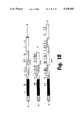
- FIG. 8 shows the nucleotide sequence and the encoded amino acids of hHS2.
- hHS2 has 756 bp with an open reading frame encoding 248 amino acids. Alignments of hPS11 and hHS2 according to the different domains at the nucleotide level and at the protein level demonstrate that hHS2 cDNA contains the N-terminal domain, the first internal repeat and part of the second repeat of hPS11 . Percent homology with the corresponding domains in hPS11 is 92.5, 95.0 and 92.7, respectively, at the nucleotide level, and 87.1, 88.2 and 94 at the amino acid level. The positions of the cysteine residues and the potential glycosylation site are conserved in hPS11 and hHS2.
- SP1 mRNA has also been identified in different hematopoietic cells. Expression of different SP1 genes appears to be lineage specific. Culture cells were stimulated for four hours with 10 ng/ml of phorbol 12-myristate 13-acetate (PMA) (P), 5 ⁇ g/ml lipopolysaccharide (LPS) (L), 100U/ml interferon-gamma (INF-gamma) (gamma), or the combination of LPS and IFN-gamma (L.gamma).
- PMA phorbol 12-myristate 13-acetate
- LPS lipopolysaccharide
- IFN-gamma interferon-gamma
- L.gamma interferon-gamma
- Northern blot analysis revealed the presence of hybridizing mRNA in unstimulated cells (C) with a 2.4 kb band in the KG-1 line (myelomonocytic) and a 3.3 kb band in the HEL line (erythroid). Stimulation with LPS and IFN-gamma have little effect on SP1 mRNA expression in KG-1 over a four hour period. However, LPS plus IFN-gamma increases SP1 mRNA expression in HEL several fold. The results demonstrate for the first time both expression of the SP1 genes in hematopoietic cells and differential expression of the SP1 genes.
- a human intestine cDNA library was screened with hPS3 using procedures described in Example 5. 21 positive clones were identified. Analysis with restriction enzymes showed that these clones can be divided into four main groups. Four of the clones have one internal EcoRI site. Polyacrylamide gel electrophoresis analysis showed that the longest clone of this group, hIS1, gave two insert fragments of 1.1 kb and 1.3 kb upon digestion with EcoRI. 17 cones have no internal EcoRI sites but include an interna XbaI site. The longest clone in this group is hIS3 with a cDNA insert of 1.5 kb.
- the other three groups do not have internal XbaI sites and have similar restriction enzyme maps as those of the three groups of placental SP1 cDNA.
- the smaller EcoRI fragments of clones hIS1and hIS2 of the first group have been sequenced. cDNA inserts of these two clones are identical.
- FIG. 9 shows the nucleotide sequence and the encoded amino acids of hIS1.
- hIS1 has 1024 bp with a 5' non-translated sequence of 61 bp, an open reading frame of 963 bp encoding a potential signal peptide of 34 amino acids, an N-terminal domain of 107 amino acids and a complete repeating unit consisting of an n-subdomain of 93 amino acids and a c-subdomain of 85 amino acids.
- Comparison of the corresponding domains of hIS1 and hPS11 shows that the percentage of homology between the two cDNAs is approximately 74.5, with the highest homology between the n-subdomains (79.3%) and the lowest homology between the c-subdomains (68.3%).
- the percentage homology of the encoded amino acid sequence of the two cDNAs is approximately 57.4, and is also higher between the n-subdomains and lower between the c-subdomains.
- a human genomic library was screened, clones identified and restriction mapped, as described in Examples 1-3. Partial restriction maps were constructed which demonstrated the presence of at least seven groups of unique SP1 genomic clones and suggested that multiple genes code for SP1. The multigene nature of SP1 was confirmed by hybridization of the SP1 cDNA probe to multiple bands on Southern blots of human genomic DNA. Further analysis with chromosomal DNA dot blot demonstrated the presence of homologous sequences on the X chromosome and autosomal chromosome 6.
- Percent of homology with the consensus amino acid sequence ranged from 92.5 to 100% in the N-terminal domain and the n-subdomains except for the N-terminal domain of hPS2 which shows only 85.9% homology. Levels of homology among the c-subdomains are slightly lower than the other domains and range from 85.9 to 97.6%. Nucleotide sequence homology of the N-terminal and n- and c-subdomains are similar to that of amino acid sequences and range from 90 to 99%.
- the amino acid and nucleotide sequences of the C-terminal domain of these cDNAs share very little sequence homology with two exceptions: the C-terminal domain of hPS11 and PSBGD are identical and the 3' non-coding sequence of PSG16 is identical with that of hPS11 other than for the deletion of 86 bp near the 3' end of the coding region. Positions of the cysteine residues in the n- and c- subdomains of all cDNAs are conserved. Positions of the potential glycosylation sites in the N-terminal domain and R1n-subdomain are also conserved.
- hPS12 differs from the other SP1 cDNAs in the deletion of three nucleotides encoding Ile-90 in the N-terminal domain.
- the comparison also makes clear that hPS12 and pSP1-i are different from each other as well as the other SP1 cDNAs.
- hPS11 and PSBGD are identical.
- PSG16 is identical to hPS11 except at four positions in the coding region resulting in the change of three amino acids, a G to C mutation in the 3' non-coding sequence and deletion of 86 bp near the 3' end of the coding region.
- PSBG is also identical to hPS11 and PSBGD except for the C-terminal domain.
- Both hPS2 and PSBGE have one less n-subdomain when compared to the other SP1 cDNAs.
- the n-subdomain of hPS2 and PSBGE are more comparable with the R1n-subdomain than the R2n-subdomain.
- the percentage of homology with the R1n-subdomain consensus sequence is 93.5 and 92.5 and that of the R2n-subdomain is 52.7 and 48.4 for hPS2 and PSBGE respectively.
- the c-subdomain of hPS2 and PSBGE are less homologous to the consensus sequence than that of the other SP1 cDNAs, being 87.1% and 85.9% respectively, while all other cDNAs show greater than 90% homology.
- the most significant difference between hPS2 and the other SP1s is the presence of the 81 amino acid hydrophobic C-terminus while all other SP1s have only relatively short (14 amino acids or less) hydrophilic C-termini.
- hPS11 , PSG16 and PSBGD are almost identical except for the deletion of 70 bp after the stop codon in PSG16 and one mismatch in both PSG16 and PSBGD when compared with hPS11.
- Examples 1 to 3 support the contention that SP1 protein in human placenta consists of products of three or more genes.
- Two of the reported placental SP1 cDNAs, hPS11 and PSBGD are identical.
- PSG16 and another partially sequenced cDNA PSG93 differ from these two cDNAs at only four bases which could correspond to polymorphisms.
- hPS11 , PSG16, PSBG93,PSBGC and PSBGD are likely to be the products of the same gene with differentially spliced exons encoding the C-terminus and 3' non-coding sequence.
- cDNAs hPS12, PSBGE and pSP1-i have 221 bp in their 3' non-coding sequence which are highly homologous (95%). Considering that this is observed in the non-coding region and that the three clones are derived from two different libraries, it is conceivable that the few differences are individual polymorphisms.
- These three cDNAs are products of splicing of different amino acid coding exons to one common exon which contains the 3' non-coding sequence.
- the exon encoding the N-terminal domain of hPS12 is unique in that it has a 3 bp deletion when compared to the other cDNAs.
- PSBGE differs from hPS12 and pSP1-i by having one less n-subdomain which could be the result of difference in splicing.
- the gene encoding hPS12/PSBGE/pSP1-i could be different from that encoding hPS11/PSG16/PSG93/PSBGC-D.
- the other SP1 cDNA, hPS2 is unique. It has no significant homology with any of the SP1 cDNAs reported.
- the presence of three different species of mRNA in human placenta was confirmed by the Northern blot analysis.
- the 3' EcoRI fragment of hPS11 contains the entire 3' non-coding sequence and is specific for that cDNA.
- the NcoI-EcoRI fragment of hPS12 contains the C-terminal 24 bp of the coding sequence and the 3' non-coding sequence and is unique for cDNAs of this group.
- the probe for hPS2 is the fragment encoding the unique hydrophobic C-terminal domain of the molecule. These three probes are cDNA specific. Hybridization of the Northern blot with these probes therefore indicates the presence of the specific mRNA species.
- the hPS2 specific probe hybridized to both the 1.65 Kb and 2.25 Kb mRNA bands, suggesting that each mRNA band contains more than one species of mRNA.
- up to the present time only one placental SP1 cDNA with sequence encoding a hydrophobic C-terminal has been found.
- the Northern blot results suggest that there may be more than one species of membrane-bound SP1 present in human placenta.
- SP1 genes like the CEA family of genes, are probably localized in clusters on chromosome 6 and the X chromosome, although there is also the possibility that all SP1 cDNAs so far reported are derived from one very large gene which gives rise to the above three groups of products by differential splicing.
- Both hPS2 and PSBGE unlike the other SP1 cDNAs, contains only one n-subdomain.
- These cDNAs could be formed by having one complete repeating unit containing an n-subdomain and c-subdomain (i.e. n-subdomain in hPS2 is R2n-subdomain) or by splicing the n-subdomain of one repeating unit with the c-subdomain of another repeating unit (i.e. n-subdomain in hPS2 is R1n-subdomain).
- hPS2 The distinctive feature of hPS2, the presence of an 81 amino acids hydrophobic C-terminus, is very similar to that observed in CEA, TM-CEA and NCA. Hydropathy plot analysis showed that this C-terminal domain of hPS2 is very hydrophobic and supports the inference that it represents the membrane-anchor region of the molecule. These results support the theory that there are two types of SP1 proteins in human placenta, the cytoplasmic or soluble SP1 and the membrane-bound SP1. Analogous phenomenon has been reported for the CEA family of proteins.
- the SP1 proteins and CEAs appear to share a lot of common properties, there are sufficient features unique to the SP1 proteins to qualify them as a separate subfamily of the Ig gene superfamily instead of being members of the CEA gene subfamily.
- the SP1s are very similar to CEA and NCA, the homology among the different SP1 family members are even higher, being consistently greater than 90% at the nucleotide level and greater than 85% at the amino acid level.
- the SP1 proteins are : also less glycosylated. The number of potential glycosylation site ranges from four in pSP1-i to eight in PSBGC with the majority having six or seven. Most of these sites are also conserved. Both CEA and NCA are more, heavily glycosylated, with twenty-seven potential sites in NCA and twelve potential sites in NCA. A number of these sites are conserved between CEA and NCA but not between CEAs and SP1s.
- cDNA clones for seven apparently different, but closely related, genes for SP1 proteins have been isolated.
- One group of clones appears to encode placenta-specific SP1 proteins. Clones in this group are characterized by not having an internal EcoRI site.
- An example is hPS12.
- a third group of clones has been isolated only from placenta and has two internal EcoRI sites.
- An example of the third group is hPS2.
- Clones have also been isolated from a HeLa cell cDNA library, a testis cDNA library and an intestinal cDNA library, which are highly homologous to placental SP1 cDNA.
- hIS1 shows higher homology to Normal Crossreacting Antigen (NCA), a member of the CEA family, than to the placental sP1 cDNAs.
- NCA Normal Crossreacting Antigen
- FIG. 10 compares the domain structures among hPS11 , hIS1 and CEA.
- hIS1 is 93% homologous at the nucleotide level, and 67% homologous at the amino acid level, with CEA.
- the N-terminal domains of both hIS1and CEA are two amino acids shorter than that of hPS11 .
- the domain structure, as well as the number of potential glycosylation sites, are more similar between hIS1 and CEA than hPS11.
- CEA carcinoembryonic antigen
- hPS2 and PSBGE have the domain structure closest to that of CEA and NCA. Both hPS2 and PSBGE, similar to NCA, have only one complete repeat unit, i.e, one n-subdomain and one c-subdomain, while CEA has three complete repeating units. All other SP1 cDNAs have one complete repeating unit and an additional n-subdomain.
- Comparison of the consensus amino acid sequences of the different domains of the SP1 cDNAs with the corresponding domains in CEA and NCA shows that the percentage homology of both CEA and NCA to SP1 is quite similar in all domains compared, ranging from 53.8% to 62.4% at the amino acid level. Nucleotide sequence comparison of the different domains of SP1 CEA and NCA showed similar results as that of amino acid comparison.
- hPS11, hPS2 and hPS12 proteins that appear to be expressed in placental tissue.
- hPS2 and hPS12 have only been found in placental tissue. Since placental tissue exists only in individuals who are pregnant, the levels of hPS11, hPS2 and hPS12 proteins can function as an index of whether the female, either a human or non-human mammal such as a rat, cow or pig, is pregnant.
- the present invention therefore includes a method and reagents for testing for pregnancy wherein a biological sample from the subject is assayed for the level of one of the placenta specific peptides, and correlating an above-normal level, if any, with pregnancy, and correlating a normal or below-normal level, if any, with the absence or abnormalities of pregnancy.
- a biological sample from the female is obtained and is treated with monoclonal antibodies against the protein described in FIG. 2.
- the level of antibody-protein reaction is measured using methods known to those skilled in the art (i.e., radiolabelling, ELISA, etc.) to determine the level of protein in the sample.
- the present invention also includes a method and compositions for enhancing the fertility of a female deficient in one of the placenta specific proteins such as the protein described in FIG. wherein an effective amount of the protein or peptide is administered to the female.
- the peptide is preferably administered in combination with a pharmaceutically acceptable carrier, as described below.
- a pharmaceutically acceptable carrier as described below.
- the preferred dosage of the peptide is based on the normal levels of the protein and is preferably administered as determined by those skilled in the art. In general, this will be between 0.1 and 50 mg/kg body weight.
- Any pharmaceutically acceptable carrier may be selected which will maintain the biological activity of the protein, such as 5% dextrose in sterile water and sterile normal saline.
- the present invention further includes a method and compositions for enhancing the viability of a fetus wherein an effective amount of a pregnancy specific peptide or protein such as the one shown in FIG. 2 is administered to either the mother or the fetus. If the mother has an inadequate level of SP1 -like proteins, a spontaneous abortion could occur. Accordingly, the administration of the composition can raise the level of SP1 proteins necessary to maintain a viable fetus.
- the peptide is preferably administered in combination with a pharmaceutically acceptable carrier, as described above.
- the SP1 proteins described here are present in cows, pigs, sheep, horses, dogs, cats, rats, and primates, including chimpanzee, cynomolgus, monkeys, and baboons, as well as humans. On this basis, it is assumed that the proteins are present in all placental mammals, and that the methods described above can therefore be utilized in all placental mammals.
Landscapes
- Health & Medical Sciences (AREA)
- Chemical & Material Sciences (AREA)
- Organic Chemistry (AREA)
- Life Sciences & Earth Sciences (AREA)
- Medicinal Chemistry (AREA)
- General Health & Medical Sciences (AREA)
- Proteomics, Peptides & Aminoacids (AREA)
- Genetics & Genomics (AREA)
- Molecular Biology (AREA)
- Biophysics (AREA)
- Biochemistry (AREA)
- Toxicology (AREA)
- Gastroenterology & Hepatology (AREA)
- Zoology (AREA)
- General Chemical & Material Sciences (AREA)
- Veterinary Medicine (AREA)
- Public Health (AREA)
- Reproductive Health (AREA)
- Animal Behavior & Ethology (AREA)
- Pharmacology & Pharmacy (AREA)
- Nuclear Medicine, Radiotherapy & Molecular Imaging (AREA)
- Chemical Kinetics & Catalysis (AREA)
- Immunology (AREA)
- Bioinformatics & Cheminformatics (AREA)
- Engineering & Computer Science (AREA)
- Pregnancy & Childbirth (AREA)
- Gynecology & Obstetrics (AREA)
- Endocrinology (AREA)
- Urology & Nephrology (AREA)
- Peptides Or Proteins (AREA)
- Medicines That Contain Protein Lipid Enzymes And Other Medicines (AREA)
- Preparation Of Compounds By Using Micro-Organisms (AREA)
Priority Applications (6)
| Application Number | Priority Date | Filing Date | Title |
|---|---|---|---|
| US07/390,409 US5169835A (en) | 1989-01-18 | 1989-08-07 | Pregancy specific proteins applications |
| AU49646/90A AU645965C (en) | 1989-01-18 | 1990-01-11 | Pregnancy specific proteins applications |
| CA002046925A CA2046925A1 (fr) | 1989-01-18 | 1990-01-11 | Applications des proteines propres a la grossesse |
| EP19900902488 EP0454765A4 (en) | 1989-01-18 | 1990-01-11 | Pregnancy specific proteins applications |
| PCT/US1990/000285 WO1990007937A1 (fr) | 1989-01-18 | 1990-01-11 | Applications de proteines specifiques a la grossesse |
| JP2502864A JPH04504655A (ja) | 1989-01-18 | 1990-01-11 | 妊娠に特異的なタンパク質の利用 |
Applications Claiming Priority (2)
| Application Number | Priority Date | Filing Date | Title |
|---|---|---|---|
| US29863889A | 1989-01-18 | 1989-01-18 | |
| US07/390,409 US5169835A (en) | 1989-01-18 | 1989-08-07 | Pregancy specific proteins applications |
Related Parent Applications (1)
| Application Number | Title | Priority Date | Filing Date |
|---|---|---|---|
| US29863889A Continuation-In-Part | 1989-01-18 | 1989-01-18 |
Publications (1)
| Publication Number | Publication Date |
|---|---|
| US5169835A true US5169835A (en) | 1992-12-08 |
Family
ID=26970789
Family Applications (1)
| Application Number | Title | Priority Date | Filing Date |
|---|---|---|---|
| US07/390,409 Expired - Fee Related US5169835A (en) | 1989-01-18 | 1989-08-07 | Pregancy specific proteins applications |
Country Status (5)
| Country | Link |
|---|---|
| US (1) | US5169835A (fr) |
| EP (1) | EP0454765A4 (fr) |
| JP (1) | JPH04504655A (fr) |
| CA (1) | CA2046925A1 (fr) |
| WO (1) | WO1990007937A1 (fr) |
Cited By (7)
| Publication number | Priority date | Publication date | Assignee | Title |
|---|---|---|---|---|
| EP0757249A1 (fr) * | 1994-03-18 | 1997-02-05 | Ivan Nikolaevich Golovistikov | Technique de mise en evidence de la composante suppressive de l'etat immunitaire chez l'homme, et mode d'application |
| US6150326A (en) * | 1994-03-18 | 2000-11-21 | Leonid Yazonovich Kacharava | Means for treating autoimmune diseases and method for the treatment thereof |
| WO2006039604A1 (fr) * | 2004-09-30 | 2006-04-13 | Applied Research Systems Ars Holding, N.V. | Utilisation d'une glycoproteine specifique a la grossesse (gsg) pour la maturation d'ovocytes |
| US20060083682A1 (en) * | 1997-09-18 | 2006-04-20 | Stemline Therapeutics, Inc. | Novel methods of cancer therapy targeted against a cancer stemline |
| US20070231896A1 (en) * | 2004-09-30 | 2007-10-04 | De Matos Daniel G | Use of Il-17-for Maturation of Oocytes |
| US20080118518A1 (en) * | 2006-09-07 | 2008-05-22 | Cirrito Thomas P | Cancer stem cell-targeted cancer therapy |
| US20110086020A1 (en) * | 2006-04-28 | 2011-04-14 | Khallar Abdumuslimovich Alikhanov | Agent exhibiting immunoregulatory properties and the use thereof for treating autoimmune diseases |
Families Citing this family (2)
| Publication number | Priority date | Publication date | Assignee | Title |
|---|---|---|---|---|
| WO1994021686A1 (fr) | 1993-03-19 | 1994-09-29 | Northern Sydney Area Health Service | Papp-a, son immunodetection et ses utilisations |
| AU2016323769B2 (en) * | 2015-09-17 | 2022-05-26 | The Henry M. Jackson Foundation For The Advancement Of Military Medicine, Inc. | Compositions comprising pregnancy specific glycoproteins and methods of use thereof |
Citations (2)
| Publication number | Priority date | Publication date | Assignee | Title |
|---|---|---|---|---|
| JPS59214767A (ja) * | 1983-05-20 | 1984-12-04 | Green Cross Corp:The | ヒト妊娠特異蛋白測定用キツト |
| US4818709A (en) * | 1983-01-21 | 1989-04-04 | Primus Frederick J | CEA-family antigens, Anti-CEA antibodies and CEA immunoassay |
Family Cites Families (2)
| Publication number | Priority date | Publication date | Assignee | Title |
|---|---|---|---|---|
| US4191533A (en) * | 1971-09-29 | 1980-03-04 | Behringwerke Aktiengesellschaft | Pregnancy-specific β1 -glycoprotein and process for isolating it |
| DE2345953A1 (de) * | 1973-09-12 | 1975-09-25 | Behringwerke Ag | Mittel zur konzeptionsverhuetung und zur schwangerschaftsunterbrechung bei primaten |
-
1989
- 1989-08-07 US US07/390,409 patent/US5169835A/en not_active Expired - Fee Related
-
1990
- 1990-01-11 CA CA002046925A patent/CA2046925A1/fr not_active Abandoned
- 1990-01-11 EP EP19900902488 patent/EP0454765A4/en not_active Withdrawn
- 1990-01-11 WO PCT/US1990/000285 patent/WO1990007937A1/fr not_active Application Discontinuation
- 1990-01-11 JP JP2502864A patent/JPH04504655A/ja active Pending
Patent Citations (2)
| Publication number | Priority date | Publication date | Assignee | Title |
|---|---|---|---|---|
| US4818709A (en) * | 1983-01-21 | 1989-04-04 | Primus Frederick J | CEA-family antigens, Anti-CEA antibodies and CEA immunoassay |
| JPS59214767A (ja) * | 1983-05-20 | 1984-12-04 | Green Cross Corp:The | ヒト妊娠特異蛋白測定用キツト |
Non-Patent Citations (68)
| Title |
|---|
| A. G. Ahmed, et al., British Journal of Obstetrics and Gynecology 90: 604 611 (1983). * |
| A. G. Ahmed, et al., British Journal of Obstetrics and Gynecology 90: 604-611 (1983). |
| Asano et al. (1990) Blood 75(8):1602 1605. * |
| Asano et al. (1990) Blood 75(8):1602-1605. |
| Beauchemin et al. (1987) Mol. Cellular Biol 7: 3221 3230. * |
| Beauchemin et al. (1987) Mol. Cellular Biol 7: 3221-3230. |
| Bischof, Contri. Gynecol. Obstet. 12, 6 92 (1984). * |
| Bischof, Contri. Gynecol. Obstet. 12, 6-92 (1984). |
| Bohn, Blut 24, 292 302 (1972) (Summary which is translated). * |
| Bohn, Blut 24, 292-302 (1972) (Summary which is translated). |
| C. H. W. Horne, et al., Placental Proteins, (Springer Verlag 1979) pp. 143 160. * |
| C. H. W. Horne, et al., Placental Proteins, (Springer Verlag 1979) pp. 143-160. |
| Chan, "The Pregnancy-Specific B1-Glycoprotein Family", (1991) Adv. Contr. Deliv. Sys., vol. VII, pp. 21-52. |
| Chan, The Pregnancy Specific B1 Glycoprotein Family , (1991) Adv. Contr. Deliv. Sys., vol. VII, pp. 21 52. * |
| Chou, Oncodevelop. Biol. Med. 4, 319 326 (1983). * |
| Chou, Oncodevelop. Biol. Med. 4, 319-326 (1983). |
| Heikinheimo, et al., J. Clin. Endocr. Metab. 51, 1432 1436 (1980). * |
| Heikinheimo, et al., J. Clin. Endocr. Metab. 51, 1432-1436 (1980). |
| Ishibashi et al. (1989) Blood 74(4):1241 1244. * |
| Ishibashi et al. (1989) Blood 74(4):1241-1244. |
| J. G. Grudzinskas, et al., Pregnancy Proteins, (Academic Press, New York 1982) pp. 251 259. * |
| J. G. Grudzinskas, et al., Pregnancy Proteins, (Academic Press, New York 1982) pp. 251-259. |
| J. G. Williams, Genetic Engineering, vol. 1 (Academic Press, New York 1981) at pp. 1 8. * |
| J. G. Williams, Genetic Engineering, vol. 1 (Academic Press, New York 1981) at pp. 1-8. |
| L. A. Tease, et al., FASEB J. 2(5): A1686 (1988). * |
| L. Johnston Dow, et al., Bio Techniques 5(8): 754 765 (1987). * |
| L. Johnston-Dow, et al., Bio-Techniques 5(8): 754-765 (1987). |
| L. Tease, et al., Fed. Proc. 45(6): 1512 (1986). * |
| Lin, et al., J. Clin. Invest. 54, 576 582 (1974). * |
| Lin, et al., J. Clin. Invest. 54, 576-582 (1974). |
| P. Bischof, Gynecology and Obstetrics, 12: 6 12 (1984) with concluding remarks and references. * |
| P. Bischof, Gynecology and Obstetrics, 12: 6-12 (1984) with concluding remarks and references. |
| Patchen et al. (1991) Blood 77(3):472 480. * |
| Patchen et al. (1991) Blood 77(3):472-480. |
| Rooney et al. (1988) Gene 71: 439 449. * |
| Rooney et al. (1988) Gene 71: 439-449. |
| Rosen, Pregnancy Proteins 223 234 (1982). * |
| Rosen, Pregnancy Proteins 223-234 (1982). |
| S. W. Rosen, et al., Placenta 7: 575 594 (1986). * |
| S. W. Rosen, et al., Placenta 7: 575-594 (1986). |
| S. Watanabe, et al., Biochemical and Biophysical Research Communications 152: 762 768 (1988). * |
| S. Watanabe, et al., Biochemical and Biophysical Research Communications 152: 762-768 (1988). |
| S. Watanabe, et al., J. Biol. Chem. 263(4): 2049 2054 (1988). * |
| S. Watanabe, et al., J. Biol. Chem. 263(4): 2049-2054 (1988). |
| Streydio, et al., Biochem. Biophys. Research Comm., 154(1), 130 137 (Jul. 1988). * |
| Streydio, et al., Biochem. Biophys. Research Comm., 154(1), 130-137 (Jul. 1988). |
| Tatarinov and Masyukevich, Bull. Exp. Biol. Med. 69, 666 668 (1970). * |
| Tatarinov and Masyukevich, Bull. Exp. Biol. Med. 69, 666-668 (1970). |
| W. Jones, et al., Pregnancy Proteins, (Academic Press, New York 1982) pp. 241 250. * |
| W. Jones, et al., Pregnancy Proteins, (Academic Press, New York 1982) pp. 241-250. |
| W. Qiu, et al., Fed. Proc. 46(6): 2186 (1987). * |
| W. Y. Chan, et al., Am. J. Hum. Genet. 41(3): A149 (1987). * |
| W. Y. Chan, et al., Am. J. Hum. Genet. 43: 152 159 (1988). * |
| W. Y. Chan, et al., Am. J. Hum. Genet. 43: 152-159 (1988). |
| W. Y. Chan, et al., DNA 7(8): 545 555 (1988). * |
| W. Y. Chan, et al., DNA 7(8): 545-555 (1988). |
| W. Y. Chan, et al., FASEB J. 2(5): A1686 (1988). * |
| W. Y. Chan, et al., Fed. Proc. 44(5): 1461 (1985). * |
| W. Y. Chan, et al., Human Reproduction 3(5): 677 685; 687 692 (1988). * |
| W. Y. Chan, et al., Human Reproduction 3(5): 677-685; 687-692 (1988). |
| W. Y. Chan, et al., J. Cell. Biol. 105(4): 258a (1987). * |
| W. Y. Chan, et al., Pediatric Research 19(4): 152A (1985). * |
| Whicher et al. (1990) Clin. Chem 36/7:1269 1272. * |
| Whicher et al. (1990) Clin. Chem 36/7:1269-1272. |
| Williams et al. (1990) Exp. Hematol 18: 69 72. * |
| Williams et al. (1990) Exp. Hematol 18: 69-72. |
| Y. S. Tatarinov, Placental Proteins, (Springer Verlag 1979) 161 171. * |
| Y. S. Tatarinov, Placental Proteins, (Springer Verlag 1979) 161-171. |
Cited By (28)
| Publication number | Priority date | Publication date | Assignee | Title |
|---|---|---|---|---|
| EP0757249A1 (fr) * | 1994-03-18 | 1997-02-05 | Ivan Nikolaevich Golovistikov | Technique de mise en evidence de la composante suppressive de l'etat immunitaire chez l'homme, et mode d'application |
| EP0757249A4 (fr) * | 1994-03-18 | 1998-12-02 | Ivan Nikolaevich Golovistikov | Technique de mise en evidence de la composante suppressive de l'etat immunitaire chez l'homme, et mode d'application |
| US6040187A (en) * | 1994-03-18 | 2000-03-21 | Ivan Nikolaevich Golovistikov | Method of determining the suppressor cell component of human immune status and a means for the realization thereof |
| US6150326A (en) * | 1994-03-18 | 2000-11-21 | Leonid Yazonovich Kacharava | Means for treating autoimmune diseases and method for the treatment thereof |
| US7361336B1 (en) * | 1997-09-18 | 2008-04-22 | Ivan Bergstein | Methods of cancer therapy targeted against a cancer stem line |
| US20090022741A1 (en) * | 1997-09-18 | 2009-01-22 | Ivan Bergstein | Novel methods of cancer diagnosis and therapy targeted against a cancer stem line |
| US20070036804A1 (en) * | 1997-09-18 | 2007-02-15 | Ivan Bergstein | Novel methods of cancer diagnosis and therapy targeted against a cancer stem line |
| US20070036801A1 (en) * | 1997-09-18 | 2007-02-15 | Ivan Bergstein | Novel methods of cancer diagnosis and therapy targeted against a cancer stem line |
| US20070036803A1 (en) * | 1997-09-18 | 2007-02-15 | Ivan Bergstein | Novel methods of cancer diagnosis and therapy targeted against a cancer stem line |
| US20070036802A1 (en) * | 1997-09-18 | 2007-02-15 | Ivan Bergstein | Novel methods of cancer diagnosis and therapy targeted against a cancer stem line |
| US20070036800A1 (en) * | 1997-09-18 | 2007-02-15 | Ivan Bergstein | Novel methods of cancer diagnosis and therapy targeted against a cancer stem line |
| US20070041984A1 (en) * | 1997-09-18 | 2007-02-22 | Ivan Bergstein | Novel methods of cancer diagnosis and therapy targeted against a cancer stem line |
| US8846325B2 (en) | 1997-09-18 | 2014-09-30 | Ivan Bergstein | Methods of cancer diagnosis and therapy targeted against a cancer stem line |
| US8715945B2 (en) | 1997-09-18 | 2014-05-06 | Ivan Bergstein | Methods of cancer diagnosis and therapy targeted against a cancer stem line |
| US8038998B2 (en) | 1997-09-18 | 2011-10-18 | Ivan Bergstein | Methods of cancer therapy targeted against a cancer stemline |
| US7608259B2 (en) | 1997-09-18 | 2009-10-27 | Ivan Bergstein | Methods of cancer diagnosis and therapy targeted against a cancer stem line |
| US7427400B2 (en) | 1997-09-18 | 2008-09-23 | Ivan Bergstein | Methods of cancer diagnosis and therapy targeted against a cancer stem line |
| US20080305107A1 (en) * | 1997-09-18 | 2008-12-11 | Ivan Bergstein | Novel methods of cancer diagnosis and therapy targeted against a cancer stem line |
| US20090011441A1 (en) * | 1997-09-18 | 2009-01-08 | Ivan Bergstein | Novel methods of cancer diagnosis and therapy targeted against a cancer stem line |
| US20090022740A1 (en) * | 1997-09-18 | 2009-01-22 | Ivan Bergstein | Novel methods of cancer diagnosis and therapy targeted against a cancer stem line |
| US20060083682A1 (en) * | 1997-09-18 | 2006-04-20 | Stemline Therapeutics, Inc. | Novel methods of cancer therapy targeted against a cancer stemline |
| US20090028879A1 (en) * | 1997-09-18 | 2009-01-29 | Ivan Bergstein | Novel methods of cancer diagnosis and therapy targeted against a cancer stem line |
| US7504103B2 (en) | 1997-09-18 | 2009-03-17 | Ivan Bergstein | Methods of cancer diagnosis and therapy targeted against a cancer stem line |
| WO2006039604A1 (fr) * | 2004-09-30 | 2006-04-13 | Applied Research Systems Ars Holding, N.V. | Utilisation d'une glycoproteine specifique a la grossesse (gsg) pour la maturation d'ovocytes |
| US20070282162A1 (en) * | 2004-09-30 | 2007-12-06 | De Matos Daniel G | Use of pregnancy specific glycoprotein for maturation of oocytes |
| US20070231896A1 (en) * | 2004-09-30 | 2007-10-04 | De Matos Daniel G | Use of Il-17-for Maturation of Oocytes |
| US20110086020A1 (en) * | 2006-04-28 | 2011-04-14 | Khallar Abdumuslimovich Alikhanov | Agent exhibiting immunoregulatory properties and the use thereof for treating autoimmune diseases |
| US20080118518A1 (en) * | 2006-09-07 | 2008-05-22 | Cirrito Thomas P | Cancer stem cell-targeted cancer therapy |
Also Published As
| Publication number | Publication date |
|---|---|
| AU4964690A (en) | 1990-08-13 |
| WO1990007937A1 (fr) | 1990-07-26 |
| EP0454765A4 (en) | 1992-11-04 |
| EP0454765A1 (fr) | 1991-11-06 |
| AU645965B2 (en) | 1994-02-03 |
| JPH04504655A (ja) | 1992-08-20 |
| CA2046925A1 (fr) | 1990-07-19 |
Similar Documents
| Publication | Publication Date | Title |
|---|---|---|
| EP0413908B2 (fr) | Fragment extracellulaire soluble du recepteur IFN-bêta 2/IL-6 humain, sa préparation et composition pharmaceutique le contenant | |
| Takahashi et al. | Cloning and sequencing of cDNA of Sarcophaga peregrina humoral lectin induced on injury of the body wall. | |
| US5948758A (en) | Methods for treating various disease states by reducing adhesion of leukocytes of target cells | |
| JP2866706B2 (ja) | 腫瘍壊死因子結合蛋白 | |
| EA005581B1 (ru) | ПОЛИПЕПТИДЫ ЦИТОКИНА Zcyto10 ЧЕЛОВЕКА И МЫШИ, КОДИРУЮЩИЕ ИХ ПОЛИНУКЛЕОТИДЫ И АНТИТЕЛА К УКАЗАННЫМ ПОЛИПЕПТИДАМ | |
| JPH04506342A (ja) | 非グリコシル化ヒトインターロイキン―3類似蛋白質 | |
| WO1988003170A1 (fr) | Proteines associees a des agents tensioactifs hydrophobes pulmonaires | |
| US5169835A (en) | Pregancy specific proteins applications | |
| Telen et al. | Molecular basis for elliptocytosis associated with glycophorin C and D deficiency in the Leach phenotype | |
| US6787328B1 (en) | Methods and compositions relating to CD39-like polypeptides and nucleic acids | |
| AU616164B2 (en) | Pulmonary hydrophobic surfactant-associated proteins | |
| CA2326066A1 (fr) | Antagoniste du recepteur de l'interleukine-1 et ses utilisations | |
| WO1995025117A1 (fr) | Polypeptides de proteine de liaison de la lipopolysaccharide | |
| Chan et al. | Pregnancy-specific β1 glycoprotein mRNA is present in placental as well as non-placental tissues | |
| CA2097424A1 (fr) | Proteine liante pour le facteur de liberation de la corticotrophine | |
| AU770000B2 (en) | Hermansky pudlak syndrome protein-interacting proteins and methods of use thereof | |
| AU645243B2 (en) | Human heat shock factor | |
| JPH04500603A (ja) | クローン化腎炎抗原 | |
| AU645965C (en) | Pregnancy specific proteins applications | |
| WO2000004041A2 (fr) | Procedes et materiaux associes a de nouveaux polypeptides semblables a cd39 | |
| US6426191B1 (en) | Assays involving an IL-1 receptor antagonist | |
| US6783959B1 (en) | Methods and compositions relating to CD39-like polypeptides and nucleic acids | |
| CA2319782A1 (fr) | Complexes proteiques de retinoblastome et proteines interagissant avec le retinoblastome | |
| US6780977B1 (en) | Methods and compositions relating to CD39-like polypeptides and nucleic acids | |
| US6899875B1 (en) | Methods and compositions relating to CD39-like polypeptides and nucleic acids |
Legal Events
| Date | Code | Title | Description |
|---|---|---|---|
| AS | Assignment |
Owner name: OKLAHOMA MEDICAL RESEARCH FOUNDATION, 825 N.E. 13T Free format text: ASSIGNMENT OF ASSIGNORS INTEREST.;ASSIGNOR:CHAN, WAI-YEE;REEL/FRAME:005111/0197 Effective date: 19890804 |
|
| REMI | Maintenance fee reminder mailed | ||
| LAPS | Lapse for failure to pay maintenance fees | ||
| FP | Lapsed due to failure to pay maintenance fee |
Effective date: 19961211 |
|
| STCH | Information on status: patent discontinuation |
Free format text: PATENT EXPIRED DUE TO NONPAYMENT OF MAINTENANCE FEES UNDER 37 CFR 1.362 |