US5030453A - Stable plurilamellar vesicles - Google Patents
Stable plurilamellar vesicles Download PDFInfo
- Publication number
- US5030453A US5030453A US06/660,573 US66057384A US5030453A US 5030453 A US5030453 A US 5030453A US 66057384 A US66057384 A US 66057384A US 5030453 A US5030453 A US 5030453A
- Authority
- US
- United States
- Prior art keywords
- splvs
- vesicles
- lipid
- aqueous
- entrapped
- Prior art date
- Legal status (The legal status is an assumption and is not a legal conclusion. Google has not performed a legal analysis and makes no representation as to the accuracy of the status listed.)
- Expired - Fee Related
Links
Images


Classifications
-
- A—HUMAN NECESSITIES
- A61—MEDICAL OR VETERINARY SCIENCE; HYGIENE
- A61K—PREPARATIONS FOR MEDICAL, DENTAL OR TOILETRY PURPOSES
- A61K9/00—Medicinal preparations characterised by special physical form
- A61K9/10—Dispersions; Emulsions
- A61K9/127—Synthetic bilayered vehicles, e.g. liposomes or liposomes with cholesterol as the only non-phosphatidyl surfactant
- A61K9/1277—Preparation processes; Proliposomes
-
- A—HUMAN NECESSITIES
- A61—MEDICAL OR VETERINARY SCIENCE; HYGIENE
- A61K—PREPARATIONS FOR MEDICAL, DENTAL OR TOILETRY PURPOSES
- A61K9/00—Medicinal preparations characterised by special physical form
- A61K9/10—Dispersions; Emulsions
- A61K9/127—Synthetic bilayered vehicles, e.g. liposomes or liposomes with cholesterol as the only non-phosphatidyl surfactant
Definitions
- This invention relates to liposomes and their uses as carriers. More specifically, it relates to a new type of lipid vesicle having unique properties which confer special advantages such as increased stability and high entrapment efficiency.
- compositions and methods described herein have a wide range of applicability to fields such as carrier systems and targeted delivery systems.
- the practice of the present invention is demonstrated herein by way of example for the treatment of brucellosis, the treatment of a systemic Salmonella infection, the treatment of ocular infections, the treatment of pyelonephritis and the treatment of lymphocytic meningitis virus infections.
- Liposomes are completely closed bilayer membranes containing an entrapped aqueous phase. Liposomes may be any variety of unilamellar vesicles (possessing a single membrane bilayer) or multilamellar vesicles (onion-like structures characterized by concentric membrane bilayers each separated from the next by a layer of water).
- the original liposome preparations of Bangham et al. (1965, J. Mol. Biol. 13:238-252) involved suspending phospholipids in an organic solvent which was then evaporated to dryness leaving a waxy deposit of phospholipid on the reaction vessel. Then an appropriate amount of aqueous phase was added, the mixture was allowed to "swell", and the resulting liposomes which consisted of multilamellar vesicles (hereinafter referred to as MLVs) were dispersed by mechanical means.
- MLVs multilamellar vesicles
- Efforts to increase the entrapped volume involved first forming inverse micelles or liposome precursors, i.e., vesicles containing an aqueous phase surrounded by a monolayer of lipid molecules oriented so that the polar head groups are directed towards the aqueous phase.
- Liposome precursors are formed by dispersing the aqueous solution to be entrapped in a solution of polar lipid in an organic solvent. The liposome precursors are then added to an aqueous medium and evaporated in the presence of excess lipid.
- the resultant liposomes consisting of an aqueous phase entrapped by a single lipid bilayer are dispersed in aqueous phase (see U.S. Pat. No. 4,224,179 issued Sept. 23, 1980 to M. Schneider).
- Papahadjopoulos U.S. Pat. No. 4,235,871 issued Nov. 25, 1980
- REVs reverse-phase evaporation vesicles
- the aqueous material to be entrapped is added to a mixture of polar lipid in an organic solvent. Then a homogeneous water-in-oil type of emulsion is formed and the organic solvent is evaporated until a gel is formed.
- the gel is then converted to a suspension by dispersing the gel-like mixture in an aqueous media.
- the REVs produced consist mostly of unilamellar vesicles (large unilamellar vesicles or LUVs) and some oligolamellar vesicles which are characterized by only a few concentric bilayers with a large internal aqueous space. Certain permeability properties of REVs were reported to be similar to those of MLVs and SUVs (see Szoka and Papahadjopoulos, 1978, Proc. Natl. Acad. Sci. U.S.A. 75:4194-4198).
- Liposomes which entrap a variety of compounds can be prepared, however, stability of the liposomes during storage is invariably limited. This loss in stability results in leakage of the entrapped aqueous soluble compound from the liposomes into the surrounding media, and can also result in contamination of the liposome contents by permeation of materials from the surrounding media into the liposome itself. As a result the storage life of traditional liposomes is very limited. Attempts to improve stability involved incorporating into the liposome membrane certain substances (hereinafter called "stabilizers") which affect the physical properties of the lipid bilayers (e.g., steroid groups). However, many of these substances are relatively expensive and the production of such liposomes is not costeffective.
- stabilizers certain substances which affect the physical properties of the lipid bilayers (e.g., steroid groups).
- MLVs can only be prepared under conditions above the phase-transition temperature of the lipid membrane. This precludes the incorporation of heat labile molecules within liposomes that are composed of phospholipids which exhibit desirable properties but possess long and highly saturated side chains.
- Desirable features of drug delivery systems depend upon the condition being treated. For example, when treating conditions which require maintenance doses of medication, resistance to rapid clearance of the drug accompanied by a sustained release of the drug which will prolong the drug's action increases the effectiveness of the drug and allows the use of fewer administrations. However, if one is treating an intracellular infection, the maintenance of stability in biological fluids, until the point that the liposome is internalized by the infected cell, is critical as is release of the liposome entrapped drug in its bio-active form.
- Liposome-entrapped materials leak when the liposomes are in contact with body fluids. This has been attributed to the removal of the liposomal phospholipids by plasma lipoproteins, or to the degradation of the liposome membrane by phospholipases, among other reasons.
- a result of the degradation of the liposomes in vivo is that almost all the liposomal contents are released in a short period of time, therefore, sustained release and resistance of the drug to clearance are not achieved.
- Liposomes are internalized by the phagocytic cells of the reticuloendothelial system (RES), and, therefore, are cleared from the system rapidly, rendering the entrapped drug largely ineffective against diseases involving cells other than the RES.
- RES reticuloendothelial system
- liposome entrapped drugs may be very useful in treating intracellular infections of the RES.
- the liposomal contents are packaged within lysosomes of the phagocytic cell and very often the degradative enzymes contained within the lysosome will degrade the entrapped compound or render the compound inactive by altering its structure or modifying the compound at its active site.
- the liposome carriers normally used in delivery systems are expensive and production is not cost-effective.
- an improved method for the chemotherapy of leishmanial infections using liposome encapsulated anti-leishmanial drugs has been reported by Steck and Alving in U.S. Pat. No. 4,186,183 issued on Jan. 29, 1980.
- the liposomes used in the chemotherapy contained a number of stabilizers which increased the stability of the liposomes in vivo.
- these stabilizers are expensive and the production of liposomes containing these stabilizers is not cost-effective.
- the problem encountered in the use of liposomes as carriers in drug delivery systems is the inability to effect a cure of the disease being treated.
- the liposomes may not deliver a dose which is effective due to the low percentage of entrapment of active compound into the vesicles when prepared.
- SPLVs stable plurilamellar vesicles
- MLVs, SUVs and REVs or LUVs lipid vesicles
- SPLVs are also prepared differently, possess unique properties, have very different pharmacological and pharmacokinetic effects, and present a variety of different advantages when compared to conventional liposomes.
- SPLVs overcome many of the problems presented by conventional lipid vesicles heretofore available.
- a heterogeneous mixture of plurilamellar lipid vesicles is realized when SPLVs are synthesized. Evidence indicates that the lipids in the SPLVs are organized in a novel supramolecular structure. Many of the lipid vesicles possess a high number of bilayers, occasionally in excess of one hundred.
- the SPLVs of the present invention are characterized by lipid bilayers enclosing aqueous compartments containing one or more entrapped solutes, the concentration of such solutes in each aqueous compartment being substantially equal to the concentration of solute used to prepare the SPLV.
- the SPLVs are not under an appreciable osmotic stress and, therefore, are not compressed.
- the properties of SPLVs include: (1) a concentration of entrapped solute in each of the aqueous compartments of SPLVs which is substantially equal to the concentration of solute used to prepare the SPLVs; (2) substantially non-compressed bilayers; (3) the entrapment of materials at a high efficiency; (4) X-ray diffraction signatures that differ from that of MLVs; (5) the ability to cure certain diseases which other drug-vesicle combinations cannot cure; (6) greatly increased stability of the SPLVs during storage in buffer; (7) the increased ability of SPLVs to withstand harsh physiologic environments; (8) the ability to stick to tissues and cells for prolonged periods of time; (9) the ability to release entrapped material slowly in body fluids; (10) the delivery and ultimate dispersal of the liposomal contents throughout the cytosol of the target cell; (11) the release of compounds in their bioactive forms in vivo and (12) improved cost-effectiveness in preparation.
- Stable plurilamellar vesicles of this invention may contain a phosphatidylcholine as a major component of the vesicles.
- Stable plurilamellar vesicles of this invention may contain an anti-oxidant such as butylated hydroxytoluene as a component of the vesicles.
- Stable plurilamellar vesicles of this invention may contain a protein entrapped within the vesicle.
- Stable plurilamellar vesicles of this invention may contain an antibacterial compound, antifungal compound, antiparasitic compound, and antiviral compound entrapped within the vesicle.
- Stable plurilamellar vesicles of this invention may contain a tumoricidal compound, toxin, cell receptor binding molecule, or immunoglobulin entrapped within the vesicle.
- Stable plurilamellar vesicles of this invention may contain an anti-inflammatory compound, anti-glaucoma compound, mydriatic compound, or local anesthetic entrapped within the vesicle.
- Stable plurilamellar vesicles of this invention may contain an enzyme, hormone, neurotransmitter, immunomodulator, nucleotide, or cyclic adenosine monophosphate entrapped within the vesicle.
- Stable plurilamellar vesicles of this invention may contain a dye, fluorescent compound, radioactive compound, or radio-opaque compound entrapped within the vesicle.
- Stable plurilamellar vesicles of this invention may contain a cosmetic preparation, a fragrance or a flavor entrapped within the vesicle.
- SPLVs Due to the unique properties of SPLVs they are particularly useful as carriers in delivery systems in vivo. Methods for the use of SPLVs for the delivery of bioactive compounds in vivo and the treatment of pathologies, such as infections, are described.
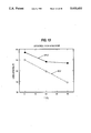
- FIG. 1 graphically demonstrates the difference in membrane stability (as reflected by % leakage) between MLVs and SPLVs treated with varying concentrations of urea.
- FIG. 2 The ratio of captured volume ( ⁇ l
- MLVs this ratio varies little over the range of lipid used.
- SPLVs the ratio rises sharply for small amounts of lipid.
- the solute was sodium 51 chromate; unentrapped material was removed by centrifugation.
- FIG. 3 graphically demonstrates the difference in the ability of ascorbate to reduce doxyl spin probes in SPLVs and in MLVs.
- FIG. 4 represents the X-ray long spacing versus temperature for MLVs and SPLVs composed of egg phosphatidylcholine.
- the characteristic forms of the MLV and SPLV curves constitute the Long Spacing Signature, or LSS (see text).
- LSS curve for SPLVs prepared by the emulsification process is the same as the LSS curve for SPLVs prepared by the monophasic solvent system process.
- FIG. 5 represents the small-angle X-ray diffraction intensity (arbitrary units) versus inverse reciprocal distance for the following liposomes composed of egg phosphatidylcholine: (a) MLVs; (b) SPLVs prepared by the emulsification process; and (c) SPLVs prepared by the monophasic solvent system process.
- the first and second orders on either side of the beam stop shadow (dip near the center) are relatively sharp.
- FIG. 6 The Wide-Angle X-ray diffraction Signature is shown for the following liposomes composed of egg phosphatidylcholine (a) SPLVs prepared by the emulsification process and MLVs; and (b) SPLVs prepared by the monophasic solvent system process.
- the signatures of MLVs and SPLVs differ as shown, however, this X-ray signature is more variable than the LSS.
- FIG. 7 The LSS for DOPC liposomes.
- data points were acquired at 40° C., 20° C., and 0° C. (in that order). These data points are connected by straight lines to guide the eye. However, it is important to realize that the true shape of the SPLV curve is smooth, as seen in FIG. 4.
- FIG. 8 The LSS for SPLVs composed of egg phosphatidylcholine and for negatively charged MLVs. Negatively charged MLVs were prepared using egg phosphatidylcholine and phosphatidic acid in a molar ratio of 8:2.
- FIG. 9 The effect of preparing egg phosphatidylcholine liposomes in 0.145 M salt but suspending them in 0.29 M salt is to make the LSS curve of SPLVs look like the LSS curve of MLVs; however both curves differ as to the absolute values.
- FIG. 10 The effect of preparing egg phosphatidylcholine MLVs in 0.145 M salt but suspending them in 0.073 M salt is to make the LSS look similar to the LSS of SPLVs.
- FIG. 11 Egg phosphatidylcholine MLVs which are prepared in 0.435 M salt, but suspended in 0.145 M salt yield an SPLV-like LSS. This is another way of enforcing a concentration gradient of the same sign as FIG. 12.
- FIG. 12 Nystatin in the presence of cholestrol is known to be a non-specific ionophore.
- MLVs were prepared using egg phosphatidylcholine , 6.6 mole % cholesterol and 1 mole % nystatin.
- the MLV LSS is shown after a few hours and after 24 hours. Note that the MLV LSS relaxes to the LSS of the SPLV control.
- the MLV and SPLV controls contained cholesterol but no nystatin. The LSS of these controls did not change over the 24 hours period. As seen in the MLV control, cholesterol affects the LSS but at low concentrations, the effects are small.
- FIG. 13 SPLVs and MLVs which were prepared using egg phosphatidylcholine and suspended in a sucrose solution without any salt also yield the familiar distinct LSS for each type of liposome.
- FIG. 14 The LSS for egg phosphatidylcholine liposomes prepared and suspended in distilled water. The two curves demonstrate an SPLV-like LSS.
- FIG. 15 The ratio of release of 14 C aqueous marker counts (NC) to 3 H membrane headgroup counts (N H ) is shown as a function of time as the liposomes are enzymatically digested.
- the steep rise of the MLV curve suggests that the outer aqueous compartments of the MLVs have a relatively low concentration of the aqueous marker solute molecules.
- each of the liposomes tested had released about 30% of the total headgroup counts.
- the MLV curve has been scaled by the factor (N C /N H ) SPLV TOT. ⁇ (N H /N C ) MLV TOT. to allow a direct comparison of the two curves.
- FIG. 16 graphically represents the retention of both the lipid and aqueous phases of SPLVs in the eyelid tissues of mice, and the sustained release of 125 I-gentamicin from the SPLVs in vivo.
- FIG. 17 represents the pharmacokinetics of gentamicin entrapped in MLVs or SPLVs administered intraperitoneally in mice. Antibiotic activity retrieved from the liver (a) and spleen (b) are plotted versus time in days.
- FIG. 18 graphically represents the effectiveness of a two stage treatment of Brucella canis infections in mice using SPLV-entrapped streptomycin based on B. canis recoverable from spleens of infected mice.
- FIG. 19 graphically represents the effectiveness of a two stage treatment of B. canis infections in mice using SPLV-entrapped streptomycin based on B. canis recoverable from organs of infected mice.
- FIG. 20 graphically represents the effectiveness of a two stage treatment of Brucella abortus in guinea pigs using SPLV-entrapped streptomycin.
- SPLVs are lipid vesicles, the lipid bilayers of which are characterized by a supramolecular organization which differs from that of conventional liposomes. Many of the lipid vesicles possess a high number of bilayers, occasionally in excess of one hundred.
- the membrane bilayer is composed of a bimolecular layer of an amphipathic lipid in which the non-polar hydrophobic hydrocarbon "tails" point inward towards the center of the bilayer and the polar, hydrophilic "heads" point towards the aqueous phase. Occluded by the bilayers is an aqueous compartment, part of which makes up the lumen of the vesicle, and part of which lies between adjacent layers.
- the concentration of solutes entrapped in each of the aqueous compartments of SPLVs are substantially equal to the concentration of solute used to prepare the SPLVs and the bilayers are substantially non-compressed.
- Complexed with the lipid bilayers can be a variety of proteins, glycoproteins, glycolipids, mucopolysaccharides, and any other hydrophobic and/or amphipathic substance.
- SPLVs may be prepared by any process that results in a substantially equal concentration of entrapped solutes in each aqueous compartment of a plurilamellar lipid vesicle that is substantially equal to the concentration of solutes used to prepare the SPLVs.
- SPLVs can be advantageously prepared by the processes described below which result in a liposome product unique from any other liposome previously described.
- Monophasic solvent system process a lipid or a mixture of lipids and an aqueous component are added to an organic solvent or a combination of organic solvents in amounts sufficient to form a monophase.
- the solvent or solvents are evaporated until a film forms. Evaporation may be accomplished by various methods, including but not limited to vacuum (e.g., by rotoevaporation or by passing an inert gas (e.g., nitrogen) over the monophase.
- an appropriate amount of aqueous component is added, and the film is resuspended and agitated in order to form the SPLVs.
- the organic solvent or combination of solvents used in the process must be miscible with water and once mixed with water should solubilize the lipids used to make the SPLVs.
- an organic solvent or mixture of solvents which satisfies the following criteria may be used in the process: (1) 5 ml of the organic solvent forms a monophase with 0.2 ml of aqueous component and (2) the lipid or mixture of lipids is soluble in the monophase.
- Solvents which may be used in this process of the present invention include but are not limited to ethanol, acetone, 2-propanol, methanol, tetrahydrofuran, glyme, dioxane, pyridine, diglyme, 1-methyl-2-pyrrolidone, butanol-2 butanol-1, isoamyl alcohol, isopropanol, 2-methoxyethanol, or a combination of chloroform:methanol (e.g., in a 1:1 ratio).
- the evaporation should be accomplished at suitable temperatures and pressures which maintain the monophase and facilitate the evaporation of the solvents.
- the temperatures and pressures chosen are not dependent upon the phase-transition temperature of the lipid used to form the SPLVs.
- the advantage of this latter point is that heat labile products which have desirable properties can be incorporated in SPLVs prepared from phospholipids such as distearoylphosphatidycholine, which can be formed into conventional liposomes only at temperatures above the phasetransition temperature of the phospholipids.
- This process usually allows more than 30-40% of the available water-soluble material to be entrapped during evaporation and 2-15% of the available water-soluble material to be entrapped during resuspension; and up to 70-80% of the available lipid-soluble material can be entrapped if the lipid:drug ratio is increased significantly.
- MLVs the entrapment of the aqueous phase, which only occurs during the rehydration step since no aqueous phase is present during the drying step, usually does not exceed 10%.
- SPLVs may be formed by adding 127 micromoles of phospholipid to 5 ml of ethanol and then adding 0.2 m ⁇ of aqueous component containing the active substance to be encapsulated.
- the resultant solution which comprises the material to be entrapped and the entrapping lipid is sonicated (sonication is an optional step) while streaming an inert gas over the mixture, thus removing most of the solvent and forming a film.
- To the resulting film is added 5-10 ml of aqueous component. The resuspended film is agitated in order to produce SPLVs.
- the agent or agents may be added to the monophase prior to evaporation and formation of the film.
- the agent or agents may be added with the aqueous component used to resuspend the film and form the SPLVs.
- the agent or agents may be added to both the monophase and to the aqueous component used to resuspend the film.
- Two or more agents can also be entrapped in one SPLV preparation by adding one agent to the monophase and the other to the aqueous component used to resuspend the film. See U.S. Application Ser. No. 633,481, filed July 25, 1984 and U.S. Application Ser. No. 518,912, filed Aug. 1, 1983 which are incorporated by reference herein.
- Emulsification process An amphipathic lipid or mixture of lipids is dissolved in an organic solvent. Many organic solvents are suitable, but diethyl ether, methylene chloride, fluorinated hydrocarbons and mixtures of fluorinated hydrocarbons and ether are preferred. To this solution are added an aqueous phase and the active ingredient to be entrapped. This biphasic mixture is converted to SPLVs by emulsifying the aqueous material within the solvent while evaporating the solvent, using any evaporative technique, e.g., evaporation by passing a stream of inert gas over the mixture, by heating, or by vacuum.
- organic solvents are suitable, but diethyl ether, methylene chloride, fluorinated hydrocarbons and mixtures of fluorinated hydrocarbons and ether are preferred.
- To this solution are added an aqueous phase and the active ingredient to be entrapped.
- This biphasic mixture is converted to SPLVs by e
- the volume of solvent used must exceed the aqueous volume by a sufficient amount so that the aqueous material can be completely emulsified in the mixture.
- a minimum of roughly 3 volumes of solvent to 1 volume of aqueous phase may be used.
- the ratio of solvent to aqueous phase can vary to up to 100 or more volumes of solvent to 1 volume aqueous phase.
- the amount of lipid must be sufficient so as to exceed that amount needed to coat the emulsion droplets (about 40 mg of lipid per ml of aqueous phase).
- the upper boundary is limited only by the practicality of cost-effectiveness, but SPLVs can be made with 15 gm of lipid per ml of aqueous phase.
- the entire process can be performed at a temperature range of 4°-60° C. regardless of the phase transition temperature of the lipid used.
- the advantage of this latter point is that heat labile products which have desirable properties, for example, easily denatured proteins, can be incorporated in SPLVs prepared from phospholipid such as distearoylphosphatidylcholine, but can be formed into conventional liposomes only at temperatures above their phase-transition-temperature.
- the process usually allows more than 20% of the available water soluble material to be encapsulated and more than 40% of the available lipid soluble material to be encapsulated. With MLVs the entrapment of aqueous phase usually does not exceed 10%.
- SPLVs may be formed by adding 50 micromoles of phospholipid to 5 ml of diethyl ether containing 5 micrograms of BHT (butylatedhydroxytoluene) and then adding 0.3 ml of aqueous phase containing the active substance to be encapsulated.
- BHT butylatedhydroxytoluene
- the resultant solution which comprises the material to be entrapped and the entrapping lipid is sonicated while streaming an inert gas over the mixture thus removing most of the solvent.
- This embodiment produces particularly stable SPLVs partially because of the incorporation of BHT into the vesicles.
- amphipathic lipids may be constituents of SPLVs.
- Suitable hydrophilic groups include but are not limited to: phosphato, carboxylic, sulphato and amino groups.
- Suitable hydrophobic groups include but are not limited to: saturated and unsaturated aliphatic hydrocarbon groups and aliphatic hydrocarbon groups substituted by at least one aromatic and/or cycloaliphatic group.
- the preferred amphipathic compounds are phospholipids and closely related chemical structures.
- Examples of these include but are not limited to: lecithin, phosphatidylethanolamine, lysolecithin, lysophatidylethanolamine, phosphatidylserine, phosphatidylinositol, sphingomyelin, cardiolipin, phosphatidic acid and the cerebrosides, ether lipids and phytanols.
- Suitable lipids useful in the production of SPLVs are phospholipids which include the natural lecithins (e.g., egg lecithin or soybean lecithin) and synthetic lecithins, such as saturated synthetic lecithins (e.g., dimyristoylphosphatidylcholine, or dipalmitoylphosphatidylcholine or distearoylphosphatidylcholine) and unsaturated synthetic lecithins (e.g., dioloyl-phosphatidylcholine or dilinoloylphosphatidylcholine.
- natural lecithins e.g., egg lecithin or soybean lecithin
- synthetic lecithins such as saturated synthetic lecithins (e.g., dimyristoylphosphatidylcholine, or dipalmitoylphosphatidylcholine or distearoylphosphatidylcholine) and unsaturated synthetic lecithin
- the SPLV bilayers can contain a steroid component such as cholesterol, coprostanol, cholestanol, cholestane and the like.
- a steroid component such as cholesterol, coprostanol, cholestanol, cholestane and the like.
- the obtained SPLVs will be anionic; with basic groups such as amino, cationic liposomes will be obtained; and with polyethylenoxy or glycol groups neutral liposomes will be obtained.
- the size of the SPLVs varies widely. The range extends from about 100 nm to about 10,000 nm (10 microns) and usually about 100 nm to about 1500 nm.
- any bioactive compound can be entrapped within a SPLV (entrapped is defined as entrapment within the aqueous compartment or within the membrane bilayer).
- Such compounds include but are not limited to nucleic acids, polynucleotides, antibacterial compounds, antiviral compounds, antifungal compounds, anti-parasitic compounds, tumoricidal compounds, proteins, toxins, enzymes, hormones, neurotransmitters, glycoproteins, immunoglobulins, immunomodulators, dyes, radiolabels, radio-opaque compounds, fluorescent compounds, polysaccharides, cell receptor binding molecules, anti-inflammatories, antiglaucomic agents, mydriatic compounds, local anesthetics, etc.
- incompatible drugs are also suitable for entrapment.
- Concurrent therapy with certain antimicrobial agents can be complicated because some agents which are particularly effective when used together in vitro cannot be formulated in a single mixture at therapeutic concentration for use in vivo due to a number of constraints.
- mixtures of gentamicin and nafcillin at therapeutic concentrations result in the formation of complexes that precipitate out of solution, and therefore, are not administered in vivo simultaneously.
- certain drug combinations are not recommended for use in vivo due to drug incompatibility (i.e., either inactivation of the drug or formation of a precipitate).
- antibiotics not be mixed with any other drug: gentamicin, kanamycin, lincomycin, cephalothin, and ampicillin (Davis and Abbitt, 1977, JAVMA 170(2): 204-207).
- certain agents cannot be solubilized in the same medium due to chemical restraints (e.g., a lipid soluble compound and a water soluble compound).
- chemical restraints e.g., a lipid soluble compound and a water soluble compound.
- SUVs and LUVs or REVs Freeze-fracture electron microscopy
- the idealized multilamellar liposome consists of a sequence of concentric lipid bilayer shells enclosing some central aqueous volume.
- EPC egg phosphatidylcholine
- the bilayers are separated by aqueous layers of a well defined width (approximately 20 ⁇ thick) which is a compromise between the repulsive and attractive forces between the bilayers (Rand, 1981 Ann. Rev. Biophys. Bioeng. 10:277-314.
- the aqueous fluid normally contains buffered physiological saline, as well as other dissolved solutes.
- MLVs The standard method for producing MLVs involves vacuum drying lipid dissolved in a solvent, such as chloroform, to a thin film onto the bottom of a round-bottom flask. MLVs are formed by adding the aqueous solution and shaking or vortexing until the dry film is removed from the wall of the flask.
- a solvent such as chloroform
- Lipid bilayers are semipermeable membranes: water passes freely through them but solutes, such as salts, are retained (Bangham et. al., 1967 Chem. Phys. Lipids 1:25). It has been generally assumed that the aqueous solute concentration is uniform throughout the MLV. In the instant specification we demonstrate that in contrast to this general assumption, standard methods of producing MLVs result in vesicles with aqueous compartments which are depleted in solutes, i.e., that the MLV formation effects a separation of water from its dissolved solutes. This leads to an osmotic stress resulting in a compressed liposome.
- the SPLV processes disclosed in the present invention produce multilamellar liposomes characterized by a concentration of entrapped solutes in each of the aqueous compartments which is substantially equal to the concentration of solute used to prepare the SPLVs, relatively little osmotic gradient on the liposomes and, therefore, an uncompressed liposome.
- the state of osmotic stress on the liposomes affects a broad spectrum of physical properties of the vesicles and has profound implications on the understanding and use of lipid bilayers.
- EPC 100 mg in chloroform, Sigma Chemical Co., type VIIe
- aqueous phase typically, HEPES buffer consisting of 72.5 mM KCl, 72.5 mM NaCl, 10 mM HEPES, pH 7.4
- HEPES buffer consisting of 72.5 mM KCl, 72.5 mM NaCl, 10 mM HEPES, pH 7.4
- the suspension was then set on the bench to equilibrate for 2 hours after which it was washed four times. Each wash was done by mixing the suspension with buffer to a total volume of 20 ml, followed by centrifugation (10,000 ⁇ g) to pellet the liposomes. The supernatant was removed and the pellet was then resuspended for further washes or to a designated final volume.
- EPC in chloroform 100 mg
- other lipids as described in this text, were rotary evaporated to dryness in 50 ml of round bottom flask.
- the lipid film was dissolved in 5 ml of ethyl ether.
- 0.3 ml of the aqueous phase typically HEPES buffer
- HEPES buffer aqueous phase
- a particular solute was to be entrapped it was dissolved in the aqueous phase prior to adding it to the ether-lipid solution.
- the two-phase mixture (aqueous and ether) was emulsified in a bath sonicator (Laboratory Supplies Co., model G1125PlG) during which time a gentle stream of nitrogen was passed over the mixture.
- An EPC film was prepared in a 50 ml round bottom flask by rotary evaporation from chloroform, as for SPLVS. Five ml of 95% ethanol and 0.2 ml of HEPES buffer were added to the flask and the flask was vortexed until the lipid film was dissolved. The result was a monophase solution of lipid, ethanol, and the buffer. The monophase was then rotary evaporated to dryness. The film contained the solutes which were dissolved in the buffer. This dry film was suspended and washed as described above in the MLV procedure.
- Stability of a lipid vesicle refers to the ability of the vesicle to sequester its occluded space from the external environment over a long period of time. For a lipid vesicle to be useful it is paramount that it be stable in storage and handling. It will be seen that SPLVs made from natural lecithin demonstrate increased stability during storage in buffer when compared to MLVs made from the same ingredients.
- vesicles There are two factors that cause vesicles to leak. One is auto-oxidation of the lipids whereby the hydrocarbon chains form peroxides which destabilize the bilayers. This oxidation can be drastically slowed down by the addition of antioxidants such as butylated hydroxy toluene (BHT) to the vesicle preparation. Vesicles can also leak because agents in the exterior environment disrupt the bilayer organization of the lipids such that the lipids remain intact, but the membrane develops a pore.
- antioxidants such as butylated hydroxy toluene (BHT)
- Preparations of lipid vesicles are white in color when first made. Upon auto-oxidation, the preparation becomes discolored (brownish).
- a comparison of MLVs to SPLVs prepared using the same lipid and aqueous components reveals that MLVs discolor within one to two weeks whereas SPLVs remain white for at least two months. This is supported by thin layer chromatography of the constituent lipids which showed degradation of the lipids in the MLVs but not of the lipids of the SPLVs.
- these vesicles are prepared by adding BHT as well as the other constituents, then MLVs appear slightly discolored within one month whereas the SPLVs remain white and appear stable for at least 6 months and longer.
- SPLVs and MLVs are also distinguished by the way they leak molecules entrapped in the aqueous compartments. In general, SPLVs maintain their entrapment dramatically longer than MLVs. Evidence indicates that SPLVs are able to sequester an encapsulated agent from molecules as small as calcium ions for more than six months.
- Arsenazo III is a dye which changes color from red to blue with the slightest amount of divalent cation present. By encapsulating the dye in SPLVs and adding calcium chloride to the storage buffer it is possible to measure the stability of the vesicles by looking for a color change. This was demonstrated by mixing the calcium sensitive dye Arsenazo III (3 mM) in the buffer used to prepare the liposomes.
- the liposomes were then washed to remove any unentrapped Arsenazo III, suspended in HEPES buffer with 500 mM CaCl 2 , purged with N 2 , sealed in screw top vials and stored at room temperature. Leakage of the Arsenazo III can be readily detected by eye by the color change with occurs when the dye contacts Ca ++ . Whereas MLVs leaked within a few days, the SPLVs have not leaked after 15 months at 4° C.
- vesicles were prepared which contained radioactive tracer molecules within the occluded aqueous compartments. When placed in a buffer containing isotonic saline at neutral pH, SPLVs containing antibiotic exhibit prolonged stability in storage.
- the vesicles were prepared, each containing one of the following radio-labeled drugs: 125 I-p-hydroxypropionic acid-derived gentamicin sulfate, 14 C-indomethacin, and 3 H-inulin. After storage at various temperatures for 14 days the vesicles were separated from the medium by centrifugation, and the relative amount of radioactivity that escaped from the vesicles into the medium was determined. The results demonstrated that SPLVs were more stable during storage than were MLVs.
- lipid vesicles in a medium which contains membrane perturbing agents is a way to probe different molecular organizations. Depending on how the membrane is organized, different vesicles will respond differently to such agents.
- vesicles were prepared which contained a radioactive tracer molecule ( 3 H-inulin) within the occluded aqueous compartment.
- Inulin a polysaccharide, partitions into the aqueous phase, and thus when radiolabeled may be used to trace the aqueous contents of lipid vesicles.
- the vesicles were separated from the medium by centrifugation, and the relative amount of radioactivity that escaped from the vesicles into the medium was determined.
- SPLVs are more stable than MLVs in hydrochloric acid.
- Table II illustrates that both MLVs and SPLVs, when made from egg lecithin, are destabilized when exposed to 0.125 N hydrochloric acid for one hour.
- the SPLVs are considerably less susceptible to the acid than MLVs. Presumably this different response reflects an intrinsic difference in the way the lipids interact with their environment.
- SPLVs also respond differently than MLVs when exposed to urea (FIG. 1 and Table II).
- Urea is a molecule with both a chaotropic effect (disrupts the structure of water) and a strong dipole moment. It is observed that SPLVs are far more susceptible to urea than they are to an osmotic agent such as sodium chloride at the same concentration (FIG. 1). MLVs do not leak significantly more in urea than they would in sodium chloride.
- guanidine a molecule similar to urea, does not destabilize SPLVs (Table II). Although guanidine is also strongly chaotropic, it does not possess a strong dipole moment.
- SPLVs are also susceptible to ammonium acetate, while MLVs are not (Table II). However, neither ammonium ion (in ammonium chloride) nor acetate (in sodium acetate) are particularly effective in causing SPLVs to destabilize. Thus it would appear that it is not the ion itself, but the polarity of the ammonium acetate which is responsible for inducing leakage.
- SPLVs are more stable in serum than MLVs.
- Many applications of lipid vesicles include administering them intraperitoneally, such as for the treatment of brucellosis. To be effective, the vesicles must survive for a sufficient time to reach their desired target.
- SPLVs and MLVs both made from egg lecithin, were exposed to fetal bovine serum which contained active complement, (Table II). After 48 hours exposure at 37° C., SPLVs are demonstrably more stable than MLVs.
- Entrapment efficiency is defined as the fraction of the aqueous compartment sequestered by bilayers.
- SPLVs entrap a larger percentage of the available active material thereby conserving material (see Table III). Entrapment is measured herein as the fraction of initial solute remaining with the liposomes after four washes.
- Another parameter used to characterize liposomes is defined as the volume enclosed by a given amount of lipid and is expressed as units of liters entrapped per mole of total lipid (1 mol -1 ).
- captured volume Another parameter used to characterize liposomes, called captured volume, is defined as the volume enclosed by a given amount of lipid and is expressed as units of liters entrapped per mole of total lipid (1 mol -1 ).
- the ratio of solute entrapped to the amount of lipid used in preparing the liposomes (i.e. captured volume) versus the starting amount lipid was determined by entrapping trace quantities of radioactively labeled water soluble markers.
- the amounts of lipid used to prepare the liposomes were varied from 15 to 658 ⁇ moles.
- Sodium 51 chromate ( 51 Cr) was entrapped in MLVs and SPLVs which were washed by centrifugation and then placed in a gamma counter to determine the amount of en
- the ratio of solute entrapped to lipid used was essentially constant for MLVs.
- the simplest interpretation of this result is that the use of more lipid resulted in the formation of more MLVs without changing the entrapped 51 Cr to lipid ratio of individual liposomes.
- SPLVs entrapped more efficiently (FIG. 2) at high aqueous to lipid ratios, suggesting that the SPLV composition changed as the initial lipid concentration was increased.
- SPLVs when collected in a pellet by centrifugation from 1,000 to 100,000 ⁇ g, SPLVs form a pellet that is substantially larger than MLVs, given the same phospholipid concentration. At a force of 16,000 ⁇ g, the SPLVs form a pellet approximately one third larger than MLVs.
- MLV and SPLV pellets behaved quite differently. After centrifugation (10,000 ⁇ g), and upon pouring off the supernatant, the MLV pellet remained intact in the bottom of the test tube. By contrast, the SPLV pellet was soft and tended to run out of the tube with the last of the supernatant. This difference was apparant even after the first preparative wash.
- SPLVs have a lower buoyant density than MLVs. This is measured by banding in a ficol gradient in which SPLVs layer above 0.5% ficol whereas MLVs layer above 1% ficol.
- the osmotic properties of MLVs greatly differ from those of SPLVs.
- the unequal concentrations of solute in the aqueous compartments of the MLV and the solute depletion in its outer layers creates an osmotic gradient that compresses the MLV.
- concentration of solute in each aqueous compartment of the SPLVs which is substantially equal to the concentration of solute used to prepare the SPLVs results in uncompressed lipid vesicles. This is discussed in more detail infra.
- SPLVs and MLVs appear similar by electron microscopy, ESR (electron spin resonance) spectroscopy reveals differences in their supramolecular structure. SPLVs can be distinguished from MLVs on the basis of their molecular architecture as evidence by greater penetrability to ascorbate. It is possible that these differences in molecular architecture contribute to their different biological effects.
- a spin probe such as 5-doxyl stearate (5DS) is incorporated into the lipid bilayer.
- the unpaired electron of the doxyl group absorbs microwave energy when the sample is inserted into a magnetic field.
- Both SPLVs and MLVs were labeled with 5-doxyl stearate as follows: SPLVs and MLVs were made as previously described, except that 1 mole percent of 5-doxyl stearate (Molecular Probes, Junction City, Oregon) was added to 40 mg of EPC in chloroform prior to the initial rotary evaporation step. After the formation of liposomes, the preparations were washed again and ESR spectra of both samples were recorded.
- LSS Long Spacing Signature
- Bragg Peak Signature the Wide Angle X-ray Signature
- X-rays were generated on a Rigaku RU-200 X-ray generator using a 0.2 x 2 mm focus cup and a loading of 50 KV, 60 mA.
- the beam was focussed horizontally via single-mirror Franks optics and collimated vertically as described in Gruner, 1977, The application of an efficient X-ray detector to diffraction from retinal rod outer segment membranes, Ph.D. Thesis, Princeton University, Princeton, N.J.
- X-rays were detected using a quantum-limited 2-dimensional slow-scan TV detector (Gruner, ibid.; Reynolds et al., 1978, Rev. Sci. Instr. 49:1241-1249), yielding the X-ray intensity in each of 240 x 240 adjacent areas or pixels. Typical X-ray exposure times were 5-30 seconds.
- the small angle diffraction consisted of concentric rings of Bragg orders arising from the liposome multilayer repeat spacing.
- X-ray patterns were real-time reduced to 1-dimensional traces of intensity vs. scattering angle by radial integrations over 20-50° of the 2-dimensional pattern.
- Multilayer repeat spacings were determined by a least-square fit to the peak positions of the Bragg orders, where the peak positions were taken as the centers of parabolas least-squares fit to the peak profiles.
- Wide-angle X-ray patterns were acquired via the TV-detector and reduced to 1-dimensional traces via radial integration. As opposed to the small-angle patterns the high -angle integrations at each radius were divided by the length of the arc integrated at that radius.
- a typical X-ray run consisted of diffraction patterns taken at several temperatures with 2 minute equilibration times after temperature changes.
- both SPLVs and MLVs exhibit three lamellar orders of diffraction which arise from the radial stacking of membranes in the liposome. If the X-ray repeat spacing (defined as the sum of the thicknesses of a lipid bilayer and of the inter-bilayer aqueous space) is graphed versus temperature, the curves for MLVs and SPLVs made using phosphatidylcholine (or any zwitterionic lipid) were seen to differ in characteristic ways (FIG. 4):
- MLVs exhibited a repeat spacing which fell linearly with increasing temperature.
- SPLV curve flattened out at a higher temperatures.
- LSS Long Spacing Signature
- the LSS is the most reliable and easily quantified of the three X-ray signatures to be discussed for a given type of liposomal preparation the curves are repeatable to within 0.5 ⁇ . Variation of the liposomal parameters (e.g., lipid or buffer composition) may cause the absolute values of the curves to shift but, in so far as the liposomes are phenomenologically distinguishable as SPLVs and MLVs, the LSS appears to distinguish between the two types of liposomes. Furthermore, the area detector that was used can generally acquire the needed small-angle X-ray exposures in 5-30 seconds. Moreover, the thermal kinetics of the LSS were at least as fast as our ability to slew over temperature (0.3° C./S) and acquire the data. Consequently, it was an experimentally convenient signature in that it could be acquired in a few minutes.
- a second signature (herein referred to as the Bragg Peak Signature) which distinguished MLVs from SPLVs was the width and asymmetry of the Bragg peaks (FIG. 5a, b & c).
- the Bragg Peak Signature When interpreting Bragg peaks of liposomes, one should compare the full width at half maximum of the peaks obtained for each order (this is indicated by horizontal bars in FIG. 5). The difference between the full width at half maximum of the first and second order peaks is greater for MLVs than for SPLVs.
- MLVs (FIG. 5a) exhibit peaks which are broader and often asymmetric. This may readily be interpreted as arising from a larger statistical variation of repeat spacings in the MLVs.
- the third X-ray signature was in the wide-angle regime (2/2 ⁇ 10 ⁇ ) and is referred to herein as the Wide-angle X-ray Signature.
- Melted-chain lipids exhibit a broad peak at about 4.4 to 4.6 ⁇ due to correlations in the hydrocarbon region (Luzzati, 1968, X-ray diffraction studes of lipid water systems; in Biological Membranes, Vol. 1., D. Chapman, ed., Academic Press, N.Y.; Costello & Gulik-Krywicki, 1976 Biochim. Biophys. Acta 445:412-434). As shown in FIG.
- the MLVs yielded a well-defined peak at 4.4-4.6 ⁇ but SPLVs exhibit diffraction which extends to much higher angles. This indicates that SPLVs have electron-density correlations which vary over a wider range of distances; in particular, over distances smaller than 4 ⁇ . This signature requires a rearrangement of the X-ray detector geometry, tended to vary, and is harder to quantify than the LSS.
- the width and asymmetry of the small-angle diffracted orders is most easily understood.
- the shape of the diffracted orders is a direct reflection of the width and asymmetry of the distribution of membrane repeat spacings.
- MLVs exhibit relatively wide, asymmetric peaks indicative of a repeat spacing whose distribution mode and mean differ. This is consistent with the data of FIG. 5a since a non-uniform distribution of solutes would lead to non-uniform osmotic forces between the layers and result in a slight variation in the repeat spacings.
- the LSS (FIG. 4) is relatively insensitive to the repeat spacing distribution; rather, by definition, it maps out the variation in the mean repeat spacing as the temperature is varied. Since the repeat spacing is the sum of the thicknesses of the bilayer and of the inter-bilayer aqueous space, both these component widths may be expected to be thermally sensitive. As the temperature is raised, more gauche rotamers are excited in the bilayer hydrocarbon, resulting in a thinner membrane and an increase in the area per molecule (Reiss-Husson, 1967, J. Mol. Biol. 25:363). In the presence of excess water, the thickness of the fluid space is set by a complicated balance of Van der Waals, hydration, membrane tension, and osmotic forces.
- the strong hydration force is likely to be coupled to the area/molecule, since it is known that the area/molecule changes as membranes are rehydrated (Small, 1967, J. Lipid Res. 8: 551-557).
- the force coefficients were thermally insensitive.
- We understanding of the statistical mechanics of the hydrocarbon, of the interactions in the lipid polar region, and of the hydration force is not yet sufficiently sophisticated to completely predict the LSS.
- the LSS maps the mean repeat spacing against temperature
- the LSS is directly related to the Bragg peak signature. If there are no peaks in the Bragg signature, no LSS can be obtained. Therefore, unlike SPLVs, unilamellar and oligolamellar vesicles will not demonstrate an LSS.
- the Wide-angle X-ray Signature (FIG. 6) is also poorly understood. Broad, diffuse diffraction in the 4.6 to 3.5 ⁇ range arises primarily from density correlations in the lipid hydrocarbon, although we cannot exclude the possibility that the wide-angle SPLV signal arises, in part, from water associated with the bilayers. The wide-angle signature may be expected to be less sensitive to changes in the aqueous region than the LSS.
- SPLV ether was measured via gas chromatography using a Beckman GC72-E gas chromagraph equipped with a flame ionization detector and a Spectra Physics "minigrator" electronic integrator.
- the column was a Waters Associates copper 6: ⁇ 1/4" poropak P.
- the carrier flow was 60 cc/min.
- the detection limit was 50 ⁇ M.
- MLVs composed of egg phosphatidylcholine and phosphatidic acid in a molar ratio of 8:2 were made and examined by X-ray diffraction. The results shown in FIG. 8 show that negatively charged MLVs do not have the same LSS as SPLVs.
- liposomes Besides lipids and water, the remaining major constituents of liposomes were the salts dissolved in the buffers used to prepared the vesicles.
- FIG. 9 shows that if SPLVs were prepared in 0.145 M salt (i.e., physiological concentration), but suspended in 0.290 M salt, the resulting LSS resembles that of MLVs. Thus it appeared that a MLV-like LSS resulted if an osmotic gradient was imposed on SPLVs by increasing the salt concentration outside the liposomes (i.e., by exposing the SPLVs to a hypertonic environment).
- FIG. 10 demonstrates, indeed, that MLVs prepared in 0.145 M salt but suspended in 0.073 M salt exhibit a SPLV-like LSS.
- FIG. 11 shows the LSS of MLVs prepared with 0.435 M salt and then suspended in 0.145 M salt; the LSS is again seen to be typical of that of that of SPLVs.
- FIGS. 9-11 established that a salt concentration gradient (salt depleted inside the liposome) was sufficient to cause SPLVs to reveal a MLV-like LSS and that relaxing or reversing this gradient yielded a SPLV-like LSS.
- a further test of this hypothesis was to prepare liposomes with a non-specific ionophore, such as nystatin (Andreoli & Monahan, 1968, J. Gen. Physiol. 52:300-325; Holz & Finkelstein, 1970, J. Gen. Physiol. 56: 125-145), incorporated into the bilayers. In the presence of cholesterol, this ionophore allows passage of salt ions and would gradually relax a salt gradient. As would be predicted, the LSS of MLVs containing nystatin gradually converted to the LSS of SPLVs (prepared without nystatin) as the salts leaked through the nystatin pores (FIG. 12).
- liposomes were prepared in 0.290 M sucrose, which is a non-ionic solute and a first approximation to the osmolarity of the 0.145 M salts that are normally used.
- FIG. 13 shows that the LSS differences between MLVs and SPLVs are preserved, suggesting that an osmotic gradient is sufficient.
- FIG. 14 shows the LSS of liposomes prepared and suspended in distilled water. Note that under these conditions, the LSS of both MLVs and SPLVs yielded SPLV-like curves.
- the LSS is subject to variation which depends upon the osmotic conditions of the buffer used in suspending the liposomes.
- the ability to adjust conditions such that the LSS of a liposome can be manipulated to be MLV-like or SPLV-like does not mean that SPLVs and MLVs are the same.
- the LSS measures only one parameter of SPLVs. For instance, an MLV placed in a hypotonic solution will swell resulting in a change in its LSS, however the other X-ray signatures may not be affected.
- a change in the LSS of the MLV due to swelling of the MLV in a hypotonic solution does not indicate a change in the entrapment or distribution of solute molecules in the MLV.
- the MLV remains solute-depleted regardless of the change in its LSS when placed in a hypotonic solution.
- MLVs solute depleted was also tested directly via NMR. It is known that the 31 P-NMR signal that is associated with phosopholipid is quenched by Mn ++ . If MLVs and SPLVs are prepared and suspended with a buffer containing Mn ++ , one expects the integrated 31 P-NMR signal to be less (per unit of lipid) for SPLVs then for MLVs. The reason for this is that the SPLVs should have Mn ++ entrapped between the bilayers and, thus, accessible to all the 31 P; consequently, the 31 P-NMR signal should be strongly quenched. By contrast, if MLVs are solute and Mn ++ depleted, less of the lipid signal should be quenched.
- MLV and SPLV signals were accumulated under identical samples were run immediately after each other so that the relative signal intensities were accurate.
- the integrated 31 P-NMR spectra were normalized by the SPLV and MLV concentrations (84.2 mg/ml and 109.9 mg/ml, resp.) so as to be directly comparable.
- the integrated SPLV signal intensity was found to be 72% that of MLVs; thus, as expected, the SPLV signal was strongly quenched.
- a solute-containing lipid film can be prepared by solubilizing lipid in a solvent (e.g., ethanol) which is also miscible with water, drying the mixture under vacuum, and then rehydrating the resulting film in buffer. Note that this final hydration step is identical to that used to prepare MLVs; only the lipid film with which one starts is different.
- the LSS of SPLVs prepared by the monophasic solvent system process is similar to that of SPLVs prepared by the emulsification process, as were the other two X-ray signatures (see FIGS. 4-6). This, again, suggested that MLVs are solute depleted.
- solute concentration profile Information about the distribution of solute within liposomes (i.e., the solute concentration profile) can be obtained via enzymatic digestion of the lipid.
- liposomes were prepared with a radioactive marker incorporated in the lipid headgroups and a different isotopic marker incorporated in the aqueous solution.
- the liposomes were slowly degraded by exposure to phospholilpase C, an enzyme which cleaves the lipid headgroup from the glycerol backbone.
- the amount of headgroup marker released by the enzyme is a measure of the number of lipid molecules digested, which should also be proportional to the area of the lipid bilayer involved.
- the ratio of the amount of headgroup to aqueous markers released measures the ratio of bilayer area to aqueous marker entrapped beneath that layer.
- the sharp X-ray diffraction peaks indicate that, even for MLVs, the aqueous space between the bilayers is constant to within a small fraction of an Angstrom. Because all the aqueous layers are essentially of fixed width, the amount of aqueous marker released per unit area of membrane is proportional to the aqueous marker solute concentration in that layer.
- the ratio of marker release (the left hand side of Eq. 3) will vary over time as the enzyme degrades deeper and deeper layers of the liposomes. If, for example, the aqueous marker concentration between the outer bilayers is of lower concentration than that between the deeper bilayers then one expects the ratio N A /N H to initially rise and then level out as the liposomes degrade.
- This experiment was performed by incorporating a small amount of 3 H-DPPC ( 3 H-dipalmitoylphosphatidylcholine) as a headgroup marker and entrapping 14 C-sucrose as an aqueous marker dividing the liposome suspensions into 9 aliquots and adding Phospholipase C. Periodically over the course of the enzyme digestion, aliquots were centrifuged to settle the liposomes and the supernatant was scintillation counted to determine the amount of released headgroup and aqueous marker counts. The method is described in detail below:
- MLVs and SPLVs were made containing a 3 H-label in the membranes and a 14 C-label in the aqueous spaces by mixing trace quantities 3 H-DPPC with the EPC and by preparing the liposomes in buffer containing traces of 14 C-sucrose.
- the 3 H-DPPC had the label on the choline head groups.
- the lipid vesicle should be multilamellar and therefore have a demonstrable LSS and peaks in its Bragg Signature
- the lipid vesicle should have a high entrapment efficiency
- the lipid vesicle should be characterized by a concentration of solute in each aqueous compartment that is substantially equal to the concentration of solute used to prepare the SPLVs;
- the lipid vesicle should be characterized by substantially non-compressed bilayers
- the LSS of a vesicle composed of zwitterionic lipids should be substantially as depicted for SPLVs in FIG. 4, provided the vesicles are suspended in a buffer that is isosmotic to the aqueous medium which was used to prepare the vesicles;
- the Bragg Peak Signature of a vesicle composed of zwitterionic lipids should be substantially as depicted for SPLVs in FIG. 5, provided the vesicles are suspended in a buffer that is isosmotic to the aqueous medium which was used to prepare the vesicles; and
- the Wide-angle X-ray Signature of a vesicle composed of zwitterionic lipids should be substantially as depicted for SPLVs in FIG. 6, provided the vesicles are suspended in a buffer that is isosmotic to the aqueous medium which was used to prepare the vesicles.
- the X-ray signatures described herein provided a rapid means of evaluating hypotheses as to the differences between the two types of liposomes. It was seen that an X-ray diffraction signature characteristic of MLVs could be obtained from SPLVS by increasing the external salt concentration and vice versa for MLVs by decreasing the external salt concentration relative to the concentration of the solution used to prepare the liposomes. This suggested that the X-ray signatures were sensitive to a concentration gradient.
- the LSS was reproduced with sucrose suggesting that the different X-ray signatures were primarily due to osmotic, as opposed to ionic, effects. Although changing the salt composition has strong effects on the LSS, MLVs and SPLVs still always differ.
- the entrapment efficiency of MLVs is lower than that of SPLVs; this parameter would not change despite the change in the LSS.
- liposomes were prepared in distilled water to remove any concentration gradients, both MLVs and SPLVs yielded an SPLV-like LSS, suggesting that MLVs had lower internal solute concentration.
- the ionophore, enzyme digestion and NMR experiments all supported the picture of an MLV which is under osmotic compression.
- SPLVs are resulting from the dynamics of coalescence of inverted water-in-ether droplets in which the lipid acts as a surfactant.
- the liposome literature is large. There are so many different ways to prepare liposomes, and so many lipids from which to prepare them, that it is fair to ask: what variables are important? Obviously, the answer to this question depends on the use which is intended for the liposomes. However, one of the most often stated reasons for studying liposomes is for the use as an in vivo drug carrier. We were alerted to the differences between MLVs and SPLVs by their vastly different pharmacokinetics. SPLVs are highly effective in curing infectious diseases, whereas MLVs are not. SPLVs are also far more stable than MLVs. Given these important differences, let us now consider the variables whose investigation forms the vast bulk of the liposome literature.
- both SPLVs and MLVs are multilamellar.
- Much of the literature is concerned with the differences between multilamellar and unilamellar liposomes. These differences are important but do not differentiate MLVs from SPLVs.
- One of the reasons given for using unilamellar liposomes is that they can have a high entrapment efficiency (e.g., Szoka and Papahadjopoulos, 1978, Proc. Natl. Acad. Sci. U.S.A. 79: 4194-4198). While this is true, in light of the results presented herein, this is not a compelling argument against the use of multilamellar vesicles, since SPLVs also have a high entrapment efficiency.
- Another highly discussed variable is the size of the liposomes. Again, this is important but does not differentiate MLVs from SPLVs. Yet another variable involves purity of the lipids and reagents. We certainly would never imply that this is an unimportant variable. However, the studies described herein were done with EPC straight from the bottle, without additional purification steps; solvents were of reagent grade and used without additional purification. Exposure to air was minimized but by no means eliminated. Yet the reproducibility of the distinctions between MLVs and SPLVs has been excellent. Again, we do not mean to belittle problems of contamination. We only wish to emphasize that this variable is secondary with respect to the structural distinctions between SPLVs and MLVs. It cannot for instance, account for the storage stability differences between the two types of liposomes.
- the lipid composition of liposomes is the concern of an enormous literature. But both MLVs and SPLVs can be made of EPC. Review of the experiments discussed Section 5.2 will show that, within narrow limits, the lipid composition can be varied without erasing the distinction between MLVs and SPLVs. Neither the buffering system, nor the specific ionic composition is of paramount importance. The most important variable appears to be the sign and magnitude of the osmotic stresses exerted upon liposomes. Although numerous studies have used the osmotic properties of liposomes, this osmotic stress has not been recognized as a variable, perhaps the most important variable, with which to differentiate certain liposomal types.
- the area/molecule is introduced as a fundamental constraint or as a parameter derived from an independent calculation of the headgroup interactions.
- the change in the area/molecule is likely to affect the hydrogen bonding and steric interactions between head-groups. It must also affect the magnitude of the interaction with water: it is known that the thickness of a bilayer is coupled to the hydration of the multilameter lattice.
- the area/molecule decreases as water is withdrawn from the lattice (Luzzati, 1968 X-ray diffraction studies of lipid water systems; in Biological membranes, Vol 1., D. Chapman, ed., Academic Press, N.Y. pp. 71-123; Rand, 1981 Ann. Rev. Biophys Bioeng.
- An MLV is a many layered vesicle with one small core. Assume this core is either iso-osmotic to the suspending buffer or, where bent, is bent into radii so small that it is constrained against collapse (recall that unilamellar vesicles have a minimum size of roughly a few hundred ⁇ (Mason & Huang, 1978, Annals N.Y. Acad. Sci. 308: 29-49. Further assume, as we believe is the case for MLVs, that the outer layers are solute depleted. The osmotic stress is of a direction such as to collapse the outer layers by withdrawal of water.
- MLV 4 shows the MLV value to be 61.7 ⁇ , indicating that the MLV layers are collapsed against the "hard-wall" of the hydration force. (Note, incidentally, that the repeat spacing of either MLVs or SPLVs in pure water, i.e., unstressed bilayers, as shown in FIG. 14, is about 62.5 ⁇ at 25° C.).
- the outer layers of the MLV in fact, are under a stress which has hitherto been rarely considered in liposomes: the bilayers are under compression. In complete contrast, the bilayers of SPLVs are uncompressed.
- Solute depletion of the outer and possibly many inner layers of MLVs has many other implications.
- one of the solutes most commonly involved is NaCl.
- the Nernst equations states that an imbalance of the concentrations, C 1 and C 2 , of an ionic species across a membrane gives rise to an electrical potential ##EQU3##
- the bilayer permeability coefficient for Na + (10 -12 cm/S) is almost 2 orders of magnitude smaller than for Cl - . This means that the Na + gradient cannot relax in weeks; the Cl - gradient relaxes almost 100 times faster, leading to a Nernst potential across the bilayer of MLVs.
- the biological implications of this are hard to access but may be significant.
- the potential may be responsible for the rheological differences between MLV and SPLV pellets.
- SPLVs are particularly useful in systems where the following factors are important: stability during storage and contact with body fluids; a relatively high degree of encapsulation; cost-effectiveness; and the preservation of the biologically active form of the entrapped compound.
- SPLVs can be resistant to rapid clearance (e.g., wherein sustained delivery is important) or can be delivered to the cells of the RES.
- the SPLVs of the invention are usefully employed in a wide variety of systems. They may be used to enhance the therapeutic efficacy of medications, to cure infections, to enhance enzyme replacement, oral drug delivery, topical drug delivery, for introducing genetic information into cells in vitro and in vivo, for the production of vaccines, for the introduction of recombinant deoxyribonucleic acid segments into cells, or as diagnostic reagents for clinical tests following release of entrapped "reporter" molecules.
- the SPLVs can also be employed to encapsulate cosmetic preparations, pesticides, compounds for sustained slow release to effect the growth of plants and the like.
- SPLVs When administered by a non-intravenous or non-intraperitoneal route, SPLVs are resistant to clearance.
- SPLVs When SPLVs are administered to an organism by routes including but not limited to subcutaneously, topically, intramuscularly, and the like, both the lipid component and the entrapped active ingredient are retained in the tissues and by the cells to which they are administered;
- SPLVs can be engineered to provide sustained release.
- the stability of SPLVs is "adjustable" in that SPLVs are very stable during storage and are stable in the presence of body fluids but when administered in vivo a slow leakage of the active ingredient permits the sustained release of the active ingredient;
- SPLVs e.g., animal cells, plant cells, protists, etc.
- Delivery of compounds to cells in vitro generally requires the addition of the SPLVs containing the compound to the cells in culture.
- SPLVs interact with cells such that a relatively large portion of the materials encapsulated inside the vesicle is dispersed throughout the cytoplasm of the cells rather than being limited to phagocytic vesicles.
- SPLVs When SPLVs are mixed with cells the two appear to coalesce.
- SPLVs unlike MLVs, interact with cells in vitro so that all the cells contain at least some of the materials originally entrapped in the SPLVs.
- This material appears to be distributed throughout each cell and not limited to just the phagocytic vesicles. This can be demonstrated by incorporating ferritin in the aqueous phase of a SPLV preparation. After coalescence with a cell in culture, ultrastructural analysis reveals that the ferritin is distributed throughout the cytosol and is not bound by intracellular membranes. While this phenomenon can be shown to occur with MLVs a greater quantity of material can be transferred by SPLVs.
- SPLVs can also be used to deliver compounds in vivo in animals (including man), plants and protists.
- the SPLVs may be administered by a number of routes: in man and animals this includes but is not limited to injection (e.g., intravenous, intraperitoneal, intramuscular, subcutaneous, intraarticular, intraauricular, intramammary, intraurethrally, etc.), topical application (e.g., on afflicted areas), and by absorption through epithelial or mucocutaneous linings (e.g., ocular epithelia, oral mucosa, rectal and vaginal epithelial linings, the respiratory tract linings, nasopharyngeal mucosa, intestinal mucosa, etc.); in plants and protists this includes but is not limited to direct application to organism, dispersion in the organism's habitat, addition to the surrounding environment or surrounding water, etc.
- injection e.g., intravenous, intraperitoneal,
- the mode of application may also determine the sites and cells in the organism to which the compound will be delivered. For instance, delivery to a specific site of infection may be most easily accomplished by topical application (if the infection is external). Delivery to the circulatory system (and hence reticuloendothelial cells), may be most easily accomplished by intravenous, intraperitoneal, intramuscular, or subcutaneous injections.
- the following experiments demonstrate some of these characteristics of SPLVs when administered topically onto the eyes of test animals.
- the SPLVs used in these experiments were prepared as previously described except that the lipid bilayer and the active ingredient were each radiolabeled in order to trace these components in the eye tissues over a period of time.
- SPLVs were prepared using 100 mg egg phosphatidylcholine (EPC) and 100mg gentamicin sulfate.
- the lipid component was radiolabeled by the incorporation of trace amounts of 125 I-phosphatidylethanolamine ( 125 I-PE) into the bilayers, whereas the active ingredient in the aqueous phase was radiolabeled by the addition of 125 I-gentamicin sulfate ( 125 I-GS).
- the SPLVs were washed with buffer repeatedly in order to effectively remove unincorporated or unencapsulated materials.
- the extraction was done as follows: 0.8 ml of 0.4M NaCl (aqueous), 1 ml chloroform, and 2 ml methanol were mixed to form a homogeneous phase. Then 4 ⁇ l of the radiolabeled SPLVs were added and mixed; as the SPLV components dissolved into the organic phase and into the aqueous phase, the mixture, which was initially turbid, became clear. The phases were separated by adding and mixing 1 ml 0.4M NaCl (aqueous) and 1 ml chloroform, which was then centrifuged at 2,800 ⁇ g for 5 minutes. An aliquot ( 1ml) of each phase was removed and the radioactivity (in cpm) was measured. (The initial ratio of 125 I-PE: 125 I-GS was 1.55:1).
- FIG. 16 graphically demonstrates the retention of each component in the eyelid tissue (expressed as the percent of the original number of cpm applied to the eye).
- FIG. 16 clearly demonstrates the retention of the SPLV lipid component in the eyelid tissue over a 24 hour period, and the sustained release of gentamicin from the SPLVs over a 24 hour period (as reflected by the percent gentamicin retained in the eyelid tissue during this time).
- FIG. 17 also demonstrates that unencapsulated gentamicin (aqueous gentamicin administered topically) is rapidly cleared from the eyelid tissue. For example, gentamicin in solution (control) was cleared from the eyelid tissue within 4 hours (less than 5% of the gentamicin remained in the eyelid tissue).
- Table IV compares the ratio of the SPLV lipid phase:aqueous phase retained in the eyelid tissue at each time point. An increase in this ratio indicates release of gentamicin from the SPLVs.
- the bioactivity of the SPLV-encapsulated gentamicin sulfate which was retained by the eyelid tissue was also evaluated.
- Gentamicin sulfate was recovered from the eyelid tissues by removing an aliquot from the aqueous phase of the eyelid extracts prepared 3 hours after the SPLV-encapsulated gentamicin sulfate was applied to the eye.
- the aqueous phase was serially diluted and 2 ⁇ l aliquots were placed onto Staphylococcus aureus lawns on agar plates; after 24 hours incubation the zones of inhibition were measured.
- the gentamicin sulfate recovered from the eyelid tissue extracts of animals treated with SPLV-encapsulated gentamicin sulfate fully retained its bioactivity.
- pathological conditions which occur in man, animals and plants may be treated more effectively by encapsulating the appropriate compound or compounds in SPLVs. These pathologic conditions include but are not limited to infections (intracellular and extracellular), cysts, tumors and tumor cells, allergies, etc.
- SPLVs are used to deliver therapeutic agents to sites of intracellular infections.
- Certain diseases involve an infection of cells of the reticuloendothelical system, e.g., brucellosis.
- These intracellular infections are difficult to cure for a number of reasons: (1) because the infectious organisms reside within the cells of the reticuloendothelial system, they are sequestered from circulating therapeutic agents which cannot cross the cell membrane in therapeutically sufficient concentrations, and, therefore, are highly resistant to treatment; (2) often the administration of toxic levels of therapeutic agents are required in order to combat such infections; and (3) the treatment has to be completely effective because any residual infection after treatment can reinfect the host organism or can be transmitted to other hosts.
- SPLVs containing an appropriate biologically active compound are administered (preferably intraperitoneally or intravenously) to the host organism or potential host organism (e.g., in animal herds, the uninfected animals as well as infected animals may be treated). Since phagocytic cells internalize SPLVs, the administration of an SPLV-encapsulated substance that is biologically active against the infecting organism will result in directing the bioactive substance to the site of infection.
- the method of the present invention may be used to eliminate infection caused by a variety of microorganisms, bacteria, parasites, fungi, mycoplasmas, and viruses, including but not limited to: Brucella sop., Mycobacterium spp., Salmonella spp., Listeria spp., Francisella spp., Histoplasma spp., Corynebacterium spp., Coccidiodes spp., Pseudomonas spp., and lymphocytic choriomeningitis virus.
- Brucella sop. Mycobacterium spp.
- Salmonella spp. Listeria spp.
- Francisella spp. Histoplasma spp.
- Corynebacterium spp. Coccidiodes spp.
- Pseudomonas spp. and lymphocytic choriomeningitis virus.
- the therapeutic agent selected will depend upon the organism causing the infection. For instance, bacterial infections may be eliminated by encapsulating an antibiotic or combinations of antibiotics.
- the antibiotic can be contained within the aqueous fluid of the SPLV and/or inserted into the vesicle bilayer.
- Suitable antibiotics include but are not limited to: penicillin, ampicillin, hetacillin, carbencillin, ticarcillin, nafcillin, tetracycline, tetracycline hydrochloride, oxytetracycline hydrochloride, chlortetracycline hydrochloride, 7-chloro-6-dimethyltetracycline, doxycycline monohydrate, methacycline hydrochloride, minocycline hydrochloride, rolitetracycline, dihydrostreptomycin, streptomycin, gentamicin, kanamycin, tobramycin, neomycin, erythromycin, carbomycin, oleandomycin, troleandomycin, Polymyxin B collistin, cephalothin sodium, cephaloridine, cephaloglycin dihydrate, and cephalexin monohydrate.
- the invention is not limited to treatment of intracellular infections.
- the SPLVs can be directed to a variety of sites of infection whether intracellular or extracellular.
- macrophages are used to carry an active agent to the site of a systemic extracellular infection.
- SPLVs are used to deliver a therapeutic substance to uninfected macrophages by administering the SPLVs in vivo (preferably intraperitoneally or intravenously).
- the macrophages will coalesce with the SPLVs and then become "loaded” with the therapeutic substance; in general, the macrophages will retain the substance for approximately 3 to 5 days.
- the pathogen Once the "loaded" macrophages reach the site of infection, the pathogen will be internalized by the macrophages. As a result, the pathogen will contact the therapeutic substance contained within the macrophage, and be destroyed.
- This embodiment of the invention is particularly useful in the treatment of pyelonephritis.
- the SPLV-entrapped therapeutic agent can be applied topically.
- a particularly useful application involves the treatment of eye afflictions.
- SPLVs containing one or more appropriate active ingredients may be applied topically to the afflicted eye.
- a number of organisms cause eye infections in animals and man.
- Such organisms include but are not limited to: Moraxella spp., Clostridium spp., Corynebacterium spp., Diplococcus spp., Flavobacterium spp., Hemophilus spp., Klebsiella spp., Leptospira spp., Mycobacterium spp., Neisseria spp., Propionibacterium spp., Proteus spp., Pseudomonas spp., Serratia spp., Escherichia spp., Staphylococcus spp., Streptococcus spp. and bacteria-like organisms including Mycoplasma spp.
- SPLVs are resistant to clearance and are capable of sustained release of their contents, SPLVs are also useful in the treatment of any affliction requiring prolonged contact with the active treating substance.
- glaucoma is a disorder characterized by a gradual rise in intraocular pressure causing progressive loss of peripheral vision, and, when uncontrolled, loss of central vision and ultimate blindness.
- Drugs used in the treatment of glaucoma may be applied topically as eyedrops. However, in some cases treatment requires administering drops every 15 minutes due to the rapid clearing of the drug from the eye socket.
- therapeutic substances as pilocarpine, Floropryl, physostigmine, carcholin, acetazolamide, ethozolamide, dichlorphenamide, carbachol, demecarium bromide, diisopropylphosphofluoridate, ecothioplate iodide, physostigmine, or neostigmine, etc. can be entrapped within SPLVs which are then applied to the affected eye.
- agents which may be encapsulated in SPLVs and applied topically include but are not limited to: mydriatics (e.g., epinephrine, phenylepinephrine, hydroxy amphetamine, ephedrine, atropine, homatropine, scopolamine, cyclopentolate, tropicamide, encatropine, etc.); local anesthetics; antiviral agents (e.g., idoxuridine, adenine arabinoside, etc.); antimycotic agents (e.g., amphoteracin B, natamycin, pimaricin, flucytosine, nystantin, thimerosal, sulfamerazine, thiobendazole, tolnaftate, grisiofulvin, etc.); antiparasitic agents (e.g., sulfonamides, pyrimethamine, clindamycin, etc.); and anti-
- SPLVs were prepared by solubilizing a phospholipid in ethanol or other appropriate solvent, adding an aqueous phase and the material to be entrapped, sonicating the mixture at 54° C. while drying under nitrogen until a film formed.
- the film containing both the lipid and the material to be entrapped was resuspended in an aqueous buffer and agitated in order to form the SPLVs.
- a sample containing 127 micromoles of egg phosphatidylcholine (EPC) in chloroform was taken to dryness in a round bottom flask.
- a 5 ml aliquot of ethanol was added to the flask to resuspend the lipid.
- a solution (0.5 ml) containing 100 mg of doxycycline monohydrate at approximately pH 7 in physiologic saline was pipetted into the glass vessel containing the ethanol solution of lipid.
- the monophase was placed in a bath sonicator type 10536 (Laboratories Supplies Co., Inc.) for several minutes, (80 kHz frequency; output 80 watts), at 54° C., while being dried to a film by passing thereover a gentle stream of nitrogen.
- SPLVs containing both gentamicin and nafcillin were prepared as described above with the following modifications: a 5 ml ethanol solution containing 100 mg EPC was prepared and the following two solutions were added to the lipidethanol solution simultaneously: 100 mg gentamicin sulfate in 0.15 ml PBS - (phosphate buffered saline lacking divalent cations) and 100 mg nafcillin in 0.15 ml PBS. The mixture was evaporated at 54° C. and the SPLVs were formed as described above.
- SPLVs containing gentamicin were prepared by the same procedure described in Section 6.2 except that 200 mg gentamicin sulfate in 0.3 ml PBS was added to the 5 ml ethanol-EPC solution.
- SPLVs containing chloramphenicol were prepared as described in Section 6.1. except that chloramphenicol (crystalline) was substituted for doxycycline.
- SPLVs containing tobramycin were prepared as follows: 300 mg EPC in chloroform rotoevaporated to dryness and resuspended in 30 ml ethanol. 300 mg tobramycin were suspended in PBS - (PBS without divalent cations) to a final volume of 0.9 ml. The two solutions were mixed together by vortexing and then rotoevaporated to dryness in a water bath at 50°-55° C. The final film was reususpended in 10 ml PBS - , washed three times at 10,000 ⁇ g for 10 minutes centrifugation. Each supernatant was discarded and the pellet resuspended in 10 ml fresh PBS - . After the third wash, the final pellet was resuspended to a final volume of 10 ml, yielding a final tobramycin concentration of 0.8 mg/ml as determined by standard microbiological assay techniques.
- SPLVs containing ticarcillin were prepared as follows: 2 grams EPC in chloroform was rotoevaporated to dryness and resuspended in 200 m ⁇ ethanol. 2 grams ticarcillin sodium was added to the EPC-ethanol mixture to form a solution. 6 ml PBS - were then added to the solution and the solution rotoevaporated to dryness in a water bath at 50°-55° C. The final film was resuspended in 30 ml PBS - and diluted to 100 ml in PBS - . The preparation was washed three times at 10,000 ⁇ g for 10 minutes centrifugation. Each supernatant was discarded and the pellet resuspended in fresh PBS - . After the final wash, the pellets were pooled and the final volume adjusted to 10 ml, yielding a final Ticarcillin concentration of 10 mg/ml as determined by standard microbiological assay techniques.
- SPLVs containing both ticarcillin and tobramycin were prepared as follows: 200 mg EPC in chloroform was rotoevaporated to dryness and resuspended in 20 ml ethanol. 200 mg ticarcillin sodium salt was dissolved in this solution. 200 mg tobramycin was dissolved in 0.6 ml PBS - and mixed with the above solution by vortexing. The mixture was rotoevaporated to dryness in a water bath at 50°-55° C. The final film was resuspended in 10 ml PBS - and the preparation washed three times at 10,000 ⁇ g for 10 minutes centrifugation.
- SPLVs were prepared as follows: 127 micromoles of EPC in chloroform was taken to dryness by rotoevaporation. The lipid was resuspended in 5 m ⁇ of ethanol and to this was added 0.2 ml water containing 3 H-inulin. The resulting preparation was treated as follows to examine the encapsulation efficiency of the resulting liposomes:
- the following example shows the encapsulation efficiency of SPLVs that are prepared in different solvent systems.
- the criteria used for the evaluation of the solvents tested in this example were the following: (1) 5 ml of the organic solvent must form a monophasic solution with 0.2 ml aqueous solvent and (2) EPC must be soluble in the monophase. Of course if less lipid is used to make the SPLVs the volumes used in the test would be adjusted accordingly.
- one basic process for preparing SPLVs involves dissolving a lipid or mixture of lipids into an organic solvent, adding an aqueous phase and the material to be encapsulated, and sonicating the mixture while sonicating by any evaporative technique.
- the SPLVs used in all of the examples contained herein were prepared as described in the following subsections (however any process which yields SPLVs may be used).
- a 5 ml diethyl ether solution of 100 mg lecithin was prepared. The mixture was placed in a round-bottom flask. Then a solution (0.3 ml) containing 100 mg of streptomycin sulfate at pH 7.4 in 5 mM HEPES (4-[2-Hydroxyethyl]piperazino 2-ethane sulfonic acid)/0.0725 M NaCl/0.0725 M KCl was pipetted into the glass vessel containing the diethyl ether solution of lipid.
- HEPES 4-[2-Hydroxyethyl]piperazino 2-ethane sulfonic acid
- the mixture was placed in a bath sonicator (Laboratory Supplies Co., Inc.) type 10536 for several minutes, (80 kHz frequency:output 80 watts) while being dried to a viscous paste by passing thereover a gentle stream of nitrogen.
- a bath sonicator Laboratory Supplies Co., Inc.
- streptomycin was substituted by each one of the following: dihydrostreptomycin, gentamicin sulfate, ampicillin, tetracyline hydrochloride, clindamycin and kanamycin.
- a 5 ml diethyl ether solution of 100 mg egg phosphatidylcholine (EPC, or egg lecithin) was prepared.
- the mixture was placed in a round bottom flask.
- a solution (0.3 ml) containing 200 mg of gentamicin or nafcillin in phosphate buffered saline (PBS, pH 7.2) was pipetted into the flask containing the deithyl ether solution of lipid.
- the mixture was placed in a bath sonicator (Laboratory Supplies Co., Inc., type 10536) for several minutes (80 kHz frequency; output 80 watts) while being dried to a viscous paste by passing a gentle stream of nitrogen over the mixture.
- SPLV/Naf nafcillin
- SPLV/Gent gentamicin
- SPLVs containing both gentamicin and clindamycin were prepared as described in Section 7.3 except 100 mg clindamycin was used in place of nafcillin.
- Undistilled ether contains an anti-oxidant, 1 ⁇ g/ml butylhydroxytoluene (BHT), for storage purposes.
- BHT butylhydroxytoluene
- Peritoneal macrophages were obtained by peritoneal lavage from C57BLK adult male mice and suspended in minimal essential medium (M.E.M.) pH 7.2 containing 10% heatinactivated fetal calf serum. Cells were suspended at a concentration of 1 ⁇ 10 6 cells per ml in 96-well tissue culture dishes. To cultures containing adherent peritoneal macrophages, were added B. canis at concentrations of 1 ⁇ 10 6 CFU (colony forming units) per ml. After 12 hours, bacteria not engulfed by peritoneal macrophages were removed by repeated washings with M.E.M. After washing of peritoneal macrophage cultures, they were divided into 5 groups, each containing 12 replicate cultures per group.
- M.E.M. minimal essential medium
- Group 1 designated Controls, received no treatment.
- Group 2 received aqueous streptomycin sulfate at a concentration of 1 mg/ml.
- Group 3 received buffer-filled SPLVs.
- Group 4 received aqueous streptomycin sulfate (1 mg/ml) plus preformed buffer-filled SPLVs.
- Group 5 received SPLVs containing streptomycin sulfate (1 mg/ml). After 24 hours, supernatants were removed by repeated washings and peritoneal macrophages were disrupted by repeated freezing and thawing. Serial dilutions of disrupted macrophages were plated onto brucella agar and, after 4 days, surviving B. canis were determined by limiting dilution techniques. Results shown in Table VII indicate that SPLV-entrapped streptomycin was totally effective in killing and eliminating the intracellular B. canis infection in vitro.
- the following examples demonstrate how SPLVs can be used in treating intracellular infections.
- the data presented demonstrates: (1) the effectiveness of using antibiotics encapsulated in SPLVs in the treatment of disease and (2) the greater efficiency which is obtained by administering multiple doses of the SPLV preparation.
- a comparison of MLVs to SPLVs as vehicles used in the protocols is described.
- Brucellosis causes worldwide economic and public health problems. Brucellosis is caused by Brucella spp. It is adapted to many mammalian species, including man, domestic animals and a variety of wild animals. Six Brucella spp. cause brucellosis in animals; they are B. abortus, B. canis, B. melitensis, B. neotomae, B. ovis and B. suis. Both domestic and wild animals serve as reservoirs for potential spread of brucellosis to other animals and man.
- Such infections cannot be cleared with antibiotics because the infectious organisms reside within the cells of the reticuloendothelial system and are highly resistant to bactericidal activities of antibiotics.
- the quantity of antibiotics required and the length of treatment results in either toxic effects on the animal or an unacceptably high concentration of the antibiotic in the tissues of the animal.
- the further difficulty in treating this disease is that the treatment has to be completely effective since any remaining infection will simply spread and the cycle commences once again.
- the economic impact of such diseases is demonstrated by the millions of dollars of valuable cattle which are lost each year due to spontaneous abortion. The only potential way to combat such infectious outbreaks is to quarantine and then slaughter the animals.
- the examples which follow comprise incorporating an antibiotic into SPLVs, and then administrating the encapsulated active substance to the animals by inoculating the infected animals intraperitoneally.
- SPLVs were prepared by the emulsification process as described in Section 7.
- Eighty adult male Swiss mice were infected intraperitoneally (I.P.) with B. canis ATCC 23365 (1 ⁇ 10 7 CFU) and divided into 8 groups of 10 mice each. Seven days post-inoculation with B.
- Group 1 designated Controls, received no treatment
- Group 2 received buffer-filled SPLVs (0.2 ml I.P.)
- Group 3 received aqueous streptomycin sulfate (1 mg/kg body weight in a total administration of 0.2 ml I.P.)
- Group 4 received aqueous streptomycin sulfate (5 mg/kg body weight) in a total administration of 0.2 ml I.P.
- Group 5 received aqueous streptomycin sulfate (10 mg/kg body weight) in a total administration of 0.2 ml I.P.
- Group 6 received SPLVs containing streptomycin sulfate (1 mg/kg body weight) in a total administration of 0.2 ml I.P.
- Group 7 received SPLVs containing streptomycin sulfate (5 mg/kg body weight) in a total administration of 0.2 ml I.P.
- Group 8 received SPLVs containing streptomycin sulfate (10 mg/
- mice Thirty adult male Swiss mice were inoculated with B. canis ATCC 23365 (1 ⁇ 10 7 CFU, I.P.). Seven days post-inoculation animals were divided into 3 groups of 10 mice each. Group 1, designated controls, received no treatment; Group 2 received (on days 7 and 10 post-inoculation) aqueous streptomycin sulfate (10 mg/kg body weight) in each administration of 0.2 m ⁇ ), I.P.; Group 3 received (on days 7 and 10 post-inoculation) SPLVs containing streptomycin sulfate (10 mg/kg body weight) in each administration of 0.2 ml, I.P. On days 14 to 75 post-inoculation with B.
- B. canis all animals were sacrificed and the following organs removed aseptically, homogenized and serially diluted onto brucella agar for isolation of B. canis: heart, lungs, spleen, liver, kidneys, testes. After 4 days incubation, results of surviving B. canis per organ are shown in FIG. 19.
- SPLVs were prepared by the monophasic solvent system method as described in Section 6.
- mice Twenty adult female Swiss Webster mice were infected with B. canis ATCC 23365 (5 ⁇ 10 6 colony forming units, CFU) intraperitoneally (I.P.) and divided into 2 groups of 10 mice each. Seven days and 10 days post-inoculation with B. canis, groups were treated as follows: Group 1, dsignated controls, received no treatment, Group 2 received SPLVs containing gentamicin (10 mg/kg body weight) in a total volume of 0.3 ml, I.P. On day 17 post-inoculation with B. canis, all animals were sacrificed and spleens removed aseptically. Spleens were homogenized and serially diluted onto brucella agar to determine the number of surviving B. canis in spleens after treatment. Results after 3 day incubation are shown in Table X.
- SPLVs were prepared by the emulsification process as described in Section 7.
- Fifteen adult male Swiss mice were inoculated with B. canis ATCC 23365 (1 ⁇ 10 7 CFU, I.P.). Seven days post-inoculation animals were divided into 3 groups of 5 mice each. Group 1, designated Controls, received no treatment; Group 2 received (on days 7 and 10 post-inoculation) MLVs containing streptomycin sulfate (10 mg/kg body weight, I.P.).
- MLVs were prepared by conventional techniques using 100 mg egg phosphatidylcholine (EPC) and 2 ml of sterile HEPES containing streptomycin sulfate (100 mg/ml).
- the lipid to streptomycin sulfate ratio was 100 mg EPC to 28 mg streptomycin sulfate in the 2 ml final MLV suspension; Group 3 received (on days 7 and 10 post-inoculation) SPLVs containing streptomycin sulfate (10 mg/kg body weight, I.P.) prepared as described in Section 7.1. with the following modifications: 100 mg EPC were used, and 0.3 ml of HEPES containing 100 mg streptomycin sulfate. The lipid to streptomycin sulfate ratio in SPLVs was 100 mg EPC to 28 mg streptomycin sulfate in a 2 m ⁇ final suspension. On day 14 post-inoculation with B.
- mice Fifty adult male Swiss mice were inoculated with B. canis ATCC 23365 (1 ⁇ 10 7 CFU, I.P.). Seven days postinoculation, animals were divided into 10 groups of 5 mice each. Group 1, designated controls, received no treatment; Group 2 received buffer-filled SPLVs (0.2 ml, I.P.) on days 7 and 10 post-inoculation; Groups 3, 4, 5 and 6 received aqueous injections (0.2 ml I.P.) of dihydrostreptomycin, gentamicin, kanamycin or streptomycin 10 mg/kg body weight, I.P. on days 7 and 10 post-inoculation (N.B. Each of these antibiotics have been shown to kill B. canis in vitro).
- Groups 7, 8, 9, and 10 received SPLVs containing dihydrostreptomycin, gentamicin, kanamycin, or streptomycin at 10 mg/kg body weight on days 7 and 10 postinoculation.
- On day 14 post-inoculation with B. canis all animals were sacrificed and spleens were removed aseptically, homogenized and serially diluted onto brucella agar for at isolation of B. canis. Results of surviving B. canis per organ after 4 days incubation are as shown in Table XII.
- SPLVs were prepared by the emulsification process as described in Section 7.
- Adult female beagles were inoculated with B. canis ATCC 23365 (1 ⁇ 10 7 CFU) orally and vaginally.
- Seven days post-inoculation dogs were divided into 3 groups.
- Group 2 received (on days 7 and 10 post-inoculation) aqueous streptomycin sulfate at 10 mg/kg body weight (each administration was 5.0 ml, I.P.).
- Group 3 received (on days 7 and 10 post-inoculation) SPLVs containing streptomycin sulfate at 10 mg/kg body weight (each administration was 3.0 ml, I.P.).
- Vaginal swabbings of dogs and heparinized blood samples were collected at regular intervals before, during, and at the termination of the study. These were cultured on brucella agar in order to isolate B. canis. Results are shown in Table XIII Serum samples were collected before, during, and at the termination of the study for determinations of serum antibody against B. canis. These results are also shown in Table XIII.
- B. canis Twenty-one days post-inoculation with B. canis, all animals were euthanized. The following tissues were removed aseptically, homogenized and serially diluted onto brucella agar for isolation of B. canis: heparinized blood, vaginal exudate, lungs, spleen, synovial fluid, uterus, ovary, popliteal lymph nodes, salivary glands, tonsils, mediastinal lymph nodes, mesenteric lymph nodes, bone marrow, superficial cervical lymph nodes, and auxiliary lymph nodes. Results of surving B. canis per tissue after 4 days incubation are shown in Table XIV.
- Results of culture and serologic tests of dogs infected with B. canis before, during, and after two-stage antibiotic administration are presented in Table XVI. All animals were serologically negative for previous exposure to B. canis as measured by negative serum titers, and were culture negative from blood cultures and cultures of vaginal swabbings. All animals were noted to be culture positive for both blood and vaginal cultures prior to treatments on days 7 and 10. Dogs treated with aqueous streptomycin or dogs receiving no treatment remained culture positive for blood and vaginal cultures during post-treatment periods prior to termination on day 21. Group 3, which received liposomes containing streptomycin, became culture negative one day after the first treatment and remained negative throughout post-treatment period.
- Dogs which received no treatment or aqueous streptomycin developed detectable serum titers against B. canis antigens by day 21 post-inoculation, while those treated with SPLVs containing antibiotics on days 7 and 10 post-inoculation did not develop any detectable antibody to B. canis antigen.
- Group 1 Fifteen adult female guinea pigs were inoculated with B. abortus ATCC 23451 (1 ⁇ 10 7 CFU, I.P.). Seven days post-inoculation animals were divided into 3 groups of 5 animals each. Group 1, designated Controls, received no treatment. Group 2 received aqueous streptomycin sulfate, I.P. injections (0.2 ml) at 10 mg/kg body weight on day 7 and 10 post-inoculation with B. abortus. Group 3 received SPLVs containing streptomycin sulfate I.P. injections (0.2 m ⁇ ) at 10 mg/kg body weight on days 7 and 10 post-inoculation with B. abortus. On day 14 post-inoculation with B.
- Cows were maintained in individual isolation stalls and separated into three groups. Treatment comprised a twodose regimen, spaced 3 days apart, as follows: (1) 3 cows were injected intraperitoneally with physiological saline. (2) 3 cows were injected intraperitoneally with aqueous antibiotic (streptomycin at 10 mg/kg body weight) plus preformed buffer-filled SPLVs. (3) 3 cows were injected intraperitoneally with SPLV-entrapped streptomycin (10 mg/kg body weight). The total volume per injection was 100 ml per animal.
- aqueous antibiotic streptomycin at 10 mg/kg body weight
- SPLV-entrapped streptomycin 10 mg/kg body weight
- lymph nodes left and right atlantal, left and right suprapharyngeal, left and right mandibular, left and right parotid, left and right prescapular, left and right prefemoral, left and right axillary, left and right popliteal, left and right internal iliac, left and right supramammary, left and right renal, bronchial, mediastinal, mesenteric, and hepatic;
- glands all four quarters of mammary gland, left and right adrenal glands and thymus (if present);
- organs and other tissues spleen, liver, left and right horn of uterus, cervix, vagina, kidney and tonsil
- tissue samples were frozen and maintained at -70° C. while in transport. Tissues were thawed, alcohol flamed, and aseptically trimmed prior to weighing. Once weights were recorded (0.2 to 1.0 grams), the tissue was homogenized in 1 ml of sterile saline and serially diluted with sterile saline to 1:10-10 of initial homogenate suspension. Aliquots (20 ⁇ l) of each dilution from serial suspensions were plated onto brucella agar and placed in 37° C. incubation. Duplicate determinations were performed for each tissue.
- Plates were read daily and scored for bacterial growth. All colonies appearing prior to 3 days were isolated, passaged, and gram stained to determine identity. On days 5, 6 and 7 during incubation colonies with morphology, growth, and gram staining characteristics consistent with B. abortus were counted; the CFU per gram tissue was then determined. Representative colonies were repassaged for bacterial confirmation of B. abortus.
- SPLVs used in the following experiment were prepared by the monophasic solvent system. SPLVs containing gentamicin, nafcillin or both were prepared as described in Section 6.2.
- mice Ten adult female Swiss Webster mice were infected with S. typhimurium (0.D. 420 of 0.430 at approximately 5 ⁇ 10 6 CFU per mouse, I.P., and divided into 2 group each.
- S. typhimurium (0.D. 420 of 0.430 at approximately 5 ⁇ 10 6 CFU per mouse, I.P., and divided into 2 group each.
- groups were treated as follows: Group 1, designated controls, received no treatment; Group 2 received SPLVs (prepared as described in section 6) containing nafcillin-gentamicin in a 1:1 ratio (100 mg/kg body weight) in a total volume of 0.3 ml I.P.
- mice survived, after 3 days no survivors were left; of Group 2, all animals survived until day 9 post-inoculation when one animal died, no other animal died during the 14 day period post-inoculation.
- SPLVs used in the following experiment were prepared by the monophasic solvent system method. SPLVs containing chloramphenicol were prepared as described in Section 6.4.
- mice Twenty adult female Swiss Webster mice were infected with S. typhimurium (0.D. 420 of 0.430 at approximately 5.5 ⁇ 10 6 , I.P., and divided into 2 groups of 10 mice each. One day post-infection and seven days post-infection groups were treated as follows: Group 1, designated controls, received no treatment; Group 2 received SPLVs containing chloramphenicol (100 mg/kg body weight) in a total volume of 0.1 ml I.P. The animals were observed over the following 14 day period for survival.
- mice One hundred twenty-five mice were infected by intraperitoneal (I.P.) inoculation of a lethal dose (i.e., 3 ⁇ 10 6 colony forming units, CFU) of Salmonella typhimurium in order to establish septicemia.
- a lethal dose i.e., 3 ⁇ 10 6 colony forming units, CFU
- mice Twenty-four hours after inoculation the mice were divided into 8 groups of mice and each was treated as follows: Group 1 (controls) received no treatment; Group 2 received aqueous nafcillin (100 mg/kg body weight, I.P.); Group 3 received aqueous gentamicin (100 mg/kg body weight, I.P.); Group 4 received a single preparation containing both aqueous gentamicin (50 mg/kg body weight, I.P.) and nafcillin (50 mg/kg body weight, I.P.); Group 5 received SPLVs containing nafcillin (100 mg/kg body weight, I.P.); Group 6 received SPLVs containing gentamicin (100 mg antibiotic/kg body weight); Group 7 received a mixture of two SPLV preparations, one containing gentamicin (50 mg/kg body weight, I.P.) and the other SPLV preparation containing nafcillin (50 mg/kg body weight, I.P.); and
- the antibacterial activity and clinical effectiveness of SPLVs containing both gentamicin and nafcillin are compared to a number of other preparations.
- the results indicate that of the preparations tested, treatment of Salmonella typhimurium is most effective when using an SPLV preparation in which gentamicin and nafcillin are both incorporated into one SPLV preparation.
- SPLVs containing no drug and SPLVs containing either gentamicin or nafcillin were prepared as by the emulsification method described in Section 7.2 using 200 mg EPC and 200 mg of drug. SPLVs containing both gentamicin and nafcillin were prepared as described in Section 7.3 using 200 mg EPC and 200 mg of each drug.
- Each SPLV preparation was washed four times and resuspended in the following solutions: (a) SPLVs containing no drug were suspended to a total volume of 2 m ⁇ using physiological saline; (b) SPLVs containing both nafcillin and gentamicin (SPLV/NAF-GENT) in one liposome preparation were suspended to a total volume of 2 ml using physiological saline; (c) SPLVs containing nafcillin were suspended to a total volume of 1 ml using physiological saline.
- Hilltop mice (20-30 mg each) were infected with Salmonella typhimurium by intraperitoneal injection of 0.3 ml of a culture of S. typhimurium in BHI broth (Brain Heart Infusion Media, BBL Microbiological Systems, Cockeysville, Md.) grown to an 0.D. 420 of about 0.18.
- BHI broth Brain Heart Infusion Media, BBL Microbiological Systems, Cockeysville, Md.
- mice Twenty seven hours after infection with S. typhimurium the mice were divided into 7 groups and each group was treated by inoculation of 0.1 ml (either I.P. or I.V., intravenous) as follows: Group 1 (controls) were untreated; Group 2 received SPLVs containing no drug (I.V.); Group 3 received SPLV/GENT in nafcillin, aq. (100 mg of each antibiotic/kg body weight, I.V.); Group 4 received SPLV/NAF in gentamicin, aq.
- mice Sixty-five mice were infected by intraperitoneal (I.P.) inoculation of a lethal dose (i.e., 5 ⁇ 10 6 CFU) of S. typhimurium in order to establish septicemia. Twenty-four hours after inoculation the mice were divided into 3 groups and treated as follows: Group 1 (controls) received no treatment; Group 2 received a single preparation containing both aqueous gentamicin (100 mg/kg body weight) and aqueous nafcillin (100 mg/kg body weight, I.P.); Group 3 received one SPLV preparation containing both gentamicin (100 mg/kg body weight, I.P.) and nafcillin (50 mg/kg body weight, I.P.). Results are shown in Table XX.
- Bacterial infections of the eye in animals and man have been reported to be caused by a variety of bacteria including but not limited to: Clostridium spp., Corynebacterium spp., Leptospira spp., Moraxella spp., Mycobacterium spp., Neisseria spp., Propionibacterium spp., Proteus spp., Pseudomonas spp., Serratia spps., E.
- Coli spp. Staphylococcus spp., Streptococcus spp. and bacteria-like organisms including Mycoplasma spp. and Rickettsia spp. Both animals and man serve as reservoirs for potential spread of infectious bacteria to each other.
- M. bovis causes infectious keratoconjunctivitis (pink-eye) in cattle. This condition is characterized by blepharospasm, lacrimation, conjunctivitis and varying degrees of corneal opacity and ulceration.
- Adult cows may develop a mild fever with slightly decreased appetite and a decreased milk production.
- antibiotics are effective against M. bovis, they must be administered early and repeated often by topical application or subconjuctival injection.
- the effectiveness and duration of action of the therapeutic substance are prolonged. It is surprising that this system is effective with only one or two administrations since such infections do not respond to simple ordinary treatment with antibiotics. The usual treatments often leave small remaining infections which reinfect the eye so that the infectious cycle will commence again, unless the infection is completely eradicated by numerous repetitions of the treatment.
- mice C57 black mice (160 mice) were divided into 8 groups. One half of each group was exposed to U.V. irradiation in each eye (in order to create corneal lesions). All animals were then inoculated with M. bovis instilled onto the right eye at concentrations of 1 ⁇ 10 6 bacteria per eye. Twenty-four hours post-inoculation all animals were scored for degree of corneal opacity.
- the eight groups were treated by topical application of the following to each eye: Groups 1 and 2 received 10 ⁇ of SPLV-entrapped streptomycin (30 mg/ml); Groups 3 and 4 received 10 ⁇ l streptomycin (100 mg/ml); Groups 5 and 6 received 10 ⁇ l of buffer-filled SPLVs suspended in aqueous streptomycin (100 mg/ml); and Groups 7 and 8 received 10 ⁇ l of sterile saline (N.B. The uninfected left eyes were treated with the same topical solutions in order to determine whether SPLVs would irritate the eye; no irritation was observed).
- M. bovis colonies were determined by colony morphology and reactivity to flourescently labeled antibody to M. bovis pili. Results, shown in Table XXI, reveal that only the SPLV-entrapped streptomycin was effective in eliminating infection.
- M. bovis ATCC strain 10900
- sterile saline 0.085% NaCl
- Aliquots (0.1 ml) of bacterial suspensions were inoculated topically into the eyes of ten adult female rabbits.
- Samples for cultures were taken daily by swabbing the conjunctivae and plated onto blood agar plates for isolation of M. bovis.
- mice Three days post-inoculation, rabbits were divided into 3 groups: 2 animals (controls) received no treatment; 4 animals received streptomycin in sterile saline (concentration 10 mg/kg body weight); and 4 animals received SPLV-entrapped streptomycin in a sterile saline solution (concentration 10 mg streptomycin/kg body weight). All solutions were administered topically into each eye. After 24 hours, the swabbing of conjunctivae of all rabbits was resumed and continued daily for seven days. The results of isolation for M. bovis on blood agar plates are shown in Table XXII.
- M. bovis ATCC strain 10900
- a concentration of 1 ⁇ 10 7 cells per ml in sterile saline were diluted to a concentration of 1 ⁇ 10 7 cells per ml in sterile saline.
- Aliquots (0.1 ml) of bacterial suspensions were inoculated into the eyes of adult rabbits which had been previously infected as described in Section 9.2. and were not treated with SPLVs.
- the right eyes of all nine rabbits were inoculated with 0.1 ml of M. bovis subcutaneously into conjunctival tissue and in the left eyes of all rabbits were inoculated with 0.1 ml of M. bovis topically. Cultures were taken daily from conjunctivae of both eyes from all rabbits and plated onto blood agar plates for isolation of M. bovis.
- mice received no treatment; 3 animals received streptomycin in a standard ophthalmic glycerin suspension (concentration of streptomycin 10mg/kg body weight); and 4 animals received a saline suspension of SPLV-entrapped streptomycin (10 mg of streptomycin sulfate per kg of body weight).
- the suspension or solution was administered topically ( 0.1 ml) into each eye. After 24 hours and on each of the next five days, conjunctival swabbings were taken from all rabbits.
- Table XXIII results of isolation for M. bovis on blood agar plates are shown in Table XXIII.
- Necropsies were performed on all animals at the termination of experiments and conjunctivae were removed from all animals. These were scored for vascularization, and were minced, homogenized and plated onto blood agar plates for isolation of M. bovis. Results are shown in Table XXIV.
- M. bovis ATCC strain 10900
- M. bovis ATCC strain 10900
- Aliquots (0.1 ml) of bacterial suspensions were inoculated subcutaneously into the conjunctival tissues of both eyes in adult rabbits. Swabbings were taken daily from conjunctivae of both eyes from all rabbits and plated onto blood agar plates for isolation of M. bovis.
- Five days post-inoculation, rabbits were divided into 6 groups: 2 animals received no treatment (controls); 3 animals received a suspension of SPLV-encapsulated streptomycin (10mg of streptomycin sulfate per kg of body weight) which when diluted 1:100 had an O.D.
- MLVS multilamellar vesicles
- the suspensions were administered topically into each eye. After 24 hours, conjunctival swabbings were taken from all rabbits daily for 9 days and plated onto blood agar. The results of isolation for M. bovis on blood agar plates are shown in Table XXV. Necropsies were performed on all animals. These were scored for lacrimal secretions, and conjunctivae were removed aseptically from all animals. These were scored for vascularization, and were minced, homogenized and plated onto blood agar plates for isolation of M. bovis. Results are shown in Table XXVI.
- Lymphocytic choriomeningitis virus a member of the Arenavirus group, is known to cause diseases in man and LCMV infection is fatal in mice inoculated intracerebrally with this virus.
- the death of mice is caused by the immune cells which react against virus-infected cells. The virus does not kill the cells in which it multiplies, therefore, the therapeutic agent used in mice must either inhibit virus multiplication so that the immune cells will not be activated, and/or inhibit the activation of immune cells.
- the following example demonstrates the effectiveness of treating viral infections by administering a SPLV-encapsulated antiviral compound.
- mice 2 months of age were inoculated intracerebrally with a lethal dose of LCM virus, i.e., 100 plaque forming units (PFU) in 0.05 ml inoculum per mouse.
- PFU plaque forming units
- Mice were divided into 4 groups of 7 animals each and were treated on days 2, 3 and 4 post-infection by intraperitoneal injections with 0.1 ml/dose/mouse as follows: (1) the "SPLV-R group" was treated with a suspension of egg phosphatidylcholine SPLVs containing 3 mg Ribavarin/ml.
- SPLVs were prepared using 100 mg lipids and 0.3 ml of 100 mg drug/ml in PBS buffer; the entrapment of drug was 10%; (2) the "R-group” was treated with a solution of Ribavarin 3 mg/m ⁇ in PBS; (3) the "SPLV-group” was treated with bufferfilled SPLVs (i.e., SPLVs prepared as above but without Ribavarin); and (4) the "control group” was treated with PBS.
- PFU plaque forming units
- Table XXVII clearly indicates a decrease in lethality and a decrease in the virus recoverable from the infected animals. We have not y et determined whether these results are due to the anti-viral activity of the ribavarin which is released from the SPLVs or whether it is due to an immunomodulation of the mouse host during the sustained release of ribavarin from the SPLVs.
- EXAMPLE ENHANCEMENT OF ANTIBACTERIAL ACTIVITY IN TREATING CORYNEBACTERIUM RENALE PYELONEPHRITIS USING SPLVS CONTAINING GENTAMICIN AND NAFCILLIN
- SPLVs containing either gentamicin or nafcillin or both were prepared as described in Section 6.2.
- mice Twenty four hours after inoculation with C. renale the mice were divided into 7 groups and each group was treated as follows: Group 1 (controls) received no treatment; Group 2 received aqueous gentamicin (100 mg/kg body weight, I.P.); Group 3 received SPLVs containing gentamicin (100 mg/kg body weight, I.P.); Group 4 received aqueous nafcillin (100 mg/kg body weight, I.P.); Group 5 received SPLVs containing nafcillin (100 mg/kg body weight, I.P.); Group 6 received a single aqueous preparation containing both gentamicin (100 mg/kg body weight) and nafcillin (100 mg/kg body weight) I.P ; and Group 7 received one SPLV preparation containing both gentamicin (100 mg/kg body weight) and nafcillin (100 mg/kg body weight) I.P.
- Results are shown in Table XXVIII. 7
- Table XXVIII clearly indicate that the SPLVs containing both gentamicin and nafcillin were most effective in preventing mortality due to C. renale pyelonephritis.
- mice were infected with C. renale as described in Section 13.2. Twenty-four hours after inoculation with C.
- mice were divided into 4 groups and each group was treated as follows: Group 1 (control) received no treatment; Group 2 received aqueous nafcillin (100 mg/kg body weight, I.P.) followed by aqueous gentamicin (100 mg/kg body weight, I.P.) administered 1 hour after the nafcillin (NAF-GENT, aq.; N.B., the aqueous preparations of nafcillin and gentamicin were administered one hour apart in order to prevent in situ inactivation of the drugs); Group 3 received a mixture of two SPLV preparations, one containing gentamicin (SPLV-GENT; 100 mg/kg body weight) and the other SPLV preparation containing nafcillin (SPLV-NAF; 100 mg/kg body weight) I.P.; and Group 4 received a SPLV preparation (SPLV/GENT-NAF) containing both gentamicin (100 mg/kg body weight) and nafc
- mice which were treated with the SPLV preparation containing both gentamicin and nafcillin were sacrificed at day 14 and the right kidneys were tested for the presence of C. renale whereas the left kidneys were analyzed histolically.
- the right kidneys were homogenized in BHI media. The homogenate was serially diluted and plated on agar. No growth of organisms was detected in cultures of the right kidneys of the 8 surviving mice. Histologic examination of the left kidney revealed no lesions in 5/8 of the kidneys sampled, minimal to moderate chronic inflammation in the lining of the pelvis of 2 mice, and purulent pyelonephritis with focal necrosis and acute purulent inflammatory reaction in the center left kidney of only 1 mouse. Thus, histologic and bacteriological cure was demonstrated in the surviving animals.
- SPLVs containing both tobramycin and ticarcillin were prepared by the monophasic solvent system method as follows: a 10 ml ethanol solution of 100 mg EPC was prepared in a round bottom flask. Then 100 mg ticarcillin in 1.5 ml PBS was added to the EPC ethanol solution to which 100 mg tobramycin in 0.5 ml PBS lacking divalent cations (PBS - ) was added. The resulting mixture (a dispersion) was evaporated at 54° C. for 3 minutes until a film formed on the side of the vessel. Then 10 ml of PBS was added and the mixture was agitated to form and resuspend the SPLVs.
- SPLVs containing either tobramycin or ticarcillin were prepared as described above except that 100 mg of either tobramycin or 100 mg of ticarcillin in PBS was added to the EPC ethanol solution.
- Sprague Dawley rats (approximately 0.2 kg each) were infected with P. aeruginosa by the following technique: female rats were anesthetized using Brevital (10.64 mg/200 gm rat) administered subcutaneously. The urinary bladder was exposed by a midline incision made after shaving the abdomen. A small incision was made in the bladder and all urine was drained after which a zinc pellet (3 mm in diameter) was inserted into the bladder. The bladder incision was tied off using size 000 black braided Type B silk thread and a 0.1 ml inoculum of P.
- aeruginosa culture which was grown overnight in TSB (Trypticase Soy Broth, BBL Microbiological Systems, Cockeysville, Md.) was injected into the bladder. The abdominal incision was then closed using stainless steel clamps on the abdominal muscle and on the skin.
- TSB Terypticase Soy Broth, BBL Microbiological Systems, Cockeysville, Md.
- Infected rats were divided into 5 groups which were treated with two doses of the following preparations administered intraperitoneally at 4 and 28 hours after inoculation with P. aeruginosa: Group 1 (controls) received no treatment; Group 2 received aqueous tobramycin (4 mg/kg body weight); Group 3 received SPLVs containing tobramycin (4 mg/kg body weight); Group 4 received aqueous tobramycin (400 mg/kg body weight); and Group 5 received one SPLV preparation (MPV/TIC-TOBRA) containing both tobramycin (4 mg/kg body weight) and ticarcillin (4 mg/kg body weight).
- Group 1 controls
- Group 2 received aqueous tobramycin (4 mg/kg body weight)
- Group 3 received SPLVs containing tobramycin (4 mg/kg body weight)
- Group 4 received aqueous tobramycin (400 mg/kg body weight)
- Group 5 received one SPLV preparation (MPV/TIC-TOBRA) containing both tobramycin (4 mg/kg body weight) and ticarcillin
- the surviving rats were sacrificed at day 6 and each pair of kidneys was tested for the presence of P. aeruginosa as follows: after each kidney was removed, it was placed on a petri dish containing ethanol, flamed and then homogenized in 2 ml TSB. The homogenate was adjusted to final volume of 10 ml using TSB. Serial 10-fold dilutions of the homogenate were plated in duplicate on agar, and the CFU/ml were determined for each pair of kidneys. Results are shown in Table XXX.
- TS Trypticase Soy
- agar plate counts of the homogenized kidneys were made for each animal and compared with the plate counts made on the diluent treated control group.
- Dilutions for untreated controls 10 -1 , 10 -2 , 10 -3 , 10 -4 , 10 -5 , 10 -6 , 10.sup. -7 .
- Dilutions for treated kidney homogenates 10 -1 , 10 - 2, 10 -3 , 10 -4 , 10 -5 , 10 -6 , 10 -7 . Results are presented in Tables XXXI to XXXIV, below.
- Table XXXI clearly shows the superiority of SPLV entrapped tobramycin in clearing infection due to P. aeruginosa in a single daily dose regimen.
- the SPLV-entrapped tobramycin was 10 times more effective than free drug alone.
- Table XXXII clearly shows the superiority of SPLV entrapped ticarcillin in clearing infection due to P. aeruginosa in a single daily dose regimen.
- the SPLV-entrapped ticarcillin was 30 times more effective than free drug alone.
- Table XXXIII shows neither SPLVs alone nore SPLVs mixed with aqueous tobramycin were as effective in eliminating infection as tobramycin entrapped within SPLVs. The efficacy of SPLVs alone or SPLVs mixed with tobramycin were only as effective as aqueous tobramycin alone.
- Table XXXIV shows that SPLV entrapped tobramycin was three times more effective in eliminating infection for equivalent single doses when compared with aqueous tobramycin administered intravenously.
- Table XXXV demonstrates the efficacy of SPLV-entrapped tobramycin administered by different routes. Intravenous and intraperitoneal injections of SPLV-entrapped tobramycin appear to be the most efficacious.
- EXAMPLE ENHANCEMENT OF ANTIBACTERIAL ACTIVITY AGAINST CLOSTRIDIUM NOVYI USING SPLVS CONTAINING GENTAMICIN AND CLINDAMYCIN
- SPLVs containing gentamicin were prepared as described in Section 7.1 using 100 mg gentamicin.
- SPLVs containing clindamycin SPLV/CLIN
- SPLVs containing both gentamicin and clindamycin in one liposome preparation were prepared by the procedure described in Section 7.4 using 100 mg of each antibiotic, gentamicin and clindamycin. All SPLV preparations were washed three times in physiological saline.
- Clostridium novyi prepared as follows: C. novyi were grown for one day to stationary phase (10 8 to 10 9 CFU/ml) in BHI media in an anaerobic blood bottle. The inoculum was prepared by diluting the culture 1:100 using fresh degassed BHI media; thus the inoculum contained approximately 10 7 CFU/ml.
- mice Twenty four hours after infection the mice were divided into 4 groups of 5 mice each which were treated as follows: Group 1 (controls) received no treatment; Group 2 received SPLVs containing gentamicin (100 mg gentamicin/kg body weight, I.P.); Group 3 received SPLVs containing clindamycin (100 mg clindamycin/kg body weight, I.P.); and Group 4 received SPLVs containing both clindamycin and gentamicin in one liposome preparation (100 mg of each antibiotic per kg body weight, I.P.). The diameters of the infected feet were measured using calipers and compared to control mice which were injected only with fresh media. Results are shown on Table XXXVI.
Landscapes
- Health & Medical Sciences (AREA)
- Chemical & Material Sciences (AREA)
- Life Sciences & Earth Sciences (AREA)
- Medicinal Chemistry (AREA)
- Pharmacology & Pharmacy (AREA)
- Epidemiology (AREA)
- Dispersion Chemistry (AREA)
- Animal Behavior & Ethology (AREA)
- General Health & Medical Sciences (AREA)
- Public Health (AREA)
- Veterinary Medicine (AREA)
- Medicinal Preparation (AREA)
- Pharmaceuticals Containing Other Organic And Inorganic Compounds (AREA)
Abstract
Description
              TABLE I
______________________________________
STABILITY OF EGG PHOSPHATIDYLCHOLINE
SPLVS AFTER STORAGE IN SEALED
CONTAINERS AT 4° C. FOR 41/2 MONTHS.sup.a
        Initial     Leakage    Biaoavailability
Entrapped
        Entrapment  Into       of Entrapped
Drug    %           Supernatant.sup.b
                               Drug (%)
______________________________________
Strepto-
        34.1        0          97
mycin
Sulfate
Spectino-
        37.2        0          84
mycin
Chloram-
        35.2        0          89
phenicol
Oxytetra-
        18.8        0          91
cycline
______________________________________
 .sup.a SPLVs were prepared using 127 μM egg phosphatidylcholine (EPC)
 and 25 μM drug. At the end of 41/2 months storage at 4° C. the
 SPLVs were separated from storage buffer by centrifugation. Serial
 dilutions of the SPLV contents and the supernatant were applied to
 bacterial lawns in order to determine bioactivity as compared to standard
 dilutions of antibiotic.
 .sup.b 0 indicates below detectable levels as determined by bioactivity.
              TABLE II
______________________________________
STABILITY OF SPLVS IN OTHER ENVIRONMENTS
                % LEAKAGE
Incubating Medium.sup.a
                  MLVs    SPLVs
______________________________________
Hydrochloric Acid
0.125M            90.5    55.2
Urea
1M                21.7    44.8
Guanidine
0.5M               5.7     7.4
1.0M               8.3    10.1
Ammonium Acetate
0.5M              27.0    67.0
1.0M              25.9    54.7-63.1
Serum             76.2    57.8
______________________________________
 .sup.a Incubation time is 2 to 4 hours except incubation in HCl was for 1
 hour at room temperature.
              TABLE III
______________________________________
COMPARISON OF MLVS AND SPLVS
           % Available Material Entrapped.sup.a
                                 Monophasic
                     Emulsification
                                 Solvent System
Encapsulation of:
             MLVs    Method      Method.sup.b
______________________________________
inulin (aqueous
             2-6     20-37       37
space marker)
bovine serum 15      20-50       .sup. ND.sup.c
albumin
streptomycin 12-15   20-40       ND
polyvinylpyrrolidone
              5      25-35       ND
(aqueous space)
.sup.125 I-Gentamicin
             ND      33          38
.sup.14 C-Indomethacin
             ND      22          15
______________________________________
 .sup.a Values are expressed as percent entrapped meaning the proportion o
 radioactive material in the liposome pellet (cpm) relative to the startin
 amount (cpm) added to the preparation.
 .sup.b Radiolabeled material to be entrapped was added to the monophase.
 After evaporation to a film and resuspension with aqueous buffer to form
 SPLVs, the preparation was pelleted and the radioactivity of the
 supernatant was determined.
 .sup.c Not determined.
(dN.sub.A /dt)/(dN.sub.H /dt)=dN.sub.A /dN.sub.H =αc (Eq. 3)
                                  TABLE IV
__________________________________________________________________________
SUSTAINED RELEASE OF SPLV-ENCAPSULATED
GENTAMICIN AFTER TOPICAL APPLICATION IN EYES OF MICE
         Total SPLV Components
                      Ratio of SPLV Lipid:
Time     Recovered from Eyelids
                      Aqueous Phase Retained In Eyelids
Post-Application
         (% Initial Dose)
                      (.sup.125 I-PE:.sup.125 I-GS)
__________________________________________________________________________
0        100          1.55
1   hr   100          2.1
3   hr   100          2.82
18  hr    94          6.89
24  hr     85.1       7.17
__________________________________________________________________________
              TABLE V
______________________________________
EFFICIENCY OF ENTRAPMENT IN SPLVS
MADE BY ALTERNATIVE METHODS
                  Encapsulation Efficiency.sup.a
Procedure           .sup.3 H-Inulin
                             .sup.14 C-Sucrose
______________________________________
(1)   Vortexing while drying
                        31.0     2.3
      under nitrogen
(2)   Hand-shaking while drying
                        29.7     2.4
      under nitrogen
(3)   Stationary drying under
                        32.6     2.2
      nitrogen
(4)   Rotoevaporation   32.2     2.2
(5)   Sonicating while drying
                        44.5     2.4
      under nitrogen.sup.b
______________________________________
 .sup.a Figures presented represent percent entrapment of the starting
 volumes used.
 .sup.b Preferred embodiment.
TABLE VI ______________________________________ SELECTION OFSOLVENTS Criterion 1Criterion 2 5 ml of solvent At 50°-60° C., are miscible with solvent, lipid and Solvent 0.2 ml H.sub.2 O H.sub.2 O are miscible ______________________________________ Ethanol Yes Yes Acetone Yes Yes Dimethylformamide Yes No DMSO Yes No Acetonitrile No Not Done 2-Propanol Yes Yes Methanol Yes Yes ______________________________________
              TABLE VII
______________________________________
ENCAPSULATION EFFICIENCIES OF
VARIOUS SOLVENT SYSTEMS
              Encapsulation Efficiency.sup.a
Organic Solvent .sup.3 H-Inulin.sup.b
                          .sup.14 C-Sucrose.sup.c
______________________________________
Ethanol         45.8      2.8
Acetone         38.3      2.3
2-Propanol      23.7      1.3
Methanol        44.5      2.4
______________________________________
 .sup.a Values are expressed as percent entrapped meaning the proportion o
 radioactive material in the pelleted liposomes (cpm) relative to the
 starting amount of radioactive material (cpm) added to the preparation.
 .sup.b Added to the monophase.
 .sup.c Added to the aqeous resuspension buffer.
              TABLE VIII
______________________________________
COLONY-FORMING UNITS OF INTRACELLULAR
BRUCELLA ISOLATED AFTER TREATMENT OF
INFECTED MACROPHAGES WITH SPLVS
CONTAINING STREPTOMYCIN
            B. canis.sup.a
                        B. abortus.sup.b
______________________________________
Controls       2.6 ± 1.13 × 10.sup.3
                           3.1 ± 0.81 × 10.sup.4
Buffer-filled 2.82 ± 0.10 × 10.sup.3
                           2.9 ± 0.17 × 10.sup.4
SPLVs
Free          3.11 ± 0.40 × 10.sup.3
                           3.3 ± 0.25 × 10.sup.4
Streptomycin.sup.c
Streptomycin  2.76 ± 0.20 × 10.sup.3
                           2.8 ± 0.42 × 10.sup.4
Plus Buffer-
filled SPLVs.sup.c
SPLV-Entrapped
              0            0
Streptomycin.sup.c
______________________________________
 .sup.a Colony forming units (CFU) of B. canis (mean + SD of 12 replicates
 isolated from equal numbers of previously infected mouse (C57Blk)
 peritoneal macrophages.
 .sup.b CFU of B. abortus (mean + SD of 12 replicates) isolated from equal
 numbers of previously infected guinea pig peritoneal macrophages.
 .sup.c Concentration of streptomycin 1 mg/ml.
                                  TABLE IX
__________________________________________________________________________
EFFECT OF A SINGLE TREATMENT.sup.a OF B. CANIS
INFECTED MICE WITH VARIOUS CONCENTRATIONS
OF FREE OR SPLV-ENTRAPPED STREPTOMYCIN
__________________________________________________________________________
            Colony-Forming Units B. Canis Per Spleen.sup.b
            No Treatment
                        Buffer-Filled SPLVs
__________________________________________________________________________
Control     3.46 × 10.sup.6 ± 2.7 × 10.sup.6
                        4.1 × 10.sup.6 ± 0.66 × 10.sup.6
__________________________________________________________________________
Streptomycin
Concentration
            Free        SPLV-Entrapped
(mg/kg body weight)
            Streptomycin
                        Streptomycin
__________________________________________________________________________
1            1.5 ± 0.45 × 10.sup.6
                        1.01 ± 0.25 × 10.sup.3
5           2.12 ± 1.71 × 10.sup.5
                         1.52 ± 0.131 × 10.sup.4
10          9.66 ± 3.68 × 10.sup.4
                        1.32 ± 1.00 × 10.sup.4
__________________________________________________________________________
 .sup.a I.P. injection in total of 0.2 ml (sterile saline).
 .sup.b Surviving B. canis was determined as the number of CFU isolated pe
 spleen and is expressed as mean ± S.D. of 10 animals per experiment
 (triplicate experiments).
              TABLE X
______________________________________
EFFECTIVENESS OF SPLVS
CONTAINING GENTAMICIN ON KILLING
OF B. CANIS IN VIVO AFTER TWO TREATMENTS.sup.a
        Colony-Forming Units per Spleen.sup.b
______________________________________
Control   2.20 ± 0.26 × 10.sup.4
SPLVs.sup.c
          0
______________________________________
 .sup.a Intraperitoneal injections, 10 mg/kg body weight, were spaced at 3
 day intervals. Controls received no treatment.
 .sup.b Surviving B. canis was determined as the number of CFU isolated pe
 spleen and is expressed as the mean ± S.D. of 20 cultures.
 .sup.c Egg phosphatidylcholine to gentamicin ratios were 100 mg lipid to
 30 mg gentamicin.
              TABLE XI
______________________________________
COMPARISON OF MLVS AND SPLVS CONTAINING
STREPTOMYCIN SULFATE ON KILLING OF
B. CANIS IN VIVO AFTER TWO TREATMENTS.sup.a
       Colony-Forming Units B. Canis per Spleen.sup.b
______________________________________
Control  2.7 ± 1.0 × 10.sup.4
MLVs.sup.c
         1.8 ± 0.4 × 10.sup.4
SPLVs.sup.c
         0
______________________________________
 .sup.a Intraperitoneal injections, 10 mg/kg body weight, were spaced at 3
 day intervals. Controls received no treatment.
 .sup.b Surviving B. canis was determined as the number of CFU isolated pe
 spleen and is expressed as the mean ± S.D. of 5 animals per group
 (duplicate determinations per animal).
 .sup.c Egg phosphatidylcholine to streptomycin sulfate ratios were 100 mg
 lipid to 28 mg streptomycin sulfate.
              TABLE XII
______________________________________
COMPARISON OF VARIOUS ANTIBIOTICS ON
KILLING OF B. CANIS IN VIVO
AFTER TWO TREATMENTS.sup.a
          Colony-Forming Units B. Canis Per Spleen.sup.b
          Aqueous     SPLV-Entrapped
          Solutions   Antibiotic
______________________________________
Untreated   3.93 ± 1.51 × 10.sup.6
                          4.66 ± 0.87 × 10.sup.6
Antibiotic.sup.c
Dihydro-    1.13 ± 0.30 × 10.sup.5
                          0
streptomycin
Gentamicin  7.06 ± 2.53 × 10.sup.5
                          0
Kanamycin   2.72 ± 0.91 × 10.sup.5
                          0
Streptomycin
            1.01 ± 0.17 × 10.sup.5
                          0
______________________________________
 .sup.a Intraperitoneal treatments, 10 mg/kg body weight, were spaced at 3
 day intervals. Controls received no treatment.
 .sup.b Surviving B. canis per organ was determined as the number of CFU
 isolated per spleen and expressed as the mean ± S.D. of 5 animals per
 groups (duplicate determinations per animal).
 .sup.c Antibiotics effective in killing B. canis in suspension culture.
                                  TABLE XIII
__________________________________________________________________________
RESULTS OF CULTURES AND SEROLOGICAL TESTING
IN B. CANIS INFECTED DOGS SUBJECTED
TO A TWO TREATMENT ANTIBIOTIC REGIMEN.sup.a
Days After                  SPLV-
Infection                   Entrapped.sup.c
with    Control   Streptomycin.sup.b
                            Streptomycin
B. Canis
        R  M  B V R  M  B V R  M  B V
__________________________________________________________________________
Pre-treatment
0       0  0  0 0 0  0  0 0 0  0  0 0
2       ND ND + + ND ND + 0 ND ND + +
4       ND ND + + ND ND + + ND ND + +
Post-treatment
8       0  0  0 + 0  0  + 0 0  0  0 0
10      0  0  0 + 0  0  0 + 0  0  0 0
21        1.5
           2  + + 1  2  + + 0  0  0 0
__________________________________________________________________________
 .sup.a R (rapid slide agglutination test) indicates the reciprocal of
 serum titer to B. canis antigen (×10.sup.2); 0 = no detectable
 titer.
 M (2mercaptoethanol tube agglutination test) indicates the reciprocal of
 serum titer to B. canis antigen (×10.sup.2); 0 = no detectable
 titer.
 In B (blood culture) and V (vaginal culture) on brucella agar: + =
 detection of greater than or equal to 1 CFU; 0 = no colonies detected.
 Controls received no treatment.
 .sup.b Streptomycin sulfate (aqueous) 10 mg/kg body weight, I.P.
 .sup.c SPLVs containing streptomycin sulfate 10 mg/kg body weight, I.P.
              TABLE XIV
______________________________________
RESULTS OF CULTURES FROM TISSUE SAMPLES
IN B. CANIS INFECTED DOGS SUBJECTED
TO A TWO TREATMENT ANTIBIOTIC REGIMEN.sup.a
               SPLVs
               Containing Strepto-
Tissue.sup.b   Streptomycin.sup.c
                          mycin.sup.d
                                    Control.sup.e
______________________________________
Whole blood    0          +         +
Vaginal swab   0          +         +
Lungs          0          +         +
Spleen         0          +         +
Synovial fluid N.D.       0         0
Uterus         0          +         +
Ovary          0          +         +
Popliteal lymph nod
               N.D.       +         +
Salivary gland 0          0         0
Tonsil         0          +         +
Mediastinal lymph node
               0          N.D.      +
Mesenteric lymph node
               N.D.       0         0
Bone marrow    0          +         +
Superficial    N.D.       N.D.      +
cervical lymph node
Axillary lymph node
               0          +         +
______________________________________
 .sup.a Animals treated on day 7 and 10 postinfection.
 .sup.b Samples taken at necropsy were serially diluted on brucella agar;
 = equal to or greater than 1 CFU; 0 = no colonies.
 .sup.c SPLVs containing streptomycin sulfate, 10 mg/kg body weight I.P.
 .sup.d Streptomycin sulfate (aqueous), 10 mg/kg body weight, I.P.
 .sup.e Controls received no treatment.
              TABLE XV
______________________________________
RESULTS OF CULTURES FROM TISSUE
SAMPLES OF B. ABORTUS INFECTED COWS
                      SPLV-Entrapped
             Untreated
                      Streptomycin
Tissue         Control    1       2    3
______________________________________
Adrenal gland L
               0          0       0    0
Adrenal gland R
               ++         0       0    +
Atlantal LN R  ++         +       0    +
Atlantal LN L  0          0       0    +
Axillary LN R  +++        0       +    0
Axillary LN L  ++         0       0    0
Bronchial LN   0          0       0    0
Cervix         0          0       0    0
Hepatic LN     ++++       0       0    0
Horn of Uterus L
               0          0       0    +
Horn of Uterus R
               0          0       0    0
Int. Illiac LN R
               ++         0       0    0
Int. Illiac LN L
               ++++       0       +    0
Kidney         0          0       0    0
Liver          0          0       0    0
Lung           0          0       0    0
Mammary Gland LF
               0          +       +    0
Mammary Gland LR
               0          0       0    +
Mammary Gland RF
               ++         0       0    0
Mammary Gland RR
               ++         0       0    0
Mandibular LN R
               +++        0       0    0
Mandibular LN L
               +++        0       0    0
Mediastinal LN ++         0       +    0
Mesenteric LN  +++        0       0    0
Parotid LN L   +++        0       0    0
Parotid LN R   +++        0       0    0
Popliteal LN L +          0       0    0
Popliteal LN R +          0       0    0
Prefemoral LN L
               +          0       0    0
Prefemoral LN R
               0          0       0    0
Prescapular LN L
               0          0       0    +
Prescapular LN R
               ++++       0       0    0
Renal LN       0          0       0    0
Spleen         +++        0       0    0
Supramammary LN L
               +++        +       0    0
Supramammary LN R
               0          0       0    0
Suprapharangeal LN L
               +          0       0    0
Suprapharangeal LN R
               0          0       0    0
Thymus         0          0       0    0
Vagina         +++        0       0    0
______________________________________
 0 No detectable bacteria by culture of 0.3-1 gm of tissue.
 + Less than 200 colonies/gm tissue.
 ++ More than 300 colonies/gm.
 +++ More than 1,000 colonies/gm.
 ++++ More than 100,000 colonies/gm.
              TABLE XVI
______________________________________
EFFECT OF A SINGLE TREATMENT OF
S. TYPHIMURIUM INFECTED MICE WITH
FREE OR SPLV-ENTRAPPED ANTIBIOTIC
           Surviving Animals
           Group.sup.a
Day          1           2       3
______________________________________
 0 (infection)
             10          10      10
 1 (treatment)
             10          10      10
 2           3           6       10
 3           2           5       10
 4           0           1       10
 5           0           1       10
 6           0           0       10
 7           0           0       10
 8           0           0       10
 9           0           0       10
14           0           0       10
______________________________________
 .sup.a Thirty mice divided into 3 groups were infected with S.
 typhimurium. The groups were treated as follows: (1) control; (2)
 nafcillin/gentamicin; (3) SPLVs containing nafcillin/gentamicin.
              TABLE XVII
______________________________________
EFFECT OF MULTIPLE TREATMENT MICE
INFECTED WITH S. TYPHIMURIUM
Surviving Animals
                   Free         SPLV/
PAI Day   Controls Chloramphenicol
                                Chloramphenicol
______________________________________
 0 (infection)
          10       10           10
 1 (treatment)
          10       10           10
 2        3        9            10
 3        3        6            10
 4        0        4            10
 5        0        4            10
 6        0        1            10
 7 (treatment)
          0        0             9
14        0        0             9
______________________________________
                                  TABLE XVIII
__________________________________________________________________________
ENHANCED EFFECT OF SPLV-ENTRAPPED GENTAMICIN AND NAFCILLIN
         SURVIVAL
         DAYS ATER INFECTION
         DAYS POST TREATMENT               %
GROUP.sup.a
         1  2  3   4   5   6   7   8   14  SURVIVAL
__________________________________________________________________________
GROUP 1        4/25
                   0/25
                       0/25
                           0/25
                               0/25
                                   0/25
                                       0/25
                                           0%
CONTROL
(untreated)
GROUP 2        0/15
                   0/15
                       0/15
                           0/15
                               0/15
                                   0/15
                                       0/15
                                           0%
NAFCILLIN
(aq.)
GROUP 3  2/15.sup.c
            0/15
               0/15
                   0/15
                       0/15
                           0/15
                               0/15
                                   0/15
                                       0/15
                                           0%
GENTAMICIN
(aq.)
GROUP 4     9/10
               5/10
                   1/10
                       0/10
                           0/10
                               0/10
                                   0/10
                                       0/10
                                           0%
GENT/NAF
(aq.)
GROUP 5            1/15
                       1/15
                           1/15
                               1/15
                                   0/15
                                       0/15
                                           0%
SPLV/
NAFCILLIN
GROUP 6            1/15
                       1/15
                           1/15
                               1/15
                                   0/15
                                       0/15
                                           0%
SPLV/
GENTAMICIN
GROUP 7            1/15
                       1/15
                           1/15
                               1/15
                                   0/15
                                       0/15
                                           0%
SPLV/
NAFCILLIN
AND SPLV/
GENTAMICIN
GROUP 8            15/15
                       15/15
                           15/15
                               15/15
                                   14/15
                                       14/15
                                           93%
SPLV/
GENT-NAF
__________________________________________________________________________
 .sup.a Each group of mice received a total of 100 mg antibiotic/kg body
 weight (except for the control group which received no treatment) 24 hour
 after infection with a lethal dose of S typhimur (3 × 10.sup.6 CFU,
 I.P.).
 .sup.b Survival is expressed as the number of mice alive divided by the
 total number of mice in the group.
 .sup.c These mice died immediately after injection of gentamicin due to
 acute toxicity of the gentamicin.
                                  TABLE XIX
__________________________________________________________________________
EFFECT ON SPLV ENTRAPPED GENTAMICIN AND
NAFCILLIN ON SALMONELLA TYPHIMURIUM INFECTION IN MICE
         SURVIVAL
         DAYS ATER TREATMENT         %
GROUP    1-3
            4-5
               6  7  8  9  10 11-12
                                  13 Survival
__________________________________________________________________________
GROUP 1  5/5
            3/5
               2/5
                  1/5
                     1/5
                        1/5
                           1/5
                              0/5 0/5
                                     0%
CONTROL
(untreated)
GROUP 2  5/5
            3/5
               3/5
                  1/5
                     0/5
                        0/5
                           0/5
                              0/5 0/5
                                     0%
SPLVS
(I.V.)
GROUP 3  5/5
            3/5
               3/5
                  3/5
                     2/5
                        2/5
                           2/5
                              1/5 1/5
                                     20%
SPLV/GENT
IN
NAFCILLIN
aq. (I.V.)
GROUP 4  5/5
            4/5
               4/5
                  0/5
                     0/5
                        0/5
                           0/5
                              0/5 0/5
                                     0%
SPLV/NAF
IN
GENTAMICIN
aq. (I.V.)
GROUP 5  5/5
            4/5
               4/5
                  3/5
                     3/5
                        3/5
                           1/5
                              1/5 0/5
                                     0%
SPLV/NAF
AND
SPLV/GENT
(I.V.)
GROUP 6  5/5
            5/5
               5/5
                  5/5
                     5/5
                        4/5
                           4/5
                              4/5 4/5
                                     80%
SPLV/
NAF-GENT
(I.V.)
GROUP 7  5/5
            5/5
               4/5
                  4/5
                     4/5
                        3/5
                           3/5
                              3/5 3/5
                                     60%
SPLV/
NAF-GENT
(I.P.)
__________________________________________________________________________
                                  TABLE XX
__________________________________________________________________________
EFFECT OF SPLV-ENTRAPPED GENTAMICIN AND NAFCILLIN
       SURVIVAL.sup.b
       DAYS AFTER INFECTION            %
GROUP.sup.a
       1-2 3   4   5   6   7-10
                               11-12
                                   13-15
                                       SURVIVAL
__________________________________________________________________________
GROUP 1
       20/20
            5/20
                2/20
                   0/20
                       0/20
                           0/20
                               0/20
                                   0/20
                                       0%
CONTROL
(untreated)
GROUP 2
       20/20
           15/20
               10/20
                   6/20
                       1/20
                           0/20
                               0/20
                                   0/20
                                       0%
GENT/NAF
(aq.)
GROUP 3
       25/25
           25/25
               25/25
                   25/25
                       25/25
                           25/25
                               24/25
                                   23/25
                                       92%
SPLV/
GENT-NAF
__________________________________________________________________________
 .sup.a Each animal received a total of 100 mg antibiotic/kg body weight
 (except for the control group which received no treatment) 24 hours after
 infection with a lethal dose of typhimurium (5 × 10.sup.6 CFU,
 I.P.).
 .sup.b Survival is expressed as the number of mice alive divided by the
 total number of mice in the group.
                                  TABLE XXI
__________________________________________________________________________
RESULTS OF TREATMENT OF INFECTIOUS KERATOCONJUNCTIVITIS
RESULTING FROM OCULAR INFECTIONS OF M. BOVIS IN MICE
          M. Bovis
          Number of Mice Per Group of 20
                                Cultures.sup.a
          Pre-Treatment
                     Post-Treatment.sup.b
                                Days Post-
          Corneal Opacity.sup.b
                     Corneal Opacity.sup.b
                                Treatment
          0 1 2 3  4 0 1 2 3  4 3  5
__________________________________________________________________________
Non-radiated Mice
Controls  16
            3 0 1  0 18
                       2 0 0  0 4/5
                                   4/5
Free Strepto-
          18
            1 1 0  0 18
                       2 0 0  0 2/5
                                   2/5
mycin.sup.c
Buffer-filled
          17
            2 1 0  0 18
                       1 1 0  0 2/5
                                   3/5
SPLVs plus free
Streptomycin.sup.c
SPLVs-Entrapped
          17
            3 0 0  0 20
                       0 0 0  0 0/5
                                   0/5
Streptomycin.sup.c
UV-Radiated Mice
Controls   1
            1 5 9  4 10
                       3 1 2  4 5/5
                                   5/5
Free       0
            4 9 7  0 14
                       3 2 1  0 3/5
                                   4/5
Streptomycin.sup.c
Buffer-filled
           0
            3 5 10 2 11
                       2 4 3  0 3/5
                                   3/5
SPLVs plus free
Streptomycin.sup.c
SPLVs-Entrapped
           0
            1 5 11 3 19
                       1 0 0  0 0/5
                                   0/5
Streptomycin.sup.c
__________________________________________________________________________
 .sup.a Culture of eyes positive for presence of M. bovis, determined by
 fluorescent antibody staining.
 .sup.b Scorinq of normal cornea: 1 = loss of normal luster; 2 = small foc
 of opacity; 3 = partial opacity of cornea; 4 = total opacity of cornea.
 .sup.c Total administration 10 μl (1.0 mg streptomycin per eye).
              TABLE XXII
______________________________________
RESULTS OF ISOLATION FROM RABBIT
CONJUNCTIVAE AFTER TOPICALLY INFECTING
WITH M. BOVIS AND TREATING WITH
AQUEOUS OR SPLV-ENCAPSULATED STREPTOMYCIN
               M. bovis Cultures.sup.a
         Animal
               Days Post-Infection
         Num-  Pre-Treatment.sup.b
                           Post-Treatment.sup.c
Group      ber     1       2     3   4   5   6   7
______________________________________
Control    1       0       +     +   +   +   +   +
           2       0       +     +   +   +   +   +
Streptomycin.sup.d
           1       0       +     +   +   +   +   +
           2       0       0     +   +   +   +   +
           3       0       +     +   +   +   +   +
           4       0       +     +   +   +   +   +
SPLV-Entrapped
           1       0       0     +   0   0   0   0
Streptomycin.sup.e
           2       0       +     +   0   0   0   0
           3       0       +     +   0   0   0   0
           4       0       +     +   0   0   0   0
______________________________________
 .sup.a Cultures scored for presence of M. bovis colonies on blood agar
 plates after 24 hours at 37° C. Plus (+) represents greater than o
 equal to 1 CFU M. bovis per isolate; 0 represents no detectable colonies.
 .sup.b All animals inoculated with 1 × 10⁶ CFU M. bovis topically i
 each eye.
 .sup.c Animals treated with 0.1 ml solution topically in each eye.
 .sup.d Streptomycin (10 mg/kg body weight) in sterile saline solution.
 .sup.e SPLVentrapped streptomycin (10 mg/kg body weight) in sterile salin
 solution.
                                  TABLE XXIII
__________________________________________________________________________
RESULTS OF ISOLATION FROM RABBIT CONJUNCTIVAE AFTER
INOCULATION OF M. BOVIS INTO CONJUNCTIVAL MEMBRANES
AND TREATMENT WITH STREPTOMYCIN IN OPHTHALMIC
GLYCERINE SOLUTION OR SPLV-ENCAPSULATED
STREPTOMYCIN IN SALINE
M. bovis Cultures.sup.a Days Post Infection.sup.c
           Animal
                 Pre-treatment
                         Post-Treatment
Group      Number.sup.b
                1    2   3    4 5
__________________________________________________________________________
Control    1    +    +   +    + +
           2    +    +   +    + +
Streptomycin
           1    +    +   +    + +
in Glycerne
           2    +    +   +    + +
solution.sup.d
           3    +    +   +    + +
SPLV-      1    +    +   0    0 0
Encapsulated
           2    +    +   0    0 0
Streptomycin.sup.e
           3    +    +   0    0 0
           4    +    +   0    0 0
__________________________________________________________________________
 .sup.a Cultures scored for presence of M. bovis colonies on blood agar
 plates after 24 hours at 37° C. Plus (+) represents greater than o
 equal to 1 CFU M. bovis per isolate; 0 represents no detectable colonies.
 .sup.b All animals were inoculated with 1 × 10.sup.6 CFU M. bovis
 topically in both eyes; 1 × 10.sup.6 CFU M. bovis was injected into
 conjunctival membranes, in right eyes; and 1 × 10.sup.6 CFU M. bovi
 was applied topically in left eyes.
 .sup.c Animals treated with 0.1 ml solution topically in each eye.
 .sup.d Animals treated topically in each eye with streptomycin (10 mg/kg
 body weight) in ophthalmic glycerine base.
 .sup.e Animals treated topically in each eye with SPLVencapsulated
 streptomycin (10 mg/kg body weight) in sterile saline solution.
                                  TABLE XXIV
__________________________________________________________________________
RESULTS FROM NECROPSY OF THE ORBIT AND ASSOCIATED
TISSUES FROM RABBITS AFTER INOCULATION WITH M. BOVIS
INTO CONJUNCTIVAL TISSUES AND TREATMENT WITH EITHER
STREPTOMYCIN IN OPHTHALMIC GLYCERINE SOLUTION OR
SPLV-ENCAPSULATED STREPTOMYCIN IN STERILE SALINE.sup.a
                Isolation
                of M. bovis
                      Vascularization
                Cultures
                      of Right Eye.sup.b
__________________________________________________________________________
Control A       +     2+
B               +     2+
Streptomycin in
Glycerine Solution
A               +     2+
B               +     1+
C               +     2+
D               +     2+
SPLV-encapsulated
Streptomycin
A               0     0
B               0     0
C               0     0
D               0     0
__________________________________________________________________________
 .sup.a Legends are same as Table XXIII, performed on day 5, post
 infection.
 .sup.b Vascularization scored as follows: 0 = vessels normal; 1 = some
 vessels definitely dilated and infiltrated by minor vessels; 2 = diffuse
 red with individual vessels not easily discernible; 3 = diffuse beefy red
 vascular leakage and effusion of blood into conjunctivae.
                                  TABLE XXV
__________________________________________________________________________
ISOLATION OF M. BOVIS FROM INFECTED RABBIT
CONJUNCTIVAE AFTER TREATMENT WITH DILUTIONS
OF SPLV-ENCAPSULATED STREPTOMYCIN IN SALINE
OR MLV-ENCAPSULATED STREPTOMYCIN IN SALINE
            Isolation of M. bovis.sup.a
            Days Post-Infection
       Animal
            Pre-Treatment Post-Treatment
Group  Number
            1 2 3 4 5 6 7 8 9 10
                                11
                                  12
                                    13
                                      14
__________________________________________________________________________
Control
       1    + + + + + + + + + + + + + +
       2    + + + + + + + + + + + + + +
MLV-
encapsulated
       1    + + + + + + + 0 0 + + + 0 +
Streptomycin
       2    + + + + + + 0 + + 0 0 + + 0
SPLV-
encapsulated
       1    + + + + + 0 0 0 0 0 0 0 0 0
Streptomycin
       2    + + + + + 0 0 0 0 0 0 0 0 0
(undiluted)
       3    + + + + + 0 0 0 0 0 0 0 0 0
SPLV-
encapsulated
       1    + + + + + + 0 + + + + + + +
Streptomycin
       2    + + + + + 0 0 + 0 0 0 0 0 0
(1:2 dilution)
       3    + + + + + 0 0 0 0 + 0 0 0 0
SPLV-
encapsulated
       1    + + + + + + + 0 0 0 0 0 0 0
Streptomycin
       2    + + + + + + 0 0 0 + 0 0 0 0
(1:4 dilution)
       3    + + + + + 0 0 + + + + + + +
SPLV-
encapsulated
       1    + + + + + 0 0 + + + + + + +
Streptomycin
       2    + + + + + 0 0 0 0 0 0 0 0 0
(1:6 dilution)
       3    + + + + + + + + 0 + + + + +
__________________________________________________________________________
 .sup.a All animals inoculated with 1 × 10.sup.6 CFU M. bovis by
 injection into conjunctival membranes of both eyes. Conjunctival swabbing
 were plated on blood agar. Cultures scored for presence of M. bovis
 colonies on blood agar plates after 24 hours at 37° C.; + = greate
 than or equal to 1 CFU; 0 = no detectable cultures.
              TABLE XXVI
______________________________________
RESULTS FROM NECROPSY OF THE ORBIT AND
ASSOCIATED TISSUES FROM RABBITS AFTER
INOCULATION WITH M. BOVIS INTO CONJUNCTIVAL
TISSUES AND TREATMENT WITH EITHER MLV-
ENCAPSULATED STREPTOMYCIN, SPLV-ENCAP-
SULATED STREPTOMYCIN OR DILUTIONS OF SPLV-
ENCAPSULATED STREPTOMYCIN.sup.a
              Isolation Vasculariza-
                                   Lacrimal
       Animal of M. Bovis
                        tion of Eyes.sup.b
                                   Discharge.sup.c
______________________________________
Control  1        +         1+        1+
         2        +         1+        1+
MLV-
encapsulated
         1        +         1+        1+
Streptomycin
         2        0         1+       0
SPLV-
encapsulated
         1        0         0        0
Streptomycin
         2        0         1+       0
(undiluted)
         3        0         0        0
SPLV-
encapsulated
         1        +         2+        2+
Streptomycin
         2        0         0        0
(1:2 dilution)
         3        0         1+       0
SPLV-
encapsulated
         1        0         0        0
Streptomycin
         2        0         1+       0
(1:4 dilution)
         3        +         1+       0
SPLV-
encapsulated
         1        +         1+        1+
Streptomycin
         2        0         1+       0
(1:6 dilution)
         3        +         1+       0
______________________________________
 .sup. a Legends are same as Table XXV, performed on day 14 postinfection.
 .sup.b Vascularization scored as follows: 0 = vessels normal; 1 = some
 vessels dilated and infiltrated by minor vessels; 2 = diffuse red with
 individual vessels not easily discernable; 3 = diffuse beefy red, vascula
 leakage and effusion of blood into conjunctivae.
 .sup.c Discharge scored as follows: 0 = no discharge; 1 = discharge with
 moistening of lids and hairs adjacent to lids; 2 = discharge with
 moistening of lids, hairs and areas adjacent to eyes.
              TABLE XXVII
______________________________________
TREATMENT -OF LETHAL LCM VIRUS INFECTION IN MICE.sup.a
                      Virus Recovered from Spleen
Group     Lethality.sup.c
                      (PFU × 10.sup.5 /ml).sup.c
______________________________________
Control   5/5         7.0
SPLV-group
          5/5         6.9
R-group   5/5         5.2
SPLV-R-Group
          3/5         3.4
______________________________________
 .sup.a Two month old mice were each inoculated intracerebrally with a
 lethal dose, i.e., 100 PFU of LCM virus in 0.05 ml inocula.
 .sup.b Lethality is expressed as number dead/number in group.
 .sup.c On the fifth day postinfection 2 mice from each group were
 sacrificed and their spleens homogenized at a concentration of 1 gm
 spleen/20 ml homogenate.
                                  TABLE XXVIII
__________________________________________________________________________
EFFECT OF SPLV ENTRAPPED GENTAMYCIN AND
NAFCILLIN ON C. RENALE PYELONEPHRITIS IN MICE
         SURVIVAL
         DAYS AFTER TREATMENT        %
GROUP.sup.a
         1   2   3   4   5   6   7   SURVIVAL
__________________________________________________________________________
GROUP 1  15/15
             15/15
                 7/15
                     1/15
                         1/15
                             0/15
                                 0/15
                                     0
CONTROLS
(untreated)
GROUP 2  10/10
             10/10
                 4/10
                     1/10
                         10/10
                             1/10
                                 1/10
                                     10
GENTAMYCIN
(100 mg/kg)
GROUP 3  10/10
             10/10
                 10/10
                     5/10
                         5/10
                             3/10
                                 0/10
                                     0
SPLV-GENT
(100 mg/kg)
GROUP 4  10/10
             10/10
                 7/10
                     0/10
                         0/10
                             0/10
                                 0/10
                                     0
NAFCILLIN
(100 mg/kg)
GROUP 5  10/10
             10/10
                 5/10
                     0/10
                         0/10
                             0/10
                                 0/10
                                     0
SPLV-NAF
(100 mg/kg)
GROUP 6  10/10
             10/10
                 8/10
                     0/10
                         0/10
                             0/10
                                 0/10
                                     0
(aq.)
GENT/NAF
(100 mg/kg
each)
GROUP 7  10/10
             10/10
                 10/10
                     10/10
                         10/10
                             10/10
                                 10/10
                                     100
SPLV-
GENT/NAF
(100 mg/kg
each)
__________________________________________________________________________
 .sup.a All mice were treated by intraperitoneal injection 24 hours after
 infection.
                                  TABLE XXIX
__________________________________________________________________________
EFFECT OF SPLV ENTRAPPED GENTAMICIN AND
NAFCILLIN ON C. RENALE PYELONEPHRITIS IN MICE
         SURVIVAL
         DAYS AFTER TREATMENT     %
GROUP.sup.
         1   2   3  4  5  6-11
                              12-14
                                  SURVIVAL
__________________________________________________________________________
GROUP 1  7/8 2/8 1/8
                    0/8
                       0/8
                          0/8 0/8 0
CONTROLS
GROUP 2  10/10
             10/10
                 1/10
                    0/10
                       0/10
                          0/10
                              0/10
                                  0
NAF-GENT
(aq.)
GROUP 3  10/10
             10/10
                 6/10
                    3/10
                       2/10
                          0/10
                              0/10
                                  0
SPLV-GENT
SPLV-NAF
GROUP 4   9/10
              9/10
                 9/10
                    9/10
                       9/10
                          9/10
                              8/10
                                  80
SPLV/
GENT/NAF
__________________________________________________________________________
 .sup.a All mice were treated by intraperitoneal injection 24 hours after
 infection.
                                  TABLE XXX
__________________________________________________________________________
EFFECT OF SPLV ENTRAPPED TOBRAMYCIN AND
TICARCILLIN ON P. AERUGINOSA PYELONEPHRITIS IN RATS
                   LOG.sub.10 CFU OF P. AERUGINOSA RECOVERED
           SURVIVORS
                   IN KIDNEY HOMOGENATE
           6 DAYS POST
                   RAT
GROUP      INFECTION
                   1  2  3  4  5   6   7
__________________________________________________________________________
GROUP 1    5/7     6  4  4  5  8   ND  ND
CONTROLS
(untreated)
GROUP 2    7/7     4  3  4  0  0   4   0
TOBRAMYCIN
(aq.)
(4 mg/kg)
GROUP 3    4/7     3  7  4  0  ND  ND  ND
SPLV-TOBRA
(4 mg/kg)
GROUP 4    5/7     2  5  4  0  0   ND  ND
TICARCILLIN
TOBRAMYCIN (aq.)
(4 mg/kg -
400 mg/kg)
GROUP 5    7/7     0  0  0  0  0   4   0
SPLV/TIC-TOBRA
(400 mg/kg-
4 mg/kg)
__________________________________________________________________________
                                  TABLE XXXI
__________________________________________________________________________
EFFECT OF SPLV-ENTRAPPED TOBRAMYCIN
ADMINISTERED INTRAPERTONEALLY
ON P. AERUGA PYELONEPHRITIS IN RATS
                                        MEAN
            I.P..sup.a         #        TITER OF
                                               %
EXPERIMENTAL
            PER DAY
                  # ANIMALS
                         #     CLEARED OF
                                        INFECTED
                                               CLEARED OF
GROUP       (mg/kg)
                  IN GROUP
                         DEATHS
                               INFECTION
                                        ANIMALS
                                               INFECTION
__________________________________________________________________________
CONTROLS    --    110    18    4        10.sup.5 -10.sup.6
                                               4.3
(UNINFECTED/
UNTREATED)
FREE TOBRAMYCIN
            4     16     2     4        10.sup.4 -10.sup.5
                                               28.6
            2      7     2     0        10.sup.4 -10.sup.5
                                               0
            1      7     1     1        10.sup.5 -10.sup.6
                                               17.0
            0.5    7     0     0        10.sup.5 -10.sup.6
                                               0
            0.25   7     1     1        10.sup.4 -10.sup.5
                                               13.8
SPLV-TOBRAMYCIN
            4     40     3     37        0     100.00
            2     28     2     25        0     96.1
            1     35     5     23       10.sup.1
                                               65.7
            0.5   14     3     7        10.sup.4
                                               70.0
            0.25  15     2     8        10.sup.5 -10.sup.7
                                               61.5
__________________________________________________________________________
  .sup.a Six daily intraperitoneal doses.
                                  TABLE XXXII
__________________________________________________________________________
EFFECT OF SPLV-ENTRAPPED TICARCILLIN
ADMINISTERED INTRAPERITONEALLY
ON P. AERUGINOSA PYELONEPHRITIS IN RATS
                                        MEAN
            I.P..sup.a         #        TITER OF
                                               %
EXPERIMENTAL
            PER DAY
                  # ANIMALS
                         #     CLEARED OF
                                        INFECTED
                                               CLEARED OF
GROUP       (mg/kg)
                  IN GROUP
                         DEATHS
                               INFECTION
                                        ANIMALS
                                               INFECTION
__________________________________________________________________________
CONTROLS    --    110    18    4        10.sup.5 -10.sup.6
                                               4.3
(UNINFECTED/
UNTREATED)
FREE TICARCILLIN
            400   9      0     8        10.sup.6
                                               89.0
            200   7      1     0        10.sup.4 -10.sup.5
                                               0
            100   7      2     1        10.sup.5 -10.sup.6
                                               16.6
            50    7      0     1        10.sup.4 -10.sup.5
                                               14.3
SPLV-TICARCILLIN
            50    9      1     8         0     100.00
            25    27     5     22        0     100.00
            12.5  28     0     26       10.sup.6
                                               92.9
            6.25  28     0     22       10.sup.5
                                               78.6
            3.67  7      1     1        10.sup.6 -10.sup.8
                                               16.7
__________________________________________________________________________
 .sup.a Six daily intraperitoneal doses.
                                  TABLE XXXIII
__________________________________________________________________________
EFFECT OF SPLV-ENTRAPPED TOBRAMYCIN
ADMINISTERED INTRAPERITONEALLY
ON P. AERUGINOSA PYELONEPHRITIS IN RATS
                                        MEAN
            I.P..sup.a         #        TITER OF
                                               %
EXPERIMENTAL
            PER DAY
                  # ANIMALS
                         #     CLEARED OF
                                        INFECTED
                                               CLEARED OF
GROUP       (mg/kg)
                  IN GROUP
                         DEATHS
                               INFECTION
                                        ANIMALS
                                               INFECTION
__________________________________________________________________________
CONTROLS    --    110    18    4        10.sup.5 -10.sup.6
                                               4.3
(INFECTED/
UNTREATED)
CONTROLS    --    7      0     N.A..sup.a
                                         0     N.A..sup.a
(UNINFECTED/
UNTREATED)
FREE TOBRAMYCIN
            4     16     2     4        10.sup.4 -10.sup.5
                                               28.6
IP (6 Dosages)
SPLV-TOBRAMYCIN
            4     40     3     37        0     100.00
(IP 6 Dosages)
BUFFER      --    7      0     2        10.sup.5 -10.sup.6
                                               28.5
Filled SPLVs
IP (6 Dosages)
BUFFER      4     7      0     1        10.sup.4
                                               14.3
Filled SPLVs and
Free Tobramycin
IP (6 Dosages)
__________________________________________________________________________
 .sup.a Not applicable.
                                  TABLE XXXIV
__________________________________________________________________________
EFFECT OF SPLV-ENTRAPPED TOBRAMYCIN
ADMINISTERED INTRAVENOUSLY
ON P. AERUGINOSA PYELONEPHRITIS IN RATS
                                        MEAN
            I.P..sup.a         #        TITER OF
                                               %
EXPERIMENTAL
            PER DAY
                  # ANIMALS
                         #     CLEARED OF
                                        INFECTED
                                               CLEARED OF
GROUP       (mg/kg)
                  IN GROUP
                         DEATHS
                               INFECTION
                                        ANIMALS
                                               INFECTION
__________________________________________________________________________
CONTROLS    --    110    18    4        10.sup.5 -10.sup.6
                                               4.3
(INFECTED/
UNTREATED)
FREE TOBRAMYCIN
            32    16     0     9        10.sup.5 -10.sup.6
                                               56.2
            16     7     1     1        10.sup.3 -10.sup.4
                                               16.7
             4    14     1     2        10.sup.5 -10.sup.6
                                               15.4
SPLV-TOBRAMYCIN
            32    14     0     14        0     100.00
            16    26     3     18        0     100.00
             8    28     3     10       10.sup.4 -10.sup.5
                                               40.0
             4    35     7     6        10.sup.5 -10.sup.6
                                               21.4
__________________________________________________________________________
 .sup.a One intravenous dose.
                                  TABLE XXXV
__________________________________________________________________________
EFFECT OF SPLV-ENTRAPPED TOBRAMYCIN
ADMINISTERED BY VARIOUS ROUTES
ON P. AERUGINOSA PYELONEPHRITIS IN RATS
                                        MEAN
            I.P..sup.a         #        TITER OF
                                               %
EXPERIMENTAL
            PER DAY
                  # ANIMALS
                         #     CLEARED OF
                                        INFECTED
                                               CLEARED OF
GROUP       (mg/kg)
                  IN GROUP
                         DEATHS
                               INFECTION
                                        ANIMALS
                                               INFECTION
__________________________________________________________________________
CONTROLS    --    110    18    2        10.sup.5 -10.sup.6
                                               4.3
(INFECTED/
UNTREATED)
FREE TOBRAMYCIN
            16    7      1     1        10.sup.3 -10.sup.4
                                               16.7
SPLV-TOBRAMYCIN
Intravenous 16    28     4     22       10.sup.4
                                               91.7
Intraperi-  16    7      0     6        10.sup.2
                                               85.7
toneal
Subcutaneous
            16    7      1     1        10.sup.4
                                               16.7
Oral        16    7      2     1        10.sup.5
                                               20.0
__________________________________________________________________________
 .sup.a One dose.
                                  TABLE XXXVI
__________________________________________________________________________
EFFECT OF SPLVS CONTAINING GENTAMICIN AND
CLINDAMYCIN ON CLOSTRIDIUM NOVYI INFECTION IN MICE
        MEAN
        FOOTPAD
               SURVIVAL
        DIAMETER
               DAYS POST INFECTION
                                 %
GROUP   (INCHES)
               1  2  3  4  5  6-19
                                 SURVIVAL
__________________________________________________________________________
GROUP 1 0.167  0/5
                  0/5
                     0/5
                        0/5
                           0/5
                              0/5
                                 0%
Control
Untreated
GROUP 2 0.177  5/5
                  3/5
                     0/5
                        0/5
                           0/5
                              0/5
                                 0%
SPLV/GENT
GROUP 3 0.177  5/5
                  1/5
                     1/5
                        0/5
                           0/5
                              0/5
                                 0%
SPLV/CLIN
GROUP 4 0.166  5/5
                  4/5
                     4/5
                        4/5
                           4/5
                              3/5
                                 60%
SPLV/
GENT-CLIN
__________________________________________________________________________
 .sup.1 The mean footpad diameter of uninfected mice inoculated with fresh
 media is 0.119.
Claims (23)
nλ=2D sin θ
where λ=X-ray wavelength and
D=repeat spacing of the lattice.
λ/20 10Å
where λ=X-ray wavelength, and
2θ=the angle of diffraction.
nλ=2D sin θ
where λ=X-ray wavelength, and
D=repeat spacing of the lattice; and
λ/20 10Å
where=X-ray wavelength, and
θ= the angle of diffraction.
Priority Applications (3)
| Application Number | Priority Date | Filing Date | Title |
|---|---|---|---|
| US06/660,573 US5030453A (en) | 1983-03-24 | 1984-10-12 | Stable plurilamellar vesicles |
| US07/679,569 US5169637A (en) | 1983-03-24 | 1991-04-02 | Stable plurilamellar vesicles |
| MX9203818A MX9203818A (en) | 1984-10-12 | 1992-06-29 | STABLE PLURILAMELAR VESICLES. |
Applications Claiming Priority (4)
| Application Number | Priority Date | Filing Date | Title |
|---|---|---|---|
| US06/476,496 US4522803A (en) | 1983-02-04 | 1983-03-24 | Stable plurilamellar vesicles, their preparation and use |
| US06/521,176 US4588578A (en) | 1983-08-08 | 1983-08-08 | Lipid vesicles prepared in a monophase |
| US06/633,481 US5000958A (en) | 1983-08-01 | 1984-07-26 | Enhancement of pharmaceutical activity |
| US06/660,573 US5030453A (en) | 1983-03-24 | 1984-10-12 | Stable plurilamellar vesicles |
Related Parent Applications (3)
| Application Number | Title | Priority Date | Filing Date |
|---|---|---|---|
| US06/476,496 Continuation-In-Part US4522803A (en) | 1982-03-29 | 1983-03-24 | Stable plurilamellar vesicles, their preparation and use |
| US06/521,176 Continuation-In-Part US4588578A (en) | 1983-03-24 | 1983-08-08 | Lipid vesicles prepared in a monophase |
| US06/633,481 Continuation-In-Part US5000958A (en) | 1983-03-24 | 1984-07-26 | Enhancement of pharmaceutical activity |
Related Child Applications (2)
| Application Number | Title | Priority Date | Filing Date |
|---|---|---|---|
| US07/086,467 Continuation-In-Part US4981692A (en) | 1983-03-24 | 1987-08-18 | Therapeutic treatment by intramammary infusion |
| US07/679,569 Division US5169637A (en) | 1983-03-24 | 1991-04-02 | Stable plurilamellar vesicles |
Publications (1)
| Publication Number | Publication Date |
|---|---|
| US5030453A true US5030453A (en) | 1991-07-09 |
Family
ID=27504215
Family Applications (1)
| Application Number | Title | Priority Date | Filing Date |
|---|---|---|---|
| US06/660,573 Expired - Fee Related US5030453A (en) | 1983-03-24 | 1984-10-12 | Stable plurilamellar vesicles |
Country Status (1)
| Country | Link |
|---|---|
| US (1) | US5030453A (en) |
Cited By (91)
| Publication number | Priority date | Publication date | Assignee | Title |
|---|---|---|---|---|
| US5246707A (en) * | 1990-04-26 | 1993-09-21 | Haynes Duncan H | Sustained release delivery of water-soluble bio-molecules and drugs using phospholipid-coated microcrystals, microdroplets and high-concentration liposomes |
| US5409698A (en) * | 1988-10-27 | 1995-04-25 | The Regents Of The University Of Minnesota | Liposome immunoadjuvants containing IL-2 |
| US5409704A (en) * | 1985-06-26 | 1995-04-25 | The Liposome Company, Inc. | Liposomes comprising aminoglycoside phosphates and methods of production and use |
| US5512753A (en) * | 1994-06-08 | 1996-04-30 | Packard Instrument, B.V. | Scintillation counting system using scintillator capsules |
| US5540936A (en) * | 1993-04-02 | 1996-07-30 | The Liposome Company, Inc. | Method of producing liposomes |
| US5552155A (en) * | 1992-12-04 | 1996-09-03 | The Liposome Company, Inc. | Fusogenic lipsomes and methods for making and using same |
| USRE35338E (en) * | 1990-04-26 | 1996-09-24 | Pharma-Logic, Inc. | Sustained release delivery of water soluble bio-molecules and drugs using phosphokipid-coated microcrystals, microdroplets and high-concentration lipsomes |
| US5583052A (en) * | 1994-05-23 | 1996-12-10 | The Liposome Company, Inc. | Formulation preparation device |
| US5614214A (en) * | 1993-05-21 | 1997-03-25 | The Liposome Company, Inc. | Reduction of liposome-induced adverse physiological reactions |
| US5631394A (en) * | 1994-02-02 | 1997-05-20 | The Liposome Company, Inc. | Pharmaceutically active ceramide-related compounds |
| US5683715A (en) * | 1993-05-17 | 1997-11-04 | The Liposome Company, Inc. | Taxane-containing phosphatidylcholine liposomes |
| US5744709A (en) * | 1996-04-25 | 1998-04-28 | University Of Florida | Measurement of specific interfacial areas of immiscible fluid interfaces in flow systems |
| US5766624A (en) * | 1993-10-25 | 1998-06-16 | The Liposme Company, Inc. | Liposomal defensins |
| US5840328A (en) * | 1994-01-11 | 1998-11-24 | The Liposome Company, Inc. | Treatment using arachidonic acid metabolite and particulate formulations |
| US5853755A (en) * | 1993-07-28 | 1998-12-29 | Pharmaderm Laboratories Ltd. | Biphasic multilamellar lipid vesicles |
| US5925375A (en) * | 1987-05-22 | 1999-07-20 | The Liposome Company, Inc. | Therapeutic use of multilamellar liposomal prostaglandin formulations |
| US6015576A (en) * | 1997-08-29 | 2000-01-18 | Bio-Sphere Technology, Inc. | Method for inducing a systemic immune response to an antigen |
| US6015716A (en) * | 1996-07-12 | 2000-01-18 | The Liposome Company, Inc. | Detection of endotoxin levels in liposomes, lipid bilayers and lipid complexes |
| US6051600A (en) * | 1995-09-12 | 2000-04-18 | Mayhew; Eric | Liposomal hydrolysis-promoting hydrophobic taxane derivatives |
| US6117449A (en) * | 1996-03-22 | 2000-09-12 | Bio-Sphere Technology, Inc. | Method for inducing a systemic immune response to a hepatitis antigen |
| US6207185B1 (en) | 1996-03-22 | 2001-03-27 | Bio-Sphere Technology | Method for inducing a systemic immune response to an HIV antigen |
| US6392063B1 (en) | 1995-09-12 | 2002-05-21 | The Liposome Company, Inc. | Hydrolysis-promoting hydrophobic taxane derivatives |
| US20020119199A1 (en) * | 1996-08-22 | 2002-08-29 | Indu Parikh | Fenofibrate microparticles |
| US20030013693A1 (en) * | 1998-02-11 | 2003-01-16 | Rtp Pharma Inc. | Method and composition for treatment of inflammatory conditions |
| US6576264B1 (en) | 1995-10-17 | 2003-06-10 | Skyepharma Canada Inc. | Insoluble drug delivery |
| US6634576B2 (en) | 2000-08-31 | 2003-10-21 | Rtp Pharma Inc. | Milled particles |
| US20040009126A1 (en) * | 2002-03-05 | 2004-01-15 | Transave, Inc. | Inhalation system for prevention and treatment of intracellular infections |
| US6682761B2 (en) | 2000-04-20 | 2004-01-27 | Rtp Pharma, Inc. | Water-insoluble drug particle process |
| US20040086571A1 (en) * | 2001-02-22 | 2004-05-06 | Skyepharma Canada Inc. | Fibrate-statin combinations with reduced fed-fasted effects |
| US20040142026A1 (en) * | 2002-10-29 | 2004-07-22 | Transave, Inc. | Sustained release of antiinfectives |
| US20050069533A1 (en) * | 2001-11-05 | 2005-03-31 | Millan Ruben D S | Process of preparation of formulations of the peptide angiotensin-(1-7) and its analogues, agonistic and antagonists using cyclodextrins, lipossomes and biodegradable polymers and/or mixtures and products thereof |
| US20050158329A1 (en) * | 2004-01-21 | 2005-07-21 | Ghosh Swapan K. | Novel phytol derived immunoadjuvants and their use in vaccine formulations |
| US6979456B1 (en) | 1998-04-01 | 2005-12-27 | Jagotec Ag | Anticancer compositions |
| US20060073198A1 (en) * | 2002-10-29 | 2006-04-06 | Transave, Inc. | Sustained release of antifectives |
| US7041705B2 (en) | 1998-08-19 | 2006-05-09 | Jagotec Ag | Injectable aqueous dispersions of propofol |
| US20060210622A1 (en) * | 1999-09-21 | 2006-09-21 | Skyepharma Canada Inc. | Surface modified particulate compositions of biologically active substances |
| US20060251726A1 (en) * | 2005-03-14 | 2006-11-09 | Jacki Lin | Bioactive FUS1 peptides and nanoparticle-polypeptide complexes |
| DE102005031206A1 (en) * | 2005-07-01 | 2007-01-11 | Rovi Gmbh & Co. Kosmetische Rohstoffe Kg | Method for the examination of liposomes |
| US20070077290A1 (en) * | 2002-10-29 | 2007-04-05 | Transave, Inc. | High delivery rates for lipid based drug formulations, and methods of treatment thereof |
| US20070196461A1 (en) * | 2005-12-08 | 2007-08-23 | Jeff Weers | Lipid-based compositions of antiinfectives for treating pulmonary infections and methods of use thereof |
| US20090012021A1 (en) * | 2005-04-15 | 2009-01-08 | Sood Anil K | Delivery of Sirna by Neutral Lipid Compositions |
| WO2009007848A2 (en) | 2007-07-12 | 2009-01-15 | Compugen Ltd. | Bioactive peptides and method of using same |
| WO2009019531A2 (en) | 2006-09-18 | 2009-02-12 | Compugen Ltd | Bioactive peptides and method of using same |
| US20100172962A1 (en) * | 2007-03-02 | 2010-07-08 | The Board Of Regents Of The University Of Texas System | Therapeutic targeting of interleukins using sirna in neutral liposomes |
| US20100196455A1 (en) * | 2007-05-04 | 2010-08-05 | Transave, Inc. | Compositions of Multicationic Drugs for Reducing Interactions with Polyanionic Biomolecules and Methods of Use Thereof |
| US7939106B2 (en) | 1998-11-20 | 2011-05-10 | Jagotec Ag | Process for preparing a rapidly dispersing solid drug dosage form |
| WO2011073326A2 (en) | 2009-12-18 | 2011-06-23 | Novartis Ag | Organic compositions to treat hsf1-related diseases |
| WO2011075692A1 (en) | 2009-12-17 | 2011-06-23 | Nativis, Inc. | Aqueous compositions and methods |
| WO2011098449A1 (en) | 2010-02-10 | 2011-08-18 | Novartis Ag | Methods and compounds for muscle growth |
| WO2011131707A1 (en) | 2010-04-23 | 2011-10-27 | Novartis Ag | ORGANIC COMPOSITIONS TO TREAT BETA-ENaC-RELATED DISEASES |
| US8206746B2 (en) | 1996-08-22 | 2012-06-26 | Jagotec Ag | Microparticles of water-insoluble substances |
| WO2012119095A1 (en) | 2011-03-02 | 2012-09-07 | Board Of Regents, The University Of Texas System | Fus1/tusc2 therapies |
| WO2012117385A2 (en) | 2011-03-03 | 2012-09-07 | WROCŁAWSKIE CENTRUM BADAŃ EIT+ Sp z o.o. | Liposome formulation comprising an anti-tumour active substance, method for its preparation and pharmaceutical compositions comprising it |
| WO2013030778A2 (en) | 2011-09-02 | 2013-03-07 | Novartis Ag | Organic compositions to treat hsf1-related diseases |
| US8415329B1 (en) | 1998-05-29 | 2013-04-09 | Jagotec Ag | Thermoprotected compositions and process for terminal steam sterilization of microparticle preparations |
| WO2013105022A2 (en) | 2012-01-09 | 2013-07-18 | Novartis Ag | Organic compositions to treat beta-catenin-related diseases |
| WO2013166004A2 (en) | 2012-05-02 | 2013-11-07 | Novartis Ag | Organic compositions to treat kras-related diseases |
| US8586094B2 (en) | 2000-09-20 | 2013-11-19 | Jagotec Ag | Coated tablets |
| WO2014026117A2 (en) | 2012-08-10 | 2014-02-13 | The Board Of Regents Of The University Of Texas System | Neuroprotective liposome compositions and methods for treatment of stroke |
| WO2014134255A2 (en) | 2013-02-28 | 2014-09-04 | Novartis Ag | Organic compositions to treat epas1-related diseases |
| WO2014152200A1 (en) | 2013-03-15 | 2014-09-25 | Cureport, Inc. | Methods and devices for preparation of lipid nanoparticles |
| WO2015051135A2 (en) | 2013-10-04 | 2015-04-09 | Novartis Ag | Organic compositions to treat hepcidin-related diseases |
| US9107824B2 (en) | 2005-11-08 | 2015-08-18 | Insmed Incorporated | Methods of treating cancer with high potency lipid-based platinum compound formulations administered intraperitoneally |
| US9114081B2 (en) | 2007-05-07 | 2015-08-25 | Insmed Incorporated | Methods of treating pulmonary disorders with liposomal amikacin formulations |
| US9119783B2 (en) | 2007-05-07 | 2015-09-01 | Insmed Incorporated | Method of treating pulmonary disorders with liposomal amikacin formulations |
| WO2016011123A1 (en) | 2014-07-16 | 2016-01-21 | Arrowhead Research Corporation | Organic compositions to treat apoc3-related diseases |
| WO2016038550A1 (en) | 2014-09-11 | 2016-03-17 | Novartis Ag | Inhibition of prmt5 to treat mtap-deficiency-related diseases |
| US9333214B2 (en) | 2007-05-07 | 2016-05-10 | Insmed Incorporated | Method for treating pulmonary disorders with liposomal amikacin formulations |
| WO2016089883A1 (en) | 2014-12-01 | 2016-06-09 | Novartis Ag | Compositions and methods for diagnosis and treatment of prostate cancer |
| WO2016193945A2 (en) | 2015-06-05 | 2016-12-08 | Novartis Ag | Methods and compositions for diagnosing, treating, and monitoring treatment of shank3 deficiency associated disorders |
| US9566234B2 (en) | 2012-05-21 | 2017-02-14 | Insmed Incorporated | Systems for treating pulmonary infections |
| WO2017106814A1 (en) | 2015-12-18 | 2017-06-22 | The Board Of Regents Of The University Of Texas System | Therapeutics for preterm labor management |
| US9895385B2 (en) | 2014-05-15 | 2018-02-20 | Insmed Incorporated | Methods for treating pulmonary non-tuberculous mycobacterial infections |
| WO2018047148A1 (en) | 2016-09-12 | 2018-03-15 | Novartis Ag | Compounds for the inhibition of mirna |
| WO2018071668A1 (en) | 2016-10-12 | 2018-04-19 | Board Of Regents, The University Of Texas System | Methods and compositions for tusc2 immunotherapy |
| WO2018083606A1 (en) | 2016-11-01 | 2018-05-11 | Novartis Ag | Methods and compositions for enhancing gene editing |
| US10124066B2 (en) | 2012-11-29 | 2018-11-13 | Insmed Incorporated | Stabilized vancomycin formulations |
| WO2019016772A2 (en) | 2017-07-21 | 2019-01-24 | Novartis Ag | Compositions and methods to treat cancer |
| WO2019150309A1 (en) | 2018-02-02 | 2019-08-08 | Hammack Scott | Modulators of gpr68 and uses thereof for treating and preventing diseases |
| WO2019213276A1 (en) | 2018-05-02 | 2019-11-07 | Novartis Ag | Regulators of human pluripotent stem cells and uses thereof |
| WO2020086701A1 (en) | 2018-10-24 | 2020-04-30 | Codiak Biosciences, Inc. | Methods to improve potency of electroporation |
| WO2020163705A1 (en) | 2019-02-08 | 2020-08-13 | Board Of Regents, The University Of Texas System | Telomerase-containing exosomes for treatment of diseases associated with aging and age-related organ dysfunction |
| WO2021216541A1 (en) | 2020-04-20 | 2021-10-28 | Board Of Regents, The University Of Texas System | Biologically active dry powder compositions and method of their manufacture and use |
| US11214801B2 (en) | 2017-09-11 | 2022-01-04 | Arrowhead Pharmaceuticals, Inc. | RNAi agents and compositions for inhibiting expression of apolipoprotein C-III (APOC3) |
| WO2022040435A1 (en) | 2020-08-19 | 2022-02-24 | The Board Of Regents Of The University Of Texas System | Nanodrugs for targeted drug delivery and use thereof |
| US11291644B2 (en) | 2012-09-04 | 2022-04-05 | Eleison Pharmaceuticals, Llc | Preventing pulmonary recurrence of cancer with lipid-complexed cisplatin |
| WO2022155500A1 (en) | 2021-01-14 | 2022-07-21 | Senti Biosciences, Inc. | Secretable payload regulation |
| US11571386B2 (en) | 2018-03-30 | 2023-02-07 | Insmed Incorporated | Methods for continuous manufacture of liposomal drug products |
| WO2023225160A1 (en) | 2022-05-18 | 2023-11-23 | The Children's Hospital Of Philadelphia | Compositions and methods for inducible alternative splicing regulation of gene expression |
| WO2024081646A1 (en) | 2022-10-11 | 2024-04-18 | Massachusetts Institute Of Technology | Reagents and methods for the conditional delivery of cargo |
| US12221610B2 (en) | 2019-01-09 | 2025-02-11 | Arrowhead Pharmaceuticals, Inc. | RNAi agents for inhibiting expression of HIF-2 alpha (EPAS1), compositions thereof, and methods of use |
Citations (2)
| Publication number | Priority date | Publication date | Assignee | Title |
|---|---|---|---|---|
| WO1983003383A1 (en) * | 1982-03-29 | 1983-10-13 | Liposome Co Inc | Stable plurilamellar vesicles |
| US4522803A (en) * | 1983-02-04 | 1985-06-11 | The Liposome Company, Inc. | Stable plurilamellar vesicles, their preparation and use |
-
1984
- 1984-10-12 US US06/660,573 patent/US5030453A/en not_active Expired - Fee Related
Patent Citations (2)
| Publication number | Priority date | Publication date | Assignee | Title |
|---|---|---|---|---|
| WO1983003383A1 (en) * | 1982-03-29 | 1983-10-13 | Liposome Co Inc | Stable plurilamellar vesicles |
| US4522803A (en) * | 1983-02-04 | 1985-06-11 | The Liposome Company, Inc. | Stable plurilamellar vesicles, their preparation and use |
Cited By (185)
| Publication number | Priority date | Publication date | Assignee | Title |
|---|---|---|---|---|
| US5409704A (en) * | 1985-06-26 | 1995-04-25 | The Liposome Company, Inc. | Liposomes comprising aminoglycoside phosphates and methods of production and use |
| US5925375A (en) * | 1987-05-22 | 1999-07-20 | The Liposome Company, Inc. | Therapeutic use of multilamellar liposomal prostaglandin formulations |
| US5650152A (en) * | 1988-10-27 | 1997-07-22 | Regents Of The University Of Minnesota | Liposome immunoadjuvants containing IL-2 |
| US5409698A (en) * | 1988-10-27 | 1995-04-25 | The Regents Of The University Of Minnesota | Liposome immunoadjuvants containing IL-2 |
| US5246707A (en) * | 1990-04-26 | 1993-09-21 | Haynes Duncan H | Sustained release delivery of water-soluble bio-molecules and drugs using phospholipid-coated microcrystals, microdroplets and high-concentration liposomes |
| USRE35338E (en) * | 1990-04-26 | 1996-09-24 | Pharma-Logic, Inc. | Sustained release delivery of water soluble bio-molecules and drugs using phosphokipid-coated microcrystals, microdroplets and high-concentration lipsomes |
| US5552155A (en) * | 1992-12-04 | 1996-09-03 | The Liposome Company, Inc. | Fusogenic lipsomes and methods for making and using same |
| US5540936A (en) * | 1993-04-02 | 1996-07-30 | The Liposome Company, Inc. | Method of producing liposomes |
| US5683715A (en) * | 1993-05-17 | 1997-11-04 | The Liposome Company, Inc. | Taxane-containing phosphatidylcholine liposomes |
| US5614214A (en) * | 1993-05-21 | 1997-03-25 | The Liposome Company, Inc. | Reduction of liposome-induced adverse physiological reactions |
| US5662930A (en) * | 1993-05-21 | 1997-09-02 | The Liposome Company, Inc. | Reduction of liposome-induced adverse physiological reactions |
| US5853755A (en) * | 1993-07-28 | 1998-12-29 | Pharmaderm Laboratories Ltd. | Biphasic multilamellar lipid vesicles |
| US5993851A (en) * | 1993-07-28 | 1999-11-30 | Pharmaderm Laboratories, Ltd. | Method for preparing biphasic multilamellar lipid vesicles |
| US5766624A (en) * | 1993-10-25 | 1998-06-16 | The Liposme Company, Inc. | Liposomal defensins |
| US5840328A (en) * | 1994-01-11 | 1998-11-24 | The Liposome Company, Inc. | Treatment using arachidonic acid metabolite and particulate formulations |
| US5631394A (en) * | 1994-02-02 | 1997-05-20 | The Liposome Company, Inc. | Pharmaceutically active ceramide-related compounds |
| US5677337A (en) * | 1994-02-02 | 1997-10-14 | The Liposome Company, Inc. | Methods of treatment using pharmaceutically active ceramide-related compositions |
| US5681589A (en) * | 1994-02-02 | 1997-10-28 | The Lipsome Company, Inc. | Liposomal ceramide-related liposomes and the therapeutic use thereof |
| US5583052A (en) * | 1994-05-23 | 1996-12-10 | The Liposome Company, Inc. | Formulation preparation device |
| US5512753A (en) * | 1994-06-08 | 1996-04-30 | Packard Instrument, B.V. | Scintillation counting system using scintillator capsules |
| US6051600A (en) * | 1995-09-12 | 2000-04-18 | Mayhew; Eric | Liposomal hydrolysis-promoting hydrophobic taxane derivatives |
| US6392063B1 (en) | 1995-09-12 | 2002-05-21 | The Liposome Company, Inc. | Hydrolysis-promoting hydrophobic taxane derivatives |
| US6482850B2 (en) | 1995-09-12 | 2002-11-19 | The Liposome Company | Hydrolysis-promoting hydrophobic taxane derivatives |
| US6576264B1 (en) | 1995-10-17 | 2003-06-10 | Skyepharma Canada Inc. | Insoluble drug delivery |
| US6974593B2 (en) | 1995-10-17 | 2005-12-13 | Jagotec Ag | Insoluble drug delivery |
| US20040018229A1 (en) * | 1995-10-17 | 2004-01-29 | Henriksen Inge B. | Insoluble drug delivery |
| US6117449A (en) * | 1996-03-22 | 2000-09-12 | Bio-Sphere Technology, Inc. | Method for inducing a systemic immune response to a hepatitis antigen |
| US6207185B1 (en) | 1996-03-22 | 2001-03-27 | Bio-Sphere Technology | Method for inducing a systemic immune response to an HIV antigen |
| US5744709A (en) * | 1996-04-25 | 1998-04-28 | University Of Florida | Measurement of specific interfacial areas of immiscible fluid interfaces in flow systems |
| US6015716A (en) * | 1996-07-12 | 2000-01-18 | The Liposome Company, Inc. | Detection of endotoxin levels in liposomes, lipid bilayers and lipid complexes |
| US7255877B2 (en) | 1996-08-22 | 2007-08-14 | Jagotec Ag | Fenofibrate microparticles |
| US8206746B2 (en) | 1996-08-22 | 2012-06-26 | Jagotec Ag | Microparticles of water-insoluble substances |
| US20020119199A1 (en) * | 1996-08-22 | 2002-08-29 | Indu Parikh | Fenofibrate microparticles |
| US6015576A (en) * | 1997-08-29 | 2000-01-18 | Bio-Sphere Technology, Inc. | Method for inducing a systemic immune response to an antigen |
| US20030013693A1 (en) * | 1998-02-11 | 2003-01-16 | Rtp Pharma Inc. | Method and composition for treatment of inflammatory conditions |
| US6979456B1 (en) | 1998-04-01 | 2005-12-27 | Jagotec Ag | Anticancer compositions |
| US8415329B1 (en) | 1998-05-29 | 2013-04-09 | Jagotec Ag | Thermoprotected compositions and process for terminal steam sterilization of microparticle preparations |
| US7097849B2 (en) | 1998-08-19 | 2006-08-29 | Jagotec Ag | Injectable aqueous dispersions of propofol |
| US7041705B2 (en) | 1998-08-19 | 2006-05-09 | Jagotec Ag | Injectable aqueous dispersions of propofol |
| US7939105B2 (en) | 1998-11-20 | 2011-05-10 | Jagotec Ag | Process for preparing a rapidly dispersing solid drug dosage form |
| US7939106B2 (en) | 1998-11-20 | 2011-05-10 | Jagotec Ag | Process for preparing a rapidly dispersing solid drug dosage form |
| US20060210622A1 (en) * | 1999-09-21 | 2006-09-21 | Skyepharma Canada Inc. | Surface modified particulate compositions of biologically active substances |
| US6682761B2 (en) | 2000-04-20 | 2004-01-27 | Rtp Pharma, Inc. | Water-insoluble drug particle process |
| US6634576B2 (en) | 2000-08-31 | 2003-10-21 | Rtp Pharma Inc. | Milled particles |
| US8586094B2 (en) | 2000-09-20 | 2013-11-19 | Jagotec Ag | Coated tablets |
| US8703202B2 (en) | 2000-09-20 | 2014-04-22 | Jagotec Ag | Coated tablets |
| US20040086571A1 (en) * | 2001-02-22 | 2004-05-06 | Skyepharma Canada Inc. | Fibrate-statin combinations with reduced fed-fasted effects |
| US20050069533A1 (en) * | 2001-11-05 | 2005-03-31 | Millan Ruben D S | Process of preparation of formulations of the peptide angiotensin-(1-7) and its analogues, agonistic and antagonists using cyclodextrins, lipossomes and biodegradable polymers and/or mixtures and products thereof |
| US7723304B2 (en) | 2001-11-05 | 2010-05-25 | Universidade Federal De Minas Gerais—Ufmg | Systems for delivery and release of angiotensin-(1-7) |
| EP2356995A2 (en) | 2001-11-05 | 2011-08-17 | Universidade Federal de Minas Gerais-UFMG | Process of preparation of formulations of the peptide Angiotensin-(1-7) and its analogues, agonists and antagonists using cyclodextrins, liposomes and biodegradable polymers and/or mixtures and products thereof |
| US20040009126A1 (en) * | 2002-03-05 | 2004-01-15 | Transave, Inc. | Inhalation system for prevention and treatment of intracellular infections |
| US7544369B2 (en) | 2002-10-29 | 2009-06-09 | Transave, Inc. | Sustained release of antiinfectives |
| US7718189B2 (en) | 2002-10-29 | 2010-05-18 | Transave, Inc. | Sustained release of antiinfectives |
| US9827317B2 (en) | 2002-10-29 | 2017-11-28 | Insmed Incorporated | Sustained release of antiinfectives |
| EP3427742A1 (en) | 2002-10-29 | 2019-01-16 | Insmed Incorporated | Liposomes comprising an aminoglycoside for the treatment of pulmonary infections |
| EP2823820A1 (en) | 2002-10-29 | 2015-01-14 | Insmed Incorporated | Liposomes comprising an aminoglycoside for the treatment of pulmonary infections |
| US20060073198A1 (en) * | 2002-10-29 | 2006-04-06 | Transave, Inc. | Sustained release of antifectives |
| US20100068257A1 (en) * | 2002-10-29 | 2010-03-18 | Transave, Inc. | Sustained Release of Antiinfectives |
| EP2363114A1 (en) | 2002-10-29 | 2011-09-07 | Transave, Inc. | Sustained release of antiinfectives |
| US20040142026A1 (en) * | 2002-10-29 | 2004-07-22 | Transave, Inc. | Sustained release of antiinfectives |
| US20070077290A1 (en) * | 2002-10-29 | 2007-04-05 | Transave, Inc. | High delivery rates for lipid based drug formulations, and methods of treatment thereof |
| US8802137B2 (en) | 2002-10-29 | 2014-08-12 | Insmed Incorporated | Sustained release of antiinfectives |
| US7879351B2 (en) | 2002-10-29 | 2011-02-01 | Transave, Inc. | High delivery rates for lipid based drug formulations, and methods of treatment thereof |
| US20050158329A1 (en) * | 2004-01-21 | 2005-07-21 | Ghosh Swapan K. | Novel phytol derived immunoadjuvants and their use in vaccine formulations |
| US8859727B2 (en) | 2005-03-14 | 2014-10-14 | The Board Of Regents Of The University Of Texas System | Bioactive FUS1 peptides and nanoparticle-polypeptide complexes |
| US8338366B2 (en) | 2005-03-14 | 2012-12-25 | The Board of Regents of the University of the Texas System | Bioactive FUS1 peptides and nanoparticle-polypeptide complexes |
| US20060251726A1 (en) * | 2005-03-14 | 2006-11-09 | Jacki Lin | Bioactive FUS1 peptides and nanoparticle-polypeptide complexes |
| US8895717B2 (en) | 2005-04-15 | 2014-11-25 | The Board Of Regents Of The University Of Texas System | Delivery of siRNA by neutral lipid compositions |
| US20090012021A1 (en) * | 2005-04-15 | 2009-01-08 | Sood Anil K | Delivery of Sirna by Neutral Lipid Compositions |
| DE102005031206A1 (en) * | 2005-07-01 | 2007-01-11 | Rovi Gmbh & Co. Kosmetische Rohstoffe Kg | Method for the examination of liposomes |
| US20080318325A1 (en) * | 2005-07-01 | 2008-12-25 | Rovi Gmbh & Co. Kosmetische Rohstoffe Kg | Method for the Analysis of Liposomes |
| EP2649988A1 (en) | 2005-07-19 | 2013-10-16 | Insmed Incorporated | Sustained release of antiinfective aminoglycosides |
| WO2007011940A2 (en) | 2005-07-19 | 2007-01-25 | Transave, Inc. | Sustained release of antiinfectives |
| US9107824B2 (en) | 2005-11-08 | 2015-08-18 | Insmed Incorporated | Methods of treating cancer with high potency lipid-based platinum compound formulations administered intraperitoneally |
| US8673349B2 (en) | 2005-12-08 | 2014-03-18 | Insmed Incorporated | Lipid-based compositions of antiinfectives for treating pulmonary infections and methods of use thereof |
| US9549939B2 (en) | 2005-12-08 | 2017-01-24 | Insmed Incorporated | Lipid-based compositions of antiinfectives for treating pulmonary infections and methods of use thereof |
| US10328071B2 (en) | 2005-12-08 | 2019-06-25 | Insmed Incorporated | Lipid-based compositions of antiinfectives for treating pulmonary infections and methods of use thereof |
| EP3718532A1 (en) | 2005-12-08 | 2020-10-07 | Insmed Incorporated | Lipid-based compositions of antiinfectives for treating pulmonary infections |
| US20070196461A1 (en) * | 2005-12-08 | 2007-08-23 | Jeff Weers | Lipid-based compositions of antiinfectives for treating pulmonary infections and methods of use thereof |
| US9402845B2 (en) | 2005-12-08 | 2016-08-02 | Insmed Incorporated | Lipid-based compositions of antiinfectives for treating pulmonary infections and methods of use thereof |
| EP3067047A1 (en) | 2005-12-08 | 2016-09-14 | Insmed Incorporated | Lipid-based compositions of antiinfectives for treating pulmonary infections |
| EP3067046A1 (en) | 2005-12-08 | 2016-09-14 | Insmed Incorporated | Lipid-based compositions of antiinfectives for treating pulmonary infections |
| US9511082B2 (en) | 2005-12-08 | 2016-12-06 | Insmed Incorporated | Lipid-based compositions of antiinfectives for treating pulmonary infections and methods of use thereof |
| US8226975B2 (en) | 2005-12-08 | 2012-07-24 | Insmed Incorporated | Lipid-based compositions of antiinfectives for treating pulmonary infections and methods of use thereof |
| US9549925B2 (en) | 2005-12-08 | 2017-01-24 | Insmed Incorporated | Lipid-based compositions of antiinfectives for treating pulmonary infections and methods of use thereof |
| US8679532B2 (en) | 2005-12-08 | 2014-03-25 | Insmed Incorporated | Lipid-based compositions of antiinfectives for treating pulmonary infections and methods of use thereof |
| US8673348B2 (en) | 2005-12-08 | 2014-03-18 | Insmed Incorporated | Lipid-based compositions of antiinfectives for treating pulmonary infections and methods of use thereof |
| US8632804B2 (en) | 2005-12-08 | 2014-01-21 | Insmed Incorporated | Lipid-based compositions of antiinfectives for treating pulmonary infections and methods of use thereof |
| US8642075B2 (en) | 2005-12-08 | 2014-02-04 | Insmed Incorporated | Lipid-based compositions of antiinfectives for treating pulmonary infections and methods of use thereof |
| EP2492278A1 (en) | 2006-09-18 | 2012-08-29 | Compugen Ltd. | Antibodies against an agonist of G-protein coupled receptors and its use indiagnosis and therapy |
| US8349801B2 (en) | 2006-09-18 | 2013-01-08 | Compugen Ltd. | Peptide ligands for G-protein coupled receptors |
| US20110104217A1 (en) * | 2006-09-18 | 2011-05-05 | Yossi Cohen | Bioactive Peptides and Method of Using Same |
| EP2752423A2 (en) | 2006-09-18 | 2014-07-09 | Compugen Ltd. | GPCR ligands and method of using same |
| US7884180B2 (en) | 2006-09-18 | 2011-02-08 | Compugen Ltd | Peptides which bind to G protein-coupled receptors |
| WO2009019531A2 (en) | 2006-09-18 | 2009-02-12 | Compugen Ltd | Bioactive peptides and method of using same |
| US8828940B2 (en) | 2006-09-18 | 2014-09-09 | Compugen Ltd. | Method of treating an ischemia-reperfusion injury-related disorder by administering GPCR ligands |
| US20090054342A1 (en) * | 2006-09-18 | 2009-02-26 | Yossi Cohen | Bioactive peptides and method of using same |
| US20100172962A1 (en) * | 2007-03-02 | 2010-07-08 | The Board Of Regents Of The University Of Texas System | Therapeutic targeting of interleukins using sirna in neutral liposomes |
| US8067390B2 (en) | 2007-03-02 | 2011-11-29 | The Board Of Regents Of The University Of Texas System | Therapeutic targeting of interleukins using siRNA in neutral liposomes |
| US20100196455A1 (en) * | 2007-05-04 | 2010-08-05 | Transave, Inc. | Compositions of Multicationic Drugs for Reducing Interactions with Polyanionic Biomolecules and Methods of Use Thereof |
| US9925205B2 (en) | 2007-05-04 | 2018-03-27 | Insmed Incorporated | Compositions of multicationic drugs for reducing interactions with polyanionic biomolecules and methods of use thereof |
| US9724301B2 (en) | 2007-05-07 | 2017-08-08 | Insmed Incorporated | Methods of treating pulmonary disorders with liposomal amikacin formulations |
| US10064882B2 (en) | 2007-05-07 | 2018-09-04 | Insmed Incorporated | Methods of treating pulmonary disorders with liposomal amikacin formulations |
| US9114081B2 (en) | 2007-05-07 | 2015-08-25 | Insmed Incorporated | Methods of treating pulmonary disorders with liposomal amikacin formulations |
| US9119783B2 (en) | 2007-05-07 | 2015-09-01 | Insmed Incorporated | Method of treating pulmonary disorders with liposomal amikacin formulations |
| US9737555B2 (en) | 2007-05-07 | 2017-08-22 | Insmed Incorporated | Method of treating pulmonary disorders with liposomal amikacin formulations |
| US9333214B2 (en) | 2007-05-07 | 2016-05-10 | Insmed Incorporated | Method for treating pulmonary disorders with liposomal amikacin formulations |
| WO2009007848A2 (en) | 2007-07-12 | 2009-01-15 | Compugen Ltd. | Bioactive peptides and method of using same |
| WO2011075692A1 (en) | 2009-12-17 | 2011-06-23 | Nativis, Inc. | Aqueous compositions and methods |
| EP3000885A2 (en) | 2009-12-18 | 2016-03-30 | Arrowhead Research Corporation | Organic compositions to treat hsf1-related diseases |
| WO2011073326A2 (en) | 2009-12-18 | 2011-06-23 | Novartis Ag | Organic compositions to treat hsf1-related diseases |
| EP3766976A1 (en) | 2009-12-18 | 2021-01-20 | Arrowhead Pharmaceuticals, Inc. | Organic compositions to treat hsf1-related diseases |
| EP3406720A1 (en) | 2009-12-18 | 2018-11-28 | Arrowhead Pharmaceuticals, Inc. | Organic compositions to treat hsf1-related diseases |
| WO2011098449A1 (en) | 2010-02-10 | 2011-08-18 | Novartis Ag | Methods and compounds for muscle growth |
| EP3061824A2 (en) | 2010-04-23 | 2016-08-31 | Arrowhead Research Corporation | Organic compositions to treat beta-enac-related diseases |
| WO2011131707A1 (en) | 2010-04-23 | 2011-10-27 | Novartis Ag | ORGANIC COMPOSITIONS TO TREAT BETA-ENaC-RELATED DISEASES |
| EP3431604A1 (en) | 2010-04-23 | 2019-01-23 | Arrowhead Pharmaceuticals, Inc. | Organic compositions to treat beta-enac-related diseases |
| WO2012119095A1 (en) | 2011-03-02 | 2012-09-07 | Board Of Regents, The University Of Texas System | Fus1/tusc2 therapies |
| WO2012117385A2 (en) | 2011-03-03 | 2012-09-07 | WROCŁAWSKIE CENTRUM BADAŃ EIT+ Sp z o.o. | Liposome formulation comprising an anti-tumour active substance, method for its preparation and pharmaceutical compositions comprising it |
| EP3098314A1 (en) | 2011-09-02 | 2016-11-30 | Arrowhead Research Corporation | Organic compositions to treat hsf1-related diseases |
| WO2013030778A2 (en) | 2011-09-02 | 2013-03-07 | Novartis Ag | Organic compositions to treat hsf1-related diseases |
| EP3521432A1 (en) | 2011-09-02 | 2019-08-07 | Arrowhead Pharmaceuticals, Inc. | Organic compositions to treat hsf1-related diseases |
| US10023862B2 (en) | 2012-01-09 | 2018-07-17 | Arrowhead Pharmaceuticals, Inc. | Organic compositions to treat beta-catenin-related diseases |
| WO2013105022A2 (en) | 2012-01-09 | 2013-07-18 | Novartis Ag | Organic compositions to treat beta-catenin-related diseases |
| EP3272868A1 (en) | 2012-05-02 | 2018-01-24 | Arrowhead Pharmaceuticals, Inc. | Organic compositions to treat kras-related diseases |
| WO2013166004A2 (en) | 2012-05-02 | 2013-11-07 | Novartis Ag | Organic compositions to treat kras-related diseases |
| EP3736333A1 (en) | 2012-05-02 | 2020-11-11 | Arrowhead Pharmaceuticals, Inc. | Organic compositions to treat kras-related diseases |
| EP4005576A1 (en) | 2012-05-21 | 2022-06-01 | Insmed Incorporated | Systems for treating pulmonary infections |
| EP4331675A2 (en) | 2012-05-21 | 2024-03-06 | Insmed Incorporated | Systems for treating pulmonary infections |
| US9566234B2 (en) | 2012-05-21 | 2017-02-14 | Insmed Incorporated | Systems for treating pulmonary infections |
| WO2014026117A2 (en) | 2012-08-10 | 2014-02-13 | The Board Of Regents Of The University Of Texas System | Neuroprotective liposome compositions and methods for treatment of stroke |
| EP3932394A1 (en) | 2012-08-10 | 2022-01-05 | The Board of Regents of the University of Texas System | Neuroprotective liposome compositions and methods for treatment of stroke |
| US11291644B2 (en) | 2012-09-04 | 2022-04-05 | Eleison Pharmaceuticals, Llc | Preventing pulmonary recurrence of cancer with lipid-complexed cisplatin |
| US10471149B2 (en) | 2012-11-29 | 2019-11-12 | Insmed Incorporated | Stabilized vancomycin formulations |
| US10124066B2 (en) | 2012-11-29 | 2018-11-13 | Insmed Incorporated | Stabilized vancomycin formulations |
| WO2014134255A2 (en) | 2013-02-28 | 2014-09-04 | Novartis Ag | Organic compositions to treat epas1-related diseases |
| US9868949B2 (en) | 2013-02-28 | 2018-01-16 | Arrowhead Pharmaceuticals, Inc. | Organic compositions to treat EPAS1-related diseases |
| US11261444B2 (en) | 2013-02-28 | 2022-03-01 | Arrowhead Pharmaceuticals, Inc. | Organic compositions to treat EPAS1-related diseases |
| US10538765B2 (en) | 2013-02-28 | 2020-01-21 | Arrowhead Pharmaceuticals, Inc. | Organic compositions to treat EPAS1-related diseases |
| US9693958B2 (en) | 2013-03-15 | 2017-07-04 | Cureport, Inc. | Methods and devices for preparation of lipid nanoparticles |
| WO2014152200A1 (en) | 2013-03-15 | 2014-09-25 | Cureport, Inc. | Methods and devices for preparation of lipid nanoparticles |
| US11497715B2 (en) | 2013-03-15 | 2022-11-15 | Cureport, Inc. | Methods and devices for preparation of lipid nanoparticles |
| WO2015051135A2 (en) | 2013-10-04 | 2015-04-09 | Novartis Ag | Organic compositions to treat hepcidin-related diseases |
| US9895385B2 (en) | 2014-05-15 | 2018-02-20 | Insmed Incorporated | Methods for treating pulmonary non-tuberculous mycobacterial infections |
| EP4122470A1 (en) | 2014-05-15 | 2023-01-25 | Insmed Incorporated | Methods for treating pulmonary non-tuberculous mycobacterial infections |
| US10251900B2 (en) | 2014-05-15 | 2019-04-09 | Insmed Incorporated | Methods for treating pulmonary non-tuberculous mycobacterial infections |
| US12168022B2 (en) | 2014-05-15 | 2024-12-17 | Insmed Incorporated | Methods for treating pulmonary non-tuberculous mycobacterial infections |
| US10398719B2 (en) | 2014-05-15 | 2019-09-03 | Insmed Incorporated | Methods for treating pulmonary non-tuberculous mycobacterial infections |
| US12168021B2 (en) | 2014-05-15 | 2024-12-17 | Insmed Incorporated | Methods for treating pulmonary non-tuberculous mycobacterial infections |
| EP4393491A2 (en) | 2014-05-15 | 2024-07-03 | Insmed Incorporated | Methods for treating pulmonary non-tuberculous mycobacterial infections |
| US10238675B2 (en) | 2014-05-15 | 2019-03-26 | Insmed Incorporated | Methods for treating pulmonary non-tuberculous mycobacterial infections |
| US10588918B2 (en) | 2014-05-15 | 2020-03-17 | Insmed Incorporated | Methods for treating pulmonary non-tuberculous mycobacterial infections |
| US12016873B2 (en) | 2014-05-15 | 2024-06-25 | Insmed Incorporated | Methods for treating pulmonary non-tuberculous mycobacterial infections |
| EP3466432A1 (en) | 2014-05-15 | 2019-04-10 | Insmed Incorporated | Methods for treating pulmonary non-tuberculous mycobacterial infections |
| US10751355B2 (en) | 2014-05-15 | 2020-08-25 | Insmed Incorporated | Methods for treating pulmonary non-tuberculous mycobacterial infections |
| US11446318B2 (en) | 2014-05-15 | 2022-09-20 | Insmed Incorporated | Methods for treating pulmonary non-tuberculous mycobacterial infections |
| US10828314B2 (en) | 2014-05-15 | 2020-11-10 | Insmed Incorporated | Methods for treating pulmonary non-tuberculous mycobacterial infections |
| US11395830B2 (en) | 2014-05-15 | 2022-07-26 | Insmed Incorporated | Methods for treating pulmonary non-tuberculous mycobacterial infections |
| EP3766501A1 (en) | 2014-05-15 | 2021-01-20 | Insmed Incorporated | Methods for treating pulmonary non-tuberculous mycobacterial infections |
| EP3736334A1 (en) | 2014-07-16 | 2020-11-11 | Arrowhead Pharmaceuticals, Inc. | Rnai compositions to treat apoc3-related diseases |
| US11066667B2 (en) | 2014-07-16 | 2021-07-20 | Arrowhead Pharmaceuticals, Inc. | Organic compositions to treat APOC3-related diseases |
| US10240153B2 (en) | 2014-07-16 | 2019-03-26 | Arrowhead Pharmaceuticals, Inc. | Organic compositions to treat APOC3-related diseases |
| WO2016011123A1 (en) | 2014-07-16 | 2016-01-21 | Arrowhead Research Corporation | Organic compositions to treat apoc3-related diseases |
| WO2016038550A1 (en) | 2014-09-11 | 2016-03-17 | Novartis Ag | Inhibition of prmt5 to treat mtap-deficiency-related diseases |
| WO2016089883A1 (en) | 2014-12-01 | 2016-06-09 | Novartis Ag | Compositions and methods for diagnosis and treatment of prostate cancer |
| WO2016193945A2 (en) | 2015-06-05 | 2016-12-08 | Novartis Ag | Methods and compositions for diagnosing, treating, and monitoring treatment of shank3 deficiency associated disorders |
| WO2017106814A1 (en) | 2015-12-18 | 2017-06-22 | The Board Of Regents Of The University Of Texas System | Therapeutics for preterm labor management |
| WO2018047148A1 (en) | 2016-09-12 | 2018-03-15 | Novartis Ag | Compounds for the inhibition of mirna |
| WO2018071668A1 (en) | 2016-10-12 | 2018-04-19 | Board Of Regents, The University Of Texas System | Methods and compositions for tusc2 immunotherapy |
| WO2018083606A1 (en) | 2016-11-01 | 2018-05-11 | Novartis Ag | Methods and compositions for enhancing gene editing |
| WO2019016772A2 (en) | 2017-07-21 | 2019-01-24 | Novartis Ag | Compositions and methods to treat cancer |
| EP4085919A2 (en) | 2017-07-21 | 2022-11-09 | Novartis AG | Compositions and methods to treat cancer |
| US11214801B2 (en) | 2017-09-11 | 2022-01-04 | Arrowhead Pharmaceuticals, Inc. | RNAi agents and compositions for inhibiting expression of apolipoprotein C-III (APOC3) |
| WO2019150309A1 (en) | 2018-02-02 | 2019-08-08 | Hammack Scott | Modulators of gpr68 and uses thereof for treating and preventing diseases |
| US12290600B2 (en) | 2018-03-30 | 2025-05-06 | Insmed Incorporated | Methods for continuous manufacture of liposomal drug products |
| US11571386B2 (en) | 2018-03-30 | 2023-02-07 | Insmed Incorporated | Methods for continuous manufacture of liposomal drug products |
| WO2019213276A1 (en) | 2018-05-02 | 2019-11-07 | Novartis Ag | Regulators of human pluripotent stem cells and uses thereof |
| WO2020086701A1 (en) | 2018-10-24 | 2020-04-30 | Codiak Biosciences, Inc. | Methods to improve potency of electroporation |
| US12221610B2 (en) | 2019-01-09 | 2025-02-11 | Arrowhead Pharmaceuticals, Inc. | RNAi agents for inhibiting expression of HIF-2 alpha (EPAS1), compositions thereof, and methods of use |
| WO2020163705A1 (en) | 2019-02-08 | 2020-08-13 | Board Of Regents, The University Of Texas System | Telomerase-containing exosomes for treatment of diseases associated with aging and age-related organ dysfunction |
| WO2021216541A1 (en) | 2020-04-20 | 2021-10-28 | Board Of Regents, The University Of Texas System | Biologically active dry powder compositions and method of their manufacture and use |
| WO2022040435A1 (en) | 2020-08-19 | 2022-02-24 | The Board Of Regents Of The University Of Texas System | Nanodrugs for targeted drug delivery and use thereof |
| WO2022155500A1 (en) | 2021-01-14 | 2022-07-21 | Senti Biosciences, Inc. | Secretable payload regulation |
| WO2023225160A1 (en) | 2022-05-18 | 2023-11-23 | The Children's Hospital Of Philadelphia | Compositions and methods for inducible alternative splicing regulation of gene expression |
| WO2024081646A1 (en) | 2022-10-11 | 2024-04-18 | Massachusetts Institute Of Technology | Reagents and methods for the conditional delivery of cargo |
Similar Documents
| Publication | Publication Date | Title |
|---|---|---|
| US5030453A (en) | Stable plurilamellar vesicles | |
| US5169637A (en) | Stable plurilamellar vesicles | |
| US4522803A (en) | Stable plurilamellar vesicles, their preparation and use | |
| US4588578A (en) | Lipid vesicles prepared in a monophase | |
| AU564876B2 (en) | Stable plurilamellar vesicles | |
| JP2847065B2 (en) | Pharmaceutical formulations containing ionizable biologically active agents encapsulated in liposomes | |
| US5643599A (en) | Intracellular delivery of macromolecules | |
| US5077056A (en) | Encapsulation of antineoplastic agents in liposomes | |
| EP0831780B1 (en) | Improved liposomal formulation | |
| EP0472639A1 (en) | Accumulation of drugs into liposomes by a proton gradient | |
| JP2760355B2 (en) | Asymmetrically induced liposomes | |
| CA1198677A (en) | Stable plurilamellar vesicles | |
| JPH0524133B2 (en) | ||
| KR860001802B1 (en) | Stable Double-Layered Flooring | |
| NO163168B (en) | PROCEDURE FOR MANUFACTURING STABLE PLURILAMELA REVICES | |
| Dékány | Examination of the structural and permeability properties of liposomes stabilized by neutral polymers | |
| Toongsuwan | Liposomal studies: I. Entrapment of aminoglycosides. II. Catalysis of phosphatidylcholine degradation by amine-containing compounds | |
| HU191858B (en) | Process for production of stabil plurilamerrical vesicula |
Legal Events
| Date | Code | Title | Description |
|---|---|---|---|
| AS | Assignment |
Owner name: LIPOSOME COMPANY, INC., THE ONE RESEARCH WAY, PRI Free format text: ASSIGNMENT OF ASSIGNORS INTEREST.;ASSIGNORS:LENK, ROBERT P.;FOUNTAIN, MICHAEL W.;JANOFF, ANDREW S.;AND OTHERS;REEL/FRAME:004354/0039;SIGNING DATES FROM 19841130 TO 19841204 |
|
| FEPP | Fee payment procedure |
Free format text: PAT HLDR NO LONGER CLAIMS SMALL ENT STAT AS SMALL BUSINESS (ORIGINAL EVENT CODE: LSM2); ENTITY STATUS OF PATENT OWNER: LARGE ENTITY |
|
| FPAY | Fee payment |
Year of fee payment: 4 |
|
| FEPP | Fee payment procedure |
Free format text: PAT HOLDER CLAIMS SMALL ENTITY STATUS - SMALL BUSINESS (ORIGINAL EVENT CODE: SM02); ENTITY STATUS OF PATENT OWNER: LARGE ENTITY |
|
| REFU | Refund |
Free format text: REFUND - PAYMENT OF MAINTENANCE FEE, 8TH YEAR, LARGE ENTITY (ORIGINAL EVENT CODE: R184); ENTITY STATUS OF PATENT OWNER: LARGE ENTITY |
|
| FPAY | Fee payment |
Year of fee payment: 8 |
|
| FEPP | Fee payment procedure |
Free format text: PAT HOLDER NO LONGER CLAIMS SMALL ENTITY STATUS, ENTITY STATUS SET TO UNDISCOUNTED (ORIGINAL EVENT CODE: STOL); ENTITY STATUS OF PATENT OWNER: LARGE ENTITY |
|
| AS | Assignment |
Owner name: ELAN PHARMACEUTICALS, INC., CALIFORNIA Free format text: MERGER;ASSIGNOR:LIPOSOME COMPANY, INC., THE;REEL/FRAME:012958/0601 Effective date: 20011228 |
|
| REMI | Maintenance fee reminder mailed | ||
| LAPS | Lapse for failure to pay maintenance fees | ||
| STCH | Information on status: patent discontinuation |
Free format text: PATENT EXPIRED DUE TO NONPAYMENT OF MAINTENANCE FEES UNDER 37 CFR 1.362 |
|
| FP | Lapsed due to failure to pay maintenance fee |
Effective date: 20030709 |