EP3932394A1 - Neuroprotective liposome compositions and methods for treatment of stroke - Google Patents
Neuroprotective liposome compositions and methods for treatment of stroke Download PDFInfo
- Publication number
- EP3932394A1 EP3932394A1 EP21170435.8A EP21170435A EP3932394A1 EP 3932394 A1 EP3932394 A1 EP 3932394A1 EP 21170435 A EP21170435 A EP 21170435A EP 3932394 A1 EP3932394 A1 EP 3932394A1
- Authority
- EP
- European Patent Office
- Prior art keywords
- composition
- liposomes
- stroke
- elip
- subject
- Prior art date
- Legal status (The legal status is an assumption and is not a legal conclusion. Google has not performed a legal analysis and makes no representation as to the accuracy of the status listed.)
- Granted
Links
- 239000002502 liposome Substances 0.000 title claims abstract description 217
- 239000000203 mixture Substances 0.000 title claims abstract description 167
- 208000006011 Stroke Diseases 0.000 title claims abstract description 89
- 238000011282 treatment Methods 0.000 title claims abstract description 57
- 238000000034 method Methods 0.000 title abstract description 137
- 230000000324 neuroprotective effect Effects 0.000 title description 9
- 238000002604 ultrasonography Methods 0.000 claims abstract description 65
- 229910052724 xenon Inorganic materials 0.000 claims abstract description 49
- FHNFHKCVQCLJFQ-UHFFFAOYSA-N xenon atom Chemical compound [Xe] FHNFHKCVQCLJFQ-UHFFFAOYSA-N 0.000 claims abstract description 37
- 230000000638 stimulation Effects 0.000 claims abstract description 27
- HVYWMOMLDIMFJA-DPAQBDIFSA-N cholesterol Chemical compound C1C=C2C[C@@H](O)CC[C@]2(C)[C@@H]2[C@@H]1[C@@H]1CC[C@H]([C@H](C)CCCC(C)C)[C@@]1(C)CC2 HVYWMOMLDIMFJA-DPAQBDIFSA-N 0.000 claims description 62
- WTJKGGKOPKCXLL-RRHRGVEJSA-N phosphatidylcholine Chemical compound CCCCCCCCCCCCCCCC(=O)OC[C@H](COP([O-])(=O)OCC[N+](C)(C)C)OC(=O)CCCCCCCC=CCCCCCCCC WTJKGGKOPKCXLL-RRHRGVEJSA-N 0.000 claims description 61
- 150000002632 lipids Chemical class 0.000 claims description 51
- 235000012000 cholesterol Nutrition 0.000 claims description 31
- 206010043647 Thrombotic Stroke Diseases 0.000 claims description 29
- 208000016988 Hemorrhagic Stroke Diseases 0.000 claims description 25
- 208000020658 intracerebral hemorrhage Diseases 0.000 claims description 24
- ATBOMIWRCZXYSZ-XZBBILGWSA-N [1-[2,3-dihydroxypropoxy(hydroxy)phosphoryl]oxy-3-hexadecanoyloxypropan-2-yl] (9e,12e)-octadeca-9,12-dienoate Chemical compound CCCCCCCCCCCCCCCC(=O)OCC(COP(O)(=O)OCC(O)CO)OC(=O)CCCCCCC\C=C\C\C=C\CCCCC ATBOMIWRCZXYSZ-XZBBILGWSA-N 0.000 claims description 21
- AWUCVROLDVIAJX-UHFFFAOYSA-N alpha-glycerophosphate Natural products OCC(O)COP(O)(O)=O AWUCVROLDVIAJX-UHFFFAOYSA-N 0.000 claims description 20
- 229920001223 polyethylene glycol Polymers 0.000 claims description 20
- 239000002202 Polyethylene glycol Substances 0.000 claims description 19
- 239000000725 suspension Substances 0.000 claims description 19
- SUHOOTKUPISOBE-UHFFFAOYSA-N O-phosphoethanolamine Chemical compound NCCOP(O)(O)=O SUHOOTKUPISOBE-UHFFFAOYSA-N 0.000 claims description 18
- 208000032851 Subarachnoid Hemorrhage Diseases 0.000 claims description 18
- 230000000926 neurological effect Effects 0.000 claims description 17
- TZCPCKNHXULUIY-RGULYWFUSA-N 1,2-distearoyl-sn-glycero-3-phosphoserine Chemical compound CCCCCCCCCCCCCCCCCC(=O)OC[C@H](COP(O)(=O)OC[C@H](N)C(O)=O)OC(=O)CCCCCCCCCCCCCCCCC TZCPCKNHXULUIY-RGULYWFUSA-N 0.000 claims description 14
- ZWZWYGMENQVNFU-UHFFFAOYSA-N Glycerophosphorylserin Natural products OC(=O)C(N)COP(O)(=O)OCC(O)CO ZWZWYGMENQVNFU-UHFFFAOYSA-N 0.000 claims description 14
- 230000006378 damage Effects 0.000 claims description 14
- FBPFZTCFMRRESA-KVTDHHQDSA-N D-Mannitol Chemical group OC[C@@H](O)[C@@H](O)[C@H](O)[C@H](O)CO FBPFZTCFMRRESA-KVTDHHQDSA-N 0.000 claims description 13
- 229930195725 Mannitol Natural products 0.000 claims description 13
- 239000002577 cryoprotective agent Substances 0.000 claims description 13
- 239000000594 mannitol Substances 0.000 claims description 13
- 235000010355 mannitol Nutrition 0.000 claims description 13
- 230000001732 thrombotic effect Effects 0.000 claims description 9
- 239000000523 sample Substances 0.000 claims description 7
- 238000010257 thawing Methods 0.000 claims description 6
- 201000008450 Intracranial aneurysm Diseases 0.000 claims description 3
- 239000007789 gas Substances 0.000 description 76
- 108090000373 Tissue Plasminogen Activator Proteins 0.000 description 64
- 102000003978 Tissue Plasminogen Activator Human genes 0.000 description 64
- 229960000187 tissue plasminogen activator Drugs 0.000 description 64
- 210000004556 brain Anatomy 0.000 description 42
- RWSOTUBLDIXVET-UHFFFAOYSA-N Dihydrogen sulfide Chemical compound S RWSOTUBLDIXVET-UHFFFAOYSA-N 0.000 description 38
- 229910000037 hydrogen sulfide Inorganic materials 0.000 description 38
- KILNVBDSWZSGLL-KXQOOQHDSA-N 1,2-dihexadecanoyl-sn-glycero-3-phosphocholine Chemical compound CCCCCCCCCCCCCCCC(=O)OC[C@H](COP([O-])(=O)OCC[N+](C)(C)C)OC(=O)CCCCCCCCCCCCCCC KILNVBDSWZSGLL-KXQOOQHDSA-N 0.000 description 36
- 241001465754 Metazoa Species 0.000 description 26
- 230000001225 therapeutic effect Effects 0.000 description 26
- 101001012741 Hordeum vulgare High molecular mass early light-inducible protein HV58, chloroplastic Proteins 0.000 description 24
- 101001012740 Hordeum vulgare Low molecular mass early light-inducible protein HV60, chloroplastic Proteins 0.000 description 24
- 101001012743 Hordeum vulgare Low molecular mass early light-inducible protein HV90, chloroplastic Proteins 0.000 description 24
- 101000921338 Pisum sativum Early light-induced protein, chloroplastic Proteins 0.000 description 24
- 206010061216 Infarction Diseases 0.000 description 23
- 230000007574 infarction Effects 0.000 description 23
- 239000003814 drug Substances 0.000 description 21
- 241000700159 Rattus Species 0.000 description 20
- 150000003904 phospholipids Chemical class 0.000 description 20
- 239000000243 solution Substances 0.000 description 20
- 210000004027 cell Anatomy 0.000 description 16
- MHAJPDPJQMAIIY-UHFFFAOYSA-N Hydrogen peroxide Chemical compound OO MHAJPDPJQMAIIY-UHFFFAOYSA-N 0.000 description 15
- 208000032843 Hemorrhage Diseases 0.000 description 14
- BIABMEZBCHDPBV-UHFFFAOYSA-N dipalmitoyl phosphatidylglycerol Chemical compound CCCCCCCCCCCCCCCC(=O)OCC(COP(O)(=O)OCC(O)CO)OC(=O)CCCCCCCCCCCCCCC BIABMEZBCHDPBV-UHFFFAOYSA-N 0.000 description 14
- 230000010410 reperfusion Effects 0.000 description 14
- -1 PEG2000-DPPE Chemical compound 0.000 description 13
- 210000003657 middle cerebral artery Anatomy 0.000 description 13
- PKDBCJSWQUOKDO-UHFFFAOYSA-M 2,3,5-triphenyltetrazolium chloride Chemical compound [Cl-].C1=CC=CC=C1C(N=[N+]1C=2C=CC=CC=2)=NN1C1=CC=CC=C1 PKDBCJSWQUOKDO-UHFFFAOYSA-M 0.000 description 12
- XKRFYHLGVUSROY-UHFFFAOYSA-N Argon Chemical compound [Ar] XKRFYHLGVUSROY-UHFFFAOYSA-N 0.000 description 12
- 238000007710 freezing Methods 0.000 description 12
- XLYOFNOQVPJJNP-UHFFFAOYSA-N water Substances O XLYOFNOQVPJJNP-UHFFFAOYSA-N 0.000 description 12
- 230000008014 freezing Effects 0.000 description 11
- 230000007935 neutral effect Effects 0.000 description 11
- 238000012360 testing method Methods 0.000 description 11
- 230000000694 effects Effects 0.000 description 10
- 238000005538 encapsulation Methods 0.000 description 10
- 210000003414 extremity Anatomy 0.000 description 10
- 229940124597 therapeutic agent Drugs 0.000 description 10
- 239000000232 Lipid Bilayer Substances 0.000 description 9
- 208000007536 Thrombosis Diseases 0.000 description 9
- 230000003542 behavioural effect Effects 0.000 description 9
- 230000015572 biosynthetic process Effects 0.000 description 9
- 238000001802 infusion Methods 0.000 description 9
- 239000007788 liquid Substances 0.000 description 9
- 239000008194 pharmaceutical composition Substances 0.000 description 9
- 210000001519 tissue Anatomy 0.000 description 9
- 102000003855 L-lactate dehydrogenase Human genes 0.000 description 8
- 108700023483 L-lactate dehydrogenases Proteins 0.000 description 8
- 230000006907 apoptotic process Effects 0.000 description 8
- 239000003937 drug carrier Substances 0.000 description 8
- 238000002474 experimental method Methods 0.000 description 8
- 230000002008 hemorrhagic effect Effects 0.000 description 8
- 239000012528 membrane Substances 0.000 description 8
- 238000001356 surgical procedure Methods 0.000 description 8
- 208000024891 symptom Diseases 0.000 description 8
- 102000004219 Brain-derived neurotrophic factor Human genes 0.000 description 7
- 108090000715 Brain-derived neurotrophic factor Proteins 0.000 description 7
- 206010008089 Cerebral artery occlusion Diseases 0.000 description 7
- 230000000740 bleeding effect Effects 0.000 description 7
- 238000005516 engineering process Methods 0.000 description 7
- 238000009472 formulation Methods 0.000 description 7
- 208000028867 ischemia Diseases 0.000 description 7
- 238000002360 preparation method Methods 0.000 description 7
- 230000004224 protection Effects 0.000 description 7
- 230000002829 reductive effect Effects 0.000 description 7
- BIABMEZBCHDPBV-BEBVUIBBSA-N 1,2-dihexadecanoyl-sn-glycero-3-phosphoglycerol Chemical compound CCCCCCCCCCCCCCCC(=O)OC[C@H](COP(O)(=O)OCC(O)CO)OC(=O)CCCCCCCCCCCCCCC BIABMEZBCHDPBV-BEBVUIBBSA-N 0.000 description 6
- 229940012999 1,2-dipalmitoyl-sn-glycero-3-phospho-(1'-rac-glycerol) Drugs 0.000 description 6
- 206010048962 Brain oedema Diseases 0.000 description 6
- PEDCQBHIVMGVHV-UHFFFAOYSA-N Glycerine Chemical compound OCC(O)CO PEDCQBHIVMGVHV-UHFFFAOYSA-N 0.000 description 6
- GQPLMRYTRLFLPF-UHFFFAOYSA-N Nitrous Oxide Chemical compound [O-][N+]#N GQPLMRYTRLFLPF-UHFFFAOYSA-N 0.000 description 6
- 229910052786 argon Inorganic materials 0.000 description 6
- 208000006752 brain edema Diseases 0.000 description 6
- 210000005013 brain tissue Anatomy 0.000 description 6
- 239000006185 dispersion Substances 0.000 description 6
- 229940079593 drug Drugs 0.000 description 6
- 238000010348 incorporation Methods 0.000 description 6
- 230000007246 mechanism Effects 0.000 description 6
- 201000007309 middle cerebral artery infarction Diseases 0.000 description 6
- 239000002105 nanoparticle Substances 0.000 description 6
- 102000004169 proteins and genes Human genes 0.000 description 6
- 108090000623 proteins and genes Proteins 0.000 description 6
- 230000004044 response Effects 0.000 description 6
- 239000002904 solvent Substances 0.000 description 6
- 238000010186 staining Methods 0.000 description 6
- 238000013042 tunel staining Methods 0.000 description 6
- SNKAWJBJQDLSFF-NVKMUCNASA-N 1,2-dioleoyl-sn-glycero-3-phosphocholine Chemical compound CCCCCCCC\C=C/CCCCCCCC(=O)OC[C@H](COP([O-])(=O)OCC[N+](C)(C)C)OC(=O)CCCCCCC\C=C/CCCCCCCC SNKAWJBJQDLSFF-NVKMUCNASA-N 0.000 description 5
- 238000009227 behaviour therapy Methods 0.000 description 5
- 230000008901 benefit Effects 0.000 description 5
- 230000005754 cellular signaling Effects 0.000 description 5
- 239000003153 chemical reaction reagent Substances 0.000 description 5
- 239000003795 chemical substances by application Substances 0.000 description 5
- 230000034994 death Effects 0.000 description 5
- 239000011521 glass Substances 0.000 description 5
- 239000001307 helium Substances 0.000 description 5
- 229910052734 helium Inorganic materials 0.000 description 5
- SWQJXJOGLNCZEY-UHFFFAOYSA-N helium atom Chemical compound [He] SWQJXJOGLNCZEY-UHFFFAOYSA-N 0.000 description 5
- 238000001990 intravenous administration Methods 0.000 description 5
- 229910052743 krypton Inorganic materials 0.000 description 5
- DNNSSWSSYDEUBZ-UHFFFAOYSA-N krypton atom Chemical compound [Kr] DNNSSWSSYDEUBZ-UHFFFAOYSA-N 0.000 description 5
- 239000003446 ligand Substances 0.000 description 5
- 229910052754 neon Inorganic materials 0.000 description 5
- GKAOGPIIYCISHV-UHFFFAOYSA-N neon atom Chemical compound [Ne] GKAOGPIIYCISHV-UHFFFAOYSA-N 0.000 description 5
- 150000008104 phosphatidylethanolamines Chemical class 0.000 description 5
- 230000009467 reduction Effects 0.000 description 5
- KWVJHCQQUFDPLU-YEUCEMRASA-N 2,3-bis[[(z)-octadec-9-enoyl]oxy]propyl-trimethylazanium Chemical compound CCCCCCCC\C=C/CCCCCCCC(=O)OCC(C[N+](C)(C)C)OC(=O)CCCCCCC\C=C/CCCCCCCC KWVJHCQQUFDPLU-YEUCEMRASA-N 0.000 description 4
- IJGRMHOSHXDMSA-UHFFFAOYSA-N Atomic nitrogen Chemical compound N#N IJGRMHOSHXDMSA-UHFFFAOYSA-N 0.000 description 4
- IAZDPXIOMUYVGZ-UHFFFAOYSA-N Dimethylsulphoxide Chemical compound CS(C)=O IAZDPXIOMUYVGZ-UHFFFAOYSA-N 0.000 description 4
- JZNWSCPGTDBMEW-UHFFFAOYSA-N Glycerophosphorylethanolamin Natural products NCCOP(O)(=O)OCC(O)CO JZNWSCPGTDBMEW-UHFFFAOYSA-N 0.000 description 4
- UFHFLCQGNIYNRP-UHFFFAOYSA-N Hydrogen Chemical compound [H][H] UFHFLCQGNIYNRP-UHFFFAOYSA-N 0.000 description 4
- 241001529936 Murinae Species 0.000 description 4
- FAPWRFPIFSIZLT-UHFFFAOYSA-M Sodium chloride Chemical compound [Na+].[Cl-] FAPWRFPIFSIZLT-UHFFFAOYSA-M 0.000 description 4
- 210000001367 artery Anatomy 0.000 description 4
- 210000001130 astrocyte Anatomy 0.000 description 4
- 230000017531 blood circulation Effects 0.000 description 4
- 239000000872 buffer Substances 0.000 description 4
- 210000000269 carotid artery external Anatomy 0.000 description 4
- 210000004004 carotid artery internal Anatomy 0.000 description 4
- 206010008118 cerebral infarction Diseases 0.000 description 4
- 238000012377 drug delivery Methods 0.000 description 4
- 230000006872 improvement Effects 0.000 description 4
- 238000001361 intraarterial administration Methods 0.000 description 4
- DNIAPMSPPWPWGF-UHFFFAOYSA-N monopropylene glycol Natural products CC(O)CO DNIAPMSPPWPWGF-UHFFFAOYSA-N 0.000 description 4
- 229910052756 noble gas Inorganic materials 0.000 description 4
- 230000036542 oxidative stress Effects 0.000 description 4
- 239000003755 preservative agent Substances 0.000 description 4
- 230000008569 process Effects 0.000 description 4
- 230000001681 protective effect Effects 0.000 description 4
- 238000011552 rat model Methods 0.000 description 4
- 238000013222 sprague-dawley male rat Methods 0.000 description 4
- 230000008685 targeting Effects 0.000 description 4
- 238000001262 western blot Methods 0.000 description 4
- HDTRYLNUVZCQOY-UHFFFAOYSA-N α-D-glucopyranosyl-α-D-glucopyranoside Natural products OC1C(O)C(O)C(CO)OC1OC1C(O)C(O)C(O)C(CO)O1 HDTRYLNUVZCQOY-UHFFFAOYSA-N 0.000 description 3
- PORPENFLTBBHSG-MGBGTMOVSA-N 1,2-dihexadecanoyl-sn-glycerol-3-phosphate Chemical compound CCCCCCCCCCCCCCCC(=O)OC[C@H](COP(O)(O)=O)OC(=O)CCCCCCCCCCCCCCC PORPENFLTBBHSG-MGBGTMOVSA-N 0.000 description 3
- NRJAVPSFFCBXDT-HUESYALOSA-N 1,2-distearoyl-sn-glycero-3-phosphocholine Chemical compound CCCCCCCCCCCCCCCCCC(=O)OC[C@H](COP([O-])(=O)OCC[N+](C)(C)C)OC(=O)CCCCCCCCCCCCCCCCC NRJAVPSFFCBXDT-HUESYALOSA-N 0.000 description 3
- CSCPPACGZOOCGX-UHFFFAOYSA-N Acetone Chemical compound CC(C)=O CSCPPACGZOOCGX-UHFFFAOYSA-N 0.000 description 3
- 101000801481 Homo sapiens Tissue-type plasminogen activator Proteins 0.000 description 3
- 208000032382 Ischaemic stroke Diseases 0.000 description 3
- HDTRYLNUVZCQOY-WSWWMNSNSA-N Trehalose Natural products O[C@@H]1[C@@H](O)[C@@H](O)[C@@H](CO)O[C@@H]1O[C@@H]1[C@H](O)[C@@H](O)[C@@H](O)[C@@H](CO)O1 HDTRYLNUVZCQOY-WSWWMNSNSA-N 0.000 description 3
- HDTRYLNUVZCQOY-LIZSDCNHSA-N alpha,alpha-trehalose Chemical compound O[C@@H]1[C@@H](O)[C@H](O)[C@@H](CO)O[C@@H]1O[C@@H]1[C@H](O)[C@@H](O)[C@H](O)[C@@H](CO)O1 HDTRYLNUVZCQOY-LIZSDCNHSA-N 0.000 description 3
- 239000007864 aqueous solution Substances 0.000 description 3
- 210000004369 blood Anatomy 0.000 description 3
- 239000008280 blood Substances 0.000 description 3
- 201000008247 brain infarction Diseases 0.000 description 3
- 210000001168 carotid artery common Anatomy 0.000 description 3
- 125000002091 cationic group Chemical group 0.000 description 3
- 230000003727 cerebral blood flow Effects 0.000 description 3
- 210000003710 cerebral cortex Anatomy 0.000 description 3
- 230000004087 circulation Effects 0.000 description 3
- 231100000135 cytotoxicity Toxicity 0.000 description 3
- 230000003013 cytotoxicity Effects 0.000 description 3
- 230000007423 decrease Effects 0.000 description 3
- 230000003247 decreasing effect Effects 0.000 description 3
- 210000001671 embryonic stem cell Anatomy 0.000 description 3
- 239000010408 film Substances 0.000 description 3
- 239000001257 hydrogen Substances 0.000 description 3
- 229910052739 hydrogen Inorganic materials 0.000 description 3
- 230000002209 hydrophobic effect Effects 0.000 description 3
- 230000000302 ischemic effect Effects 0.000 description 3
- 238000004519 manufacturing process Methods 0.000 description 3
- 210000002569 neuron Anatomy 0.000 description 3
- 230000003961 neuronal insult Effects 0.000 description 3
- 230000006911 nucleation Effects 0.000 description 3
- 238000010899 nucleation Methods 0.000 description 3
- 239000000546 pharmaceutical excipient Substances 0.000 description 3
- 239000012071 phase Substances 0.000 description 3
- 238000007619 statistical method Methods 0.000 description 3
- 238000002560 therapeutic procedure Methods 0.000 description 3
- 230000002537 thrombolytic effect Effects 0.000 description 3
- 239000003981 vehicle Substances 0.000 description 3
- FVXDQWZBHIXIEJ-LNDKUQBDSA-N 1,2-di-[(9Z,12Z)-octadecadienoyl]-sn-glycero-3-phosphocholine Chemical compound CCCCC\C=C/C\C=C/CCCCCCCC(=O)OC[C@H](COP([O-])(=O)OCC[N+](C)(C)C)OC(=O)CCCCCCC\C=C/C\C=C/CCCCC FVXDQWZBHIXIEJ-LNDKUQBDSA-N 0.000 description 2
- PZNPLUBHRSSFHT-RRHRGVEJSA-N 1-hexadecanoyl-2-octadecanoyl-sn-glycero-3-phosphocholine Chemical compound CCCCCCCCCCCCCCCCCC(=O)O[C@@H](COP([O-])(=O)OCC[N+](C)(C)C)COC(=O)CCCCCCCCCCCCCCC PZNPLUBHRSSFHT-RRHRGVEJSA-N 0.000 description 2
- RFVFQQWKPSOBED-PSXMRANNSA-N 1-myristoyl-2-palmitoyl-sn-glycero-3-phosphocholine Chemical compound CCCCCCCCCCCCCCCC(=O)O[C@@H](COP([O-])(=O)OCC[N+](C)(C)C)COC(=O)CCCCCCCCCCCCC RFVFQQWKPSOBED-PSXMRANNSA-N 0.000 description 2
- 201000006474 Brain Ischemia Diseases 0.000 description 2
- CURLTUGMZLYLDI-UHFFFAOYSA-N Carbon dioxide Chemical compound O=C=O CURLTUGMZLYLDI-UHFFFAOYSA-N 0.000 description 2
- 206010008120 Cerebral ischaemia Diseases 0.000 description 2
- HEDRZPFGACZZDS-UHFFFAOYSA-N Chloroform Chemical compound ClC(Cl)Cl HEDRZPFGACZZDS-UHFFFAOYSA-N 0.000 description 2
- GZDFHIJNHHMENY-UHFFFAOYSA-N Dimethyl dicarbonate Chemical compound COC(=O)OC(=O)OC GZDFHIJNHHMENY-UHFFFAOYSA-N 0.000 description 2
- 241000196324 Embryophyta Species 0.000 description 2
- WSFSSNUMVMOOMR-UHFFFAOYSA-N Formaldehyde Chemical compound O=C WSFSSNUMVMOOMR-UHFFFAOYSA-N 0.000 description 2
- 241000282412 Homo Species 0.000 description 2
- MWUXSHHQAYIFBG-UHFFFAOYSA-N Nitric oxide Chemical compound O=[N] MWUXSHHQAYIFBG-UHFFFAOYSA-N 0.000 description 2
- 239000004677 Nylon Substances 0.000 description 2
- 229920001213 Polysorbate 20 Polymers 0.000 description 2
- 206010047163 Vasospasm Diseases 0.000 description 2
- 239000004480 active ingredient Substances 0.000 description 2
- 238000000540 analysis of variance Methods 0.000 description 2
- 230000001640 apoptogenic effect Effects 0.000 description 2
- 239000012736 aqueous medium Substances 0.000 description 2
- 239000008346 aqueous phase Substances 0.000 description 2
- 230000000903 blocking effect Effects 0.000 description 2
- 230000036770 blood supply Effects 0.000 description 2
- 210000004204 blood vessel Anatomy 0.000 description 2
- 210000004958 brain cell Anatomy 0.000 description 2
- 230000003925 brain function Effects 0.000 description 2
- 235000011089 carbon dioxide Nutrition 0.000 description 2
- 210000001627 cerebral artery Anatomy 0.000 description 2
- 230000003788 cerebral perfusion Effects 0.000 description 2
- 238000006243 chemical reaction Methods 0.000 description 2
- 238000000576 coating method Methods 0.000 description 2
- 238000002648 combination therapy Methods 0.000 description 2
- 238000011156 evaluation Methods 0.000 description 2
- 230000006870 function Effects 0.000 description 2
- 230000009931 harmful effect Effects 0.000 description 2
- 230000036541 health Effects 0.000 description 2
- 230000006698 induction Effects 0.000 description 2
- 238000003331 infrared imaging Methods 0.000 description 2
- 230000003993 interaction Effects 0.000 description 2
- 230000009545 invasion Effects 0.000 description 2
- 239000010410 layer Substances 0.000 description 2
- 239000006193 liquid solution Substances 0.000 description 2
- 229920001427 mPEG Polymers 0.000 description 2
- HQKMJHAJHXVSDF-UHFFFAOYSA-L magnesium stearate Chemical compound [Mg+2].CCCCCCCCCCCCCCCCCC([O-])=O.CCCCCCCCCCCCCCCCCC([O-])=O HQKMJHAJHXVSDF-UHFFFAOYSA-L 0.000 description 2
- 239000000463 material Substances 0.000 description 2
- 238000005259 measurement Methods 0.000 description 2
- 239000002609 medium Substances 0.000 description 2
- 239000000155 melt Substances 0.000 description 2
- 125000000956 methoxy group Chemical group [H]C([H])([H])O* 0.000 description 2
- 238000002156 mixing Methods 0.000 description 2
- 238000012986 modification Methods 0.000 description 2
- 230000004048 modification Effects 0.000 description 2
- 230000007659 motor function Effects 0.000 description 2
- 239000004090 neuroprotective agent Substances 0.000 description 2
- 210000000440 neutrophil Anatomy 0.000 description 2
- 229910052757 nitrogen Inorganic materials 0.000 description 2
- 102000039446 nucleic acids Human genes 0.000 description 2
- 108020004707 nucleic acids Proteins 0.000 description 2
- 150000007523 nucleic acids Chemical class 0.000 description 2
- 229920001778 nylon Polymers 0.000 description 2
- YBYRMVIVWMBXKQ-UHFFFAOYSA-N phenylmethanesulfonyl fluoride Chemical compound FS(=O)(=O)CC1=CC=CC=C1 YBYRMVIVWMBXKQ-UHFFFAOYSA-N 0.000 description 2
- 239000002953 phosphate buffered saline Substances 0.000 description 2
- 150000008105 phosphatidylcholines Chemical class 0.000 description 2
- 229940067605 phosphatidylethanolamines Drugs 0.000 description 2
- 230000004962 physiological condition Effects 0.000 description 2
- 239000000256 polyoxyethylene sorbitan monolaurate Substances 0.000 description 2
- 235000010486 polyoxyethylene sorbitan monolaurate Nutrition 0.000 description 2
- 229920002981 polyvinylidene fluoride Polymers 0.000 description 2
- 235000013772 propylene glycol Nutrition 0.000 description 2
- 238000011002 quantification Methods 0.000 description 2
- 238000010814 radioimmunoprecipitation assay Methods 0.000 description 2
- 108020003175 receptors Proteins 0.000 description 2
- 102000005962 receptors Human genes 0.000 description 2
- 150000003839 salts Chemical class 0.000 description 2
- 239000002002 slurry Substances 0.000 description 2
- 239000011780 sodium chloride Substances 0.000 description 2
- 239000007787 solid Substances 0.000 description 2
- 238000000527 sonication Methods 0.000 description 2
- 210000000130 stem cell Anatomy 0.000 description 2
- 238000003860 storage Methods 0.000 description 2
- 230000004083 survival effect Effects 0.000 description 2
- 238000003786 synthesis reaction Methods 0.000 description 2
- 231100000419 toxicity Toxicity 0.000 description 2
- 230000001988 toxicity Effects 0.000 description 2
- 230000001960 triggered effect Effects 0.000 description 2
- 230000002792 vascular Effects 0.000 description 2
- YKIOPDIXYAUOFN-YACUFSJGSA-N (2-{[(2r)-2,3-bis(icosanoyloxy)propyl phosphonato]oxy}ethyl)trimethylazanium Chemical compound CCCCCCCCCCCCCCCCCCCC(=O)OC[C@H](COP([O-])(=O)OCC[N+](C)(C)C)OC(=O)CCCCCCCCCCCCCCCCCCC YKIOPDIXYAUOFN-YACUFSJGSA-N 0.000 description 1
- WKJDWDLHIOUPPL-JSOSNVBQSA-N (2s)-2-amino-3-({[(2r)-2,3-bis(tetradecanoyloxy)propoxy](hydroxy)phosphoryl}oxy)propanoic acid Chemical compound CCCCCCCCCCCCCC(=O)OC[C@H](COP(O)(=O)OC[C@H](N)C(O)=O)OC(=O)CCCCCCCCCCCCC WKJDWDLHIOUPPL-JSOSNVBQSA-N 0.000 description 1
- DNIAPMSPPWPWGF-GSVOUGTGSA-N (R)-(-)-Propylene glycol Chemical compound C[C@@H](O)CO DNIAPMSPPWPWGF-GSVOUGTGSA-N 0.000 description 1
- LVNGJLRDBYCPGB-LDLOPFEMSA-N (R)-1,2-distearoylphosphatidylethanolamine Chemical compound CCCCCCCCCCCCCCCCCC(=O)OC[C@H](COP([O-])(=O)OCC[NH3+])OC(=O)CCCCCCCCCCCCCCCCC LVNGJLRDBYCPGB-LDLOPFEMSA-N 0.000 description 1
- QFMZQPDHXULLKC-UHFFFAOYSA-N 1,2-bis(diphenylphosphino)ethane Chemical compound C=1C=CC=CC=1P(C=1C=CC=CC=1)CCP(C=1C=CC=CC=1)C1=CC=CC=C1 QFMZQPDHXULLKC-UHFFFAOYSA-N 0.000 description 1
- LZLVZIFMYXDKCN-QJWFYWCHSA-N 1,2-di-O-arachidonoyl-sn-glycero-3-phosphocholine Chemical compound CCCCC\C=C/C\C=C/C\C=C/C\C=C/CCCC(=O)OC[C@H](COP([O-])(=O)OCC[N+](C)(C)C)OC(=O)CCC\C=C/C\C=C/C\C=C/C\C=C/CCCCC LZLVZIFMYXDKCN-QJWFYWCHSA-N 0.000 description 1
- CITHEXJVPOWHKC-UUWRZZSWSA-N 1,2-di-O-myristoyl-sn-glycero-3-phosphocholine Chemical compound CCCCCCCCCCCCCC(=O)OC[C@H](COP([O-])(=O)OCC[N+](C)(C)C)OC(=O)CCCCCCCCCCCCC CITHEXJVPOWHKC-UUWRZZSWSA-N 0.000 description 1
- AEUCYCQYAUFAKH-DITNKEBASA-N 1,2-di-[(11Z)-eicosenoyl]-sn-glycero-3-phosphocholine Chemical compound CCCCCCCC\C=C/CCCCCCCCCC(=O)OC[C@H](COP([O-])(=O)OCC[N+](C)(C)C)OC(=O)CCCCCCCCC\C=C/CCCCCCCC AEUCYCQYAUFAKH-DITNKEBASA-N 0.000 description 1
- 229940083937 1,2-diarachidoyl-sn-glycero-3-phosphocholine Drugs 0.000 description 1
- SLKDGVPOSSLUAI-PGUFJCEWSA-N 1,2-dihexadecanoyl-sn-glycero-3-phosphoethanolamine zwitterion Chemical compound CCCCCCCCCCCCCCCC(=O)OC[C@H](COP(O)(=O)OCCN)OC(=O)CCCCCCCCCCCCCCC SLKDGVPOSSLUAI-PGUFJCEWSA-N 0.000 description 1
- KLFKZIQAIPDJCW-GPOMZPHUSA-N 1,2-dihexadecanoyl-sn-glycero-3-phosphoserine Chemical compound CCCCCCCCCCCCCCCC(=O)OC[C@H](COP(O)(=O)OC[C@H](N)C(O)=O)OC(=O)CCCCCCCCCCCCCCC KLFKZIQAIPDJCW-GPOMZPHUSA-N 0.000 description 1
- DSNRWDQKZIEDDB-SQYFZQSCSA-N 1,2-dioleoyl-sn-glycero-3-phospho-(1'-sn-glycerol) Chemical compound CCCCCCCC\C=C/CCCCCCCC(=O)OC[C@H](COP(O)(=O)OC[C@@H](O)CO)OC(=O)CCCCCCC\C=C/CCCCCCCC DSNRWDQKZIEDDB-SQYFZQSCSA-N 0.000 description 1
- OZSITQMWYBNPMW-GDLZYMKVSA-N 1,2-ditetradecanoyl-sn-glycerol-3-phosphate Chemical compound CCCCCCCCCCCCCC(=O)OC[C@H](COP(O)(O)=O)OC(=O)CCCCCCCCCCCCC OZSITQMWYBNPMW-GDLZYMKVSA-N 0.000 description 1
- BIABMEZBCHDPBV-MPQUPPDSSA-N 1,2-palmitoyl-sn-glycero-3-phospho-(1'-sn-glycerol) Chemical compound CCCCCCCCCCCCCCCC(=O)OC[C@H](COP(O)(=O)OC[C@@H](O)CO)OC(=O)CCCCCCCCCCCCCCC BIABMEZBCHDPBV-MPQUPPDSSA-N 0.000 description 1
- RYCNUMLMNKHWPZ-SNVBAGLBSA-N 1-acetyl-sn-glycero-3-phosphocholine Chemical compound CC(=O)OC[C@@H](O)COP([O-])(=O)OCC[N+](C)(C)C RYCNUMLMNKHWPZ-SNVBAGLBSA-N 0.000 description 1
- AXAVXPMQTGXXJZ-UHFFFAOYSA-N 2-aminoacetic acid;2-amino-2-(hydroxymethyl)propane-1,3-diol Chemical compound NCC(O)=O.OCC(N)(CO)CO AXAVXPMQTGXXJZ-UHFFFAOYSA-N 0.000 description 1
- NEZDNQCXEZDCBI-UHFFFAOYSA-N 2-azaniumylethyl 2,3-di(tetradecanoyloxy)propyl phosphate Chemical compound CCCCCCCCCCCCCC(=O)OCC(COP(O)(=O)OCCN)OC(=O)CCCCCCCCCCCCC NEZDNQCXEZDCBI-UHFFFAOYSA-N 0.000 description 1
- FWBHETKCLVMNFS-UHFFFAOYSA-N 4',6-Diamino-2-phenylindol Chemical compound C1=CC(C(=N)N)=CC=C1C1=CC2=CC=C(C(N)=N)C=C2N1 FWBHETKCLVMNFS-UHFFFAOYSA-N 0.000 description 1
- 108010088751 Albumins Proteins 0.000 description 1
- 102000009027 Albumins Human genes 0.000 description 1
- GUBGYTABKSRVRQ-XLOQQCSPSA-N Alpha-Lactose Chemical compound O[C@@H]1[C@@H](O)[C@@H](O)[C@@H](CO)O[C@H]1O[C@@H]1[C@@H](CO)O[C@H](O)[C@H](O)[C@H]1O GUBGYTABKSRVRQ-XLOQQCSPSA-N 0.000 description 1
- 208000000044 Amnesia Diseases 0.000 description 1
- 206010002091 Anaesthesia Diseases 0.000 description 1
- NLZUEZXRPGMBCV-UHFFFAOYSA-N Butylhydroxytoluene Chemical compound CC1=CC(C(C)(C)C)=C(O)C(C(C)(C)C)=C1 NLZUEZXRPGMBCV-UHFFFAOYSA-N 0.000 description 1
- 102000020313 Cell-Penetrating Peptides Human genes 0.000 description 1
- 108010051109 Cell-Penetrating Peptides Proteins 0.000 description 1
- 241000282693 Cercopithecidae Species 0.000 description 1
- 206010008190 Cerebrovascular accident Diseases 0.000 description 1
- 241000699800 Cricetinae Species 0.000 description 1
- 206010011951 Decompression Sickness Diseases 0.000 description 1
- KLFKZIQAIPDJCW-HTIIIDOHSA-N Dipalmitoylphosphatidylserine Chemical compound CCCCCCCCCCCCCCCC(=O)OCC(COP(O)(=O)OC[C@H](N)C(O)=O)OC(=O)CCCCCCCCCCCCCCC KLFKZIQAIPDJCW-HTIIIDOHSA-N 0.000 description 1
- LVGKNOAMLMIIKO-UHFFFAOYSA-N Elaidinsaeure-aethylester Natural products CCCCCCCCC=CCCCCCCCC(=O)OCC LVGKNOAMLMIIKO-UHFFFAOYSA-N 0.000 description 1
- 206010014498 Embolic stroke Diseases 0.000 description 1
- LFQSCWFLJHTTHZ-UHFFFAOYSA-N Ethanol Chemical compound CCO LFQSCWFLJHTTHZ-UHFFFAOYSA-N 0.000 description 1
- KRHYYFGTRYWZRS-UHFFFAOYSA-M Fluoride anion Chemical compound [F-] KRHYYFGTRYWZRS-UHFFFAOYSA-M 0.000 description 1
- 206010017577 Gait disturbance Diseases 0.000 description 1
- WQZGKKKJIJFFOK-GASJEMHNSA-N Glucose Natural products OC[C@H]1OC(O)[C@H](O)[C@@H](O)[C@@H]1O WQZGKKKJIJFFOK-GASJEMHNSA-N 0.000 description 1
- 102000018899 Glutamate Receptors Human genes 0.000 description 1
- 108010027915 Glutamate Receptors Proteins 0.000 description 1
- 206010019233 Headaches Diseases 0.000 description 1
- 101000823778 Homo sapiens Y-box-binding protein 2 Proteins 0.000 description 1
- 102000008100 Human Serum Albumin Human genes 0.000 description 1
- 108091006905 Human Serum Albumin Proteins 0.000 description 1
- 229920002153 Hydroxypropyl cellulose Polymers 0.000 description 1
- WHUUTDBJXJRKMK-VKHMYHEASA-N L-glutamic acid Chemical compound OC(=O)[C@@H](N)CCC(O)=O WHUUTDBJXJRKMK-VKHMYHEASA-N 0.000 description 1
- GUBGYTABKSRVRQ-QKKXKWKRSA-N Lactose Natural products OC[C@H]1O[C@@H](O[C@H]2[C@H](O)[C@@H](O)C(O)O[C@@H]2CO)[C@H](O)[C@@H](O)[C@H]1O GUBGYTABKSRVRQ-QKKXKWKRSA-N 0.000 description 1
- 101150018665 MAPK3 gene Proteins 0.000 description 1
- 208000026139 Memory disease Diseases 0.000 description 1
- 241000699666 Mus <mouse, genus> Species 0.000 description 1
- 241000699670 Mus sp. Species 0.000 description 1
- HOKKHZGPKSLGJE-GSVOUGTGSA-N N-Methyl-D-aspartic acid Chemical compound CN[C@@H](C(O)=O)CC(O)=O HOKKHZGPKSLGJE-GSVOUGTGSA-N 0.000 description 1
- 206010030113 Oedema Diseases 0.000 description 1
- FVJZSBGHRPJMMA-IOLBBIBUSA-N PG(18:0/18:0) Chemical compound CCCCCCCCCCCCCCCCCC(=O)OC[C@H](COP(O)(=O)OC[C@@H](O)CO)OC(=O)CCCCCCCCCCCCCCCCC FVJZSBGHRPJMMA-IOLBBIBUSA-N 0.000 description 1
- 229910019142 PO4 Inorganic materials 0.000 description 1
- 206010033799 Paralysis Diseases 0.000 description 1
- 229940122907 Phosphatase inhibitor Drugs 0.000 description 1
- 239000004698 Polyethylene Substances 0.000 description 1
- 101001000212 Rattus norvegicus Decorin Proteins 0.000 description 1
- 206010063837 Reperfusion injury Diseases 0.000 description 1
- 241000283984 Rodentia Species 0.000 description 1
- 229920002472 Starch Polymers 0.000 description 1
- 108700019146 Transgenes Proteins 0.000 description 1
- 241000364021 Tulsa Species 0.000 description 1
- DSNRWDQKZIEDDB-GCMPNPAFSA-N [(2r)-3-[2,3-dihydroxypropoxy(hydroxy)phosphoryl]oxy-2-[(z)-octadec-9-enoyl]oxypropyl] (z)-octadec-9-enoate Chemical compound CCCCCCCC\C=C/CCCCCCCC(=O)OC[C@H](COP(O)(=O)OCC(O)CO)OC(=O)CCCCCCC\C=C/CCCCCCCC DSNRWDQKZIEDDB-GCMPNPAFSA-N 0.000 description 1
- CWRILEGKIAOYKP-SSDOTTSWSA-M [(2r)-3-acetyloxy-2-hydroxypropyl] 2-aminoethyl phosphate Chemical compound CC(=O)OC[C@@H](O)COP([O-])(=O)OCCN CWRILEGKIAOYKP-SSDOTTSWSA-M 0.000 description 1
- JLPULHDHAOZNQI-JLOPVYAASA-N [(2r)-3-hexadecanoyloxy-2-[(9e,12e)-octadeca-9,12-dienoyl]oxypropyl] 2-(trimethylazaniumyl)ethyl phosphate Chemical compound CCCCCCCCCCCCCCCC(=O)OC[C@H](COP([O-])(=O)OCC[N+](C)(C)C)OC(=O)CCCCCCC\C=C\C\C=C\CCCCC JLPULHDHAOZNQI-JLOPVYAASA-N 0.000 description 1
- SORGEQQSQGNZFI-UHFFFAOYSA-N [azido(phenoxy)phosphoryl]oxybenzene Chemical compound C=1C=CC=CC=1OP(=O)(N=[N+]=[N-])OC1=CC=CC=C1 SORGEQQSQGNZFI-UHFFFAOYSA-N 0.000 description 1
- 239000003070 absorption delaying agent Substances 0.000 description 1
- 238000009825 accumulation Methods 0.000 description 1
- 239000013543 active substance Substances 0.000 description 1
- 230000001154 acute effect Effects 0.000 description 1
- 230000002411 adverse Effects 0.000 description 1
- 230000001476 alcoholic effect Effects 0.000 description 1
- 230000000172 allergic effect Effects 0.000 description 1
- 230000037005 anaesthesia Effects 0.000 description 1
- 230000036592 analgesia Effects 0.000 description 1
- 238000004458 analytical method Methods 0.000 description 1
- 238000010171 animal model Methods 0.000 description 1
- 239000005557 antagonist Substances 0.000 description 1
- 239000003242 anti bacterial agent Substances 0.000 description 1
- 230000000844 anti-bacterial effect Effects 0.000 description 1
- 239000002260 anti-inflammatory agent Substances 0.000 description 1
- 229940121363 anti-inflammatory agent Drugs 0.000 description 1
- 230000003110 anti-inflammatory effect Effects 0.000 description 1
- 239000003146 anticoagulant agent Substances 0.000 description 1
- 229940121375 antifungal agent Drugs 0.000 description 1
- 239000003429 antifungal agent Substances 0.000 description 1
- 239000004599 antimicrobial Substances 0.000 description 1
- 239000003963 antioxidant agent Substances 0.000 description 1
- 235000006708 antioxidants Nutrition 0.000 description 1
- 239000008365 aqueous carrier Substances 0.000 description 1
- 239000003125 aqueous solvent Substances 0.000 description 1
- 230000001174 ascending effect Effects 0.000 description 1
- 238000003556 assay Methods 0.000 description 1
- QVGXLLKOCUKJST-UHFFFAOYSA-N atomic oxygen Chemical compound [O] QVGXLLKOCUKJST-UHFFFAOYSA-N 0.000 description 1
- 208000010668 atopic eczema Diseases 0.000 description 1
- 230000001580 bacterial effect Effects 0.000 description 1
- 230000006736 behavioral deficit Effects 0.000 description 1
- 239000003012 bilayer membrane Substances 0.000 description 1
- 230000008499 blood brain barrier function Effects 0.000 description 1
- 210000001218 blood-brain barrier Anatomy 0.000 description 1
- 230000036760 body temperature Effects 0.000 description 1
- 230000006931 brain damage Effects 0.000 description 1
- 231100000874 brain damage Toxicity 0.000 description 1
- 208000029028 brain injury Diseases 0.000 description 1
- SIEYLFHKZGLBNX-UHFFFAOYSA-N bupivacaine hydrochloride (anhydrous) Chemical compound [Cl-].CCCC[NH+]1CCCCC1C(=O)NC1=C(C)C=CC=C1C SIEYLFHKZGLBNX-UHFFFAOYSA-N 0.000 description 1
- RYYVLZVUVIJVGH-UHFFFAOYSA-N caffeine Chemical compound CN1C(=O)N(C)C(=O)C2=C1N=CN2C RYYVLZVUVIJVGH-UHFFFAOYSA-N 0.000 description 1
- 210000001715 carotid artery Anatomy 0.000 description 1
- 239000000969 carrier Substances 0.000 description 1
- 230000015556 catabolic process Effects 0.000 description 1
- 230000005779 cell damage Effects 0.000 description 1
- 230000003915 cell function Effects 0.000 description 1
- 208000037887 cell injury Diseases 0.000 description 1
- 230000003833 cell viability Effects 0.000 description 1
- 239000001913 cellulose Substances 0.000 description 1
- 229920002678 cellulose Polymers 0.000 description 1
- 208000026106 cerebrovascular disease Diseases 0.000 description 1
- 238000012512 characterization method Methods 0.000 description 1
- 239000002738 chelating agent Substances 0.000 description 1
- 238000011260 co-administration Methods 0.000 description 1
- 239000011248 coating agent Substances 0.000 description 1
- 150000001875 compounds Chemical class 0.000 description 1
- 239000012141 concentrate Substances 0.000 description 1
- 230000008602 contraction Effects 0.000 description 1
- 239000000599 controlled substance Substances 0.000 description 1
- 238000001816 cooling Methods 0.000 description 1
- 230000036757 core body temperature Effects 0.000 description 1
- 230000002596 correlated effect Effects 0.000 description 1
- 230000000875 corresponding effect Effects 0.000 description 1
- 230000009193 crawling Effects 0.000 description 1
- 239000013078 crystal Substances 0.000 description 1
- 238000002425 crystallisation Methods 0.000 description 1
- 230000008025 crystallization Effects 0.000 description 1
- 230000006735 deficit Effects 0.000 description 1
- 230000003111 delayed effect Effects 0.000 description 1
- 230000001419 dependent effect Effects 0.000 description 1
- 238000013461 design Methods 0.000 description 1
- 239000003599 detergent Substances 0.000 description 1
- 239000008121 dextrose Substances 0.000 description 1
- 238000003745 diagnosis Methods 0.000 description 1
- MTHSVFCYNBDYFN-UHFFFAOYSA-N diethylene glycol Chemical compound OCCOCCO MTHSVFCYNBDYFN-UHFFFAOYSA-N 0.000 description 1
- 238000003748 differential diagnosis Methods 0.000 description 1
- BPHQZTVXXXJVHI-UHFFFAOYSA-N dimyristoyl phosphatidylglycerol Chemical compound CCCCCCCCCCCCCC(=O)OCC(COP(O)(=O)OCC(O)CO)OC(=O)CCCCCCCCCCCCC BPHQZTVXXXJVHI-UHFFFAOYSA-N 0.000 description 1
- 229960003724 dimyristoylphosphatidylcholine Drugs 0.000 description 1
- 229960005160 dimyristoylphosphatidylglycerol Drugs 0.000 description 1
- MWRBNPKJOOWZPW-CLFAGFIQSA-N dioleoyl phosphatidylethanolamine Chemical compound CCCCCCCC\C=C/CCCCCCCC(=O)OCC(COP(O)(=O)OCCN)OC(=O)CCCCCCC\C=C/CCCCCCCC MWRBNPKJOOWZPW-CLFAGFIQSA-N 0.000 description 1
- LOKCTEFSRHRXRJ-UHFFFAOYSA-I dipotassium trisodium dihydrogen phosphate hydrogen phosphate dichloride Chemical compound P(=O)(O)(O)[O-].[K+].P(=O)(O)([O-])[O-].[Na+].[Na+].[Cl-].[K+].[Cl-].[Na+] LOKCTEFSRHRXRJ-UHFFFAOYSA-I 0.000 description 1
- 229940042399 direct acting antivirals protease inhibitors Drugs 0.000 description 1
- ZGSPNIOCEDOHGS-UHFFFAOYSA-L disodium [3-[2,3-di(octadeca-9,12-dienoyloxy)propoxy-oxidophosphoryl]oxy-2-hydroxypropyl] 2,3-di(octadeca-9,12-dienoyloxy)propyl phosphate Chemical compound [Na+].[Na+].CCCCCC=CCC=CCCCCCCCC(=O)OCC(OC(=O)CCCCCCCC=CCC=CCCCCC)COP([O-])(=O)OCC(O)COP([O-])(=O)OCC(OC(=O)CCCCCCCC=CCC=CCCCCC)COC(=O)CCCCCCCC=CCC=CCCCCC ZGSPNIOCEDOHGS-UHFFFAOYSA-L 0.000 description 1
- 239000002612 dispersion medium Substances 0.000 description 1
- FVJZSBGHRPJMMA-UHFFFAOYSA-N distearoyl phosphatidylglycerol Chemical compound CCCCCCCCCCCCCCCCCC(=O)OCC(COP(O)(=O)OCC(O)CO)OC(=O)CCCCCCCCCCCCCCCCC FVJZSBGHRPJMMA-UHFFFAOYSA-N 0.000 description 1
- 238000009826 distribution Methods 0.000 description 1
- BPHQZTVXXXJVHI-AJQTZOPKSA-N ditetradecanoyl phosphatidylglycerol Chemical compound CCCCCCCCCCCCCC(=O)OC[C@H](COP(O)(=O)OC[C@@H](O)CO)OC(=O)CCCCCCCCCCCCC BPHQZTVXXXJVHI-AJQTZOPKSA-N 0.000 description 1
- 231100000673 dose–response relationship Toxicity 0.000 description 1
- 239000002961 echo contrast media Substances 0.000 description 1
- 230000001490 effect on brain Effects 0.000 description 1
- 229920001971 elastomer Polymers 0.000 description 1
- 238000001962 electrophoresis Methods 0.000 description 1
- 238000011067 equilibration Methods 0.000 description 1
- LVGKNOAMLMIIKO-QXMHVHEDSA-N ethyl oleate Chemical compound CCCCCCCC\C=C/CCCCCCCC(=O)OCC LVGKNOAMLMIIKO-QXMHVHEDSA-N 0.000 description 1
- 229940093471 ethyl oleate Drugs 0.000 description 1
- 230000003492 excitotoxic effect Effects 0.000 description 1
- 230000007717 exclusion Effects 0.000 description 1
- 210000001105 femoral artery Anatomy 0.000 description 1
- 210000003191 femoral vein Anatomy 0.000 description 1
- 239000003527 fibrinolytic agent Substances 0.000 description 1
- 239000012530 fluid Substances 0.000 description 1
- 210000003194 forelimb Anatomy 0.000 description 1
- 239000012634 fragment Substances 0.000 description 1
- 239000000499 gel Substances 0.000 description 1
- 238000001502 gel electrophoresis Methods 0.000 description 1
- 229930195712 glutamate Natural products 0.000 description 1
- 150000002327 glycerophospholipids Chemical class 0.000 description 1
- 231100000869 headache Toxicity 0.000 description 1
- 210000002216 heart Anatomy 0.000 description 1
- 230000000887 hydrating effect Effects 0.000 description 1
- 239000001863 hydroxypropyl cellulose Substances 0.000 description 1
- 235000010977 hydroxypropyl cellulose Nutrition 0.000 description 1
- 238000003384 imaging method Methods 0.000 description 1
- 238000003119 immunoblot Methods 0.000 description 1
- 239000012535 impurity Substances 0.000 description 1
- 238000001727 in vivo Methods 0.000 description 1
- 239000011261 inert gas Substances 0.000 description 1
- 230000005764 inhibitory process Effects 0.000 description 1
- 239000007972 injectable composition Substances 0.000 description 1
- 238000002347 injection Methods 0.000 description 1
- 239000007924 injection Substances 0.000 description 1
- 208000001286 intracranial vasospasm Diseases 0.000 description 1
- 238000002608 intravascular ultrasound Methods 0.000 description 1
- 238000010253 intravenous injection Methods 0.000 description 1
- 239000007951 isotonicity adjuster Substances 0.000 description 1
- 239000008101 lactose Substances 0.000 description 1
- 230000002045 lasting effect Effects 0.000 description 1
- 230000000670 limiting effect Effects 0.000 description 1
- 150000002634 lipophilic molecules Chemical class 0.000 description 1
- 239000007791 liquid phase Substances 0.000 description 1
- 239000006194 liquid suspension Substances 0.000 description 1
- 208000019423 liver disease Diseases 0.000 description 1
- 238000011068 loading method Methods 0.000 description 1
- 230000008338 local blood flow Effects 0.000 description 1
- 230000007774 longterm Effects 0.000 description 1
- 210000003141 lower extremity Anatomy 0.000 description 1
- ZLNQQNXFFQJAID-UHFFFAOYSA-L magnesium carbonate Chemical compound [Mg+2].[O-]C([O-])=O ZLNQQNXFFQJAID-UHFFFAOYSA-L 0.000 description 1
- 239000001095 magnesium carbonate Substances 0.000 description 1
- 229910000021 magnesium carbonate Inorganic materials 0.000 description 1
- 235000019359 magnesium stearate Nutrition 0.000 description 1
- 230000014759 maintenance of location Effects 0.000 description 1
- 229940106885 marcaine Drugs 0.000 description 1
- 230000001404 mediated effect Effects 0.000 description 1
- PSGAAPLEWMOORI-PEINSRQWSA-N medroxyprogesterone acetate Chemical compound C([C@@]12C)CC(=O)C=C1[C@@H](C)C[C@@H]1[C@@H]2CC[C@]2(C)[C@@](OC(C)=O)(C(C)=O)CC[C@H]21 PSGAAPLEWMOORI-PEINSRQWSA-N 0.000 description 1
- 238000002844 melting Methods 0.000 description 1
- 230000008018 melting Effects 0.000 description 1
- 230000006984 memory degeneration Effects 0.000 description 1
- 208000023060 memory loss Diseases 0.000 description 1
- 244000005700 microbiome Species 0.000 description 1
- 230000000116 mitigating effect Effects 0.000 description 1
- 210000004165 myocardium Anatomy 0.000 description 1
- 210000001577 neostriatum Anatomy 0.000 description 1
- 230000001537 neural effect Effects 0.000 description 1
- 230000004693 neuron damage Effects 0.000 description 1
- 230000004112 neuroprotection Effects 0.000 description 1
- 239000001272 nitrous oxide Substances 0.000 description 1
- 231100001083 no cytotoxicity Toxicity 0.000 description 1
- 231100000252 nontoxic Toxicity 0.000 description 1
- 230000003000 nontoxic effect Effects 0.000 description 1
- 231100000862 numbness Toxicity 0.000 description 1
- 235000015097 nutrients Nutrition 0.000 description 1
- 239000003921 oil Substances 0.000 description 1
- 235000019198 oils Nutrition 0.000 description 1
- 230000003287 optical effect Effects 0.000 description 1
- 210000000056 organ Anatomy 0.000 description 1
- 150000002895 organic esters Chemical class 0.000 description 1
- 239000003960 organic solvent Substances 0.000 description 1
- 229910052760 oxygen Inorganic materials 0.000 description 1
- 239000001301 oxygen Substances 0.000 description 1
- 230000036961 partial effect Effects 0.000 description 1
- 239000002245 particle Substances 0.000 description 1
- 239000000137 peptide hydrolase inhibitor Substances 0.000 description 1
- 230000035699 permeability Effects 0.000 description 1
- 239000000825 pharmaceutical preparation Substances 0.000 description 1
- NBIIXXVUZAFLBC-UHFFFAOYSA-K phosphate Chemical compound [O-]P([O-])([O-])=O NBIIXXVUZAFLBC-UHFFFAOYSA-K 0.000 description 1
- 239000010452 phosphate Substances 0.000 description 1
- 150000003905 phosphatidylinositols Chemical class 0.000 description 1
- 239000013612 plasmid Substances 0.000 description 1
- 229920002946 poly[2-(methacryloxy)ethyl phosphorylcholine] polymer Polymers 0.000 description 1
- 229920002401 polyacrylamide Polymers 0.000 description 1
- 229920000573 polyethylene Polymers 0.000 description 1
- 238000004321 preservation Methods 0.000 description 1
- 230000002335 preservative effect Effects 0.000 description 1
- 239000002510 pyrogen Substances 0.000 description 1
- 238000002310 reflectometry Methods 0.000 description 1
- 230000004895 regional blood flow Effects 0.000 description 1
- 239000012146 running buffer Substances 0.000 description 1
- CVHZOJJKTDOEJC-UHFFFAOYSA-N saccharin Chemical compound C1=CC=C2C(=O)NS(=O)(=O)C2=C1 CVHZOJJKTDOEJC-UHFFFAOYSA-N 0.000 description 1
- 229920006395 saturated elastomer Polymers 0.000 description 1
- 239000012047 saturated solution Substances 0.000 description 1
- 238000007789 sealing Methods 0.000 description 1
- 230000035945 sensitivity Effects 0.000 description 1
- 239000002356 single layer Substances 0.000 description 1
- 210000003625 skull Anatomy 0.000 description 1
- FGGPAWQCCGEWTJ-UHFFFAOYSA-M sodium;2,3-bis(sulfanyl)propane-1-sulfonate Chemical compound [Na+].[O-]S(=O)(=O)CC(S)CS FGGPAWQCCGEWTJ-UHFFFAOYSA-M 0.000 description 1
- 150000003408 sphingolipids Chemical class 0.000 description 1
- 238000012453 sprague-dawley rat model Methods 0.000 description 1
- 239000003381 stabilizer Substances 0.000 description 1
- 229910001220 stainless steel Inorganic materials 0.000 description 1
- 239000010935 stainless steel Substances 0.000 description 1
- 239000008107 starch Substances 0.000 description 1
- 235000019698 starch Nutrition 0.000 description 1
- 230000004936 stimulating effect Effects 0.000 description 1
- 235000000346 sugar Nutrition 0.000 description 1
- 150000008163 sugars Chemical class 0.000 description 1
- 239000004094 surface-active agent Substances 0.000 description 1
- 238000011477 surgical intervention Methods 0.000 description 1
- 238000010863 targeted diagnosis Methods 0.000 description 1
- 238000002626 targeted therapy Methods 0.000 description 1
- 239000010409 thin film Substances 0.000 description 1
- 229960000103 thrombolytic agent Drugs 0.000 description 1
- 230000000699 topical effect Effects 0.000 description 1
- 238000012546 transfer Methods 0.000 description 1
- 230000001052 transient effect Effects 0.000 description 1
- 238000011269 treatment regimen Methods 0.000 description 1
- 239000003656 tris buffered saline Substances 0.000 description 1
- 210000001364 upper extremity Anatomy 0.000 description 1
- 230000006441 vascular event Effects 0.000 description 1
- 235000015112 vegetable and seed oil Nutrition 0.000 description 1
- 239000008158 vegetable oil Substances 0.000 description 1
- 238000005406 washing Methods 0.000 description 1
- 238000005303 weighing Methods 0.000 description 1
Images


Classifications
-
- A—HUMAN NECESSITIES
- A61—MEDICAL OR VETERINARY SCIENCE; HYGIENE
- A61K—PREPARATIONS FOR MEDICAL, DENTAL OR TOILETRY PURPOSES
- A61K9/00—Medicinal preparations characterised by special physical form
- A61K9/10—Dispersions; Emulsions
- A61K9/127—Liposomes
-
- A—HUMAN NECESSITIES
- A61—MEDICAL OR VETERINARY SCIENCE; HYGIENE
- A61K—PREPARATIONS FOR MEDICAL, DENTAL OR TOILETRY PURPOSES
- A61K33/00—Medicinal preparations containing inorganic active ingredients
-
- A—HUMAN NECESSITIES
- A61—MEDICAL OR VETERINARY SCIENCE; HYGIENE
- A61K—PREPARATIONS FOR MEDICAL, DENTAL OR TOILETRY PURPOSES
- A61K41/00—Medicinal preparations obtained by treating materials with wave energy or particle radiation ; Therapies using these preparations
- A61K41/0028—Disruption, e.g. by heat or ultrasounds, sonophysical or sonochemical activation, e.g. thermosensitive or heat-sensitive liposomes, disruption of calculi with a medicinal preparation and ultrasounds
-
- A—HUMAN NECESSITIES
- A61—MEDICAL OR VETERINARY SCIENCE; HYGIENE
- A61K—PREPARATIONS FOR MEDICAL, DENTAL OR TOILETRY PURPOSES
- A61K9/00—Medicinal preparations characterised by special physical form
- A61K9/0012—Galenical forms characterised by the site of application
- A61K9/0019—Injectable compositions; Intramuscular, intravenous, arterial, subcutaneous administration; Compositions to be administered through the skin in an invasive manner
-
- A—HUMAN NECESSITIES
- A61—MEDICAL OR VETERINARY SCIENCE; HYGIENE
- A61P—SPECIFIC THERAPEUTIC ACTIVITY OF CHEMICAL COMPOUNDS OR MEDICINAL PREPARATIONS
- A61P7/00—Drugs for disorders of the blood or the extracellular fluid
- A61P7/02—Antithrombotic agents; Anticoagulants; Platelet aggregation inhibitors
-
- A—HUMAN NECESSITIES
- A61—MEDICAL OR VETERINARY SCIENCE; HYGIENE
- A61P—SPECIFIC THERAPEUTIC ACTIVITY OF CHEMICAL COMPOUNDS OR MEDICINAL PREPARATIONS
- A61P7/00—Drugs for disorders of the blood or the extracellular fluid
- A61P7/04—Antihaemorrhagics; Procoagulants; Haemostatic agents; Antifibrinolytic agents
-
- A—HUMAN NECESSITIES
- A61—MEDICAL OR VETERINARY SCIENCE; HYGIENE
- A61P—SPECIFIC THERAPEUTIC ACTIVITY OF CHEMICAL COMPOUNDS OR MEDICINAL PREPARATIONS
- A61P9/00—Drugs for disorders of the cardiovascular system
-
- A—HUMAN NECESSITIES
- A61—MEDICAL OR VETERINARY SCIENCE; HYGIENE
- A61P—SPECIFIC THERAPEUTIC ACTIVITY OF CHEMICAL COMPOUNDS OR MEDICINAL PREPARATIONS
- A61P9/00—Drugs for disorders of the cardiovascular system
- A61P9/10—Drugs for disorders of the cardiovascular system for treating ischaemic or atherosclerotic diseases, e.g. antianginal drugs, coronary vasodilators, drugs for myocardial infarction, retinopathy, cerebrovascula insufficiency, renal arteriosclerosis
Definitions
- the present invention relates generally to the field of medicine, biochemistry and molecular biology. More particularly, it concerns compositions and methods of using biologically protective liposomes for the treatment of stroke.
- thrombootic and hemorrhagic strokes also known as cerebrovascular accidents (CVA) are, together, the fourth leading cause of death in the United States and the most common cause of adult disabilities. Both types of stroke are characterized by a rapid loss of brain function due to disturbance in the blood supply to the brain.
- CVA cerebrovascular accidents
- occlusion of a cerebral artery caused by a blood clot results in brain tissue ischemia or obstruction of cerebral blood flow to a portion of the brain, and ultimately brain damage.
- hemorrhagic stroke blood leaks or bursts from broken blood vessels inside or on the surface of the brain leading to neurological damage.
- tPA tissue plasminogen activator
- a method for treating a stroke in a subject comprising administering an effective amount of a composition comprising Xenon-loaded echogenic liposomes (Xe-ELIP) to the subject.
- Xe-ELIP Xenon-loaded echogenic liposomes
- the subject has been determined to have a hemorrhagic stroke.
- a method treating a stroke of undetermined origin in a subject i.e., a subject not yet determined to have a thrombotic or hemorrhagic stroke
- administering an effective amount of a composition comprising Xe-ELIP to the subject.
- a method for treating both hemorrhagic and thrombotic stroke in a subject (e.g., a subject that has been diagnosed with a hemorrhagic and/or thrombotic stroke).
- a method of the embodiments is defined as a method of treating an intracranial aneurysm or subarachnoid hemorrhage in a subject comprising administering an effective amount of a composition comprising an effective amount of Xe-ELIP to the subject.
- a composition comprising Xe-ELIP in a pharmaceutically acceptable carrier.
- echogenic liposomes for use according to the embodiments are loaded with Xenon, Helium, Argon, Krypton, Neon or a mixture thereof.
- a method for treating a stroke in a subject comprising administering an effective amount of a composition comprising Xenon, Helium, Argon, Krypton, or Neon-loaded echogenic liposomes to the subject.
- the subject has been determined to have a hemorrhagic stroke.
- a method treating a stroke of undetermined origin in a subject comprising administering an effective amount of a composition comprising Xenon, Helium, Argon, Krypton, or Neon-loaded echogenic liposomes to the subject.
- a method is provided for treating both hemorrhagic and thrombotic stroke in a subject (e.g., a subject that has been diagnosed with a hemorrhagic and/or thrombotic stroke).
- a method of the embodiments is defined as a method of treating an intracranial aneurysm or subarachnoid hemorrhage in a subject comprising administering an effective amount of a composition comprising an effective amount of Xenon, Helium, Argon, Krypton, or Neon-loaded echogenic liposomes to the subject.
- a composition comprising Xenon, Helium, Argon, Krypton, or Neon-loaded echogenic liposomes in a pharmaceutically acceptable carrier.
- a method of treating a thrombotic stroke in a subject comprising (a) administering an effective amount of a first composition comprising Xe-ELIP to the subject; and (b) administering an effective amount of a second composition comprising tissue plasminogen activator (tPA) to the subject.
- Xe-ELIP and tPA are administered sequentially or essentially simultaneously (e.g., in a composition comprising Xe-ELIP and tPA).
- a method of the embodiments comprises administering the second composition about or less than about 2, 3, 4, 5, 6, 7, or 8 hours after administration of the first composition.
- administration of the first composition is within about 6 hours or less of stroke onset.
- the second composition further comprises and effective amount of Xe-ELIP.
- a method of the embodiments comprises (a) administering an effective amount of a first composition comprising Xe-ELIP to a subject having a stroke or symptoms of a stroke of undetermined origin; (b) identifying whether the subject is suffering from a thrombotic or hemorrhagic stroke; and (c) administering an effective amount of a second composition comprising tPA to a subject identified as having a thrombotic stroke.
- a pharmaceutical composition comprising Xe-ELIP and tPA.
- the tPA of the composition is comprised in liposomes (e.g., echogenic liposomes).
- the composition comprises a slurry of at least two different liposome where the first liposomes are comprised of Xe-ELIP and the second liposomes comprise tPA (e.g., wherein tPA liposomes are essentially free of Xe).
- a pharmaceutical composition comprising noble gas-loaded-ELIP (e.g., Xe-ELIP) and liposomes (e.g., echogenic liposomes) loaded with a further biologically active gas component.
- noble gas-loaded echogenic liposomes may further comprise (or may be administered in conjunction with liposomes comprising) H 2 S and/or H 2 .
- noble gas-loaded echogenic liposomes may further comprise (or may be administered in conjunction with liposomes comprising) nitrous oxide and/or nitric oxide.
- the gases are comprised in separate liposomes in the composition (e.g., a slurry of liposomes loaded with different gases).
- two or more of the gases are comprised in the same liposomes.
- a composition can comprise liposomes that comprise Xe and NO, Xe and N 2 O or Xe, NO and N 2 O.
- a composition can comprise liposomes that comprise Xe and H 2 , Xe and H 2 S or Xe, H 2 and H 2 S.
- liposomes of the embodiments comprise between about 0.1% and 5%, 0.1% and 3% or 0.5% and 2% H 2 S (as a percent of total gas in the liposome).
- the liposomes can comprise about 1% H 2 S and about 99% Xe.
- liposomes of the embodiments comprise between about 1% and 50%, 5% and 40% or 10% and 40% H 2 (as a percent of total gas in the liposome).
- the liposomes can comprise about 30% H 2 S and about 70% Xe.
- compositions comprising tPA.
- the tPA is purified or recombinant mammalian tPA (e.g., human tPA).
- Such tPA compositions may be comprised in a pharmaceutically acceptable carrier.
- the tPA is comprised in liposomes. Liposomes for use in encapsulating tPA can be selected from any other those known in art or detailed herein.
- the tPA is comprised in echogenic liposomes of the embodiments.
- liposomes comprising tPA are essentially free of Xe (i.e., tPA and Xe are not loaded into the same liposome vesicle).
- compositions in accordance with the embodiments can be administered to a subject via an array of methods.
- compositions e.g., Xe- or tPA-containing compositions
- compositions of the embodiments are administered intravenously, intra-arterially, intracranially, via intravenous infusion or via intra-arterial infusion.
- compositions of the embodiments are administered shortly after the onset of a stroke or stroke symptoms, such as about or less than about 1, 2, 3, 4, 5, 6 or 8 hours of stroke onset or the onset of stroke symptoms.
- compositions of the embodiments are administered by a first responder (e.g., a nurse or medical technician).
- a method of the embodiments comprises administering one or more doses of an ELIP composition (e.g., a Xe-ELIP composition) to a subject.
- an ELIP composition e.g., a Xe-ELIP composition
- a subject is administered a dose of between about 0.6 and 3.0 mg/kg of Xe-ELIP, such as a dose of between about 0.8 and 2.8 mg/kg (mg-lipid/kg-subject).
- a subject is administered a dose of between about 1.0 and 2.5 mg/kg of an ELIP composition, such as about 1.14 mg/kg or about 2.27 mg/kg.
- ELIP compositions that comprise a single unit dosage (e.g., of Xe-ELIP) in a suitable containment means.
- the single unit dosage i.e., a dose suitable for a human subject of about 60 kg
- the single unit dosage can be about 100 to about 3,000 mg; about 250 to about 2,000 mg; about 250 to about 1,000 mg; or about 400 to about 900 mg (e.g., 480-900 mg) of ELIP with an encapsulated gas (based on total lipid weight).
- a single unit dosage of Xe-ELIP can be defined by the total volume of encapsulated Xe.
- the total volume of Xe in a single dose is less than about 5 ml, such as between about 0.1 and 2.5 ml; about 0.2 and 2.0 ml; or 0.5 and 1.5 ml (e.g., a dose of about 1.0 ml) of Xe.
- a single unit dosage of Xe-ELIP is provided comprising between 250 to about 1,000 mg of lipid and between about 0.1 and 2.5 ml of Xe in suitable containment means.
- any of the forgoing dose ranges may also be applied to Xe-ELIP compositions that include H 2 and/or H 2 S (in this case gas volumes can be applied to the total amount of encapsulated gas).
- a composition e.g., an ELIP composition
- the composition is administered to the subject a second, third or fourth time.
- the formulation, dosage or route of the second dosage or subsequent administration can be adjusted relative to the first administration.
- the second or subsequent administration is about or less than about 2, 3, 4, 5, 6, 7, or 8 hours after the initial administration (e.g., within about 4-6 hours after the first administration).
- a composition of the embodiments is administered to a subject twice within about 12 hours of the onset of a stroke or stroke symptoms.
- a subject for treatment in accordance with the embodiments is a human in subject.
- the subject can be a non-human animal, such a non-human primate or a domesticated animal such a horse, dog or cat.
- the subject is identified as having symptoms of stroke, such as sudden memory loss, full or partial paralysis, disorientation, trouble speaking, sudden vision impairment, numbness of a portion of the body, sudden, severe, headache or trouble walking or balancing.
- the subject having a stroke or stroke symptoms has not been identified as having a thrombotic stroke or a hemorrhagic stroke.
- the subject has been identified as having a thrombotic stroke and/or a hemorrhagic stroke.
- administration of an ELIP composition of the embodiments to a subject further comprises applying ultrasound stimulation to the subject in an amount effective to promote gas release from the liposomes.
- the ultrasound stimulation can be applied after or concomitantly with administration of the ELIP composition.
- the ultrasound stimulation is applied after the administration of an ELIP composition (e.g., Xe-ELIP) such as about or less than about 10 seconds, 20, seconds, 30 seconds or 1, 2, 3, 4 or 5 minutes after the administration.
- the ultrasound stimulation is applied at or near the site of desired gas release by the liposomes.
- ultrasound stimulation can be applied to the head or neck (e.g., at the carotid artery) thereby stimulating release of liposomal payload proximal to the brain.
- the ultrasound stimulation can be applied with a conventional ultrasound probe or a cervical collar ultrasound device ( e.g., to provide stimulation at the neck).
- the power and frequency of ultrasound stimulation applied to a subject can vary, but generally will be an amount effective to promote Xe release (e.g., in vivo release) from liposomes.
- the ultrasound stimulation can be applied at a frequency of between about 1 and 8 MHz, with a mechanical index of between about 0.1 and 1.4.
- a method of the embodiments further comprises administering at least a second therapeutic agent to the subject.
- the second therapeutic can be a blood clot reducing thrombolytic agent.
- the second therapeutic agent is an anti-inflammatory agent or a neuroprotective agent.
- tPA is administered to a subj ect.
- the second therapeutic agent comprises H 2 S and/or H 2 -loaded echogenic liposomes.
- the H 2 S and/or H 2 -loaded echogenic liposomes and the Xe-loaded echogenic liposomes are comprised in the same composition.
- a composition of the embodiments can comprise echogenic liposomes that, separately, comprise Xe, H 2 S and/or H 2 .
- the compositions can comprise echogenic liposomes that comprise two or three gases (e.g., two or more gases selected from Xe, H 2 S and H 2 ).
- a method of administration in accordance with the embodiments comprises preparing a liquid liposome suspension prior to administration to a subject.
- the liquid liposome suspension is prepared no more than 30 minutes, 10 minutes, 5 minutes or 2 minutes prior to administration.
- preparing a liquid liposome suspension comprises suspending lyophilized liposomes in a solution or thawing a frozen liposome suspension.
- an ELIP composition of the embodiments comprises additional components, such as preservatives, stabilizers and/or salts.
- ELIP compositions comprise at least a first cryoprotectant.
- Cryoprotectants for use according to the embodiments include, without limitation, mannitol, glycerol, trehalose, 1,2-propanediol or dimethylsulfoxide (DMSO).
- an echogenic liposome refers to a liposome that can be imaged by ultrasound.
- an echogenic liposome is a liposome that comprises a gas component (e.g., Xe, H 2 S and/or H 2 ), such as gas comprised in the hydrophobic layer of the liposome.
- Echogenic liposome compositions and methods for making such composition are provided for example is U.S. Pats. 5,612,057; 5,858,399; and 7,976,743, each of which is incorporated herein by reference.
- liposomes of the embodiments e.g., ELIP compositions
- the liposomes have an average size of about 0.4 to 10 microns or 0.8 to 10 microns (e.g., an average size of about 0.4, 0.5, 0.6, 0.7, 0.8, 0.9, 1.0, 2.0, 3.0, 4.0, 5.0, 6.0, 7.0, 8.0, 9.0 or 10.0 microns).
- a liposome of the embodiments can comprise any form of phosphatidylcholine (PC) (such as dipalmitoyl phosphatidylcholine (DPPC)), any form of phosphoethanolamine (PE), a polyethylene glycol (PEG) or a PEGylated phospholipid, any form of phosphatidylglycerol (PG) (such as 1,2-dipalmitoyl-sn-glycero-3-phospho-(1'-rac-glycerol) (DPPG)) and/or cholesterol.
- PC phosphatidylcholine
- DPPC dipalmitoyl phosphatidylcholine
- PE phosphoethanolamine
- PEG polyethylene glycol
- PEGylated phospholipid any form of phosphatidylglycerol (PG) (such as 1,2-dipalmitoyl-sn-glycero-3-phospho-(1'-rac-glycerol) (DPPG)
- DPPG 1,
- a liposome comprises at least one PC, PE, negatively-charged lipid, PEGylated lipid (e.g., PEG2000-DPPE) and cholesterol molecule.
- a liposome comprises DPPC, Egg PC (EPC), PEG2000-DPPE, DPPG and cholesterol.
- EPC Egg PC
- PEG2000-DPPE PEG2000-DPPE
- DPPG cholesterol
- a liposome of the embodiments consists of or consists essentially of DPPC, EPC, PEG2000-DPPE, DPPG, cholesterol and Xenon.
- a liposome consists of or consists essentially of DPPC, EPC, PEG2000-DPPE, DPPG, cholesterol, Xe and H 2 , H 2 S or a combination of H 2 and H 2 S.
- Stroke also known as cerebrovascular accident (CVA)
- CVA cerebrovascular accident
- a stroke is characterized by a rapid loss of brain function due to disturbance in the blood supply to the brain.
- occlusion a blood clot of a cerebral artery, resulting in obstructed blood flow to a portion of the brain.
- hemorrhagic stroke blood leaks or bursts from blood vessels in the brain leading to neurological damage.
- Early therapeutic and/or surgical intervention is crucial in mitigating neurological damage from stroke.
- Xe-ELIP echogenic liposome that encapsulates Xenon gas
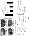
- the Xe-ELIP formulations are demonstrated to quickly and effectively release encapsulated Xe upon application of ultrasound ( FIG. 1 ).
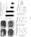
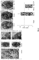
- Compositions comprising these liposomes are also shown to be effective not only in the treatment of thrombotic stroke, but also surprisingly, for treatment of hemorrhagic stroke (see FIGs. 2 , 4 , 8 , and 9 ).
- Xe-ELIP represents a therapeutic that can be administered to a patient immediately following a stroke (or the onset of stroke symptoms) and before the type of stroke can be positively identified.
- Xe-ELIP can protect neurons from the insults that result from both blood clot (ischemia) and hemorrhage.
- Xe-ELIP buys the patient crucial time by protecting the brain from excessive neurological damage (as assessed both by actual neuronal damage and behavioral testing) while surgical or other therapeutic interventions can be implemented. Accordingly, this new class of therapeutic offers the possibility of significantly improving clinical outcome of all classes of stroke.
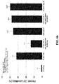
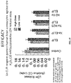
- H 2 and H 2 S when administered in conjunction with Xe-ELIP, are able to further reduce infarct volume and further improve outcome in stoke subjects as assessed by behavioral testing.
- none of the ELIP compositions (comprising Xe or Xe and H 2 or H 2 S) showed significant toxicity as assessed in murine embryonic stem cells. Accordingly, the further incorporation of H 2 and/or H 2 S into Xe-ELIP compositions may yet further enhance their neuroprotective efficacy.
- a "liposome” is a generic term encompassing a variety of single and multilamellar lipid vehicles formed by the generation of enclosed lipid bilayers or aggregates. Liposomes may be characterized as having vesicular structures with a bilayer membrane, generally comprising a phospholipid, and an inner medium that generally comprises an aqueous composition. Liposomes provided herein include unilamellar liposomes, multilamellar liposomes and multivesicular liposomes. Liposomes provided herein may be composed of positively charged, negatively charged or neutral phospholipidsl. In certain embodiments, the liposomes are neutral in charge.
- a multilamellar liposome has multiple lipid layers separated by aqueous medium. They form spontaneously when lipids comprising phospholipids are suspended in an excess of aqueous solution. The lipid components undergo self-rearrangement before the formation of closed structures and entrap water and dissolved solutes between the lipid bilayers (Ghosh and Bachhawat, 1991). Lipophilic molecules or molecules with lipophilic regions may also dissolve in or associate with the lipid bilayer.
- a gas is capsulated in a liposome to generate an echogenic liposome that can be imaged and/or disrupted by the appropriate application of ultrasound.
- Specific methods for gas encapsulation are detailed below and exemplified in Example 1 and FIGs. 1 and 7 .
- a liposome used according to the present embodiments can be made by different methods, as would be known to one of ordinary skill in the art.
- a phospholipid (Avanti Polar Lipids, Alabaster, AL), such as for example the neutral phospholipid dioleoylphosphatidylcholine (DOPC), Dipalmitoyl Phosphatidylcholine (DPPC) and/or EPC, can be dissolved in an alcohol or other organic solvent and then mixed with a component for inclusion in the lipid bilayer.
- the mixture may further include various detergents.
- a lipid mixture is vortexed, frozen in a dry ice/acetone bath and lyophilized overnight.
- the lyophilized preparation is stored at -20°C or less for extended periods of time. When required the lyophilized liposomes are reconstituted, for example, in 0.9% saline.
- a liposome can be prepared by mixing lipids in a solvent in a container, e.g., a glass, pear-shaped flask.
- a container e.g., a glass, pear-shaped flask.
- the container should have a volume ten-times greater than the volume of the expected suspension of liposomes.
- the solvent is removed at approximately 40°C under negative pressure.
- the solvent normally is removed within about 5 min. to 2 hours, depending on the desired volume of the liposomes.
- the composition can be dried further in a desiccator under vacuum. The dried lipids generally are discarded after about 1 week because of a tendency to deteriorate with time.
- liposomes can be prepared in accordance with other known laboratory procedures (e.g., see Bangham et al., 1965; Gregoriadis, 1979; Deamer and Uster, 1983; Szoka and Papahadjopoulos, 1978, each incorporated herein by reference in relevant part).
- Additional liposomes which may be useful with the present embodiments include cationic liposomes, for example, as described in WO02/100435A1 , U.S Patent 5,962,016 , U.S. Application 2004/0208921 , WO03/015757A1 , WO04029213A2 , U.S. Patent 5,030,453 , and U.S.
- Patent 6,680,068 all of which are hereby incorporated by reference in their entirety without disclaimer.
- a process of making liposomes is also described in WO04/002453A1 .
- Neutral lipids can be incorporated into cationic liposomes (e.g., Farhood et al., 1995).
- Various neutral liposomes which may be used in certain embodiments are disclosed in U.S. Patent 5,855,911 , which is incorporated herein by reference. These methods differ in their respective abilities to entrap aqueous material and their respective aqueous space-to-lipid ratios.
- the size of a liposome varies depending on the method of synthesis. Liposomes in the present embodiments can be a variety of sizes. In certain embodiments, the liposomes are small, e.g., less than about 100 nm, about 90 nm, about 80 nm, about 70 nm, about 60 nm, or less than about 50 nm in external diameter.
- any protocol described herein, or as would be known to one of ordinary skill in the art may be used. Additional non-limiting examples of preparing liposomes are described in U.S. Patents 4,728,578 , 4,728,575 , 4,737,323 , 4,533,254 , 4,162,282 , 4,310,505 , and 4,921,706 ; International Applications PCT/US85/01161 and PCT/US89/05040 ; U.K. Patent Application GB 2193095 A ; Mayer et al., 1986; Hope et al., 1985; Mayhew et al. 1987; Mayhew et al., 1984; Cheng et al., 1987; and Liposome Technology, 1984, each incorporated herein by reference).
- the lipid based nanoparticle is a neutral liposome (e.g., a DOPC liposome).
- neutral liposomes or “non-charged liposomes”, as used herein, are defined as liposomes having one or more lipid components that yield an essentially-neutral, net charge (substantially non-charged).
- neutral liposomes By “essentially neutral” or “essentially non-charged”, it is meant that few, if any, lipid components within a given population (e.g., a population of liposomes) include a charge that is not canceled by an opposite charge of another component (i.e., fewer than 10% of components include a non-canceled charge, more preferably fewer than 5%, and most preferably fewer than 1%).
- neutral liposomes may include mostly lipids and/or phospholipids that are themselves neutral under physiological conditions (i.e., at about pH 7).
- Liposomes and/or lipid-based nanoparticles of the present embodiments may comprise a phospholipid.
- a single kind of phospholipid may be used in the creation of liposomes (e.g., a phospholipid, such as DPPC (composed of all saturated phosphatidylglycerol or phosphatidylserine), may be used to generate a liposome).
- DPPC phospholipid, such as DPPC (composed of all saturated phosphatidylglycerol or phosphatidylserine)
- more than one kind of phospholipid may be used to create liposomes (e.g., DPPC and EPC).
- Phospholipids include, for example, phosphatidylcholines, phosphatidylglycerols, and phosphatidylethanolamines; because phosphatidylethanolamines and phosphatidyl cholines are non-charged under physiological conditions (i.e., at about pH 7), these compounds may be particularly useful for generating neutral liposomes.
- the phospholipid DOPC is used to produce non-charged liposomes.
- a lipid that is not a phospholipid e.g., a cholesterol
- Phospholipids include glycerophospholipids and certain sphingolipids.
- Phospholipids include, but are not limited to, dioleoylphosphatidylycholine ("DOPC"), egg phosphatidylcholine (“EPC”), dilauryloylphosphatidylcholine (“DLPC”), dimyristoylphosphatidylcholine (“DMPC”), dipalmitoylphosphatidylcholine (“DPPC”), distearoylphosphatidylcholine (“DSPC”), 1-myristoyl-2-palmitoyl phosphatidylcholine (“MPPC”), 1-palmitoyl-2-myristoyl phosphatidylcholine (“PMPC”), 1-palmitoyl-2-stearoyl phosphatidylcholine (“PSPC”), 1-stearoyl-2-palmitoyl phosphatidylcholine (“SPPC”), dilauryloylphosphatidy
- natural sources such as egg or soybean phosphatidylcholine, brain phosphatidic acid, brain or plant phosphatidylinositol, heart cardiolipin and plant or bacterial phosphatidylethanolamine (or hydrogenated versions thereof) are used
- a liposome comprises DPPC:EPC:PEG2000-DPPE:DPPG:CH in a ratio of about 30-50:10-30:5-15:5-15:10-20, or about 40-50:20-30:5-10:5-10:10-20.
- Some examples of specific ratios include, without limitation, 50:20:10:10:15; 60:30:10:10:12; 46:23:8:8:15; 47:27:9:8:13; or 48:28:7:7:13.
- the lipid-based vesicle is a DOTAP:cholesterol nanoparticle.
- DOTAP:cholesterol nanoparticles are prepared by mixing the cationic lipid DOTAP (1,2-bis(oleoyloxy)-3-(trimethylammonio)-propane) with cholesterol.
- Vesicles can further be prepared with a nucleic acid and can form a structure (called a "sandwich') where the nucleic acid appears to be condensed between two lipid bilayers ( U.S. Patents 6,770,291 and 6,413,544 ).
- the present invention provides methods for the facile production of gas-containing liposomes with simultaneous drug encapsulation.
- liposomes of phospholipid and cholesterol were prepared by conventional procedures of hydrating the lipid film, sonicating, freezing and thawing.
- the lipids generated contain air by including a step after sonication where the lipid is placed under pressure with the gas of interest. After equilibration, the sample is frozen. The pressure is then reduced to atmospheric and the suspension thawed. This procedure leads to entrapment of air in amounts up to about 10% by volume by lipid dispersions at moderate (10 mg/ml) concentrations.
- the amount of gas encapsulated increases with gas pressure and lipid concentration.
- the present invention provides methods to introduce gas into liposomes such that they not only reflect ultrasound, but also release their contents when exposed to ultrasound or other triggering procedure.
- the method which, in certain embodiments, uses elevated-pressure in combination with freezing, is very simple and allows ready encapsulation of solutes along with incorporation of a gas of choice.
- the method is suitable for the preparation of both an ultrasound contrast agent and an ultrasound-controlled drug delivery system.
- the pressure-freeze method of certain embodiments of the present invention is based on this principle.
- An essential role of freezing is to concentrate both gas and solute molecules so as to favor their encapsulation. Indeed, the basic phenomenon, that during freezing, air is released and often trapped as bubbles in the resultant ice, has been known for many years, and, moreover, that bubble formation in cells contributes significantly to freezing damage in long-term preservation of cells and tissues.
- Exemplary steps of the methods of producing echogenic liposomes are provided below, particularly in Example 1 and FIGs. 1 and 7 .
- Gas incorporation in liposomes is proportional to pressure. As noted above, this is to be expected from Henry's Law if gas uptake by the liposomes is proportional to the amount in solution at the first step. While this influences gas entrapment, it is the freezing step that has a large influence on dissolved gas and hence on gas encapsulation. While the present invention is not limited to any mechanism, it is believed that freezing probably serves two purposes, increasing the local concentration of dissolved gas and nucleating formation of small pockets of bulk gas phase. Gases, like other solutes, are more soluble in liquid water than in solid ice.
- dissolved gas is progressively displaced from ice to unfrozen solution, with the result that the dissolved gas becomes increasingly concentrated in the ever-diminishing volume of liquid solution.
- a gas bubble may nucleate and grow.
- bubbles form when the difference between the total dissolved gas pressure and the ambient pressure in the surrounding liquid exceeds the Laplace pressure (the pressure created in a bubble due to the contraction of the surface under the influence of the surface tension).
- Mannitol is rather distinctive among sugars in readily crystallizing out of solution upon cooling.
- freezing a mannitol solution inflicts damage upon liposomes.
- nucleation theory is a suggestion made a number of years ago that the polar-nonpolar interface at the surface of damaged membranes may be the preferred site of nucleation of release of nitrogen bubbles in decompression sickness which affects divers who are rapidly decompressed.
- the present invention is not limited to any particular mechanism, the following is believed to be part of the gas-containing liposome formation process.
- supersaturation of the liquid phase during ice formation should cause incipient air pockets to form, it is unlikely that this is the whole story, for, if it were, the echogencity should not be particularly low when samples are thawed prior to reducing the pressure back to ambient pressure. Under these conditions, the ice melts and the water produced is essentially degassed, so air associated with lipid (in all forms) will diffuse into this water.
- the pressure is lowered first and the sample thawed second, the air concentration in the solution that melts initially is high because it contains most of the air that dissolved in the suspension upon pressurization.
- co-encapsulation of gas e.g., Xe
- an aqueous solute has advantages in drug delivery as it allows for release of liposomal contents by application of ultrasound. Since acoustically active liposomes also reflect ultrasound, it is possible to not only to localize the release of the drug according to the site of application of ultrasound, but also to image the therapeutic agent while it is being activated for delivery. Moreover, molecular targeting of the liposomes themselves is possible.
- ultrasound can have effects on the tissue that synergize with drug delivery, namely cavitation effects of ultrasound which can facilitate access of the drug to its target.
- site-specific drug delivery can be achieved by destroying drug-filled contrast microbubbles in the target area with high-intensity ultra-sound ( Porter et al., J Ultrasound Med 1996; 15(8):577-84 .).
- Shohet et al. (Circulation 2000;101(22):2554-6 .) found that albumin-coated microbubbles could be used to effectively deliver an adenoviral transgene to rat myocardium via US-mediated microbubble destruction.
- Prior work has also found enhanced uptake of plasmid DNA in the presence of acoustically active liposomes and with the simultaneous application of ultrasound ( Huang et al., Mol. Ther. 2003; 7 (5): 422 Part 2 ).
- the sensitivity of echogenic liposomes to ultrasound stimulation may be able to be improved further by varying the liposomal composition, the encapsulated gas and/or the ultrasound application parameters.
- the lipid bilayer is held together by hydrophobic interactions that tend to endow it with self-sealing properties such that the lipid shell of a liposome rapidly reseals following surface alternations. It is therefore probable that changing the rigidity of the lipid membrane will affect its response to ultrasound.
- the choice of the optimal gas will involve both high volume in the liposomes and low rate of release in the blood stream. The most effective ultrasound pulses would seem to be a small number at the highest intensity that the tissue can sustain.
- Targeted delivery is achieved by the addition of ligands without compromising the ability of liposomes to deliver their payloads. It is contemplated that this will enable delivery to specific cells, tissues and organs (e.g., specific sites in the brain).
- the targeting specificity of the ligand-based delivery systems is based on the distribution of the ligand receptors on different cell types.
- the targeting ligand may either be non-covalently or covalently associated with a nanoparticle, and can be conjugated to the nanoparticles by a variety of methods as discussed herein.
- Examples of molecules that could be used to target liposomes of the embodiments include antibodies (or fragments thereof) and apatmers. Alternatively or additional it is contemplated that cell-penetrating peptides may be used to deliver liposomes directly into cells.
- liposome complex e.g., liposomes comprising gases
- liposome complex it will be necessary to prepare the liposome complex as a pharmaceutical composition appropriate for the intended application.
- this will entail preparing a pharmaceutical composition that is essentially free of pyrogens, as well as any other impurities that could be harmful to humans or animals.
- Aqueous compositions of the present invention comprise an effective amount of Xe encapsulated in a liposome as discussed above, further dispersed in pharmaceutically acceptable carrier or aqueous medium. Such compositions also are referred to as inocula.
- phrases “pharmaceutically” or “pharmacologically acceptable” refer to compositions that do not produce an adverse, allergic or other untoward reaction when administered to an animal, or a human, as appropriate.
- pharmaceutically acceptable carrier includes any and all solvents, dispersion media, coatings, antibacterial and antifungal agents, isotonic and absorption delaying agents and the like. The use of such media and agents for pharmaceutical active substances is well known in the art. Except insofar as any conventional media or agent is incompatible with the active ingredient, its use in the therapeutic compositions is contemplated. Supplementary active ingredients also can be incorporated into the compositions.
- Solutions of therapeutic compositions can be prepared in water suitably mixed with a surfactant, such as hydroxypropylcellulose. Dispersions also can be prepared in glycerol, liquid polyethylene glycols, mixtures thereof and in oils. Under ordinary conditions of storage and use, these preparations contain a preservative to prevent the growth of microorganisms.
- the therapeutic compositions of the present invention are advantageously administered in the form of injectable compositions either as liquid solutions or suspensions; solid forms suitable for solution in, or suspension in, liquid prior to injection may also be prepared. These preparations also may be emulsified.
- a typical composition for such purpose comprises a pharmaceutically acceptable carrier.
- the composition may contain 10 mg, 25 mg, 50 mg or up to about 100 mg of human serum albumin per milliliter of phosphate buffered saline.
- Other pharmaceutically acceptable carriers include aqueous solutions, non-toxic excipients, including salts, preservatives, buffers and the like.
- non-aqueous solvents examples include propylene glycol, polyethylene glycol, vegetable oil and injectable organic esters such as ethyloleate.
- Aqueous carriers include water, alcoholic/aqueous solutions, saline solutions, parenteral vehicles such as sodium chloride, Ringer's dextrose, etc.
- Intravenous vehicles include fluid and nutrient replenishers.
- Preservatives include antimicrobial agents, anti-oxidants, chelating agents and inert gases. The pH and exact concentration of the various components the pharmaceutical composition are adjusted according to well known parameters. Additional formulations are suitable for oral administration.
- Oral formulations include such typical excipients as, for example, pharmaceutical grades of mannitol, lactose, starch, magnesium stearate, sodium saccharine, cellulose, magnesium carbonate and the like.
- the compositions generally will take the form of solutions or suspensions.
- the therapeutic compositions of the present embodiments may include classic pharmaceutical preparations. Administration of therapeutic compositions according to the present invention will be via any common route so long as the target tissue is available via that route. In this case, intravenous injection or infusion may be preferred. Such compositions would normally be administered as pharmaceutically acceptable compositions that include physiologically acceptable carriers, buffers or other excipients.
- unit dose refers to physically discrete units suitable for use in a subject, each unit containing a predetermined-quantity of the therapeutic composition calculated to produce the desired responses, discussed above, in association with its administration, i.e., the appropriate route and treatment regimen.
- the quantity to be administered both according to number of treatments and unit dose, depends on the protection desired.
- HED human equivalent dose
- K m factors in conversion results in more accurate HED values, which are based on body surface area (BSA) rather than only on body mass.
- BSA body surface area
- K m values for humans and various animals are well known. For example, the K m for an average 60 kg human (with a BSA of 1.6 m 2 ) is 37, whereas a 20 kg child (BSA 0.8 m 2 ) would have a K m of 25.
- mice K m of 3 (given a weight of 0.02 kg and BSA of 0.007); hamster K m of 5 (given a weight of 0.08 kg and BSA of 0.02); rat K m of 6 (given a weight of 0.15 kg and BSA of 0.025) and monkey K m of 12 (given a weight of 3 kg and BSA of 0.24).
- the amount of therapeutic liposome (e.g., Xe-ELIP) dose in a human (adult) will be greater than about 0.568 mg/kg.
- the human dose range can be between about 0.6 and 3.0 mg/kg, between about 0.8 and 2.8 mg/kg, or between about 1.0 and 2.5 mg/kg.
- Xe-ELIP is administered to a human subject in a dose of between about 1.14 mg/kg and about 2.27 mg/kg.
- the present invention relates to the following items:
- Xenon-ELIP were produced using the freeze thaw protocol that is schematically illustrated in FIG. 7 (see also, U.S. Pat. 7,976,743 , incorporated herein by reference). Briefly, liposomes were composed of L- ⁇ -phosphatidylcholine (egg PC), 1,2-dipalmitoyl-sn-glycero-3-phosphocholine (DPPC; Avanti Polar Lipids, Alabaster, Ala); 1,2-dipalmitoyl-sn-glycero-3-phosphoethanolamine-N-[methoxy(polyethylene glycol)-2000] (16:0 PEG2000 PE) and cholesterol (Sigma, St Louis, Mo).
- egg PC L- ⁇ -phosphatidylcholine
- DPPC 1,2-dipalmitoyl-sn-glycero-3-phosphocholine
- DPPC 1,2-dipalmitoyl-sn-glycero-3-phosphoethanolamine-N-[methoxy(polyethylene glycol
- lipids Five milligrams of lipids was mixed in chloroform, and the solvent was evaporated with argon in a 50°C water bath to form a thin film on the glass vial.
- the lipid film was placed under vacuum for 4 to 6 hours for complete solvent removal.
- the dried lipid film was hydrated with 0.32 mol/L mannitol to a concentration of 10 mg lipid per milliliter, followed by sonication for 5 minutes.
- the sonicated liposomes were transferred to a 2-mL glass vial with a cap sealed with a Teflon-rubber septum.
- Xe (100%) (Concorde Specialty Gas Inc, Eatontown, NJ) was injected into the glass vial through the Teflonrubber septum with a 12-mL syringe attached to a 27-guage-1/2-inch needle (note that at this stage other gases and/or gas mixtures may be incorporated).
- the pressurized liposomal dispersion was frozen at -70°C with dry ice for at least half an hour.
- the liposomal dispersion was allowed to thaw after the vial was unpressurized by removing the cap.
- the structure and gas retention properties of Xe-ELIP are shown in FIG. 1 .
- a 1 mm diameter burr hole was made in the skull to facilitate local cerebral perfusion (CP) measurement before occluding the MCA.
- CCA right common carotid artery
- ECA right external carotid artery
- the internal artery was isolated and separated from adjacent tissues.
- a fabricated 25-cm 4-0 nylon monofilament was advanced from the right ECA and inserted into the right MCA for 2 hours to provoke ischemia. Interruption of local blood flow through the MCA was verified with a laser Doppler flowmeter placed over the ischemic area at 2 mm posterior and 6 mm lateral to the bregma.
- body temperature was maintained at 37°C during ischemia.
- a polyethylene catheter was introduced into the right femoral artery for pressure recordings.
- mice were randomly divided into four groups (n 8 in each group), (1) no treatment group- MCA occlusion only; (2) treatment group "a" -Xe-ELIP administration 10 min after reperfusion; (3) treatment group "b” - Xe-ELIP administration 60 min after reperfusion; (4) treatment group "c” - Xe-ELIP administration 180 min after reperfusion. All rats in each treatment group were administrated 200 ⁇ l of Xe-ELIP over a period of 4 minutes by cannulating the right internal carotid artery with modified PESO tubing. The ICA was exposed to 1-MHz continuous wave ultrasound at a peak-to-peak pressure amplitude of 0.18 MPa (1-W/cm2 dial setting) during Xe-ELIP administration. Neurological assessments were conducted over the following three days. On the third day after MCAO the infarct volume was determined by 2% 2, 3, 5-triphenyltetrazolium chloride (TTC) staining.
- TTC 5-triphenyltetrazol
- the ICA was exposed to 1-MHz continuous wave ultrasound at a peak-to-peak pressure amplitude of 0.18 MPa (1-W/cm2 dial setting) during Xe-ELIP administration.
- the infarct volume was determined 3 days after MCAO by TTC staining. Neurological assessments were conducted over the following three days. On the third day after MCAO the infarct volume was determined by 2% TTC staining.
- Limb placement was assessed by observing the animal's ability to lift its head and extend its forelimbs toward a table while the animal was suspended over the table by its tail (zero score- no response; score of 1 -10 when response was sluggish or delayed; score of 2 when response was rapid and fully executed).
- the ability to walk across a beam was assessed by observing the ability to maintain balance while navigating across the beam.
- the response scores were assigned as follows: score 0-traversed the beam with no foot slip; score 1 -traversed with grasping of the lateral side of the beam; score 2 -showed difficulty crawling across the beam but able to traverse; score 3- required a more than 10 seconds to traverse the beam due to difficulty in walking; score 4- unable to traverse the beam; score 5 - unable to move the body or any limb on the beam; score 6 - unable to stay on the beam for more than 10 seconds.
- Grid walking ability was assessed by placing the animal on a stainless steel grid floor (20 cm x 40 cm with a mesh size of 2 cm x 2 cm). The total number of steps was counted up to a maximum of 50 steps. The number of foot fault errors as defined by the misplacement of a forelimb or hindlimb that fell through the grid was recorded.
- Infarct volume was calculated by measuring infarct areas on evenly sliced (1 mm) brain sections and adding them together (Simpson's rule). Normalized infarct volume with respect to whole brain volume was calculated by dividing the volume of TTC unstained (infarcted) tissue by that of the whole brain.
- Brain tissue slices were collected and homogenized in 1 ml of RIPA (Radio Immuno Precipitation Assay) buffer (Cell Signaling Technology, MA, USA) containing the protease inhibitors, phenylmethlylsulfonyl fluoride (PMSF, 1mM), and phosphatase inhibitor cocktail (Santa Cruz Biotechnology, CA, USA). Brain tissues were harvested at 7 and 24 hours after stroke onset.
- RIPA Radio Immuno Precipitation Assay
- Non Mammalian Blocking Reagent (LI-COR Biosciences, NE, USA) without tween-20 for 1 hour
- the membranes were incubated with primary antibodies of BDNF(1:250, Santa Cruz Biotechnology, CA, USA), phosphorylated AKT (1:250, Cell Signaling Technology, MA, USA) or total Akt (1:500, Cell Signaling Technology, MA, USA), and Phospho-ERK1/2 ( 1:250, Cell Signaling Technology, MA, USA) or Erk1/2 ( :500, Cell Signaling Technology, MA, USA) at 4°C overnight.
- membranes were reprobed with Anti b-actin (Sigma, MO, USA) for 1 hour at room temperature, and then incubated with IRDye 680 L T Gt Anti-Mouse IgG (H+L) (LI-COR Biosciences, NE, USA) for 1 hour.
- IRDye 680 L T Gt Anti-Mouse IgG (H+L) (LI-COR Biosciences, NE, USA) for 1 hour.
- Membranes were scanned using same odyssey infrared imaging system. The optical densities of all protein bands were analyzed by using NIH ImageJ software. All target proteins were quantified by normalizing to b-actin and calculated as fold of the corresponding control group.
- Nonparametric statistical analyses were performed by the Wilcoxon rank test for two groups or Kruskal-Wallis analysis of variance (ANOVA) for multiple groups, and reported as mean and standard deviation for most experiments. When differences were detected in global comparison, the multiple comparisons of mean ranks for all groups were performed for all pairwise comparisons. Neurologic outcome comparison between the treatment groups was reported as median and quartiles. Statistica (Version 9, StatSoft Inc., Tulsa, OK) software were utilized for statistical analyses. A p ⁇ 0.05 was considered significant.
- the dose dependency Xe-ELIP therapy was first investigated by injecting Xe-ELIP at dosage range of 1-4 mg/rat (3.5, 7 and 14 mg/kg) at 3 hours after stroke onset on a Male Sprague-Dawley rat model with occlusion (for 2 hours) of the right transient endovascular filament middle cerebral artery (MCAO).
- This study demonstrates that Xe-ELIP administered within 3 hours after stroke onset at a dosage larger than 2-4 mg (e.g., 7 mg/kg or greater) provide best neuroprotection.
- Xe-ELIP The therapeutic time window of Xe-ELIP was then further investigated on both rat filament MCAO and thrombotic MCAO model.
- Xe-ELIP was administrated through the ascending right common carotid artery at 2, 3, and 5 hour after stroke onset (10 min, 1 hour and 3 hour after reperfusion) on filament MCAO model.
- a large infarction developed and predominantly involved the cerebral cortex and striatum with normalized infarct volume of 16 ⁇ 5.2% (228 ⁇ 74 mm 3 ) of whole brain ( FIG. 2a, e ).
- Behavioral assessment of neurological damage were conducted by recording limb placing, beam walking and grid walking abilities in a quiet and low-lit room in an observer blinded manner at days 1, 2 and 3 after surgery.
- the group with Xe-ELIP administered at 3 hours after stroke on set demonstrated marginal improvements in performing behavioral tasks.
- Both Xe-ELIP administered at 10 minutes and 1 hour groups demonstrated improved performance in all behavioral tests from day 1 with marked improvements in all tests by day 3 (F IG. 2f-h ). It was demonstrated that Xenon protected neuron damage as a glutamate receptors (NMDA) antagonist while the excitotoxic effect caused by excessive extracellular glutamate accumulation was absorbed in an early event of ischemia.
- NMDA glutamate receptors
- the therapeutic time window of Xe-ELIP at 2 hour after stroke onset (10 min after reperfusion) showed therapeutic effect.
- the tPA administration for stroke treatment is limited by its narrow therapeutic time window.
- 85% of strokes are due to occlusion of cerebral artery by a circulating clot, 15% of strokes are hemorrhagic.
- IV tPA cannot be administrated until the exclusion of hemorrhage stroke from thrombotic stroke.
- neuroprotective agent administration before IV tPA to prolong tPA therapeutic time window is a very promising clinical relevant strategy.
- the neuroprotective effect of Xe-ELIP administration was further investigated at 2 hour after stroke onset but before and after reperfusion.
- Xe-ELIP before reperfusion and after reperfusion reduced the normalized infract size by 86 ⁇ 12% and 67 ⁇ 7%, respectively.
- tPA thrombolytic efficiency of tPA in the present of Xe-ELIP was compared with tPA alone on porcine blood clot.
- the thrombolytic effect of tPA was inhibited after 30 minute incorporation with free Xe (Xe saturated solution).
- Xe free Xe
- tPA was incorporated with Xe-ELIP, it had the same thrombotic effect as tPA alone. This demonstrated a protective effect of ELIP on Xe from interaction with tPA ( FIG. 6b ).
- Xe-ELIP The therapeutic effect of Xe-ELIP in a rat embolic stroke model was next investigated.
- tPA 10 mg/kg
- Xe-ELIP was administrated intravenously before IV tPA.
- Continuous wave ultrasound (1 MHz, 50% duty cycle, 0.5 W/cm 2 ) was applied to trigger Xe release from ELIP during the 5 min of Xe-ELIP administration.
- the thrombotic stroke control group without any treatment exhibited the largest damage and infarct size (17 ⁇ 5% of the whole brain) ( FIG. 5a-e ).
- Behavioral deficit correlated inversely with infarct volume.
- Regional blood flow velocity monitored by a laser Doppler flow meter was similar in both tPA and tPA + Xe-ELIP treatment groups ( FIG. 5f-h ).
- FIG. 3 The effects of Xe-ELIP on BDNF expression and apoptosis was also assessed (see, e.g., FIGs. 3 ).
- Western blot analysis of BDNF (a), phos-Akt (b) and phos-ERK (c) in cerebral cortex tissue 24 h after stroke showed that Xe increased the expression of BDNF (d), total Akt (e) and phos-ERK (f) ( FIG. 3a-f ).
- TUNEL staining in the penumbral region of brain sections from the sham-operated group (g), stroke group (h) and stroke with Xe-ELIP treatment group (i) showed reduction of apoptosis in Xe-ELIP-treated animals ( FIG. 3 ).
- the Western blots and photomicrographs of apoptosis are representative of three independent experiments. Data are means ⁇ SD.
- Xe-ELIP compositions were produced as detailed in Example 1.
- a rat model of subarachnoid hemorrhage was employed. Briefly, healthy male Sprague Dawley rats (Harlan Laboratories Inc., Indianapolis, IN) weighing between 260-280 grams were obtained. All surgical procedures were performed under dissecting microscope on anesthetized animal. The right external carotid artery was isolated and a 4.0 fabricated sharp nylon monofilament was introduced through the internal artery to perforate the middle cerebral artery. The monofilament was immediately retracted to resume blood flow to the middle cerebral artery. The blood flow was monitored to confirm the bleeding.
- Xe-ELIP 600 ⁇ l, 10 mg/ml
- simultaneous ultrasound application 0.5 MPa
- mice were sacrificed for SAH grading to evaluate the degree of bleeding, brain water contain to evaluate the edema and TUNEL staining to check the apoptosis, on the third day following neurological and behavior assessments.
- results from the physical examination of brain tissue are shown in FIG. 8 .
- Basal cistern was divided into six segments. Bleeding was assessed in each of these segments and scored from 0 to 3 (0: no SAH; 1: minimal SAH; 2: moderate blood clot with recognizable arteries; 3: blood clot obliterate the arteries). The total scores were added and the severity of bleeding was scaled as: 0-7: mild SAH; 8-12: moderate SAH; 13-18: severe SAH.
- Xe-ELIP decreases the bleeding in filament perforation subarachnoid hemorrhage (SAH) rat model
- Results of behavioral testing of the treated rats are shown in FIG. 9 .
- Lower panel illustrates the array of behavioral tests to which animals were subject.
- Results of neurological evaluation, beam walking and grid walking are shown in the graphs of the upper panel.
- rats treated with Xe-ELIP performed significantly better than untreated rats.
- microscopic examination of brain sections using TUNEL staining FIG. 10
- Xe-ELIP treatment decreased the death rate of SAH rats but did not show significant effects on brain edema and cerebral blood flow ( FIG. 11 ).
- Brain edema is the major life-threatening complication of stroke. It is frequently associated with subarachnoid hemorrhage, vasospasm and ischemic reperfusion damage.
- the results shown here demonstrate that ELIP formulations containing only one gas (Xe) does not affect the brain edema and vasoactivity. It has been shown that hydrogen administration after stroke can eliminate the brain edema by decreasing blood-brain barrier permeability, and H 2 S inhibits vasospasm by anti-inflammatory effect.
- formulations that co-encapsulate hydrogen gas and/or hydrogen sulfide gas with Xe-ELIP would have added effect on brain edema and cerebral vasospasm.
- ELIP immunosorbent assays for co-encapsulation of Xe with hydrogen or hydrogen sulfide into ELIP.
- ELIP were composed of phospholipids and cholesterol and were produced as detailed in Example 1. In this case, however, gas mixtures of 30% hydrogen + 70% xenon or 1% hydrogen sulfide + 99% xenon were loaded onto ELIP by the pressurized-freeze method in addition to use of 100% Xe.
- both addition of H 2 and H 2 S further increased the ability of Xe-ELIP to reduce the normalized infarct volume in the brains of treated rats.
- addition of H 2 S further increased the ability of Xe-ELIP to reduce the number of TUNEL positive cells in the brains of treated rats, indicated reduce neuron cell damage.
- Analysis of neutrophil invasion across the vascular wall in the brains of treated rats showed decreased invasion following Xe-ELIP treatment, which was enhanced by H 2 S. Inhibition of neutrophil transfer across the vascular wall is one potential mechanism for H 2 S/Xe-ELIP neurovascular unit protection.
- Example 5 - Xe-ELIP protection of culture human brain astrocytes against hydrogen peroxide (H 2 O 2 ) cytotoxicity or oxidative stress
- Human brain astrocytes play a key role in maintaining nerve cell function and survival against oxidative stress. Exposure of cultured human brain astrocytes to H 2 O 2 causes significant damages to the cells, and caused them to release large amounts of LDH. However, pretreatment of the brain cells with Xe-ELIP reagents markedly reduced LDH release ( FIG. 15 ), indicating a protective effect of Xe-ELIP on the brain cells injured by the oxidative stress. No or little protective effect was found in the cells treated with ELIP alone or control media ( FIG. 15 ).
- Example 6 - Xe-ELIP has no cytotoxicity to murine stem cells
- Murine embryonic stem cells were examined for their growth and survival when they were treated with or without H 2 O 2 in the presence or absence of Xe-ELIP. The cell viability was determined by assessing the release of LDH. In the presence of ELIP loaded with or without Xe or other gases, LDH levels in the cultures remained significant levels ( FIG. 16 ). By contrast, addition of H 2 O 2 (10 mM), significant LDH release was found within 2 hours of exposure to the oxidative stress agent H 2 O 2 ( FIG. 16 ).
Landscapes
- Health & Medical Sciences (AREA)
- Chemical & Material Sciences (AREA)
- Medicinal Chemistry (AREA)
- Pharmacology & Pharmacy (AREA)
- Life Sciences & Earth Sciences (AREA)
- Animal Behavior & Ethology (AREA)
- General Health & Medical Sciences (AREA)
- Public Health (AREA)
- Veterinary Medicine (AREA)
- Epidemiology (AREA)
- Engineering & Computer Science (AREA)
- Bioinformatics & Cheminformatics (AREA)
- Organic Chemistry (AREA)
- General Chemical & Material Sciences (AREA)
- Nuclear Medicine, Radiotherapy & Molecular Imaging (AREA)
- Chemical Kinetics & Catalysis (AREA)
- Dispersion Chemistry (AREA)
- Inorganic Chemistry (AREA)
- Dermatology (AREA)
- Cardiology (AREA)
- Heart & Thoracic Surgery (AREA)
- Diabetes (AREA)
- Hematology (AREA)
- Vascular Medicine (AREA)
- Urology & Nephrology (AREA)
- Medicinal Preparation (AREA)
- Pharmaceuticals Containing Other Organic And Inorganic Compounds (AREA)
- Medicines That Contain Protein Lipid Enzymes And Other Medicines (AREA)
Abstract
Description
- This application claims the benefit of
United States Provisional Patent Application No 61/682,130, filed August 10, 2012 Chinese Patent Application no. 201210356929.9, filed September 21, 2012 - The invention was made with government support under Grant Nos. NS067454, HL 074002 and HL 059586 awarded by the National Institutes of Health. The government has certain rights in the invention.
- The present invention relates generally to the field of medicine, biochemistry and molecular biology. More particularly, it concerns compositions and methods of using biologically protective liposomes for the treatment of stroke.
- Thrombotic and hemorrhagic strokes, also known as cerebrovascular accidents (CVA), are, together, the fourth leading cause of death in the United States and the most common cause of adult disabilities. Both types of stroke are characterized by a rapid loss of brain function due to disturbance in the blood supply to the brain. In thrombotic stroke, occlusion of a cerebral artery caused by a blood clot, results in brain tissue ischemia or obstruction of cerebral blood flow to a portion of the brain, and ultimately brain damage. Conversely, in hemorrhagic stroke, blood leaks or bursts from broken blood vessels inside or on the surface of the brain leading to neurological damage. Regardless of the type of stroke, early protection of brain tissues against the acute vascular events caused by thrombosis or hemorrhage by medical intervention remains the most crucial element to save patients' lives. The early brain tissue protection can broaden the safe window for differential diagnosis and effective treatment. Despite similar initial symptoms at onset, effective therapeutic intervention depends upon the type of stroke that a patient is experiencing. For example, administration of tissue plasminogen activator (tPA) has proven at least partially effective in treatment of thrombotic stroke, but is counter indicated for treatment of hemorrhagic stroke. There remains a need for new effective therapeutics for treatment of stroke, in particular, therapeutics that are amenable to immediate administration upon stroke onset.
- In a first embodiment there is provided a method for treating a stroke in a subject comprising administering an effective amount of a composition comprising Xenon-loaded echogenic liposomes (Xe-ELIP) to the subject. In some aspects, the subject has been determined to have a hemorrhagic stroke. In a further embodiment, there is provided a method treating a stroke of undetermined origin in a subject (i.e., a subject not yet determined to have a thrombotic or hemorrhagic stroke) comprising administering an effective amount of a composition comprising Xe-ELIP to the subject. Thus, in some aspects, a method is provided for treating both hemorrhagic and thrombotic stroke in a subject (e.g., a subject that has been diagnosed with a hemorrhagic and/or thrombotic stroke). In still further aspects, a method of the embodiments is defined as a method of treating an intracranial aneurysm or subarachnoid hemorrhage in a subject comprising administering an effective amount of a composition comprising an effective amount of Xe-ELIP to the subject. In some embodiments there is provided a composition comprising Xe-ELIP in a pharmaceutically acceptable carrier.
- In a further embodiment methods and compositions of the embodiments employ a different noble gas loaded in echogenic liposomes (in place of or in addition to Xenon). For example, in some aspects, echogenic liposomes for use according to the embodiments are loaded with Xenon, Helium, Argon, Krypton, Neon or a mixture thereof. Thus, in some aspects, there is provided a method for treating a stroke in a subject comprising administering an effective amount of a composition comprising Xenon, Helium, Argon, Krypton, or Neon-loaded echogenic liposomes to the subject. In further aspects, the subject has been determined to have a hemorrhagic stroke. In a further embodiment, there is provided a method treating a stroke of undetermined origin in a subject (i.e., a subject not yet determined to have a thrombotic or hemorrhagic stroke) comprising administering an effective amount of a composition comprising Xenon, Helium, Argon, Krypton, or Neon-loaded echogenic liposomes to the subject. Thus, in some aspects, a method is provided for treating both hemorrhagic and thrombotic stroke in a subject (e.g., a subject that has been diagnosed with a hemorrhagic and/or thrombotic stroke). In still further aspects, a method of the embodiments is defined as a method of treating an intracranial aneurysm or subarachnoid hemorrhage in a subject comprising administering an effective amount of a composition comprising an effective amount of Xenon, Helium, Argon, Krypton, or Neon-loaded echogenic liposomes to the subject. In some embodiments there is provided a composition comprising Xenon, Helium, Argon, Krypton, or Neon-loaded echogenic liposomes in a pharmaceutically acceptable carrier.
- In a further embodiment there is provided a method of treating a thrombotic stroke in a subject comprising (a) administering an effective amount of a first composition comprising Xe-ELIP to the subject; and (b) administering an effective amount of a second composition comprising tissue plasminogen activator (tPA) to the subject. For example, in certain aspects, Xe-ELIP and tPA are administered sequentially or essentially simultaneously (e.g., in a composition comprising Xe-ELIP and tPA). In some aspects, a method of the embodiments comprises administering the second composition about or less than about 2, 3, 4, 5, 6, 7, or 8 hours after administration of the first composition. In still further aspects, administration of the first composition is within about 6 hours or less of stroke onset. In yet still further aspects, the second composition further comprises and effective amount of Xe-ELIP. In still a further aspect, a method of the embodiments comprises (a) administering an effective amount of a first composition comprising Xe-ELIP to a subject having a stroke or symptoms of a stroke of undetermined origin; (b) identifying whether the subject is suffering from a thrombotic or hemorrhagic stroke; and (c) administering an effective amount of a second composition comprising tPA to a subject identified as having a thrombotic stroke.
- Thus, in further embodiment, there is provided a pharmaceutical composition comprising Xe-ELIP and tPA. In some aspects, the tPA of the composition is comprised in liposomes (e.g., echogenic liposomes). In further aspects, the composition comprises a slurry of at least two different liposome where the first liposomes are comprised of Xe-ELIP and the second liposomes comprise tPA (e.g., wherein tPA liposomes are essentially free of Xe).
- In yet a further embodiment there is provided a pharmaceutical composition comprising noble gas-loaded-ELIP (e.g., Xe-ELIP) and liposomes (e.g., echogenic liposomes) loaded with a further biologically active gas component. For example, in some aspects noble gas-loaded echogenic liposomes may further comprise (or may be administered in conjunction with liposomes comprising) H2S and/or H2. Likewise, in some aspects, noble gas-loaded echogenic liposomes may further comprise (or may be administered in conjunction with liposomes comprising) nitrous oxide and/or nitric oxide. For example, in some aspects, the gases are comprised in separate liposomes in the composition (e.g., a slurry of liposomes loaded with different gases). In further aspects, two or more of the gases are comprised in the same liposomes. For example, a composition can comprise liposomes that comprise Xe and NO, Xe and N2O or Xe, NO and N2O. In another example, a composition can comprise liposomes that comprise Xe and H2, Xe and H2S or Xe, H2 and H2S. In further aspects, liposomes of the embodiments comprise between about 0.1% and 5%, 0.1% and 3% or 0.5% and 2% H2S (as a percent of total gas in the liposome). For example, the liposomes can comprise about 1% H2S and about 99% Xe. In yet further aspects, liposomes of the embodiments comprise between about 1% and 50%, 5% and 40% or 10% and 40% H2 (as a percent of total gas in the liposome). For example, the liposomes can comprise about 30% H2S and about 70% Xe.
- Certain aspects of the embodiments concern compositions comprising tPA. In some aspects, the tPA is purified or recombinant mammalian tPA (e.g., human tPA). Such tPA compositions may be comprised in a pharmaceutically acceptable carrier. In certain aspects, the tPA is comprised in liposomes. Liposomes for use in encapsulating tPA can be selected from any other those known in art or detailed herein. For instance, in some aspects, the tPA is comprised in echogenic liposomes of the embodiments. In certain preferred aspects, liposomes comprising tPA are essentially free of Xe (i.e., tPA and Xe are not loaded into the same liposome vesicle).
- Compositions in accordance with the embodiments can be administered to a subject via an array of methods. For example, in some aspects, compositions (e.g., Xe- or tPA-containing compositions) are administered intravenously, intra-arterially, intracranially, via intravenous infusion or via intra-arterial infusion. In preferred aspects, compositions of the embodiments are administered shortly after the onset of a stroke or stroke symptoms, such as about or less than about 1, 2, 3, 4, 5, 6 or 8 hours of stroke onset or the onset of stroke symptoms. Thus, in some aspects, compositions of the embodiments are administered by a first responder (e.g., a nurse or medical technician).
- In certain aspects, a method of the embodiments comprises administering one or more doses of an ELIP composition (e.g., a Xe-ELIP composition) to a subject. For example, in some aspects, a subject is administered a dose of between about 0.6 and 3.0 mg/kg of Xe-ELIP, such as a dose of between about 0.8 and 2.8 mg/kg (mg-lipid/kg-subject). In further aspects, a subject is administered a dose of between about 1.0 and 2.5 mg/kg of an ELIP composition, such as about 1.14 mg/kg or about 2.27 mg/kg. In still further aspects, ELIP compositions are provided that comprise a single unit dosage (e.g., of Xe-ELIP) in a suitable containment means. For example, the single unit dosage (i.e., a dose suitable for a human subject of about 60 kg) can be about 100 to about 3,000 mg; about 250 to about 2,000 mg; about 250 to about 1,000 mg; or about 400 to about 900 mg (e.g., 480-900 mg) of ELIP with an encapsulated gas (based on total lipid weight). In still further aspects, a single unit dosage of Xe-ELIP can be defined by the total volume of encapsulated Xe. In some aspects, the total volume of Xe in a single dose is less than about 5 ml, such as between about 0.1 and 2.5 ml; about 0.2 and 2.0 ml; or 0.5 and 1.5 ml (e.g., a dose of about 1.0 ml) of Xe. Thus, in some specific aspects, a single unit dosage of Xe-ELIP is provided comprising between 250 to about 1,000 mg of lipid and between about 0.1 and 2.5 ml of Xe in suitable containment means. A skilled artisan will recognize that any of the forgoing dose ranges may also be applied to Xe-ELIP compositions that include H2 and/or H2S (in this case gas volumes can be applied to the total amount of encapsulated gas).
- In still a further aspect, a composition (e.g., an ELIP composition) of the embodiments is administered to a subject in multiple doses. For example, in some aspects, the composition is administered to the subject a second, third or fourth time. In certain aspects, the formulation, dosage or route of the second dosage or subsequent administration can be adjusted relative to the first administration. In some aspects, the second or subsequent administration is about or less than about 2, 3, 4, 5, 6, 7, or 8 hours after the initial administration (e.g., within about 4-6 hours after the first administration). In some aspects, a composition of the embodiments is administered to a subject twice within about 12 hours of the onset of a stroke or stroke symptoms.
- In preferred aspects a subject for treatment in accordance with the embodiments is a human in subject. However, in some aspects, the subject can be a non-human animal, such a non-human primate or a domesticated animal such a horse, dog or cat. In further aspects, the subject is identified as having symptoms of stroke, such as sudden memory loss, full or partial paralysis, disorientation, trouble speaking, sudden vision impairment, numbness of a portion of the body, sudden, severe, headache or trouble walking or balancing. In some aspects, the subject having a stroke or stroke symptoms has not been identified as having a thrombotic stroke or a hemorrhagic stroke. In still further aspects, the subject has been identified as having a thrombotic stroke and/or a hemorrhagic stroke.
- In further aspects, administration of an ELIP composition of the embodiments to a subject further comprises applying ultrasound stimulation to the subject in an amount effective to promote gas release from the liposomes. For example, the ultrasound stimulation can be applied after or concomitantly with administration of the ELIP composition. In some aspects, the ultrasound stimulation is applied after the administration of an ELIP composition (e.g., Xe-ELIP) such as about or less than about 10 seconds, 20, seconds, 30 seconds or 1, 2, 3, 4 or 5 minutes after the administration. In certain cases the ultrasound stimulation is applied at or near the site of desired gas release by the liposomes. For example, in the case of a subject having a stroke, ultrasound stimulation can be applied to the head or neck (e.g., at the carotid artery) thereby stimulating release of liposomal payload proximal to the brain.
- A variety of methods for applying ultrasound stimulation to a subject are known and can be used in accordance with the embodiments. For example, the ultrasound stimulation can be applied with a conventional ultrasound probe or a cervical collar ultrasound device (e.g., to provide stimulation at the neck). The power and frequency of ultrasound stimulation applied to a subject can vary, but generally will be an amount effective to promote Xe release (e.g., in vivo release) from liposomes. For example, the ultrasound stimulation can be applied at a frequency of between about 1 and 8 MHz, with a mechanical index of between about 0.1 and 1.4.
- In still further aspects a method of the embodiments further comprises administering at least a second therapeutic agent to the subject. For example, in the case of thrombotic stroke the second therapeutic can be a blood clot reducing thrombolytic agent. In further aspects, the second therapeutic agent is an anti-inflammatory agent or a neuroprotective agent. In some specific aspects, e.g., in the case of thrombotic stroke, tPA is administered to a subj ect.
- In certain specific aspects, the second therapeutic agent comprises H2S and/or H2-loaded echogenic liposomes. In some cases, the H2S and/or H2-loaded echogenic liposomes and the Xe-loaded echogenic liposomes are comprised in the same composition. For example, a composition of the embodiments can comprise echogenic liposomes that, separately, comprise Xe, H2S and/or H2. Alternatively, the compositions can comprise echogenic liposomes that comprise two or three gases (e.g., two or more gases selected from Xe, H2S and H2).
- In still further aspects, a method of administration in accordance with the embodiments comprises preparing a liquid liposome suspension prior to administration to a subject. For example, in some aspects, the liquid liposome suspension is prepared no more than 30 minutes, 10 minutes, 5 minutes or 2 minutes prior to administration. For example, in some aspects, preparing a liquid liposome suspension comprises suspending lyophilized liposomes in a solution or thawing a frozen liposome suspension.
- In further aspects an ELIP composition of the embodiments comprises additional components, such as preservatives, stabilizers and/or salts. In some aspects, ELIP compositions comprise at least a first cryoprotectant. Cryoprotectants for use according to the embodiments include, without limitation, mannitol, glycerol, trehalose, 1,2-propanediol or dimethylsulfoxide (DMSO).
- As used herein an echogenic liposome refers to a liposome that can be imaged by ultrasound. In particular aspects, an echogenic liposome is a liposome that comprises a gas component (e.g., Xe, H2S and/or H2), such as gas comprised in the hydrophobic layer of the liposome. Echogenic liposome compositions and methods for making such composition are provided for example is U.S. Pats. 5,612,057; 5,858,399; and 7,976,743, each of which is incorporated herein by reference. In some aspects, liposomes of the embodiments (e.g., ELIP compositions) are defined by the average particle size. For example, in some aspects the liposomes have an average size of about 0.4 to 10 microns or 0.8 to 10 microns (e.g., an average size of about 0.4, 0.5, 0.6, 0.7, 0.8, 0.9, 1.0, 2.0, 3.0, 4.0, 5.0, 6.0, 7.0, 8.0, 9.0 or 10.0 microns).
- A wide array of components can be used to formulate a liposome of the embodiments, such as ELIP loaded with a gas such as Xe, H2 and/or H2S. For example, a liposome of the embodiments can comprise any form of phosphatidylcholine (PC) (such as dipalmitoyl phosphatidylcholine (DPPC)), any form of phosphoethanolamine (PE), a polyethylene glycol (PEG) or a PEGylated phospholipid, any form of phosphatidylglycerol (PG) (such as 1,2-dipalmitoyl-sn-glycero-3-phospho-(1'-rac-glycerol) (DPPG)) and/or cholesterol. In some aspects, a liposome comprises at least one PC, PE, negatively-charged lipid, PEGylated lipid (e.g., PEG2000-DPPE) and cholesterol molecule. In some specific aspects, a liposome comprises DPPC, Egg PC (EPC), PEG2000-DPPE, DPPG and cholesterol. In still further aspects a liposome of the embodiments consists of or consists essentially of DPPC, EPC, PEG2000-DPPE, DPPG, cholesterol and Xenon. In still further aspects, a liposome consists of or consists essentially of DPPC, EPC, PEG2000-DPPE, DPPG, cholesterol, Xe and H2, H2S or a combination of H2 and H2S.
- As used herein the specification, "a" or "an" may mean one or more. As used herein in the claim(s), when used in conjunction with the word "comprising", the words "a" or "an" may mean one or more than one.
- The use of the term "or" in the claims is used to mean "and/or" unless explicitly indicated to refer to alternatives only or the alternatives are mutually exclusive, although the disclosure supports a definition that refers to only alternatives and "and/or." As used herein "another" may mean at least a second or more.
- Throughout this application, the term "about" is used to indicate that a value includes the inherent variation of error for the device, the method being employed to determine the value, or the variation that exists among the study subjects.
- Other objects, features and advantages of the present invention will become apparent from the following detailed description. It should be understood, however, that the detailed description and the specific examples, while indicating preferred embodiments of the invention, are given by way of illustration only, since various changes and modifications within the spirit and scope of the invention will become apparent to those skilled in the art from this detailed description.
- The following drawings form part of the present specification and are included to further demonstrate certain aspects of the present invention. The invention may be better understood by reference to one or more of these drawings in combination with the detailed description of specific embodiments presented herein.
-
FIG. 1 : Characterization of xenon-containing liposome and ultrasound-triggered xenon release. (a) Traditional liposome without gas. (b) Liposome containing Xe, made by the pressurized-freezing method, with Xe entrapped in the lipid bilayer as a dissolved gas or a bubble. (c and d) Electron microscopic image of traditional liposome and gas-containing liposome showed a wide lipid bilayer for gas-containing liposomes. Intravascular ultrasound imaging of traditional liposome (e) and Xe-containing liposome (f) showed a high ultrasound reflectivity from Xe-containing liposomes. (g) Ultrasound release of xenon from Xe-ELIP has two phases: a fast release in first 30 min followed by a slow release lasting more than 18 h (half-life 4.97 ± 0.7 hours). Ultrasound triggered the release of Xe from Xe-ELIP in a power-dependent manner (h). -
FIG. 2 : Time window of Xe-ELIP's neuroprotective effect. Coronal brain sections (TTC staining) of middle cerebral artery occlusions without treatment (a) and with Xe-ELIP (7 mg/kg) treatment at 10 min (b), 1 h (c) and 3 h (d) after reperfusion. The white areas are the infarcted regions after middle cerebral artery occlusion; Quantification of the infarct volume of the brain showed that Xe-ELIP administration at 10 min and 1 h after reperfusion was significantly different from the no treatment group. Neurological assessments of limb placement (f), beam walking (g), and grid walking (h) showed similar results as the TTC staining. Data are means ± standard error. -
FIG. 3 : Effects of Xe-ELIP on BDNF expression and apoptosis. Western blot analysis of BDNF (a), phos-Akt (b) and phos-ERK (c) in cerebral cortex tissue 24 h after stroke showed that Xe increased the expression of BDNF (d), total Akt (e) and phos-ERK (f). TUNEL staining in the penumbral region of brain sections from the sham-operated group (g), stroke group (h) and stroke with Xe-ELIP treatment group (i) showed reduction of apoptosis in Xe-ELIP-treated animals. The Western blots and photomicrographs of apoptosis are representative of three independent experiments. Data are means ± SD. -
FIG. 4 : Dose response of Xe-ELIP's neuroprotective effect. Coronal brain sections (stained with TTC) of middle cerebral artery occlusions were imaged without treatment (a), and with 3.5 mg/kg (b), 7 mg/kg (c) and 14 mg/kg (d) of Xe-ELIP. The white areas are the infarcted regions after middle cerebral artery occlusion. Quantification of the infarct volume of the brain is shown in (e). Neurological assessments of limb placement (f), beam walking (g), and grid walking (h) are provided in the indicated graphs. Data are means ± standard error. -
FIG. 5 : Effects of IV tPA in combination with Xe delivery on cerebral ischemia. Representative TTC-stained coronal brain sections showing brain infarction inrats 3 days after middle cerebral artery occlusion were imaged after (a) sham, (b) ischemic stroke without treatment, (c) ischemic stroke with tPA treatment (d) ischemia stroke with Xe-ELIP in combination with tPA. (e) A comparison of infarct sizes between treatment groups showing a 69% reduction in infarct volume with tPA alone and a 75% reduction with Xe-ELIP combined with IV tPA. Neurological assessments of limb placement (f), beam walking (g) and grid walking (h) are shown in the indicated graphs. Data are means ± standard error. -
FIG. 6a -b: (a), Xe-ELIP provides neuroprotective effects when administered either before or after restoring blood flow. (b), Percent clot mass loss in animals after the indicated treatments with Xe-ELIP and/or tPA. -
FIG. 7 : Schematically shown is an example protocol for the preparation of echogenic liposomes of the embodiments. -
FIG. 8 : Results show that Xe-ELIP decreases the bleeding in a filament perforation subarachnoid hemorrhage (SAH) rat model. -
FIG. 9 : Results show that Xe-ELIP improves the general neurological evaluation scales and motor function of SAH rats. -
FIG. 10 : Results show that Xe-ELIP prevents neuronal apoptotic cell death. Representative photomicrographs show TUNEL staining of brain sections from hemorrhage stroke group (upper panels), and hemorrhage stroke with Xe-ELIP treatment group (bottom panels). The left panels are DAPI stain of total cells. Center panels are tunnel stain of apoptotic cells. Right panels are merged images. -
FIG. 11 : Results show that Xe-ELIP decreases the mortality rate of SAH rats but does not greatly affect brain edema and cerebral blood flow. -
FIG. 12 : Schematics show time lines for the experiments of Example 4. -
FIG. 13 : Results of stroke treatment with Xe-ELIP, H2S/Xe-ELIP and H2/Xe-ELIP in the tMCAO model are shown. (a) Representative TTC-stained coronal brain sections showing brain infarction. (b) A summary comparison of infarct volumes between treatment groups. (c) Graphical representation of TUNEL staining of brain sections from each treatment group. -
FIG. 14 : Graphs show the results of behavioral tests for neurologic disability in rats treated with the indicated composition as assed by limb placement, beam walking and grid walking, as indicated. -
FIG. 15 : Graphs show the results of the tests of efficacy of Xe-ELIP reagents in the protection of human brain astrocytes against the H2O2 (10 mM) cytotoxicity using lactate dehydrogenase (LDH) release assays. -
FIG. 16 : Graphs show the results of the tests of Xe-ELIP cytotoxicity in stem cells. H2O2 but not the Xe and Xe-ELIP reagents cause significant LDH release in murine embryonic stem cells. Cells were treated with Xe-reagents (3 mg ELIP/30 µl gas/ml) and control medium for 4 hours, and 2 hours before the end of cultures; 10 mM H2O2 was added. - Stroke also known as cerebrovascular accident (CVA), is the third leading cause of death in the United States and the most common cause of adult disabilities. A stroke is characterized by a rapid loss of brain function due to disturbance in the blood supply to the brain. In thrombotic stroke, occlusion (a blood clot) of a cerebral artery, resulting in obstructed blood flow to a portion of the brain. Conversely, in hemorrhagic stroke blood leaks or bursts from blood vessels in the brain leading to neurological damage. Early therapeutic and/or surgical intervention is crucial in mitigating neurological damage from stroke. However, depending upon the type of stroke the therapy administered is quite different, to the extent that therapeutics, such as tPA, cannot be administered to a patent having a stroke unless the type of stroke has been positively identified. Unfortunately, the most crucial factor in treatment of stroke is timely intervention, which limits the usefulness of therapeutics such as tPA.
- Studies detailed here demonstrate the synthesis of a new kind of echogenic liposome that encapsulates Xenon gas (Xe-ELIP). The Xe-ELIP formulations are demonstrated to quickly and effectively release encapsulated Xe upon application of ultrasound (
FIG. 1 ). Compositions comprising these liposomes are also shown to be effective not only in the treatment of thrombotic stroke, but also surprisingly, for treatment of hemorrhagic stroke (seeFIGs. 2 ,4 ,8 , and9 ). Thus, for the first time, Xe-ELIP represents a therapeutic that can be administered to a patient immediately following a stroke (or the onset of stroke symptoms) and before the type of stroke can be positively identified. Unlike any other identified therapy Xe-ELIP thus, can protect neurons from the insults that result from both blood clot (ischemia) and hemorrhage. Importantly, Xe-ELIP buys the patient crucial time by protecting the brain from excessive neurological damage (as assessed both by actual neuronal damage and behavioral testing) while surgical or other therapeutic interventions can be implemented. Accordingly, this new class of therapeutic offers the possibility of significantly improving clinical outcome of all classes of stroke. - Interestingly, studies detailed herein likewise demonstrate that Xe-ELIP can work in concert with tPA administration in providing effective treatment for thrombotic stroke (see,
FIG. 5 ). Here again, early treatment with Xe-ELIP prevents excessive neuronal damage while the stroke diagnosis is assessed. Subsequent administration of tPA (with or without additional Xe-ELIP) then mediates clot break-down, resulting in significantly better clinical outcome as compared to tPA alone. - Further studies presented herein demonstrate the neuroprotective effect of Xe-ELIP can be yet further enhanced by co-administration (or administration of co-encapsulated) H2 or H2S gas. As shown in
FIGs. 13-14 both H2 and H2S, when administered in conjunction with Xe-ELIP, are able to further reduce infarct volume and further improve outcome in stoke subjects as assessed by behavioral testing. Furthermore, as demonstrated inFIG. 16 none of the ELIP compositions (comprising Xe or Xe and H2 or H2S) showed significant toxicity as assessed in murine embryonic stem cells. Accordingly, the further incorporation of H2 and/or H2S into Xe-ELIP compositions may yet further enhance their neuroprotective efficacy. - A "liposome" is a generic term encompassing a variety of single and multilamellar lipid vehicles formed by the generation of enclosed lipid bilayers or aggregates. Liposomes may be characterized as having vesicular structures with a bilayer membrane, generally comprising a phospholipid, and an inner medium that generally comprises an aqueous composition. Liposomes provided herein include unilamellar liposomes, multilamellar liposomes and multivesicular liposomes. Liposomes provided herein may be composed of positively charged, negatively charged or neutral phospholipidsl. In certain embodiments, the liposomes are neutral in charge.
- A multilamellar liposome has multiple lipid layers separated by aqueous medium. They form spontaneously when lipids comprising phospholipids are suspended in an excess of aqueous solution. The lipid components undergo self-rearrangement before the formation of closed structures and entrap water and dissolved solutes between the lipid bilayers (Ghosh and Bachhawat, 1991). Lipophilic molecules or molecules with lipophilic regions may also dissolve in or associate with the lipid bilayer.
- In specific aspects, a gas is capsulated in a liposome to generate an echogenic liposome that can be imaged and/or disrupted by the appropriate application of ultrasound. Specific methods for gas encapsulation are detailed below and exemplified in Example 1 and
FIGs. 1 and7 . - A liposome used according to the present embodiments can be made by different methods, as would be known to one of ordinary skill in the art. For example, a phospholipid (Avanti Polar Lipids, Alabaster, AL), such as for example the neutral phospholipid dioleoylphosphatidylcholine (DOPC), Dipalmitoyl Phosphatidylcholine (DPPC) and/or EPC, can be dissolved in an alcohol or other organic solvent and then mixed with a component for inclusion in the lipid bilayer. The mixture may further include various detergents. Typically, a lipid mixture is vortexed, frozen in a dry ice/acetone bath and lyophilized overnight. The lyophilized preparation is stored at -20°C or less for extended periods of time. When required the lyophilized liposomes are reconstituted, for example, in 0.9% saline.
- Alternatively, a liposome can be prepared by mixing lipids in a solvent in a container, e.g., a glass, pear-shaped flask. The container should have a volume ten-times greater than the volume of the expected suspension of liposomes. Using a rotary evaporator, the solvent is removed at approximately 40°C under negative pressure. The solvent normally is removed within about 5 min. to 2 hours, depending on the desired volume of the liposomes. The composition can be dried further in a desiccator under vacuum. The dried lipids generally are discarded after about 1 week because of a tendency to deteriorate with time.
- In other alternative methods, liposomes can be prepared in accordance with other known laboratory procedures (e.g., see Bangham et al., 1965; Gregoriadis, 1979; Deamer and Uster, 1983; Szoka and Papahadjopoulos, 1978, each incorporated herein by reference in relevant part). Additional liposomes which may be useful with the present embodiments include cationic liposomes, for example, as described in
WO02/100435A1 U.S Patent 5,962,016 ,U.S. Application 2004/0208921 ,WO03/015757A1 WO04029213A2 U.S. Patent 5,030,453 , andU.S. Patent 6,680,068 , all of which are hereby incorporated by reference in their entirety without disclaimer. A process of making liposomes is also described inWO04/002453A1 U.S. Patent 5,855,911 , which is incorporated herein by reference. These methods differ in their respective abilities to entrap aqueous material and their respective aqueous space-to-lipid ratios. - The size of a liposome varies depending on the method of synthesis. Liposomes in the present embodiments can be a variety of sizes. In certain embodiments, the liposomes are small, e.g., less than about 100 nm, about 90 nm, about 80 nm, about 70 nm, about 60 nm, or less than about 50 nm in external diameter.
- In preparing such liposomes, any protocol described herein, or as would be known to one of ordinary skill in the art may be used. Additional non-limiting examples of preparing liposomes are described in
U.S. Patents 4,728,578 ,4,728,575 ,4,737,323 ,4,533,254 ,4,162,282 ,4,310,505 , and4,921,706 ;International Applications PCT/US85/01161 andPCT/US89/05040 ;U.K. Patent Application GB 2193095 A - In certain embodiments, the lipid based nanoparticle is a neutral liposome (e.g., a DOPC liposome). "Neutral liposomes" or "non-charged liposomes", as used herein, are defined as liposomes having one or more lipid components that yield an essentially-neutral, net charge (substantially non-charged). By "essentially neutral" or "essentially non-charged", it is meant that few, if any, lipid components within a given population (e.g., a population of liposomes) include a charge that is not canceled by an opposite charge of another component (i.e., fewer than 10% of components include a non-canceled charge, more preferably fewer than 5%, and most preferably fewer than 1%). In certain embodiments, neutral liposomes may include mostly lipids and/or phospholipids that are themselves neutral under physiological conditions (i.e., at about pH 7).
- Liposomes and/or lipid-based nanoparticles of the present embodiments may comprise a phospholipid. In certain embodiments, a single kind of phospholipid may be used in the creation of liposomes (e.g., a phospholipid, such as DPPC (composed of all saturated phosphatidylglycerol or phosphatidylserine), may be used to generate a liposome). In other embodiments, more than one kind of phospholipid may be used to create liposomes (e.g., DPPC and EPC).
- Phospholipids include, for example, phosphatidylcholines, phosphatidylglycerols, and phosphatidylethanolamines; because phosphatidylethanolamines and phosphatidyl cholines are non-charged under physiological conditions (i.e., at about pH 7), these compounds may be particularly useful for generating neutral liposomes. In certain embodiments, the phospholipid DOPC is used to produce non-charged liposomes. In certain embodiments, a lipid that is not a phospholipid (e.g., a cholesterol) may be used
- Phospholipids include glycerophospholipids and certain sphingolipids. Phospholipids include, but are not limited to, dioleoylphosphatidylycholine ("DOPC"), egg phosphatidylcholine ("EPC"), dilauryloylphosphatidylcholine ("DLPC"), dimyristoylphosphatidylcholine ("DMPC"), dipalmitoylphosphatidylcholine ("DPPC"), distearoylphosphatidylcholine ("DSPC"), 1-myristoyl-2-palmitoyl phosphatidylcholine ("MPPC"), 1-palmitoyl-2-myristoyl phosphatidylcholine ("PMPC"), 1-palmitoyl-2-stearoyl phosphatidylcholine ("PSPC"), 1-stearoyl-2-palmitoyl phosphatidylcholine ("SPPC"), dilauryloylphosphatidylglycerol ("DLPG"), dimyristoylphosphatidylglycerol ("DMPG"), dipalmitoylphosphatidylglycerol ("DPPG"), distearoylphosphatidylglycerol ("DSPG"), distearoyl sphingomyelin ("DSSP"), distearoylphophatidylethanolamine ("DSPE"), dioleoylphosphatidylglycerol ("DOPG"), dimyristoyl phosphatidic acid ("DMPA"), dipalmitoyl phosphatidic acid ("DPPA"), dimyristoyl phosphatidylethanolamine ("DMPE"), dipalmitoyl phosphatidylethanolamine ("DPPE"), dimyristoyl phosphatidylserine ("DMPS"), dipalmitoyl phosphatidylserine ("DPPS"), brain phosphatidylserine ("BPS"), brain sphingomyelin ("BSP"), dipalmitoyl sphingomyelin ("DPSP"), dimyristyl phosphatidylcholine ("DMPC"), 1,2-distearoyl-sn-glycero-3-phosphocholine ("DAPC"), 1,2-diarachidoyl-sn-glycero-3-phosphocholine ("DBPC"), 1,2-dieicosenoyl-sn-glycero-3-phosphocholine ("DEPC"), dioleoylphosphatidylethanolamine ("DOPE"), palmitoyloeoyl phosphatidylcholine ("POPC"), palmitoyloeoyl phosphatidylethanolamine ("POPE"), lysophosphatidylcholine, lysophosphatidylethanolamine, and dilinoleoylphosphatidylcholine.
- Phospholipids may be from natural or synthetic sources. Phospholipids from natural sources, such as egg or soybean phosphatidylcholine, brain phosphatidic acid, brain or plant phosphatidylinositol, heart cardiolipin and plant or bacterial phosphatidylethanolamine (or hydrogenated versions thereof) are used, in certain embodiments, as the phosphatide. In some aspects, PEGylated lipids are employed such as PEG2000-DPPE=1,2-dipalmitoyl-sn-glycero-3-phosphoethanolamine-N-[methoxy(polyethylene glycol)-2000] (which could be all mPEG mPEG Phospholipids or all Phosphatidylethanolamine).
- It will likewise be understood by a skilled worker that the molar ratios of various liposome components may be adjusted to optimize delivery, encapsulation, etc. In some aspects, for example, a liposome comprises DPPC:EPC:PEG2000-DPPE:DPPG:CH in a ratio of about 30-50:10-30:5-15:5-15:10-20, or about 40-50:20-30:5-10:5-10:10-20. Some examples of specific ratios include, without limitation, 50:20:10:10:15; 60:30:10:10:12; 46:23:8:8:15; 47:27:9:8:13; or 48:28:7:7:13.
- In certain embodiments, the lipid-based vesicle is a DOTAP:cholesterol nanoparticle. DOTAP:cholesterol nanoparticles are prepared by mixing the cationic lipid DOTAP (1,2-bis(oleoyloxy)-3-(trimethylammonio)-propane) with cholesterol. Vesicles can further be prepared with a nucleic acid and can form a structure (called a "sandwich') where the nucleic acid appears to be condensed between two lipid bilayers (
U.S. Patents 6,770,291 and6,413,544 ). - The present invention, in certain embodiments, provides methods for the facile production of gas-containing liposomes with simultaneous drug encapsulation. In exemplary embodiments (see Example 1), liposomes of phospholipid and cholesterol were prepared by conventional procedures of hydrating the lipid film, sonicating, freezing and thawing. The lipids generated contain air by including a step after sonication where the lipid is placed under pressure with the gas of interest. After equilibration, the sample is frozen. The pressure is then reduced to atmospheric and the suspension thawed. This procedure leads to entrapment of air in amounts up to about 10% by volume by lipid dispersions at moderate (10 mg/ml) concentrations. The amount of gas encapsulated increases with gas pressure and lipid concentration. Utilizing 0.32 M mannitol to provide an aqueous phase with physiological osmolarity, 1, 2, 4 or 6 atm of pressure was applied to 4 mg of lipid. This would led to encapsulation of 10, 15, 20, and 30 µl of gas, respectively. While the present embodiments are not limited to any particular mechanism, the mechanism for gas encapsulation presumably depends upon the fact that air (predominantly nitrogen and oxygen), like most solutes, dissolves poorly in ice and is excluded from the ice that forms during freezing. The excluded air then comes out of solution as air pockets that are stabilized in some form by a lipid coating. The presence of air in these preparations sensitizes them to ultrasound such that up to half of their aqueous contents (which could include a water soluble drug) can be released by short (e.g., 10 second) applications of ultrasound.
- The present invention provides methods to introduce gas into liposomes such that they not only reflect ultrasound, but also release their contents when exposed to ultrasound or other triggering procedure. Of practical importance is that the method, which, in certain embodiments, uses elevated-pressure in combination with freezing, is very simple and allows ready encapsulation of solutes along with incorporation of a gas of choice. The method is suitable for the preparation of both an ultrasound contrast agent and an ultrasound-controlled drug delivery system.
- Conventional procedures for preparing liposomes do not allow for incorporation of a gas because the solubility of gas in water is low. According to Henry's Law, however, the solubility of a gas in a liquid is directly proportional to the pressure of that gas over the liquid. A solution is regarded as undersaturated, saturated or supersaturated when the pressure of the gas is less than, equal to or larger than the equilibrium saturation value in local temperature. Thus, if the pressure is increased, the gas molecule concentration in solution is increased, and when the pressure is lowered, the excess gas is released as vapor.
- The pressure-freeze method of certain embodiments of the present invention is based on this principle. An essential role of freezing is to concentrate both gas and solute molecules so as to favor their encapsulation. Indeed, the basic phenomenon, that during freezing, air is released and often trapped as bubbles in the resultant ice, has been known for many years, and, moreover, that bubble formation in cells contributes significantly to freezing damage in long-term preservation of cells and tissues.
- Exemplary steps of the methods of producing echogenic liposomes are provided below, particularly in Example 1 and
FIGs. 1 and7 . Gas incorporation in liposomes is proportional to pressure. As noted above, this is to be expected from Henry's Law if gas uptake by the liposomes is proportional to the amount in solution at the first step. While this influences gas entrapment, it is the freezing step that has a large influence on dissolved gas and hence on gas encapsulation. While the present invention is not limited to any mechanism, it is believed that freezing probably serves two purposes, increasing the local concentration of dissolved gas and nucleating formation of small pockets of bulk gas phase. Gases, like other solutes, are more soluble in liquid water than in solid ice. Thus, as the ice crystals grow, dissolved gas is progressively displaced from ice to unfrozen solution, with the result that the dissolved gas becomes increasingly concentrated in the ever-diminishing volume of liquid solution. When the dissolved gas concentration becomes sufficiently high, a gas bubble may nucleate and grow. According to the nucleation theory, bubbles form when the difference between the total dissolved gas pressure and the ambient pressure in the surrounding liquid exceeds the Laplace pressure (the pressure created in a bubble due to the contraction of the surface under the influence of the surface tension). - Although it is clear that freezing expels the gas from the aqueous phase, it is unknown where the bubbles so expelled reside within the frozen dispersion. In order for the dispersion to become ultrasound-reflective, there must be pockets of air with surfaces of high acoustic impedance (as shown in
FIG. 1 ). The gas might come out of solution in contact with the hydrophobic interior of the lipid bilayer, which has a relatively low surface tension against air; however, the effect of trehalose on air incorporation suggests a more complex process is involved. Trehalose, which functions as a cryoprotectant by favoring glass formation rather than crystallization (either of itself or of the water), supported much less echogenicity than did mannitol. Mannitol is rather distinctive among sugars in readily crystallizing out of solution upon cooling. Previously, it was proposed that freezing a mannitol solution inflicts damage upon liposomes. Consistent with that finding and based on nucleation theory is a suggestion made a number of years ago that the polar-nonpolar interface at the surface of damaged membranes may be the preferred site of nucleation of release of nitrogen bubbles in decompression sickness which affects divers who are rapidly decompressed. - Again, while the present invention is not limited to any particular mechanism, the following is believed to be part of the gas-containing liposome formation process. Although supersaturation of the liquid phase during ice formation should cause incipient air pockets to form, it is unlikely that this is the whole story, for, if it were, the echogencity should not be particularly low when samples are thawed prior to reducing the pressure back to ambient pressure. Under these conditions, the ice melts and the water produced is essentially degassed, so air associated with lipid (in all forms) will diffuse into this water. On the other hand, when the pressure is lowered first and the sample thawed second, the air concentration in the solution that melts initially is high because it contains most of the air that dissolved in the suspension upon pressurization. Because of its high solute (mannitol) content, the ice in the environment of the liposomes will melt first and immediately expose the lipid to ambient (1 atm) pressure. This initially melting phase is not only highly supersaturated with air, but it is also likely, as described in the preceding paragraph, to contain air pockets that will grow when exposed to ambient pressure. Hence, air will come out of solution, expanding the gas nuclei that presumably formed during freezing. The result is the formation of air pockets that are stabilized by a monolayer of lipid.
- Furthermore co-encapsulation of gas (e.g., Xe) and an aqueous solute has advantages in drug delivery as it allows for release of liposomal contents by application of ultrasound. Since acoustically active liposomes also reflect ultrasound, it is possible to not only to localize the release of the drug according to the site of application of ultrasound, but also to image the therapeutic agent while it is being activated for delivery. Moreover, molecular targeting of the liposomes themselves is possible.
- In addition to releasing liposomal contents and providing an image of the process, ultrasound can have effects on the tissue that synergize with drug delivery, namely cavitation effects of ultrasound which can facilitate access of the drug to its target. For example, prior methods found site-specific drug delivery can be achieved by destroying drug-filled contrast microbubbles in the target area with high-intensity ultra-sound (Porter et al., J Ultrasound Med 1996; 15(8):577-84.). In addition, Shohet et al. (Circulation 2000;101(22):2554-6.) found that albumin-coated microbubbles could be used to effectively deliver an adenoviral transgene to rat myocardium via US-mediated microbubble destruction. Prior work has also found enhanced uptake of plasmid DNA in the presence of acoustically active liposomes and with the simultaneous application of ultrasound (Huang et al., Mol. Ther. 2003; 7 (5): 422 Part 2).
- The sensitivity of echogenic liposomes to ultrasound stimulation may be able to be improved further by varying the liposomal composition, the encapsulated gas and/or the ultrasound application parameters. The lipid bilayer is held together by hydrophobic interactions that tend to endow it with self-sealing properties such that the lipid shell of a liposome rapidly reseals following surface alternations. It is therefore probable that changing the rigidity of the lipid membrane will affect its response to ultrasound. The choice of the optimal gas will involve both high volume in the liposomes and low rate of release in the blood stream. The most effective ultrasound pulses would seem to be a small number at the highest intensity that the tissue can sustain.
- Targeted delivery is achieved by the addition of ligands without compromising the ability of liposomes to deliver their payloads. It is contemplated that this will enable delivery to specific cells, tissues and organs (e.g., specific sites in the brain). The targeting specificity of the ligand-based delivery systems is based on the distribution of the ligand receptors on different cell types. The targeting ligand may either be non-covalently or covalently associated with a nanoparticle, and can be conjugated to the nanoparticles by a variety of methods as discussed herein.
- Examples of molecules that could be used to target liposomes of the embodiments include antibodies (or fragments thereof) and apatmers. Alternatively or additional it is contemplated that cell-penetrating peptides may be used to deliver liposomes directly into cells.
- Where clinical application of liposomes (e.g., liposomes comprising gases) is undertaken, it will be necessary to prepare the liposome complex as a pharmaceutical composition appropriate for the intended application. Generally, this will entail preparing a pharmaceutical composition that is essentially free of pyrogens, as well as any other impurities that could be harmful to humans or animals. One also will generally desire to employ appropriate buffers to render the complex stable and allow for uptake by target cells. Aqueous compositions of the present invention comprise an effective amount of Xe encapsulated in a liposome as discussed above, further dispersed in pharmaceutically acceptable carrier or aqueous medium. Such compositions also are referred to as inocula. The phrases "pharmaceutically" or "pharmacologically acceptable" refer to compositions that do not produce an adverse, allergic or other untoward reaction when administered to an animal, or a human, as appropriate. As used herein, "pharmaceutically acceptable carrier" includes any and all solvents, dispersion media, coatings, antibacterial and antifungal agents, isotonic and absorption delaying agents and the like. The use of such media and agents for pharmaceutical active substances is well known in the art. Except insofar as any conventional media or agent is incompatible with the active ingredient, its use in the therapeutic compositions is contemplated. Supplementary active ingredients also can be incorporated into the compositions.
- Solutions of therapeutic compositions can be prepared in water suitably mixed with a surfactant, such as hydroxypropylcellulose. Dispersions also can be prepared in glycerol, liquid polyethylene glycols, mixtures thereof and in oils. Under ordinary conditions of storage and use, these preparations contain a preservative to prevent the growth of microorganisms. The therapeutic compositions of the present invention are advantageously administered in the form of injectable compositions either as liquid solutions or suspensions; solid forms suitable for solution in, or suspension in, liquid prior to injection may also be prepared. These preparations also may be emulsified. A typical composition for such purpose comprises a pharmaceutically acceptable carrier. For instance, the composition may contain 10 mg, 25 mg, 50 mg or up to about 100 mg of human serum albumin per milliliter of phosphate buffered saline. Other pharmaceutically acceptable carriers include aqueous solutions, non-toxic excipients, including salts, preservatives, buffers and the like.
- Examples of non-aqueous solvents are propylene glycol, polyethylene glycol, vegetable oil and injectable organic esters such as ethyloleate. Aqueous carriers include water, alcoholic/aqueous solutions, saline solutions, parenteral vehicles such as sodium chloride, Ringer's dextrose, etc. Intravenous vehicles include fluid and nutrient replenishers. Preservatives include antimicrobial agents, anti-oxidants, chelating agents and inert gases. The pH and exact concentration of the various components the pharmaceutical composition are adjusted according to well known parameters. Additional formulations are suitable for oral administration. Oral formulations include such typical excipients as, for example, pharmaceutical grades of mannitol, lactose, starch, magnesium stearate, sodium saccharine, cellulose, magnesium carbonate and the like. The compositions generally will take the form of solutions or suspensions.
- The therapeutic compositions of the present embodiments may include classic pharmaceutical preparations. Administration of therapeutic compositions according to the present invention will be via any common route so long as the target tissue is available via that route. In this case, intravenous injection or infusion may be preferred. Such compositions would normally be administered as pharmaceutically acceptable compositions that include physiologically acceptable carriers, buffers or other excipients.
- An effective amount of the therapeutic composition is determined based on the intended goal. The term "unit dose" or "dosage" refers to physically discrete units suitable for use in a subject, each unit containing a predetermined-quantity of the therapeutic composition calculated to produce the desired responses, discussed above, in association with its administration, i.e., the appropriate route and treatment regimen. The quantity to be administered, both according to number of treatments and unit dose, depends on the protection desired.
- An effective dose range of a therapeutic can be extrapolated from effective doses determined in animal studies. In general a human equivalent dose (HED) in mg/kg can be calculated in accordance with the following formula (see, e.g., Reagan-Shaw et al., FASEB J., 22(3):659-661, 2008, which is incorporated herein by reference):

- Use of the Km factors in conversion results in more accurate HED values, which are based on body surface area (BSA) rather than only on body mass. Km values for humans and various animals are well known. For example, the Km for an average 60 kg human (with a BSA of 1.6 m2) is 37, whereas a 20 kg child (BSA 0.8 m2) would have a Km of 25. Km for some relevant animal models are also well known, including: mice Km of 3 (given a weight of 0.02 kg and BSA of 0.007); hamster Km of 5 (given a weight of 0.08 kg and BSA of 0.02); rat Km of 6 (given a weight of 0.15 kg and BSA of 0.025) and monkey Km of 12 (given a weight of 3 kg and BSA of 0.24).
- Precise amounts of the therapeutic composition depend on the judgment of the practitioner and are peculiar to each individual. Nonetheless, a calculated HED dose provides a general guide. Other factors affecting the dose include the physical and clinical state of the patient, the route of administration, the intended goal of treatment and the potency, stability and toxicity of the particular therapeutic formulation. For the instant embodiments, it is envisioned that the amount of therapeutic liposome (e.g., Xe-ELIP) dose in a human (adult) will be greater than about 0.568 mg/kg. For example, the human dose range can be between about 0.6 and 3.0 mg/kg, between about 0.8 and 2.8 mg/kg, or between about 1.0 and 2.5 mg/kg. In some specific aspects Xe-ELIP is administered to a human subject in a dose of between about 1.14 mg/kg and about 2.27 mg/kg.
- Furthermore, the present invention relates to the following items:
- 1. A method of treating a hemorrhagic stroke in a subject comprising administering an effective amount of a composition comprising xenon-loaded echogenic liposomes to the subject.
- 2. The method of
item 1, wherein the composition is administered intravenously, intra-arterially, intracranially, via intravenous infusion or via intra-arterial infusion. - 3. The method of
item 1, further comprising applying ultrasound stimulation to the subject in an amount effective to promote xenon release from the liposomes. - 4. The method of
item 3, wherein the ultrasound stimulation is applied with a conventional ultrasound probe or a cervical collar ultrasound device. - 5. The method of
item 3, wherein the ultrasound stimulation is applied to the neck or head of the subject. - 6. The method of
item 3, wherein the ultrasound stimulation is applied at a frequency of between about 1 and 8 MHz and with a mechanical index of between about 0.1 and 1.4. - 7. The method of
item 1, wherein the administration is within 6 hours of stroke onset. - 8. The method of
item 1, wherein the administration is within 4, 3, 2 or 1 hour of stroke onset. - 9. The method of
item 1, wherein the subject is human. - 10. The method of
item 1, wherein the composition is administered to the subject a second time. - 11. The method of
item 10, wherein the second administration is about 2, 3, 4, 5, 6, 7, or 8 hours after the initial administration. - 12. The method of
item 1, further comprising administering a second therapeutic agent to the subject. - 13. The method of
item 12, wherein the second therapeutic agent comprises H2S or H2-loaded echogenic liposomes. - 14. The method of item 13, wherein the H2S or H2-loaded echogenic liposomes and the Xe-loaded echogenic liposomes are comprised in the same composition.
- 15. The method of
item 14, wherein the composition comprises liposomes loaded with two gases. - 16. The method of
item 1, wherein administering the composition comprises suspending lyophilized liposomes in a solution and administering the suspension to the subject. - 17. The method of
item 1, wherein administering the composition comprises thawing a frozen liposome suspension and administering the suspension to the subject. - 18. The method of
item 1, wherein the composition further comprises a cryoprotectant. - 19. The method of item 18, wherein the cryoprotectant is mannitol.
- 20. The method of
item 1, wherein the liposomes comprise a phosphatidylcholine (PC), a phosphoethanolamine (PE), polyethylene glycol (PEG), a phosphatidylglycerol (PG) or a phosphatidylserine (PS) or cholesterol. - 21. The method of
item 20, wherein the liposomes comprise at least one PC, PE, negatively-charged lipid, PEGylated lipid and cholesterol molecule. - 22. The method of
item 20, wherein:- (a) the PC comprises dipalmitoyl phosphatidylcholine (DPPC) or Egg Phosphatidylcholine (EPC);
- (b) the PEG comprises PEG2000-DPPE; or
- (c) the PG comprises 1,2-dipalmitoyl-sn-glycero-3-phospho-(1'-rac-glycerol).
- 23. The method of item 22, wherein the liposomes comprise Dipalmitoyl Phosphatidylcholine (DPPC), Egg Phosphatidylcholine (EPC), PEG2000-DPPE, DPPG and cholesterol.
- 24. The method of item 23, wherein the liposomes consist essentially of DPPC, EPC, PEG2000-DPPE, DPPG, cholesterol and xenon.
- 25. The method of
item 1, wherein the liposomes have an average size of 0.4 to 10 microns. - 26. The method of
item 1, wherein the subject has been diagnosed with a hemorrhagic stroke. - 27. A method of treating stroke in a subject who has not been determined to have a thrombotic or a hemorrhagic stroke comprising administering an effective amount of a composition comprising xenon-loaded echogenic liposomes to the subject.
- 28. The method of item 27, wherein the subject has not been determined to have a thrombotic stroke.
- 29. The method of item 27, wherein the composition is administered intravenously, intra-arterially, intracranially, via intravenous infusion or via intra-arterial infusion.
- 30. The method of item 27, further comprising applying ultrasound stimulation to the subject in an amount effective to promote gas release from the liposomes.
- 31. The method of
item 30, wherein the ultrasound stimulation is applied with a conventional ultrasound probe or a cervical collar ultrasound device. - 32. The method of
item 30, wherein the ultrasound stimulation is applied to the neck or head of the subject. - 33. The method of
item 30, wherein the ultrasound stimulation is applied at a frequency of between about 1 and 8 MHz and with a mechanical index of between about 0.1 and 1.4. - 34. The method of item 27, wherein the administration is within 6 hours of stroke onset.
- 35. The method of item 27, wherein the administration is within 4, 3, 2 or 1 hour of stroke onset.
- 36. The method of item 27, wherein the subject is human.
- 37. The method of item 27, wherein the composition is administered to the subject a second time.
- 38. The method of item 37, wherein the second administration is about 2, 3, 4, 5, 6, 7, or 8 hours after the initial administration.
- 39. The method of item 27, further comprising administering a second therapeutic agent to the subject.
- 40. The method of item 39, wherein the second therapeutic agent comprises H2S or H2-loaded echogenic liposomes.
- 41. The method of
item 40, wherein the H2S or H2-loaded echogenic liposomes and the Xe-loaded echogenic liposomes are comprised in the same composition. - 42. The method of item 27, wherein administering the composition comprises suspending lyophilized liposomes in a solution and administering the solution to the subject.
- 43. The method of item 27, wherein administering the composition comprises thawing a frozen liposome suspension and administering the suspension to the subject.
- 44. The method of item 27, wherein the composition further comprises a cryoprotectant.
- 45. The method of
item 44, wherein the cryoprotectant is mannitol. - 46. The method of item 27, wherein the liposomes comprise a phosphatidylcholine (PC), a phosphoethanolamine (PE), polyethylene glycol (PEG), a phosphatidylglycerol (PG) or a phosphatidylserine (PS) or cholesterol.
- 47. The method of item 46, wherein the liposomes comprise at least one PC, PE, negatively-charged lipid, PEGylated lipid and cholesterol molecule.
- 48. The method of item 46, wherein:
- (a) the PC comprises dipalmitoyl phosphatidylcholine (DPPC) or Egg Phosphatidylcholine (EPC);
- (b) the PEG comprises PEG2000-DPPE; or
- (c) the PG comprises 1,2-dipalmitoyl-sn-glycero-3-phospho-(1'-rac-glycerol).
- 49. The method of item 48, wherein the liposomes comprise Dipalmitoyl Phosphatidylcholine (DPPC), Egg Phosphatidylcholine (EPC), PEG2000-DPPE, DPPG and cholesterol.
- 50. The method of item 49, wherein the liposomes consist essentially of DPPC, EPC, PEG2000-DPPE, DPPG, cholesterol and xenon.
- 51. The method of item 27, wherein the liposomes have an average size of 0.4 to 10 microns.
- 52. A method of treating both hemorrhagic and thrombotic stroke in a subject comprising administering an effective amount of a composition comprising xenon-loaded echogenic liposomes to the subject.
- 53. The method of item 52, wherein the subject has been diagnosed with a hemorrhagic and thrombotic stroke.
- 54. The method of item 52, wherein it has not been determined whether the subject is having a hemorrhagic or thrombotic stroke.
- 55. A method of treating stroke in a subject comprising:
- (a) identifying a subject having a stroke who has not been determined to have a thrombotic or a hemorrhagic stroke; and
- (b) administering an effective amount of a composition comprising xenon-loaded echogenic liposomes to the subject.
- 56. A method of treating a thrombotic stroke in a subject comprising:
- (a) administering an effective amount of a first composition comprising xenon-loaded echogenic liposomes to the subject; and
- (b) administering an effective amount of a second composition comprising tissue plasminogen activator (tPA) to the subject.
- 57. The method of item 56, wherein the tPA is comprised in a liposome.
- 58. The method of item 56, wherein the tPA is recombinant human tPA.
- 59. The method of item 56, wherein the second composition further comprises xenon-loaded echogenic liposomes.
- 60. The method of item 56, wherein the first or second composition is administered intravenously, intra-arterially, intracranially, via intravenous infusion or via intra-arterial infusion.
- 61. The method of item 56, further comprising applying ultrasound stimulation to the subject after said first composition is administered in an amount effective to promote gas release from the liposomes.
- 62. The method of item 61, wherein the ultrasound stimulation is applied with a conventional ultrasound probe or a cervical collar ultrasound device.
- 63. The method of item 61, wherein the ultrasound stimulation is applied to the neck or head of the subject.
- 64. The method of item 56, wherein the ultrasound stimulation is applied at a frequency of between about 1 and 8 MHz and with a mechanical index of between about 0.1 and 1.4.
- 65. The method of item 56, wherein the administration of the first composition is within 6 hours of stroke onset.
- 66. The method of item 56, wherein the administration of the first composition is within 4, 3, 2 or 1 hour of stroke onset.
- 67. The method of item 56, wherein the subject is human.
- 68. The method of item 56, wherein the administration of the second composition is administered about 2, 3, 4, 5, 6, 7, or 8 hours after administration of the first composition.
- 69. The method of item 56, wherein the first or second composition comprise a further therapeutic agent.
- 70. The method of item 69, wherein the further therapeutic agent comprises H2S or H2-loaded echogenic liposomes.
- 71. The method of item 56, wherein administering the first composition comprises suspending lyophilized liposomes in a solution and administering said solution to the subject.
- 72. The method of item 56, wherein administering the first composition comprises thawing a frozen liposome suspension and administering the suspension to the subject.
- 73. The method of item 56, wherein the first or second composition further comprises a cryoprotectant.
- 74. The method of item 73, wherein the cryoprotectant is mannitol.
- 75. The method of item 56, wherein the liposomes of the first composition comprise a phosphatidylcholine (PC), a phosphoethanolamine (PE), polyethylene glycol (PEG), a phosphatidylglycerol (PG) or a phosphatidylserine (PS) or cholesterol.
- 76. The method of
item 75, wherein the liposomes comprise at least one PC, PE, negatively-charged lipid, PEGylated lipid and cholesterol molecule. - 77. The method of
item 75, wherein:- (a) the PC comprises dipalmitoyl phosphatidylcholine (DPPC) or Egg Phosphatidylcholine (EPC);
- (b) the PEG comprises PEG2000-DPPE; or
- (c) the PG comprises 1,2-dipalmitoyl-sn-glycero-3-phospho-(1'-rac-glycerol).
- 78. The method of item 77, wherein the liposomes comprise Dipalmitoyl Phosphatidylcholine (DPPC), Egg Phosphatidylcholine (EPC), PEG2000-DPPE, DPPG and cholesterol.
- 79. The method of item 78, wherein the liposomes consist essentially of DPPC, EPC, PEG2000-DPPE, DPPG, cholesterol and xenon.
- 80. The method of item 56, wherein the liposomes of the first composition have an average size of 0.8 to 10 microns.
- 81. The method of item 56, wherein the subject has been diagnosed with a thrombotic stroke.
- 82. A pharmaceutical composition comprising xenon-loaded echogenic liposomes and tPA.
- 83. The composition of item 82, wherein the tPA is comprised in a liposome.
- 84. The composition of item 82, wherein the tPA is recombinant human tPA.
- 85. A pharmaceutical composition comprising Xe-loaded echogenic liposomes and liposomes loaded with H2S or H2.
- 86. The composition of
item 85, wherein the gases are comprised in separate liposomes in the composition. - 87. The composition of
item 85, wherein the gases are comprised in the same liposomes. - 88. The composition of
item 85, comprising liposomes loaded with Xe, H2S and H2. - 89. The composition of
item 82 or 85, wherein the liposomes comprise a phosphatidylcholine (PC), a phosphoethanolamine (PE), polyethylene glycol (PEG), a phosphatidylglycerol (PG) or a phosphatidylserine (PS) or cholesterol. - 90. The composition of item 89, wherein the liposomes comprise at least one PC, PE, negatively-charged lipid, PEGylated lipid and cholesterol molecule.
- 91. The composition of
item 90, wherein:- (a) the PC comprises dipalmitoyl phosphatidylcholine (DPPC) or Egg Phosphatidylcholine (EPC);
- (b) the PEG comprises PEG2000-DPPE; or
- (c) the PG comprises 1,2-dipalmitoyl-sn-glycero-3-phospho-(1'-rac-glycerol).
- 92. The composition of item 91, wherein the liposomes comprise Dipalmitoyl Phosphatidylcholine (DPPC), Egg Phosphatidylcholine (EPC), PEG2000-DPPE, DPPG and cholesterol.
- 93. The composition of item 92, wherein the liposomes consist essentially of DPPC, EPC, PEG2000-DPPE, DPPG, cholesterol and xenon.
- 94. A pharmaceutical composition comprising Xe-loaded echogenic liposomes wherein the said liposomes are comprised of (1) a first PC component, (2) a second PC component, (3) a PEGylated lipid component, (4) a PG component and (5) a cholesterol component and where in said components are present ratios of about 30-60:10-30:5-15:5-15:10-20, respectively.
- 95. The composition of item 94, wherein said components are present in ratios of about 40-50:20-30:5-10:5-10:10-20, respectively.
- 96. The composition of item 95, wherein:
- (a) the first PC component is dipalmitoyl phosphatidylcholine (DPPC);
- (b) the second PC component is Egg Phosphatidylcholine (EPC);
- (c) the PEGylated lipid component is PEG2000-DPPE; or
- (d) the PG component is 1,2-dipalmitoyl-sn-glycero-3-phospho-(1'-rac-glycerol).
- 97. The composition of any of
items 82, 85 or 94, wherein the composition further comprises a cryoprotectant. - 98. The composition of item 97, wherein the cryoprotectant is mannitol.
- 99. The composition of any of
items 82, 85 or 94, wherein the liposomes have an average size of 0.4 to 10 microns. - 100. The composition of any of
items 82, 85 or 94, wherein the composition is lyophilized or frozen. - The following examples are included to demonstrate preferred embodiments of the invention. It should be appreciated by those of skill in the art that the techniques disclosed in the examples which follow represent techniques discovered by the inventor to function well in the practice of the invention, and thus can be considered to constitute preferred modes for its practice. However, those of skill in the art should, in light of the present disclosure, appreciate that many changes can be made in the specific embodiments which are disclosed and still obtain a like or similar result without departing from the spirit and scope of the invention.
- Xenon-ELIP were produced using the freeze thaw protocol that is schematically illustrated in
FIG. 7 (see also,U.S. Pat. 7,976,743 , incorporated herein by reference). Briefly, liposomes were composed of L-α-phosphatidylcholine (egg PC), 1,2-dipalmitoyl-sn-glycero-3-phosphocholine (DPPC; Avanti Polar Lipids, Alabaster, Ala); 1,2-dipalmitoyl-sn-glycero-3-phosphoethanolamine-N-[methoxy(polyethylene glycol)-2000] (16:0 PEG2000 PE) and cholesterol (Sigma, St Louis, Mo). Five milligrams of lipids was mixed in chloroform, and the solvent was evaporated with argon in a 50°C water bath to form a thin film on the glass vial. The lipid film was placed under vacuum for 4 to 6 hours for complete solvent removal. The dried lipid film was hydrated with 0.32 mol/L mannitol to a concentration of 10 mg lipid per milliliter, followed by sonication for 5 minutes. The sonicated liposomes were transferred to a 2-mL glass vial with a cap sealed with a Teflon-rubber septum. Six milliliters of Xe (100%) (Concorde Specialty Gas Inc, Eatontown, NJ) was injected into the glass vial through the Teflonrubber septum with a 12-mL syringe attached to a 27-guage-1/2-inch needle (note that at this stage other gases and/or gas mixtures may be incorporated). The pressurized liposomal dispersion was frozen at -70°C with dry ice for at least half an hour. The liposomal dispersion was allowed to thaw after the vial was unpressurized by removing the cap. The structure and gas retention properties of Xe-ELIP are shown inFIG. 1 . - All animal experiments were approved by the Animal Welfare Committee at The University of Texas Health Science Center at Houston. Male Sprague-Dawley rats (260-280 g, Harlan Laboratories Inc., Indianapolis, IN) were fasted for 24 hours with free access to water prior to surgery. Before surgery, anesthesia was induced by placing rodent in a sealed induction chamber (ask Melanie) for 5 minutes with a continuous flow of isoflorane. Marcaine (2 mg/kg) was injected subcutaneously at the surgical site to provide topical analgesia. Cerebral ischemia was induced by occluding the right middle cerebral artery (MCA) for 2 hours using the intraluminal suture method described previously (Britton, et al. 2010, incorporated herein by reference). In brief, a 1 mm diameter burr hole was made in the skull to facilitate local cerebral perfusion (CP) measurement before occluding the MCA. Next, the right common carotid artery (CCA) was exposed through a midline neck incision. The right external carotid artery (ECA) was then ligated close to its distal end. The internal artery was isolated and separated from adjacent tissues. A fabricated 25-cm 4-0 nylon monofilament was advanced from the right ECA and inserted into the right MCA for 2 hours to provoke ischemia. Interruption of local blood flow through the MCA was verified with a laser Doppler flowmeter placed over the ischemic area at 2 mm posterior and 6 mm lateral to the bregma. In all experiments, body temperature was maintained at 37°C during ischemia. A polyethylene catheter was introduced into the right femoral artery for pressure recordings.
- Animals were randomly divided into four groups (n=8 in each group), (1) no treatment group- MCA occlusion only; (2) treatment group "a" -Xe-
ELIP administration 10 min after reperfusion; (3) treatment group "b" - Xe-ELIP administration 60 min after reperfusion; (4) treatment group "c" - Xe-ELIP administration 180 min after reperfusion. All rats in each treatment group were administrated 200 µl of Xe-ELIP over a period of 4 minutes by cannulating the right internal carotid artery with modified PESO tubing. The ICA was exposed to 1-MHz continuous wave ultrasound at a peak-to-peak pressure amplitude of 0.18 MPa (1-W/cm2 dial setting) during Xe-ELIP administration. Neurological assessments were conducted over the following three days. On the third day after MCAO the infarct volume was determined by 2% 2, 3, 5-triphenyltetrazolium chloride (TTC) staining. - Animals were randomly divided into four groups (n=8 in each group), (1) no treatment group- MCA occlusion only; (2) treatment group "a" - received a 100 µl dose of Xe (10 mg Xe-ELIP/ml); (3) treatment group "b" - received a 200 µl dose of Xe-ELIP (10 mg Xe-ELIP/ml); (3) treatment group "c" received a 400 µl dose of Xe-ELIP (10 mg Xe-ELIP/ml). All rats in each treatment groups received Xe-
ELIP 60 min after reperfusion by cannulating the right internal carotid artery with modified PESO tubing. The ICA was exposed to 1-MHz continuous wave ultrasound at a peak-to-peak pressure amplitude of 0.18 MPa (1-W/cm2 dial setting) during Xe-ELIP administration. The infarct volume was determined 3 days after MCAO by TTC staining. Neurological assessments were conducted over the following three days. On the third day after MCAO the infarct volume was determined by 2% TTC staining. - All behavioral tests in mouse were conducted in a quiet and low-lit room by an observer blinded with respect to the treatment groups. At
days - Limb placement was assessed by observing the animal's ability to lift its head and extend its forelimbs toward a table while the animal was suspended over the table by its tail (zero score- no response; score of 1 -10 when response was sluggish or delayed; score of 2 when response was rapid and fully executed). The ability to walk across a beam (2.5x2.5x80 cm) was assessed by observing the ability to maintain balance while navigating across the beam. The response scores were assigned as follows: score 0-traversed the beam with no foot slip; score 1 -traversed with grasping of the lateral side of the beam; score 2 -showed difficulty crawling across the beam but able to traverse; score 3- required a more than 10 seconds to traverse the beam due to difficulty in walking; score 4- unable to traverse the beam; score 5 - unable to move the body or any limb on the beam; score 6 - unable to stay on the beam for more than 10 seconds. Grid walking ability was assessed by placing the animal on a stainless steel grid floor (20 cm x 40 cm with a mesh size of 2 cm x 2 cm). The total number of steps was counted up to a maximum of 50 steps. The number of foot fault errors as defined by the misplacement of a forelimb or hindlimb that fell through the grid was recorded.
- Animals were sacrificed on the third day following neurological assessment. Brains were harvested. Using a Jacobowitz brain slicer, 2 mm thick coronal sections were cut prior to staining with 2% 2,3,5-triphenyltetrazolium chloride (TTC) in PBS for 20 minutes at 3rC for infarct volume determination. Stained sections were transferred to 10% phosphate buffered formalin for storage. Sections were photographed with a Canon G7 10.0 megapixel camera fitted on a Polaroid land-tripod at an object distance of 8.5 cm. Images were transferred and analyzed with Image Pro-Plus to calculate infarct volumes. Infarct volume was calculated by measuring infarct areas on evenly sliced (1 mm) brain sections and adding them together (Simpson's rule). Normalized infarct volume with respect to whole brain volume was calculated by dividing the volume of TTC unstained (infarcted) tissue by that of the whole brain.
- Animals were subjected into three groups: 1) sham surgery without ischemia; 2) MCAO for 2 hours without treatment; and 3) MCAO for 2 hours and reperfusion for 1 hour following Xe-ELIP (400 µl) administration intra-artery. Brain tissue slices were collected and homogenized in 1 ml of RIPA (Radio Immuno Precipitation Assay) buffer (Cell Signaling Technology, MA, USA) containing the protease inhibitors, phenylmethlylsulfonyl fluoride (PMSF, 1mM), and phosphatase inhibitor cocktail (Santa Cruz Biotechnology, CA, USA). Brain tissues were harvested at 7 and 24 hours after stroke onset. Whole cell protein was extracted and sonicated with SONICS Vibra Cell (SONICS & MATERIALS Inc, CT, USA) for three times. The supernates were collected and protein concentration was measured. Equal amount of protein (80 µg) were loaded and separated on 12% SDS-Polyacrylamide gels with electrophoresis of Tris-glycine running buffer system for 2 hours, and then transferred to a polyvinylidene difluoride (PVDF) membrane (Millipore, MA, USA). After blocking in Non Mammalian Blocking Reagent (LI-COR Biosciences, NE, USA) without tween-20 for 1 hour, the membranes were incubated with primary antibodies of BDNF(1:250, Santa Cruz Biotechnology, CA, USA), phosphorylated AKT (1:250, Cell Signaling Technology, MA, USA) or total Akt (1:500, Cell Signaling Technology, MA, USA), and Phospho-ERK1/2 ( 1:250, Cell Signaling Technology, MA, USA) or Erk1/2 ( :500, Cell Signaling Technology, MA, USA) at 4°C overnight. After washing membranes with Tris-buffered saline containing 0.1% Tween-20 (TBS-T), the membrane was incubated with IRDye 800CW Dky Anti-Rabbit IgG secondary antibody (H+L) (LI-COR Biosciences, NE, USA) at room temperature for 1 hour. After extensive washes (Rinse twice and wash 3 times X 5 min) in TBS-T 0.1 %, blots were visualized by odyssey infrared imaging system (LI-COR Biosciences, NE, USA). To ensure equivalent protein loading, membranes were reprobed with Anti b-actin (Sigma, MO, USA) for 1 hour at room temperature, and then incubated with IRDye 680 L T Gt Anti-Mouse IgG (H+L) (LI-COR Biosciences, NE, USA) for 1 hour. Membranes were scanned using same odyssey infrared imaging system. The optical densities of all protein bands were analyzed by using NIH ImageJ software. All target proteins were quantified by normalizing to b-actin and calculated as fold of the corresponding control group.
- Nonparametric statistical analyses were performed by the Wilcoxon rank test for two groups or Kruskal-Wallis analysis of variance (ANOVA) for multiple groups, and reported as mean and standard deviation for most experiments. When differences were detected in global comparison, the multiple comparisons of mean ranks for all groups were performed for all pairwise comparisons. Neurologic outcome comparison between the treatment groups was reported as median and quartiles. Statistica (
Version 9, StatSoft Inc., Tulsa, OK) software were utilized for statistical analyses. A p <0.05 was considered significant. - The dose dependency Xe-ELIP therapy was first investigated by injecting Xe-ELIP at dosage range of 1-4 mg/rat (3.5, 7 and 14 mg/kg) at 3 hours after stroke onset on a Male Sprague-Dawley rat model with occlusion (for 2 hours) of the right transient endovascular filament middle cerebral artery (MCAO). Treatment groups that received 7 mg/kg or 14 mg/kg of Xe-ELIP at 3 hours after stroke onset reduced the normalized infarct size to 6.0±2% (p=0.04) and 3.7±2%(p=0.002) respectively (
FIG. 4a-e ). This study demonstrates that Xe-ELIP administered within 3 hours after stroke onset at a dosage larger than 2-4 mg (e.g., 7 mg/kg or greater) provide best neuroprotection. - Behavioral assessments of neurological damage were conducted by recording limb placing, beam walking and grid walking abilities in a quiet and low-lit room in an observer blinded manner at
days FIG. 4f-h demonstrate very significant behavioral improvement in animals treated with 7 mg/kg or 14 mg/kg. - The therapeutic time window of Xe-ELIP was then further investigated on both rat filament MCAO and thrombotic MCAO model. Xe-ELIP was administrated through the ascending right common carotid artery at 2, 3, and 5 hour after stroke onset (10 min, 1 hour and 3 hour after reperfusion) on filament MCAO model. In the non-treatment group, a large infarction developed and predominantly involved the cerebral cortex and striatum with normalized infarct volume of 16±5.2% (228± 74 mm3) of whole brain (
FIG. 2a, e ). Xe-ELIP administration at 10 min and 2 hours after stroke onset reduced the normalized infract size from 15±5.1% (control) to 4.9±1.2% (p=0.005) and 6.0±3.4% (p=0.002), respectively (FIG. 2a-e ). There was no difference in core body temperature between the groups during MCA occlusion and the initial hours of reperfusion. - Behavioral assessment of neurological damage were conducted by recording limb placing, beam walking and grid walking abilities in a quiet and low-lit room in an observer blinded manner at
days day 1 with marked improvements in all tests by day 3 (FIG. 2f-h ). It was demonstrated that Xenon protected neuron damage as a glutamate receptors (NMDA) antagonist while the excitotoxic effect caused by excessive extracellular glutamate accumulation was absorbed in an early event of ischemia. - The therapeutic time window of Xe-ELIP at 2 hour after stroke onset (10 min after reperfusion) showed therapeutic effect. In the clinical setting, the tPA administration for stroke treatment is limited by its narrow therapeutic time window. Although 85% of strokes are due to occlusion of cerebral artery by a circulating clot, 15% of strokes are hemorrhagic. IV tPA cannot be administrated until the exclusion of hemorrhage stroke from thrombotic stroke. Thus, neuroprotective agent administration before IV tPA to prolong tPA therapeutic time window is a very promising clinical relevant strategy. Thus the neuroprotective effect of Xe-ELIP administration was further investigated at 2 hour after stroke onset but before and after reperfusion. The reduction of brain infarct by Xe-ELIP before and after treatment is shown in
FIG. 6a . Xe-ELIP before reperfusion and after reperfusion reduced the normalized infract size by 86±12% and 67±7%, respectively. - The administration of tPA within 4.5 h of ischemic stroke onset remains the only treatments that have been shown to have clinical benefit. Neuroprotective combination therapy may minimize the harmful effects of ischemic neuronal damage. To test the effect of Xe-ELIP on tPA activity, thrombolytic efficiency of tPA in the present of Xe-ELIP was compared with tPA alone on porcine blood clot. The thrombolytic effect of tPA was inhibited after 30 minute incorporation with free Xe (Xe saturated solution). When tPA was incorporated with Xe-ELIP, it had the same thrombotic effect as tPA alone. This demonstrated a protective effect of ELIP on Xe from interaction with tPA (
FIG. 6b ). - The therapeutic effect of Xe-ELIP in a rat embolic stroke model was next investigated. Thrombotic strokes were induced in male Sprague-Dawley rats (n=16) by injecting a 13 mm long blood clot into the middle cerebral artery. In the treatment group, tPA (10 mg/kg) was infused intravenously at 2 hours after the onset occlusion. Xe-ELIP was administrated intravenously before IV tPA. Continuous wave ultrasound (1 MHz, 50% duty cycle, 0.5 W/cm2) was applied to trigger Xe release from ELIP during the 5 min of Xe-ELIP administration. The thrombotic stroke control group without any treatment exhibited the largest damage and infarct size (17 ± 5% of the whole brain) (
FIG. 5a-e ). The tPA treatment reduced the damage and the infarct size to 5.2 ± 0.4% (p=0.025 vs. stroke). The tPA treatment in combination with Xe-ELIP further reduced infarct size to 1.5 ± 0.4% (p=0.05 vs. tPA group). Behavioral deficit correlated inversely with infarct volume. Regional blood flow velocity monitored by a laser Doppler flow meter was similar in both tPA and tPA + Xe-ELIP treatment groups (FIG. 5f-h ). This study demonstrated a neuroprotective effect of ELIP encapsulated xenon released by application of 1 MHz ultrasound. Xe-ELIP can be used in combination with tPA without affecting tPA thrombolytic activity. Death rates resulting from the treatment conditions are shown in Table 1 below.Table 1: Group Death rate Occlusion rate Reperfusion rate after IV tPA Sham 0 0 - Stroke 60% 58 ± 9% - Stroke + tPA 31% 50 ± 8% 21 ± 17% Stroke + Xe-ELIP + tPA 29% 53 ± 6% 23 ± 14% - The effects of Xe-ELIP on BDNF expression and apoptosis was also assessed (see, e.g.,
FIGs. 3 ). Western blot analysis of BDNF (a), phos-Akt (b) and phos-ERK (c) in cerebral cortex tissue 24 h after stroke showed that Xe increased the expression of BDNF (d), total Akt (e) and phos-ERK (f) (FIG. 3a-f ). TUNEL staining in the penumbral region of brain sections from the sham-operated group (g), stroke group (h) and stroke with Xe-ELIP treatment group (i) showed reduction of apoptosis in Xe-ELIP-treated animals (FIG. 3 ). The Western blots and photomicrographs of apoptosis are representative of three independent experiments. Data are means ± SD. - Xe-ELIP compositions were produced as detailed in Example 1. To assess the efficacy Xe-ELIP a rat model of subarachnoid hemorrhage was employed. Briefly, healthy male Sprague Dawley rats (Harlan Laboratories Inc., Indianapolis, IN) weighing between 260-280 grams were obtained. All surgical procedures were performed under dissecting microscope on anesthetized animal. The right external carotid artery was isolated and a 4.0 fabricated sharp nylon monofilament was introduced through the internal artery to perforate the middle cerebral artery. The monofilament was immediately retracted to resume blood flow to the middle cerebral artery. The blood flow was monitored to confirm the bleeding.
- Following the induction of bleeding Xe-ELIP (600 µl, 10 mg/ml) was infused for 5 minutes through the femoral vein with simultaneous ultrasound application (0.5 MPa) over the internal carotid artery to trigger the release of xenon from circulating Xe-ELIP into brain. Neurological and behavioral tests were conducted for 3 days following surgery. Animals were sacrificed for SAH grading to evaluate the degree of bleeding, brain water contain to evaluate the edema and TUNEL staining to check the apoptosis, on the third day following neurological and behavior assessments.
- Results from the physical examination of brain tissue are shown in
FIG. 8 . For scoring the basal cistern was divided into six segments. Bleeding was assessed in each of these segments and scored from 0 to 3 (0: no SAH; 1: minimal SAH; 2: moderate blood clot with recognizable arteries; 3: blood clot obliterate the arteries). The total scores were added and the severity of bleeding was scaled as: 0-7: mild SAH; 8-12: moderate SAH; 13-18: severe SAH.Xe-ELIP decreases the bleeding in filament perforation subarachnoid hemorrhage (SAH) rat model - Results of behavioral testing of the treated rats are shown in
FIG. 9 . Lower panel illustrates the array of behavioral tests to which animals were subject. Results of neurological evaluation, beam walking and grid walking are shown in the graphs of the upper panel. In each case rats treated with Xe-ELIP performed significantly better than untreated rats. Indeed, microscopic examination of brain sections using TUNEL staining (FIG. 10 ) showed that brain sections from hemorrhage stroke group, upper panels, had significantly more apoptotic cells as compared to the hemorrhage stroke with Xe-ELIP treatment group, bottom (compare center panels). Perhaps most importantly, Xe-ELIP treatment decreased the death rate of SAH rats but did not show significant effects on brain edema and cerebral blood flow (FIG. 11 ). Brain edema is the major life-threatening complication of stroke. It is frequently associated with subarachnoid hemorrhage, vasospasm and ischemic reperfusion damage. The results shown here demonstrate that ELIP formulations containing only one gas (Xe) does not affect the brain edema and vasoactivity. It has been shown that hydrogen administration after stroke can eliminate the brain edema by decreasing blood-brain barrier permeability, and H2S inhibits vasospasm by anti-inflammatory effect. Thus, in some aspects, formulations that co-encapsulate hydrogen gas and/or hydrogen sulfide gas with Xe-ELIP would have added effect on brain edema and cerebral vasospasm. - Studies were next undertaken to evaluate co-encapsulation of Xe with hydrogen or hydrogen sulfide into ELIP. ELIP were composed of phospholipids and cholesterol and were produced as detailed in Example 1. In this case, however, gas mixtures of 30% hydrogen + 70% xenon or 1% hydrogen sulfide + 99% xenon were loaded onto ELIP by the pressurized-freeze method in addition to use of 100% Xe.
- The design of efficacy experiments is graphically represented in
FIG. 12 . As indicated, in order to test the therapeutic effect of H2/Xe-ELIP or H2S/Xenon ELIP, 400 µl of each (in additional Xe-ELIP alone) were administered into Sprague-Dawley rats intravenously separately after (at 3h) right middle cerebral artery occlusion. One-megahertz low-amplitude (0.18 MPa) continuous wave ultrasound directed onto the internal carotid artery was used to trigger gas release from circulating Xe-ELIP. - Animals were then subjected to behavioral testing and their brains examined to assess the physical damage present. As shown in
FIGs. 13a and b , both addition of H2 and H2S further increased the ability of Xe-ELIP to reduce the normalized infarct volume in the brains of treated rats. As shown inFIG. 13c , addition of H2S further increased the ability of Xe-ELIP to reduce the number of TUNEL positive cells in the brains of treated rats, indicated reduce neuron cell damage. Analysis of neutrophil invasion across the vascular wall in the brains of treated rats showed decreased invasion following Xe-ELIP treatment, which was enhanced by H2S. Inhibition of neutrophil transfer across the vascular wall is one potential mechanism for H2S/Xe-ELIP neurovascular unit protection. - Perhaps more importantly animals treated Xe-ELIP combined with of H2 or H2S also tended to perform better in behavioral testing that included limb placement, beam walking and grid walking (
FIG. 14 ). In particular, the combined therapy was show to be significantly better in improvement of grid walking ability as compared to both control (untreated) rats and rats treated with Xe-ELIP alone. - Human brain astrocytes play a key role in maintaining nerve cell function and survival against oxidative stress. Exposure of cultured human brain astrocytes to H2O2 causes significant damages to the cells, and caused them to release large amounts of LDH. However, pretreatment of the brain cells with Xe-ELIP reagents markedly reduced LDH release (
FIG. 15 ), indicating a protective effect of Xe-ELIP on the brain cells injured by the oxidative stress. No or little protective effect was found in the cells treated with ELIP alone or control media (FIG. 15 ). - Murine embryonic stem cells were examined for their growth and survival when they were treated with or without H2O2 in the presence or absence of Xe-ELIP. The cell viability was determined by assessing the release of LDH. In the presence of ELIP loaded with or without Xe or other gases, LDH levels in the cultures remained significant levels (
FIG. 16 ). By contrast, addition of H2O2 (10 mM), significant LDH release was found within 2 hours of exposure to the oxidative stress agent H2O2 (FIG. 16 ). - All of the methods disclosed and claimed herein can be made and executed without undue experimentation in light of the present disclosure. While the compositions and methods of this invention have been described in terms of preferred embodiments, it will be apparent to those of skill in the art that variations may be applied to the methods and in the steps or in the sequence of steps of the method described herein without departing from the concept, spirit and scope of the invention. More specifically, it will be apparent that certain agents which are both chemically and physiologically related may be substituted for the agents described herein while the same or similar results would be achieved. All such similar substitutes and modifications apparent to those skilled in the art are deemed to be within the spirit, scope and concept of the invention as defined by the appended claims.
- The following references, to the extent that they provide exemplary procedural or other details supplementary to those set forth herein, are specifically incorporated herein by reference.
-
U.S. Pat No. 4,162,282 -
U.S. Pat No. 4,310,505 -
U.S. Pat No. 4,533,254 -
U.S. Pat No. 4,728,575 -
U.S. Pat No. 4,728,578 -
U.S. Pat No. 4,737,323 -
U.S. Pat No. 4,921,706 -
U.S. Pat. No. 5,030,453 -
U.S. Pat. No. 5,612,057 -
U.S. Pat. No. 5,855,911 -
U.S. Pat. No. 5,858,399 -
U.S Pat. No. 5,962,016 -
U.S. Pat. No. 6,413,544 -
U.S. Pat. No. 6,680,068 -
U.S. Pat. No. 6,770,291 -
U.S. Pat. No. 7,976,743 -
U.S. App. No. 2004/0208921 -
PCT/US85/01161 -
PCT /US89/0504 -
GB 2193095 A -
WO02/100435A1 -
WO03/015757A1 -
WO04/002453A1 -
WO04/029213A2 - Bangham et al., J. Mol. Biol., 13:238-252, 1965.
- Britton, et al., Circulation, 122:1578-1587, 2010.
- Cheng et al., Invest. Radiol., 22:47-55, 1987.
- Deamer and Uster, Liposome preparation: methods and mechanisms, In: Liposomes, (Ostro M.J., Ed.), .
- Farhood et al., Biochim. Biophys. Acta, 1235:289-295, 1995.
- Ghosh and Bachhawat, In: Liver Diseases, Targeted Diagnosis and Therapy Using Specific Receptors and Ligands, We et al. (Eds.), Marcel Dekker, New York, p. 87-104, 1991.
- Gregoriadis, In: Drug Carriers in Biology and Medicine, Gregoriadis (Ed.), p 287-341, 1979.
- Hope et al., Biochim. Biophys. Acta, 812:55, 1985.
- Huang et al., Mol. Ther., 7(5):422 .
- Liposome Technology, CRC Press, 1984.
- Mayer et al., Chem. Phys. Lipids, 40:333-346, 1986.
- Mayhew et al., Biochim. Biophys. Acta, 775:169, 1984.
- Mayhew etal., Exp. Cell Res., 171:195-202, 1987.
- Porter et al., J. UltrasoundMed., 15(8):577-584, 1996.
- Reagan-Shaw et al., FASEB J., 22(3):659-661, 2008.
- Shohet et al., Circulation, 101(22):2554-2556, 2000.
- Szoka and Papahadjopoulos, Proc. Natl. Acad. Sci. USA, 75:4194-4198, 1978.
Claims (15)
- A composition comprising xenon-loaded echogenic liposomes, wherein the composition is for use in treating a stroke, wherein the stroke is of undetermined origin, wherein an effective amount of the composition is to be administered to the subject.
- The composition for use of claim 1, wherein the subject has not yet been determined to have a thrombotic or hemorrhagic stroke at the time of administration.
- The composition for use of claim 1 or 2, wherein the administration is within about 6 hours or less of stroke onset.
- The composition for use of claim 1 or 2, wherein the administration is before the type of stroke can be positively identified.
- The composition for use of any one of claims 1-4, wherein the use mitigates neurological damage from the stroke.
- A composition comprising xenon-loaded echogenic liposomes, wherein the composition is for use in treating an intracranial aneurysm or subarachnoid hemorrhage, wherein an effective amount of the composition is to be administered to the subject.
- The composition for use of claim 1 or 6, wherein the composition is to be prepared by suspending lyophilized liposomes in a solution and wherein said suspension is to be administered to the subject; or wherein the composition is to be prepared by thawing a frozen liposome suspension and wherein said suspension is to be administered to the subject.
- The composition for use of claim 1 or 6, wherein the composition further comprises a cryoprotectant, preferably wherein the cryoprotectant is mannitol.
- The composition for use of claim 1 or 6, wherein the xenon-loaded echogenic liposomes comprise a phosphatidylcholine (PC), a phosphoethanolamine (PE), polyethylene glycol (PEG), a phosphatidylglycerol (PG), a phosphatidylserine (PS), or cholesterol.
- The composition for use of claim 1 or 6, wherein the xenon-loaded echogenic liposomes comprise a phosphatidylglycerol (PG) or a phosphatidylserine (PS).
- The composition for use of claim 10, wherein the xenon-loaded echogenic liposomes comprise at least one phosphatidylcholine (PC), PEGylated lipid, and cholesterol.
- The composition for use of claim 1 or 6, wherein the liposomes have an average size of 0.4 to 10 microns.
- The composition for use of claim 1 or 6, wherein the treatment further comprises ultrasound stimulation to be applied to the subject in an amount effective to promote xenon release from the liposomes.
- The composition for use of claim 13, wherein the ultrasound stimulation is to be applied with a conventional ultrasound probe or a cervical collar ultrasound device.
- The composition for use of claim 13, wherein the ultrasound stimulation is to be applied at a frequency of between about 1 and 8 MHz and with a mechanical index of between about 0.1 and 1.4.
Applications Claiming Priority (4)
| Application Number | Priority Date | Filing Date | Title |
|---|---|---|---|
| US201261682130P | 2012-08-10 | 2012-08-10 | |
| CN201210356929.9A CN103565745A (en) | 2012-08-10 | 2012-09-21 | Neuroprotective liposome compositions and methods for treatment of stroke |
| PCT/US2013/054349 WO2014026117A2 (en) | 2012-08-10 | 2013-08-09 | Neuroprotective liposome compositions and methods for treatment of stroke |
| EP13828100.1A EP2882422B1 (en) | 2012-08-10 | 2013-08-09 | Neuroprotective liposome compositions and methods for treatment of stroke |
Related Parent Applications (1)
| Application Number | Title | Priority Date | Filing Date |
|---|---|---|---|
| EP13828100.1A Division EP2882422B1 (en) | 2012-08-10 | 2013-08-09 | Neuroprotective liposome compositions and methods for treatment of stroke |
Publications (2)
| Publication Number | Publication Date |
|---|---|
| EP3932394A1 true EP3932394A1 (en) | 2022-01-05 |
| EP3932394B1 EP3932394B1 (en) | 2024-08-07 |
Family
ID=50039028
Family Applications (2)
| Application Number | Title | Priority Date | Filing Date |
|---|---|---|---|
| EP13828100.1A Active EP2882422B1 (en) | 2012-08-10 | 2013-08-09 | Neuroprotective liposome compositions and methods for treatment of stroke |
| EP21170435.8A Active EP3932394B1 (en) | 2012-08-10 | 2013-08-09 | Neuroprotective liposome compositions and methods for treatment of stroke |
Family Applications Before (1)
| Application Number | Title | Priority Date | Filing Date |
|---|---|---|---|
| EP13828100.1A Active EP2882422B1 (en) | 2012-08-10 | 2013-08-09 | Neuroprotective liposome compositions and methods for treatment of stroke |
Country Status (7)
| Country | Link |
|---|---|
| US (3) | US10369103B2 (en) |
| EP (2) | EP2882422B1 (en) |
| JP (1) | JP6297040B2 (en) |
| CN (2) | CN110464709A (en) |
| AU (1) | AU2013299453B2 (en) |
| CA (2) | CA2881096C (en) |
| WO (1) | WO2014026117A2 (en) |
Families Citing this family (8)
| Publication number | Priority date | Publication date | Assignee | Title |
|---|---|---|---|---|
| CN110464709A (en) | 2012-08-10 | 2019-11-19 | 德克萨斯州大学系统董事会 | For treating the neuroprotective liposome composition and method of apoplexy |
| CN105228591B (en) | 2013-03-15 | 2019-08-13 | 德克萨斯州大学系统董事会 | Liquid and its preparation and application rich in rare gas |
| US10456483B2 (en) | 2014-12-03 | 2019-10-29 | University Of Cincinnati | Gas-encapsulated acoustically responsive stabilized microbubbles and methods for treating cardiovascular disease |
| US10500227B2 (en) * | 2014-12-03 | 2019-12-10 | University Of Cincinnati | Bioactive gas-encapsulated echogenic liposomes and methods for treating cardiovascular disease |
| AU2016281656B2 (en) * | 2015-06-23 | 2019-05-30 | Nobilis Therapeutics, Inc. | Therapeutic immune modulation using noble gas compositions |
| EP3158992A1 (en) * | 2015-10-21 | 2017-04-26 | Universität Heidelberg | Liposomes containing cell penetrating peptides and tetraetherlipids for the oral delivery of macromolecules |
| CZ307851B6 (en) * | 2017-02-14 | 2019-06-26 | Ústav experimentální medicíny AV ČR, v. v. i. | Medicinal product to prevent and treat inflammatory and degenerative diseases |
| JP2020033304A (en) * | 2018-08-30 | 2020-03-05 | 医療法人ふじいやさか | Agent or supplement, composition, and use of hydrogen feeder |
Citations (29)
| Publication number | Priority date | Publication date | Assignee | Title |
|---|---|---|---|---|
| US4162282A (en) | 1976-04-22 | 1979-07-24 | Coulter Electronics, Inc. | Method for producing uniform particles |
| US4310505A (en) | 1979-11-08 | 1982-01-12 | California Institute Of Technology | Lipid vesicles bearing carbohydrate surfaces as lymphatic directed vehicles for therapeutic and diagnostic substances |
| US4533254A (en) | 1981-04-17 | 1985-08-06 | Biotechnology Development Corporation | Apparatus for forming emulsions |
| GB2193095A (en) | 1986-07-29 | 1988-02-03 | Univ Ramot | Contrast agent for NMR imaging |
| US4728578A (en) | 1986-08-13 | 1988-03-01 | The Lubrizol Corporation | Compositions containing basic metal salts and/or non-Newtonian colloidal disperse systems and vinyl aromatic containing polymers |
| US4728575A (en) | 1984-04-27 | 1988-03-01 | Vestar, Inc. | Contrast agents for NMR imaging |
| US4737323A (en) | 1986-02-13 | 1988-04-12 | Liposome Technology, Inc. | Liposome extrusion method |
| US4921706A (en) | 1984-11-20 | 1990-05-01 | Massachusetts Institute Of Technology | Unilamellar lipid vesicles and method for their formation |
| US5030453A (en) | 1983-03-24 | 1991-07-09 | The Liposome Company, Inc. | Stable plurilamellar vesicles |
| US5612057A (en) | 1992-04-09 | 1997-03-18 | Northwestern University | Acoustically reflective liposomes and methods to make and use same |
| US5855911A (en) | 1995-08-29 | 1999-01-05 | Board Of Regents, The University Of Texas System | Liposomal phosphodiester, phosphorothioate, and P-ethoxy oligonucleotides |
| US5858399A (en) | 1992-04-09 | 1999-01-12 | Northwestern University | Acoustically reflective liposomes and methods to make and use the same |
| US5962016A (en) | 1997-01-31 | 1999-10-05 | Depotech Corporation | Multivesicular liposomes utilizing neutral lipids to modify in vivo release |
| US20010031243A1 (en) * | 1997-09-15 | 2001-10-18 | Imarx Pharmaceutical Corp. | Novel methods of ultrasound treatment using gas or gaseous precursor-filled compositions |
| US6413544B1 (en) | 1996-08-19 | 2002-07-02 | The United States Of America As Represented By The Department Of Health And Human Services | Liposome complexes for increased systemic delivery |
| WO2002100435A1 (en) | 2001-06-11 | 2002-12-19 | Centre Hospitalier Universitaire De Montreal | Compositions and methods for enhancing nucleic acid transfer into cells |
| WO2003015757A1 (en) | 2001-08-16 | 2003-02-27 | The Trustees Of The University Of Pennsylvania | Synthesis and use of reagents for improved dna lipofection and/or slow release prodrug and drug therapies |
| WO2003105871A1 (en) * | 2002-06-12 | 2003-12-24 | Messer Griesheim Gmbh | Cerebral protection with a gas comprising xenon |
| WO2004002453A1 (en) | 2002-06-28 | 2004-01-08 | Protiva Biotherapeutics Ltd. | Method and apparatus for producing liposomes |
| US6680068B2 (en) | 2000-07-06 | 2004-01-20 | The General Hospital Corporation | Drug delivery formulations and targeting |
| WO2004029213A2 (en) | 2002-09-28 | 2004-04-08 | Massachusetts Institute Of Technology | Compositions and methods for delivery of short interfering rna and short hairpin rna |
| US6770291B2 (en) | 1996-08-30 | 2004-08-03 | The United States Of America As Represented By The Department Of Health And Human Services | Liposome complexes for increased systemic delivery |
| US20040208921A1 (en) | 2003-01-14 | 2004-10-21 | Ho Rodney J. Y. | Lipid-drug formulations and methods for targeted delivery of lipid-drug complexes to lymphoid tissues |
| US20080305156A1 (en) * | 2007-03-09 | 2008-12-11 | Laing Susan T | Echogenic Vehicle for Clinical Delivery of Plasminogen Activator and Other Fibrin-Binding Therapeutics to Thrombi |
| US7976743B2 (en) | 2006-10-16 | 2011-07-12 | Northwestern University | Gas-containing liposomes |
| WO2012096697A1 (en) * | 2011-01-14 | 2012-07-19 | Simpkins Cuthbert O | Methods and compositions for treating conditions related to lack of blood supply, shock and neuronal injuries |
| US8501161B2 (en) | 2006-05-09 | 2013-08-06 | Colgate-Palmolive Company | Oral care regimen |
| US8900504B2 (en) | 2007-12-21 | 2014-12-02 | Weir Vulco, S.A. | Method for manufacturing a mandrel |
| US8905040B2 (en) | 2012-03-23 | 2014-12-09 | Njoy, Inc. | Electronic cigarette having a paper label |
Family Cites Families (54)
| Publication number | Priority date | Publication date | Assignee | Title |
|---|---|---|---|---|
| US5013556A (en) * | 1989-10-20 | 1991-05-07 | Liposome Technology, Inc. | Liposomes with enhanced circulation time |
| US5228446A (en) | 1989-12-22 | 1993-07-20 | Unger Evan C | Gas filled liposomes and their use as ultrasonic contrast agents |
| US5542935A (en) | 1989-12-22 | 1996-08-06 | Imarx Pharmaceutical Corp. | Therapeutic delivery systems related applications |
| US5305757A (en) | 1989-12-22 | 1994-04-26 | Unger Evan C | Gas filled liposomes and their use as ultrasonic contrast agents |
| US5585112A (en) | 1989-12-22 | 1996-12-17 | Imarx Pharmaceutical Corp. | Method of preparing gas and gaseous precursor-filled microspheres |
| US6088613A (en) | 1989-12-22 | 2000-07-11 | Imarx Pharmaceutical Corp. | Method of magnetic resonance focused surgical and therapeutic ultrasound |
| US5639441A (en) | 1992-03-06 | 1997-06-17 | Board Of Regents Of University Of Colorado | Methods for fine particle formation |
| EP0693924B2 (en) | 1993-02-22 | 2008-04-09 | Abraxis BioScience, Inc. | Methods for (in vivo) delivery of biologics and compositions useful therefor |
| US7083572B2 (en) * | 1993-11-30 | 2006-08-01 | Bristol-Myers Squibb Medical Imaging, Inc. | Therapeutic delivery systems |
| US5545396A (en) | 1994-04-08 | 1996-08-13 | The Research Foundation Of State University Of New York | Magnetic resonance imaging using hyperpolarized noble gases |
| US6123919A (en) | 1994-04-08 | 2000-09-26 | The Trustees Of Princeton University | Magnetic resonance imaging using hyperpolarized noble gases |
| ATE209922T1 (en) | 1995-10-20 | 2001-12-15 | Air Liquide | PHARMACEUTICAL COMPOSITION OF NITROGEN MONOXIDE |
| DE19709704C2 (en) | 1997-03-10 | 1999-11-04 | Michael Georgieff | Use of a liquid preparation of xenon for intravenous administration when inducing and / or maintaining anesthesia |
| US6050444A (en) | 1998-07-22 | 2000-04-18 | Sugg; James Wesley | Consumable beverage dispenser with one-way valve |
| DE19910986C2 (en) | 1999-03-11 | 2001-06-07 | Aga Ab | Use of xenon in the treatment of neurointoxication |
| GB9913677D0 (en) | 1999-06-11 | 1999-08-11 | Imperial College | Formulation |
| DE19933704A1 (en) | 1999-07-19 | 2001-01-25 | Michael Georgieff | Use of liquid preparations containing lipophilic gases such as xenon for neuroprotection or neuroregeneration e.g. in cases of cerebral hypoxia and ischemia |
| US6653354B2 (en) | 1999-07-29 | 2003-11-25 | Protexeon Limited | NMDA antagonist comprising xenon |
| GB9917822D0 (en) | 1999-07-29 | 1999-09-29 | Imperial College | Nmda antagonist |
| DE10054563A1 (en) | 2000-11-03 | 2002-05-16 | Messer Griesheim Gmbh | Method and device for making concrete |
| WO2002045721A1 (en) | 2000-12-04 | 2002-06-13 | Uws Ventures Limited | Noble gas complexes |
| DK1228770T3 (en) | 2001-01-31 | 2005-11-07 | Bracco Int Bv | Dry contrast agent |
| US7202089B2 (en) | 2001-09-14 | 2007-04-10 | Alan Kleinfeld | Early diagnosis of stroke |
| GB0209998D0 (en) | 2002-05-01 | 2002-06-12 | Protexeon Ltd | Use |
| DE10328272A1 (en) | 2002-06-22 | 2004-01-15 | Messer Griesheim Gmbh | Inhalable spasmolytic agent, useful for e.g. treating vasospasms or cerebral blood flow disorders, comprises xenon or gas mixture containing xenon |
| JP2006508040A (en) | 2002-07-05 | 2006-03-09 | メツサー グリースハイム ゲゼルシヤフト ミツト ベシユレンクテル ハフツング | Adjuvants containing xenon |
| GB0218153D0 (en) | 2002-08-05 | 2002-09-11 | Ic Innovations Ltd | An analgesic agent for newborn or retal subjects |
| DE10336768B4 (en) | 2002-08-10 | 2010-09-09 | Air Liquide Deutschland Gmbh | Drug for improving oxygenutilation |
| DE10336778B4 (en) | 2002-08-10 | 2010-09-16 | Air Liquide Deutschland Gmbh | Stabilizing a patient with a xenon-containing gas |
| AU2003298749A1 (en) | 2002-11-26 | 2004-06-18 | Seacoast Neuroscience, Inc. | Buoyant polymer particles delivering therapeutic agents |
| FR2849779B1 (en) | 2003-01-15 | 2006-07-14 | Air Liquide Sante Sa | USE OF XENON OR N2O IN THE TREATMENT OF POST-ISCHEMIC CEREBRAL CELL DETERIORATIONS |
| US7198179B2 (en) | 2003-02-25 | 2007-04-03 | Therox, Inc. | System for storing and dispensing a gas-solubilized product |
| US20060019868A1 (en) | 2004-01-30 | 2006-01-26 | Pendharkar Sanyog M | Hemostatic compositions and devices |
| CA2538104C (en) | 2003-10-10 | 2013-12-10 | Protexeon Limited | Use of xenon with hypothermia for treating neonatal asphyxia |
| EP1552840A1 (en) | 2004-01-07 | 2005-07-13 | Aga Ab | Use of a xenon/carbon monoxide mixture for the protection of cells |
| GB0418540D0 (en) | 2004-08-19 | 2004-09-22 | Protexeon Ltd | Use |
| EP1714642A1 (en) * | 2005-04-18 | 2006-10-25 | Bracco Research S.A. | Pharmaceutical composition comprising gas-filled microcapsules for ultrasound mediated delivery |
| AU2005332157A1 (en) | 2005-05-23 | 2006-11-30 | Mebiopharm Co., Ltd., | Method of producing liposomes containing gas enclosed therein |
| US20080187605A1 (en) | 2007-02-02 | 2008-08-07 | Olney John W | Preventing pathological nerve cell suicide (neuroapoptosis) in immature nervous systems |
| EP1980260A1 (en) | 2007-04-10 | 2008-10-15 | Nicholas Peter Franks | Use of hyperbaric conditions to provide neuroprotection |
| WO2008132239A1 (en) | 2007-04-30 | 2008-11-06 | Nnoxe Pharmaceutiques Inc | Pharmaceutical composition comprising at least one thrombolytic agent (a) and at least one gas (b) selected from the group consisting of nitrous oxide, argon, xenon, helium, neon |
| ITMI20071031A1 (en) | 2007-05-22 | 2008-11-23 | Acetilene & Deriv Ati Siad Spa | USE OF XENON FOR THE PROTECTION OF DAMAGED DAMAGE ORGANS |
| US20110104052A1 (en) * | 2007-12-03 | 2011-05-05 | The Johns Hopkins University | Methods of synthesis and use of chemospheres |
| US8765172B2 (en) | 2007-12-10 | 2014-07-01 | Epitarget As | Acoustically sensitive drug delivery particles comprising non-lamellar forming lipids |
| US8618056B2 (en) * | 2007-12-22 | 2013-12-31 | Cuthbert O. Simpkins | Methods and compositions for treating conditions related to lack of blood supply, shock and neuronal injuries |
| PT2349300T (en) | 2008-11-10 | 2019-12-19 | Moleac Pte Ltd | Combination therapy for treatment of patients with neurological disorders and cerebral infarction |
| EP2391341A4 (en) | 2009-01-30 | 2012-05-09 | Cuthbert O Simpkins | Resuscitation fluid |
| FR2944969B1 (en) | 2009-04-29 | 2011-08-26 | Air Liquide | USE OF N2O GASES IN THE TREATMENT OF CHRONIC PAIN |
| PT3100728T (en) * | 2009-05-13 | 2020-02-21 | Cydex Pharmaceuticals Inc | Pharmaceutical compositions comprising prasugrel and cyclodextrin derivatives and methods of making and using the same |
| EP2445339B1 (en) * | 2009-06-22 | 2019-08-07 | Diffusion Pharmaceuticals LLC | Diffusion enhancing compound and its use with a thrombolytic |
| WO2011091160A1 (en) | 2010-01-20 | 2011-07-28 | Henry Wu | Custom-formulated phospholipid microbubbles and methods and uses thereof |
| FR2956323B1 (en) | 2010-02-15 | 2013-12-20 | Air Liquide | ARGON-BASED INHALABLE GAS MEDICINE AGAINST PERIPHERAL ORGAN DEFECTS OR MALFUNCTIONS |
| DK3338807T3 (en) * | 2010-08-09 | 2021-02-22 | Bracco Suisse Sa | TARGETED CONSTRUCTION FOR GAS-FILLED MICROVESICLES |
| CN110464709A (en) | 2012-08-10 | 2019-11-19 | 德克萨斯州大学系统董事会 | For treating the neuroprotective liposome composition and method of apoplexy |
-
2012
- 2012-09-21 CN CN201910643063.1A patent/CN110464709A/en active Pending
- 2012-09-21 CN CN201210356929.9A patent/CN103565745A/en active Pending
-
2013
- 2013-08-09 EP EP13828100.1A patent/EP2882422B1/en active Active
- 2013-08-09 EP EP21170435.8A patent/EP3932394B1/en active Active
- 2013-08-09 US US14/420,429 patent/US10369103B2/en active Active
- 2013-08-09 WO PCT/US2013/054349 patent/WO2014026117A2/en active Application Filing
- 2013-08-09 CA CA2881096A patent/CA2881096C/en active Active
- 2013-08-09 CA CA3132444A patent/CA3132444C/en active Active
- 2013-08-09 JP JP2015526733A patent/JP6297040B2/en active Active
- 2013-08-09 AU AU2013299453A patent/AU2013299453B2/en active Active
-
2019
- 2019-06-26 US US16/453,252 patent/US10973764B2/en active Active
-
2021
- 2021-04-13 US US17/229,627 patent/US11872312B2/en active Active
Patent Citations (29)
| Publication number | Priority date | Publication date | Assignee | Title |
|---|---|---|---|---|
| US4162282A (en) | 1976-04-22 | 1979-07-24 | Coulter Electronics, Inc. | Method for producing uniform particles |
| US4310505A (en) | 1979-11-08 | 1982-01-12 | California Institute Of Technology | Lipid vesicles bearing carbohydrate surfaces as lymphatic directed vehicles for therapeutic and diagnostic substances |
| US4533254A (en) | 1981-04-17 | 1985-08-06 | Biotechnology Development Corporation | Apparatus for forming emulsions |
| US5030453A (en) | 1983-03-24 | 1991-07-09 | The Liposome Company, Inc. | Stable plurilamellar vesicles |
| US4728575A (en) | 1984-04-27 | 1988-03-01 | Vestar, Inc. | Contrast agents for NMR imaging |
| US4921706A (en) | 1984-11-20 | 1990-05-01 | Massachusetts Institute Of Technology | Unilamellar lipid vesicles and method for their formation |
| US4737323A (en) | 1986-02-13 | 1988-04-12 | Liposome Technology, Inc. | Liposome extrusion method |
| GB2193095A (en) | 1986-07-29 | 1988-02-03 | Univ Ramot | Contrast agent for NMR imaging |
| US4728578A (en) | 1986-08-13 | 1988-03-01 | The Lubrizol Corporation | Compositions containing basic metal salts and/or non-Newtonian colloidal disperse systems and vinyl aromatic containing polymers |
| US5612057A (en) | 1992-04-09 | 1997-03-18 | Northwestern University | Acoustically reflective liposomes and methods to make and use same |
| US5858399A (en) | 1992-04-09 | 1999-01-12 | Northwestern University | Acoustically reflective liposomes and methods to make and use the same |
| US5855911A (en) | 1995-08-29 | 1999-01-05 | Board Of Regents, The University Of Texas System | Liposomal phosphodiester, phosphorothioate, and P-ethoxy oligonucleotides |
| US6413544B1 (en) | 1996-08-19 | 2002-07-02 | The United States Of America As Represented By The Department Of Health And Human Services | Liposome complexes for increased systemic delivery |
| US6770291B2 (en) | 1996-08-30 | 2004-08-03 | The United States Of America As Represented By The Department Of Health And Human Services | Liposome complexes for increased systemic delivery |
| US5962016A (en) | 1997-01-31 | 1999-10-05 | Depotech Corporation | Multivesicular liposomes utilizing neutral lipids to modify in vivo release |
| US20010031243A1 (en) * | 1997-09-15 | 2001-10-18 | Imarx Pharmaceutical Corp. | Novel methods of ultrasound treatment using gas or gaseous precursor-filled compositions |
| US6680068B2 (en) | 2000-07-06 | 2004-01-20 | The General Hospital Corporation | Drug delivery formulations and targeting |
| WO2002100435A1 (en) | 2001-06-11 | 2002-12-19 | Centre Hospitalier Universitaire De Montreal | Compositions and methods for enhancing nucleic acid transfer into cells |
| WO2003015757A1 (en) | 2001-08-16 | 2003-02-27 | The Trustees Of The University Of Pennsylvania | Synthesis and use of reagents for improved dna lipofection and/or slow release prodrug and drug therapies |
| WO2003105871A1 (en) * | 2002-06-12 | 2003-12-24 | Messer Griesheim Gmbh | Cerebral protection with a gas comprising xenon |
| WO2004002453A1 (en) | 2002-06-28 | 2004-01-08 | Protiva Biotherapeutics Ltd. | Method and apparatus for producing liposomes |
| WO2004029213A2 (en) | 2002-09-28 | 2004-04-08 | Massachusetts Institute Of Technology | Compositions and methods for delivery of short interfering rna and short hairpin rna |
| US20040208921A1 (en) | 2003-01-14 | 2004-10-21 | Ho Rodney J. Y. | Lipid-drug formulations and methods for targeted delivery of lipid-drug complexes to lymphoid tissues |
| US8501161B2 (en) | 2006-05-09 | 2013-08-06 | Colgate-Palmolive Company | Oral care regimen |
| US7976743B2 (en) | 2006-10-16 | 2011-07-12 | Northwestern University | Gas-containing liposomes |
| US20080305156A1 (en) * | 2007-03-09 | 2008-12-11 | Laing Susan T | Echogenic Vehicle for Clinical Delivery of Plasminogen Activator and Other Fibrin-Binding Therapeutics to Thrombi |
| US8900504B2 (en) | 2007-12-21 | 2014-12-02 | Weir Vulco, S.A. | Method for manufacturing a mandrel |
| WO2012096697A1 (en) * | 2011-01-14 | 2012-07-19 | Simpkins Cuthbert O | Methods and compositions for treating conditions related to lack of blood supply, shock and neuronal injuries |
| US8905040B2 (en) | 2012-03-23 | 2014-12-09 | Njoy, Inc. | Electronic cigarette having a paper label |
Non-Patent Citations (19)
| Title |
|---|
| BANGHAM ET AL., J. MOL. BIOL., vol. 13, 1965, pages 238 - 252 |
| BRITTON ET AL., CIRCULATION, vol. 122, 2010, pages 1578 - 1587 |
| CHENG ET AL., INVEST. RADIOL., vol. 22, 1987, pages 47 - 55 |
| DEAMERUSTER: "Liposomes", 1983, MARCEL DEKKER, article "Liposome preparation: methods and mechanisms" |
| FARHOOD ET AL., BIOCHIM. BIOPHYS. ACTA, vol. 1235, 1995, pages 289 - 295 |
| G. L. BRITTON ET AL: "In Vivo Therapeutic Gas Delivery for Neuroprotection With Echogenic Liposomes", CIRCULATION, vol. 122, no. 16, 4 October 2010 (2010-10-04), pages 1578 - 1587, XP055196184, ISSN: 0009-7322, DOI: 10.1161/CIRCULATIONAHA.109.879338 * |
| GHOSH AND BACHHAWAT: "Liver Diseases, Targeted Diagnosis and Therapy Using Specific Receptors and Ligands", 1991, MARCEL DEKKER, pages: 87 - 104 |
| GREGORIADIS: "Drug Carriers in Biology and Medicine", 1979, pages: 287 - 341 |
| HOPE ET AL., BIOCHIM. BIOPHYS. ACTA, vol. 812, 1985, pages 55 |
| HUANG ET AL., MOL. THER., vol. 7, no. 5, 2003, pages 422 |
| LIPOSOME TECHNOLOGY, 1984 |
| MAYER ET AL., CHEM. PHYS. LIPIDS, vol. 40, 1986, pages 333 - 346 |
| MAYHEW ET AL., BIOCHIM. BIOPHYS. ACTA, vol. 775, 1984, pages 169 |
| MAYHEW ET AL., EXP. CELL RES., vol. 171, 1987, pages 195 - 202 |
| PORTER ET AL., J ULTRASOUND MED, vol. 15, no. 8, 1996, pages 577 - 84 |
| PORTER ET AL., J. ULTRASOUNDMED., vol. 15, no. 8, 1996, pages 577 - 584 |
| REAGAN-SHAW ET AL., FASEB J., vol. 22, no. 3, 2008, pages 659 - 661 |
| SHOHET ET AL., CIRCULATION, vol. 101, no. 22, 2000, pages 2554 - 2556 |
| SZOKAPAPAHADJOPOULOS, PROC. NATL. ACAD. SCI. USA, vol. 75, 1978, pages 4194 - 4198 |
Also Published As
| Publication number | Publication date |
|---|---|
| US20210378959A1 (en) | 2021-12-09 |
| CA2881096A1 (en) | 2014-02-13 |
| US20150216800A1 (en) | 2015-08-06 |
| US11872312B2 (en) | 2024-01-16 |
| US10973764B2 (en) | 2021-04-13 |
| AU2013299453A1 (en) | 2015-02-26 |
| EP2882422A2 (en) | 2015-06-17 |
| JP2015528441A (en) | 2015-09-28 |
| US10369103B2 (en) | 2019-08-06 |
| CN103565745A (en) | 2014-02-12 |
| JP6297040B2 (en) | 2018-03-20 |
| EP3932394B1 (en) | 2024-08-07 |
| CA2881096C (en) | 2021-11-16 |
| EP2882422B1 (en) | 2021-04-28 |
| WO2014026117A3 (en) | 2014-10-23 |
| EP2882422A4 (en) | 2016-03-16 |
| AU2013299453B2 (en) | 2017-12-14 |
| CA3132444C (en) | 2023-11-07 |
| CA3132444A1 (en) | 2014-02-13 |
| CN110464709A (en) | 2019-11-19 |
| WO2014026117A2 (en) | 2014-02-13 |
| US20200101015A1 (en) | 2020-04-02 |
Similar Documents
| Publication | Publication Date | Title |
|---|---|---|
| US11872312B2 (en) | Neuroprotective liposome compositions and methods for treatment of stroke | |
| JP4555569B2 (en) | Lipid carrier composition having enhanced blood stability | |
| US7976743B2 (en) | Gas-containing liposomes | |
| RU2641605C2 (en) | Improved liposomal compositions of lipophilic compounds | |
| EP3142656B1 (en) | Multivesicular liposome formulations of tranexamic acid | |
| AU2012297589B2 (en) | Liposomal compositions | |
| JPH05507685A (en) | Injectable formulations of water-insoluble drugs that are microcrystalline coated with phospholipids | |
| CN107206110B (en) | Targeted gas-filled microencapsulated formulations | |
| CA2495913A1 (en) | Non-vesicular cationic lipid formulations | |
| US20050008664A1 (en) | Compositions and methods related to lipid:emodin formulations | |
| Uhl et al. | Reduction of skin flap necrosis by transdermal application of buflomedil bound to liposomes | |
| EP3389650B1 (en) | Therapeutics for preterm labor management | |
| JP2002503209A (en) | Liposomal antitumor therapy with significantly improved antitumor activity | |
| JP2018197208A (en) | Nitrogen monoxide-containing bubble liposome, and use of the same |
Legal Events
| Date | Code | Title | Description |
|---|---|---|---|
| PUAI | Public reference made under article 153(3) epc to a published international application that has entered the european phase |
Free format text: ORIGINAL CODE: 0009012 |
|
| STAA | Information on the status of an ep patent application or granted ep patent |
Free format text: STATUS: THE APPLICATION HAS BEEN PUBLISHED |
|
| AC | Divisional application: reference to earlier application |
Ref document number: 2882422 Country of ref document: EP Kind code of ref document: P |
|
| AK | Designated contracting states |
Kind code of ref document: A1 Designated state(s): AL AT BE BG CH CY CZ DE DK EE ES FI FR GB GR HR HU IE IS IT LI LT LU LV MC MK MT NL NO PL PT RO RS SE SI SK SM TR |
|
| B565 | Issuance of search results under rule 164(2) epc |
Effective date: 20211208 |
|
| STAA | Information on the status of an ep patent application or granted ep patent |
Free format text: STATUS: REQUEST FOR EXAMINATION WAS MADE |
|
| 17P | Request for examination filed |
Effective date: 20220630 |
|
| RBV | Designated contracting states (corrected) |
Designated state(s): AL AT BE BG CH CY CZ DE DK EE ES FI FR GB GR HR HU IE IS IT LI LT LU LV MC MK MT NL NO PL PT RO RS SE SI SK SM TR |
|
| STAA | Information on the status of an ep patent application or granted ep patent |
Free format text: STATUS: EXAMINATION IS IN PROGRESS |
|
| 17Q | First examination report despatched |
Effective date: 20221020 |
|
| GRAP | Despatch of communication of intention to grant a patent |
Free format text: ORIGINAL CODE: EPIDOSNIGR1 |
|
| STAA | Information on the status of an ep patent application or granted ep patent |
Free format text: STATUS: GRANT OF PATENT IS INTENDED |
|
| RIC1 | Information provided on ipc code assigned before grant |
Ipc: A61P 7/04 20060101ALI20240207BHEP Ipc: A61K 33/00 20060101ALI20240207BHEP Ipc: A61K 49/22 20060101ALI20240207BHEP Ipc: A61K 9/10 20060101ALI20240207BHEP Ipc: A61K 9/127 20060101AFI20240207BHEP |
|
| INTG | Intention to grant announced |
Effective date: 20240226 |
|
| RIN1 | Information on inventor provided before grant (corrected) |
Inventor name: MCPHERSON, DAVID D. Inventor name: KIM, HYUNGGUN Inventor name: GENG, YONG-JIAN Inventor name: KLEGERMANN, MELVIN E. Inventor name: HUANG, SHAO-LING |
|
| GRAS | Grant fee paid |
Free format text: ORIGINAL CODE: EPIDOSNIGR3 |
|
| GRAA | (expected) grant |
Free format text: ORIGINAL CODE: 0009210 |
|
| STAA | Information on the status of an ep patent application or granted ep patent |
Free format text: STATUS: THE PATENT HAS BEEN GRANTED |
|
| AC | Divisional application: reference to earlier application |
Ref document number: 2882422 Country of ref document: EP Kind code of ref document: P |
|
| AK | Designated contracting states |
Kind code of ref document: B1 Designated state(s): AL AT BE BG CH CY CZ DE DK EE ES FI FR GB GR HR HU IE IS IT LI LT LU LV MC MK MT NL NO PL PT RO RS SE SI SK SM TR |
|
| REG | Reference to a national code |
Ref country code: GB Ref legal event code: FG4D |
|
| REG | Reference to a national code |
Ref country code: CH Ref legal event code: EP |
|
| REG | Reference to a national code |
Ref country code: IE Ref legal event code: FG4D |
|
| REG | Reference to a national code |
Ref country code: DE Ref legal event code: R096 Ref document number: 602013085984 Country of ref document: DE |
|
| P01 | Opt-out of the competence of the unified patent court (upc) registered |
Free format text: CASE NUMBER: APP_50853/2024 Effective date: 20240909 |